icp monitoring - splashgypsyrn.net/medical/icp/icpmonitoring.doc · web viewnoninvasive icp...
TRANSCRIPT

ICP Monitoring
1- What is intra-cranial pressure monitoring?2- What is the ICP?3- What is the “Modified Monro-Kellie Doctrine”?4- Why does ICP rise or fall?5- What is the normal range for ICP?6- Why is elevated ICP so bad?7- What does a patient look like when she has a rising ICP?
- A story
8- What is the CPP?9- What is the CCCP?10- How is ICP measured?11- What is a “bolt”?
a- ventriculostomyb- subarachnoid screwc- fiberoptic monitorsd- epidural sensorse- noninvasive ICP monitoring
12- How long can it stay in?13- How do I take care of it?
a: ventriculostomies- levelling- the foramen of Monro- drainage
b: subarachnoid bolts- the fiberoptic cable
14- What is a bolt waveform supposed to look like?15- What is it not supposed to look like?16- What if the waveform is dampened, or goes away?17- How is a high ICP treated?
- drainage- positioning and treatments: do’s and don’ts- hyperventilation, or not - mannitol – “drying out the brain”- steroids- anticonvulsants- barbiturarte “coma”- other sedatives- cooling- oxygenation- hypertensive therapy
1

This one is definitely a little different. This time the preceptor really has only the most basic idea of what is going on, since we see these things only once or twice in a year. So this time the project involved going out into the field, looking around, writing stuff down, and coming back to tell the rest of the tribe what I saw. Tribe: please correct things in this file that are not the way they should be. This is serious stuff, and we need to get it right every time. Do not use this article as a primary reference or substitute for in-house training on this subject! If one of these devices should show up, call the neuro ICU RNs; they are always happy to come down, inspect the setup, and give advice.
References: We’re getting better at this – wherever possible we’re including website sources as embedded hyperlinks in the text. If you’re reading this text on a machine that’s hooked up to the web, clicking the blue link should take you to the site that we got the material from.
1. What is intra-cranial pressure monitoring?
A little more than a third of all victims of traumatic brain injury develop enough cerebral swelling to threaten their lives – if it can be adequately treated, then they may recover their function. It’s all about the pressure in the head. Normal is good, high is bad. For a number of reasons, the brain can swell up – treat it right away, and maybe the patient can be saved from death, or maybe worse, from a life of severe disability.
2. What is the ICP?
The ICP is the number that represents the pressure inside the head, affected by adding up really not too many components and facts:
a- Cranial size is fixed. Sounds right.b- The volume of blood in the head. (I definitely don’t get enough. Or maybe it’s too much.
Maybe both.)c- The volume of csf in the head.d- The constriction or dilation of the vessels in the head (Hmm. What do migraines do to ICP?
What’s that migraine stuff they give nowadays – opens up the constricted vessels? - could that be used for ICP? Quick look at the web – um, Imitrex, yeah, sumatriptan, uh-huh, works by producting vasoconstriction? Oh. Forget that then.)
e- Anything else that’s taking up space inside the head: edema, tumors, etc. (Or as my mother-in-law would say: “A big hunk of stupid!”)
3. What is the “Modified Monro-Kellie Doctrine”?
Look back up a paragraph: the Monro-Kellie doctrine is the mathematical way of expressing what we just looked at. Now look down – here comes an equation. No panic, okay? - an easy one – just addition. Remember this important thing, which we might call Rule # 3 of the ICU: “A lot of this stuff really is easier than it looks.”
(For those of you with elevated ICP, here are rules 1 and 2:
2

1- “There are no stupid questions.”2- “Refer to question number one.”)
Okay: the Monro-Kellie doctrine, “modified”. (What was it before it was modified? “Monro-Kellie and P.Diddy vs. Godzilla and Mothra”?):
“v.intracranial (constant) = v.brain + v.CSF + v.blood + v.mass lesion”
It’s just a matter of adding them up. (We won’t even do the numbers – just trying to get the idea across.)
The “v” stands for volume. Each of the separate, smaller volumes adds up to the total volume of what there is inside the head.
If one part of the total contents of the brain increases in size, then something else is going to have to shrink, or the pressure inside the head is going to rise. (Well, no kiddin’! Can I have a Nobel Prize too? I seem to recall that one guy won the prize for being the first to thread a catheter along the veins in his arm, and then running downstairs to get an x-ray of himself, thus inventing central lines, or central Foley catheters, or central something…I hope the money paid for his hospitalization afterwards, ‘cause I don’t remember reading if he took it out again or not.)
(For a short and maybe a little more accurate story about someone who won the Nobel Prize - in a Honda - take a look at the FAQ article on “Labs”. They didn’t give it to him in the Honda…)
Where were we? Actually, and here’s another example of the skills of the Great BioMedical Engineer – the brain turns out to have a neat autoregulatory maneuver that it performs, trying to keep its perfusion (the Cerebral Perfusion Pressure, aka CPP) nice and steady. Let’s see if I have this right:
If the systemic blood pressure rises, then the cerebral vessels will tighten up, to maintain a nice even perfusion pressure.
If the systemic pressure falls, then the vessels will dilate to allow better flow, with the same goal in mind. (In mind! Ha!) (Jayne: is this one right?)
This is a pretty effective mechanism – apparently it can keep the CPP fairly even, despite really wide swings in the patient’s systemic blood pressure. The mechanism can fail after a traumatic injury.
4- Why does ICP rise or fall?
Remember Monro and Kellie? The whole point was what? – that there’s basically not a whole lot of room in the closed box of the head, and that only one of the separate volumes has to change - just a little - for the pressure inside the box to rise. If something else can shrink – maybe the size of the vessels – that helps.
Some of the main causes of rising ICP:
3

something that blocks the normal drainage of csf
bleeding inside the head
edema (there are a couple of kinds, but both will make the affected tissue swell)
“mass effect” – something’s in there that shouldn’t be, and it’s taking up space where there isn’t any. If it’s big enough it can shove the brain over to one side, producing a “shift”.
Rising pC02 will make the cerebral vessels dilate, taking up more space. (Here comes a question: it seems to me that they order nipride a lot in cases of increasing blood pressure stemming from rising ICP. Nipride dilates blood vessels – should we be doing this? Just a question…)
Valsalva maneuvers, coughing, suctioning, noxious stimuli (“You moron!”), seizure activity, and even putting the patient in the wrong position will cause the ICP to rise.
Reasons for ICP to drop: anything, basically, that reverses one of the processes just listed: csf not draining? – drain some off! Bleeding inside the head? Take out the clot! Got edema? Do the mannitol thing, and so on. Obviously the treatment is going to vary with the cause.
5- What is the normal range for ICP?
The normal is 0-10 mm Hg. Greater than 20 is bad, and often seems to be the treatment threshold: call the team, open the drain, both, etc. Greater than 40 is usually super bad.
6- Why is elevated ICP so bad?
It’s all about perfusion, what they call the CBF – cerebral blood flow. If the parenchyma gets squeezed, then the perfusion is going to get worse. Cerebral ischemia. I hate it when that happens!
The worst thing of course is herniation – the brain tries to escape downwards throught the foramen magnum, which I think was named by Dr. Tom Selleck. Maybe it was Dr. Eastwood. Herniation basically equals death, and the name of the whole game is: try to prevent it.
7- What does a patient look like when she has a rising ICP?
The first sign is a change in mentation. People learn all about Cushing’s triad: dropping heart rate, dropping respiratory rate, widening pulse pressure – that stuff all shows up late; you definitely do not want to wait for that stuff to appear!
A story – I think this has appeared elsewhere, but it’s probably useful here: I was working in a medical CCU, this is back in the middle 1980’s, long before I was anybody’s preceptor, on the night shift, and the word came that I, as the owner of the only open ICU bed in the entire hospital was going to receive a patient from the OR, status post craniotomy, 20 something years old, evacuation of a head bleed. Did I have an anxiety attack?!
4
Patient comes up, extubated, sleepy, but speaking. Holy cow –along with what was ordered, I thought of absolutely every neuro thing I’d ever seen or heard of to document what this kid was doing, and I did it every five minutes, then every ten minutes, then every 20, then every half hour – I did his vital signs, I looked at his pupils, I checked his grasps, I had him step on the gas with either foot, I asked him who the President was, what year it was, what size shoes I had on – I had him stick his tongue out (it’s supposed to be at the midline), I asked him to grin (supposed to be symmetrical), I did everything I could think of, created a little chart, checked it all off with times and all.
The surgeons did a postop check about two hours along, looked at my little chart – the kid was doing fine. Then word came again from on high: the patient was to be transferred to the floor, so that a crash bed (mine) could be opened. Thanks a lot – I already had my crash for the evening, thanks! Off he went.
So what happens? I get a call from the floor – this is not 20 minutes after he left – had the kid been unresponsive when he left the CCU? What!?…. he had re-bled into his head.
Lessons:
a. Change in mental status (for the worse) is the first sign of bad things happening inside the head. (Yours or the patient’s!)
b. Know what to look for.c. Document everything very carefully.
Other changes that may signal problems are the ones you know about: changes in pupil sizes, change in the strength of an extremity (or two), recurrent or worsening headache (I definitely get a worsening headache in a situation like this), nausea and/or emesis – don’t wait for Dr. Cushing to show up! – you should be on a hair trigger in these circumstances.
Another scary symptom that can show up is a truly frightening fever – what they used to call a “cone fever” back in the ancient days, “coning” being the sort of crude term for describing the form the brain would take as it tried to squeeze it’s way through into the spinal column. Temps up to 108 degrees F, usually taken to mean that – is it the hypothalamus? – is being squeezed. Ack!
8- What is the CPP?
Cerebral Perfusion Pressure: this is what you’re trying to preserve; the pressure pushing blood through the brain. The brain uses something like 20% of all the available oxygen taken up by the lungs, and can definitely use all that it gets. Like myocardium, right? Like the feet, nose, liver – perfusion is the thing. I wonder if ENT people watch the nasal perfusion pressure…nah.
Numerically the definition of the CPP is the patient’s MAP, minus her ICP. The patient we had just recently with the subarachnoid fiberoptic device had a monitor hooked up separately from ours, which calculated the CPP continously. The usual goal is 70 – 80mm Hg; some say 80 – 100mm Hg, with the goal of preserving the CBF.
That nice autoregulatory trick that the brain uses to keep the CPP constant – remember that? Dilating, constricting? It often loses this ability after a traumatic injury – the brain is at the mercy of changes in BP – and ischemia can result. Try to avoid wide swings in BP for these patients – smooth perfusion at the right pressure is the goal. (Ha – try that when the patient is on two pressors, and propofol, and a vent, and going for CT scans every eight freakin’ hours…).
5

9- What is the CCCP?
There ain’t one any more. Where you been for the past ten years comrade, under a rock?
10- How is ICP measured?
There are a several devices that are used:
Thanks for this image to Mary B. P.!
Of the ones in the picture, we usually only see (infrequently) the fiberobptic subarachnoid bolt and the intraventricular catheter.
11- What is a “bolt”?
In the MICU we call anything that gets put into someone’s head a “bolt”, but actually there are several kinds of devices that are placed. Let’s listen to a short audio on this subject:
(Theme plays from: “The Dating Game” Meets “World-Neuro-Federation Wrestling”)
Bob: And heeeere they are! Johnny, tell the guests what’s behind each of the curtains! Remember, contestants, you only get to pick one out of all of these choices, so make sure it’s the right one!
6

Johnny: Bob, let’s get ready to rummmbbblllle! Behind curtain number one is the device we know the best. Yes contestants, this is the one that’s really going to tell you and your neurosurgeon what you want to know – it’s the most invasive, the most terrifying, yet the most versatile, the most useful of all the monitoring/drainage devices: the one, the only, the infamous, (a) ventriculostomy draaaiiinnn! From Neu-ronco, maker of the famous Pocket Burr-holer!” (Wild applause from the neurosurgical residents in the audience, waving sterile drills. Audience chanting: “Drill, drill, drill, drill!”)
(Johnny continues): “The intraventricular catheter is a soft tube placed through a burr hole into the lateral ventricle (audience: “Burr hole! Burr hole!”), and allows for both monitoring and for therapeutic drainage of CSF to reduce the ICP. It can be inserted in either the OR or in the ICU (groups of nurses from the OR and from the ICU throwing folding chairs at each other, neurosurgeons running down the aisles to drill burrholes for the head injuries), and connects to a standard transducer set, which is never pressurized. (“Pressurize! Pressurize!”)
There is a greater risk for infection with this device (boos from the audience, a shout of “culture this!”), since it is the most invasive (“Invasive! Invasive!”). It can also cause bleeding (“O neg! O neg!”), and must be carefully leveled to the Foramen of Monro. (“Leveling is for wimps!” More chair-throwing.)
Fluid drained must be monitored for amount, color, and clarity at hourly intervals, (“Measure this!”), and drainage can be either constant or intermittent. (“Constant!” “Intermittent!” – chairs fly.)
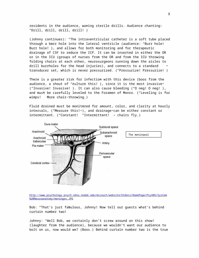
http://www.psychology.psych.ndsu.nodak.edu/mccourt/website/htdocs/HomePage/Psy486/System%20Neuroanatomy/meninges.JPG
Bob: “That’s just fabulous, Johnny! Now tell our guests what’s behind curtain number two!”
Johnny: “Well Bob, we certainly don’t screw around on this show! (laughter from the audience), because we wouldn’t want our audience to bolt on us, now would we? (Boos.) Behind curtain number two is the true screw of the bunch, the second choice of champions after the ventriculostomy drain, the one we all want to thread our way toward, the (b) - subarachnoid screw!” (The crowd goes wild!)
7
The meningeal layers.

The subarachnoid screw (or “bolt”) is considered the second choice of devices placed by neurosurgeons for monitoring ICP. They are relatively easy to install, but their accuracy is apparently significantly less than the more direct ventriculostomy drain.
(In a soft, rapid voice: “Members of the neurology and neurosurgery departments and their families are not eligible. Void where prohibited by law. Your mileage may vary.”)
Anyone need more of Bob and Johnny? Didn’t think so – me neither.
c- Fiberoptic Monitors: Pretty much what they sound like, I guess. The fiberoptic device has a pressure sensor at the tip, and it can be placed into the ventricle, the subarachnoid space, etc. I think we got one of these a while back and it was hooked up to some kind of neat self-contained monitoring device instead of using our usual transducer-to-monitor setup. Very cool.
An update: apparently fiberoptic monitors don’t have to be leveled and recalibrated – the transducer is built into the tip of the device, and gets calibrated once just before insertion.
d- Epidural sensor: this device is less invasive – I’m not sure we’ve ever seen one. CSF can’t be removed through this one.
e- Noninvasive ICP monitoring: This is the one I want. It turns out that NASA is working on a monitor that doesn’t require drilling. (McCoy: “ You mean you’re actually going to drill into that man’s head!? Is this the Middle Ages?”) Apparently the skull moves a bit, hard as it may be, and the fluctuations can be measured, etc. Here’s a reference:
http://nesb.larc.nasa.gov/NNWG/VOL8.2/TASKS/ARC/arc82_1.html
12- How long can it stay in?
We had a neuro ICU nurse come down recently to take a look at the subarachnoid bolt that one of our patients had – he’d fallen off the train platform onto the tracks and hit his head. He had an impressive tox screen too – anyway, she said these things usually stay in for about two weeks.
13- How do I take care of it?
The nurse told us that the site itself is dressed the same way a central line is, every four days unless the dressing gets gnarly. You also have to make sure that the system is patent – it never gets flushed into the patient, but sometimes gets flushed “backwards”, towards the transducer. I never did it, so I really need to ask around and find out what that means; when in doubt, I don’t do anything to one of these devices without getting the specialty nurses down to show us how.
Ventriculostomies:
8
Levelling: make sure that the transducer is levelled properly. The patient above had the fiberoptic device in – no leveling - but we had someone else a while back who had an intraventricular catheter that had to be levelled just so. It also had a drainage bag arrangement that had to be at the proper level. According to the NIH the patient should be consistently head up at 30-45 degrees for the measurement. So the patient has to be up at the right angle, the transducer has to be level, the bag has to be level, the whole thing is complicated. The transducer is supposed to be leveled at the part of the patient’s face that corresponds to the Foramen of Monro – a document at the NIH website says this should be the outer canthus of the eye.
http://www.cc.nih.gov/ccmd/pdf_doc/Clinical%20Monitoring/04-Intracranial%20Pressure%20Mo.pdf
Another sources says the level should be halfway between the outer canthus of the eye and the tragus of the ear.
The point that is always stressed: just as we do with PA-lines and the like, the transducer must always be leveled to the same point on the patient. So pick one, mark it, and stick with it.
Drainage: with a ventriculostomy, there need to be specific orders for the height of the drainage bag. The bag has a scale of cm on the side, and it has to be hung at just the right point. The way I understand the source text that I used, the height of the bag relative to the patient’s head determines whether CSF is going to flow outwards or not. Too high – won’t flow out. Too low, and too much flows out. Just right – the fluid will only drain if the pressure in the head is above the prescribed limit. There should be orders for specific ICP numbers that will be the “threshold” for drainage.
We also had to measure and record the hourly drainage, and check the waveform to make sure it was still clear. If not, the system might need backflushing towards the transducer. What would you do if the drain suddenly stopped draining?
An apparently important point: the system has to be filled with normal saline that has no bacteriostatic preservative in it, which is not the usual stuff we keep at hand.
Subarachnoid bolts:
9
The tragus. (Uh – Jayne? Where exactly did Ray say she was going the last time she went to New Hampshire?)

These are a lot less invasive than the ventriculostomies, and it’s usually one of the fiberoptic gadgets that gets put through them, so there’s none of the levelling and zeroing going on. A couple of things to watch for:
the fiberoptic cable itself is fragile, and can break if twisted, stretched, or tightly bent. apparently it’s possible for brain tissue to herniate upwards into the bolt if the ICP rises
uncontrolllably. (“Uh, Ralph? You want to come and take a look at this?”)
14- What is a bolt waveform supposed to look like?
Here’s one off the web - a normal tracing:
http://www.son.washington.edu/courses/nurs405/lecturedocs/icp.doc
This is a pretty clear trace. Each of the waves is made up of three smaller waves: P1, P2, and P3. It’s hard to see all three – here’s the same image blown up:
P1 P2 P3
I couldn’t really see the third one until I enlarged the image. Getting old. There does seem to be some respiratory variation – see how the whole wave system goes up and down like that?
Here’s another good one:
10

15- What is it not supposed to look like?
A tracing showing “badness”:
http://www.son.washington.edu/courses/nurs405/lecturedocs/icp.doc
Hey, how about putting a numeric scale on the strips, you guys? See how P2 is higher than her sister waves? P1 is supposed to be the highest. Also the entire amplitude of the wave is greater – that’s to say, it goes up and down more. It’s bigger - higher. Not a good thing – this means that overall the ICP is rising, right? – higher wave, higher pressure. The waves are interpreted according to the rules of the mystical cult of neurological astrology (which is how they probably look at balloon pumps) – the image reference says that the elevated P2 means that the intracranial compliance is probably decreasing, as the pressure is rising. Makes sense – pressure rises, things get less compliant, more rigid. (Now what the heck does that remind me of…? It’ll come to me.)
Here’s another bad one. Looks pretty high to me. It’s doing that P2 thing again too:
16- What if the waveform is dampened, or goes away?
It’s not supposed to get dampened or flattened out – this usually means that the transducer system is getting plugged up in some way. Check the system for air in the tubing; air doesn’t conduct pressure waves along the tube systeme the way water does. Check for leaks, disconnections, correct level, problems at the insertion site. Call the neuro nurses, or the neurosurg person on call (I’d think about calling both.)
11

A dampened trace:
17- How is a high ICP treated?
Drainage: The gold standard treatment is apparently the drainage of some of the CSF through a ventriculostomy device.
Positioning and Treatments:
Sit the patient up - It helps lower the ICP. There’s argument about this one – I guess there are studies pointing in different directions. (Why do I imagine two guys – always guys – getting really angry, waving their studies in each others’ faces, then rolling them up and smacking each other upside the head? Do the studies say they should get mannitol?)
Lie the patient down. It helps the cerebral perfusion. Hey, what do I know? They say sit ‘em up, I sit ‘em up; they say lie ‘em down, I lie ‘em down.
Don’ts in relation to positioning and treatment:
- don’t lift the patient’s legs up unnecessarily
- don’t position the patient on her side
- don’t flex her neck
- don’t repeatedly go at the patient with tasks to be done
Do’s:
- touch and massage the patient – but monitor the effects.
- let family visit and speak to the patient – but monitor the effects.
- try to get things done and then let the patient rest.
Apparently there are all sorts of studies that point in all sorts of directions about all of these maneuvers. Figure out what works best for your patient, then communicate the plan.
Hyperventilation, or not: I remember this one - this used to be a standard move, overbreathing the patient on the vent to get her pCO2 down to about 25; apparently not any more. The idea is
12

that lowering the pCO2 has the effect of lowering the ICP, but in a bad way – it works by constricting the cerebral blood vessels – a bad thing to do if perfusion is what you want. Apparently this maneuver only works for a short while anyway, and the ICP can pop back up suddenly if discontinued.
Mannitol: more arguments. Back in cave-woman days we were taught to practically keep a bag of mannitol in our hand, and that at the first sign of increasing ICP (what is it, you guys?), up it went. Now we give something like 100Gm IV q 4hours to keep the serum osm up – which means drying out their brain, right?
Let’s go really quickly over the drying-out-the-brain thing. Everyone remembers osmosis? (No, it’s not what you do after drinking too late at Chuck’s Pub, and adding extra salt to the margaritas does not help.)
Osmosis – start with a semi-permeable membrane, like a net. Some stuff will pass through it, some stuff won’t. Water molecules will.
Now take a bathtub. Pour some salt into the water, stir it up, dissolve it up good. Well. Properly. Yeah. Halfway along the length of the tub, divide the water with a film of the membrane, from the surface down to the bottom. Got that? Now dump some more salt into side – what does the water do?
When she was inventing chemistry, the Great Biomedical Engineer made a commandment unto the water: When thou art nearby to a semipermeable membrane, thou shalt goeth to where the more Dissolved Stuff is, and where the lesser of the Dissolved Stuff is, shalt thou not remaineth, except until thou hast tried to make the Stuff equally diluted on both sides of ye membrane.
The water moves – some of it, anyway, across the membrane, towards where there is more dissolved Stuff, trying to make the concentration equal on both sides. The water level on the dilute side of the membrane should go down. Where’s Bill Nye when I need him?
Now take a brain. See all the little brain cells? See how they have “water” in them?, and see how they’re surrounded by blood vessels, which also have “water” in them? The coverings of those cells are the semipermeable membranes this time, and the other side of the bathtub is the blood serum in all the zillions of little capillaries surrounding all the cells. See that? Make sense? Now if you dump something concentrated into one side – say, by infusing something really concentrated, oh, let’s just say by chance, hmm – how about mannitol? Mannitol is, as we say up here in MA, wicked hypersomotic.
“Osmotic-ness” is measured by a number – “osmolality” – “Yo Jeannie, check off an osm on that blood gas, okay?” The normal range is something like 280 – 300 mOsm/kg. High is more concentrated – either you’ve added more stuff, or you’ve removed some water. Lower is more dilute – told you not to drink all that water! Gatorade much better – water is hypo-osmotic, otherwise known as hypo-tonic. Gatorade is closer to iso-osmotic, or iso-tonic.
So okay – patient’s got brain cells swelling up – becoming edematous. You want to shrink those cells back down if you can, right? Give the patient an IV dose of that nice, hyperosmotic mannitol. What happens? – the serum osm goes way up – the goal is high, around 310, but not too high; keep it <320. The water molecules inside the brain cells say: “Yo! Time to cross the membrane towards that greater concentration thing over across there!” And so they do, according to the Engineer’s Design – each and every one of those little brain cells sends water out of itself, through its membrane, out into the surrounding blood vessels, where it stays, and mannitol having a diuretic effect, then gets peed out.
13

Result – the brain cells, losing some of their water, shrink down. All of them. And the tissue edema shrinks down. And the ICP goes down. And the brain avoids herniation. A life is saved. Woo-hoo!
(But you’d better know when to give it.)
! - A very important point goes here. Patients with cerebral edema issues should only get hypertonic or isotonic IV fluids. If hypotonic fluids were given, they would do the osmotic thing the wrong way, and go into the cells, making the edema worse. Examples we’ve seen of appropriate fluids are normal saline, Ringer’s lactate, albumin (5% or 25%? – probably 25%, since it’s more osmotic, hmm?)
Another point: even if you’re diuresing your patient with mannitol, the sources all say that it’s just as important to re-hydrate the patient, to keep her euvolemic, rather than total-body dry. You’re trying to shrink the brain and keep it perfused, all at the same time.
Steroids: apparently a big hairy no-no in head trauma or in either kind of stroke, (what are the two kinds? – this here is basic now, know what I’m sayin’?). I think they do still use them for brain mets – I remember when my mom got them…I think they helped, for a while. She missed out on Zofran. And grandchildren. Don’t smoke, you guys.
Here’s granddaughter # 1 holding one of our two stress advisors. (Girl takes the NCLEX this July. How the &^%! did that happen? I mean, she was getting on the kindergarten bus, what – last month? Two months, tops. She had the nicest little lunchbox. Sniff.)
Anticonvulsants: apparently meds like dilantin and tegretol are useful in treating seizures that occur soon after a brain injury, but preventing those seizures doesn’t seem to help in the ultimate outcome. I’m pretty sure that all of our acute neuro patients get dilantin-loaded – although most of our patients aren’t traumatic injuries; we get the subdural hematomas, it seems like. Do the same rules apply for bleeds and traumas?
Barbiturate “coma”: I want to say that the young female children of America are in a Barbie coma…maybe I shouldn’t. (Whoa! Brain wave! How about “Burr-hole Barbie”? I mean, girls grow up to neurosurgeons too, don’t they? Whoa! Another one!: “Oy mate, throw another Barbie on the… never mind.) Anyhow, the idea here is that the barbs (usually pentobarbital – seems like we’ve used phenobarbital too) have two effects: they lower the ICP, and they also lower the rates of the body’s metabolic processes – which should take some of the strain off the central processing unit while it tries to recover.
But – bad things can happen with barbs. One study compared the use of barbs against the use of mannitol in head injury (the one or the other only, I guess) – the barb people did much worse. The other thing is that the barbs produce systemic hypotension. With the recent emphasis on letting, or making these patients be a bit hypertensive, this would seem to be your basic Bad Thing. Not really sure when I saw them used last.
Other sedatives: the sources list ‘em all: opiates, benzos, propofol – even paralysis is on the list, apparently also with lowered metabolic demands in mind.
14

Cooling: Apparently a change of one degree in temperature produces something like a 7% decrease change in the overall metabolic demand. Cooling blankets work better under the patient. Don’t set it lower than 60 degrees. I find that making the patient nakey (we still watch Rugrats in our house), and covering him with a few carefully placed wet towels helps cool things down very well. Tepid water – they’ll get cool fast enough!
Oxygenation: Keep the sat greater than 95% - maintain good oxygen delivery to the brain.
Hypertensive therapy: this seems to be one of the more recent strategies in improving CBF – shrink the brain, keep them full, keep up the MAP, keep up the perfusion. Lately the MAP and SBP goal ranges have been rising on these patients. The texts say that pressors can be used – hey guys, most of those alpha pressors vasoconstrict, y’know! Dobutamine might be the way to go. They also say that pressures can get too high: a MAP of greater than 130 should probably be brought back down. Don’t want to pop anything in there…labetolol was cited as the drug to use in that case, along with oral calcium channel blockers. Hmm…not nipride?
15