identification of unknown photo-initiators in offset uv-inks and prints
TRANSCRIPT

Confidential 1 Bart Jansen
Identification of Unknown
Photo-initiators in offset UV-inks
and prints
Name; Bart Jansen
Student Number; 5846994
MSc. Chemistry, Analytical Sciences
University of Amsterdam
Daily Supervisor: Chris Geurts
Supervisor; dr. W. Th. Kok
Date; 6-Apr-12

Confidential 2 Bart Jansen
1 Abstract
More advertising is being personalized every day, digital printers are encounter problems
related to the use of offset printed materials (pre-printed). For better understanding of these
problems it is necessary to better understand the formulation of these offset UV-inks.
In this study a method has been developed for the identification of photo-initiators in offset UV
inks and in printed materials. It is concluded that methylene chloride is the best extraction
solvent for the different photo initiators in the different inks. Analysis using a DB-5MS column
gives good separation for all the initiators identified.
For the identification of the unknown initiators a combination of normal electron ionization (EI)
and chemical ionization (CI) has been used. Most of the photo initiators do not give any
information about the molecular mass, chemical ionization is being used. It is concluded that a
combination of CH4-CI and iC4H10-CI gives valuable information towards the identity of the
unknown photo-initiator. Many initiators have methoxy or acids groups which become visible
using CH4-CI, which results in the loss of water or methanol. For determination of the
molecular ion iC4H10-CI results in het most abundant molecular signal. In comparison to CH4-
CI, iC4H10-CI gives simple chromatogram without additional signals from addition reactions.
In total 11 different photo-initiators have been identified in 29 different inks from 5 different
suppliers. It was found that different photo initiators were used in different colours from a single
brand of ink.
In the final step this method is being used on printed media, from which the extracted photo
initiators are being compared to the pure ink. A print with 4 different inks and two different cure
settings has been used. In all the prints all the photo initiators could be identified. Showing that
the developed working method can also be used for identification of photo initiators when
printed, regardless of curing degree or offset printer.

Confidential 3 Bart Jansen
Table of contents
1 ABSTRACT.............................................................................................. 2
2 INTRODUCTION ...................................................................................... 5
3 PRINCIPLE .............................................................................................. 6
3.1 UV-inks...........................................................................................................................................6
3.2 Gas Chromatography....................................................................................................................9
3.3 Mass Spectrometry......................................................................................................................10 3.3.1 Electron Ionization .................................................................................................................10 3.3.2 Chemical Ionization ...............................................................................................................11
4 EXPERIMENTAL.................................................................................... 14
4.1 Materials.......................................................................................................................................14
4.2 Sample preparation .....................................................................................................................14 4.2.1 Inks .........................................................................................................................................14 4.2.2 Pre-printed media ..................................................................................................................14
4.3 Apparatus and method................................................................................................................14 4.3.1 GC-MS (EI) ............................................................................................................................14 4.3.2 GC-MS (CI) ............................................................................................................................14
5 RESULTS AND DISCUSSION............................................................... 15
5.1 Method optimization ...................................................................................................................15 5.1.1 Column ...................................................................................................................................15 5.1.2 Solvent ....................................................................................................................................17 5.1.3 Chemical Ionization ...............................................................................................................19 5.1.4 Conclusions method optimization...........................................................................................22
5.2 Inks ...............................................................................................................................................23 5.2.1 Ink A .......................................................................................................................................23 5.2.2 Ink B .......................................................................................................................................31 5.2.3 Ink E .......................................................................................................................................35 5.2.4 Conclusions ink analysis ........................................................................................................36
5.3 Pre-printed media........................................................................................................................37 5.3.1 Comparison between print and ink.........................................................................................38 5.3.2 Comparison between different prints .....................................................................................39 5.3.3 Sub Conclusions .....................................................................................................................40
6 CONCLUSIONS ..................................................................................... 41
7 REFERENCES ....................................................................................... 42
8 APPENDIX ............................................................................................. 43

Confidential 4 Bart Jansen
8.1 Overview of names and structures.............................................................................................43
8.2 Overview of Spectra ....................................................................................................................45 8.2.1 Irgacure 184 ...........................................................................................................................45 8.2.2 Benzophenone.........................................................................................................................46 8.2.3 Quantacure EDB ....................................................................................................................48 8.2.4 DETX......................................................................................................................................49 8.2.5 Quantacure ITX......................................................................................................................51 8.2.6 Irgacure 907 ...........................................................................................................................52 8.2.7 Irgacure 651 / Photocure 51 ..................................................................................................54 8.2.8 Quantacure BMS ....................................................................................................................55 8.2.9 Escalol 507.............................................................................................................................57 8.2.10 Methyl-Benzoylbenzoate .....................................................................................................58 8.2.11 4-Phenyl-Benzophenone .....................................................................................................60
8.3 Column .........................................................................................................................................62
8.4 Ink A-1..........................................................................................................................................64 8.4.1 Yellow .....................................................................................................................................64 8.4.2 Red032....................................................................................................................................64
8.5 IR analysis on Pigments ..............................................................................................................67 8.5.1 Yellow .....................................................................................................................................67 8.5.2 Red 032...................................................................................................................................68
8.6 Overview of all inks .....................................................................................................................69
8.7 Spectra from pre-print................................................................................................................71 8.7.1 Photocure 51 ..........................................................................................................................71 8.7.2 Methyl-benzoylbenzoate .........................................................................................................71 8.7.3 Escalol 507.............................................................................................................................72 8.7.4 Irgacure 907 ...........................................................................................................................72 8.7.5 Quantacure ITX......................................................................................................................73 8.7.6 4-phenyl-benzophenone..........................................................................................................73
8.8 Comparison Pre-print and ink ...................................................................................................74

Confidential 5 Bart Jansen
2 Introduction
More mail, like advertising and personal bills, is being personalized today than 10 years ago.
To create this personal mail, a digital process is needed like printing. However digital colour
printing is more expensive in comparison to offset printing. Therefore the market in which both
techniques are combined is growing. First the colour print is made using offset printing (called
pre-printed), after which it is personalized using a digital printer.
For offset printing there are two main types of inks available, oxidative drying inks and UV
curable inks. In this report the composition of these UV inks is being investigated. The main
components in UV inks are reactive acrylates (monomers) and initiators.
There are two main applications areas in which UV offset inks are being used. One of the main
applications of UV inks is for offset printing off food packaging (i.e. milk packaging), because of
the relative high quality and low residue of these prints, in comparison to oxidative drying inks.
Some of these components are being labelled as having a health risk. Within Europe there are
regulations towards the use of these initiators and the maximal concentration present in the
food is being regulated1. Recent awareness of photo-initiators (especially ITX) in food by the
European Food Safety Authority (EFSA)2, has led to an increase in the analysis of photo-
initiators in food.
The use of UV inks in can also give problems when a digital printer is being used to
personalize offset printed media. Most printers use a warm fusing step in which the toner is
fused to the media. During this process remaining components deriving from the UV inks are
transferred to the digital printing system. When these compounds deposit on critical parts of a
printer, these can result in a service call and repairs. Therefore the composition of different
offset inks used is of interest for the printing industry to better understand the mechanisms
responsible for these failures.
In this report, the photo-initiators used in different UV inks are being identified. The difference
in the type of photo-initiators used by different manufactures is being investigated, as well as
the possible differences between colours of the same ink.
In literature different LC-MS methods3,4,5,6,7,8
are being described, in which the presence of
these initiators is measured. Most of these methods need a clean-up before analysis like; solid
phase, or liquid liquid extraction. Similar methods are being described using GC9 10
, in which
also the samples are pre-treated using SPE. Only limited amount of effort has been taken in
the analysis of the packaging. But still a clean up and purification of the sample is required6,11
.
One of the goals of this research is to develop a simple method without extensive cleanup for
direct analysis.
All described methods are based on pre-selected photo initiators. In this research the focus is
on the identification of the photo-initiators. A simple gas chromatographic method combined
with mass spectrometric detection is being developed for the identification of photo-initiators in
the ink. Because of the reactive nature of photo initiators, EI ionization mass spectra usually
only gives limited information about the molecular mass. For confirmation about the identity
also chemical ionization is used.
1 Schweizerische Eidgenossenschaft, Verordnung des EDI über Bedarfsgegenstände (SR 817.023.21) vom 23.
November 2005, Anhang 6 2 R. Anton, S. Barlow, D. Boskou, L. Castle, R. Crebelli, W. Dekant, K.H. Engel, S. Forsythe, W. Grunow, M.
Heinonen, J.C. Larsen, C. Leclercq, W. Mennes, M.R. Milana, I. Pratt, I. Rientjes, K. Stevensson, P. Tobback, F.
Toldrá, EFSA J. 293 (2005) 1. 3 Cuilian Sun, Sheot Harn Chan, Dan Lu, Hui Min Wendy Lee, Bosco Chen Bloodworth, Journal of Chromaotgraphy A,
1143 (2007) 162-167 4 Ana Gil-Vergara, Christina Blasco, Yolanda Picó, Anal. Bioanal. Chem., 389 (2007) 605-617
5 H. Gallart-Ayala, E. Moyano, M.T. Galceran, Journal of Chromatography A, 1208 (2008) 182-188
6 Gianni Sagratini, Giovanni Caprioli, Gloria Cristalli, Dario Giardiná, Massimo Ricciutelli, Rosaria Volpini, Yanting Zuo,
Sauro Vittori, Journal of Chromatography A, 1194 (2008) 213-220 7 H. Gallart-Ayala, O. Núñez, E. Moyano, M.T. Galceran, Journal of Chromatography A, 1218 (2011) 459-466
8 R. Bagnati, G. Bianchi, E. Marangon, E. Zuccato, R. Franelli, E. Davoli, Rpid. Comm. Mass. Spec. 21 (2007 1998
9 Gianna Allegrone, Ilaria Tamaro, Shara Spinardi, Giorgio Grosa, Journal of Chromatography A, 1214 (2008) 128-133
10 E. Van Hoeck, T. De Schaetzen, C. Pacquet, F. Bolle, L. Boxus, J. Van Loco, Analytica Chimica Acta, 663 (2010)
55-59 11
N. Negreire, I. Rodríguez, E. Rubí, R. Cela, Talanta, 82 (2010) 269-303

Confidential 6 Bart Jansen
3 Principle
3.1 UV-inks
For the hardening of a UV-curable offset ink a mixture of different photo-initiators are being
used. An ink usually consists of multiple photo-initiators, because of the different lamps used
by different offset printers. The effective absorption spectrum is different for each initiator13
.
Because of the use of different pigments (colours), other photo-initiators are needed which are
effective in a different spectral region than the pigment.
Photo-initiators can be divided into two main groups according to their working principle12
.
Norrish type I: which are initiators who decay after being exposed to UV light, through
homogenic splitting into radicals, and are build into the polymer.
Norrish type II: These initiators need a synergist (hydrogen donator) to produce radicals, and
the initiator does not react into the polymer.
The type I initiators are the most reactive, because of the use of a benzoyl radical. The
disadvantage of some of these initiators is their colour, which turns yellow after reacting.
An example of a Norrish type one photo-initiator is Irgacure 651 (also know as photocure 51)
as shown in Figure 3.1.1. The primary reaction is the Norrish type I splitting, in which a benzoyl
and a benzyl radical are being formed. The benzyl radical can split into a highly reactive
methyl radical and the stable methyl benzoate. This reaction is temperature depended, and is
de main mechanism at room temperature but will not happen at 0°C.
O
CH3
O
CH3
O
C
O
C
O
CH3
O
CH3
+
OCH3
O
+CH3
Initiation
Initiation
Figure 3.1.1. Example of Norrish type I initiation of Irgacure 651 (Photocure 51)
Other examples of the Norrish type I photo-initiators are the α-amino alkylphenonen, such as
Irgacure 369 and Irgacure 907, see figure 3.1.3. Because of the benzoyl group the absorption
spectra of these initiators is shifted13
(towards the 300-322 nm) see Figure 3.1.2, while smaller
photo-initiators have their absorption well below the 300nm (i.e. benzophenone, 240-250 nm).
This is an advantage in pigmented inks, which partial block the UV light.
12
IR 97322.022, Literatuurstudie naar foto-initiatoren voor UV uitharding, J van de Reek 13
Ciba, Photoinitiators for UV curing, key product selection guide 2003, g-48/2003 October

Confidential 7 Bart Jansen
Figure 3.1.2. Absorption spectra of some photo initiators.
CH3
S
O
CH3
CH3
N
O
Irgacure 907 CH3
S
C
O
C
CH3
CH3
N
O
+
Figure 3.1.3. Example of Irgacure 907 initiation.
The Norrish-type II photo initiators consist of a benzophenone or thioxantone group in
combination with a hydrogen donor (synergist). The reaction mechanism consists of 4 steps:
1. Absorption of UV light, crating a single state electron at the keton.
2. Transition from the single state to the triple state (inter system crossing).
3. Creating of radical�s trough hydrogen splitting from a synergist or hydrogen donor
(usually a tert-amine).
4. Initiating through addition of the radicals with a monomer.
For the working of a type II photo-initiator the transfer of a proton is needed14
. The active
protons are located at the α-position of the tert-amine or alcohol.
This results in ketyl radical and an α-amino-alkylradical. The ketyl radicals are not used in the
initiaton, and are being terminated by recombination or by transition back into the initial state.
The α-amino-alkylradicals are used in the initiation as shown in Figure 3.1.4.
One other problem when using radical initiation is oxygen inhibition. The use of Norrish type II
initiators, are less sensitive towards oxygen inhibition. Therefore usually a mixture is used in
the ink receipt.
14
H.F. Gruber/ Photo initiators for free radical polymerization / Prog. Polym. Sci. / Vol. 17 / 953-1044 / 1992

Confidential 8 Bart Jansen
+
O
O
electron transfer
C
O-
proton transfer
N
OH
OH CH
OH
C
OH
N
OH
OH
OH
N
OH
OH
OH
N+
OH
OH
OHH
initiation
+
Figure 3.1.4. Example of the Norrish type II initiation
A second example of the Norrish-Type II initiator is the reaction of benzophenone with
Michler�s keton (4,4-bis-(dimethylamino)benzophenone), and is one of the most reactive
initiators. Because of combination of the benzophenone with the amino groups, a very reactive
system is created, in comparison to the normal amine.
O
O
N N
CH3
CH3
CH3
CH3
C
OH
O
N N
CH3CH3
CH3 CH2+
Figure 3.1.5. Example of Norrish type II initiation
However the Michler�s keton is prohibited, because of its carcinogenic properties. As substitute
other initiators are being used with similar properties. As replacement a photo initiator with only
one dimethyl amino group 4-(dimethylamino)benzophenone (DMAB), or with the methyl
groups replaced by ethyl groups 4,4-bis-(diethylamino)benzophenone (BDEAB), is being used.

Confidential 9 Bart Jansen
O
N
CH3
CH3
O
N NCH3
CH3 CH3
CH3
4-(dimethylamino)benzophenone (DMAB)
4,4-bis-(diethylamino)benzophenone (BDEAB)
Figure 3.1.6. Structure of DMAB and BDEAB
3.2 Gas Chromatography
The most common GC15
analysis starts with the injection of a volatile sample into a hot injector
for evaporation, and transfer to the column (there are many different injection techniques for
GC analysis available, which shall not being explained in this thesis). The components are
separated based on their difference in boiling point and interaction with the stationary phase of
the column giving the components retention. At the end of the column the separated
components are detected using a detector. In this research a single quadrupole mass detector
has been used.
Column type
The purpose of the column is to give the components with similar boiling points different
retention, resulting into separation. The most simple is the HP-1 (or DB-1) column, of which
the packing material is made of dimethylpolysiloxane.
One of the most widely used columns is HP-5 column, in which the packing material is also
made of dimethylpolysiloxane of which 5% of the methyl groups have been replaced by phenyl
groups. To reduce bleeding of the column, the methyl group situated at the other side of the
phenyl group is replaced by a second phenyl group. This makes the polymer more ridged and
lowers the possibility to create cyclic siloxanes (which mainly causes the bleeding).
15
M.C. McMaster, GC/MS A Pratical User�s Guide 2nd
edition, Wiley Interscience, 2008, pag. 25-28

Confidential 10 Bart Jansen
3.3 Mass Spectrometry
3.3.1 Electron Ionization
The most commonly used ionization technique in GC-MS is EI (Electron Ionization)16
. Using
this technique an electron is knocked of a molecule, leaving behind a molecular ion with a
positive charge. This ion is directed towards the entrance of the mass analyser by a repellor.
The electrons are produced by a filament, which are repelled with 70 eV to produce a stream
of electrons. The energy of the electrons is high enough not only to produce molecular ion, but
also to produce fragments of these ionized molecules. The fragmentation pattern of the
sample ions formed is related to the energy by which the electrons are being repelled (70eV),
resulting in a specific fingerprint of ions. As result it is possible to create commercially available
libraries. Because EI is a relatively hard ionization technique often the molecular ion is not
detected.
3.3.1.1 Fragmentation using EI
If sufficiently excited the M+�
ion can form a variety of product ions depending on their stability
and formation energy by which rearrangement can occur17
.
During fragmentation using electron ionization the most important factor for ion formation is the
stability of the product ion. The stability of the ion can be increased by separating the charge
over the ion. Two of the main mechanism are electron sharing (in which the charge is devided
over two atoms by a nonbonding orbital i.e carbonyl group) and resonance stabilization (in
which the charge is located over the π-electrons i.e. benzyl cation).
To most simplistic way to predict the fragmentation is to assume that the reactions are initiated
at the favoured site of the unpaired electron. The most favored molecular ion is arising from
the loss of an electron with the lowest energy, which is generally σ- < π- < n-electrons.
Sigma (σ) : RH2C:CH2R� -> RH2C+�
CH2R�
Pi (π) : RHC::CHR� -> RHC:+�
CHR�
Non-bonding (n) : R-O-R -> R-O+�
-R
Cleavage of a single bond results into an odd-electron (OE+�
) molecular ion should result into
an even-electron fragment ion (EE+) and the loss of a neutral radical fragment. The formation
of each ion should be equally based on coincidence.
CH3CH2+�
CH3 -> CH3CH2+ + �CH3
-> CH3CH2� + CH3+
Formation of the OE+ ion from an EE
+� ions involve energetically favourable rearrangement
and charge retention.
CH3CH2-O+=CH2 -> CH3CH2
+ + O=CH2 (charge migration)
-> CH2=CH2 + HO+=CH2 (rearrangement, charge retention)
Reactions initiated at the radical sites from free electron pair, arises from their tendency for
electron pairing to from a new bond with an adjacted atom. This is followed by the homolytic
cleavage of the second bond to that α-atom, and is therefore called α-cleavage.
Saturated site: R─CR2── Y+�─R
R� + CR2=Y+─R
Unsaturated hetero atom: R─CR══ Y+�
R� + CR≡Y
+
An unpaired electron can also be donated from an adjacent atom through space, so called β-
cleavage. This is one of the most familiarly rearrangements and is also called the McLafferty
16
M.C. McMaster, GC/MS A Practical User�s Guide 2nd
edition, Wiley Interscience, 2008, 40-42 17
Interpretation of Mass Spectra, 4th edition, F.W. McLafferty / F. Tureček, Universal science books, 1993, 52-83

Confidential 11 Bart Jansen
rearrangement. For components containing an unsaturated carbonyl group, the unpaired
electron can be donated from a hydrogen atom to form a new bond. As part of the driving force
is the strong O-H bond, followed by the rearrangement which is favoured by a six-membered-
ring transition state. For this rearrangement to occur the third position from the carbonyl group
should consist of a hydrogen atom (see Figure 3.3.1).
R
H
O+
R
1
23
R
H
O+
R
1
2
3 McLafferty
R CH2
O+
H
+CH2
R
Figure 3.3.1 McLafferty trough γ-H rearrangement with a carbonyl group.
An second example is the rearrangement of the fragmentation of Escalol 507, resulting in the
stable m/z 165.
O+
O
N
CH3
CH3
CH3
CH3
H
N
CH3
CH3
O+
O
H
McLaffertyM
+ = 277
O+
O
CN
CH3
CH3
CH3
CH3
H
+
CH2
CH3
CH3
m/z = 165
Figure 3.3.2 McLafferty rearrangement on Escalol 507
3.3.2 Chemical Ionization
Using Chemical ionization (CI) a large amount of reagent gas is introduced into the ion source.
Since there is much more reagent gas than sample, most of the emitted electrons ionize the
reagent gas forming reagent ions instead of the sample molecules. These reagent ions react
with each other and with the sample in various ways. The energy of the reagent gas is
transferred to the sample by transferring a proton. The amount of energy transferred is
therefore based on the difference in the proton affinity between the reagent gas and the
sample.
Many different chemical ionization reagent gasses have been investigated, with each their own
specific purposes. The energy of the reactant ions are based on their proton affinities.

Confidential 12 Bart Jansen
Table 3.3.1. Overview of some reagent gases18
.
Reagent gases Proton affinities
[kcal/mol]
Reactant ions
H2 423 H3+
CH4 551 CH5+ and C2H5
+
(CH3)2CHCH3 816 C4H9+
CH3OH 761 CH3OH2+ and (CH3OH)nH
+
NH3 854 NH4+ and (NH3)nH
+
There are four basic ionization processes that take place during (positive) CI.
Proton Transfer
Hydride Abstraction
Addition
Charge Exchange
Charge exchange ionization processes are not often used, and require noble gasses as
reagent gas. These shall not be used in this study, and shall therefore not be further explained.
Proton Transfer
The majority of the ionization occurs by proton transfer to species of a higher proton affinity
(Brönsted acid reagent system)18
. The proton affinity is the energy change when a proton is
added to neutral molecule to form a protonated cation. AHHA
The proton affinity is defined as:
)()()()298( MHHHHAHHPA fffr
When a molecule with a higher affinity is found, the proton will be transferred. MHAMAH
Therefore the choice of the reagent gases is of influence on the amount of protonated
molecular ions formed, especially for instable molecules.
One of the most used reagent gases is methane. When methane19
is ionized using electron
impact (EI) three major ions are formed; CH4+.
, CH3+ and CH2
+. This result in three major ions,
CH5+, C2H5
+, and C3H5
+, as shown below.
25243
3544
234
44
22
2
HHCCHCH
CHCHCHCH
HCHCH
eCHeCH
A second frequently used reagent gas is isobutane, with a higher proton affinity in comparison
to methane. Isobutane yields mainly C4H9+ as reagent ion with a minor yield of C3H3
+ (3%), as
a result isobutane generally yield more simple spectra in comparison to methane. Using
isobutane as reagent gas the main ionization shall occur trough proton transfer. In comparison
to methane almost non addition reaction shall occur.
294104
104104
22 HHCHCi
HCiHCi
18
A.G. Harrison, Chemical Ionization Mass Spectrometry, CRC press, 1983 19
F.G. Kitson, B.S. Larsen, C.N. McEwen, Gas Chromatography and Mass Spectrometry a practical guide, 1996,
Academic Press Inc.

Confidential 13 Bart Jansen
Hydride abstraction
The created reactant ions using CI can have relative high hydride-ion (H-) affinities.
In methane-CI both CH5+ and C2H5
+ are capable of hydride extraction, which results in the loss
of H- according to the general reaction:
245 HCHHMMCH
This reaction is often seen at long chain alkanes. When using CH4-CI it is possible both MH+
by proton transfer and M-H+ by hydride extraction can occur in one spectrum (i.e. methyl-
esters).
Addition
Besides proton transfer and hydride abstraction also addition reactions can occur. Often
reagents gasses are reactive enough to combine with the analyte molecules by condensation
or association (addition reactions) in so called adduct ions. These adduct ions are often seen
when using Methane as reagent gas, forming [M+C2H5]+ and [M+C3H5]
+ ions.
Addition reactions are also very important when using ammonia as reagent gas, as ammonia
has a very high proton affinity and only few organic compounds will undergo proton transfer as
main ionization step. Using ammonia as reagent gas the mayor ions formed are NH4+,
[NH4NH3]+, and [NH4(NH3)2]
+. This will give rise to an intense [M+NH4]
+ peak by the addition
reaction.
Because of the relative small difference in energy between the reagent gas and the sample, CI
is a so called soft ionization technique which results in high abundant molecular ion. However
still fragmentation and rearrangement can occur, driven by the stability of the formed product.
One of the mechanisms seen is hydrogen shift, especially when oxygen of nitrogen groups are
present in the analyte. A very often seen rearrangement is the loss of water in the analysis of
alcohols, aldehydes, ketones, and acids, see Figure 3.3.3.
The mechanism for alcohols is driven by the relative instable MOH2+ ion for alcohols higher
than C4. For acids higher than C4 this is usually followed by the loss of CO, strongly seen with
aromatic acids like benzoic acid or phenyl acetic acid in which the ion is stabilized in the
aromatic ring.
OH
O
CH5
+OH
O+
H
-H2O
O+
O+
-CO
CH2
+
CH+
m/z = 91
m/z = 119
Figure 3.3.3 Example of the chemical ionization using CH4 reagent gas

Confidential 14 Bart Jansen
4 Experimental
4.1 Materials
All solvents used are purchased from Merck; Methylene Chloride (>99.8%), 2-butanone
(>99.9%), Cyclohexane (>99.5%), Methanol, (>99.5%). For chemical ionization gasses are
used from Air Products with research quality.
All inks are obtained through different ink suppliers. Because of the confidential nature in the
collaboration of Océ Technologies and offset ink suppliers the exact name and type of the
analyzed inks can not be published.
4.2 Sample preparation
4.2.1 Inks
Extraction of the inks was performed in a 25ml vial with screw silicon caps coated with PFTE.
About 100mg of the ink is extracted using about 5g of extraction solvent. For extraction the
vials are shaken for 2 hours using an automated vortex shaker from Scientific Industries. To
remove the pigments from the ink the extract was transferred in 4 ml vial with screw silicon
caps coated with PFTE, and centrifuged for 15 minutes at 5000G using a Microcentrifuge 154
from the firm OLE DICH. The clear supernatant was transferred in a 2 ml GC vial for analysis.
4.2.2 Pre-printed media
The pre-printed media were cut using a punch of 5cm x 2 cm. The punched out piece of paper
were cut into small strokes of about 2mm*20mm and transferred into a 4 ml vials with screw
silicon caps coated with PFTE. The papers were extracted for 2 hours in 1.5 g of MC. To
perform blank references a piece of unprinted paper was extracted according to the same
procedure. The vials are centrifuged for 15 minutes after which the supernatant was
transferred in a 2ml GC vial.
4.3 Apparatus and method
4.3.1 GC-MS (EI)
All experiments have been conducted using an Agilent 7890A GC with an Agilent 5975C EI/CI
MSD detector. The columns used are both from Agilent, DB-1MS, 128-0112, 12m x 0.2 mm x
0.33µm, and DB-5MS, 122-5532, 30m x 0.25 mm x 0.25µm. For analysis 1.0 µl of sample is
injected using a 5 µl syringe into a glass liner filled with glasswool. Sample injections are
performed using a split of 25:1. The GC inlet temperature is 325ºC. The oven temperature is
programmed from 80ºC, held for 2 minutes, raised with a rate of 8ºC/min to 325ºC, held for 5
minutes. The total duration of the analysis is 37.6 minutes. The MSD transfer line is set at a
temperature of 325ºC. The ion source is set at 230ºC and the quad at 150ºC. Before analysis
the MSD is automatically tuned using PFTBA with an �atune�. The MS data is collected in scan
mode from m/z 14 to 600, at 70 eV electron energy. The solvent delay is set to 3 minutes.
4.3.2 GC-MS (CI)
For CI analysis the same Agilent 7890A GC and 5975C MSD has been used. For CI analysis
only the Agilent DB-5MS, 122-5532, 30m x 0.25 mm x 0.25µm column has been used. For CI
analysis the same GC instrument parameters are as for EI analysis. Ionization has been
performed using a high pressure CI ion source. The MS Source temperature is set at 250 ºC,
and the Quad at 150ºC. Before CH4-CI analysis the MSD has been automatically tuned using
PFTBA with a �pcich4.u� tune. The gas flow is set at 14%. The MS data is collected in scan
mode from m/z 14 to 600, at 40 eV electron energy. The solvent delay is set to 1.5 minutes.
Performing iC4H10-CI the same GC instrument parameters are used as for CI analysis using
CH4. Before iC4H10-CI analysis the MSD has been automatically tuned in CH4-CI mode using

Confidential 15 Bart Jansen
PFTBA with a �pcich4.u� tune. The MSD is switched to iC4H10-CI mode, and the gas-flow is
optimized using the reagent ions m/z 43 and m/z 57, the tune file is saved as �PCIC4H10.U�.
The gas flow for iso-butane has been optimized and set on 13% (see § 5.1.3.).
5 Results and discussion
5.1 Method optimization
Before the starting with the identification of the photo initiators in the different inks, the working
method has been optimized.
The optimization consists of:
1. Choice of column
2. Choice of Solvent
3. Optimization of the CI parameters
5.1.1 Column
In the first step, two different column types have been investigated, both from J&W Scientific.
The column which is most frequently used is the HP-5MS6,11
column. This column shall be
compared to normal DB-1MS column.
1. DB-1MS 12m x 0.2mm x 0.33µm
2. DB-5MS 30m x 0.25mm x 0.25µm
Three different reflex blue inks have been used in the comparison.
1. Ink A-1
2. Ink B-1
3. Ink B-3
The Samples have been dissolved in methyl ethyl keton (MEK), with a concentration of 20-30
mg/g. using identical GC parameters both columns are being evaluated.
A comparison between two types of columns has been made, by differences seen in the peak
shape, and the retention of the components. Because the DB-5MS column is 30m while the
HP-1MS is only 12m long, the retention times are a higher using the DB-5MS column, as
shown in Figure 5.1.1.
Figure 5.1.1. Ink A-1 measured on the HP-1 column

Confidential 16 Bart Jansen
The components analyzed using the HP-1 column clearly shown non-symmetric peak shape,
and most peaks show an extensive tailing. The in red en bleu highlighted parts are given in
Figure 5.1.2 and Figure 5.1.3.
Figure 5.1.2. Ink A-1 measured on the DB-5 column
When the same sample is being analyzed using a DB-5 column, the peak shape has
significantly improved, see Figure 5.1.2. Possibly there is more column interaction on the HP-5
column resulting in a more stable chromatography, as most of the initiators have aromatic
groups.
Figure 5.1.3. Comparison between some components.

Confidential 17 Bart Jansen
The DB-5 column consists of 5% phenyl groups, which results in more retention for some of
the components. The
In the chromatogram using the DB-5 column the Irgacure 907 has more retention than Escalol
507, while on the HP-1 column the Escalol has more retention, (see Figure 5.1.3). This is most
likely caused by the double ring structure in Irgacure 907 (see Figure 5.1.4), which has more
column interaction with the DB-5 stationary phase.
Figure 5.1.4. Structure of Escalol 507 and Irgacure 907.
Similar results are seen with ITX and 4-phenyl-benzophenone, in relation to an acrylate
present in the chromatogram (see Figure 5.1.3). Using a HP-1 column the acrylate is has got
the more retention time, however on the DB-5 column both initiators have more retention in
comparison to the acrylate. This extra retention is caused by the interaction between the
aromatic groups of the initiators with the phenyl groups of the stationary phase. This
interaction results in better chromatographic separation, and therefore better peak shape.
The DB-5 column is therefore the preferred column for the analysis and therefore used in this
study. More examples are given in the appendix 8.3.
S
CH3
CH3
ITX
C17H18S M = 254
O
4-Phenyl-Benzophenone
C19H14O M = 258
Figure 5.1.5. Structure of ITX and �phenyl-benzophenone.
5.1.2 Solvent
The inks will be analyzed by simple direct dissolving of the ink and direct analysis. Therefore
four different solvents have been compared.
1. Cyclohexane (CH)
2. Methanol (MeOH)
3. Methylene Chloride (MC)
4. 2-butanone (MEK)
To ensure that enough different initiators are being evaluated, four different reflex blue inks
have been used in the comparison.
1. Ink A-1
2. Ink B-1
3. Ink B-3
4. Ink C-1
O
O
N
CH3
CH3
CH3 CH3
Escalol 507
C17H27NO2
CH3
S
O
CH3
CH3
N
O
Irgacure 907
C15H21NO2S M = 279

Confidential 18 Bart Jansen
The solvents are being compared by calculating the response of each photo-initiator in the
different solvents.
Table 5.1.1. Calculated response [Area*10E6/mg] in the 4 solvents
CH MeOH MC MEK
Ink Ink B-1
Benzophenone 5,6 11,0 22,1 12,5
Irgacure 184 3,6 10,5 19,7 10,8
EDB 3,4 9,0 18,5 9,7
Photocure 51 3,8 11,0 21,9 12,2
Ink Ink C-1
4-methyl-benzophenone 3,9 11,8 29,4 18,2
Acrylate [m/z = 55/113] 3,1 23,8 56,0 30,9
Ink Ink A-1
Photocure 51 0,6 7,9 16,5 9,5
Escalol 507 1,0 14,0 28,3 15,7
Irgacure 907 0,8 10,7 20,5 11,5
ITX 0,4 4,5 9,4 5,1
4-phenyl-benzophenone 0,3 4,5 9,7 5,2
Ink Ink B-3
EDB 1,0 6,2 14,1 8,3
Photocure 51 0,8 7,2 15,0 8,5
Unknown Acrylate 0,3 5,6 14,0 8,3
Irgacure 907 0,5 5,4 11,8 6,6
Unknown Acrylate 0,3 6,4 20,5 12,3
DETX 1,5 15,0 32,9 19,2
The responses of the different photo-initiators have been calculated for each solvent.
Extraction using cyclohexane results in the lowest response and ethylene chloride gives the
highest response. An example of the difference between cyclohexane and methylene chloride
is given in Figure 5.1.6 below.
Figure 5.1.6 Chromatogram of a extract using MC and CH for ink C-1.
Using MC as extraction solvent, the highest response for all of the different initiators is

Confidential 19 Bart Jansen
generated. Also some acrylates which shall not been identified, give the highest response
using MC. Because all samples have been extracted for 2 hours using an automated vortex, it
is expected that not all of the initiators are dissolved in all of the investigated solvents. Possibly
some components could be better dissolved when using longer extraction times, this is not
further investigated.
Therefore MC has been used as extraction solvent for the inks in the identification of the
photo-initiators. Using the DB5 column no problems with overlapping of peaks are being
expected.
5.1.3 Chemical Ionization
In the next step a comparison between the identification using normal Electron Ionization (EI)
and Chemical Ionization (CI) is being made. For the CI measurements methane and iso-
butane are being used as reagent gas.
Using methane as reagent gas, different reagent ions are responsible for the ionization.
Using methane the following ions can be responsible for the ionization:
CH5+ = m/z 17
C2H5+ = m/z 29
C3H5+ = m/z 41
When using iso-butane (i-C4H10) two ions are responsible for the ionization.
C4H9+ = m/z 57
C3H7+ = m/z 43
0 5 10 15 20 25 30 35 40 45 50 55 60
0
1000000
2000000
3000000
4000000
5000000
6000000
7000000
8000000
m/z-->
Abundance
Scan 2678 (20.795 min): BSO_S03.D\data.ms
17.1 29.141.0
57.145.033.0 53.0 61.021.1 48.937.125.2
Figure 5.1.7 Example of the ions using CH4-CI
Using the Agilent 7890 GC in combination with the Agilent 5975C MSD the amount of reagent
gas is controlled by adjusting the gas valve (set in %), as shown in the schematic overview
below. When using methane, the system can be tuned using PFTBA (Perfluor-tributaneamine).
Because of the high proton affinity of isobutane PFTBA can not be used for tuning the MS.

Confidential 20 Bart Jansen
Figure 5.1.8 Schematic overview of the CI installation of the Agilent 5975C MSD.
Table 5.1.2. Setting the gas valve for methane
Valve setting [%] Area in 103
m/z 17 m/z 29 m/z 41
5% 470 779 133
10% 640 1090 262
14% 660 1140 303
20% 642 1098 287
Table 5.1.3. Setting the gas valve for iso-butane
Valve setting [%] Area in 103
m/z 57 m/z 43
5% 1379 69
10% 2067 94
13% 2110 103
16% 2082 101
Using methane the valve is set on 14%, and for iso-butane the valve is set on 13% resulting in
the maximum amount of regent ions.
The ink A-1 has been analyzed for comparison of both chemical ionization modes and electron
ionization. The total ion chromatograms are shown in Figure 5.1.9.(because of some small
differences in the initial oven time, the response times are all shifted for 1 minute, this has no
influence on the results). All the methods of ionization result in similar chromatograms, with
only small differences in response.
Looking at the EI spectrum of Irgacure 184 no molecular ion can be detected, see Figure
5.1.10. Therefore CI is could be used to determine the molecular mass, which is helpful in the
identification of unknown initiators. Using CH4-CI an abundant mass of 187 can be detected,
see Figure 5.1.11. However in the case of Irgacure 184 this is not the molecular ion, but this
ion is the molecular ion with the loss of water. This becomes clear when using iC4H10-CI, which
results in an abundant mass of 205, which is the MH+ mass, as shown in Figure 5.1.11. Using
iC4H10-CI still an abundant signal of 187 can be detected, but the protonated molecular mass is
clearly 205.

Confidential 21 Bart Jansen
Figure 5.1.9 Comparison of the TIC with EI, CH4-CI and iC4H10-CI.
Figure 5.1.10 EI spectrum of Irgacure 184
Figure 5.1.11 CI spectra of Irgacure 184
A disadvantage of using methane as reagent gas is the possibility to form ions by addition
reactions of C2H5+. As example this is shown when analyzing ITX (2-Isopropyl-thioxanthone).
ITX is a relative stable molecule which already gives the molecular ion during EI. Using CH4-CI
besides the ionization by proton transfer, also ionization by addition of C2H5+ is seen. When
using iC4H10-CI only the MH mass is detected, as shown in Figure 5.1.12.

Confidential 22 Bart Jansen
Figure 5.1.12 Comparison of CH4-CI and C4H10-CI spectra of ITX
It can be concluded that using methane as reagent gas, does not ensure visibility of the
protonated molecular ion. However this can also be an advantage as it gives extra structural
information about the possible presence of acids- or methoxy-groups.
Besides more fragmentation also addition reactions can occur. Using isobutane as reagent
gas results in more clean spectra as less fragmentation and no addion reactions have been
seen. Furthermore the use of isobutane results in a clear protonated molecular ion. Therefore
isobutane is more suitable as additional reagent gas in addition on normal EI analysis.
5.1.4 Conclusions method optimization
For identification of the photo-initiators in the inks, the inks shall be dissolved in MC for direct
analysis. Analysis shall be performed using a 30m DB5 column.
For identification a combination of EI and CI has been used. Normal EI mode gives structural
information about the initiator. The use of CH4-CI can be to identify the presence of acids,
alcohol, or methoxy groups. These are often not clearly visible in EI because of the very high
fragmentation. Therefore CH4-CI gives additional information about the structure of the
initiator. Using iC4H10-CI only the MH mass is detected for conformation about the molecular
weight. In comparison to CH4-CI no addition reactions are observed which results in more
simple mass spectra, and clear confidence about the molecular weight.

Confidential 23 Bart Jansen
5.2 Inks
The method as described in §3.1 has been used in the identification of unknown photo-
initiators in UV inks. Several inks shall be investigated.
5.2.1 Ink A
The first ink of which the photo-initiators are being examined is UV Ink A-1. There are different
colours available of this ink.
Reflex Bleu
Red032
Black
Cyan
Yellow
Magenta
5.2.1.1 Reflex Blue ink
The first colour investigated is the Reflex Blue. One of the first unknown initiators has
fragments at m/z 105, 77, and 51 reveal the presence of an aromatic group. The high
abundance of m/z 151 reveals the presence of an aromatic di-methoxy group. Most likely this
is the most abundant signal because the carbonyl group is stabilizing the free radical, and the
charge is set on the aromatic di-methoxygroup. Most likely this is the photo-initiator Irgacure
651/Photocure 51.
Figure 5.2.1 EI spectrum of Irgacure 651/Photocure 51
The identity of this initiator is being verified using CH4-CI. The M+H mass (m/z = 257) can be
found, however this component is too reactive to form an abundant M+H signal. The loss of a
methanol from one of the methoxy groups, giving mass m/z 225 as the most abundant ion.
When the identity is unknown, this signal could be mistaken as being the M+H signal. However
the loss of methanol confirms the presence of the methoxy groups.
In the next step, CI is performed using isobutane (iC4H10) as reaction gas. Using iC4H10-CI,
only the M+H mass of m/z 255 is found without the loss of water as shown with CH4-CI.
The presence of m/z 255 is confirming the identity as Irgacure 651/Photocure 51.

Confidential 24 Bart Jansen
Figure 5.2.2. CI-Spectra (CH4 and iC4H10) Irgacure 651 / Photocure 51
For the next initiator, similar fragmentation using EI measurement is observed, see appendix
8.2.10. Using CI with methane as reaction gas the dominant mass is m/z 209, which results
from the loss of m/z 31 from the presence of a methoxy group. As expected with CH4-CI also a
large amount of addition reactions take place, resulting in masses higher that the M+H mass.
As final confirmation of the structure the molecular mass is being verified using iC4H10-CI,
resulting in an abundant m/z 241 peak, see appendix 8.2.10.
Figure 5.2.3 CH4-CI spectra of methyl benzoylbenzoate

Confidential 25 Bart Jansen
The next initiator at Rt 27.9 in the ink has very characteristic EI spectra. This EI spectra
consists of an abundant molecular ion and a clear Mc Lafferty rearrangement, see Figure
5.2.4. Therefore this component can be identified as being Escalol 507. The molecular mass is
verified using iC4H10-CI, resulting in an abundant m/z 278 peak as the protonated molecular
ion, see appendix 8.2.9.
Figure 5.2.4. EI spectra of Escalol 507.
Very close to Escalol 507 the initiator is located in the chromatogram at Rt 28.1, see Figure
5.2.7. This initiator gives no molecular ion using EI mode but mainly a high abundant fragment
at m/z 128. Chemical ionization using iC4H10 results in a clear M+H ion at m/z 280, confirming
that m/z 128 is not the molecular ion, see Figure 5.2.6. Looking at the EI spectrum also m/z
151 can be found. The combination of m/z 128 and m/z 151 results in a total molecule mass of
279. Therefore it is concluded that this component is Irgacure 907, in which the radical is
located on the carbonyl site, resulting in a stable m/z 128 ion.
If Methane is being used as ionization gas, already an abundant M+H peak can be identified,
however more fragmentation and addition reactions are shown as well.
Figure 5.2.5. EI spectra of Irgacure 907

Confidential 26 Bart Jansen
Figure 5.2.6. iC4H10-CI spectra of Irgacure 907
The next two initiators at Rt 29.2 and Rt 29.9 (ITX and 4-phenyl benzophenone) are easily
identified using EI mass spectra, see appendix 8.2.5. and 8.2.11. In total 6 initiators and one
synergist could be indentified in the Reflex Blue ink of Ink A-1.
Figure 5.2.7 Total ion chromatogram of Ink A-1 Reflex blue.

Confidential 27 Bart Jansen
5.2.1.2 Black ink
In the next step the A-1 Black ink is compared to the Reflex blue ink. In addition to the reflex
blue ink an extra component has been identified in the black ink. This mass spectrum is
characterized by mass 99 and 81, as shown in Figure 5.2.9.
The mass 99 is identified as the cyclohexanol fragment. The mass of 81 is the cyclohexanol
with the loss of water. This is normally seen with straight aliphatic chain with easy excess of
hydrogen. However the loss of water is also seen in the direct analysis of cyclohexanol, shown
in spectrum from the NIST library, see Figure 5.2.10.
Figure 5.2.8 Total ion chromatogram of ink A-1 Reflex blue in comparison to ink-A1 black.
Figure 5.2.9 EI spectra of Irgacure 184

Confidential 28 Bart Jansen
Figure 5.2.10 NIST library spectrum of cyclohexanol.
The EI spectrum also reveals the presence of an aromatic group, but does not give any
information about the molecular mass. The CH4-CI spectrum gives a large mass at m/z 187,
which is the result of the loss of water from the hydroxyl group. Using iC4H10-CI the protonated
molecular ion could be identified as being m/z 205, and still an abundant peak of m/z 187
could be found. The combination of an aromatic group, a cyclohexane group, and a hydroxyl
group this initiator could be identified as being Irgacure 184.
Figure 5.2.11 i-C4H10-CI spectra of Irgacure 184.

Confidential 29 Bart Jansen
5.2.1.3 Yellow Ink
In the Yellow ink dimethyl benzenamine is being identified, see appendix 8.4.1. This could be
an extra synergist, however this amine could also be deriving from the yellow pigment. The
yellow pigment is most likely a diarylide yellow pigment (see Figure 5.2.13), which is widely
used in offset printing inks20
because of their transparency and temperature resistance during
grinding. The diarylide pigments are classified as the bis-azo pigments. These are
characterized by the azo bond (-N=N-). The synthesis of these pigments involves the coupling
of di- (or tetra-) substituted diamino-diphenyls as diazonium salts with acetoacetic arylides as
coupling components. The resulting pigments vary from greenish yellow to reddish yellow by
changing the groups of the bi-phenyl with Chloride (X and Y), and replacing the end groups
with methyl or methoxy groups (R1-R3), see figure below.
Figure 5.2.12 Overlay of the chromatogram of Reflex blue and yellow
CH3
O
O
NH
R2
N N
NN
CH3
O
O
NH R1
R2R3
R1
R3
Cl
ClY
Y
Figure 5.2.13 Basic chemical structure of diarylide yellow pigments.
Using IR analysis the presence of such a pigment is being verified. The pigment has been
isolated by extraction using MEK and MC. Between each step the sample is being centrifuged
to precipitate the pigment, and removal of the solvent. The dried residue has been analyzed
using transmission IR, and compared to possible pigments from the library. This resulted in a
good match with pigment Yellow 13, see appendix 8.5.1. This pigment has di-methylphenyl
groups at both sides as end groups. The presence of dimethyl-aminobenzene components in
the yellow ink are therefore most likely deriving from the pigment. Especially because there are
only encountered in the yellow ink.
20
W. Herbts, K. Hunger, Industrial Organic Pigments, 2nd edition, VCH, 1997 [p. 239-261]

Confidential 30 Bart Jansen
5.2.1.4 Red032 ink
In the red ink also some extra peaks were found in comparison to the Reflex Blue ink (see
Figure 5.2.14), which are di- and tri-chlorobenzene with in two causes also an amine group,
see appendix 8.4.2 for the EI spectra. These could, similar to yellow, be deriving from the red
pigment. One group of commercially used red pigments are the Naphthol AS pigments21
.
These are obtained by coupling of a substituted aryl diazonium salts with arylides of 2-
hydroxy-3-naphtoic acids (Naphtol AS). By changing the aromatic end groups, the color
differences between medium red to brown and violet.
Similar to yellow, the pigment has been isolated from the ink, and is being identified using
transmission IR and identification using a library, see appendix 8.5.2.
For the red032 ink pigment red 112 could be identified as the pigment used in the ink. This
pigment contains trichloro-benzene end groups. One of the most abundant peaks in the
chromatogram is of trichloro-benzeneamine. The chloride atoms are very specific in the mass
spectrum, as they have a clear isotope pattern with 2 Da difference. The presence of this
component also verified the pigment as being Pigment Red112, as being the only red pigment
known from literature with 3 chloride groups.
Figure 5.2.14 Overlay of the chromatogram of the ink A-1 Red032 with the ink A1 Reflex Blue.
5.2.1.5 Sub-conclusion
Using GC-MS with EI as ionization technique some of the photo initiators could already be
identified. However due to the reactive nature of some of these initiators the molecular mass
could not be determined using EI.
For determination of the molecular mass CI has been used. However when using methane as
reagent gas still fragmentation can occur. Because many initiators have a carbonyl or methoxy
groups, the loss of water or methanol is being observed giving extra structural information.
When using isobutane as reagent gas during CI, the most abundant mass is for the initiators
always the M+H+ peak. For some component still small amount of fragmentation can be
observed, which were dominant in methane-CI.
21
W. Herbts, K. Hunger, Industrial Organic Pigments, 2nd edition, VCH, 1997 [p. 288-300]

Confidential 31 Bart Jansen
Table 5.2.1. Molecular ion clearly visible using different ionization techniques.
Photo-initiator EI CH4-CI iC4H10-CI
Methyl-benzoylbenzoate Yes Yes Yes
Irgacure 184 No No Yes
Irgacure 651 No No Yes
Escalol 507 Yes Yes Yes
Irgacure 907 No Yes Yes
Quantacure ITX Yes Yes Yes
4-phenyl benzophenone Yes Yes Yes
The A-1 reflex blue ink consists of 6 different photo-initiators, as shown in Figure 5.2.7. In the
Cyan and Magenta ink, no other initiators were found in comparison to the reflex blue ink.
However in the black ink also Irgacure 184 could be indentified. Most likely the initiator mix is
being adapted because of the absorbance of a part of the UV-light spectrum by the black
pigment.
Table 5.2.2. Overview of the photo initiators identified in the A-1 ink.
Photo-initiator Reflex Blue Red032 Black Cyan Magenta Yellow
Methyl-benzoylbenzoate X X X X X X
Irgacure 184 X
Irgacure 651 X X X X X X
Escalol 507 X X X X X X
Irgacure 907 X X X X X X
Quantacure ITX X X X X X X
4-phenyl benzophenone X X X X X X
5.2.2 Ink B
5.2.2.1 Ink B-1 reflex Blue
In the Ink A-1 in total 7 photo initiators could be identified. To check the working method using
GC-MS, this method is being applied to a second ink supplier.
From the Ink B-1, three colours are available for analysis, namely Reflex Blue, Black and
yellow. In reflex blue ink B-1 some photo-initiators could be identified, which were also present
in the A1 ink.
The first component at Rt 20,1 is Benzophenone, which gives a simple EI spectra which can
easily be used for the identification, see appendix 8.2.2. The molecular mass can be verified
using CI, which gives mainly an M+H peak.
Besides Benzophenone two other initiators could be indentified: EDB and DETX. These are
also easily identified using their EI spectra and an abundant molecular ion using CI, see
appendix 8.2.3 and 8.2.4. In total 7 different photo initiators could be identified in the Ink B-1.

Confidential 32 Bart Jansen
Figure 5.2.15 Overview of the B-1 Reflex Blue ink
5.2.2.2 Ink B-1 Black
In the black ink, some differences were found in comparison to the Reflex Blue ink. The photo-
initiator ITX has been replaced by a larger amount of DETX. However the amount of DETX
looks lower than the amount of ITX which has been removed. Because the
Figure 5.2.16 Overview of the Ink B-1 black ink

Confidential 33 Bart Jansen
Figure 5.2.17 Overlay of the B-1 Black en Reflex Blue inks.
Similar as measured in the A-1 inks, the recipe of the black ink is different than the reflex blue
ink. The black ink is characterized by the absence of ITX, and a higher concentration of DETX
in comparison to the Reflex Blue ink.
5.2.2.3 Ink B-1 Yellow
Similar to the black ink, in the yellow ink the ITX has been replaced by a larger amount of
DETX in comparison to the reflex blue ink. Furthermore a dimethyl-benzene amine has been
found, which is most likely deriving from the pigment as shown in the analysis of the Yellow A-
1 ink.
Figure 5.2.18. Overlay of the B-1 Yellow and the Reflex Blue ink.

Confidential 34 Bart Jansen
5.2.2.4 B-2 Cyan
From the B-2 ink, only the Cyan, Magenta and yellow inks were available for analysis
Similar to the B-1 ink the B-2 ink contains Irgacure 907 and DETX, and very small amount of
Irgacure 651/Photocure 51. In comparison to the ink B-1 the B-2 also contains Escalol 507, 4-
phenyl-benzophenone. These were already identified in the A-1 inks.
In the cyan ink also N-(toluyl)-N-ethylanaline (NTEA) and Bisphenol A could be indentified (as
shown in the figure below)
The NTEA is probably deriving from the pigment, because this amine could not be found in
yellow or magenta. The Bisfenol A is most likely deriving from the use of Bisphenol A acrylate,
and is found in all the three colours.
Figure 5.2.19 EI-TIC of the B-2 cyan ink
5.2.2.5 Sub-Conclusions
In total seven photo initiators could be identified in the B-1 ink. In total 3 new initiators could be
identified. All these photo/initiators already give an abundant molecular ion, which can easily
be verified using CI.
Comparable to the A-1 ink, in the B-1 ink differences were found in the initiator mix of each
colour. In the Reflex Blue ink ITX was identified as extra initiator.
Confidential 35 Bart Jansen
Table 5.2.3. Photo initiators present in the B-1 ink.
Reflex Blue Black Yellow
Irgacure 184 X X X
Benzophenone X X X
Quantacure EDB X X X
Irgacure 651 X X X
Quantacure ITX X
Irgacure 907 X X X
DETX X X X
In the B-2 ink differences from the B-1 ink, as it contains a complete difference mix of photo-
initiators. However no new initiators were identified, as Escalol 507 and 4-phenyl-
benzophenone were already identified in the A-1 ink.
Table 5.2.4. Overview of the photo-initiators in the B-2 ink.
Cyan Magenta Yellow
Photocure 51 X X X
Escalol 507 X X X
4-phenyl-benzophenone X X X
Irgacure 907 X X X
DETX X X X
5.2.3 Ink E
5.2.3.1 Ink E-1
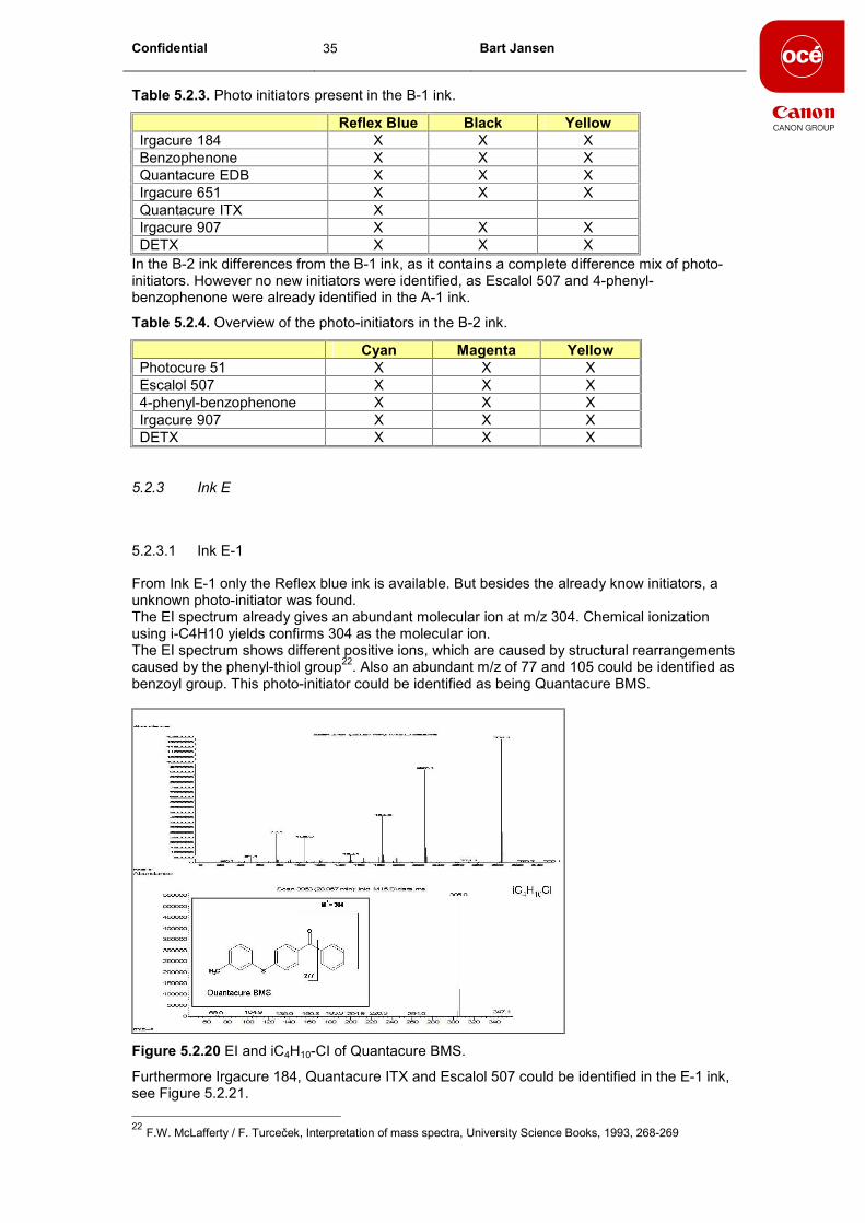
From Ink E-1 only the Reflex blue ink is available. But besides the already know initiators, a
unknown photo-initiator was found.
The EI spectrum already gives an abundant molecular ion at m/z 304. Chemical ionization
using i-C4H10 yields confirms 304 as the molecular ion.
The EI spectrum shows different positive ions, which are caused by structural rearrangements
caused by the phenyl-thiol group22
. Also an abundant m/z of 77 and 105 could be identified as
benzoyl group. This photo-initiator could be identified as being Quantacure BMS.
Figure 5.2.20 EI and iC4H10-CI of Quantacure BMS.
Furthermore Irgacure 184, Quantacure ITX and Escalol 507 could be identified in the E-1 ink,
see Figure 5.2.21.
22
F.W. McLafferty / F. Turceček, Interpretation of mass spectra, University Science Books, 1993, 268-269

Confidential 36 Bart Jansen
Figure 5.2.21. Chromatogram of ink E-1.
5.2.4 Conclusions ink analysis
Using a combination of EI and CI all of the photo-initiators in inks from different suppliers and
colours could be identified. All the identified photo-initiators gave an abundant M+H ion when
using isobutane as reagent gas in CI. Using methane as reagent gas, results in the loss of
water or methanol as most of the initiators contain hydroxy or methoxy groups. An overview of
the most abundant ion and the presence of the molecular ion is given in Table 5.2.5.
Other components present in the inks are acrylates or smaller molecules deriving from the
pigments used. Isolation of the pigment and IR analysis can be used to identify the pigment
used, and the source for these smaller molecules.
Using this method also some other inks have been analyzed, but only known initiators were
identified. For a complete overview of all the photo-initiators present in the inks, see appendix
8.6. In total 11 different photo-initiators have been indentified in 29 different inks.
Large differences have been found between the inks, varying from 2 photo-initiators in an ink,
up to a mixture of 7 photo-initiators in one ink.
Table 5.2.5. Overview of the most abundant ion and the presence of the molecular ion.
EI Mol. Ion CH4-CI Mol. Ion +H iC4H10-CI Mol. Ion +H
Irgacure 184 99 No 105 No 205 Yes
Benzophenone 105 Yes 183 Yes 183 Yes
Quantacure EDB 148 Yes 194 Yes 194 Yes
DETX 268* Yes 269 Yes 269 Yes
Quantacure ITX 239 Yes 255 Yes 255 Yes
Irgacure 907 128 No 280 Yes 280 Yes
Photocure 51 151 No 225 Yes** 255 Yes
Quantacure BMS 304* Yes 305 Yes 305 Yes
Escalol 507 165 Yes 166 Yes 278 Yes
Methyl-Benzoylbenzoate 163 Yes 209 Yes** 241 Yes
4-phenyl-benzophenone 181 Yes 259 Yes 259 Yes
* Molecular ion
** Very low abundancy

Confidential 37 Bart Jansen
5.3 Pre-printed media
In the next step, the method used to indentify the different initiators in the inks has been
applied to pre-printed media. Because of the very low amount of ink present on the media
followed by a curing step, it is investigated whether still all the initiators from the ink could still
be found.
There can be a large difference between different offset printers, and the UV lamps used. As
the amount of ink set on the media can vary per offset printer, and the intensity of the lamps
used can differ.
Therefore the prints with different cure settings and from two different offset printers are being
investigated.
Figure 5.3.1. Example of the test print
The test print consist of 4 different inks, together with 2 lamp setting and 2 different offset
printers giving in total 16 different samples which are being investigated. The sample has been
made on a offset printer using 2 UV lamps and 3 UV lamps.
All the strokes have been printed using Reflex blue ink, as the mix of photo-initiators can differ
per colour.
For the analysis a piece of the print has been cut out using a punch of 6 cm2. This piece of the
media has been cut into small pieces and is extracted using 2 ml of MC. The MC has been
analyzed directly using the GC method which was also used in the analysis of the inks.

Confidential 38 Bart Jansen
5.3.1 Comparison between print and ink
The first sample is printed using the Ink A-1 ink. In the print all the photo-initiators which are
present in the ink could be found, see Figure 5.3.2.
Figure 5.3.2. Comparison between ink and pre-print
The extract of the pre-print has been analyzed in normal EI mode and using iC4H10-CI. All the
6 photo-initiators could be identified using both ionization techniques. All of the Spectra are
given in appendix 8.7.
The relative amount of photo initiator does not change after printing and curing. The relative
amounts for the 4 prints is given in Table 5.3.1. The differences measured could be due to
batch differences, this is not further examined.
The exception is the print with ink B-1 in which not all of the photo initiators present in the ink
could be found (see Figure 8.8.1., in §8.8). The ITX which is clearly present in the ink is not
found in the print. Instead a larger amount of DETX is measured in the print. Most likely this is
caused by differences in ink batches, as the ink analysed is not from the same batch as the
batch which has been used for the print. This could not be verified as the ink used for the print
was not present. To investigate whether different photo initiator mixtures are being used in one
type of ink more batches need to be analyzed.

Confidential 39 Bart Jansen
Table 5.3.1. Relative amount of photo initiator in the print and ink.
Ink A-1 Print Ink
Photocure 51 18% 17%
Methyl-benoylbenzoat 11% 11%
Escalol 507 28% 30%
Irgacure 907 19% 22%
ITX 12% 11%
4-phenyl-benzophenone 12% 10%
Ink B-1
Benzophenone 20% 20%
Irgacure 184 15% 18%
EDB 12% 17%
Photocure 51 22% 20%
Irgacure 907 4% 5%
ITX 0% 17%
DETX 27% 2%
Ink D-1
Irgacure 184 51% 43%
EDB 15% 24%
DETX 34% 33%
Ink E-1
Irgacure 184 15% 12%
Escalol 507 39% 46%
ITX 11% 10%
BMS 35% 31%
5.3.2 Comparison between different prints
In the next step a comparisons has been made between the print cured with 2 lamps and with
3 lamps, see Figure 5.3.3. Both prints give similar amounts of initiators and no changes in the
relative amounts. Larger differences are seen between the acrylates, which are not being
identified in this study. Similar results were obtained in the comparison between the two offset
printers, see Figure 5.3.4.
Figure 5.3.3. Comparison between the extract of the pre-print cured with 2 and 3 lamps.

Confidential 40 Bart Jansen
Figure 5.3.4. Comparison between two different offset printers
Similar to the differences between the print cured with 2 and 3 lamps, the main difference is
seen in the amount of acrylates. The amount of initiator has slightly lower in the print from
offset printer 1, the relative amount between the initiators remain the same.
5.3.3 Sub Conclusions
For three of the four inks of the pre-print all the photo-initiators could be identified, see
appendix 8.8. With the exception of one ink in which one initiator could not be found, this is
possibly due to batch differences. Because of some small differences in the oven temperature
program used, small differences are seen in retention time.
Some differences in abundance between the initiators in the pre-print and the ink, but all were
present. The relative amount of the initiator does not change by printing or curing.
Some small differences were found using 2 or 3 UV lamps, but still all of the initiators could be
identified without any problems.
The method used for the analysis of the inks, can also be applied for pre-printed materials.
Analysis can be performed by extraction of the pre-print followed by direct analysis using GC-
MS.

Confidential 41 Bart Jansen
6 Conclusions
Different photo-initiators can be identified in UV inks, through direct analysis using GC-MS. No
clean up steps are necessary before analysis, simple dissolving the ink in MC for direct
analysis.
Using a combination of EI and CI all of the photo-initiators in the inks can be identified. All the
identified photo-initiators gave an abundant M+H ion when using isobutane as reagent gas in
CI. Using methane as reagent gas results often in the loss of water or methanol as most of the
initiators contain hydroxy or methoxy groups.
Using this method, in total 11 different photo-initiators have been identified in the UV inks.
Large differences have been found between different inks, were some inks only contain 2
initiators (Ink B-3) up to a mixture of 7 initiators (B-1 ink). Smaller differences were found
between different colours of the same ink.
Other components present in the inks are acrylates or smaller molecules deriving from the
pigments used. Isolation of the pigment and IR analysis can be used to identify the pigment
used, and the source for these smaller molecules.
Similar approach can be used in the analysis of pre-printed materials. After a simple extraction
using MC the initiators can be identified using simple GC-MS analysis. A comparison between
the inks and the pre-prints showed that all different photo-initiators could be detected. The
relative amount of the photo initiators do not change by printing and curing. The differences
measured are most likely due to batch differences, as the inks measured are not from the
same batch as the ink used for the prints.
No significant difference between pre-prints made using different cure setting or different offset
printer could be found.

Confidential 42 Bart Jansen
7 References
1. R. Anton, S. Barlow, D. Boskou, L. Castle, R. Crebelli, W. Dekant, K.H. Engel, S.
Forsythe, W. Grunow, M. Heinonen, J.C. Larsen, C. Leclercq, W. Mennes, M.R.
Milana, I. Pratt, I. Rientjes, K. Stevensson, P. Tobback, F. Toldrá, EFSA J. 293 (2005)
1.
2. Schweizerische Eidgenossenschaft, Verordnung des EDI über Bedarfsgegenstände
(SR 817.023.21) vom 23. November 2005, Anhang 6
3. Cuilian Sun, Sheot Harn Chan, Dan Lu, Hui Min Wendy Lee, Bosco Chen Bloodworth,
Journal of Chromaotgraphy A, 1143 (2007) 162-167
4. Ana Gil-Vergara, Christina Blasco, Yolanda Picó, Anal. Bioanal. Chem., 389 (2007)
605-617
5. H. Gallart-Ayala, E. Moyano, M.T. Galceran, Journal of Chromatography A, 1208
(2008) 182-188
6. Gianni Sagratini, Giovanni Caprioli, Gloria Cristalli, Dario Giardiná, Massimo
Ricciutelli, Rosaria Volpini, Yanting Zuo, Sauro Vittori / Journal of Chromatography A /
1194 (2008) 213-220
7. H. Gallart-Ayala, O. Núñez, E. Moyano, M.T. Galceran, Journal of Chromatography A,
1218 (2011) 459-466
8. R. Bagnati, G. Bianchi, E. Marangon, E. Zuccato, R. Franelli, E. Davoli, Rpid. Comm.
Mass. Spec. 21 (2007) 1998
9. Gianna Allegrone, Ilaria Tamaro, Shara Spinardi, Giorgio Grosa, Journal of
Chromatography A, 1214 (2008) 128-133
10. E. Van Hoeck, T. De Schaetzen, C. Pacquet, F. Bolle, L. Boxus, J. Van Loco,
Analytica Chimica Acta, 663 (2010) 55-59
11. N. Negreire, I. Rodríguez, E. Rubí, R. Cela, Talanta, 82 (2010) 269-303
12. IR 97322.022, Literatuurstudie naar foto-initiatoren voor UV uitharding, J van de Reek
13. Ciba, Photo initiators for UV curing, key product selection guide 2003, g-48/2003
October
14. H.F. Gruber/ Photo initiators for free radical polymerization / Prog. Polym. Sci. / Vol. 17
/ 953-1044 / 1992
15. M.C. McMaster, GC/MS A Pratical User�s Guide 2nd
edition, Wiley Interscience, 2008,
25-28
16. M.C. McMaster, GC/MS A Practical User�s Guide 2nd
edition, Wiley Interscience, 2008,
40-42
17. Interpretation of Mass Spectra, 4th edition, F.W. McLafferty / F. Tureček, Universal
science books, 1993, 52-83
18. A.G. Harrison, Chemical Ionization Mass Spectrometry, CRC press, 1983
19. F.G. Kitson, B.S. Larsen, C.N. McEwen, Gas Chromatography and Mass
Spectrometry a practical guide, 1996, Academic Press Inc.
20. W. Herbts, K. Hunger, Industrial Organic Pigments, 2nd edition, VCH, 1997 [p. 239-
261]
21. W. Herbts, K. Hunger, Industrial Organic Pigments, 2nd edition, VCH, 1997 [p. 288-
300]
22. F.W. McLafferty / F. Turceček, Interpretation of mass spectra, University Science
Books, 1993, 268-269

Confidential 43 Bart Jansen
8 Appendix
8.1 Overview of names and structures
Irgacure 184 (Ciba)
1-hydroxy-cyclohexyl-phenylketone
O
OH
Benzophenone
C
O
Quantacure EDB
Ethyl 4-(dimethylamino)benzoaat
N
CH3
CH3
C
O
O C2H5
DETX
2,4-Diethyl-9H-thioxanthen-9-one (DETX)
S
O
C2H5
C2H5
Quantacure ITX
Isopropyl- 9H-thioxanthen-9-one (ITX)
S
O
CH3
CH3
CH
Irgacure 907 (Ciba)
3-Methyl-1-[4-(methylthio)fenyl]-2-(4-morfolino)-1-propanon
(MMMP)

Confidential 44 Bart Jansen
O
SCH3
CH3
CH3
N O
Irgacure 651 (Ciba)
Also known as Photocure 51
2,2-Dimethoxy-1,2-difenylethan-1-one (BDK)
C
C
O
O
O
CH3
CH3
Quantacure BMS
4-(p-Tolylthio)benzofenon (BMS)
S
CH3
O
Escalol 507
2-Ethylhexyl 4-(dimethylamino)benzoaat (EHA)
N
CH3
CH3
C
O
O CH2 CH
C2H5
C4H9
Methyl-4-benzoylbenzoate (MBBZ)
O
O
CH3
O
4-Phenyl-benzophenone
4-Benzoylbifenyl (PBZ)
O

Confidential 45 Bart Jansen
8.2 Overview of Spectra
8.2.1 Irgacure 184

Confidential 46 Bart Jansen
8.2.2 Benzophenone

Confidential 47 Bart Jansen

Confidential 48 Bart Jansen
8.2.3 Quantacure EDB

Confidential 49 Bart Jansen
8.2.4 DETX

Confidential 50 Bart Jansen

Confidential 51 Bart Jansen
8.2.5 Quantacure ITX

Confidential 52 Bart Jansen
8.2.6 Irgacure 907

Confidential 53 Bart Jansen

Confidential 54 Bart Jansen
8.2.7 Irgacure 651 / Photocure 51

Confidential 55 Bart Jansen
8.2.8 Quantacure BMS

Confidential 56 Bart Jansen

Confidential 57 Bart Jansen
8.2.9 Escalol 507

Confidential 58 Bart Jansen
8.2.10 Methyl-Benzoylbenzoate

Confidential 59 Bart Jansen

Confidential 60 Bart Jansen
8.2.11 4-Phenyl-Benzophenone

Confidential 61 Bart Jansen
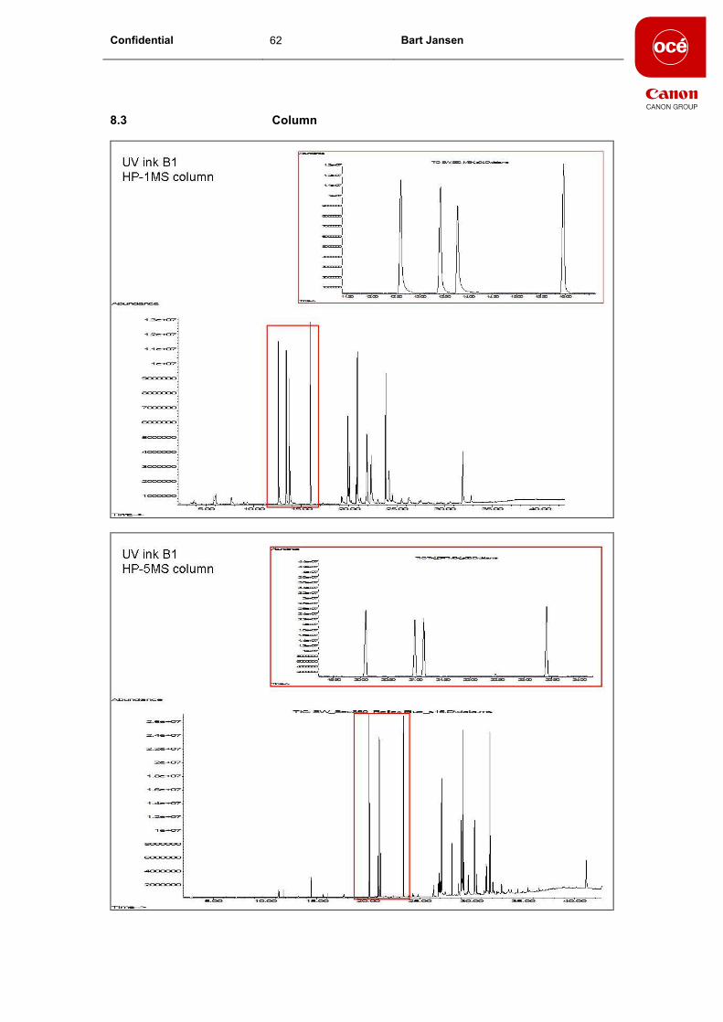
Confidential 62 Bart Jansen
8.3 Column

Confidential 63 Bart Jansen

Confidential 64 Bart Jansen
8.4 Ink A-1
8.4.1 Yellow
8.4.2 Red032

Confidential 65 Bart Jansen

Confidential 66 Bart Jansen

Confidential 67 Bart Jansen
8.5 IR analysis on Pigments
8.5.1 Yellow
IR analysis of Pigment Yellow of Ink A-1.
Name Value Unit
Name 3,3'-dichlorobenzidine -> acetoacetic arylide-2,4-dimethylanilide
Source of Sample Ciba-Geigy
Technique KBr pellet
Mol.Weight 684.20
Formula (Empirical)= C36H34Cl2N6O4
Comments HUMMEL DECIMAL NUMBER=
012221Description= APPEARANCE=yellow solid
Synonyms Irgalite Yellow BAWP
Color Properties Colour Index Name= Pigment Yellow
13Colour Index Number= 21100
Cl
Cl
NN
H
N
OO
NN
N
H
O O
wavenumbers
4000 3800 3600 3400 3200 3000 2800 2600 2400 2200 2000 1800 1600 1400 1200 1000 800 600 400
0
0.2
0.4
0.6
0.8
1 HC #3563; 3,3'-dichlorobenzidine -> acetoacetic arylide-2, ...
Pigment Sun Chemicals laser Yellow
Figure 8.5.1. IR spectrum and library match with Pigment Yellow 13.
CH3
O
O
NH
CH3
N N
NN
CH3
O
O
NH CH3
CH3
CH3 Cl
Cl
Figure 8.5.2. Pigment yellow 13.

Confidential 68 Bart Jansen
8.5.2 Red 032
IR analysis of Pigment Red032 of Ink A-1.
Name Value Unit
Name 2,4,5-trichloroaniline -> 2-hydroxynaphthoic arylide-2-methylanilide
Source of Sample Hoechst
Technique KBr pellet
Mol.Weight 483.03
Formula (Empirical)= C24H16Cl3N3O2
Comments HUMMEL DECIMAL NUMBER=
012212Description= APPEARANCE=red solid
Synonyms Permanent Rot FGR 70
Color Properties Colour Index Name= Pigment Red
112Colour Index Number= 12370
N
HO
N
NH
O
Cl
Cl
Cl
wavenumbers
4000 3800 3600 3400 3200 3000 2800 2600 2400 2200 2000 1800 1600 1400 1200 1000 800 600 400
0
0.05
0.1
0.15
0.2
HP #3515; 2,4,5-trichloroaniline -> 2-hydroxynaphthoic ar ...
Rood-Oranje pigment
Figure 8.5.3. IR spectrum and library match with Pigment Red 112
NH
NN
Cl
Cl
Cl
CH3
OH O
Figure 8.5.4. Pigment Red 112

Confidential 69 Bart Jansen
8.6 Overview of all inks
Irgacure 184 Benzophenone
Quantacure
EDB DETX
Quantacure
ITX
Irgacure
907
Photocure
51
Quantacure
BMS
Escalol
507
Methyl-
Benzoylbenzoate
4-phenyl-
benzophenone
Ink A-1
Cyan X X X X X X
Magenta X X X X X X
Yellow X X X X X X
Black X X X X X X X
Red032 X X X X X X
Reflex Blue X X X X X X
Ink B-1
Yellow X X X X X X
Black X X X X X X
Reflex Blue X X X X X X X
Ink B-2
Cyan X X X X X
Magenta X X X X X
Yellow X X X X X
Ink B-3
Black X X
Reflex Blue X X
Ink C-1
Cyan X X
Magenta X X
Yellow X X
Black X X
Red032 X X
Reflex Blue X X
Ink D-1

Confidential 70 Bart Jansen
Cyan X X X
Magenta X X X
Yellow X X X
Black X X X
Red032 X X X X
Reflex Blue X X X X
Ink D-2
Reflex Blue X X X
Red032 X X X
Ink E-1
Reflex Blue X X X X

Confidential 71 Bart Jansen
8.7 Spectra from pre-print
8.7.1 Photocure 51
8.7.2 Methyl-benzoylbenzoate

Confidential 72 Bart Jansen
8.7.3 Escalol 507
8.7.4 Irgacure 907

Confidential 73 Bart Jansen
8.7.5 Quantacure ITX
8.7.6 4-phenyl-benzophenone

Confidential 74 Bart Jansen
8.8 Comparison Pre-print and ink
Figure 8.8.1. Ink B-1
Figure 8.8.2. Ink E-1

Confidential 75 Bart Jansen
Figure 8.8.3. Ink D-1
Figure 8.8.4. Ink A-1.