identification of zebrafish insertional mutants with defects - genetics
TRANSCRIPT
Copyright © 2005 by the Genetics Society of AmericaDOI: 10.1534/genetics.104.039727
Identification of Zebrafish Insertional Mutants With Defects inVisual System Development and Function
Jeffrey M. Gross,*,1 Brian D. Perkins,*,2 Adam Amsterdam,† Ana Egana,* Tristan Darland,*Jonathan I. Matsui,* Salvatore Sciascia,* Nancy Hopkins† and John E. Dowling*
*Department of Molecular and Cellular Biology, Harvard University, Cambridge, Massachusetts 02138 and †Center for Cancer Research andDepartment of Biology, Massachusetts Institute of Technology, Cambridge, Massachusetts 02139
Manuscript received December 13, 2004Accepted for publication January 21, 2005
ABSTRACTGenetic analysis in zebrafish has been instrumental in identifying genes necessary for visual system develop-
ment and function. Recently, a large-scale retroviral insertional mutagenesis screen, in which 315 differentgenes were mutated, that resulted in obvious phenotypic defects by 5 days postfertilization was completed.That the disrupted gene has been identified in each of these mutants provides unique resource throughwhich the formation, function, or physiology of individual organ systems can be studied. To that end, ascreen for visual system mutants was performed on 250 of the mutants in this collection, examining eachof them histologically for morphological defects in the eye and behaviorally for overall visual systemfunction. Forty loci whose disruption resulted in defects in eye development and/or visual function wereidentified. The mutants have been divided into the following phenotypic classes that show defects in: (1)morphogenesis, (2) growth and central retinal development, (3) the peripheral marginal zone, (4) retinallamination, (5) the photoreceptor cell layer, (6) the retinal pigment epithelium, (7) the lens, (8) retinalcontainment, and (9) behavior. The affected genes in these mutants highlight a diverse set of proteinsnecessary for the development, maintenance, and function of the vertebrate visual system.
THE zebrafish has been an important model through apparent by the 18–19 SS. The first postmitotic neuronsof the retina are generated at 28 hr postfertilizationwhich genes necessary for visual system develop-
ment and function have been identified (reviewed in (hpf) and by 72 hpf the retina is functional (Easterand Nicola 1996; Hu and Easter 1999; Schmitt andEaster and Malicki 2002 and Neuhauss 2003). Zebra-
fish eyes are large, easily accessible, and structurally Dowling 1999). Retinas of many fish and amphibiansalso possess a specialized region at their margins, termedsimilar to the human eye. Eye formation in zebrafish is
analogous to that observed in other vertebrate embryos, peripheral or ciliary marginal zones, that perpetuallyadds cells to the retina during the lifetime of the animalthus providing an excellent model system with which
the understanding of vertebrate eye development can (Johns 1977).Several generations of chemically based forward ge-be advanced. Additionally, many disrupted genes and
pathways identified as integral to the formation of the netic screens have been undertaken in zebrafish (Drieveret al. 1996; Haffter et al. 1996; Matsuda and Mishinazebrafish eye produce phenotypes that resemble disor-
ders of the human visual system. Thus, characterization 2004), some of which have focused on eye developmentand function (Malicki et al. 1996; Fadool et al. 1997;of the molecular mechanisms of eye development in zebra-
fish should facilitate a better understanding of these hu- Neuhauss et al. 1999). While these chemically basedscreens have been instrumental in generating interestingman pathologies (Goldsmith and Harris 2003).
Eye development in zebrafish first becomes morpho- mutant phenotypes, positional cloning of these mutationsis still quite laborious, despite the genomic advances oflogically obvious at the 6 somite stage (SS), the time at
which the optic lobes evaginate from the diencephalon the last few years. Retrovirus-mediated insertional muta-(Schmitt and Dowling 1994). Thereafter, eye devel- genesis provides an attractive alternative to chemicalopment proceeds rapidly with lens induction occurring mutagenesis techniques since the affected gene can bearound the 14–15 SS and morphological distinction be- rapidly identified using the proviral insert as a moleculartween the retina and retinal pigment epithelium (RPE) tag to localize the site of insertion in the genome and
thereby to identify the mutated gene (Gaiano et al.1996; Amsterdam et al. 1999; Amsterdam 2003). In-
1Corresponding author: Department of Molecular and Cellular Biol- deed, a large-scale insertional mutagenesis screen per-ogy, Harvard University, 16 Divinity Ave., Cambridge, MA 02138. formed over the last 6 years has generated �500 inser-E-mail: [email protected]
tional mutants of which 315 different affected loci have2Present address: Department of Biology, Texas A&M University, Col-lege Station, TX 77843. been identified (Golling et al. 2002; Amsterdam et al.
Genetics 170: 245–261 (May 2005)
Dow
nloaded from https://academ
ic.oup.com/genetics/article/170/1/245/6060386 by guest on 22 February 2022
246 J. M. Gross et al.
1 � 30 min and incubated in secondary antibody, diluted in2004). This collection of mutants presents a wealth of5% NGS, for 90 min at RT. Slides were washed 3 � 15 minpossible analyses since studies are not limited to grossin PBS at RT and mounted in Vectashield mounting medium
phenotypic characterizations; one can target for study containing DAPI (Vector Laboratories, Burlingame, CA). Thespecific physiological processes and biochemical path- following antibodies and dilutions were used: 1d1 (1:30), 5e11
(1:100), zn5 (1:100), and goat anti-mouse Cy3 secondaryways in which the mutated gene’s protein product would(1:500). Imaging was performed in a Zeiss 510 laser scanningnormally function.confocal microscope. A total of 3–5 optical sections (1 �m inIt is estimated that this collection of 315 insertionalthickness) were collected and projected using Zeiss confocal
mutants represents �22% of the vertebrate gene com- software.plement that can be mutated to result in a visible embry- Optokinetic response assay : Optokinetic response (OKR)
assays were performed after Brockerhoff et al. (1995). Foronic phenotype (Amsterdam et al. 2004). With thattesting, two to four embryos at a time were immobilized in autility in mind, a shelf screen was performed on 250small Petri dish containing 5% methylcellulose. The Petri dishdifferent insertional loci for which the affected geneswas positioned in the center of a drum lined with vertical
have been cloned to identify those possessing defects black and white stripes, each 1 cm wide. The drum was illumi-in the development and function of the visual system. nated with a tungsten light source attenuated by up to 3.5 log
units with neutral density filters. The drum rotated at �8 rpmForty mutations were identified that affected the visualand, during 30-sec trials, the direction of rotation was changedsystem and this article reports the identification andfour to six times. Beginning at full light intensity, embryosinitial characterization of each of these mutants.were tested to see if they could respond to the moving stripes. Aresponse was defined as the demonstration of either a smoothpursuit-saccade cycle or eye tracking movements in both the
MATERIALS AND METHODS counterclockwise and the clockwise directions depending onthe rotation of the drum.Animals: The methods for the generation and identification
Electroretinography: Isolated whole-eye electroretinogramsof insertional mutants have been reported previously in Gai-(ERGs) were obtained using methods described in Kainz etano et al. (1996) and Amsterdam et al. (1999). Embryos wereal. (2003). Briefly, 5 days postfertilization (dpf), light-adaptedobtained from the natural spawning of heterozygous carrierslarvae were placed onto filter paper moistened with Mangel’ssetup in pairwise crosses. Embryos were collected and raisedRinger solution with 20 mm dextrose (pH 7.8). A pair ofat 28.5� after Westerfield (1995) and were staged accordingforceps held the animal and a small loop made of tungstento Kimmel et al. (1995).wire gently removed the eye, which was placed onto the filterHistology: Mutants and wild-type siblings were collected andpaper, cornea side up. A pulled glass micropipette (tip 10 �mfixed overnight at 4� in a solution of 1% (w/v) paraformalde-in diameter) containing Ringer solution and a chloride-coatedhyde, 2.5% glutaraldehyde, and 3% sucrose in PBS. They weresilver wire was inserted into the eye at the border of thewashed 3 � 5 min in PBS and refixed for 90 min at 4� in amarginal zone and the lens. A ground wire was placed under2% OsO4 solution, washed 3 � 5 min in PBS at RT, andthe moistened filter paper within the recording chamber. Thedehydrated through a graded ethanol series (50, 70, 80, 90,eye was bathed in Ringers throughout the course of a re-2 � 100%). Embryos were further dehydrated 2 � 10 min incording session, which lasted 30–75 min. ERGs were recordedpropylene oxide and infiltrated 1–2 hr in a 50% propyleneat 24�–25�. Responses were amplified by a Dagan Cornerstoneoxide/50% Epon/Araldite mixture (Polysciences). Embryosamplifier bandpass filtered (0.1–100 Hz), total gain �10,000,were then incubated overnight at RT in 100% Epon/Aralditeand collected using PClamp software (Axon Instruments, Bur-resin with caps open to allow for propylene oxide evaporationlingame, CA). A 1409 W/cm2 background light attenuated byand resin infiltration, embedded and baked at 60� for 2–3a �1.6 log unit ND filter was used. The stimulus was produceddays. Sections 1–1.25 �m were cut, mounted on glass slides,by a tungsten halogen light, 9503 �W/cm2 unattenuated inten-and stained in a 1% methylene blue/1% borax solution. Sec-sity and was adjusted with neutral density filters. The durationtions were mounted in DPX (Electron Microscopy Sciences,of the stimulus was 1000 msec, while the interstimulus timeFort Washington, PA) and photographed on a Leica DMRBwas 15 sec.microscope mounted with a QImaging Retiga EXi digital cam-
era. Images were subsequently processed using Adobe Pho-toshop 5.0.
RESULTSAcridine orange staining: Acridine orange (Molecular Probes,Eugene, OR) was diluted in fish water to a final concentration Insertional mutagenesis screen: The generation, screen-of 1 �g/ml. Embryos were placed in this solution for 10 min,
ing, and cataloging of zebrafish insertional mutants havewashed briefly five times in fish water, and immediately ob-been described elsewhere (Gaiano et al. 1996; Amster-served under GFP optics on a Leica dissecting scope.
Immunohistochemistry: Mutants and wild-type siblings were dam et al. 1999, 2004; Golling et al. 2002). In this study,collected and fixed overnight at 4� in a solution of 4% para- 250 different insertional mutants were screened for de-formaldehyde in PBS. Embryos were washed at RT 3 � 5 min fects in eye morphology and for deficits in visual behav-in PBS and then infiltrated by 35% sucrose for 1–2 hr at RT.
ior. Figure 1 provides a general summary of the screenEmbryos were then arranged and embedded in plastic moldsand its results. The morphology screen sought to iden-containing TBS cryopreservation media (Triangle Biomedical
Sciences, Durham, NC). Cryosections 12 �m in thickness were tify mutants with abnormal development or mainte-cut on a Leica CM1900 cryostat and adhered to gelatin-coated nance of eye structures at 5 dpf. Of the initial 250 lines,slides. After drying for 1–2 hr at RT, slides were lined with a 192 survived until 5 dpf and these were further studied.hydrophobic marker (PAP pen), rehydrated briefly in PBS,
To assay eye development, several mutant embryos fromand blocked for 1–2 hr in 5% NGS. Primary antibodies, dilutedeach of these 192 lines were fixed, processed, and sec-in 5% NGS, were added and slides were incubated overnight
at 4�. Slides were then washed in PBS at RT 2 � 5 min and tioned for histological examination.
Dow
nloaded from https://academ
ic.oup.com/genetics/article/170/1/245/6060386 by guest on 22 February 2022

247Zebrafish Visual System Mutants
Figure 1.—Summary of the screen. The af-fected loci in 315 insertional mutants have beenidentified (Amsterdam et al. 2004). This screenstudied 250 of these, 192 of which survived until5 dpf and whose histology was examined. Of these192 mutants, 81 showed a nonspecific morpholog-ical phenotype in the eye likely as a secondaryresult of more general systemic defects; 38 showeda morphological phenotype in the eye that ap-peared direct, i.e., not a secondary result of gen-eral systemic defects; 40 mutants showed smalleyes with normal morphology; and 33 total mu-tants had wild-type eye size and morphology, 2 ofwhich, however, failed behavioral testing for visualfunction.
Obvious phenotypic defects in eye development were 1). Eighty-one mutant loci displayed eye phenotypesthat were deemed likely to be secondary to more generalobserved in �60% of the mutant lines sectioned. These
phenotypes mostly manifest as severe retinal degenera- system-wide defects (see supplementary Table 1 at http://www.genetics.org/supplemental/). In addition, 33 othertion evident by the presence of many pyknotic nuclei
scattered through all retinal cell layers as well as large mutants displayed wild-type eye size and morphology (seesupplementary Table 2 at http://www.genetics.org/supregions of acellular holes, indicative of prior cell death.
In addition, many mutants exhibited severe lens degen- plemental/; Figure 10), whereas 40 mutants displayedsmaller eyes with no other apparent morphological de-eration in conjunction with the above retinal degenera-
tion. Thus, it became necessary to separate those pheno- fects in eye development (see supplementary Table 3 athttp://www.genetics.org/supplemental/). In the lattertypes likely resulting from a direct role of the affected
gene product in the eye from those resulting from mutants, all cell layers had formed and were properlypatterned but the size of the eye was smaller overall.multisystem defects and, therefore, presumably second-
ary to a more general set of physiological problems. A These mutants were not pursued further from the mor-phological standpoint of the screen.set of criteria was established to score the overall health
of the mutant embryos up to 5 dpf and to correlate For each of the 38 morphological mutants describedherein, the phenotypes were observed in multiple em-these with the nature of the eye phenotype observed
after histological analysis. These criteria included daily bryos from a minimum of two independent crosses. Thehistology presented is representative of that observedobservations of overall embryo health as well as a variety
of physical features such as the amount of unconsumed in all insertional mutants at this locus. Because of thelarge number of mutants screened, this report describesyolk, overall head and body size, presence of cardiac
edema, blood circulation, general or localized necrosis, only the preliminary phenotypic characterization oftheir eye defects. A more detailed molecular and ultra-and locomotion. Analysis of the histological data in con-
junction with these physical observations identified a structural analysis of each will be required to fully char-acterize their defects. Additionally, detailed informationsubset of mutant lines that might represent an eye phe-
notype directly related to the mutated gene’s normal about defects in other organ systems in each of thesemutants can be found in Amsterdam et al. (2004). Thecellular role rather than those phenotypes resulting
from general physiological causes. In addition, roughly mutants have been grouped into eight phenotypic cate-gories on the basis of defects observed in: (1) morpho-one-fifth of the mutants that presented severe eye de-
generation phenotypes at 5 dpf showed a more subtle genesis, (2) growth and central retinal development, (3)the peripheral marginal zone, (4) retinal lamination, (5)phenotype at earlier stages. These mutants were re-
screened at 3 dpf since it was possible that relevant eye the photoreceptor cell layer, (6) the retinal pigment epi-thelium, (7) the lens, and (8) retinal containment.defects were obscured by overall eye degeneration by 5
dpf. All rescreens entailed a second round of histology A second screen performed on the insertional mutantsassayed the function of their visual systems using the OKR,for phenotypic verification, as well as acridine orange
staining at 3 dpf to provide an indication of the amount a robust assay of visual behavior (reviewed in Neuhauss2003). This assay has proven useful in identifying manyof cell death occurring in the mutants. Acridine orange
staining enabled a distinction between generalized CNS visually deficient zebrafish mutants (Brockerhoff et al.1995, 1998; Neuhauss et al. 1999). As detailed above,degeneration and cell death localized to the eye, possi-
bly in conjunction with other limited CNS structures. many of the insertional mutants presented system-widedefects that would obviously prevent them from behav-Of the 192 mutant loci histologically examined, 38
were ultimately identified that fit our criteria for pos- ing normally in this screen. These have not been in-cluded in this report as their behavioral deficit is likelysessing a relevant morphological eye phenotype (Table
Dow
nloaded from https://academ
ic.oup.com/genetics/article/170/1/245/6060386 by guest on 22 February 2022
248 J. M. Gross et al.
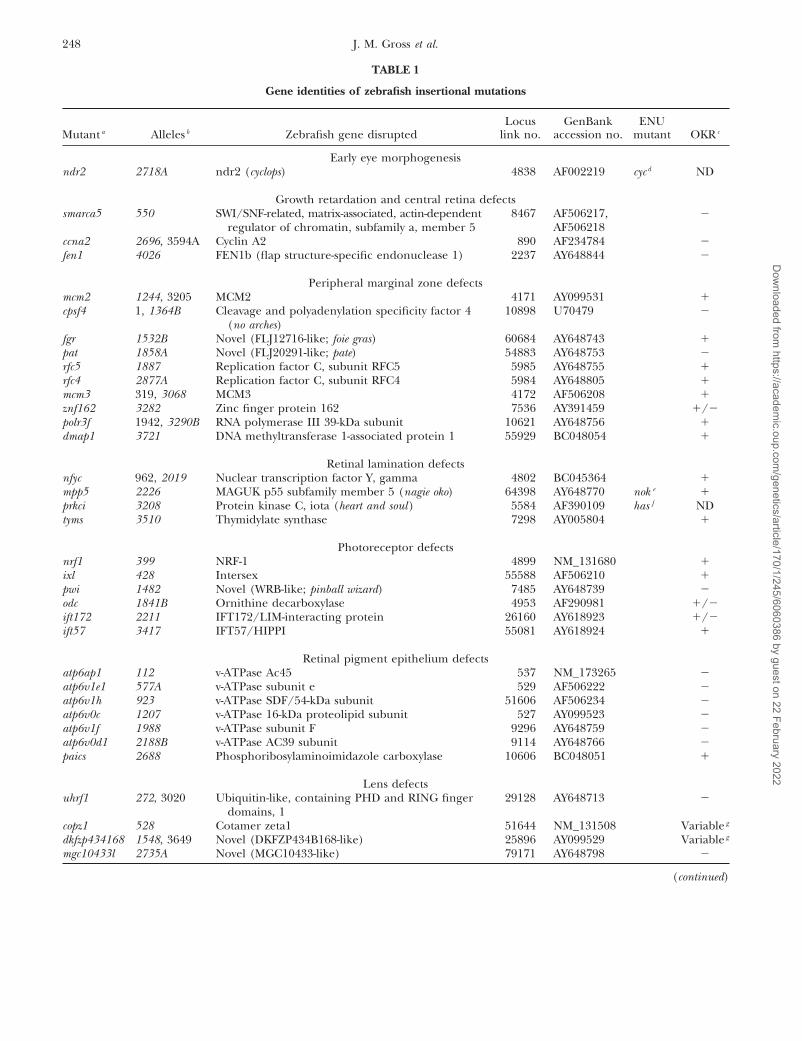
TABLE 1
Gene identities of zebrafish insertional mutations
Locus GenBank ENUMutant a Alleles b Zebrafish gene disrupted link no. accession no. mutant OKR c
Early eye morphogenesisndr2 2718A ndr2 (cyclops) 4838 AF002219 cyc d ND
Growth retardation and central retina defectssmarca5 550 SWI/SNF-related, matrix-associated, actin-dependent 8467 AF506217, �
regulator of chromatin, subfamily a, member 5 AF506218ccna2 2696, 3594A Cyclin A2 890 AF234784 �fen1 4026 FEN1b (flap structure-specific endonuclease 1) 2237 AY648844 �
Peripheral marginal zone defectsmcm2 1244, 3205 MCM2 4171 AY099531 �cpsf4 1, 1364B Cleavage and polyadenylation specificity factor 4 10898 U70479 �
(no arches)fgr 1532B Novel (FLJ12716-like; foie gras) 60684 AY648743 �pat 1858A Novel (FLJ20291-like; pate) 54883 AY648753 �rfc5 1887 Replication factor C, subunit RFC5 5985 AY648755 �rfc4 2877A Replication factor C, subunit RFC4 5984 AY648805 �mcm3 319, 3068 MCM3 4172 AF506208 �znf162 3282 Zinc finger protein 162 7536 AY391459 �/�polr3f 1942, 3290B RNA polymerase III 39-kDa subunit 10621 AY648756 �dmap1 3721 DNA methyltransferase 1-associated protein 1 55929 BC048054 �
Retinal lamination defectsnfyc 962, 2019 Nuclear transcription factor Y, gamma 4802 BC045364 �mpp5 2226 MAGUK p55 subfamily member 5 (nagie oko) 64398 AY648770 nok e �prkci 3208 Protein kinase C, iota (heart and soul) 5584 AF390109 has f NDtyms 3510 Thymidylate synthase 7298 AY005804 �
Photoreceptor defectsnrf1 399 NRF-1 4899 NM_131680 �ixl 428 Intersex 55588 AF506210 �pwi 1482 Novel (WRB-like; pinball wizard) 7485 AY648739 �odc 1841B Ornithine decarboxylase 4953 AF290981 �/�ift172 2211 IFT172/LIM-interacting protein 26160 AY618923 �/�ift57 3417 IFT57/HIPPI 55081 AY618924 �
Retinal pigment epithelium defectsatp6ap1 112 v-ATPase Ac45 537 NM_173265 �atp6v1e1 577A v-ATPase subunit e 529 AF506222 �atp6v1h 923 v-ATPase SDF/54-kDa subunit 51606 AF506234 �atp6v0c 1207 v-ATPase 16-kDa proteolipid subunit 527 AY099523 �atp6v1f 1988 v-ATPase subunit F 9296 AY648759 �atp6v0d1 2188B v-ATPase AC39 subunit 9114 AY648766 �paics 2688 Phosphoribosylaminoimidazole carboxylase 10606 BC048051 �
Lens defectsuhrf1 272, 3020 Ubiquitin-like, containing PHD and RING finger 29128 AY648713 �
domains, 1copz1 528 Cotamer zeta1 51644 NM_131508 Variable g
dkfzp434168 1548, 3649 Novel (DKFZP434B168-like) 25896 AY099529 Variable g
mgc10433l 2735A Novel (MGC10433-like) 79171 AY648798 �
(continued)
Dow
nloaded from https://academ
ic.oup.com/genetics/article/170/1/245/6060386 by guest on 22 February 2022
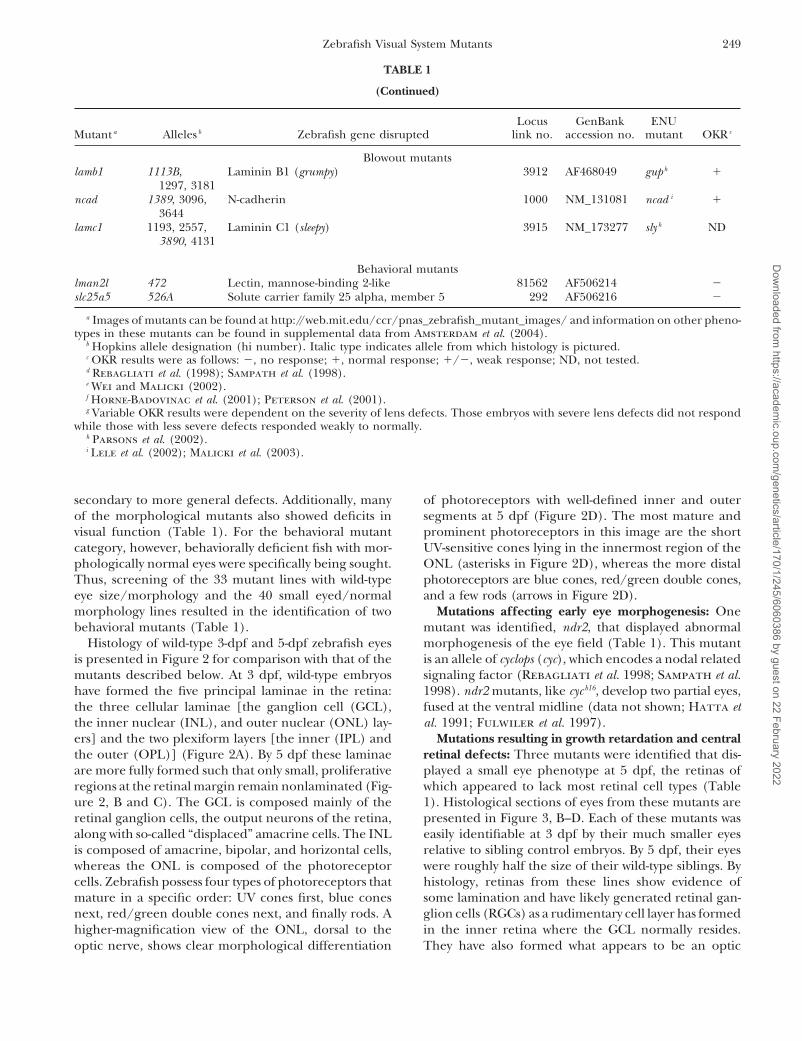
249Zebrafish Visual System Mutants
TABLE 1
(Continued)
Locus GenBank ENUMutant a Alleles b Zebrafish gene disrupted link no. accession no. mutant OKR c
Blowout mutantslamb1 1113B, Laminin B1 (grumpy) 3912 AF468049 gup h �
1297, 3181ncad 1389, 3096, N-cadherin 1000 NM_131081 ncad i �
3644lamc1 1193, 2557, Laminin C1 (sleepy) 3915 NM_173277 sly h ND
3890, 4131
Behavioral mutantslman2l 472 Lectin, mannose-binding 2-like 81562 AF506214 �slc25a5 526A Solute carrier family 25 alpha, member 5 292 AF506216 �
a Images of mutants can be found at http://web.mit.edu/ccr/pnas_zebrafish_mutant_images/ and information on other pheno-types in these mutants can be found in supplemental data from Amsterdam et al. (2004).
b Hopkins allele designation (hi number). Italic type indicates allele from which histology is pictured.c OKR results were as follows: �, no response; �, normal response; �/�, weak response; ND, not tested.d Rebagliati et al. (1998); Sampath et al. (1998).e Wei and Malicki (2002).f Horne-Badovinac et al. (2001); Peterson et al. (2001).g Variable OKR results were dependent on the severity of lens defects. Those embryos with severe lens defects did not respond
while those with less severe defects responded weakly to normally.h Parsons et al. (2002).i Lele et al. (2002); Malicki et al. (2003).
secondary to more general defects. Additionally, many of photoreceptors with well-defined inner and outersegments at 5 dpf (Figure 2D). The most mature andof the morphological mutants also showed deficits in
visual function (Table 1). For the behavioral mutant prominent photoreceptors in this image are the shortUV-sensitive cones lying in the innermost region of thecategory, however, behaviorally deficient fish with mor-
phologically normal eyes were specifically being sought. ONL (asterisks in Figure 2D), whereas the more distalphotoreceptors are blue cones, red/green double cones,Thus, screening of the 33 mutant lines with wild-type
eye size/morphology and the 40 small eyed/normal and a few rods (arrows in Figure 2D).Mutations affecting early eye morphogenesis: Onemorphology lines resulted in the identification of two
behavioral mutants (Table 1). mutant was identified, ndr2, that displayed abnormalmorphogenesis of the eye field (Table 1). This mutantHistology of wild-type 3-dpf and 5-dpf zebrafish eyes
is presented in Figure 2 for comparison with that of the is an allele of cyclops (cyc), which encodes a nodal relatedsignaling factor (Rebagliati et al. 1998; Sampath et al.mutants described below. At 3 dpf, wild-type embryos
have formed the five principal laminae in the retina: 1998). ndr2 mutants, like cyc b16, develop two partial eyes,fused at the ventral midline (data not shown; Hatta etthe three cellular laminae [the ganglion cell (GCL),
the inner nuclear (INL), and outer nuclear (ONL) lay- al. 1991; Fulwiler et al. 1997).Mutations resulting in growth retardation and centralers] and the two plexiform layers [the inner (IPL) and
the outer (OPL)] (Figure 2A). By 5 dpf these laminae retinal defects: Three mutants were identified that dis-played a small eye phenotype at 5 dpf, the retinas ofare more fully formed such that only small, proliferative
regions at the retinal margin remain nonlaminated (Fig- which appeared to lack most retinal cell types (Table1). Histological sections of eyes from these mutants areure 2, B and C). The GCL is composed mainly of the
retinal ganglion cells, the output neurons of the retina, presented in Figure 3, B–D. Each of these mutants waseasily identifiable at 3 dpf by their much smaller eyesalong with so-called “displaced” amacrine cells. The INL
is composed of amacrine, bipolar, and horizontal cells, relative to sibling control embryos. By 5 dpf, their eyeswere roughly half the size of their wild-type siblings. Bywhereas the ONL is composed of the photoreceptor
cells. Zebrafish possess four types of photoreceptors that histology, retinas from these lines show evidence ofsome lamination and have likely generated retinal gan-mature in a specific order: UV cones first, blue cones
next, red/green double cones next, and finally rods. A glion cells (RGCs) as a rudimentary cell layer has formedin the inner retina where the GCL normally resides.higher-magnification view of the ONL, dorsal to the
optic nerve, shows clear morphological differentiation They have also formed what appears to be an optic
Dow
nloaded from https://academ
ic.oup.com/genetics/article/170/1/245/6060386 by guest on 22 February 2022

250 J. M. Gross et al.
to sibling controls and are highly disorganized. For ex-ample, relative to the wild-type retina at 5 dpf and nor-malized to their smaller size, these mutants, on average,have 48% less 5e11-stained amacrine cells in their reti-nas (data not shown). Localized regions of cell deathin the brains of these mutants were obvious at 2 dpfand acridine orange staining at 3 dpf identified apo-ptotic nuclei scattered throughout their retinas. At laterdays, however, continuing cell death in the CNS was nolonger obvious and the mutants appeared to developnormally (data not shown). By 5 dpf, they were vigorousswimmers and displayed robust touch responses and,other than having much smaller heads and eyes, theyappeared generally healthy. That the eyes in these mu-tants are small and appear to lack large numbers ofretinal neurons suggests that a defect in progenitor cellmaintenance might have occurred. While more detailedexperiments will be necessary to ascertain the nature oftheir phenotype, that these loci encode proteins in-volved in transcriptional and cell cycle regulation sup-ports this hypothesis (Table 1).
Mutations affecting the peripheral marginal zone: Asnoted above, the teleost retina possesses a peripheralmarginal zone (MZ) that in many cold-blooded speciesperpetually adds cells to the retina during the animal’slife (Figure 2C). Cells of the marginal zone retain stemcell-like characteristics and can continually generate alltypes of retinal cells (Johns 1977; Wetts and FraserFigure 2.—Wild-type development of the zebrafish retina.1988). Some higher vertebrates, such as birds and mice,(A) A 3-dpf retina with well-formed laminae in the central
retina and large regions of undifferentiated cells at the retinal may also possess a similar region between the neuralperiphery. (B) A 5-dpf retina exhibiting nuclear laminae and retina and the ciliary epithelium at their retinal periph-cell types characteristic of the mature retina. (C) High-magni- ery (Fischer and Reh 2000; Tropepe et al. 2000). Severalfication view of the dorsal peripheral retina. Note the change
mutations that disrupt the formation or maintenance ofin cell shape as epithelial progenitor cells divide at the periph-the peripheral marginal zone have been reported in ze-eral-most dMZ (arrow) and are subsequently displaced into
the central retina to differentiate into more rounded ap- brafish, but the affected loci have not yet been identifiedpearing retinal neurons (arrowhead). (D) High-magnification (Fadool et al. 1997; Link and Darland 2001). Ten inser-view of the central retina and optic nerve. Note the prominent tional mutations were identified in this screen that affectshort single cones in the innermost region of the ONL (aster-
loci involved in marginal zone formation or mainte-isks) and the elongated rod outer segments in the outer ONL,nance (Table 1). Retinal histology from each of theseprotruding into the RPE (arrows). Dorsal is up in all panels.
Bar, 100 �m. RPE, retinal pigment epithelium; ONL, outer mutants is presented in Figure 4.nuclear layer; OPL, outer plexiform layer; INL, inner nuclear Mutants in this category appear quite similar in thatlayer; IPL, inner plexiform layer; GCL, ganglion cell layer; central retinal development and patterning is normaldMZ, dorsal marginal zone; vMZ, ventral marginal zone.
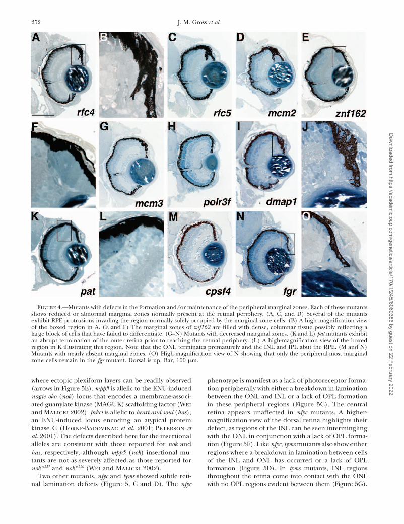
whereas peripheral marginal zones are greatly reducedboth dorsally and ventrally (Figure 4). Differing levelsof phenotypic severity are observed between the lines innerve, although it is possible this is a remnant of the
optic stalk. The INL and ONL of these mutants are this group. For example, the rfc4, rfc5, and mcm2 mutantsshow abnormal RPE invasion into regions of the retinalpoorly formed. It is difficult to morphologically identify
many of the cells in these layers as specific neuronal periphery, resulting in the retinal tissue there being nearlyencased in RPE (Figure 4, A–D). On the basis of theirsubtypes purely by histological means.
To begin to understand what cell types are present morphology, the cells in these regions appear to be largelyundifferentiated (Figure 4B) although the significance ofin retinas of these mutants, immunohistochemistry was
performed on 5-dpf retinal sections using markers for the RPE protrusions is currently unknown. The marginalzones of one mutant, znf162, are filled with dense, co-RGCs (zn5; Figure 3, E–H), amacrine cells (5e11; Figure
3, I–L), and rods (1d1; Figure 3, M–P). It is apparent lumnar cells possibly reflecting a large block of cellsthat have failed to differentiate (Figure 4, E and F). Thefrom this marker staining that neurons composing each
of the three main cellular laminae are present, although mcm3, polr3f, and dmap1 mutants all exhibit reducedmarginal zones (Figure 4, G–I). The pat mutant showsthey are observed in much reduced numbers relative
Dow
nloaded from https://academ
ic.oup.com/genetics/article/170/1/245/6060386 by guest on 22 February 2022

251Zebrafish Visual System Mutants
Figure 3.—Mutants withgrowth retardation and reti-nal degeneration. (A) Wildtype, (B) smarca5, (C) ccna2,and (D) fen1 have drasticallysmaller eyes with defects inthe number and organiza-tion of retinal neurons at 5dpf. Retinal patterning is se-verely affected with the mostobvious defects in the outerretina. (E–P) Immunohis-tochemical analysis of wild-type (E, I, and M), smarca5(F, J, and N), ccna2 (G, K,and O), and fen1 (H, L, andP) mutants at 5 dpf. (E–H)zn5 staining of RGCs. Eachof the mutants differentiateda population of RGCs, the ax-ons of which form an opticnerve of significantly less di-ameter than that of wild-typesiblings. (I–L) 5e11 stainingof amacrine cells and theirprocesses. Each of the mu-tants has differentiated am-acrine cells, although theirnumber is much reduced andtheir distribution is chaoticrelative to wild-type siblings.(M–P) 1d1 staining of rodphotoreceptors. Each of themutants has formed rodsbut in numbers reduced rel-ative to wild-type siblings.Dorsal is up. Bars, 100 �min A–E, I, and M and 70 �min F–H, J–L, and N–P.
an abrupt termination between the cells of the outer proteins of unknown biochemical function, while theother eight loci encode proteins involved in variousretina and the region normally occupied by the MZ
(Figure 4K). A high-magnification view of this region aspects of DNA replication and mRNA modification(Table 1).highlights the abrupt outer retina termination at the
retinal periphery (Figure 4L). Here, the INL, OPL, and Mutations affecting retinal lamination: Lamination inthe zebrafish retina becomes evident by 3 dpf when theONL extend to the retinal margin with no intervening
cells (compare Figure 4L with Figure 2C). The two re- three principal cellular laminae can be readily observed(Figure 2A). As the retina grows and new neurons aremaining mutants, cpsf4 and fgr , have even more reduced
marginal zone regions (Figure 4, M–O). In addition, generated by the MZs, the new cells maintain appro-priate laminar positions for their cell types such thatfgr mutants also frequently have malformed lenses (data
not shown). Several of the marginal zone mutants show only the MZs remain nonlaminated at later days of devel-opment (Figure 2, B and C). Four insertional mutantslocalized regions of cell death in their MZs when viewed
at earlier days (data not shown). In general, this class of with defects in retinal lamination were identified (Table1). Retinal histology from each of these mutants is pre-mutants frequently showed phenotypic defects in other
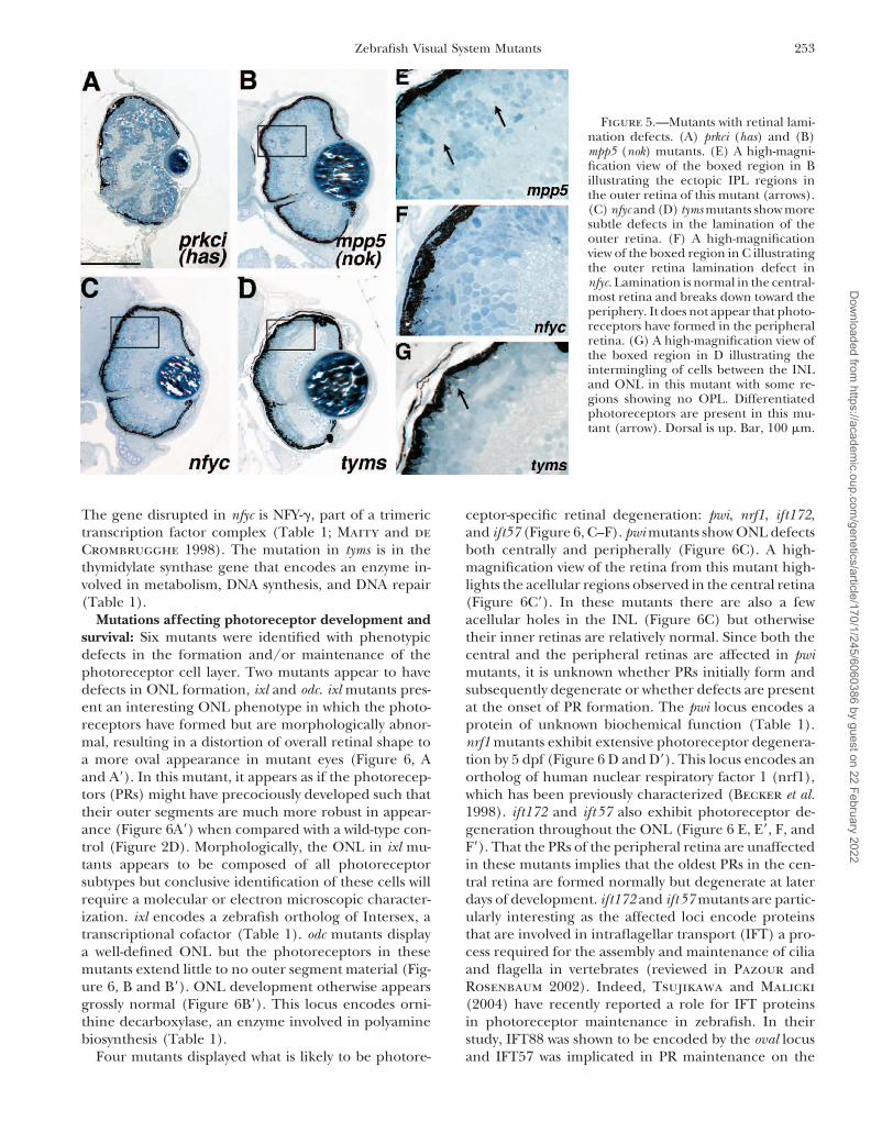
proliferative regions of the body, including the brain, sented in Figure 5.prkci and mpp5 mutants both present defects in retinalliver, and gut (Amsterdam et al. 2004; data not shown).
Thus, it is possible that these mutants represent a more lamination within their outer retinas (Figure 5, A andB). prkci mutants also showed aberrant RPE formation.general growth inhibition at later stages of development
rather than specific marginal zone functions (see dis- A high-magnification view of the mpp5 retina highlightsthe breakdown in lamination observed in this mutantcussion). Two of the loci affected, fgr and pat, encode
Dow
nloaded from https://academ
ic.oup.com/genetics/article/170/1/245/6060386 by guest on 22 February 2022
252 J. M. Gross et al.
Figure 4.—Mutants with defects in the formation and/or maintenance of the peripheral marginal zones. Each of these mutantsshows reduced or abnormal marginal zones normally present at the retinal periphery. (A, C, and D) Several of the mutantsexhibit RPE protrusions invading the region normally solely occupied by the marginal zone cells. (B) A high-magnification viewof the boxed region in A. (E and F) The marginal zones of znf162 are filled with dense, columnar tissue possibly reflecting alarge block of cells that have failed to differentiate. (G–N) Mutants with decreased marginal zones. (K and L) pat mutants exhibitan abrupt termination of the outer retina prior to reaching the retinal periphery. (L) A high-magnification view of the boxedregion in K illustrating this region. Note that the ONL terminates prematurely and the INL and IPL abut the RPE. (M and N)Mutants with nearly absent marginal zones. (O) High-magnification view of N showing that only the peripheral-most marginalzone cells remain in the fgr mutant. Dorsal is up. Bar, 100 �m.
where ectopic plexiform layers can be readily observed phenotype is manifest as a lack of photoreceptor forma-tion peripherally with either a breakdown in lamination(arrows in Figure 5E). mpp5 is allelic to the ENU-induced
nagie oko (nok) locus that encodes a membrane-associ- between the ONL and INL or a lack of OPL formationin these peripheral regions (Figure 5C). The centralated guanylate kinase (MAGUK) scaffolding factor (Wei
and Malicki 2002). prkci is allelic to heart and soul (has), retina appears unaffected in nfyc mutants. A higher-magnification view of the dorsal retina highlights theiran ENU-induced locus encoding an atypical protein
kinase C (Horne-Badovinac et al. 2001; Peterson et defect, as regions of the INL can be seen interminglingwith the ONL in conjunction with a lack of OPL forma-al. 2001). The defects described here for the insertional
alleles are consistent with those reported for nok and tion (Figure 5F). Like nfyc, tyms mutants also show eitherregions where a breakdown in lamination between cellshas, respectively, although mpp5 (nok) insertional mu-
tants are not as severely affected as those reported for of the INL and ONL has occurred or a lack of OPLformation (Figure 5D). In tyms mutants, INL regionsnokm227 and nokm520 (Wei and Malicki 2002).
Two other mutants, nfyc and tyms showed subtle reti- throughout the retina come into contact with the ONLwith no OPL regions evident between them (Figure 5G).nal lamination defects (Figure 5, C and D). The nfyc
Dow
nloaded from https://academ
ic.oup.com/genetics/article/170/1/245/6060386 by guest on 22 February 2022
253Zebrafish Visual System Mutants
Figure 5.—Mutants with retinal lami-nation defects. (A) prkci (has) and (B)mpp5 (nok) mutants. (E) A high-magni-fication view of the boxed region in Billustrating the ectopic IPL regions inthe outer retina of this mutant (arrows).(C) nfyc and (D) tyms mutants show moresubtle defects in the lamination of theouter retina. (F) A high-magnificationview of the boxed region in C illustratingthe outer retina lamination defect innfyc. Lamination is normal in the central-most retina and breaks down toward theperiphery. It does not appear that photo-receptors have formed in the peripheralretina. (G) A high-magnification view ofthe boxed region in D illustrating theintermingling of cells between the INLand ONL in this mutant with some re-gions showing no OPL. Differentiatedphotoreceptors are present in this mu-tant (arrow). Dorsal is up. Bar, 100 �m.
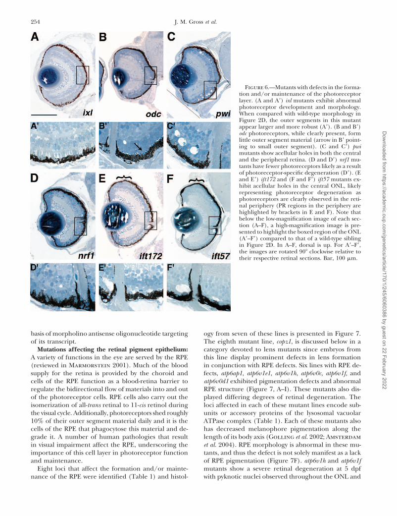
The gene disrupted in nfyc is NFY-�, part of a trimeric ceptor-specific retinal degeneration: pwi, nrf1, ift172,and ift57 (Figure 6, C–F). pwi mutants show ONL defectstranscription factor complex (Table 1; Maity and de
Crombrugghe 1998). The mutation in tyms is in the both centrally and peripherally (Figure 6C). A high-magnification view of the retina from this mutant high-thymidylate synthase gene that encodes an enzyme in-
volved in metabolism, DNA synthesis, and DNA repair lights the acellular regions observed in the central retina(Figure 6C�). In these mutants there are also a few(Table 1).
Mutations affecting photoreceptor development and acellular holes in the INL (Figure 6C) but otherwisetheir inner retinas are relatively normal. Since both thesurvival: Six mutants were identified with phenotypic
defects in the formation and/or maintenance of the central and the peripheral retinas are affected in pwimutants, it is unknown whether PRs initially form andphotoreceptor cell layer. Two mutants appear to have
defects in ONL formation, ixl and odc. ixl mutants pres- subsequently degenerate or whether defects are presentat the onset of PR formation. The pwi locus encodes aent an interesting ONL phenotype in which the photo-
receptors have formed but are morphologically abnor- protein of unknown biochemical function (Table 1).nrf1 mutants exhibit extensive photoreceptor degenera-mal, resulting in a distortion of overall retinal shape to
a more oval appearance in mutant eyes (Figure 6, A tion by 5 dpf (Figure 6 D and D�). This locus encodes anortholog of human nuclear respiratory factor 1 (nrf1),and A�). In this mutant, it appears as if the photorecep-
tors (PRs) might have precociously developed such that which has been previously characterized (Becker et al.1998). ift172 and ift57 also exhibit photoreceptor de-their outer segments are much more robust in appear-
ance (Figure 6A�) when compared with a wild-type con- generation throughout the ONL (Figure 6 E, E�, F, andF�). That the PRs of the peripheral retina are unaffectedtrol (Figure 2D). Morphologically, the ONL in ixl mu-
tants appears to be composed of all photoreceptor in these mutants implies that the oldest PRs in the cen-tral retina are formed normally but degenerate at latersubtypes but conclusive identification of these cells will
require a molecular or electron microscopic character- days of development. ift172 and ift57 mutants are partic-ularly interesting as the affected loci encode proteinsization. ixl encodes a zebrafish ortholog of Intersex, a
transcriptional cofactor (Table 1). odc mutants display that are involved in intraflagellar transport (IFT) a pro-cess required for the assembly and maintenance of ciliaa well-defined ONL but the photoreceptors in these
mutants extend little to no outer segment material (Fig- and flagella in vertebrates (reviewed in Pazour andRosenbaum 2002). Indeed, Tsujikawa and Malickiure 6, B and B�). ONL development otherwise appears
grossly normal (Figure 6B�). This locus encodes orni- (2004) have recently reported a role for IFT proteinsin photoreceptor maintenance in zebrafish. In theirthine decarboxylase, an enzyme involved in polyamine
biosynthesis (Table 1). study, IFT88 was shown to be encoded by the oval locusand IFT57 was implicated in PR maintenance on theFour mutants displayed what is likely to be photore-
Dow
nloaded from https://academ
ic.oup.com/genetics/article/170/1/245/6060386 by guest on 22 February 2022
254 J. M. Gross et al.
Figure 6.—Mutants with defects in the forma-tion and/or maintenance of the photoreceptorlayer. (A and A�) ixl mutants exhibit abnormalphotoreceptor development and morphology.When compared with wild-type morphology inFigure 2D, the outer segments in this mutantappear larger and more robust (A�). (B and B�)odc photoreceptors, while clearly present, formlittle outer segment material (arrow in B� point-ing to small outer segment). (C and C�) pwimutants show acellular holes in both the centraland the peripheral retina. (D and D�) nrf1 mu-tants have fewer photoreceptors likely as a resultof photoreceptor-specific degeneration (D�). (Eand E�) ift172 and (F and F�) ift57 mutants ex-hibit acellular holes in the central ONL, likelyrepresenting photoreceptor degeneration asphotoreceptors are clearly observed in the reti-nal periphery (PR regions in the periphery arehighlighted by brackets in E and F). Note thatbelow the low-magnification image of each sec-tion (A–F), a high-magnification image is pre-sented to highlight the boxed region of the ONL(A�–F�) compared to that of a wild-type siblingin Figure 2D. In A–F, dorsal is up. For A�–F�,the images are rotated 90� clockwise relative totheir respective retinal sections. Bar, 100 �m.
basis of morpholino antisense oligonucleotide targeting ogy from seven of these lines is presented in Figure 7.The eighth mutant line, copz1, is discussed below in aof its transcript.
Mutations affecting the retinal pigment epithelium: category devoted to lens mutants since embryos fromthis line display prominent defects in lens formationA variety of functions in the eye are served by the RPE
(reviewed in Marmorstein 2001). Much of the blood in conjunction with RPE defects. Six lines with RPE de-fects, atp6ap1, atp6v1e1, atp6v1h, atp6v0c, atp6v1f, andsupply for the retina is provided by the choroid and
cells of the RPE function as a blood-retina barrier to atp6v0d1 exhibited pigmentation defects and abnormalRPE structure (Figure 7, A–I). These mutants also dis-regulate the bidirectional flow of materials into and out
of the photoreceptor cells. RPE cells also carry out the played differing degrees of retinal degeneration. Theloci affected in each of these mutant lines encode sub-isomerization of all-trans retinal to 11-cis retinol during
the visual cycle. Additionally, photoreceptors shed roughly units or accessory proteins of the lysosomal vacuolarATPase complex (Table 1). Each of these mutants also10% of their outer segment material daily and it is the
cells of the RPE that phagocytose this material and de- has decreased melanophore pigmentation along thelength of its body axis (Golling et al. 2002; Amsterdamgrade it. A number of human pathologies that result
in visual impairment affect the RPE, underscoring the et al. 2004). RPE morphology is abnormal in these mu-tants, and thus the defect is not solely manifest as a lackimportance of this cell layer in photoreceptor function
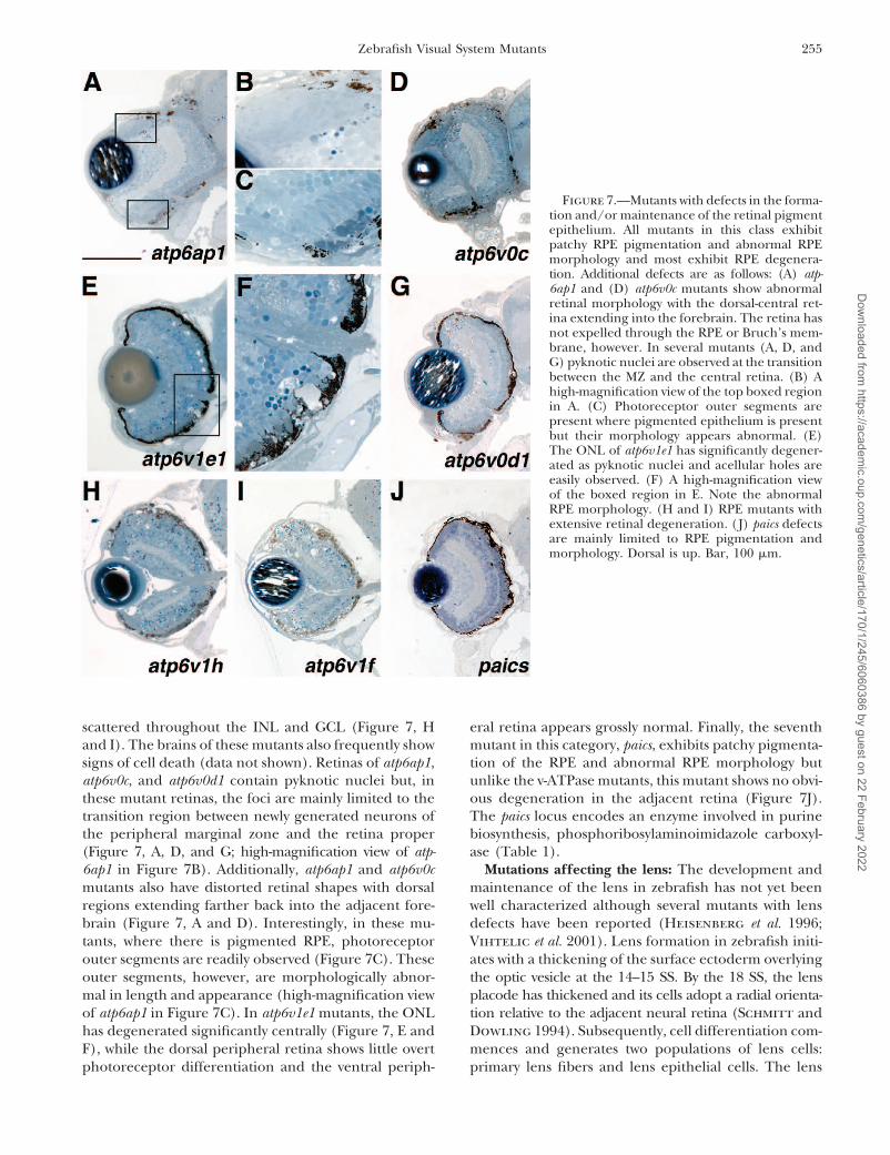
and maintenance. of RPE pigmentation (Figure 7F). atp6v1h and atp6v1fmutants show a severe retinal degeneration at 5 dpfEight loci that affect the formation and/or mainte-
nance of the RPE were identified (Table 1) and histol- with pyknotic nuclei observed throughout the ONL and
Dow
nloaded from https://academ
ic.oup.com/genetics/article/170/1/245/6060386 by guest on 22 February 2022
255Zebrafish Visual System Mutants
Figure 7.—Mutants with defects in the forma-tion and/or maintenance of the retinal pigmentepithelium. All mutants in this class exhibitpatchy RPE pigmentation and abnormal RPEmorphology and most exhibit RPE degenera-tion. Additional defects are as follows: (A) atp-6ap1 and (D) atp6v0c mutants show abnormalretinal morphology with the dorsal-central ret-ina extending into the forebrain. The retina hasnot expelled through the RPE or Bruch’s mem-brane, however. In several mutants (A, D, andG) pyknotic nuclei are observed at the transitionbetween the MZ and the central retina. (B) Ahigh-magnification view of the top boxed regionin A. (C) Photoreceptor outer segments arepresent where pigmented epithelium is presentbut their morphology appears abnormal. (E)The ONL of atp6v1e1 has significantly degener-ated as pyknotic nuclei and acellular holes areeasily observed. (F) A high-magnification viewof the boxed region in E. Note the abnormalRPE morphology. (H and I) RPE mutants withextensive retinal degeneration. (J) paics defectsare mainly limited to RPE pigmentation andmorphology. Dorsal is up. Bar, 100 �m.
scattered throughout the INL and GCL (Figure 7, H eral retina appears grossly normal. Finally, the seventhmutant in this category, paics, exhibits patchy pigmenta-and I). The brains of these mutants also frequently show
signs of cell death (data not shown). Retinas of atp6ap1, tion of the RPE and abnormal RPE morphology butunlike the v-ATPase mutants, this mutant shows no obvi-atp6v0c, and atp6v0d1 contain pyknotic nuclei but, in
these mutant retinas, the foci are mainly limited to the ous degeneration in the adjacent retina (Figure 7J).The paics locus encodes an enzyme involved in purinetransition region between newly generated neurons of
the peripheral marginal zone and the retina proper biosynthesis, phosphoribosylaminoimidazole carboxyl-ase (Table 1).(Figure 7, A, D, and G; high-magnification view of atp-
6ap1 in Figure 7B). Additionally, atp6ap1 and atp6v0c Mutations affecting the lens: The development andmaintenance of the lens in zebrafish has not yet beenmutants also have distorted retinal shapes with dorsal
regions extending farther back into the adjacent fore- well characterized although several mutants with lensdefects have been reported (Heisenberg et al. 1996;brain (Figure 7, A and D). Interestingly, in these mu-
tants, where there is pigmented RPE, photoreceptor Vihtelic et al. 2001). Lens formation in zebrafish initi-ates with a thickening of the surface ectoderm overlyingouter segments are readily observed (Figure 7C). These
outer segments, however, are morphologically abnor- the optic vesicle at the 14–15 SS. By the 18 SS, the lensplacode has thickened and its cells adopt a radial orienta-mal in length and appearance (high-magnification view
of atp6ap1 in Figure 7C). In atp6v1e1 mutants, the ONL tion relative to the adjacent neural retina (Schmitt andDowling 1994). Subsequently, cell differentiation com-has degenerated significantly centrally (Figure 7, E and
F), while the dorsal peripheral retina shows little overt mences and generates two populations of lens cells:primary lens fibers and lens epithelial cells. The lensphotoreceptor differentiation and the ventral periph-
Dow
nloaded from https://academ
ic.oup.com/genetics/article/170/1/245/6060386 by guest on 22 February 2022
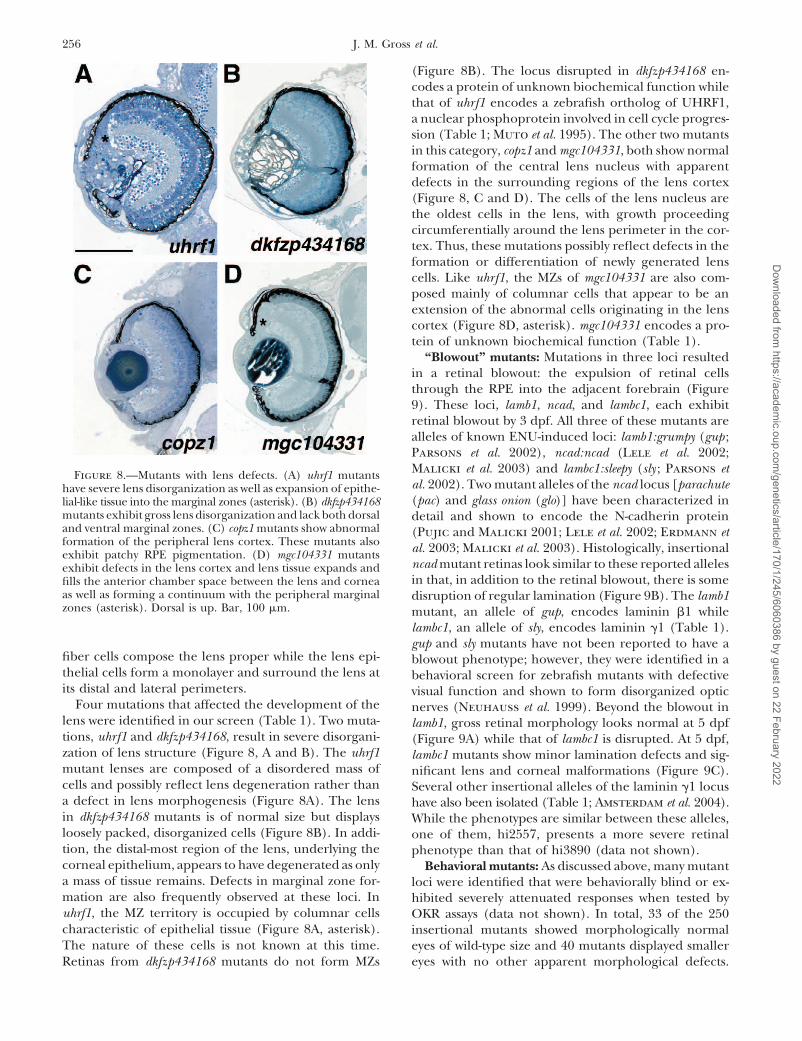
256 J. M. Gross et al.
(Figure 8B). The locus disrupted in dkfzp434168 en-codes a protein of unknown biochemical function whilethat of uhrf1 encodes a zebrafish ortholog of UHRF1,a nuclear phosphoprotein involved in cell cycle progres-sion (Table 1; Muto et al. 1995). The other two mutantsin this category, copz1 and mgc104331, both show normalformation of the central lens nucleus with apparentdefects in the surrounding regions of the lens cortex(Figure 8, C and D). The cells of the lens nucleus arethe oldest cells in the lens, with growth proceedingcircumferentially around the lens perimeter in the cor-tex. Thus, these mutations possibly reflect defects in theformation or differentiation of newly generated lenscells. Like uhrf1, the MZs of mgc104331 are also com-posed mainly of columnar cells that appear to be anextension of the abnormal cells originating in the lenscortex (Figure 8D, asterisk). mgc104331 encodes a pro-tein of unknown biochemical function (Table 1).
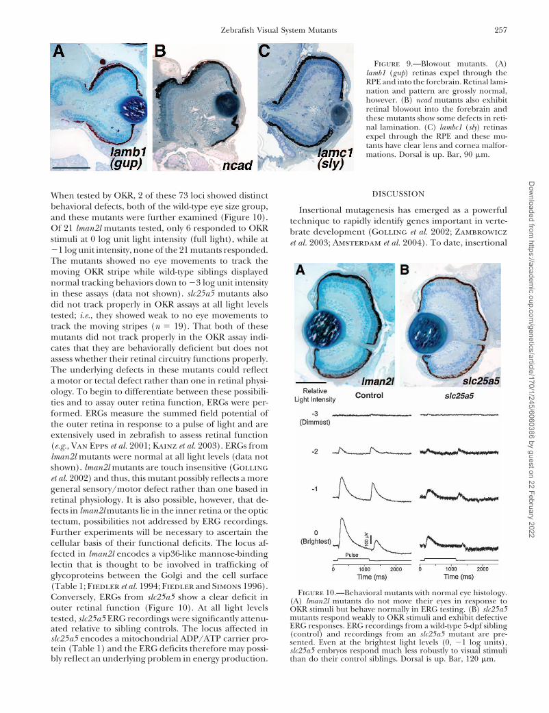
“Blowout” mutants: Mutations in three loci resultedin a retinal blowout: the expulsion of retinal cellsthrough the RPE into the adjacent forebrain (Figure9). These loci, lamb1, ncad, and lambc1, each exhibitretinal blowout by 3 dpf. All three of these mutants arealleles of known ENU-induced loci: lamb1:grumpy (gup ;Parsons et al. 2002), ncad:ncad (Lele et al. 2002;Malicki et al. 2003) and lambc1:sleepy (sly ; Parsons etFigure 8.—Mutants with lens defects. (A) uhrf1 mutantsal. 2002). Two mutant alleles of the ncad locus [parachutehave severe lens disorganization as well as expansion of epithe-(pac) and glass onion (glo)] have been characterized inlial-like tissue into the marginal zones (asterisk). (B) dkfzp434168
mutants exhibit gross lens disorganization and lack both dorsal detail and shown to encode the N-cadherin proteinand ventral marginal zones. (C) copz1 mutants show abnormal (Pujic and Malicki 2001; Lele et al. 2002; Erdmann etformation of the peripheral lens cortex. These mutants also al. 2003; Malicki et al. 2003). Histologically, insertionalexhibit patchy RPE pigmentation. (D) mgc104331 mutants
ncad mutant retinas look similar to these reported allelesexhibit defects in the lens cortex and lens tissue expands andin that, in addition to the retinal blowout, there is somefills the anterior chamber space between the lens and cornea
as well as forming a continuum with the peripheral marginal disruption of regular lamination (Figure 9B). The lamb1zones (asterisk). Dorsal is up. Bar, 100 �m. mutant, an allele of gup, encodes laminin 1 while
lambc1, an allele of sly, encodes laminin �1 (Table 1).gup and sly mutants have not been reported to have a
fiber cells compose the lens proper while the lens epi- blowout phenotype; however, they were identified in athelial cells form a monolayer and surround the lens at behavioral screen for zebrafish mutants with defectiveits distal and lateral perimeters. visual function and shown to form disorganized optic
Four mutations that affected the development of the nerves (Neuhauss et al. 1999). Beyond the blowout inlens were identified in our screen (Table 1). Two muta- lamb1, gross retinal morphology looks normal at 5 dpftions, uhrf1 and dkfzp434168, result in severe disorgani- (Figure 9A) while that of lambc1 is disrupted. At 5 dpf,zation of lens structure (Figure 8, A and B). The uhrf1 lambc1 mutants show minor lamination defects and sig-mutant lenses are composed of a disordered mass of nificant lens and corneal malformations (Figure 9C).cells and possibly reflect lens degeneration rather than Several other insertional alleles of the laminin �1 locusa defect in lens morphogenesis (Figure 8A). The lens have also been isolated (Table 1; Amsterdam et al. 2004).in dkfzp434168 mutants is of normal size but displays While the phenotypes are similar between these alleles,loosely packed, disorganized cells (Figure 8B). In addi- one of them, hi2557, presents a more severe retinaltion, the distal-most region of the lens, underlying the phenotype than that of hi3890 (data not shown).corneal epithelium, appears to have degenerated as only Behavioral mutants: As discussed above, many mutanta mass of tissue remains. Defects in marginal zone for- loci were identified that were behaviorally blind or ex-mation are also frequently observed at these loci. In hibited severely attenuated responses when tested byuhrf1, the MZ territory is occupied by columnar cells OKR assays (data not shown). In total, 33 of the 250characteristic of epithelial tissue (Figure 8A, asterisk). insertional mutants showed morphologically normalThe nature of these cells is not known at this time. eyes of wild-type size and 40 mutants displayed smaller
eyes with no other apparent morphological defects.Retinas from dkfzp434168 mutants do not form MZs
Dow
nloaded from https://academ
ic.oup.com/genetics/article/170/1/245/6060386 by guest on 22 February 2022
257Zebrafish Visual System Mutants
Figure 9.—Blowout mutants. (A)lamb1 (gup) retinas expel through theRPE and into the forebrain. Retinal lami-nation and pattern are grossly normal,however. (B) ncad mutants also exhibitretinal blowout into the forebrain andthese mutants show some defects in reti-nal lamination. (C) lambc1 (sly) retinasexpel through the RPE and these mu-tants have clear lens and cornea malfor-mations. Dorsal is up. Bar, 90 �m.
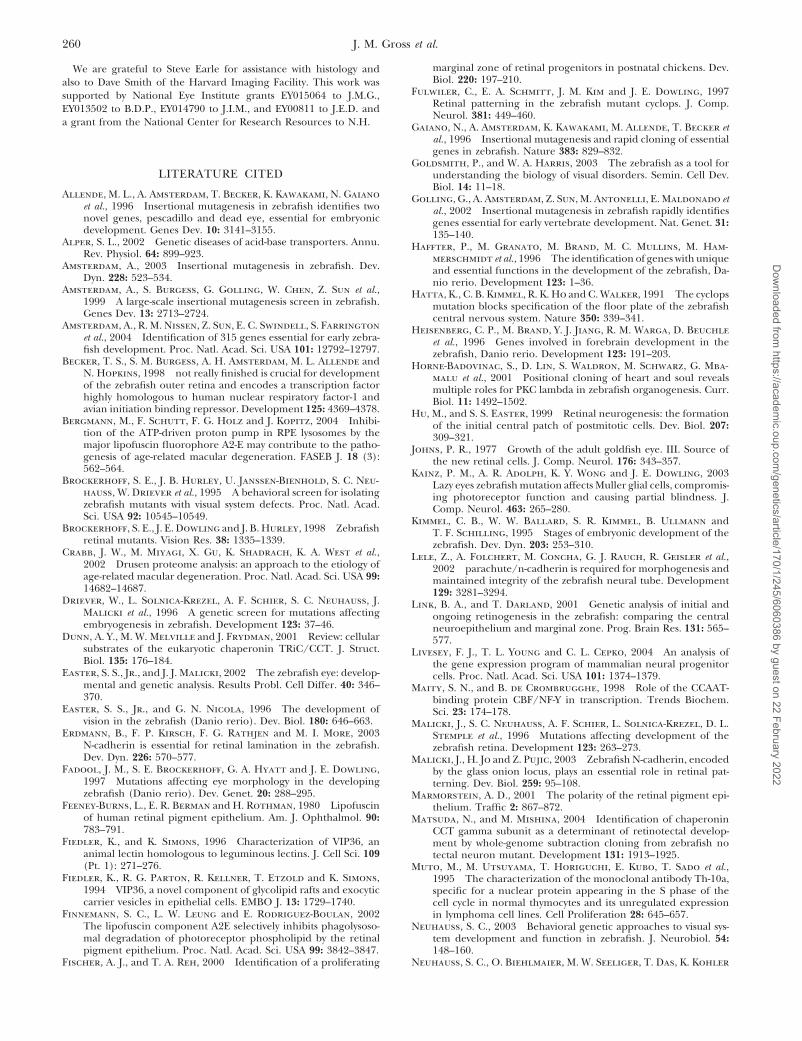
DISCUSSIONWhen tested by OKR, 2 of these 73 loci showed distinctbehavioral defects, both of the wild-type eye size group, Insertional mutagenesis has emerged as a powerfuland these mutants were further examined (Figure 10). technique to rapidly identify genes important in verte-Of 21 lman2l mutants tested, only 6 responded to OKR brate development (Golling et al. 2002; Zambrowiczstimuli at 0 log unit light intensity (full light), while at et al. 2003; Amsterdam et al. 2004). To date, insertional�1 log unit intensity, none of the 21 mutants responded.The mutants showed no eye movements to track themoving OKR stripe while wild-type siblings displayednormal tracking behaviors down to �3 log unit intensityin these assays (data not shown). slc25a5 mutants alsodid not track properly in OKR assays at all light levelstested; i.e., they showed weak to no eye movements totrack the moving stripes (n 19). That both of thesemutants did not track properly in the OKR assay indi-cates that they are behaviorally deficient but does notassess whether their retinal circuitry functions properly.The underlying defects in these mutants could reflecta motor or tectal defect rather than one in retinal physi-ology. To begin to differentiate between these possibili-ties and to assay outer retina function, ERGs were per-formed. ERGs measure the summed field potential ofthe outer retina in response to a pulse of light and areextensively used in zebrafish to assess retinal function(e.g., Van Epps et al. 2001; Kainz et al. 2003). ERGs fromlman2l mutants were normal at all light levels (data notshown). lman2l mutants are touch insensitive (Gollinget al. 2002) and thus, this mutant possibly reflects a moregeneral sensory/motor defect rather than one based inretinal physiology. It is also possible, however, that de-fects in lman2l mutants lie in the inner retina or the optictectum, possibilities not addressed by ERG recordings.Further experiments will be necessary to ascertain thecellular basis of their functional deficits. The locus af-fected in lman2l encodes a vip36-like mannose-bindinglectin that is thought to be involved in trafficking ofglycoproteins between the Golgi and the cell surface(Table 1; Fiedler et al. 1994; Fiedler and Simons 1996).
Figure 10.—Behavioral mutants with normal eye histology.Conversely, ERGs from slc25a5 show a clear deficit in (A) lman2l mutants do not move their eyes in response toouter retinal function (Figure 10). At all light levels OKR stimuli but behave normally in ERG testing. (B) slc25a5
mutants respond weakly to OKR stimuli and exhibit defectivetested, slc25a5 ERG recordings were significantly attenu-ERG responses. ERG recordings from a wild-type 5-dpf siblingated relative to sibling controls. The locus affected in(control) and recordings from an slc25a5 mutant are pre-slc25a5 encodes a mitochondrial ADP/ATP carrier pro- sented. Even at the brightest light levels (0, �1 log units),
tein (Table 1) and the ERG deficits therefore may possi- slc25a5 embryos respond much less robustly to visual stimulithan do their control siblings. Dorsal is up. Bar, 120 �m.bly reflect an underlying problem in energy production.
Dow
nloaded from https://academ
ic.oup.com/genetics/article/170/1/245/6060386 by guest on 22 February 2022

258 J. M. Gross et al.
mutagenesis in zebrafish has identified 315 loci that pic nature of their phenotype did not fit the criteria ofour screen as possessing eye-specific defects and, thus,when disrupted, produce visible embryonic phenotypesthey were not included in this report. The CCT complexby 5 dpf and for which the disrupted genes have beenis composed of eight subunits (reviewed in Dunn et al.identified (Golling et al. 2002; Amsterdam et al. 2004).2001) and in addition to the �-subunit, insertional mu-Of 250 insertional loci screened, 40 were identified thattants at four other CCT subunit-encoding loci have beendirectly affected the development and function of theidentified (Golling et al. 2002; Amsterdam et al. 2004).visual system. Of these loci, only 8 have been previouslyMuch like cct3, each of these mutants has small eyes andascribed a role in vertebrate eye development, nearlyoverall CNS degeneration (J. M. Gross, unpublishedall from mutational and functional studies in zebrafish.observations). By our screening criteria, these did notIn addition to these 40 mutants, 40 additional mutantsrepresent eye-specific phenotypes and so these mutantswere identified that had smaller eyes but were morpho-were not included in this report. These examples arelogically normal (see supplementary Table 3 at http://meant to highlight the importance of specifically defin-www.genetics.org/supplemental/). These mutants likelying the screening criteria utilized herein. Other screen-reflect defects in eye growth, possibly in the context ofing parameters might identify as eye mutants some ofoverall CNS growth defects. MZs in these mutants werethe insertional mutants excluded here and proceed inproportional for the reduced eye size. Since we werecharacterizing them as such.seeking mutants with clear morphological defects, and
From this screen, a diverse set of genes has beento limit the scope of the screen, these mutants were notidentified as playing important roles in eye developmentfurther studied. Morphological defects were observedand visual function. Many of these have not yet beenin 81 other insertional mutants in the collection (seeimplicated in or well studied during eye developmentsupplementary Table 1 at http://www.genetics.org/supand therefore their molecular characterization shouldplemental/). These mutants were not included in thisgreatly increase our understanding of these processesreport, however, as their eye phenotypes were in theas well as human pathologies that affect the eye. Forcontext of massive CNS degeneration and/or accompa-example, two mutants, ift172 and ift57, show significantnied by multiple defects in other organ systems. Eyedegrees of photoreceptor degeneration in their centralphenotypes observed in these lines could not be sepa-retinas (Figure 6, E, E�, F, and F�). These loci encoderated from these more general system-wide mutationmembers of the IFT protein family that are requiredeffects. The morphological portion of this screen soughtin cilia for anterograde transport of proteins from theto identify eye mutations that were generally healthy at 5cytoplasm, along the ciliary axoneme to the distal ciliadpf and thus likely reflecting the loss of a direct cellular(reviewed in Pazour and Rosenbaum 2002). The dis-
function for the affected locus in the eye. Changing thisruption of several IFT proteins has been associated with
parameter and thereby the threshold for inclusion in retinitis pigmentosa-like pathologies, both in mouse andthe screen would certainly increase the number of mu- in zebrafish (Pazour et al. 2002; Tsujikawa and Malickitants identified on the basis of these new criteria. 2004). Additionally, insertional mutations at these two
Indeed, several zebrafish mutants with reported eye loci also result in polycystic kidneys, implicating a commondefects were excluded from our screen. For example, pathway in the zebrafish embryo for the generation and/one such insertional mutation, that at the dead eye locus or maintenance of ciliated cell types (Sun et al. 2004).(dye), results in severe necrosis in the eyes and tectum Thus, these mutants should provide an excellent in vivovisible at 2 dpf (Allende et al. 1996). dye mutants die model for understanding intraflagellar transport andat 5 dpf and show severe necrosis in all regions of the its potential role in PR degeneration.brain in addition to lacking most of their pharyngeal We identified six insertional loci with abnormal RPEskeleton. The pleiotropic nature of the dye phenotype morphology and pigmentation and varying degrees ofmade it too severe to be included in this screen for retinal degeneration that encode components of therelatively visual system-specific mutants. Another zebra- vacuolar ATPase (v-ATPase) protein complex or v-ATPfish mutant with eye defects, no tectal neuron (ntn), gener- ase-associated proteins (Table 1). The v-ATPase is com-ated in a trimethylpsoralen mutagenesis screen (Mat- posed of two domains: the peripheral V1 complex con-suda and Mishina 2004), affects the cct3 locus that sisting of eight subunits and the integral V0 complexencodes the �-subunit of chaperonin containing TCP-1 consisting of five subunits (reviewed in Nishi and For-(CCT). The ntn mutants show extensive cell death in gac 2002). v-ATPases are best known for their roles inthe eye and tectum by 2 dpf and this death continues proton transport through which they play importantsuch that by 4 dpf the embryo has very small eyes with roles in acidification of endosomes, protein degrada-protruding lenses, a further deteriorated tectum, re- tion, endocytosis, intracellular transport, and membraneduced jaw structures, and small pectoral fins. Two inser- fusion. Several human pathologies have been attributedtional mutant alleles at the cct3 locus, hi383A and hi1867, to defects in v-ATPases, such as osteoporosis, renal tubularlook phenotypically very similar to ntn (J. M. Gross, un- acidosis, deafness, and possibly age-related macular degen-
eration (reviewed in Alper 2002; Bergmann et al. 2004).published observations). These mutants and the pleiotro-
Dow
nloaded from https://academ
ic.oup.com/genetics/article/170/1/245/6060386 by guest on 22 February 2022

259Zebrafish Visual System Mutants
Age-related macular degeneration (AMD) affects approxi- opment are thereby normal, but upon depletion of thesematernal stores, a phenotype is manifest in the continu-mately one-fourth of the population over age 65, result-ally proliferative MZs. Indeed, a significant role for ma-ing in varying degrees of visual impairment. The molec-ternal factors in morphogenesis and establishment ofular mechanisms underlying AMD, however, are largelythe embryonic body plan after the zygotic transition hasunknown. A hallmark of AMD progression is the accum-recently been reported (Wagner et al. 2004). Theseulation of drusen, storage bodies composed mainly ofphenotypes therefore might represent a continuum, notundegraded photoreceptor outer segment lipids (Feeney-of specific gene product function but actually of mater-Burns et al. 1980; Crabb et al. 2002). A major compo-nal load. Many mutants died or showed pleiotropic de-nent of drusen, A2E, accumulates in lysosomes andfects casting doubt on the specificity of their retinalblocks their ability to degrade outer segment materialphenotype and were not included in this report. Many(Finnemann et al. 2002). The v-ATPase pump is inhib-of these mutants might represent one end of this contin-ited by high concentrations of A2E, suggesting a possibleuum: low maternal load. The three mutants that dis-link between v-ATPase function and AMD progressionplayed growth retardation and central retinal defects(Bergmann et al. 2004). The six v-ATPase mutants iden-likely due to progenitor cell death might reside some-tified in this screen show differing degrees of retinalwhere in the middle of the continuum. These mutantsdegeneration, ranging from minor degeneration inmay have possessed enough maternal mRNA or proteinatp6v0d1 (V0 subunit d; Figure 7G) to severe degenera-to develop generally well but they manifest defects intion in atp6v1h (V1 subunit H; Figure 7H) and atp6v1fslightly later aspects of retinal development when these(V1 subunit F; Figure 7I). It will be interesting to lookmaternal loads were depleted. Finally, the MZ mutantsfor adult phenotypes in heterozygous animals at thesewould reside at the other end of the continuum whereloci to determine if these are applicable animal modelsa high maternal complement enabled them to developfor the study of AMD and AMD-related pathologies inquite normally and phenotypic defects were obvioushumans, several of which are autosomal dominant disor-only in the latest aspects of retinal development, i.e., inders.MZ maintenance. Thus, are these proteins specificallyWe identified several mutations that affected the de-involved in retinal development or the maintenance ofvelopment of the central retina or the development ofMZ cells in the retina or are they actually required inthe peripheral MZs and that resulted from the disrup-all proliferating cells, a function masked to differingtion of genes encoding proteins that function in aspectsdegrees by maternal stores? A survey of gene expressionof transcription, translation, and cell cycle regulationin mammalian neural progenitors relative to differenti-(Table 1). It is interesting to speculate on the natureated neurons has pointed to a significant enrichment
of these mutations with respect to early retinal develop-of transcripts in progenitors that encode proteins in-
ment as well as stem cell maintenance in the retinal volved in DNA replication, protein synthesis, proteinperiphery. It is possible that the central retinal defects turnover, and chromatin remodeling (Livesey et al.manifest in the smarca5, ccna2, and fen1 mutants result 2004). This is not surprising given the high transcrip-from specific roles of these proteins in early events of tional activity and unique developmental functions ofretinal development, i.e., in the formation of specific neural progenitor cells, suggesting that the latter of theretinal cell types. It is also possible, however, that these above scenarios might be the case. Further characteriza-phenotypes result simply from a block in retinoblast tion of these mutants will clearly be necessary however,proliferation such that most retinal cell types are miss- to differentiate between these possibilities.ing, since their progenitors die at earlier phases of devel- In summary, in this screen we have identified a diverseopment. Thus, in these mutants, the role for the disrupted set of genes that are involved in visual system developmentprotein may not be specifically in retinal patterning, but and function. Many of these have not yet been implicatedrather may be in maintaining progenitor populations such in, or studied during, eye development and therefore theirthat an appropriate number of progenitors are present molecular characterization should greatly increase theto generate all retinal cell neurons. This logic can be understanding of these processes as well as human pa-applied to the mutants that displayed reduced MZs as thologies that affect the eye. Additionally, multiplewell. Most of these resulted from disruption of a gene screens of this collection of insertional mutants, similarencoding a factor involved in DNA replication or mRNA in scope to the one reported here, are currently under-modification (Table 1). Are these factors specifically way to assay a variety of developmental, physiological,required only for maintenance of the MZ stem cell pop- and behavioral parameters. As more of these screensulation? It is doubtful that such proteins are not utilized are completed, an integrated picture should emerge asin early retinal development. This suggests that a sig- to the roles that each of these loci play during embryonicnificant maternal complement of mRNA or protein must development. The synthesis of data from several suchbe present, and/or maternally supplied proteins must screens will likely reveal previously unidentified similari-have unusually high stability such that they persist through ties between the formation and maintenance of multiple
organ systems and physiological processes.several days of development. Early aspects of retinal devel-
Dow
nloaded from https://academ
ic.oup.com/genetics/article/170/1/245/6060386 by guest on 22 February 2022
260 J. M. Gross et al.
marginal zone of retinal progenitors in postnatal chickens. Dev.We are grateful to Steve Earle for assistance with histology andBiol. 220: 197–210.also to Dave Smith of the Harvard Imaging Facility. This work was
Fulwiler, C., E. A. Schmitt, J. M. Kim and J. E. Dowling, 1997supported by National Eye Institute grants EY015064 to J.M.G.,Retinal patterning in the zebrafish mutant cyclops. J. Comp.EY013502 to B.D.P., EY014790 to J.I.M., and EY00811 to J.E.D. andNeurol. 381: 449–460.
a grant from the National Center for Research Resources to N.H. Gaiano, N., A. Amsterdam, K. Kawakami, M. Allende, T. Becker etal., 1996 Insertional mutagenesis and rapid cloning of essentialgenes in zebrafish. Nature 383: 829–832.
Goldsmith, P., and W. A. Harris, 2003 The zebrafish as a tool forLITERATURE CITED understanding the biology of visual disorders. Semin. Cell Dev.
Biol. 14: 11–18.Allende, M. L., A. Amsterdam, T. Becker, K. Kawakami, N. Gaiano Golling, G., A. Amsterdam, Z. Sun, M. Antonelli, E. Maldonado et
et al., 1996 Insertional mutagenesis in zebrafish identifies two al., 2002 Insertional mutagenesis in zebrafish rapidly identifiesnovel genes, pescadillo and dead eye, essential for embryonic genes essential for early vertebrate development. Nat. Genet. 31:development. Genes Dev. 10: 3141–3155. 135–140.
Alper, S. L., 2002 Genetic diseases of acid-base transporters. Annu. Haffter, P., M. Granato, M. Brand, M. C. Mullins, M. Ham-Rev. Physiol. 64: 899–923. merschmidt et al., 1996 The identification of genes with unique
Amsterdam, A., 2003 Insertional mutagenesis in zebrafish. Dev. and essential functions in the development of the zebrafish, Da-Dyn. 228: 523–534. nio rerio. Development 123: 1–36.
Amsterdam, A., S. Burgess, G. Golling, W. Chen, Z. Sun et al., Hatta, K., C. B. Kimmel, R. K. Ho and C. Walker, 1991 The cyclops1999 A large-scale insertional mutagenesis screen in zebrafish. mutation blocks specification of the floor plate of the zebrafishGenes Dev. 13: 2713–2724. central nervous system. Nature 350: 339–341.
Amsterdam, A., R. M. Nissen, Z. Sun, E. C. Swindell, S. Farrington Heisenberg, C. P., M. Brand, Y. J. Jiang, R. M. Warga, D. Beuchleet al., 2004 Identification of 315 genes essential for early zebra- et al., 1996 Genes involved in forebrain development in thefish development. Proc. Natl. Acad. Sci. USA 101: 12792–12797. zebrafish, Danio rerio. Development 123: 191–203.
Becker, T. S., S. M. Burgess, A. H. Amsterdam, M. L. Allende and Horne-Badovinac, S., D. Lin, S. Waldron, M. Schwarz, G. Mba-N. Hopkins, 1998 not really finished is crucial for development malu et al., 2001 Positional cloning of heart and soul revealsof the zebrafish outer retina and encodes a transcription factor multiple roles for PKC lambda in zebrafish organogenesis. Curr.highly homologous to human nuclear respiratory factor-1 and Biol. 11: 1492–1502.avian initiation binding repressor. Development 125: 4369–4378. Hu, M., and S. S. Easter, 1999 Retinal neurogenesis: the formation
Bergmann, M., F. Schutt, F. G. Holz and J. Kopitz, 2004 Inhibi- of the initial central patch of postmitotic cells. Dev. Biol. 207:tion of the ATP-driven proton pump in RPE lysosomes by the 309–321.major lipofuscin fluorophore A2-E may contribute to the patho- Johns, P. R., 1977 Growth of the adult goldfish eye. III. Source ofgenesis of age-related macular degeneration. FASEB J. 18 (3): the new retinal cells. J. Comp. Neurol. 176: 343–357.562–564. Kainz, P. M., A. R. Adolph, K. Y. Wong and J. E. Dowling, 2003
Brockerhoff, S. E., J. B. Hurley, U. Janssen-Bienhold, S. C. Neu- Lazy eyes zebrafish mutation affects Muller glial cells, compromis-hauss, W. Driever et al., 1995 A behavioral screen for isolating ing photoreceptor function and causing partial blindness. J.zebrafish mutants with visual system defects. Proc. Natl. Acad. Comp. Neurol. 463: 265–280.Sci. USA 92: 10545–10549. Kimmel, C. B., W. W. Ballard, S. R. Kimmel, B. Ullmann and
Brockerhoff, S. E., J. E. Dowling and J. B. Hurley, 1998 Zebrafish T. F. Schilling, 1995 Stages of embryonic development of theretinal mutants. Vision Res. 38: 1335–1339. zebrafish. Dev. Dyn. 203: 253–310.
Crabb, J. W., M. Miyagi, X. Gu, K. Shadrach, K. A. West et al., Lele, Z., A. Folchert, M. Concha, G. J. Rauch, R. Geisler et al.,2002 Drusen proteome analysis: an approach to the etiology of 2002 parachute/n-cadherin is required for morphogenesis andage-related macular degeneration. Proc. Natl. Acad. Sci. USA 99: maintained integrity of the zebrafish neural tube. Development14682–14687. 129: 3281–3294.
Driever, W., L. Solnica-Krezel, A. F. Schier, S. C. Neuhauss, J. Link, B. A., and T. Darland, 2001 Genetic analysis of initial andMalicki et al., 1996 A genetic screen for mutations affecting ongoing retinogenesis in the zebrafish: comparing the centralembryogenesis in zebrafish. Development 123: 37–46. neuroepithelium and marginal zone. Prog. Brain Res. 131: 565–
Dunn, A. Y., M. W. Melville and J. Frydman, 2001 Review: cellular 577.substrates of the eukaryotic chaperonin TRiC/CCT. J. Struct. Livesey, F. J., T. L. Young and C. L. Cepko, 2004 An analysis ofBiol. 135: 176–184. the gene expression program of mammalian neural progenitor
Easter, S. S., Jr., and J. J. Malicki, 2002 The zebrafish eye: develop- cells. Proc. Natl. Acad. Sci. USA 101: 1374–1379.mental and genetic analysis. Results Probl. Cell Differ. 40: 346– Maity, S. N., and B. de Crombrugghe, 1998 Role of the CCAAT-370. binding protein CBF/NF-Y in transcription. Trends Biochem.
Easter, S. S., Jr., and G. N. Nicola, 1996 The development of Sci. 23: 174–178.vision in the zebrafish (Danio rerio). Dev. Biol. 180: 646–663. Malicki, J., S. C. Neuhauss, A. F. Schier, L. Solnica-Krezel, D. L.
Erdmann, B., F. P. Kirsch, F. G. Rathjen and M. I. More, 2003 Stemple et al., 1996 Mutations affecting development of theN-cadherin is essential for retinal lamination in the zebrafish. zebrafish retina. Development 123: 263–273.Dev. Dyn. 226: 570–577. Malicki, J., H. Jo and Z. Pujic, 2003 Zebrafish N-cadherin, encoded
Fadool, J. M., S. E. Brockerhoff, G. A. Hyatt and J. E. Dowling, by the glass onion locus, plays an essential role in retinal pat-1997 Mutations affecting eye morphology in the developing terning. Dev. Biol. 259: 95–108.zebrafish (Danio rerio). Dev. Genet. 20: 288–295. Marmorstein, A. D., 2001 The polarity of the retinal pigment epi-
Feeney-Burns, L., E. R. Berman and H. Rothman, 1980 Lipofuscin thelium. Traffic 2: 867–872.of human retinal pigment epithelium. Am. J. Ophthalmol. 90: Matsuda, N., and M. Mishina, 2004 Identification of chaperonin783–791. CCT gamma subunit as a determinant of retinotectal develop-
Fiedler, K., and K. Simons, 1996 Characterization of VIP36, an ment by whole-genome subtraction cloning from zebrafish noanimal lectin homologous to leguminous lectins. J. Cell Sci. 109 tectal neuron mutant. Development 131: 1913–1925.(Pt. 1): 271–276. Muto, M., M. Utsuyama, T. Horiguchi, E. Kubo, T. Sado et al.,
Fiedler, K., R. G. Parton, R. Kellner, T. Etzold and K. Simons, 1995 The characterization of the monoclonal antibody Th-10a,1994 VIP36, a novel component of glycolipid rafts and exocytic specific for a nuclear protein appearing in the S phase of thecarrier vesicles in epithelial cells. EMBO J. 13: 1729–1740. cell cycle in normal thymocytes and its unregulated expression
Finnemann, S. C., L. W. Leung and E. Rodriguez-Boulan, 2002 in lymphoma cell lines. Cell Proliferation 28: 645–657.The lipofuscin component A2E selectively inhibits phagolysoso- Neuhauss, S. C., 2003 Behavioral genetic approaches to visual sys-mal degradation of photoreceptor phospholipid by the retinal tem development and function in zebrafish. J. Neurobiol. 54:pigment epithelium. Proc. Natl. Acad. Sci. USA 99: 3842–3847. 148–160.
Neuhauss, S. C., O. Biehlmaier, M. W. Seeliger, T. Das, K. KohlerFischer, A. J., and T. A. Reh, 2000 Identification of a proliferating
Dow
nloaded from https://academ
ic.oup.com/genetics/article/170/1/245/6060386 by guest on 22 February 2022
261Zebrafish Visual System Mutants
et al., 1999 Genetic disorders of vision revealed by a behavioral Sun, Z., A. Amsterdam, G. J. Pazour, D. G. Cole, M. S. Miller etal., 2004 A genetic screen in zebrafish identifies cilia genes asscreen of 400 essential loci in zebrafish. J. Neurosci. 19: 8603–a principal cause of cystic kidney. Development 131: 4085–4093.8615.
Tropepe, V., B. L. Coles, B. J. Chiasson, D. J. Horsford, A. J.Nishi, T., and M. Forgac, 2002 The vacuolar (H�)-ATPases—Elia et al., 2000 Retinal stem cells in the adult mammalian eye.nature’s most versatile proton pumps. Nat. Rev. Mol. Cell. Biol.Science 287: 2032–2036.3: 94–103.
Tsujikawa, M., and J. Malicki, 2004 Intraflagellar transport genesParsons, M. J., S. M. Pollard, L. Saude, B. Feldman, P. Coutinhoare essential for differentiation and survival of vertebrate sensoryet al., 2002 Zebrafish mutants identify an essential role for lami-neurons. Neuron 42: 703–716.nins in notochord formation. Development 129: 3137–3146.
Van Epps, H. A., C. M. Yim, J. B. Hurley and S. E. Brockerhoff,Pazour, G. J., and J. L. Rosenbaum, 2002 Intraflagellar transport2001 Investigations of photoreceptor synaptic transmission andand cilia-dependent diseases. Trends Cell Biol. 12: 551–555.light adaptation in the zebrafish visual mutant nrc. Invest. Oph-Pazour, G. J., S. A. Baker, J. A. Deane, D. G. Cole, B. L. Dickertthalmol. Visual Sci. 42: 868–874.et al., 2002 The intraflagellar transport protein, IFT88, is essen-
Vihtelic, T. S., Y. Yamamoto, M. T. Sweeney, W. R. Jeffery andtial for vertebrate photoreceptor assembly and maintenance. J.D. R. Hyde, 2001 Arrested differentiation and epithelial cellCell Biol. 157: 103–113.degeneration in zebrafish lens mutants. Dev. Dyn. 222: 625–636.Peterson, R. T., J. D. Mably, J. N. Chen and M. C. Fishman, 2001
Wagner, D. S., R. Dosch, K. A. Mintzer, A. P. Wiemelt and M. C.Convergence of distinct pathways to heart patterning revealedMullins, 2004 Maternal control of development at the midblas-by the small molecule concentramide and the mutation heart-tula transition and beyond: mutants from the zebrafish II. Dev.and-soul. Curr. Biol. 11: 1481–1491.Cell 6: 781–790.Pujic, Z., and J. Malicki, 2001 Mutation of the zebrafish glass onion
Wei, X., and J. Malicki, 2002 nagie oko, encoding a MAGUK-familylocus causes early cell-nonautonomous loss of neuroepithelialprotein, is essential for cellular patterning of the retina. Nat.integrity followed by severe neuronal patterning defects in the Genet. 31: 150–157.retina. Dev. Biol. 234: 454–469. Westerfield, M., 1995 The Zebrafish Book. University of OregonRebagliati, M. R., R. Toyama, P. Haffter and I. B. Dawid, 1998 Press, Eugene, OR.
cyclops encodes a nodal-related factor involved in midline signal- Wetts, R., and S. E. Fraser, 1988 Multipotent precursors can giveing. Proc. Natl. Acad. Sci. USA 95: 9932–9937. rise to all major cell types of the frog retina. Science 239: 1142–
Sampath, K., A. L. Rubinstein, A. M. Cheng, J. O. Liang, K. Fekany et 1145.al., 1998 Induction of the zebrafish ventral brain and floorplate Zambrowicz, B. P., A. Abuin, R. Ramirez-Solis, L. J. Richter, J.requires cyclops/nodal signalling. Nature 395: 185–189. Piggott et al., 2003 Wnk1 kinase deficiency lowers blood pres-
Schmitt, E. A., and J. E. Dowling, 1994 Early eye morphogenesis in sure in mice: a gene-trap screen to identify potential targetsthe zebrafish, Brachydanio rerio. J. Comp. Neurol. 344: 532–542. for therapeutic intervention. Proc. Natl. Acad. Sci. USA 100:
Schmitt, E. A., and J. E. Dowling, 1999 Early retinal development 14109–14114.in the zebrafish, Danio rerio: light and electron microscopicanalyses. J. Comp. Neurol. 404: 515–536. Communicating editor: M. Justice
Dow
nloaded from https://academ
ic.oup.com/genetics/article/170/1/245/6060386 by guest on 22 February 2022

Dow
nloaded from https://academ
ic.oup.com/genetics/article/170/1/245/6060386 by guest on 22 February 2022