insertional mutagenesis strategies
TRANSCRIPT


Insertional Mutagenesis Strategiesin Cancer Genetics


Adam J. Dupuy · David A. LargaespadaEditors
Insertional MutagenesisStrategies in Cancer Genetics
13
EditorsAdam J. DupuyDepartment of Anatomy & Cell BiologyCarver College of MedicineUniversity of IowaIowa City, IA 52242, USA
David A. Largaespada, Ph.DThe Department of Genetics, Cell Biology
& DevelopmentUniversity of MinnesotaMinneapolis, MN 55455, USA
ISBN 978-1-4419-7655-0 e-ISBN 978-1-4419-7656-7DOI 10.1007/978-1-4419-7656-7Springer New York Dordrecht Heidelberg London
Library of Congress Control Number: 2010938734
© Springer Science+Business Media, LLC 2011All rights reserved. This work may not be translated or copied in whole or in part without the writtenpermission of the publisher (Springer Science+Business Media, LLC, 233 Spring Street, New York,NY 10013, USA), except for brief excerpts in connection with reviews or scholarly analysis. Use inconnection with any form of information storage and retrieval, electronic adaptation, computer software,or by similar or dissimilar methodology now known or hereafter developed is forbidden.The use in this publication of trade names, trademarks, service marks, and similar terms, even if they arenot identified as such, is not to be taken as an expression of opinion as to whether or not they are subjectto proprietary rights.While the advice and information in this book are believed to be true and accurate at the date of goingto press, neither the authors nor the editors nor the publisher can accept any legal responsibility forany errors or omissions that may be made. The publisher makes no warranty, express or implied, withrespect to the material contained herein.
Printed on acid-free paper
Springer is part of Springer Science+Business Media (www.springer.com)

Contents
1 Insertional Mutagenesis: A Powerful Tool in Cancer Research . . . . 1Anton Berns
2 Retroviral Insertional Mutagenesis in Mouse Modelsof Leukemia and Lymphoma . . . . . . . . . . . . . . . . . . . . . . . 19David A. Largaespada
3 Gene Discovery by MMTV Mediated Insertional Mutagenesis . . . . 39Annabel Vendel-Zwaagstra and John Hilkens
4 Chicken Models of Retroviral Insertional Mutagenesis . . . . . . . . 77Vladimír Pecenka, Petr Pajer, Vít Karafiát, and Michal Dvorák
5 Sleeping Beauty Models of Cancer . . . . . . . . . . . . . . . . . . . . 113Jesse D. Riordan, Laura M. Rogers,Katherine E. Berquam-Vrieze, and Adam J. Dupuy
6 Insertional Mutagenesis in Hematopoietic Cells: LessonsLearned from Adverse Events in Clinical Gene Therapy Trials . . . . 131Lars U. Müller, Michael D. Milsom, and David A. Williams
7 Bioinformatics of High-Throughput Insertional Mutagenesis . . . . . 167Keiko Akagi, Ming Yi, Jean Roayaei, and Robert M. Stephens
Index . . . . . . . . . . . . . . . . . . . . . . . . . . . . . . . . . . . . . 189
v

Contributors
Keiko Akagi Mouse Cancer Genetics Program, National Cancer Institute, Centerfor Cancer Research, Frederick, MD 21702, USA; Comprehensive Cancer Center,Ohio State University, Columbus, OH 43210, USA, [email protected]
Anton Berns Division of Molecular Genetics, Centre of Biomedical Genetics, andCancer Genomics Centre, The Netherlands Cancer Institute, 1066 CX, Amsterdam,The Netherlands, [email protected]
Katherine E. Berquam-Vrieze Department of Anatomy and Cell Biology,University of Iowa, Iowa City, IA 52241, USA, [email protected]
Adam J. Dupuy Department of Anatomy and Cell Biology, Carver College ofMedicine, University of Iowa, Iowa City, IA 52242, USA,[email protected]
Michal Dvorák Department of Molecular Virology, Institute of MolecularGenetics, Prague CZ–14220, Czech Republic, [email protected]
John Hilkens Divisions of Molecular Genetics, The Netherlands Cancer Institute,1066 CX Amsterdam, The Netherlands, [email protected]
Vít Karafiát Department of Molecular Virology, Institute of Molecular Genetics,Prague CZ–14220, Czech Republic, [email protected]
David A. Largaespada The Department of Genetics, Cell Biology andDevelopment, The Department of Pediatrics, Masonic Cancer Center, The Centerfor Genome Engineering, The University of Minnesota, Minneapolis, MN 55455,USA, [email protected]
Michael D. Milsom Harvard Medical School, Children’s Hospital Boston, Boston,MA 02115, USA, [email protected]
Lars U. Müller Harvard Medical School, Children’s Hospital Boston, Boston, MA02115, USA, [email protected]
Petr Pajer Department of Molecular Virology, Institute of Molecular Genetics,Prague CZ–14220, Czech Republic, [email protected]
vii
viii Contributors
Vladimír Pecenka Department of Molecular Virology, Institute of MolecularGenetics, Prague CZ–14220, Czech Republic, [email protected]
Jesse D. Riordan Department of Anatomy and Cell Biology, University of Iowa,Iowa City, IA 52242, USA, [email protected]
Jean Roayaei Statistical Consulting Group, SAIC-Frederick, Frederick, MD21701, USA, [email protected]
Laura M. Rogers Department of Anatomy and Cell Biology, University of Iowa,Iowa City, IA 52242, USA, [email protected]
Robert M. Stephens Advanced Technology Program, Advanced BiomedicalComputing Center, SAIC-Frederick Inc., NCI-Frederick, Frederick, MD 21702,USA, [email protected]
Annabel Vendel-Zwaagstra Divisions of Molecular Genetics, The NetherlandsCancer Institute, 1066 CX Amsterdam, The Netherlands,[email protected]
David A. Williams Harvard Medical School, Children’s Hospital Boston, Boston,MA 02115, USA, [email protected]
Ming Yi Advanced Technology Program, Advanced Biomedical ComputingCenter, SAIC-Frederick Inc., NCI-Frederick, Frederick, MD 21702, USA,[email protected]
Chapter 1Insertional Mutagenesis: A Powerful Toolin Cancer Research
Anton Berns
1.1 Introduction
Cancer arises largely as a result of mutations in genes regulating growth anddifferentiation of cells. In most tumors multiple genes are usually affected, result-ing in either loss or gain-of-function of the encoded protein. The former categoryencompasses tumor suppressor genes whereas the latter belong to the oncogenesclass. However, one should keep in mind that this designation can be context-dependent, and consequently a gene might act as an oncogene in one specific contextand as a tumor suppressor in another. The number and nature of genes needed todrive cancerous growth can vary substantially and might lie between three and 10and can greatly differ between different tumor types. If mutations occur in earlyprogenitors, the cells might already be endowed with self-renewal capacity andtherefore the additional mutations required to give rise to a malignant transfor-mation might be very different from those needed to confer cancerous growth onfurther differentiated cells. The burning questions cancer researchers are facing arewhich combination of mutations are required to drive tumor growth in which of thecell types that constitute the tumor, and to what extent tumor growth is “addicted”to these mutations. Further insight into the mechanisms that promote metastasisand that can make tumor cells refractory to chemotherapy and targeted drugs areimmediate next questions that we need to answer to develop more effective interven-tion strategies. This requires a detailed insight into how tumor cell proliferation andmetastasis is controlled. Gene mutations critical for tumorigenesis have been foundin a number of ways: by identifying transforming genes through DNA transfec-tion experiments, by mapping recurrent translocations, amplifications, and deletionsin chromosomal regions, by identifying the genes captured by acute transform-ing retroviruses, by defining the insertion sites of slow transforming retrovirusesand transposons, and, more recently by the high throughput sequencing of cancer
A. Berns (B)Division of Molecular Genetics, Centre of Biomedical Genetics, and Cancer Genomics Centre,The Netherlands Cancer Institute, 1066 CX, Amsterdam, The Netherlandse-mail: [email protected]
1A.J. Dupuy, D.A. Largaespada (eds.), Insertional Mutagenesis Strategiesin Cancer Genetics, DOI 10.1007/978-1-4419-7656-7_1,C© Springer Science+Business Media, LLC 2011
2 A. Berns
genomes. A number of general conclusions can be drawn from these studies. First,the number of recurrent mutations that confer a selective advantage to tumor cellsis likely larger than previously thought. Second, a greater variety of genes than ear-lier suspected might have transforming potential when mutated or inappropriatelyexpressed. Third, many of the genes contributing to tumorigenesis show a strongcontext dependence, and their expression levels might have to be within particularboundaries to effectively mediate the oncogenic effect, indicating that there is muchmore subtlety in the evolutionary process of tumor development than previouslythought.
Insertional mutagenesis should be viewed as a “tool” in this context as it seems toplay only a modest role in spontaneous tumorigenesis in man. However, it providesus with a potent methodology that can answer many of the burning questions. Itcan be used nearly in the same way as suppressor and enhancer screens previouslyperformed in fruit flies and worms, to identify components of signaling pathwaysand to elucidate genetic interactions between pathways. This chapter is written toconvey concepts rather than to provide the reader with experimental details. Thesecan undoubtedly be found in many of the more specific chapters in this book.
1.2 Historical Perspective
Insertional mutagenesis has, in fact, a long history, although the underlying mech-anism has remained obscure for more than half a century. It was at the basis ofthe high incidence of mammary tumors conveyed by cell-depleted milk of high-incidence mammary tumor strains as first reported by Bittner almost 70 years ago[4]. Insertional mutagenesis is also the cause of the high spontaneous lymphomaincidence recognized in some strains of mice in this same period ([30], #3801). Thediscovery of reverse transcription and oncogenic retroviruses focused much atten-tion on the role of retroviruses in cancer. However, the difference between acutetransforming retroviruses that carry oncogenes in their genomes and slow trans-forming viruses that can activate resident proto-oncogenes, was not immediatelyrecognized. The first observation pointing in this direction was the observation thatthe spontaneous lymphoma development in the AKR strain was associated with anincrease of the number of proviral copies in tumor DNA [3]. However, it took severalyears before the underlying mechanism, i.e., insertion of proviruses in the vicinity ofproto-oncogenes or within tumor suppressor genes with their concomitant activationand inactivation, respectively, was elucidated.
In the first report to this effect it was shown that proviral insertions had occurredin the close proximity of an already well-known proto-oncogene—Myc in AvianLeukosis virus-induced tumors [16, 27]. While initially this provided informationon the mechanism of action of these slow transforming viruses, it was quickly rec-ognized that this could be used as a tool to identify new cancer-causing genes. Theconcept behind this is simple: Although the integration machinery of retrovirusesand transposons does not cause fully random insertions—chromatin structure and
1 Insertional Mutagenesis: A Powerful Tool in Cancer Research 3
sequence context undoubtedly leads to skewing—the process appeared sufficientlyrandom to assume that insertion will occur in or near almost all genes when manymillions of insertions take place in millions of cells. Occasional insertions nearproto-oncogenes or within tumor suppressor genes might endow these cells withsuch a prominent selective growth advantage that this results in the outgrowth ofclones that harbor insertions close to the same (putative) proto-oncogenes in inde-pendent transposon-induced tumors. The experimental strategy then is to inducetumors in a series of mice, e.g., by retroviral infection, and determine the insertionsites of the proviruses in the resulting, mostly clonal tumors. If proviral insertionsor transposons cluster in the same region of DNA in tumors of independent micemore frequently than would be expected by chance, it is very likely that these inser-tions are associated with a selective advantage for the tumor cell. Such insertionclusters are called “Common Insertion Sites” (CIS). CISs are to be distinguishedfrom “preferential insertion sites,” the term that indicates that some loci might beoccupied more frequently than others as a result of the interplay between chro-matic structure, primary sequence and the integration machinery of the retrovirus ortransposon. Preferential insertion sites constitute the background noise in insertionalmutagenesis screens.
At the time the sequence of the mouse genome was not yet known and sequenc-ing techniques archaic in comparison with current technologies, CISs had to bedetermined by cloning the flanking sequences of proviral insertions and using thoseas probes to ask whether changes could be found in the sizes of the correspond-ing genomic restriction fragments of DNA isolated from independent tumors usingSouthern blotting. Although laborious, this approach has identified many interestingnew oncogenes that have provided important insights in the underlying mechanismsof tumorigenesis. The analysis of Mouse Mammary Tumor Virus (MMTV)-inducedmammary carcinomas resulted in the discovery of the Wnt family of oncogenescritically important for stem cell self-renewal [28]. Another gene critical for stemcell renewal, Bmi1, was also identified by insertional mutagenesis [36]. A numberof groups have exploited insertional mutagenesis to identify new oncogenes andtumor suppressor genes (for review, see [32]). In many instances it could be shownthat genes carrying insertions within or nearby were deregulated or truncated by theinsertions and had acquired oncogenic capacity or had lost tumor suppressor activ-ity, thereby validating the robustness of the strategy. However, long-range effectshave also been noted, making it sometimes cumbersome to identify the gene orgenes near an insertion cluster conferring the selective advantage [20]. Altogether,the number of candidate oncogenes and tumor suppressor genes identified by inser-tional mutagenesis is impressive and in the same range as the number discoveredby all other methods together, assuming the vast majority proofs to be genuine can-cer genes. However, to identify these candidates required a relatively small effortin comparison with other approaches. Furthermore, it can serve as a valuable com-plementary strategy to the current deep sequencing initiatives of cancer genomes.Genes found by both methods are almost certainly involved in cancer and there-fore insertional mutagenesis is very suitable as a cross-validation method for deepsequencing.
4 A. Berns
1.3 The Site of Insertion can Provide Mechanistic Insights
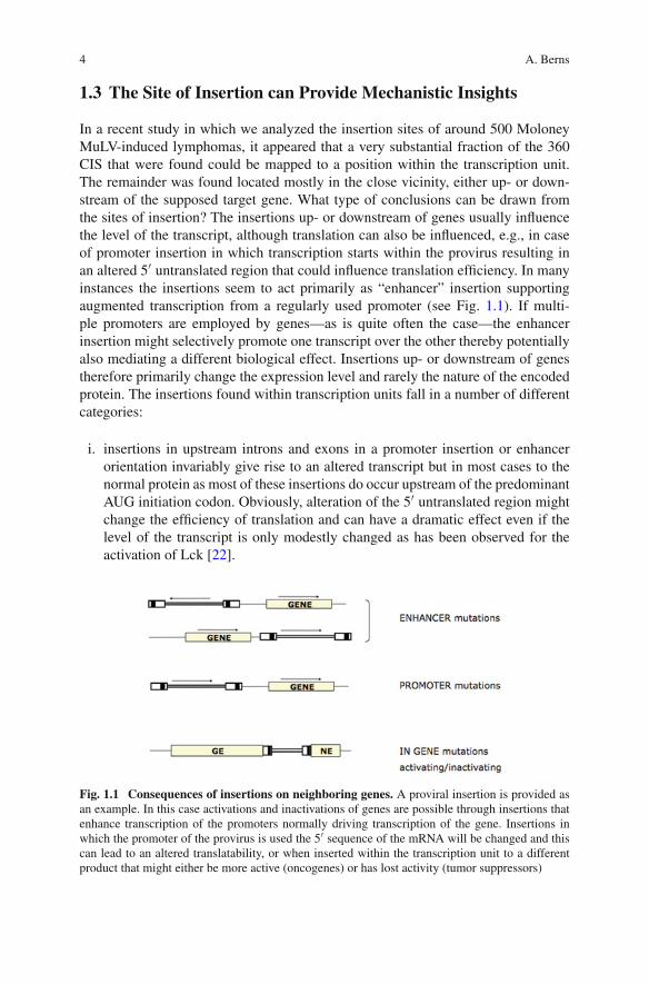
In a recent study in which we analyzed the insertion sites of around 500 MoloneyMuLV-induced lymphomas, it appeared that a very substantial fraction of the 360CIS that were found could be mapped to a position within the transcription unit.The remainder was found located mostly in the close vicinity, either up- or down-stream of the supposed target gene. What type of conclusions can be drawn fromthe sites of insertion? The insertions up- or downstream of genes usually influencethe level of the transcript, although translation can also be influenced, e.g., in caseof promoter insertion in which transcription starts within the provirus resulting inan altered 5′ untranslated region that could influence translation efficiency. In manyinstances the insertions seem to act primarily as “enhancer” insertion supportingaugmented transcription from a regularly used promoter (see Fig. 1.1). If multi-ple promoters are employed by genes—as is quite often the case—the enhancerinsertion might selectively promote one transcript over the other thereby potentiallyalso mediating a different biological effect. Insertions up- or downstream of genestherefore primarily change the expression level and rarely the nature of the encodedprotein. The insertions found within transcription units fall in a number of differentcategories:
i. insertions in upstream introns and exons in a promoter insertion or enhancerorientation invariably give rise to an altered transcript but in most cases to thenormal protein as most of these insertions do occur upstream of the predominantAUG initiation codon. Obviously, alteration of the 5′ untranslated region mightchange the efficiency of translation and can have a dramatic effect even if thelevel of the transcript is only modestly changed as has been observed for theactivation of Lck [22].
Fig. 1.1 Consequences of insertions on neighboring genes. A proviral insertion is provided asan example. In this case activations and inactivations of genes are possible through insertions thatenhance transcription of the promoters normally driving transcription of the gene. Insertions inwhich the promoter of the provirus is used the 5′ sequence of the mRNA will be changed and thiscan lead to an altered translatability, or when inserted within the transcription unit to a differentproduct that might either be more active (oncogenes) or has lost activity (tumor suppressors)

1 Insertional Mutagenesis: A Powerful Tool in Cancer Research 5
ii. Insertion in a noncoding region of a 3′ exon. Usually, the provirus can act hereboth as an enhancer and a transcriptional termination site and can so removeelements from the transcript that are targeted by micro-RNAs, thereby modu-lating either mRNA stability or its translation. This might explain the narrowdomain in which proviral insertion site might be found. A good example is theCIS cluster near the translational stop codon in lymphomas with a provirallyactivated N-myc gene [34].
iii. Insertions in internal introns or exons in the same transcriptional orientationas the gene. This usually results in the expression of truncated proteins, eitherencoded by 5′ region of the gene or by the 3′ region of the gene starting transla-tion either from an internal AUG or from sequences in the provirus with splicingto a downstream exon. Examples of both are found. Deletion of the carboxyter-minal region of the Tpl2 (or Cot) protein kinase as a result of insertions withinthe last intron results in a constitutive active kinase since the carboxyterminaldomain requires phosphorylation to permit activation of the kinase [5]. On theother hand, insertion in the Notch gene results in a mRNA that expresses thecarboxyterminal region of Notch [17]. This is the business end of the proteinand is normally produced through a gamma-secretase-dependent mechanismthat relies on ligand-induced regulated intramembranous proteolysis. The car-boxyterminal fragment that is produced then translocates to the nucleus whereit becomes engaged in transcription regulation.
iv. Insertions within the transcription unit in either orientation can also result in aprotein that acts in a dominant negative fashion or in its complete inactivation.This type of insertion is found in tumor suppressor genes leading to their inac-tivation. One might find insertions in both alleles of the tumor suppressor geneindicating that each insertion does confer substantial selective growth advantageto the cell.
Insertional mutagenesis is probably one of the most powerful methods toidentify haplo-insufficient tumor suppressor genes as other genetic evidence forhaploinsufficiency is often difficult to obtain. The different effects of provirusesor transposon on gene function is schematically depicted in Fig. 1.1.
1.4 Methodologies
Other chapters in this book will describe in detail specific new strategies in whichDNA transposons are used that facilitate identification of the genes that are affectedby the insertion. Therefore, I will limit myself describing a number of general fea-tures of insertional mutagenesis screens. A critical condition for successful screensis to generate a sufficient number of insertion events in order to have sufficient prob-ability to achieve insertions near genes that can confer a selective advantage. Thereplication competent retroviruses are particular powerful in this respect as theyenable multiple rounds of infection leading to the accumulation of proviral copiesthat can amount to often more than 20. This requires escape from interference by
6 A. Berns
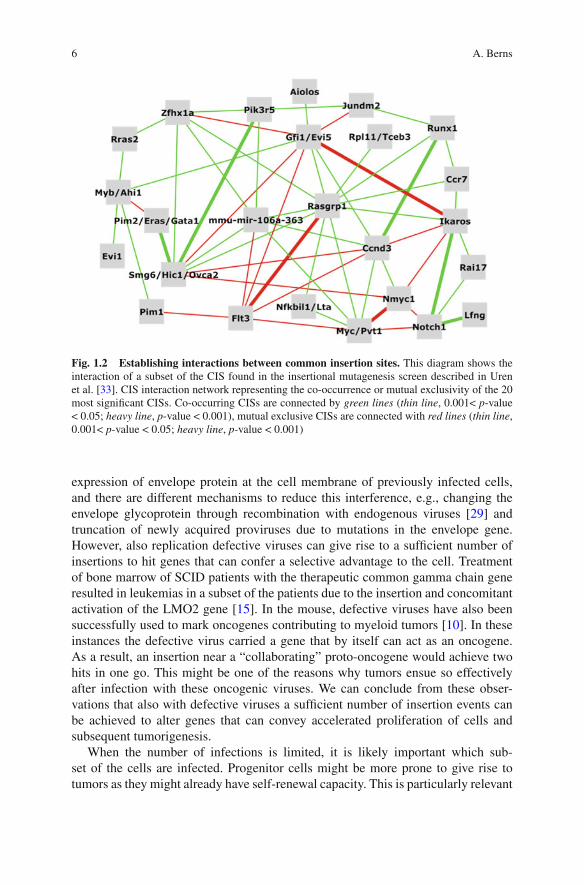
Fig. 1.2 Establishing interactions between common insertion sites. This diagram shows theinteraction of a subset of the CIS found in the insertional mutagenesis screen described in Urenet al. [33]. CIS interaction network representing the co-occurrence or mutual exclusivity of the 20most significant CISs. Co-occurring CISs are connected by green lines (thin line, 0.001< p-value< 0.05; heavy line, p-value < 0.001), mutual exclusive CISs are connected with red lines (thin line,0.001< p-value < 0.05; heavy line, p-value < 0.001)
expression of envelope protein at the cell membrane of previously infected cells,and there are different mechanisms to reduce this interference, e.g., changing theenvelope glycoprotein through recombination with endogenous viruses [29] andtruncation of newly acquired proviruses due to mutations in the envelope gene.However, also replication defective viruses can give rise to a sufficient number ofinsertions to hit genes that can confer a selective advantage to the cell. Treatmentof bone marrow of SCID patients with the therapeutic common gamma chain generesulted in leukemias in a subset of the patients due to the insertion and concomitantactivation of the LMO2 gene [15]. In the mouse, defective viruses have also beensuccessfully used to mark oncogenes contributing to myeloid tumors [10]. In theseinstances the defective virus carried a gene that by itself can act as an oncogene.As a result, an insertion near a “collaborating” proto-oncogene would achieve twohits in one go. This might be one of the reasons why tumors ensue so effectivelyafter infection with these oncogenic viruses. We can conclude from these obser-vations that also with defective viruses a sufficient number of insertion events canbe achieved to alter genes that can convey accelerated proliferation of cells andsubsequent tumorigenesis.
When the number of infections is limited, it is likely important which sub-set of the cells are infected. Progenitor cells might be more prone to give rise totumors as they might already have self-renewal capacity. This is particularly relevant

1 Insertional Mutagenesis: A Powerful Tool in Cancer Research 7
when transposons are used that are inserted into the genome of all cells by germlinemodification and whose transposition is initiated by the cell-type specific activationof the transposase. The transposase might need to be activated in the right (pro-genitor) cell to permit tumor development. While this is a potential limitation, italso provides a unique opportunity to determine whether only particular cells ina tissue have the capacity to command tumorigenesis, e.g., by using distinct Crestrains with a highly specific target cell specificity. A high level of selectivity hasalso been observed with replication competent retroviruses that give rise to specifictumors, such as MMTV, Moloney murine leukemia virus, Friend murine leukemiavirus and AKR endogenous ecotropic virus. However, one should realize that alsothe genetic background of the strains in which tumorigenesis is induced plays animportant role both in the type of tumor that will arise and the latency period [12].Although Moloney and Friend murine leukemia viruses are very similar in primarysequence, they give rise to different tumors in the same host due to small differ-ences in their primary sequence [26]. These differences can influence many stepsof the infection cycle of retroviruses and thereby influence the number of inser-tions in a particular cell type. With the DNA transposons that are now becomingthe preferred insertional mutagen, we acquire more control over the process ofinsertional mutagenesis. This includes on the one hand the selective initiation oftransposition in the cell type of choice at the desired time by Cre mediated activa-tion of the transposase. In this setting the Cre is fused to a genetically modifiedestrogen-binding domain making it responsive to Tamoxifen, and driven from acell-type specific promoter. Alternatively, Cre-mediated recombination can be usedto make a transposase responsive to tetracycline regulation thereby providing theopportunity to switch the transposase on and off during tumor initiation and pro-gression. This would potentially permit control over consecutive (in)activationsof genes contributing to tumorigenesis and prevent undesired excision of trans-posons from loci that have contributed to the tumorigenic process. It is evidentfrom the data that are now becoming available that insertional mutagenesis usingDNA transposons works very efficiently and can give rise to multiple hits withina single tumor [11]. The added value of transposons is that they can be equippedwith splice donor or splice acceptor sites making identification of the affected genemuch less cumbersome. A drawback of the transposon systems is the preferenceto transpose locally in the vicinity of where the donor concatemer is inserted. Firstgeneration sleeping beauty transposons are known for this. However, the resultingskewing of insertion site can be simply ignored, and through the use of indepen-dent strains in which the donor concatemer is integrated in different chromosomesone can simply focus on the common insertion sites shared by the tumors in thosestrains.
One point important to note is that transposons, but even more so retroviruses,might elicit biological effects from the cell that are not related to insertional muta-genesis per se. In the case of retroviruses it is clear that additional mechanismsassist in the effective viral replication thereby facilitating insertional mutagenesis.The envelope might evoke a proliferative response [21] or, as is the case for MMTV,in which the synthesis of superantigens exploits the immune system to establish a
8 A. Berns
chronic infection allowing infection of the mammary gland and transmission of thevirus through the milk [1]. Similarly, transposition might elicit genetic instabilityor activate repair pathways requiring insertions in other gene sets in order to com-pensate for these “transposon-specific” effects. These “compensating mutations”might therefore mark genes that are unique for insertional mutagenesis model sys-tem without necessarily bearing relevance for tumors arising in man. Therefore, itis important to determine whether the “hits” of insertional mutagenesis screens playalso a role in tumorigenesis in other settings, either experimentally or in humantumor cohorts.
1.5 Pairing Insertional Mutagenesis with Other Genetic Tools
Insertional mutagenesis can confer a selective advantage to cells. This advantagecan be any feature one can select for. In such a way one can tune the system sothat selection for particular features dominate, leading to the identification of geneswhose altered expression or activity play a role in this process. The simplest tuningregards selecting for mutations in a distinct tissue or cell type. This can be achievedeither by the nature of the infectious agent, e.g., MMTV gives rise to mammarytumors, MoMuLV to mostly lymphomas, and Friend MuLV to erythroleukemias,or by directing transposition to subsets of cells via manipulating the expression oftransposases. In this way transposition can be activated in any cell lineage for whicha tissue-specific promoter is available. Especially the DNA transposons that can becontrolled by two levels of regulation (cell-type specific Cre making the transposeresponsive to tetracycline) are particular powerful in this respect. In addition, trans-posons can be manipulated to skew their action pattern, e.g., to activate adjacentgenes by equipping them with the appropriate enhancer elements in combinationwith a promoter and splice donor site, or to inactivate genes, by including a spliceacceptor without an enhancer or promoter. This permits preferential identificationof proto-oncogenes or (haplo-insufficient) tumor suppressor genes.
Insertional mutagenesis is also very suitable for use in sensitized screens. In thissetting mice might already be predisposed to tumorigenesis and in that setting theprime question becomes which mutations are most effectively complementing thealready present lesions. These lesions could have very different features, such asmutations in the Bloom’s syndrome protein (Blm), permitting phenotype-drivenrecessive screens in diploid cells that made it possible to identify genes involvedin mismatch repair [14]. We have used the strategy of sensitized screens by infect-ing tumor-prone Eμ-Myc or Eμ-Pim1 mice with MoMuLV and scored for new CISsthat most effectively synergized with the predisposing lesions [35, 36]. One of thegenes identified in this way was Bmi1, now known to be of critical importance forstem cell maintenance [31]. The same principle underlies the screen we recentlyperformed in which we asked whether we would find skewing of insertion siteswhen comparing the CISs in lymphomas of wild-type, p19Arf–/–, and p53–/– mice.
1 Insertional Mutagenesis: A Powerful Tool in Cancer Research 9
Indeed we could find subsets of insertions that were highly enriched in each of thesedifferent genotypes [33] indicating unique interaction patterns with these residentlesions.
One can also utilize insertional mutagenesis to ask how to compensate for genemutations that delay tumorigenesis. We have observed in the past that the co-expression of Myc and the protein kinase Pim1 results in a very strong synergism.Compound transgenic Eμ-Myc;Eμ-Pim1 mice are highly tumor prone, often suc-cumbing already during embryogenesis from extensive lymphoid proliferation [37].No other combination of oncogenes shows a similar high potency. This strong syn-ergism suggested that Pim-controlled pathways are very important in tumors arisingin Myc transgenic mice. We speculated that a screen in a Myc transgenic line lack-ing Pim might identify genes that can substitute for Pim. Therefore, we generatedEμ-Myc;Pim1–/–;Pim2–/– compound mutant mice and accelerated tumorigene-sis by MoMuLV infection. Tumors that ensued showed distinct gene activations,among which Pim3, the last member of the Pim family, was found as a predom-inant target, as were other genes that likely can effectively compensate for Pimloss [24].
Using a different selection scheme one can also focus on genes involved in tumorprogression or genes conferring drug resistance. The latter might be particularlyrelevant to understand the underlying mechanisms of drug resistance that often fol-lows tumor regression imposed by cytotoxic or targeted drugs. A nice example hasrecently been published in which RUNX genes were identified as playing a criticalrole in conferring resistance to imatinib in chronic myeloid leukemia [23].
Finally, insertional mutagenesis does not necessarily have to act on genes directlybut on any genetic element that can alter gene expression. In this respect it isnoteworthy that expression of several micro-RNAs were found to be affected byinsertions, in some cases with a very high incidence [19, 33]. Obviously, othernot yet defined modulators of gene expression could be derailed by the insertionalmutagenesis and, in fact, insertional mutagenesis might assist in identifying these.
Even without any specific predisposition one might simply ask the questionwhether parameters that differ among a group of animals in a particular experimentare associated with distinct insertion patterns. An obvious example is variation intumor phenotype. In screens, some variation in tumor marker profiles might occurand be often associated with distinct CISs. But in fact many more correlations canbe found, such as between sites of insertion and features like age of tumor onset,gender of the mice, or specific genotypic variations occurring in the experimentalgroup.
I hope this convinces the reader that insertional mutagenesis is an extremely ver-satile and powerful genetic screening method that can be applied in an in-vivosetting. Especially now high throughput sequencing makes it possible to iden-tify large numbers of insertion sites the approach will gain further momentumfrom the utilization of DNA transposons, which give rise to substantially moreinsertions per tumor cell clone than we are used to with replication competentretroviruses.
10 A. Berns
1.6 Mining “Hidden Information” in Mutagenesis Screens
Several studies either utilizing replication competent retroviruses or SleepingBeauty transposons to accelerate tumorigenesis have shown that multiple CISs areoften found in a single clonal tumor. This implies that insertional mutagenesis cancatalyze tumor progression by mutating genes that effectively collaborate in thetumorigenic process. Since it is statistically nearly impossible to acquire almostsimultaneously insertions near or in two or more cooperating genes, one has toassume consecutive insertions. The first insertion then should provide the cell with aselective advantage to promote its expansion, thereby generation a cell population inwhich a second collaborating “hit” becomes statistically feasible. The nature of thefirst “hit” has also consequences for the second “hit” that is selected: a second hitthat complements the first mutation best has a higher probability to become enrichedin the outgrowing clone. In the recent large-scale insertional mutagenesis screen weperformed, this was precisely what we observed [33]. A number of facts are worthnoting in this regard.
i. Particular combinations of insertions are found frequently. This is in line withthe collaborating oncogene theory that has been proposed long ago [18]. Using ascreen for lymphoma development, we did observe a high incidence of events lead-ing to activation of components in the Myc, Ras and PI3kinase pathway. Most ofthe Ras-related mutations did not concern Ras proteins themselves—not surpris-ingly since insertional mutagenesis cannot induce point mutations and thereforethe typical mutations found in Ras genes in many tumors cannot be achieved byan insertion in these screens—but rather other components stimulating the path-way such as the GDP/GTP guanine nucleotide exchange factor RasGRP1 that canindirectly enhance Ras-mediated signaling. Many other specific combinations werefound. In fact, specific insertion clusters associated with the same proto-oncogeneoccurred often together with other distinct CIS. This might indicate that a particularlevel of expression is preferentially accompanied by insertions near defined othergenes, likely because different levels of oncogenes expression require distinct col-laborating events, e.g., in the case of Myc a number of CIS clusters can be foundin its direct vicinity. Most if not all of these insertions, especially those that clus-ter close to the Myc gene, will likely result in enhanced Myc mRNA expressionand Myc protein levels. Insertions at larger distance require closer scrutiny as theymight influence other genes or control elements, such as CIS in the Pvt1 locus [2].Nevertheless, the co-mutation spectrum even of CIS that evidently affect Myc isdistinctly different for the various clusters.
There are several explanations for this observation:
i. The level of Myc expression is a determining factor in what are the mosteffective second lesions for oncogenes collaboration. The selection pressure foreffective oncogene collaboration is in that model determined by the level ofMYC protein.
ii. Alternatively, other oncogenic insertion might have preceded the insertionnear Myc and determine what MYC levels are tolerated, e.g., without cells
1 Insertional Mutagenesis: A Powerful Tool in Cancer Research 11
undergoing apoptosis. One might envisage that insertions leading to impairedapoptosis would indirectly permit higher levels of MYC with concomitanthigher proliferation and therefore positive selection for insertions causinghigher MYC levels.
iii. Insertions in a particular region might be dictated by a different local chro-matin structure resulting in a bias for insertions in particular subregions. Thosebiases might even be created by insertions near other genes that directly affectchromatin structure. Since a substantial fraction of the targets of insertionalmutagenesis are in fact genes coding for chromatin-modifying proteins, this isan explanation that cannot be easily refuted.
Therefore, it is most likely that the specific combinations of collaboratinginsertions that are found in these mutagenesis screens reflect a well-tuned col-laboration between these genes in tumorigenesis. An important question thenis to what extent the tumor cell depends for its maintenance on this collabora-tion. If they do, drugs against either component, or both, might be particularlypromising in treating tumors with co-mutations in the pathways in which thesegenes are involved.
iv. Some of the collaborations seem more straight-forward but nevertheless intrigu-ing as they appear to enhance signaling in the same pathway. Examples includeinsertions resulting in overexpression of a normal Notch protein [33], whichis frequently accompanied by insertions near Lunatic fringe, a glycosyltrans-ferase known to modulate Notch signaling. The observation that insertions inthe Notch gene directly giving rise to the active carboxyterminal region of Notchdo not carry insertions near Lunatic fringe is in line with this explanation.
The occurrence of very specific combinations of insertions has also other practi-cal ramifications: First, the occurrence of specific combinations reduces the chancethat these insertions actually represent “preferential insertion sites” that are occupieddue to the preferences of the integration machinery for chromatin or sequence con-text. Second, even if individual insertion might not occur frequently enough to reachstatistical significance over background, specific co-occurrences almost invariablemake these combinations highly significant. A good example represents insertionsnear the common gamma chain gene and Lmo2 (the combination also found inSCID patients that received retroviral gene therapy using a common gamma chain).A more obvious but highly specific combination represents independent insertionsin each of the alleles of a tumor suppressor gene. This finding implies that insertionin one of the alleles has conferred already a selective advantage allowing selectiveexpansion of cells carrying that insertion. The notion that p53 is a relatively raretarget of insertional mutagenesis using MoMuLV might indicate that inactivation ofone allele provides insufficient selective advantage, while this is clearly the case fordisruption of both alleles as has been illustrated by the high lymphoma incidencein p53–/– mice. This would be in line with the observation that dominant negativemutations in p53 are very predominant. If this assumption were correct, one wouldexpect p53 to be a more effective target for insertional inactivation in a P53+/–background.
12 A. Berns
Besides scoring for co-occurrences one might also check for lack of distinctco-occurrences as these could indicate that mutually-exclusive mutations fulfill asimilar role in the tumorigenic process. This mutual exclusiveness is often seen forfamily members of oncogenes such as insertion near c-Myc and N-Myc are mutu-ally exclusive, as are insertions near Pim1 and Pim2. While this argument is ratherobvious for gene family members, mutual exclusiveness is also observed with genesthat do not belong to the same gene family and this information can assist us inconstructing wiring diagrams relevant for tumorigenesis.
1.7 Importance of Thorough Bioinformatics Analysis
The specific points made above with respect to extracting “hidden information” isonly possible when using rigorous statistical analyses and advanced bioinformaticstools. The ability to identify multiple integration clusters in the vicinity of genesdepends on methods that allow the investigator to vary parameters, such as the“window width”/“kernel width” and the statistical methods to reliably score for theco-occurrence in datasets with very large numbers of insertion sites [7, 8]. Sinceboth the insertion machineries of the various retroviruses [9, 25] and DNA trans-posons [38, 39] differ and chromatin structure likely influences accessibility weencounter cold and hot DNA regions that might be poor or very well accessible toretroviral insertion or transposition. Ideally, one would like to establish the distri-bution of insertion sites in the absence of any selection pressure conveyed by theinsertion-driven expansion of cell clones. This is complicated by two factors:
i. It is difficult to collect a large number of insertions with retroviruses in vivo asone has to retrieve cells shortly after infection and under those circumstancesa relatively small fraction of the cells will carry retroviral insertions. However,time-controlled activation of transposase in transposon carrying animals doesallow such analysis. It would be interesting to determine how random the initialinsertions actually are and which regions in the genome register as “hot” and“cold”.
ii. The insertion pattern is likely dependent on the differentiation stage of the tar-get cell. This is for the moment an assumption and in vitro experiments using,e.g., ES cells that can be differentiated toward distinct lineages might give us aglimpse of how the variation in accessibility of chromosomal regions is dictatedby the differentiation stage or other conditions of the cells.
None of the insertional mutagenesis screens performed up till now have includedsuch background “controls” and this makes careful statistical analysis of the dataeven more important.
No doubt there is more information to be extracted from the datasets that we haveso far. Factors that limit the power of these systems include uncertainty on whetherco-occurrences observed in a tumor reflect co-occurrences within the same cell
1 Insertional Mutagenesis: A Powerful Tool in Cancer Research 13
clone or in different clones composing the tumor. Especially when tumors developvery quickly there is the risk that they are oligoclonal. If a tumor used for analysisin fact harbored multiple cell clones this makes it more difficult to draw conclusionsabout co-occurrent or mutually exclusive insertion sites. This might be resolved byperforming the analyses on single cells of a tumor or by choosing systems in whichtumor development is relatively slow so that clonal tumors are more predominant.
Apart from improving the quality of the dataset there is also a lot to gain frommore sophisticated analyses. Besides determining the interdependence of CISs, it isworthwhile to search for correlations between insertion sites and gene expressionprofiles using high density expression arrays. If those would show distinct corre-lations, one would like to determine whether similar expression profiles are alsodiscernable in human tumors as this might point to the presence of correspondingdriver mutations in human tumors.
1.8 The Added Value of Large Numbers
Insertional mutagenesis has been used since the early eighties and usually with smalltumor sets (mostly less than 50). Recent larger studies, made possible by the avail-ability of the complete mouse genome sequence and development of faster andcheaper sequence techniques, show that tumor panels of several hundreds tumorsoffer much more information, showing low frequency insertions and co-mutationfrequencies that will be missed in small tumor panels.
The current methods in which insertions are exactly mapped to the nucleotideon the mouse genome sequence has a tremendous advantage over the “Southernblotting” approach of the early days of insertional mutagenesis screens. Differentdatasets were at the time almost impossible to compare unless identical digestsand probes were used and even then one would easily miss significant CIS thatwere located just outside the diagnostic fragment used in the Southern blot analy-sis. Sequencing and exact mapping of the insertion sites overcome these limitationsand allow pooling of datasets generated in different labs in different models. Now,the data have become cumulative, meaning that combined data sets provide moreinformation and more power. This should serve as a stimulus to the field not only toproduce large, complete, and thoroughly-curated datasets but also to put the infor-mation in a format that makes it easy to combine datasets produced in the differentlabs. In this respect it is very important to agree on a number of guidelines so thatone can be assured that the datasets meet defined quality standards as to prevent lossof information when insertional mutagenesis datasets are combined.
1.9 Crossvalidation with Other Genetic Datasets
One of the elegant aspects of insertional mutagenesis screens is that it can iden-tify at relatively low cost very large numbers of genes and control elements that

14 A. Berns
likely play a role in cancer. At the same time one has to realize that a CIS doesnot unequivocally identify a gene or regulatory element as the culprit. This requiresindependent confirmation, e.g., by generating transgenic mice or by reproducingtumor proneness by overexpression or inhibition of the affected gene or controllingelement in an appropriate cell type in vivo or in vitro using gene transfer. This hasbeen used successfully in a number of cases for the validation of oncogenes act-ing in the hematopoietic system. Retroviral gene transfer into HSC with subsequentgrafting in sublethally-irradiated mice then results in accelerated tumorigenesis. Asa strategy to confirm the oncogenic potential conveyed by the large number of CISidentified in recent years, this appears a formidable task especially if one realizesthat soon the number of putative oncogenes and tumor suppressor genes identifiedby insertional mutagenesis will probably amount to several thousands.
Interestingly, other methods to identify putative new lesions that play a role intumorigenesis, such as the sequencing of cancer genomes, suffer from the samelimitation. However, by directly comparing these different datasets, e.g., if muta-tions are found in genes that also are frequent target for insertional mutagenesis, inwhich both methods lead to either activation or inactivation of those genes, one canbe fairly assured that the gene plays a role in tumorigenesis. In this way one canvalidate genes in both datasets and even gather additional information about theirmechanism of action.
In comparing our recent dataset obtained in approximately 500 lymphomas anddefining almost 350 oncogenes, tumor suppressor genes and control elements, weobserved an almost 20% overlap with datasets of (putative) oncogenes/tumor sup-pressor genes generated by other methods in multiple human tumor types, includingsequencing of cancer genomes. These numbers suggest that many oncogenic lesionshave still to be identified in human tumors, although one should realize that notevery genuine oncogene found by insertional mutagenesis might have a point-mutated, translocated or amplified counterpart in human tumors, simply becausethere might be other, more preferred routes to activate the pathway. The expec-tation is that with the application of transposons to induce a larger variety oftumors we will likely find much more overlap with the lesions found by theseother methods. As sequencing of cancer genomes will soon become the methodof choice to identify new lesions and knowing that the majority of the mutationsfound in tumor DNA actually represent background noise [13], the combinationof high throughput insertional mutagenesis in different tumor models could be avery effective and economic way to unequivocally identify new cancer causingmutations.
1.10 Are New Cancer Causing Pathways to be Found?
In our recent large scale analysis of (500) lymphomas we asked the question whetheramong the close to (350) putative oncogenes and tumor suppressor genes evidentcandidates are found that on the basis of their sequence or known function would

1 Insertional Mutagenesis: A Powerful Tool in Cancer Research 15
mark new signaling pathway not previously suspected to be involved in tumorigene-sis. We analyzed the CIS for nearby genes that might contribute to tumorigenesis bycompletely different mechanisms. In view of the increasing evidence of an importantrole of changes in metabolism promoting of facilitating cancer development [6], wescrutinized the dataset for candidates that might act in one of its pathways. Althoughwe cannot exclude that a small subset of the CIS actually targets new classes of puta-tive oncogenes or tumor suppressor genes, we have not found clear evidence for this.Rather, the insertions seem to mostly influence genes that are known to act in thecanonical cancer causing pathways. Time will tell whether some of the unknowngenes act in canonical cancer causing pathways or point to new pathways whoserole in tumorigenesis still has to be resolved. Possibly, transposon tagging leadingto epithelial tumors might point to new pathways that were not recognized in studiesfocusing on lymphomas.
1.11 Conclusions and Recommendations
There is little doubt that insertional mutagenesis when conducted at a sufficientlylarge scale can provide a wealth of information on new putative oncogenes, tumorsuppressor genes and other controlling elements present in genomes of higher organ-isms. No doubt, sophisticated bioinformatics analysis of the datasets can bringnew important synergistic interactions between genes to light. The potency of theapproach can be summarized as follows:
i. Insertional mutagenesis has the potential to identify very large numbers of newoncogenes, tumor suppressor genes including haploinsufficient tumor suppres-sor genes, and other control elements present in the genome of mammals.
ii. Scrutinizing the spectrum of co-mutations and mutually exclusive insertions,as well as the nature of mutations introduced within transcription units by CISclusters, can provide important information about the underlying mechanism oftumor acceleration.
iii. Comparison of the “hits” with hit lists of other datasets, e.g., translocations,amplifications and deletions, and mutations identified by sequencing of can-cer genomes will provide an “easy” but nevertheless robust route to validatecomponents found in both datasets.
iv. In order to maximize the information from these datasets it will be critical toput in place accessible ways to share the datasets. This requires guidelines, bothwith regard to the quality of the datasets and the format in which the data aremade available to the scientific community. Detailed information, not only aboutthe statistical methods used to assign CIS status to a particular insertion clusterbut also availability and accessibility to complementary information collectedfrom those tumor samples, such as the marker profile of the tumors cells, andtheir expression profiles, will become of crucial importance to maximize theinformation.

16 A. Berns
Acknowledgements The author wants to thank Jaap Kool and Anthony Uren for their commentson the manuscript.
References
1. Acha-Orbea, H., Shakhov, A. N., & Finke, D. (2007). Immune response to MMTV infection.Frontiers in Bioscience, 12, 1594–1609.
2. Beck-Engeser, G. B., Lum, A. M., Huppi, K., Caplen, N. J., Wang, B. B., & Wabl, M. (2008).Pvt1-encoded microRNAs in oncogenesis. Retrovirology, 5, 4.
3. Berns, A., & Jaenisch, R. (1976). Increases of AKR-specific sequences in tumor tissuesof leukemicAKR mice. Proceedings of the National Academy of Sciences of the USA, 73,2448–2452.
4. Bittner, J. J. (1939). Relation of nursing to the extra-chromosomal theory of breast cancer inmice. The American Journal of Cancer, 35, 90–97.
5. Ceci, J. D., Patriotis, C. P., Tsatsanis, C., Makris, A. M., Kovatch, R., Swing, D. A., et al.(1997). Tpl-2 is an oncogenic kinase that is activated by carboxy-terminal truncation. Genesand Development, 11, 688–700.
6. Christofk, H. R., Vander Heiden, M. G., Harris, M. H., Ramanathan, A., Gerszten, R. E., Wei,R., et al. (2008). The M2 splice isoform of pyruvate kinase is important for cancer metabolismand tumour growth. Nature, 452, 230–233.
7. de Ridder, J., Kool, J., Uren, A., Bot, J., Wessels, L., & Reinders, M. (2007). Co-occurrenceanalysis of insertional mutagenesis data reveals cooperating oncogenes. Bioinformatics(Oxford, England), 23, i133–i141.
8. de Ridder, J., Uren, A., Kool, J., Reinders, M., & Wessels, L. (2006). Detecting statisti-cally significant common insertion sites in retroviral insertional mutagenesis screens. PLoSComputational Biology, 2, e166.
9. Derse, D., Crise, B., Li, Y., Princler, G., Lum, N., Stewart, C., et al. (2007). Human T-cellleukemia virus type 1 integration target sites in the human genome: Comparison with those ofother retroviruses. Journal of Virology, 81, 6731–6741.
10. Du, Y., Spence, S. E., Jenkins, N. A., & Copeland, N. G. (2005). Cooperating cancer-gene identification through oncogenic-retrovirus-induced insertional mutagenesis. Blood,106, 2498–2505.
11. Dupuy, A. J., Akagi, K., Largaespada, D. A., Copeland, N. G., & Jenkins, N. A. (2005).Mammalian mutagenesis using a highly mobile somatic Sleeping Beauty transposon system.Nature, 436, 221–226.
12. Gilbert, D. J., Neumann, P. E., Taylor, B. A., Jenkins, N. A., & Copeland, N. G. (1993).Susceptibility of AKXD recombinant inbred mouse strains to lymphomas. Journal ofVirology, 67, 2083–2090.
13. Greenman, C., Stephens, P., Smith, R., Dalgliesh, G. L., Hunter, C., Bignell, G., et al. (2007).Patterns of somatic mutation in human cancer genomes. Nature, 446, 153–158.
14. Guo, G., Wang, W., & Bradley, A. (2004). Mismatch repair genes identified using geneticscreens in Blm-deficient embryonic stem cells. Nature, 429, 891–895.
15. Hacein-Bey-Abina, S., Von Kalle, C., Schmidt, M., McCormack, M. P., Wulffraat, N.,Leboulch, P., et al. (2003). LMO2-associated clonal T cell proliferation in two patients aftergene therapy for SCID-X1. Science, 302, 415–419.
16. Hayward, W. S., Neel, B. G., & Astrin, S. M. (1981). Activation of a cellular onc gene bypromoter insertion in ALV-induced lymphoid leukosis. Nature, 290, 475–480.
17. Hoemann, C. D., Beaulieu, N., Girard, L., Rebai, N., & Jolicoeur, P. (2000). Two distinctNotch1 mutant alleles are involved in the induction of T-cell leukemia in c-myc transgenicmice. Molecular and Cellular Biology, 20, 3831–3842.
18. Land, H., Parada, L. F., & Weinberg, R. A. (1983). Tumorigenic conversion of primary embryofibroblasts requires at least two cooperating oncogenes. Nature, 304, 596–602.

1 Insertional Mutagenesis: A Powerful Tool in Cancer Research 17
19. Landais, S., Landry, S., Legault, P., & Rassart, E. (2007). Oncogenic potential of themiR-106-363 cluster and its implication in human T-cell leukemia. Cancer Research, 67,5699–5707.
20. Lazo, P. A., Lee, J. S., & Tsichlis, P. N. (1990). Long-distance activation of the Myc pro-tooncogene by provirus insertion in Mlvi-1 or Mlvi-4 in rat T-cell lymphomas. Proceedingsof the National Academy of Sciences of the USA, 87, 170–173.
21. Li, J. P., & Baltimore, D. (1991). Mechanism of leukemogenesis induced by mink cell focus-forming murine leukemia viruses. Journal of Virology, 65, 2408–2414.
22. Marth, J. D., Overell, R. W., Meier, K. E., Krebs, E. G., & Perlmutter, R. M. (1988).Translational activation of the lck proto-oncogene. Nature, 332, 171–173.
23. Miething, C., Grundler, R., Mugler, C., Brero, S., Hoepfl, J., Geigl, J., et al. (2007). Retroviralinsertional mutagenesis identifies RUNX genes involved in chronic myeloid leukemia diseasepersistence under imatinib treatment. Proceedings of the National Academy of Sciences of theUSA, 104, 4594–4599.
24. Mikkers, H., Allen, J., Knipscheer, P., Romeijn, L., Hart, A., Vink, E., et al. (2002). High-throughput retroviral tagging to identify components of specific signaling pathways in cancer.Nature Genetics, 32, 153–159.
25. Mitchell, R. S., Beitzel, B. F., Schroder, A. R., Shinn, P., Chen, H., Berry, C. C., et al. (2004).Retroviral DNA integration: ASLV, HIV, and MLV show distinct target site preferences. PLoSBiology, 2, E234.
26. Mukhopadhyaya, R., Richardson, J., Nazarov, V., Corbin, A., Koller, R., Sitbon, M., et al.(1994). Different abilities of Friend murine leukemia virus (MuLV) and Moloney MuLV toinduce promonocytic leukemia are due to determinants in both psi-gag-PR and env regions.Journal of Virology, 68, 5100–5107.
27. Neel, B. G., Hayward, W. S., Robinson, H. L., Fang, J., & Astrin, S. M. (1981). Avian leukosisvirus-induced tumors have common proviral integration sites and synthesize discrete newRNAs: Oncogenesis by promoter insertion. Cell, 23, 323–334.
28. Nusse, R., & Varmus, H. E. (1982). Many tumors induced by the mouse mammary tumorvirus contain a provirus integrated in the same region of the host genome. Cell, 31,99–109.
29. Quint, W., Boelens, W., van Wezenbeek, P., Cuypers, H. T., Robanus Maandag, E., Selten,G., et al. (1984). Generation of AKR mink cell focus-forming viruses: A conserved single-copy xenotropic-like provirus provides recombinant long terminal repeat sequences. Journalof Virology, 50, 432–438.
30. Schwartz, S., Schoolman, H., & Szanto, P. (1956). Studies in leukemia. IV. The accelerationof the development of AKR lymphoma by means of cell-free filtrates. Cancer Research, 16,559–564.
31. Sparmann, A., & van Lohuizen, M. (2006). Polycomb silencers control cell fate, developmentand cancer. Nature Reviews Cancer, 6, 846–856.
32. Uren, A. G., Kool, J., Berns, A., & van Lohuizen, M. (2005). Retroviral insertionalmutagenesis: Apast, present and future. Oncogene, 24, 7656–7672.
33. Uren, A. G., Kool, J., Matentzoglu, K., de Ridder, J., Mattison, J., van Uitert, M., et al. (2008).Large-scale mutagenesis in p19(ARF)- and p53-deficient mice identifies cancer genes andtheir collaborative networks. Cell, 133, 727–741.
34. van Lohuizen, M., Breuer, M., & Berns, A. (1989a). N-myc is frequently activated by proviralinsertion in MuLV-induced T cell lymphomas. The EMBO Journal, 8, 133–136.
35. van Lohuizen, M., Verbeek, S., Krimpenfort, P., Domen, J., Saris, C., Radaszkiewicz, T., et al.(1989b). Predisposition to lymphomagenesis in pim-1 transgenic mice: Cooperation with c-myc and N-myc in murine leukemia virus-induced tumors. Cell, 56, 673–682.
36. van Lohuizen, M., Verbeek, S., Scheijen, B., Wientjens, E., van der Gulden, H., & Berns,A. (1991). Identification of cooperating oncogenes in E mu-myc transgenic mice by provirustagging [see comments]. Cell, 65, 737–752.

18 A. Berns
37. Verbeek, S., van Lohuizen, M., Van der Valk, M., Domen, J., Kraal, G., & Berns, A. (1991).Mice bearing the E mu-myc and E mu-pim-1 transgenes develop pre-B- cell leukemiaprenatally. Molecular and Cellular Biology, 11, 1176–1179.
38. Wilson, M. H., Coates, C. J., & George, A. L., Jr. (2007). PiggyBac transposon-mediated genetransfer in human cells. Molecular Therapy, 15, 139–145.
39. Yant, S. R., Wu, X., Huang, Y., Garrison, B., Burgess, S. M., & Kay, M. A. (2005). High-resolution genome-wide mapping of transposon integration in mammals. Molecular andCellular Biology, 25, 2085–2094.

Chapter 2Retroviral Insertional Mutagenesis in MouseModels of Leukemia and Lymphoma
David A. Largaespada
Abstract Leukemia and lymphoma are cancers derived from cellular elements ofthe hematopoietic system. While they make up a minority of human cancer mor-bidity and mortality, the study of these cancers has illuminated many importantaspects of cancer development and biology. In fact, the leukemias and lymphomasare among the best-studied and well understood types of cancer from a genetic per-spective. In part, this may derive from the fact that these types of cancer are highlyamenable to study using models in which mice are chronically infected with a retro-virus so as to induce or accelerate the disease. In this chapter, I have briefly reviewedthe long and rich history of cancer studies using the murine leukemia viruses (MLV).Special attention has been paid to the replication competent MLV that typicallycause cancer after a long latency and via insertional mutagenesis. This is followedby a discussion of the limitations of these models and suggestions for future work.
2.1 Introduction to the Murine Leukemia Viruses
The Murine Leukemia Viruses (MLV) are members of a large, naturally occurringgroup of type C gammaretroviruses that can infect rodents (reviewed in [22, 57]).The MLV were discovered many decades ago by careful observations, indicating a“filterable agent” could induce a malignancy of the lymphatic system in susceptiblemice. These experiments were strong early examples that a virus could cause cancer.Like all retroviruses, the MLV carry an RNA genome that is reverse-transcribed intoa double stranded DNA copy called a provirus. The integrated provirus is inheritedby all daughter cells of the infected cell after cell division. The provirus serves asthe template for the production of the messenger RNAs (mRNAs), which encode
D.A. Largaespada (B)The Department of Genetics, Cell Biology and Development, The Department of Pediatrics,Masonic Cancer Center, The Center for Genome Engineering, The University of Minnesota,Minneapolis, MN 55455, USAe-mail: [email protected]
19A.J. Dupuy, D.A. Largaespada (eds.), Insertional Mutagenesis Strategiesin Cancer Genetics, DOI 10.1007/978-1-4419-7656-7_2,C© Springer Science+Business Media, LLC 2011
20 D.A. Largaespada
the proteins required for virion production, and the viral RNA (vRNA) that willbe packaged into new viral particles produced by the infected cell. The MLV aretypically non-cytopathic viruses that do not result in lysis of virus producing cells.This fact, combined with the effects of integration of the provirus, described in moredetail below, create the conditions that can lead to cancer development in infectedanimals.
The MLV can be divided into subgroups. The reader is referred to other compre-hensive reviews on the structure, genetics, and distribution of the MLV, and otherretroviruses [22]. Suffice it to say that early research showed MLV could be detectedby their ability to induce syncytia formation in a cell line called XC, or focus forma-tion in a cell line called SC [22]. Super-infection interference is a phenomenon thatwas observed when different isolates of MLV were used to infect the same cells inseries. The second infection was blocked because of receptor occupancy by retrovi-ral envelope protein produced by the first infection. This phenomenon, and the celltype tropism of the MLV, was used to show that these viruses could be placed intogroups based on common usage of a cell surface receptor. The xenotropic MLV caninfect non-rodent, but not rodent cells, despite the fact they exist in the genome ofsome mice. The ecotropic MLV can infect rodent cells, but not most other cell typesincluding primate cells. The amphotropic MLV infect both rodent and primate cells.In most leukemia or lymphoma induction experiments done in laboratory mice, theecotropic MLV are used. However, some of these studies have used amphotropicMLV, particularly in studies on acute myeloid leukemia (AML) induction.
All three types of MLV share a similar overall genome organization, but theyhave different envelope genes (env) that encode proteins, which use different cellsurface receptors to gain entry into cells. The MLV are enveloped viruses, mean-ing they are surrounded by a lipid membrane bilayer derived from the infectedcell that produced the virion and studded with env protein required to infect targetcells. The env gene is expressed from a spliced mRNA, while the two other MLVgenes—gag and pol—are expressed from the same full-length RNA that also servesas the genomic viral RNA (vRNA) destined to be packaged into new viral parti-cles. Splicing between a splice donor (SD) and splice acceptor (SA) occurs with anappropriate frequency to generate env protein for virion production, while removinga special sequence called ψ, required to package the full-length vRNA into new viri-ons. The gag gene encodes nucleocapsid proteins, while the pol gene encodes thereverse transcriptase and integrase enzymes required to produce the proviral DNAand integrate it into the genome of an infected host cell after viral infection.
The process of reverse transcription and integration of the proviral DNA is acomplex process reviewed elsewhere [74]. However, the integrated provirus consistsof the gag, pol, and env genes flanked by long terminal repeats (LTRs), which aresub-divided into the unique 3′ (U3), repeat (R) and unique 5′ (U5) regions. The U3contains the enhancer and promoter sequences required to initiate transcription ofthe vRNA and spliced mRNA. The U5 region contains a polyadenylation signal.The U3 and U5 sequences, as well as the SD and SA sequences, are important inthe process of insertional mutagenesis because they can alter the expression andprocessing of endogenous RNAs.

2 Retroviral Leukemia Models 21
This review focuses on pressing issues related to the identification and studyof leukemia and lymphoma genes identified via insertional mutagenesis by MLV.However, it is worth pointing out that replication-defective, partially deleted formsof the MLV, and other retroviruses, have been discovered that carry processed formsof endogenous proto-oncogenes. These viral oncogenes are found in acute trans-forming retroviruses (reviewed in [40, 47]). The study of these acute transformingretroviruses was pivotal in cancer research because they helped prove that cancer hasa genetic origin and helped in the identification of many important targets of muta-tion in human cancer, such as the RAS oncogenes and MYC. The acute transformingretroviruses can be distinguished from the replication competent, slow transformingretroviruses, including many MLV, that are the subject of this review.
2.2 Viral and Host Determinants of Disease
Much prior research on the MLV was done to discover and map viral and host deter-minants of disease induction by the MLV. These studies are less often performednow, but were very critical from a practical standpoint because they taught lessonsabout the mechanisms by which the MLV cause cancer in the first place. It is worthpointing out that studies have allowed scientists to create new chimeric MLV usefulfor studying particular types of leukemia (for example [77]).
The murine host strain can influence disease by restricting infection or replicationof the MLV. The best studied example is the Fv1 gene, which exists in laboratorystrains of mice in at least two different forms: Fv1b, which restricts infection byb-ecotropic MLV; and Fv1n, which restricts infection by n-tropic MLV [3]. Thus,most laboratory strains of mice carry one or the other allele and can be infected byonly one or the other type of ecotropic MLV. Beyond the effects of Fv1 and otherrestriction factors, the host strain background interacts with the strain of MLV todictate the ultimate disease course. Thus, the same MLV can induce different dis-eases in the different inbred strains of mice. These differences could reflect inherentdifferences in susceptibility to various forms of leukemia in different inbred strainsof mice. For example, a typical b-ecotropic MLV causes acute myeloid leukemia inBXH-2 strain mice [3, 25]. The susceptibility of BXH-2 strain to AML inductionin this model, rather than other forms of leukemia, has been tentatively traced to aunique BXH-2-specific germline mutation in the Icsbp1 gene in this strain [62].
One often ignored host strain determinant of leukemia induction by MLV is thedevelopment of other replication-competent viruses in the infected host via recom-bination. It is common for mice that are chronically infected with an ecotropicMLV to develop recombined MLV called mink cell focus forming (MCF) viruses[57]. The MCF are generated by recombination events between ecotropic MLV andvRNAs produced by endogenous MLV which lead to replacement of env gene andLTR sequences. The MCF utilize a different cell surface receptor for infection thando ecotropic MLV and can escape superinfection interference by ecotropic MLV.The MCF may drive leukemogenesis by allowing more proviral insertion mutations
22 D.A. Largaespada
to accumulate in pre-neoplastic clones. The details of the strain-specific effects ofsome MLV are in general not known and useful models of MLV-driven leukemiaare generally found, or tested empirically, rather than being deliberately created.
Viral determinants of disease specificity have also been discovered. Often this hasbeen done by switching sequences and genes within one MLV, with those of anotherMLV to determine what sequences determine disease specificity. The determinantsof leukemia specificity have often been shown to map to the LTRs [51] and within anLTR, to the enhancer sequences in the U3 region [77]. These studies have suggestedthat the ability of some MLV to specifically induce one type of leukemia, and notanother, may be due to the activity of the LTR. If an LTR promoter were especiallyactive in one cell type versus another, it is possible that viral spread within thatcell type would be greater, and that activating mutations would be more likely. TheMLV LTR is a major driver of gene activation in leukemia induction studies becauseit can enhance transcription from endogenous promoters and drive chimeric RNAscomposed of viral and endogenous mRNA sequences. These events are common inthe activation of many endogenous genes by MLV insertion. It should be noted thatsome mechanisms of MLV mutagenesis do not seem to require LTR activity. In somecases, MLV insertions seems to alter the splicing and processing of an endogenousmRNA encoded by genes that have suffered proviral insertions. These events candecrease the expression of a tumor suppressor gene [35], or stabilize an oncogenemRNA by replacing 3′ UTR sequences that contains destabilizing motifs [67, 69].The viral determinants of MLV leukemia induction are in general not well enoughknown to predict a priori what sequence alterations to the viral sequence shouldbe made to produce specific types of leukemia, let alone specific types of cancerin mice. Since MLV, and other gamma retroviruses, can only efficiently infect cellsundergoing mitosis [43], there must be inherent limitations in the tissue tropism thatcan be built into the MLV. Nevertheless, it would be highly desirable to generatenew recombinant MLV with useful properties, such as the ability to induce each ofthe forms of leukemia and lymphoma that characterize human disease. For example,among the eight or more French-American-British (FAB) subtypes of AML knownto occur in humans, not all can be induced in mice. It would be useful to have MLV-induced models of M6 and M7 AML, but the alterations that might allow this arenot easy to predict. Despite these issues, there have been engineered MLV that caninduce a spectrum of leukemia not usually seen with the parental viruses and whichhave other useful properties.
The MOL4070LTR virus is a chimeric MLV containing U3 LTR sequences fromthe amphotropic MLV called 4070A and other sequences from the Moloney-MLV[77]. The 4070A virus has been shown to induce AML in some strains of mice[76]. However, this virus is restricted in the number of common laboratory strainsin which it can be used, as 4070A is an n-tropic MLV. Moloney-MLV is an nb-tropicMLV and so can infect and replicate in mice that carry either the Fv1n or Fv1b alle-les common to various laboratory strains [77]. However, Moloney-MLV inducesalmost exclusively T cell leukemias in mice [20]. The MOL4070LTR virus hasthe nb-tropism of Moloney MLV, which is determined in the gag gene sequences[54], and the ability to induce AML in some strains of mice, from the 4070A
2 Retroviral Leukemia Models 23
parent virus, a trait apparently largely influenced by the U3 sequences [77]. Thisvirus has been shown to induce AML and ALL in various laboratory strains ofmice, such as FVB/n and 129/SvJ, and in various leukemia-susceptible transgenicmice [6]. The MOL4070LTR virus thus seems to be generally useful for findingcooperating mutations in AML and ALL using strains commonly used to maketransgenic mice. For example, this virus was used to find gene mutations thatcould cooperate with expression of the NUP98-HOXD13 oncogene [59]. We haveused the MOL4070LTR virus to find gene mutations that could cooperate withexpression of the MLL-AF9 oncogene in AML and ALL development (unpublishedobservations).
Others have replaced the enhancers within an MLV to alter its disease speci-ficity. The PyF101 + Mo M-MuLV consists of a Moloney-MLV in which the viralenhancers in the U3 region are replaced with those from the SV40 early promoter[21]. This virus induces myeloid and T lymphoid leukemia in mice. These kindsof experiments are perhaps hampered by our inability to fully predict what kind ofdisease will result, and from the fact that the alterations that are made could impairthe ability of the virus to replicate. Despite these concerns, the example providedby the PyF101 + Mo M-MuLV suggest that perhaps hematopoietic lineage-specificenhancers could be included in the U3 region of a recombinant MLV to achieve spe-cific leukemia phenotypes in mice. Transposable elements, such as Sleeping Beauty,provide an alternative to this approach [60]. These are described in a differentchapter of this book.
2.3 Challenges in MLV Mutagenesis Studies
There are many challenges in MLV mutagenesis studies. Among the first to con-sider, when starting a project, is the number of independent leukemia samples thatshould be generated for study. As has been observed before, “quantity has a qualityall its own.” Unfortunately, experience has shown that more statistically signifi-cant CIS and associated genes are discovered as more leukemias are studied, evenwhen one approaches and surpasses 100 individual leukemias. The large number ofleukemias required for identification of rare CIS—involved in <10% of cases, can becost prohibitive to generate. Our usual goal is to obtain 60+ mice of each experimen-tal group expected to get leukemia. This number is based on having the precisionto detect any common insertion site present in 10% of the tumors balanced againstthe cost and expense of generating and aging these cohorts. If we assume that eachmouse will develop a malignancy with on average one cloned insertion per tumorthe R statistical package (http://www.r-project.org) calculates the binomial proba-bility of missing a common insertion site, with a 10% true insertion frequency, as1.38% at this sample size. This is a conservative estimate, as usually many morethan one insertion per leukemia is recovered. As we have seen, not only are manymice needed, but slow transforming retroviruses typically induce leukemia after 5
24 D.A. Largaespada
or more months. It can often cost $20-30,000 just in mouse animal housing chargesfor a project like this one—assuming it lasts 1 year.
One other complication of studies of this sort has to do with recovery of proviralinsertion sites. Many different approaches to accomplish this have been described inthe literature in the past. Methods based on the use of primers specific to the MLVand which use the polymerase chain reaction (PCR) are currently preferred [37].This issue is complicated by the fact that any MLV induced leukemia is actually acollection of some number of related, but genetically distinct clones. Some insertionmutations are present in only a subset of leukemia cells. The significance of thesesub-clonal insertion mutations is not entirely clear. Work by investigators, such asDr. Philip Tsichlis, clearly shows that new subclones with new growth-promotingproviral insertion mutations can be identified in MLV induced leukemias as theyare passaged in vivo or in vitro [55]. Recently, ligation-mediated PCR (LM-PCR)based methods for proviral or transposon insertion site recovery have been linkedto “single molecule” or “pyrosequencing” approaches, such as that made possibleby the Roche GS-FLX machine [60, 64]. Thus, hundreds of thousands of amplifiedprovirus/cellular genomic DNA junction PCR products can be sequenced, from 100or more leukemias, en masse, without the need for cloning them beforehand. Byusing bar-coded PCR primers for the secondary PCR in the LM-PCR reaction, it ispossible to determine, after sequence analysis, which insertions were derived fromwhich individual leukemia. A benefit of this approach is that the number of timesany one specific insertion sequence is read may be related to the clonal abundanceof that insertion mutation in the leukemia. So, two sorts of data are obtained: theidentity of all insertions and their relative abundance in the tumor clone. However,for these data to be most meaningful, it is most desirable for the recovery of inser-tion sites to be saturating. Therefore, we recommend that insertions are amplifiedand sequenced from both ends of the integrated provirus and that multiple restrictionenzymes be used to generate the junction fragments that will be amplified. We referthe reader to several methods articles for details on this kind of approach [34, 64,80]. A few complicating issues bear mentioning, however. It is important to keep inmind that when amplifying and sequencing proviral insertion sites the fact that theyare flanked by LTRs presents a special problem. One must introduce a restrictionenzyme digestion step to avoid recovery of internal proviral sequences downstreamof the 5′ LTR, when amplifying “right-hand” junction fragments downstream of the3′LTR. Similarly, a restriction enzyme digestion step is introduced to avoid recoveryof internal proviral sequences upstream of the 3′ LTR, when amplifying “left-hand”junction fragments upstream of the 5′LTR. We have developed methods for provi-ral insertion site amplification using LM-PCR that rely on the use of blunt-endedrestriction enzymes, or sticky-ended restriction enzyme digestion followed by fill-ing in the overhangs with Taq polymerase, thus generating products with a singleadenosine (A) overhang [80]. In each case, after generation of junction fragments,they are then ligated to linkers and amplified using two rounds of PCR. In nearlyall of the techniques used to generate amplified LM-PCR products the linkers aredouble-stranded and some provision is made to prevent PCR amplification of “linkerto linker” fragments and allow “linker to proviral LTR” PCR amplification. This
2 Retroviral Leukemia Models 25
is accomplished by using as a linker primer, a sequence that has no homologoussequence to anneal to until and unless the LTR-specific primer has annealed to atemplate junction fragment allowing the PCR polymerase to generate a product thatextends into the linker and generate sequence complementary to the linker-specificprimer. This can be done using two primers of unequal length to create a partiallydouble-stranded linker, in which the 3′ end of the shorter primer is blocked to pre-vent the polymerase from using it to create a fully double-stranded primer duringthe PCR reaction, which would cause the generation of many contaminating “linkerto linker” PCR products.
It is possible that if one shears leukemia genomic DNA, “blunt ends” all thefragments using a polymerase, and then does LM-PCR using blunt-ended primers,it may be possible to recover more insertion sites than using restriction enzymesto generate fragments. Innovations such as this one will allow greater recovery ofall MLV proviral insertions that are present. In the future, it seems possible thatMLV mutagenesis studies would benefit from whole genome re-sequencing, oncesequencing technology becomes cheap enough to perform on a large number ofsamples. Such data may reveal cooperating mutations not induced by MLV inser-tions, but by other mechanisms such as translocations, point mutations or deletions.In any case, very robust bioinformatics analysis of proviral insertion site sequenc-ing results is required before determining what significant, recurrently occurring,insertion site mutations have been discovered. It is important to remember that LM-PCR may generate products from endogenous retroviruses. In some cases, theseare well known retroviruses as in the endogenous b-tropic MLV in BXH-2 strainmice [80]. In other cases, they can be recognized because identical insertion sitesare amplified from multiple independent mice. In addition, the sequence analysisalgorithm must be designed to recognize bona fide MLV insertion sites by recog-nition of LTR sequences downstream of the second LTR-specific primer, followedby genomic sequence mapping with high confidence to one unique position in themouse genome. Once a non-redundant set of insertions is defined, the definition ofcommon sites of insertion (CIS) follows.
Several methods for determining which genomic loci have suffered a statisticallysignificant number of independent insertion mutations have been published [13, 78].However, no clear consensus exists for the correct methods. It was assumed in thepast that MLV integration was a random process, not influenced by the genomicarchitecture. This meant that Monte Carlo simulations could be performed using anumber of randomly selected genomic positions and calculating the expected frac-tion of random or background CIS detected in different window sizes and thesewere used to select cut-offs for significant CIS observed in actual data [39]. Otherapproaches calculated CIS based on the assumption of a Poisson distribution ofinsertion mutations in the genome [42]. However, more recent analyses show thatMLV insertion is not random and is heavily influenced by the presence of genes,with insertions near the start site of gene transcription being preferred. Thus, a nullset of MLV insertions has been obtained which provides a different estimate of sta-tistically significant CIS [78]. It has been estimated that up to 2/3rds of the CISdefined in the Retroviral Tagged Cancer Genome Database [1] can be explained
26 D.A. Largaespada
by the null hypothesis, that is, the lack of any selective pressure for their muta-tion by MLV insertion [78]. It was recommended that CIS defined by four or fewerinsertion mutations be considered cautiously [78]. The problem of background falsepositive CIS is compounded by the large data sets obtained using LM-PCR and highthroughput sequencing—resulting in definitions of CIS requiring large numbers ofinsertions within small regions of the genome. Thus, CIS in tumor suppressor genesthat are very large and where insertions in various parts of the gene could disruptthem might be missed. Similarly, if MLV can effect the transcription of a geneat a long distance, CIS might be missed in a large dataset of insertions becausethey are spread out over a large area. To correct for these problems, Dr. Wesselsand colleagues in the Netherlands have defined CIS using an entirely different andsophisticated method [13]. In this method, an expected distribution, called a kerneldensity estimate, of insertion mutation density was defined across the entire genomebased on permutations of all the data and which can be modified based on assump-tions about any non-biased insertion preferences of MLV, such as the tendency toinsertion near transcription start sites of genes. This approach allows one to discoverCIS defined by a peak of insertions that exceed an alpha value for significance with-out suffering from high false positive rates dues to large numbers of insertions. Sincethe method is scalable to different intervals, significant CIS defining small or largerintervals can be defined, which is important since it is possible that biologicallysignificant intervals vary depending on the genomic context of a given region. Forexample, CIS could be defined in large tumor suppressor genes using this methodthat might be missed if CIS over very large intervals are not sought.
These same researchers have gone on the apply this method for detection ofstatistically significant pairs of common co-occurrence of insertions (CCIs), whichcould define pairs of cooperating insertion mutations[12]. This method uses a two-dimensional version of the kernel convolution density estimate method describedabove. These authors defined 86 significant CCIs using published data, amongwhich were previously noted collaborating oncogenes discovered using MLV muta-genesis, such as Hoxa9 with Meis1 and Myc with Pim1 [12]. This is among themost exciting attributes of MLV mutagenesis studies, that is, the ability to detectnetworks of cooperating oncogenes. Such cooperating oncogenes could reveal tar-gets for therapy. For example, the cooperative relationship between Pim1 and Myccould suggest that transformation by Myc oncogenes requires Pim1 kinase activity,a prediction that was recently demonstrated experimentally [82]. Therefore, Pim1kinase inhibition may be of therapeutic value in the many forms of human cancerthat have aberrant Myc activity. Attempts to define CCI are just the beginning ofmore sophisticated network analyses that should be possible using data from MLVmutagenesis studies.
It seems likely that a network of CCI relationships could be assembled by link-ing all such associations detected in an MLV mutagenesis screen. Data published byde Ridder et al. already establishes that Myc exists at a “node” having cooperativerelationships with several other oncogenes [12]. By linking all CCIs into a networkone might define complexes or signaling pathways. These authors have also sug-gested that some genes associated with insertion mutations could be clustered into
2 Retroviral Leukemia Models 27
functional classes or pathways prior to analysis to determine if a gene interacts withany of a member of a functional class or pathway. They detected a potential interac-tion between Sox4 and any of a number of cyclin dependent kinase genes in this way[12]. In the future, it would also be highly desirable to link these genetic data withspecific phenotypes such as the subtype of leukemia (e.g. lymphoid versus myeloidleukemia), disease latency, and gene expression profiling.
We have started to develop methods for analysis of CIS-associated genes andleukemia phenotypes. We find that reiterating Fisher’s Exact test can be usedto associate insertion mutations at specific loci and phenotypes such as surfaceimmunophenotype. Other analytical approaches should be developed to visualizeand analyze a systems network for MLV mutagenesis experiments. One complica-tion of this kind of analysis, and CCI analyses, is that we usually assume that anyamplified and sequenced insertion mutation is present in all or nearly all cells ofthe tumor mass. Thus, we assume that any two insertion mutations are present inthe same cell and could potentially be cooperating. Similarly, we assume that if aleukemia clone expresses myeloid surface markers, those cells also harbor a particu-lar insertion mutation that was amplified and sequenced from this sample. However,the insertion mutation in question might have been present only in a rare subclonethat expresses lymphoid markers and is distantly or unrelated to the majority clone.One method for getting around this problem is to introduce a specific mutation intothe germline of mice, as a transgene, and then using MLV mutagenesis to acceleratedisease. Then one can potentially identify insertion mutations more prevalent in thetransgenic cohort of leukemias compared to non-transgenic control leukemia pop-ulations. Many such “sensitized” MLV mutagenesis screens have been performedand are the most common type underway today.
Sensitized MLV mutagenesis screens were pioneered by Dr. Anton Berns atthe Netherlands Cancer Institute. He and his collaborators noticed that amongT cell leukemias induced by Moloney MLV certain insertion mutations tended tobe present within the same clones [4]. Notably, an association was seen betweenactivation of the Cmyc gene and the Pim1 kinase. Indeed, when MLV was used toaccelerate T cell leukemia in Cmyc overexpressing transgenic mice, the Pim1 genewas a frequent target of MLV activation [68]. In a brilliant experiment, the MLVinfection was used to accelerate disease in Cmyc transgenic, Pim1–/– mice and theresultant leukemias now had MLV insertion mutations in the related Pim2 gene [66].This experiment revealed that it should be possible to use MLV mutagenesis to iso-late genes that play a role downstream of another oncogene, or which can act ina parallel pathway, much the way in which Drosophila genetics has been used todefine multiple members of a pathway important for some developmental process.
More recent sensitized MLV mutagenesis screens have utilized genetic back-grounds of mice carrying alterations similar to known human leukemia-associatedmutations, such as tumor suppressor gene loss or expression of specific fusiononcogene products. Several recurrent chromosomal translations create fusion onco-protein genes in human myeloid leukemia. Some of these have been engineered intomice using homologous recombination in mouse embryonic stem cells. Perhaps notsurprisingly, most of these fusion oncoproteins by themselves cannot produce rapid,
28 D.A. Largaespada
highly penetrant leukemia in mice. Therefore, investigators have used MLV mutage-nesis in attempts to define genes and pathways that can cooperate with expression ofthese fusion genes. We have investigated genetic events that can cooperate with lossof the Nf1 tumor suppressor gene in AML development using MLV mutagenesis.The Myb and Bcl11a genes, among others, were found to cooperate with loss of theNf1 gene [79]. These experiments were done by backcrossing an Nf1 loss of func-tion allele to BXH-2 strain mice, which develop spontaneous AML due to chronicinfection with a typical MLV that is passed from mother to offspring [33]. This isa laborious process, and so alternative MLV have been sought for use in strainsof mice that are typically used to generate transgenic mice such as the FVB/n and129/sv strains. For example, the amphotropic MLV, 4070A, has been used to acceler-ate AML in mice expressing a CBFbeta-MYH11 transgene, which mimics the effectof the inv(16) event in human AML [6]. Activation of the Plg1 and Plgl2 genes wereidentified as cooperating genetic events, shown to be overexpressed in some humanAML, and capable of cooperating with CFBbeta-MYH11 in a mouse model [32].The 4070A virus is unfortunately n-tropic and so not useful in the many laboratorystrains of mice that carry the Fv1b allele, such as C57BL/6 [76]. In addition, 4070Acan infect human cells and so safety issues are raised. Dr. Linda Wolff has createda chimeric MLV retaining beneficial properties of Moloney and 4070A MLV [77].This virus, called MOL4070LTR, can induce AML, like 4070A, or T cell ALL andis nb-tropic, like Moloney MLV, and can therefore be used in most laboratory strainsof mice. We have used MOL4070LTR to investigate genetic events that cooperatewith a prototypical fusion oncogene involving the MLL gene, called MLL-AF9. TheMLL gene encodes a Trithorax-related chromatin protein that seems to be requiredfor expression of specific target genes such as some of the HOX genes [9]. Whilesome of the fusion oncogenes encode chimeric transcription factors whose activitymay be hard to target pharmacologically, it is possible that MLV mutagenesis willuncover critical cooperating genetic events that can be targeted pharmacologically.The same could be said for loss of tumor suppressor genes.
Recently, Berns and colleagues used MLV mutagenesis to investigate geneticevents that could cooperate with loss of p53 or p19Arf function in T cell leukemiagenesis [63]. This work is notable for several reasons. The first being that p53 andp19Arf had often been assumed to suppress tumor formation by acting in the samepathway only, and so one might expect identical cooperating events would be iden-tified. However, different cooperating genetic events were found in the two groups.The work also demonstrated the power of using very large numbers of leukemiaspecimens, several hundred, and using high throughput amplification and sequenc-ing methods to identify MLV insertion mutations. Certainly, greater numbers ofgenes can be implicated on cancer development by doing so.
Other desirable sensitized, or context-specific, genetic screens can be envisionedusing MLV mutagenesis. Of course, the issue of multi-clonality described abovecomplicates all studies in which the tumor genotype (in this case specific inser-tion mutations) is to be associated with specific phenotypes (e.g., gene expressionpatterns, surface marker expression) or behaviors (e.g., response to chemotherapy).However, this issue can be ameliorated in part by the study of very large data sets.
2 Retroviral Leukemia Models 29
Nevertheless, it would be ideal to unequivocally separate all genetically distincttumor clones prior to any type of analysis. This issue was reviewed recently byKool and Berns [30]. Methods to do this could involve creating permanent celllines from MLV-induced leukemias, passaging limited numbers of leukemia cellsin mice, or developing methods for analysis of insertions and gene expression pat-terns from one or very few cells in a section of an MLV-induced leukemia. Noneof these is a perfect solution to the problem, but some attempt should be made tomore reliably allow one to correlate leukemia phenotype with insertion mutationgenotype. MLV mutagenesis studies have been most often done in order to discovernew leukemia genes and pathways. In the future, it would be desirable to be able toassociate specific insertion mutations with specific clinically relevant traits, such aschemotherapy resistance, disease aggressiveness or ability to synergize with specificgermline mutations or exposure histories. Several examples of work such as this aredescribed below.
Using MLV-accelerated, Nf1-deficient, primary AML samples, Lauchle et al.,were able to select in vivo for AML cells resistant to a MEK inhibitor [36].Interestingly, the authors were able to find MEK inhibitor resistant forms of theprimary AMLs that harbored insertions affecting Rasgrp1, Rasgrp4 and Mapk14.The authors found that expression of the Rasgrp genes at high levels, or downregu-lation of Mapk14, could induce MEK inhibitor resistance. Another group used MLVmutagenesis to select for lymphoma cells with enhanced ability to invade and crawlunderneath a monolayer of fibroblasts in culture [23]. This led to the identifica-tion of the Tiam1 gene, encoding a guanine nucleotide exchange factor (GEF) forRac GTPases, helping to establish a role for Rac proteins in cell migration andtumor dispersal. Many similar studies should be undertaken. In general, modelsof MLV-induced leukemia and lymphoma have been underutilized for treatmentstudies using conventional chemotherapy or molecularly targeted therapies. Indeed,mouse modeling of human cancer has been mostly limited to the study of cancer asit might first present, but not as it exists in patients after responding to chemotherapyand/or radiation. It will be very interesting to examine the evolution of a transformedclone in response to selective pressure applied by therapeutic agents. Such studiescould allow the identification of resistance mechanisms and/or define the geneticsubtypes that will be most sensitive to a given treatment.
2.4 Comparative Oncogenomics
Once the screen for cancer-related genes has been undertaken using MLV mutage-nesis and significant CIS-associated genes have been identified, many challengesremain. Among the most challenging is interpreting the results of viral insertionalmutagenesis screens to determine what the results have to say about the process ofcancer development in humans. Insertional mutagenesis screens have led to the iden-tification of genes that play important biological roles in the normal development oftarget tissues. However, the prime motivation of these studies is the identification

30 D.A. Largaespada
of novel human cancer genes and pathways. A second motivation, only recentlystudied on a large scale, is the identification of specific patterns of concurrent genemutation. It can be challenging to achieve these goals because the correspondingtumor type which should be examined in human patients is not always clear, basedonly on the MLV-induced leukemias. Also, the MLV leukemias may not recapit-ulate certain types of mutagenic alterations that are common in human leukemia.For example, the Ras genes are not common targets of MLV insertional mutage-nesis, but are frequently mutated by activating point mutations in specific codonsin human leukemias. It is possible that Ras genes are not identified at CIS becauseRas gene overexpression is not sufficient to induce a strong enough transformingsignal to be selected for in hematopoietic cells. However, the guanine nucleotideexchange factors, such as Rasgrp1, are frequently targeted at CIS [16, 28, 37].These GEFs activate Ras proteins downstream and can transform cells simply byoverexpression [16, 29, 53, 56]. So, in addition to considering the specific genesassociated with CIS, one must consider the pathways revealed by MLV insertionalmutagenesis screens. Nevertheless, certain examples in which CIS-associated tar-get genes play a direct role in human leukemia as targets of mutation deservemention.
Several important transcription factors involved in leukemia development havebeen identified at common sites of MLV integration. The Myc gene itself wasidentified both as an acute transforming viral oncogene in an acute transformingretrovirus from chicken and murine retroviruses and as a recurrent target of MLVintegration [70]. The Myb gene is a well known target of insertional mutagene-sis in myeloid and lymphoid leukemia [28, 44, 76]. This transcription factor isrequired for normal hematopoiesis [45], and was long thought to not play a directrole in human leukemia. However, recent studies show that these conclusions werepremature as MYB is often amplified and overexpressed in human leukemia [8,31, 46]. These studies show that even well known CIS associated genes shouldbe carefully examined for their potential role in human leukemia, in light of newgenome wide approaches for characterizing cancer genomes. A large public cata-log of CIS-associated genes identified by MLV mutagenesis studies is maintainedby Dr. Keiko Agaki, now at the Ohio State University, which lists over 300 CISgenes [1]. Although recent analyses suggest that many of these may not be statisti-cally significant [13, 78] given the fact that MLV do not actually randomly integrateinto the genome, this list is still highly likely to contain many loci directly involvedin human leukemia via the acquisition of somatically acquired genetic or epige-netic changes. For example, studies on the Evi2 CIS helped identify the humangene for Neurofibromatosis Type 1 (NF1), a tumor suppressor gene that causescancer susceptibility when inherited in a mutant form [52]. The fact that humanHOX genes can be direct leukemia genes was made possible by studies of BXH-2 strain MLV-induced AMLs [49, 50]. The HOX co-factor Meis1 was discoveredas a Hoxa9 cooperating AML oncogene in studies of the same strain. Hoxa andMeis1 gene activity characterizes the large subset of human leukemias with MLLgene rearrangements and the activity of these proteins seems to play a major rolein transformation by MLL fusion oncoproteins [2]. This finding was an important
2 Retroviral Leukemia Models 31
outcome of the MLV mutagenesis studies done in BXH-2 strain mice and subse-quent validation studies.
Evidence that MLV screens can identify relevant pathways for human leukemiahas been obtained by large-scale comparative studies using gene expression profil-ing of human leukemia genes also. Scientists at Erasmus University in Rotterdam,The Netherlands, have compared CIS-associated genes with gene expression profil-ing from human AML [18]. Their results show that genes near CIS from the RTCDGdatabase are very significantly enriched for those that are differentially expressedamong human AML subtypes, a feature not seen for genes farther away from CIS.Therefore, the expression of these genes may underlie specific programs of leukemictransformation, perhaps downstream of certain well-known fusion oncoproteins.Their level of expression could also be determinants of the varying clinical behaviorof different cases of leukemia.
Much attention has recently been paid to the role of microRNA genes in humancancer development. Not surprisingly, MLV mutagenesis has recently been shownto activate miRNA genes in leukemia also [10, 59, 71]. It seems possible that MLVmutagenesis could also pinpoint other types of encoded genomic information that,when altered, could cause cancer. Such encoded information could include othernon-protein encoding RNA species or CIS regulatory elements. As more is learnedabout the architecture of the genome, the results of CIS mutagenesis projects willneed to be re-evaluated.
2.5 Validation of Candidates
Perhaps one of the most difficult challenges faced by an investigator once a largenumber of CIS-associated genes has been identified in an MLV mutagenesis study isto decide which of these genes deserve further study. Two central questions come tomind: Which of these genes can play a role in cellular transformation when altered?And which of the genes, or pathways they act in, are directly involved in humancancer development? The advent of human whole cancer genome re-sequencing,methylation arrays, and extensive mRNA and miRNA expression profiling make itpossible to address the second question. However, the answer to the first questionremains a challenge for the field of cancer genomics in general.
Despite the seemingly daunting task of studying the function of hundreds ofgenes in cultured cells, living mice, or other model organisms such as the zebrafish,there are now examples that we can point to as evidence of the practicality of suchapproaches. One method that is finding widespread utility in the study of cancergenes in maintenance of transformation is based on the use of RNAi [11, 17, 73].The set of CIS-associated genes identified in a screen can be tested for their rolein tumor cell maintenance using cultured leukemia cell lines. However, it is likelythat some genes recovered in MLV screens are tumor suppressor genes and wouldthus not score in such an assay. So for this reason, the validation of some genes mayrequire that they be overexpressed from cDNA expression vectors in cell lines that

32 D.A. Largaespada
are partially, but not fully, transformed. While this is conceptually straightforward,the choice of such a cell line is not a simple matter. However, large scale projectsto produce useful cDNA vectors for all genes in the human and mouse genomesare underway [48]. The projects will of course be very valuable assets in the goalof validating cancer genes and placing them in known signaling pathways. In anycase, a role in transformation is not the same as a role in leukemia cell mainte-nance. Moreover, the effects of knocking down expression of any one gene mayhave a context dependent effect, and so the phenotype under examination may onlybe revealed in cell lines that provide the appropriate context. Thus it would perhapsbe best to use cell lines derived from the MLV model from which the candidate genewas identified. Finally, some oncogenes probably make a contribution to leukemiadevelopment that is not easily revealed in a cell culture assay designed to measurea decrease in cell proliferation or viability. Such phenotypes could have to do withself-renewal, differentiation, interaction with non-tumor cells (i.e., other cells of theimmune system), or the response to endogenous growth factors which may not bepresent in the cell culture system. For this reason, the closer the assay is to trueleukemia formation in vivo, the better.
The well known bone marrow transduction and transplantation (BMTT) assaywould seem to be ideally suited to validating candidate oncogenes from MLV muta-genesis screens and indeed the assay has been proposed for this use [14], and usedby many for such a purpose [79]. However, the assay is technically demanding andthe generation of 100 or more high titer MLV based retroviral pools for a projectlike this is not a simple matter. Even so, Dr. Scott Lowe’s lab has succeeded in usingpools of large numbers of retroviral shRNA vectors to screen for genes that, when“knocked down” in p53-deficient hepatoblasts, can induce hepatocellular carcinomain mice [81]. This and other projects make it clear that retroviral transduction studiesare a useful method for validating cancer genes in vivo in mice.
Other approaches to validate candidate cancer genes in transgenic mice may bemore flexible and powerful. One creative method, developed by Dr. Pentao Liu,involves cloning candidate oncogenes into transposon vectors in which the cDNAcan only be expressed after mobilization into an actively transcribed gene [61]. Bycreating mice carrying many such vectors with a variety of oncogenes and mobiliz-ing them tissue-specifically to induce tumors, one can determine which have beenactivated by insertion into an active gene. This method has the advantage of allow-ing sets of candidate oncogenes to be simultaneously tested in a variety of tissuesusing a Cre/LoxP-regulated transposase transgene and allows for the possibility thatonly a certain combination of oncogenes might collaborate to induce cancer. Whileclever, this approach still requires the production of gene-trap cDNA vectors and atransgenic line of mice. It is also still unclear how many different oncogene candi-dates would be practical to include in a single transgenic mouse strain. In any case,this technology is emblematic of the kind of new thinking that should be undertakenin projects aimed at in vivo functional validation of oncogene candidates.
One approach is to use transposon-based methods of gene delivery, which usetransfected plasmids, rather than retroviral vectors. This approach has the advantagethat one need only produce plasmid expression vectors for each cDNA or shRNA
2 Retroviral Leukemia Models 33
to be tested. We, and others, have shown that the Sleeping Beauty transposon sys-tem can be used to deliver of cDNAs and shRNAs to cause tumors in mice whendelivered along with a source of the Sleeping Beauty transposase [5, 27, 72]. Whilethese were solid tumors, recent data shows that it is possible to use Sleeping Beautyto efficiently deliver expressed cDNAs to early hematopoietic precursors via elec-troporation [41]. This approach has strong potential because cDNA delivery viatransposition also allows efficient co-delivery of multiple vectors to the same cell invivo, as has been demonstrated for glioma induced in mice [75].
2.6 Do Murine Retroviruses Cause Human Cancer?
Recent studies have indicated that a class of xenotropic murine retroviruses (XMRV)are present in some human tissue samples [15, 65]. This virus is closely related tothe MLV. XMRV has been linked to both prostate cancer and chronic fatigue devel-opment [38]. Interestingly, some reports suggest that XMRV infection is present inprostate tumors, but only common in prostate tumors from patients with a polymor-phism in the Rnasel gene, which seems to render them susceptible to viral infections[65]. Controversy surrounds the question of how XMRV may contribute to prostatecancer development. Some reports show that XMRV sequences are not commonin human prostate cancer samples [24] or lymphocytes from chronic fatigue syn-drome patients [19]. Other reports suggest that the epithelial tumor cells themselvesare infected with XMRV and that among the insertion sites are genes with roles ingrowth regulation [58]. It is therefore unclear whether XMRV infection might con-tribute to prostate cancer development by an insertional mutagenesis mechanism.If XMRV can contribute to human disease, the implications are profound and thelessons learned from the study of MLV-induced leukemia and lymphoma in rodentswill be useful in interpreting the situation with XMRV.
2.7 Summary and Future Perspectives
The future of MLV mutagenesis studies promises to be very exciting. It shouldinvolve selection for novel and more specific phenotypes. The history of Drosophilagenetics shows that screens for more specific phenotypes was the key to unravel-ing specific and critical pathways in developmental processes. Although difficult todesign and implement, screens for such specific subtypes of leukemia, or leukemicphenotypes, are the best way to generate the most useful lists of CIS-associatedgenes. Insertion mutations that allow survival and expansion in a specific settingwould seem to offer the best chance to understand how the leukemia phenotypeevolves and is maintained. However, several problems must be addressed if MLVmutagenesis is to achieve this goal. This includes the best definition of a significantCIS and co-occurring CIS, methods to ensure saturation recovery of MLV insertionmutations, and identification of the targeted gene(s) at each CIS. Moreover, methodsto deal with the clonal heterogeneity of MLV-induced leukemias, which complicates
34 D.A. Largaespada
associations of genotype with phenotype, must be addressed. The best methodsfor comparisons to human leukemia genome alterations must be sought. Finally,new higher throughput methods for functional validation of CIS-associated genesshould be developed so that more candidates can be tested in a reasonable periodof time.
References
1. Akagi, K., Suzuki, T., Stephens, R. M., Jenkins, N. A., & Copeland, N. G. (2004). RTCGD:Retroviral tagged cancer gene database. Nucleic Acids Research, 32, D523–D527.
2. Ayton, P. M., & Cleary, M. L. (2003). Transformation of myeloid progeni-tors by MLL oncoproteins is dependent on Hoxa7 and Hoxa9. Genes Dev, 17,2298–2307.
3. Bedigian, H. G., Taylor, B. A., & Meier, H. (1981). Expression of murine leukemia viruses inthe highly lymphomatous BXH-2 recombinant inbred mouse strain. Journal of Virology, 39,632–640.
4. Berns, A. (1988). Provirus tagging as an instrument to identify oncogenes and to establishsynergism between oncogenes. Archives of Virology, 102, 1–18.
5. Carlson, C. M., Frandsen, J. L., Kirchhof, N., McIvor, R. S., & Largaespada, D. A. (2005).Somatic integration of an oncogene-harboring Sleeping Beauty transposon models liver tumordevelopment in the mouse. Proceedings of the National Academy of Sciences of the USA, 102,17059–17064.
6. Castilla, L. H., Perrat, P., Martinez, N. J., Landrette, S. F., Keys, R., Oikemus, S., et al.(2004). Identification of genes that synergize with Cbfb-MYH11 in the pathogenesis ofacute myeloid leukemia. Proceedings of the National Academy of Sciences of the USA, 101,4924–4929.
7. Caudell, D., Harper, D. P., Novak, R. L., Pierce, R. M., Slape, C., Wolff, L., et al. Retroviralinsertional mutagenesis identifies Zeb2 activation as a novel leukemogenic collaborating eventin CALM-AF10 transgenic mice. Blood, 115, 1194–1203.
8. Clappier, E., Cuccuini, W., Kalota, A., Crinquette, A., Cayuela, J. M., Dik, W. A., et al. (2007).The C-MYB locus is involved in chromosomal translocation and genomic duplications inhuman T-cell acute leukemia (T-ALL), the translocation defining a new T-ALL subtype invery young children. Blood, 110, 1251–1261.
9. Collins, E. C., & Rabbitts, T. H. (2002). The promiscuous MLL gene links chromosomaltranslocations to cellular differentiation and tumour tropism. Trends in Molecular Medicine,8, 436–442.
10. Cui, J. W., Li, Y. J., Sarkar, A., Brown, J., Tan, Y. H., Premyslova, M., et al. (2007). Retroviralinsertional activation of the Fli-3 locus in erythroleukemias encoding a cluster of microRNAsthat convert Epo-induced differentiation to proliferation. Blood, 110, 2631–2640.
11. DasGupta, R., Kaykas, A., Moon, R. T., & Perrimon, N. (2005). Functional genomic analysisof the Wnt-wingless signaling pathway, 308, 826–833 (Epub 2005 Apr 2007).
12. de Ridder, J., Kool, J., Uren, A., Bot, J., Wessels, L., & Reinders, M. (2007). Co-occurrenceanalysis of insertional mutagenesis data reveals cooperating oncogenes. Bioinformatics, 23,i133–i141.
13. de Ridder, J., Uren, A., Kool, J., Reinders, M., & Wessels, L. (2006). Detecting statisti-cally significant common insertion sites in retroviral insertional mutagenesis screens. PLoSComputationl Biology, 2, e166.
14. DePinho, R. A., & Jacks, T. (1999). A bumper crop of cancer genes. Nature Genetics, 23,253–254.
15. Dong, B., Kim, S., Hong, S., Das Gupta, J., Malathi, K., Klein, E. A., et al. (2007). Aninfectious retrovirus susceptible to an IFN antiviral pathway from human prostate tumors.Proceedings of the National Academy of Sciences of the USA, 104, 1655–1660.
2 Retroviral Leukemia Models 35
16. Dupuy, A. J., Morgan, K., von Lintig, F. C., Shen, H., Acar, H., Hasz, D. E., et al. (2001).Activation of the Rap1 guanine nucleotide exchange gene, CalDAG-GEF I, in BXH2 murinemyeloid leukemia. Journal of Biological Chemistry, 22, 22.
17. Ebert, B. L., Pretz, J., Bosco, J., Chang, C. Y., Tamayo, P., Galili, N., et al. (2008).Identification of RPS14 as a 5q- syndrome gene by RNA interference screen. Nature, 451,335–339.
18. Erkeland, S. J., Verhaak, R. G., Valk, P. J., Delwel, R., Lowenberg, B., & Touw, I. P. (2006).Significance of murine retroviral mutagenesis for identification of disease genes in humanacute myeloid leukemia. Cancer Research, 66, 622–626.
19. Erlwein, O., Kaye, S., McClure, M. O., Weber, J., Wills, G., Collier, D., et al. Failure to detectthe novel retrovirus XMRV in chronic fatigue syndrome. PLoS One, 5, e8519.
20. Fan, H. (1997). Leukemogenesis by Moloney murine leukemia virus: A multistep process.Trends in Microbiology, 5, 74–82.
21. Fan, H., Mittal, S., Chute, H., Chao, E., & Pattengale, P. K. (1986). Rearrangements and inser-tions in the Moloney murine leukemia virus long terminal repeat alter biological properties invivo and in vitro. Journal of Virology, 60, 204–214.
22. Gardner, M. B. (1978). Type C viruses of wild mice: Characterization and natural his-tory of amphotropic, ecotropic, and xenotropic MuLv. Current Topics in Microbiology andImmunology, 79, 215–259.
23. Habets, G. G., Scholtes, E. H., Zuydgeest, D., van der Kammen, R. A., Stam, J. C., Berns, A.,et al. (1994). Identification of an invasion-inducing gene, Tiam-1, that encodes a protein withhomology to GDP-GTP exchangers for Rho-like proteins. Cell, 77, 537–549.
24. Hohn, O., Krause, H., Barbarotto, P., Niederstadt, L., Beimforde, N., Denner, J., et al. (2009).Lack of evidence for xenotropic murine leukemia virus-related virus(XMRV) in Germanprostate cancer patients. Retrovirology, 6, 92.
25. Jenkins, N. A., Copeland, N. G., Taylor, B. A., Bedigian, H. G., & Lee, B. K. (1982).Ecotropic murine leukemia virus DNA content of normal and lymphomatous tissues ofBXH-2 recombinant inbred mice. Journal of Virology, 42, 379–388.
26. Jin, S., Zhao, H., Yi, Y., Nakata, Y., Kalota, A., & Gewirtz, A. M. (2010). c-Myb bindsMLL through menin in human leukemia cells and is an important driver of MLL-associatedleukemogenesis. Journal of Clinical Investigation, 120, 593–606.
27. Keng, V. W., Villanueva, A., Chiang, D. Y., Dupuy, A. J., Ryan, B. J., Matise, I., et al. (2009). Aconditional transposon-based insertional mutagenesis screen for genes associated with mousehepatocellular carcinoma. Nature Biotechnology, 27, 264–274.
28. Kim, R., Trubetskoy, A., Suzuki, T., Jenkins, N. A., Copeland, N. G., & Lenz, J. (2003).Genome-based identification of cancer genes by proviral tagging in mouse retrovirus-inducedT-cell lymphomas. Journal of Virology, 77, 2056–2062.
29. Klinger, M. B., Guilbault, B., Goulding, R. E., & Kay, R. J. (2005). Deregulated expressionof RasGRP1 initiates thymic lymphomagenesis independently of T-cell receptors. Oncogene,24, 2695–2704.
30. Kool, J., & Berns, A. (2009). High-throughput insertional mutagenesis screens in mice toidentify oncogenic networks. Nature Review Cancer, 9, 389–399.
31. Lahortiga, I., De Keersmaecker, K., Van Vlierberghe, P., Graux, C., Cauwelier, B., Lambert,F., et al. (2007). Duplication of the MYB oncogene in T cell acute lymphoblastic leukemia.Nature Genetics, 39, 593–595.
32. Landrette, S. F., Kuo, Y. H., Hensen, K., S., Perrat, P. N., Van de Ven, W. J., et al. (2005).Plag1 and Plagl2 are oncogenes that induce acute myeloid leukemia in cooperation with Cbfb-MYH11. Blood, 105, 2900–2907.
33. Largaespada, D. A., Brannan, C. I., Jenkins, N. A., & Copeland, N. G. (1996). Nf1 deficiencycauses Ras-mediated granulocyte/macrophage colony stimulating factor hypersensitivity andchronic myeloid leukaemia. Nature Genetics, 12, 137–143.
34. Largaespada, D. A., & Collier, L. S. (2008). Transposon-mediated mutagenesis in somaticcells: Identification of transposon-genomic DNA junctions. Methods in Molecular Biology,435, 95–108.
36 D.A. Largaespada
35. Largaespada, D. A., Shaughnessy, J. D., Jenkins, N. A., & Copeland, N. G. (1995).Retroviral insertion at the Evi-2 locus in BXH-2 myeloid leukemia cell lines disruptsNf1 expression without changes in steady state ras-GTP levels. Journal of Virology,69, 5095–5102.
36. Lauchle, J. O., Kim, D., Le, D. T., Akagi, K., Crone, M., Krisman, K., et al. (2009). Responseand resistance to MEK inhibition in leukaemias initiated by hyperactive Ras. Nature, 461,411–414.
37. Li, J., Shen, H., Himmel, K. L., Dupuy, A. J., Largaespada, D. A., Nakamura, T., et al. (1999).Leukaemia disease genes: Large-scale cloning and pathway predictions [see comments].Nature Genetics, 23, 348–353.
38. Lombardi, V. C., Ruscetti, F. W., Das Gupta, J., Pfost, M. A., Hagen, K. S., Peterson, D. L.,et al. (2009). Detection of an infectious retrovirus, XMRV, in blood cells of patients withchronic fatigue syndrome. Science, 326, 585–589.
39. Lund, A. H., Turner, G., Trubetskoy, A., Verhoeven, E., Wientjens, E., Hulsman, D., et al.(2002). Genome-wide retroviral insertional tagging of genes involved in cancer in Cdkn2a-deficient mice. Nature Genetics, 32, 160–165.
40. Maeda, N., Fan, H., & Yoshikai, Y. (2008). Oncogenesis by retroviruses: Old and newparadigms. Reviews in Medical Virology, 18, 387–405.
41. Mates, L., Chuah, M. K., Belay, E., Jerchow, B., Manoj, N., Acosta-Sanchez, A., et al. (2009).Molecular evolution of a novel hyperactive Sleeping Beauty transposase enables robust stablegene transfer in vertebrates. Nature Genetics, 41, 753–761.
42. Mikkers, H., Allen, J., Knipscheer, P., Romeijn, L., Hart, A., Vink, E., et al. (2002). High-throughput retroviral tagging to identify components of specific signaling pathways in cancer.Nature Genetics, 32, 153–159.
43. Miller, D. G., Adam, M. A., & Miller, A. D. (1990). Gene transfer by retrovirus vectors occursonly in cells that are actively replicating at the time of infection. Molecular and CellularBiology, 10, 4239–4242.
44. Mukhopadhyaya, R., & Wolff, L. (1992). New sites of proviral integration associatedwith murine promonocytic leukemias and evidence for alternate modes of c-myb activa-tion [published erratum appears in J Virol 1993 May;67(5):2960]. Journal of Virology, 66,6035–6044.
45. Mukouyama, Y., Chiba, N., Mucenski, M. L., Satake, M., Miyajima, A., Hara, T., et al. (1999).Hematopoietic cells in cultures of the murine embryonic aorta-gonad-mesonephros region areinduced by c-Myb. Current Biology, 9, 833–836.
46. Murati, A., Gervais, C., Carbuccia, N., Finetti, P., Cervera, N., Adelaide, J., et al. (2009).Genome profiling of acute myelomonocytic leukemia: Alteration of the MYB locus inMYST3-linked cases. Leukemia, 23, 85–94.
47. Muriaux, D., & Rein, A. (2003). Encapsidation and transduction of cellular genes byretroviruses. Frontiers in Bioscience, 8, d135–d142.
48. Nagase, T., Yamakawa, H., Tadokoro, S., Nakajima, D., Inoue, S., Yamaguchi, K., et al.(2008). Exploration of human ORFeome: High-throughput preparation of ORF clones andefficient characterization of their protein products. DNA Research, 15, 137–149.
49. Nakamura, T., Largaespada, D. A., Lee, M. P., Johnson, L. A., Ohyashiki, K., Toyama,K., et al. (1996a). Fusion of the nucleoporin gene NUP98 to HOXA9 by the chromo-some translocation t(7;11)(p15;p15) in human myeloid leukaemia. Nature Genetics, 12,154–158.
50. Nakamura, T., Largaespada, D. A., Shaughnessy, J. D., Jr., Jenkins, N. A., & Copeland,N. G. (1996b). Cooperative activation of Hoxa and Pbx1-related genes in murine myeloidleukaemias. Nature Genetics, 12, 149–153.
51. Nishigaki, K., Hanson, C., Thompson, D., Yugawa, T., Hisasue, M., Tsujimoto, H., et al.(2002). Analysis of the disease potential of a recombinant retrovirus containing Friend murineleukemia virus sequences and a unique long terminal repeat from feline leukemia virus.Journal of Virology, 76, 1527–1532.
2 Retroviral Leukemia Models 37
52. O’Connell, P., Viskochil, D., Buchberg, A. M., Fountain, J., Cawthon, R. M., Culver, M.,et al. (1990). The human homolog of murine Evi-2 lies between two von Recklinghausenneurofibromatosis translocations. Genomics, 7, 547–554.
53. Oki-Idouchi, C. E., & Lorenzo, P. S. (2007). Transgenic overexpression of RasGRP1 in mouseepidermis results in spontaneous tumors of the skin. Cancer Research, 67, 276–280.
54. Ou, C. Y., Boone, L. R., Koh, C. K., Tennant, R. W., & Yang, W. K. (1983). Nucleotidesequences of gag-pol regions that determine the Fv-1 host range property of BALB/c N-tropicand B-tropic murine leukemia viruses. Journal of Virology, 48, 779–784.
55. Patriotis, C., Makris, A., Bear, S. E., & Tsichlis, P. N. (1993). Tumor progression locus 2(Tpl-2) encodes a protein kinase involved in the progression of rodent T-cell lymphomasand in T-cell activation. Proceedings of the National Academy of Sciences of the USA, 90,2251–2255.
56. Reuther, G. W., Lambert, Q. T., Rebhun, J. F., Caligiuri, M. A., Quilliam, L. A., & Der, C. J.(2002). RasGRP4 is a novel Ras activator isolated from acute myeloid leukemia. Journal ofBiological Chemistry, 277, 30508–30514.
57. Risser, R., Horowitz, J. M., & McCubrey, J. (1983). Endogenous mouse leukemia viruses.Annual Review of Genetics, 17, 85–121.
58. Schlaberg, R., Choe, D. J., Brown, K. R., Thaker, H. M., & Singh, I. R. (2009). XMRVis present in malignant prostatic epithelium and is associated with prostate cancer, espe-cially high-grade tumors. Proceedings of the National Academy of Sciences of the USA, 106,16351–16356.
59. Slape, C., Hartung, H., Lin, Y. W., Bies, J., Wolff, L., & Aplan, P. D. (2007). Retroviral inser-tional mutagenesis identifies genes that collaborate with NUP98-HOXD13 during leukemictransformation. Cancer Research, 67, 5148–5155.
60. Starr, T. K., & Largaespada, D. A. (2005). Cancer gene discovery using the sleeping beautytransposon. Cell Cycle, 4, 1744–1748.
61. Su, Q., Prosser, H. M., Campos, L. S., Ortiz, M., Nakamura, T., Warren, M., et al. (2008). ADNA transposon-based approach to validate oncogenic mutations in the mouse. Proceedingsof the National Academy of Sciences of the USA, 105, 19904–19909.
62. Turcotte, K., Gauthier, S., Tuite, A., Mullick, A., Malo, D., & Gros, P. (2005). A mutationin the Icsbp1 gene causes susceptibility to infection and a chronic myeloid leukemia-likesyndrome in BXH-2 mice. Journal of Experimental Medicine, 201, 881–890.
63. Uren, A. G., Kool, J., Matentzoglu, K., de Ridder, J., Mattison, J., van Uitert, M., et al. (2008).Large-scale mutagenesis in p19(ARF)- and p53-deficient mice identifies cancer genes andtheir collaborative networks. Cell, 133, 727–741.
64. Uren, A. G., Mikkers, H., Kool, J., van der Weyden, L., Lund, A. H., Wilson, C. H., et al.(2009). A high-throughput splinkerette-PCR method for the isolation and sequencing ofretroviral insertion sites. Nature Protocols, 4, 789–798.
65. Urisman, A., Molinaro, R. J., Fischer, N., Plummer, S. J., Casey, G., Klein, E. A., et al.(2006). Identification of a novel Gammaretrovirus in prostate tumors of patients homozygousfor R462Q RNASEL variant. PLoS Pathogens, 2, e25.
66. van der Lugt, N. M., Domen, J., Verhoeven, E., Linders, K., van der Gulden, H., Allen, J., et al.(1995). Proviral tagging in E mu-myc transgenic mice lacking the Pim-1 proto-oncogene leadsto compensatory activation of Pim-2. EMBO Journal, 14, 2536–2544.
67. van Lohuizen, M., Breuer, M., & Berns, A. (1989). N-myc is frequently activated by proviralinsertion in MuLV-induced T cell lymphomas. EMBO Journal, 8, 133–136.
68. van Lohuizen, M., Verbeek, S., Scheijen, B., Wientjens, E., van der Gulden, H., & Berns,A. (1991). Identification of cooperating oncogenes in E mu-myc transgenic mice by provirustagging [see comments]. Cell, 65, 737–752.
69. van Ooyen, A., & Nusse, R. (1984). Structure and nucleotide sequence of the putative mam-mary oncogene int-1; proviral insertions leave the protein-encoding domain intact. Cell, 39,233–240.
70. Varmus, H. E. (1983). Using retroviruses as insertional mutagens to identify cellular onco-genes. Progress in Clinical and Biological Research, 119, 23–35.

38 D.A. Largaespada
71. Wang, C. L., Wang, B. B., Bartha, G., Li, L., Channa, N., Klinger, M., et al. (2006). Activationof an oncogenic microRNA cistron by provirus integration. Proceedings of the NationalAcademy of Sciences of the USA, 103, 18680–18684.
72. Wangensteen, K. J., Wilber, A., Keng, V. W., He, Z., Matise, I., Wangensteen, L., et al.(2008). A facile method for somatic, lifelong manipulation of multiple genes in the mouseliver. Hepatology, 47, 1714–1724.
73. Westbrook, T. F., Martin, E. S., Schlabach, M. R., Leng, Y., Liang, A. C., Feng, B., et al.(2005). A genetic screen for candidate tumor suppressors identifies REST. Cell, 121, 837–848.
74. Whitcomb, J. M., & Hughes, S. H. (1992). Retroviral reverse transcription and integration:Progress and problems. Annual Review of Cell Biology, 8, 275–306.
75. Wiesner, S. M., Decker, S. A., Larson, J. D., Ericson, K., Forster, C., Gallardo, J. L., et al.(2009). De novo induction of genetically engineered brain tumors in mice using plasmid DNA.Cancer Research, 69, 431–439.
76. Wolff, L., Koller, R., & Davidson, W. (1991). Acute myeloid leukemia induction byamphotropic murine retrovirus (4070A): Clonal integrations involve c-myb in some but notall leukemias. Journal of Virology, 65, 3607–3616.
77. Wolff, L., Koller, R., Hu, X., & Anver, M. R. (2003). A Moloney murine leukemia virus-basedretrovirus with 4070A long terminal repeat sequences induces a high incidence of myeloid aswell as lymphoid neoplasms. Journal of Virology, 77, 4965–4971.
78. Wu, X., Luke, B. T., & Burgess, S. M. (2006). Redefining the common insertion site. Virology,344, 292–295.
79. Yin, B., Delwel, R., Valk, P. J., Wallace, M. R., Loh, M. L., Shannon, K. M., et al. (2009).A retroviral mutagenesis screen reveals strong cooperation between Bcl11a overexpressionand loss of the Nf1 tumor suppressor gene. Blood, 113, 1075–1085.
80. Yin, B., & Largaespada, D. A. (2007). PCR-based procedures to isolate insertion sites of DNAelements. Biotechniques, 43, 79–84.
81. Zender, L., Xue, W., Zuber, J., Semighini, C. P., Krasnitz, A., Ma, B., et al. (2008). Anoncogenomics-based in vivo RNAi screen identifies tumor suppressors in liver cancer. Cell,135, 852–864.
82. Zippo, A., De Robertis, A., Serafini, R., & Oliviero, S. (2007). PIM1-dependent phosphoryla-tion of histone H3 at serine 10 is required for MYC-dependent transcriptional activation andoncogenic transformation. Nature Cell Biology, 9, 932–944.

Chapter 3Gene Discovery by MMTV Mediated InsertionalMutagenesis
Annabel Vendel-Zwaagstra and John Hilkens
3.1 Introduction
Transformation of a normal cell into a cancer cell requires sequential accumula-tion of several genetic changes that affect various collaborating signaling cascadesmainly involved in cell proliferation, survival and development [46]. To developnovel specific therapeutic compounds for cancer treatment, identification of thesecancer causing genes, and subsequently the oncogenic pathways in which thesegenes act, is of utmost importance. Although a great deal of insight in the cellu-lar mechanisms that lead to cancer has been obtained, many key players are stillunidentified. Retroviral insertional mutagenesis (IM) screens provide one of themost efficient tools to identify genes involved in tumorigenesis and from there thespecific oncogenic pathways involved. Retroviruses integrate their proviral DNAinto the host genome as part of their replication cycle. Integration of the provirusin the genomic DNA of the host cell can lead to inappropriate activation of flank-ing genes or other mutational events that can cause oncogenic transformation ofthe infected cells. The genes affected by the inserted provirus can be predictedby determining the chromosomal localization of the proviral DNA (reviewed by[51, 62, 145, 147]. Although insertional mutagenesis is less suitable to trace tumorsuppressor genes, it is particularly suited for the discovery of oncogenes.
In the western world, breast cancer is the most prevailing cancer among women.In some countries, one in eight women may contract this disease and approximately25% will die from it [59]. Several genes have been shown to be involved in humanbreast cancer, including ERBB2/Neu/Her2, ERBB1/EGFR, PIK3CA, MYC, CCND1,P53, PTEN, CDH1, RB1, CDKN2A(p14), BRCA1 and BRCA2 (selected from theCancer Gene Census and Catalogue of Somatic Mutations in Cancer (COSMIC)databases). To identify other important genes involved in human breast cancerdevelopment in a systematic way, a detour to mouse models for breast cancer is
J. Hilkens (B)Divisions of Molecular Genetics, The Netherlands Cancer Institute, 1066 CX Amsterdam,The Netherlandse-mail: [email protected]
39A.J. Dupuy, D.A. Largaespada (eds.), Insertional Mutagenesis Strategiesin Cancer Genetics, DOI 10.1007/978-1-4419-7656-7_3,C© Springer Science+Business Media, LLC 2011
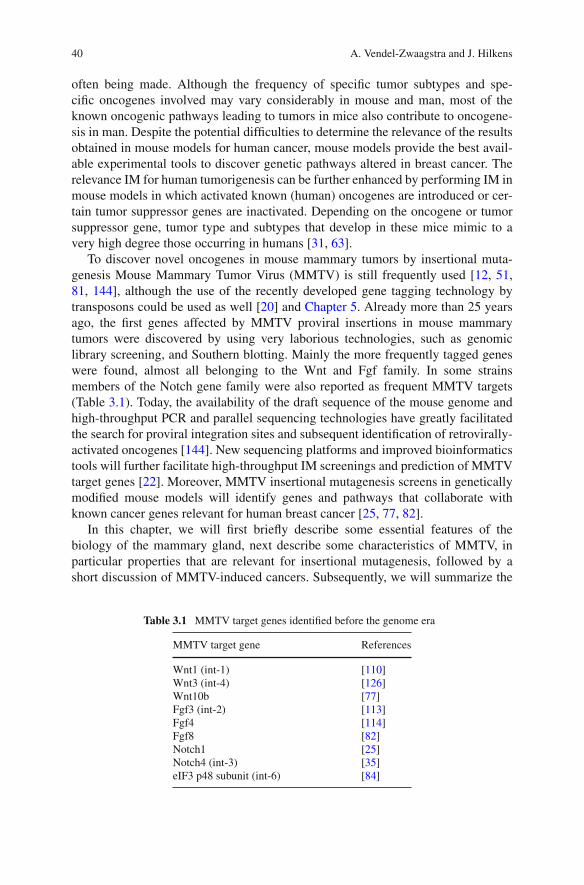
40 A. Vendel-Zwaagstra and J. Hilkens
often being made. Although the frequency of specific tumor subtypes and spe-cific oncogenes involved may vary considerably in mouse and man, most of theknown oncogenic pathways leading to tumors in mice also contribute to oncogene-sis in man. Despite the potential difficulties to determine the relevance of the resultsobtained in mouse models for human cancer, mouse models provide the best avail-able experimental tools to discover genetic pathways altered in breast cancer. Therelevance IM for human tumorigenesis can be further enhanced by performing IM inmouse models in which activated known (human) oncogenes are introduced or cer-tain tumor suppressor genes are inactivated. Depending on the oncogene or tumorsuppressor gene, tumor type and subtypes that develop in these mice mimic to avery high degree those occurring in humans [31, 63].
To discover novel oncogenes in mouse mammary tumors by insertional muta-genesis Mouse Mammary Tumor Virus (MMTV) is still frequently used [12, 51,81, 144], although the use of the recently developed gene tagging technology bytransposons could be used as well [20] and Chapter 5. Already more than 25 yearsago, the first genes affected by MMTV proviral insertions in mouse mammarytumors were discovered by using very laborious technologies, such as genomiclibrary screening, and Southern blotting. Mainly the more frequently tagged geneswere found, almost all belonging to the Wnt and Fgf family. In some strainsmembers of the Notch gene family were also reported as frequent MMTV targets(Table 3.1). Today, the availability of the draft sequence of the mouse genome andhigh-throughput PCR and parallel sequencing technologies have greatly facilitatedthe search for proviral integration sites and subsequent identification of retrovirally-activated oncogenes [144]. New sequencing platforms and improved bioinformaticstools will further facilitate high-throughput IM screenings and prediction of MMTVtarget genes [22]. Moreover, MMTV insertional mutagenesis screens in geneticallymodified mouse models will identify genes and pathways that collaborate withknown cancer genes relevant for human breast cancer [25, 77, 82].
In this chapter, we will first briefly describe some essential features of thebiology of the mammary gland, next describe some characteristics of MMTV, inparticular properties that are relevant for insertional mutagenesis, followed by ashort discussion of MMTV-induced cancers. Subsequently, we will summarize the
Table 3.1 MMTV target genes identified before the genome era
MMTV target gene References
Wnt1 (int-1) [110]Wnt3 (int-4) [126]Wnt10b [77]Fgf3 (int-2) [113]Fgf4 [114]Fgf8 [82]Notch1 [25]Notch4 (int-3) [35]eIF3 p48 subunit (int-6) [84]
3 Gene Discovery by MMTV Mediated Insertional Mutagenesis 41
MMTV-IM screens performed so far, highlight the major target genes and the path-ways activated. Finally, we will focus on the relevance of these target genes and thecorresponding pathways for human breast cancer.
3.1.1 Mouse Mammary Gland Biology
The mouse mammary gland is a dynamic organ that after embryonic developmentundergoes several developmental changes during, puberty, pregnancy, lactation andinvolution. At embryonic day 11–11.5 five pairs of lens-shaped mammary placodesare formed in the ventral ectoderm in response to signals from the underlying mes-enchyme. At day 12.5 the placodal epithelial cells migrate into the dense mammarymesenchyme and form small epithelial buds that slowly increase in size. Wnt andFgf signalling plays an important role at these stages [18, 156]. At day 15.5 a mam-mary sprout grows from each placode into the mammary fat pad. By day 16 thesprouts start to branch and a lumen develops. At birth the female mammary gland iscomposed of a rudimentary ductal tree consisting of the primary duct and 15–20 sec-ondary branches. In response to ovarian steroid hormones during puberty (mainlyestrogen) this rudimentary ductal system elongates through proliferation of cap cellsin the terminal end buds located at the tips. The elongating ducts bifurcate a fewtimes, which leads to a ductal tree of primary and secondary ducts that fills the entiremammary fat pad. During the recurrent estrous cycles and during pregnancy, lateralbuds, also called alveolar buds, form from quiescent cells in the secondary ducts.The lateral buds contain stem cells with the capacity to form either ductal or lumi-nal alveolar cell types. Pregnancy hormones activate rapid proliferation of stem cellpopulations in the lateral buds and induce an extensive network of tertiary branchesthat differentiate into secretory alveoli that produce milk that is secreted into theductal lumens during lactation. Following weaning, massive apoptosis of the lobu-lar alveolar cells takes place resulting in regression of the alveolar structures whilestem cells in the alveolar buds await another round of growth and differentiation dur-ing subsequent pregnancies [122, 160]. The adult mammary gland is comprised ofthree cell lineages: myoepithelial cells forming the basal layer of ducts and alveoli,ductal epithelial cells lining the lumen of the ducts, and alveolar epithelial cells thatproduce the milk proteins and fat during lactation. To generate the entire mammarygland during the next pregnancy, stem cells in the alveolar buds have the capacity ofself-renewal and the ability to generate again the three cell lineages that comprisethe lactating mammary gland.
3.1.2 Mouse Mammary Tumor Virus
MMTV belongs to the family of Betaretroviridae (B-type retroviruses), which areretroviruses with a distinct morphology such as an eccentric positioned nucleus.Intracytoplasmic A-particles found in MMTV producing cells are the immature,

42 A. Vendel-Zwaagstra and J. Hilkens
Fig. 3.1 Electronmicrographs of differentstages of MMTVmaturation. (a) shows aA-particle (left), which is anintracellular immature formof MMTV, and a B-particle(right) which is the maturevirion. Note the eccentriccore of the B-particle,whereas the A-particle doesnot have a defined core anddoes not contain a lipidenvelope. (b) shows abudding virion. The virion isenclosed by the plasmamembrane and the coreproteins have not matured (H.Jansen and Dr J. Calafat, NKIAmsterdam)
non-enveloped, intracellular precursors of the mature virion (B-particle) (Fig. 3.1).MMTV is a slow transforming retrovirus mainly causing mammary tumors with alatency of 6–12 months. The virus was initially described as an active milk factorthat plays an important role in the development of mouse mammary tumors [9].Besides the exogenous form, which is transmitted through the milk of infected micefrom mother to offspring, MMTV can also be transmitted endogenously throughstably integrated proviral copies in the germ line. In addition to the mode oftransmission and the tropism, regulation of expression of the virus by pregnancyhormones is another important difference between MMTV and other retroviruses[45].
Active infectious mammary tumor virus has only been unequivocally identified inmice. Although sequences related to MMTV (e.g. HERV-K sequences) are presentin the human genome and expression of MMTV env gene-like sequences have beenreported in human breast carcinomas [158, 159], neither an etiological relationshipbetween expression of these sequences and breast cancer nor infectious human MTVparticles have been found [6,83]. Also the reported serologic relationships betweenMMTV and proteins present in human breast cancer cells are highly controversial[38].
In the next paragraphs, we will describe the most important features of MMTV,in particular those that discriminate MMTV from other retroviruses and propertiesthat are relevant for insertional mutagenesis.
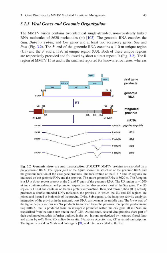
3 Gene Discovery by MMTV Mediated Insertional Mutagenesis 43
3.1.3 Viral Genes and Genomic Organization
The MMTV virion contains two identical single-stranded, non-covalently linkedRNA molecules of 8620 nucleotides (nt) [102]. The genomic RNA encodes theGag, Dut/Pro, Pol/In, and Env genes and at least two accessory genes, Sag andRem (Fig. 3.2). The 5′ end of the genomic RNA contains a 110 nt unique region(U5) and the 3′ end a 1197 nt unique region (U3). Both of these unique regionsare respectively preceded and followed by short a direct repeat, R (Fig. 3.2). The Rregion of MMTV 15 nt and is the smallest reported for known retroviruses, whereas
Fig. 3.2 Genomic structure and transcription of MMTV. MMTV proteins are encoded on apolycystronic RNA. The upper part of the figure shows the structure of the genomic RNA andthe genomic location of the viral gene products. The localization of the R, U3 and U5 regions areindicated on the genomic RNA and the provirus. The entire genomic RNA is 8620 nt. The R regionis a 15 nt direct repeat present at the 5′ and 3′ ends of the genomic RNA. The U3 region is ∼1200nt and contains enhancer and promoter sequences but also encodes most of the Sag gene. The U5region is 110 nt and contains no known protein information. Reversed transcription (RT) activityproduces a double stranded DNA molecule, the provirus, in which the U3 and U5 regions arejoined and located at both ends of the proviral DNA. Subsequently, the integrase activity catalysesintegration of the provirus in the genomic host DNA, as shown in the middle part. The lower part ofthe figure depicts various mRNA products transcribed from the provirus. Except the predominantSag mRNA, that is produced from an intragenic promoter within the env gene all mRNAs aretranscribed from the same start site in the 5′ LTR. As indicated, several viral proteins share part oftheir coding regions; this is further outlined in the text. Introns are depicted by v-shaped dotted linesand exons by solid lines. SD: splice donor site; SA: splice acceptor site; RT: reversed transcription.The figure is based on Mertz and colleagues [91] and references cited in the text
44 A. Vendel-Zwaagstra and J. Hilkens
the MMTV U3 region is relatively long as compared to other retroviruses. As inother retroviruses, the U3 region contains most elements that regulate transcriptionof the viral genes. Upon reversed transcription, the long terminal repeats (LTR)are formed at the 5′ and 3′ end of the provirus. The LTR is composed of the R,U3 and U5 regions that are joined as a consequence of the position of the reversetranscriptase primer binding site in the viral RNA and the template switches duringproviral DNA synthesis (Fig. 3.2) (for details see [142]).
Figure 3.1 depicts the map of the MMTV proviral DNA and the genes it encodes.The Gag gene encodes a polypeptide that is proteolytically processed into the viralcore proteins. Dut encodes a deoxyuridine triphosphatase (dUTPase). The dUTPasedecreases the dUTP pool in the infected cell and thus prevents incorporation ofdUTP into DNA during reverse transcription. Pro is a protease required for pro-cessing of some of the viral polypeptide precursors. Pol and In encode the reversetranscriptase (RT) and integrase genes respectively. The RT also has RNase H activ-ity, which degrades the RNA strand in the newly synthesized RNA-DNA hybridintermediates. The integrase catalyses the insertion of proviral DNA into the hostDNA (reviewed by [11]). Env directs the synthesis of a 73 kDa glycosylated pre-cursor protein, which is proteolytically cleaved by cellular furin proteases into thesurface gp52 and transmembrane gp36 proteins. Rem (regulator of export) is arecently identified gene that encodes a protein that facilitates the export of unsplicedviral mRNAs. Rem-like genes are also found in complex retrovirusses, like Revin HIV [91] but not in simple retroviruses like Murine Leukemia Virus (MuLV).The Sag (superantigen) gene is transcribed from the U3 region in the 3′ LTR. Thegene is unique for MMTV and essential for its normal live cycle. The existenceof yet another accessory protein, Naf (negative-acting factor), encoded by the U3open reading frame has been reported [130]. This protein may negatively affectexpression of certain cellular genes, but it is yet poorly characterized and not furtherdiscussed here. The other genes will be discussed in more detail below.
3.1.4 Transcription and Translation of the Viral Genes
After insertion into the host DNA, various viral mRNAs are synthesized as depictedin Fig. 3.2. The main product is genome-size mRNA that starts at the U3/R junctionand continues into the 3′ LTR where the R/U5 junction contains the polyadenylationsite. Part of this RNA is incorporated into new virions, while the remaining partis used for translation of the Gag, Dut/Pro, Pol/In genes. These genes overlap indifferent reading frames leading to the Gag, Gag-Dut/Pro and Gag-Dut/Pro-Pol/Inpolypeptide precursors. The translation of the Gag-Dut/Pro precursor requires aribosomal frame shift near the Gag translational stop codon, while expression ofthe Gag-Dut/Pro-Pol/In precursor polypeptide requires a double frame shift nearthe Pro-Pol junction [102]. The double frame shift needed to translate the latter pre-cursor is rather unique for MMTV and is not required for most other retroviruses.These subsequent ribosomal frame shifts are carried out with low efficiency and

3 Gene Discovery by MMTV Mediated Insertional Mutagenesis 45
provide a mechanism to reduce the ratio between the more 3′ located enzymaticactive proteins that are required in minor amounts and the structural Gag proteinsthat are needed in much larger quantities. The 5′ end of Dut overlaps with the 3′ endof the Gag sequence, while both full length gene products are present in the Gag-Dut/Pro precursor (Fig. 3.2). Dut is a transframe protein that arises from a ribosomalframeshift at the Gag-Dut/Pro junction [72]. Also, the RT enzyme is a transframeprotein containing at its N-terminus sequences from the C-terminus of Pro [29]. Thevarious polypeptide precursors that arise from this complex mode of translation aresubsequently proteolytically processed to release the active proteins.
The Env encoding mRNA also starts from the transcription start site at the U3/Rboundary in the 5′ LTR but the Gag-Dut/Pro-Pol/In genes are spliced out using the5′ splice donor (SD) and the first downstream splice acceptor (SA) site (Fig. 3.1).The Sag gene is translated from two spliced mRNAs. A minor Sag RNA speciesalso starts from the standard transcription start site in the 5′ LTR, extends until thefirst SD site and continues from the second SA site into the U3 region of the 3′LTR. The predominant Sag transcript starts from a transcription start site withinthe Env gene as depicted in Fig. 3.2 [106] As a consequence of the location of thetranscription start sites of both Sag mRNAs, Sag can only be transcribed from the3′ LTR in the provirus [17]. Sag transcription is driven by lymphoid lineage specificpromoter elements within the Env and Pol genes [121] and thus expression is limitedto lymphoid cells. Rem is produced from a double spliced mRNA, which partiallyoverlaps in frame with the 5′ and 3′ ends of the Env genes, and the 5′ end of the Saggene [91].
3.1.5 Virion Structure and Assembly
Dimeric viral genomic RNA is encapsulated by unprocessed Gag, Gag-Dut/Pro andGag-Dut/Pro-Pol/In polypeptides to form a spherical core. This cytoplasmic RNA-protein core is termed intracytoplasmic A particle, which is an immature B-particle[136]. The viral core in turn is surrounded during the budding process by a plasmamembrane domain enriched in Env glycoproteins (see below). Subsequently, theGag containing precursors are proteolytically processed by Pro in at least six nong-lycosylated proteins including the matrix, capsid and nucleocapsid proteins. Thelatter component directly interacts with the viral RNA. The Gag derived proteins,which are highly overrepresented relative to the proteins with enzymatic activityform the actual structural components of the core in which the enzymes are alsoincluded. The processing of the precursors into the respective functional proteinsoccurs in a very ordered fashion and is intimately linked to virion assembly, buddingand maturation (reviewed by Vogt [157]). Processing of the Gag proteins results ina mature virion with an eccentric core (Fig. 3.1).
Env encoded peripheral membrane protein gp52 and transmembrane proteingp36 form a stable complex that is incorporated in the plasma membrane and mayaccumulate in certain membrane domains. Subsequently, the cytoplasmic domain
46 A. Vendel-Zwaagstra and J. Hilkens
of gp36 interacts with the N-terminally located future matrix protein in the unpro-cessed Gag polypetide of the intracytoplasmic A particle. Trimers of the gp52–gp36heterodimer form the characteristic surface knobs of MMTV. Gp52 binds to theMMTV receptor on the cell surface during infection. A more detailed description ofthe viral gene expression and processing of these gene products is provided by Vogtet al. [157].
3.1.6 Infectious Life Cycle
The infectious life cycle of MMTV contains several features that are unique amongretroviruses. Most remarkably, the virus exploits the cellular host immune systemto ultimately infect mammary epithelial cells. MMTV virions ingested via the milkby new born mice travel through the intestinal wall and infect first dendritic cells(DCs) and subsequently B and T cells in the gut wall and Peyer’s patches [21, 48].Infection takes place upon binding of the envelope gp52/gp36 complex to the mousetransferrin receptor on the surface of the host cells [52, 129]. Subsequently, the virallipid bilayer and host cell membrane fuse, delivering the core of the virus into thecytoplasm of the host cell. At this point, the RNA is reverse transcribed by the viralRT (included in the viral core), resulting in a double stranded DNA molecule withat each end identical long terminal repeat (LTR) regions as outlined above. Thisfull length proviral DNA translocates to the nucleus where it integrates essentiallyrandomly into the genome, catalyzed by the viral integrase. Next, cellular RNApolymerase II synthesizes the viral genomic RNA and various spliced messengerRNAs as discussed above and depicted in Fig. 3.2.
Unique for MMTV is the crucial role of the Sag gene during the early stages ofinfection of new born mice. In the infected professional antigen presenting cells,DCs and B cells, the Sag gene is expressed at the cell surface in association with theMajor Histocompatibility Complex (MHC) class II proteins. The Sag-MHC com-plex strongly interacts with T cell receptors on CD4 positive T cells that containparticular Vβ chains and provokes a superantigen response that strongly stimulatesthe proliferation of the Sag-reactive T cells (reviewed by [1]). Cytokines released bythe activated T cells in turn drive the expansion of the MMTV infected B cell pop-ulation [49]. During this process, T cells are also infected and some of the infectedB cells mature into long-lived memory cells. In this way, a reservoir of MMTVinfected B and T cells is generated in suckling mice that can transport the virus toall parts of the body during a longer time period. This is an indispensable step forefficient infection of mammary epithelial cells since the virus cannot infect the rest-ing, non-differentiated mammary epithelial cells until these cells start to proliferateat puberty. At that point, the infected circulating lymphocytes in the mammary glandwill infect the expanding mammary epithelial cells [39]. As a consequence, MMTVcannot effectively infect DC or B cell deficient mice or mice that lack Sag specific Tcells [21, 39]. Infection of mammary epithelial cells proceeds essentially the sameas infection of cells in the hematopoietic lineage, except that Sag plays no role and

3 Gene Discovery by MMTV Mediated Insertional Mutagenesis 47
expression of the viral proteins is largely hormonally regulated. As a consequence,after initial infection of mammary cells, the virus will remain largely repressed inthe mammary gland until pregnancy and lactation when pregnancy hormones stimu-late viral protein expression. This allows infectious virions to further spread throughthe mammary epithelium, thus allowing the virus to be subsequently secreted intothe milk.
3.1.7 Tissue Specificity and Hormonal Regulation of MMTVReplication
The mammary gland is the predominant site of MMTV replication and consequentlythe main site of MMTV induced malignant transformation. Since the cellular recep-tor for MMTV binding and entry into the cell is the transferrin receptor, which isubiquitously expressed on growing cells, the receptor is unlikely to determine thetropism of the virus. Indeed, in MMTV infected animals, viral proteins are alsoexpressed at relatively low levels and in a strain dependent fashion in epithelialcells in a variety of other tissues such as salivary glands, kidneys, lungs, sem-inal vesicles, prostate, testes and in some lymphoid tissues [50, 58]. However,high expression levels are only found in the mammary epithelial cells. Currentevidence indicates that mammary specific expression and replication of MMTVis primarily controlled at the transcriptional level. Composite enhancer elementsthat contribute to mammary cell-restricted expression have been mapped to the 5′proximal end of the U3 region [41, 42, 168]. Negative regulatory elements (NREs)at the 3′ proximal end of the U3 region are involved in MMTV suppression innon-epithelial tissues including lymphoid cells [100, 128]. Both regions stronglydetermine the mammary gland specificity of MMTV. MMTV expression in themammary gland is most abundant during pregnancy and lactation. Along with otherfactors, this is regulated by the homeodomain containing repressor protein (CDP)that binds to multiple sites in the NRE. The CDP protein is highly expressed inundifferentiated cells, but its expression is absent in differentiated cells, includingthose in the lactating mammary gland [170]. In addition, Stat5a and Stat5b, whichare prolactin-inducible transcription factors, bind to the U3 domain and stimulateMMTV expression during lactation [117]. CDP and Stat5a/Stat5b act in concertto limit MMTV expression to the lactation period when the virus needs to betransmitted.
Viral gene expression is strongly dependent on steroid hormones [155] In particu-lar, glucorticoids and progesterone strongly enhance the expression of viral proteinsand replication of the virus in the mammary gland [15, 112, 124]. Without thesehormones, the strength of the MMTV-LTR promoter is attenuated. As a conse-quence, MMTV infected ovariectomized mice rarely develop tumors. The steroidhormone dependent viral gene expression is caused by hormone response elements(HRE) located just upstream of the transcription start site within the U3 region ofthe LTR [15, 57, 131], and reviewed in by Gunzburg et al. [45]. The HRE contains at
48 A. Vendel-Zwaagstra and J. Hilkens
least four overlapping recognition sites to which both activated glucocorticoid andprogestin receptors can bind [16]. The transcriptional repressor CDP also binds tosites overlapping with and adjacent to the steroid binding elements. This allowsCDP to compete with steroid hormone binding, and thus further contributes tothe pregnancy and lactation dependent expression [170]. In addition to the regu-latory domains discussed above, several more regulatory domains within the LTRhave been identified that contribute to viral protein expression regulation [100].Moreover, the transcriptional activity of the genomic region where the proviral DNAintegrates may also control MMTV expression and replication.
The pregnancy dependent regenerative cycles of the mammary gland is anotherfactor that may influence MMTV replication, and consequently, MMTV inducedtumorigenicity in the mammary gland as compared to other infected tissues such asthe salivary gland. Although the mammary gland cellularity increases about 30-foldduring each pregnancy, most cells die during involution of the gland after lactationends. This suggests that mammary stem cells or lineage progenitors that persistafter involution are the (oncogenic) targets of MMTV [73], since expansion of themammary gland from these cells during subsequent pregnancies coincides with astrong increase of MMTV positive cells. However, the tumors induced by differentvirus strains can vary in hormone dependence (see below). Therefore, these MMTVstrains may infect or replicate in different types of progenitor cells.
3.1.8 Exogenous MMTV
All mouse strains with a high-incidence of mammary tumors horizontally trans-mit infectious MMTV via milk to their offspring. These viruses are designatedexogenous MMTV variants. Replication competent MMTV can also be verti-cally transmitted when a provirus stably integrates in cells of the germline. Thefirst MMTV variants are exclusively transmitted via milk and were derived frommouse strains with a high mammary tumor incidence. These exogenous virusesare carried among others by the A, DBA, C3H, BR6, RIII and CzechII strains.The exogenous MMTV variants from these mouse strains are highly virulent, andthus most relevant for insertional mutagenesis. However, the exogenous MMTVvariants are often lost in the present day germ-free maintained mouse strains asa result of foster-nursing on non-virus carriers. MMTV from the various mousestrains does not necessarily induce the same type of tumors. As will be discussedbelow, some MMTV strains induce hormone dependent tumors, while other virusstrains mainly induce hormone independent tumors. Conversely, the same virus mayinduce different histological types of tumors in different mouse strains. For exam-ple, C3H-MMTV causes an approximately equal percentage of low grade acinarcarcinomas (Dunn type A tumors) and low grade solid carcinomas (Dunn type Btumors) in C3H mice, while in BALB/c mice it induces almost exclusively carcino-mas of the acinar type that have a relatively high propensity to metastasize to thelung [150].
3 Gene Discovery by MMTV Mediated Insertional Mutagenesis 49
3.1.9 Endogenous MMTV
Pups of high mammary tumor incidence strains (e.g., C3H) can be foster-nursedon mice without the milk factor. These “virus free” animals still develop mam-mary tumors, albeit with a longer latency and lower incidence, due to activationof endogenous proviral MMTV copies. Also, the tumors arising in low incidencestrains (e.g., BALB/c) are also thought to be caused by activation of replicationcompetent endogenous MMTV copies. In fact, the genome of almost all inbredmouse strains harbors multiple copies of endogenous MMTV proviruses, mostlyreplication defective, epigenetically silenced or inserted into transcriptionally inac-tive chromatin regions. The number of MMTV proviral copies varies per strain,ranging from none to as many as twenty independent proviral insertions. Theseproviral copies result from the occasional infection of germline cells and are presentin all somatic cells of offspring that develop from these infected gametes. If notinactivated, the endogenous provirus will produce infectious virus. For example,the endogenous C3H virus responsible for the late mammary tumors referred toabove, is encoded by a gene designated Mtv1 on chromosome 7 [153]. GR micecarry five endogenous MMTV copies, of which one (Mtv2) is active and responsi-ble for the high incidence of early mammary tumors in this strain. As a consequence,foster-nursing GR pups on a dame without milk-transmitted virus does not decreasethe tumor incidence [154]. Moreover, the MMTV variant expressed by Mtv2 canalso be transmitted horizontally via the milk from a GR female to another strain,and subsequently propagated as an exogenous virus. In contrast to Mtv1 and Mtv2,most other Mtv genes are partially or completely inactive [109]. A typical exam-ple of the latter gene is Mtv8, which is present in almost all inbred mouse strains.Other endogenous Mtv genes apparently have been incorporated in the germlineonly recently, and are thus unique to specific strains. The CzechII mouse strainis rather exceptional in that it harbors no endogenous MMTV copies [35], whichhas facilitated insertional mutagenesis screens in these mice using exogenousvirus.
3.1.10 MMTV-Induced Tumors
MMTV-infected mice usually develop mammary tumors at a relatively young ageand at high frequency. Tumor incidence and latency depends on the efficiency ofviral infection and replication in the different mouse strains rather than on tumori-genesis itself [127]. Some strains, such as the C57Bl/6, are rather resistant to MMTVinfection and tumor induction. Resistance can be due to the inability of MHC classII genes to present SAG, a crucial step required for infection. In some strainsendogenous Mtv genes produce SAG protein that is autoreactive with the sameVβ T cell receptor chains as the infecting virus, and thus have deleted the respon-sive T cells that are required to establish a productive infection with exogenousMMTV [127].

50 A. Vendel-Zwaagstra and J. Hilkens
In wild type mice, MMTV mainly induces two types of mammary tumors: lowgrade acinar carcinoma and low grade solid tumors (previously indicated as Dunntype A and B respectively) or mixtures of these histotypes, depending on the mousestrain [14]. These tumors are often considered as rather benign. However, the fre-quency of lung metastases in mammary tumor-bearing BALB/c mice that acquiredthe C3H MMTV by foster nursing (BALB/cfC3H) was reported to be 63 and 16%in BALB/cfRIII females [138]. In our own studies, we found that approximately onethird of the BALB/cfC3H mammary tumor-bearing mice had developed lung metas-tases at the time the primary tumor had reached ∼1 cm3 (J. Hilkens and M. Boer,unpublished results).
In some strains, MMTV initially induces premalignant mammary lesions andtumors that are pregnancy hormone dependent, while in other strains all MMTVinduced palpable tumors are hormone independent. In the next paragraphs, wewill briefly review the biology of MMTV induced tumors including hormonedependence.
3.1.11 MMTV Induced Hormone Dependent Premalignant Lesions
Mammary tumors in the mouse usually develop through premalignant hyperplas-tic lesions. The most frequent hyperplastic lesions in the mouse are hyperplasticalveolar nodules (HANs) and ductal hyperplasia’s (designated plaques). HANsare commonly found in all high-incidence mammary tumor strains and are focalproliferations from lobuloalveolar mammary epithelium [23, 89]. HANs containimmortalized cells that can be propagated by serial transplantations in mammaryfat pads cleared from normal epithelium [87]. At onset, HANs are hormone depen-dent but when palpable, HANs have usually become hormone independent. Plaquesare ductal hyperplasia’s that initially regress after each parturition but reappear at thesame site in subsequent pregnancies. However, after several parities, regression ofthese nodules is less complete and they eventually progress to pregnancy indepen-dent tumors [139, 154]. Plaques are commonly present in high-incidence MMTVstrains such as GR and RIII females. Note that the pregnancy hormone (estrogenand progesterone) dependent tumor growth discussed in this paragraph should notbe confused with the pregnancy dependence of MMTV replication, which affectsthe frequency of mutagenic events and is mainly glucocorticoid dependent.
HANs and plaques are associated with MMTV infection and express largeamounts of MMTV in high-incidence MMTV strains. The discovery that HANsand plaques show MMTV proviral insertions near Wnt and Fgf genes indicatesthat MMTV insertional mutagenesis in concert with hormonal stimulation give riseto these hyperplasia’s. However, HANs can also be observed in mice treated withchemical carcinogens in low-incidence MMTV strains, indicating that developmentof mammary tumors from premalignant lesions is a general phenomenon (reviewedby [88]).
3 Gene Discovery by MMTV Mediated Insertional Mutagenesis 51
3.1.12 Hormone-Dependent Mammary Tumors and MMTV
Plaques in the GR mouse strain that develop into mammary tumors are initiallyestrogen dependent and do not grow in ovariectomized mice; instead they progressfrom a hormone-dependent growth phase to autonomous, hormone-independenttumors [151, 152]. Hormone-dependent mammary tumors in ovariectomized GRmice only develop upon treatment with progesterone and estrogen. Transplantationof hormone dependent GR tumors usually leads, after several passages, to the emer-gence of hormone independent tumor variants as a result of a clonal selectionprocess [96, 135]. MMTV is essential for the development of these tumors as theydo not arise in GR congenic mice that lack the active endogenous provirus Mtv2. Ofnote, Wnt and Fgf genes are already activated in GR tumors by insertional muta-genesis before they become hormone independent [92, 103]. This indicates thatexpression of these oncogenes does not cause hormone independence, but one ormore additional insertional mutations events or other genetic or epigenetic changesconfer the hormone independent growth.
Squartini et al. [139] demonstrated that the mammary tumors induced by dif-ferent strains of exogenous virus progress differently. In their study, BALB/cmice, which have a low incidence of mammary tumors, show a high incidenceof hormone-independent tumors when infected with C3H-MMTV but develophormone-dependent tumors progressing to hormone-independence when infectedwith RIII-MMTV. These results show that development of hormone dependenttumors is virus strain dependent. The exact cause of this virus strain specifichormone dependence is currently unknown.
3.1.13 MMTV-Induced Tumors in other Tissues
As discussed above, expression and replication of MMTV is low in most non-mammary tissues because of its tissue and hormonal-specific regulation. Thislargely precludes proviral integrations and tumor induction in these tissues.However, MMTV infection of hematopoietic cells does occur, although the LTRenhancer/promoter is probably inefficient in these cells. Evidence that the regulatorysequences in the MMTV-LTR are crucial for its tissue specific oncogenic activitycomes from relatively rare MMTV induced lymphomas that develop at a low fre-quency late in life in some MMTV infected mouse strains, such as the GR andDBA/2 [95]. The genome of these lymphomas contains many new MMTV proviralcopies with consistent deletions and gains in the enhancer-like elements in the U3region of the LTR that alter the behavior of the LTR enhancer/promoter in lympho-cytes [5, 55, 97, 167]. Because these sequence changes in the MMTV-LTR requiretime to accumulate within an infected animal, lymphomas occur with a long latencyand are only observed in males as the females succumb to mammary tumors at ayounger age. The acquired mutations in the LTR of the lymphotropic MMTV more
52 A. Vendel-Zwaagstra and J. Hilkens
readily allow viral replication and proviral integration in lymphoid tissues, and thiseventually causes lymphomas through insertional mutagenesis [7, 90, 167].
Based on these findings, Yanagawa et al. [167] and Bhadra et al. [7], were able toconstruct infectious B-type T cell tropic viruses that are poorly expressed in mam-mary epithelial cells and are not transmitted via the milk, but instead replicate onlyin lymphoid cells due to removal of the negative regulatory element and acquisitionof T cell specific enhancer elements. Interestingly, the Sag gene is not required forthe B-type T cell tropic virus, indicating that expression of Sag is only required togenerate a B and T cell population that can infect mammary cells [105]. Insertionalmutagenesis screens using B-type leukemogenic virus showed that this virus dereg-ulates some of the same genes that are also frequently tagged by Moloney virus inlymphomas, such as Myc and Rorc [10, 119, 148].
MMTV-associated kidney carcinomas have also been observed in Balb/c miceinfected with milk-borne C3H MMTV. The kidney tumors harbored newly inte-grated exogenous MMTV proviruses that showed striking alterations in the U3region of the LTRs that might be responsible for the difference in tissue specificityof the virus [162]. Changes in the U3 region were also reported for newly acquiredproviral copies in AtT-20 mouse pituitary tumor cells although it is unclear whetherthese copies are involved in tumorigenesis [118]. The reported tissue-specific alter-ations in the LTR predict that highly infectious MMTV versions with modified LTRsmay be useful for insertional mutagenesis in non-mammary tissues.
3.2 Gene Discovery by MMTV Mediated InsertionalMutagenesis
3.2.1 Common Integration Sites
MMTV randomly integrates in the murine genome [32], but in contrast to someother retroviruses such as Moloney Murine Leukemia virus, MMTV does not showa preference for transcription start regions and CpG islands [166]. If the mutagenicevent caused by an MMTV proviral insertion affects a gene that confers a selectivegrowth advantage, it will expand the mutated cell population, which may lead tohyperplasia. In some cells, additional hits near other cancer genes may provide thesecells with an additional growth advantage or other tumor related properties, and thiswill eventually lead to full-blown mammary tumors. Most tumors are oligoclonalfor proviral insertions, which is caused by subsequent insertions in different cells ofthe early tumor that provide similar selective advantages during tumor progression.However, many if not most additional insertions are simply fortuitous, irrelevantinsertions that are piggybacking on the growth promoting insertions when the tumorexpands. When analyzing the pattern of MMTV insertion sites in a collection ofmammary tumors, these background insertion events should show a random distri-bution pattern. By contrast, insertion events causally linked to tumor formation arepredicted to impact a subset of genes (i.e. proto-oncogenes), and thus should appear
3 Gene Discovery by MMTV Mediated Insertional Mutagenesis 53
as nonrandom clusters found near these genes. Therefore, only those insertions thatare clustered more densely than would be expected by chance in a set of independenttumors are likely to be responsible for tumor induction. These insertions are consid-ered to be present in common integration sites (CISs) and indicate the presence of anearby oncogene.
Mikkers et al. [99] calculated the size of the genomic window in which twoor more insertions can be expected to be nonrandom, and thus defined a CIS as afunction of the number of analyzed tumors and detected insertions. However, mod-ern high-throughput screens produce increasingly large data sets and consequently,the calculated window may become quite small and no longer biologically relevantsince the viral enhancers may act over much larger distances than the calculatedwindow. Similarly, two or more insertions within the same gene may have the sameeffect on gene function but may not be present within the same calculated win-dow. Recently, a statistical framework based on Gaussian kernel convolution hasbeen developed [22], that is capable of detecting CISs in a noisy and biased envi-ronment using a predefined significance level while controlling the probability ofdetecting false CISs. Gaussian kernel convolution (summing Gaussian shaped ker-nel functions placed at every RIS, akin to a sliding window), results in a smootheddensity distribution of inserts over the entire genome. Depending on the width of thekernel function, closely spaced RISs give rise to high peaks, indicating CISs. Thesignificance of the height of the peaks can be determined by comparison with thepeak heights observed in randomly distributed RISs after appropriate multiple test-ing correction. By varying the width of the kernel function, CISs at any biologicallyrelevant scale can be detected.
3.2.2 Modes by Which Insertional Mutagenesis AffectsCancer Genes
The LTRs of proviral MMTV contain promoter and enhancer sequences that notonly control the transcriptional activation of the viral genes, but also can influenceexpression of adjacent host genes. There are three major modes by which a proviruscan affect the expression of nearby host genes: (1) promoter insertion, (2) enhance-ment of a nearby promoter or (3) integration within a gene causing truncation of thetranscript (see Fig. 3.3). For promoter insertion to take place, the proviral MMTVmust integrate in close proximity of the 5′ end and in the same transcriptional orien-tation as the target gene. In this way, the promoter within the 3′ LTR of the proviruscan replace the endogenous gene promoter. Transcription of an adjacent gene canalso start from the 5′ LTR after deletion of the 3′ end of the provirus or by removalof most of the 3′ viral transcript by splicing.
Since transcriptional enhancers act in an orientation-independent manner, thestrong enhancer elements within the U3 region of the 5′ LTR cannot only stimulatethe viral promoter but also the promoter of an adjacent cellular gene. Transcriptionalenhancement will occur when the provirus integrates either downstream of a cellular

54 A. Vendel-Zwaagstra and J. Hilkens
Fig. 3.3 Mechanisms of retroviral insertional mutagenesis. The most frequent mechanisms ofretroviral insertional mutagenesis are depicted. Promoter insertion: the provirus inserts upstream ofthe target gene in the same transcriptional orientation and the viral promoter replaces the endoge-nous promoter of the target gene, which may lead to enhanced expression of the target gene.Variants of this mechanism may lead to alternative spliced forms of the mRNA by removal ofone or more of the 5′ exons (not shown). Enhancer insertion: insertions 5′ in the reverse orien-tation or 3′ in the same orientation relative to the target gene, which may lead to activation ofthe endogenous promoter and subsequently to enhanced mRNA expression. Truncating insertion:Insertion in an exon or intron within the gene; these insertions may lead to inactive proteins, toenhanced expression of 5′ or 3′ truncated mRNAs resulting in N- or C-terminal truncated proteinsthat may be constitutively active due to removal of inhibitory domains, or to mRNA stabiliza-tion by removal of destabilizing motifs in the 3′ mRNA. The novel 5′ truncated messenger willterminate on a cryptic poly-adenylation site within the proviral LTR (not indicted). The lightand dark blue bars represents MMTV proviral DNA enclosed by two LTRs (dark blue). Thepurple bars represent the open reading frame of the target gene; the promoter and untranslatedregions of the target gene are depicted by red and black bars respectively. The figure is based on[62, 145]
gene in the same transcriptional orientation or upstream of a gene in the reverseorientation. Transcriptional enhancement is the most frequent mode of gene acti-vation by retroviral insertion, as enhancement allows more flexibility with respectto the orientation of the virus and the distance to the target gene. The rangeby which enhancement can occur is illustrated by our finding that the normallysilent Dpp10 gene is activated by proviral insertion at a distance of no less than700 kb [144].
3 Gene Discovery by MMTV Mediated Insertional Mutagenesis 55
Intragenic insertions that lead to truncation of the gene can have different out-comes. For instance, integration within the 3′ end of a gene can result in enhancedexpression of the gene by removal of destabilizing signals within the 3′ untrans-lated region of its mRNA. MMTV insertion in the same transcriptional orientationwithin the coding region of a gene may give rise to truncated transcripts that encodeN- or C-terminal protein truncations. The enhancer in the 5′ LTR can stimulatetranscription of a truncated 5′ transcript of the target gene, while the promoter inthe 3′ LTR can drive transcription of a truncated 3′ transcript if there is a spliceacceptor site in 3′ end of the gene (Fig. 3.3). Whether any of these truncated pro-teins are active obviously depends on the target gene and the exact insertion site.Intragenic proviral integration may also lead to an inactive gene that may pro-mote tumorigenesis if the affected target gene is a tumor suppressor. However,a proviral insertion only affects a single allele of a gene. Thus an inactivatinginsertion in the second allele required to effectuate the oncogenic potential of atumor suppressor gene is statistically very rare. However, an inactivating inser-tion in a single allele of a haploinsufficient tumor suppressor gene may lead to aselective advantage. Also, subsequent inactivation of the second tumor suppres-sor allele by loss of heterozygosity or promoter methylation could lead to clonaloutgrowth. However, a selective advantage due to provirally-induced haploinsuffi-ciency is apparently rather rare. For instance, haploinsufficiency of Pten is knownto cause mammary tumorigenesis in mice and collaborates with Wnt1 activation[78], one of the most frequent MMTV targets. Nevertheless, we did not find anyproviral MMTV insertion in the Pten locus among approximately 2000 proviralMMTV insertions in mouse mammary tumors (Vendel-Zwaagstra A., Theodorou V.and Hilkens J., unpublished). Also, other known human or mouse tumor suppressorgenes such as Rb1 were not associated with common insertion sites in our screen.The genomic regions containing these genes could be poor targets for proviral inte-gration, as these genes were also rarely found near 10 000 CIS in a MuLV screen of510 lymphomas [148]. Interestingly, in their analysis almost half the MuLV inser-tions were intragenic, of which several occurred in genes that are known tumorsuppressors such as Ikaros, Nfl and E2f2; the latter being a known haploinsufficientgene.
The above outline describes the most common mechanisms of by which genescan be activated but other more exotic mechanisms have also been described [62,145, 147].
As discussed, the MMTV promoter activity is greatly enhanced by steroidhormones (e.g. glucocorticoids, progestins) and is weak without hormones. Thequestion arises whether this will affect expression of the targeted oncogene.Sonnenberg et al. [137], showed that activation of Fgf2 by proviral MMTV insertion(RIII-derived) in the opposite transcriptional orientation was hormone-independent,while MMTV expression in the same cells remained hormone-dependent. Grimmand Nordeen [41] investigated this further using luciferase reporter assays and con-firmed that activation of the promoter of at least some cellular MMTV target genesby the MMTV-LTR enhancer (C3H derived) is relatively independent of steroidhormones.
56 A. Vendel-Zwaagstra and J. Hilkens
3.3 Gene Families and Pathways Frequently Targeted by MMTV
The first oncogenes identified using MMTV-induced insertional mutagenesis werediscovered in conventional MMTV positive inbred mouse strains such as the C3H,RIII, BR6, GR and CzechII strains. The first identified MMTV common integrationsite was located adjacent to the int-1 gene, which later was identified as the orthologof the wingless gene in Drosophila [123], and was subsequently designated Wnt1. Asecond common MMTV-insertion site, designated int-2 [113], was found to encodea member of the fibroblast growth factor family (Fgf3) [24]. A frequent MMTVtarget in CzechII mice, int-3 [35] was later recognized as a member of the Notchfamily, Notch4 [34, 125]. These first three MMTV targets are members of three dif-ferent gene families critical for normal patterning during embryogenesis. Presently,three additional members of the Wnt family, Wnt3 [126], Wnt3a 144] and Wnt-10b [77], and three additional Fgf-family members, Fgf4 [114], Fgf8 [132], Fgf10[143], and one additional Notch-family member, Notch1 [25] have been associatedwith MMTV common insertion sites.
The early studies showed that usually only a few members within a gene fam-ily are MMTV targets. Also in our high-throughput IM study [144], in mammarytumors from MMTV infected BALB/c mice, we only rarely found that other mem-bers of the three above mentioned gene families were tagged, suggesting thatspecific gene family members are oncogenic in the mammary gland. We will ten-tatively refer to the MMTV targets within a gene family as the “oncogenic” familymembers. However, it remains possible that tagging of other members of these genefamilies does not confer a selective advantage due to the absence of the appropriatereceptors in mammary epithelial cells (in case of secreted factors), constrains on theinteraction between the target gene promoter and MMTV enhancer or the presenceof some of these gene family members in transcriptionally inactive DNA regions.The former option appears unlikely for Fgfs as some non-targeted Fgfs use the sameFgf receptors as targeted Fgfs. The latter option seems also unlikely as the targetedWnt members are present in evolutionary conserved, closely spaced pairs of whichonly one is targeted by MMTV.
Genes that are transcriptionally activated by proviral insertion are usually silentor expressed at very low levels in normal postnatal mammary epithelium. Therefore,the distinction between oncogenic and non-oncogenic family members is furthersupported by the fact that only non-oncogenic Wnt and Fgf-genes are expressed inpostnatal normal mammary epithelial cells (e.g. Wnt4, Wnt6), and thus are unlikelyto be oncogenic unless their normal activity is kept in check by a strictly tem-poral regulation (e.g. only during pregnancy). In that case, however, it can easilybe envisaged that MMTV insertion would interfere with such temporal regula-tion. Interestingly, Wnt10b was reported to be an MMTV target [77], despite thatit is expressed in normal mammary epithelium [75, 77]. However, in our own IMstudies, we noticed that many insertions predicted to activate Wnt10b also, and occa-sionally even exclusively, activate the nearby Wnt1 gene. Conversely, the majorityof the tumors expressed Wnt10b without insertion in or nearby the Wnt10b/Wnt1locus [144]. Therefore, Wnt10b is probably not a bona fide MMTV target and

3 Gene Discovery by MMTV Mediated Insertional Mutagenesis 57
is not considered to be an strongly oncogenic Wnt, despite the fact that highoverexpression in transgenic mice leads to mammary tumors [75].
Our high-throughput IM study and subsequent expression analysis (M. Kimm,M. Boer and J. Hilkens, unpublished) showed that only rarely is more than oneoncogenic Wnt or Fgf gene expressed in individual tumors. Clearly, activation of asingle member of each oncogenic family is sufficient to fully activate the respectivepathways and activation of additional genes in the same pathway provide no fur-ther selective advantage in most cases, although some exceptions to this rule will bediscussed bellow. In nearly all MMTV induced BALB/c mammary tumors an onco-genic Wnt gene and an oncogenic Fgf are co-targeted, suggesting interdependence ofboth gene families in tumorigenesis. A preference for specific combinations of Wntand Fgf family members was not observed despite some reports to the contrary [64].
There is some evidence that MMTV insertions within multiple genes in the samepathway provide a selective advantage when they occur within a single tumor. TheFgf-receptor1 (Fgfr1) and Fgfr2 genes are occasionally tagged in the same tumors inwhich an Fgf ligand is also tagged. Apparently, the Fgf receptor is not expressed atsufficiently high levels, and therefore enhanced expression as a result of MMTVtagging can provide a further selective advantage. Whether the tumors withoutMMTV-induced Fgfr expression use other ways to upregulate the appropriate recep-tor has not been investigated. This finding shows that cooperativity between MMTVtarget genes can occur within the same pathway under certain circumstances.
Another example of genes collaborating within the same pathway is provided bythe R-spondin genes. Two genes of the R-spondin gene family, Rspo2 and Rspo3,are frequently associated with CISs [37, 81, 144]. The R-spondin gene family com-prises four members that are characterized by a thrombospondin type 1 domainand two furin domains. Normal mammary epithelial cells transduced with cDNAencoding Rspo3 are tumorigenic in vivo, validating this MMTV target as a novelproto-oncogene [144]. In particular, Rspo2 and Rspo3 amplify Wnt1 and Wnt3a sig-naling, while R-spondins alone have only a minor effect on the Wnt-pathway [65,67, 107]. Rspo’s bind to the Wnt co-receptor LRP6 [8, 161],and antagonize DKK1activity by interfering with DKK mediated LRP6 and Kremen association, leadingto down regulation of the Frizzled-LRP receptor complex [68, 108]. In other words,activation of an Rspo gene may be required to prevent DKK-mediated inhibitionof the Wnt pathway. Indeed, MMTV insertions in Rspo2 and Rspo3 are frequentlyfound together with a Wnt gene insertion in the same tumor (Vendel-Zwaagstra andHilkens, unpublished) and represent another example how cooperativity betweengenes in the same pathway can provide a selective advantage to the tumor cell.
Notch4 is targeted with high frequency in CzechII mammary tumors but onlyat low frequently in MMTV induced BALB/c tumors. However, activation of theNotch pathway may be underestimated, as other genes in the Notch signaling cas-cade are occasionally tagged in the Balb/c tumors [35, 144]. Fgf and Wnt genes wereusually activated in the tumors in which Notch is provirally tagged. This suggeststhat these pathways are collaborating, although Notch pathway activation does notseem to be a prerequisite for Wnt/Fgf induced mammary tumorigenesis in BALB/cmice.
58 A. Vendel-Zwaagstra and J. Hilkens
3.3.1 Infrequently Targeted Genes and Pathways
Recently, we have published a list of 33 candidate genes in MMTV-induced tumorsfound in 160 wild type BALB/c mice [144]. Insertional mutagenesis in additionalmouse mammary tumor models extended this list to more than 40 CISs (A. Vendel-Zwaagstra, M. Boer and J. Hilkens, unpublished). In addition to the frequentlytargeted genes acting in the Wnt, Fgf and Notch pathways that are discussed above,we also found a large series of infrequently tagged genes not acting in one of thesepathways ([144], and A. Vendel-Zwaagstra, M. Boer and J. Hilkens, unpublished).Among these are genes acting in a variety of pathways such as Igf2/insulin path-way including Igf2 and Irs4, members of the Odz, Robo and glypican gene families,Eras, Id2, Map3K8, Astn2, Sfmbt2, Dpp10, Zfp521 and others. The function of mostof these genes in mammary tumorigenesis has not been elucidated.
Some of the infrequently targeted genes may in fact activate the same pathway.For instance, among the infrequently tagged genes are three genes that may activatethe PI3Kinase pathway: Irs4, a member of the Insulin Receptor Substrate family[76], Igf2 a secreted growth factor that among others activates the PI3K pathway andEras which directly activates the PI3K pathway [141]. An expression analysis showsthat these genes are expressed in 15–20% of the mammary tumors depending on thegenetic background, although this analysis does not necessarily represent the tag-ging frequency but may better indicate the importance of these genes. Several genesacting in this signaling cascade are well established cancer genes, like PIK3CA,encoding the catalytic moiety of the PI3-kinase and the tumor suppressor genePTEN (reviewed in [26]). Loss of PTEN expression or activating PIK3CA muta-tions occur in more than 50% of the human breast cancers and Pten loss wasalso shown to accelerate mouse mammary tumorigenesis [78]. Of note is that Irs4and Igf2 may also be involved in other oncogenic pathways, like the MAPK/ERKpathway.
Theodorou et al. [144] showed that “single” insertion sites (insertion sites presentin only a single tumor) can still be informative because some genes may be taggedinfrequently as they activate less dominant pathways, or the same pathway may beactivated by multiple genes from the same gene family decreasing the probability tobe present in a CIS. Indeed, analysis of the complete dataset of MMTV retroviralinsertion sites by using various bioinformatics tools and public databases revealedthat certain gene families, protein domains, and pathways in addition to the onesassociated with CISs are overrepresented among genes associated with single inser-tion sites. Gene family analysis of 160 MMTV-induced BALB/c tumors revealedthat among others Ras, cyclin, cadherins and T-box gene families are overrepre-sented. Protein domain analysis of the genes tagged by single insertions showed thatgenes encoding proteins with a thrombospondin type 1 (TSP-1), phosphokinase, orcation ATPase domain were also overrepresented and likely important in carcino-genesis. Proteins with a TSP-1 domain also include Rspo2 and Rspo3, mentionedabove, and several Adamts family members some of which also have been impli-cated in cancer [101]. Based on these results, we can also consider genes present in“common gene families” as candidate cancer genes.
3 Gene Discovery by MMTV Mediated Insertional Mutagenesis 59
3.4 Comparison of MMTV Targets in Mammary Tumorsand MuLV Targets in Lymphomas
Theodorou et al. [144] investigated the overlap between the entire sets of genestargeted by MMTV and MuLV (deposited in the Mouse Retrovirus TaggedCancer Gene Database, RTCGD, http://rtcgd.abcc.ncifcrf.gov/mm8/index.html) andwhether genes targeted by both viruses are acting in the same or in differentpathways. The comparison showed that MMTV and MuLV target genes and sig-naling pathways only partially overlap. In lymphomas MuLV frequently tags genesinvolved in the development of the hematopoietic and immune system, whereasMMTV preferentially tags genes related to mammary development. This couldeither be due to the nature of the different retroviruses or to the tissues that areinfected by the viruses. Comparing the MuLV targets in brain tumors presentin the RTCGD with the MuLV targets in lymphomas suggest that tumor forma-tion is mainly dictated by the nature of the tissue rather than the nature of thevirus.
3.5 Core and Sporadic Activated Cancer Pathways
In most mammary tumors, several CISs are present. Some of these CISs occur withvery high frequency while others are only infrequently found. Callahan and Smith[13] designated the former as core CISs. The core CISs activate mainly the Wnt,Fgf and Notch pathways in a large proportion of MMTV induced mammary tumorsalthough the frequency may vary somewhat (for Wnt, Fgf) or significantly (forNotch) in different mouse strains and may also depend on the MMTV variant usedto induce the tumors. Consistent with Callahan and Smith, we will designate thesepathways as the core oncogenic pathways in MMTV-induced tumors in contrast tothe infrequently activated pathways, which we will designate as sporadic pathways.The essential role of the core pathways in mouse mammary tumorigenesis is clearlyillustrated in wild-type Balb/c tumors in which Wnts and Fgfs are activated in over90% of the cases. Although both pathways act synergistically in bitransgenic mice,together these pathways cannot drive tumorigenesis as Wnt1/Fgf3 bitransgenic micedevelopment tumors with latencies between 3 and 10 months indicating additionalgenetic or epigenetic events are required [74, 53]. Therefore, it is not surprisingthat in almost all tumors we found MMTV proviral insertions in addition to thoseactivating core pathways. However, these insertions tag a variety of genes with lowfrequency, of which some are in CISs. These insertions activate genes that partici-pate in various pathways that probably collaborate with one or more core pathways.Interestingly, both Fgf3 and Wnt1 transgenic and bitransgenic mice develop hyper-plastic mammary glands, suggesting that the additional insertions may cause themalignant transformation. However, these hits may only contribute to tumor pro-gression [13]. The clonality of the sporadically activated genes in the tumor mayprovide some clues in this respect.

60 A. Vendel-Zwaagstra and J. Hilkens
In the next paragraphs, we will briefly discuss the core pathways activated byMMTV proviral insertion in mammary epithelial tissues.
3.5.1 Wnt Signaling
Wnt signaling plays a key role in many aspects of embryogenesis and throughoutadult life by regulating morphology, proliferation, motility, cell fate and stem cellmaintenance. Somatic mutations in components of the Wnt pathway are also fre-quently seen in a variety of human cancers, although only infrequently in humanbreast cancer. However, nuclear β-catenin (a read out for activation of the canonicalWnt-pathway) has been observed in 60% of tumors, indicating the importance ofthis pathway also for human breast cancer [79]. Wnts are lipid modified secretedmolecules. Nineteen highly conserved Wnt genes have been identified in mice andhumans, but only some Wnts have been shown to be involved in mouse tumori-genesis. Also, the transforming capacity of the various Wnt genes in vitro variesgreatly [164, 134]. In addition to the MMTV-tagged Wnts, also Wnt2 and Wnt7Acan transform C57MG cells in vitro.
Wnt signaling induces stabilization of β-catenin, which is subsequently translo-cated to the nucleus where it acts as cofactor for TCF/LEF transcription factors.TCF/LEF transcription factors then regulate genes involved in cell proliferation suchas cyclinD1 and c-Myc. This is considered the canonical Wnt-pathway, recentlyreviewed by Clevers [19]. In addition, several non-canonical pathways have beendescribed including the planar cell polarity and cell migration pathways throughactivation of Rho family GTPases, Jnk and the Ca2+ pathway through phospholipaseC activation.
Wnts act through Frizzled receptors, of which no less than ten have been identi-fied. However, Wnts can also bind to other membrane molecules acting as Frizzledcoreceptors or as independent receptors—including Lrp5, Lrp6, Ror2, Ryk andproteoglycan cofactors such as syndecans and glypicans [66]. Recent evidenceshows that the pathway that is engaged by Wnts depends on the specific receptor-coreceptor complex present at the cell surface and the cellular context [149]. Thecanonical pathway is only activated by a Frizzled-Lrp complex, whereas the non-canonical Wnt signaling pathway may involve Frizzled receptors, but in associationwith different coreceptors [149].
Only the canonical Wnt pathway seems to be involved in mouse mammarytumorigenesis as ectopic expression of stabilized β-catenin in the mammary glandis sufficient to cause mammary tumors that are indistinguishable from Wnt inducedtumors [94]. While the canonical pathway is involved in initiation, the non-canonicalpathways may only contribute to tumor cell migration and invasiveness [60]. In ourMMTV insertional mutagenesis screen we only find genes acting in the canonicalpathway such as the Rspo genes but no genes known to act in one of the non-canonical pathways. This again indicates that the canonical pathway is one of thedominant oncogenic pathways in mouse mammary tumorigenesis.
3 Gene Discovery by MMTV Mediated Insertional Mutagenesis 61
Mice transgenic for Wnt1 driven by the MMTV-LTR display accelerated pro-liferation of the mammary ductal and alveolar epithelium resulting in lobuloalve-olar hyperplasia throughout the mammary glands and the development of focalmammary tumors with a median latency of approximately 5 months [146]. Thedevelopment of generalized mammary hyperplasia and subsequent solitary mam-mary tumors with a relatively long latency suggest that Wnt gene activation is anearly event in mammary tumorigenesis. Subsequent collaborating genetic eventsare required for malignant tumor formation. However, continued Wnt stimulationremains required for tumor maintenance and growth [44]. Involvement of Wntsin tumor initiation is in keeping with the observation that Wnt gene activation inMMTV induced mammary tumors is an early event [146].
3.5.2 Fgf Signaling
In mammals, the fibroblast growth factor gene family comprises of 22 members.Nearly all Fgf genes are expressed at some point during embryonic developmentand are essential for organogenesis and limb development. Fgf signaling results ina variety of cellular responses, like proliferation, migration and survival (for reviewsee [30, 43, 116]). Fgfs are secreted molecules that signal in an autocrine as wellas paracrine fashion through cell-surface tyrosine kinase receptors encoded by fourgenes (Fgfr1-4). Alternative splicing of the Fgfr1, 2 and 3 transcripts results in atleast 7 variants, which bind the various Fgfs with different affinities. The variousreceptors show different biological activities which adds to the complexity of Fgfsignaling [116]. Activation of the FGFRs also involves polysulphated proteogly-cans like heparin or heparan (HSPG) that bind Fgfs and are required as accessoryreceptors further enhancing the complexity of Fgf-signaling. Upon ligand binding,FGFRs dimerize and autophosphorylate specific tyrosine residues in their cyto-plasmic domains. The phosphorylated domains form docking sites for moleculesinvolved a various signaling pathways of which the Ras/MAPK, PI3K/Akt andPLCγ pathways are the most prominent [30]. In vitro, FGFs are involved in cell pro-liferation, migration, differentiation and survival. In vivo, some FGFs have strongangiogenic activity while others are involved in tissue remodeling and wound repair.Fgf overexpression in tumor cells could act as an autocrine growth factor and/oras an angiogenic factor. Interestingly, the Fgf family members with the strongestangiogenic capacity, such as Fgf1 and Fgf2, are never targeted by MMTV, suggest-ing that the main contribution of FGFs in mouse mammary tumorigenesis might bestimulation of cell proliferation and/or survival.
Mammary glands of MMTV-Fgf3 transgenic mice exhibit extensive epithelialproliferation and develop ductal hyperplasia, which only in some transgenic strainsgives rise to tumors relatively late in life [74, 77, 104]. As discussed above, Fgf andWnt genes are usually co-targeted by MMTV, suggesting that both gene familiesclosely collaborate. Wnt and Fgf genes are already co-mutated in mammary hyper-plasia, which shows that activation of both genes are early events in tumorigenesis
62 A. Vendel-Zwaagstra and J. Hilkens
[103, 115]. Indeed, Wnt1 and Fgf3 transgenic mice develop generalized diffusemammary hyperplasia confirming that Wnts and Fgfs are involved in early stages ofcarcinogenesis [104, 146].
The compound MMTV-Wnt1/MMTV-Fgf3 transgenic mice also developedtumors with a shorter latency than transgenic mice expressing only a single trans-gene, proving that Wnt and Fgf signaling collaborate in mammary tumorigenesis.However, additional genetic and/or epigenetic events are required for tumorigenesis[53, 74]. Also, our insertional mutagenesis studies show that together with Wnt andFgf genes a variety of additional genes are activated that are likely to collaboratewith these genes.
3.5.3 Notch Signaling
Notch signaling determines cell fate decisions such as differentiation, proliferationand apoptosis. The Notch family includes four transmembrane receptors, Notch1-4,and five ligands, Jag1, Jag2, Dll1, Dll3, and Dll4. In addition, numerous modulatorsof the pathway have been described. Notch receptors are synthesized as precursorproteins, which upon cleavage form a non-covalently bound heterodimer at the cellsurface. Notch ligands are present at the surface of adjacent cells, which means thatNotch signaling is only limited to neighboring cells. Upon ligand binding to thereceptor, the Notch intracellular domain (N-ICD) is proteolytically released and istranslocated to the nucleus. Subsequently, N-ICD binds to the transcription factorCSL, which releases the CSL-bound repressor and activates transcription of targetgenes (reviewed in [71]). Hes and Hey family members are among the N-ICD/CSLtarget genes, but it has also been shown that the Myc oncogene along with thecell cycle regulators Cdkn1a (p21waf1) and Ccnd1 are targets of Notch signaling[70, 98].
Proviral insertion in Notch4 is always intragenic in such a way that the insertedprovirus leads to an N-terminal truncated Notch protein that only contains the trans-membrane and intracellular domains (in essence the N-ICD). The inserted MMTVpromoter constitutively drives the expression of this truncated Notch form. Thisgain-of-function mutation mimics ligand-activated Notch4. As a result, MMTVproviral insertion enables activation of Notch target genes in a ligand-independentmanner. Also MuLV targeting of Notch genes in lymphomas leads to a gain-of-function by the same mechanism. While Notch2 and Notch3 are not targeted byMMTV, similar intragenic proviral insertions as in Notch4 were also found inNotch1 in MMTV induced tumors from ErbB2 transgenic mice [25]. The con-tribution of Notch signaling to cancer has been linked to the suppression ofapoptosis through activation of AKT [93]. Transgenic mice expressing the trun-cated Notch4 genomic fragment encoding the ICD under control of the MMTV-LTRshow impaired mammary gland differentiation and develop poorly differentiatedadenocarcinomas of the mammary gland [36, 61]. Interestingly, similar transgenicmice expressing human Notch1-ICD first show lactation-dependent tumors that

3 Gene Discovery by MMTV Mediated Insertional Mutagenesis 63
regress after weaning, but eventually develop hormone-independent non-regressingadenocarcinomas [70].
3.6 MMTV Targets and Mammary Stem Cells
Notch and Wnt signaling have been implicated in mammary stem cell self-renewal[3, 27]. In MMTV-Wnt1 transgenic mice, the number of stem cells is expanded over6-fold suggesting that Wnt-signaling participates in controlling the self-renewal ofmammary stem cells [133]. Notch and Wnt involvement in mammary stem self-renewal is reminiscent of its function in hematopoietic stem cells. Also, the Fgfs(e.g. Fgf4) have been implicated in stem cell maintenance. FGFs are commonlyused to expand tissue specific stem cells and embryonic stem cells (reviewed in[40]). The Hedgehog signaling pathway has also been implicated in mammary stemcell self-renewal [80]. However, so far only one component, Ptchd3, was found tobe infrequently tagged by MMTV, suggesting that this pathway plays a minor rolein mouse mammary tumorigenesis. It is often speculated that deregulation of stemcell self-renewal is one of the key events involved in carcinogenesis. The fact thatthree MMTV induced core oncogenic pathways also act in stem cell self-renewalsupports this notion.
3.7 Relevance of Insertional Mutagenesis for HumanBreast Cancer
At first sight, many frequently activated genes in MMTV-induced mouse mam-mary tumors from wild-type mice are distinct from those commonly mutated inhuman breast cancers. This may not be surprising as over 60% of human breastcancers are hormone-dependent, while most mouse mammary tumors are hormone-independents. Also the histopathology is different. Most human breast tumors areinvasive ductal and lobular carcinomas, whereas most mammary tumors in themouse are low grade and rather benign. However, the histology of many mammarytumors that arise in genetically modified mouse strains is often much more humanlike. One of the most “humanized” mammary tumors arises in the ErbB2 transgenicmouse strains. These tumors not only histological mimic human Neu/Her2 tumors,but also show similarity in gene expression profiles [2]. Therefore, it can be expectedthat genes activated by insertional mutagenesis in these models are more relevant forhuman breast cancer than genes activated in wild-type mice.
It is also becoming increasingly clear that activation of the oncogenic pathwaysis of more universal importance than the individually activated genes. For instance,several of the genes initially discovered by insertional mutagenesis, such as Wnt1,are only rarely expressed in human breast cancers. However, the canonical Wnt-pathway is activated in a large proportion of human breast cancers as shown bystabilization of β-catenin and upregulation Axin2 and Lef1 [4, 54, 79]. Moreover,

64 A. Vendel-Zwaagstra and J. Hilkens
activation of the Wnt-pathway in human primary mammary epithelial cells leads totransformation in vitro and tumor formation upon orthotopic transplantation intoimmunocompromised mice [4]. Several lines of evidence indicate that the Wntpathway in human breast cancers is frequently activated by epigenetic silencingof inhibitory genes in the pathway (reviewed in [69]). In contrast, in colon cancerthe Wnt pathway is most often activated by mutations in the APC gene, preventingdegradation of β-catenin. These and similar observations illustrate that activationof the same oncogenic pathways in different species, or even different tissues, canoccur through different mechanisms. Therefore, the pathways affected by MMTV-induced mutations in mouse models may be more indicative for human breast cancerthan the MMTV target genes themselves.
In some pathways, however, the same genes are affected in mouse and humancancer. For instance, the human orthologs of the MMTV targets Fgf8, Fgf10, Fgfr1and Fgfr2 are overexpressed and Fgf3 and Fgfr2 are frequently amplified in humanbreast cancer [85, 143, 169]. The latter gene is also associated with a high risk oflocal-regional recurrence in node-negative cancers [33]. Genome-wide associationstudies using single nucleotide polymorphisms identified FGFR2 as a breast cancersusceptibility gene [28, 56]. In conclusion, genes in the Fgf-pathway are oncogenictargets in mouse and human breast cancer, further emphasizing the Fgf-pathway asa key oncogenic pathway.
In the Notch pathway, some components such as Notch genes themselves areMMTV targets and are also associated with several human cancers. Most notably,activating mutations in NOTCH1 are present in over 50% of the T-cell acute lym-phoblastic leukemias [163]. In a small study of twenty human breast carcinomas,accumulation of the Notch-ICD was found in all samples [140]. Also loss of NUMB,a negative regulator of Notch signalling, and expression of known downstream tar-get genes was observed indicating that aberrantly activated Notch signalling is afrequent event in breast carcinomas [140]. Moreover, patients with tumors express-ing high levels of JAG1 or NOTCH1 had a significantly poorer overall survivalcompared with patients expressing low levels of these genes [120]. Notch ligands,however, are only rarely tagged by MMTV.
Taken together, all three core oncogenic pathways that are activated by MMTVinsertional mutagenesis in mouse mammary tumors are also relevant for humanbreast cancer. However, it seems unlikely that all three core pathways are relevantfor all different breast cancer subtypes. For instance, in our own study we rarelyfind Wnt or Fgf tagging in mammary tumors derived from MMTV infected ErbB2transgenic mice.
Igf2 and Irs4 are both acting in the insulin-like signalling pathway and are presentamong the infrequently MMTV tagged genes ([144], Zwaagstra, Boer and Hilkens,unpublished). Igf2 is a paternal imprinted gene in humans and mice. Loss of imprint-ing of Igf2 has been observed in a significant proportion of human breast cancers[86]. Also, enhanced levels of circulating Igf1 has been implicated breast cancer[47]. Although both Igf2 and Igf1 activate the insulin-like growth factor-1 recep-tor, Igf1 is not an MMTV target. Possibly, IGF2 is more critical for tumorigenesisas IGF1 is secreted by the normal mammary stroma, while the stroma surrounding
3 Gene Discovery by MMTV Mediated Insertional Mutagenesis 65
the malignant mammary epithelium switches expression to IGF2 [111]. IRS4 wasfound to be mutated in eleven breast cancer samples tested by Wood et al. [165],suggesting that it is also relevant in human breast cancer.
Of the 33 genes associated with novel MMTV CISs reported by Theodorouet al. [144], seven sporadic activated genes (ASTN2, FGFR2, JMJD1C, DPP10,NOTCH4, ODZ1, PROS1, RREB1) were found to be mutated in eleven human breasttumor samples tested in a screen for mutated genes [165]. In addition, we found inour own gene expression analysis using microarray data from 295 breast carcino-mas that expression of ASTN2, CENTG2, EGR3, FGFR2, GSE1, JMJD1C, IGF2LAMB1, PDGFRB, PROS1, RREB1 was deregulated in more than 5% of the can-cers. The large overlap between the affected genes in both studies indicates thesignificance for human breast cancer of the sporadic tagged genes in the mousemodels.
3.8 Concluding Remarks
Recent years have shown that insertional mutagenesis screens, including the screensfor MMTV proviral insertions, lead to the discovery of a wealth of novel candidatecancer genes. Many of the target genes act in known human oncogenic pathwaysindicating the validity of insertional mutagenesis screens for human breast cancer.However, the number of more sporadically activated genes that are validated as gen-uine cancer genes is rather limited. This validation, although urgently required, maynot be simple. Most sporadic target genes are likely to act as cancer gene only inconcert with one or more specific gene mutations. In the next years to come, morefunctional analysis of individual genes and gene sets should be performed to fullybenefit from the present screens. Finding the pathways in which some of the unchar-acterized proviral MMTV target genes act may greatly profit from gene expressionanalyses in the same tumors. Combining these technologies, however, has not yetbeen explored on a large scale, but has a great potential. In fact, it may be most use-ful as a rapid method to determine whether a candidate gene participates in a noveloncogenic pathway or simply activates one of the known core pathways. At present,insertional mutagenesis, as well as tumor genome sequencing, have revealed anenormous complexity and diversity of genes involved in mammary tumorigenesis.One of the questions that must be solved is whether this complexity can be reducedwhen these genes are assigned to an oncogenic pathway.
Presently most techniques to identify retroviral or transposon insertions are basedon conventional sequencing of individually amplified sequences adjacent to theinsert. In addition to the collection of the tumor samples, this is one of the limit-ing steps in the identification of novel CIS. The new “next generation” sequencingmethods allow the parallel sequencing of all insertions in a single tumor with highsensitivity and potentially can give a good indication of the clonality of a specificinsert. The clonality is a measure to distinguish genes that act early and late intumorigenesis, which is yet difficult to determine. Tumors may be more addicted to
66 A. Vendel-Zwaagstra and J. Hilkens
early events than to the relatively late events that are more likely involved in tumorprogression and properties related to invasion and metastasis.
Insertional mutagenesis identifies genes and signaling pathways causally relatedto mammary tumors in the mouse. One drawback of insertional mutagenesis is that itcan only be performed in mouse models, although this may be largely solved usingimproved genetic modified mouse models for human cancer. As we have shownabove, a large proportion of MMTV target genes are also deregulated or mutatedin human breast cancer, while other genes act in oncogenic pathways known to beinvolved in human breast cancer. So this drawback of insertional mutagenesis maybe not so serious after all. CGH and mutation analyses of the entire genomes ofhuman tumors may largely benefit from the insertional mutagenesis studies. In fact,databases containing the genomic landscapes of mammary tumors and the retroviraltargets may be cross compared to exclude genes with passenger mutations and viceversa to confirm the relevance of MMTV targets in mouse mammary tumors forhuman breast cancer. Similarly, the relevant cancer genes in the amplified genomicregions defined by CGH studies of human breast cancers may be identified usinginsertional mutagenesis data.
Presently, it is unlikely that we have obtained the complete picture of all rele-vant pathways in mammary carcinogenesis. Novel candidate cancer genes may befound in various genetically modified mouse models. It can be envisaged that theuse of transposon tagging identifies additional candidate mammary cancer genesfor instance when the transposons is provided with different (non-LTR) promotersor if the transposon is mobilized in different progenitor cells in the mammary gland.Such screens may aid to elaborate the interplay between different oncogenic path-ways, which is required to develop more efficient and specific anti-cancer therapiesand to develop new prognostic and predictive indicators.
References
1. Acha-Orbea, H., & MacDonald, H. R. (1995). Superantigens of mouse mammary tumorvirus. Annual Reviews of Immunology, 13, 459–486.
2. Andrechek, E. R., Laing, M. A., Girgis-Gabardo, A. A., Siegel, P. M., Cardiff, R. D., &Muller, W. J. (2003). Gene expression profiling of neu-induced mammary tumors from trans-genic mice reveals genetic and morphological similarities to ErbB2-expressing human breastcancers. Cancer Research, 63, 4920–4926.
3. Androutsellis-Theotokis, A., Leker, R. R., Soldner, F., Hoeppner, D. J., Ravin, R., Poser, S.W., et al. (2006). Notch signalling regulates stem cell numbers in vitro and in vivo. Nature,442, 823–826.
4. Ayyanan, A., Civenni, G., Ciarloni, L., Morel, C., Mueller, N., Lefort, K., et al. (2006).Increased Wnt signaling triggers oncogenic conversion of human breast epithelial cells by aNotch-dependent mechanism. Proceedings of the National Academy of Sciences USA, 103,3799–3804.
5. Ball, J. K., Diggelmann, H., Dekaban, G. A., Grossi, G. F., Semmler, R., Waight, P. A., et al.(1988). Alterations in the U3 region of the long terminal repeat of an infectious thymotropictype B retrovirus. Journal of Virology, 62, 2985–2993.

3 Gene Discovery by MMTV Mediated Insertional Mutagenesis 67
6. Bannert, N., & Kurth, R. (2004). Retroelements and the human genome: New perspectiveson an old relation. Proceedings of the National Academy of Sciences USA, 101(Suppl 2),14572–14579.
7. Bhadra, S., Lozano, M. M., & Dudley, J. P. (2005). Conversion of mouse mammary tumorvirus to a lymphomagenic virus. Journal of Virology, 79, 12592–12596.
8. Binnerts, M., Kim, K., Bright, J., Patel, S., Tran, K., Zhou, M., et al. (2007). R-Spondin1regulates Wnt signaling by inhibiting internalization of LRP6. Proceedings of the NationalAcademy of Sciences USA, 104, 14700–14705.
9. Bittner, J. J. (1936). Some possible effects of nursing on the mammary gland tumor incidencein mice. Science, 84, 162.
10. Broussard, D. R., Lozano, M. M., & Dudley, J. P. (2004). Rorgamma (Rorc) is a com-mon integration site in type B leukemogenic virus-induced T-cell lymphomas. Journal ofVirology, 78, 4943–4946.
11. Brown, P. O. (1997). Integration. In J. M. Coffin, S. H. Hughes, & H. E. Varmus (Eds.),Retroviruses (pp. 161–203). Cold Spring Harbor Laboratory Press (NY), USA.
12. Callahan, R., & Smith, G. H. (2000). MMTV-induced mammary tumorigenesis: Genediscovery, progression to malignancy and cellular pathways. Oncogene, 19, 992–1001.
13. Callahan, R., & Smith, G. H. (2008). Common integration sites for MMTV in viral inducedmouse mammary tumors. Journal of Mammary Gland Biology and Neoplasia, 13, 309–321.
14. Cardiff, R. D., Anver, M. R., Gusterson, B. A., Hennighausen, L., Jensen, R. A., Merino,M. J., et al. (2000). The mammary pathology of genetically engineered mice: The consensusreport and recommendations from the annapolis meeting. Oncogene, 19, 968–988.
15. Cato, A. C., Miksicek, R., Schutz, G., Arnemann, J., & Beato, M. (1986). The hormone reg-ulatory element of mouse mammary tumour virus mediates progesterone induction. EMBOJournal, 5, 2237–2240.
16. Chalepakis, G., Arnemann, J., Slater, E., Bruller, H. J., Gross, B., & Beato, M. (1988).Differential gene activation by glucocorticoids and progestins through the hormone regu-latory element of mouse mammary tumor virus. Cell, 53, 371–382.
17. Choi, Y., Kappler, J. W., & Marrack, P. (1991). A superantigen encoded in the open readingframe of the 3’ long terminal repeat of mouse mammary tumour virus. Nature, 350, 203–207.
18. Chu, E. Y., Hens, J., Andl, T., Kairo, A., Yamaguchi, T. P., Brisken, C., et al. (2004).Canonical WNT signaling promotes mammary placode development and is essential forinitiation of mammary gland morphogenesis. Development, 131, 4819–4829.
19. Clevers, H. (2006). Wnt/beta-catenin signaling in development and disease. Cell, 127,469–480.
20. Collier, L. S., Carlson, C. M., Ravimohan, S., Dupuy, A. J., & Largaespada, D. A. (2005).Cancer gene discovery in solid tumours using transposon-based somatic mutagenesis in themouse. Nature, 436, 272–276.
21. Courreges, M. C., Burzyn, D., Nepomnaschy, I., Piazzon, I., & Ross, S. R. (2007). Criticalrole of dendritic cells in mouse mammary tumor virus in vivo infection. Journal of Virology,81, 3769–3777.
22. de Ridder, J., Uren, A., Kool, J., Reinders, M., & Wessels, L. (2006). Detecting statisti-cally significant common insertion sites in retroviral insertional mutagenesis screens. PLoSComputational Biology, 2, e166.
23. DeOme, K. B., Faulkin, L. J., Jr., Bern, H. A., & Blair, P. B. (1959). Development of mam-mary tumors from hyperplastic alveolar nodules transplanted into gland-free mammary fatpads of female C3H mice. Cancer Research, 19, 515–520.
24. Dickson, C., & Peters, G. (1987). Potential oncogene product related to growth factors.Nature, 326, 833.
25. Dievart, A., Beaulieu, N., & Jolicoeur, P. (1999). Involvement of Notch1 in the developmentof mouse mammary tumors. Oncogene, 18, 5973–5981.
26. Dillon, R. L., White, D. E., & Muller, W. J. (2007). The phosphatidyl inositol 3-kinasesignaling network: Implications for human breast cancer. Oncogene, 26, 1338–1345.
68 A. Vendel-Zwaagstra and J. Hilkens
27. Dontu, G., Jackson, K. W., McNicholas, E., Kawamura, M. J., Abdallah, W. M., & Wicha,M. S. (2004). Role of Notch signaling in cell-fate determination of human mammarystem/progenitor cells. Breast Cancer Research, 6, R605–R615.
28. Easton, D. F., Pooley, K. A., Dunning, A. M., Pharoah, P. D., Thompson, D., Ballinger, D. G.,et al. (2007). Genome-wide association study identifies novel breast cancer susceptibilityloci. Nature, 447, 1087–1093.
29. Entin-Meer, M., Avidan, O., & Hizi, A. (2003). The mature reverse transcriptase moleculesin virions of mouse mammary tumor virus possess protease-derived sequences. Virology,310, 157–162.
30. Eswarakumar, V. P., Lax, I., & Schlessinger, J. (2005). Cellular signaling by fibroblastgrowth factor receptors. Cytokine and Growth Factor Reviews, 16, 139–149.
31. Fantozzi, A., & Christofori, G. (2006). Mouse models of breast cancer metastasis. BreastCancer Research, 8, 212.
32. Faschinger, A., Rouault, F., Sollner, J., Lukas, A., Salmons, B., Gunzburg, W. H., et al.(2008). Mouse mammary tumor virus integration site selection in human and mousegenomes. Journal of Virology, 82, 1360–1367.
33. Fioravanti, L., Cappelletti, V., Coradini, D., Miodini, P., Borsani, G., Daidone, M. G.,et al. (1997). Int-2 oncogene amplification and prognosis in node-negative breast carcinoma.International Journal of Cancer, 74, 620–624.
34. Gallahan, D., & Callahan, R. (1997). The mouse mammary tumor associated gene INT3 isa unique member of the NOTCH gene family (NOTCH4). Oncogene, 14, 1883–1890.
35. Gallahan, D., & Callahan, R. (1987). Mammary tumorigenesis in feral mice: Identificationof a new int locus in mouse mammary tumor virus (Czech II)-induced mammary tumors.Journal of Virology, 61, 66–74.
36. Gallahan, D., Jhappan, C., Robinson, G., Hennighausen, L., Sharp, R., Kordon, E., et al.(1996). Expression of a truncated Int3 gene in developing secretory mammary epitheliumspecifically retards lobular differentiation resulting in tumorigenesis. Cancer Research, 56,1775–1785.
37. Gattelli, A., Zimberlin, M. N., Meiss, R. P., Castilla, L. H., & Kordon, E. C. (2006). Selectionof early-occurring mutations dictates hormone-independent progression in mouse mammarytumor lines. Journal of Virology, 80, 11409–11415.
38. Goedert, J. J., Rabkin, C. S., & Ross, S. R. (2006). Prevalence of serologic reactivity againstfour strains of mouse mammary tumour virus among US women with breast cancer. BritishJournal of Cancer, 94, 548–551.
39. Golovkina, T. V., Dudley, J. P., & Ross, S. R. (1998). B and T cells are required for mousemammary tumor virus spread within the mammary gland. Journal of Immunology, 161,2375–2382.
40. Gotoh, N. (2009). Control of stemness by fibroblast growth factor signaling in stem cellsand cancer stem cells. Current Stem Cell Research and Therapy, 4, 9–15.
41. Grimm, S. L., & Nordeen, S. K. (1998). Mouse mammary tumor virus sequences responsiblefor activating cellular oncogenes. Journal of Virology, 72, 9428–9435.
42. Grimm, S. L., & Nordeen, S. K. (1999). A composite enhancer element directing tissue-specific expression of mouse mammary tumor virus requires both ubiquitous and tissue-restricted factors. Journal of Biological Chemistry, 274, 12790–12796.
43. Grose, R., & Dickson, C. (2005). Fibroblast growth factor signaling in tumorigenesis.Cytokine Growth Factor Reviews, 16, 179–186.
44. Gunther, E. J., Moody, S. E., Belka, G. K., Hahn, K. T., Innocent, N., Dugan, K. D.,et al. (2003). Impact of p53 loss on reversal and recurrence of conditional Wnt-inducedtumorigenesis. Genes and Development, 17, 488–501.
45. Gunzburg, W. H., & Salmons, B. (1992). Factors controlling the expression of mousemammary tumour virus. Biochemical Journal, 283(Pt 3), 625–632.
46. Hanahan, D., & Weinberg, R. A. (2000). The hallmarks of cancer. Cell, 100, 57–70.
3 Gene Discovery by MMTV Mediated Insertional Mutagenesis 69
47. Hankinson, S. E., Willett, W. C., Colditz, G. A., Hunter, D. J., Michaud, D. S., Deroo, B.,et al. (1998). Circulating concentrations of insulin-like growth factor-I and risk of breastcancer. Lancet, 351, 1393–1396.
48. Held, W., Shakhov, A. N., Izui, S., Waanders, G. A., Scarpellino, L., MacDonald, H. R., et al.(1993a). Superantigen-reactive CD4+ T cells are required to stimulate B cells after infectionwith mouse mammary tumor virus. Journal of Experimental Medicine, 177, 359–366.
49. Held, W., Waanders, G. A., Shakhov, A. N., Scarpellino, L., Acha-Orbea, H., & MacDonald,H. R. (1993b). Superantigen-induced immune stimulation amplifies mouse mammary tumorvirus infection and allows virus transmission. Cell, 74, 529–540.
50. Henrard, D., & Ross, S. R. (1988). Endogenous mouse mammary tumor virus is expressedin several organs in addition to the lactating mammary gland. Journal of Virology, 62,3046–3049.
51. Hilkens, J. (2006). Recent translational research: Oncogene discovery by insertional muta-genesis gets a new boost. Breast Cancer Research, 8, 102.
52. Hilkens, J., van der, Z. B., Buijs, F., Kroezen, V., Bleumink, N., & Hilgers, J. (1983).Identification of a cellular receptor for mouse mammary tumor virus and mapping of itsgene to chromosome 16. Journal of Virology, 45, 140–147.
53. Hollmann, C. A., Kittrell, F. S., Medina, D., & Butel, J. S. (2001). Wnt-1 and int-2 mammaryoncogene effects on the beta-catenin pathway in immortalized mouse mammary epithelialcells are not sufficient for tumorigenesis. Oncogene, 20, 7645–7657.
54. Howe, L. R., & Brown, A. M. (2004). Wnt signaling and breast cancer. Cancer Biology andTherapy., 3, 36–41.
55. Hsu, C. L., Fabritius, C., & Dudley, J. (1988). Mouse mammary tumor virus proviruses inT-cell lymphomas lack a negative regulatory element in the long terminal repeat. Journal ofVirology, 62, 4644–4652.
56. Hunter, D. J., Kraft, P., Jacobs, K. B., Cox, D. G., Yeager, M., Hankinson, S. E., et al.(2007). A genome-wide association study identifies alleles in FGFR2 associated with risk ofsporadic postmenopausal breast cancer. Nature Genetics, 39, 870–874.
57. Hynes, N., van Ooyen, A. J., Kennedy, N., Herrlich, P., Ponta, H., & Groner, B. (1983).Subfragments of the large terminal repeat cause glucocorticoid-responsive expression ofmouse mammary tumor virus and of an adjacent gene. Proceedings of the National Academyof Sciences of the USA, 80, 3637–3641.
58. Imai, S., Morimoto, J., Tsubura, Y., Iwai, Y., Okumoto, M., Takamori, Y., et al. (1983).Tissue and organ distribution of mammary tumor virus antigens in low and high mam-mary cancer strain mice. European Journal of Cancer and Clinical Oncology, 19,1011–1019.
59. Jemal, A., Siegel, R., Ward, E., Hao, Y., Xu, J., Murray, T., et al. (2008). Cancer statistics.CA Cancer Journal of Clinicians, 58, 71–96.
60. Jessen, J. R. (2009). Noncanonical Wnt signaling in tumor progression and metastasis.Zebrafish, 6, 21–28.
61. Jhappan, C., Gallahan, D., Stahle, C., Chu, E., Smith, G. H., Merlino, G., et al. (1992).Expression of an activated Notch-related int-3 transgene interferes with cell differentia-tion and induces neoplastic transformation in mammary and salivary glands. Genes andDevelopment, 6, 345–355.
62. Jonkers, J., & Berns, A. (1996). Retroviral insertional mutagenesis as a strategy to identifycancer genes. Biochimica et Biophysica Acta, 1287, 29–57.
63. Jonkers, J., & Berns, A. (2002). Conditional mouse models of sporadic cancer. NatureReviews Cancer, 2, 251–265.
64. Kapoun, A. M., & Shackleford, G. M. (1997). Preferential activation of Fgf8 by proviralinsertion in mammary tumors of Wnt1 transgenic mice. Oncogene, 14, 2985–2989.
65. Kazanskaya, O., Glinka, A., Barco Barrantes, I., Stannek, P., Niehrs, C., & Wu, W. (2004).R-Spondin2 is a secreted activator of Wnt/beta-catenin signaling and is required for Xenopusmyogenesis. Developmental Cell, 7, 525–534.
70 A. Vendel-Zwaagstra and J. Hilkens
66. Kikuchi, A., Yamamoto, H., & Sato, A. (2009). Selective activation mechanisms of Wntsignaling pathways. Trends in Cell Biology, 19, 119–129.
67. Kim, K. A., Kakitani, M., Zhao, J., Oshima, T., Tang, T., Binnerts, M., et al. (2005).Mitogenic influence of human R-spondin1 on the intestinal epithelium. Science, 309,1256–1259.
68. Kim, K. A., Wagle, M., Tran, K., Zhan, X., Dixon, M. A., Liu, S., et al. (2008). R-Spondinfamily members regulate the Wnt pathway by a common mechanism. Molecular Biology ofthe Cell, 19, 2588–2596.
69. Klarmann, G. J., Decker, A., & Farrar, W. L. (2008). Epigenetic gene silencing in the Wntpathway in breast cancer. Epigenetics, 3, 59–63.
70. Klinakis, A., Szabolcs, M., Politi, K., Kiaris, H., Artavanis-Tsakonas, S., & Efstratiadis, A.(2006). Myc is a Notch1 transcriptional target and a requisite for Notch1-induced mammarytumorigenesis in mice. Proceedings of the National Academy Of Sciences of theUSA, 103,9262–9267.
71. Kopan, R., & Ilagan, M. X. (2009). The canonical Notch signaling pathway: Unfolding theactivation mechanism. Cell, 137, 216–233.
72. Koppe, B., Menendez-Arias, L., & Oroszlan, S. (1994). Expression and purification of themouse mammary tumor virus gag-pro transframe protein p30 and characterization of itsdUTPase activity. Journal of Virology, 68, 2313–2319.
73. Kordon, E. C., & Smith, G. H. (1998). An entire functional mammary gland may comprisethe progeny from a single cell. Development, 125, 1921–1930.
74. Kwan, H., Pecenka, V., Tsukamoto, A., Parslow, T. G., Guzman, R., Lin, T. P., et al. (1992).Transgenes expressing the Wnt-1 and int-2 proto-oncogenes cooperate during mammarycarcinogenesis in doubly transgenic mice. Molecular and Cellular Biology, 12, 147–154.
75. Lane, T. F., & Leder, P. (1997). Wnt-10b directs hypermorphic development and transfor-mation in mammary glands of male and female mice. Oncogene, 15, 2133–2144.
76. Lavan, B. E., Fantin, V. R., Chang, E. T., Lane, W. S., Keller, S. R., & Lienhard, G. E.(1997). A novel 160-kDa phosphotyrosine protein in insulin-treated embryonic kidney cellsis a new member of the insulin receptor substrate family. Journal of Biological Chemistry,272, 21403–21407.
77. Lee, F. S., Lane, T. F., Kuo, A., Shackleford, G. M., & Leder, P. (1995). Insertional mutage-nesis identifies a member of the Wnt gene family as a candidate oncogene in the mammaryepithelium of int-2/Fgf-3 transgenic mice. Proceedings of the National Academy of Sciencesof the USA, 92, 2268–2272.
78. Li, Y., Podsypanina, K., Liu, X., Crane, A., Tan, L. K., Parsons, R., et al. (2001).Deficiency of Pten accelerates mammary oncogenesis in MMTV-Wnt-1 transgenic mice.BMC Molecular Biology, 2, 2.
79. Lin, S. Y., Xia, W., Wang, J. C., Kwong, K. Y., Spohn, B., Wen, Y., et al. (2000). Beta-catenin, a novel prognostic marker for breast cancer: its roles in cyclin D1 expressionand cancer progression. Proceedings of the National Academy of Sciences of the USA, 97,4262–4266.
80. Liu, S., Dontu, G., Mantle, I. D., Patel, S., Ahn, N. S., Jackson, K. W., et al. (2006).Hedgehog signaling and Bmi-1 regulate self-renewal of normal and malignant humanmammary stem cells. Cancer Research, 66, 6063–6071.
81. Lowther, W., Wiley, K., Smith, G. H., & Callahan, R. (2005). A new common integrationsite, Int7, for the mouse mammary tumor virus in mouse mammary tumors identifies a genewhose product has furin-like and thrombospondin-like sequences. Journal of Virology, 79,10093–10096.
82. MacArthur, C. A., Shankar, D. B., & Shackleford, G. M. (1995). Fgf-8, activated by proviralinsertion, cooperates with the Wnt-1 transgene in murine mammary tumorigenesis. Journalof Virology, 69, 2501–2507.
83. Mant, C., Hodgson, S., Hobday, R., D’Arrigo, C., & Cason, J. (2004). A viral aetiology forbreast cancer: Time to re-examine the postulate. Intervirology, 47, 2–13.
3 Gene Discovery by MMTV Mediated Insertional Mutagenesis 71
84. Marchetti, A., Buttitta, F., Miyazaki, S., Gallahan, D., Smith, G. H., & Callahan, R. (1995).Int-6, a highly conserved, widely expressed gene, is mutated by mouse mammary tumorvirus in mammary preneoplasia. Journal of Virology, 69, 1932–1938.
85. Marsh, S. K., Bansal, G. S., Zammit, C., Barnard, R., Coope, R., Roberts-Clarke, D., et al.(1999). Increased expression of fibroblast growth factor 8 in human breast cancer. Oncogene,18, 1053–1060.
86. McCann, A. H., Miller, N., O’Meara, A., Pedersen, I., Keogh, K., Gorey, T., et al. (1996).Biallelic expression of the IGF2 gene in human breast disease. Human Molecular Genetics,5, 1123–1127.
87. Medina, D. (1996). The mammary gland: A unique organ for the study of development andtumorigenesis. Journal of Mammary Gland Biology and Neoplasia, 1, 5–19.
88. Medina, D. (2000). The preneoplastic phenotype in murine mammary tumorigenesis.Journal of Mammary Gland Biology and Neoplasia, 5, 393–407.
89. Medina, D. (2008). Premalignant and malignant mammary lesions induced by MMTV andchemical carcinogens. Journal of Mammary Gland Biology and Neoplasia, 13, 271–277.
90. Mertz, J. A., Mustafa, F., Meyers, S., & Dudley, J. P. (2001). Type B leukemogenic virus hasa T-cell-specific enhancer that binds AML-1. Journal of Virology, 75, 2174–2184.
91. Mertz, J. A., Simper, M. S., Lozano, M. M., Payne, S. M., & Dudley, J. P. (2005). Mousemammary tumor virus encodes a self-regulatory RNA export protein and is a complexretrovirus. Journal of Virology, 79, 14737–14747.
92. Mester, J., Wagenaar, E., Sluyser, M., & Nusse, R. (1987). Activation of int-1 and int-2 mam-mary oncogenes in hormone-dependent and -independent mammary tumors of GR mice.Journal of Virology, 61, 1073–1078.
93. Meurette, O., Stylianou, S., Rock, R., Collu, G. M., Gilmore, A. P., & Brennan, K. (2009).Notch activation induces Akt signaling via an autocrine loop to prevent apoptosis in breastepithelial cells. Cancer Research, 69, 5015–5022.
94. Michaelson, J. S., & Leder, P. (2001). beta-catenin is a downstream effector of Wnt-mediatedtumorigenesis in the mammary gland. Oncogene, 20, 5093–5099.
95. Michalides, R., Wagenaar, E., Hilkens, J., Hilgers, J., Groner, B., & Hynes, N. E. (1982a).Acquisition of proviral DNA of mouse mammary tumor virus in thymic leukemia cells fromGR mice. Journal of Virology, 43, 819–829.
96. Michalides, R., Wagenaar, E., & Sluyser, M. (1982b). Mammary tumor virus DNA as amarker for genotypic variance within hormone-responsive GR mouse mammary tumors.Cancer Research, 42, 1154–1158.
97. Michalides, R., Wagenaar, E., & Weijers, P. (1985). Rearrangements in the long ter-minal repeat of extra mouse mammary tumor proviruses in T-cell leukemias of mousestrain GR result in a novel enhancer-like structure. Molecular and Cellular Biology, 5,823–830.
98. Miele, L. (2006). Notch signaling. Clinical Cancer Research, 12, 1074–1079.99. Mikkers, H., Allen, J., Knipscheer, P., Romeijn, L., Hart, A., Vink, E., et al. (2002). High-
throughput retroviral tagging to identify components of specific signaling pathways incancer. Nature Genetics, 32, 153–159.
100. Mink, S., Ponta, H., & Cato, A. C. (1990). The long terminal repeat region of the mousemammary tumour virus contains multiple regulatory elements. Nucleic Acids Research, 18,2017–2024.
101. Mochizuki, S., & Okada, Y. (2007). ADAMs in cancer cell proliferation and progression.Cancer Science, 98, 621–628.
102. Moore, R., Dixon, M., Smith, R., Peters, G., & Dickson, C. (1987). Complete nucleotidesequence of a milk-transmitted mouse mammary tumor virus: Two frameshift suppressionevents are required for translation of gag and pol. Journal of Virology, 61, 480–490.
103. Morris, D. W., Barry, P. A., Bradshaw, H. D., Jr., & Cardiff, R. D. (1990). Insertion mutationof the int-1 and int-2 loci by mouse mammary tumor virus in premalignant and malignantneoplasms from the GR mouse strain. Journal of Virology, 64, 1794–1802.
72 A. Vendel-Zwaagstra and J. Hilkens
104. Muller, W. J., Lee, F. S., Dickson, C., Peters, G., Pattengale, P., & Leder, P. (1990). Theint-2 gene product acts as an epithelial growth factor in transgenic mice. EMBO Journal, 9,907–913.
105. Mustafa, F., Bhadra, S., Johnston, D., Lozano, M., & Dudley, J. P. (2003). The type B leuke-mogenic virus truncated superantigen is dispensable for T-cell lymphomagenesis. Journal ofVirology, 77, 3866–3870.
106. Mustafa, F., Lozano, M., & Dudley, J. P. (2000). C3H mouse mammary tumor virus super-antigen function requires a splice donor site in the envelope gene. Journal of Virology, 74,9431–9440.
107. Nam, J. S., Turcotte, T. J., Smith, P. F., Choi, S., & Yoon, J. K. (2006). Mouse cristin/R-spondin family proteins are novel ligands for the Frizzled 8 and LRP6 receptors andactivate beta-catenin-dependent gene expression. Journal of Biological Chemistry, 281,13247–13257.
108. Niehrs, C. (2006). Function and biological roles of the dickkopf family of Wnt modulators.Oncogene, 25, 7469–7481.
109. Nusse, R., de Moes, J., Hilkens, J., & van Nie, R. (1980). Localization of a gene for expres-sion of mouse mammary tumor virus antigens in the GR/Mtv-2- mouse strain. Journal ofExperimental Medicine, 152, 712–719.
110. Nusse, R., & Varmus, H. E. (1982). Many tumors induced by the mouse mammary tumorvirus contain a provirus integrated in the same region of the host genome. Cell, 31, 99–109.
111. Paik, S. (1992). Expression of IGF-I and IGF-II mRNA in breast tissue. Breast CancerResearch and Treatment, 22, 31–38.
112. Parks, W. P., Ransom, J. C., Young, H. A., & Scolnick, E. M. (1975). Mammary tumor virusinduction by glucocorticoids. Characterization of specific transcriptional regulation. Journalof Biological Chemistry, 250, 3330–3336.
113. Peters, G., Brookes, S., Smith, R., & Dickson, C. (1983). Tumorigenesis by mouse mammarytumor virus: Evidence for a common region for provirus integration in mammary tumors.Cell, 33, 369–377.
114. Peters, G., Brookes, S., Smith, R., Placzek, M., & Dickson, C. (1989). The mouse homologof the hst/k-FGF gene is adjacent to int-2 and is activated by proviral insertion in somevirally induced mammary tumors. Proceedings of the National Academy of Science of theUSA, 86, 5678–5682.
115. Peters, G., Lee, A. E., & Dickson, C. (1984). Activation of cellular gene by mouse mammarytumour virus may occur early in mammary tumour development. Nature, 309, 273–275.
116. Powers, C. J., McLeskey, S. W., & Wellstein, A. (2000). Fibroblast growth factors, theirreceptors and signaling. Endocrine Related Cancer, 7, 165–197.
117. Qin, W., Golovkina, T. V., Peng, T., Nepomnaschy, I., Buggiano, V., Piazzon, I., et al. (1999).Mammary gland expression of mouse mammary tumor virus is regulated by a novel elementin the long terminal repeat. Journal of Virology, 73, 368–376.
118. Racevskis, J., & Beyer, H. (1989). Amplification of mouse mammary tumor virus genomesin non-mammary tumor cells. Journal of Virology, 63, 456–459.
119. Rajan, L., Broussard, D., Lozano, M., Lee, C. G., Kozak, C. A., & Dudley, J. P. (2000). Thec-myc locus is a common integration site in type B retrovirus-induced T-cell lymphomas.Journal of Virology, 74, 2466–2471.
120. Reedijk, M., Odorcic, S., Chang, L., Zhang, H., Miller, N., McCready, D. R., et al. (2005).High-level coexpression of JAG1 and NOTCH1 is observed in human breast cancer and isassociated with poor overall survival. Cancer Research, 65, 8530–8537.
121. Reuss, F. U., & Coffin, J. M. (1998). Mouse mammary tumor virus superantigen expressionin B cells is regulated by a central enhancer within the pol gene. Journal of virology, 72,6073–6082.
122. Richert, M. M., Schwertfeger, K. L., Ryder, J. W., & Anderson, S. M. (2000). An atlas ofmouse mammary gland development. Journal of Mammary Gland Biology and Neoplasia,5, 227–241.
3 Gene Discovery by MMTV Mediated Insertional Mutagenesis 73
123. Rijsewijk, F., Schuermann, M., Wagenaar, E., Parren, P., Weigel, D., & Nusse, R. (1987).The Drosophila homolog of the mouse mammary oncogene int-1 is identical to the segmentpolarity gene wingless. Cell, 50, 649–657.
124. Ringold, G. M., Yamamoto, K. R., Tomkins, G. M., Bishop, M., & Varmus, H. E. (1975).Dexamethasone-mediated induction of mouse mammary tumor virus RNA: A system forstudying glucocorticoid action. Cell, 6, 299–305.
125. Robbins, J., Blondel, B. J., Gallahan, D., & Callahan, R. (1992). Mouse mammary tumorgene int-3: a member of the notch gene family transforms mammary epithelial cells. Journalof Virology, 66, 2594–2599.
126. Roelink, H., Wagenaar, E., Lopes, dS., & Nusse, R. (1990). Wnt-3, a gene activated byproviral insertion in mouse mammary tumors, is homologous to int-1/Wnt-1 and is nor-mally expressed in mouse embryos and adult brain. Proceedings of theNational Academy ofSciences of theUSA, 87, 4519–4523.
127. Ross, S. R. (2008). MMTV infectious cycle and the contribution of virus-encoded proteinsto transformation of mammary tissue. Journal of Mammary Gland Biology and Neoplasia,13, 299–307.
128. Ross, S. R., Hsu, C. L. L., Choi, Y., Mok, E., & Dudley, J. P. (1990). Negative regulationin correct tissue-specific expression of mouse mammary tumor virus in transgenic MICE.Molecular and Cellular Biology (November), 10, 5822–5829.
129. Ross, S. R., Schofield, J. J., Farr, C. J., & Bucan, M. (2002). Mouse transferrin receptor1 is the cell entry receptor for mouse mammary tumor virus. Proceedings of the NationalAcademy of Sciences of the USA, 99, 12386–12390.
130. Salmons, B., Erfle, V., Brem, G., & Gunzburg, W. H. (1990). naf, a trans-regulating negative-acting factor encoded within the mouse mammary tumor virus open reading frame region.Journal of virology, 64, 6355–6359.
131. Scheidereit, C., Geisse, S., Westphal, H. M., & Beato, M. (1983). The glucocorticoid recep-tor binds to defined nucleotide sequences near the promoter of mouse mammary tumourvirus. Nature, 304, 749–752.
132. Shackleford, G. M., MacArthur, C. A., Kwan, H. C., & Varmus, H. E. (1993). Mouse mam-mary tumor virus infection accelerates mammary carcinogenesis in Wnt-1 transgenic miceby insertional activation of int-2/Fgf-3 and hst/Fgf-4. Proceedings of the National Academyof Sciences of the USA, 90, 740–744.
133. Shackleton, M., Vaillant, F., Simpson, K. J., Stingl, J., Smyth, G. K., Asselin-Labat, M. L.,et al. (2006). Generation of a functional mammary gland from a single stem cell. Nature,439, 84–88.
134. Shimizu, H., Julius, M. A., Giarre, M., Zheng, Z., Brown, A. M., & Kitajewski, J. (1997).Transformation by Wnt family proteins correlates with regulation of beta-catenin. CellGrowth and Differentiation, 8, 1349–1358.
135. Sluyser, M., & van Nie, R. (1974). Estrogen receptor content and hormone-responsivegrowth of mouse mammary tumors. Cancer Research, 34, 3253–3257.
136. Smith, G. H. (1978). Evidence for a precursor-product relationship between intracytoplas-mic a particles and mouse mammary tumour virus cores. Journal of General Virology, 41,193–200.
137. Sonnenberg, A., van Balen, P., Hilgers, J., Schuuring, E., & Nusse, R. (1987). Oncogeneexpression during progression of mouse mammary tumor cells; activity of a proviralenhancer and the resulting expression of int-2 is influenced by the state of differentiation.EMBO Journal, 6, 121–125.
138. Squartini, F., & Bistocchi, M. (1977). Bioactivity of C3H and RIII mammary tumor virusesin virgin female BALB/c mice. Journal of National Cancer Institute, 58, 1845–1847.
139. Squartini, F., Rossi, G., & Paoletti, I. (1963). Characters of mammary tumours in BALB/cfemale mice foster-nursed by C3H and RIII mothers. Nature, 197, 505–506.
140. Stylianou, S., Clarke, R. B., & Brennan, K. (2006). Aberrant activation of notch signaling inhuman breast cancer. Cancer Research, 66, 1517–1525.
74 A. Vendel-Zwaagstra and J. Hilkens
141. Takahashi, K., Mitsui, K., & Yamanaka, S. (2003). Role of ERas in promoting tumour-likeproperties in mouse embryonic stem cells. Nature, 423, 541–545.
142. Telesnitsky, A., & Goff, S. P. (1997). Reversed transcription and the generation of retroviralDNA. In J. M. Coffin, S. H. Hughes, & H. E. Varmus (Eds.), Retroviruses (pp. 121–160).Cold Spring Harbor Laboratory Press (NY), USA.
143. Theodorou, V., Boer, M., Weigelt, B., Jonkers, J., Van der Valk, M., & Hilkens, J.(2004). Fgf10 is an oncogene activated by MMTV insertional mutagenesis in mouse mam-mary tumors and overexpressed in a subset of human breast carcinomas. Oncogene, 23,6047–6055.
144. Theodorou, V., Kimm, M., Boer, M., Wessels, L., Theelen, W., Jonkers, J., et al. (2007).MMTV insertional mutagenesis identifies genes, gene families and pathways involved inmammary cancer. Nature of Genetics, 39, 759–769.
145. Touw, I. P., & Erkeland, S. J. (2007). Retroviral insertion mutagenesis in mice as a compar-ative oncogenomics tool to identify disease genes in human leukemia. Molecular Therapy,15, 13–19.
146. Tsukamoto, A. S., Grosschedl, R., Guzman, R. C., Parslow, T., & Varmus, H. E. (1988).Expression of the int-1 gene in transgenic mice is associated with mammary glandhyperplasia and adenocarcinomas in male and female mice. Cell, 55, 619–625.
147. Uren, A. G., Kool, J., Berns, A., & van Lohuizen, M. (2005). Retroviral insertionalmutagenesis: Past, present and future. Oncogene, 24, 7656–7672.
148. Uren, A. G., Kool, J., Matentzoglu, K., de Ridder, J., Mattison, J., van Uitert, M., et al.(2008). Large-scale mutagenesis in p19(ARF)- and p53-deficient mice identifies cancergenes and their collaborative networks. Cell, 133, 727–741.
149. van Amerongen, R., Mikels, A., & Nusse, R. (2008). Alternative wnt signaling is initiatedby distinct receptors. Science Signaling, 1, re9.
150. Van Der Valk, M. A. (1981). Tumor incidence of the inbred mouse strains in the Netherlandscancer institute. In J. Hilgers & M. Sluyser (Eds.), Mammary tumors in the mouse (pp.45–115). Amsterdam: Elsevier.
151. Van Nie, R. (1981). Mammary tumorigenesis in the GR mouse strain. In M. Sluyser & J.Hilgers (Eds.), Mammary tumors in the mouse (pp. 202–266). Amsterdam: Elsevier/North-Holland Publishing.
152. van Nie, R., & Dux, A. (1971). Biological and morphological characteristics of mammarytumors in GR mice. Journal of National Cancer Institute, 46, 885–897.
153. van Nie, R., & Verstraeten, A. A. (1975). Studies of genetic transmission of mammarytumour virus by C3Hf mice. International Journal of Cancer, 16, 922–931.
154. van Nie, R., Verstraeten, A. A., & de Moes, J. (1977). Genetic transmission of mammarytumour virus by GR mice. International Journal of Cancer, 19, 383–390.
155. Varmus, H. E., Quintrell, N., Medeiros, E., Bishop, J. M., Nowinski, R. C., & Sarkar, N. H.(1973). Transcription of mouse mammary tumor virus genes in tissues from high and lowtumor incidence mouse strains. Journal of Molecular Biology, 79, 663–679.
156. Veltmaat, J. M., Mailleux, A. A., Thiery, J. P., & Bellusci, S. (2003). Mouse embryonicmammogenesis as a model for the molecular regulation of pattern formation. Differentiation,71, 1–17.
157. Vogt, V. M. (1997). Retroviral virions and genomes. In J. M. Coffin, S. H. Hughes and H. E.Varmus (Eds.), Retroviruses (pp. 27–69). Cold Spring Harbor Laboratory Press (NY), USA.
158. Wang, Y., Holland, J. F., Bleiweiss, I. J., Melana, S., Liu, X., Pelisson, I., et al. (1995).Detection of mammary tumor virus env gene-like sequences in human breast cancer. CancerResearch, 55, 5173–5179.
159. Wang-Johanning, F., Frost, A. R., Johanning, G. L., Khazaeli, M. B., LoBuglio, A. F., Shaw,D. R., et al. (2001). Expression of human endogenous retrovirus k envelope transcripts inhuman breast cancer. Clinical Cancer Research, 7, 1553–1560.
160. Watson, C. J., & Khaled, W. T. (2008). Mammary development in the embryo and adult:A journey of morphogenesis and commitment. Development, 135, 995–1003.
3 Gene Discovery by MMTV Mediated Insertional Mutagenesis 75
161. Wei, Q., Yokota, C., Semenov, M., Doble, B., Woodgett, J., & He, X. (2007). R-spondin1 is ahigh affinity ligand for LRP6 and induces LRP6 phosphorylation and beta-catenin signaling.Journal of Biological Chemistry, 282, 15903–15911.
162. Wellinger, R. J., Garcia, M., Vessaz, A., & Diggelmann, H. (1986). Exogenous mouse mam-mary tumor virus proviral DNA isolated from a kidney adenocarcinoma cell line containsalterations in the U3 region of the long terminal repeat. Journal of Virology, 60, 1–11.
163. Weng, A. P., Ferrando, A. A., Lee, W., Morris, J. P., Silverman, L. B., Sanchez-Irizarry,C., et al. (2004). Activating mutations of NOTCH1 in human T cell acute lymphoblasticleukemia. Science, 306, 269–271.
164. Wong, G. T., Gavin, B. J., & McMahon, A. P. (1994). Differential transformation ofmammary epithelial cells by Wnt genes. Molecular and Cellular Biology, 14, 6278–6286.
165. Wood, L. D., Parsons, D. W., Jones, S., Lin, J., Sjoblom, T., Leary, R. J., et al. (2007). Thegenomic landscapes of human breast and colorectal cancers. Science, 318, 1108–1113.
166. Wu, X., Li, Y., Crise, B., & Burgess, S. M. (2003). Transcription start regions in the humangenome are favored targets for MLV integration. Science, 300, 1749–1751.
167. Yanagawa, S., Kakimi, K., Tanaka, H., Murakami, A., Nakagawa, Y., Kubo, Y., et al.(1993). Mouse mammary tumor virus with rearranged long terminal repeats causes murinelymphomas. Journal of Virology, 67, 112–118.
168. Yanagawa, S., Tanaka, H., & Ishimoto, A. (1991). Identification of a novel mammary cellline-specific enhancer element in the long terminal repeat of mouse mammary tumor virus,which interacts with its hormone-responsive element. Journal of Virology, 65, 526–531.
169. Yoshimura, N., Sano, H., Hashiramoto, A., Yamada, R., Nakajima, H., Kondo, M., et al.(1998). The expression and localization of fibroblast growth factor-1 (FGF-1) and FGFreceptor-1 (FGFR-1) in human breast cancer. Clinical Immunology and Immunopathology,89, 28–34.
170. Zhu, Q., & Dudley, J. P. (2002). CDP binding to multiple sites in the mouse mammarytumor virus long terminal repeat suppresses basal and glucocorticoid-induced transcription.Journal of Virology, 76, 2168–2179.

Chapter 4Chicken Models of Retroviral InsertionalMutagenesis
Vladimír Pecenka, Petr Pajer, Vít Karafiát, and Michal Dvorák
The concept of transposon tagging or insertional mutagenesis as a strategy forfishing out genes connected with the phenotype of interest was emerging sincethe early 1980s. Study of genetic basis of tumorigenesis is one of the fields whereinsertional mutagenesis proved to be exceptionally powerful. Crucial elements ofthis experimental approach have been retroviruses whose unique properties haverevolutionized the work in the field of oncogenesis. Retroviruses contributed toour knowledge of tumor formation in two ways. First, some of them transduceoncogenes—mutants of normal cellular genes with an oncogenic potential. And itwas the comparison of viral and cellular alleles of these genes that allowed com-prehending the principles of oncogenic activation of genes. Second, retroviruses notcarrying oncogenes can induce tumors by affecting host genes. Through integrationof their proviral DNA into chromosomes they can activate tumorigenic potentialof oncogenes or inactivate tumor suppressor genes. The mechanism is referred toas oncogenesis by insertional mutagenesis. The insertional mutagenesis by retro-viruses is very efficient. Perhaps each locus of a host genome can be hit by theprovirus insertion in many cells of an infected tissue. If any of these insertions ortheir combinations incites malignant transformation, the touched cell outgrows andcan give rise to a tumor. Affected host gene loci can be easily identified since theyare tagged by integrated proviral sequences.
Chicken retroviruses and chickens as experimental animals laid the grounds tothe entire field. Chicken B–lymphoma induced by the avian leukosis virus (ALV)was the first tumor type where this mechanism of tumorigenesis was reportedin 1981. In these tumors the cellular oncogene c–myc was found activated by aprovirus integration. In coming years, these pioneering works were followed byfurther papers that included other retroviruses and animal models and found otherinsertionally activated oncogenes in other tumor types, thus establishing insertionalmutagenesis as a new paradigm in retroviral oncogenesis. The speed of furtherdata accumulation was increasing with continuous improvement of the techniques
V. Pecenka (B)Department of Molecular Virology, Institute of Molecular Genetics, Prague, CZ–14220,Czech Republice-mail: [email protected]
77A.J. Dupuy, D.A. Largaespada (eds.), Insertional Mutagenesis Strategiesin Cancer Genetics, DOI 10.1007/978-1-4419-7656-7_4,C© Springer Science+Business Media, LLC 2011
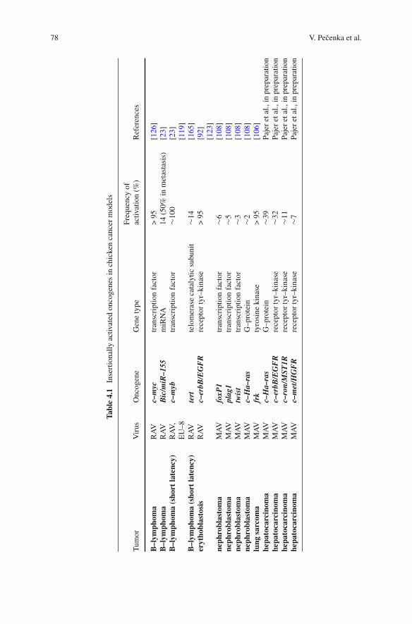
78 V. Pecenka et al.
Tabl
e4.
1In
sert
iona
llyac
tivat
edon
coge
nes
inch
icke
nca
ncer
mod
els
Tum
orV
irus
Onc
ogen
eG
ene
type
Freq
uenc
yof
activ
atio
n(%
)R
efer
ence
s
B–l
ymph
oma
RA
Vc–
myc
tran
scri
ptio
nfa
ctor
>95
[126
]B
–lym
phom
aR
AV
Bic
/miR
–155
miR
NA
14(5
0%in
met
asta
sis)
[23]
B–l
ymph
oma
(sho
rtla
tenc
y)R
AV
,E
U–8
c–m
ybtr
ansc
ript
ion
fact
or∼1
00[2
3][1
19]
B–l
ymph
oma
(sho
rtla
tenc
y)R
AV
tert
telo
mer
ase
cata
lytic
subu
nit
∼14
[165
]er
ytho
blas
tosi
sR
AV
c–er
bB/E
GF
Rre
cept
orty
r–ki
nase
>95
[92]
[123
]ne
phro
blas
tom
aM
AV
foxP
1tr
ansc
ript
ion
fact
or∼6
[108
]ne
phro
blas
tom
aM
AV
plag
1tr
ansc
ript
ion
fact
or∼5
[108
]ne
phro
blas
tom
aM
AV
twis
ttr
ansc
ript
ion
fact
or∼3
[108
]ne
phro
blas
tom
aM
AV
c–H
a–ra
sG
–pro
tein
∼2[1
08]
lung
sarc
oma
MA
Vfr
kty
rosi
neki
nase
>95
[106
]he
pato
carc
inom
aM
AV
c–H
a–ra
sG
–pro
tein
∼39
Paje
ret
al.,
inpr
epar
atio
nhe
pato
carc
inom
aM
AV
c–er
bB/E
GF
Rre
cept
orty
r–ki
nase
∼32
Paje
ret
al.,
inpr
epar
atio
nhe
pato
carc
inom
aM
AV
c–ro
n/M
ST1R
rece
ptor
tyr–
kina
se∼1
1Pa
jer
etal
.,in
prep
arat
ion
hepa
toca
rcin
oma
MA
Vc–
met
/HG
FR
rece
ptor
tyr–
kina
se∼7
Paje
ret
al.,
inpr
epar
atio
n
4 Chicken Models of Retroviral Insertional Mutagenesis 79
for isolating provirus-flanking sequences (inverse PCR and related techniques) andfor their analysis (automatic sequencing machines). The final explosion of datastarted after the complete genome sequences of individual model organisms hadbecome available. From that point it has been a relatively easy task to assort indi-vidual provirus insertion sites along the chromosomes, to pick up common sites ofintegration and associate them with suspect genes.
In this chapter we will describe several chicken models of tumorigenesis throughretroviral insertional mutagenesis that have been thoroughly investigated (for theoverview, see Table 4.1). More space will be given to the recently advanced mod-els: chicken nephroblastomas, lung sarcomas, liver carcinomas and the industasisphenomenon. We will focus on unique features of the chicken models includingmechanistic details of gene activation by defective proviruses. Insertional inactiva-tion of tumor suppressor genes will not be discussed since it has not been provenin chicken system so far. For general issues we refer to other parts of this book andto the reviews [75, 150]. In the end, we will discuss the future of chicken modelsin the era of high throughput oncogene screening in mouse models and large scalesequencing of human cancer genomes.
4.1 The Beginning of the Story: the Case of ChickenBursal Lymphomas
Bursal lymphoma (malignant lymphoma of the bursa of Fabricius named alsolymphoid leukosis or lymphomatosis) is the most common neoplasia of domesticchickens. The viral etiology of bursal lymphomas has been known since 1908 whencell-free extract was shown to transmit the neoplasia [41]. In farms, the causativeagent, now called avian leukosis virus (ALV), is spread mainly by congenital infec-tion from hens to embryos [16]. Experimentally, the virus can be transmitted byintraperitoneal injection of plasma of infected animal into newly hatched suscepti-ble chickens. Several weeks after infection many microscopic transformed folliclesarise in bursa of Fabricius (see also Section 4.7). Within months, one or more macro-scopically observable nodules—bursal lymphomas—develop. The animals die afterprogressive metastatic tumors develop in many organs [4, 28].
ALVs are typical representatives of simple slowly oncogenic retroviruses car-rying only viral replicative genes gag, pol and env to which no substantial trans-forming potential could be ascribed [25]. The first suggestion about the mechanismby which these transforming gene-lacking viruses could induce neoplasia emergedfrom the study of recombinants between oncogenic ALVs (RAV–1 and RAV–2,Rous associated viruses subgroups A and B) and their nononcogenic homolog(RAV–0, Rous associated virus subgroup E). The oncogenicity of the recombinantalways correlated with the presence of U3 region derived from RAV–1 or RAV–2[31, 149]. The U3 region just precedes the start of viral mRNA transcription at the 5′end of the integrated DNA provirus. Not surprisingly, sequence analyses have local-ized typical promoter sequences for RNA polymerase II in the U3 region [164].
80 V. Pecenka et al.
Since the U3 region, as part of the long terminal repeat (LTR), is also present atthe 3′ end of the provirus, it could drive transcription not only of retroviral genes,but also of downstream located host genes (the mechanism named “promoter inser-tion”, see Fig. 4.1a). Hybrid RNAs containing both viral and cellular sequenceshave indeed been found in infected cells [122]. The non-oncogenicity of RAV–0
E1 E2 E35´ LTR 3´ LTRgag/pol env
polyA
polyApolyA
a
polyA
e
polyA
d
polyA
f
polyA
polyA
c
+
g+
polyA
h+
polyA
bpolyApolyA
Fig. 4.1 Schemes of gene activation by promoter insertion (a–f) and enhancer insertion(f–h). a Nondeleted provirus. Predominant transcripts driven by proviral LTRs are full-lengthand spliced copies of the provirus initiated at the 5′LTR promoter. Transcription starting inthe 5′LTR and proceeding through the 3′LTR arrests the promoter activity of the 3′LTR.
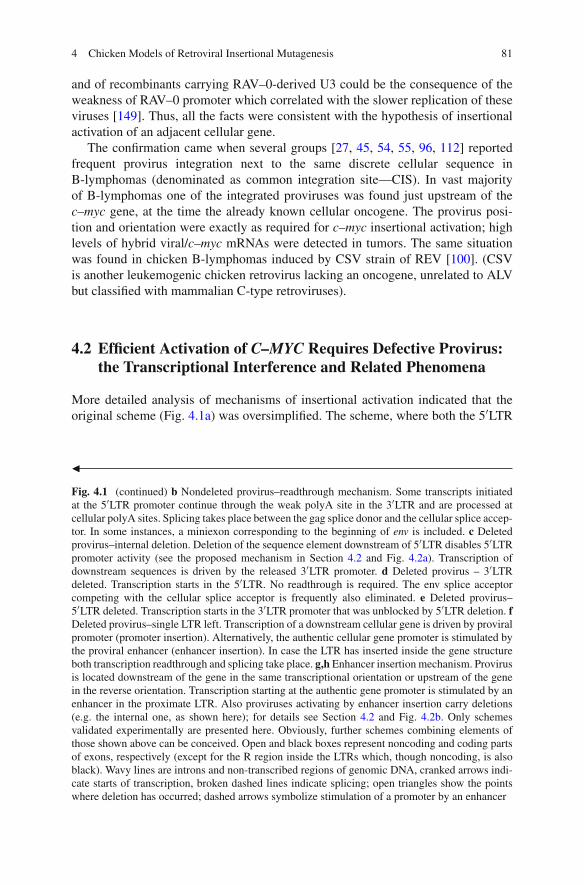
4 Chicken Models of Retroviral Insertional Mutagenesis 81
and of recombinants carrying RAV–0-derived U3 could be the consequence of theweakness of RAV–0 promoter which correlated with the slower replication of theseviruses [149]. Thus, all the facts were consistent with the hypothesis of insertionalactivation of an adjacent cellular gene.
The confirmation came when several groups [27, 45, 54, 55, 96, 112] reportedfrequent provirus integration next to the same discrete cellular sequence inB-lymphomas (denominated as common integration site—CIS). In vast majorityof B-lymphomas one of the integrated proviruses was found just upstream of thec–myc gene, at the time the already known cellular oncogene. The provirus posi-tion and orientation were exactly as required for c–myc insertional activation; highlevels of hybrid viral/c–myc mRNAs were detected in tumors. The same situationwas found in chicken B-lymphomas induced by CSV strain of REV [100]. (CSVis another leukemogenic chicken retrovirus lacking an oncogene, unrelated to ALVbut classified with mammalian C-type retroviruses).
4.2 Efficient Activation of C–MYC Requires Defective Provirus:the Transcriptional Interference and Related Phenomena
More detailed analysis of mechanisms of insertional activation indicated that theoriginal scheme (Fig. 4.1a) was oversimplified. The scheme, where both the 5′LTR
�
Fig. 4.1 (continued) b Nondeleted provirus–readthrough mechanism. Some transcripts initiatedat the 5′LTR promoter continue through the weak polyA site in the 3′LTR and are processed atcellular polyA sites. Splicing takes place between the gag splice donor and the cellular splice accep-tor. In some instances, a miniexon corresponding to the beginning of env is included. c Deletedprovirus–internal deletion. Deletion of the sequence element downstream of 5′LTR disables 5′LTRpromoter activity (see the proposed mechanism in Section 4.2 and Fig. 4.2a). Transcription ofdownstream sequences is driven by the released 3′LTR promoter. d Deleted provirus – 3′LTRdeleted. Transcription starts in the 5′LTR. No readthrough is required. The env splice acceptorcompeting with the cellular splice acceptor is frequently also eliminated. e Deleted provirus–5′LTR deleted. Transcription starts in the 3′LTR promoter that was unblocked by 5′LTR deletion. fDeleted provirus–single LTR left. Transcription of a downstream cellular gene is driven by proviralpromoter (promoter insertion). Alternatively, the authentic cellular gene promoter is stimulated bythe proviral enhancer (enhancer insertion). In case the LTR has inserted inside the gene structureboth transcription readthrough and splicing take place. g,h Enhancer insertion mechanism. Provirusis located downstream of the gene in the same transcriptional orientation or upstream of the genein the reverse orientation. Transcription starting at the authentic gene promoter is stimulated by anenhancer in the proximate LTR. Also proviruses activating by enhancer insertion carry deletions(e.g. the internal one, as shown here); for details see Section 4.2 and Fig. 4.2b. Only schemesvalidated experimentally are presented here. Obviously, further schemes combining elements ofthose shown above can be conceived. Open and black boxes represent noncoding and coding partsof exons, respectively (except for the R region inside the LTRs which, though noncoding, is alsoblack). Wavy lines are introns and non-transcribed regions of genomic DNA, cranked arrows indi-cate starts of transcription, broken dashed lines indicate splicing; open triangles show the pointswhere deletion has occurred; dashed arrows symbolize stimulation of a promoter by an enhancer
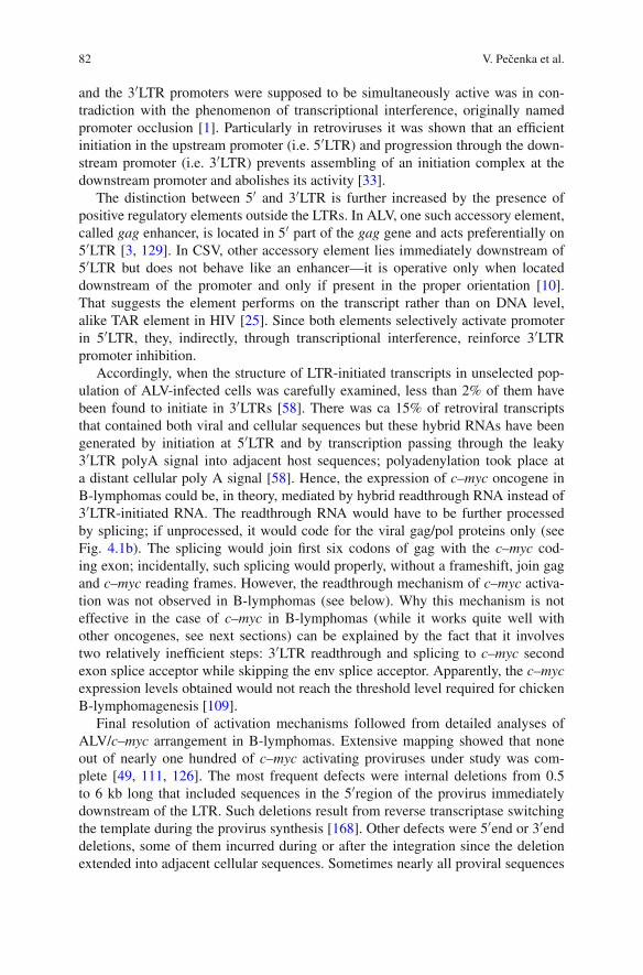
82 V. Pecenka et al.
and the 3′LTR promoters were supposed to be simultaneously active was in con-tradiction with the phenomenon of transcriptional interference, originally namedpromoter occlusion [1]. Particularly in retroviruses it was shown that an efficientinitiation in the upstream promoter (i.e. 5′LTR) and progression through the down-stream promoter (i.e. 3′LTR) prevents assembling of an initiation complex at thedownstream promoter and abolishes its activity [33].
The distinction between 5′ and 3′LTR is further increased by the presence ofpositive regulatory elements outside the LTRs. In ALV, one such accessory element,called gag enhancer, is located in 5′ part of the gag gene and acts preferentially on5′LTR [3, 129]. In CSV, other accessory element lies immediately downstream of5′LTR but does not behave like an enhancer—it is operative only when locateddownstream of the promoter and only if present in the proper orientation [10].That suggests the element performs on the transcript rather than on DNA level,alike TAR element in HIV [25]. Since both elements selectively activate promoterin 5′LTR, they, indirectly, through transcriptional interference, reinforce 3′LTRpromoter inhibition.
Accordingly, when the structure of LTR-initiated transcripts in unselected pop-ulation of ALV-infected cells was carefully examined, less than 2% of them havebeen found to initiate in 3′LTRs [58]. There was ca 15% of retroviral transcriptsthat contained both viral and cellular sequences but these hybrid RNAs have beengenerated by initiation at 5′LTR and by transcription passing through the leaky3′LTR polyA signal into adjacent host sequences; polyadenylation took place ata distant cellular poly A signal [58]. Hence, the expression of c–myc oncogene inB-lymphomas could be, in theory, mediated by hybrid readthrough RNA instead of3′LTR-initiated RNA. The readthrough RNA would have to be further processedby splicing; if unprocessed, it would code for the viral gag/pol proteins only (seeFig. 4.1b). The splicing would join first six codons of gag with the c–myc cod-ing exon; incidentally, such splicing would properly, without a frameshift, join gagand c–myc reading frames. However, the readthrough mechanism of c–myc activa-tion was not observed in B-lymphomas (see below). Why this mechanism is noteffective in the case of c–myc in B-lymphomas (while it works quite well withother oncogenes, see next sections) can be explained by the fact that it involvestwo relatively inefficient steps: 3′LTR readthrough and splicing to c–myc secondexon splice acceptor while skipping the env splice acceptor. Apparently, the c–mycexpression levels obtained would not reach the threshold level required for chickenB-lymphomagenesis [109].
Final resolution of activation mechanisms followed from detailed analyses ofALV/c–myc arrangement in B-lymphomas. Extensive mapping showed that noneout of nearly one hundred of c–myc activating proviruses under study was com-plete [49, 111, 126]. The most frequent defects were internal deletions from 0.5to 6 kb long that included sequences in the 5′region of the provirus immediatelydownstream of the LTR. Such deletions result from reverse transcriptase switchingthe template during the provirus synthesis [168]. Other defects were 5′end or 3′enddeletions, some of them incurred during or after the integration since the deletionextended into adjacent cellular sequences. Sometimes nearly all proviral sequences
4 Chicken Models of Retroviral Insertional Mutagenesis 83
were found deleted leaving only solitary LTR, probably the result of homologousrecombination between the two LTRs.
Frequent generation of defects during retrovirus propagation has been well doc-umented [72, 79, 135, 155]. That, however, does not explain why every provirus inc–myc locus carries a deletion while majority of other proviruses in the same cellsare nondefective. It had to be postulated that only defective proviruses could activatec–myc and only cells with such insertion were selected during oncogenesis. Indeed,it can be demonstrated that defects in activating proviruses eliminate obstructionsto efficient activation. When 3′ LTR is deleted (Fig. 4.1d), inefficient readthroughstep is obviated—all transcripts proceed from 5′LTR into the host sequences; more-over, competing env splice acceptor is often also deleted which makes the splicingfrom gag donor to the c–myc second exon acceptor much more efficient. When 5′LTR is deleted (Fig. 4.1e) or there is only solitary LTR left (Fig. 4.1f), transcriptionstarts in the remaining LTR whose activity was released from transcriptional inter-ference. In all the above cases the authentic c–myc promoter is still present upstreamof the inserted LTR but its activity is low and does not interfere with the LTRactivity.
Internal deletions in proviruses (Fig. 4.1c) had somewhat unexpected conse-quences. These deletions completely turned off 5′LTR while 3′LTR became fullyactive [49]. The very same situation (i.e. disabling 5′LTR by deletion of adjacentdownstream sequences) was found in c–myc activating proviruses in chicken B-lymphomas induced by CSV [147]. This provokes a question of why 5′LTRbecomes completely inactive after losing its downstream accessory elements while3′LTR that also does not have these elements turns fully active. Presently we canonly hypothesize that regulatory interrelations in retroviral provirus are more com-plex then we have supposed and that any disruption of the provirus integrity canmodify activity of both LTRs in a way we are still unable to predict. The exis-tence of additional proviral regulatory elements and interactions including mutualinteractions of both LTRs has already been suggested by [101].
The explanation can ensue when the original promoter occlusion/transcriptionalinterference hypothesis is modified to comply with the present-day knowl-edge of how distant regulatory regions on chromosome execute their effects.Transcriptionaly active regions including retroviral proviruses form dynamic chro-matin loop structures. Enhancer and promoter regions, together with all associatedregulatory proteins, are attached to each other at the loop base creating so calledactive chromatin hub. After a pioneer round of transcription, a terminator region isalso juxtaposed to the promoter. In this way a transcriptional unit is demarcated andrecycling of transcriptional apparatus in this unit is facilitated [35, 40, 116, 159]. Inthe integrated provirus, however, the situation is more intricate: there are two iden-tical sets of enhancer, promoter and terminator (i.e. 5′LTR and 3′LTR), preceded byoriginal c–myc promoter plus enhancer and followed by original c–myc terminator.Thus, several alternative looping structures may form; the final structure is a result ofmutual competition for complex formation between individual elements. We suggestthat accessory element in 5′part of the complete provirus assist preferential joiningof 5′ and 3′LTRs into the loop, presumably by itself forming part of the complex
84 V. Pecenka et al.
gag enhancer
gene promoter
gene first exon
Completeprovirus
Defectiveprovirus
3´-LTR
deleted gag enhancer
5´-LTR
a bEnhancer insertionPromoter insertion
3´-LTR
5´-LTR
gene last exon
transcription START
host sequences
proviral sequences
3´-LTR
5´-LTR
proviral LTR
3´-LTR
5´-LTR
transcription STOP
Fig. 4.2 Organization of transcription units controlled by promoters and enhancers of nondeletedand internally deleted integrated proviruses: A hypothesis. a Promoter insertion. The completeintegrated provirus forms an isolated highly active transcription unit through the formation of acompact chromatin loop structure (active chromatine hub–encircled gray area) that is stabilized byinteraction of the accessory gag enhancer with both LTRs. In this loop the transcriptional machin-ery is permanently recycled from the 3′LTR to the 5′LTR. When the gag enhancer is deleted(defective provirus), the proviral loop is destabilized, 5′LTR turns inactive and 3′LTR can formthe active transcriptional loop with downstream sequences. b Enhancer insertion. The completeprovirus forms the active transcriptional unit in which the proviral enhancer is engaged, as in a.Therefore the proviral enhancer exerts no influence on regulation of neighboring genes. However,when the provirus chromatine loop is destabilized by deletion (defective provirus), the enhanceris free to interact with neighboring host sequences and stimulates the authentic host gene pro-moter. All the contacts are mediated by protein complexes associated with promoter, enhancer andterminator regions (not shown in this schematic drawing)
(Fig. 4.2a). The accessory element would thus share some functional propertieswith the promoter targeting sequence (PTS) described in Drosophila which pro-motes association of a specific enhancer with a specific distant promoter even whenan insulator/enhancer blocker element is present between them [80, 170].
The 5′LTR—3′LTR chromatine hub may be arranged so that an initiation com-plex is formed only in 5′LTR promoter; however, the existence of the loop itself issufficient to determine which of the two proviral promoters remains active. Whentranscription starts in 3′LTR, the transcription complex leaves the loop and newround of promoter selection begins; when transcription starts in 5′LTR, the promoter

4 Chicken Models of Retroviral Insertional Mutagenesis 85
in 3′LTR is silenced by transcriptional interference and transcription machinery ispermanently recycled from 3′LTR back to 5′LTR. In such arrangement, enhancersin both LTRs may stimulate transcription from a single, 5′LTR promoter—the effectobserved by [101]. When internal proviral accessory element is deleted as it is thecase in most of the c–myc activating proviruses the proviral transcriptional unit isdestabilized and other looping structures preferentially form including the 3′LTR—c–myc polyA loop that demarcates new, highly active c–myc transcriptional unit(Fig. 4.2a).
In several tumors [49, 111, 126], the provirus was located upstream of c–mycin the reverse transcriptional orientation or downstream of c–myc in the same tran-scriptional orientation (Fig. 4.1g and 1h). Such arrangement did not conform withthe promoter insertion model as transcription starting from any provirus LTR couldnot proceed to c–myc sequences. Nevertheless, substantially increased levels of thec–myc mRNA have been found. Its synthesis started at the authentic c–myc promoterthe activity of which was, apparently, stimulated by juxtaposed powerful enhancer inproviral LTR. This configuration has been denominated “enhancer insertion”. It hasbeen reported that also isolated LTR can activate by the enhancer insertion mech-anism [81]: in two B-lymphoma lines carrying a single LTR insertion in the firstc–myc intron in the same transcriptional orientation the LTR-contained enhancerstimulated transcription from the original c–myc promoter while the LTR-containedpromoter was silenced by transcriptional interference (Fig. 4.1f).
Also proviruses activating c–myc by enhancer insertion have been shown to carryinternal deletions [49, 126]. That is not surprising when we accept the model pre-sented in previous paragraphs: to be able to attach and stimulate c–myc promoter,the relevant proviral enhancer must first be released from the complex with otherproviral elements. Deletion of accessory elements in 5′ part of the provirus may bethe easiest way of such enhancer liberation (Fig. 4.2b). Obviously, previous reason-ing presumes that proviral enhancer cannot make a complex with and stimulate twopromoters simultaneously. Such assumption is quite acceptable considering verycondensed arrangement of the enhancer into less than 100 bp; it is also in com-pliance with the provirus tendency to form isolated highly active transcriptionalunit.
The support for the above model can be found in a way how MLV and FeLVactivate oncogenes. They do it predominantly by enhancer insertion mechanism;however, in contrast to ALV, activating MLV and FeLV proviruses are generallynot defective [97, 143]. At first sight it may seem to contradict the proposedmodel. However, the opposite is true. The above model can, in fact, explain onepeculiarity of oncogenesis by MLVs and FeLVs: absolute majority of activatingproviruses carry enhancer duplications or even higher level multiplications. Forexample, lymphomagenesis in some mice strains like AKR relies on spontaneousgenesis of oncogenic recombinants (named MCF, mink cell focus-forming virus)from endogenous MLV proviruses present in the strain. Indispensable componentof the oncogenic MCF recombinant is a presence in LTR of two copies (in tan-dem) of strong enhancer acquired from an endogenous provirus in which onlysingle copy of the enhancer is present [143]. Highly lymphomagenic exogenous
86 V. Pecenka et al.
MLV strains isolated from naturally occurred tumors, like Mo–MLV, already con-tain duplicated enhancer [25]. In tumorigenic exogenous FeLVs there is only asingle copy of an enhancer; however, in induced T-lymphomas, where c–myc isactivated by the enhancer insertion mechanism, all activating proviruses carry tan-dem enhancer duplication acquired always de novo during infection of the animal[97].
The enhancer duplication was shown to have only mild effect on virus replica-tion and provirus transcription. When single-enhancer MLV or FeLV viruses arepropagated in culture the anticipated variants with duplication do not arise (or, moreaccurately, are not positively selected for). Hence, rather than being important forthe virus spread in the animal, the duplication is required for the mechanism of onco-genesis itself [97, 143]. We suggest that presence of tandem copies of an enhancerenables provirus 5′LTR to form two chromatin loops simultaneously. While oneenhancer is engaged in the formation of proviral transcriptional unit the other onemay contact oncogene promoter; no provirus defect is needed for the oncogeneactivation.
To summarize, distinct ways of how to make provirus capable of stronglyaffecting host genes are being employed during lymphomagenesis by avian versusmammalian retroviruses. In the ALV model, compactness of the proviral transcrip-tional unit is broken by different types of provirus deletions, mostly by deletionof the accessory element outside the LTR. Transcription of proximate host genesis then driven by the promoter of defective provirus (mechanism of promoterinsertion). In MLV and closely related FeLV, however, no internal accessory ele-ments have been reported. It is possible that compactness of the MLV and FeLVtranscriptional units is not dependent on internal accessory elements; thus it can-not be so easy disrupted by provirus deletion. Instead, another way to generatestrongly activating proviruses is realized: enhancer multiplication that capacitatesprovirus to boost transcription of neighboring genes from the genes’ own pro-moters at a distance (mechanism of enhancer insertion). Accordingly, while themechanism of oncogene activation by enhancer insertion is found only excep-tionally in chicken models, it is the dominating mechanism in MLV and FeLVmodels.
In this context it would be interesting to find out how gene activation works inMMTV model, where enhancer insertion is also the prevalent mechanism. Thoughthe data concerning structural changes in oncogene-activating MMTV provirusesare scarce, it seems that in mammary carcinomas neither deletion nor enhancerduplication takes place [102] while in variant MMTV-induced T-lymphomasenhancer multiplications may play an important role [6, 14]
For the sake of completeness we have to mention additional possible effects ofprovirus insertion that have not been discussed above. First, an important part ofinsertional activation mechanism may be separating negative regulatory elementslike silencers from the oncogene promoter. Eventually, cellular transcription paus-ing sites may be shifted away from the transcription unit by the provirus insertion.Presence of negative elements is easy to imagine in cases where the activatedgene has been completely inactive before provirus integration. These mechanistic
4 Chicken Models of Retroviral Insertional Mutagenesis 87
details, however, are commonly not addressed by the investigators. Second, therange of effects incited by the provirus integrating into the gene structure is notlimited to the level of transcription. For example, any change in mRNA structure(truncation, fusion with viral sequences, activation of cryptic promoters and splic-ing or polyadenylation sites etc.) may have profound impact on its export fromthe nucleus, stability, and rate of translation. In mouse models, the most commonmechanism of this type may be the frequent truncation of 3′untranslated regionscontaining destabilizing AT rich sequences as well as target sequences for miR-NAs which downregulate both stability and translatability of affected mRNA. Sucheffects during insertional activation have occasionally been suspected or even doc-umented, see e.g. [20, 61, 132–134, 152], but only recently have also been morethoroughly analyzed [34].
4.3 Readthrough Activation of C–MYB in B-lymphomas:the Case of “Superactivating ALV”
When 10–14 day old embryos, instead of newly hatched chickens, were intra-venously injected with ALV, some of them developed, primarily in livers, highlyaggressive B-lymphomas after latency of only several weeks (from now on thesetumors will be called short latency lymphomas as opposed to long latency lym-phomas arising after infection of newborn chicks—see Section 4.2). In most ofshort latency lymphomas ALV provirus was detected around (upstream, inside ordownstream) the first exon of the c–myb gene, a cellular progenitor of retrovi-rally transduced oncogene v–myb [23, 117, 118]. The proviruses had no defects andwere integrated in the same transcriptional orientation as the c–myb gene. ChimericALV/c–myb mRNAs were synthesized by a typical readthrough mechanism (seeSection 4.2 and Fig. 4.1b): they were initiated in 5′LTR, polyadenylated at c–mybpolyA site and processed by splicing the ALV gag donor to the c–myb second exonacceptor. Gag and c–myb were not coded by the same reading frame in this mes-sage. Moreover, gag start codon was followed by three in frame stop codons at thebeginning of c–myb; translation of c–myb relied on reinitiating at the codon 21. Theproduced slightly truncated protein was strongly oncogenic, in contrast to the wtprotein [63].
Profound effect of infection timing suggests that target cells for short latencylymphomas are different from target cells for long latency lymphomas (presumablyprebursal stem cells versus bursal stem cells) and that the first ones are absent inchickens after hatching [118]. In consequence, different target cells are sensitiveto activation of different oncogenes (c–myb versus c–myc). Consistently, no lym-phomas develop when infection of the bursa is postponed to the time when bursalstem cells are no more present (ca 3 weeks after hatching), even in animals withsubsequent strong lifelong viremia [15].
The frequency of short latency lymphomas and c–myb activations were consid-erably increased when specific recombinant ALV strains were used for infection.
88 V. Pecenka et al.
In these “superactivating” strains an element in the 5′part of gag gene, called nega-tive regulator of splicing (NRS), was knocked out by either deletion or mutation[8, 71, 119, 136, 138]. NRS has two roles in ALV both of which help to ful-fill peculiar requirements of retroviral replication: to leave substantial proportionof retroviral RNA unspliced and, at the same time, to ensure polyadenylation atthe 3′end of such RNA (which is generally coupled to splicing); for details see[24, 84]. To this end NRS first acts as a pseudo-splice donor, forming nonproductivesplicing complexes with downstream acceptors thus blocking them from splicingto authentic donor sites. Second, by binding factors that interact with polyadeny-lation machinery, NRS helps to recruit and stabilize processing complexes at the3′LTR polyA site. The second effect is dependent on the first one, since only theformation of an abortive splicing complex between NRS and downstream splic-ing acceptor brings NRS close enough to 3′LTR polyA site to exert its effect onpolyadenylation [84].
As shown by [93] on RSV model, deletion of NRS has serious consequencesto viral RNA processing. First, 3′LTR readthrough is strongly elevated (up to over50%). Second, while the frequency of splicing from the gag donor to the env accep-tor is, surprisingly, little affected, the splicing to a more distant acceptor is boostedca four times. Similar (though less conspicuous) effects were observed for “superac-tivating” ALVs with the mutated NRS [105, 138]. Thus, when such ALV integratesupstream of a cellular gene, the spliced readthrough mRNA coding for the cellulargene is produced with much higher efficiency compared to wt ALV.
4.4 Insertional Activation Can Be Accompanied by ExtensiveAlterations of the Oncogene Structure and by Formationof an Oncogene-Transducing Virus: the Case of ChickenErythroblastosis
Besides short- or long-latency B-lymphomas, chickens infected with ALV caninfrequently develop other types of neoplasia (see Section 4.8). Among them,the most thoroughly analyzed was erythroblastosis, a disease characterized byuncurbed proliferation of erythroblasts with arrested differentiation. In sensitivechicken strains high amounts of erythroblasts appear in blood ca 2 months afterinfection with ALV; at the same time they start to massively infiltrate spleen andliver causing rapid death of the animal. Paradoxically, erythroblastosis is oftenaccompanied by anemia since feedback mechanisms triggered by the flood of trans-formed erythroblasts block further development of nontransformed red blood cellprecursors.
In 1983, recurrent insertional activation of the c–erbB protooncogene (orEGFR—epidermal growth factor receptor gene) in chicken erythroblastosis wasfirst reported [46]. Further papers [50, 92, 99, 123] described peculiar molecu-lar details of this activation. In all analyzed cases, the full-length ALV provirus

4 Chicken Models of Retroviral Insertional Mutagenesis 89
was integrated in the same intron in the middle of c–erbB gene in the sametranscriptional orientation. High levels of c–erbB specific mRNAs were pro-duced. The mechanism of activation was much the same as activation ofc–myb in short latency B-lymphomas (Section 4.3): readthrough transcript start-ing in 5′LTR and polyadenylated at one of the two authentic c–erbB polyA siteswas processed by splicing that brought c–erbB coding sequences close to the startof mRNA (Fig. 4.1b). There were, however, two differences. First, mRNA process-ing was more complex. An alternative short exon demarcated by env splice acceptorand cryptic splice donor 159 bp downstream was included in high proportion of mes-sages. Second, the extent of oncogene truncation was much more extensive then inthe case of c–myb. The protein encoded by the spliced mRNAs consisted of 6 AA ofgag (plus 53 AA of env in some messages) fused to the C-terminal part of c–erbB. Inboth protein versions the N-terminal part—the EGF binding extracellular domain—was missing. Such truncation is known to result in a constitutive kinase activity ofthe receptor [77]. It was suggested that the 53 AA+ version of activated c–erbB maybe the major mediator of oncogenesis. The alternative short exon codes for a signalpeptide that normally targets env protein for membrane translocation. This functionwas retained also in a fusion with truncated c–erbB, where it substituted for missingauthentic c–erbB signal peptide. During the processing the 53 AA+ protein had thesignal sequence cleaved off, was fully glycosylated and exposed on the cell surface,in contrast to the 53 AA– version, which was only partially glycosylated and wasnot exposed on the cell surface [85].
The frequency of erythroblastosis is just several percent in most chicken strainsincluding outbred flocks; there are however inbred strains that develop ALV-inducederythroblastosis with penetrance ca 80%. This sensitivity is a dominant trait not con-nected with the ability of ALV to activate c–erbB, but rather determining sensitivityof the animal to such activation [127].
In high proportion of tumors the situation was obscured by formation ofc–erbB-transducing recombinant virus. While oncogene transduction is generallya rare event [25], highly leukemogenic viruses carrying truncated version of c–erbBcan be isolated from up to 50% of chickens with ALV-induced erythroblastosis[92, 124]. That has made erythroblastosis an attractive model and rich source ofmaterial for studying mechanisms of transduction. For nearly all other isolates ofacutely transforming retroviruses the original tumor where the virus has originatedis not available and the virus has further evolved during repeated passages sincethen. Here, by comparing sequences of the transducing viruses with sequences of theinsertionally activated c–erbB genes and fusion mRNAs synthesized in the tumorsit was possible to confirm basic scheme of transduction as suggested earlier. Detailsof the mechanism are beyond the scope of this book; for them, see [25, 59, 92, 124,128, 146]. The important point is that the first step of transduction is the synthesisof hybrid mRNA containing both the retroviral and oncogene sequences, i.e. thetransduction is preceded by an insertional mutagenesis. Hence it is not surprisingthat the sites of truncation in insertionally activated c–erbB and in the transducedv–erbB precisely coincided.
90 V. Pecenka et al.
4.5 Chicken Nephroblastomas Induced by MAV: Complex ModelSuitable for High Throughput Oncogene Screening
In previous chapters we have described mechanisms of insertional mutagenesis byretroviruses in hematopoietic cells, which led to the identification of several onco-genes. Each of the models has been dominated by a single CIS. Hematopoieticmalignancies are generally characterized by a less diverse pattern of mutated genesthan solid tumors. Solid tumors, especially carcinomas, display high complexityand variability of karyotypic alterations, which seriously complicates the search foroncogenes and tumor suppressor genes in these tumors [94]. The complexity prob-ably results from the complex homeostatic control of cells in solid tissues whichmust be overcome. Thus, finding appropriate model tumors which would enablea large-scale search for solid tumor oncogenes and tumor suppressor genes hasbeen a challenge. The models based on MAV retroviruses are among the successfulones.
MAV–1 and MAV–2 (myeloblastosis-associated viruses 1 and 2) are simpleslowly oncogenic retroviruses classified with ALVs. They differ by their env gene(and, consequently, by host specificity), MAV–1 belonging to subgroup A, MAV–2to subgroup B. They are highly homologous to other ALVs except one region: theU3 part of LTR, the region containing proviral enhancer/promoter and playing cru-cial role in insertional activation (see Sections 4.1 and 4.2). It is this MAV-specificregion that confers very distinct oncogenic potential on MAV retroviruses [67].When MAV–1 or MAV–2 are injected into 12 day old embryos or newly hatchedchicks they induce nephroblastomas with efficiency 70–100% depending on chickenstrain. Multiple tumors become visible on kidney of an infected animal as early as 2months after infection and they rapidly grow into massive size [68, 140, 158, 160].
The chicken nephroblastomas are embryonic tumors derived from nephrogenicblastema cells which persist in the newborn kidney for several days after hatching.They consist mainly of undifferentiated mesenchyme and aberrant differentiatingepithelial renal elements reminiscent of developing kidney. They are comparableto human nephroblastomas—Wilms’ tumors both by their histology and presumedtarget cells of tumorigenesis [7, 11, 56, 62].
First attempts to identify CISs in chicken nephroblastomas had only limited suc-cess. Two groups reported two different loci hit by proviral integration in chickennephroblastomas—Ha–ras [160] and nov/ccn3 [68]. Although deregulation of thesegenes in the chicken or mammalian cells have been proven to result in a transformedphenotype, each of them have been found hit only in a single case and therefore theycould not be classified as CIS.
Restriction mapping of integration sites in clonal nephroblastomas usingSouthern blot analyses revealed unexpectedly complex pattern from which exis-tence of CISs could not be definitely inferred [114]. Three interpretations have beenput forward. Firstly, there may be a major CIS but it represents region too large tobe disclosed by restriction mapping on the background of several further integratedproviruses in each nephroblastoma clone. Secondly, there might be no major CISbut rather numerous less frequent ones. Thirdly, activating proviruses could carry
4 Chicken Models of Retroviral Insertional Mutagenesis 91
deletions similarly to the situation in B-lymphomas which would invalidate the usedmapping strategy.
Later on, an improvement of the techniques for isolation and analysis of provirus-flanking sequences enabled more comprehensive survey of VISs (“Viral IntegrationSites”) in nephroblastomas. As described in Section 4.2, defective proviruses arevery potent mutagens and deletion in the provirus may be a prerequisite for onco-gene activation. Frequent occurrence of defective proviruses in nephroblastomashas been documented before [115, 140, 160]. Therefore, to start with, molecularcloning of VISs harboring defective proviruses in six randomly chosen nephroblas-tomas have been carried out, leading to identification of the insertionally activatedgene twist. Subsequent screening of a panel of chicken nephroblastomas have shownthat the twist gene is the first true CIS in these tumors—it was hit in approximately4% of analyzed tumors [107].
These results strongly favored the hypothesis of numerous rather infrequent CISsin the chicken nephroblastomas. To prove it, high throughput screening of VIS inhundreds of nephroblastoma samples had to be performed. To achieve a maximumcoverage of VISs, two independent and overlapping methods for the VIS identifica-tion were employed [108]: inverse PCR (iPCR) and LTR-based rapid amplificationof 3′cDNA ends (LTR RACE). The iPCR technique provided genomic sequencesflanking the integrated provirus. The technique was optimized to the level whenflanking sequences of all clonally integrated proviruses could be gel-isolated aftersingle PCR reaction; routine VISs recovery was 90–100% in each tumor. LTR–RACE technique provided sequences of mRNA species containing provirus LTRat its 5′end, including hybrid mRNAs generated by readthrough transcription orby transcription starting in the promoter of a defective provirus (see Section 4.2and Fig. 4.1). LTR–RACE analysis covered up to 30% of VISs. The lower yieldachieved by LTR–RACE is compensated by the fact that found VISs were the mostrelevant ones—those where high expression of cellular sequences was driven byan integrated provirus. At the same time, the structure of LTR–RACE productsrevealed details of the mechanism of host gene activation. Importantly, both tech-niques detected only insertions present within a substantial proportion of tumor cellsas opposed to currently used sequencing strategies that record also oncogenicallyunselected integrations present even in a single cell.
To date more than 1100 unique VISs have been identified in 187 MAV-inducednephroblastoma clones [108] and (Pajer, unpublished). Out of them 69 candidatetumor-related loci (denominated NALs, “nephroblastoma associated loci”) havebeen selected according to any of the following three criteria:
(a) two and more VISs in a close genomic distance; the distance limit was setarbitrary to 20 kb;
(b) two and more VISs within one annotated gene;(c) VISs from the tumors harboring only a single integrated provirus as demon-
strated both by Southern blot hybridization and iPCR; to date only three suchsamples have been found.
92 V. Pecenka et al.
It should be emphasized that not all candidate NALs necessarily have to representcancer-related genes, especially those hit by a provirus in two samples only. It hasbeen shown that using the sole criterion of multiple insertions into the same locusin retroviral oncogene screens may lead to high proportion of false positives in con-sequence of retroviral integration preferences [163]. The most relevant loci (thosehit in at least four tumors) were the foxP1, the plag1, the twist, and the c–Ha–ras.FoxP1 (forkhead box P1) encodes a widely expressed transcription factor impor-tant for heart, lung and lymphocyte development; it was shown to function eitheras an oncogene or a tumor suppressor gene depending on cell type [76]. Plag1codes for a zinc finger transcription factor in humans expressed only in embry-onal tissues; its inapropriate activation is responsible for induction of several typesof human tumors [151]. Twist encodes a basic helix-loop-helix transcription factorregulating epithelial–mesenchymal transition during embryogenesis and metastasisand supporting survival of cancer cells [2, 22].
Analysis of produced mRNAs and provirus alignment in the selected CISsshowed that the molecular mechanism of activation was different in each case (seealso Fig. 4.3). All twist-activating proviruses were integrated in the same transcrip-tional orientation in a narrow region just upstream of the twist initiation codon andmost of them carried various deletions or rearrangements, alike the ALV provirusesactivating the c–myc gene in chicken B-lymphomas (Section 4.2, Fig. 4.1c–f). Veryhigh expression of twist mRNA was driven mostly by 3′LTR promoter. Neither nor-mal adult kidney nor nephroblastomas without MAV insertion in the twist locus didcontain detectable amount of twist mRNA while moderate level of twist mRNA wasdetected in the embryonic kidney.
Similarly, in tumors with activated plag1 gene very high levels of the plag1mRNA were found. This RNA was generated by a readthrough mechanism followedby splicing between the MAV gag donor site and the plag1 second exon acceptorsite, much like during c–myb activation in short latency lymphomas (Section 4.3,Fig. 4.1b). The hybrid mRNA coded for full-length plag1 protein.
A strikingly different situation was found in the case of foxP1. Proviruses wereinserted downstream of the first or second foxP1 coding exons mostly in the sameorientation as the gene’s transcription. No gross change in overall expression offoxP1 mRNA has been observed in any tumor harboring affected allele of the gene.Hybrid MAV/foxP1 mRNAs, however, could be detected and analyzed using RT–PCR; they coded for the N-terminally truncated FoxP1 proteins. Further analysis ofthe effect of foxP1 truncation was complicated by the existence of alternative start-ing points, alternative exons and alternative polyA signals in foxP1 gene. Moreover,it has been observed that both the amount and subcellular localization of FoxP1 pro-tein was highly variable in individual nephroblastoma samples, including those notharboring viral integration in foxP1 gene.
As to the c–Ha–ras activation, provirus insertions have been found in5′untranslated region of the gene in the same transcriptional orientation. Both hybridMAV/c–Ha–ras mRNAs and normal c–Ha–ras mRNAs were observed; the hybridmRNAs coded for normal c–Ha–ras protein but they were missing 5′untranslatedregion present in normal c–Ha–ras mRNA. The overall level of c–Ha–ras mRNA

4 Chicken Models of Retroviral Insertional Mutagenesis 93
10 kbp
500 bp
plag1
twist
foxP1
10 kbp
Ha - ras
Splice acceptor for hybrid mRNAs
–100–200–300–400 bp–500–1000
frk
10 kbp
Fig. 4.3 Genomic organization, distribution of insertion sites and mRNA expression inselected CISs. The CISs were identified in chicken nephroblastomas (foxP1, plag1, twist and Ha–ras) or lung sarcomas (frk). Sites and orientations of individual proviral insertions are indicatedby arrows. Positions of translation initiation are marked by asterisks. Panels to the right showNorthern blot hybridization profile of a few representative tumor samples, except for the frk locuswhere RT–PCR profile is shown. The samples harboring a provirus in the given locus are markedby arrows
was the same in nephroblastomas with or without insertion in c–Ha–ras locus andin the normal kidney. No point mutations, the archetypal mode of Ha–ras activationin human and mouse tumors, have been found (Pajer, unpublished). How such “acti-vation”, which affects neither mRNA level nor the structure of the produced proteincan participate in nephroblastoma induction is not clear. We suggest that deleting5′untranslated region might result in the increased translation of the hybrid mRNA.Alternatively, Ha–ras activation could be important only during the induction stagein nephroblastema cells, the target cells of oncogenesis—for instance, if basal levelof Ha–ras expression is low in these cells (hit and run hypothesis).
The results draw a highly complex picture of virus-induced mutations innephroblastomas. Out of 69 candidate NALs only four were hit in a reasonable
94 V. Pecenka et al.
percentage of samples: the foxP1 (in about 6% of tumors), the plag1 (ca 5%of tumors), the twist (ca 3% of tumors) and the c–Ha–ras (ca 2% of tumors).All the identified NALs together would explain for induction of ca 130 out of197 nephroblastomas (ca 65%). Evidently, many alternative cancer-related genes(or combinations of them) may be deregulated by the provirus integration innephroblastomas.
The special feature of the chicken nephroblastoma model is the high efficiencyof infection of nephrogenic blastema cells [114, 160]. Each cell acquires numer-ous integrations thus generating enormous amount of combinations of insertionalmutations. These facts establish the chicken nephroblastoma as a perfect model forthe study of oncogene/tumor suppressor gene cooperation during multistep tumorinduction.
4.6 Extension of Spectrum of MAV-Induced Tumors By LocalHomeostasis Perturbation: Lung Sarcomas, LiverCarcinomas and the Industasis Phenomenon
In about 7% of MAV–2-infected animals late lung sarcomas or liver carcinomasdeveloped in addition to nephroblastomas. These tumors were independent primaryclonal outgrowths, not metastases originated from the nephroblastoma as shown bydistinct patterns of proviral integrations. While nephroblastomas and liver carcino-mas appeared as nodules clearly separated from normal tissue, the lung sarcomaswere highly invasive: one tumor clone was frequently disseminated into severalfoci in both lungs and in some animals these foci constituted majority of lung tis-sue [106]. Presence of considerable amount of tumor cells could even be detectedby PCR in macroscopically normal lung tissue samples (Pajer, unpublished). Bothtypes of late tumors would represent new interesting models for oncogene screeningif it were not for the crucial drawback: they were too rare to provide enough samplesfor thorough analysis.
The obstacle of low penetrance was cleared by discovery that injection of MAV–2producing chicken cells instead of virions intravenously into the embryos or newlyhatched chickens changes formerly rare tumors into frequent ones. In the case oflung tumors the process of tumorigenesis was also markedly accelerated—the lungtumors appeared in most animals within 1–2 months after cell injection, prior tonephroblastomas and liver tumors, the latency of which did not change. The cell-assisted tumors did not differ from the rare ones neither by gross morphology norby histology. Preventing the division of injected MAV–2 producing cells by treat-ment with mitomycin did not abolish their ability to promote tumor formation.Consistently, the cell-assisted lung and liver tumors were shown to be of the hostorigin and not to contain detectable amounts of the injected cells [106] and (Pajeret al., in preparation).
Surprisingly, the same tumor-promoting effect was observed when animals wereinjected first with MAV–2 virus at embryonal day 12 and then with uninfected

4 Chicken Models of Retroviral Insertional Mutagenesis 95
chicken embryonal fibroblasts 9 days later, shortly after hatching. At that time all tis-sues of the animal including lungs and liver were already fully infected, suggestingthat injected cells do not function only as reservoirs spreading virus in the animal.This conclusion was further supported by the fact that injected cells affected nei-ther the spectrum of infected tissues nor the level of their infection. However, wheninjections were carried out in the opposite order, i.e. the virus was injected 9 daysafter the cells, no tumor promotion was observed [106]. This fact suggests that thetumor-promoting effect of the injected cells persists not longer than several days.
To follow the fate of the injected cells, [35S]methionine-labelled embryonalfibroblasts were used and monitored in individual embryonic tissues both bytotal tissue radioactivity and by paraffin-embedded section autoradiography. Theexperiments showed a prompt passage of the injected cells from blood into thetissues where they settled as individual stray cells. 20 h after injection the cellswere detected in all analyzed embryonic tissues including brain; the highest levelswere found in the liver, kidney, and lungs while the residual levels in the blood werevery low.
Based on these observations the concept of industasis was proposed—a promo-tion of fully malignant phenotype of an incipient tumor cell by a stray cell througha disruption of local homeostasis; for details see [106]. It was suggested that thephenomenon of industasis might be the underlying cause of many human multipleprimary tumors when a stray cell released by an advanced tumor promotes tumorformation from another, mutated and potentially malignant cell that was kept undercontrol by tissue homeostasis.
Besides its potential impact on our understanding of mechanisms of oncogenesisin general, the industasis fenomenon advanced the model of MAV–2 insertionalmutagenesis as it enabled a large scale analysis of lung and liver tumors. Bothrare virus-only-induced and frequent cell-assisted tumors have been searched forCISs using the same methods as described in Section 4.5, i.e. iPCR and LTRRACE. Quite dissimilar pattern of CISs was revealed in the lung versus livertumors; importantly, however, there was no difference between the rare and thecell-promoted tumors [106].
In contrast to nephroblastomas, where numerous low frequency CISs have beenfound, lung sarcomas were dominated by a single one. In more than 95% lungtumors a provirus insertion was observed in the gene frk/rak [106] and (Pajeret al., unpublished); no other CIS was identified. The frk (fyn-related-kinase gene)encodes a tyrosine kinase expressed in humans predominantly in epithelial tissues[19]. It was shown to act as a tumor suppressor gene that negatively regulatesPI3K/Akt pathway through stabilisation of PTEN, another tumor suppressor andnegative regulator of PI3K [13, 167]. Surprisingly, frk-knockout mice displayedvery mild phenotype and no increase in tumor incidence [21]. The model of chickenlung sarcomas has demonstrated for the first time the oncogenic capability of frkoverexpression, previously being only suspected [60].
The provirus/frk arrangement was much alike provirus/c–myc arrangement inB-lymphomas: defective proviruses had integrated in the same transcriptionalorientation into a very narrow area within the promoter/5′UTR region in front
96 V. Pecenka et al.
of the gene’s coding sequences. Most defects were internal deletions compris-ing the enhancer element in 5′part of gag (Pecenka et al., in preparation). Highlevels of MAV–2/frk hybrid mRNA initiating in the proviral 3′LTR have beendetected in all samples carrying provirus insertion in the frk locus. The pro-tein coding sequence was not afflicted in any sample. No frk expression wasdetectable neither in tumors with unaffected frk locus nor in the non-tumor lungtissue [106].
In liver tumors, the pattern of CISs was more variable and different fromthe pattern in kidney or lung tumors (Pajer et al., in preparation) and (Pecenkaet al., in preparation). The vast majority of liver tumors carried MAV–2 provirusinserted within one of four genes: c–Ha–ras (39% of tumors), c–erbB/EGFR (32%),c–ron/c–stk/c–sea/MST1R (11%) or c–met/HGFR (7%); no other CIS was recorded.The last three genes encode receptor tyrosine kinases expressed in many types ofepithelial cells and implicated in several carcinoma classes in humans [47, 104,156]. Interestingly, they all control, among others, signaling pathways convergingon the c–Ha–ras, the most frequently activated gene in chicken liver tumors. Thatsuggests that activation of the c–Ha–ras signaling is the pivotal disturbance in allMAV–2 induced liver tumors and that insertions into four alternative genes mightbe just four different ways how to affect c–Ha–ras and pathways downstream ofit. Consistently, simultaneous activation of two or more of these genes in the sametumor was never observed, though, on mere statistical basis, c–Ha–ras should behit in about one third of tumors with c–erbB activation and vice versa. It must bepointed out, however, that the screen did not comprise genes connected with pro-gression steps since all samples analyzed were early stages of tumor development,according to terminology of [73] classified as preneoplastic nodules or adenomas(Pajer et al., in preparation).
Similarly to nephroblastomas, c–Ha–ras gene was activated by provirus inser-tion into the gene’s 5′untranslated region; the c–Ha–ras coding sequences werepreserved. Alike in nephroblastomas, no point mutations have been found. Unlikenephroblastomas, however, insertions resulted in substantial overexpression ofhybrid MAV–2/c–Ha–ras mRNA initiating in the proviral 3′–most LTR. All activat-ing proviruses have been heavily rearranged including large deletions, duplicationsand inversions (Pecenka et al., in preparation).
The mode of c–erbB activation resembled the situation in ALV-induced ery-throblastosis (Section 4.4): all proviruses had integrated into the same intron in themiddle of the gene. That resulted in high expression of hybrid mRNA encoding theN-terminally truncated c–erbB/EGFR fused to the first 6 AA of gag gene. Contraryto erythroblastosis the c–erbB-activating proviruses had been heavily rearrangedmuch alike the c–Ha–ras-activating ones. Due to the provirus rearrangement, vari-ant mRNA containing env mini-exon described in erythroblastosis model could notbe generated.
The c–ron activation paralleled the c–erbB activation in all aspects describedabove including the truncation of gene’s 5′part that codes for the receptor’s extracel-lular ligand-binding domain. Alike in case of c–erbB, N-terminal truncation confersconstitutive kinase activity on c-ron protein [82].
4 Chicken Models of Retroviral Insertional Mutagenesis 97
The details of c–met activation could not be conclusively established sinceRNA from these tumors was not available. Based on DNA analysis it seems thatthe integrated defective proviruses could drive overexpression of the full-lengthprotein—they were located upstream of the gene’s initiation codon (in the promoteror 5′untranslated region), in the same transcriptional orientation.
4.7 Evidence of Multistage Cancerogenesis in Chicken Models
Tumors are end products of successive evolution through selection of progressivelymore malignant cell subclones which is reflected by complex pattern of accumu-lated mutations of oncogenes and tumor suppressor genes (“cancer genes”). Suchcomplex pattern is typical for human tumors and has also been found in mousetumor models. In majority of chicken models the mutational pattern seems to bequite simple (see previous Sections); that may, however, be just due to the absenceof thorough high throughput analyses. Nevertheless, the multistep nature of onco-genesis in chicken models can be evidenced by studying progressive evolution oftumors.
The examination of early stages of B-lymphoma development (Section 4.1)showed that cells with a proviral integration in the c–myc locus appear early afterALV infection. The chicken Bursa of Fabricius is compartmentalized into ca 10 000follicles which are colonized by B-cell progenitors during late embryonic develop-ment [103]. One month after ALV infection of young chicks, up to 100 hyperplastictransformed follicles are observable in bursa. Each of them represents a clonal out-growth of a different cell with activated c–myc [9, 48]. The hyperplastic state is nota consequence of accelerated proliferation but of blockage of lymphoblast differen-tiation and emigration from bursa [12]. Nearly all transformed follicles disappearduring bursa involution, only one or a few (if any) of them progress to the stage ofB-lymphoma. Distinct and (to a lesser extent) overgrown clones of cells with acti-vated c–myc gene have also been observed 1 month after infection in tissues whereprimary tumors do not develop (spleen, bone marrow). Thus, c–myc activation is theearly step in oncogenesis, but it is not sufficient for B-lymphoma formation; furthersteps must follow. Indeed, when chicken B-lymphomas were searched for additionalCISs, the gene bic coding for regulatory RNA miR–155 was found insertionallyactivated in a significant proportion of chicken B-lymphomas, most frequentlyin metastases together with activated c–myc [23, 38]. Undoubtedly, an extensiveanalysis of chicken B-lymphomas would discover further activated genes coop-erating with c–myc during tumor development much like in mouse MLV-inducedT-lymphomas where many such genes have already been found [91, 142].
A very similar situation was observed during short latency lymphoma devel-opment (Section 4.3). Here the c–myb gene was found activated in hyperplasticfollicles in bursa early after infection. Contrary to long latency B-lymphomas, emi-gration of the transformed lymphoblasts was not fully blocked. Cells harboringactivated c–myb also infiltrated neighbouring follicles (without compromising thestructure of bursa) so that substantial proportion of bursa was formed by clusters
98 V. Pecenka et al.
of hyperplastic follicles [98, 117]. While bursa mostly stayed in a hyperplastic statewithout tumors, the lymphoblasts released from it pervaded liver (plus bone marrowand other organs) and formed B-lymphomas there. Even in chicks which eventuallydeveloped no lymphoma, the bursa of Fabricius was mostly overgrown by oligo-clonal population of cells with activated c–myb. Obviously, lymphoblast clones withactivated c–myb had a strong advantage when populating bursa, but absolute major-ity of them did not progress to form tumors. Thus, again, c–myb activation is onlyone event in a multistep process of oncogenesis. The additional steps, however, seemmostly not to involve insertional activation since a high proportion of short latencyB-lymphomas contain only a single ALV provirus—the one in the c–myb locus [71].
Recently, a frequent insertional activation of telomerase reverse transcriptasegene (TERT) was discovered in short latency B-lymphomas [165]. Activation ofTERT and c–myb, however, seem to represent alternative, not cooperating eventssince tumor clones carrying activated TERT did not contain activated c–myb andvice versa.
Also the development of erythroblastosis (Section 4.4) shows the same signsof multistep tumorigenesis: multiple distinct clones of cells with insertionally acti-vated c–erbB originate and overgrow in hematopoetic tissues but only one or fewclones in spleen or bone marrow go on to form a tumor [48]. So far, no search forc–erbB-cooperating genes in chicken erythroblastosis was performed. Similarly, nosystematic attempts to identify cooperating oncogenes in MAV-induced tumors havebeen carried out yet.
It must be emphasized that some steps in a multistage retroviral oncogenesismay have no relation to provirus insertions. Spontaneous mutations and chromoso-mal rearrangements, the major mechanisms operating during human tumorigenesis,may be involved, especially when we realize the huge reservoir of premalignantcells created by the first step of tumorigenesis, i.e. by the insertional activation ofan oncogene. However, to discover genes affected by stochastic processes is incom-parably more difficult than to pick up genes tagged by integrated proviruses andrelevant technologies are very labor-intensive and costly. Consequently, no data areavailable about possible involvement of point mutations or chromosomal rearrange-ments in the models described above. Limited searches for Ha–ras mutations inchicken nephroblastomas and liver tumors (Pajer, unpublished) provided negativeresults.
4.8 The Future of Models Based on Chicken Retroviruses
During the last years the effort to identify genes implicated in cancer inductionand development (“cancer genes”) has intensified. High throughput forward geneticscreens based on insertional mutagenesis have been employed and transposons asa new versatile insertional mutagens have been introduced [37, 150]. Supremelyinformative data started to be acquired by genome-wide analyses of human tumors[29, 144]. All this effort is driven by hopes that with detailed knowledge of “genetic
4 Chicken Models of Retroviral Insertional Mutagenesis 99
landscape of cancer” it will be possible to design new therapeutics targeted directlyagainst the crucial mutated cancer genes. Such therapeutics are expected to be moreefficient and have less detrimental side effects compared to the conventional onesthat are directed nondiscriminatingly against all rapidly dividing cells.
Accumulating results show that the number of genes that can be implicated evenin one particular type of cancer is formidable albeit in a single tumor only several ofthem are mutated. Thus, except for a few commonest oncogenes and tumor suppres-sor genes, each potential cancer gene is mutated only in a very small proportion ofthe tumors. To be able to design personalized therapy in every cancer case enormousnumbers of targeted therapeutics would have to be developed. Fortunately, major-ity of the discovered cancer genes fall into a handful of signaling pathways whichare, to a large extent, shared between different tumor classes. With little exceptionsonly one member of the pathway is mutated in one tumor reaffirming the notionthat deregulation of a particular pathways, not of a particular genes, is what mattersin tumorigenesis. Consequently, it should suffice to develop therapeutics targetedagainst the selected key components of the pathways, not against all their members.
The genome-wide analyses of human tumors must comprise huge sample col-lections due to large heterogeneity of tumors, large number of alternative cancergenes and presence, in each tumor, of immense numbers of fortuitous passengermutations not related to cancerogenesis. The results have to be processed using sta-tistical analyses and the screens provide rather suspect than undoubted cancer genes[162]. Moreover, these analyses reveal only static endpoint-set of genetic alterationsnot showing the history and dynamics of tumor development; also the fundamen-tal importance of stromal cells and tumor microenvironment is missed out. Allwhat was said underscores the imperative that extensive cancer gene screens werefollowed by experiments—to validate the gene’s causative role in cancerogenesis,show in what step they act, establish their grouping into individual signaling path-ways, elucidate their mutual cooperation and study the role of microenvironment.Since all this is impossible to pursue clinically, animal models are indispensable.
While mouse models of human cancerogenesis are now generally accepted [44]a question remains if and to what extent the models based on evolutionary more dis-tant species like chickens are relevant to humans. The answer is certainly positiveas majority of organ systems are shared between chickens, humans and mice, theirtissues have the same organisational principles and contain the same types of inter-communicating cells. The signal transduction pathways including those involved intumorigenesis are conserved over much larger evolutionary distances, beyond thephylum chordata. Nevertheless, different elements of the pathway or even differ-ent pathways are often preferentially mutated in different species during oncogenictransformation of a particular cell type. Such predisposition suggests that differencesin regulatory networks between different vertebrate species do exist or, at least, thatin some pathways different elements in different species are the rate-limiting ones.Well known and frequently encountered difference is a varying pattern of tissuespecific expression of functionally redundant genes in different species. An illus-trative example is induction of retinoblastoma in humans versus mice. In humans,retinoblastoma originates after Rb1 gene has been homozygously inactivated in a
100 V. Pecenka et al.
single cell of developing retina. To induce retinoblastoma in mice, both Rb1 and itsrelative p107 must be inactivated. Different sensitivity to Rb1 inactivation resultsfrom the simple fact that both Rb1 and p107 are expressed in developing mouseretina and can compensate for each other’s inactivation while only Rb1 is expressedduring retinal development in humans [36].
As a matter of fact, certain cancerogenesis-influencing differences in geneticbackground exist even between strains of the same species and between individ-uals from non-inbred population, which includes human population as well. All themore significant differences must be expected between members of different animalclasses like between humans and chickens. Faithful modeling of a specific humancancer is challenging even in mouse system [44, 125], therefore chickens do notrepresent a suitable system for this type of modeling. When we use the term “ani-mal model” in the next paragraphs, we mean nothing more than productive andconvenient instruments to search for cancer genes and to study their function andcooperation.
Cancer gene screens in animals, especially those based on mouse retroviruses,have been very successful in gathering data applicable for outlining genetic land-scape of cancer. For a long time, however, the investigation has been limited tohematopoetic and mammary tumors due to a limited tissue tropism of the usedmouse retroviruses (MLV and MMTV). To progress further it was essential toextend the range of analyzed tumor types and, ideally, to create models corre-sponding to the most relevant human tumors. This objective was fulfilled with greatsuccess by the development of universal transposon-based technology [26, 73, 141].
The tropism of avian retroviruses is not as strictly limited as is the case for MLVand MMTV. In addition to neoplasias described above (B-lymphoma, erythroblasto-sis, nephroblastoma, lung sarcoma and hepatocarcinoma) simple avian retrovirusesare capable of inducing many other tumor types, e.g. myeloid leukosis, histiocyticsarcoma, angiosarcoma, fibrosarcoma, renal carcinoma, mesothelioma, and pancre-atic adenocarcinoma [15, 110, 113, 121, 137, 153]. Though the penetrance of thesemalignancies is mostly very low, the fundamental ability to transform a wide rangeof target cells is apparent. To carry out cancer gene screens, however, the frequencyof rare tumor classes have to be increased. Below we suggest two ways how toachieve that; no doubt further possibilities can be conceived or will come up overtime.
Nongenetic non-cell-autonomous factors that influence tissue homeostasis, suchas wound healing and local inflammation, have long been known to play an impor-tant role in both experimental and human oncogenesis [69, 74, 86, 154]. Theindustasis phenomenon (Section 4.6) illustrates that these factors may as well oper-ate during oncogenesis through insertional mutagenesis. Classical tumor promoterslike phorbol esters fall into the same sort of factors. Many different tumor promot-ers have been described, some of them tissue specific [87]. Hence, the penetranceof the rare retrovirus-induced cancer classes can be increased by tumor promot-ers either through their tissue-specific delivery or by using the tissue-specific ones.The important point is that assistance of tumor promoters does not bring any addi-tional genetic changes into the cells and that tumors originate from the same pool
4 Chicken Models of Retroviral Insertional Mutagenesis 101
of dormant mutagenized cells that give rise to the rare tumors when no promotion isapplied (see Section 4.6). In addition to the increased penetrance, tumor promoterscould also reduce latency thus facilitating study of tumor progression.
The oncogenic spectrum of simple retroviruses is primarily determined by thepromoter/enhancer sequences in provirus LTR—see, for example, the properties ofMAV/RAV recombinants [67]. Consistently, enhancer mutations have been shownto change oncogenic spectrum of the retrovirus [39, 139]. This is not surprising sincetissue-specific activity of promoter/enhancer not only determines the tissue wherethe virus will replicate best, but also the cell type where the integrated provirus willaffect neighboring genes most vigorously. Manipulating the enhancer is thus anotherway how to shift retroviral oncogenic spectrum. For example, segments of knowntissue specific promoters/enhancers may be inserted into viral U3 region. Definitely,problems with the proper enhancer design can be anticipated, resulting from ourlack of understanding how combined enhancer segments interact with each other.These problems, however, may be surmounted by generating many alternative vari-ants (e.g. by shotgun approach when assembling the LTR or by employing randommutagenesis) and selecting the successful variants in cell culture or animals. Similarapproaches have already been utilized, though with varying success, see e.g. [5, 42,43, 52, 88].
To understand fully the relevance of results obtained in animal cancer genescreens, we must contemplate also another factor, independent of evolutionarydistance, responsible for the incomplete overlap of cancer genes mutated in exper-imental model versus human cancer. It is the dissimilar molecular mechanisms ofcancer gene mutation/deregulation. In humans the cancer genes are affected largelyby point mutations and chromosome rearrangements while in animal screens theyare altered by provirus/transposon insertions. This introduces bias to the spectrumof mutated cancer genes because (i) oncogenic changes of a cancer gene introducedby point mutations or chromosome rearrangement cannot always be accurately sim-ulated by proviral insertion and (ii) the starting distributions (prior to oncogenicselection) of chromosome rearrangements, point mutations and proviral insertions,respectively, throughout the genome are uneven and differ from each other.
(i) Evidently, the effect of human cancer gene amplification (with resulting over-expression) or, conversely, the effect of gene knockout/deactivation can bereproduced by provirus/transposon insertion. Also the effect of chromoso-mal translocation can mostly be simulated by insertional mutagenesis, seec–myc activation by translocation in human Burkitt lymphomas [32] as wellas by insertion in chicken B-lymphomas or mouse T-lymphomas [30, 126].In these cases the full-length cancer gene is overexpressed as a result ofbeing placed under the control of strong non-authentic promoter/enhancer.Frequently, however, the translocated human cancer gene is not only over-expressed, but also truncated and fused to another gene which results inproduction of a chimeric protein [94]. Effects of such translocations can bemimicked by insertional mutagenesis only in those instances when the over-expression and/or truncation alone, without a fusion, is sufficient to activate
102 V. Pecenka et al.
the gene’s oncogenic potential or when the cancer gene’s fusion partner canbe substituted by retrovirus/transposon-derived sequences like in the case ofbcr–abl and gag–abl fusions [161]. Point mutations, the typical mode of onco-genic activation of many cancer genes in humans, cannot, obviously, be inflictedby provirus/transposon insertion. In spite of it, many of these genes repeat-edly emerge in insertional mutagenesis screens. It is so because they canalso be activated by other mechanisms than point mutation, e.g. by amplifica-tion/overexpression as exemplified by Ha–ras gene [83, 108, 120, 145, 148] orby truncation as exemplified by B–raf gene [26, 90]. Certainly, cancer genesmutated by different mechanisms may have not fully equivalent oncogenicproperties; that, however, is irrelevant since we are discussing forward geneticscreening, not modeling of a particular human tumor.
(ii) Even if particular gene can be, in principle, oncogenically activated by differ-ent mechanisms (for example by chromosomal translocation in humans and byprovirus/transposon insertion in experimental animals) the relative frequencyof such events may be very different. The primary choice of chromosomalrecombination sites as well as selection of integration sites are far from random.Particularly the frequency of breakage and recombination in different parts ofhuman genome is highly non-uniform as manifested by the existence of hotspots of recombination and chromosomal fragile sites. This bias is caused byspecific local DNA and chromatin features that are unrelated to function of theresiding genes. Different features are relevant for chromosomal recombinationversus provirus/transposon insertion [17, 18, 166, 169]. In consequence, whenparticular pathway is to be deregulated, different elements of the pathway willbe preferentially mutated in humans versus experimental animals.
The propensity to mutate preferentially different genes exists even between dif-ferent insertional mutagens. For instance, H–ras gene is frequently activated byretroviral proviruses [83, 108, 145, 148] but never by Sleeping Beauty transposon[141]. Even closely related retroviruses that induce identical subclass of tumors butdiffer in their enhancer sequences may activate very different sets of genes [65].Most plausible explanation of this phenomenon is that there exists significant (yetso far overlooked) interaction of inserted enhancer sequences with the host regula-tory sequences at the site of insertion; this interaction may be productive in case ofone enhancer and counterproductive in case of another one.
There are some practical advantages of chicken system worth to mention. Forexample manipulating and monitoring chicken embryo is much more feasible andconvenient. A great advantage of chicken system is that gene activation is preferen-tially carried out by the promoter insertion mechanism, which is characterized by anarrow region of insertions in front of the activated gene. This eliminates dilemmafrequently encountered in mouse models: which of the genes in the CIS locus is thecancer-related one, see [53, 131].
One disadvantage of chicken models must also be mentioned here. It is theabsence of the huge variety of strains predisposed to specific tumor types due tothe presence of transgenic or knockout cancer gene alleles, which are available
4 Chicken Models of Retroviral Insertional Mutagenesis 103
in mouse system. Insertional mutagenesis in genetically engineered animals is apowerful method for studying cancer genes cooperation. The first generation ofgenetically engineered mice had, however, certain week sides: the oncogenic muta-tion was expressed already during embryogenesis and in all cells of the target tissue,which is very different condition compare to spontaneous tumor origination andmay significantly influence course and outcome of tumorigenesis [44, 130, 171]. Topreclude this drawback, new generations of genetically engineered mice are beingconstructed in which the oncogene/tumor suppressor gene is activated/deleted laterduring the lifetime and only in sporadic cells [66, 89, 95, 157].
The technology for generating transgenic chickens is now at hand [51, 70]; how-ever, the range of genetically modified chicken strains can never come close to thevariety available in mouse system. Fortunately, production of predisposed genet-ically modified chickens can be conveniently substituted by using in vivo genetransfer mediated by retroviral or transposon-based vectors that carry an activatedoncogene, inhibitory RNA directed against tumor suppressor gene or dominant-negative form of tumor suppressor gene. This technique, moreover, does notcomprise the drawbacks of the transgenic approach mentioned above. Combinationof insertional mutagenesis and in vivo gene transfer would enable not only to inves-tigate cancer genes cooperation in chicken models but also to redirect the retrovirustumorigenic activity into selected tissue either by tissue-specific vector delivery orthrough the tissue-specific capacity of the delivered cancer gene. Efficacy of in vivogene transfer strategies has already been proven in mouse system [57, 64, 73, 78].
The conclusion of the above discussion is that no cancer gene screening modelfully reproduces corresponding human cancer. Multiple models including multi-ple model animals must be combined to cover all players involved in the disease.Comparing and integrating results obtained in different models and in genome-widescreening of human tumors may prove to be especially beneficial for delineating thegenetic landscape of cancer. Thus, we are convinced that models based on chickenretroviruses can still considerably contribute to our knowledge of genes, pathwaysand networks implicated in cancerogenesis.
References
1. Adhya, S., & Gottesman, M. (1982). Promoter occlusion: Transcription through a promotermay inhibit its activity. Cell, 29, 939–944.
2. Ansieau, S., Bastid, J., Doreau, A., Morel, A. P., Bouchet, B. P., Thomas, C., et al. (2008).Induction of EMT by twist proteins as a collateral effect of tumor-promoting inactivation ofpremature senescence. Cancer Cell, 14, 79–89.
3. Arrigo, S., Yun, M., & Beemon, K. (1987). cis-acting regulatory elements within gag genesof avian retroviruses. Molecular and Cellular Biology, 7, 388–397.
4. Baba, T. W., & Humphries, E. H. (1985). Formation of a transformed follicle is necessary butnot sufficient for development of an avian leukosis virus-induced lymphoma. Proceedingsof the National Academy of Sciences of the USA, 82, 213–216.
5. Bahrami, S., Duch, M., & Pedersen, F. S. (2004). Change of tropism of SL3-2 murineleukemia virus, using random mutational libraries. Journal of Virology, 78, 9343–9351.
104 V. Pecenka et al.
6. Ball, J. K., Diggelmann, H., Dekaban, G. A., Grossi, G. F., Semmler, R., Waight, P. A., et al.(1988). Alterations in the U3 region of the long terminal repeat of an infectious thymotropictype B retrovirus. Journal of Virology, 62, 2985–2993.
7. Beard, J. W., Chabot, J. F., Beard, D., Heine, U., & Houts, G. E. (1976). Renal neoplasticresponse to leukosis virus strains BAI A (avian myeloblastosis virus) and MC29. CancerResearch, 36, 339–353.
8. Beemon, K. L., O‘Reilly, M. M., Smith, M. R., Smith, R. E., Dunkel, I., & Hayward, W. S.(1997). Rapid induction of B-cell lymphomas by avian leukosis virus. Leukemia, 11(Suppl3), 179–182.
9. Bird, K. J., Semus, H. L., & Ruddell, A. (1999). Resistance to avian leukosis viruslymphomagenesis occurs subsequent to proviral c-myc integration. Oncogene, 18, 201–209.
10. Boerkoel, C. F., & Kung, H. J. (1992). Transcriptional interaction between retroviral longterminal repeats (LTRs): Mechanism of 5′ LTR suppression and 3′ LTR promoter activationof c-myc in avian B-cell lymphomas. Journal of Virology, 66, 4814–4823.
11. Boni-Schnetzler, M., Boni, J., Ferdinand, F. J., & Franklin, R. M. (1985). Developmentaland molecular aspects of nephroblastomas induced by avian myeloblastosis-associated virus2-O. Journal of Virology, 55, 213–222.
12. Brandvold, K. A., Ewert, D. L., Kent, S. C., Neiman, P., & Ruddell, A. (2001). BlockedB cell differentiation and emigration support the early growth of Myc-induced lymphomas.Oncogene, 20, 3226–3234.
13. Brauer, P. M., & Tyner, A. L. (2009). RAKing in AKT: A tumor suppressor function for theintracellular tyrosine kinase FRK. Cell Cycle, 8, 2728–2732.
14. Broussard, D. R., Mertz, J. A., Lozano, M., & Dudley, J. P. (2002). Selection for c-mycintegration sites in polyclonal T-cell lymphomas. Journal of Virology, 76, 2087–2099.
15. Brown, D. W., Blais, B. P., & Robinson, H. L. (1988). Long terminal repeat (LTR) sequences,env, and a region near the 5′ LTR influence the pathogenic potential of recombinants betweenRous-associated virus types 0 and 1. Journal of Virology, 62, 3431–3437.
16. Burmester, B. R., & Waters, N. F. (1955). The role of the infected egg in the transmission ofvisceral lymphomatosis. Poultry Science, 34, 1415–1429.
17. Bushman, F., Lewinski, M., Ciuffi, A., Barr, S., Leipzig, J., Hannenhalli, S., et al. (2005).Genome-wide analysis of retroviral DNA integration. Nature Reviews Microbiology, 3,848–858.
18. Buttel, I., Fechter, A., & Schwab, M. (2004). Common fragile sites and cancer: Targetedcloning by insertional mutagenesis. Annals of the New York Academy Science, 1028,14–27.
19. Cance, W. G., Craven, R. J., Bergman, M., Xu, L., Alitalo, K., & Liu, E. T. (1994). Rak, anovel nuclear tyrosine kinase expressed in epithelial cells. Cell Growth and Differentiation,5, 1347–1355.
20. Ceci, J. D., Patriotis, C. P., Tsatsanis, C., Makris, A. M., Kovatch, R., Swing, D. A., et al.(1997). Tpl-2 is an oncogenic kinase that is activated by carboxy-terminal truncation. Genesand Development, 11, 688–700.
21. Chandrasekharan, S., Qiu, T. H., Alkharouf, N., Brantley, K., Mitchell, J. B., & Liu, E.T. (2002). Characterization of mice deficient in the Src family nonreceptor tyrosine kinaseFrk/rak. Molecular and Cellular Biology, 22, 5235–5247.
22. Cheng, G. Z., Zhang, W., & Wang, L. H. (2008). Regulation of cancer cell survival,migration, and invasion by Twist: AKT2 comes to interplay. Cancer Research, 68, 957–960.
23. Clurman, B. E., & Hayward, W. S. (1989). Multiple proto-oncogene activations in avianleukosis virus-induced lymphomas: Evidence for stage-specific events. Molecular andCellular Biology, 9, 2657–2664.
24. Cochrane, A. W., McNally, M. T., & Mouland, A. J. (2006). The retrovirus RNA traffickinggranule: From birth to maturity. Retrovirology, 3, 18.
25. Coffin, J. M., Hughes, S. H., & Varmus, H. E. (1997). Retroviruses. New York: Cold SpringHarbor Laboratory Press.
4 Chicken Models of Retroviral Insertional Mutagenesis 105
26. Collier, L. S., Carlson, C. M., Ravimohan, S., Dupuy, A. J., & Largaespada, D. A. (2005).Cancer gene discovery in solid tumours using transposon-based somatic mutagenesis in themouse. Nature, 436, 272–276.
27. Cooper, G. M., & Neiman, P. E. (1981). Two distinct candidate transforming genes oflymphoid leukosis virus-induced neoplasms. Nature, 292, 857–8.
28. Cooper, M. D., Payne, L. N., Dent, P. B., Burmester, B. R., & Good, R. A. (1968).Pathogenesis of avian lymphoid leukosis. I. Histogenesis. Journal of the National CancerInstitute, 41, 373–378.
29. Copeland, N. G., & Jenkins, N. A. (2009). Deciphering the genetic landscape of cancer -from genes to pathways. Trends in Genetics, 25, 455–462.
30. Corcoran, L. M., Adams, J. M., Dunn, A. R., & Cory, S. (1984). Murine T lymphomasin which the cellular myc oncogene has been activated by retroviral insertion. Cell, 37,113–122.
31. Crittenden, L. B., Hayward, W. S., Hanafusa, H., & Fadly, A. M. (1980). Induction ofneoplasms by subgroup E recombinants of exogenous and endogenous avian retroviruses(Rous-associated virus type 60). Journal of Virology, 33, 915–919.
32. Croce, C. M., & Nowell, P. C. (1985). Molecular basis of human B cell neoplasia. Blood,65, 1–7.
33. Cullen, B. R., Lomedico, P. T., & Ju, G. (1984). Transcriptional interference in avianretroviruses–implications for the promoter insertion model of leukaemogenesis. Nature, 307,241–245.
34. Dabrowska, M. J., Dybkaer, K., Johnsen, H. E., Wang, B., Wabl, M., & Pedersen, F. S.(2009). Loss of MicroRNA targets in the 3′ untranslated region as a mechanism of retroviralinsertional activation of growth factor independence 1. Journal of Virology, 83, 8051–8061.
35. de Laat, W., & Grosveld, F. (2003). Spatial organization of gene expression: The activechromatin hub. Chromosome Research, 11, 447–459.
36. Donovan, S. L., Schweers, B., Martins, R., Johnson, D., & Dyer, M. A. (2006).Compensation by tumor suppressor genes during retinal development in mice and humans.BMC Biology, 4, 14.
37. Dupuy, A. J., Jenkins, N. A., & Copeland, N. G. (2006). Sleeping beauty: A novel cancergene discovery tool. Human Molecular Genetics, 15(Spec No 1), R75–R79.
38. Eis, P. S., Tam, W., Sun, L., Chadburn, A., Li, Z., Gomez, M. F., et al. (2005). Accumulationof miR-155 and BIC RNA in human B cell lymphomas. Proceedings of the NationalAcademy of Sciences of the USA, 102, 3627–3632.
39. Ejegod, D., Sorensen, K. D., Mossbrugger, I., Quintanilla-Martinez, L., Schmidt, J.,& Pedersen, F. S. (2009). Control of pathogenicity and disease specificity of aT-lymphomagenic gammaretrovirus by E-box motifs but not by an overlapping glucocor-ticoid response element. Journal of Virology, 83, 336–346.
40. El Kaderi, B., Medler, S., Raghunayakula, S., & Ansari, A. (2009). Gene looping is conferredby activator-dependent interaction of transcription initiation and termination machineries.Journal of Biological Chemistry, 284, 25015–25025.
41. Ellerman, V., & Bang, O. (1908). Experimentelle Leukämie bei Hühnern. Zentralblatt furBakteriologi, Parasitenkunde, Infektionskrinkheiten und Hygiene Abteilung Originale, 46,595–609.
42. Ethelberg, S., Sorensen, A. B., Schmidt, J., Luz, A., & Pedersen, F. S. (1997). An SL3-3murine leukemia virus enhancer variant more pathogenic than the wild type obtained byassisted molecular evolution in vivo. Journal of Virology, 71, 9796–9799.
43. Feuer, G., & Fan, H. (1990). Substitution of murine transthyretin (prealbumin) regulatorysequences into the Moloney murine leukemia virus long terminal repeat yields infectiousvirus with altered biological properties. Journal of Virology, 64, 6130–6140.
106 V. Pecenka et al.
44. Frese, K. K., & Tuveson, D. A. (2007). Maximizing mouse cancer models. Nature ReviewsCancer, 7, 645–658.
45. Fung, Y. K., Fadly, A. M., Crittenden, L. B., & Kung, H. J. (1981). On the mechanismof retrovirus-induced avian lymphoid leukosis: Deletion and integration of the proviruses.Proceedings of the National Academy of Sciences of the USA, 78, 3418–3422.
46. Fung, Y. K., Lewis, W. G., Crittenden, L. B., & Kung, H. J. (1983). Activation of the cellularoncogene c-erbB by LTR insertion: Molecular basis for induction of erythroblastosis byavian leukosis virus. Cell, 33, 357–368.
47. Gentile, A., Trusolino, L., & Comoglio, P. M. (2008). The Met tyrosine kinase receptor indevelopment and cancer. Cancer Metastasis Reviews, 27, 85–94.
48. Gong, M., Semus, H. L., Bird, K. J., Stramer, B. J., & Ruddell, A. (1998). Differential selec-tion of cells with proviral c-myc and c-erbB integrations after avian leukosis virus infection.Journal of Virology, 72, 5517–5525.
49. Goodenow, M. M., & Hayward, W. S. (1987). 5′ long terminal repeats of myc-associatedproviruses appear structurally intact but are functionally impaired in tumors induced by avianleukosis viruses. Journal of Virology, 61, 2489–2498.
50. Goodwin, R. G., Rottman, F. M., Callaghan, T., Kung, H. J., Maroney, P. A., & Nilsen, T. W.(1986). c-erbB activation in avian leukosis virus-induced erythroblastosis: Multiple epider-mal growth factor receptor mRNAs are generated by alternative RNA processing. Molecularand Cellular Biology, 6, 3128–3133.
51. Han, J. Y. (2009). Germ cells and transgenesis in chickens. Comparative Immunology,Microbiology and Infectious Diseases, 32, 61–80.
52. Hanecak, R., Pattengale, P. K., & Fan, H. (1991). Deletion of a GC-rich region flanking theenhancer element within the long terminal repeat sequences alters the disease specificity ofMoloney murine leukemia virus. Journal of Virology, 65, 5357–5363.
53. Hanlon, L., Barr, N. I., Blyth, K., Stewart, M., Haviernik, P., Wolff, L., et al. (2003). Long-range effects of retroviral insertion on c-myb: Overexpression may be obscured by silencingduring tumor growth in vitro. Journal of Virology, 77, 1059–1068.
54. Hayward, W. S., Neel, B. G., & Astrin, S. M. (1981a). Activation of a cellular onc gene bypromoter insertion in ALV-induced lymphoid leukosis. Nature, 290, 475–480.
55. Hayward, W. S., Neel, B. G., Fang, J., Robinson, H. L., & Astrin, S. M. (1981b).Avian lymphoid leukosis is correlated with the appearance of discrete new RNAs con-taining viral and cellular genetic information. Hematology and Blood Transfusion, 26,439–444.
56. Heine, U., De The, G., Ishiguro, H., Sommer, J. R., Beard, D., & Beard, J. W. (1962).Multiplicity of cell response to the BAI strain A (myeloblastosis) avian tumor virus. II.Nephroblastoma (Wilms’ tumor): Ultrastructure. Journal of the National Cancer Institute,29, 41–105.
57. Hemann, M. T., Fridman, J. S., Zilfou, J. T., Hernando, E., Paddison, P. J., Cordon-Cardo,C., et al. (2003). An epi-allelic series of p53 hypomorphs created by stable RNAi producesdistinct tumor phenotypes in vivo. Nature Genetics, 33, 396–400.
58. Herman, S. A., & Coffin, J. M. (1986). Differential transcription from the long terminalrepeats of integrated avian leukosis virus DNA. Journal of Virology, 60, 497–505.
59. Herman, S. A., & Coffin, J. M. (1987). Efficient packaging of readthrough RNA in ALV:Implications for oncogene transduction. Science, 236, 845–848.
60. Hosoya, N., Qiao, Y., Hangaishi, A., Wang, L., Nannya, Y., Sanada, M., et al. (2005).Identification of a SRC-like tyrosine kinase gene, FRK, fused with ETV6 in a patientwith acute myelogenous leukemia carrying a t(6;12)(q21;p13) translocation. GenesChromosomes and Cancer, 42, 269–279.
61. Huppi, K., Siwarski, D., Shaughnessy, J. D., Jr., & Mushinski, J. F. (1993). Co-amplificationof c-myc/pvt-1 in immortalized mouse B-lymphocytic cell lines results in a novel pvt-1/AJ-1transcript. International Journal of Cancer, 53, 493–498.
62. Ishiguro, H., Beard, D., Sommer, J. R., Heine, U., De The, G., & Beard, J. W. (1962).Multiplicity of cell response to the BAI strain A (myeloblastosis) avian tumor virus. II.

4 Chicken Models of Retroviral Insertional Mutagenesis 107
Nephroblastoma (Wilms’ tumor): Gross and microscopic pathology. Journal of the NationalCancer Institute, 29, 1–17.
63. Jiang, W., Kanter, M. R., Dunkel, I., Ramsay, R. G., Beemon, K. L., & Hayward, W. S.(1997). Minimal truncation of the c-myb gene product in rapid-onset B-cell lymphoma.Journal of Virology, 71, 6526–6533.
64. Johansson, F. K., Brodd, J., Eklof, C., Ferletta, M., Hesselager, G., Tiger, C. F., et al. (2004).Identification of candidate cancer-causing genes in mouse brain tumors by retroviral tagging.Proceedings of the National Academy of Sciences of the USA, 101, 11334–11337.
65. Johnson, C., Lobelle-Rich, P. A., Puetter, A., & Levy, L. S. (2005). Substitution of felineleukemia virus long terminal repeat sequences into murine leukemia virus alters the patternof insertional activation and identifies new common insertion sites. Journal of Virology, 79,57–66.
66. Johnson, L., Mercer, K., Greenbaum, D., Bronson, R. T., Crowley, D., Tuveson, D. A., et al.(2001). Somatic activation of the K-ras oncogene causes early onset lung cancer in mice.Nature, 410, 1111–1116.
67. Joliot, V., Khelifi, C., Wyers, M., Dambrine, G., Lasserre, F., Lemercier, P., et al. (1996).The noncoding and surface envelope coding sequences of myeloblastosis-associated virusare respectively responsible for nephroblastoma development and renal hyperplasia. Journalof Virology, 70, 2576–2580.
68. Joliot, V., Martinerie, C., Dambrine, G., Plassiart, G., Brisac, M., Crochet, J., et al. (1992).Proviral rearrangements and overexpression of a new cellular gene (nov) in myeloblastosis-associated virus type 1-induced nephroblastomas. Molecular and Cellular Biology, 12,10–21.
69. Jones, F. S., & Rous, P. (1914). On the cause of the localization of secondary tumors at pointsof injury. J Exp Med, 20, 404–412.
70. Kalina, J., Senigl, F., Micakova, A., Mucksova, J., Blazkova, J., Yan, H., et al. (2007).Retrovirus-mediated in vitro gene transfer into chicken male germ line cells. Reproduction,134, 445–453.
71. Kanter, M. R., Smith, R. E., & Hayward, W. S. (1988). Rapid induction of B-cell lym-phomas: Insertional activation of c-myb by avian leukosis virus. Journal of Virology, 62,1423–1432.
72. Kawai, S., & Yamamoto, T. (1970). Isolation of different kinds of non-virus producing chickcells transformed by Schmidt-Ruppin strain (subgroup A) of Rous sarcoma virus. JapaneseJournal of Experimental Medicine, 40, 243–256.
73. Keng, V. W., Villanueva, A., Chiang, D. Y., Dupuy, A. J., Ryan, B. J., Matise, I., et al.(2009). A conditional transposon-based insertional mutagenesis screen for genes associatedwith mouse hepatocellular carcinoma. Nature Biotechnology, 27, 264–274.
74. Kluwe, J., Mencin, A., & Schwabe, R. F. (2009). Toll-like receptors, wound healing, andcarcinogenesis. Journal of Molecular Medicine, 87, 125–138.
75. Kool, J., & Berns, A. (2009). High-throughput insertional mutagenesis screens in mice toidentify oncogenic networks. Nature Reviews Cancer, 9, 389–399.
76. Koon, H. B., Ippolito, G. C., Banham, A. H., & Tucker, P. W. (2007). FOXP1: A potentialtherapeutic target in cancer. Expert Opinion on Therapeutic Targets, 11, 955–965.
77. Lax, I., Kris, R., Sasson, I., Ullrich, A., Hayman, M. J., Beug, H., et al. (1985). Activation ofc-erbB in avian leukosis virus-induced erythroblastosis leads to the expression of a truncatedEGF receptor kinase. EMBO Journal, 4, 3179–3182.
78. Lewis, B. C., Klimstra, D. S., Socci, N. D., Xu, S., Koutcher, J. A., & Varmus, H. E. (2005).The absence of p53 promotes metastasis in a novel somatic mouse model for hepatocellularcarcinoma. Molecular and Cellular Biology, 25, 1228–1237.
79. Li, T., & Zhang, J. (2002). Intramolecular recombinations of Moloney murine leukemia virusoccur during minus-strand DNA synthesis. Journal of Virology, 76, 9614–9623.
80. Lin, Q., Chen, Q., Lin, L., Smith, S., & Zhou, J. (2007). Promoter targeting sequence medi-ates enhancer interference in the Drosophila embryo. Proceedings of the National Academyof Sciences of the USA, 104, 3237–3242.

108 V. Pecenka et al.
81. Linial, M., & Groudine, M. (1985). Transcription of three c-myc exons is enhanced inchicken bursal lymphoma cell lines. Proceedings of the National Academy of Sciences ofthe USA, 82, 53–57.
82. Lu, Y., Yao, H. P., & Wang, M. H. (2007). Multiple variants of the RON receptor tyrosinekinase: Biochemical properties, tumorigenic activities, and potential drug targets. CancerLetters, 257, 157–164.
83. Lund, A. H., Turner, G., Trubetskoy, A., Verhoeven, E., Wientjens, E., Hulsman, D., et al.(2002). Genome-wide retroviral insertional tagging of genes involved in cancer in Cdkn2a-deficient mice. Nature Genetics, 32, 160–165.
84. Maciolek, N. L., & McNally, M. T. (2007). Serine/arginine-rich proteins contribute to nega-tive regulator of splicing element-stimulated polyadenylation in rous sarcoma virus. Journalof Virology, 81, 11208–11217.
85. Maihle, N. J., Raines, M. A., Flickinger, T. W., & Kung, H. J. (1988). Proviral inser-tional activation of c-erbB: Differential processing of the protein products arising from twoalternate transcripts. Molecular and Cellular Biology, 8, 4868–4876.
86. Mantovani, A., Allavena, P., Sica, A., & Balkwill, F. (2008). Cancer-related inflammation.Nature, 454, 436–444.
87. Marks, F., Schwartz, M., & Furstenberg, G. (1995). Promotion and cocarcinogenesis. InJ. C. Arcos (ed.), Chemical induction of cancer: Modulation and combination effects.An inventory of the many factors which influence carcinogenesis (pp. 123–184.). Boston:Burkhauser.
88. McGee-Estrada, K., Palmarini, M., Hallwirth, C., & Fan, H. (2005). A Moloney murineleukemia virus driven by the Jaagsiekte sheep retrovirus enhancers shows enhanced speci-ficity for infectivity in lung epithelial cells. Virus Genes, 31, 257–263.
89. Meuwissen, R., Linn, S. C., van der Valk, M., Mooi, W. J., & Berns, A. (2001). Mousemodel for lung tumorigenesis through Cre/lox controlled sporadic activation of the K-Rasoncogene. Oncogene, 20, 6551–6558.
90. Michaloglou, C., Vredeveld, L. C., Mooi, W. J., & Peeper, D. S. (2008). BRAF(E600) inbenign and malignant human tumours. Oncogene, 27, 877–895.
91. Mikkers, H., Allen, J., Knipscheer, P., Romeijn, L., Hart, A., Vink, E., et al. (2002). High-throughput retroviral tagging to identify components of specific signaling pathways incancer. Nature Genetics, 32, 153–159.
92. Miles, B. D., & Robinson, H. L. (1985). High-frequency transduction of c-erbB in avianleukosis virus-induced erythroblastosis. Journal of Virology, 54, 295–303.
93. Miller, J. T., & Stoltzfus, C. M. (1992). Two distant upstream regions containing cis-actingsignals regulating splicing facilitate 3′-end processing of avian sarcoma virus RNA. Journalof Virology, 66, 4242–4251.
94. Mitelman, F., Johansson, B., & Mertens, F. (2007). The impact of translocations and genefusions on cancer causation. Nature Reviews Cancer, 7, 233–245.
95. Muzumdar, M. D., Luo, L., & Zong, H. (2007). Modeling sporadic loss of heterozygosity inmice by using mosaic analysis with double markers (MADM. Proceedings of the NationalAcademy of Sciences of the USA, 104, 4495–4500.
96. Neel, B. G., Hayward, W. S., Robinson, H. L., Fang, J., & Astrin, S. M. (1981). Avian leuko-sis virus-induced tumors have common proviral integration sites and synthesize discrete newRNAs: Oncogenesis by promoter insertion. Cell, 23, 323–334.
97. Neil, J. C., Fulton, R., Rigby, M., & Stewart, M. (1991). Feline leukaemia virus: Generationof pathogenic and oncogenic variants. Current Topics in Microbiology and Immunology,171, 67–93.
98. Neiman, P. E., Grbic, J. J., Polony, T. S., Kimmel, R., Bowers, S. J., Delrow, J., et al. (2003).Functional genomic analysis reveals distinct neoplastic phenotypes associated with c-mybmutation in the bursa of fabricius. Oncogene, 22, 1073–1086.
99. Nilsen, T. W., Maroney, P. A., Goodwin, R. G., Rottman, F. M., Crittenden, L. B., Raines, M.A., et al. (1985). c-erbB activation in ALV-induced erythroblastosis: Novel RNA processing

4 Chicken Models of Retroviral Insertional Mutagenesis 109
and promoter insertion result in expression of an amino-truncated EGF receptor. Cell, 41,719–726.
100. Noori-Daloii, M. R., Swift, R. A., Kung, H. J., Crittenden, L. B., & Witter, R. L. (1981).Specific integration of REV proviruses in avian bursal lymphomas. Nature, 294, 574–576.
101. Norton, P. A., & Coffin, J. M. (1987). Characterization of Rous sarcoma virus sequencesessential for viral gene expression. Journal of Virology, 61, 1171–1179.
102. Nusse, R., Theunissen, H., Wagenaar, E., Rijsewijk, F., Gennissen, A., Otte, A., et al. (1990).The Wnt-1 (int-1) oncogene promoter and its mechanism of activation by insertion ofproviral DNA of the mouse mammary tumor virus. Molecular and Cellular Biology, 10,4170–4179.
103. Olah, I., & Glick, B. (1978). The number and size of the follicular epithelium (FE) andfollicles in the bursa of Fabricius. Poultry Science, 57, 1445–1450.
104. Olayioye, M. A., Neve, R. M., Lane, H. A., & Hynes, N. E. (2000). The ErbB signal-ing network: Receptor heterodimerization in development and cancer. EMBO Journal, 19,3159–3167.
105. O’Sullivan, C. T., Polony, T. S., Paca, R. E., & Beemon, K. L. (2002). Rous sarcoma virusnegative regulator of splicing selectively suppresses SRC mRNA splicing and promotespolyadenylation. Virology, 302, 405–412.
106. Pajer, P., Karafiat, V., Pecenka, V., Prukova, D., Dudlova, J., Plachy, J., et al. (2009).Industasis, a promotion of tumor formation by nontumorigenic stray cells. Cancer Research,69, 4605–4612.
107. Pajer, P., Pecenka, V., Karafiat, V., Kralova, J., Horejsi, Z., & Dvorak, M. (2003). Thetwist gene is a common target of retroviral integration and transcriptional deregulation inexperimental nephroblastoma. Oncogene, 22, 665–673.
108. Pajer, P., Pecenka, V., Kralova, J., Karafiat, V., Prukova, D., Zemanova, Z., et al. (2006).Identification of potential human oncogenes by mapping the common viral integration sitesin avian nephroblastoma. Cancer Research, 66, 78–86.
109. Parghi, S. S., Brandvold, K. A., Bowers, S. J., Neiman, P. E., & Ruddell, A. (2004). ReducedMyc overexpression and normal B-cell differentiation mediate resistance to avian leukosisvirus lymphomagenesis. Oncogene, 23, 4413–4421.
110. Payne, L. N. (1992). Developments in avian leukosis research. Leukemia, 6(Suppl 3),150S–152S.
111. Payne, G. S., Bishop, J. M., & Varmus, H. E. (1982). Multiple arrangements of viral DNAand an activated host oncogene in bursal lymphomas. Nature, 295, 209–214.
112. Payne, G. S., Courtneidge, S. A., Crittenden, L. B., Fadly, A. M., Bishop, J. M., & Varmus,H. E. (1981). Analysis of avian leukosis virus DNA and RNA in bursal tumours: Viral geneexpression is not required for maintenance of the tumor state. Cell, 23, 311–322.
113. Payne, L. N., & Fadly, A. M. (1997). Leukosis/sarcoma group. In: B. W. Calnek, H. J.Barnes, C. W. Beard, L. R. McDougald, & Y. M. Saif, (Ed.), Diseases of Poultry (10thed., pp. 414–466). Ames, IA: Iowa State University Press.
114. Pecenka, V., Dvorak, M., Karafiat, V., Sloncova, E., Hlozanek, I., Travnicek, M., et al.(1988a). Avian nephroblastomas induced by a retrovirus (MAV-2) lacking oncogene. II.Search for common sites of proviral integration in tumour DNA. Folia Biologica (Praha),34, 147–169.
115. Pecenka, V., Dvorak, M., Karafiat, V., & Travnicek, M. (1988b). Avian nephroblastomasinduced by a retrovirus (MAV-2) lacking oncogene. III. Presence of defective MAV-2proviruses in tumour DNA. Folia Biologica (Praha), 34, 215–232.
116. Perkins, K. J., Lusic, M., Mitar, I., Giacca, M., & Proudfoot, N. J. (2008). Transcription-dependent gene looping of the HIV-1 provirus is dictated by recognition of pre-mRNAprocessing signals. Molecular Cell, 29, 56–68.
117. Pizer, E. S., Baba, T. W., & Humphries, E. H. (1992). Activation of the c-myb locus is insuf-ficient for the rapid induction of disseminated avian B-cell lymphoma. Journal of Virology,66, 512–523.
110 V. Pecenka et al.
118. Pizer, E., & Humphries, E. H. (1989). RAV-1 insertional mutagenesis: Disruption ofthe c-myb locus and development of avian B-cell lymphomas. Journal of Virology, 63,1630–1640.
119. Polony, T. S., Bowers, S. J., Neiman, P. E., & Beemon, K. L. (2003). Silent point mutationin an avian retrovirus RNA processing element promotes c-myb-associated short-latencylymphomas. Journal of Virology, 77, 9378–9387.
120. Pulciani, S., Santos, E., Long, L. K., Sorrentino, V., & Barbacid, M. (1985). rasgene Amplification and malignant transformation. Molecular and Cellular Biology, 5,2836–2841.
121. Purchase, H. G., Okazaki, W., Vogt, P. K., Hanafusa, H., Burmester, B. R., & Crittenden, L.B. (1977). Oncogenicity of avian leukosis viruses of different subgroups and of mutants ofsarcoma viruses. Infection and Immunity, 15, 423–428.
122. Quintrell, N., Hughes, S. H., Varmus, H. E., & Bishop, J. M. (1980). Structure of viralDNA and RNA in mammalian cells infected with avian sarcoma virus. Journal of MolecularBiology, 143, 363–393.
123. Raines, M. A., Lewis, W. G., Crittenden, L. B., & Kung, H. J. (1985). c-erbB activation inavian leukosis virus-induced erythroblastosis: Clustered integration sites and the arrange-ment of provirus in the c-erbB alleles. Proceedings of the National Academy of Sciences ofthe USA, 82, 2287–2291.
124. Raines, M. A., Maihle, N. J., Moscovici, C., Crittenden, L., & Kung, H. J. (1988).Mechanism of c-erbB transduction: Newly released transducing viruses retain poly(A) tractsof erbB transcripts and encode C-terminally intact erbB proteins. Journal of Virology, 62,2437–2443.
125. Rangarajan, A., & Weinberg, R. A. (2003). Opinion: Comparative biology of mouse versushuman cells: Modelling human cancer in mice. Nature Reviews Cancer, 3, 952–959.
126. Robinson, H. L., & Gagnon, G. C. (1986). Patterns of proviral insertion and deletion in avianleukosis virus-induced lymphomas. Journal of Virology, 57, 28–36.
127. Robinson, H. L., Miles, B. D., Catalano, D. E., Briles, W. E., & Crittenden, L. B. (1985).Susceptibility to erbB-induced erythroblastosis is a dominant trait of 151 chickens. Journalof Virology, 55, 617–622.
128. Roebroek, A. J., Schalken, J. A., Onnekink, C., Bloemers, H. P., & Van de Ven, W. J. (1987).Structure of the feline c-fes/fps proto-oncogene: Genesis of a retroviral oncogene. Journalof Virology, 61, 2009–2016.
129. Ryden, T. A., de Mars, M., & Beemon, K. (1993). Mutation of the C/EBP binding sites inthe Rous sarcoma virus long terminal repeat and gag enhancers. Journal of Virology, 67,2862–2870.
130. Sage, J., Miller, A. L., Perez-Mancera, P. A., Wysocki, J. M., & Jacks, T. (2003). Acutemutation of retinoblastoma gene function is sufficient for cell cycle re-entry. Nature, 424,223–228.
131. Sauvageau, M., Miller, M., Lemieux, S., Lessard, J., Hebert, J., & Sauvageau, G. (2008).Quantitative expression profiling guided by common retroviral insertion sites reveals noveland cell type specific cancer genes in leukemia. Blood, 111, 790–799.
132. Schubach, W. H., & Horvath, G. (1988). Alternate structures and stabilities of c-myc RNAin a bursal lymphoma cell line. Nucleic Acids Research, 16, 11171–11186.
133. Selten, G., Cuypers, H. T., & Berns, A. (1985). Proviral activation of the putative oncogenePim-1 in MuLV induced T-cell lymphomas. EMBO Journal, 4, 1793–1798.
134. Shaughnessy, J. D., Jr., Owens, J. D., Jr., Wiener, F., Hilbert, D. M., Huppi, K., Potter, M.,et al. (1993). Retroviral enhancer insertion 5′ of c-myc in two translocation-negative mouseplasmacytomas upregulates c-myc expression to different extents. Oncogene, 8, 3111–3121.
135. Shtivelman, E., Zakut, R., & Canaani, E. (1984). Frequent generation of nonrescuable reor-ganized Moloney murine sarcoma viral genomes. Proceedings of the National Academy ofSciences of the USA, 81, 294–298.
4 Chicken Models of Retroviral Insertional Mutagenesis 111
136. Simon, M. C., Neckameyer, W. S., Hayward, W. S., & Smith, R. E. (1987). Genetic deter-minants of neoplastic diseases induced by a subgroup F avian leukosis virus. Journal ofVirology, 61, 1203–1212.
137. Simon, M. C., Smith, R. E., & Hayward, W. S. (1984). Mechanisms of oncogenesis bysubgroup F avian leukosis viruses. Journal of Virology, 52, 1–8.
138. Smith, M. R., Smith, R. E., Dunkel, I., Hou, V., Beemon, K. L., & Hayward, W. S. (1997).Genetic determinant of rapid-onset B-cell lymphoma by avian leukosis virus. Journal ofVirology, 71, 6534–6540.
139. Sorensen, K. D., Quintanilla-Martinez, L., Kunder, S., Schmidt, J., & Pedersen, F. S. (2004).Mutation of all Runx (AML1/core) sites in the enhancer of T-lymphomagenic SL3-3 murineleukemia virus unmasks a significant potential for myeloid leukemia induction and favorsenhancer evolution toward induction of other disease patterns. Journal of Virology, 78,13216–13231.
140. Soret, J., Dambrine, G., & Perbal, B. (1989). Induction of nephroblastoma bymyeloblastosis-associated virus type 1: State of proviral DNAs in tumor cells. Journal ofVirology, 63, 1803–1807.
141. Starr, T. K., Allaei, R., Silverstein, K. A., Staggs, R. A., Sarver, A. L., Bergemann, T. L.,et al. (2009). A transposon-based genetic screen in mice identifies genes altered in colorectalcancer. Science, 323, 1747–1750.
142. Stewart, M., Mackay, N., Hanlon, L., Blyth, K., Scobie, L., Cameron, E., et al. (2007).Insertional mutagenesis reveals progression genes and checkpoints in MYC/Runx2 lym-phomas. Cancer Research, 67, 5126–5133.
143. Stoye, J. P., Moroni, C., & Coffin, J. M. (1991). Virological events leading to spontaneousAKR thymomas. Journal of Virology, 65, 1273–1285.
144. Stratton, M. R., Campbell, P. J., & Futreal, P. A. (2009). The cancer genome. Nature, 458,719–724.
145. Suzuki, T., Shen, H., Akagi, K., Morse, H. C., Malley, J. D., Naiman, D. Q., et al. (2002).New genes involved in cancer identified by retroviral tagging. Nature Genetics, 32, 166–174.
146. Swanstrom, R., Parker, R. C., Varmus, H. E., & Bishop, J. M. (1983). Transduction of acellular oncogene: The genesis of Rous sarcoma virus. Proceedings of the National Academyof Sciences of the USA, 80, 2519–2523.
147. Swift, R. A., Boerkoel, C., Ridgway, A., Fujita, D. J., Dodgson, J. B., & Kung, H. J. (1987).B-lymphoma induction by reticuloendotheliosis virus: Characterization of a mutated chickensyncytial virus provirus involved in c-myc activation. Journal of Virology, 61, 2084–2090.
148. Theodorou, V., Kimm, M. A., Boer, M., Wessels, L., Theelen, W., Jonkers, J., et al. (2007).MMTV insertional mutagenesis identifies genes, gene families and pathways involved inmammary cancer. Nature Genetics, 39, 759–769.
149. Tsichlis, P. N., & Coffin, J. M. (1980). Recombinants between endogenous and exogenousavian tumor viruses: Role of the C region and other portions of the genome in the control ofreplication and transformation. Journal of Virology, 33, 238–249.
150. Uren, A. G., Kool, J., Berns, A., & van Lohuizen, M. (2005). Retroviral insertionalmutagenesis: Past, present and future. Oncogene, 24, 7656–7672.
151. Van Dyck, F., Declercq, J., Braem, C. V., & Van de Ven, W. J. (2007). PLAG1, the prototypeof the PLAG gene family: Versatility in tumour development (review). International Journalof Oncology, 30, 765–774.
152. van Lohuizen, M., Breuer, M., & Berns, A. (1989). N-myc is frequently activated by proviralinsertion in MuLV-induced T cell lymphomas. EMBO Journal, 8, 133–136.
153. Venugopal, K. (1999). Avian leukosis virus subgroup J: A rapidly evolving group ofoncogenic retroviruses. Research in Veterinary Science, 67, 113–119.
154. Virchow, R. (1863). Aetiologie der neoplastischen Geschwlste/Pathogenie der neoplastis-chen Geschwlste. In: R. Virchow, (Ed.), Die krankhaften Geschwlste (pp. 57–101). Berlin:Verlag von August von Hirschwald.
112 V. Pecenka et al.
155. Vogt, P. K. (1971). Spontaneous segregation of nontransforming viruses from clonedsarcoma viruses. Virology, 46, 939–946.
156. Wagh, P. K., Peace, B. E., & Waltz, S. E. (2008). Met-related receptor tyrosine kinase Ronin tumor growth and metastasis. Advance in Cancer Research, 100, 1–33.
157. Wang, W., Warren, M., & Bradley, A. (2007). Induced mitotic recombination of p53 in vivo.Proceedings of the National Academy of Sciences of the USA, 104, 4501–4505.
158. Watts, S. L., & Smith, R. E. (1980). Pathology of chickens infected with avian nephoblas-toma virus MAV-2(N. Infection and Immunity, 27, 501–512.
159. West, A. G., & Fraser, P. (2005). Remote control of gene transcription. Human MolecularGenetics, 14(Spec No 1), R101–R111.
160. Westaway, D., Papkoff, J., Moscovici, C., & Varmus, H. E. (1986). Identification of a provi-rally activated c-Ha-ras oncogene in an avian nephroblastoma via a novel procedure: cDNAcloning of a chimaeric viral-host transcript. EMBO Journal, 5, 301–309.
161. Wong, S., & Witte, O. N. (2004). The BCR-ABL story: Bench to bedside and back. AnnualReview of Immunology, 22, 247–306.
162. Wood, L. D., Parsons, D. W., Jones, S., Lin, J., Sjoblom, T., Leary, R. J., et al. (2007). Thegenomic landscapes of human breast and colorectal cancers. Science, 318, 1108–1113.
163. Wu, X., Luke, B. T., & Burgess, S. M. (2006). Redefining the common insertion site.Virology, 344, 292–295.
164. Yamamoto, T., de Crombrugghe, B., & Pastan, I. (1980). Identification of a functionalpromoter in the long terminal repeat of Rous sarcoma virus. Cell, 22, 787–797.
165. Yang, F., Xian, R. R., Li, Y., Polony, T. S., & Beemon, K. L. (2007). Telomerase reversetranscriptase expression elevated by avian leukosis virus integration in B cell lymphomas.Proceedings of the National Academy of Sciences of the USA, 104, 18952–18957.
166. Yant, S. R., Wu, X., Huang, Y., Garrison, B., Burgess, S. M., & Kay, M. A. (2005). High-resolution genome-wide mapping of transposon integration in mammals. Molecular andCellular Biology, 25, 2085–2094.
167. Yim, E. K., Peng, G., Dai, H., Hu, R., Li, K., Lu, Y., et al. (2009). Rak functions as a tumorsuppressor by regulating PTEN protein stability and function. Cancer Cell, 15, 304–314.
168. Zhang, J., & Ma, Y. (2001). Evidence for retroviral intramolecular recombinations. Journalof Virology, 75, 6348–6358.
169. Zhang, Y., & Rowley, J. D. (2006). Chromatin structural elements and chromosomaltranslocations in leukemia. DNA Repair (Amst), 5, 1282–1297.
170. Zhou, J., & Levine, M. (1999). A novel cis-regulatory element, the PTS, mediates an anti-insulator activity in the Drosophila embryo. Cell, 99, 567–575.
171. Zhu, Y., Ghosh, P., Charnay, P., Burns, D. K., & Parada, L. F. (2002). Neurofibromas in NF1:Schwann cell origin and role of tumor environment. Science, 296, 920–922.
Chapter 5Sleeping Beauty Models of Cancer
Jesse D. Riordan, Laura M. Rogers, Katherine E. Berquam-Vrieze,and Adam J. Dupuy
5.1 Introduction
The many advantages of retroviral insertional mutagenesis have been discussed inthe previous chapters, and this strategy has played a significant role in furthering ourcurrent understanding of the genetic basis of cancer. However, retroviral insertionalmutagenesis has two main limitations that have prevented this approach from beingapplied to many forms of cancer. First, naturally-occurring slow transforming retro-viruses have a restricted cellular tropism within the infected host, thus limiting thetypes of cells that can be mutagenized by these viruses [29]. Second, retrovirusesrequire the host cell to undergo mitosis in order to gain access to the nuclear genomeand integrate as a provirus. The combined effects of these drawbacks have limitedthe application of retroviral insertional mutagenesis to the study of hematopoieticmalignancies and mammary cancer in mice.
Recent work has begun to exploit another potential insertional mutagen in mice-transposable elements (i.e. transposons). Transposons have proven to be valuablein the genetic analysis of invertebrate organisms and have been used in forwardgenetic screens for nearly 30 years [2, 19]. Unfortunately, similar approaches werenot available for the study of mammalian genetics due to the lack of active trans-posons in these species. However, this changed with the development of SleepingBeauty—a reverse engineered Tc1/mariner transposon that was shown to functionin vertebrate cells [20]. This chapter describes the recent application of the SleepingBeauty transposon for cancer gene discovery in the mouse.
J.D. Riordan (B)Department of Anatomy and Cell Biology, University of Iowa, Iowa City, IA 52242, USAe-mail: [email protected]
113A.J. Dupuy, D.A. Largaespada (eds.), Insertional Mutagenesis Strategiesin Cancer Genetics, DOI 10.1007/978-1-4419-7656-7_5,C© Springer Science+Business Media, LLC 2011
114 J.D. Riordan et al.
5.2 Resurrecting a DNA Fossil: the Sleeping BeautyTransposon System
The Sleeping Beauty (SB) transposon was resurrected from an ancient Tc1/marinersuperfamily of “cut and paste” DNA transposons (i.e. class II transposable elements)cloned from salmonid fish [20]. The individual transposons were inactive due tothe acquisition of mutations over evolutionary time. However, Ivics and colleaguesspeculated that the sequence of the active ancestral element could be deduced byderiving a consensus sequence from the alignment of many independent Tc1-liketransposable elements (TCEs) from several different species of fish, each havingacquired different inactivating mutations. By reverting these mutations, they wereable to awaken Sleeping Beauty from its slumber and restore its ability to functionas a transposable element. Subsequent work has shown the SB transposon to beactive in numerous cell types from a wide variety of vertebrate organisms includingzebrafish, medaka, Xenopus, rat, mouse and human [9, 16, 21, 27, 33, 44].
5.3 Transposase/Transposon Structure
Like all cut-and-paste transposons, the SB system consists of two functional parts:the transposase enzyme and the transposon vector. The SB transposase has severalconserved domains that are critical for its function. At the N-terminus of the trans-posase is a bipartite DNA-binding domain [22]. This bipartite domain consists ofa PAI domain and an RED domain, which are bridged by a small GRRR domainthat is similar to an AT-hook. The PAI and RED domains confer the specificity bywhich the transposase is able to recognize its target [8]. The C-terminal catalyticdomain, which mediates the cleavage and joining reactions of the target DNA, is aDDE motif—so named for the two invariable aspartic acid and single glutamic acidresidues which define this domain. The DDE catalytic domain is a common featurein transposase, recombinase and viral integrase enzymes.
As previously mentioned, the work of Ivics and colleagues generated the firstactive form of the SB transposase, called SB10 [20]. While the SB10 transposasewas shown to have activity in a number of cell lines [20, 21, 33] as well as in mice[11, 13, 17], the transposition efficiency often limited the applications of the SBsystem. Thus a number of independent groups made additional modifications to theSB10 transposase in an effort to increase its efficiency. These efforts produced threeimproved versions of the SB transposase: SB11 [15], HSB17 [1] and SB100X [35]which have an activity 10–100 fold greater than that of SB10.
The second part of the SB system, the transposon vector, has a structure similarto that of other class II transposable elements. The boundaries at the 5′ and 3′ endsof an SB transposon are defined by the presence of an inverted repeat (IR), alsoreferred to as an inverted terminal repeat (ITR). A specific right (IRR) and left (IRL)inverted repeat are required in order for a SB transposon to be functional [7]. Each
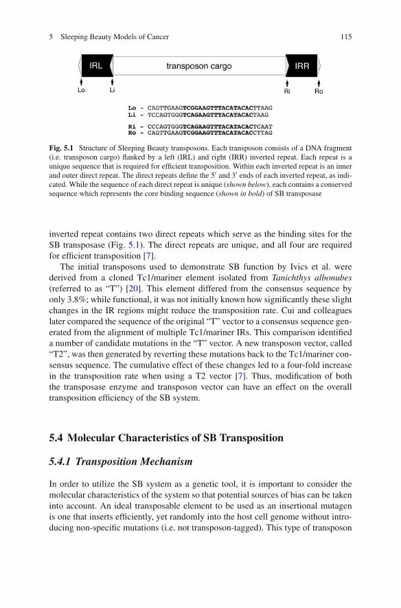
5 Sleeping Beauty Models of Cancer 115
Fig. 5.1 Structure of Sleeping Beauty transposons. Each transposon consists of a DNA fragment(i.e. transposon cargo) flanked by a left (IRL) and right (IRR) inverted repeat. Each repeat is aunique sequence that is required for efficient transposition. Within each inverted repeat is an innerand outer direct repeat. The direct repeats define the 5′ and 3′ ends of each inverted repeat, as indi-cated. While the sequence of each direct repeat is unique (shown below), each contains a conservedsequence which represents the core binding sequence (shown in bold) of SB transposase
inverted repeat contains two direct repeats which serve as the binding sites for theSB transposase (Fig. 5.1). The direct repeats are unique, and all four are requiredfor efficient transposition [7].
The initial transposons used to demonstrate SB function by Ivics et al. werederived from a cloned Tc1/mariner element isolated from Tanichthys albonubes(referred to as “T”) [20]. This element differed from the consensus sequence byonly 3.8%; while functional, it was not initially known how significantly these slightchanges in the IR regions might reduce the transposition rate. Cui and colleagueslater compared the sequence of the original “T” vector to a consensus sequence gen-erated from the alignment of multiple Tc1/mariner IRs. This comparison identifieda number of candidate mutations in the “T” vector. A new transposon vector, called“T2”, was then generated by reverting these mutations back to the Tc1/mariner con-sensus sequence. The cumulative effect of these changes led to a four-fold increasein the transposition rate when using a T2 vector [7]. Thus, modification of boththe transposase enzyme and transposon vector can have an effect on the overalltransposition efficiency of the SB system.
5.4 Molecular Characteristics of SB Transposition
5.4.1 Transposition Mechanism
In order to utilize the SB system as a genetic tool, it is important to consider themolecular characteristics of the system so that potential sources of bias can be takeninto account. An ideal transposable element to be used as an insertional mutagenis one that inserts efficiently, yet randomly into the host cell genome without intro-ducing non-specific mutations (i.e. not transposon-tagged). This type of transposon
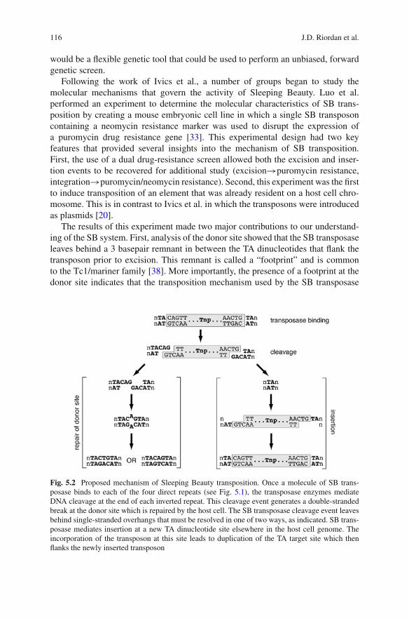
116 J.D. Riordan et al.
would be a flexible genetic tool that could be used to perform an unbiased, forwardgenetic screen.
Following the work of Ivics et al., a number of groups began to study themolecular mechanisms that govern the activity of Sleeping Beauty. Luo et al.performed an experiment to determine the molecular characteristics of SB trans-position by creating a mouse embryonic cell line in which a single SB transposoncontaining a neomycin resistance marker was used to disrupt the expression ofa puromycin drug resistance gene [33]. This experimental design had two keyfeatures that provided several insights into the mechanism of SB transposition.First, the use of a dual drug-resistance screen allowed both the excision and inser-tion events to be recovered for additional study (excision→puromycin resistance,integration→puromycin/neomycin resistance). Second, this experiment was the firstto induce transposition of an element that was already resident on a host cell chro-mosome. This is in contrast to Ivics et al. in which the transposons were introducedas plasmids [20].
The results of this experiment made two major contributions to our understand-ing of the SB system. First, analysis of the donor site showed that the SB transposaseleaves behind a 3 basepair remnant in between the TA dinucleotides that flank thetransposon prior to excision. This remnant is called a “footprint” and is commonto the Tc1/mariner family [38]. More importantly, the presence of a footprint at thedonor site indicates that the transposition mechanism used by the SB transposase
Fig. 5.2 Proposed mechanism of Sleeping Beauty transposition. Once a molecule of SB trans-posase binds to each of the four direct repeats (see Fig. 5.1), the transposase enzymes mediateDNA cleavage at the end of each inverted repeat. This cleavage event generates a double-strandedbreak at the donor site which is repaired by the host cell. The SB transposase cleavage event leavesbehind single-stranded overhangs that must be resolved in one of two ways, as indicated. SB trans-posase mediates insertion at a new TA dinucleotide site elsewhere in the host cell genome. Theincorporation of the transposon at this site leads to duplication of the TA target site which thenflanks the newly inserted transposon
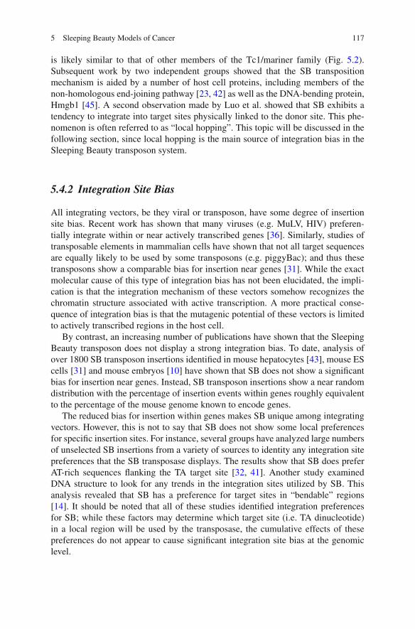
5 Sleeping Beauty Models of Cancer 117
is likely similar to that of other members of the Tc1/mariner family (Fig. 5.2).Subsequent work by two independent groups showed that the SB transpositionmechanism is aided by a number of host cell proteins, including members of thenon-homologous end-joining pathway [23, 42] as well as the DNA-bending protein,Hmgb1 [45]. A second observation made by Luo et al. showed that SB exhibits atendency to integrate into target sites physically linked to the donor site. This phe-nomenon is often referred to as “local hopping”. This topic will be discussed in thefollowing section, since local hopping is the main source of integration bias in theSleeping Beauty transposon system.
5.4.2 Integration Site Bias
All integrating vectors, be they viral or transposon, have some degree of insertionsite bias. Recent work has shown that many viruses (e.g. MuLV, HIV) preferen-tially integrate within or near actively transcribed genes [36]. Similarly, studies oftransposable elements in mammalian cells have shown that not all target sequencesare equally likely to be used by some transposons (e.g. piggyBac); and thus thesetransposons show a comparable bias for insertion near genes [31]. While the exactmolecular cause of this type of integration bias has not been elucidated, the impli-cation is that the integration mechanism of these vectors somehow recognizes thechromatin structure associated with active transcription. A more practical conse-quence of integration bias is that the mutagenic potential of these vectors is limitedto actively transcribed regions in the host cell.
By contrast, an increasing number of publications have shown that the SleepingBeauty transposon does not display a strong integration bias. To date, analysis ofover 1800 SB transposon insertions identified in mouse hepatocytes [43], mouse EScells [31] and mouse embryos [10] have shown that SB does not show a significantbias for insertion near genes. Instead, SB transposon insertions show a near randomdistribution with the percentage of insertion events within genes roughly equivalentto the percentage of the mouse genome known to encode genes.
The reduced bias for insertion within genes makes SB unique among integratingvectors. However, this is not to say that SB does not show some local preferencesfor specific insertion sites. For instance, several groups have analyzed large numbersof unselected SB insertions from a variety of sources to identity any integration sitepreferences that the SB transposase displays. The results show that SB does preferAT-rich sequences flanking the TA target site [32, 41]. Another study examinedDNA structure to look for any trends in the integration sites utilized by SB. Thisanalysis revealed that SB has a preference for target sites in “bendable” regions[14]. It should be noted that all of these studies identified integration preferencesfor SB; while these factors may determine which target site (i.e. TA dinucleotide)in a local region will be used by the transposase, the cumulative effects of thesepreferences do not appear to cause significant integration site bias at the genomiclevel.
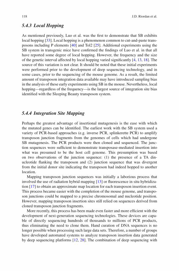
118 J.D. Riordan et al.
5.4.3 Local Hopping
As mentioned previously, Luo et al. was the first to demonstrate that SB exhibitslocal hopping [33]. Local hopping is a phenomenon common to cut-and-paste trans-posons including P elements [40] and Tol2 [25]. Additional experiments using theSB system in transgenic mice have confirmed the findings of Luo et al. in that allhave reported some degree of local hopping. However, the frequency and the sizeof the genetic interval affected by local hopping varied significantly [4, 13, 18]. Thesource of this variation is not clear. It should be noted that these initial experimentswere performed prior to the development of deep sequencing technology, and insome cases, prior to the sequencing of the mouse genome. As a result, the limitedamount of transposon integration data available may have introduced sampling biasin the analysis of these early experiments using SB in the mouse. Nevertheless, localhopping—regardless of the frequency—is the largest source of integration site biasidentified with the Sleeping Beauty transposon system.
5.4.4 Integration Site Mapping
Perhaps the greatest advantage of insertional mutagenesis is the ease with whichthe mutated genes can be identified. The earliest work with the SB system used avariety of PCR-based approaches (e.g. inverse PCR, splinkerette PCR) to amplifytransposon junction fragments from the genomes of cells which had undergoneSB mutagenesis. The PCR products were then cloned and sequenced. The junc-tion sequences were sufficient to demonstrate transposase-mediated insertion intowhat was presumed to be the host cell genome. This presumption was basedon two observations of the junction sequence: (1) the presence of a TA din-ucleotide flanking the transposon and (2) junction sequence that was divergentfrom the initial donor site indicating the transposon had indeed hopped to anotherlocation.
Mapping transposon junction sequences was initially a laborious process thatinvolved the use of radiation hybrid mapping [13] or fluorescence in situ hybridiza-tion [17] to obtain an approximate map location for each transposon insertion event.This process became easier with the completion of the mouse genome, and transpo-son junctions could be mapped to a precise chromosomal and nucleotide position.However, mapping transposon insertion sites still relied on sequences derived fromcloned transposon junction fragments.
More recently, this process has been made even faster and more efficient with thedevelopment of next-generation sequencing technologies. These devices are capa-ble of directly sequencing hundreds of thousands to millions of PCR products,thus eliminating the need to clone them. Hand curation of DNA sequences is nolonger possible when processing such large data sets. Therefore, a number of groupshave developed automated systems to analyze transposon insertion data generatedby deep sequencing platforms [12, 28]. The combination of deep sequencing with
5 Sleeping Beauty Models of Cancer 119
these automated sequence analysis systems has provided an opportunity to study theprocess of SB transposition in much greater detail.
5.5 Sleeping Beauty in Cancer Research
Most of the initial publications that described the use of the SB system in micefocused on germline mutagenesis to isolate mutant strains with developmental phe-notypes. Unfortunately, this approach was limited by the relatively low rate of SBmutagenesis compared to chemical mutagenesis using compounds such as ethyl-nitrosourea (ENU). This effort was also confounded by the high rate of localhopping which further reduced the efficiency of whole-genome mutagenesis usingSB. Despite these limitations, several groups began to adapt the SB system to muta-genize somatic cells to induce tumors in mice. It was thought that the low rate ofmutagenesis would be sufficient to induce tumors, given enough time. If so, the SBsystem could be used to perform forward genetic screens to identify cancer genes inmice.
Understanding which genes contribute to cancer has long been a major goalof cancer researchers. Next-generation DNA sequencing technologies have greatlyreduced the cost and dramatically increased the pace at which mutation profilesfrom individual tumors can be generated [34]. Unfortunately, recent work suggeststhat human tumors have a mutator phenotype with a mutation rate ~200 fold greaterthan normal tissue [3]. As a consequence, the precise identification of the mutationswhich contribute to the cancer phenotype (i.e. driver mutations) among the moreabundant background mutations (i.e. passenger mutations) is a major challengeto the field of cancer genetics. However, Sleeping Beauty transposon mutagene-sis could prove to be a useful tool for carrying out forward genetic screens in mice,allowing researchers to more rapidly identify somatic mutations that promote cancerin vivo. By combining data from these SB screens with sequence data from humancancer genomes, perhaps driver mutations, and thus more appropriate therapeutictargets, could be identified.
5.5.1 Modifications of Transposon Design
Initial efforts using the SB system as a germline insertional mutagen in mice madeuse of gene trap vectors that would express a reporter gene (e.g. EGFP, β-gal) upontransposon insertion within a transcription unit [4, 17]. This approach is useful forthe identification and isolation of individual transposon-induced mutations, giventhe inefficiency of the SB system in this context. However, since cancer exhibits astrong positive selection in vivo, reporter genes have little utility. Moreover, severalgroups have shown that the efficiency of SB-induced transposition is greatly reducedas the transposon size increases [15, 24]. These factors influenced transposon design
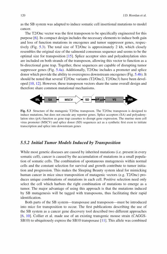
120 J.D. Riordan et al.
as the SB system was adapted to induce somatic cell insertional mutations to modelcancer.
The T2/Onc vector was the first transposon to be specifically engineered for thispurpose [6]. Its compact design includes the necessary elements to induce both gainand loss of function mutations in oncogenes and tumor suppressor genes, respec-tively (Fig. 5.3). The total size of T2/Onc is approximately 2 kb, which closelyresembles the original size of the salmonid consensus sequence and seems to be theoptimal size for transposition [15]. Splice acceptor sites and polyadenylation sitesare included on both strands of the transposon, allowing this vector to function as abi-directional gene trap. Together, these sequences are capable of disrupting tumorsuppressor genes (Fig. 5.4a). Additionally, T2/Onc includes a promoter and splicedonor which provide the ability to overexpress downstream oncogenes (Fig. 5.4b). Itshould be noted that several T2/Onc variants (T2/Onc2, T2/Onc3) have been devel-oped [10, 12]. However, these transposon vectors share the same overall design andtherefore share common mutational mechanisms.
Fig. 5.3 Structure of the mutagenic T2/Onc transposon. The T2/Onc transposon is designed toinduce mutations, but does not encode any reporter genes. Splice acceptors (SA) and polyadeny-lation sites (pA) function as gene trap cassettes to disrupt gene expression. The murine stem cellvirus promoter (MSCV) and splice donor (SD) sequences act as a synthetic first exon to initiatetranscription and splice into downstream genes
5.5.2 Initial Tumor Models Induced by Transposition
While most genetic diseases are caused by inherited mutations (i.e. present in everysomatic cell), cancer is caused by the accumulation of mutations in a small popula-tion of somatic cells. The combination of spontaneous mutagenesis within normalcells and the constant selection for survival and growth contribute to tumor initia-tion and progression. This makes the Sleeping Beauty system ideal for mimickinghuman cancer in mice since transposition of mutagenic vectors (e.g. T2/Onc) pro-duces unique combinations of mutations in each cell. Positive selection need onlyselect the cell which harbors the right combination of mutations to emerge as atumor. The major advantage of using this approach is that the mutations inducedby SB mutagenesis will be tagged with transposons, thus facilitating their rapididentification.
Both parts of the SB system—transposase and transposon—must be introducedinto mice for transposition to occur. The first publications describing the use ofthe SB system as a cancer gene discovery tool described two different approaches[6, 10]. Collier et al. made use of an existing transgenic mouse strain (CAGGS-SB10) to ubiquitously express the SB10 transposase [11]. This allele was combined
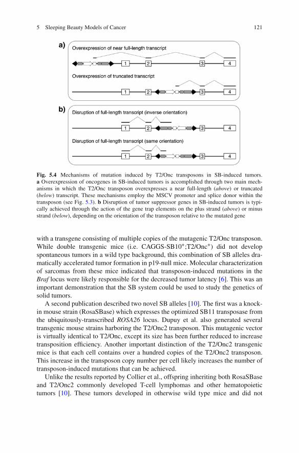
5 Sleeping Beauty Models of Cancer 121
Fig. 5.4 Mechanisms of mutation induced by T2/Onc transposons in SB-induced tumors.a Overexpression of oncogenes in SB-induced tumors is accomplished through two main mech-anisms in which the T2/Onc transposon overexpresses a near full-length (above) or truncated(below) transcript. These mechanisms employ the MSCV promoter and splice donor within thetransposon (see Fig. 5.3). b Disruption of tumor suppressor genes in SB-induced tumors is typi-cally achieved through the action of the gene trap elements on the plus strand (above) or minusstrand (below), depending on the orientation of the transposon relative to the mutated gene
with a transgene consisting of multiple copies of the mutagenic T2/Onc transposon.While double transgenic mice (i.e. CAGGS-SB10+;T2/Onc+) did not developspontaneous tumors in a wild type background, this combination of SB alleles dra-matically accelerated tumor formation in p19-null mice. Molecular characterizationof sarcomas from these mice indicated that transposon-induced mutations in theBraf locus were likely responsible for the decreased tumor latency [6]. This was animportant demonstration that the SB system could be used to study the genetics ofsolid tumors.
A second publication described two novel SB alleles [10]. The first was a knock-in mouse strain (RosaSBase) which expresses the optimized SB11 transposase fromthe ubiquitously-transcribed ROSA26 locus. Dupuy et al. also generated severaltransgenic mouse strains harboring the T2/Onc2 transposon. This mutagenic vectoris virtually identical to T2/Onc, except its size has been further reduced to increasetransposition efficiency. Another important distinction of the T2/Onc2 transgenicmice is that each cell contains over a hundred copies of the T2/Onc2 transposon.This increase in the transposon copy number per cell likely increases the number oftransposon-induced mutations that can be achieved.
Unlike the results reported by Collier et al., offspring inheriting both RosaSBaseand T2/Onc2 commonly developed T-cell lymphomas and other hematopoietictumors [10]. These tumors developed in otherwise wild type mice and did not
122 J.D. Riordan et al.
require a sensitizing mutation, such as the p19-null background used by Collieret al. However, the combination of RosaSBase and T2/Onc2 had a major limitationin that the majority of double transgenic mice died at midgestation, likely due toactive transposition. Additionally, mice that survived until birth developed predomi-nantly T-cell lymphoma rather than a diverse array of tumor types. Nevertheless, thiswork showed that the efficiency of the SB system was sufficient to induce tumors inwild type mice.
The origin of the apparent discrepancy in transposition efficiencies of theSB models described by Collier et al. and Dupuy et al. was not immediatelyclear. The improved efficiency described in RosaSBase;T2/Onc2 double trans-genic mice could be due to enhanced transposase expression from the ROSA26locus, increased copy number of the mutagenic transposon vector or a combi-nation of both. Recent work by Collier et al. has shown that both transposonand transposase alleles make a contribution to the efficiency of the system [5].These experiments combined the RosaSBase allele with the lower copy T2/Onctransgenic strains. The resulting double transgenic mice developed tumors with-out the requirement of a sensitizing mutation (e.g. p19–/–), suggesting that theRosaSBase allele drives a higher rate of transposition, compared to the CAGGS-SB10 allele. However, RosaSBase;T2/Onc mice did not display an embryoniclethal phenotype and developed tumors with an increased latency compared toRosaSBase;T2/Onc2 mice [5]. This result indicates that the increased transposoncopy number also contributes to transposition rate. Thus the nature of both the trans-posase and transposon components affect the tumor phenotype in SB models ofcancer.
5.5.3 Identification of Driver Mutations in SB-Induced Tumors
As noted earlier, a number of PCR-based approaches have recently been describedto identify transposon integration sites in SB-induced tumors [5, 12, 26, 39]. Directsequencing of PCR products using next generation sequencing platforms has greatlyimproved the sensitivity of detection for transposon-induced mutations in thesetumors. However, the increased scale of sequencing has also uncovered significantgenetic complexity in SB-induced tumors. While this complexity is a necessarycomponent of a good model of human cancer, it also produces a similar problemthat has been encountered with the sequencing of human cancer genomes—how arerare driver mutations identified among the large number of passenger mutations?
This problem can easily be addressed when using the SB system. As previ-ously noted, the SB transposase exhibits very weak integration site bias. Therefore,unselected SB insertion events can be modeled in silico by randomly selectingTA dinucleotide sites throughout the genome. Currently, this is achieved using aMonte Carlo simulation to identify common integration sites (CISs)—regions ofthe genome that suffer transposon insertion in SB-induced tumors at a rate morefrequently predicted by chance [5, 12, 26, 39]. Significance is a function of the
5 Sleeping Beauty Models of Cancer 123
number of tumors having the observed number of integration events within a spec-ified genomic region (see Chapter 7 for more detail). It is important to note thatsuch methods cannot easily be employed in the identification of driver mutationsin human cancer since spontaneous mutations do not occur randomly and areinfluenced by a number of factors.
One caveat in the identification of driver mutations in SB-induced tumorsinvolves local hopping—the main source of integration bias in the SB system. Thereis not a well-established definition of the local hopping interval for the SB systemcurrently in use. In fact, it appears that the rate of transposon insertion is elevatedalong the entire length of the chromosome that harbors the transposon transgene(our unpublished data). Consequently, the genes which map to this chromosomeare subject to an increased likelihood of transposon-induced mutation compared tothe rest of the genome. Currently, the only method to prevent the identification offalse-positive CISs due to local hopping in SB models of cancer is to discard all inte-gration events which map to the local chromosome. While this approach leads to theloss of data from an entire chromosome in each transposon strain, multiple mousestrains harboring transposon transgenes which map to different chromosomes canbe used to compensate and to negate the loss of the local chromosome in eachindividual strain.
5.6 Modifications to the SB System
While the initial SB transposase (CAGGS-SB10, RosaSBase) and mutagenic trans-poson alleles (T2/Onc, T2/Onc2) showed that the SB system could be used toproduce tumors in vivo, the earliest SB models were limited in the number of tumortypes that could be produced. However, this limitation was unlikely due to the activ-ity of the SB system, as previous work had observed transposition in a variety oftissue types [6, 10, 17]. Thus, it seemed likely that modifications to the existingtransposase and transposon alleles could provide more precise control, allowing fortissue-specific transposon mutagenesis.
The structure of the mutagenic transposon has recently been investigated asone manner in which the SB system can be modified. Both the T2/Onc andT2/Onc2 transposons contain the murine stem cell virus (MSCV) promoter todrive overexpression of downstream oncogenes. This promoter is most active incells of hematopoietic origin, which could explain why mutagenesis screens con-ducted with T2/Onc and T2/Onc2 transposons yielded primarily hematopoieticmalignancies [5, 12]. In a modified version of the mutagenic transposon, calledT2/Onc3, the MSCV promoter has been replaced with the cytomegalovirus earlyenhancer/chicken β-actin (CAG) promoter, which displays its highest activity incells of epithelial origin [37]. The tumor profile obtained from a mutagenesis screenusing T2/Onc3 transposons consisted mostly of carcinomas [12]. The ability togenerate this type of profile is highly advantageous, as the majority of human malig-nancies are solid tumors. As evidenced by the different tumor profiles generated by
124 J.D. Riordan et al.
these two independent screens, altering the promoter within mutagenic SB trans-posons can dramatically affect the nature of tumors resulting from integrationevents.
A potential application of this finding is the generation of transposons containingtissue-specific promoters whose activity is limited to a specific organ of interest. Amutagenesis screen utilizing this type of transposon would be predicted to generatetumors primarily in the organ of interest. This would allow rapid generation of alarge number of independent tumors from that organ, which would facilitate in-depth analysis of the mutation profile of that specific tumor type. By performingindependent mutagenesis screens with different transposons, novel oncogenes andtumor suppressors involved in tumorigenesis of a wide variety of tissue types couldbe identified.
A second approach to generate tissue-specific tumor models with the SB systemis to limit transposition events, and therefore mutations, to the tissue of inter-est. This was recently achieved by generating a conditional transposase allele(RosaSBase-LsL) that is only expressed upon Cre recombinase-mediated removalof a Lox-stop-Lox (LsL) cassette [12]. As with constitutive SB models, the SB trans-posase gene is knocked into the ROSA26 locus to allow its expression in any tissueof the mouse. Expression is prevented, however, by inclusion of an LsL cassettejust upstream of the transposase coding region. This cassette, which consists of anEGFP cDNA followed by three polyadenylation signals, traps transcripts initiatedfrom the ROSA26 promoter and prevents expression of the downstream transposasegene. Thus, no transposition occurs in mice harboring both the conditional trans-posase allele and a transposon transgene due to a lack of transposase expression.Tissue-specific expression of Cre recombinase leads to excision of the LsL cassette,and thus transposase expression.
The RosaSBase-LsL allele has been used successfully to generate specific SB-induced carcinoma models. For example, [26] initiated transposon mutagenesisspecifically in hepatocytes by expressing Cre from a transgene driven by the albu-min promoter [26]. These mice develop preneoplastic liver nodules at ~160 days ofage, some of which progress to hepatocellular carcinoma (HCC) as the mice age.Analysis of the transposon integration sites from these tumors identified a num-ber of genes already associated with HCC (e.g. Egfr, Met) as well as a number ofcandidate genes not previously implicated in this disease (e.g. Ube2h). A secondpublication generated a model of colorectal cancer (CRC) by inducing transposi-tion in the gastrointestinal tract using a Cre transgene expressed from the villinpromoter [39]. Transposon mutagenesis produced multiple intestinal adenomas inapproximately half of the mutagenized mice. As was the case with the SB-inducedHCCs, analysis of transposon-induced mutations in these lesions identified a num-ber of genes known to be involved in CRC, as well as a large number of candidategenes. These experiments are an important demonstration of the flexibility of theSB system to induce a broad spectrum of tumors in a controlled fashion. Moreover,the identification of genes known to be involved in human HCC and CRC inboth of these screens implies that this is a valid approach to identify novel cancergenes.
5 Sleeping Beauty Models of Cancer 125
5.7 Future Directions
5.7.1 Identifying Genes Involved in Metastasis
As described above, the SB insertional mutagenesis system provides a means toidentify oncogenes and tumor suppressors that drive tumor formation and devel-opment in various tissues throughout the body. In addition to identifying genesinvolved in primary tumor formation, the system could be used to discover thosethat contribute to other aspects of tumor biology, such as metastasis. This approachwould likely require a tissue-specific tumor model, as mice with ubiquitous trans-position develop several tumors in multiple organs, which generally prevents themfrom surviving long enough for metastases to develop. Mice with tumorigenesistargeted to a single organ can survive longer, which may allow sufficient time formetastasis to occur, as was observed when transposition was limited to hepatocytes[26].
Once a model system has been established in which SB transposition drives for-mation of primary tumors that metastasize, the profile of transposon integration sitescould be compared between primary tumors and their metastatic derivatives. Eachmetastatic tumor could be paired with the primary tumor from which it was derivedbased on similarities in overall integration profile. This type of analysis may leadto the identification of genes that are common sites of integration in metastases, butnot in primary tumors. Depending on the nature of the mutation induced by trans-poson insertion, these genes would be implicated as either promoters or inhibitorsof metastasis.
Another useful approach would be to compare integration profiles of primarytumors with identified metastases to those of primary tumors lacking metastases.Such an approach could lead to the discovery of gene disruptions in primary tumorsthat are predictive of metastatic potential. Additionally, genes may be identified thatcommonly contain transposon integrations in primary, but not metastatic tumors.This type of pattern would suggest that disruption of these genes may promote pri-mary tumor formation, but may not be compatible with metastasis. Finally, analysisof integration profiles derived from tumors isolated from different metastatic sites(e.g. brain, lung, liver) may identify specific mutation events that promote metasta-sis to each distinct site. Such information could eventually prove useful in predictingpatient outcome and treatment strategies.
5.7.2 Identifying Genes Involved in Treatment Response
A recent retroviral mutagenesis screen demonstrated the utility in using insertionalmutagenesis to study treatment response in mice [30]. The SB system could beused in a similar manner to identify mutations that confer drug susceptibility orresistance. For this approach, a cohort of mice could be generated with SB-inducedtumors. These mice could be treated with the drug of interest, then separated into
126 J.D. Riordan et al.
two groups: those that respond to treatment and those that do not. Transposonintegration profiles could be compared between responders and non-responders toidentify mutations which are correlated with either resistance of susceptibility.
A modification to this approach could involve collection of tumor biopsies fromanimals before and after drug treatment. This would allow the investigator to fol-low genetic progression within an individual tumor in response to drug treatment.In this case, drug treatment would act as a selection event in which tumor cellsthat acquired mutations conferring drug resistance would preferentially replicate.Thus, transposon-induced mutations that appear to expand within the tumor massfollowing drug treatment would be candidate resistance mutations.
5.7.3 Identifying Cooperating Mutations
It is well established that cancer is the result of accumulated mutations which collec-tively work to transform a cell. As such, these mutations likely work in a synergisticfashion to modify signaling pathways or other subcellular processes. In most cases,however, it is not clear which specific mutations and/or pathways work in concertto contribute to cancer. Here again, SB models of cancer could be used to identifymutations that cooperate in generating the tumorigenic phenotype. In this scenario,transposon mutagenesis could be induced in mice carrying a predisposing mutationwhich serves as the initiating genetic event. Ongoing transposition would generateadditional mutations promoting tumor progression. This approach could be utilizedin the context of different predisposing mutations for a single tumor type. Distinctsets of cooperating genes may be identified for each initiating mutation by compar-ing transposon integration profiles derived from each model. Such information couldlead to the identification of novel combinations of therapeutic targets for distinctsubsets of tumors within a particular cancer type.
5.8 Challenges Remaining
As described in the previous sections, the current SB mutagenesis system hasbroad utility in the identification of genes associated with tumorigenesis andcould potentially be used to study many other aspects of tumor biology. However,some challenges remain, which if properly addressed, would allow even broaderapplicability of the system.
Efforts to date have failed to efficiently generate tumors in certain organs. Forexample, a screen in which transposition was targeted to the prostate using theprobasin-Cre strain failed to generate tumors. Similarly, targeting transposition tothe mammary gland using the whey acidic protein (WAP)-Cre strain was not effec-tive in generating mammary tumors (our unpublished data). At this point, it remainsunclear whether this organ-specific resistance to SB-induced tumorigenesis resultsfrom a property of the SB system itself or from properties intrinsic to the target
5 Sleeping Beauty Models of Cancer 127
tissues. One possibility is that the promoters used to express Cre recombinase donot have the correct spatial and/or temporal expression pattern to drive mutagenesisin the appropriate initiating cell population for these forms of cancer. Consistentwith this idea, mammary tumors have been induced using T2/Onc3 mutagenesiscombined with ubiquitous transposition [12]. Another possibility deals with theactivity of the transposon-encoded promoter. Changing the promoter within muta-genic transposons has been shown to dramatically affect the profile of tumors thatare developed in a ubiquitous transposition model [12]. It is possible that the trans-posons used for tissue-specific prostate and mammary models do not efficientlyinitiate transcription in those tissues. Another factor that could influence tumordevelopment in different tissues is the availability of host cell proteins used usedby the SB transposase during transposition (e.g. Hmgb1) [45]. The expression levelof these co-factors may be too low in some tissues to achieve the degree of mutage-nesis required for transformation. Additional experiments are required to determineif the existing SB system will permit tissue-specific tumor models to be generatedfor these cancer types or if additional modifications will be necessary.
Another issue which has complicated the use of the Cre-inducible SB system isthat many Cre transgenic mouse strains display some level of expression in multipletissues. These Cre strains could produce a variety of tumor types when used toactivate SB mutagenesis in mice. This would be problematic if Cre were expressedin the hematopoietic system, as the SB system is particularly efficient at inducinglymphomas. This issue could be partially addressed by combining the Cre-inducibletransposition system with mutagenic transposons containing a promoter specific tothe tissue of interest. While transposition may occur in multiple tissues, gain-of-function mutations, and thus tumor formation, would likely be limited to the tissueof interest.
All versions of the SB system currently in use to model cancer lack the abil-ity to terminate transposon mutagenesis once it has been initiated. Achieving thisgoal would be advantageous for a number of reasons. First, it could facilitate theidentification of genes associated with drug resistance or susceptibility. For exam-ple, transposon mobilization could be used to drive tumorigenesis and then shutoff. Following this, drug treatment could be used to induce tumor regression. Aftersignificant regression, transposition could be reactivated, with the expectation thatintegrations causing disruption of genes conferring drug susceptibility would beselected for in the tumors. Additionally, the level of background integrations notcontributing to tumor development or progression (i.e. passenger mutations) couldbe decreased by turning off transposition following initiation of tumorigenesis.Finally, the ability to turn transposition on and off would also allow a more in-depthanalysis of distinct stages of tumor progression.
References
1. Baus, J., Liu, L., Heggestad, A. D., Sanz, S., & Fletcher, B. S. (2005). Hyperactive transposasemutants of the Sleeping Beauty transposon. Molecular Therapy, 12, 1148–1156.
128 J.D. Riordan et al.
2. Bazopoulou, D., & Tavernarakis, N. (2009). The NemaGENETAG initiative: large scaletransposon insertion gene-tagging in Caenorhabditis elegans. Genetica, 137, 39–46.
3. Bielas, J. H., Loeb, K. R., Rubin, B. P., True, L. D., & Loeb, L. A. (2006). Human cancersexpress a mutator phenotype. Proceedings of the National Academy of Sciences of the USA,103, 18238–18242.
4. Carlson, C. M., Dupuy, A. J., Fritz, S., Roberg-Perez, K. J., Fletcher, C. F., &Largaespada, D. A. (2003). Transposon mutagenesis of the mouse germline. Genetics, 165,243–256.
5. Collier, L. S., Adams, D. J., Hackett, C. S., Bendzick, L. E., Akagi, K., Davies, M. N., et al.(2009). Whole-body sleeping beauty mutagenesis can cause penetrant leukemia/lymphomaand rare high-grade glioma without associated embryonic lethality. Cancer Research, 69,8429–8437.
6. Collier, L. S., Carlson, C. M., Ravimohan, S., Dupuy, A. J., & Largaespada, D. A. (2005).Cancer gene discovery in solid tumours using transposon-based somatic mutagenesis in themouse. Nature, 436, 272–276.
7. Cui, Z., Geurts, A. M., Liu, G., Kaufman, C. D., & Hackett, P. B. (2002). Structure-function analysis of the inverted terminal repeats of the sleeping beauty transposon. Journalof Molecular Biology, 318, 1221–1235.
8. Czerny, T., Schaffner, G., & Busslinger, M. (1993). DNA sequence recognition by Pax pro-teins: bipartite structure of the paired domain and its binding site. Genes and Development, 7,2048–2061.
9. Davidson, A. E., Balciunas, D., Mohn, D., Shaffer, J., Hermanson, S., Sivasubbu, S., et al.(2003). Efficient gene delivery and gene expression in zebrafish using the Sleeping Beautytransposon. Developmental Biology, 263, 191–202.
10. Dupuy, A. J., Akagi, K., Largaespada, D. A., Copeland, N. G., & Jenkins, N. A. (2005).Mammalian mutagenesis using a highly mobile somatic Sleeping Beauty transposon system.Nature, 436, 221–226.
11. Dupuy, A. J., Fritz, S., & Largaespada, D. A. (2001). Transposition and gene disruption in themale germline of the mouse. Genesis, 30, 82–88.
12. Dupuy, A. J., Rogers, L. M., Kim, J., Nannapaneni, K., Starr, T. K., Liu, P., et al. (2009).A modified sleeping beauty transposon system that can be used to model a wide variety ofhuman cancers in mice. Cancer Research, 69, 8150–8156.
13. Fischer, S. E., Wienholds, E., & Plasterk, R. H. (2001). Regulated transposition of a fishtransposon in the mouse germ line. Proceedings of the National Academy of Sciences of theUSA, 98, 6759–6764.
14. Geurts, A. M., Hackett, C. S., Bell, J. B., Bergemann, T. L., Collier, L. S., Carlson, C. M.,et al. (2006). Structure-based prediction of insertion-site preferences of transposons intochromosomes. Nucleic Acids Research, 34, 2803–2811.
15. Geurts, A. M., Yang, Y., Clark, K. J., Liu, G., Cui, Z., Dupuy, A. J., et al. (2003). Gene transferinto genomes of human cells by the sleeping beauty transposon system. Molecular Therapy,8, 108–117.
16. Grabher, C., Henrich, T., Sasado, T., Arenz, A., Wittbrodt, J., & Furutani-Seiki, M. (2003).Transposon-mediated enhancer trapping in medaka. Gene, 322, 57–66.
17. Horie, K., Kuroiwa, A., Ikawa, M., Okabe, M., Kondoh, G., Matsuda, Y., et al. (2001).Efficient chromosomal transposition of a Tc1/mariner- like transposon Sleeping Beauty inmice. Proceedings of the National Academy of Sciences of the USA, 98, 9191–9196.
18. Horie, K., Yusa, K., Yae, K., Odajima, J., Fischer, S. E., Keng, V. W., et al. (2003).Characterization of Sleeping Beauty transposition and its application to genetic screeningin mice. Molecular and Cellular Biology, 23, 9189–9207.
19. Hummel, T., & Klambt, C. (2008). P-element mutagenesis. Methods in Molecular Biology,420, 97–117.
20. Ivics, Z., Hackett, P. B., Plasterk, R. H., & Izsvak, Z. (1997). Molecular reconstruction ofSleeping Beauty, a Tc1-like transposon from fish, and its transposition in human cells. Cell,91, 501–510.

5 Sleeping Beauty Models of Cancer 129
21. Izsvak, Z., Ivics, Z., & Plasterk, R. H. (2000). Sleeping beauty, a wide host-range trans-poson vector for genetic transformation in vertebrates. Journal of Molecular Biology, 302,93–102.
22. Izsvak, Z., Khare, D., Behlke, J., Heinemann, U., Plasterk, R. H., & Ivics, Z. (2002).Involvement of a bifunctional, paired-like DNA-binding domain and a transpositionalenhancer in Sleeping Beauty transposition. Journal of Biological Chemistry, 277, 34581–34588.
23. Izsvak, Z., Stuwe, E. E., Fiedler, D., Katzer, A., Jeggo, P. A., & Ivics, Z. (2004). Healing thewounds inflicted by sleeping beauty transposition by double-strand break repair in mammaliansomatic cells. Molecular Cell, 13, 279–290.
24. Karsi, A., Moav, B., Hackett, P., & Liu, Z. (2001). Effects of insert size on transpositionefficiency of the sleeping beauty transposon in mouse cells. Marine Biotechnology (New York),3, 241–245.
25. Keng, V. W., Ryan, B. J., Wangensteen, K. J., Balciunas, D., Schmedt, C., Ekker, S.C., et al. (2009a). Efficient transposition of Tol2 in the mouse germline. Genetics, 183,1565–1573.
26. Keng, V. W., Villanueva, A., Chiang, D. Y., Dupuy, A. J., Ryan, B. J., Matise, I., et al.(2009b). A conditional transposon-based insertional mutagenesis screen for genes associatedwith mouse hepatocellular carcinoma. Nature Biotechnology, 27, 264–274.
27. Kitada, K., Ishishita, S., Tosaka, K., Takahashi, R., Ueda, M., Keng, V. W., et al. (2007).Transposon-tagged mutagenesis in the rat. Nature Methods, 4, 131–133.
28. Kong, J., Zhu, F., Stalker, J., & Adams, D. J. (2008). iMapper: a web application for theautomated analysis and mapping of insertional mutagenesis sequence data against Ensemblgenomes. Bioinformatics, 24, 2923–2925.
29. Kool, J., & Berns, A. (2009). High-throughput insertional mutagenesis screens in mice toidentify oncogenic networks. Nature Reviews Cancer, 9, 389–399.
30. Lauchle, J. O., Kim, D., Le, D. T., Akagi, K., Crone, M., Krisman, K., et al. (2009). Responseand resistance to MEK inhibition in leukaemias initiated by hyperactive Ras. Nature, 461,411–414.
31. Liang, Q., Kong, J., Stalker, J., & Bradley, A. (2009). Chromosomal mobilization andreintegration of sleeping beauty and PiggyBac transposons. Genesis, 47, 404–408.
32. Liu, G., Geurts, A. M., Yae, K., Srinivasan, A. R., Fahrenkrug, S. C., Largaespada, D. A.,et al. (2005). Target-site preferences of Sleeping Beauty transposons. Journal of MolecularBiology, 346, 161–173.
33. Luo, G., Ivics, Z., Izsvak, Z., & Bradley, A. (1998). Chromosomal transposition of aTc1/mariner-like element in mouse embryonic stem cells. Proceedings of the NationalAcademy of Sciences of the USA, 95, 10769–10773.
34. Mardis, E. R., & Wilson, R. K. (2009). Cancer genome sequencing: a review. HumanMolecular Genetics, 18, R163–R168.
35. Mates, L., Chuah, M. K., Belay, E., Jerchow, B., Manoj, N., Acosta-Sanchez, A., et al. (2009).Molecular evolution of a novel hyperactive Sleeping Beauty transposase enables robust stablegene transfer in vertebrates. Nature Genetics, 41, 753–761.
36. Mitchell, R. S., Beitzel, B. F., Schroder, A. R., Shinn, P., Chen, H., Berry, C. C., et al. (2004).Retroviral DNA integration: ASLV, HIV, and MLV show distinct target site preferences. PLoSBiology, 2, E234.
37. Okabe, M., Ikawa, M., Kominami, K., Nakanishi, T., & Nishimune, Y. (1997). ‘Green mice’as a source of ubiquitous green cells. FEBS Letters, 407, 313–319.
38. Plasterk, R. H., Izsvak, Z., & Ivics, Z. (1999). Resident aliens: the Tc1/mariner superfamilyof transposable elements. Trends in Genetics, 15, 326–332.
39. Starr, T. K., Allaei, R., Silverstein, K. A., Staggs, R. A., Sarver, A. L., Bergemann, T. L.,et al. (2009). A transposon-based genetic screen in mice identifies genes altered in colorectalcancer. Science, 323, 1747–1750.
40. Tower, J., Karpen, G. H., Craig, N., & Spradling, A. C. (1993). Preferential transposition ofDrosophila P elements to nearby chromosomal sites. Genetics, 133, 347–359.

130 J.D. Riordan et al.
41. Vigdal, T. J., Kaufman, C. D., Izsvak, Z., Voytas, D. F., & Ivics, Z. (2002). Common physicalproperties of DNA affecting target site selection of sleeping beauty and other Tc1/marinertransposable elements. Journal of Molecular Biology, 323, 441–452.
42. Yant, S. R., & Kay, M. A. (2003). Nonhomologous-end-joining factors regulate DNA repairfidelity during Sleeping Beauty element transposition in mammalian cells. Molecular andCellular Biology, 23, 8505–8518.
43. Yant, S. R., Wu, X., Huang, Y., Garrison, B., Burgess, S. M., & Kay, M. A. (2005). High-resolution genome-wide mapping of transposon integration in mammals. Molecular andCellular Biology, 25, 2085–2094.
44. Yergeau, D. A., Johnson Hamlet, M. R., Kuliyev, E., Zhu, H., Doherty, J. R., Archer, T.D., et al. (2009). Transgenesis in Xenopus using the Sleeping Beauty transposon system.Developmental Dynamics, 238, 1727–1743.
45. Zayed, H., Izsvak, Z., Khare, D., Heinemann, U., & Ivics, Z. (2003). The DNA-bending pro-tein HMGB1 is a cellular cofactor of Sleeping Beauty transposition. Nucleic Acids Research,31, 2313–2322.
Chapter 6Insertional Mutagenesis in Hematopoietic Cells:Lessons Learned from Adverse Eventsin Clinical Gene Therapy Trials
Lars U. Müller, Michael D. Milsom, and David A. Williams
6.1 Introduction
From an early stage in the development of retroviral vectors for gene therapy appli-cations, there has been a concern that recombinant vectors could elicit cellulartransformation by altering expression of either cellular proto-oncogenes or tumorsuppressor genes that are proximal to the genomic integration site (Fig. 6.1). Thisphenomenon, referred to as insertional mutagenesis, was characterized as a propertyof wild type gammaretroviral viruses (previously known as murine oncoretroviralviruses) which are the prototype virus from which the recombinant vectors used inthe majority of the clinical trials described in this chapter were derived (reviewedin [178]). Wild type retroviruses fall into two categories with regards to their trans-forming properties: Slow transforming and acute transforming retroviruses. Acutetransforming retroviruses contain viral versions of cellular proto-oncogenes such asMyb, Myc and Src and, as the name suggests, are able to induce cellular transforma-tion within a short latency period [15, 62, 149]. Slow transforming retroviruses donot contain oncogenes but instead lead to transformation of the host cell via inser-tional mutagenesis. The fact that transformation by this route will likely requirethe accumulation of many proviral integrations within a given cell until a combi-nation which can co-operate in transformation is achieved accounts for the longerlatency period. Since cellular transformation was perceived to be a consequence ofco-operating hits from multiple vector insertions, early pioneers in the gene ther-apy field focused upon developing vector systems which minimized the risk ofgenerating a replication competent recombinant vector and which were unlikelyto rescue dormant endogenous retroviral sequences present in the human genome.Such efforts involved the segregation of viral proteins essential for viral propaga-tion (GAG, POL and ENV) onto packaging plasmids which were separate fromthe packaged recombinant viral genome. The identification of the gammaretroviral
D.A. Williams (B)Harvard Medical School, Children’s Hospital Boston, Boston, MA 02115, USAe-mail: [email protected]
Lars U. Müller and Michael D. Milsom have equally contributed to this work.
131A.J. Dupuy, D.A. Largaespada (eds.), Insertional Mutagenesis Strategiesin Cancer Genetics, DOI 10.1007/978-1-4419-7656-7_6,C© Springer Science+Business Media, LLC 2011
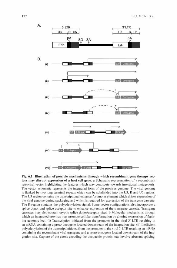
132 L.U. Müller et al.
Fig. 6.1 Illustration of possible mechanisms through which recombinant gene therapy vec-tors may disrupt expression of a host cell gene. a Schematic representation of a recombinantretroviral vector highlighting the features which may contribute towards insertional mutagenesis.The vector schematic represents the integrated form of the provirus genome. The viral genomeis flanked by two long terminal repeats which can be subdivided into the U3, R and U5 regions.The U3 region contains the transcriptional enhancer/promoter element which drives expression ofthe viral genome during packaging and which is required for expression of the transgene cassette.The R region contains the polyadenylation signal. Some vector configurations also incorporate asplice donor and splice acceptor site to enhance expression of the transgene cassette. Transgenecassettes may also contain cryptic splice donor/acceptor sites. b Molecular mechanisms throughwhich an integrated provirus may promote cellular transformation by altering expression of flank-ing genomic loci. (i) Transcription initiated from the promoter in the viral 3′ LTR resulting inan mRNA containing a proto-oncogene located downstream of the integration site. (ii) Inefficientpolyadenylation of the transcript initiated from the promoter in the viral 5′ LTR resulting an mRNAcontaining the recombinant viral transgene and a proto-oncogene located downstream of the inte-gration site. Capture of the exons encoding the oncogenic protein may involve aberrant splicing.

6 Insertional Mutagenesis in Hematopoietic Cells 133
packaging signal (�) facilitated the deletion of this element in recombinant pack-aging constructs containing GAG, POL and ENV, thus eliminating the normalmechanism via which these coding sequences are incorporated into the viral parti-cle [105]. In order to minimize the probability that segregated packaging constructswould recombine with either endogenous retroviral sequences in the target cellgenome or with each other to form replication competent virus, packaging systemswith minimal overlapping sequence homology were developed (reviewed in [112]).Stable cell lines to package gammaretroviral vectors, including the GP + envAm12and PG13 lines, were created via stable transfection of packaging constructs intoestablished cell lines [107, 112, 113]. Such packaging lines allowed the isolation andcharacterization of individual packaging clones following a subsequent stable trans-fection with a construct containing the recombinant retroviral genome. Althoughthere are examples of replication competent virus being generated in packaging celllines, individual clones can be determined as free of replication competent virus andcan be subsequently expanded and used to produce large scale preparations of retro-viral supernatant for use in clinical trials [36, 57, 140]. Such modifications in vectorproduction technology made the risk of producing a supernatant which containedreplication competent retrovirus almost negligible.
Having greatly reduced the likelihood that retroviral gene therapy vectors couldgenerate replication competent virus, the risk of a recombinant vector being ableto transform a cell via insertional mutagenesis was perceived to be very low [125].The lack of efficacy in pre-clinical models using human hematopoietic stem andprogenitor (CD34+) cells somewhat shifted emphasis further away from the risksof insertional mutagenesis. How would it be possible to transform a human bonemarrow stem cell with a recombinant retroviral vector if it was extremely diffi-cult to even transduce human CD34+ cells at a clinically relevant level with sucha vector? Much of the energy in the field was therefore devoted to advancingstem cell transduction methodology and developing retroviral vectors which would
�Fig. 6.1 (continued) (iii) and (iv) Enhancer effect from the viral U3-encoded enhancer elementon an endogenous proto-oncogene promoter located either downstream (iii), or upstream (iv) ofthe integration site. (v) Integration within a tumor suppressor gene resulting in abrogation of geneexpression either via premature termination of the transcript; the production of a fusion proteinwhich may have no functional activity or dominant negative activity; or the introduction of a frameshift mutation resulting in premature termination of translation. The above may be mediated by anaberrant splicing event. (vi) Transcription initiated from the viral LTR resulting in the production ofa mRNA which is complimentary to that of a tumor suppressor gene which is located downstreamand inverted relative to the integration site. Such a complimentary mRNA may promote degrada-tion of the tumor suppressor gene transcript or interfere with its translation (vii) Integration intothe 3′ untranslated region of a proto-oncogene or tumor suppressor gene resulting in perturbedpost-transcriptional regulation of gene expression either via destabilizing/stabilizing the mRNA;interfering with mRNA localization; or enhancing/repressing the initiation of translation. LTR,long terminal repeat; U5, unique 5′ LTR sequence; R, repeat LTR sequence; U3, unique 3′ LTRsequence; E/P, enhancer/promoter; pA, polyadenylation signal; SD, splice donor site; SA, spliceacceptor site; +, enhancing effect upon transcription initiation; T-Suppr, tumor suppressor gene; 3′UTR, 3′ untranslated region
134 L.U. Müller et al.
express transgene cassettes at levels which would be high enough to elicit a ther-apeutic benefit. Subsequent advances in vector design such as the optimization ofLTR enhancer/promoter elements and viral leader sequences resulted in recombi-nant vectors which were able to mediate high level transgene expression in bothprimitive and mature hematopoietic cells [9, 40, 54, 75, 78, 104, 148]. Differentviral envelopes were used to pseudotype recombinant particles in order to more effi-ciently target CD34 cells [60, 81, 84, 147, 153]. The development of improved invitro growth media formulations incorporating novel cytokine cocktails achievedthe dual aim of promoting hematopoietic stem cell division, which is required fortransduction with gammaretroviral vectors, while minimizing stem cell loss in vitrovia apoptosis or differentiation [13, 41, 46, 61, 85, 92, 133]. Where poly-cationssuch as polybrene had previously been used to enhance transduction frequenciesby negating electrostatic charge repulsion between target cells and viral particles,the characterization of the CH296 fibronectin fragment (Retronectin) as a matrixupon which one could co-localize hematopoietic stem cells and viral particleswas a significant advance in the quest to improve CD34+ transduction frequencies[38, 70, 106, 127]. The development of antibody-based enrichment of the CD34+
hematopoietic stem and progenitor cell compartment from human hematopoietictissues using magnetic column purification provided a rapid, clinically applicablemethod to further enhance retroviral transduction by increasing the vector to targetcell ratio [11, 12, 51, 175]. Taken together, these technological advances served asthe platform for the first successful gene therapy trial in humans.
The correction of X-linked severe combined immunodeficiency (SCID-X1) viatransplant of retroviral corrected human CD34+ cells was reported in 2000 byFischer and colleagues [34]. The success of this clinical trial finally realized thepromise of retroviral-mediated gene therapy of human bone marrow CD34+ cells,more than 15 years after the first demonstration that murine hematopoietic stem cellscould be effectively transduced by recombinant retroviral vectors [189]. Moreover,a second independent gene therapy trial was able to recapitulate the correction ofthe SCID-X1 phenotype using a similar gene therapy protocol [58]. This validationof the potential effectiveness of gene therapy was particularly timely in the wakeof the negative press that the gene therapy field received following the death of apatient in a phase 1 clinical trial for ornithine transcarbamylase deficiency, resultingfrom a systemic inflammatory response following administration of a recombinantadenovirus vector [146, 170]. However, soon after the publication detailing the effi-cacy of the Paris-based SCID-X1 trial, work emerged from the group of Baum andcolleagues that would re-establish the importance of insertional mutagenesis as asignificant risk factor in the retroviral-mediated genetic correction of hematopoieticcells, and which ultimately predicted the outcome of 5 out of 20 of the patientsenrolled across both SCID-X1 gene therapy trials. Baum demonstrated for the firsttime that a replication incompetent retroviral vector backbone designed for genetherapy applications could cause cellular transformation via insertional mutagene-sis in the context of a transplant model of transduced murine hematopoietic stemcells [101]. In this and a subsequent study, it was found that a single retroviral inser-tion in the vicinity of the ecotropic viral integration site 1 (Evi1) gene or the related
6 Insertional Mutagenesis in Hematopoietic Cells 135
PR domain containing 16 (PRDM16) gene resulted in their overexpression and wassufficient to initiate a cascade of events resulting in leukemic transformation in vivo[101, 121]. Furthermore, a high copy number infection of murine bone marrowwith recombinant retroviral vectors was able to facilitate combinatorial hits whichcaused leukemogenesis [120]. The pattern of cellular genes which combine to pro-mote cellular transformation demonstrated a significant overlap with those that aredisregulated in experiments which employed replication competent retrovirus vec-tors to provoke the development of leukemia [120]. Although murine hematopoieticstem cells likely represent a more readily transformed target than their human coun-terparts, these studies formally established the mutagenic potential of recombinantretroviral vectors intended for gene therapy applications.
Baum’s group then made the seminal observation that at low copy number,retroviral-transduced murine hematopoietic stem cells are selectively expanded dur-ing transplant dependent upon proviral insertion site [93]. These non-malignantdominant clones are enriched for proviral integration sites in the locale of genesencoding signal transduction molecules and growth promoting genes [93, 94].Analysis of mRNA expression levels in these clones revealed that the proviral inser-tion did indeed alter transcriptional regulation of genes proximal to the integrationsite and led to the hypothesis that this was a powerful method to identify pro-engraftment genes through positive selection. These observations were found tohave direct translational relevance in a gene therapy trial for chronic granuloma-tous disease, where the non-malignant expansion of dominant retroviral transducedclones in two patients was found to correlate with insertional up-regulation ofgrowth promoting genes [138].
In this chapter, we will discuss the results of key clinical gene therapy trials with afocus on retroviral-mediated insertional mutagenesis of human hematopoietic cells.We will also review how the serious adverse events in three of these trials havestimulated novel lines of research resulting in safety modification of gene therapyprotocols.
6.2 Experience with X-Linked Severe CombinedImmunodeficiency (SCID-X1)
Severe combined immunodeficiency (SCID) comprises a number of rare mono-genic diseases with the common feature of a block in T-cell differentiation andimpaired B-cell and natural killer (NK) cell immunity [52]. Studies of pattern ofinheritance, immune function, and genotypes have led to the identification of atleast 11 distinct SCID conditions. The most common variant of SCID results fromthe deficiency in expression or function of the common cytokine receptor γ chain,which is shared by the receptors for interleukin (IL)-2, IL-4, IL-7, IL-9, IL-15,and IL-21. This condition is inherited in a sex-linked fashion (X-linked SCID orSCID-X1) and accounts for 40–50% of all SCID cases [132, 174]. SCID-X1 ischaracterized by abnormal development or function of T, B and NK cells. Survival
136 L.U. Müller et al.
depends on the reconstitution of T-cell development and function by allogeneicbone marrow transplantation [5, 26]. If a genotypically matched family donor isavailable, hematopoietic stem cell transplantation (HSCT) confers greater than 80%chance of long term survival [5]. The absence of T- and NK cells in the patientallows for the engraftment of donor cells without preparative chemotherapy con-ditioning, and thus this is the treatment of choice with minimal toxicity. When agenotypically matched family member is not available, haploidentical donors (forexample a parent) or closely matched unrelated donors are used, with varying pref-erence from center to center, and survival of 64–78% has been reported [5, 14,26, 63, 68]. These inferior outcomes may be attributed to the increased risk ofgraft rejection or graft versus host disease (GVHD), as well as the effects of T-cell depletion and immune suppression, causing slower immune reconstitution withincreased risk of from infection [111, 130]. Haploidentical transplants rigorouslydepleted of T-cells, like genotypically related transplant, may be performed withoutpreparative chemotherapy conditioning; however, B cell reconstitution is poor andthe majority of patients require intravenous immunoglobulin replacement for life[111, 130]. Interestingly, spontaneous partial correction of severe T-cell immun-odeficiencies, including SCID-X1and ADA-SCID, have previously been reported,suggesting a selective advantage of wild type T-cells over defective T-cells [23,77, 171]. Therefore, two independent gene therapy trials, aimed at correcting theimmunologic defect of SCID-X1 patients who lack a genotypically matched bonemarrow donor have been undertaken [34, 58]. Thus far, a total of 20 patients havebeen treated on these studies. Despite minor technical differences in the two pro-tocols, the basic design of both gene therapy trials is quite similar: The completecoding region of the human γ chain was cloned into a pMFG gammaretroviral vec-tor regulated by the MLV long terminal repeat (LTR) sequences, which was used toinfect bone marrow derived CD34+ cells in vitro. The transduction occurred in thepresence of early acting cytokines (stem cell factor, thrombopoietin, interleukin-3 (IL-3), and FMS-like tyrosine kinase 3 (FLT 3) ligand) and the CH296 humanfragment of fibronectin. Cells were subsequently infused without prior conditioningor cytoreductive treatment. Minor differences between the two protocols includethe uses of a three-fold higher concentration of IL-3 and 4% fetal cell serum inthe French trial. Additionally, the French investigators utilized the amphotropicpseudotype, as compared to the use of gibbon-ape-leukemia-virus (GALV) enve-lope in the British trial [34, 58]. Results in both trials have been extremelyencouraging.
In the French trial, 10 children under the age of 1 year were enrolled between1999 and 2002 [33, 34]. Nine of ten infants developed normal numbers of T- andNK cells, with good immune function [64]. In seven of the nine patients whodeveloped T-cells, T-cell counts reached normal levels within 3 months and haveremained normal at the time of the last published follow up [32]. Protective lev-els of antibodies, including antibody production after immunization, were achievedand the prophylactic administration of intravenous immune globulin (IVIG) wasdiscontinued [64]. At almost 8 years post gene therapy, these patients continue to
6 Insertional Mutagenesis in Hematopoietic Cells 137
retain a functional immune system, enabling them to live normally [32]. However,severe adverse events related to gene therapy has been reported in four patients inthe French trial, occurring 31–68 months post gene therapy [32, 65, 66]. In thesepatients, untoward effects of viral integration into the genome resulted in clonal T-cell proliferation, leading to the death of one of the four affected patients. Muchresearch has subsequently been directed at elucidating the mechanism responsiblefor these adverse events. It has now been clearly demonstrated that retroviral inte-gration in the proximity of proto-oncogenes, particularly the LIM domain only 2(LMO2) promoter, was involved in leukemogenesis in three patients of the Frenchpatients and one British patient [32, 80]. An integration of the unaltered γ chain-encoding viral vector on chromosome 11q13, near the first exon of the LMO2 gene,led to the unregulated transcription of LMO2, giving rise to a T-cell acute lym-phoblastic leukemia (T-ALL)-like lymphoproliferation in the initial two patients[65]. LMO2 is a master regulator of human hematopoiesis, involved in stem cellgrowth and is not normally expressed in T-cells. However, LMO2 activation hasbeen implicated in some cases of human T-cell leukemia [193]. In addition, LMO2transgenic mice have been shown to develop T-ALL within 10 months [55, 129]. Itis increasingly clear that retroviral vectors may “turn-on” cellular proto-oncogenesadjacent to their integration site in the genome. The strong promoter/enhancer activ-ity of the retroviral LTR element shows particular propensity to the up regulation ofgenes neighboring the integration site [93, 101, 120]. Multiple studies now indi-cate that gammaretroviral vectors such as the vectors used in the two SCID-X1trials preferentially integrate into the 5′ end of genes, near the transcription startunit [192]. In addition, gammaretroviral vectors have been shown to integrate in ornear a number of proto-oncogenes that are actively expressed in human CD34+ cells.When human CD34+ cells were transduced with retroviral vectors in vitro, 21% ofretroviral integrations occurred at recurrent insertion sites (“hot spots”), which werehighly enriched for proto-oncogenes and growth-controlling genes [31]. A recentseries of papers investigating the vector integration sites in both SCID-X1 trials andthe Italian ADA-SCID trial, observed a greater than random frequency of vectorintegrations near the transcription start site of genes that are active in hematopoi-etic stem cells [1, 45, 163]. Interestingly, in the SCID-X1 trial a skewing of vectorintegration site distribution in vivo was noted. Compared to retroviral integrationsites (RIS) recovered from transduced CD34+ cells, RIS recovered from T-cells invivo 9–30 months after transplantation, showed an overrepresentation of RIS withinor near genes encoding proteins with kinase activity, transferase activity, or pro-teins involved in phosphorous metabolism. This skewing of RIS in vivo suggests aselection of T-cells as a result of viral integration in certain growth and survival pro-moting genes [45, 163]. Strikingly, in contrast to 5 cases of insertional mutagenesisin the two SCID-X1 trials, no adverse events have been reported in the 10 patientstreated in the Italian ADA-SCID trial, despite a similar RIS pattern observed inthis patient group [1]. This observation has led to the proposal of a “disease effect”contributing to oncogenesis of γ chain gene therapy. Woods et al. demonstratedlentiviral transduction of γc–/– mice with vectors containing the human common γ

138 L.U. Müller et al.
chain (cγ) or an inert control gene at very high viral doses. They observed the induc-tion of T-cell malignancies in a third of the animals receiving cγ transduced cells,but not in the control groups [191]. The merit of this very limited study was sub-sequently challenged, primarily on the basis of a viral dose much higher than thatused in clinical scenarios and incomplete data on the pathogenesis of the malignan-cies, in particular as it relates to the downstream activation level of a key signalingtarget of the common γ chain, JAK3, in the tumors [142, 177]. In addition, thelentiviral vector used in this study incorporated a hybrid promoter/enhancer elementwhich is extremely powerful and likely to possess a greater trans-activating potentialthan promoter/enhancer elements that would be considered for clinical gene therapyuse [191].
The experience of the 10 patients treated in the British SCID-X1 trial has recentlybeen updated [80]. Previously, the scientific community was perplexed at the lackof adverse events in this trial, despite a similar approach to that used in the Frenchtrial. Even in the absence of a statistically significant difference in the occurrenceof insertional mutagenesis induced leukemia (now 4/10 in the Fresh trial and 1/10in the British trial), questions regarding the viral pseudotype (amphotropic versusGALV), the use of fetal cell serum, and the dosing if IL-3 were raised. It was hypoth-esized that these factors may have contributed to the more rapid kinetics of T-cellreconstitution that were observed in patients in the Paris trial [34, 58]. It now appearsthat these minor differences are unlikely to contribute to a significant difference inoutcome. The shorter observation period in the British trial and small sample size aremore likely to account for this difference. Howe et al. are now reporting the occur-rence of a T-ALL, promoted by insertional mutagenesis in one of the 10 BritishSCID-X1 patients. In this patient, the integration of the vector 35 kb upstream ofthe LMO2 locus cooperated with secondary genetic aberrations, including a gain offunction mutation of NOTCH1, a deletion at the CDKN2A tumor suppressor genelocus, and a translocation of the T-cell receptor-β region, to give rise to T-ALL [80].The patient had been treated with gene therapy for molecularly confirmed SCID-X1 at 13 months of age and initially experience satisfactory immunologic recovery.IVIG replacement was temporarily suspended at 13 months of age but recommencedat 16 months due to persistently low immunoglobulin levels. The patient was diag-nosed with T-ALL at 24 months of age and achieved a complete remission withstandard T-ALL chemotherapy.
In summary, thus far 5 of 20 patients treated with gene therapy for SCID-X1have encountered a live-threatening severe adverse event, thought to be triggeredby retroviral activation of LMO2 in four patients. Four patients were salvaged withchemotherapy and one patient succumbed to the disease following an unsuccess-ful allogeneic bone marrow transplantation. The risk and benefits of gene therapyfor SCID need to be carefully reassessed. Clearly, the use of MLV-based retrovi-ral vectors with LTR promoter enhancer elements is viewed as contraindicated inthis disease by most investigators in the field. The continued development of safety-enhanced vectors and the validation of these vectors in clinically relevant systemshas emerged as a major priority in the field.
6 Insertional Mutagenesis in Hematopoietic Cells 139
6.3 Experience with Adenosine Deaminase Deficiency SevereCombined Immunodeficiency
Adenosine deaminase (ADA) is a housekeeping enzyme of the purine metabolicpathway, expressed in all tissues of the body [73]. Deficiency of this enzyme leadsto a build-up of toxic metabolites with detrimental systemic effects, including neuro-developmental deficiencies, sensori-neuronal deafness, and skeletal abnormalities.Importantly, ADA deficiency causes abnormal T, B, and NK cell development,resulting in severe-combined immunodeficiency. As is the case with the morecommon SCID-X1, untreated patients generally succumb to severe opportunisticinfections in the first year of live. Treatment strategies employed to manage affectedpatients include allogeneic stem cell transplantation (SCT), enzyme replacementtherapy, and more recently gene therapy [19]. Allogeneic SCT from an HLA-matched family donor offers good immunological and biochemical correction with73% survival. However, outcomes following mismatched and haploidentical trans-plants are less impressive [5]. Likewise, the exogenous replacement of ADA,administered in a polyethylene glycol (PEG) conjugate by intra-muscular injectionon a weekly or twice weekly schedule, will result in systemic detoxification andimmune reconstitution. In the long term, however, about half of the patients receiv-ing PEG-ADA replacement continue to require IVIG infusions and some patientsshow a decline in T-cell numbers over time. A number of gene therapy trials forADA deficiency were initiated in the early 1990s, targeting retroviral gene trans-fer into various cell types, including peripheral blood lymphocytes, umbilical cordblood, bone marrow, and CD34+ selected stem cells [16, 20, 79, 90]. These earlystudies failed to produce clear efficacy. By contrast, more recent studies introducedkey modifications to the gene therapy protocol, including the use of a reducedintensity myelosuppressive conditioning regimen and the withdrawal of PEG-ADAreplacement [2, 59]. The Milan-based group of Aiuti and colleagues has thus farenrolled ten children. Patients were conditioned with 4 mg/kg of Busulfan prior tothe infusion of transduced cells. The mean age at the time of gene therapy was 2.2years. All children on this trial are healthy and thriving, with the longest publishedfollow-up now over 64 months [1, 3, 19]. Gene therapy has resulted in a substantialincrease of lymphocyte counts and normalization of T-cell function. Similarly, fourpatients have been treated in London [179]. One patient that has been reported indetail had been treated with PEG-ADA for 3 years but showed a gradual decline inT-cell numbers, despite effective metabolic correction. As a matched bone marrowdonor was not available, the patient was enrolled on the ADA-SCID gene therapytrial. PEG-ADA replacement was stopped 1 month prior to gene therapy and thepatient was conditioned with a single dose of 140 mg/m2 of Melphalan prior tothe infusion of the transduced bone marrow CD34+ cells [59]. At the time of thelast published follow up, the patient was 2 years out from gene therapy, clinicallywell and off prophylactic antibiotic therapy. An increase in T-cell numbers and nor-malization of the proliferative response have been noted [19, 59]. Importantly, noadverse events have occurred thus far in the patients treated for ADA-SCID at these
140 L.U. Müller et al.
two centers. Aiuti et al. recently published a comprehensive genome wide analysisof retroviral integrations sites (RIS) of five patients treated in Milan [1]. This paperanalyzed the RIS patterns in CD34+ cells prior to infusion as well as RIS in vivo,up to 47 months post gene therapy. As anticipated, a non-random pro-viral inte-gration pattern, favoring transcription start sites (TSS) and gene dense regions, wasobserved in the pre-transplant cells. RIS observed in vivo in T-cells were addition-ally enriched for TSS, suggesting the occurrence of in vivo selection. More recently,Aiuti and colleagues have demonstrated that cellular genes in the proximity of theproviral integration site are subject to moderate disregulation in gene modified T-cell clones isolated from patients [30]. However, in contrast to the SCID-X1 trial,no in vivo skewing toward RIS in genes affecting survival, cell cycling, signal trans-duction, or proliferation were observed, making a clonal dominance effect appearless likely. Interestingly, only one RIS was detected at the MDS-EVI1 locus andbecame undetectable at later time points. This is in contrast to the clonal dominanceof MDS-EVI1 integration sites observed in the X-CDG trial [138]. Additionally, anoverrepresentation of RIS was noted in the proximity of the CCND2 and LMO2gene with a total of 5 of 523 RIS recovered in vivo. Notably, the CCND2 inser-tions were detected only in the first 2 years of follow up and not subsequently.LMO2 insertions were also overrepresented in the pre-transplant CD34+ samples,highlighting the fact that the LMO2 gene is a hot spot for retroviral integration inhuman CD34+ cells [1]. The lack of in vivo expansion of clones carrying LMO2 RISindicates that this integration site may not be sufficient to mediate clonal dominanceand leukemic transformation. Rather, additional cooperating mutations or insertionsare required for malignant transformation. The lack of malignant transformationin two ADA-SCID trials may point to the role of the genetic background or therole of the therapeutic transgene introduced into human CD34+ cells. However, thepatient cohort remains relatively small and follow up is still short term. Overall, thegenotoxicity profile in these two ADA-SCID trials has been sufficiently favorable tocontinue to recommend this experimental therapy to patients and families lacking aperfectly matched sibling donor.
6.4 Experience with Chronic Granulomatous Disease
Chronic granulomatous disease (CGD) is an inherited disorder of phagocyte dys-function, characterized by often life threatening invasive fungal and bacterialinfections and by granuloma formation in vital organs. CGD results from a muta-tion in one of four subunits of the nicotinamide adenine dinucleotide phosphate(NADPH) oxidase of phagocytes. The inability to form microbiocidal oxygenspecies renders the phagocytes unable to fight invasive infections [165]. Almost70% of CGD cases result from defects in the X-linked gene encoding gp91phox
(X-CGD). With conventional therapy, including lifelong antimicrobial prophylaxisand interferon-gamma therapy, the yearly mortality rate of X-CGD remains at 5%[190]. Bone marrow transplantation is curative for patients with a perfectly matched
6 Insertional Mutagenesis in Hematopoietic Cells 141
sibling donor but remains risky in patients with active infections. Unrelated donortransplantations are not routinely recommended [166]. Thus, the development of agene therapy approach that utilizes autologous HSC provides an important therapeu-tic advance for this patient group. In previous clinical gene therapy trials conductedwithout myeloreductive conditioning, the engraftment level of gene modified cellsremained low [103]. In 2002, the German group of Grez and colleagues in Frankfurt,Germany, initiated a gene therapy trial of X-CGD. The initial patients received amild immunosuppressive preparative regimen and failed to engraft significant num-bers of gene modified cells. However, 2 years later, low-dose busulfan – modeledon the successful gene therapy trial for ADA-SCID [2] – was incorporated intothe preparative regimen and an additional six patients have been treated thus far[137]. This group of patients has been followed with unprecedented sophisticationby the prospective monitoring of integration sites that mark each hematopoietic cellprior to transplantation and then allow the tracking of these cells in vivo [138].The initial two patients treated were 26 and 25 years old, respectively. Both sub-jects carried the diagnosis of X-CGD and had failed to clear invasive infections,including a Staphylococcus aureus liver abscess and pulmonary aspergillosis, withmedical treatment. Thus, autologous peripheral blood CD34+ cells were mobilizedwith G-CSF and collected. Gene transfer was performed utilizing a gammaretroviralvector SF71gp91phox. This vector, containing the spleen focus-forming virus longterminal repeat (LTR) elements was chosen for its ability to achieve high expres-sion levels in transduced hematopoietic stem cells [76]. The in vitro transductionrates in the two patients were 45 and 39.5%, respectively, with a pro-viral copynumber of 2.6 and 1.5 per transduced cell. Pro-viral integration occurred preferen-tially in gene-coding regions (47–52%) and was highly skewed towards the 5 kbsequence surrounding the transcription start sites. Moreover, the clonal distribu-tion pattern was not stable over time. Rather, starting 5 months after therapy, aless diverse integration pattern emerged, indicating the appearance of dominantclones. Clinically, following a period of cytopenia after the conditioning and cellinfusion, the initial engraftment rates detected in the peripheral blood were 12–13%. Significant improvement in the previously refractory infections was noted50–60 days after therapy. Surprisingly, a gradual increase in the number of gene-corrected cells up to 50–60% of all peripheral blood cells was observed, startingaround day 150 post transplant. This coincided with increased oxidase activity andoccurred in the absence of altered blood counts. These events were accompaniedby a selective outgrowth of progenitors carrying vector insertions that activated oneof three oncogenes, PRDM16, SETBP1, and most notably MDS-EVI1. While allthree genes are well-known cancer-associated genes, most clonal outgrowths wereexhausted after a few months with the exception of MDS-EVI1, which increasedto 67–90% in both patients approximately 1 year post cell infusion. Of note, thedominant MDS-EVI1 clones initially did not transgress the boundaries of the nor-mal myeloid pool, as these cells remained cytokine-dependent in vitro and failed toengraft in immunocompromised mice, suggesting their benign nature [138]. Thus,the expansion of gp91phox+ cells clearly provid therapeutic benefit during the ini-tial phase. More recent follow up on these two study patients has been provided in
142 L.U. Müller et al.
abstract form [139]. Indeed, while gene marking remained high in both patients,down regulation of trans gene expression was noted as a result of CpG methy-lation in the viral LTR promoter. As a consequence, gp91phox expression wassuppressed but the capacity of the LTR encoded enhancer to transactivate nearbygenes remained intact [139]. One patient died 2.5 years post therapy of severe sep-sis [4]. The second patient developed monosomy 7 and myelodysplastic syndrome(MDS) and died following an unsuccessful unrelated donor BMT (personal commu-nication, Manuel Grez). Of note, the EVI1 locus has previously been identified as acommon target of retroviral oncogenesis [27, 101]. EVI1, which is not detected innormal hematopoietic cells, has been associated with myeloid leukemia and MDS[8, 150]. The constitutive overexpression of Evi1 in mouse bone marrow cells hasbeen shown to induce MDS in mice [28]. Despite these molecular events, the infu-sion of gene corrected CD34+ cells was highly effective with regard to clearingrefractory pyogenic infections [138], raising the possibility of using gene therapyto bridge patients with refractory pyogenic infections into eligibility for allogeneicHSCT.
6.5 Preliminary Data from Other Trials
In addition to the five gene therapy clinical trials that we have already discussed,there is preliminary data from at least three other clinical trials in which significantmarking of reconstituting CD34+ cells has been achieved.
6.5.1 Wiskott-Aldrich Syndrome (WAS)
WAS is an X-linked immunodeficiency which is caused by inactivating mutationsin the WAS protein (WASP). WASP plays a regulatory role in cell signaling andcytoskeletal reorganization in hematopoietic cells [22]. The disease is fatal andis characterized by severe combined immunodeficiency, thrombocytopenia, ele-vated frequency of tumor formation, eczma, and other autoimmune manifestations[134]. The only currently available curative therapy for WAS is bone marrow trans-plant but, as with the other primary immunodeficiencies, the availability of suitablymatched donors is limiting [135]. A clinical trial for the genetic correction of WASvia retroviral delivery of the WASP cDNA into autologous CD34+ cells is currentlyunderway [25]. A combination of a relatively high cell dose (8–7 × 10⁶ CD34+/kgbody weight) and good transduction efficiency led to gene marking across bothmyeloid and lymphoid lineages. A marked clinical benefit from gene therapy hasbeen reported in one of the patients. Both patients are being closely monitoredfor evidence of clonal imbalance, as determined by molecular analysis of provi-ral integration site pattern. To date, there has been no reported incidence of clonalexpansion of transduced hematopoietic cells, nor has there been any evidence ofmorphologic or cytogenetic abnormalities in bone marrow, although the period of
6 Insertional Mutagenesis in Hematopoietic Cells 143
follow up is short. Of particular note is the fact that the architecture of the vectorbackbone used in this trial is similar to that employed in both the CGD and theLondon-based ADA-SCID trials described above. That is, the gammaretroviral vec-tor has an intact LTR which contains the enhancer/promoter from the spleen focusforming virus.
6.5.2 X-Linked Adrenoleukodystrophy (ALD)
ALD in children is a fatal neurodegenerative disease which results from progres-sive neural demyelination within the brain [83, 128]. The defective gene which isresponsible for the phenotype, ATP-binding cassette D1 (ABCD1), encodes a trans-membrane transport protein which is responsible for the shuttling of fatty acids intoperoxisomes where they are subsequently degraded [87]. ALD is characterized byan accumulation of very long chain fatty acids, although the exact pathophysiologyof the disease is unknown. Allogeneic bone marrow transplant has been found to betherapeutic for cerebral demyelination, presumably due to the infiltration of donor-derived microglia cells into the brain [168]. However, because of the progressivenature of the demyelination and the time required to generate mature microgliafrom transplanted hematopoietic stem cells, bone marrow transplant is most effec-tive as soon after development of demyelination evident by imaging methods aspossible [168]. Because of the time constraints imposed upon finding a suitableHLA-matched donor, the genetic correction of autologous bone marrow CD34+
cells is an attractive experimental therapeutic option. A phase 1 gene therapy trialusing a recombinant HIV-1 based lentiviral vector to deliver the ABCD1 cDNA intoCD34+ cells is currently underway in Paris [35]. In the three patients that have beenenrolled to date, a transduction efficiency of 30–50% in CD34+ cells was achievedwith mean vector copy numbers of 0.6–0.7 per cell. Patients were pre-conditionedwith cyclophosphamide and busulfan and between 9 and 23% multilineage genemarked chimerism has been reported in a follow up period that extends up to 16months. In the two patients that have been followed for 16 months post-transplant,hematopoietic stem cell gene therapy resulted in neurologic stabilization which aresimilar to those achieved with allogeneic transplant. This trial is particularly note-worthy in that it is the first trial to report reconstitution of bone marrow cells whichhave been transduced with a recombinant lentiviral vector. Molecular characteriza-tion of lentiviral integration sites in engrafting cells indicated that reconstitution waspolyclonal but detailed analysis of genomic loci targeted by this vector has yet to bereported.
6.5.3 β Thalassemia
A second clinical trial which has employed a lentiviral vector is taking place inParis for the correction of β-thalassemia. Thalassemia’s result from mutations whichattenuate the expression of either the α or β globin chains which compromises

144 L.U. Müller et al.
hemoglobin synthesis and thus causes inefficient erythropoiesis [136, 187]. As adulthemoglobin consists of a tetramer of two α and two β chains, inherited mutations atthe β globin locus cause a mismatch in the ratio of these two chains and thus pre-vents the correct assembly of the hemoglobin molecule. Clinically, this may resultin transfusion dependent anemia which in turn can promote the serious side effectof iron overload. In general, disease severity correlates with the degree to whichmutations inhibit β globin expression. However, there are other genetic loci that canmodulate the disease phenotype, for example by inducing adult expression of thefetal γ globin gene which can be efficiently incorporated into functional hemoglobinin place of the β globin chain [154, 187]. Gene therapy of β-thalassemia is compli-cated by the requirement for an exact stoichiometry of α and β globin chains inorder to facilitate efficient assembly of hemoglobin. Thus, an effective gene therapyvector must be able to facilitate high level expression of β-globin in the range of thatmediated by the normal endogenous gene in an erythroid specific context. Lineagespecific expression of the β globin chain is particularly important in the context ofgenetic modification of HSC in order to prevent high level expression of β globin inother hematopoietic lineages.
The lentiviral vector used in this trial comprises a SIN configuration with ele-ments from the β globin locus control region (LCR) driving expression of theβ globin cDNA which has been mutated to enhance β globin chain stability.Additionally, the vector’s expression cassette is flanked by chromatin insulatorelements which putatively function to both prevent silencing of the transgeneexpression cassette by inhibitory chromatin structure surrounding the integrationsite, and to prevent the vector-encoded enhancer from modulating expression ofendogenous genes near to the insertion site (6.4 Modification of vector archi-tecture to minimize insertional mutagenesis). An interim report on the findingsof the clinical trial describe that in 2007, a single patient suffering from severetransfusion-dependent β thalassemia major received CD34+ cells transduced withthe lentiviral vector described above [Philippe Leboulch, presentation to the 2009Annual Meeting of the American Society of Gene Therapy; Agence Française deSécurité Sanitaire des Produits de Santé report]. There was a clear demonstrationof clinical efficacy as the patient has been transfusion independent for 15 months. Ofnote, post-transplant molecular analysis revealed a mild clonal skewing comprisingless than 5% of peripheral blood cells. The clone, which harbors a pro-viral integra-tion within the high mobility group A2 proteins (HMGA2) gene locus, is reportedto be stable. Insertion of the provirus into this locus resulted in the production ofa 3′ truncated mRNA resulting from the introduction of a cryptic splice acceptor,present in the vector insulator element, into intron 3 of the gene. This truncatedmRNA, comprising exons 1–3, was expressed at elevated levels due to a loss ofnegative post-transcriptional regulation by Let-7 miRNAs, since the miRNA targetsequence in exon 5 was lost. HMGA2 has been found to be mutated in chromoso-mal translocations primarily in benign tumors and less often in malignant tumors[37]. Although the clinical implications of this clonal outgrowth are unclear, thisevent clearly demonstrates that lentiviral vectors can contribute to insertional muta-genesis albeit in this case via modulation of post-transcriptional regulation of geneexpression.
6 Insertional Mutagenesis in Hematopoietic Cells 145
6.6 Lessons Learned and the Way Forward
In considering the lessons learned from the clinical trials described in this chapter,one must recognize that these trials represent pioneering work which demonstratesthat using current technology and across a range of inherited disorders, hematopoi-etic stem cells can be corrected by gene transfer and subsequently transplanted backinto patients. Following autologous transplantation, these cells have elicited thera-peutic effects. Despite the serious adverse events that have been reported in threeof these trials, it is important to weigh the risk versus benefit ratio for each individ-ual trial in the context of the conventional therapeutic options which are currentlyavailable. Gene therapy certainly holds a significant advantage over conventionalallogeneic transplant in terms of facilitating a therapeutic autologous transplant.That is, the use of autologous cells reduces the likelihood of transplant-related mor-bidity and mortality from the side effects of ablative conditioning regimens and fromgraft versus host disease and the ensuing immunosuppressive regimens. Having saidthis, the onus is clearly upon the gene therapy community to make every effort tomodify current gene therapy protocols in order to minimize the likelihood of adverseevents occurring in future trials. Indeed, since the first report of insertional mutage-nesis in the SCID-X1 trial was published in 2003, the efforts of the field have beenfocused upon defining the mechanisms through which gene therapy vectors elicitinsertional mutagenesis and developing new technologies to overcome this prob-lem. In this section, we will summarize the significant advances that have beenmade towards these goals.
6.6.1 Advances in the Analysis of Integration Site Preference
To facilitate the characterization of retroviral integration sites from clinical andpre-clinical samples, the group of von Kalle and colleagues has pioneered thedevelopment of two PCR-based strategies for amplifying and isolating the genomicDNA flanking proviral insertion sites. Both extended primer tag selection ligation-mediated (EPTS LM) PCR and linear amplification-mediated (LAM) PCR allowthe isolation of discrete genomic integration sites from highly complex DNA sam-ples [160–162]. Because of the superior performance of these techniques overprevious technologies in terms of allowing robust recovery of integration sitesfrom a modest amount of DNA, EPTS LM and LAM PCR have been usedby the vast majority of researchers to interrogate samples. More recently, thesetechnologies have been combined with high throughput sequencing techniquesin order to facilitate the identification of thousands of integration sites within asingle sequencing reaction [185]. In the wake of the Paris trial, PCR-based inte-gration site analysis has been invaluable in establishing the clonality of genemodified grafts and measuring the development of clonal imbalance in both pre-clinical and clinical samples. Integration site analysis has also been extensivelyused to investigate the integration site preferences of different gene therapy vec-tors in order to determine whether some systems were inherently safer thanothers.
146 L.U. Müller et al.
Wild type and recombinant retroviral vectors (including alpha, gamma, spumaand lenti) integrate into the host genome in a semi-random manner and demonstrateinsertion site biases which are dependent upon the accessibility of the insertionsite in the target cell and variations in the viral integrase enzyme which dependupon retroviral genus [42, 96]. Gammaretroviruses such as murine leukemia virus(MLV) have been shown to exert a clear preference for integration in the regionimmediately surrounding the TSS of actively transcribed genes [95, 118, 183, 192].While lentiviral vectors also demonstrate a preference to integrate within the lociof actively transcribed genes, their integration profile favors sites which are down-stream of the TSS represented within the body of the primary transcript [10, 31,44, 67, 72, 118, 192]. Using viral chimeras, it has been shown that incorpora-tion of MLV integrase into an HIV-1 based vector alters the integration patternof the lentivirus to more closely resemble that associated with a gammaretrovi-ral vector [97]. It is possible that this phenomenon results from the binding of theMLV integrase with cell type specific transcription factors resulting in the recruit-ment of the pre-integration complex to the promoter/enhancer region of activelytranscribed genes [50]. This work clearly demonstrates that gammaretroviral andlentiviral vectors have developed distinct mechanisms of integrase-dependent inte-gration which may have an impact upon the mutagenic potential of recombinantretroviral vectors. If one considers the possibility of integrating vectors upregulat-ing oncogene expression via either readthrough transcription or enhancer effectson the endogenous promoter, then gammaretroviral vectors could be considered aspotentially more mutagenic than lentiviral vectors in this context, due to their pref-erence for integration near the TSS (Fig. 6.1). Conversely, preferential integrationwithin the body of the primary transcript may result in lentiviral vectors havinga higher probability of interrupting tumor suppressor gene expression (Fig. 6.1).Progress has been made in the development of model systems to functionallyevaluate the relative mutagenic potential of different vector systems (6.2 Novelmodel systems to characterize insertional mutagenesis). However, the model sys-tems developed to date have a clear preference to detect mutagenesis mediated viaupregulation of oncogene transcription. It is not clear whether this is a reflectionof tumor suppressor gene inactivation being inconsequential as a mechanism ofinsertional mutagenesis, or is a result of bias within the model system. Clearly, thepreliminary results from the β thalassemia trial described above (5.3 β thalassemia)demonstrate that lentiviral vectors may mediate insertional mutagenesis via alternatemechanisms.
Other related retroviral vector systems have also been shown to have an inte-gration pattern which is distinct from gammaretroviral vectors and as such mayrepresent a safer vector configuration. Recombinant foamy virus vectors do not pref-erentially integrate within genes and their integration pattern does not significantlycorrelate with actively transcribed genes [180]. Likewise, avian sarcoma leukosisvirus vectors do not favor gene rich regions or TSS as preferred integration sites[82]. However, these novel vectors systems have not been as well characterized asgammaretroviral or lentiviral vectors with regards to safety and efficacy, thereforethey unlikely to be translated to clinical use in the near future.
6 Insertional Mutagenesis in Hematopoietic Cells 147
6.6.2 Novel Model Systems to Characterize InsertionalMutagenesis
A number of different model systems have been devised to assess the relativemutagenic potential of different vector systems or protocols (reviewed in [131]).These include in vitro assays using cell lines or primary cells with gene activation,establishment of growth factor independence, or immortalization as a readout fol-lowing retroviral transduction [18, 21, 49, 108, 120, 151]. Additionally, a numberof murine in vivo assays have been developed which examine the onset of clonaldominance or transformation as a result of retroviral transduction. These in vivomodels include the use of high copy number transduction; leukemia prone knock-out mice; or disease-specific knockout/transgenic mice [93, 119, 123, 164, 169].Despite the variations in experimental design encompassed by these models, anumber of the findings appear to be consistent across several studies. First, gam-maretroviral vectors which harbor an LTR-containing promoter/enhancer readilycontribute to clonal dominance and cellular transformation via oncogene upregu-lation [93, 94, 101, 119, 120, 123, 151]. Second, lentiviral vectors which harbor anLTR-containing promoter/enhancer are also able to mediate insertional mutagenesisvia similar molecular mechanisms to gammaretroviral vectors albeit with a lowerincidence of transformation [18, 108, 124]. Third, a self-inactivating (SIN) configu-ration incorporating an internal promoter is less mutagenic for both gammaretroviraland lentiviral vectors [120, 122, 124]. Fourth, the use of a weaker cellular internalpromoter dramatically reduces the genotoxic potential of both gammaretroviral andlentiviral vectors [122, 124, 196]. The one notable exception to this observation isa recent study that demonstrated immortalization of primary murine bone marrowcells using a high copy number infection with a SIN gammaretroviral vector con-taining the human cellular phosphoglycerate kinase internal promoter [21]. Fifth,the propensity of retroviral vectors to upregulate oncogene expression is directlylinked to the transcriptional enhancer element contained within the vector [39, 108,122, 196]. Sixth, given a comparable vector configuration and promoter/enhancer,lentiviral vectors may have a slightly reduced ability to upregulate oncogene expres-sion compared to gammaretroviral vectors [122, 124]. Seventh, the majority ofthese model systems highly favors insertional mutagenesis via abnormal inductionof oncogene expression and may not fully evaluate the transforming potential ofrecombinant retroviral vectors. As discussed previously, this last point could indi-cate that tumor suppressor gene inactivation is not a major mechanism throughwhich retroviral-mediated insertional mutagenesis operates, or it could be attributedto the lack of sensitivity of these assays to detect transformation via downregula-tion of tumor suppressor gene inactivation. Certainly when using cells from highlyinbred mouse strains where the majority of genetic loci will comprise the homozy-gous state, one can hypothesize that the tandem inactivation of both alleles of atumor suppressor gene within the same cell is highly unlikely. Indeed, Copeland andcolleagues demonstrated that in the mouse model, a genetic background which pro-moted an increased frequency of sister chromatid exchanges was required to detecttumor suppressor gene inactivation by insertional mutagenesis [173]. Notably, in
148 L.U. Müller et al.
a study which used a real time PCR-based approach to examine genotoxicity as afunction of altered gene expression independent of biological effect, lentiviral vec-tors were found to mediate downregulation of gene expression with an equivalentfrequency to which they were able to upregulate gene expression [108]. In contrast,gammaretroviral vectors were found to not contribute to decreased gene expression.
Gene therapy protocols have also been assessed using large animal transplantstudies incorporating outbred dogs and non-human primates. For the most part,these studies have provided little evidence of transformation or clonal dominanceresulting from retroviral insertional mutagenesis [24, 86]. Dunbar and colleagueshave observed an overrepresentation of integrations near to the MDS1/EVI1locus in long-term primate repopulating cells which had been transduced withgammaretroviral vectors but not in equivalent cells transduced with simian immun-odeficiency virus-based lentiviral vectors [29, 88]. However, this was not associatedwith any evidence of clonal outgrowth or leukemia. Conversely, a comparison ofintegration sites between an HIV-1 based lentiviral vector and an MLV-based gam-maretroviral vector in repopulating cells from pigtailed macaques and baboonsrevealed that both vector systems demonstrated a propensity to integrate near tooncogenes [10]. Again, there were no documented adverse effects arising as aresult of retroviral transduction. Other than a study which detailed the inductionof T-cell lymphomas in rhesus macaques which arose from CD34+ transductionby a contaminating replication competent recombinant MMLV vector, there havebeen two documented cases of leukemia arising as a result of retroviral insertionalmutagenesis in large animal studies [47, 182]. The first was also reported by thegroup of Dunbar and colleagues and relates to a study which examined the deliv-ery of a mutated version of the drug resistance gene dihydrofolate reductase intoCD34+ cells from a rhesus macaque using an LTR-driven gammaretroviral vec-tor. Five years after transplant with transduced CD34+ cells, the animal developeda fatal AML resulting from a transduced clone harboring two retroviral insertionsites [167]. One of these insertion sites was within the BCL2-A1 anti-apoptoticgene suggesting that this event may have initiated leukemic transformation. Furtherinterpretation of this study is complicated by the fact that this animal received ananti-folate-based chemotherapeutic regimen following hematopoietic reconstitutionwhich could have contributed to evolution of leukemia through provoking cell stressand DNA damage. The second large animal study to report an incidence of leukemiaarising from insertional mutagenesis details the use of an LTR-driven gammaretro-virus to overexpress the homeobox transcription factor HOXB4 in both canine andnon-human primate repopulating cells [195]. HOXB4 has previously been shown toperturb hematopoietic differentiation in both murine and human HSC/P but does notlead to leukemic transformation in murine transplant/xenotransplant studies [116,155, 159]. Although the leukemic cells were shown to be dependent upon HOXB4overexpression to elicit a block in differentiation, the leukemic clones isolated fromthe three diseased animals all had retroviral insertion sites which activated expres-sion of oncogenes including PRDM16 and C-MYB. Of particular note, a leukemicclone isolated from a macaque harbored two insertions within introns of the tumorsuppressor gene single-stranded DNA binding protein 2 (SSBP2) which resulted in
6 Insertional Mutagenesis in Hematopoietic Cells 149
its downregulation. Clearly, large animal studies appear to be good models to studythe long term effects of retroviral genotoxicity on transplanted HSC. Unfortunately,the cost of performing these studies prohibits the analysis of retroviral-mediatedmutagenesis that has been performed in murine models due to the limitation in thenumber of animals that can be included in each study. Ultimately, it would be idealif an assay could be developed to assess retroviral genotoxicity in the context ofhuman CD34+ cells.
6.6.3 Disease Specific Effects Upon Insertional Mutagenesis
Several model systems have been developed to examine potential disease specificeffects upon the incidence of adverse effects from insertional mutagenesis. Giventhe controversy surrounding the potential role of cγ overexpression in the pathogen-esis of the leukemia observed in the SCID-X1 trial, significant effort has focusedupon determining whether there is indeed a disease-specific component which wasrequired for the high incidence of leukemia observed in the British and French tri-als [142, 177, 191]. Shou at al., describe the derivation of a tumor prone mousein which both the Arf tumor suppressor gene and the cγ gene were knocked out[169]. Using this model, a high rate of transformation was observed upon retroviral-mediated gene therapy which was dependent upon both insertional activation ofproto-oncogenes and upon the SCID-X1 background. It was noted that the cγ knock-out was associated with an increased frequency of hematopoietic progenitors whichthe authors hypothesize may act as a source of cells which are amenable to trans-formation. In a separate study, the large scale identification of integration siteswithin leukemic clones generated using replication-competent retrovirus identifiedthat cells which harbored integrations near the LMO2 locus were more likely to alsocontain insertion sites near the cγ gene [43]. Although the authors contend that thecombined mutagenesis of the LMO2 and cγ loci is insufficient to directly promoteleukemogenesis and requires other co-operating oncogenic hits, they conclude thatthe overrepresentation of leukemic clones which contain integration sites near toboth LMO2 and cγ suggests a synergistic effect in transformation. In direct contrast,Pike-Overzet et al. demonstrate that LMO2 overexpression is sufficient to block thein vitro differentiation of T-cells from transduced human CD34+ cells, but that cγoverexpression has no effect upon this process [143]. Instead, these authors pro-pose that repair of cγ expression is required to facilitate T-cell proliferation and thatthe repair of this pathway is also essential in order for LMO2-expressing leukemicclones to undergo abnormal proliferation [142]. Thus, the authors contend that cγexpression is likely required for leukemic progression but is not causative. In supportof this conclusion, Scobie et al. have developed a knock-in mouse which expressescγ under the control of the CD2 promoter and locus control region [164]. AlthoughT-cell development was mildly perturbed in these mice, there was no evidence ofincreased propensity towards leukemia even when the transgene was crossed ontoa tumor prone background. Of note, transduction with an MLV-based vector did
150 L.U. Müller et al.
not promote leukemogenesis in cγ knock-in cells but was associated with leukemictransformation in cγ–/– bone marrow cells which appears to agree with previouslydescribed findings using the tumor prone cγ–/– model [169].
In addition to SCID-X1, there may be other hematopoietic diseases which arecandidates for retroviral gene therapy but which require special consideration withregards to their potential increased sensitivity to insertional mutagenesis. The consti-tutive overexpression of a transgene which elicits a pro-leukemic effect on processessuch as proliferation, differentiation or apoptosis is a likely candidate to promoteleukemia in co-operation with retroviral-mediated insertional mutagenesis. As dis-cussed above, this is certainly the case for the retroviral overexpression of HOXB4[195]. Another example would be the correction of congenital amegakaryocytic ane-mia resulting from mutations in the thrombopoietin receptor Mpl. Overexpressionof Mpl using a gammaretroviral vector incorporating the SFFV promoter/enhancerresulted in chronic myeloproliferative disorder in mice transplanted with transducedcells [188]. This was associated with insertional upregulation of disease-promotinggenomic loci such as Fli1; Sfpi1 and Klf3. The use of a weaker cellular promoter or afraction of the endogenous promoter to drive Mpl expression resulted in genetic cor-rection in the absence of myeloproliferative disorder. Another disease-specific factorthat may make cells more susceptible to retroviral-mediated genotoxicity is the asso-ciation of a disease phenotype with genetic instability. For example, Fanconi anemia(FA) patients suffer from bone marrow failure resulting from inactivating mutationsin proteins which propagate an epistatic pathway involved in sensing DNA dam-age and stress [56, 186]. While the retroviral-mediated delivery of the correctingFA gene has been shown to repair the defective DNA repair pathway, the in vitromanipulation of FA cells which would be required for retroviral transduction hasbeen shown to dramatically reduce the repopulating potential of transduced cells andmay result in clastogenic mutations that could promote transformation in collabo-ration with retroviral-mediated insertional mutagenesis [69, 100, 184]. For instancethe evolution of monosomy 7 in the CGD trial is particularly noteworthy given thepropensity for FA patients to develop this cytogenetic abnormality as a pre-leukemicclone. Finally, factors relating to the gene therapy protocol may increase the like-lihood that transduced cells may succumb to insertional mutagenesis-mediatedtransformation. One of the longest standing proposed applications for retroviralgene therapy of hematopoietic stem cells is the introduction of a chemotherapeuticdrug resistance gene in order to facilitate both the protection of the hematopoieticsystem in the face of anti-tumor chemotherapy and the selective enrichment of genemodified hematopoietic cells upon drug treatment [114, 189]. While gene modifiedcells should be protected from the cytotoxic effects of the chemotherapeutic agent,the requirement on them to rapidly expand to replace the ablated non-transducedcells will put a significant proliferative stress on the hematopoietic stem cells andprogenitors. In addition, since many of the chemotherapeutic agents used to elicitin vivo selection are DNA damaging agents, it is conceivable that transduced cellsmay encounter some genotoxic damage resulting from the chemotherapeutic regi-men (Reviewed in [115]). These processes may promote the formation of mutationsthat could act to co-operate with retroviral genotoxicity.
6 Insertional Mutagenesis in Hematopoietic Cells 151
6.6.4 Modification of Vector Architecture to Minimize InsertionalMutagenesis
Based upon the integration site preferences described above, it may be possibleto reduce the risk of insertional mutagenesis by using a vector system which isbased upon a different parent virus and thus take advantage of properties which areinherent to the wild type virus. However, pre-clinical studies suggest that alteringvector architecture may have a greater impact upon the genotoxic potential of agiven recombinant vector [122, 124]. Furthermore, these vector modifications couldconceivably be applied to any retroviral vector system and so could be combinedwith a safer integration profile in order to minimize the risk of insertional mutagen-esis. There are a number of modifications which can be made to increase safety ofretroviral vectors (Fig. 6.2).
The presence of a strong viral enhancer/promoter within the retroviral LTR isa major determinant of the ability of provirus to transactivate a neighboring gene[108, 120]. Thus, the deletion of the viral enhancer/promoter element from the U3region of the 3′ LTR creates a SIN vector which has been shown to decrease themutagenic potential of both gammaretroviruses and lentiviruses, even when a strongviral enhancer/promoter such as SFFV is simply shifted from the LTR into an inter-nal configuration [120, 122, 124, 196]. One of the major hurdles to overcome inorder to translate this technology to the clinic was the difficulty in producing SINvectors at a high titer at a clinical scale via transient transfection of producer cells.However, novel vector production techniques including the use of wave bioreac-tors and optimization of packaging constructs has facilitated the production of SINvectors at a titer which will facilitate transduction of human CD34+ cells [157, 158].
In order to further reduce the genotoxic potential of SIN vectors, a weaker cel-lular promoter/enhancer can be incorporated into the internal position of the vectorin order to drive expression of the transgene cassette (Fig. 6.2). The use of a weakerinternal promoter significantly decreases the ability of both gammaretroviral andlentiviral vectors to transactivate nearby cellular genes [122, 124, 196]. Althoughcellular promoters may be safer, careful pre-clinical testing is required in order toestablish that they are able to drive transgene expression at a level which is therapeu-tically relevant [117, 176, 194]. Alternatively, the transgene’s endogenous promotercould be used to drive its own expression if the regulatory regions have been wellenough defined. The minimal regulatory elements of the beta-globin locus controlregion (LCR) have been successfully cloned into lentiviral vectors in order to medi-ate high level, erythroid specific expression of globin genes for the treatment ofthalassemias and sickle cell anemia [110, 141, 152]. Although the LCR has recentlybeen shown to mediate insertional activation of cellular genes near the insertion siteof a recombinant lentiviral vector, this occurred in the context of a mature erythroidcells [71]. It has been hypothesized that the LCR will have little enhancer activity inimmature cells which are the likely target for cellular transformation and has subse-quently been demonstrated to have low transforming potential compared to internalconstitutive promoters [7]. Yet another alternative is the use of an enhancer-lesspromoter such as the ubiquitous chromatin opening element (UCOE). The UCOE
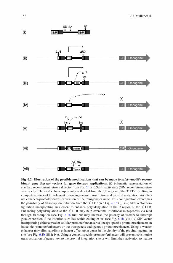
152 L.U. Müller et al.
Fig. 6.2 Illustration of the possible modifications that can be made to safety-modify recom-binant gene therapy vectors for gene therapy applications. (i) Schematic representation ofstandard recombinant retroviral vector from Fig. 6.1. (ii) Self-inactivating (SIN) recombinant retro-viral vector. The viral enhancer/promoter is deleted from the U3 region of the 3′ LTR resulting incomplete absence of this element following reverse transcription and proviral integration. An inter-nal enhancer/promoter drives expression of the transgene cassette. This configuration overcomesthe possibility of transcription initiation from the 3′ LTR (see Fig. 6.1b (i)). (iii) SIN vector con-figuration incorporating an element to enhance polyadenylation in the R region of the 3′ LTR.Enhancing polyadenylation at the 3′ LTR may help overcome insertional mutagenesis via readthrough transcription (see Fig. 6.1b (ii)) but may increase the potency of vectors to interruptgene expression if the insertion sites lies within coding exons (see Fig. 6.1b (v)). (iv) SIN vectorincorporating either a weaker cellular promoter/enhancer; a lineage specific promoter/enhancer; aninducible promoter/enhancer; or the transgene’s endogenous promoter/enhancer. Using a weakerenhancer may eliminate/limit enhancer effect upon genes in the vicinity of the proviral integrationsite (see Fig. 6.1b (iii & iv)). Using a context specific promoter/enhancer will prevent constitutivetrans-activation of genes next to the proviral integration site or will limit their activation to mature
6 Insertional Mutagenesis in Hematopoietic Cells 153
can be incorporated into the internal position of a SIN vector and, even thoughit is only able to drive very low expression of a transgene, it has been shown tobe sufficient to express γc at a level which corrects SCID-X1 [194]. Although theUCOE is unable to transactivate neighboring genes via an enhancer-mediated effect,it remains to be seen whether the maintenance of a permissive chromatin structurehas any impact in terms of altering expression of cellular genes. Finally, one couldincorporate an inducible promoter into the internal position of a SIN vector. Whilethis system would be highly desirable in terms of allowing the physician to directlyregulate the timing and level of transgene expression, inducible systems such asthe tetracycline-based system are problematic in terms of achieving tight regulatedexpression in the context of including all the required regulatory elements within asingle retroviral vector [144].
While SIN vectors reduce the risk of enhancer-mediated insertional mutagenesis,they are still susceptible to the potential activation of transcripts downstream of theintegration site via leaky polyadenylation in the 3′ LTR R region. There are a numberof viral and cellular elements which have been defined to enhance the efficiency ofpolyadenylation when they are positioned upstream of the polyadenylation signal.Both the woodchuck hepatitis virus post-transcriptional regulatory element (wPRE)and tandem insertions of the SV40 upstream sequence element (USE) have beenincorporated into SIN vectors and successfully demonstrated to enhance polyadeny-lation in the 3′ LTR and minimize read-through transcription [74, 156]. It must benoted that while this strategy may reduce the risk of gene activation via read-throughtranscription, it has a theoretical risk of increasing the propensity of the vector tointerrupt gene expression if the proviral integration site is within a gene’s activelytranscribed region.
Another commonly proposed strategy to minimize the ability of a retroviral-encoded transcript to transactivate neighboring genes is to incorporate chromatininsulating elements into the recombinant vector flanking the expression cassette.
�
Fig. 6.2 (continued) cell types which have a decreased transformation potential. (v) Incorporationof insulating elements flanking the promoter/enhancer and transgene expression cassette. The useof insulating elements may prevent trans-activation of neighboring genes via enhancer effect andmay additionally make expression of the transgene cassette more robust by forming a barrierbetween inhibitory chromatin structure and the expression cassette. (vi) Addition of a suicide genewithin the co-expression cassette. Co-expression of a pro-drug activating enzyme allows selectivekilling of gene modified cells upon detection of clonal imbalance/transformation. (vii) Codon opti-mization of transgene. Conservative mutations can be made in the transgene cDNA in order toremove cryptic splice donor/acceptor sites, enhancer sequences and non-canonical polyadenyla-tion signals such that the provirus has less potential to interfere with endogenous gene expression.Additionally, optimization of codon usage to favor amino acyl-tRNA molecules that are mostabundant in mammalian cells will potentiate higher protein expression per recombinant mRNAmolecule. �U3, LTR U3 region deleted for the sequences encoding the viral enhancer/promoter;E/P, enhancer/promoter; pA, polyadenylation signal; PRE, post-transcriptional regulatory element;+, augmenting effect upon polyadenylation; X, no enhancer effect upon the promoter of endoge-nous genes; Ins, insulating element; SA, splice acceptor site; T-Suppr, tumor suppressor gene;T-gene, therapeutic transgene

154 L.U. Müller et al.
The HS4 chromatin insulator (cHC4) element from the chicken beta globin LCRhas been defined as possessing both insulator activity and barrier activity [6, 48,98]. That is, it can insulate the surrounding genomic locus from the enhancer effectof the retroviral-encoded enhancer while acting as a barrier to the inhibitory effectof the chromatin structure surrounding the insertion site on the retroviral expressioncassette. It appears that the full length cHS4 element can compromise vector titerif inserted into the LTR region [7]. Several groups have used either the full lengthcHS4 element or various truncated versions of this element in order to reduce thetransactivating potential of retroviral vectors [7, 49, 99, 151]. Furthermore, the abil-ity of this element to prevent vector silencing and position dependent effects ongene expression may facilitate the incorporation of weaker cellular promoters intoSIN vectors while promoting robust expression of the transgene cassette.
If retroviral insertional mutagenesis does result in the generation of a transformedclone, then a mechanism through which the gene modified clone could be selec-tively eliminated would be highly desirable. Pro-drug activating enzymes such asherpes simplex virus thymidine kinase or bacterial nitoreductase can specificallyconvert a harmless pro-drug into a toxic metabolite specifically within the cell thatthe enzyme is expressed and can thus act as a suicide gene. The co-expressionof such a suicide within a retroviral gene expression cassette would allow theselective elimination of the gene modified graft upon systemic treatment with thepro-drug [17, 181]. This approach has been somewhat limited by problems relatingto adequate expression levels in eukaryotic cells and the immunogenicity of theseenzymes. However, site directed mutagenesis to increase enzyme stability/activityhas resulted in the derivation of suicide gene which demonstrate increased efficacy[17, 91, 102, 172]. The characterization of mammalian pro-drug activating enzymessuch as carboxylesterase may allow the substitution of bacterial/viral enzymes withless immunogenic proteins [109].
Finally, transgene expression cassettes can contain cryptic post-transcriptionalregulatory elements such as splice donor/acceptor sites and polyadenylation sites.While these elements will be detrimental in terms of reducing transgene expressionlevel, they also have the potential to mediate insertional mutagenesis via increasingthe potential of the vector to interrupt gene expression if the proviral integrationsite lies in the transcribed region of a gene, as seen in the French β thalassemiatrial. Insertional mutagenesis or complete codon optimization can be performed toeliminate these potentially mutagenic sequences [53, 89, 126, 145]. As with the useof insulator elements, this has the added benefit of increasing transgene expressionlevels and my therefore facilitate the use of weaker promoter elements which havea reduced genotoxic potential.
6.7 Summary
The severe adverse events that have occurred in three gene therapy trials relatingto retroviral-mediated insertional mutagenesis have been a setback in the effort to
6 Insertional Mutagenesis in Hematopoietic Cells 155
translate the promise of hematopoietic gene therapy into successful clinical thera-pies. Nonetheless, these unfortunate events have been the catalyst that has driventhe field to devise methods to dissect the mechanisms underlying the phenomenonof insertional mutagenesis and to design new strategies to overcome this problem.The resulting technology which has been developed to address this issue does notonly have direct application to the field of gene therapy, but may be used to studyother biological phenomenon relating to areas such as retroviral pathogenesis, stemcell biology, cellular transformation, and cell signaling.
Acknowledgments We thank Megan Smith, Miriam Edlund and Elise Porter for administrativesupport and members of our laboratory and the Transatlantic Gene Therapy Consortium for helpfuldiscussions. This work is supported by NIH HL081499.
References
1. Aiuti, A., et al. (2007). Multilineage hematopoietic reconstitution without clonal selec-tion in ADA-SCID patients treated with stem cell gene therapy. The Journal of ClinicalInvestigation, 117, 2233–2240.
2. Aiuti, A., et al. (2002). Correction of ADA-SCID by stem cell gene therapy combined withnonmyeloablative conditioning. Science, 296, 2410–2413.
3. Aiuti, A., et al. (2009). Gene therapy for immunodeficiency due to adenosine deaminasedeficiency. The New England Journal of Medicine, 360, 447–458.
4. Alexander, B. L., et al. (2007). Progress and prospects: Gene therapy clinical trials (part 1).Gene Therapy, 14, 1439–1447.
5. Antoine, C., et al. (2003). Long-term survival and transplantation of haemopoietic stemcells for immunodeficiencies: Report of the European experience 1968–1999. Lancet, 361,553–560.
6. Arumugam, P. I., Scholes, J., Perelman, N., Xia, P., Yee, J. K., & Malik, P. (2007). Improvedhuman beta-globin expression from self-inactivating lentiviral vectors carrying the chickenhypersensitive site-4 (cHS4) insulator element. Molecular Therapy, 15, 1863–1871.
7. Arumugam, P. I., et al. (2009). Genotoxic potential of lineage-specific lentivirus vectorscarrying the beta-globin locus control region. Molecular Therapy, 17, 1929–1937.
8. Barjesteh van Waalwijk van Doorn-Khosrovani, S., et al. (2003). High EVI1 expressionpredicts poor survival in acute myeloid leukemia: A study of 319 de novo AML patients.Blood, 101, 837–845.
9. Baum, C., Hegewisch-Becker, S., Eckert, H. G., Stocking, C., & Ostertag, W. (1995). Novelretroviral vectors for efficient expression of the multidrug resistance (MDR-1) gene in earlyhematopoietic cells. Journal of Virology, 69, 7541–7547.
10. Beard, B. C., et al. (2007). Comparison of HIV-derived lentiviral and MLV-based gam-maretroviral vector integration sites in primate repopulating cells. Molecular Therapy, 15,1356–1365.
11. Berenson, R. J., et al. (1988). Antigen CD34+ marrow cells engraft lethally irradiatedbaboons. The Journal of Clinical Investigation, 81, 951–955.
12. Berenson, R. J., et al. (1991). Engraftment after infusion of CD34+ marrow cells in patientswith breast cancer or neuroblastoma. Blood, 77, 1717–1722.
13. Berger, F., et al. (2001). Efficient retrovirus-mediated transduction of primitive humanperipheral blood progenitor cells in stroma-free suspension culture. Gene Therapy, 8,687–696.
14. Bertrand, Y., et al. (1999). Influence of severe combined immunodeficiency phenotype onthe outcome of HLA non-identical, T-cell-depleted bone marrow transplantation: A retro-spective European survey from the European group for bone marrow transplantation and theEuropean society for immunodeficiency. The Journal of Pediatrics, 134, 740–748.

156 L.U. Müller et al.
15. Beug, H., von Kirchbach, A., Doderlein, G., Conscience, J. F., & Graf, T. (1979). Chickenhematopoietic cells transformed by seven strains of defective avian leukemia viruses displaythree distinct phenotypes of differentiation. Cell, 18, 375–390.
16. Blaese, R. M., et al. (1995). T lymphocyte-directed gene therapy for ADA-SCID: Initial trialresults after 4 years. Science, 270, 475–480.
17. Blumenthal, M., Skelton, D., Pepper, K. A., Jahn, T., Methangkool, E., & Kohn, D. B.(2007). Effective suicide gene therapy for leukemia in a model of insertional oncogenesisin mice. Molecular Therapy, 15, 183–192.
18. Bokhoven, M., et al. (2009). Insertional gene activation by lentiviral and gammaretroviralvectors. Journal of Virology, 83, 283–294.
19. Booth, C., et al. (2007). Management options for adenosine deaminase deficiency; proceed-ings of the EBMT satellite workshop (Hamburg, March 2006. Clinical Immunology, 123,139–147.
20. Bordignon, C., et al. (1995). Gene therapy in peripheral blood lymphocytes and bone marrowfor ADA-immunodeficient patients. Science, 270, 470–475.
21. Bosticardo, M., Ghosh, A., Du, Y., Jenkins, N., Copeland, N., & Candotti, F.(2009). Self-inactivating retroviral vector-mediated gene transfer induces oncogene acti-vation and immortilization of primary murine bone marrow cells. Molecular Therapy,17, 1910–1918.
22. Bosticardo, M., Marangoni, F., Aiuti, A., Villa, A., & Grazia Roncarolo, M. (2009). Recentadvances in understanding the pathophysiology of Wiskott-Aldrich syndrome. Blood, 113,6288–6295.
23. Bousso, P., et al. (2000). Diversity, functionality, and stability of the T cell repertoire derivedin vivo from a single human T cell precursor. Proceedings of the National Academy ofSciences of the USA, 97, 274–278.
24. Bozorgmehr, F., et al. (2007). No evidence of clonal dominance in primates up to 4years following transplantation of multidrug resistance 1 retrovirally transduced long-termrepopulating cells. Stem Cells, 25, 2610–2618.
25. Boztug, K., et al. (2007). Hematopoietic stem cell gene therapy for Wiskott-AldrichSyndrome. p. 502.
26. Buckley, R. H., et al. (1999). Hematopoietic stem-cell transplantation for the treatmentof severe combined immunodeficiency. The New England Journal of Medicine, 340,508–516.
27. Buonamici, S., Chakraborty, S., Senyuk, V., & Nucifora, G. (2003). The role of EVI1 innormal and leukemic cells. Blood Cells, Molecules and Diseases, 31, 206–212.
28. Buonamici, S., et al. (2004). EVI1 induces myelodysplastic syndrome in mice. The Journalof Clinical Investigation, 114, 713–719.
29. Calmels, B., et al. (2005). Recurrent retroviral vector integration at the Mds1/Evi1 locus innonhuman primate hematopoietic cells. Blood, 106, 2530–2533.
30. Cassani, B., et al. (2009). Integration of retroviral vectors induces minor changes in thetranscriptional activity of T cells from ADA-SCID patients treated with gene therapy. Blood,114, 3546–3556.
31. Cattoglio, C., et al. (2007). Hot spots of retroviral integration in human CD34+ hematopoi-etic cells. Blood, 110, 1770–1778.
32. Cavazzana-Calvo, M., & Fischer, A. (2007). Gene therapy for severe combined immunode-ficiency: Are we there yet? The Journal of Clinical Investigation, 117, 1456–1465.
33. Cavazzana-Calvo, M., Lagresle, C., Hacein-Bey-Abina, S., & Fischer, A. (2005).Gene therapy for severe combined immunodeficiency. Annual Review of Medicine, 56,585–602.
34. Cavazzana-Calvo, M., et al. (2000). Gene therapy of human severe combined immunodefi-ciency (SCID)-X1 disease [see comments]. Science, 288, 669–672.
35. Cavazzana-Calvo, M., et al. (2008). Hematopoietic Stem Cell Gene Therapy Trial withLentiviral Vector in X-Linked Adrenoleukodystrophy. American Society of Hematology,ISI:000262104701045, 304–305.
6 Insertional Mutagenesis in Hematopoietic Cells 157
36. Chong, H., & Vile, R. G. (1996). Replication-competent retrovirus produced by a ‘split-function’ third generation amphotropic packaging cell line. Gene Therapy, 3, 624–629.
37. Cleynen, I., & Van de Ven, W. J. (2008). The HMGA proteins: A myriad of functions[Review]. International Journal of Oncology, 32, 289–305.
38. Cornetta, K., & Anderson, W. F. (1989). Protamine sulfate as an effective alternative to poly-brene in retroviral-mediated gene-transfer: Implications for human gene therapy. Journal ofVirological Methods, 23, 187–194.
39. Cornils, K., et al. (2009). Stem cell marking with promotor-deprived self-inactivatingretroviral vectors does not lead to induced clonal imbalance. Molecular Therapy, 17,131–143.
40. Couture, L. A., Mullen, C. A., & Morgan, R. A. (1994). Retroviral vectors containingchimeric promoter/enhancer elements exhibit cell-type-specific gene expression. HumanGene Therapy, 5, 667–677.
41. Crooks, G. M., & Kohn, D. B. (1993). Growth factors increase amphotropic retrovirusbinding to human CD34+ bone marrow progenitor cells. Blood, 82, 3290–3297.
42. Daniel, R., & Smith, J. A. (2008). Integration site selection by retroviral vectors: Molecularmechanism and clinical consequences. Human Gene Therapy, 19, 557–568.
43. Dave, U. P., et al. (2009). Murine leukemias with retroviral insertions at Lmo2 are predic-tive of the leukemias induced in SCID-X1 patients following retroviral gene therapy. PLoSGenetics, 5, e1000491.
44. De Palma, M., et al. (2005). Promoter trapping reveals significant differences in integrationsite selection between MLV and HIV vectors in primary hematopoietic cells. Blood, 105,2307–2315.
45. Deichmann, A., et al. (2007). Vector integration is nonrandom and clustered and influencesthe fate of lymphopoiesis in SCID-X1 gene therapy. The Journal of Clinical Investigation,117, 2225–2232.
46. Dilber, M. S., Bjorkstrand, B., Li, K. J., Smith, C. I., Xanthopoulos, K. G., & Gahrton,G. (1994). Basic fibroblast growth factor increases retroviral-mediated gene transfer intohuman hematopoietic peripheral blood progenitor cells. Experimental Hematology, 22,1129–1133.
47. Donahue, R. E., et al. (1992). Helper virus induced T cell lymphoma in nonhuman primatesafter retroviral mediated gene transfer. The Journal of Experimental Medicine, 176, 1125.
48. Emery, D. W., Yannaki, E., Tubb, J., Nishino, T., Li, Q., & Stamatoyannopoulos, G. (2002).Development of virus vectors for gene therapy of beta chain hemoglobinopathies: Flankingwith a chromatin insulator reduces gamma-globin gene silencing in vivo. Blood, 100,2012–2019.
49. Evans-Galea, M. V., Wielgosz, M. M., Hanawa, H., Srivastava, D. K., & Nienhuis, A. W.(2007). Suppression of clonal dominance in cultured human lymphoid cells by addition ofthe cHS4 insulator to a lentiviral vector. Molecular Therapy, 15, 801–809.
50. Felice, B., et al. (2009). Transcription factor binding sites are genetic determinants ofretroviral integration in the human genome. PloS One, 4, e4571.
51. Feron, C., et al. (1993). Purification, expansion, and multiple fluorochrome labeling of cordblood hemopoietic precursors: Preliminary results. Journal of Hematotherapy, 2, 259–261.
52. Fischer, A., et al. (1997). Naturally occurring primary deficiencies of the immune system.Annual Review of Immunology, 15, 93–124.
53. Foster, H., et al. (2008). Codon and mRNA sequence optimization of microdystrophin trans-genes improves expression and physiological outcome in dystrophic mdx mice followingAAV2/8 gene transfer. Molecular Therapy, 16, 1825–1832.
54. Freas-Lutz, D. L., Correll, P. H., Dougherty, S. F., Xu, L., Pluznik, D. H., & Karlsson, S.(1994). Expression of human glucocerebrosidase in murine macrophages: Identification ofefficient retroviral vectors. Experimental Hematology, 22, 857–865.
55. Garcia, I. S., et al. (1991). A study of chromosome 11p13 translocations involving TCR betaand TCR delta in human T cell leukaemia. Oncogene, 6, 577–582.

158 L.U. Müller et al.
56. Garcia-Higuera, I., et al. (2001). Interaction of the Fanconi anemia proteins and BRCA1 ina common pathway. Molecular Cell, 7, 249–262.
57. Garrett, E., Miller, A. R., Goldman, J. M., Apperley, J. F., & Melo, J. V. (2000).Characterization of recombination events leading to the production of an ecotropicreplication-competent retrovirus in a GP+envAM12-derived producer cell line. Virology,266, 170–179.
58. Gaspar, H. B., et al. (2004). Gene therapy of X-linked severe combined immunodeficiencyby use of a pseudotyped gammaretroviral vector. Lancet, 364, 2181–2187.
59. Gaspar, H. B., et al. (2006). Successful reconstitution of immunity in ADA-SCID bystem cell gene therapy following cessation of PEG-ADA and use of mild preconditioning.Molecular Therapy, 14, 505–513.
60. Glimm, H., et al. (1997). Efficient gene transfer in primitive CD34+/CD38lo human bonemarrow cells reselected after long-term exposure to GALV-pseudotyped retroviral vector.Human Gene Therapy, 8, 2079–2086.
61. Goerner, M., Bruno, B., McSweeney, P. A., Buron, G., Storb, R., & Kiem, H. P. (1999). Theuse of granulocyte colony-stimulating factor during retroviral transduction on fibronectinfragment CH-296 enhances gene transfer into hematopoietic repopulating cells in dogs.Blood, 94, 2287–2292.
62. Gonda, T. J., Sheiness, D. K., & Bishop, J. M. (1982). Transcripts from the cellularhomologs of retroviral oncogenes: Distribution among chicken tissues. Molecular andCellular Biology, 2, 617–624.
63. Grunebaum, E., et al. (2006). Bone marrow transplantation for severe combined immunedeficiency. Jama, 295, 508–518.
64. Hacein-Bey-Abina, S., et al. (2002). Sustained correction of X-linked severe combinedimmunodeficiency by ex vivo gene therapy. The New England Journal of Medicine, 346,1185–1193.
65. Hacein-Bey-Abina, S., et al. (2003). LMO2-associated clonal T cell proliferation in twopatients after gene therapy for SCID-X1. Science, 302, 415–419.
66. Hacein-Bey-Abina, S., et al. (2008). Insertional oncogenesis in 4 patients after retrovirus-mediated gene therapy of SCID-X1. The Journal of Clinical Investigation, 118, 3132–3142.
67. Hacker, C. V., et al. (2006). The integration profile of EIAV-based vectors. MolecularTherapy, 14, 536–545.
68. Haddad, E., et al. (1998). Long-term immune reconstitution and outcome afterHLA-nonidentical T-cell-depleted bone marrow transplantation for severe combinedimmunodeficiency: A European retrospective study of 116 patients. Blood, 91,3646–3653.
69. Haneline, L. S., et al. (2003). Retroviral-mediated expression of recombinant Fanccenhances the repopulating ability of Fancc–/– hematopoietic stem cells and decreases therisk of clonal evolution. Blood, 101, 1299–1307.
70. Hanenberg, H., Xiao, X. L., Dilloo, D., Hashino, K., Kato, I., & Williams, D. A. (1996).Colocalization of retrovirus and target cells on specific fibronectin fragments increasesgenetic transduction of mammalian cells. Nature Medicine, 2, 876–882.
71. Hargrove, P. W., et al. (2008). Globin lentiviral vector insertions can perturb the expres-sion of endogenous genes in beta-thalassemic hematopoietic cells. Molecular Therapy, 16,525–533.
72. Hematti, P., et al. (2004). Distinct genomic integration of MLV and SIV vectors in primatehematopoietic stem and progenitor cells. PLoS Biology, 2, e423.
73. Hershfield, M. S. (1998). Adenosine deaminase deficiency: Clinical expression, molecularbasis, and therapy. Seminars in Hematology, 35, 291–298.
74. Higashimoto, T., et al. (2007). The woodchuck hepatitis virus post-transcriptional regula-tory element reduces readthrough transcription from retroviral vectors. Gene Therapy, 14,1298–1304.
6 Insertional Mutagenesis in Hematopoietic Cells 159
75. Hildinger, M., Abel, K. L., Ostertag, W., & Baum, C. (1999). Design of 5′ untrans-lated sequences in retroviral vectors developed for medical use. Journal of Virology, 73,4083–4089.
76. Hildinger, M., Eckert, H. G., Schilz, A. J., John, J., Ostertag, W., & Baum, C. (1998). FMEVvectors: Both retroviral long terminal repeat and leader are important for high expression intransduced hematopoietic cells. Gene Therapy, 5, 1575–1579.
77. Hirschhorn, R., Yang, D. R., Puck, J. M., Huie, M. L., Jiang, C. -K., & Kurlandsky, L. E.(1996). Spontaneous in vivo reversion to normal of an inherited mutation in a patient withadenosine deaminase deficiency. Nature Genetics, 13, 290–295.
78. Hock, R. A., Miller, A. D., & Osborne, W. R. (1989). Expression of human adenosine deam-inase from various strong promoters after gene transfer into human hematopoietic cell lines.Blood, 74, 876–881.
79. Hoogerbrugge, P. M., et al. (1996). Bone marrow gene transfer in three patients withadenosine deaminase deficiency. Gene Therapy, 3, 179–183.
80. Howe, S. J., et al. (2008). Insertional mutagenesis combined with acquired somatic muta-tions causes leukemogenesis following gene therapy of SCID-X1 patients. The Journal ofClinical Investigation, 118, 3143–3150.
81. Hu, J., et al. (2003). Direct comparison of RD114-pseudotyped versus amphotropic-pseudotyped retroviral vectors for transduction of rhesus macaque long-term repopulatingcells. Molecular Therapy, 8, 611–617.
82. Hu, J., et al. (2008). Reduced genotoxicity of avian sarcoma leukosis virus vectors in rhe-sus long-term repopulating cells compared to standard murine retrovirus vectors. MolecularTherapy, 16, 1617–1623.
83. Hudspeth, M. P., & Raymond, G. V. (2007). Immunopathogenesis of adrenoleukodystrophy:Current understanding. Journal of Neuroimmunology, 182, 5–12.
84. Kiem, H. P., et al. (1997). Gene transfer into marrow repopulating cells: Comparisonbetween amphotropic and gibbon ape leukemia virus pseudotyped retroviral vectors in acompetitive repopulation assay in baboons. Blood, 90, 4638–4645.
85. Kiem, H. -P., et al. (1998). Improved gene transfer into baboon marrow repopulating cellsusing recombinant human fibronectin fragment CH-296 in combination with IL-6, stemcell factor, FLT-3 ligand, and megakaryocyte growth and development factor. Blood, 92,1878–1886.
86. Kiem, H. P., et al. (2004). Long-term clinical and molecular follow-up of large ani-mals receiving retrovirally transduced stem and progenitor cells: No progression to clonalhematopoiesis or leukemia. Molecular Therapy, 9, 389–395.
87. Kim, W. S., Weickert, C. S., & Garner, B. (2008). Role of ATP-binding cassette trans-porters in brain lipid transport and neurological disease. Journal of Neurochemistry, 104,1145–1166.
88. Kim, Y. J., et al. (2009). Sustained high-level polyclonal hematopoietic marking and trans-gene expression 4 years after autologous transplantation of rhesus macaques with SIVlentiviral vector-transduced CD34+ cells. Blood, 113, 5434–5443.
89. Knipper, R., et al. (2001). Improved post-transcriptional processing of an MDR1 retroviruselevates expression of multidrug resistance in primary human hematopoietic cells. GeneTherapy, 8, 239–246.
90. Kohn, D. B., et al. (1995). Engraftment of gene-modified umbilical cord blood cells inneonates with adenosine deaminase deficiency. Nature Medicine, 1, 1017–1023.
91. Kokoris, M. S., Sabo, P., Adman, E. T., & Black, M. E. (1999). Enhancement oftumor ablation by a selected HSV-1 thymidine kinase mutant. Gene Therapy, 6,1415–1426.
92. Kurre, P., Morris, J., Horn, P. A., Harkey, M. A., Andrews, R. G., & Kiem, H. P. (2001). Genetransfer into baboon repopulating cells: A comparison of Flt-3 Ligand and megakaryocytegrowth and development factor versus IL-3 during ex vivo transduction. Molecular Therapy,3, 920–927.
160 L.U. Müller et al.
93. Kustikova, O., et al. (2005). Clonal dominance of hematopoietic stem cells triggered byretroviral gene marking. Science, 308, 1171–1174.
94. Kustikova, O. S., et al. (2007). Retroviral vector insertion sites associated with dominanthematopoietic clones mark “stemness” pathways. Blood, 109, 1897–1907.
95. Laufs, S., Nagy, K. Z., Giordano, F. A., Hotz-Wagenblatt, A., Zeller, W. J., & Fruehauf, S.(2004). Insertion of retroviral vectors in NOD/SCID repopulating human peripheral bloodprogenitor cells occurs preferentially in the vicinity of transcription start regions and inintrons. Molecular Therapy, 10, 874–881.
96. Lewinski, M. K., & Bushman, F. D. (2005). Retroviral DNA integration–mechanism andconsequences. Advances in Genetics, 55, 147–181.
97. Lewinski, M. K., et al. (2006). Retroviral DNA integration: Viral and cellular determinantsof target-site selection. PLoS Pathogens, 2, e60.
98. Li, C. L., & Emery, D. W. (2008). The cHS4 chromatin insulator reduces gammaretrovi-ral vector silencing by epigenetic modifications of integrated provirus. Gene Therapy, 15,49–53.
99. Li, C. L., Xiong, D., Stamatoyannopoulos, G., & Emery, D. W. (2009). Genomic and func-tional assays demonstrate reduced gammaretroviral vector genotoxicity associated with useof the cHS4 chromatin insulator. Molecular Therapy, 17, 716–724.
100. Li, X., et al. (2005). Ex vivo culture of Fancc–/– stem/progenitor cells predisposes cellsto undergo apoptosis, and surviving stem/progenitor cells display cytogenetic abnormalitiesand an increased risk of malignancy. Blood, 105, 3465–3471.
101. Li, Z., et al. (2002). Murine leukemia induced by retroviral gene marking. Science,296, 497.
102. Likar, Y., et al. (2008). A new acycloguanosine-specific supermutant of herpes simplex virustype 1 thymidine kinase suitable for PET imaging and suicide gene therapy for potential usein patients treated with pyrimidine-based cytotoxic drugs. Journal of Nuclear Medicine, 49,713–720.
103. Malech, H., et al. (1997). Prolonged production of NADPH oxidase-corrected granulocytesafter gene therapy of chronic granulomatous disease. Proceedings of the National Academyof Sciences of the USA, 94, 12133–12138.
104. Malik, P., Krall, W. J., Yu, X. J., Zhou, C., & Kohn, D. B. (1995). Retroviral-mediated geneexpression in human myelomonocytic cells: A comparison of hematopoietic cell promotersto viral promoters. Blood, 86, 2993–3005.
105. Mann, R., Mulligan, R. C., & Baltimore, D. (1983). Construction of a retrovirus packagingmutant and its use to produce helper-free defective retrovirus. Cell, 33, 153–159.
106. Manning, J. S., Hackett, A. J., & Darby, N. B., Jr. (1971). Effect of polycations on sensitivityof BALD-3T3 cells to murine leukemia and sarcoma virus infectivity. Applied Microbiology,22, 1162–1163.
107. Markowitz, D., Goff, S., & Bank, A. (1988). Construction and use of a safe and efficientamphotropic packaging cell line. J of Virology, 167, 400–406.
108. Maruggi, G., et al. (2009). Transcriptional enhancers induce insertional gene deregulationindependently from the vector type and design. Molecular Therapy, 17, 851–856.
109. Matzow, T., et al. (2007). Hypoxia-targeted over-expression of carboxylesterase as a meansof increasing tumour sensitivity to irinotecan (CPT-11) . The Journal of Gene Medicine, 9,244–252.
110. May, C., et al. (2000). Therapeutic haemoglobin synthesis in beta-thalassaemic miceexpressing lentivirus-encoded human beta-globin. Nature, 406, 82–86.
111. Mazzolari, E., et al. (2007). Long-term immune reconstitution and clinical outcome afterstem cell transplantation for severe T-cell immunodeficiency. Journal of Allergy and ClinicalImmunology, 120, 892–899.
112. Miller, A. D. (1990). Retrovirus packaging cells. Human Gene Therapy, 1, 5–14.113. Miller, A. D., Garcia, J. V., von Suhr, N., Lynch, C. M., Wilson, C., & Eiden, M. V. (1991).
Construction and properties of retrovirus packaging cells based on gibbon ape leukemiavirus. Journal of Virology, 65, 2220–2224.
6 Insertional Mutagenesis in Hematopoietic Cells 161
114. Milsom, M. D., & Fairbairn, L. J. (2004). Protection and selection for gene therapy in thehematopoietic system. The Journal of Gene Medicine, 6, 133–146.
115. Milsom, M. D., & Williams, D. A. (2007). Live and let die: In vivo selection of gene-modified hematopoietic stem cells via MGMT-mediated chemoprotection. DNA Repair(Amst), 6, 1210–1221.
116. Milsom, M. D., et al. (2005). Overexpression of HOXB4 confers a myelo-erythroiddifferentiation delay in vitro. Leukemia, 19, 148–153.
117. Milsom, M. D., et al. (2008). Reciprocal relationship between O6-methylguanine-DNAmethyltransferase P140K expression level and chemoprotection of hematopoietic stem cells.Cancer Research, 68, 6171–6180.
118. Mitchell, R. S., et al. (2004). Retroviral DNA integration: ASLV, HIV, and MLV showdistinct target site preferences. PLoS Biology, 2, E234.
119. Modlich, U., et al. (2005). Leukemias following retroviral transfer of multidrug resistance 1(MDR1) are driven by combinatorial insertional mutagenesis. Blood, 105, 4235–4246.
120. Modlich, U., et al. (2006). Cell culture assays reveal the importance of retroviral vectordesign for insertional genotoxicity. Blood, 108, 2545–2553.
121. Modlich, U., et al. (2008). Leukemia induction after a single retroviral vector insertion inEvi1 or Prdm16. Leukemia, 22, 1519–1528.
122. Modlich, U., et al. (2009). Insertional transformation of hematopoietic cells by self-inactivating lentiviral and gammaretroviral vectors. Molecular Therapy, 17, 1919–1928.
123. Montini, E., et al. (2006). Hematopoietic stem cell gene transfer in a tumor-prone mousemodel uncovers low genotoxicity of lentiviral vector integration. Nature Biotechnology, 24,687–696.
124. Montini, E., et al. (2009). The genotoxic potential of retroviral vectors is strongly modulatedby vector design and integration site selection in a mouse model of HSC gene therapy. TheJournal of Clinical Investigation, 119, 964–975.
125. Moolten, F. L., & Cupples, L. A. (1992). A model for predicting the risk of cancerconsequent to retroviral gene therapy. Human Gene Therapy, 3, 479–486.
126. Moreno-Carranza, B., et al. (2009). Transgene optimization significantly improves SIN vec-tor titers, gp91phox expression and reconstitution of superoxide production in X-CGD cells.Gene Therapy, 16, 111–118.
127. Moritz, T., Patel, V. P., & Williams, D. A. (1994). Bone marrow extracellular matrixmolecules improve gene transfer into human hematopoietic cells via retroviral vectors. TheJournal of Clinical Investigation, 93, 1451–1457.
128. Moser, H. W., Mahmood, A., & Raymond, G. V. (2007). X-linked adrenoleukodystrophy.Nature Clinical Practice, 3, 140–151.
129. Neale, G. A., Rehg, J. E., & Goorha, R. M. (1995). Ectopic expression of rhombotin-2 causesselective expansion of CD4-CD8- lymphocytes in the thymus and T-cell tumors in transgenicmice. Blood, 86, 3060–3071.
130. Neven, B., et al. (2009). Long-term outcome after hematopoietic stem cell transplantation ofa single-center cohort of 90 patients with severe combined immunodeficiency. Blood, 113,4114–4124.
131. Nienhuis, A. (2006). Assays to Evaluate the Genotoxicity of Retroviral Vectors. MolecularTherapy, 13, 461–462.
132. Noguchi, M., et al. (1993). Interleukin-2 receptor gamma chain mutation results in X-linkedsevere combined immunodeficiency in humans. Cell, 73, 147–157.
133. Nolta, J. A., Crooks, G. M., Overell, R. W., Williams, D. E., & Kohn, D. B. (1992). Retroviralvector-mediated gene transfer into primitive human hematopoietic progenitor cells: Effectsof mast cell growth factor (MGF) combined with other cytokines. Experimental Hematology,20, 1065–1071.
134. Notarangelo, L. D., Miao, C. H., & Ochs, H. D. (2008). Wiskott-Aldrich syndrome. CurrentOpinion in Hematology, 15, 30–36.
162 L.U. Müller et al.
135. Ochs, H. D., Filipovich, A. H., Veys, P., Cowan, M. J., & Kapoor, N. (2008). Wiskott-Aldrich syndrome: Diagnosis, clinical and laboratory manifestations, and treatment. Biologyof Blood and Marrow Transplant, 15, 84–90.
136. Orkin, S. H., & Nathan, D. G. (1981). The molecular genetics of thalassemia. Advances inHuman Genetics, 11, 233–280.
137. Ott, M. G., Seger, R., Stein, S., Siler, U., Hoelzer, D., & Grez, M. (2007). Advances inthe treatment of chronic granulomatous disease by gene therapy. Current Gene Therapy, 7,155–161.
138. Ott, M. G., et al. (2006). Correction of X-linked chronic granulomatous disease by genetherapy, augmented by insertional activation of MDS1-EVI1, PRDM16 or SETBP1. NatureMedicine, 12, 401–409.
139. Ott, M. G., et al. (2007). Phase I/II gene therapy study for Chronic Granulomatous Disease:Results, lessons and perspectives. American Society of Hematology, ISI:000251100800504,503.
140. Otto, E., et al. (1994). Characterization of a replication-competent retrovirus result-ing from recombination of packaging and vector sequences. Human Gene Therapy, 5,567–575.
141. Perumbeti, A., et al. (2009). A novel human gamma-globin gene vector for genetic correc-tion of sickle cell anemia in a humanized sickle mouse model: Critical determinants forsuccessful correction. Blood, 114, 1174–1185.
142. Pike-Overzet, K., et al. (2006). Gene therapy: Is IL2RG oncogenic in T-cell development?Nature, 443, E5, discussion E6–7.
143. Pike-Overzet, K., et al. (2007). Ectopic retroviral expression of LMO2, but not IL2Rgamma,blocks human T-cell development from CD34+ cells: Implications for leukemogenesis ingene therapy. Leukemia, 21, 754–763.
144. Pluta, K., Luce, M. J., Bao, L., Agha-Mohammadi, S., & Reiser, J. (2005). Tight controlof transgene expression by lentivirus vectors containing second-generation tetracycline-responsive promoters. The Journal of Gene Medicine, 7, 803–817.
145. Radcliffe, P. A., et al. (2008). Analysis of factor VIII mediated suppression of lentiviralvector titres. Gene Therapy, 15, 289–297.
146. Raper, S. E., et al. (2003). Fatal systemic inflammatory response syndrome in a ornithinetranscarbamylase deficient patient following adenoviral gene transfer. Molecular Geneticsand Metabolism, 80, 148–158.
147. Relander, T., et al. (2005). Gene transfer to repopulating human CD34+ cells usingamphotropic-, GALV-, or RD114-pseudotyped HIV-1-based vectors from stable producercells. Molecular Therapy, 11, 452–459.
148. Robinson, D., Elliott, J. F., & Chang, L. J. (1995). Retroviral vector with a CMV-IE/HIV-TAR hybrid LTR gives high basal expression levels and is up-regulated by HIV-1 Tat. GeneTherapy, 2, 269–278.
149. Roussel, M., et al. (1979). Three new types of viral oncogene of cellular origin specific forhaematopoietic cell transformation. Nature, 281, 452–455.
150. Russell, M., et al. (1994). Expression of EVI1 in myelodysplastic syndromes and otherhematologic malignancies without 3q26 translocations. Blood, 84, 1243–1248.
151. Ryu, B. Y., Evans-Galea, M. V., Gray, J. T., Bodine, D. M., Persons, D. A., & Nienhuis, A.W. (2008). An experimental system for the evaluation of retroviral vector design to diminishthe risk for proto-oncogene activation. Blood, 111, 1866–1875.
152. Samakoglu, S., et al. (2006). A genetic strategy to treat sickle cell anemia by coreg-ulating globin transgene expression and RNA interference. Nature Biotechnology, 24,89–94.
153. Sandrin, V., et al. (2002). Lentiviral vectors pseudotyped with a modified RD114 enve-lope glycoprotein show increased stability in sera and augmented transduction of primarylymphocytes and CD34+ cells derived from human and nonhuman primates. Blood, 100,823–832.
6 Insertional Mutagenesis in Hematopoietic Cells 163
154. Sankaran, V. G., et al. (2008). Human fetal hemoglobin expression is regulated by thedevelopmental stage-specific repressor BCL11A. Science, 322, 1839–1842.
155. Sauvageau, G., et al. (1995). Overexpression of HOXB4 in hematopoietic cells causesthe selective expansion of more primitive populations in vitro and in vivo. Genes andDevelopment, 9, 1753–1765.
156. Schambach, A., Galla, M., Maetzig, T., Loew, R., & Baum, C. (2007). Improving transcrip-tional termination of self-inactivating gamma-retroviral and lentiviral vectors. MolecularTherapy, 15, 1167–1173.
157. Schambach, A., Swaney, W. P., & van der Loo, J. C. (2009). Design and production ofretro- and lentiviral vectors for gene expression in hematopoietic cells. Methods in MolecularBiology, 506, 191–205.
158. Schambach, A., et al. (2006). Woodchuck hepatitis virus post-transcriptional regulatory ele-ment deleted from X protein and promoter sequences enhances retroviral vector titer andexpression. Gene Therapy, 13, 641–645.
159. Schiedlmeier, B., et al. (2003). High-level ectopic HOXB4 expression confers a profoundin vivo competitive growth advantage on human cord blood CD34+ cells, but impairslymphomyeloid differentiation. Blood, 101, 1759–1768.
160. Schmidt, M., et al. (2001). Detection and direct genomic sequencing of multiplerare unknown flanking DNA in highly complex samples. Human Gene Therapy, 12,743–749.
161. Schmidt, M., et al. (2002). Polyclonal long-term repopulating stem cell clones in a primatemodel. Blood, 100, 2737–2743.
162. Schmidt, M., et al. (2007). High-resolution insertion-site analysis by linear amplification-mediated PCR (LAM-PCR). Nature Methods, 4, 1051–1057.
163. Schwarzwaelder, K., et al. (2007). Gammaretrovirus-mediated correction of SCID-X1 isassociated with skewed vector integration site distribution in vivo. The Journal of ClinicalInvestigation, 117, 2241–2249.
164. Scobie, L., et al. (2009). A novel model of SCID-X1 reconstitution reveals predispositionto retrovirus-induced lymphoma but no evidence of gammaC gene oncogenicity. MolecularTherapy, 17, 1031–1038.
165. Segal, A. W. (1996). The NADPH oxidase and chronic granulomatous disease. MolecularMedicine Today, 2, 129–135.
166. Seger, R. A., et al. (2002). Treatment of chronic granulomatous disease with myeloablativeconditioning and an unmodified hemopoietic allograft: A survey of the European experience,1985–2000. Blood, 100, 4344–4350.
167. Seggewiss, R., et al. (2006). Acute myeloid leukemia is associated with retrovi-ral gene transfer to hematopoietic progenitor cells in a rhesus macaque. Blood, 107,3865–3867.
168. Semmler, A., Kohler, W., Jung, H. H., Weller, M., & Linnebank, M. (2008).Therapy of X-linked adrenoleukodystrophy. Expert review of Neurotherapeutics, 8,1367–1379.
169. Shou, Y., Ma, Z., Lu, T., & Sorrentino, B. P. (2006). Unique risk factors for insertional muta-genesis in a mouse model of XSCID gene therapy. Proceedings of the National Academy ofSciences of the USA, 103, 11730–11735.
170. Somia, N., & Verma, I. M. (2000). Gene therapy: Trials and tribulations. Nature Reviews, 1,91–99.
171. Stephan, V., et al. (1996). Atypical X-linked severe combined immunodeficiency due topossible spontaneous reversion of the genetic defect in T cells. The New England Journal ofMedicine, 335, 1563–1567.
172. Stolworthy, T. S., et al. (2008). Yeast cytosine deaminase mutants with increased ther-mostability impart sensitivity to 5-fluorocytosine. Journal of Molecular Biology, 377,854–869.
164 L.U. Müller et al.
173. Suzuki, T., Minehata, K., Akagi, K., Jenkins, N. A., & Copeland, N. G. (2006). Tumor sup-pressor gene identification using retroviral insertional mutagenesis in Blm-deficient mice.EMBO Journal, 25, 3422–3431.
174. Takeshita, T., et al. (1992). Cloning of the gamma chain of the human IL-2 receptor. Science,257, 379–382.
175. Tassi, C., Fortuna, A., Bontadini, A., Lemoli, R. M., Gobbi, M., & Tazzari, P. L. (1991).CD34 or S313 positive cells selection by avidin-biotin immunoadsorption. Haematologica,76(Suppl 1), 41–43.
176. Thornhill, S. I., et al. (2008). Self-inactivating gammaretroviral vectors for gene therapy ofX-linked severe combined immunodeficiency. Molecular Therapy, 16, 590–598.
177. Thrasher, A. J., et al. (2006). Gene therapy: X-SCID transgene leukaemogenicity. Nature,443, E5–6, discussion E6–7.
178. Touw, I. P., & Erkeland, S. J. (2007). Retroviral insertion mutagenesis in mice as a compar-ative oncogenomics tool to identify disease genes in human leukemia. Molecular Therapy,15, 13–19.
179. Trasher, A. J., & Gaspar, B. Successful bone marrow gene therapy for SCID. Blood CellsMolecules, and Diseases, 40, 287–288.
180. Trobridge, G. D., et al. (2006). Foamy virus vector integration sites in normal human cells.Proceedings of the National Academy of Sciences of the USA, 103, 1498–1503.
181. Uchiyama, T., et al. (2006). Application of HSVtk suicide gene to X-SCID gene therapy:Ganciclovir treatment offsets gene corrected X-SCID B cells. Biochemical and BiophysicalResearch Communications, 341, 391–398.
182. Vanin, E. F., Kaloss, M., Broscius, C., & Nienhuis, A. W. (1994). Characterization ofreplication-competent retroviruses from non-human primates with virus-induced T cell lym-phomas and observations regarding the mechanism of oncogenesis. Journal of Virology, 68,4241–4250.
183. Wagner, W., et al. (2005). Retroviral integration sites correlate with expressed genes inhematopoietic stem cells. Stem Cells, 23, 1050–1058.
184. Walsh, C. E., et al. (1994). A functionally active retrovirus vector for gene therapy in Fanconianemia group C. Blood, 84, 453–459.
185. Wang, G. P., et al. (2008). DNA bar coding and pyrosequencing to analyze adverse events intherapeutic gene transfer. Nucleic Acids Research, 36, e49.
186. Wang, W. (2007). Emergence of a DNA-damage response network consisting of Fanconianaemia and BRCA proteins. Nature Reviews, 8, 735–748.
187. Weatherall, D. J. (1998). Pathophysiology of thalassaemia. Bailliere’s ClinicalHaematology, 11, 127–146.
188. Wicke, D. C., et al. (2008). Ectopic Expression of the Extracellular Domain of Mpl IsSufficient to Induce a Hematopoietic Population Crisis. American Society of Hematology,ISI:000262104703324, 994.
189. Williams, D. A., Lemischka, I. R., Nathan, D. G., & Mulligan, R. C. (1984). Introduction ofnew genetic material into pluripotent haematopoietic stem cells of the mouse. Nature, 310,476–480.
190. Winkelstein, J. A., et al. (2000). Chronic granulomatous disease. Report on a nationalregistry of 368 patients. Medicine, 79, 155–169.
191. Woods, N. B., Bottero, V., Schmidt, M., von Kalle, C., & Verma, I. M. (2006). Gene therapy:Therapeutic gene causing lymphoma. Nature, 440, 1123.
192. Wu, X., Li, Y., Crise, B., & Burgess, S. M. (2003). Transcription start regions in the humangenome are favored targets for MLV integration. Science, 300, 1749–1751.
193. Yamada, Y., Warren, A. J., Dobson, C., Forster, A., Pannell, R., & Rabbitts, T. H. (1998). TheT cell leukemia LIM protein Lmo2 is necessary for adult mouse hematopoiesis. Proceedingsof the National Academy of Sciences of the USA, 95, 3890–3895.

6 Insertional Mutagenesis in Hematopoietic Cells 165
194. Zhang, F., et al. (2007). Lentiviral vectors containing an enhancer-less ubiquitously act-ing chromatin opening element (UCOE) provide highly reproducible and stable transgeneexpression in hematopoietic cells. Blood, 110, 1448–1457.
195. Zhang, X. B., et al. (2008). High incidence of leukemia in large animals after stem cell genetherapy with a HOXB4-expressing retroviral vector. The Journal of Clinical Investigation,118, 1502–1510.
196. Zychlinski, D., et al. (2008). Physiological promoters reduce the genotoxic risk of integratinggene vectors. Molecular Therapy, 16, 718–725.

Chapter 7Bioinformatics of High-Throughput InsertionalMutagenesis
Keiko Akagi, Ming Yi, Jean Roayaei, and Robert M. Stephens
Abstract Bioinformatics plays critical roles to handle large amount of sequencedata from insertional mutagenesis. First, computational approaches are used todevelop rapid sequence analysis pipelines and biological databases. Millions ofreads from an insertion mutagenesis screening are mapped to genomic locations andbe annotated to their target genes rapidly by pipeline, and such sequence-based datais stored and managed in database to share the information in the scientific com-munity. Second, statistical techniques are used to distinguish true common insertionsites (loci that have been hit by insertions in multiple tumors: candidate loci forcancer genes) from background insertions in large-scale screenings. Finally, theadvanced data mining techniques, pathway and network analysis, are used to givefurther biological meaning to insertion sites by identifying the interaction of genesin cancer. In this chapter, we discuss features of these three topics and address theirfuture roles: (1) development of sequence analysis pipeline and database, (2) detec-tion of common insertion sites, and (3) network and pathway analysis of insertionsites.
7.1 Sequence Analysis Pipeline and Database
7.1.1 Sequencing and Barcoding Technologies
Insertional mutagenesis is a great tool to find cancer gene candidates from varioustumor models [22]. The attempt to screen the insertion sites from retrovirus-inducedtumors started by using Southern blotting analysis and genomic library screen-ing in 1980s [31, 70], but the utility of this approach greatly increased after the
K. Akagi (B)Mouse Cancer Genetics Program, National Cancer Institute, Center for Cancer Research,Frederick, MD 21702, USA; Comprehensive Cancer Center, Ohio State University, Columbus,OH 43210, USAe-mail: [email protected]
Sections 7.1 to 7.6 are written by Keiko Akagi and Jean Roayaei and Sections 7.7 to 7.10 arewritten by Ming Yi and Robert M. Stephens.
167A.J. Dupuy, D.A. Largaespada (eds.), Insertional Mutagenesis Strategiesin Cancer Genetics, DOI 10.1007/978-1-4419-7656-7_7,C© Springer Science+Business Media, LLC 2011
168 K. Akagi et al.
development of rapid insert cloning methods and the completion of mouse genomeproject [43, 47, 66].
Recently, the non-Sanger-based next-generation sequencing technologiesenabled us to read the high volume of sequences in a reduced cost [59], and theirdevelopment will further increase the power of insertional mutagenesis as highthroughput screening approach. Currently, three major high-throughput sequenceplatforms are the Genome Sequencers from Roche/454 Life Sciences [46], theG2 Analyzer from Illumina/Solexa [6], and the SOLiD System from AppliedBiosystems (http://solid.appliedbiosystems.com). The comparison between threeplatforms show a trade-offs between average sequence read length and the numberof sequence reads produced in a single run. The Illumina and SOLiD systems gener-ate ten-fold more reads than Roche/454 Sequencers, but they provide much shorterread length of 35–50 bp than that of Roche/454 Sequencers, 250 bp [57, 59]. Thelonger read length of Roche/454 Sequencers makes the system ideal for insertionalmutagenesis screenings with a barcoding approach. Historically, “barcodes”, uniqueDNA sequence identifiers, have been used in EST-based gene discovery projects todistinguish tissue sources of pooled cDNA libraries [26]. In insertional mutagenesisscreenings, researchers analyze multiple tumors and/or tissues from many individ-uals, and the number of necessary insertion reads in each sample is much smallerthan the capacity of the Roche/454 sequencer. To overcome the limited number ofsamples sequenced in parallel by the physical separators of sequencer, barcodes(∼10 bp) are assigned to each insertional mutagenesis sample. The presence of bar-codes allows independent samples to be pooled together for sequencing and allowsthe bioinformatic segregation of the output from sequencers afterwards. The lengthof the barcodes should be determined to allow the unambiguous assignment of bar-codes and to minimize the base reads used for barcodes. For Roche/454 sequencers,the design of barcodes should consider the pyrosequencing error characteristics. Forexample, to avoid homopolymeric runs in pyrosequencing technologies, the bar-codes should avoid including more than two successive occurrences of the samenucleotides for 10 bp barcodes [52]. With the careful designed barcodes, ∼98%of the sequences can be assigned to a correct sample [38]. This approach greatlyenhances the saturation of insertional mutagenesis screenings.
7.2 Sequence Mapping
Collected sequences from insertional mutagenesis are mapped to the genomeassembly of model organisms to identify the genomic characteristics of inser-tion sites. The mouse genome was sequenced from C57BL/6 J mouse strain, andthe assembled genome was distributed to three genome browsers (UCSC, NCBI,and Ensembl) [74]. The genome sequence assembly in these genome browsersis updated periodically (the latest release: NCBI build m37, October 2007), andgenomic coordinates along the chromosome can change dramatically in differentassemblies because of changes in sequence data or algorithm implementation. When
7 Bioinformatics of High-Throughput Insertional Mutagenesis 169
there is a doubt of error in a region of the assembly, it is worth to review themapping record of insertion sites for older versions of the genome assembly. NCBIalso provides limited genome assemblies from alternative mouse strains (mixedstrains by Celera whole genome shot-gun sequencing and 129 substrains) [50], andSanger Institute is attempting de nove assembly of sequences of 17 mouse straingenomes on Illumina’s GA platform [63]. These alternative assemblies can be usedto map insertional mutagenesis sequences to strain specific regions.
To align sequences of insertion sites to genome, two sequence alignment pro-grams are used frequently: BLAST (Basic Local Alignment Search Tool) [3] andBLAT (BLAST-like Alignment Tool) [39]. Both programs find regions of local sim-ilarity between sequences, and they rapidly scans for short sequence matches andextends these matches into high scoring pairs. However, the BLAT algorithm buildsan index of the genome assemblies before any searches are performed to achievefaster speed than BLAST does. The BLAT program is sometimes not as sensitive asBLAST, but it is much faster and often used to find the position of an insertion sitesequence in genome.
Once mapped, the insertion site sequence is checked for the alignment at thejunction of mutagens (such as retrovirus’ long terminal repeat) to identify the posi-tion and direction of insertion in genome. The direction of insertion site helps toidentify the target genes because mutagens affect genes more severely in certaingene locations and directions. For example, retrovirus insertions are more likelyto enhance expression of genes when they are located in the upstream of genes inthe antisense orientation or downstream in the sense orientation [72]. MutagenicSleeping Beauty transposon vectors are designed to affect genes in a directionalmanner [14, 23]. In addition to the direction of insertion site, the number of redun-dant sequences, how many redundant sequences arose from the same tumor andmapped to the same position in the genome, also provides useful information. Ina shotgun-cloning project, insertions with higher redundancy are more likely to beclonal and early insertions in tumorigenesis by Southern blot [19]. If there is nosevere bias in cloning step, the frequency of redundant read for an insertion site serveas an indicator of the copy number of insertion, penetrance, in the tumor library.
7.3 Target Gene Identification
After the mapping of insertion sequences to genome, the annotation of these inser-tions sites to neighboring target genes becomes an important step. RefSeq is a cratednon-redundant collection of genomes, transcripts, and proteins [54]. The genes inRefSeq are supported by experimental evidence and multiple quality assessmenttests; the gene set is ideal to annotate insertion sites to confirmed genes. Recently,reports show the involvement of microRNAs and non-coding RNAs in retrovirusinsertion sites [17, 34, 61], and some microRNAs are suggested to act as onco-genes in human cancer [8]. The miRBase [28], database for published microRNAs,is another useful gene set to identify the potential involvement of microRNAs ininsertion sites.
170 K. Akagi et al.
In addition to gene databases, medical and phenotype databases contribute to theprocess of target gene identification for insertion sites. To identify whether the targetgene of insertion site is a known human cancer gene, Cancer Gene Census worksas a great resource. The Cancer Genome Project at Sanger Institute conducted acensus of cancer-causing genes from literature [25], and they provide the list of can-cer genes and their mutation/tumor types (367 gene; October 2007 release). Morethan 40 of these loci are also identified as common insertion sites from the retro-virus insertional mutagenesis screening [72]. To search non-cancerous phenotypesin human, Online Mendelian Inheritance in Man (OMIM) [30] provides informa-tion about human genes and their disease phenotypes. To identify the phenotypein mice, Jackson Laboratory’s Mouse Genome Database [11] provides informationabout phenotypes for gene-target mouse.
7.4 Insertional Mutagenesis Database
To efficiently store and manage sequences and annotated data, a relational databaseplays an important role for insertional mutagenesis. A relational database isa database that organizes data into tables and represents relationships amongthose tables, and these relationships enable users to combine data from multipletables [18]. To manage relational databases, there are many database managementsystems available. Popular systems are Oracle (http://www.oracle.com), MySQL(http://www.mysql.com), and PostgreSQL (http://www.postgresql.org). In typi-cal insertional mutagenesis screenings, a database requires following tables toperform basic functions: (1) genotype or phenotype information of mice, (2) his-tology/pathology report of tumors, (3) raw and processed sequence of insertionsites, (4) mapping information of insertion sites, and (5) annotation informationon mapped insertion sites. Each table is linked to one or more table through keycolumns such as mouse ID, tumor ID, and sequence ID, and SQL queries areperformed to merge distinct tumor data sets and to facilitate annotation process.
To share the data of insertional mutagenesis in wide scientific community, severalgroups created web-accessible databases [1, 56, 65]. One such database, RetroviralTagged Cancer Gene Database (http://rtcgd.ncifcrf.gov), was created to provide theopportunity to analyze multiple data sets from mouse tumor models to be analyzedas one [1]. This database contains a collection of more than 15 retroviral inser-tion screens and 2 transposon insertion screens for cancer genes. Currently, thereare approximately 14 000 insertions derived from 1900 tumors in the database, andthe tally is regularly updated to include new screening sets from recent publica-tions. This resource provides tools to compare different screenings from differentmouse backgrounds and to find interactions between cancer genes. In addition, thesedatasets have been linked to other biological resources and been annotated ontothe mouse genome browsers at UCSC (http://genome.ucsc.edu) to reflect the latestgenome annotation around the insertion sites.
7 Bioinformatics of High-Throughput Insertional Mutagenesis 171
There are a couple of ideas to enhance the insertional mutagenesis resources.First, current resources show only cancer gene candidates identified in each screen-ing set. It would be useful to introduce a tool to reveal the cancer gene candidatesacross the multiple screening sets. Second, the resources lack a tool to identifycancer gene candidates those are highly specific to a specific tumor type or tumormodel. For example, it is useful to find genes significantly enriched in B-cell tumorscompared to T-cell tumors. Similarly, as an extension of this tool, a tool to iden-tify specific biological pathways in specific tumor type and model would be useful,too. Finally, the current and future resources should collect more information abouttumors from data source. Adding high-throughput data from various microarraytechnologies, such as gene expression and copy number variation, to database willfurther enhance the understanding of effect of insertions in tumorigenesis.
7.5 Detection of Common Insertion Sites
7.5.1 Statistical Approaches of Common Insertion Sites Detection
Common insertion sites (CISs) are loci that have been hit by insertions in multipletumors. Since insertions occur independently in each tumor, it is unlikely to findthe same locus from multiple tumors by chance alone. Such insertion hotspots aremore likely to be the result of selective advantage of tumor cells by these inser-tion loci, and CISs are highly correlated to cancer-causing disease genes [22]. Inthe early days, researchers screened CISs from small number of insertions andconfirmed them by southern blot method. However, when researchers started toperform large-scale insertional mutagenesis screenings by PCR-based methods,the definition of CISs started to raise statistical questions. Since the total size ofmouse/human genome stays constant, it is more likely a screening contains ran-dom/background CISs when the screening collects large number of insertions. Inaddition, since the probability of observing common insertion sites depends onthe number of possible pairings of genomic slots, the probability increases veryrapidly when the size of screening grows [24]. To analyze large-scale screenings forCISs, it became necessary to attach statistical significance to the definition of CISs,and three statistical approaches are developed to control the probability of findingfalse CISs.
First, Mikkers et al. used mathematical approach to define CISs [47]. Theyassumed the random insertions in genome follow the Poisson distribution anddefined CISs as two insertions within 26 kb or three insertions within 236 kb fora set of 500 retroviral insertions. The basic statistical assumption here is to treatrandom common insertions in the mouse genome follow a Poisson probability dis-tributions [7]. These insertions are occurring at a constant rate say, at per unit oftime. They divide each unit of time into N subunits and generate a random numberto determine whether a new CIS has arrived. They statistically generate these times
172 K. Akagi et al.
using an exponential probability distribution. The approach is effective to calculatethe expected numbers of CISs based on formula quickly, but this method does notallow taking into account of preferential insertion loci.
Next, Suzuki et al. performed computer simulations to assess non-randomness ofinsertion sites in tumors [66]. They created 1200 random insertions into a genome(2.6 × 10⁶ kb in length) and counted the number of insertions in various windowsizes for 100 000 Monte Carlo trials. Based on these computer simulations, theyused 30, 50, or 100 kb window sized for two insertions, three insertions, or fourinsertions, respectively. Monte Carlo Simulation is a statistical method of itera-tively evaluating a deterministic model (a model that does not include a randomerror term) using sets of random numbers as inputs or explanatory variables [75].This technique is used when the deterministic model is highly nonlinear or includesseveral parameters subject to variation. The Monte Carlo technique is one way toanalyze uncertainties introduced into a deterministic model. The statistical aim isto determine how random variations, lack of a priori knowledge, or random erroraffects the sensitivity, precision or reliability of the biological system that is beingstatistically modeled. This approach allows precise modeling of genomic insertions,but the computation takes long time to identify the ideal window sizes for differentscreening sets.
Finally, Jeron de Ridder et al. have used a statistical technique called “KernelDensity Estimation” to locate the regions in the genome that show a significantincrease in insertion density [21]. In previous two methods, one, two, or three prede-termined window sizes are used to detect CISs. In contrast, they have implementedthis technique at different scales to be able to evaluate the data set at any rele-vant scale. For any position in the genome, an estimate of the number of insertionscan be calculated by summing all the kernel functions. Applying a kernel convo-lution would smooth the observed insertion in a region close to this insertion, andusing kernel convolution removes the problems associated with the data sparsenessin genomic data sets. The authors have used a Gaussian probability distribution askernel function to locate CISs. Their approach is an improvement over ad hoc andnon-axiomatic probability models such as using different simulation models thatare not reproducible and repeatable for other biological scientists to follow. Theauthors also attempted the background correction for murine leukemia virus (MLV)insertions based on their preference near transcription start sites.
7.6 Bias of Insertion Site Preference in Genome
To distinguish cancer-causing CISs from background CISs, researchers need toknow the bias of insertion site preferences of mutagens. Both retroviruses and trans-posons have distinctive preferred targeting regions in genome [12]. As for SleepingBeauty (SB) transposon, it targets TA motif widely in genome and shows less inser-tion site bias toward genes than retroviruses do [73], [78] #18. SB transposon isan attractive mutagen for disease gene screens, but it has a strong preference to
7 Bioinformatics of High-Throughput Insertional Mutagenesis 173
transpose around the donor concatamer (local hopping). Because of this nature, theSB insertion sites mapped to the same chromosome as the donor concatamer cannotbe distinguished from background CISs easily. As for retroviruses, the situation ismore serious. For example, murine leukemia virus (MLV) so strongly favors pro-moters of active genes [76] that CIS by chance alone can happen more frequentlythan expected by the statistical consideration [77]. To detect CISs from highly non-random insertions, the biological characteristics of insertions and CISs should beutilized.
First, it is necessary to build more realistic assumption of background inser-tions than random insertions in entire genome. Ideally, researchers should samplethe large numbers of unselected insertion sites from the mutagen treated host-celltype for their screenings. When it is difficult to obtain the experimental data forunselected insertions, the estimated distribution should be build from the knownpreference of mutagen. For example, since MLV insertions tend to occur near pro-moter regions of active genes, the distribution of MLV insertions can be estimatedbased on genomic information such as known transcription start sites, expressionlevels of transcripts, and chromatin structure in host-cells. Based on the realisticbackground distribution of insertions, statisticians can calculate the number of CISsby chance and can adjust the significance estimates of CISs for hotspots.
Second, the frequency of insertion reads (number of redundant reads from high-throughput sequencing) can work as a filter for CIS detection. Current statisticalapproaches treat all insertion sites equally, but the frequency of insertion readsin tumor can be used to weight or prioritize insertion sites in statistics. A cancercausing insertion happens early in the tumor development and has many copies intumor library. Although there is a chance that a non-oncogenic insertion is pig-gybacked to a cancer-causing insertion in tumor, insertions with many redundantreads are more likely to play important roles in tumorigenesis than insertions repre-sented by only one read. By using the frequency of insertion reads, it is possibleto filter or eliminate non-oncogenic insertions from high-throughput sequencingscreens.
Finally, the patterns of insertion site directions can be an indicator for selection ofcancerous mutation. In retrovirus tagged cancer gene database (RTCGD), commoninsertion sites (CISs) show strong orientation bias relative to genes. For example,70% of retrovirus CISs are antisense oriented in 5′ of genes while 45% of non-CISretrovirus insertions are antisense oriented in 5′ of genes (Table 7.1). As for SBtransposon insertions in RTCGD, 75% (75/99) of CISs are sense oriented in insideof genes while 51% (102/199) of insertions in control embryo samples are senseoriented in inside of gene. Since retroviruses and transposons show no orientationbias relative to genes at the time of insertion (i.e. 50% in sense direction) [5], thesedirection biases are likely the selection result of tumorigenesis. When an insertioncluster shows patterned insertion orientation (such as a cluster of five insertions with100% antisense orientation at 5′ location), the cluster has higher potential to be atrue cancer-causing CIS. By applying simple statistics (such as binomial statistics),the significance of orientation bias in a CIS cluster is evaluated and can be used topredict the biological importance of the CIS.

174 K. Akagi et al.
Table 7.1. Location and orientation of retrovirus insertion sites relative to RefSeq
A. Retrovirus insertions not marked as CISs in RTCGD
5 prime inside 3 prime
direction Frequency Total (%) Frequency Total (%) Frequency Total (%)
sense 747 54.61 909 51.62 344 46.30antisense 621 45.39 852 48.38 399 53.70Total 1368 100.00 1761 100.00 743 100.00
B. Retrovirus insertions marked as CISs in RTCGD
5 prime inside 3 prime
direction Frequency Total (%) Frequency Total (%) Frequency Total (%)
sense 314 29.65 416 55.03 298 62.08antisense 745 70.35 340 44.97 182 37.92Total 1059 100.00 756 100.00 480 100.00
7.7 Pathway and Network Analysis
Although retrovirus-based high throughput (HTP) insertional mutagenesis andgenetic screening technologies have received a great deal of attention in past fewyears, many of the studies have been have primarily focused on the large numbersof identified common insertion sites (CISs) individually. In addition, those insertionsthat were found in only a single tumor are generally considered as random eventsand not considered further in terms of their potential biological impacts on tumori-genesis. In recent years, pathway and biological network analysis have emerged ascategories of promising analysis methods for HTP data. Such an analysis methodis getting more and more attention in genomics and other “omics” fields in bothacademic and industrial settings. The advances in algorithms, methodology anddatabases in pathway analysis provide a great opportunity for retrieval and interpre-tation of underlying biological themes from the insertional mutagenesis screeningdata in an integrative way. This holds even greater promise because of the possi-bility of considering genes proximal to the insertion sites not only at the individualgene basis as in conventional CISs, but also at the level of pathways, gene sets,and even biological networks. Furthermore, it is possible that the rare insertion sitesnot previously considered can be included together for possible additional insights.This chapter will focus specifically on pathway and network analysis of insertionalmutagenesis screening data. First, we will provide a brief overview of conceptsand methodologies of pathway and network analysis. Then, we will describe thepotential applications of pathway and network analysis to insertional mutagenesisscreening data. Lastly, we will describe the perspectives of integrative discoveryby combining microarray data (expression, CGH) and other HTP technologies withinsertional mutagenesis screening data for pathway and network analysis.

7 Bioinformatics of High-Throughput Insertional Mutagenesis 175
7.8 Overview of Pathway and Network Analysis
Pathways have been conventionally used for display, representation and interpreta-tion of biological processes in a visual fashion in biomedical research for a longtime. At the molecular level, a picture of an active cell or organism can be envi-sioned as an interconnected network of molecular components, which consists offunctional units in topologies characteristic of pathways functionally linked to oneanother as biological processes of certain well-defined cellular functions. Everyonewith coursework in biochemistry will remember these diagrams. This networkedview of biology, along with pathway-level details brings the biological context forsystematic understanding of molecular systems of living entities.
Since the earliest pathway visualization web interface of Kyoto Encyclopedia ofGenes and Genomes (KEGG) [36, 49], many well-defined pathways were curatedinto a variety of pathway databases including the BioCarta pathway collections(www.biocarta.com/genes/allpathways.asp) and the Science STKE Cell Signalingpathway database (http://stke.sciencemag.org/cm). These databases are often hand-curated by the actual communities performing various research projects so there isconsiderable validation associated with the data. Until recently, software tools wereimplemented to incorporate pathway content mainly for the purpose of visualizingand analyzing high-throughput data including but not limited to GenMAPP [16],Pathway Processor [29], Pathway Tool [37], Cytoscape [60], ViMac [44], Osprey[10], WPS [79], PathSys [4]. Among them, Cytoscape has gained increased pop-ularity largely due to the contributions from many groups and the application stillcontinues to evolve.
Most of the pathway analysis tools focus more on visualization of data in thecontext of pathways and/or networks, and only some tools may provide statisti-cal assessment of the reliability of each differentially expressed gene [29]. Amongthem, WPS was probably the first tool that allowed simultaneous visualization ofmultiple HTP data in the context of one or multiple pathways [79]. WPS is alsoprobably the first tool that attempted to integrate analysis results from enrich-ment analysis or over-representation analysis (ORA) with networks of genes andassociated pathways or terms.
More recently, part of the collection of pathway and network analysis toolsalso came from commercial sources including Pathway Studio [51]; a product ofAriadne Genomics, www.ariadnegenomics.com), PathArt (a product of JubilantBiosys Ltd, www.jubilantbiosys.com), Ingenuity Pathways Analysis tool (a productof Ingenuity Systems Inc, www.ingenuity.com), MetaCore (a product of GeneGOInc, www.genego.com) and the Genomatix software suite GmbH (a product ofGenomatix, www.genomatix.de). These tools provide a variety of interfaces forthe visualization of gene networks, natural language processing (NLP) extracted,or hand-curated biological pathway/association network databases from literaturemining, and usually accepted gene-list based data input for data integration. Some ofthe tools have nice integration with enrichment analysis and promoter analysis suchas the Genomatix promoter analysis module MatInspector [13] and BiblioSpheremodule [58] as well as the Ingenuity pathway tool.

176 K. Akagi et al.
A conventional analysis scheme using these tools usually begins with identi-fying differentiated genes using statistical methods and tools [80]. The next stepis typically to use pathway/network-based tools to map these identified genes intothe context of pathways or biological networks and seek the connection of thesegenes within pathways and networks for clues of embedded biological themes bymeans of color cues for data integration, gene-gene or gene-term association rela-tions implicated by connections of nodes in pathways or networks, and higher levelsof network features such as hubs derived from the graphical layout of network archi-tecture. Some tools have the capacity to use the results of enrichment analysis tofilter and simplify the networks to focus on specific sub-domains of the networksor sub-networks including WPS [79]. Others such as Cytoscape [60], MintViewer[81], and Osprey [10] that focus more on network views and queries of the data,have included features for viewing and querying larger subsets of the networks ofassociation relations such as the interactome on a more global scale. One tool namedVisANT [33], which attempts to integrate interactome data from different sources,has the ability to uncover orthologous networks, and perform exploratory data min-ing and basic graph operations on arbitrary networks and sub-networks, includingloop detection, degree distribution (the distribution of edges per node) and short-est path identification between various component genes or proteins. Some veryrecent efforts have focused more on exploring the topology and architectures ofthe networks in conjunction with high-throughput data to seek biological scenarios[42, 71].
It should be pointed out that due to the relatively large number of network andpathway tools available for biologists and bioinformatitians to use, that making thedecision of which should be used for each application can be a complex process thatoften involves personal preferences as much as anything else. Side-by-side com-parisons among these tools should be made to help users make the best use of thetools. Some comparative efforts have been made [64] [79]. Such a comparison taskis complicated by the fact that many of these applications are considered “works inprogress” with additional features and capabilities being added almost daily.
As a close relative to pathway/network based analysis approaches, there are a cat-egory of related but different methods based upon pre-declared gene-sets. A gene setis a collection of genes that have some functional relevance or relationship (eitherknown or hypothetical) that are put together as a group or a set, which were anno-tated as representing a certain biological meaning (e.g., GO terms; GSEA annotationterms). In the broader pathway definition, a pathway consists of not only a set ofgenes, but also includes some physical connection or gene-to-gene relations, usu-ally presented graphically. In contrast to a pathway-based method, a gene-set basedapproach only considers the fact that the genes in a defined gene set are groupedtogether based on their association with an annotation term, common functional orstructural feature, but without regard to information of any direct relationship suchas protein-protein interaction, kinase versus substrate etc.
Gene-set based analysis methods, also referred to as modular methods, includebut are not restricted to enrichment-based analysis (or over-representation analysis,ORA), functional class scoring (FCS), global tests, and a singular value decompo-sition or SVD-based method.

7 Bioinformatics of High-Throughput Insertional Mutagenesis 177
Over-representation analysis (ORA), also called enrichment analysis is a verypopular current approach that begins with pre-defined gene lists (e.g. differentialgenes between tumor and normal tissues), which are subjected to analysis for enrich-ment levels that evaluate which functional categories are represented in the listsmore than expected by chance. These methods are usually based on a one-tailedFisher’s exact test [2, 32, 79]. Many software tools have implemented this algo-rithm including but not limited to EASE/DAVID [20, 32], GOminer [82]; Fatigo[2], T-profiler [9], WPS [79]. One caveat for such methods is that since they presenta ranked list of terms based on the Fisher’s exact test p-values or enrichment typescores, they are quite sensitive to the cutoff value used for getting the gene lists [53]initially.
As an alternative, functional class scoring (FCS) usually starts with all genesfrom a dataset that are then ranked based on their expression differences in termsof statistical significance (e.g., t-test p-values between the two classes and p-valuebased aggregate scoring FCS method [53]), based on their expression differencesin terms of fold change versus a normal distribution [40], or based on correlationlevels between their expression in the two classes (e.g., SNR, the signal to noiseratio in GSEA method) [47, 62, 67], or based on correlated expression pattern [41].Once these rankings, statistical values, or correlation levels of individual genes arederived, aggregate class scores for functional categories are derived with differentalgorithms that rank the functional terms. The most popular one among this cate-gory is probably the GSEA method (www.broad.mit.edu/gsea). The GSEA methodhas recently been improved by the addition of more GSEA annotation terms as wellas the ability to deal with terms or gene sets of different sizes [48, 62], since it wasargued by others that the GSEA method may be biased toward assigning higherenrichment scores to gene sets of large size [17]. Such methods have demonstratedincreased sensitivity in detecting subtle consistent changes within a gene-set acrosssamples of a class relative to another class that were missed by methods that con-sider only single gene-based enrichment. This suggests that further gains the abilityto draw insights may be made by additional approaches that view individual genechanges in a pathway context more generally (see below).
Other group testing methods include the global test method, which looks for asso-ciations between the global expression pattern for a group of genes and a variableof interest (e.g., a clinical outcome) [27]. A singular value decomposition or SVD-based method has been developed that uses the first metagene derived from singularvalue decomposition (SVD) as the basis for calculation of a defined pathway activitylevel [69].
7.9 Pathway and Network Analysis of Insertional MutagenesisScreening Data
Early work on insertional mutagenesis screening has been mostly focused on com-mon insertional sites (CISs), which have led to identification of many genes that areinvolved in mouse tumorigenesis. Such genes are well documented in the RTCGD
178 K. Akagi et al.
database (http://RTCGD.ncifcrf.gov) [1]. Recently, high-throughput screening hassignificantly increased the amount of data and at the same time also increased thechallenge and the burden of data analysis. Ultimately, the goal of such HTP screen-ing is to not only identify as many CISs as possible to uncover the proximal genesof these CISs individually, but it may be even more important to put all the piecestogether to interpret the underlying biology and the connections among these genesat CISs and hopefully generate insights for tumorigenesis mechanisms. Pathway andnetwork analysis provides a great opportunity to fulfill such a goal. Evidence hasshown that many pathway/gene set based analysis methods or group testing meth-ods identify the same pathways that had already been shown to be involved in thepathogenesis of prostate cancer derived from different prostate datasets, and thesepathways/gene sets appeared to be more consistent than simple gene signatures [45].More interestingly, with the help of pathway and network analysis, it is even possi-ble to include the insertions found in only a single tumor that have previously beenignored in the analysis.
In conventional analysis methods for high throughput data such as that producedby microarrays, gene signatures or differentiated genes can be generated based ontheir consistent behavior across the sample population. Gene signatures have usu-ally been referred to as a set of genes whose change in behavior (e.g., transcriptionlevel, protein expression levels) reflects the change of biological states or stages ofdisease progression. Some of these gene signatures have been used as biomark-ers or therapeutic target lists. Usually, signature genes were identified and thenconfirmed in follow-up studies. In other cases, signature genes have been widelyused to develop classifiers or predictive models for the purpose of diagnostic classprediction. Similar to the conventional way of identifying gene signatures or dif-ferentiated genes, the genes at or near CISs were selected as significant genes frominsertional mutagenesis screening. The screening procedure is based on the repeatedand consistent incidences that the virus insertion sites occur around these genes inindependent tumors significantly more than expected by chance [35, 47, 66]. Thesegenes can be treated as “signature” genes, which in many cases can be biologicallyrelevant to the tumorigenesis that is caused by these viral insertions and their corre-sponding impact on the proximal genes. However, although efforts have been madeto use computational approaches to correct for the increased probability of findingfalse CISs as the amount of available data increases [21], it is still possible thatsome of these CISs may solely reflect the “easy” or “open accessible” sites for viralinsertions in the genome, which may not be the real “causes” for the underlyingtumorigenesis mechanisms we are searching for. These CISs have been classifiedas bystander integrations and result from the fact that many of the tumors analyzedcontain multiple integration sites so that the real culprit is not always immediatelyapparent. In contrast, some other CISs or even rare single insertion sites may havea real impact on tumorigenesis-relevant genes by working together at the level ofa pathway or gene set in each individual. These rare sites in concert could in somecases be the real biological processes relevant to tumorigenesis that we are search-ing for. In other words, it may be the common biological processes, pathways, orgene sets that lead to the tumorigenesis, which are not necessarily common at thegene level or at the level of insertion sites.

7 Bioinformatics of High-Throughput Insertional Mutagenesis 179
Cancer is a complex disease thought to be caused by many biological and geneticevents that break through the multiple checkpoints required for tumor progression.Genes that are involved in these multiple checkpoints or biological processes arenot necessarily interfered with by the viral CISs, considering the fact that althoughthese insertional mutagenesis screenings were performed at high throughput scale inthe sense of over the whole genome, but the number of subjects or mice used in thescreenings were still largely limited and relatively small compared to a large num-ber of genes and insertional sites in the genome. This fact would cause many of thegenes residing nearby CISs and involved in tumorigenesis, which would be observedas CISs in a larger population, to be observed only as rare or single insertion sitesin current relatively small sample population due to the scope of study and/or otherfactors such as budget constrains. Thus, some of the CISs may represent false dis-covery due to the existence of “easy” or “open accessible” viral insertion sites in thegenome, whereas some rare insertion sites would be biologically relevant, whichwere “downgraded” from “CISs” due to sample size. CISs-based approach alonemay be limited by the “random” and biologically unrelated but “easy” insertionsites in the genome and consequently may not be useful for uncovering the wholerepertoire of tumorigenesis mechanisms. Fortunately, pathway and network analy-sis would be able to help put all these pieces together and uncover the underlyingtumorigenesis mechanisms, not only at level of individual genes initially relying onthe CISs, but also more commonly than the CISs at the level of pathways or genesets by taking account of these known CISs as well as genes that are influenced byother rare insertion sites.
Indeed, there are some great advances that have been made recently withpathway-level efforts. In one of the studies, CIS-associated genes were first iden-tified and associated pathways such as Ras signaling pathways and Notch pathwayswere uncovered [68]. Then genes that were never linked to cancer were also furtherpursued and some of them such as Rspo3 turned out to strongly enhance onco-genicity [68], suggesting it is always a possibility that genes and pathways thatwere not related to cancer previously may be indeed involved in oncogensis. Theenrichment levels of common gene families and protein domains among the genesassociated with RISs (Retroviral integration sites) were analyzed and many of genefamilies that are known to be involved in cancer were identified as the result ofsuch enrichment analysis. In addition, the pathways and cellular processes that theseRIS-associated genes are known to be involved in could also be verified throughcommercially and publicly accessible pathway and/or network analysis tools suchas the Ingenuity Pathway Analysis tool mentioned earlier [68]. In addition, taggedgenes in three different tumors are compared. Microarray data and qRT-PCR forexpression analysis were performed to confirm the de-regulation of these CISs orRISs associated genes in general in mouse tumors [68]. Although several similarstudies exist, this study is a very nice example of how pathway and network analysiscan have significant impact on interpreting the screening results at both the individ-ual gene and pathway levels. Since pathway and network analysis has a much widerconceptual range and many categories of functional annotations that consider thecombined functionality of multiple genes as a functional group, biological themes

180 K. Akagi et al.
that could have been missed from gene-level or CISs-based analysis now can beretrieved as novel underlying biological themes.
Despite the recent effort using pathway analysis in the above example, there aremore comprehensive ways to consider the same screening data. First of all, it wouldbe more helpful to have more thorough comparison of different tagged genes in threedifferent tumors at the pathway-level, not just in a limited table format. Secondly, forthe genes that are associated with CISs and even the genes associated with the singleinsertion site, although this study did use enrichment analysis to identify enrichedgene families and signaling pathways, the combined gene list was used for the analy-sis. This would give only an overall estimation of the involved genes of the insertionsites in the sample population of the entire data set screened using the enrichmentlevels of signaling pathways. However, it should be noted that each of the tumorsamples obviously had derived the ability to produce a tumor and thus must havealtered sufficient genes through its insertion sites for the transformation process tooccur in the first place. In addition, one insertion at the proximal region of one or twocritical genes in a pathway could change the entire pathway. Since such screeningonly looks over much lower numbers of genes at the viral insertion sites, it is veryimportant for the analysis to take account of both ListHits (number of genes fromthe interested gene list hit or are annotated in a pathway) and enrichment level (witha significantly higher than expected number of genes annotated in the pathway fromthe interested gene list, in this case, genes coming from the screening) consistencyacross the screening sample population for the intended pathway would be a betterchoice compared to simple enrichment analysis on the overall gene list from theinsertion sites. All of these analyses can be potentially done in one single softwaretool: a Pathway Pattern Extraction pipeline (Yi and Stephens, unpublished workon Pathway Pattern Extraction pipeline). This Pathway Pattern Extraction pipelinecan collect the ListHits, enrichment levels, or even FDRs of each individual samplescreening for a pathways and then combined into a matrix file for pattern extractionat the pathway-level and look for the commonly hit pathways with ListHits, or com-mon enriched pathways with enrichment scores (or levels) or FDRs. This pipeline isflexible in setting up the template for extraction of patterns at pathway level acrosslists using ListHits or enrichment scores. For example, as for the screening datamentioned above, the comparison of different tagged genes in three different tumorsat the pathway-level can be used with this pipeline. Common or unique “hit” orenriched pathways can be extracted and used to create heatmap type views for theListHits or enrichment level across the different tumors and this is much easier tovisualize than tabular format produced with other methods. Using this method, thedetails of each pathway either common to all or unique to individual tumors can beexamined more closely for their associated genes in each tumor. In addition, sample-wise gene lists that were associated with CISs and even the genes associated withthe single insertion sites in each screening sample can be analyzed in a similar waywith this pipeline, considering for their ListHits and enrichment level of pathwaysand combined for consistency analysis or pattern extraction at pathway-level. Suchan analysis scheme in combination with what has been done in the study [68] couldset up an alternative way to analyze insertional mutagenesis data in general.
7 Bioinformatics of High-Throughput Insertional Mutagenesis 181
With the help of large pathway and gene-gene association annotation knowl-edge databases, either from literature mining or from hand-curation efforts in manycommercial and academic pathway and network analysis tools mentioned earlier,such an analysis scheme can be done in a more integrative fashion. As seen in thestudy mentioned as above [68] it is common to use the whole gene lists derivedfrom CISs and even single insertion sites to generate gene-gene association networkfor pathway and network analysis, and then analyze the network as a whole forembedded biological themes from the gene-gene associations and functional cat-egory enrichments. That analysis gives us the underlying biology from the entirescreening experiment as a whole. However, each individual screening sample devel-oped a tumor on their own on top of the viral insertion sites within the genome ofcorresponding sample. Between these individual tumors, they may share commongenes from CISs, but even more likely they may share the same biological processesor pathways that the insertions on each individual have impacts. One way to capturethis additional information is to use the Pathway Pattern Extraction Pipeline applieddirectly to each insertion site-associated gene list from each screening tumor sam-ple as described earlier (Yi and Stephens, unpublished work on Pathway PatternExtraction pipeline). An alternative way would be to generate association networksfrom each insertion site-associated gene list from each screening tumor sample, andtry to compare the embedded biology combined within these networks. In spite ofa lack of solid algorithms and methods for direct comparison of association net-works derived from different studies or corresponding gene lists, the comparisoncan be done based on enrichment level for pathways or gene sets similarly usingthe Pathway Pattern Extraction pipeline [82]. However, with extra genes derivedfrom the association networks, which were built with the original insertion site-associated gene list, based on curated association relations, such comparisons canbe further performed using the associated genes derived from search of the anno-tation enriched knowledge databases, which extends the scope of genes just at theproximal regions of the insertion sites of screening to their associated genes at largefrom other studies in the field based on literature. This effort would connect the localgene contexts from screening with the global gene contexts that were derived from amuch large scale analysis in the literature. This analysis scheme is expected to helpdeepen the pathway or network context from the local domain to the much widerglobal domain and would help uncover novel pathways that previously were notknown to be involved in tumorigenesis. We have performed a preliminary analysisbased on this method and which demonstrated its effectiveness [19]
7.10 Integrative Discovery with Other Sources of HTP Data
Insertional mutagenesis screening usually only retrieves a limited number of genesat the proximal regions of CISs. Although one insertion at the proximal regionof a critical gene of a pathway or biological process could have the potential tochange the entire relevant pathway, many of the changes that occur in a pathway
182 K. Akagi et al.
would not be revealed without the help of other high-throughput technology suchas microarray or proteomics. Evidence has shown that with the help of othersources of data such as genome-wide expression data measured by microarraytechnology or high throughput qRT-PCR, or even clinicopathological data for thepurpose of validation and verification, the discovery would be largely consolidatedwith much greater confidence [68]. Although public data (e.g., Microarray datapreviously published or stored in Gene Expression Omnibus or GEO database:http://www.ncbi.nlm.nih.gov/geo) could be an easily accessible data resource usedfor integrative discovery [68], it would be better if it is feasible to perform microar-ray experiments on the same tumor subjects that come from the screening. Themicroarray experiment on the screening subjects would provide direct evidence onthe interference of genes by proximal viral insertion based on the change of theirexpression levels, provided they were compared with expression data derived fromthe same mouse strain. In addition, since microarray measures expression levels atthe genome-wide scale, the responses of other related genes such as genes in thesame pathway or under control of the interfered gene if it is a transcription factorcould have been also measured in the same microarray data for side-by-side compar-ison. Such data would give first-hand evidence that a viral insertion would generatea cascade of responses at transcription level that are either at the scope of a pathwayor as co-regulated by a transcription factor that was impacted by the viral insertion.If compatible microarray data in terms of tissue type or tumor type is available frompublic data repository such as GEO database, they can be downloaded and used foranalysis along with the screening data in a similar way as described et al [68]. Otherresources include Oncomine database [55] and ArrayExpress However, since themicroarray data and screening are not derived from the same subject and each tumormore likely would behave differently at individual gene level, only overall impactscan be assessed at more of population level in between the downloaded microarraydata and screening data. Only the microarray data from the same screening subjectwith each tumor would match with the specific viral insertion scheme in the corre-sponding tumor and would measure the specific response from such viral insertionscheme in this tumor. One of the potential issues in such a situation is that microar-ray data usually has to compare something as baseline expression such as tumorvs. normal, treated vs. untreated. This may be overcome by using the tumor andnormal tissue of the same type from the same screening mouse. In addition, due tothe high throughput feature of microarray technology, it is usually performed witheither technical or biological replicates. In our screening experiment, it would behard to have “true” biological replicates since most of the tumors may have differentschemes of insertion sites in large portion of viral insertion sites in spite of existenceof CISs. However, they can be treated as biological replicates as the same classin comparison with the normal tissues as baseline class, such that in this case, thecommon transcription responses among the tumor population could be uncovered inline with the genes of CISs. Alternatively, each tumor would be treated differently,but with the help of technical replicates it is possible to uncover the specific tran-scription response for each individual tumor with a specific viral insertion scheme.The combination of each of the viral insertion positions in individual tumors would
7 Bioinformatics of High-Throughput Insertional Mutagenesis 183
provide clues to the complete repertoire of tumorigenesis mechanisms. Therefore,depending on how to use the microarray data, different aspects of the study wouldbe addressed accordingly. Furthermore, although one viral insertion as one of theCISs at the proximal region of a critical gene of a tumorigenesis-related pathwayor biological process could be one common initiating process for tumorgenesis ingeneral, at the individual level of tumor samples, the changes of proximal genescaused by multiple rare insertion sites could have synergistic effect at a pathwaylevel when multiple genes at the pathway were targeted by the corresponding viralinsertions. Genome-wide transcription level survey by expression microarray couldlargely address and confirm such incidences along with the powerful pathway andnetwork analysis. An additional integrative approach that would help to tease apartthe various components contributing to the initiation, maintenance and progressionof tumors would be to study the same impact on mouse expression patterns resultingfrom controlled expression of know oncogenes in the same context. For example,if similar impacts were observed in expression changes between one of the CISsand expression of v-myc, then any expression effects not seen in the v-myc mousemight suggest additional processes being altered through possibly an additional CISin that mouse.
The microarray data mentioned above primarily referred to expression arrayresults. Advances in microarray technology including other array types such asCGH (comparative genomic hybridization) array, microRNA array, and ChIP-chip(chromatin immunoprecipitation on chip) array or ChIP-Seq (chromatin immuno-precipitation with the Next Generation Sequencing technology) would provide greatopportunity to dissect in depth the CISs and their impacts on genomic stability,miRNA expression, promoter of critical genes involved in tumorgenesis, as well asinvolved pathways at different levels of regulatory mechanisms. In particular, mostrecently, the promising Next Generation Sequencing technology for sequencing ofgenome, transcriptome and epigenome would provide higher resolution and greatercoverage of the insertional mutagenesis screening than the array counterparts withdecreasing cost.
Although an obvious impact that the viral insertion sites would have is to changethe expression pattern of the neighborhood gene(s) directly, there might be otherlevels of regulation. For example, if a viral insertion, whether it is one of the CISsor rare insertion site, would change the regulation of a proximal miRNA clusterin the genome, it would in turn change the patterns of potential gene targets ofthe corresponding miRNA(s). This scenario can be carefully investigated with helpof miRNA array. Alternatively, if a viral insertion influenced a significant changeon the expression of a proximal transcription factor, say p53, with help of ChIP-chip array or Chip-Seq for p53 protein, the target genes of this transcription factorscould be refined and validated in genome-wide scale, which may help understandthe specific target genes that are essentially for tumorigenesis. In addition, in-siliconpromoter analysis of potential target genes of a transcription factor that is targeted bya proximal viral insertion could also provide insight on what impacts the viral inser-tion could have on biological processes at both individual gene and pathway levels.As mentioned earlier, Genomatix promoter analysis module MatInspector [13] in
184 K. Akagi et al.
combination with BiblioSphere [58] would be a good choice to do such promoteranalysis. With the help of CGH array or Next Generation Sequencing of genome,the CNV (copy number variation) can be studied tumor by tumor, which wouldbe particularly useful to study the possibility whether viral insertions could causegenome instability, e.g., loss or gain of particular genome region that could residepotential oncogenic or tumor suppressor genes. Such hypothetical CNVs triggeredby viral insertions could be a critical tumorgenesis mechanism as well. Other HTPtechnologies developed and improved rapidly in recent years such as proteomics canprovide more useful information from different aspects of gene regulation as wellas signaling events at pathway or network contexts.
7.11 Conclusions
Over the course of their relatively short history, pathway analysis tools have evolvedinto a set of powerful methods for placing data into the context of their associ-ated biology. More recently, the field of pathway and network analysis has achieveda dramatic enhancement in capabilities through conceptual expansion of both themethodologies they employ, but also the expansion of the sources of biologicaldatabases and increasly available pathway information that they utilize to gainbiological insights. Pathway and network analysis now represent a major way toanalyze high throughput data through pathway signatures, pathway-level patterns,pathway-level consistencies, which have been proven to be increasingly effective inuncovering biological themes. Pathway signatures and pathway classifiers derivedfrom datasets for a particular study may turn out to be more generic than gene sig-natures and classifiers and may be more useful when used to understand underlyingbiological mechanisms such as tumorigenesis mechanisms. In addition, biologicalchanges at different levels of regulation, including transcription, protein expres-sion, and phosphorylation occurring in the same individual samples, which nowcan be measured with all types of HTP technologies as they become increasly fea-sible, could be more likely integrated for discovery of underlying biological themesunder the context of pathways, gene sets, biological modules, as well as complicatednetworks. Therefore, the integrative discovery approach that employs pathway andnetwork analysis as its base of methodology and biology knowledge enriched path-way databases as a base of contexts in combination of other sources of HTP dataderived from various microarray technologies, and other HTP platforms includingthe promising next-generation sequencing technologies, would be the ultimate wayto elevate the approach of viral insertional mutagenesis screening up to the systemsbiology level in so-called post-genomic era. Clearly, one of the advantages of usingmouse as an animal model for cancer is the ability to design experiments that lever-age both the inbred nature of the mouse giving them a much more uniform geneticbackground than has been observed in the human population, but also the avail-able reagents such as retroviral insertional mutagenesis and associated controlledoncogene expression studies to uncover clues about the many processes that mustbe altered in cells through the initiation, maintenance and progression of cancer.

7 Bioinformatics of High-Throughput Insertional Mutagenesis 185
References
1. Akagi, K., Suzuki, T., Stephens, R. M., Jenkins, N. A., & Copeland, N. G. (2004).RTCGD: Retroviral tagged cancer gene database. Nucleic Acids Research, 32(Database issue),D523–D527.
2. Al-Shahrour, F., Díaz-Uriarte, R., & Dopazo, J. (2004). FatiGO: A web tool for findingsignificant associations of Gene Ontology terms with groups of genes. Bioinformatics, 20,578–580.
3. Altschul, S. F., Gish, W., et al. (1990). Basic local alignment search tool. Journal of MolecularBiology, 215(3), 403–410.
4. Baitaluk, M., Qian, X., Godbole, S., Raval, A., Ray, A., & Gupta, A. (2006). PathSys:Integrating molecular interaction graphs for systems biology. BMC Bioinformatics., 7, 55.
5. Barr, S. D., Leipzig, J., et al. (2005). Integration targeting by avian sarcoma-leukosis virusand human immunodeficiency virus in the chicken genome. Journal of Virology, 79(18),12035–12044.
6. Bennett, S. T., Barnes, C., et al. (2005). Toward the 1,000 dollars human genome.Pharmacogenomics, 6(4), 373–382.
7. Berger, R., & Casella, G. (2001, June 18). Statistical inferences (2nd ed., p. 92). Pacific Grove,CA: Duxbury Press.
8. Blenkiron, C., & Miska, E. A. (2007). miRNAs in cancer: Approaches, aetiology, diagnosticsand therapy. Human Molecular Genetics, 16(Spec No 1), R106–R113.
9. Boorsma, A., Foat, B. C., Vis, D., Klis, F., & Bussemaker, H. J. (2005). T-profiler: Scoringthe activity of predefined groups of genes using gene expression data. Nucleic Acids Research,33(web server issue), W592–W595.
10. Breitkreutz, B. J., Stark, C., & Tyers, M. (2003). Osprey: A network visualization system.Genome Biology, 4(3), R22.
11. Bult, C. J., Eppig, J. T., et al. (2008). The mouse genome database (MGD): Mouse biology andmodel systems. Nucleic Acids Research, 36(Database issue), D724–D728.
12. Bushman, F. D. (2003). Targeting survival: Integration site selection by retroviruses and LTR-retrotransposons. Cell, 115(2), 135–138.
13. Cartharius, K., Frech, K., Grote, K., Klocke, B., Haltmeier, M., Klingenhoff, A., et al. (2005).MatInspector and beyond: Promoter analysis based on transcription factor binding sites.Bioinformatics, 21(13), 2933–2942.
14. Collier, L. S., Carlson, C. M., et al. (2005). Cancer gene discovery in solid tumours usingtransposon-based somatic mutagenesis in the mouse. Nature, 436(7048), 272–276.
15. Cui, J. W., Li, Y. J., et al. (2007). Retroviral insertional activation of the Fli-3 locus in ery-throleukemias encoding a cluster of microRNAs that convert Epo-induced differentiation toproliferation. Blood, 110(7), 2631–2640.
16. Dahlquist, K. D., Salomonis, N., Vranizan, K., Lawlor, S. C., & Conklin, B. R. (2002).GenMAPP, a new tool for viewing and analyzing microarray data on biological pathways.Nature Genetics, 31(1), 19–20.
17. Damian, D., & Gorfine, M. (2004). Statistical concerns about the GSEA procedure. NatureGenetics, 36(7), 663.
18. Date, J. C. (2003). Introduction to Database Systems.19. Dave, U. P., Akagi, K., et al. (2009). Murine leukemias with retroviral insertions at Lmo2 are
predictive of the leukemias induced in SCID-X1 patients following retroviral gene therapy.PLoS Genetics, 5(5), e1000491.
20. Dennis, G., Jr., Sherman, B. T., Hosack, D. A., Yang, J., Gao, W., Lane, H. C., et al. (2003).DAVID: Database for annotation, visualization, and integrated discovery. Genome Biology,4(5), P3.
21. de Ridder, J., Uren, A., Kool, J., Reinders, M., & Wessels, L. (2006). Detecting statisti-cally significant common insertion sites in retroviral insertional mutagenesis screens. PLoSComputational Biology, 2(12), e166.
186 K. Akagi et al.
22. Dudley, J. P. (2003). Tag, you’re hit: Retroviral insertions identify genes involved in cancer.Trends in Molecular Medicine, 9(2), 43–45.
23. Dupuy, A. J., Akagi, K., Largaespada, D. A., Copeland, N. G., & Jenkins, N. A. (2005, July 14).Mammalian mutagenesis using a highly mobile somatic Sleeping Beauty transposon system.Nature, 436(7048), 221–226. PubMed PMID: 16015321.
24. Fehse, B., & Roeder, I. (2008). Insertional mutagenesis and clonal dominance: Biological andstatistical considerations. Gene Therapy, 15(2), 143–153.
25. Futreal, P. A., Coin, L., et al. (2004). A census of human cancer genes. Nature Reviews Cancer,4(3), 177–183.
26. Gavin, A. J., Scheetz, T. E., et al. (2002). Pooled library tissue tags for EST-based genediscovery. Bioinformatics, 18(9), 1162–1166.
27. Goeman, J. J., van de Geer, S. A., de Kort, F., & van Houwelingen, H. C. (2004). A global testfor groups of genes: Testing association with a clinical outcome. Bioinformatics, 20, 93–99.
28. Griffiths-Jones, S. (2006). miRBase: The microRNA sequence database. Methods in MolecularBiology, 342, 129–138.
29. Grosu, P., Townsend, J. P., Hartl, D. L., & Cavalieri, D. (2002). Pathway processor: A toolfor integrating whole-genome expression results into metabolic networks. Genome Research,12(7), 1121–1126.
30. Hamosh, A., Scott, A. F., et al. (2005). Online Mendelian inheritance in man (OMIM), a knowl-edgebase of human genes and genetic disorders. Nucleic Acids Research, 33(Database issue),D514–D517.
31. Hayward, W. S., Neel, B. G., et al. (1981). Activation of a cellular onc gene by promoterinsertion in ALV-induced lymphoid leukosis. Nature, 290(5806), 475–480.
32. Hosack, D. A., Dennis, G., Jr., Sherman, B. T., Lane, H. C., & Lempicki, R. A. (2003).Identifying biological themes within lists of genes with EASE. Genome Biology, 4, R70.
33. Hu, Z., Mellor, J., Wu, J., & DeLisi, C. (2004). VisANT: An online visualization and analysistool for biological interaction data. BMC Bioinformatics, 5, 17.
34. Huppi, K., Volfovsky, N., et al. (2007). MicroRNAs and genomic instability. Seminars inCancer Biology, 17(1), 65–73.
35. Johansson, F. K., et al. (2004). Identification of candidate cancer-causing genes in mouse braintumor by retroviral tagging. Proceedings of the National Academy of Sciences of the USA, 101,11334–11337.
36. Kanehisa, M., Goto, S., Kawashima, S., & Nakaya, A. (2002). The KEGG databases atGenomeNet. Nucleic Acids Research, 30, 42–46.
37. Karp, P. D., Paley, S., & Romero, P. (2002). The pathway tools software. Bioinformatics, 18,S225–S232.
38. Keng, V. W., Villanueva, A., Chiang, D. Y., Dupuy, A. J., Ryan, B. J., Matise, I., et al. (2009,March). A conditional transposon-basedinsertional mutagenesis screen for genes associatedwith mouse hepatocellular carcinoma. Nature Biotechnology, 27(3), 264–274. Epub 2009February 22. PubMed PMID: 19234449; PubMed Central PMCID: PMC2712727.
39. Kent, W. J. (2002). BLAT—the BLAST-like alignment tool. Genome Research, 12(4),656–664.
40. Kim, S. -Y., & Volsky, D. (2005). PAGE: Parametric analysis of gene set enrichment. BMCBioinformatics, 6, 144.
41. Lamb, L., Ramaswamy, S., Ford, H. L., Contreras, B., Martinez, R. V., Kittrell, F. S., et al.(2003). A mechanism of cyclin D1 action encoded in the patterns of gene expression in humancancer. Cell, 114, 323–334.
42. Lu, X., Jain, V. V., Finn, P. W., & Perkins, D. L. (2007). Hubs in biological interaction networksexhibit low changes in expression in experimental asthma. Molecular Systems Biology, 3, 98.
43. Lund, A. H., Turner, G., et al. (2002). Genome-wide retroviral insertional tagging of genesinvolved in cancer in Cdkn2a-deficient mice. Nature Genetics, 32(1), 160–165.
44. Luyf, A. C. M., de Cast, J., & van Kampen, A. H. C. (2002). Visualizing metabolic activity ona genome-wide scale. Bioinformatics, 18, 813–818.
7 Bioinformatics of High-Throughput Insertional Mutagenesis 187
45. Manoli, T., Gretz, N., Grone, H. -J., Kenzelmann, M., Elis, R., & Brors, B. (2006).Group testing for pathway analysis improves comparability of different microarray datasets.Bioinformatics, 22, 2500–2506.
46. Margulies, M., Egholm, M., et al. (2005). Genome sequencing in microfabricated high-densitypicolitre reactors. Nature, 437(7057), 376–380.
47. Mikkers, H., Allen, J., et al. (2002). High-throughput retroviral tagging to identify componentsof specific signaling pathways in cancer. Nature Genetics, 32(1), 153–159.
48. Mootha, V. K., Lindgren, C. M., Eriksson, K. F., Subramanian, A., Sihag, S., Lehar, J.,et al. (2003). PGC-1-responsive genes involved in oxidative phosphorylation are coordinatelydownregulated in human diabetes. Nature Genetics, 34, 267–273.
49. Nakao, M., Bono, H., Kawashima, S., Kamiya, T., Sato, K., Goto, S., et al. (1999). Genome-scale gene expression analysis and pathway reconstruction in KEGG. Genome Inform SerWorkshop, 10, 94–103.
50. NCBI Mouse Genome Resources. (2008). http://www.ncbi.nlm.nih.gov/genome/guide/mouse,NCBI Bulid 37(2008/08).
51. Nikitin, A., Egorov, S., Daraselia, N., & Mazo, I. (2003). Pathway studio—the analysis andnavigation of molecular networks. Bioinformatics, 19(16), 2155–2157.
52. Parameswaran, P., Jalili, R., et al. (2007). A pyrosequencing-tailored nucleotide barcode designunveils opportunities for large-scale sample multiplexing. Nucleic Acids Research, 35(19),e130.
53. Pavlidis, P., Qin, J., Arango, V., Mann, J. J., & Sibille, E. (2004). Using the gene ontologyfor microarray data mining: A comparison of methods and application to age effects in humanprefrontal cortex. Neurochemical Research, 29, 1213–1222.
54. Pruitt, K. D., Tatusova, T., et al. (2007). NCBI reference sequences (RefSeq): A curated non-redundant sequence database of genomes, transcripts and proteins. Nucleic Acids Research,35(Database issue), D61–D65.
55. Rhodes, D. R., Yu, J., Shanker, K., Deshpande, N., Varambally, R., Ghosh, D., et al. (2004).ONCOMINE: A Cancer Microarray Database and Data-Mining Platform. Neoplasia, 6(1),1–6.
56. Roberg-Perez, K., Carlson, C. M., et al. (2003). MTID: A database of Sleeping Beautytransposon insertions in mice. Nucleic Acids Research, 31(1), 78–81.
57. Shendure, J., & Ji, H. (2008). Next-generation DNA sequencing. Nature Biotechnology,26(10), 1135–1145.
58. Scherf, M., Epple, A., & Werner, T. (2005). The next generation of literature analysis:Integration of genomic analysis into text mining. Briefings in Bioinformatics, 6(3), 287–297.
59. Schuster, S. C. (2008). Next-generation sequencing transforms today’s biology. NatureMethods, 5(1), 16–18.
60. Shannon, P., Markiel, A., Ozier, O., Baliga, N. S., Wang, J. T., Ramage, D., et al. (2003).Cytoscape: A software environment for integrated models of biomolecular interaction net-works. Genome Research, 13, 2498–2504.
61. Slape, C., Hartung, H., et al. (2007). Retroviral insertional mutagenesis identifies genes thatcollaborate with NUP98-HOXD13 during leukemic transformation. Cancer Research, 67(11),5148–5155.
62. Subramanian, A., Tamayo, P., Mootha, V., Mukherjee, S., Ebert, B. L., Gillette, M. A., et al.(2005). Gene set enrichment analysis: A knowledge-based approach for interpreting genome-wide expression profiles. Proceedings of the National Academy of Sciences of the USA, 102,15545–15550.
63. Sudbery, I., Stalker, J., et al. (2009). Deep short-read sequencing of chromosome 17 from themouse strains A/J and CAST/Ei identifies significant germline variation and candidate genesthat regulate liver triglyceride levels. Genome Biology, 10(10), R112.
64. Suderman, M., & Hallett, M. (2007, October 15). Tools for visually exploring biologi-cal networks. Bioinformatics, 23(20), 2651–2659. Epub 2007 August 25. Review. PubMedPMID:17720984.

188 K. Akagi et al.
65. Sun, L. V., Jin, K., et al. (2008). PBmice: An integrated database system of piggyBac (PB)insertional mutations and their characterizations in mice. Nucleic Acids Research, 36(Databaseissue), D729–D734.
66. Suzuki, T., Shen, H., et al. (2002). New genes involved in cancer identified by retroviraltagging. Nature Genetics, 32(1), 166–174.
67. Sweet-Cordero, A., Mukherjee, S., Subramanian, A., You, H., Roix, J. J., Ladd-Acosta, C.,et al. (2005). An oncogenic KRAS2 expression signature identified by cross-species gene-expression analysis. Nature Genetics, 37, 48–55.
68. Theodorou, V., Kimm, M. A., Boer, M., Wessels, L., Theelen, W., Jonkers, J., et al. (2007).MMTV insertional mutagenesis identifies genes, gene families and pathways involved inmammary cancer. Nature Genetics, 39(6), 759–769.
69. Tomfohr, J., Lu, J., & Kepler, T. B. (2005). Pathway level analysis of gene expression usingsingular value decomposition. BMC Bioinformatics, 6, 225.
70. Tsichlis, P. N., Strauss, P. G., et al. (1983). A common region for proviral DNA integration inMoMuLV-induced rat thymic lymphomas. Nature, 302(5907), 445–449.
71. Ulitsky, I., & Shamir, R. (2007). Identification of functional modules using network topologyand high-throughput data. BMC Systems Biology, 1, 8.
72. Uren, A. G., Kool, J., et al. (2005). Retroviral insertional mutagenesis: Past, present and future.Oncogene, 24(52), 7656–7672.
73. Vigdal, T. J., Kaufman, C. D., et al. (2002). Common physical properties of DNA affectingtarget site selection of sleeping beauty and other Tc1/mariner transposable elements. Journalof Molecular Biology, 323(3), 441–452.
74. Waterston, R. H., Lindblad-Toh, K., et al. (2002). Initial sequencing and comparative analysisof the mouse genome. Nature, 420(6915), 520–562.
75. Weisstein, E. W. (2004). Monte carlo method. Retrieved October 06, 2008, fromhttp://mathworld.wolfram.com/MonteCarloMethod.html.
76. Wu, X., Li, Y., et al. (2003). Transcription start regions in the human genome are favoredtargets for MLV integration. Science, 300(5626), 1749–1751.
77. Wu, X., Luke, B. T., et al. (2006). Redefining the common insertion site. Virology, 344(2),292–295.
78. Yant, S. R., Wu, X., Huang, Y., Garrison, B., Burgess, S. M., & Kay, M. A. (2005, March).High-resolution genome-wide mapping of transposon integration in mammals. Molecular andCellular Biology, 25(6), 2085–2094. PubMed PMID: 15743807; PubMed Central PMCID:PMC1061620.
79. Yi, M., Horton, J. D., Cohen, J. C., Hobbs, H. H., & Stephens, R. M. (2006).WholePathwayScope: A comprehensive pathway-based analysis tool for high-throughput data.BMC Bioinformatics, 7, 30.
80. Yi, M., Mudunuri, U., Che, A., & Stephens, R. M. (2009, June 29). Seeking unique andcommon biological themes in multiple gene lists or datasets: Pathway pattern extractionpipeline for pathway-level comparative analysis. BMC Bioinformatics, 10, 200. PubMedPMID: 19563622; PubMed Central PMCID: PMC2709625.
81. Zanzoni, A., Montecchi-Palazzi, L., Quondam, M., Ausiello, G., Helmer-Citterich, M., &Cesareni, G. (2002). MINT: A molecular interaction database. FEBS Letters, 513, 135–140.
82. Zeeberg, B. R., Feng, W., Wang, G., Wang, M. D., Fojo, A. T., Sunshine, M., et al. (2003).GoMiner: A resource for biological interpretation of genomic and proteomic data. GenomeBiology, 4, R28.
Index
A
Note: The letters ‘f’ and ‘t’ following the locators refer to figures and tables respectively.
Acute myeloid leukemia (AML), 20–23,28–31, 148
Acute transforming retroviruses, 1–2, 21,30, 131
Adenosine deaminase deficiency (ADA) SCID,139–140
allogeneic stem cell transplantation(SCT), 139
polyethylene glycol (PEG), 139-ADA replacement, 139
retroviral integrations sites (RIS), 140T/B/NK cell development, 139transcription start sites (TSS), 140X-CDG trial, 140
Avian leukosis virus (ALV), 2, 77, 79, 81–82,85–89, 92, 96–98
4070A virus, 22, 28
BBioinformatics analysis, 12–13
high density expression arrays, 13insertion-driven expansion of cell clones,
factors influencing, 12window width/kernel width, statistical
methods, 12Bloom’s syndrome protein (Blm), 8Bone marrow transduction and transplantation
(BMTT) assay, 32Breast cancer, human
genes involved in, 39relevance of insertional mutagenesis,
63–65in ErbB2 transgenic mouse
strains, 63Fgf-pathway, 64gene expression analysis, 65insulin-like signalling pathway,
64–65
Notch pathway, 64sporadic activated genes, 65Wnt1 gene or Wnt pathway, 63–64
Bursa of Fabricius, 79, 97–98
CCancer causing pathways, 14–15Cancer Gene Census and Catalogue of Somatic
Mutations in Cancer (COSMIC), 39Cancer genes, insertional mutagenesis
affecting, 53–55activation of Fgf2, 55gene within integration, 53, 55haploinsufficient tumor suppressor
gene, 55mechanisms of retroviral insertional
mutagenesis, 54fMuLV insertions, 55promoter insertion, 53transcriptional enhancement, 53–54Wnt1 activation, 55
Cancerogenesis in chicken models, multistage,97–98
c–erbB-cooperating genes, 98c–myb activation, 97–98c–myc activation, 97oncogenes and tumor suppressor genes
(cancer genes), 97spontaneous mutations and chromosomal
rearrangements, 98telomerase reverse transcriptase gene
(TERT), activation of, 98Chicken bursal lymphomas, case, 79–81
avian leukosis virus (ALV), 79promoter insertion, 79–80RAV–1 or RAV–2, 79U3 region, 79–80
c–myc insertional activation, 81common integration site (CIS), 81
A.J. Dupuy, D.A. Largaespada (eds.), Insertional Mutagenesis Strategiesin Cancer Genetics, DOI 10.1007/978-1-4419-7656-7,C© Springer Science+Business Media, LLC 2011
189
190 Index
Chicken bursal lymphomas, case (cont.)gene activation by promoter/enhancer
insertion, 80f–81flymphoid leukosis or lymphomatosis, 79
Chicken erythroblastosis, case, 88–8953 AA+ version, 89anemia, 88c–erbB-transducing recombinant virus, 89recurrent insertional activation of c–erbB
protooncogene, 88–89Chicken models of retroviral insertional
mutagenesis, 77–103chicken bursal lymphomas, case, 79–81chicken erythroblastosis, case, 88–89chicken nephroblastomas induced by MAV,
90–94chicken retroviruses, future models,
98–103c–myb in B-lymphomas, activation of,
87–88c–myc, activation of, 81–87insertionally activated oncogenes, 78tMAV-induced tumors, 94–97multistage cancerogenesis, 97–98
Chicken nephroblastomas induced by MAV,90–94
FoxP1 proteins, 92genomic organization/distribution
of insertion sites and mRNAexpression in CIS, 93f
Ha–ras and nov/ccn3 loci, 90hematopoietic malignancies, 90inverse PCR (iPCR), 91LTR-based rapid amplification of 3’cDNA
ends (LTR RACE), 91MAV–1 and MAV–2
(myeloblastosis-associated viruses1 and 2), 90
NALc–Ha–ras activation, 92–93FoxP1 proteins, 92plag1 protein, 92twist mRNA, 92
restriction mapping (Southern blotanalyses), 90
retroviral oncogene screens, 92VIS (viral integration sites), 90–91Wilms’ tumors, 90
Chicken retroviruses, future models,98–103
animal model, 100cancer gene screens in animals, 100
provirus/transposon insertions, 101
cancer gene screens in humansadvantages of chicken system, 102cancer gene amplification/
translocations, effects of, 101–102chromosomal recombination sites, 102disadvantage of chicken models,
102–103generating transgenic chickens,
technology for, 103point mutations and chromosome
rearrangements, 101enhancer mutations, 101genetic landscape of cancer, 98–99genome-wide analyses of human
tumors, 99mouse models of human
cancerogenesis, 99simple avian retroviruses, 100tissue homeostasis, factors affecting, 100
ChIP-chip (chromatin immunoprecipitation onchip) array, 183
ChIP-Seq (chromatin immunoprecipitationwith the Next GenerationSequencing technology), 183
Chronic granulomatous disease (CGD),140–142
CpG methylation, 142gene transfer, 141myelodysplastic syndrome (MDS), 142nicotinamide adenine dinucleotide
phosphate (NADPH), 140PRDM16/SETBP1/MDS-EVI1,
oncogenes, 141pro-viral integration, 141X-linked gene encoding gp91phox
(X-CGD), 140–141c–myb in B-lymphomas, activation of, 87–88
c–myb vs. c–myc, 87negative regulator of splicing (NRS), 88superactivating ALV, 87–88viral RNA processing, 88
c–myc, activation of, 81–875’ and 3’ LTR, differentiation, 81–82expression of c–myc oncogene, 82
deletions of proviruses, 82–83enhancer insertion, 85enhancers (promoter and terminator),
83, 84fMLV and FeLV proviruses, activation
of, 85–86MMTV proviruses, activation of, 86occlusion/transcriptional interference
hypothesis, 83
Index 191
promoter targeting sequence (PTS), 84reverse transcriptase, 82splicing, 82
insertional activation mechanism, 86–87MLV and FeLV proviruses, activation of,
85–86MMTV proviruses, activation of, 86
Colorectal cancer (CRC), 124Common co-occurrence of insertions (CCI), 26
Fisher’s Exact test, 27Hoxa9 with Meis1/Myc with Pim1,
oncogenes, 26kernel convolution density estimate
method, 26network of CCI relationships, 26–27
Common insertion sites (CIS), 122and associated genes, 23definition, 25–26Gaussian probability distribution, 172kernel convolution, 172kernel density estimation, 172Monte Carlo simulations, 25, 172PCR-based methods, 171Poisson probability distributions, 171statistical approaches, 171–172
Comparative genomic hybridization (CGH)array, 66, 174, 183–184
DDatabase, insertional mutagenesis, 170–171
database management systemsMySQL, 170Oracle, 170PostgreSQL, 170
resources, 171Retroviral Tagged Cancer Gene
Database, 170screenings, functions, 170
Disease specific effects, insertionalmutagenesis, 149–150
LMO2 overexpressionin vitro differentiation of T-cells, 149MLV-based vector, 149–150replication-competent retrovirus, 149
SCID-X1 trial, 149cγ overexpression, role of, 149disease-promoting genomic loci
(Fli1/Sfpi1/Klf3), 150Fanconi anemia (FA), 150HOXB4, retroviral overexpression
of, 150Mpl, overexpression of, 150retroviral gene therapy, 150
EEcotropic viral integration site 1 (Evi1) gene,
134–135, 140–142, 148Endogenous cellular oncogenes, 21–22, 49, 53,
54f, 144, 151, 152f–153fEndogenous MMTV, 49
BALB/c strains, 49C3H virus, 49CzechII mouse strain, 49Mtv genes, 49
Enrichment analysis, see Over-representationanalysis (ORA)
Ethylnitrosourea (ENU), 119Exogenous MMTV, 48
A/DBA/C3H/BR6/RIII/CzechII strains, 48C3H-MMTV, 48
Dunn type A tumors, 48Dunn type B tumors, 48
transmission through milk, 48
FFgf signaling, 61–62
Fgfr1-4, 61MMTV-Fgf3 transgenic mice, 61MMTV-Wnt1 transgenic mice, 62Ras/MAPK, PI3K/Akt and PLCγ
pathways, 61Fisher’s Exact test, 27, 177French-American-British (FAB) subtypes of
AML, 22Fv1 gene
Fv1b (b-ecotropic MLV), 21AML in BXH-2 strain mice, 21BXH-2-specific germline mutation in
Icsbp1 gene, 21Fv1n (n-tropic MLV), 21
GGag enhancer, 82, 84fGammaretroviral viruses, 131
stable cell lines, 133Gene discovery by MMTV mediated
insertional mutagenesis, 39–66cancer genes, insertional mutagenesis
affecting, 53–55common integration sites, 52–53MMTV/MuLV targets in mammary tumors
and lymphomas, comparison of, 59core and sporadic activated cancer
pathways, 59–60Fgf signaling, 61–62Notch signaling, 62–63Wwnt signaling, 60–61
endogenous MMTV, 49
192 Index
Gene discovery by MMTV (cont.)exogenous MMTV, 48frequently targeted genes and pathways,
56–57hormone-dependent mammary tumors, 51hormone dependent premalignant
lesions, 50infectious life cycle, 46–47infrequently targeted genes and
pathways, 58MMTV-induced tumors, 49–50, 51–52MMTV targets and mammary stem
cells, 63mouse mammary gland biology, 41mouse mammary tumor virus, 41–42relevance of insertional mutagenesis for
human breast cancer, 63–65tissue specificity and hormonal regulation,
47–48transcription and translation of the viral
genes, 44–45viral genes and genomic organization,
43–44virion structure and assembly, 45–46
Gene Expression Omnibus (GEO)database, 182
Gene therapyclinical gene therapy trials, 131–155post-transplant, hematopoietic stem cell
gene therapy, 143protocols, 148recombinant gene therapy vectors,
132f–133f, 152f–153fretroviral gene therapy, 11, 133, 150
Genetic tools, pairing insertional mutagenesiswith, 8–9
Bmi1 gene, 8phenotype-driven recessive screens
(Blm), 8Pim-controlled pathways, 9RUNX genes, 9
Genomic library screening, 40, 167Genotoxicity, 140, 148–150Graft versus host disease (GVHD), 136, 145
HHematopoietic cells, insertional mutagenesis
in, 131–155ADA severe combined immunodeficiency,
139–140chronic granulomatous disease, 140–142disease specific effects, 149–150integration site preference, advances,
145–146
vector architecture to minimize insertionalmutagenesis, 151–154
novel model systems to characterizeinsertional mutagenesis, 147–149
recombinant gene therapy vectors,132f–133f
β Thalassemia, 143–144Wiskott-Aldrich Syndrome (WAS),
142–143X-linked adrenoleukodystrophy
(ALD), 143X-linked severe combined
immunodeficiency (SCID-X1),135–138
Hepatocellular carcinoma (HCC), 32, 124Hidden information in mutagenesis screens,
mining, 10–12Myc, factors affecting, 10–11Myc, Ras and PI3kinase pathway, 10Pim1 and Pim2 insertions, 12preferential insertion sites, 11
High throughput (HTP) insertionalmutagenesis, bioinformatics of,167–184
bias of insertion site preference in genome,172–174
detection of common insertion sitesstatistical approaches, 171–172
insertional mutagenesis database, 170–171integrative discovery with HTP data,
181–184pathway and network analysis, 174
of insertional mutagenesis screeningdata, 177–181
overview, 175–177sequence analysis pipeline and database
sequence mapping, 168–169sequencing and barcoding technologies,
167–168target gene identification, 169–170
HTP data, integrative discovery with, 181–184ArrayExpress, 182GEO database, 182genome-wide transcription, 183microarray technology
CGH (comparative genomichybridization) array, 183
ChIP-chip (chromatinimmunoprecipitation on chip)array, 183
ChIP-Seq (chromatinimmunoprecipitation with the

Index 193
Next Generation Sequencingtechnology), 183
microRNA array, 183microarray technology or HTP
qRT-PCR, 182Next Generation Sequencing
technology, 183Oncomine database, 182tumorigenesis-related pathway or
biological process, 183
IImmunodeficiency disease
severe combined immunodeficiency(SCID), 142, 148
X-linked severe combinedimmunodeficiency (SCID-X1),134–138
Industasis phenomenon, 79, 94–97, 100Infectious life cycle, 46–47
envelope gp52/gp36 complex, 46infected B and T cells, 46MMTV virions, 46RNA, reverse transcription in, 46Sag gene, role of, 46Sag-MHC complex, 46
Insertional mutagens, 98, 102, 169, 172Insertion sites, mechanistic insights, 4–5
consequences of insertions on neighboringgenes, 4f
within transcription units, categories, 4–5insertion in a noncoding region of a 3’
exon, 5insertions in upstream introns and
exons, 4insertions within the transcription
unit, 5isertions in internal introns or exons, 5
Insertion sites preference in genome, 172–174cancer-causing insertion, 173common insertion sites (CIS), 173
frequency of insertion, 173location/orientation of retrovirus insertion
sites relative to RefSeq, 174tmurine leukemia virus (MLV), 173
background insertions, 173retroviruses and transposons, 172retrovirus tagged cancer gene database
(RTCGD), 173retrovirus insertions, CIS, 174t
Sleeping Beauty (SB) transposon, 172–173Integration site preference, advances in
analysis of, 145–146
avian sarcoma leukosis virus vectors, 146extended primer tag selection
ligationmediated (EPTS LM), 145HIV-1 based vector, 146murine leukemia virus (MLV), 146PCR and linear amplification-mediated
(LAM) PCR, 145recombinant foamy virus vectors, 146TSS, 146wild type and recombinant retroviral
vectors, 146Inverted repeat (IR), see Inverted terminal
repeat (ITR)Inverted terminal repeat (ITR), 114
KKernel density estimation, 172
LLigation-mediated PCR (LM-PCR) based
methods, 24blunt-ended primers, 25endogenous retroviruses, 25high throughput sequencing, 26linker to linker fragments, 24“linker to proviral LTR” PCR amplification,
24–25restriction enzyme digestion step, 24Roche GS-FLX machine, 24“single molecule” or “pyrosequencing”
approaches, 24Taq polymerase, 24whole genome re-sequencing, 25
Long terminal repeats (LTR), 20–22, 24–25,43–48, 51–55, 61–62, 66, 80–92,95–96, 101, 132f–133f, 134,136–138, 141–143, 147–148,151–154
Lymphoid leukosis or lymphomatosis,see Bursa of Fabricius
MMajor Histocompatibility Complex (MHC)
class II proteins, 46, 49Messenger RNA (mRNA), 19–20, 43f, 44–46,
54f, 81, 87, 89, 91–92Mink cell focus forming (MCF) viruses, 21, 85MLV-accelerated, Nf1-deficient, primary AML
samples, 29guanine nucleotide exchange factor
(GEF), 29Mapk14, downregulation of, 29Rasgrp genes, 29Tiam1 gene, 29
194 Index
MOL4070LTR virus, 22–23, 28Mouse mammary gland biology, 41
adult mammary glandalveolar epithelial cells, 41ductal epithelial cells, 41myoepithelial cells, 41
ductal tree, 41puberty/pregnancy/lactation/involution,
developmental changes, 41rudimentary ductal system, 41
Mouse mammary tumor virus (MMTV), 41–42active milk factor, 42Betaretroviridae (B-type retroviruses), 41electron micrographs of different stages of
MMTV maturation, 42fenv gene-like sequences, 42frequently targeted genes and pathways,
56–57Fgf-receptor Fgfr1 and Fgfr2 genes, 57int-2/Wnt1/Notch4 (MMTV targets),
56–57MMTV positive inbred mouse
strains, 56non-oncogenic Wnt and Fgf -genes, 56oncogenic family members, 56–57R-spondin gene family (Rspo2 and
Rspo3), 57Wnt10b, 56Wnt/Fgf/Notch family members
(additional targets), 56HERV-K sequences, 42induced hormone dependent premalignant
lesions, 50ductal hyperplasia’s (designated
plaques), 50GR and RIII females, plaques in, 50hyperplastic alveolar nodules
(HAN), 50-induced tumors, 49–52
infectious B-type T cell tropicviruses, 52
low grade acinar carcinoma, 50low grade solid tumors, 50milk-borne C3H MMTV, 52MMTV-LTR, 51palpable tumors, 50premalignant mammary lesions and
tumors, 50infrequently targeted genes and
pathways, 58Adamts family members, 58common gene families, 58Igf2/insulin pathway, 58
PIK3CA, 58PI3Kinase pathway, 58Ras, cyclin, cadherins and T-box gene
families, 58TSP-1 domain (Rspo2 and Rspo3), 58
intracellular precursors of mature virion(B-particles), 42
intracytoplasmic A-particles, 41–42target genes identified before genome
era, 40ttargets and mammary stem cells, 63MuLV targets in lymphomas/mammary
tumors, comparison of, 59Murine leukemia virus (MLV), 19–21
acute myeloid leukemia (AML)induction, 20
acute transforming retroviruses, 20RAS oncogenes and MYC, 21
background insertions, 173envelope genes (env), 20filterable agent, 19genes–gag and pol, 20long terminal repeats (LTR), 20messenger RNA (mRNA), production of,
19–20mutagenesis studies, challenges in, 23–29
common co-occurrence of insertions(CCI), 26
common sites of insertion (CIS), 25–26ligation-mediated PCR (LM-PCR)
based methods, 24MLV-accelerated, Nf1-deficient,
primary AML samples, 29polymerase chain reaction (PCR), 24R statistical package, 23sensitized MLV mutagenesis screens,
27–28sensitized or context-specific, genetic
screens, 28–29See also Individual entries
non-cytopathic viruses, 20provirus, 19reverse transcription and integration, 20slow transforming retroviruses, 21subgroups
SC (focus formation), 20XC (syncytia formation), 20
super-infection interferencephenomenon, 20
type C gammaretroviruses, 19types
amphotropic MLV, 20ecotropic MLV, 20
Index 195
xenotropic MLV, 20viral RNA (vRNA), 20
Murine oncoretroviral viruses,see Gammaretroviral viruses
Murine retroviruses cause human cancer, 33Rnasel gene, 33xenotropicmurine retroviruses (XMRV), 33
Murine stem cell virus (MSCV), 120f,121f, 123
mybc–myb, see c-myb in B-lymphomas,
activation ofv–myb, 87
mycin Avian Leukosis virus-induced tumors,
2–3cellular proto-oncogenes, 131c–myc, activation of, 12, 60, 77, 81–87, 92,
95, 97, 101Eμ-myc of Eμ-Pim1 mice, 8–9factors affecting, 10–11N-myc gene, 5, 12Ras and PI3kinase pathway, 10v-myc, expression of, 183
Myeloblastosis-associated viruses (MAV)-induced tumors by local homeostasis
perturbation, 94–97c–erbB/EGFR activation, 96c–Ha–ras signaling, 96c–met activation, 97frk (fyn-related-kinase gene), 95MAV–2 virus, 94–95provirus/frk arrangement, 95–96[35S]methionine-labelled embryonal
fibroblasts, 95tumor clone, 94
retroviruses, 90Myelodysplastic syndrome (MDS),
140–142, 148
NNatural killer (NK) cell immunity,
135–136, 139Negative regulator of splicing (NRS), 88Notch signaling, 62–63
five ligands (Jag1/Jag2/Dll1/Dll3/Dll4), 62four transmembrane receptors
(Notch1-4), 62MMTV-LTR, 62MuLV targeting genes, 62N-ICD/CSL target genes, 62Notch intracellular domain (N-ICD), 62
Novel model systems, insertional mutagenesis,147–149
gammaretroviral/lentiviral vectorsLTR-containing promoter/
enhancer, 147retroviral vectors, 147self-inactivating (SIN)
configuration, 147vector configuration and promoter/
enhancer, 147weaker cellular internal promoter, 147
gene therapy protocols, 148BCL2-A1 anti-apoptotic gene, 148HIV-1 based lentiviral vector, 148homeobox transcription factor
HOXB4, 148LTR-driven gammaretroviral
vector, 148Mds1/Evi1 locus, 148MLV-based gammaretroviral
vector, 148PRDM16 and c-myb, oncogenes, 148retroviral insertional mutagenesis, 148single-stranded DNA binding protein 2
(SSBP2), 148–149T-cell lymphomas in rhesus
macaques, 148in vitro assays, 147
disease-specific knockout/transgenicmice, 147
high copy number transduction, 147leukemia prone knockout mice, 147
OOncogenomics, comparative, 29–31
gene expression profiling, 31guanine nucleotide exchange factors
(Rasgrp1), 30insertional mutagenesis screens, 29microRNA genes, role of, 31MLV integration, 30Ras genes, 30
Over-representation analysis (ORA), 175–177
PPathway and network analysis, 174
BioCarta pathway collections, 175gene-set based analysis methods
functional class scoring (FCS), 176global tests, 176over-representation analysis
(ORA), 176singular value decomposition
(SVD), 176genetic screening technologies, 174Genomatix promoter analysis

196 Index
Pathway and network analysis (cont.)BiblioSphere module, 175MatInspector, 175
GSEA method, 177Ingenuity pathway tool, 175of insertional mutagenesis screening data,
177–181CIS-based approach, 179common insertional sites (CIS), 177conventional analysis methods, 178gene signatures, 178ListHits, 180microarray data and qRT-PCR for
expression analysis, 179pathway/gene set based analysis
methods, 178Pathway Pattern Extraction pipeline,
180–181Ras signaling pathways and Notch
pathways, 179RIS (Retroviral integration sites), 179RTCGD database, 177–178
Kyoto Encyclopedia of Genes andGenomes (KEGG), 175
MintViewer and Osprey, 176natural language processing (NLP), 175over-representation analysis (ORA), 176
enrichment analysis, 176Fisher’s exact test, 176software tools (EASE/DAVID,
GOminer, Fatigo, T-profiler,WPS), 177
pathway and network analysis toolsGenomatix software suite GmbH, 175Ingenuity Pathways Analysis tool, 175MetaCore, 175PathArt, 175Pathway Studio, 175
retrovirus-based HTP insertionalmutagenesis, 174
Science STKE Cell Signaling, 175singular value decomposition (SVD) based
method, 177software tools, 175
Cytoscape, 175GenMAPP, 175Osprey, 175PathSys, 175Pathway Processor, 175Pathway Tool, 175ViMac, 175WPS, 175
VisANT, 176
Polymerase chain reaction (PCR), 24–26, 40,79, 91–94, 118, 122, 145, 148, 171,179, 182
PR domain containing 16 (PRDM16) gene,135, 141, 148
Promoter insertion mechanism, 4, 53, 54f, 80,80f–81f, 84f, 80f–81f, 84f,85–86, 102
Promoter occlusion, 82–83Pro-viral integration, 140–141, 144
RRetronectin, 133Retroviral insertional mutagenesis (IM),
39, 113in mouse models of leukemia and
lymphoma, 19–34comparative oncogenomics, 29–31MLV mutagenesis studies, challenges
in, 23–29murine leukemia viruses (MLV), 19–21murine retroviruses cause human
cancer, 33validation of candidates, 31–33viral and host determinants of disease,
21–23Retroviral vector
gammaretroviral vectors, 133–134,148, 150
LTR-driven, 143, 148MLV-based, 138, 148pMFG, 136SF71gp91phox, 141SIN, 147
recombinant retroviral vector, 132f,133–135, 146–147
replication incompetent, 134self-inactivating (SIN) recombinant, 152f
Retrovirusesacute transforming, 1–2, 21, 89, 131B-type, 41chicken, 77, 98–103infection cycle, 7mammalian C-type, 81Moloney murine leukemia virus
(MMTV), 52murine, 33
xenotropicmurine retroviruses(XMRV), 33
murine leukemia virus (MLV/MuLV),19–20, 44, 146
myeloblastosis-associated virus (MAV), 90replication competent, 5, 7, 9–10

Index 197
reverse transcription and oncogenic, 2slow transforming, 1, 21, 23type C gammaretroviruses, 19
Retrovirus tagged cancer gene database(RTCGD), 59, 170, 173, 174t,177–178
SSB in cancer research
driver mutations in SB-induced tumors,122–123
common integration sites (CIS), 122local hopping, 123PCR-based approaches, 122
initial tumor models induced by transposition,120–122
advantage, 120cancer gene discovery tool, 120–121double transgenic mice (CAGGS-
SB10+;T2/Onc+), 121knock-in mouse strain (RosaSBase),
121–122spontaneous mutagenesis, 120
modifications of transposon design,119–120
CAGGS-SB10 transposase, 120EGFP, β-gal reporter gene, 119T2/Onc vector, 120, 120f, 121f
SB10 transposase, 114SB transposition, molecular characteristics of
integration site bias, 117piggyBac transposons, 117SB transposon insertions, 117TA dinucleotide, 117
integration site mapping, 118–119deep sequencing platforms, 118inverse PCR/splinkerette PCR
approaches, 118transposon junction sequences, 118
local hopping, 118cut-and-paste transposons
(P elements/Tol2), 118deep sequencing technology, 118
transposition mechanism, 115–117, 116fdual drug-resistance screen, 116footprint, Tc1/mariner family, 116local hopping, 117non-homologous end-joining
pathway, 117non-specific mutations (not
transposon-tagged), 115SC (focus formation), 20Sensitized MLV mutagenesis screens, 27–28
CBFbeta-MYH11 transgene, 28Cmyc gene and Pim1 kinase, activation
of, 27Drosophila genetics, 27human leukemia-associated mutations, 27MLL-AF9, 28MOL4070LTR virus, 28Myb and Bcl11a genes, 28p53 or p19Ar f function, 28
Sensitized or context-specific, genetic screens,28–29
Sequence analysis pipeline and databasesequence mapping, 168–169
BLAST (Basic Local Alignment SearchTool), 169
BLAT (BLAST-like AlignmentTool), 169
genome browsers (UCSC/NCBI/Ensembl), 168–169
mutagenic Sleeping Beauty transposonvectors, 169
sequencing and barcoding technologies,167–168
G2 Analyzer from Illumina/Solexa, 168Genome Sequencers from Roche/454
Life Sciences, 168genomic library screening in
1980s, 167non-Sanger-based next-generation
sequencing technologies, 168SOLiD System from Applied
Biosystems, 168Southern blotting analysis, 167
target gene identification, 169–170Cancer Genome Project at Sanger
Institute, 170Jackson Laboratory’s Mouse Genome
Database, 170miRBase database, 169Online Mendelian Inheritance in Man
(OMIM), 170RefSeq, 169
Severe combined immunodeficiency (SCID),135, 139–140, 142
Short latency lymphomas, 87, 92Sleeping beauty (SB) models of cancer,
113–127future directions
identifying cooperating mutations, 126identifying genes involved in
metastasis, 125identifying genes involved in treatment
response, 125–126
198 Index
Sleeping beauty (SB) models of cancer (cont.)modifications to SB system, 123–124
colorectal cancer (CRC), 124conditional transposase allele
(RosaSBase-LsL), 124hepatocellular carcinoma (HCC), 124murine stem cell virus (MSCV), 123ROSA26 locus, 123T2/Onc3, 123T2/Onc and T2/Onc2 transposons, 123
molecular characteristics of SBtransposition
integration site bias, 117integration site mapping, 118–119local hopping, 118transposition mechanism, 115–117
SB in cancer researchdriver mutations in SB-induced tumors,
122–123initial tumor models induced by
transposition, 120–122modifications of transposon design,
119–120SB transposon system, 114
“cut and paste” DNA transposons, 114Tc1-like transposable elements
(TCE), 114transposase/transposon structure,
114–115Slow transforming retroviruses, 1, 21, 23, 42,
113, 131Southern blotting, 3, 13, 40, 167
TTc1-like transposable elements
(TCE), 114β Thalassemia, 143–144
CD34+ cells, 144high mobility group A2 proteins (HMGA2)
gene locus, 144α or β globin chains, 143–144SIN configuration, 144
Tissue specificity and hormonalregulation, 47–48
containing repressor protein (CDP), 47hormone response elements (HRE), 47mammary stem cells or lineage
progenitors, 48negative regulatory elements (NRE), 47transcriptional repressor CDP, 48viral gene/proteins expression, 47
Tool in cancer research, insertionalmutagenesis, 1–15
bioinformatics analysis, importance of,12–13
cancer causing pathways, 14–15cross validation with other genetic datasets,
13–14historical perspective, 2–3
common insertion sites (CIS), 3high spontaneous lymphoma, 2mouse mammary tumor virus
(MMTV), 3preferential insertion sites, 3proto-oncogene, myc in avian leukosis
virus-induced tumors, 2–3retroviruses in cancer, role of, 2
methodologies, 5–8AKR endogenous ecotropic virus, 7compensating mutations, 8Cre-mediated recombination, 7DNA transposons, 5–6Friend murine leukemia virus, 7interactions between common insertion
sites, 6fMMTV, 7replication defective viruses, 6
mining “hidden information” inmutagenesis screens, 10–12
pairing insertional mutagenesis withgenetic tools, 8–9
site of insertion, mechanistic insights for,4–5
Transposase/transposon structure, 114–115inverted repeat (IR), 114PAI and RED domains, 114SB11/HSB17/SB100X transposase, 114SB10 transposase, 114specific right (IRR) and left (IRL) inverted
repeat, 114structure of SB transposons, 115fTanichthys albonubes (T vector), 115
TransposonsDNA, 5, 7–9, 12, 114piggyBac, 117sleeping beauty (SB), 7, 10, 115f, 124T2/Onc, 121f, 123
Tumor promotion, 95, 101Tumor suppressor genes, 1–3, 5, 8, 14–15, 26,
28, 31, 39–40, 55, 77, 79, 90, 97,99, 120, 121f, 131, 184
VVector architecture to minimize insertional
mutagenesis, 151–154beta-globin locus control region
(LCR), 151
Index 199
cryptic post-transcriptional regulatoryelements, 154
gammaretroviral and lentiviral vectors, 151HS4 chromatin insulator (cHC4)
element, 154pro-drug activating enzymes, 154safety-modify recombinant gene therapy
vectors, 152f–153fSIN vectors, 151SV40 upstream sequence element
(USE), 153ubiquitous chromatin opening element
(UCOE), 151–153viral enhancer/promoter, 151woodchuck hepatitis virus
post-transcriptional regulatoryelement (wPRE), 153
Viral and host determinants of disease,21–23
FAB subtypes of AML, 22M6 and M7, 22
Fv1 gene, 21Fv1b (b-ecotropic MLV), 21Fv1n (n-tropic MLV), 21
LTR, 22MCF viruses, 21MLV insertions, 22MOL4070LTR virus, 22
4070A virus (n-tropic MLV), 22CALM-AF10 oncogene, expression
of, 23MLL-AF9 oncogene, expression of, 23Moloney-MLV (nb-tropic MLV), 22mutations in AML and ALL, 23NUP98-HOXD13 oncogene, expression
of, 23T cell leukemias in mice, 22
PyF101 + Mo M-MuLVMoloney-MLV, 23myeloid and T lymphoid leukemia, 23SB, transposable elements, 23
Viral genesand genomic organization, 43–44
deoxyuridine triphosphatase(dUTPase), 44
Gag/Dut/Pro/Pol/In/Env genes, 43–44genomic structure and transcription of
MMTV, 43flong terminal repeats (LTR), 44Murine Leukemia Virus (MuLV), 44Naf (negative-acting factor), 44Rem-like genes, 44reverse transcriptase (RT), 44
Sag and Rem, accessory genes, 43unique regions (U3)/(U5), 43–44
transcription and translation, 44–45Gag-Dut/Pro-Pol/In precursor, 44–45Gag-Dut/Pro precursor, 44–45predominant Sag transcript, 45
Virion structure and assembly, 45–46Env gene
peripheral membrane protein gp52, 45transmembrane protein gp36, 45
Gag derived proteins, 45VisANT, 176
WWiskott-Aldrich Syndrome (WAS),
142–143bone marrow transplant, 142WASP cDNA, retroviral delivery
of, 142WAS protein (WASP), 142
Wnt signaling, 60–61canonical/non-canonical Wnt pathway, 60Frizzled coreceptors or independent
receptors, 60MMTV-LTR, 61nuclear β-catenin, 60TCF/LEF transcription factors, 60
XXC (syncytia formation), 20Xenotropicmurine retroviruses (XMRV), 33X-linked adrenoleukodystrophy (ALD), 143
allogeneic bone marrow transplant, 143ATP-binding cassette D1 (ABCD1), 143post-transplant, hematopoietic stem cell
gene therapy, 143recombinant HIV-1 based lentiviral
vector, 143X-linked severe combined immunodeficiency
(SCID-X1), 135–138ADA-SCID trial, 137antibody production, 136British SCID-X1 trial, 138cytokines, 136gibbon-ape-leukemia-virus (GALV), 136graft versus host disease (GVHD), 136hematopoietic stem cell transplantation
(HSCT), 136impaired B-cell, 135interleukin (IL-2/IL-4/IL-7/IL-9/IL-15/
IL-21), 135LIM domain only 2 (LMO2) promoter,
137–138natural killer (NK) cell immunity, 135

200 Index
X-linked severe combined (cont.)pMFG gammaretroviral vector, 136retroviral integration sites (RIS), 137severe combined immunodeficiency
(SCID), 135T-cell differentiation, 135
T/B and NK cells, 135–136
T-cell acute lymphoblastic leukemia(T-ALL), 137–138
T-cell immunodeficiencies
SCID-X1and ADA-SCID, 136