insertional activation ofcepa leadsto high-level 3
TRANSCRIPT

Vol. 176, No. 14JOURNAL OF BACTERIOLOGY, JUlY 1994, p. 4376-43840021-9193/94/$04.00+0Copyright X 1994, American Society for Microbiology
Insertional Activation of cepA Leads to High-Level 3-Lactamase Expression in Bacteroides fragilis Clinical IsolatesMARC B. ROGERS,t TAMARA K. BENNETT, CATHERINE M. PAYNE, AND C. JEFFREY SMITH*
Department of Microbiology and Immunology, East Carolina University School of Medicine,Greenville, North Carolina 27858-4354
Received 10 March 1994/Accepted 12 May 1994
Bacteroides fragilis is an important opportunistic pathogen of humans and is resistant to many drugscommonly used to treat anaerobic infections, including P-lactams. A strain set comprised of B. fragilis isolatesproducing either low or high levels of the endogenous cephalosporinase activity, CepA, has been describedpreviously (M. B. Rogers, A. C. Parker, and C. J. Smith, Antimicrob. Agents Chemother. 37:2391-2400, 1993).Clones containing cepA genes from each of seven representative strains were isolated, and the DNA sequenceswere determined. Nucleotide sequence comparisons revealed that there were few differences between the cepAcoding sequences of the low- and high-activity strains. The cepA coding sequences were cloned into anexpression vector, pFD340, and analyzed in a B.fragilis 638 cepA mutant. The results of j-lactamase assays andampicillin MICs showed that there was no significant difference in the enzymatic activity of structural genesfrom the high- or low-activity strains. Comparison of sequences upstream of the cepA coding region revealedthat 50 bp prior to the translation start codon, the sequence for high-activity strains change dramatically. Thisregion of the high-activity strains shared extensive homology with IS21, suggesting that an insertion wasresponsible for the increased expression of cepA in these isolates. Northern (RNA) blot analysis of total RNAby using cepA-specific DNA probes supported the idea that differential cepA expression in low- and high-activitystrains was controlled at the level of transcription. However, the insertion did not alter the cepA transcriptionstart site, which occurred 27 bp upstream of the ATG translation start codon in both expression classes.Possible mechanisms of cepA activation are discussed.
The anaerobe Bacteroides fragilis contains an endogenous,chromosomally encoded P-lactamase which preferentially hy-drolyzes cephalosporins and is responsible for the intrinsicresistance to most penicillins and cephalosporins (6, 15). Theseorganisms are generally susceptible to some of the newer,B-lactams such as cephamycins (cefoxitin) and carbapenems(imipenem), although newly acquired 3-lactamases capable ofdegrading these antibiotics have been described (18, 21, 38).The indigenous 3-lactamase is present in between 90 and 99%of B. fragilis strains and at the biochemical level, this P-lacta-mase has been shown to be species specific (8, 16). Recently,the gene for this enzyme, cepA, was cloned and the nucleotidesequence was determined (27). Southern hybridization analy-ses with a cepA probe showed that there was homology onlywith other B. fragilis strains, and construction of a cepA mutantprovided evidence that this gene did in fact encode for theendogenous ,-lactamase. Comparison of the predicted CepAamino acid sequence with other 3-lactamase sequences indi-cated that it was not in the Ambler molecular class C like thechromosomal ,-lactamases of most other gram-negative bac-teria, but rather CepA belonged to the class A 3-lactamases.The CepA enzyme together with two other Bacteroides ,-lac-tamases formed a unique group that diverged very early in theevolution of the class A enzymes.
B. fragilis clinical isolates producing high levels of theendogenous P-lactamase activity are being isolated more fre-quently, and these strains are often grouped on the basis of the
* Corresponding author. Mailing address: Dept. of Microbiologyand Immunology, School of Medicine, East Carolina University,Greenville, NC 27858-4354. Phone: (919) 816-3127. Fax: (919) 816-3535. Electronic mail address: [email protected].
t Present address: Department of Medicine, Infectious DiseaseUnit, Massachusetts General Hospital, Boston, MA 02114.
level of enzymatic activity (reviewed in references 9, 15, and20). We have described a set of strains that possess only theendogenous P-lactamase, and these strains clearly fall into twoexpression classes (27). Low-activity strains (0.004 to 0.013 Umg-') and high-activity strains (>0.1 U mg-') display a10-fold difference in activity, but the enzymes have the same pI(4.9) and molecular weight (31,500). On the basis of Southernfilter hybridizations, the two expression classes could be dis-tinguished at the DNA level by their different patterns ofhomology with a cepA gene probe (27). The nature of thesestructural differences and the mechanisms responsible for highor low activity were not known and are the subject of thisreport.
Unlike the chromosomal 3-lactamases of many other gram-negative eubacteria, the B. fragilis cephalosporinase appears tobe constitutively expressed at a low level and is not inducible bysubinhibitory concentrations of P-lactam drugs (15). There issome evidence that the enzyme may be moderately growth rateregulated, with activity steadily increasing during logarithmicgrowth and reaching a maximum in early stationary phase (5).Little additional information on ,B-lactamase regulation in B.fragilis is available, so in order to address the possibility ofdifferential cepA regulation in the high- and low-activityclasses, we cloned cepA homologs from several representativesof each class. Analysis of the DNA sequences showed fewamino acid substitutions in the structural genes, and these didnot account for the altered enzymatic activity. However, high-activity strains produced significantly higher levels of cepAmRNA, and we provide evidence that this activation of tran-scription is due to the presence of an insertion sequenceelement. The insertion was observed 50 or 51 bp upstream ofthe cepA ATG start codon in all of the high-activity strainsexamined but not in the low-activity strains. The mechanism of
4376
ACTIVATION OF B. FRAGILIS 1-LACTAMASE 4377
TABLE 1. Plasmids used for analysis of cepA clones and their relevant properties
Plasmid Relevant characteristicsa Reference
VectorspFD288 (Spr) Ccr, oniT, pUC19::pBI143 8.8-kb shuttle vector 35pFD340 (Apr) Ccr, oriT, IS4351 promoter 36pFD395 (Spr) Ccr, oniT, rmB terminators, CAT reporter gene 36
Plasmids containing cepA homologspFD457 (Spr) Ccr, 1.2-kb RBF49 cepA gene fragment in pFD288 TbpFD470 (Spr) Ccr, 1.4-kb CS44 cepA gene fragment in pFD288 TpFD471 (Spr) CCr 1.4-kb ATCC 25285 cepA gene fragment in pFD288 TpFD48O (Spr) Q,r 1.2-kb RBF103 cepA gene fragment in pFD288 TpFD488 (Spr) QC,r 1.4-kb CS29 cepA gene fragment in pFD288 TpFD528 (Spr) Ccr, 2.8-kb CS14 cepA gene fragment in pFD288 T
Plasmid clones for cepA structural gene analysespFD512 (Apr) Ccr pFD340:ATCC 25285 cepA fusion TpFD513 (Apr) Ccr pFD340:CS29 cepA fusion TpFD514 (Apr) CCr pFD340:RBF103 cepA fusion TpFD515 (Apr) Ccr pFD340:CS14 cepA fusion Ta Antibiotic resistance designations in parentheses are expressed only in E. coli; the other determinants are expressed in Bacteroides species.b T, this study.
insertional activation with regard to increased ,-lactamaseproduction in B. fragilis is discussed.
MATERIALS AND METHODS
Strains and media, MICs, and DNA transfer. The various B.fragilis vectors used in this study are listed in Table 1. Thestandard laboratory strain used in these studies was a rifampin-resistant derivative of B. fragilis 638 (19). Bacteroides strainswere grown at 37°C anaerobically in supplemented brain heartinfusion (Difco Laboratories, Detroit, Mich.) broth or agar asdescribed previously (33). Antibiotic MICs were measured bythe standard agar dilution method with Wilkins-Chalgren agar(Difco) after 48 h of growth. The following antibiotic concen-trations were used unless noted otherwise: ampicillin, 50,ug/ml; clindamycin, 5 ,ug/ml; gentamicin, 25 ,ug/ml; rifampin,20 ,ug/ml; and tetracycline, 5 ,ug/ml. Escherichia coli DH5otMCR [F- lacZ deoR recAl endA1 hsdRl7 supE44 thi-1 gyrA96reLA1 mcrA (mrr-hsdRMS-mcrBC)] was grown aerobically at37°C in Luria-Bertani broth (agar) supplemented with kana-mycin, spectinomycin, and X-Gal (5-bromo-4-chloro-3-indolyl-P-D-galactopyranoside) at 50 jig/ml as appropriate.
Standard filter mating protocols were used to transferplasmids in triparental matings from E. coli donors to Bacte-roides recipients. The E. coli donors contained the helperplasmid RK231, and filters were incubated aerobically at 37°Covernight (32). E. coli transformations were done by themethod of Hanahan (11).
Isolation of cepA homologs from low- and high-activitystrains. Chromosomal DNA preparations of B. fragilis strainsin Table 2 (excluding CS30) were purified by CsCl-ethidiumbromide density gradient ultracentrifugation and partially di-gested with Sau3A1, and fragments of between 5 and 15 kbwere pooled from linear sucrose density gradients. The DNAfragments were ligated into the BglII site of positive selectionvectors pJST61.kan (27) or pEcoR251 (37), and then E. coliHB101 was transformed by electroporation and plated on Lagar with ampicillin or kanamycin. The cepA-containing cloneswere identified by colony hybridizations (28) using a cepA-specific DNA probe (cepA bp 247 to 902 [27]). Colonies thathybridized to the probe were purified, and plasmid DNA wasextracted and hybridized again to the cepA-specific probe.DNA manipulations, sequence analysis, and PCR. Large-
scale plasmid DNA preparations from Bacteroides strains were
obtained by CsCl-ethidium bromide ultracentrifugation ofcrude lysates prepared by alkaline denaturation (33). PlasmidDNA preparations from E. coli transformants were performedby the alkaline lysis method (2). Routine DNA ligation,restriction endonuclease digestion, Klenow reactions, radiola-beling of DNA probes, and agarose gel electrophoresis havebeen described elsewhere (28). Individual restriction frag-ments or PCR products were excised from Tris acetate-EDTAagarose gels and purified by adsorption to glass beads, using aGene Clean kit (Bio 101, La Jolla, Calif.) according to thesupplied instructions.DNA sequence analysis of the cepA homologs was per-
formed by dideoxy nucleotide sequencing (29) of the recom-binant plasmid clones, using modified T7 polymerase (Seque-nase 2.0; U.S. Biochemical Corp., Cleveland, Ohio). DNAprimers used for sequencing were based on sequence obtainedfor cepA of B. fragilis CS30 (27). Reaction mixtures wereanalyzed on 0.2-mm-thick 6% polyacrylamide gels (6% T, 5%C) containing 42% (wt/vol) urea (28), and sequence informa-tion was analyzed with a MicroVAX computer system andUniversity of Wisconsin Genetics Computer Group DNAsequence analysis software (7).DNA amplification of cepA coding or promoter regions by
PCR was performed with the corresponding primers describedin the appropriate text. Generally, plasmid DNA template (100ng) was amplified with 2 U of Vent DNA polymerase (NewEngland Biolabs, Inc., Beverly, Mass.) according to suppliedinstructions, using a twin-block thermal cycler (Ericomp, Inc.,San Diego, Calif.) set for 17 cycles of 1 min at 94°C, 1 min at37°C, and 2 min at 72°C, 1 cycle of 10 min at 72°C, and then 1cycle of 1 min 4 s at 27°C. The amplified products werephenol-chloroform extracted and electrophoresed in Tris ace-tate-EDTA agarose gels for subsequent purification of ampli-fied fragments. Unless otherwise noted, PCR products werefirst cloned in pUC19 and then subcloned into the appropriatevector. All PCR fragments were sequenced to verify theirstructures.The copy number of pFD340 constructs bearing cepA genes
was estimated by Southern hybridization. Total genomic DNAfrom plasmid-containing strains was digested with BamHI,serial dilutions were electrophoresed, and the gels were blottedonto nitrocellulose filters. These were then probed with thecepA structural gene probe (Fig. 1), and the resulting autora-diographs were analyzed by densitometry as described below.
VOL. 176, 1994

4378 ROGERS ET AL.
=ff _
<s co.2 I
w8CO. a
Eco
I 1111I I I I I
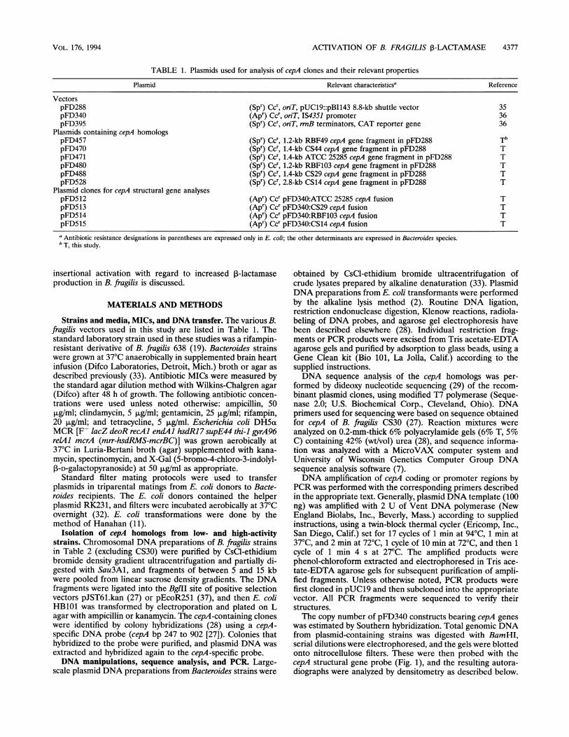
FIG. 1. Restriction map and open reading frames of cepA30H. The restriction sites and open reading frames (orf) were deduced from DNAsequence analysis of the cloned cepA gene from B. fragilis CS30 (27). The probe used for cloning additional cepA homologs is shown by the hashedbox A, and the cepA structural gene probe is shown by the hashed box B. The location of primers used for PCR amplification of structural genes
are shown by the arrowheads.
The chromosomal and plasmid copies of cepA migrate differ-ently in BamHI-digested samples; thus, it was possible toestimate a copy number from the relative hybridization inten-sities of the bands.RNA isolation from B. fragilis and Northern (RNA) blot
analysis. Total RNA was isolated by the hot phenol method ofAiba et al. (1). Briefly, chloramphenicol (to 100 p,g/ml [14])was added to Bacteroides cultures in late logarithmic phase,and the cultures were then immediately centrifuged at 4°C.The cell pellet was suspended in AE buffer (20 mM sodiumacetate [pH 5.5], 0.5% [wt/vol] sodium dodecyl sulfate [SDS],1 mM EDTA) and quickly extracted with 3 ml of phenol.Phenol (U.S. Biochemical) was equilibrated with 20 mMsodium acetate (pH 5.5) until the pH of the phenol was 5.5.Before extraction of the cell suspension, phenol was heated to65°C. Cells were extracted with phenol for S min at 65°C andcentrifuged. Phenol extraction was repeated, and the finalaqueous phase was precipitated a total of three times with 3 volof ethanol at -70°C. The final RNA pellet was dissolved indeionized formamide and stored at -70°C. Concentration was
determined by measuring A260RNA samples (5 to 50 ,ug) and size standards (0.24- to 9.5-kb
RNA ladder) were electrophoresed in large gels (25.3 by 15.1cm2 and 1 cm thick) containing 1.1% (wtlvol) agarose, 1xMOPS buffer (40 mM 3-[N-morpholino]propanesulfonic acid,10 mM sodium acetate, 1 mM EDTA [pH 7]), and 2.2 Mformaldehyde. RNA was transferred to nylon membranes(Hybond N; Amersham Corp., Arlington Heights, Ill.) bycapillary action in 1ox SSC (lx SSC is 0.15 M NaCl plus 15mM sodium citrate [pH 7]), and cross-linked by UV irradia-tion. DNA probes were labeled with [a-32P]dCTP by therandom primer reaction. Prehybridization (4 h) and overnighthybridization were performed at 50°C. Prehybridization buffercontained 50% deionized formamide, 4x SSC, Sx Denhardt'ssolution (1 x Denhardt's solution contains, per liter, 0.2 g eachof Ficoll 400, polyvinylpyrrolidone, and bovine serum albumin[Pentex fraction V; Miles Laboratories]), 50 mM Na2HPO4(pH 7.0), 0.1 mg of yeast RNA per ml, 1% SDS, and 0.5 mg ofNaPP1 per ml. Hybridization buffer was identical except Den-hardt's solution was used at 1x concentration. Nylon blotswere washed in 0.1 x SSC-0.1% SDS for 20 min each at roomtemperature, 50°C, and 65°C.Primer extension analysis of total RNA. Primer extension
analysis of total RNA using cepA-specific oligonucleotideprimers was performed as described previously (3, 28), with
slight modifications. Primers (400 pmol of 5' ends) were
labeled with [y-32P]ATP, and 105 cpm (0.045 ,uCi) of oligonu-cleotide was precipitated with total RNA (up to 50 ,ug forhigh-activity strains and 100 ,ug for low-activity strains) indiethyl pyrocarbonate-treated tubes. The resulting pellet was
dried, resuspended in hybridization buffer {80% formamide,0.4 M NaCl, 1 mM EDTA (pH 8), 40 mM PIPES [piperazine-N,N'-bis-(2-ethanesulfonic acid); pH}, incubated at 85°C for10 min, and then annealed overnight (8-12 h) at 40°C. Afterethanol precipitation and centrifugation, the pellet was dried,resuspended in 20 ,u of RT buffer (50 mM Tris [pH 7.6], 60mM KCl, 10 mM MgCl2, 1 mM each deoxynucleoside triphos-phate, 1 mM dithiothreitol, 1 U of RNasin RNase inhibitor[Promega, Madison, Wis.] per ml, 50 ,ug of actinomycin D per
ml, 50 U of Moloney murine leukemia virus reverse tran-scriptase [Gibco/BRL]), and incubated 2 h at 37°C. Reversetranscriptase was inactivated by addition of EDTA, and thenRNase A (DNase free; 5 ,ug/ml) was added to digest the RNAtemplates. The volume was brought up to 150 ,ul with Tris-EDTA, and the reaction mixtures were phenol-chloroformextracted. The supernatants were precipitated, dried, resus-
pended in 4 ,ul of formamide loading buffer, and electropho-resed on 8% polyacrylamide gels containing urea. A sequenc-ing ladder was prepared with a template covering thetranscription start site region, using the same oligonucleotidesthat were used for the reverse transcription reactions.
Densitometry analysis of autoradiographs was performedwith a Hewlett-Packard ScanJet Plus flatbed scanner inter-faced with a Macintosh Ilci computer. Collage imaging soft-ware (Fotodyne, Inc.) was used to quantitate the band inten-sities.
j-Lactamase analysis. Cell extracts for ,-lactamase activityassays and isoelectric focusing were prepared with a Frenchpressure cell (American Instrument Company, Inc., SilverSpring, Md.) in 20 mM sodium phosphate [pH 7] as describedpreviously (18). Activity was measured spectrophotometricallywith nitrocefin, and specific activity is expressed as micromolesof substrate consumed per minute per milligram of protein (17,18). Protein concentrations were determined by the method ofBradford (4).
Nucleotide sequence accession numbers. The gene designa-tions and GenBank accession numbers for the DNA sequencesof the high-activity strains are as follows: CS30, cepA30H L13472(27); RBF103, cepAl103H, U05888; and RBF49, cepA49-H,U05886. Those for the low activity strains are as follows: ATCC
:3co.,.CO)II
Eco
1.
0.
CO)II I I I I I I a I
I I
-,11 ...I j
J. BACTERIOL.
I II 500bp I

ACTIVATION OF B. FRAGILIS 1-LACTAMASE 4379
TABLE 2. ,B-Lactamase and ampicillin MICs for B. fragilis wild-type strains and pFD288 recombinant plasmids containing cepA
,3-LactamaseB. fragilis straina activity Ampicillin MIC(U/mg of (ug/ml)
protein)b
Wild typeCS29 0.004 (low) 8CS14 0.006 (low) 16638 0.007 (low) 16CS44 0.010 (low) 32ATCC 25285 0.013 (low) 32RBF49 0.110 (high) 500RBF43 0.150 (high) 500CS30 0.230 (high) 750RBF103 0.270 (high) 750
With cloned cepA genes638(pFD288) 0.009 16CePA 14-L 0.019 32CePA29-L 0.014 16CePA85-L 0.014 16CePAI03H 0.415 ± 0.15 >800
a All strains are B. fragilis sensu strictu and contain a single I-lactamase withan isoelectric point of 4.9. Cefoxitin MICs were s16 pg/ml for all strains.
b Measured in crude cell extracts with nitrocefin as a substrate. All ,B-lactamaseactivities were inhibited >50% by 1 ,uM clavulanate and 1 p,M cefoxitin.
25285, cepA85-L, U05887; CS14, cepA14-L, U05883; CS29,cepA29 L, U05884; and CS44, cepA44-L, U05885.
RESULTS
Analysis of cloned cepA genes. cepA homologs from B.fragilis strains representing the high- and low-activity classes(Table 2) were cloned in E. coli and identified by hybridizationto the cepA DNA probe shown in Fig. 1 (probe A). Severalpositive clones were obtained from each of the strains, and thecloned inserts ranged in size from 3 to 16 kb. Subsequently, thecepA regions were sequenced as described in Materials andMethods, using oligonucleotide primers derived from the CS30cepA.
Several of the cepA homologs were subcloned into theshuttle vector pFD288, transferred to B. fragilis 638, andassayed for ,B-lactam resistance (Table 2). The results showthat the low-activity class cepA homologs were on average nomore resistant to ampicillin than the parent strain 638, and the1-lactamase activities were only slightly higher than back-ground. In contrast to this, strains with the cloned cepA30H(27) or cepAlO3H (Table 2) were highly resistant to ampicillin,and the ,B-lactamase activity was 40-fold greater than seen in638 containing just the vector and no insert. As noted previ-ously (27), clones containing the high-activity cepA homologswere very unstable and yielded widely varying 1-lactamaseactivities.The DNA sequences of the cepA homologs were analyzed
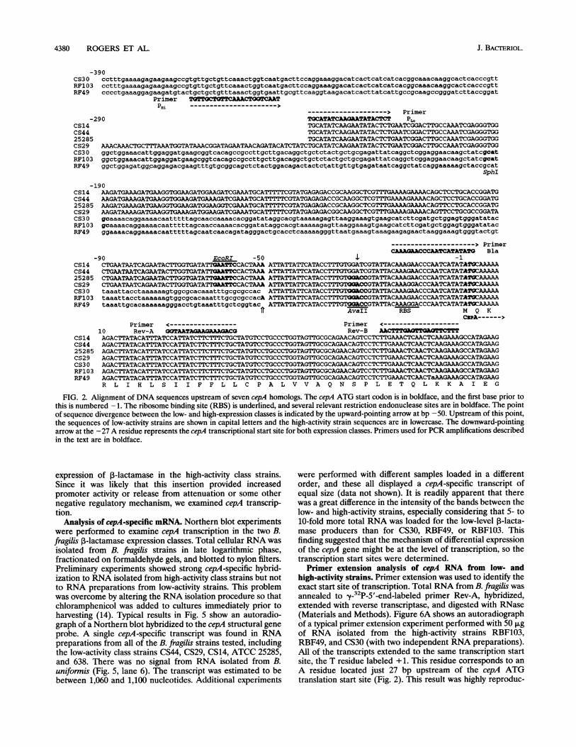
for clues to the differential expression. Examination of thesequences showed that relative to cepAS5L, there were a totalof 16 unique nucleotide changes in the seven sequences (Fig. 2and data not shown). These corresponded to eight differentamino acid substitutions, but only one (in cepA29L) was withinany of the highly conserved ,B-lactamase structural motifs.Also, in the case of cepA85-L and CePAl103H, the predictedamino acid sequences were the same. These observationssuggested that high B-lactamase activity was not the result of astructural gene point mutation leading to altered activity asexemplified by many of the TEM enzymes (12).
Expression of cepA structural genes in pFD340. Furtherexperiments on the cepA structural genes confirmed the DNAsequence findings. cepA homologs from high- and low-activitystrains were cloned into the Bacteroides expression vectorpFD340 so that ,B-lactamase activities could be compared forthese genes under control of the same promoter. The precisecloning was accomplished by PCR amplification using oligo-nucleotide primers shown in Fig. 1 and 2. DNA sequenceanalysis of the amplified fragments confirmed that no nucle-otide substitutions had occurred during the amplification pro-cedure. The pFD340 constructs were transferred by conjuga-tion into a B. fragilis 638 cepA mutant (27) for determination ofboth ampicillin MICs and ,B-lactamase specific activities (Fig.3). Generally, the cepA fusions di'splayed similar 1-lactamaseactivities and ampicillin MICs when controlled from theIS4351 promoter. One exception, the cepA29L gene fusion,consistently produced ,B-lactamase activities and ampicillinMICs about half of the values for the other cepA homologs.This finding suggested that the amino acid substitutions in theCS29 enzyme directly influenced the intrinsic activity of this1-lactamase. For the other cepA homologs, however, bothampicillin MICs and ,B-lactamase specific activities were verysimilar, and specific activities were more than 100-fold greaterthan for the pFD340 control. Isoelectric focusing gels of cellextracts from these strains all displayed a nitrocefin-reactiveprotein which focused at pH 4.9, while the 638 cepA mutantstrain containing only pFD340 did not (data not shown). Therewas some variation in the estimated copy number of thevarious pFD340 constructs (Fig. 3), but these differences ingene copy did not significantly influence the results. These datashow that the major differences in 3-lactamase activity seenbetween the two ,B-lactamase expression classes are not due toamino acid substitutions'in the cepA structural'genes and that,3-lactamase activity apparently can be influenced by promoterstrength.Comparison of upstream DNA sequences ofcepA genes from
low- and high-activity strains. Subsequent examination of thecepA DNA sequences focused on regions flanking the struc-tural gene. Downstream from the TAA translation stop codon,the nucleotide sequences for all strains were nearly identicalfor the -400 bp of available sequence data. In Fig. 2, 390 and238 bp of sequence are shown for regions upstream of theATG start site of high- and low-activity strains, respectively.Comparison of these upstream sequences yielded a notablediscovery. Exactly 50 bp (or 51 bp for RBF103) upstream of theATG translation start codon, the DNA sequences divergedcompletely between the low- and high-activity strains. All ofthe low-activity class sequences were nearly identical to eachother. The high-activity class sequences from CS30 andRBF103 were identical to each other, and RBF49, thoqgh notidentical, shared 67% homology.The nucleotide sequences for the upstream regions have
been compared against the nucleic acid and protein databases,and for the low-activity strains, no significant matches werefound. On the other hand, DNA sequence from the high-activity strains revealed an open reading frame starting at the5' end of the available-data. When this peptide was comparedagainst the databases, we observed a high degree of similarityto the insertion sequences related to IS21 (25, 26). Morespecifically, the partial open reading frames from CS30,RBF103, and RBF49 shared about 30% amino acid identitywith the istB gene product, and a comparison with IstB fromIS21 and IS5376 is shown in Fig. 4. Remarkably, RBF49, whileclearly diverged from CS30 and RBF103, shared the same levelof homology to the IS21 family. These results suggested that aninsertion 50 bp upstream from the cepA start codon activated
VOL. 176, 1994
4380 ROGERS ET AL.
CS30RF103RF49
-390
PrimerPHI
--------------------> Primer-290 TGCATATCAAG&ATATACTCT P,
CS14 TGCATATCAAGAATATACTCTGAATCGGACTTGCCAAATCGAGGGTGGCS44 TGCATATCAAGAATATACTCTGAATCGGACTTGCCAAATCGAGGGTGG25285 TGCATATCAAGAATATACTCTGAATCGGACTTGCCAAATCGAGGGTGGCS29 AAACAAACTGCTTTAAATGGTATAAACGGATAGAATAACAGATACATCTATCTGCATATCAAGAATATACTCTGAATCGGACTTGCCAAATCGAGGGTGGCS30gg0tggaaacattggaggatgaagcggtcacagccgccttgcttgacaggctgctctactgctgcgagattatcaggctcggaggaacaagctatcocatRF103 ggctggaaacattggaggatgaagcggtcacagccgccttgcttgacaggctgctctactgctgcgagattatcaggctcggaggaacaagctatcocatRF49 ggctggagatgg.aggagacgaagtttgtgcggcagctctactggacagactactctattgttgtgagataatcaggctatcaggaaaaagctaccgcat
SphI
-190CS14 AAGATGAAAGAAGAAATGCAT7TTTCGTATGAGAGACCGCAAGGCTCGTTUUAAGAAAACAGCTCCTGCACCGGATGCS44 AAGATGAAAGATGAAGGTGAAGATGAAAGATCGAAATGCATTTTTCGTATGAGAGACCGCAAGGCTCGTT¶ hAAAAGAAAACAGCTCCTGCACCGGATG25285 AAGATGAAAGATGAAGGTGGAAGATGGAAGGTCGAAATGCA?1 CGTATGAGAGACCGCAAGGCTCGTTTGAAAAGAAAACAGTTCCTGCACCGGATGCS29 TCGAAATGCATrTTTCGTATGAGAGACGGCAAGGCTCGTTTGAAAAGAAAACAGTTCCTGCGCCGGATACS30g0aaaacaggaaaacaatttttagcaaccaaaacacggatataggcacgtaaaaagagttaaggaaagtgaagcatcttcgatgctggagtgggatatacRF103 gcaaaacaggaaaacaatttttagcaaccaaaacacggatataggcacgtaaaaagagttaaggaaagtgaagcatcttcgatgctggagtgggatatacRF49 ggaaaacaggaaaacaatttttagcaatcAacagatagggactgcacctcaaaaagggttaatgaaagtaaagaagagaactaaggaaagtgggtactgt
---------------------> PrimerCAAAG&ACCCAATCATATATO Bla
CS14CS442528ECS29CS3 0RF10:RF49
CS14CS44
-90 EcoRI -50 4 -1CTGAATAATCAGAATACTTGGTGATATTI'ATTCCACTAAA ATTATTATTCATACCTTTGTGGATCGTATTACAAAGAACCCAATCATATATOCAAAAACTGAATAATCAGAATACTTGGTGATATTGkATTCCACTAAA ATTATTATTCATACCTTTGTGGATCGTATTACAAAGAACCCAATCATATA2OCAAAAA
5 CTGAATAATCAGAATACTTGGTGATATTGQATTCCACTAAA ATTATTATTCATACCTTTGTO'ACCGTATTACAAAGAACCCAATCATATATOCAAAAACTGAATAATCAGAATACTTGGTGATATTGQATTCCACTAAA ATTATTATTCATACCTTTGTQGQCCGTATrACAAAGGACCCAATCATATAIOCAAAAAtaaattacctaaaaaagtggcgcacaaatttgcgcgccac ATTATTATTCATACCTTTGTGOACCGTATTACAAAGAACCCAATCATATA2rAAAAA
3 taaattacctaaaaaagtggcgcacaaatttgcgcgccacA ATTATTATTCATACCTTTGTOGQCCGTAT1ACAAAGAACCCAATCATATA2ISAAAAAtaaattgcacaaaaaagggacctgtaaatttgctcggtac ATTATTATTCATACCTTITGTOGQCCGTATTACAAAGGACCCAATCATATAT2CAAAAA
fr AvaII RBS M Q KCsPA------>
Primer <----------------- Primer <-------------------10 Rev-A GQTAATAGaAG AAGAC Rev-B AACTTTKAOTTYAGMTCTTTAGACTTATACATTTATCCATTATCTTCTTTCTGCTATGTCCTGCCCTGGTAGTTGCGCAGAACAGTCCTCTTGAAACTCAACTCAAGAAAGCCATAGAAGAGACTTATACATTTATCCATTATCTTCTTTCTGCTAT TCCTGCCCTGGTAGTTGCGCAGAACAGTCCTCTTGAAACTCAACTCAAGAAAGCCATAGAAG
25285 AGACTTATACATTTATCCATTATCTTCTTTCTGCTATGTCCTGCCCTGGTAGTTGCGCAGAACAGTCCTCTTGAAACTCAACTCAAGAAAGCCATAGAAGCS29 AGACTTATACATITATCCATTATCTTCTTTCTGCTATGTCCTGCCCTGGTAGTTGCGCAGAACAGTCCTCTTGAAACTCAACTCAAGAAAGCCATAGAAGCS30 AGACTTATACATTTATCCATTATCTTCTTTCTGCTATGTCCTGCCCTGGTAGTTGCGCAGAACAGTCCTCTTGAAACTCAACTCAAGAAAGCCATAGAAGRF103 AGACTTATACATTTATCCATTATCTTCTTTCTGCTATGTCCTGCCCTGGTAGTTGCGCAGAACAGTCCTCTTGAAACTCAACTCAAGAAAGCCATAGAAGRF49 AGACTTATACATTTATCCATTATCTTCTTTCTGCTATGTCCTGCCCTGGTAGTTGCGCAGAACAGTCCTCTTGAAACTCAACTaAAGAAAGCCATAGAAG
R L I H L S I I F F L L C P A L V V A Q N S P L E T Q L K K A I E G
FIG. 2. Alignment of DNA sequences upstream of seven cepA homologs. The cepA ATG start codon is in boldface, and the first base prior tothis is numbered -1. The ribosome binding site (RBS) is underlined, and several relevant restriction endonuclease sites are in boldface. The pointof sequence divergence between the low- and high-expression classes is indicated by the upward-pointing arrow at bp -50. Upstream of this point,the sequences of low-activity strains are shown in capital letters and the high-activity strain sequences are in lowercase. The downward-pointingarrow at the -27 A residue represents the cepA transcriptional start site for both expression classes. Primers used for PCR amplifications describedin the text are in boldface.
expression of ,B-lactamase in the high-activity class strains.Since it was likely that this insertion provided increasedpromoter activity or release from attenuation or some othernegative regulatory mechanism, we examined cepA transcrip-tion.
Analysis of cepA-specific mRNA. Northern blot experimentswere performed to examine cepA transcription in the two B.fragilis 3-lactamase expression classes. Total cellular RNA was
isolated from B. fragilis strains in late logarithmic phase,fractionated on formaldehyde gels, and blotted to nylon filters.Preliminary experiments showed strong cepA-specific hybrid-ization to RNA isolated from high-activity class strains but notto RNA preparations from low-activity strains. This problemwas overcome by altering the RNA isolation procedure so thatchloramphenicol was added to cultures immediately prior toharvesting (14). Typical results in Fig. 5 show an autoradio-graph of a Northern blot hybridized to the cepA structural geneprobe. A single cepA-specific transcript was found in RNApreparations from all of the B. fragilis strains tested, includingthe low-activity class strains CS44, CS29, CS14, ATCC 25285,and 638. There was no signal from RNA isolated from B.uniformis (Fig. 5, lane 6). The transcript was estimated to bebetween 1,060 and 1,100 nucleotides. Additional experiments
were performed with different samples loaded in a differentorder, and these all displayed a cepA-specific transcript ofequal size (data not shown). It is readily apparent that therewas a great difference in the intensity of the bands between thelow- and high-activity strains, especially considering that 5- to10-fold more total RNA was loaded for the low-level 3-lacta-mase producers than for CS30, RBF49, or RBF103. Thisfinding suggested that the mechanism of differential expressionof the cepA gene might be at the level of transcription, so thetranscription start sites were determined.Primer extension analysis of cepA RNA from low- and
high-activity strains. Primer extension was used to identify theexact start site of transcription. Total RNA from B. fragilis wasannealed to _y-32P-5'-end-labeled primer Rev-A, hybridized,extended with reverse transcriptase, and digested with RNase(Materials and Methods). Figure 6A shows an autoradiographof a typical primer extension experiment performed with 50 ,ugof RNA isolated from the high-activity strains RBF103,RBF49, and CS30 (with two independent RNA preparations).All of the transcripts extended to the same transcription startsite, the T residue labeled +1. This residue corresponds to anA residue located just 27 bp upstream of the cepA ATGtranslation start site (Fig. 2). This result was highly reproduc-
cctttgaaaagagaagaagccgtgttgctgttcaaactggtcaatgacttccaggaaaggacatcactcatcatcacggcaaacaaggcactcacccgttcccctgaaagga4aagatgtactgctgctgtttaaactggtgaattgcgttcaaggtaagacatcacttatcattgccgcaagccgggatcttaccggat
J. BACTERIOL.
_>

ACTIVATION OF B. FRAGILIS P-LACTAMASE 4381
Aatli EcoRI pFD340Xbel MCSICS
P31143 , IS4351 PUCI9(rtp) % promoter (rep)
:
%
cepA insert Copy No. P-lactamase activity
NONE - <0.001
CS14
CS29
25285
RBF1 03
7.9
7.7
10.0
11.1
0.138
0.084
0.180
0.172
FIG. 3. 1-Lactamase activities and ampicillin MICs forpFD340::cepA constructs in a B. fragilis cepA mutant. The restrictionmap of the expression vector pFD340 shows the cloning strategy forthe cepA structural gene inserts. The ampicillin MICs were determinedafter growth for 48 h at 37°C on Wilkins-Chalgren agar containingampicillin. ,-Lactamase specific activities are averages of at least twoexperiments performed in triplicate.
ible, and control reactions with just the primer or RNAtemplate yielded no reaction products. The experiments wererepeated with RNA prepared from low-activity strains, using100 ,ug of RNA in each reaction, and the results are shown inFig. 6B. The low-activity class cepA-specific transcripts (lanes 2to 6) were extended to the same residue as the RBF103transcript (lane 1), corresponding to cepA bp -27. Resultsobtained using the Rev-A primer, which annealed 25 bpdownstream of the ATG translation start codon, were con-firmed by using a different primer, Rev-B, which annealed 79bp downstream of the ATG codon.
In addition, the amount of radiolabeled primer used in theextension reactions was in excess; thus, it was possible toquantitate the total amount of cepA-specific message in thesamples and compare them with each other (28). Hybridizationintensities from the Rev-A primer autoradiographs over a
I821RBF49RBF1O3I85376
181 230PMNR&SLF FRLLNRRYUK ASIILTSNKG FADWGIMFGD HVLATAILDRPLKGEDVLLL FKLVNCVQGK T8LIIAASRD LTGWLZMAGD EVCAAALLDRPLKRUNAVLL FKLVNDFQNR TSLIITANKA LTRWLNTLED EAVTAALLDRKLDPNSAHYL FQVIARRYIH APIILTSNXS FGEWGIIVGD SVLATAMLDR
Cons PL--EEA-LL F-LVN---E- -S-I-T-NK- ---W-E--GD -V-A-ALLDR
IS21RBF49RBF1O3IS5376
231 280LLHHSTTLNI KGE8YRLKEK RKAGVLTKNT TPISDDEMVK SGQHQ.....LLYCCEIIRL SGKSYRMENR KTIFSNQQIG TAPQKGLMKV KKRTKESGYCLLYCCEIIRL GGTBYRMQNR KTIFSNQNTD IGT*...............LLHHSIIFNL KGESYRLREK RLQEEKQKDQ *...................
Cons LL----I--L -G-SYR---- ------Q--- ---------- ----------
FIG. 4. Alignment of sequences of the IstB proteins from IS21 andIS5376 with sequences from B. fragilis RBF103 and RBF49. Aminoacid sequence from the C terminus of two IstB proteins was obtainedfrom GenBank and aligned with the protein coding sequences foundupstream of cepA in RBF103 and RBF49. Sequences were aligned byusing the Genetics Computer Group Pileup program, and a three-of-four consensus (Cons) is presented below the aligned sequences.Residues contributing to the consensus are in boldface.
kb 1 2 3 4 5 6 7 8 9
4.40 _
2.3 7
1.35 -
,....~~~~~~~~~~~~~~~~~.w.
0.24 -
FIG. 5. Autoradiograph of a Northern blot of total RNA fromBacteroides strains. The blot was hybridized to an a-32P-labeled DNAprobe encompassing the cepA85-L structural gene. Exposure was for 48h at -70°C. Sources and amounts of total RNA loaded per lane are asfollows: 1, RBF103, 10 ,Ig; 2, RBF49, 10 p.g; 3, CS44, 50 jig; 4, CS29,50 ,ug; 5, CS14, 50 jig; 6, B. uniformis 1001, 50 jig; 7, ATCC 25285, 50jig; 8, 638, 50 jig; and 9, CS30, 5 jig. Size markers from a 0.24- to 9.5-kbRNA ladder are indicated by the arrows. The size of the cepA-specifictranscript is approximately 1,100 nucleotides.
range of exposures were quantitated by measuring pixel inten-sity of a scanned image. The results of these analyses are listedin Table 3. Assuming that the starting amount of total RNAwas 100 ,ug per reaction (50 ,ug for the RBF103 reaction), itwas concluded that the cepA 103-H transcripts were 40-fold moreabundant than the cepA44-L transcripts, 35-fold more abundantthan the 638 cepA transcripts, 25-fold more abundant than thecepAl4-L transcripts, 18-fold more abundant than the cepA85Ltranscripts, and 13-fold more abundant than the cepA29-Ltranscripts.cepA promoter activity in the CAT fusion vector pFD395.
Another approach to look at differences in cepA transcriptionwas to clone the low- and high-activity promoters into a vectorwith the chloramphenicol acetyltransferase (CAT) reportergene. PCR amplification with primers PHi and PL,O (Fig. 2) wasused to isolate the promoter regions, and these were clonedinto pFD395. The resulting plasmids were transferred into B.fragilis 638 and tested for CAT activity. Transconjugants frommatings with plasmid containing the PLO constructs were foundto have little or no CAT above the background levels measuredfor B. fragilis 638 containing just the vector (data not shown).These results were similar to the low levels of 1-lactamaseactivity seen with the cloned low-level cepA homologs (Table2).
In contrast to the P,O results, no results were obtained withpFD395 constructs bearing the PHi regions. These recombi-nant plasmids failed to transfer from E. coli to B. fragilis 638.The infrequent transconjugants that were obtained had beendeleted for the cloned PH, DNA. A modified version ofpFD395, pFD551, containing the trp terminator inserted down-stream of the cat gene, was tested for cloning these promoters,but this vector also failed to yield transconjugants with intact
VOL. 176, 1994

4382 ROGERS ET AL.
G A T C 1 2 3 4som
It
af
_F 0 -o_m
am
Ag
ft
_g_
al
ow
47_Rt.t
IO,.
B,
3 5A TATATC GA TC GC G
-_*--+1 T AG CG CC GA TT AA T\A T5 3
G A T C
I1t
Xl :
d'o46
tp6
as
1 23456
FIG. 6. Autoradiographs of primer extension reactions with total RNA prepared from high- and low-activity strains. Lanes G, A, T, and Ccorrespond to the DNA sequencing reactions prepared using the same Rev-A primer (see Fig. 2) as used for the primer extension reactions. (A)Primers were extended with reverse transcriptase after hybridization to 50 ,ug of total RNA. The entire contents of each reaction were
electrophoresed in each lane, and the gel was exposed for 24 h at room temperature. The sources of starting RNAs for the numbered lanes are
as follows: 1, RBF103; 2, CS30; 3, CS30 prepared separately from that of lane 2; and 4, RBF49. The arrow points to the T residue correspondingto the 5' end of the cepA mRNA transcript, which is in boldface, and corresponds to the A residue at bp -27 (Fig. 2). (B) The sources and amountsof starting RNA per reaction for the numbered lanes are as follows: 1, RBF103, 50 jig; 2, ATCC 25285, 100 ,ug; 3, CS29, 100 ,ug; 4, 638, 100 ,ug;5, CS14, 100 ,ug; and 6, CS44, 100 ,ug. This gel was exposed for 71.6 h, but all other conditions were as described above.
plasmids. The PHi regions proved to be deleterious to B. fragilis(but not E. coli) when cloned into the standard shuttle/cloningvector pFD288. This was shown by cloning the cepA30H andcepA103-H promoter regions into the multiple cloning site ofpFD288 and mating these constructs with 638. As seen with theCAT fusions, transconjugants with intact plasmids were notobtained.
DISCUSSIONWe have begun to elucidate the basis for the increased
,B-lactamase activity in B. fragilis CS30, RBF103, and RBF49.Previously, results obtained by Southern hybridizations indi-cated that high levels of activity were not due to an increase ingene copy number, but the two expression classes could bedifferentiated on the basis of their hybridization patterns (27).Also, the increased specific activities are not due to thepresence of additional 3-lactamases in these strains, as onlyone nitrocefin-reactive band can be detected in cell extracts onisoelectric focusing or renatured SDS-polyacrylamide gels(27). Comparison of the DNA sequences upstream of the cepAgenes revealed that in high-activity strains an insertion had
TABLE 3. Quantitation of cepA-specific transcripts from primerextension analysis of RNAs from B. fragilis strainse
Pixel ~~~~RelativeRNA source (pg) Pixel Rltvintensity amt"
RBF103 (50) 9,355,120 39.6CS29 (100) 1,433,520 3.0ATCC 25285 (100) 1,040,640 2.2CS14 (100) 754,960 1.6638 (100) 534,032 1.1CS44 (100) 472,976 1.0
a Quantitation was determined by counting pixel intensity of a scanned imageof the autoradiograph from Fig. 6B.
b Calculated by comparison of pixel intensity values with the lowest value, thatof CS44. Analysis of other exposures of the same autoradiograph gave compa-rable results.
occurred 50 bp (or 51 bp for RBF103) before the ATGtranslation start site (Fig. 2). This insertion event was found tobe responsible for the different hybridization patterns of thetwo classes. Analysis of the insertions showed a similarity to theistB gene of insertion sequence (IS) elements in the IS21 family(Fig. 4).The discovery of an IS-like element upstream of cepA
suggests several possible mechanisms for the up-regulation ofexpression: (i) there may be strong outward-directed promot-ers present on the IS21-like element, and these promotehigh-level cepA transcription; (ii) the insertion event couldhave created a stronger promoter for cepA transcription byproviding new -35 sequences; (iii) the insertion event dis-places or disrupts a regulatory region, allowing increased cepAtranscription; and (iv) any combination of these mechanisms.The importance of gene activation by IS elements is beingrecognized more and more, and there are examples of theseactivation mechanisms in the literature. The classic example ofactivation of prokaryotic gene transcription by promoterspresent on IS elements is the cryptic bgl operon of E. coli,which encodes enzymes required for 3-glucoside utilization(30). In Bacteroides species, IS elements have been implicatedin the activation of antibiotic resistance genes. These includethe activation of ermF by IS4351 in Tn4351 on pBF4 plasmid(23, 24) and activation of ermFS by IS4351 in the oppositeorientation in Tn4551 on pBI136 (34). In addition, a 1,598-bpIS element, IS942, has been shown to have integrated 19 bpupstream of the proposed initiation codon for ccrA, the geneencoding the class B metallo-,B-lactamase of B. fragilisTAL3636 (22). However, in none of these Bacteroides exam-
ples has the mechanism of activation been established.IS21 has also been shown to be a source of mobile promot-
ers; one active promoter has been localized to within 170 bp ofthe left end of IS21, and the other promoter located closer tothe center of IS21 reads into the element (31). IS2 has beenshown to form a new promoter upon insertion into target DNAand to up-regulate transcription of the E. coli ampC 1-lacta-mase gene 20-fold (13). The -10 region from the original
A
J. BAcrERIOL.

ACTIVATION OF B. FRAGILIS 3-LACTAMASE 4383
ampC promoter was retained in these insertion mutants, butthe -35 region(s) wasIS2 derived. Importantly, the transcrip-tion initiation site in the ampC:JS2 mutant was identical withthat of the wild-type ampC promoter (13). In E. coli HB251, a
116-bp insert with similarity to IS1 was found to have insertedin the native promoter region of the blaT-6 gene encodingTEM-6 ,-lactamase. The original -10 region was retained,and the new -35 sequences were provided by the ISl-likeelement, increasing activity of the promoter 10-fold (10).These latter examples are analogous to the situation ob-
served with the IS21-like element present upstream of cepA inthe high-activity strains. This insertion does not alter thelocation of the cepA transcription start site from that observedfor low-activity 3-lactamase strains. However, one needs tokeep in mind the differences between E. coli and B. fragilis. Wehave shown previously that the consensus -10 and -35regions of E. coli promoters are not recognized as such by B.fragilis transcriptional machinery (36). It is possible that Bac-teroides genes contain their own versions of -10 and -35recognition sequences, of which the latter has been disruptedand perhaps altered by the IS21-like element. Regardless ofthe specific structures required of a Bacteroides promoter,these hybrid promoters from the high-p-lactamase-activitystrains are very strong, resulting in up to 40-fold-higher levelsof cepA message. It does not seem likely that message stabilityplays a significant role in differential regulation of cepAbecause the transcription start sites were the same and nucle-otide sequences downstream of cepA were nearly identical forboth expression classes.The results presented above strongly support a role for the
ISs in transcriptional activation of cepA. However, there isevidence that the insertion also may have disrupted normalcepA regulation. This evidence stems from the first experi-ments, in which the increase in P-lactamase activity observedfor the subcloned low-activity cepA genes was not consistentwith the high copy number of the vector (Table 2). Thisobservation was confirmed latter by the lack of detectable CATactivity in B. fragilis 638 extracts containing the pFD395:1ow-activity class cepA promoter constructs. We had expected tosee low-level CAT activity, similar to the relatively low amountof,-lactamase activity seen in wild-type B. fragilis 638 or 25285cells, especially since these plasmids are present in copy
numbers greater than one. It is possible that some form ofnegative regulation maintains the expression of cepA at a very
low level and that the insertion disrupts a repressor binding siteand provides an improved promoter structure, leading to the20- to 60-fold increases in ,-lactamase activity.
ACKNOWLEDGMENTS
We thank J. Coleman and A. Sage for helpful advice and discus-sions.
This work was supported by Public Health Service grant AI-28884and an undergraduate fellowship from the Burroughs Wellcomecompany to C.M.P.
REFERENCES1. Aiba, H., S. Adhya, and B. deCrombrugghe. 1981. Evidence for
two functional gal promoters in intact Escherichia coli cells. J. Biol.Chem. 256:11905-11910.
2. Birnboim, H. C., and J. Doly. 1979. A rapid alkaline extractionprocedure for screening recombinant plasmid DNA. NucleicAcids Res. 7:1513-1523.
3. Boorstein, W. R., and E. A. Craig. 1989. Primer extension analysisof RNA. Methods Enzymol. 180:347-369.
4. Bradford, M. M. 1976. A rapid and sensitive method for quanti-tation of microgram quantities of protein utilizing the principle ofprotein-dye binding. Anal. Biochem. 72:248-254.
5. Britz, M. L., and R. G. Wilkinson. 1978. Purification and proper-ties of a beta-lactamase from Bacteroides fragilis. Antimicrob.Agents Chemother. 13:373-382.
6. Cornick, N. A., G. J. Cuchural, D. R. Snydman, N. V. Jacobus, P.lannini, G. Hill, T. Cleary, J. P. O'Keefe, C. Pierson, and S. M.Finegold. 1990. The antimicrobial susceptibility patterns of theBacteroidesfragilis group in the United States, 1987. J. Antimicrob.Chemother. 25:1011-1019.
7. Devereux, J., P. Haeberli, and 0. Smithies. 1984. A comprehensiveset of sequence analysis programs for the VAX. Nucleic AcidsRes. 12:387-395.
8. Edwards, R., and D. Greenwood. 1992. An investigation of f-lac-tamases from clinical isolates of Bacteroides species. J. Med.Microbiol. 36:89-95.
9. Eley, A., and D. Greenwood. 1986. Characterization of P-lactama-ses in clinical isolates of Bacteroides. J. Antimicrob. Chemother.18:325-333.
10. Goussard, S., W. Sougakoff, C. Mabilat, A. Bauernfeind, and P.Courvalin. 1991. An IS1-like element is responsible for high-levelsynthesis of extended-spectrum 1-lactamase TEM-6 in Enterobac-teriaceae. J. Gen. Microbiol. 137:2681-2687.
11. Hanahan, D. 1983. Studies on the transformation of Escherichiacoli with plasmids. J. Mol. Biol. 166:557-580.
12. Jacoby, F. A., and A. A. Medeiros. 1991. More extended-spectrum,B-lactamases. Antimicrob. Agents Chemother. 35:1697-1704.
13. Jaurin, B., and S. Normark. 1983. Insertion of IS2 creates a novelampC promoter in Escherichia coli. Cell 32:809-816.
14. Liao, H. H., and J. C. Rabinowitz. 1980. Clostridial apoferredoxinmessenger ribonucleic acid assay and partial purification. Biochim.Biophys. Acta 608:301-314.
15. Nord, C. E., and M. Hedberg. 1990. Resistance to P-lactamantibiotics in anaerobic bacteria. Rev. Infect. Dis. 12(Suppl.2):S231-S234.
16. Nord, C. E., and B. Olsson-Liljequist. 1981. Resistance to P-lac-tam antibiotics in Bacteroides species. J. Antimicrob. Chemother.8(Suppl. D):33-42.
17. O'Callaghan, C. H., A. Morris, S. M. Kirby, and A. H. Shingler.1972. Novel method for detection of ,3-lactamases by using achromogenic cephalosporin substrate. Antimicrob. Agents Che-mother. 1:283-288.
18. Parker, A. C., and C. J. Smith. 1993. Genetic and biochemicalanalysis of a novel Ambler class A ,B-lactamase responsible forcefoxitin resistance in Bacteroides species. Antimicrob. AgentsChemother. 37:1028-1036.
19. Privitera, G., A. Dublanchet, and M. Sebald. 1979. Transfer ofmultiple antibiotic resistance between subspecies of Bacteroidesfragilis. J. Infect. Dis. 139:97-101.
20. Rasmussen, B. A., K. Bush, and F. P. Tally. 1993. Antimicrobialresistance in Bacteroides. Clin. Infect. Dis. 16(Suppl. 4):S390-S400.
21. Rasmussen, B. A., Y. Gluzman, and F. P. Tally. 1990. Cloning andsequencing of the class B 3-lactamase gene (ccrA) from Bacteroidesfragilis TAL3636. Antimicrob. Agents Chemother. 34:1590-1592.
22. Rasmussen, B. A., and E. Kovacs. 1991. Identification and DNAsequence of a new Bacteroides fragilis insertion sequence-likeelement. Plasmid 25:141-144.
23. Rasmussen, J. L., D. A. Odelson, and F. L. Macrina. 1986.Complete nucleotide sequence and transcription of ermF, a mac-rolide-lincosamide-streptogramin B resistance determinant fromBacteroides fragilis. J. Bacteriol. 168:523-533.
24. Rasmussen, J. L., D. A. Odelson, and F. L. Macrina. 1987.Complete nucleotide sequence of insertion element IS4351 fromBacteroides fragilis. J. Bacteriol. 169:3573-3580.
25. Reimmann, C., and D. Haas. 1990. The istA gene of insertionsequence IS21 is essential for cleavage at the inner 3' ends oftandemly repeated IS21 elements in vitro. EMBO J. 9:4055-4063.
26. Reimmann, C., R. Moore, S. Little, A. Savioz, N. S. Willetts, andD. Haas. 1989. Genetic structure, function and regulation of thetransposable element IS21. Mol. Gen. Genet. 215:416-424.
27. Rogers, M. B., A. C. Parker, and C. J. Smith. 1993. Cloning andcharacterization of the endogenous cephalosporinase gene, cepA,from Bacteroidesfragilis reveals a new subgroup of Ambler class A,B-lactamases. Antimicrob. Agents Chemother. 37:2391-2400.
28. Sambrook, J., E. F. Fritsch, and T. Maniatis. 1989. Molecular
VOL. 176, 1994

4384 ROGERS ET AL.
cloning: a laboratory manual. Cold Spring Harbor LaboratoryPress, Cold Spring Harbor, N.Y.
29. Sanger, F., S. Nicklen, and A. R Coulson. 1977. DNA sequencingwith chain-terminating inhibitors. Proc. Natl. Acad. Sci. USA74:5463-5467.
30. Schnetz, K, and B. Rak 1992. IS5: a mobile enhancer of tran-scription in Escherichia coli. Proc. Natl. Acad. Sci. USA 89:1244-1248.
31. Schurter, W., and B. W. Holloway. 1986. Genetic analysis ofpromoters on the insertion sequence IS21 of plasmid R68.45.Plasmid 15:8-18.
32. Shoemaker, N. B., C. Getty, J. F. Gardner, and A. A. Salyers. 1986.Tn4351 transposes in Bacteroides spp. and mediates the integra-tion of plasmid R751 into the Bacteroides chromosome. J. Bacte-riol. 165:929-936.
33. Smith, C. J. 1985. Characterization of Bacteroides ovatus plasmid
pBI136 and structure of its clindamycin resistance region. J.Bacteriol. 161:1069-1073.
34. Smith, C. J. 1987. Nucleotide sequence analysis of Tn4551: use ofermFS operon fusions to detect promoter activity in Bacteroidesfragilis. J. Bacteriol. 169:4589-4596.
35. Smith, C. J. 1989. Clindamycin resistance and the development ofgenetic systems in the Bacteroides. Dev. Ind. Microbiol. 30:23-33.
36. Smith, C. J., M. B. Rogers, and M. L, McKee. 1992. Heterologousgene expression in Bacteroides fragilis. Plasmid 27:141-154.
37. Southern, J. A., J. R. Parker, and D. R Woods. 1986. Expressionand purification of glutamine synthetase cloned from Bacteroidesfragilis. J. Gen. Microbiol. 132:2827-2835.
38. Thompson, J. S., and M. H. Malamy. 1990. Sequencing the genefor an imipenem-cefoxitin-hydrolyzing enzyme (CfiA) from Bac-teroides fragilis TAL2480 reveals strong similarity between CfiAand Bacillus cereus ,-lactamase II. J. Bacteriol. 172:2584-2593.
J. BACTERIOL.