images and case reports in arrhythmia and electrophysiology
TRANSCRIPT
Images and Case Reports in Arrhythmiaand Electrophysiology
Ablation of Ventricular Tachycardia in Chronic ChagasicCardiomyopathy With Giant Basal Aneurysm
Carto Sound, CT, and MRI Merge
Bruno P. Valdigem, MD; Fabio B.F.C.G. Pereira, MD; Nilton J. Carneiro da Silva, MD;Cristiano O. Dietrich, MD; Ricardo Sobral, MD; Fernando Lopes Nogueira, MD;
Roberto C. Berber, MD; Fabricio Mallman, MD; Ibraim M. Pinto, MD; Gilberto Szarf, MD;Claudio Cirenza, MD, PhD; Angelo A.V. de Paola, MD, PhD
Chronic chagasic cardiomyopathy (CCC) is a parasiticdisease that presents with life-threatening ventricular
arrhythmias, dilated cardiomyopathy, or sudden death. Basaland posterior wall motion abnormalities and left apicalaneurysms are common.
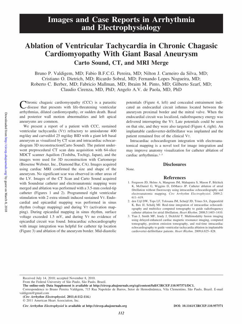
We present a report of a patient with CCC, sustainedventricular tachycardia (VT) refractory to amiodarone 400mg/day and carvedilol 25 mg/day BID with a giant left basalaneurysm as visualized by CT scan and intracardiac echocar-diogram 3D reconstruction(Carto Sound). The patient under-went preprocedural CT scan data acquisition with 64-sliceMDCT scanner Aquilion (Toshiba, Tochigi, Japan), and theimages were used for 3D reconstruction with Cartomerge(Biosense Webster, Inc., Diamond Bar, CA). Images acquiredusing cardiac MRI confirmed the size and shape of theaneurysm. No significant scar was observed in other areas ofthe LV. Images of the CT Scan and Carto Sound acquiredwith Soundstar catheter and electroanatomic mapping weremerged and ablation was performed with a 3.5-mm cooled-tipcatheter (Figures 1 and 2). Programmed right ventricularstimulation with 2 extra stimuli induced sustained VT. Endo-cardial and epicardial mapping was performed in sinusrhythm (voltage mapping) and during VT (activation map-ping). During epicardial mapping in sinus rhythm, surfacevoltage exceeded 1.5 mV, and during VT no evidence ofepicardial circuit was found. Intracardiac echocardiographywith image integration was helpful for catheter tip location(Figure 3) and ablation of the aneurysm border. Mid-diastolic
potentials (Figure 4, left) and concealed entrainment indi-cated an endocardial circuit isthmus located between theaneurysm proximal border and the mitral valve. When theendocardial circuit was localized, radiofrequency energy wasdelivered interrupting the VT. Late potentials could be seenon that site, and they were also targeted (Figure 4, right). Animplantable cardioverter-defibrillator was implanted and thepatient remained free of the clinical VT.
Intracardiac echocardiogram integration with electroana-tomical mapping is a novel tool for image integration andmay improve anatomy visualization for catheter ablation ofcardiac arrhythmias.1–3
DisclosuresNone.
References1. Ferguson JD, Helms A, Mangrum JM, Mahapatra S, Mason P, Bilchick
K, McDaniel G, Wiggins D, DiMarco JP. Catheter ablation of atrialfibrillation without fluoroscopy using intracardiac echocardiography andelectroanatomic mapping. Circ Arrhythm Electrophysiol. 2009;2:611–619.
2. den Uijl DW, Tops LF, Tolosana JM, Schuijf JD, Trines SA, ZeppenfeldK, Bax JJ, Schalij MJ. Real-time integration of intracardiac echocardi-ography and multislice computed tomography to guide radiofrequencycatheter ablation for atrial fibrillation. Heart Rhythm. 2008;5:1403–1410.
3. Tian J, Smith MF, Jeudy J, Dickfeld T. Multimodality fusion imagingusing delayed-enhanced cardiac magnetic resonance imaging, computedtomography, positron emission tomography, and real-time intracardiacechocardiography to guide ventricular tachycardia ablation in implantablecardioverter-defibrillator patients. Heart Rhythm. 2009;6:825–828.
Received July 14, 2010; accepted November 8, 2010.From the Federal University of Sao Paulo, Sao Paulo, Brazil.The online-only Data Supplement is available at http://circep.ahajournals.org/cgi/content/full/CIRCEP.110.957571/DC1.Correspondence to Bruno Pereira Valdigem, 715 Rua Napoleao de Barros, Setor de Hemodinamica, Vila Clementino, Sao Paulo, Brazil. E-mail
[email protected](Circ Arrhythm Electrophysiol. 2011;4:112-114.)© 2011 American Heart Association, Inc.
Circ Arrhythm Electrophysiol is available at http://circep.ahajournals.org DOI: 10.1161/CIRCEP.110.957571
112
by guest on March 8, 2018
http://circep.ahajournals.org/D
ownloaded from
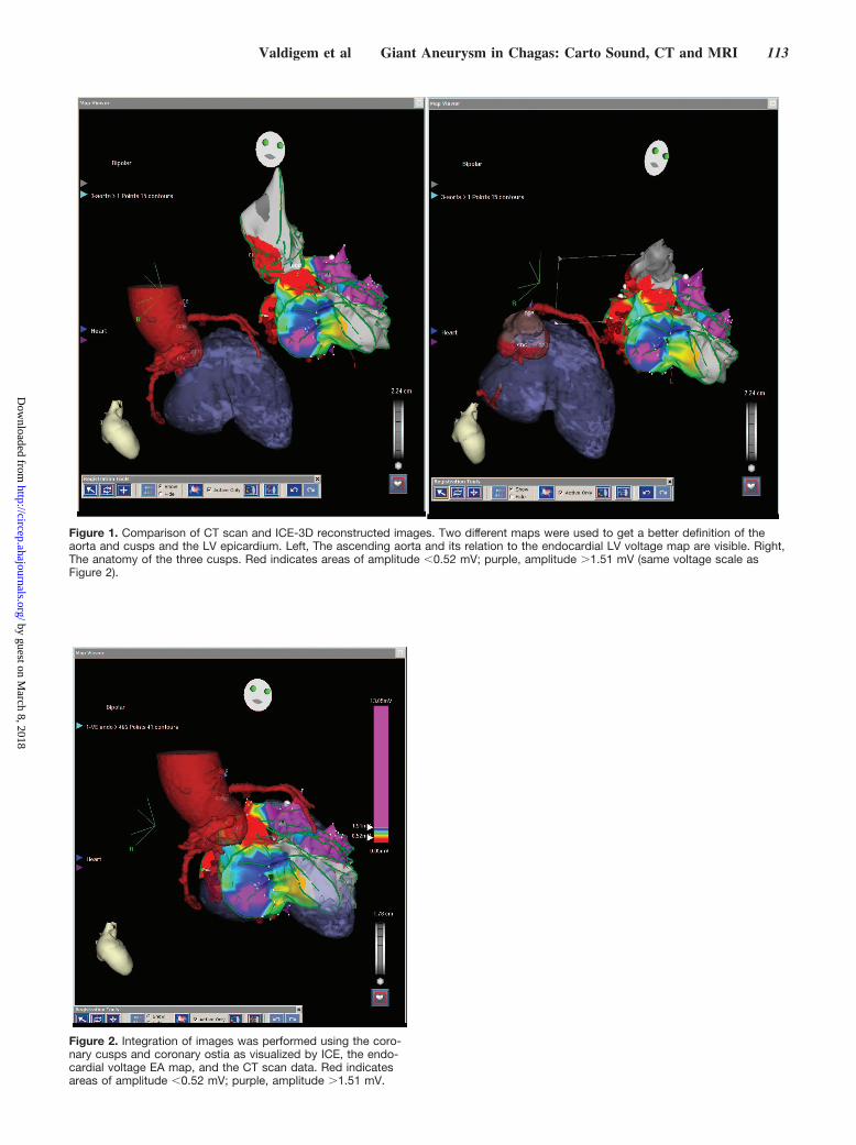
Figure 1. Comparison of CT scan and ICE-3D reconstructed images. Two different maps were used to get a better definition of theaorta and cusps and the LV epicardium. Left, The ascending aorta and its relation to the endocardial LV voltage map are visible. Right,The anatomy of the three cusps. Red indicates areas of amplitude �0.52 mV; purple, amplitude �1.51 mV (same voltage scale asFigure 2).
Figure 2. Integration of images was performed using the coro-nary cusps and coronary ostia as visualized by ICE, the endo-cardial voltage EA map, and the CT scan data. Red indicatesareas of amplitude �0.52 mV; purple, amplitude �1.51 mV.
Valdigem et al Giant Aneurysm in Chagas: Carto Sound, CT and MRI 113
by guest on March 8, 2018
http://circep.ahajournals.org/D
ownloaded from

Figure 3. Images were acquired in real time during ablation. Left, A view with only ICE. Right, An LV endocardial voltage map is com-bined with CT scan data reconstruction. The catheter tip is near the mitral valve, and the aneurysm borders are clearly visible, as wellas the distance between the proximal and the distal borders of the aneurysm. Red indicates areas of amplitude �0.52 mV; purple,amplitude �1.51 mV.
Figure 4. Left, Clinical VT induced and nid-diastolic potentials. Middle, ICE-3D endocardial LV activation map in sinus rhythm with suc-cessful ablation target highlighted. Right, VT termination during ablation in a site with late fragmented potentials. The dashed red circleindicates the catheter tip position during the events described in the left and right panels (mid-diastolic potentials, VT termination duringablation, and late potentials).
114 Circ Arrhythm Electrophysiol February 2011
by guest on March 8, 2018
http://circep.ahajournals.org/D
ownloaded from
Pinto, Gilberto Szarf, Claudio Cirenza and Angelo A.V. de PaolaRicardo Sobral, Fernando Lopes Nogueira, Roberto C. Berber, Fabricio Mallman, Ibraim M.
Bruno P. Valdigem, Fabio B.F.C.G. Pereira, Nilton J. Carneiro da Silva, Cristiano O. Dietrich,Basal Aneurysm: Carto Sound, CT, and MRI Merge
Ablation of Ventricular Tachycardia in Chronic Chagasic Cardiomyopathy With Giant
Print ISSN: 1941-3149. Online ISSN: 1941-3084 Copyright © 2011 American Heart Association, Inc. All rights reserved.
Avenue, Dallas, TX 75231is published by the American Heart Association, 7272 GreenvilleCirculation: Arrhythmia and Electrophysiology
doi: 10.1161/CIRCEP.110.9575712011;4:112-114Circ Arrhythm Electrophysiol.
http://circep.ahajournals.org/content/4/1/112World Wide Web at:
The online version of this article, along with updated information and services, is located on the
http://circep.ahajournals.org//subscriptions/
is online at: Circulation: Arrhythmia and Electrophysiology Information about subscribing to Subscriptions:
http://www.lww.com/reprints Information about reprints can be found online at: Reprints:
document. Answer
Permissions and Rights Question andunder Services. Further information about this process is available in thepermission is being requested is located, click Request Permissions in the middle column of the Web pageClearance Center, not the Editorial Office. Once the online version of the published article for which
can be obtained via RightsLink, a service of the CopyrightCirculation: Arrhythmia and Electrophysiologyin Requests for permissions to reproduce figures, tables, or portions of articles originally publishedPermissions:
by guest on March 8, 2018
http://circep.ahajournals.org/D
ownloaded from