imaging education manual final 4.15.15. · pdf filethe current status of diagnostic and...
TRANSCRIPT
IMAGING EDUCATION MANUAL FOR DOCTOR OF PHYSICAL THERAPY PROFESSIONAL DEGREE PROGRAMS
Sponsored by the: Imaging Special Interest Group, Orthopaedic Section, APTA, Inc. 2015 Downloaded at orthopt.org. For academic educational use only. No other uses without permission. Copyright © 2015, Orthopaedic Section, APTA, Inc.

2 Imaging Education Manual 2015
PREFACE The current status of diagnostic and procedural imaging in doctor of physical therapy education and
practice is marked by variability and inconsistencies.1 As such, the enclosed Imaging Educational Manual
for Doctor of Physical Therapy Professional Degree Programs (Imaging Education Manual) has been
developed to provide a rich set of resource information that will assist faculty in on‐going curriculum
assessment and development in this content area. Additional resources can be accessed online at
www.orthopt.org. Faculty responsible for teaching imaging content will likely find the evidence review
and curriculum resource information useful in course development and other aspects of instruction.
Information in the manual will also be useful to faculty members who may be called upon to provide
testimony or opinion when regulatory or legislative imaging issues arise in your state. In addition,
academic coordinators of clinical education may wish to share materials in the manual with clinical
instructors to facilitate further student development of relevant skills during clinical internships.
As physical therapist practice evolves, including patient direct access, the ability to refer patients directly
for diagnostic imaging could enhance efficiency and effectiveness of care delivery.2‐4 Doing so is
contingent upon doctors of physical therapy having the requisite knowledge and skills of appropriate
patient referral for imaging. Published research describing physical therapist use of ultrasound imaging
(USI) in patient management has been growing since the 1990s. The practicality of incorporating USI at
the point‐of‐care has been greatly enhanced with improvement in ultrasound technology resulting in
smaller machines, higher and improved resolution, and much lower equipment costs.5
While imaging as a content area is noted in the Normative Model of Physical Therapist Professional
Education6 and the Commission on Accreditation in Physical Therapy Education’s (CAPTE) Evaluative
Criteria,7 there is no accepted standard or guidelines for the breadth and depth of this content. As such,
the Imaging Special Interest Group of the Orthopaedic Section, APTA, Inc. has created a Steering
3 Imaging Education Manual 2015
Committee (made up of individuals representing practice, academic, and advocacy arenas) to develop
an Imaging Education Manual for doctor of physical therapy academic and clinical educators. The
shared goal of these entities is that all professional educational programs provide appropriate imaging
education based on sound evidence, current and future practice models are marked by successful
integration of imaging, and that APTA Positions and Guidelines appropriately reflect projected future
practice as described by the APTA Vision Statement and societal needs.
Should you have any questions, please do not hesitate to contact any member of the Steering
Committee or Imaging Special Interest Group of the Orthopaedic Section, APTA.
LIMITATIONS The Imaging Education Manual is produced by the Imaging Special Interest Group of the Orthopaedic
Section, APTA. As such, the content is focused on musculoskeletal imaging. Physical therapists in several
other specialties utilize imaging in practice, education, and research including neurologic, sports,
women’s health, pediatrics, geriatrics, clinical electrophysiology, and cardiovascular and pulmonary. It is
hoped that leaders in these practice areas will develop similar resources to promote appropriate
utilization and application of diagnostic and procedural imaging.
Medical imaging is constantly evolving with research advances driving new technology, improved health
care service delivery models, and reduced equipment costs. This manual reflects expert consensus
opinion based on published evidence, current musculoskeletal imaging practice, and the projected
future role of imaging and physical therapist practice. Between manual publication and planned
revisions in 2020, it is anticipated, and hoped for, that advances in musculoskeletal imaging will coincide
with further integration of imaging in doctor of physical therapist practice, education, and research.

4 Imaging Education Manual 2015
ACKNOWLEDGEMENTS Members of the Steering Committee who contributed to the development of the Imaging Education
Manual were: Douglas M. White (Chair), William Boissonnault, Robert Boyles, Charles Hazle, Aimee
Klein, Becky Rodda, Rich Souza, and Deydre Teyhen. All members contributed to the concept, design,
and content. The Steering Committee members’ credentials and contact information are found in the
contact list at the end of the manual. The Steering Committee Chair wishes to particularly recognize
William Boissonnault and Charles Hazle for the extensive time and expertise they contributed to the
development of this manual.

5 Imaging Education Manual 2015
BACKGROUND
Purpose The purpose of this section is to provide an overview of the activities and information that influenced
and supported the development of this manual.
Contents Introduction History: Imaging and Physical Therapist Practice Positions and Practice Standards Related to Imaging Legislative and Regulatory Background Imaging and Entry‐level Physical Therapist Education Foundational Basis for Imaging in Physical Therapy Education
Introduction The Imaging Education Manual has been created to facilitate the continued development of imaging
instruction in physical therapist education and to heighten awareness among academic and clinical
faculty of the legislative and regulatory issues the physical therapy profession is facing. The IEM provides
resource information to assist faculty in curriculum planning and imaging instruction in both academic
and clinical venues. The ultimate goal is that all physical therapist professional educational programs
provide appropriate imaging education based on sound evidence, current APTA positions/policies, and
projected future practice models as endorsed by the APTA Vision Statement and other association
documents.
History: Imaging and Physical Therapist Practice The ability of Physical Therapists (PTs) to refer for diagnostic imaging is a concept that has been in
existence for decades. An obvious example is the U.S. military, where, since 1972, PTs practice as direct
access providers with imaging privileges.4,8 Brought on by an overwhelmed health care system,
providers and administrators recognized that PTs were needed as physician extenders to manage
patients with nonsurgical, musculoskeletal disorders in a timely fashion. In the 40+ years since, PTs in

6 Imaging Education Manual 2015
the military system have been recognized as the musculoskeletal providers of choice and an invaluable
asset to the health care team. One aspect of this vital role includes referring patients for appropriate
diagnostic imaging evaluations.
Having PTs serve in physician‐extender roles has been shown to be an effective method in reducing the
number of extraneous images ordered while maintaining high levels of diagnostic accuracy.9 Physical
therapists have been shown to have similar diagnostic accuracy as orthopaedic surgeons and be more
accurate than non‐orthopaedic providers.9 Additionally, PTs in this physician‐extender role have
demonstrated no difference in patient outcomes, a greater than 50% reduction in radiographic
examinations, and higher levels of patient satisfaction, as well as increases in access to orthopaedic
surgeons and PT job satisfaction.10 Moore et al11 conducted a retrospective study over a 40‐month
period in a military facility and collected data on over 50,000 new patients seen in direct access military
physical therapy clinics. Over this period, there were no reported adverse events, no PTs had their
credentials or state licenses modified or revoked for disciplinary action, and there were no litigation
cases filed against the United States government involving PTs.11 Boissonnault et al12 assert the military’s
long track record of PTs in the physician‐extender role dispels concerns among decision‐makers who
may believe that PTs seek to operate in an allegedly untested practice model. This model is not solely
within the military, as other government agencies have adopted this model. Physical therapists in the
Public Health Service, Indian Health Service, the Veterans Administration Health System, and the Bureau
of Prisons now have imaging privileges.13
Positions and Practice Standards Related to Imaging The APTA Position (HOD P06‐12‐10‐09), Diagnosis by Physical Therapists states: “…….When indicated,
PTs order appropriate tests including but not limited to imaging and other studies, that are performed
and interpreted by other health professionals. Physical therapists may also perform or interpret selected

7 Imaging Education Manual 2015
imaging or other studies……”14 This would imply PTs have the knowledge base related to appropriate
referral of patients for imaging tests and can perform certain imaging procedures. This would
encompass knowing when and when not to refer patients and what imaging modality would be most
appropriate based on patient examination findings.
The Federation of State Boards of Physical Therapy (FSBPT) model practice act of 2013 does not include
affirmative language about the use of imaging for physical therapy.15 The use of imaging can be
considered to fall under the general description of testing in the model practice act.
The FSBPT published a resource paper in 2010 on Rehabilitative Ultrasound Imaging16 which concludes
in part, “…there is a historical basis, available education and training as well as an educational
foundation in the CAPTE criteria, and supportive scientific evidence for including rehabilitative
ultrasound imaging in the scope of practice of physical therapist, … [T]he appropriate machine settings
and the ability to differentiate structures and comprehend the images obtained during rehabilitative
ultrasound imaging, at this time, are not entry level skills and should require additional training.” The
terminology “rehabilitative ultrasound imaging” has been used to describe USI performed by PTs. This
terminology is not recommended at this time.
In 2009, the American Academy of Orthopaedic Manual Physical Therapists adopted the position
statement, “It is the position of the AAOMPT that ultrasound imaging is within the scope of physical
therapist practice.”17
Legislative and Regulatory Background A review of all 50 states and the District of Columbia practice acts and rules was performed by Boyles et
al.2 They found 21 states, plus the District of Columbia, have no mention of radiology or the terms
roentgen rays or radium in their statutes (Table 1). While 29 state practice acts include language in their

8 Imaging Education Manual 2015
definition or limitation of authority sections, the specific wording of the restriction can vary and appears
to be specifically focused on restricting PTs from using radioactive materials for therapeutic or
diagnostic purposes.18 A further analysis of the 51 practice acts revealed that the standard language of
"the use of roentgen rays and radium for diagnostic and therapeutic purposes” appears in only 16
practice acts. An additional 13 acts have language that may be more or less restrictive with terminology
such as, “shall not include radiology” (AL, AS, DE, ID, IL), “not include use of roentgen rays for any
purpose” (NY, MS, WI), only include restriction for therapeutic purpose (CO), “excludes the taking of
radiologic studies” (NJ), and “excludes the taking of x‐rays” (UT). One state (SC) has language that
specifically restricts a PT from “ordering lab or other medical tests.” Finally, Colorado further adds as
grounds for disciplinary action the ordering or performing, without clinical justification, (emphasis
added) any service, x‐ray, or treatment that is contrary to recognized standards of the practice of
physical therapy as interpreted by the director.19 The Wisconsin Physical Therapist Affiliated
Credentialing Board in 2005 ruled the language restricting PTs from using roentgen rays or radium
actually meant the “taking of x‐rays” (ie, flipping the switch).20 In many instances, practice acts contain
language that is not consistent with present education and practice.
Many state practice acts, as well as various APTA documents, including the Guide to Physical Therapist
Practice,21 A Normative Model of Physical Therapist Professional Education,6 APTA House of Delegates
Policies, and the APTA Code of Ethics for the Physical Therapist include language that requires a PT to
refer to a physician specialist or other health care provider under certain circumstances.18 Twenty‐nine
states have specific language requiring a PT to refer to another health care provider if it is determined
that symptoms or a condition require services beyond the scope of physical therapy or if physical
therapy may be contraindicated, as with the case of a fracture (Table 2). This can either be an
affirmative statement as a duty to refer as a part of practice or listed under grounds for disciplinary
action. The duty to refer includes referring for imaging if indicated. An example is a recent District of

9 Imaging Education Manual 2015
Columbia Board of Physical Therapy ruling. It was determined that “under section 17 DCMR § 6710.13
the Board believes that a physical therapist may refer a patient for diagnostic imaging to a health care
provider who is qualified to perform such testing, provided the other conditions as set forth in the
regulation are met.” The Wisconsin Physical Therapist Affiliated Credentialing Board, in its 2005 opinion,
further stated that “a physical therapist is obligated to refer his/her patient to an appropriate health
care professional who is qualified to perform the test and obtain the results of the test.”20 In all
jurisdictions, PTs utilize test results, including imaging, which may be performed and/or interpreted by
other health care professionals.
The use of imaging in physical therapy practice is complicated by the differences between actual
statutory authority (practice act) and payment policy (insurance company and/or third party payer
policy). While payment is an important aspect, practice patterns should not be driven exclusively by
payment. One should look to the state practice act for authority to refer for imaging with the
expectation that payment will follow practice patterns and not the reverse.
Table 1. Practice Act Language Regarding Radiology
Practice Act Language States
Silent‐‐No mention of radiology AZ, DC, GA, HI, IA,IN, MD, MA, MI, MN, MO, MT, NV, NM, ND, OR, PA, RI, SD, TN, VT, WY
“Does not include the use of roentgen rays and radioactive materials for diagnosis and therapeutic purposes”
AK, CA, CT, FL, KS, KY, LA, NE, NH, OH, OK, SC, TX, VA, WA, WV
“Use of roentgen rays and radioactive materials for therapeutic purposes”
CO
“Physical therapy does not include the use of roentgen rays and radium for any purpose”
MS, WI, NY
“Physical therapy does not include radiology” AL, AR, DE, ID, IL
“…and may not use roentgen rays or radium” ME
“Nothing in P.L…. shall be construed to authorize the taking of radiological studies”
NJ
“Physical therapy does not include the application of roentgen rays or radioactive materials”
NC
“Nothing in this chapter shall be construed to authorize a physical therapist to prescribe medications or order
SC
10 Imaging Education Manual 2015
laboratory or other medical tests”
“Physical therapy" or "physiotherapy" does not include (iv) taking x‐rays”
UT
Table 2. Referral Practice Act Language
Practice Act Language States
DUTY TO REFER. A physical therapist shall refer a patient to an appropriate health care practitioner if the physical therapist has reasonable cause to believe that symptoms or conditions are present that require services beyond the scope of the practice of physical therapy. Grounds for disciplinary action. Failure to refer a patient to the appropriate licensed health care practitioner when the services required by the patient are beyond the level of competence of the physical therapist or beyond the scope of physical therapy practice.
AK, AZ,CO,CT, DC, FL, GA, ID, IN, KS, LA, ME, MA, MN, NH, NJ, ND, NC, OH, OK, OR, RI, SC, TN,TX, VA, WA, WI, WY
Imaging and Entry‐level Physical Therapist Education Diagnostic imaging, specifically related to the musculoskeletal system, has been always been a
component of a physical therapist’s clinical decision making. The Guide to Physical Therapist Practice,
3.0, makes clear the physical therapist should consider the impact of other clinical tests as part of the
examination and management of the patient.21 In addition, content related to diagnostic imaging has
been added to physical therapist education since the transition to the Doctor of Physical Therapy (DPT).
Domholdt et al22 reported two separate studies of DPT programs, as well as data collected in the
CAPTE’s Biennial Accreditation Report. These studies indicated that, in the main, programs converting to
offering the DPT are making important, substantial changes, such as increased content in areas such as
diagnostics, imaging, and pharmacology. Biologic and clinical sciences, which would include knowledge
and skills related to diagnostic imaging, should be part of the curricular outline in professional physical
therapist education programs.23 Finally, in June 2012, the governing body of the American Physical
Therapy Association, the House of Delegates, amended the Position on Diagnosis By Physical Therapists
to include the following language, “When indicated, physical therapists order appropriate tests, including

11 Imaging Education Manual 2015
but not limited to imaging and other studies, that are performed and interpreted by other health
professionals. Physical therapists may also perform or interpret selected imaging or other studies.”14
A Normative Model of Physical Therapist Professional Education6 states the graduate of a physical
therapy program should use data from the history and systems review to guide selection of specific tests
and measures based on best evidence. This would include results of diagnostic imaging studies.
Although specific guidelines related to depth and breadth of imaging content are not found in the
Normative Model of Physical Therapist Professional Education, numerous relevant statements do exist.
These statements provide some examples of Terminal Behavioral and Instructional Objectives.
Statements directly and indirectly related to imaging include the following:
collect information from laboratory and diagnostic tests as part of the examination; provide a rationale for choice of tests and measures selected;
determine the need for referral to other health care providers;
assess health needs and risk factors of different individuals, groups, and communities;
recognize clusters of symptoms that denote medical emergency;
identify on a radiograph the components and relationships of the skeletal system;
explain the laboratory and imaging tests used to differentiate musculoskeletal,
neuromuscular, cardiovascular, and cardiopulmonary diagnoses;
compare the advantages and disadvantages of magnetic resonance imaging (MRI) and
computed tomography (CT) scans for diagnosing conditions such as bone tumors and
lumbar disk problems;
define positive MRI findings supporting the diagnosis of multiple sclerosis;
identify the results of imaging on the practice of physical therapy with respect to the
skeletal system;
identify the needs of various imaging procedures in the examination of the patient/client
with skeletal conditions; and
use results of various imaging procedures for the skeletal system in patient/client
management.
There are at least 9 performance criteria in the physical therapist Clinical Performance Instrument that
give guidance to the clinical instructor (CI) for the purposes of evaluating the imaging skills of the
student across all domains (cognitive, psychomotor, and affective). The list below identifies the criteria
12 Imaging Education Manual 2015
that can be used to examine and evaluate the imaging skills of the student. These are the same criteria
that are used to evaluate the clinical performance of the student in all areas of the clinical experience.
Safety (#1)
Legal practice standards (#5)
Critical inquiry and the use of logic and evidence in practice (#9)
Performance of the examination (#11)
Evaluation of findings (includes diagnosis and prognosis) (#12)
Design of the plan of care (#13)
Performance of interventions (#14)
Education of the patient/client and others (#15)
Self‐directed plan for learning imaging (#23) It is critical that CIs provide documentation to support the skill (cognitive, psychomotor) level of the
student. The Normative Model of Physical Therapy Professional Education provides examples of
instructional objectives that are appropriate for the clinical education component.
Perform an examination of a patient/client within a prescribed amount of time.
Conduct a patient/client, family, or caregiver interview that contributes to the formulation of the differential diagnosis.
Perform tests and measures efficiently.
Provide a rationale for the choice of tests and measures selected.
Collect information from laboratory and diagnostic tests as part of the examination.
Collect patient/client pharmacological history as part of the examination.
Ensure the safety of the patient/client throughout the clinical intervention.
Assess patient/client response to the intervention and modify accordingly. Although survey results describing diagnostic and procedural imaging curricula in physical therapist
entry‐level programs revealed a great deal of variability, there are certain areas marked by consistency.
For example, regarding assessment of student competence associated with plain film radiography 90.7%
of programs tested students on their ability to identify normal anatomy, 87.4% to identify skeletal
pathology/injuries, and 86.6% to utilize clinical guidelines for patient referral. For assessment of student
competence associated with MRI, 78.8% of programs tested students on their ability to identify soft
tissue pathology/injuries, 76.8% to identify normal anatomy, and 72.8% to identify skeletal
13 Imaging Education Manual 2015
pathology/injuries.1 See the Curricular Resource Section of this manual for specific examples of learning
objectives and testing strategies.
Foundational Basis for Imaging in Physical Therapist Education Doctor of physical therapy educational programs are charged with preparing future clinicians for
forthcoming practice. Ever changing practice and emerging practice areas are particular challenges for
educators. Historically, accreditation criteria have driven curriculum content changes; however, some
programs have proactively adopted content for future practice. Recently, manipulation and PTs as
primary contact clinicians have become areas in which educational programs have made substantial
changes for once emerging, and now routine, practice. These areas were often included as part of
preparation for practice by educational programs, their comprehensive inclusion is relatively recent.
Similarly, the broader use of imaging by PTs is expected, which challenges educational programs to
ensure their content will meet societal demands on future practitioners.
Educational programs have long taught future practitioners to integrate all relevant clinical data as
necessary for optimal patient management. This has rationally included the results of imaging,
incorporated with findings from the clinical examination, medical history, and other laboratory and
testing data into an overall diagnostic framework to guide clinical decision making for patients. Multiple
studies have also highlighted the need for PTs to undertake regular medical screening along with
detailed clinical examination and assessment of patients presenting for physical therapy management.24‐
27 Additionally, most state physical therapy practice acts mandate initial assessments, which inherently
include medical screening and recognition of indicators of conditions beyond the scope of physical
therapy. The definition of physical therapy, according to the language of the majority of state practice
acts, typically includes terminology such as “examination,” “evaluation,” “tests” (or “testing”), and
“assessment.” Thus, PTs are legally bound to seek and assimilate into their patient data multiple sources

14 Imaging Education Manual 2015
of information to formulate and execute plans of care.28 Educational curricula have long prepared future
professionals for these demands consistent with established accreditation criteria.
Incomplete evaluation of patients for serious pathologies prior to referral for physical therapy25,26,29 and
referral sources with inadequate education and training in musculoskeletal health conditions31‐34 have
contributed to the necessity of PTs becoming judicious providers of care.26 Physical therapists’
appropriate use of screening and examination processes, including the recognition of the indications for
imaging, has been documented for 40 years.4,10 While much of this has occurred within the military
model of practice, examples also exist in civilian settings in the United States and abroad.34‐36 Since May
2012, registered physiotherapists in the province of Alberta, Canada, once trained and tested as
competent in imaging, can order diagnostic imaging to expedite and direct care for their patients as
appropriate.37 Consistently, the utilization of physical therapy in such roles has proven successful in
patient satisfaction, reduction in wait time for care, fewer surgical referrals, and decreases in costs of
care.10,34‐36 Notably, these studies have demonstrated PTs can utilize imaging appropriately within direct
access and diagnostic management contexts.9,10,34,35 Additionally, a reduction of imaging without
compromise of quality care has led to decreases in exposure to medical imaging using ionizing radiation
and the associated risks.10,34 Thus, the use of imaging by PTs has sound foundation for expansion in
future practice and the need for commensurate emphasis in physical therapy education.
In 2000, to meet the future physical therapy needs of society, APTA developed a goal of autonomous
practice, which included unrestricted direct patient access, the ability to refer to other providers, and
the ability to refer for diagnostic tests.38 The APTA’s House of Delegates adopted a new Vision
Statement for Physical Therapy in 2013.39 This Vision Statement proposed that PTs in the future “will
embrace best practice standards in examination, diagnosis/classification…,” which would logically
include diagnostic procedures such as imaging. Further, the APTA proposes the PTs of the future will

15 Imaging Education Manual 2015
collaborate with other providers across the continuum of care while serving a larger role in
coordination, management, and administration of care. As such, the ability to recognize indications for
diagnostic procedures, refer for their completion, and act on their results fundamentally assumes
knowledge of imaging.39 Current CAPTE criteria specifically describes “…changing expectations for
graduates resulting from significant changes in practice.” To elaborate further, the criteria specifically
cites the need for increased curricular content in diagnostics and imaging.7
The most comprehensive current set of guidelines for diagnostic imaging in the United States is the
American College of Radiology Appropriateness Criteria.40 The variables and determinants of whether
imaging is indicated and what type of imaging is most appropriate for a particular patient are key
elements a PT would obtain in routine interactions with patients, including the history and clinical
examination findings. Thus, the profession of physical therapy is well‐suited to guide the utilization of
imaging and the assimilation of imaging results into decision making for optimal patient‐centered care.
As such, educational programs will prudently include comprehensive imaging content to optimally
prepare future practitioners.

16 Imaging Education Manual 2015
CURRICULUM RESOURCES By design, imaging of the musculoskeletal system is the focus of the curricular content
recommendations presented here and are consistent with the purpose of the Imaging Special Interest
Group of the Orthopaedic Section and the expertise of the Steering Committee. Imaging has broad
applications across most body systems and physical therapy specialties. The Steering Committee hopes
the publication of this manual will foster development of additional resources addressing imaging
education in other specialties of physical therapy practice.
Imaging is one of many diagnostic tests and adjuncts to interventions PTs may select to optimally
manage their patient’s care. The emphasis on imaging in the curricular information in this manual does
not imply that it is the sole diagnostic test or that imaging is necessary to guide interventions. Imaging
has robust evidence to support its judicious use in clinical practice given the appropriate patient
presentation. The optimal choices for instructional activities for PT students are those that offer them
the opportunity to develop clinical decision‐making skills in the judicious integration of imaging in
patient management, as well as to develop technical skill in the application of imaging.
Purpose The purpose of this section is to provide resources to assist academic and clinical faculty in the
development of imaging content within the curricula, courses, and instructional activities related to
imaging. Specifically, the resources will provide a rich background of information on imaging, focusing
on radiology and USI and touching on magnetic resonance and other imaging studies. Radiology and USI
are the focus as these are the two imaging modalities most likely to be ordered or utilized by a PT in
day‐to‐day patient management.
An overview of the regulatory and legislative issues relevant to imaging, as well as curricular content, is
provided elsewhere in this manual. Recommended curricular content related to imaging in didactic,
17 Imaging Education Manual 2015
practical, and clinical education is outlined. Sample instructional materials and an instructional resource
list are provided. Also incorporated in these materials are faculty qualifications, opportunities for faculty
development, and resources for identifying qualified faculty.
As the profession of physical therapy continues to advance and evolve to meet the needs of society,
particularly with first contact patient access to PTs, knowledge and expertise has grown to include
imaging. The emphasis of this manual will be to recognize normal anatomy on an imaging study and to
be able to view common studies such as radiographs and sonograms. Physical therapists need to know
when to refer for or perform imaging studies, and which modality would optimally benefit the patient.
This manual will assist in the development of these skills.

18 Imaging Education Manual 2015
Contents Legislative and Regulatory Considerations Curricular Content
Legislative and Regulatory Considerations
Purpose The purpose of this section is to provide an overview of legislative and regulatory considerations relevant to academic and clinical instruction in imaging in professional PT education.
Contents Scope of Practice Regulation Classroom and Clinical Physical Therapist Laboratory Instruction Clinical Internship Considerations Summary The primary regulatory consideration in teaching imaging in PT professional education programs is
language related to the use of imaging by PTs in state physical therapy practice acts and in PT licensing
board’s rules and regulations. Other standards to be considered are CAPTE’s evaluative criteria,7 the
accepted physical therapy scope of practice as defined in APTA documents including the Guide to
Physical Therapist Practice (Guide)21 and A Normative Model of Physical Therapist Professional
Education.6
State Practice Regulation The physical therapist legal scope of practice is regulated by each United States jurisdiction through the
passage and regulation of the jurisdictional physical therapy practice act. Typically, each practice act
includes language that requires the individuals licensed under the physical therapy practice act to be of
good moral character, to have graduated from a program accredited by a nationally recognized agency
or an agency approved by the state board, and to pass the physical therapy licensure examination.
Classroom and Clinical Physical Therapist Laboratory Instruction The focus of classroom and clinical physical therapist laboratory instruction is in accordance with
meeting CAPTE’s evaluative criteria7 that includes teaching the scope of physical therapy practice as

19 Imaging Education Manual 2015
described in the Guide to Physical Therapist Practice 3.0. The terminology used and defined in the
Guide21 is taught and used in physical therapist professional education programs. Consultation with
university legal counsel is advisable if there are any concerns regarding freedom to include certain
aspects of the scope of PT practice in the curriculum due to language in state laws and/or regulations.
Clinical Internship Considerations Clinical internships of student PTs are under the direct supervision of PTs who are CIs. Clinical instructors
and students must adhere to the rules and regulations of the physical therapy practice act regarding
both scope of practice and use of accepted terminology to describe that practice in the jurisdiction
where they practice.
Summary The CAPTE’s evaluative criteria and the Guide’s description of PT practice, support the premise for
imaging to be taught in all doctor of physical therapy education programs; however, faculty must
consider any restrictions placed on academic and/or clinical instruction by existing state law. Academic
faculty members have latitude to meet CAPTE’s evaluative criteria and be consistent with the Guide in
curriculum development and level of instruction, but are encouraged to consider the contents of this
manual. Clinical instructors and student PTs must comply with state practice acts regarding both scope
of practice and accepted terminology during instructional activities conducted at clinical internship sites.
Further clarification of regulatory issues can be sought by consulting with legal counsel.

20 Imaging Education Manual 2015
Curricular Content
Purpose The purpose of this section is to outline the essential elements of a curriculum in imaging instruction
appropriate for doctor of physical therapy education programs. Recommended didactic content
includes:
(1) Theory and principles of imaging.
(2) Practical instruction in utilizing imaging is a necessary component of a curriculum in imaging
instruction for PTs, as is practice of imaging procedures (in most situations ultrasound) during
clinical education experiences. Guiding commentary related to both practical instruction and
clinical education are presented in this section.
Contents Imaging Theory and Principles Didactic Instruction Practical Instruction – Ultrasound Imaging Procedures Faculty Resources Fundamentals of Ultrasound Overview of Clinical Applications of Ultrasound Ultrasound Equipment Ultrasound Teaching Models Clinical Education Clinical Instruction and Documentation of Student Performance
Imaging Theory and Principles Theory and principles of imaging effects, effectiveness, safety, and technical application are described
and examined in a multitude of texts and articles. The reader is encouraged to select current
instructional materials from the list of resources provided elsewhere in this manual and on the
Orthopaedic Section, APTA website available at: www.orthopt.org.

21 Imaging Education Manual 2015
The principles of imaging through common modalities are important for the doctor of physical therapy
student to understand and are available in a number of imaging textbooks, including several focused on
imaging in rehabilitation.
Other Resources: Additionally, there are several websites that offer high quality images and information for independent
study or enhancement of formal study. Most notably, examples of images demonstrating normal
anatomy, anatomical variants, and results consistent with clinical syndromes are readily available. Those
websites directly associated with professional organizations and accredited educational institutions are
likely to be the resources having undergone peer review with resultant valid and reliable information.
The American College of Radiology Appropriateness Criteria is one resource of critical value for
educators, students, and clinicians. This concise compilation of regularly reviewed and updated imaging
guidelines offers valuable information essential for evidence informed decision making and is available
at: http://www.acr.org/quality‐safety/appropriateness‐criteria.
Expected Learning Outcomes: While a thorough understanding regarding the physics of imaging and the parameters that govern
imaging prescription are possibly beyond the scope of what should be expected of a DPT student, the
basic principles of each of the most common imaging modalities should be expected so that the DPT can
make informed decisions regarding the indications, contraindications, advantages, and disadvantages of
each modality. The expected learning outcomes include a basic understanding of principles of the
following modalities be taught in all DPT programs: CT, MRI, radiography, scintigraphy, ultrasound (US),
and duel energy x‐ray absorptiometry (DEXA or DXA) imaging.
I. Normative Model of DPT Physical Therapist Professional Education
o Statements related to imaging include:
Collect information from laboratory and diagnostic tests as part of the
examination.
22 Imaging Education Manual 2015
Provide a rationale for choice of tests and measures selected.
Determine the need for referral to other health care providers.
Assess health needs and risk factors of different individuals, groups and
communities.
Recognize clusters of symptoms that denote medical emergency.
Identify on a radiograph the components and relationships of the skeletal
system.
Explain the laboratory and imaging tests used to differentiate musculoskeletal,
neuromuscular, cardiovascular, and cardiopulmonary diagnoses.
Compare the advantages and disadvantages of MRI, CT scan for diagnosing
conditions such as bone tumors and lumbar disk problems.
Define positive MRI findings supporting the diagnosis of multiple sclerosis.
Identify the results of imaging on the practice of physical therapy with respect
to the skeletal system.
Identify the needs of various imaging procedures in the examination of the
patient/client with skeletal conditions.
Use results of various imaging procedures for the skeletal system in
patient/client management.
Section 1. General Instructional Concepts
1. The properties of commonly used imaging modalities with particular reference to: a. their capabilities and limitations in identifying anatomy and patho‐anatomy; b. the basic physics of their operation; c. relative risk, complications, and contraindications of commonly used imaging modalities
including life‐time cumulative radiation exposure and age‐related radiation exposure; d. the rationale for applications of each in common clinical scenarios; and e. the performance and interpretation of ultrasound.
2. Imaging for appropriate medical screening of patients including the specific history and clinical examination findings, and where relevant clinical guidelines, indicating the need for imaging.
3. Recognition of normal musculoskeletal anatomy, common anatomical variants, and developmental/lifespan changes, including the imaging modality best demonstrating the anatomy.
4. Specific history elements and clinical examination findings consistent with indications for imaging, including:
a. age, sex, and race; b. individual health history; c. chief presenting complaint; d. palpatory examination; e. joint/ligament stability testing; f. passive mobility testing; g. neuromuscular examination; h. special tests; and i. other observations.
23 Imaging Education Manual 2015
5. The appropriate application of established imaging decision guidelines, such as the applicable American College of Radiology Appropriateness Criteria, American Institute of Ultrasound in Medicine, and body region specific decision guides.
6. Integrating imaging results with the clinical presentation, particularly relating to clinical reasoning and decision making in patient management.
7. Understanding the radiologist’s report and communications with other providers relating to imaging.
8. The role of the physical therapist in patient education of imaging results and its impact on PT care.
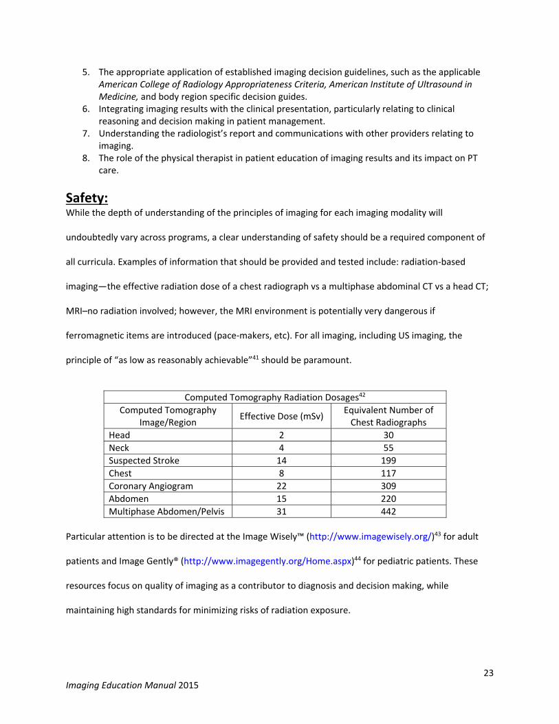
Safety: While the depth of understanding of the principles of imaging for each imaging modality will
undoubtedly vary across programs, a clear understanding of safety should be a required component of
all curricula. Examples of information that should be provided and tested include: radiation‐based
imaging—the effective radiation dose of a chest radiograph vs a multiphase abdominal CT vs a head CT;
MRI–no radiation involved; however, the MRI environment is potentially very dangerous if
ferromagnetic items are introduced (pace‐makers, etc). For all imaging, including US imaging, the
principle of “as low as reasonably achievable”41 should be paramount.
Computed Tomography Radiation Dosages42
Computed Tomography Image/Region
Effective Dose (mSv) Equivalent Number of Chest Radiographs
Head 2 30
Neck 4 55
Suspected Stroke 14 199
Chest 8 117
Coronary Angiogram 22 309
Abdomen 15 220
Multiphase Abdomen/Pelvis 31 442
Particular attention is to be directed at the Image Wisely™ (http://www.imagewisely.org/)43 for adult
patients and Image Gently® (http://www.imagegently.org/Home.aspx)44 for pediatric patients. These
resources focus on quality of imaging as a contributor to diagnosis and decision making, while
maintaining high standards for minimizing risks of radiation exposure.

24 Imaging Education Manual 2015
Example Objectives: The following example objectives are derived from CAPTE Evaluative Criteria7:
1. Use and understand imaging terminology appropriately, expressively, and receptively with imaging professionals, and discuss the differences between various imaging modality terminologies. (CC‐5.17; CC‐5.31)
2. Understand the basic physics of MRI, its advantages and limitations, and be able to
discuss briefly how MR images are acquired and how key parameters such as repetition time (TR) and echo time (TE) influence image weighting. (CC‐3)
3. Understand the uses and capabilities of ultrasound imaging and discuss its uses in the
physical therapy setting. (CC‐5.30 b, e, f, k)

25 Imaging Education Manual 2015
Basic Imaging Modality Properties
Imaging Modality
Mechanism Assets Limitations/ Concerns
Relative Radiation Dosages*
Radiography Imaging obtained by detection of x‐rays projected through human tissues with varying degrees of radiolucency and radio‐opacity demonstrated.
Allows good appreciation of basic bony anatomy.
Complex bony anatomy superimposition. Limited capacity for soft tissue demonstration.
Low
Computed Tomography
(CT)
Computerized image reconstruction of multiple slices of tissues through which x‐rays have been passed and detected.
Excellent demonstration of cortical bone anatomy. High sensitivity to variances in density of tissues. Multiplanar views of anatomy; 3‐D images possible.
Soft tissue demonstration dependent on CT unit. Expensive.
High
Magnetic Resonance
Imaging (MRI)
A method of exposing tissues to magnetic fields and radiofrequency waves to detect properties of tissues and thereby produce images.
Excellent demonstration of soft tissues and bone marrow. Multiplanar views of anatomy; 3‐D images possible. Multiple sequences allowing various tissue characteristics.
Lack of signal in cortical bone. Expensive.
None
Scintigraphy (Bone Scan)
Imaging obtained by introduction of radio‐isotopes into the body that are subsequently concentrated in areas of increased metabolic activity and recorded by tracer sensitive detection.
Good sensitivity to increased metabolic activity/bone turnover.
Lack of diagnostic specificity with increased radio‐isotope uptake.
Moderate
Sonography (Ultrasound)
A method of passing high frequency sound waves through tissues that are reflected or absorbed at varying levels depending on tissue properties. The reflected sound waves are detected and serve as a basis for the image.
Good demonstration of soft tissues. Allows real time, dynamic imaging. Convenience and cost.
Image yield highly dependent on operator.
None
Dual Energy X‐Ray
Absorptiometry (DEXA or DXA)
Measurement of relative x‐ray attenuation as a function of tissue density.
Simple, quick, accurate, modest cost. Suited to serial testing, if same device.
Inconsistent performance between devices from different manufacturers.
Low
*Actual dosages dependent on body region imaged and exam specifics.

26 Imaging Education Manual 2015
Didactic Instruction The recent survey1 of accredited PT education curricula revealed imaging content is typically introduced
in the first or second year of the curriculum. Approximately half of the responding programs included a
stand‐alone required course, with 58% of these programs also integrating imaging content throughout
the remainder of the curriculum. Irrespective of a stand‐alone imaging course, the recommendation is
to introduce the content as early as possible and then integrate where appropriate in the clinical science
tracks. Opportunities for early introduction exist in the basic science track; including imaging in the
human anatomy and neuroanatomy courses, providing a great example of how students will incorporate
imaging and apply anatomy in the clinic. Once students understand the properties of various imaging
modalities, a better understanding will develop of when particular modalities are indicated based on
initial patient presentation and ongoing management. Last, utilizing imaging to evaluate pathological
processes will promote optimal patient care. The following are provided as suggestions, as a guide for
introducing and integrating imaging content into the curriculum. Specific instructional and student
assessment content are included.
Section 1. General Instructional Concepts (continued from page 22)
9. The properties of commonly used imaging modalities with particular reference to: a. their capabilities and limitations in identifying anatomy and patho‐anatomy; b. the basic physics of their operation; c. relative risk, complications, and contraindications of commonly used imaging modalities
including life‐time cumulative radiation exposure and age‐related radiation exposure; d. the rationale for applications of each in common clinical scenarios; and e. the performance and interpretation of ultrasound.
10. Imaging for appropriate patient medical screening includes the specific history and clinical
examination findings, and where relevant, clinical guidelines, indicate the need for imaging.
11. Recognition of normal musculoskeletal anatomy, common anatomical variants, and developmental/lifespan changes, including the imaging modality, best demonstrates the anatomy.
12. Specific history elements and clinical examination findings consistent with indications for
imaging, includes: a. age, sex, and race;

27 Imaging Education Manual 2015
b. individual health history; c. chief presenting complaint; d. palpatory examination; e. joint/ligament stability testing; f. passive mobility testing; g. neuromuscular examination; h. special tests; and i. other observations.
13. Appropriate application of established imaging decision guidelines, such as the applicable
American College of Radiology Appropriateness Criteria, American Institute of Ultrasound in Medicine, and body region specific decision guides.
14. Integrating imaging results with the clinical presentation, particularly relating to clinical reasoning and decision making in patient management.
15. Understanding the radiologist’s report and communications with other providers relating to
imaging.
16. The role of the physical therapist in patient education of imaging results and its impact on physical therapist care.
Example Student Assessment/Competency Items: (answers italicized)
1. In any patient having sustained a fracture, physical therapists often want to know if the fracture is
“adequately” healed to safely accept increases of exercise or functional loads. To determine if a
fracture is healed “clinically,” the orthopaedist typically considers two practical criteria. One is
radiographic evidence of healing; the other is often based on clinical examination.
a) Briefly describe the basic radiographic evidence of healing.
blunting, widening, callus, sclerosis, bridging, remodeling
b) What is the practical, clinical evidence that the fracture is healing?
painfree at rest and with activity; subjective reports of stability
2. Arrange the biologic and non‐biologic samples in order of most radiolucent to most radiopaque.
_B__1. A. Bone _C__2. B. Air _E__3. C. Fat _A__4. D. Metal _D__5. E. Water

28 Imaging Education Manual 2015
3. For the imaging modalities listed in the first row of the table below, in each box describe what the
appearance of the structure will be on a gray scale.
CT T1‐W MRI T2‐W MRI
Swelling/Effusion Gray/dark/hypodense Low signal intensity Dark
High signal intensity Bright
Fat Dark/gray/black High signal intensity Bright
Intermediate signal intensity Gray
Bone Cortex White Low signal intensity Dark
Low signal intensity Dark
Bone Marrow Dark/gray High to intermediate signal intensity Bright/Gray to white (depends on marrow type)
Intermediate signal intensity Gray
4. Which imaging modality uses markers to measure metabolism and blood flow and is used in early
detection of stress fractures and skeletal metastases?
a. bone scan.
b. radiographs.
c. magnetic resonance imaging.
d. computed tomography scan.
5. Place the imaging modality in order of lowest to highest radiation dose to the patient.
a. magnetic resonance imaging, computed tomography scan, radiography.
b. radiography, magnetic resonance imaging, computed tomography scan.
c. magnetic resonance imaging, radiography, computed tomography scan.
d. ultrasound, radiography, computed tomography scan, magnetic resonance imaging.
6. The imaging modality that most accurately depicts the location, size, and orientation of fracture
fragments is:
a. three‐dimensional ultrasound.
b. radiography.
c. computed tomography.
d. magnetic resonance imaging.
29 Imaging Education Manual 2015
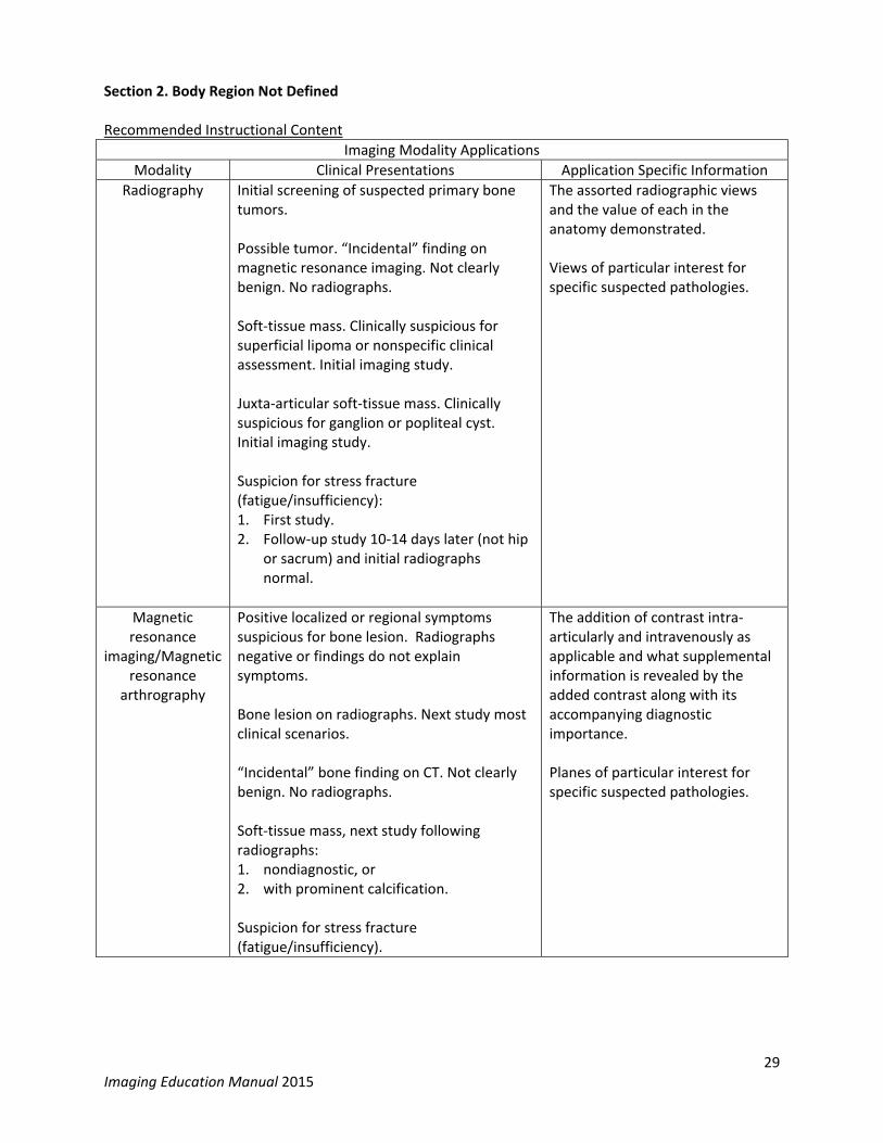
Section 2. Body Region Not Defined Recommended Instructional Content
Imaging Modality Applications
Modality Clinical Presentations Application Specific Information
Radiography Initial screening of suspected primary bone tumors. Possible tumor. “Incidental” finding on magnetic resonance imaging. Not clearly benign. No radiographs. Soft‐tissue mass. Clinically suspicious for superficial lipoma or nonspecific clinical assessment. Initial imaging study. Juxta‐articular soft‐tissue mass. Clinically suspicious for ganglion or popliteal cyst. Initial imaging study. Suspicion for stress fracture (fatigue/insufficiency): 1. First study. 2. Follow‐up study 10‐14 days later (not hip
or sacrum) and initial radiographs normal.
The assorted radiographic views and the value of each in the anatomy demonstrated. Views of particular interest for specific suspected pathologies.
Magnetic resonance
imaging/Magnetic resonance
arthrography
Positive localized or regional symptoms suspicious for bone lesion. Radiographs negative or findings do not explain symptoms. Bone lesion on radiographs. Next study most clinical scenarios. “Incidental” bone finding on CT. Not clearly benign. No radiographs. Soft‐tissue mass, next study following radiographs: 1. nondiagnostic, or 2. with prominent calcification. Suspicion for stress fracture (fatigue/insufficiency).
The addition of contrast intra‐articularly and intravenously as applicable and what supplemental information is revealed by the added contrast along with its accompanying diagnostic importance. Planes of particular interest for specific suspected pathologies.

30 Imaging Education Manual 2015
Example Student Assessment/Competency Items: (answers italicized)
1. Which imaging modality is typically the best option to allow differentiation of a benign versus a
pathological fracture?
a. bone scan.
b. computed tomography.
c. conventional radiography.
d. magnetic resonance imaging.
2. A limitation of radiographs that in some cases determines the use of computed tomography to be
preferable to detect or rule out fractures is:
a. insensitivity to bone mass loss.
b. overlapping bony layers.
c. inability to demonstrate surrounding edema.
d. difficulty with allowing visualization of ligaments.
3. The imaging modality that generally best reveals non‐displaced fractures is:
a. magnetic resonance imaging.
b. computed tomography.
c. radiography.
d. ultrasound.
Computed tomography
Bone lesion demonstrated on radiographs. Radiographic and/or clinical pattern suspicious for osteoid osteoma.
The addition of contrast intra‐articularly and intravenously as applicable and what supplemental information is revealed by the added contrast along with its accompanying diagnostic importance. Planes of particular interest for specific suspected pathologies.
Ultrasound Soft‐tissue mass. Clinically suspect superficial lipoma. Initial imaging study. Suspected deep vein thrombosis (Doppler). Superficial soft tissue foreign body evaluation. Muscle integrity, contraction ability, volume, and degenerative changes. Soft tissue and joint swelling evaluation.
Particular operator and equipment considerations in conducting the examination for specific suspected pathologies or anatomical concerns.

31 Imaging Education Manual 2015
4. In the ABCs approach of identifying content of images, the “A” represents:
a. air.
b. alignment.
c. adjacent structures.
d. asymmetry.

32 Imaging Education Manual 2015
Section 3. Cervical Spine Region Recommended Instructional Content
Typical Imaging Modality Applications
Modality Clinical Presentations Application Specific Information
Radiography
Investigation of suspected or further study of:1. Follow‐up for cervical spine trauma 2. Chronic neck pain
a. with or without prior trauma b. with prior history of malignancy c. with prior cervical spine surgery
The assorted radiographic views and the value of each in the anatomy demonstrated. Views of particular interest for specific suspected pathologies.
Magnetic resonance
imaging/Magnetic resonance
arthrography
Investigation of suspected or further study of:1. Acute cervical spine trauma with
a. neurological involvement b. mechanical instability
2. Chronic neck pain, neurological signs or symptoms present
3. Infection or malignancy
The addition of contrast intra‐articularly and intravenously as applicable and what supplemental information is revealed by the added contrast along with its accompanying diagnostic importance. Planes of particular interest for specific suspected pathologies.
Computed tomography
Investigation of suspected or further study of:1. Acute cervical spine trauma 2. Chronic neck pain with prior cervical
spine surgery 3. Posterior longitudinal ligament
ossification
The addition of contrast intra‐articularly and intravenously as applicable and what supplemental information is revealed by the added contrast along with its accompanying diagnostic importance. Planes of particular interest for specific suspected pathologies.
Specific Imaging Decision Guides: Canadian Cervical Spine Rule National Emergency X‐Ray Rule (NEXUS)
Example Student Assessment/Competency Items: (answers italicized)
1. Which are common criteria for both the Canadian Cervical Spine Rule and the NEXUS Low Risk Rule?
a. cervical spine tenderness to palpation; neurological signs or symptoms.
b. age 65 years or greater; evidence of intoxication.
c. distraction from other injury; ambulatory status.
d. age 65 years or greater; range of motion limitation.
2. You are observing a lateral view radiograph of the cervical spine in a patient with rheumatoid
arthritis who is currently at your facility. You observe an anterior atlantodental interval of 6.0 mm,
consistent with that reported by the radiologist. Your response to this as a physical therapist is
understanding _____________________ and you determine to __________________.
a. Normative values have not been established; proceed with care as previously planned.
33 Imaging Education Manual 2015
b. 3.5 mm is considered the upper normative limit; conduct a detailed neurological exam.
c. 4.5 mm is considered the upper normative limit; do a reductive mobilization technique.
d. 5.0 mm is considered the upper normative limit; begin stabilization exercise.
3. You are seeing a 71‐year‐old woman following rotator cuff repair. At two months postsurgery, she
enters today for her 6th visit and tells you of a fall she had at home yesterday. She says she
extended both arms to protect herself during the fall. She now reports increased pain in her
involved shoulder and neck. You note her neck motions are significantly limited and she has midline
posterior neck tenderness. Her active shoulder motion was 100° of flexion in sitting last week and is
now pain limited at 60°. Upon considering her presentation today, your greatest concern is:
a. whether she reinjured her shoulder and if MRI of her shoulder is needed to evaluate her status
for continuing rehabilitation.
b. whether she reinjured her shoulder and how you will modify her program today to be reduced
for a few days before progressing again to higher level exercise.
c. whether she has a significant cervical spine injury and to stabilize her neck until imaging of her
neck can be completed.
d. whether she now has a cervical radiculopathy and how you will address that problem in addition
to her shoulder.
4. An otherwise healthy 32‐year‐old female was in a motor vehicle accident two days ago. She was
rear‐ended while stopped at a busy intersection during rush hour traffic. She states she is surprised
“my neck is this sore because my back bumper had only a small dent in it.” Shortly after the accident
and in the ensuing hours, she reports that she experienced gradually increasing left posterolateral
neck stiffness and pain. The next morning, she awoke with a dull ache in the left deltoid muscle
area, and after washing her hair she noted the ache moved down to her elbow. The physical
examination reveals pain limited cervical active range of motion with extension at 25°, left rotation
limited to 55°, and left lateral flexion to 20°. Her other cervical active ranges are with end‐range pain
at normal limits for her age. The neurological examination, including myotome and dermatome
testing along with reflexes, is without deficits or abnormal findings.
Based on this much information, would you refer this patient for imaging?
No, imaging is not currently indicated.
Give a rationale for your response.
Her presentation is not consistent with an elevated fracture risk being negative on the Canadian
Cervical Spine Rule and NEXUS Low Risk Rules criteria within the American College of Radiology
Appropriateness Criteria. Specifically, the mechanism of injury is not consistent with a
“dangerous mechanism,” her range of motion limitations are not severe, and her neurological
exam is negative. She also has no other personal risk factors to elevate the risk of bone
compromise, instability, or other related problems.

34 Imaging Education Manual 2015
5. A 55‐year‐old male enters your clinic with his right upper extremity elevated and his wrist resting on
the top of his head. He states this is the only position of relief for him and it has been this way for 3
weeks. His specific description of symptoms is of right neck, periscapular, and proximal arm pain,
accompanied by intermittent distal paresthesia. On clinical examination, you find grossly limited
cervical range of motion in all planes, and cervical compression and Spurling’s maneuver are both
provocative of the upper extremity symptoms. Traction is equivocal for reduction of symptoms.
Sensory testing suggests a decrement in light touch, but the area is poorly defined. Manual muscle
testing is difficult to interpret as exertion at the shoulder and elbow are both pain limited. Grip
strength on his affected dominant side is 38 pounds and 95 pounds on the left. Based on this
information, your best course of action today is:
a. seek an MRI to assist in physical therapy decision making.
b. refer back to the physician as he clearly has a problem beyond your scope of care.
c. defer imaging, begin conservative care, and monitor his status closely.
d. obtain radiographs before beginning conservative care.

35 Imaging Education Manual 2015
Section 4. Shoulder Region Recommended Instructional Content
Typical Imaging Modality Applications
Modality Clinical Presentations Application Specific Information
Radiography Investigation of suspected or further study of:1. Acute shoulder pain of any etiology 2. Septic arthritis 3. Thoracic outlet syndrome (chest
radiograph)
The assorted radiographic views and the value of each in the anatomy demonstrated. Views of particular interest for specific suspected pathologies.
Magnetic resonance
imaging/Magnetic resonance
arthrography
Investigation of suspected or further study of:1. Persistent significant pain 2. Labral injury, with or without indications
of instability on examination, age prior to anticipated degenerative changes
3. Bursitis or long head of biceps tenosynovitis
4. Impingement, age after which degenerative changes anticipated, and radiographs normal or demonstrating coracoacromial arch osteophytes/ syndesmophytes
5. Re‐tear status post prior rotator cuff tear 6. Thoracic outlet syndrome (chest MRA)
The addition of contrast intra‐articularly and intravenously as applicable and what supplemental information is revealed by the added contrast along with its accompanying diagnostic importance. Planes of particular interest for specific suspected pathologies.
Computed tomography
Investigation of suspected or further study of:1. Scapular fracture45‐49 2. Osseous glenoid fossa lesions50‐54
The addition of contrast intra‐articularly and intravenously as applicable and what supplemental information is revealed by the added contrast along with its accompanying diagnostic importance. Planes of particular interest for specific suspected pathologies.

36 Imaging Education Manual 2015
Ultrasound Investigation of suspected or further study of:1. Bursitis 2. Long head of biceps tendinopathy or
displacement 3. Impingement, age after which
degenerative changes anticipated, and radiographs normal or demonstrating coracoacromial arch osteophytes/ syndesmophytes
4. Rotator cuff tendinopathy with/or without prior shoulder arthroplasty
5. Re‐tear status post prior rotator cuff repair
6. Acromioclavicular joint integrity, degeneration of effusion
Particular operator and equipment considerations in conducting the examination for specific suspected pathologies or anatomical concerns.
Example Student Assessment/Competency Items: (answers italicized)
1. Your 56‐year‐old male patient experienced a fall on his back a week ago while cleaning leaves from
his house gutters. He went to the emergency room and had radiographs completed of his shoulder.
These were interpreted as negative, but also with the comment of being underexposed. He presents
to you now with shoulder and left upper back pain severely limiting his motion actively and
passively. Manual muscle testing of his left shoulder from a neutral position reveals 4/5 pain limited
weakness of the shoulder in all planes. On palpation, his left upper back is exquisitely tender. The
neurological screen is negative. You have an increased suspicion of what injury and seek to have
what diagnostic test performed to allow better decision making?
a. rotator cuff tear; magnetic resonance imaging.
b. suprascapular nerve injury; electroneuromyography.
c. capsulolabral injury; magnetic resonance imaging.
d. scapular fracture; computerized tomography scan.
2. Match the MRI finding of the rotator cuff with the likely interpretation:
__C__ Complete tear A. Heterogenous signal intensity
__A__ Tendinopathy B. Fiber fraying
__B__ Partial tear C. Interposed fluid signal
3. For imaging of the rotator cuff, which statement is most accurate?
a. ultrasound and magnetic resonance imaging are equivalent in allowing appreciation of focal
tendon abnormalities. The selection of which is most appropriate depends on potential for
degenerative changes and other suspected pathology.
b. ultrasound is superior to magnetic resonance imaging in that it performs as well as magnetic
resonance imaging overall and is much less expensive. Thus, ultrasound is preferable in most
situations.

37 Imaging Education Manual 2015
c. magnetic resonance imaging is superior to ultrasound, even considering the greater cost. As
such, ultrasound should be considered a secondary exam procedure after magnetic resonance
imaging.
d. magnetic resonance arthrogram is indicated in most suspected rotator cuff pathologies.
4. The imaging modality which best identifies and discriminates labral lesions in the shoulder is:
a. magnetic resonance imaging.
b. magnetic resonance arthrography.
c. computed tomography with contrast.
d. single photon emission computed tomography.
e. high resolution ultrasound.
38 Imaging Education Manual 2015
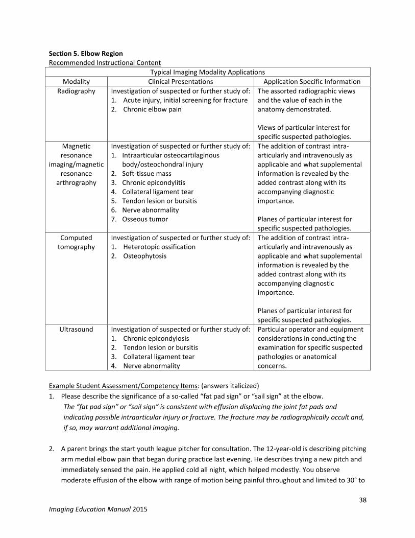
Section 5. Elbow Region Recommended Instructional Content
Typical Imaging Modality Applications
Modality Clinical Presentations Application Specific Information
Radiography Investigation of suspected or further study of:1. Acute injury, initial screening for fracture 2. Chronic elbow pain
The assorted radiographic views and the value of each in the anatomy demonstrated. Views of particular interest for specific suspected pathologies.
Magnetic resonance
imaging/magnetic resonance
arthrography
Investigation of suspected or further study of:1. Intraarticular osteocartilaginous
body/osteochondral injury 2. Soft‐tissue mass 3. Chronic epicondylitis 4. Collateral ligament tear 5. Tendon lesion or bursitis 6. Nerve abnormality 7. Osseous tumor
The addition of contrast intra‐articularly and intravenously as applicable and what supplemental information is revealed by the added contrast along with its accompanying diagnostic importance. Planes of particular interest for specific suspected pathologies.
Computed tomography
Investigation of suspected or further study of:1. Heterotopic ossification 2. Osteophytosis
The addition of contrast intra‐articularly and intravenously as applicable and what supplemental information is revealed by the added contrast along with its accompanying diagnostic importance. Planes of particular interest for specific suspected pathologies.
Ultrasound Investigation of suspected or further study of:1. Chronic epicondylosis 2. Tendon lesion or bursitis 3. Collateral ligament tear 4. Nerve abnormality
Particular operator and equipment considerations in conducting the examination for specific suspected pathologies or anatomical concerns.
Example Student Assessment/Competency Items: (answers italicized)
1. Please describe the significance of a so‐called “fat pad sign” or “sail sign” at the elbow.
The “fat pad sign” or “sail sign” is consistent with effusion displacing the joint fat pads and
indicating possible intraarticular injury or fracture. The fracture may be radiographically occult and,
if so, may warrant additional imaging.
2. A parent brings the start youth league pitcher for consultation. The 12‐year‐old is describing pitching
arm medial elbow pain that began during practice last evening. He describes trying a new pitch and
immediately sensed the pain. He applied cold all night, which helped modestly. You observe
moderate effusion of the elbow with range of motion being painful throughout and limited to 30° to
39 Imaging Education Manual 2015
100°. There is exquisite tenderness medially and laxity upon valgus testing. The first step for this
patient is:
a. radiographs to investigate for medial epicondyle fracture.
b. magnetic resonance imaging to evaluate for ligament injury.
c. computed tomography angiography to evaluate for vascular compromise.
d. bone scan to investigate for a Salter‐Harris fracture.
3. Your patient sustained right elbow trauma in a motor vehicle accident. She was the front seat
passenger and extended her arms to protect herself from an anticipated front impact, but her
vehicle was first struck from the side before the front impact, triggering the airbag. Her primary
description is now of medial elbow pain and upper extremity weakness. On clinical examination,
your most notable finding is pain and laxity with valgus stress testing of the joint. The imaging to
best reveal the pathology you suspect and the hallmark finding on this imaging would be:
a. magnetic resonance imaging; increased signal intensity at the medial epicondyle.
b. magnetic resonance arthrography; “T‐sign.”
c. computed tomography; intraarticular fracture.
d. radiography; radial head fracture.
4. Which of the following radiographic views would give the most information about the carrying angle
of the elbow joint?
a. lateral view.
b. oblique: internal rotation view.
c. oblique: external rotation view.
d. anterior‐posterior view.

40 Imaging Education Manual 2015
Section 6. Wrist and Hand Region
Recommended Instructional Content
Typical Imaging Modality Applications
Modality Clinical Presentations Application Specific Information
Radiography Investigation of suspected or further study of: 1. Wrist, hand, and distal forearm trauma
including suspected fractures and dislocations
2. Chronic wrist pain 3. Carpal tunnel syndrome
The assorted radiographic views and the value of each in the anatomy demonstrated. Views of particular interest for specific suspected pathologies.
Magnetic resonance
imaging/Magnetic resonance
arthrography
Investigation of suspected or further study of: 1. Occult or stress fractures, including
scaphoid 2. Non‐union, malunion, osteonecrosis,
and/or posttraumatic arthritis 3. Ligamentous injury, including thumb
ulnar collateral ligament 4. Ganglion cyst or other palpable wrist
mass 5. Inflammatory arthritis 6. Chronic wrist pain 7. Possible infection
The addition of contrast intra‐articularly and intravenously as applicable and what supplemental information is revealed by the added contrast along with its accompanying diagnostic importance. Planes of particular interest for specific suspected pathologies.
Computed tomography
Investigation of suspected or further study of: 1. Occult fracture, including hook of hamate
and scaphoid 2. Non‐union, malunion, osteonecrosis,
and/or posttraumatic arthritis 3. Distal radio‐ulnar joint dislocation 4. Intraarticular fracture
The addition of contrast intra‐articularly and intravenously as applicable and what supplemental information is revealed by the added contrast along with its accompanying diagnostic importance. Planes of particular interest for specific suspected pathologies.
Ultrasound Investigation of suspected or further study of: 1. Tendon injury55‐59 2. Nerve lesion 3. Ganglion cyst and soft tissue masses
Particular operator and equipment considerations in conducting the examination for specific suspected pathologies or anatomical concerns.

41 Imaging Education Manual 2015
Example Student Assessment/Competency Items: (answers italicized)
1. Your 38‐year‐old male patient who fell off scaffolding 3 weeks ago has persistent radial aspect wrist
pain without improvement over the past two weeks along with tenderness in the anatomical
snuffbox, pain with longitudinal thumb compression, and prior negative radiographs of the hand and
wrist. Your next step is:
a. seek repeat radiographs or magnetic resonance imaging of the wrist.
b. seek computed tomography with intraarticular contrast.
c. allow two more weeks of conservative care, but less aggressively with plans to contact the
physician as the patient returns for the next follow‐up to the physician at the end of the two‐
week period.
d. immobilize the wrist, use therapeutic ultrasound and electrical stimulation to promote healing
and re‐evaluate in 10 days.
2. Your patient with distal radial forearm and thumb pain who has continued working and has been
slow to respond to interventions is now scheduled to undergo an MRI of the involved extremity.
Based on isolated swelling in the area, exquisite tenderness, and pain with ulnar deviation, you
expect the radiologist’s report to include language consistent with increased signal intensity
circumferentially enveloping the straight fibrilinear pattern of tendons on the T2‐weighted sequence
and that result is most likely consistent with:
a. an intrasubstance partial tear.
b. a complete tendon rupture.
c. fatty infiltration.
d. tenosynovitis.
3. You are seeing a 36‐year‐old female driver for a package delivery service who reports wrist pain. She
had a similar bout of symptoms one year ago that reduced, but did not completely resolve. During
this prior episode of increased symptoms, a positive ulnar variance was identified on radiographs.
Given this background, what type of pathology would you suspect?
a. excessive loading on the lunate and potentially Kienböck’s disease.
b. ulnar neuropathy.
c. ganglion cysts and tenosynovitis.
d. proximal scaphoid fracture and avascular necrosis.
4. Your 62‐year‐old patient, who is right hand dominant, is a recreational golfer of less than average
skill. One particular day, he attempts a difficult shot and strikes the ground firmly with his club. He
describes this as causing an immediate onset of pain at the proximal, ulnar aspect of his left hand.
Upon presenting to you two days later, his symptoms are still present with wrist motion moderately
limited by pain, a significant reduction in grip strength, and tenderness throughout the hypothenar
eminence. He has particular worsening of symptoms with resisted flexion of the 4th and 5th digits.
Match the pathology for which you are most suspicious and the two best imaging options to detect
this.
a. pisiform fracture: magnetic resonance imaging or posterior‐anterior radiograph.

42 Imaging Education Manual 2015
b. ulnar styloid process fracture: magnetic resonance imaging or lateral view radiograph.
c. hook of hamate fracture: carpal tunnel view radiograph or computed tomography.
d. shaft of 5th metacarpal fracture: oblique view radiograph or computed tomography.
5. Your patient has a longstanding history of painful popping and clicking in the wrist with activities
requiring use of her dominant right hand. She also has pain‐limited wrist motion, decreased grip
strength, and well‐localized tenderness at the proximal dorsal aspect of the wrist.
a. What is the most probable pathology?
Scapholunate dissociation.
b. What radiographic view would best reveal this pathology?
A clenched fist stress view.
c. What specific result would indicate the presence of this instability?
An interval between the scaphoid and lunate of 5 mm or more.

43 Imaging Education Manual 2015
Section 7. Thoracolumbar Spine Region Recommended Instructional Content
Typical Imaging Modality Applications
Modality Clinical Presentations Application Specific Information
Radiography Investigation of suspected or further study of: 1. Thoracic or lumbar spine in child (< 14
yrs) with known cervical, thoracic, or lumbar fracture
2. Rib fracture
The assorted radiographic views and the value of each in the anatomy demonstrated. Views of particular interest for specific suspected pathologies.
Magnetic resonance
imaging/Magnetic resonance
arthrography
Investigation of suspected or further study of: 1. Back pain with,
a. Low velocity trauma b. Osteoporosis c. Focal and/or progressive neurological
deficit d. Prolonged symptom duration e. Age > 70 years f. Cancer g. Infection h. Immunosuppression
2. Back pain and/or radiculopathy 3. Back pain with prior lumbar surgery 4. Cauda equina syndrome, multifocal or
progressive neurological deficit 5. Blunt trauma of thoracic or lumbar spine
with neurological abnormalities
The addition of contrast intra‐articularly and intravenously as applicable and what supplemental information is revealed by the added contrast along with its accompanying diagnostic importance. Planes of particular interest for specific suspected pathologies.
Computed tomography
Investigation of suspected or further study of: 1. Blunt trauma to the thoracic or lumbar
spine with neurological abnormalities 2. Thoracic or lumbar spine in child (<14 yrs)
with known cervical fracture 3. Abdominal aortic aneurysm
The addition of contrast intra‐articularly and intravenously as applicable and what supplemental information is revealed by the added contrast along with its accompanying diagnostic importance. Planes of particular interest for specific suspected pathologies.
Dual energy x‐ray absorptiometry
Investigation of suspected or further study of: 1. Low bone density and fracture risk
identification (DXA of spine & hip) 2. Suspected fracture (DXA vertebral
fracture assessment of spine)
Ultrasound Initial screening for abdominal aortic aneurysm
Particular operator and equipment considerations in conducting the examination for specific suspected pathologies or anatomical concerns.
44 Imaging Education Manual 2015
Example Student Assessment/Competency Items: (answers italicized)
1. When considering imaging studies for lumbar pain, research shows the modality that offers the
highest sensitivity and specificity for detecting cancer, infections, and intervertebral disk changes is:
a. magnetic resonance imaging.
b. radiographs.
c. bone scan.
d. computed tomography.
2. You are seeing a 15‐year‐old female gymnast and cheerleader for low back and bilateral buttock
pain. Given her history of pain for 6 months, you base your examination on the suspicion that she
could potentially have a pars interarticularis injury. As such, you select the clinical examination
procedures found to have very good psychometric values in comparison to imaging evidence in
those with spondylolysis/spondylolisthesis in order to rule out that problem. Those specific
procedures are:
a. palpation for presence of a step sign at the probable level.
b. palpation of passive intervertebral motion at the suspected segment.
c. one leg hyperextension test looking for posterior pain provocation.
d. no exam, procedure has good correlation to image findings.
3. You are treating a 60‐year‐old woman with back pain. Your examination and assessment yields
findings that suggest manual therapy may be indicated. Before you determine that she is safe for
the application of end‐range manual techniques, you would like to have more definitive, objective
data. To be confident in your clinical decision‐making to proceed with manual techniques, you
would like to have a:
a. magnetic resonance imaging to rule out overt radiculopathy.
b. dual energy x‐ray absorptiometry scan with values greater than ‐1.0 on the t‐score.
c. dual energy x‐ray absorptiometry scan with values less than ‐2.5 on the t‐score.
d. computed tomography scan to rule out fracture in her remote past.
4. The most accurate early diagnostic combination matched with the implicated disorder is which of
the below:
a. elevated sedimentation rate, radiography; rheumatoid arthritis.
b. Short Tau Inversion Recovery (STIR)‐MRI, presence of a specific leukocyte antigen; ankylosing
spondylitis.
c. radiography, venogram; abdominal aortic aneurysm.
d. computed tomography, McMurray's test; tibial chondral lesion.
45 Imaging Education Manual 2015
Section 8. Hip/Pelvis Region Recommended Instructional Content
Typical Imaging Modality Applications
Modality Clinical Presentations Application Specific Information
Radiography Investigation of suspected or further study of:1. Fracture 2. Avascular necrosis 3. Chronic pain 4. Possible referred pain to exclude hip 5. Stress (fatigue/insufficiency) fracture
The assorted radiographic views and the value of each in the anatomy demonstrated. Views of particular interest for specific suspected pathologies.
Magnetic resonance
imaging/Magnetic resonance
arthrography
Investigation of suspected or further study of:1. Fracture 2. Avascular necrosis 3. Osseous or surrounding soft tissue
abnormality 4. Labral tear or femoroacetabular
impingement 5. Pigmented villonodular synovitis 6. Osteochondromatosis
The addition of contrast intra‐articularly and intravenously as applicable and what supplemental information is revealed by the added contrast along with its accompanying diagnostic importance. Planes of particular interest for specific suspected pathologies.
Computed tomography
Investigation of suspected or further study of:1. Osteoid osteoma 2. Acetabular fracture
The addition of contrast intra‐articularly and intravenously as applicable and what supplemental information is revealed by the added contrast along with its accompanying diagnostic importance. Planes of particular interest for specific suspected pathologies.
Dual energy x‐ray absorptiometry
Identification and follow‐up of low bone density and fracture risk assessment in absence of symptoms but elevated risk
Ultrasound Investigation or further study of: 1. Developmental dysplasia in infants
Particular operator and equipment considerations in conducting the examination for specific suspected pathologies or anatomical concerns.
Example Student Assessment/Competency Items: (answers italicized)
1. A frequently overlooked cause of low back pain that may escape radiographic detection in older
women is:
a. occult femoral neck fracture.
b. sacral insufficiency fracture.
c. pubic ramus fracture.
d. lumbar disk herniation.

46 Imaging Education Manual 2015
2. The imaging method of screening the hips of infants for developmental dysplasia, which is typically
chosen based on accuracy, convenience, and minimizing risk is:
a. radiography.
b. magnetic resonance imaging.
c. computed tomography.
d. bone scan.
e. ultrasound.
3. Which of the following statements is FALSE regarding pelvic fractures?
a. with pelvic trauma, an anterior‐posterior view radiograph is usually sufficient.
b. widening of the symphysis pubis > 1 cm is abnormal.
c. the pelvis is a ring and fractures usually occur in only one area.
d. “open book” injuries refer to a loss of osseoligamentous stability.
4. Your 70‐year‐old female patient is complaining of intense inguinal pain with no prior history of
trauma, especially with weight bearing, but relieved with rest and sleep. Her family practice
physician has completed radiography noting “mild degenerative changes” in the painful hip along
with osteopenic appearance. Your greatest immediate concern at the time of her initial evaluation
is:
a. if the degeneration might be more than indicated on the radiograph. b. if a proximal femur fracture has been adequately ruled out. c. if she might have septic arthritis of the hip. d. if a neoplasm might be the cause of her complaint.

47 Imaging Education Manual 2015
Section 9. Knee Region Recommended Instructional Content
Typical Imaging Modality Applications
Modality Clinical Presentations Application Specific Information
Radiography Investigation of suspected or further study of: 1. Fracture or dislocation 2. Routine monitoring after arthroplasty 3. Suspected periprosthetic infection 4. Atraumatic knee pain
The assorted radiographic views and the value of each in the anatomy demonstrated. Views of particular interest for specific suspected pathologies.
Magnetic resonance
imaging/Magnetic resonance
arthrography
Investigation of suspected or further study of: 1. Persistent pain 2. Osteochondral lesion, internal
derangement, or joint effusion 3. Avascular necrosis 4. Posterior dislocation following significant
trauma
The addition of contrast intra‐articularly and intravenously as applicable and what supplemental information is revealed by the added contrast along with its accompanying diagnostic importance. Planes of particular interest for specific suspected pathologies.
Computed tomography
Investigation of suspected or further study of: 1. Tibial plateau fracture 2. Periprosthetic infection or prosthesis
loosening
The addition of contrast intra‐articularly and intravenously as applicable and what supplemental information is revealed by the added contrast along with its accompanying diagnostic importance. Planes of particular interest for specific suspected pathologies.
Ultrasound Investigation of suspected or further study of: 1. Tendon lesion 2. Popliteal cyst 3. Superficial ligament lesion
Particular operator and equipment considerations in conducting the examination for specific suspected pathologies or anatomical concerns.
Specific Imaging Decision Guides: Ottawa Knee Rule
Example Student Assessment/Competency Items: (answers italicized)
1. The most common site for osteochondritis dissecans along with the best imaging modality to
identify it is:
a. medial femoral condyle; magnetic resonance imaging.
b. capitulum; computed tomography.
c. dome of the talus; magnetic resonance imaging.
d. femoral head; radiograph.
e. radius at radiocarpal joint; computed tomography.

48 Imaging Education Manual 2015
2. A 48‐year‐old school teacher presents to you with right knee pain, which she describes as “a dull
ache—like a tooth ache.” She reports the pain started 4 to 5 months ago and denies any incident or
accident. She further describes her symptoms being worse, and walking more difficult early in the
morning with improvement over the morning, but worsening again by day’s end. At the end of the
day, she reports sitting for 30 minutes with improvement in her knee symptoms. You observe her
gait lacks extension at terminal swing on the right along with an abbreviated stance period. The
overall pattern of knee pain has remained relatively consistent over the past 3 months and she
states, “just finally I am sick of it and it’s why I came to PT.” She also tells you of a severe right knee
injury in high school, resulting in ligament and meniscus tears. She reports having had surgery, but is
vague on the details. You note a faded surgical scar approximating the medial joint line. The current
right knee active range of motion is 3° to 110°. Palpation reveals that the knee is generally tender
with a modest increase along the joint lines. The only medication she lists is acetaminophen for
pain. She denies significant illnesses.
a. Would you refer this patient for imaging?
No.
b. Please provide a rationale for this decision.
It is very likely that she has degenerative changes in her knee and that is the source of her
problem. The overall course of her knee condition is proposed to be stable with no suggestions of
serious pathology. Similarly, there are no elements in her overall presentation indicating
systemic or medical issues beyond the scope of physical therapy. Because imaging findings are
unlikely to change her course of care, imaging before beginning intervention is not warranted.
This decision can be reviewed at any time, particularly if she fails to progress after a reasonable
time/number of visits for a favorable response.
3. A 27‐year‐old, who happens to be a weekend rugby player, stops by your clinic unscheduled after
having had radiographs made of his knee. He was injured one week ago in a match and he wants
you to look at his knee images and then examine his knee. He explains that he was on crutches the
first 4 days after the injury because of swelling and difficulty weight bearing. He has since stopped
using the crutches as he has returned to full weight bearing although with an antalgic gait pattern.
He said the swelling reduced a couple of days ago. He asks you what he should do next as no further
follow‐up is planned. He was told by his physician to ride a bike and then gradually return to activity
as tolerated. Upon looking at the images and seeing the radiologist’s report, you note the equivocal
presence of a small bone fragment immediately lateral to the proximal tibial rim on the A‐P
radiograph. He asks you. “what does this mean?” Upon seeing this, your next step is answer his
question and undertake what specific procedures?
a. explain that he should not have had the radiographs made because he did not meet the criteria
of the Ottawa Knee Rule. Examine his crutch walking on various surfaces and undertake gait
training.
b. explain the tiny bone fragment is inconsequential as an anatomical variant and is an incidental
finding. Instruct him in a home program and plan follow‐up in one week.
c. explain that he may have a significant intraarticular injury yet to be adequately described.
Complete ligamentous stability testing with particular attention to Lachman’s test.

49 Imaging Education Manual 2015
d. explain that he does not need to see an orthopaedist for further follow‐up and simply needs to
start rehabilitation. Further standard exam of motion, strength, and functional mobility are
needed.
4. A woman, 35 years of age, reports to you with knee pain after stumbling and losing control of a
piece of furniture she was carrying with another person last weekend. She says her pain is
essentially unchanged from the onset 5 days ago and the swelling that gradually accumulated has
also not changed. She says her pain is focused on the inside (medial aspect) of her knee and is at its
worst when she is trying to climb or descend stairs. On clinical examination, you find mild joint
effusion, range of motion at 10° to 110°, mild effusion, and joint line tenderness, medial greater
than lateral. Her gait is noteworthy for a decreased stance period as limited by pain on her affected
side. You are hypothesizing what pathology is most likely to be present, and would be detected with
what further clinical examination procedure, and potentially confirmed by what imaging?
a. medial collateral ligament injury; valgus stress test; computed tomography.
b. medial meniscus injury; Thessaly test; magnetic resonance imaging.
c. tibial plateau fracture; Apley’s compression and distraction; computed tomography.
d. patellar medial subluxation; apprehension test; lateral view radiograph.

50 Imaging Education Manual 2015
Section 10. Ankle and Foot Region Recommended Instructional Content
Typical Imaging Modality Applications
Modality Clinical Presentations Application Specific Information
Radiography Investigation of suspected or further study of: 1. Acute injury imaging for suspected fracture,
instability, or penetrating trauma 2. Osteonecrosis 3. Degenerative joint disease 4. Chronic pain 5. Osteomyelitis in presence of diabetes
mellitus
The assorted radiographic views and the value of each in the anatomy demonstrated. Views of particular interest for specific suspected pathologies.
Magnetic resonance
imaging/Magnetic resonance
arthrography
Investigation of suspected or further study of: 1. Ankle impingement syndrome 2. Osteochondral lesion 3. Musculotendinous disruption 4. Ligamentous injury or instability, including
syndesmotic injury 5. Stress injury or occult fracture 6. Rheumatoid arthritis, inflammatory
arthropathy, or other inflammatory disorder 7. Neuroma or local neuropathy, including
tarsal tunnel syndrome 8. Chronic heel pain or plantar fasciitis 9. Osteomyelitis in presence of diabetes
mellitus
The addition of contrast intra‐articularly and intravenously as applicable and what supplemental information is revealed by the added contrast along with its accompanying diagnostic importance. Planes of particular interest for specific suspected pathologies.
Computed tomography
Investigation of suspected or further study of: 1. Talus60‐62 or calcaneal fracture63‐65 2. Tarsal coalition or bony anomaly
The addition of contrast intra‐articularly and intravenously as applicable and what supplemental information is revealed by the added contrast along with its accompanying diagnostic importance. Planes of particular interest for specific suspected pathologies.
Ultrasound Investigation of suspected or further study of: 1. Muscle or tendon lesion 2. Penetrating trauma/foreign body 3. Neuroma or local neuropathy, including
tarsal tunnel syndrome 4. Plantar fascia lesion 5. Ligament lesion
Particular operator and equipment considerations in conducting the examination for specific suspected pathologies or anatomical concerns.
Scintigraphy Investigation of suspected or further study of: 1. Complex regional pain syndrome
Information provided by phases and variations of nuclear imaging procedure.
51 Imaging Education Manual 2015
Specific Imaging Decision Guides: Ottawa Ankle & Foot Rules
Example Student Assessment/Competency Items: (answers italicized)
1. Which of the following criteria is NOT a criterion of the Ottawa Ankle & Foot Rules?
a. incapable of standing or walking on the affected limb shortly after the injury and at present.
b. bone tenderness of the distal posterior 2.5 inches of the fibula.
c. bone tenderness at the attachment point of the fibularis brevis.
d. tenderness at the dome of the calcaneus.
2. Your patient had an ankle injury in a pick‐up basketball game 4 weeks ago. He was referred to you
3weeks ago with negative radiographs having been completed immediately prior to his first visit.
After 6 visits with you, he is still struggling to make progress and requires crutches to ambulate
because of ankle pain with weight bearing. He has persistent ankle effusion, worsened with
ambulation more than short distances. His anterior drawer test is mildly positive, but your
impression is this may not be consistent with the severity of his complaints. For which condition are
you having increased suspicion and how would it be detected or ruled out?
a. stress fracture; magnetic resonance imaging.
b. osteochondral lesion; magnetic resonance imaging.
c. pathologic fracture; bone scan.
d. Jones fracture; computed tomography scan.
3. A 24‐year‐old female using crutches hopped into your clinic using crutches and not weight bearing
on her left lower extremity. She described falling the night before while walking on ice and injured
her left foot in the accident. She has not had any emergent care prior to visiting you. She just now
borrowed the crutches from her neighbor and does not know how to use them. You observe mild
swelling across the middle of the foot and palpation of her foot provokes pain over the mid‐shaft of
the 3rd metatarsal.
Would you recommend radiographs for this patient?
Yes.
Give a rationale for your answer.
Bone tenderness and impaired weight bearing elevate suspicion of fracture.
4. A 17‐year‐old male cross country runner presents to your clinic with a description of pain in his right
anterior‐medial tibia just below the knee. He reports an insidious onset about 4 weeks ago. The pain
is mostly present when he runs but sometimes hurts just walking around school. There is no soft
tissue swelling. Local tenderness to palpation is not especially impressive.
a. list 2 or 3 possible diagnoses and rank from the most likely diagnosis to the least likely
diagnosis.
Stress fracture/reaction, pes anserine bursitis, meniscus tear, patellofemoral, etc.
b. what imaging studies would you recommend be performed to rule in/out the diagnosis you
listed first? Briefly provide a rationale for your choice.
52 Imaging Education Manual 2015
MRI: stress reaction/swelling/fracture/soft tissue. MRI will allow for earlier presentation of fluid
or fracture, whereas a radiograph/plain film will not demonstrate any change.
The recommended imaging content as previously described is based on the American College of
Radiology Appropriateness Criteria by the American College of Radiology (ACR Appropriateness
Criteria®. Available at www.acr.org/ac. Accessed November 28, 2014) and supplemented by the
evidence cited below.
Practical Instruction – Ultrasound Imaging Procedures Recent advances in USI technology have made it increasingly practical to incorporate instruction of USI into doctor of physical therapy education curriculum. The cost of USI equipment has decreased considerably in recent years while the quality of images has steadily improved. Image quality of lower cost portable USI equipment is comparable to hospital radiology department USI. Teaching USI is time intensive and somewhat resource intensive. Programs can understandably be reluctant to add to an already full curriculum. By incorporating USI in a longitudinal fashion to their programs, academic programs will see the benefits outweigh the challenges of adding USI as it is ideally suited to teaching anatomy and movement. The advantage to USI is real‐time imaging of live subjects. No other practical tool offers this ability in the laboratory and classroom. The most obvious indications for US are instruction in normal and abnormal anatomic variability, pathology, kinesiology, pathokinesiology, physiology, and pathophysiology. Once the fundamentals of USI physics, acquiring images, and understanding morphology is learned, USI can be incorporated throughout the curriculum in didactic and laboratory instruction. As faculty become skilled at using and incorporating USI, other less effective teaching tools and methods can be discontinued allowing for minimal overall impact to teaching time and resources. Ultrasound imaging can also be readily employed in student self‐directed learning. Ultrasound imaging is rapidly being adopted by PTs in clinical practice. Using USI in examination and management is commonly referred to as point‐of‐care application. There is an increasing body of research of USI and physical therapy. Growing point‐of‐care and research activity strengthen the case for PT student education in USI.
Faculty Resources Prior to incorporating the practical instruction of US into PT curriculum, faculty must be trained in USI. The predominant method for non‐radiologist health care practitioners to learn USI is by continuing education, self‐study, colleague mentoring, and instruction. A listing of continuing education and self‐study resources is provided at www.orthopt.org. The most common multi‐disciplinary credential for musculoskeletal USI is offered by the American Registry of Diagnostic Medical Sonography. There is no PT specific credential offered by a United States professional society.
Fundamentals of Ultrasound Imaging Ultrasound imaging can be introduced early in the curriculum for students to gain fundamental knowledge of the use of USI, equipment, and applications. Some of the fundamentals of USI overlap with the fundamentals of other imaging modalities and can be covered as part of an overall imaging

53 Imaging Education Manual 2015
curriculum. Broad topic areas that should be covered in learning the fundamentals of USI are adapted from the National Ultrasound Curriculum for Medical Students66:
Basic principles of the physics of ultrasound
Image acquisition: sound transmission and reflection, scatter, acoustic window
Image optimization: depth, focal zone, time‐gain compensation, gain, field of view
Posterior acoustic properties: posterior enhancement (increased sound transmission), sound attenuation, acoustical shadowing
Artifacts: near field, reverberation, edge refraction, mirror image
Terminology: echogenic, hyperechoic, hypoechoic, anechoic, isoechoic
Characteristics of simple fluid, complex fluid, soft tissue/solid, air/gas, bone/calcium
Imaging modes: B‐mode, Doppler, M‐mode
Knowledge of instrumentation
Describe advantages of ultrasound
Describe limitations of ultrasound
Correlate patient safety and ultrasound
Identify classic appearances of normal structures on ultrasound
Overview of Clinical Applications of Ultrasound Imaging The PT point‐of‐care application of USI can be described in two broad categories, diagnostic and procedural. Diagnostic USI in point‐of‐care applications are using USI as one component of the diagnostic process. The PT utilizes information gained from USI combined with patient history and physical examination findings along with other relevant data to arrive at a diagnosis. Point‐of‐care diagnostic USI is distinct from USI examinations performed by a consultant who does not participate in patient care beyond the USI examination and rendering a report to the treating clinician. Procedural US in PT practice encompasses many interventions that include but are not limited to patient biofeedback, neuromuscular re‐education, monitoring real‐time changes in morphology and movement during interventions, documenting change in clinical conditions, using US localize to target areas for manual interventions and physical agents, and using US to guide needle placement in interventions such as dry needling, electromyography, and nerve conduction velocity.
Ultrasound Imaging Equipment Ultrasound imaging equipment is available from many manufacturers. Elsewhere in this manual are resources that provide guidance on equipment selection. In particular, the Royal College of Radiologists: Standards for Ultrasound Equipment67 and the American Institute of Ultrasound in Medicine: Practice Guideline for the Performance of a Musculoskeletal Ultrasound Examination68are particularly informative.
Ultrasound Imaging Teaching Models Teaching the practical aspect of USI at a minimum requires a student, an instructor, an ultrasound machine, and a live or simulated model. Commonly, PT students use their classmates as models for other content areas and the same approach can be used in teaching USI. Consideration can also be given for using USI on human and animal cadavers. There are artificial USI simulators available but generally they are not optimal for physical therapy education. Individuals solicited from the community can be excellent models. Community models have an advantage of generally providing greater anatomic and

54 Imaging Education Manual 2015
age variation than classmates. When utilizing human models, one needs to consider developing a policy to address the disclosure of incidental findings obtained during USI and to limit pressure on models to expose sensitive body areas. A valuable resource in multiple aspects of teaching ultrasound is the Alliance of Medical Student Educators in Radiology (AMSER) Curriculum, Competencies, and Objectives.69
Clinical Education Clinical instructors are a contracted extension of the academic institution. Therefore Cis are expected to provide opportunities for the student to learn and apply judicious use of imaging in patient management, as allowed and appropriate for the patient population at the facility, the skills of the CI, and state regulations. Adequate instruction from a properly qualified CI is a continuum of the academic program’s instruction and is essential in students’ development of skill in the utilization of imaging. Given the appropriate academic background and guided practice, students can be expected to incorporate imaging safely and effectively in patient management.
Clinical Instruction and Documentation of Student Performance The CI should use progressive student questioning on the safety, theory, indications, and contraindications for imaging. In settings where US is available with appropriately trained CIs in USI, the student should practice the application of US in a safe and progressive fashion, and offer feedback on the skill performance and critical thinking process.
REFERENCES 1. Boissonnault WG, White DM, Carney S, Malin B, Smith W. Diagnostic and procedural imaging
curricula in physical therapist professional degree programs. J Orthop Sports Phys Ther. 2014;44(8):579‐586, B571‐512.
2. Boyles RE, Gorman I, Pinto D, Ross MD. Physical therapist practice and the role of diagnostic imaging. J Orthop Sports Phys Ther. 2011;41(11):829‐837.
3. Elliott JM. Magnetic resonance imaging: generating a new pulse in the physical therapy profession. J Orthop Sports Phys Ther. 2011;41(11):803‐805.
4. Greathouse DG, Schreck RC, Benson CJ. The United States Army physical therapy experience: evaluation and treatment of patients with neuromusculoskeletal disorders. J Orthop Sports Phys Ther. 1994;19(5):261‐266.
5. Moore CL, Copel JA. Point‐of‐care ultrasonography. N Engl J Med. 2011;364(8):749‐757. 6. American Physical Therapy Association. A Normative Model of Physical Therapist Professional
Education. Alexandria, VA: American Physical Therapy Association; 2004. 7. Commission on Accreditation in Physical Therapy Education. Evaluative Criteria Physical Therapy
Programs. Alexandria, VA: American Physical Therapy Association; 2014. 8. Benson CJ, Schreck RC, Underwood FB, Greathouse DG. The role of Army physical therapists as
nonphysician health care providers who prescribe certain medications: observations and experiences. Phys Ther. 1995;75(5):380‐386.
9. Moore JH, Goss DL, Baxter RE, et al. Clinical diagnostic accuracy and magnetic resonance imaging of patients referred by physical therapists, orthopaedic surgeons, and nonorthopaedic providers. J Orthop Sports Phys Ther. 2005;35(2):67‐71.
10. James JJ, Stuart RB. Expanded role for the physical therapist. Screening musculoskeletal disorders. Phys Ther. 1975;55(2):121‐131.

55 Imaging Education Manual 2015
11. Moore JH, McMillian DJ, Rosenthal MD, Weishaar MD. Risk determination for patients with direct access to physical therapy in military health care facilities. J Orthop Sports Phys Ther. 2005;35(10):674‐678.
12. Boissonnault WG, Badke MB, Powers JM. Pursuit and implementation of hospital‐based outpatient direct access to physical therapy services: an administrative case report. Phys Ther. 2010;90(1):100‐109.
13. Dininny P. More than a uniform: the military model of physical therapy. PT Magazine. 1995;3:40‐48.
14. American Physical Therapy Association. Diagnosis By Physical Therapists. Vol HOD P06‐12‐10‐09 [Amended HOD P06‐08‐06‐07; HOD P06‐97‐06‐19; HOD 06‐95‐12‐07; HOD 06‐94‐22‐35; Initial HOD 06‐84‐19‐78] [Position] 2012.
15. Federation of State Boards of Physical Therapy. The Model Practice Act for Physical Therapy. A Tool for Public Protection and Legislative Change. 2006;4.
16. Federation of State Boards of Physical Therapy. Rehabilitative Ultrasound Imaging: A Resource Guide. 2010.
17. American Academy of Orthopaedic Physical Therapists. Ultrasound Imaging. http://www.aaompt.org/about/statements.cfm. Published October 17, 2009. Accessed December 9, 2014.
18. Barr J. Integration of Imaging in Physical Therapy Practice. Philadelphia, PA: F.A. Davis; 2010. 19. Colorado Physical Therapy Practice Act. CRS Title 12 Article 41. 2011. 20. Thorman M. PT Connections. PT Connections, The Newsletter for the Wisconsin Physical Therapy
Association. 2007;37(1). 21. American Physical Therapy Association. Guide to Physical Therapist Practice 3.0. Alexandria, VA:
American Physical Therapy Association; 2014. Available at: http://guidetoptpractice.apta.org/. Accessed November 10, 2014.
22. Dumholdt E, Emery M, Harris MJ. The DPT: What We Know, What We Don’t. Annual Conference and Exposition of the American Physical Therapy Association, June 2004, Chicago, Illinois.
23. American Physical Therapy Association. APTA Vision sentence for physical therapy 2020 and APTA vision statement for physical therapy 2020 HOD P06‐00‐24‐35 [Position]. 2010.
24. Overmeer T, Linton SJ, Holmquist L, Eriksson M, Engfeldt P. Do Evidence‐Based Guidelines Have an Impact in Primary Care? A Cross‐Sectional Study of Swedish Physicians and Physiotherapists. Spine (Phila Pa 1976). 2005;30(1):146‐151.
25. Bishop PB, Wing PC. Knowledge transfer in family physicians managing patients with acute low back pain: a prospective randomized control trial. Spine J. 2006;6(3):282‐288.
26. Boissonnault WG, Ross MD. Physical therapists referring patients to physicians: a review of case reports and series. J Orthop Sports Phys Ther. 2012;42(5):446‐454.
27. González‐Urzelai V, Palacio‐Elua L, López‐de‐Munain J. Routine primary care management of acute low back pain: adherence to clinical guidelines. Eur Spine J. 2003;12(6):589‐594.
28. American Physical Therapy Association. Today’s Physical Therapist: A Comprehensive Review of a 21st‐Century Health Care Profession. Alexandria, VA: American Physical Therapy Association; 2011.
29. Gonzalez‐Urzelai V, Palacio‐Elua L, Lopez‐de‐Munain J. Routine primary care management of acute low back pain: adherence to clinical guidelines. Eur Spine J. 2003;12(6):589‐594.
30. Matzkin E, Smith EL, Freccero D, Richardson AB. Adequacy of education in musculoskeletal medicine. J Bone Joint Surg Am. 2005;87(2):310‐314.
31. Freedman KB, Bernstein J. Educational deficiencies in musculoskeletal medicine. J Bone Joint Surg Am. 2002;84‐A(4):604‐608.

56 Imaging Education Manual 2015
32. Freedman KB, Bernstein J. The adequacy of medical school education in musculoskeletal medicine. J Bone Joint Surg Am. 1998;80(10):1421‐1427.
33. Stockard AR, Allen TW. Competence levels in musculoskeletal medicine: comparison of osteopathic and allopathic medical graduates. J Am Osteopath Assoc. 2006;106(6):350‐355.
34. Daker‐White G, Carr AJ, Harvey I, et al. A randomised controlled trial. Shifting boundaries of doctors and physiotherapists in orthopaedic outpatient departments. J Epidemiol Community Health. 1999;53(10):643‐650.
35. Pham HH, Ginsburg PB, McKenzie K, Milstein A. Redesigning care delivery in response to a high‐performance network: the Virginia Mason Medical Center. Health Affairs. 2007;26(4):w532‐544.
36. Blackburn MS, Cowan SM, Cary B, Nall C. Physiotherapy‐led triage clinic for low back pain. Aust Health Rev. 2009;33(4):663‐670.
37. Alberta P. Practice StandardProfessional Qualifications and Competence: Performance of Restricted Activities. 2012. http://www.physiotherapyalberta.ca/physiotherapists/what_you_need_to_know_to_practice_in_alberta/standards_of_practice/performance_of_restricted_activities. Accessed December 9, 2014.
38. American Physical Therapy Association. Vision 2020. http://www.apta.org/Vision2020/. Accessed July 21, 2014.
39. American Physical Therapy Association. Vision Statement for the Physical Therapy Profession and Guiding Principles to Achieve the Vision. 2014; http://www.apta.org/Vision/. Accessed July 15, 2014.
40. American College of Radiology. ACR Appropriateness Criteria®. http://www.acr.org/ac. Accessed July 7, 2014.
41. United States Nuclear Regulatory Commission. ALARA. 2014; Defined in Title 10, Section 20.1003, of the Code of Federal Regulations (1010 CFR 1020.1003). http://www.nrc.gov/reading‐rm/basic‐ref/glossary/alara.html. Accessed December 2, 2014.
42. Smith‐Bindman R, Lipson J, Marcus R, et al. Radiation dose associated with common computed tomography examinations and the associated lifetime attributable risk of cancer. Arch Intern Med. 2009;169(22):2078‐2086.
43. Joint Task Force on Adult Radiation Protection. Image Wisely: Radiation Safety in Adult Medical Imaging. 2010; http://www.imagewisely.org/. Accessed December 2, 2014.
44. The Alliance for Radiation Safety in Pediatric Imaging. Image Gently. 2007; http://www.imagegently.org/Home.aspx. Accessed December 9, 2014.
45. Armitage BM, Wijdicks CA, Tarkin IS, et al. Mapping of scapular fractures with three‐dimensional computed tomography. J Bone Joint Surg Am. 2009;91(9):2222‐2228.
46. Bartoníček J, Tuček M, Frič V, Obruba P. Fractures of the scapular neck: diagnosis, classifications and treatment. Int Orthop. 2014;38(10):2163‐2173.
47. Harvey E, Audige L, Herscovici D, Jr., et al. Development and validation of the new international classification for scapula fractures. J Orthop Trauma. 2012;26(6):364‐369.
48. Neuhaus V, Bot AG, Guitton TG, et al. Scapula fractures: interobserver reliability of classification and treatment. J Orthop Trauma. 2014;28(3):124‐129.
49. Cole PA, Gauger EM, Schroder LK. Management of scapular fractures. J Am Acad Orthop Surg. 2012;20(3):130‐141.
50. van Oostveen DP, Temmerman OP, Burger BJ, van Noort A, Robinson M. Glenoid fractures: a review of pathology, classification, treatment and results. Acta Orthop Belg. 2014;80(1):88‐98.
51. Anavian J, Gauger EM, Schroder LK, Wijdicks CA, Cole PA. Surgical and functional outcomes after operative management of complex and displaced intra‐articular glenoid fractures. J Bone Joint Surg Am. 2012;94(7):645‐653.

57 Imaging Education Manual 2015
52. Jaeger M, Lambert S, Sudkamp NP, et al. The AO Foundation and Orthopaedic Trauma Association (AO/OTA) scapula fracture classification system: focus on glenoid fossa involvement. J Shoulder Elbow Surg. 2013;22(4):512‐520.
53. Patel RM, Amin NH, Lynch TS, Miniaci A. Management of bone loss in glenohumeral instability. Orthop Clin North Am. 2014;45(4):523‐539.
54. Larribe M, Laurent PE, Acid S, Aswad R, Champsaur P, Le Corroller T. Anterior shoulder instability: the role of advanced shoulder imaging in preoperative planning. Semin Musculoskelet Radiol. 2014;18(4):398‐403.
55. Neumann JA, Leversedge FJ. Flexor tendon injuries in athletes. Sports Med Arthrosc. 2014;22(1):56‐65.
56. Schoffl V, Heid A, Kupper T. Tendon injuries of the hand. World J Orthop. 2012;3(6):62‐69. 57. Zhang GY, Zhuang HY, Wang LX. Value of high frequency ultrasonography in diagnosis and
surgical repair of traumatic finger tendon ruptures. Med Princ Pract. 2012;21(5):472‐475. 58. Nugent N, Coyle J, Barry J, O'Shaughnessy M. The use of ultrasound in evaluating flexor tendons
following surgical repair. Plastic Reconstruct Surg. 2012;129(2):392e‐394e. 59. Martinoli C, Bianchi S, Cotten A. Imaging of rock climbing injuries. Semin Musculoskelet Radiol.
2005;9(4):334‐345. 60. Shakked RJ, Tejwani NC. Surgical treatment of talus fractures. Orthop Clin North Am.
2013;44(4):521‐528. 61. Dale JD, Ha AS, Chew FS. Update on talar fracture patterns: a large level I trauma center study.
Am J Roentgenol. 2013;201(5):1087‐1092. 62. Bykov Y. Fractures of the talus. Clin Podiatr Med Surg. 2014;31(4):509‐521. 63. Beerekamp MS, Luitse JS, Ubbink DT, Maas M, Schep NW, Goslings JC. Evaluation of reduction
and fixation of calcaneal fractures: a Delphi consensus. Arch Ortho Trauma Surg. 2013;133(10):1377‐1384.
64. Badillo K, Pacheco JA, Padua SO, Gomez AA, Colon E, Vidal JA. Multidetector CT evaluation of calcaneal fractures. Radiographics. 2011;31(1):81‐92.
65. Daftary A, Haims AH, Baumgaertner MR. Fractures of the calcaneus: a review with emphasis on CT. Radiographics. 2005;25(5):1215‐1226.
66. Baltarowich OH, Di Salvo DN, Scoutt LM, et al. National ultrasound curriculum for medical students. Ultrasound Q. 2014;30(1):13‐19.
67. Board of the Faculty of Clinical Radiology. Standards for Ultrasound Equipment, London, UK: 2005.
68. American College of Radiology, Society for Pediatric Radiology, Society of Radiologists in Ultrasound. AIUM practice guideline for the performance of a musculoskeletal ultrasound examination. J Ultrasound Med. 2012;31(9):1473‐1488.
69. Alliance of Medical Student Educators in Radiology. Alliance of Medical Student Educators in Radiology (AMSER) Curriculum, Competencies, and Learning Objectives. Association of University Radiologists; 2014.

58 Imaging Education Manual 2015
CONTACT LIST This section includes a contact list for physical therapists searching for information to assist with the development and implementation of curricula and courses covering imaging. Organizations and individuals are listed that can be utilized to answer questions, gain guidance, and/or other forms of assistance.
Steering Committee Members Douglas White, DPT, OCS, RMSK Chair President, Imaging Special Interest Group Orthopaedic Section, APTA 191 Blue Hills Parkway Milton, MA 02186 [email protected] William Boissonnault, PT, DPT, DHSc, FAPTA, FAAOMPT
Professor, Physical Therapy Program Department of Orthopedics and Rehabilitation University of Wisconsin‐Madison 1300 University Avenue 5190 MSC Building Madison, WI 53562 [email protected] Robert Boyles, PT, DSc Board Certified Orthopaedic Clinical Specialist Fellow, American Academy of Orthopaedic Manual Physical Therapists Clinical Associate Professor School of Physical Therapy University of Puget Sound 1500 N. Warner Tacoma, WA 98416 [email protected] Charles Hazle, PT, PhD Associate Professor, Division of Physical Therapy Center for Rural Health University of Kentucky 750 Morton Boulevard Hazard, Kentucky 41701 [email protected] Aimee Klein, PT, DPT, DSc, OCS Associate Professor| Coordinator, Post‐Professional
Residency Education School of Physical Therapy & Rehabilitation Sciences USF Health Morsani College of Medicine University of South Florida 12901 Bruce B. Downs Blvd., MDC 77 Tampa, FL 33612 Email: [email protected]
Becky Rodda, PT, DPT, OCS Clinical Associate Professor Physical Therapy Department School of Health Professions and Studies University of Michigan‐Flint Flint, MI 48502 [email protected] Richard Souza, PT, PhD Associate Professor Department of Physical Therapy & Rehabilitation Science
Department of Radiology and Biomedical Imaging University of California, San Francisco [email protected] Deydre Teyhen, DPT, PhD, OCS HQDA, Office of the Surgeon General 7700 Arlington Blvd Falls Church, VA 22042 [email protected]
Organizations Imaging Special Interest Group, Orthopaedic Section, APTA, Inc. 2920 East Avenue South, Suite 200 La Crosse, WI 54601‐7202 (800) 444‐3982 Fax: (608) 788‐3965 www.orthopt.org American Physical Therapy Association 1111 North Fairfax Street Alexandria, VA 22314 (800) 999‐2782 www.apta.org