imm12213
TRANSCRIPT
8/13/2019 imm12213
http://slidepdf.com/reader/full/imm12213 1/43
A c c
e p t e d A
r t i c l e
This article has been accepted for publication and undergone full peer review but has not been
through the copyediting, typesetting, pagination and proofreading process which may lead to
differences between this version and the Version of Record. Please cite this article as an'Accepted Article', doi: 10.1111/imm.12213
This article is protected by copyright. All rights reserved.
Received Date : 20-Oct-2013
Revised Date : 05-Nov-2013
Accepted Date : 08-Nov-2013
Article type : Review
Discovery of protective B-cell epitopes for development of antimicrobial vaccines
and antibody therapeutics
Jacqueline Sharon,1 Michael J. Rynkiewicz,2 Zhaohua Lu,1 and Chiou-Ying Yang1*
1Department of Pathology and Laboratory Medicine, and 2 Department of Physiology and
Biophysics, Boston University School of Medicine, Boston, MA, USA
*Primary affiliation: Institute of Molecular Biology, National Chung Hsing University, Taichung,
Taiwan.
Correspondence: J. Sharon, Department of Pathology and Laboratory Medicine, Boston
University School of Medicine, 670 Albany Street, 4th Floor, Boston, MA 02118, USA. Email:
Senior author: Jacqueline Sharon
Running title: Protective B cell epitopes
Abbreviations: bNAb, broadly neutralizing antibody; cryoEM, cryoelectron microscopy; DXMS,
deuterium exchange / mass spectrometry; HIV, human immunodeficiency virus; IC50, half-
maximal inhibitory concentration; LPS, lipopolysaccharide; NMR, nuclear magnetic resonance;
RSV, respiratory syncitial virus; STD, saturation transfer difference; scFv, single-chain variable
8/13/2019 imm12213
http://slidepdf.com/reader/full/imm12213 2/43
A c c
e p t e d A
r t i c l e
This article is protected by copyright. All rights reserved.
fragment
SummaryProtective antibodies play an essential role in immunity to infection by neutralizing microbes or
their toxins and recruiting microbicidal effector functions. Identification of the protective B-cell
epitopes, those parts of microbial antigens that contact the variable regions of the protective
antibodies, can lead to development of antibody therapeutics, guide vaccine design, enable
assessment of protective antibody responses in infected or vaccinated individuals, and uncover
or localize pathogenic microbial functions that could be targeted by novel antimicrobials.
Monoclonal antibodies are required to link in vivo or in vitro protective effects to specific
epitopes and may be obtained from experimental animals or from humans, and their binding
localized to specific regions of antigens by immunochemical assays. The epitopes are then
identified with mapping methods such as X-ray crystallography of antigen-antibody complexes,
antibody-inhibition of hydrogen-deuterium exchange in the antigen, antibody-induced alteration
of the nuclear magnetic resonance spectrum of the antigen, and experimentally-validated
computational docking of antigen-antibody complexes. The diversity in shape, size and structure
of protective B-cell epitopes, and the increasing importance of protective B-cell epitope
discovery to development of vaccines and antibody therapeutics are illustrated through
examples from different microbe categories, with emphasis on epitopes targeted by broadly
neutralizing antibodies to pathogens of high antigenic variation. Examples include the V-shaped
Ab52 glycan epitope in the O-antigen of Francisella tularensis, the concave CR6261 peptidic
epitope in the hemagglutinin stem of influenza virus H1N1, and the convex/concave PG16
glycopeptidic epitope in the gp120 V1/V2 loop of human immunodeficiency virus type 1.

8/13/2019 imm12213
http://slidepdf.com/reader/full/imm12213 3/43
A c c
e p t e d A
r t i c l e
This article is protected by copyright. All rights reserved.
Introduction
Antibodies (Abs) participate in the immune response to microbes by interacting with surface or
secreted microbial antigens (Ags). Each Ab binds to an epitope, defined as the three-
dimensional structure of the amino acids, sugars, or other residues in an Ag that can be
contacted by the variable (V) regions of an Ab.1, 2 The most protective Abs against viruses and
toxins target those epitopes on microbial Ags that interact with host receptors to invade host
cells, and block invasion. They are therefore neutralizing Abs.3-7 The most protective Abs
against extracellular microbes target carbohydrate epitopes on capsular or other cell surface
polysaccharides,7 enabling microbe-killing through Fc-mediated effector mechanisms:
complement-dependent killing, phagocytosis, and Ab-dependent cellular cytotoxicity.8 These Ab
functions are major determinants for the success of vaccination at preventing infectious disease.
Currently licensed vaccines are directed against: viruses, including measles, mumps,
rubella, rabies, poliovirus, varicella, papillomavirus; extracellular bacteria – Streptococcus,
Meningococcus, Haemophilus influenzae type b; and bacterial toxins – diphtheria and tetanus
toxins (http://www.cdc.gov/vaccines/schedules/).7 These vaccines work by inducing microbe- or
toxin-specific protective Abs,3-7 although T cells are required for somatic hypermutation to
produce high affinity IgG and IgA Abs, and are important for generation of B cell memory.9
The essential role of Abs against infectious diseases is further evidenced by the
effectiveness of passively administered intravenous immune globulin (IVIG) for long-term
treatment of immune deficiencies like X-linked agammaglobulinemia and hyper-IgM syndrome.10
Specific immune globulins, such as hepatitis B immune globulin
(http://www.cdc.gov/mmwr/preview/mmwrhtml/00022736.htm), tetanus immune globulin,11 and
rabies immune globulin12 are successfully used as post-exposure prophylaxis. And in recent
years, monoclonal Abs (mAbs) have been used for prophylaxis against infections with
respiratory syncytial virus (RSV)13 and rabies,12 and for treatment of inhalational anthrax.14

8/13/2019 imm12213
http://slidepdf.com/reader/full/imm12213 4/43
A c c
e p t e d A
r t i c l e
This article is protected by copyright. All rights reserved.
Despite the clinical success of licensed vaccines and passively administered Ab
preparations, development of effective vaccines and therapeutic Abs against viruses and
extracellular bacteria that exhibit high antigenic variation, for non-viral intracellular pathogens
like fungi and intracellular bacteria, and for microbes with multi-stage life-cycles like protozoan
and metazoan parasites – has proven to be challenging.6, 7 Furthermore, vaccines,
immunotherapeutics, and other antimicrobials are needed for prophylaxis and treatment of
diseases caused by emerging and re-emerging infectious agents and potential agents of
bioterrorism, including naturally evolving or intentionally-engineered drug-resistant variants
(http://www.niaid.nih.gov/topics/emerging/pages/list.aspx).
Development of vaccines and Ab therapeutics is greatly aided by identification of
microbial epitopes targeted by protective Abs – protective B-cell epitopes. This knowledge can
lead directly to development of therapeutic Abs, as it has for infections with RSV,13 rabies,12 and
anthrax.14 It could also guide the design of subunit vaccines to include protective epitopes and
exclude any identified pathogenic epitopes that might induce crossreactive autoimmune15 or
infection-enhancing Abs.16-18 Furthermore, known protective B-cell epitopes could be used to
monitor the quality of Ab responses in infected or vaccinated individuals.19-21
Lastly,
identification of protective B cell epitopes may uncover or localize pathogenic microbial
functions which, as has been suggested22, 23 and demonstrated,24 may lead to development of
novel antimicrobials. We review here recent approaches to discovery of protective microbial B-
cell epitopes, based largely on examples tabulated at the end of the article.
Strategies to identify and characterize anti-microbial protective mAbs
Identification of protective B-cell epitopes requires protective mAbs which, through their
interaction with Ag, prevent or contribute to prevention of microbial pathogenesis. How are
protective mAbs obtained? In some cases one or more protective Ags in a given microbe are
known and mAbs to a target Ag, or fragments thereof, are generated and tested for efficacy

8/13/2019 imm12213
http://slidepdf.com/reader/full/imm12213 5/43
A c c
e p t e d A
r t i c l e
This article is protected by copyright. All rights reserved.
against the microbe in vivo and/or in vitro. In other cases no knowledge of protective microbial
Ags is available, or such knowledge is ignored, and a collection of mAbs to microbial surface
components and/or secreted products is first generated and divided into groups in which all
group members bind to the same Ag in immunoassays. The target Ags are identified and
representative mAbs against each Ag are then tested for efficacy against the microbe.25
The mAbs are derived from lymphocyte-containing samples obtained from immunized
experimental animals, most often mice, usually by hybridoma production,25-32 or from naturally-
infected or vaccinated humans by one of several methods. These include: cloning of variable
(V) region genes from bone marrow or peripheral blood B cells or plasma cells into phage-
display22, 33, 34 or yeast display35 vectors, in vitro activation and expansion of memory B cells,21
cloning of the V region genes from single memory B cells21, 22, 36-40 or plasma cells41, 42 into IgG
expression vectors, or Epstein Barr Virus-transformation of B cells40, 43 optionally followed by
fusion with myeloma cells.44 The target Ag of each mAb is identified by immunochemical assays
including ELISA and Western blot analysis on purified candidate microbial Ags.25, 45, 46 Whether
or not purified (native or recombinant) Ag is available, the protein and/or carbohydrate nature of
the target epitope can be determined by pretreatment of the Ag or Ag mixture with proteases25
or glycosidases37, 47, 48 in ELISA or Western blot. Protein Ags can be identified by proteome
microarray analysis, in which reactivity of each mAb to the recombinantly-expressed microbial
proteins is assessed;25 or by mass spectrometric analysis of an SDS-polyacrylamide-gel band
immuneprecipitated from a microbial extract by the mAb49 or of a mAb-reactive spot on a 2D-
gel.50 Alternatively, mAbs are screened directly in a functional assay such as neutralization,21, 36-
38
or memory B cells are selected by fluorescence-activated cell sorting for binding to the Ag but
not to an Ag variant in which the epitope region of interest has been deleted.39
MAbs specific for the same microbial Ag can be subgrouped by the germline genes that
encode their VH and VL regions22, 29, 38, 42, 44, 51 and by their ability to block each other’s binding
to the Ag in competition immunoassays.29, 37-40, 45, 46 Thus, sharing of the same VH, D, JH, VL
8/13/2019 imm12213
http://slidepdf.com/reader/full/imm12213 6/43
A c c
e p t e d A
r t i c l e
This article is protected by copyright. All rights reserved.
and JL genes, or even just the same VH and VL genes is reflected in high amino acid sequence
homology (http://www.imgt.org/) and indicates specificity for the same epitope. For Ags with
sequentially-repeating epitopes, as often found in capsular and outer-membrane microbial
polysaccharides, mAbs that block each other’s binding to the Ag target the same or an
overlapping epitope.29, 32, 52 Although this is also true for protein Ags, some anti-protein mAbs
that block each other’s binding to the Ag may do so not by targeting an overlapping epitope but
by interfering with each other’s binding sterically or allosterically.38 However, in a group of mAbs
specific for the same Ag, those mAbs that do not block each other’s Ag-binding, or that show
different crossblocking profiles with third-party mAbs, define different epitopes.
In vivo testing of mAbs for anti-microbial efficacy is done in animal models of infection,
including mice,22, 28, 29, 33, 53-55 rats,36 guinea pigs,56 non-human primates,57, 58 and humanized
mice,59 in which protective mAbs are identified by their ability to confer or prolong survival or
reduce microbial burden. In addition, or alternatively, mAbs are functionally evaluated in vitro for
ability to cause killing of target microbes or a reduction in the microbes’ replication,27, 30 block
host-cell invasion by microbes or their products (neutralize),21, 22, 33, 36-39, 42, 44, 46, 56, 58, 60-63 block
the binding of microbial factors to host components,30, 33
or interfere with assembly of microbial
toxins.64 Use of human immune components in these in vitro assays, such as human cell
lines,33, 36-38, 46, 55 can validate results obtained in vivo in animal models. Although efficacy
indicates that the targeted epitope is protective, lack of efficacy does not necessarily mean that
the epitope is non-protective because in addition to epitope specificity the protective efficacy of
mAbs depends on their avidity and isotype.29, 52, 54 A flow diagram of general strategies for
generation and identification of protective mAbs is shown in Fig. 1.
Types, definitions, and documentation of microbial B-cell epitopes
Microbial B-cell epitopes are located mainly on the exposed parts of microbial Ags,65 generally
protein and carbohydrate components, and may consist entirely of amino acids in peptidic

8/13/2019 imm12213
http://slidepdf.com/reader/full/imm12213 7/43
A c c
e p t e d A
r t i c l e
This article is protected by copyright. All rights reserved.
epitopes, sugar residues in glycan epitopes, or combinations thereof in glycopeptidic epitopes.
Many glycan epitopes and a small minority of peptidic epitopes consist of contiguous residues
and are, therefore, referred to as continuous (or linear). However, the vast majority of peptidic
epitopes are discontinuous, consisting of one to several amino acids that are separated by a
few or many non-contact amino acids in the primary sequence but are brought together by the
folding of the protein.2 Therefore, the existence of discontinuous peptidic epitopes depends on a
supporting scaffold of non-contact residues. The minimal contiguous amino acid sequence
containing all residues of a discontinuous epitope, which is required for proper conformation of
the contact residues, has been termed the ‘contact residue span’ and may range from 20 to 400
amino acids (most frequently 50-79) in native proteins.66 Thus, a B-cell epitope is defined both
by the identities of the contact residues and by their conformation, which is determined by the
three-dimensional fold of the contact residue span.
Information on published and investigator-submitted microbial B cell epitopes can be
found at the Immune Epitope Database and Analysis Resource (IEDB) (http://www.iedb.org/),
which houses B- and T-cell epitope data and makes them accessible and searchable.1, 67 This
includes the contact residues of the epitopes, Ab-binding and protection assays, and tools for
mapping of discontinuous epitopes onto 3D Ag structures and for epitope prediction.67 Among
the tens of thousands of documented microbial B-cell epitopes, the vast majority were defined
based on Ag-derived synthetic peptides used to induce Abs in experimental animals. Some of
the Abs to these peptides crossreact with the native and/or recombinant microbial Ag
(http://www.iedb.org/), but their affinities for the native microbial Ag are generally several orders
of magnitude lower than those of Abs induced by immunization with the native Ag.2
Among the
discontinuous peptidic and the non-peptidic epitopes only some have been shown to be
protective, and no or few B-cell epitopes have been studied for several of the microbes
considered to be emerging or reemerging infectious agents or potential agents of bioterrorism
(http://www.iedb.org/).

8/13/2019 imm12213
http://slidepdf.com/reader/full/imm12213 8/43
A c c
e p t e d A
r t i c l e
This article is protected by copyright. All rights reserved.
Methods of B-cell epitope mapping
Epitope mapping refers to identification of the Ag residues that are contacted by a
complementary Ab during Ag-Ab binding. As a first step in epitope mapping, the binding of
protective mAbs (often referred to as just Abs) can be localized to specific regions of Ags by
testing the reactivity of the Abs to Ag subunits or fragments.26, 30, 68, 69 And, for microbial
carbohydrate chains with repeating units, Abs that bind to unique epitopes at the non-reducing
end can be distinguished from those that bind to repeating internal epitopes by their Western
blot patterns, where the binding intensity of the latter but not the former increases with
increasing chain size. This is exemplified in Fig. 2 for O-Ag chains that comprise a capsular
polysaccharide and part of the lipopolysaccharide (LPS) of Francisella tularensis, a potential
bioterrorism agent.29, 32
Finer mapping methods for peptidic, glycan and glycopeptidic epitopes include: X-ray
crystallography of Ag-Ab complexes, Ab-inhibition of hydrogen-deuterium exchange in the Ag,
Ab-induced alteration of the nuclear magnetic resonance spectrum of the Ag, glycan microarray
probing of the Ab, oligosaccharide (glycan) competition with Ag for Ab-binding, selection or
screening of Ag-derived proteolytic fragments or peptides for Ab-binding, testing the Ag-Ab
reactivity of site-directed (Ag or Ab) or random (Ag) mutants, electron and cryoelectron
microscopy of Ag-Ab complexes, and experimentally-validated computational docking of Ag-Ab
complexes.
X-ray crystallographic analysis of Ag-Ab complexes reveals the identities of the contact
residues and conformations of both the epitope and the complementary binding-site of the Ab.21,
30, 33, 42, 48, 52, 62, 70-75 It can be applied to any type of Ag including protein and carbohydrate Ags. In
the first step of this method, favorable conditions for nucleation and growth of high-quality
crystals of the Ag-Ab complex are identified by testing a wide variety of crystallization solutions.
The crystal of choice is exposed in multiple orientations to a beam of monochromatic X-rays,
which are scattered by the electrons of its atoms, and the resulting diffraction patterns of spots
8/13/2019 imm12213
http://slidepdf.com/reader/full/imm12213 9/43
A c c
e p t e d A
r t i c l e
This article is protected by copyright. All rights reserved.
(or reflections) are recorded. The information in the diffraction data is then used to calculate a
three-dimensional electron density map of the molecules that make up the crystal, in which the
known (non-hydrogen) atoms of the Ag and Ab (from primary sequence analysis) are positioned
to create a model of the complex. The contact residues that comprise the epitope can be
defined by identifying all Ag residues within 4-5 Å of the Ab, a distance used by the Molecular
Modeling Database to define contacts.76, 77 X-ray crystallography requires large amounts of
highly pure Ag and Ab, usually Fab Ab fragment, and is limited by inability to obtain Ag-Ab co-
crystals with some Ags, especially membrane proteins.
Ab-inhibition of hydrogen-deuterium exchange in the Ag, which involves the deuterium
exchange – mass spectrometry (DXMS) technology,78, 79 reveals small segments containing
contact residues in protein Ags.30, 44 DXMS exploits the continuous reversible exchange of
peptide-amide hydrogens in proteins with water hydrogens, with the exchange rate of each
hydrogen correlating directly with the extent to which it is exposed (accessible) to solvent. The
exchange rates are determined by incubating the protein in buffer with deuterated water (D2O)
for graded time periods followed by proteolysis into overlapping peptides, which are separated
chromatographically. The deuterium content of each peptide is then analyzed by mass
spectrometry to obtain a ‘heat’ map of the exchange rate for the entire protein. Subtraction of
the DXMS heat map of the Ab-bound Ag from the DXMS heat map of the free Ag reveals the
sequences(s) of the Ag where deuterium exchange was inhibited by Ab-binding. Although the
spatial resolution of DXMS is not at the single residue level, the Ag segments comprising the
epitope are localized to within a few amino acids.79 DXMS can be performed with lower-purity
preparations of Ag and Ab than in X-ray crystallography, does not require crystals, and intact Ab
molecules, rather than Fab fragments, can be routinely used.
Ab-induced alteration of the nuclear magnetic resonance (NMR) spectrum of the Ag
reveals contact residues of both protein and carbohydrate Ags.48, 80-83 NMR spectroscopy is
based on the ability of the nuclei of some isotopes, like 1H, 2H, 13C and 15N, to absorb and re-
8/13/2019 imm12213
http://slidepdf.com/reader/full/imm12213 10/43
A c c
e p t e d A
r t i c l e
This article is protected by copyright. All rights reserved.
emit electromagnetic radiation at a specific resonance frequency when placed in a magnetic
field (http://www.cis.rit.edu/htbooks/nmr/). The resonance frequency of different nuclei of the
same isotope is altered by the electrons of neighboring atoms. This results in different signals
(chemical shifts) relative to a standard reference molecule, generating a spectrum that yields
information about the chemical environment of each nucleus, from which its identity can be
determined. The complexity of NMR spectra makes it difficult to determine the structure of large
Ag-Ab complexes. However, the Ag residues within 5 Å of the Ab (the contact residues) can be
determined if the Ag spectrum does not overlap with the Ab spectrum, as will be the case for
carbohydrate Ags or for recombinant protein Ags that have been labeled, for example, with 2H,
13C and 15N.81, 84 Subtraction of the NMR spectrum of the Ab-bound Ag from the NMR spectrum
of the free Ag is then used to identify contact residues by the changes in chemical shifts that
occur when residues exposed to solvent in the free Ag are buried in the Ag-Ab complex. This
technique, called saturation transfer difference (STD) NMR, is similar in principle to DXMS
except that both peptidic and non-peptidic epitopes can be mapped and the actual contact
residues are identified in STD NMR. However, only relatively small Ag fragments can be used
for epitope mapping by STD NMR, and labeled recombinant protein fragments are needed.
Oligosaccharides, synthesized or purified from microbial sources, can be used to probe
the binding of Ab in glycan microarrays37 or in competition immunoassays.29, 37, 54, 80, 85 For
glycan microarray probing, large numbers of different glycans are immobilized (printed) on glass
slides in microspots, and Ab-binding is detected with fluorescent reagents, which are quantified
in a fluorescence scanner.86 For oligosaccharide competition, short Ag-derived oligosaccharides
of different length and structure are used as competitors in immunoassays that measure binding
of the Ab to the Ag. Percent binding-inhibition versus competitor concentration is plotted for
each competitor and the most potent competitor, which requires the lowest concentration for
half-maximal binding-inhibition, is deduced to comprise the glycan epitope. These methods are
limited by the difficulty of synthesizing some oligosaccharides or purifying sufficient quantities
8/13/2019 imm12213
http://slidepdf.com/reader/full/imm12213 11/43
A c c
e p t e d A
r t i c l e
This article is protected by copyright. All rights reserved.
from microbial sources. Furthermore, the spatial resolution will not be at the single residue level
if oligosaccharides that differ by single sugar residues are not available.29, 54
Ag-derived overlapping or non-overlapping proteolytic fragments and peptides can be
displayed on the surface of phage particles30 or yeast cells26, 68 for selection by the Ab or can be
used in Western blot or ELISA or microarrays to screen for Ab-binding (peptide scanning).21, 30,
40, 75 This approach is limited by the dependence of many epitopes on the three-dimensional
structure of the larger Ag75 and the likelihood of identifying only partial epitopes.2, 30
Site-directed mutations (point mutations, insertions, deletions) can be introduced into
recombinant versions of either the Ag21, 38, 40, 43, 44, 62, 87 or the Ab21, 48, 52, 74 and the mutants tested
for loss of Ag-Ab binding or other functions. Mutants are often obtained by changing entire
segments of the Ag or specific residues to alanine (alanine shaving)88 and verifying proper
folding of the mutant proteins by their binding to mAbs that target other epitopes and/or to
polyclonal Abs.39, 46 Alternatively, systematic random mutations to alanine can be introduced
along an entire protein Ag (alanine scanning) and mutants probed for Ab-binding, to obtain
partial epitope information.21, 26, 38, 46, 48, 87
Electron microscopy (EM) allows visualization of Ag-Ab complexes.44, 52
Ag-Ab mixtures
are spread on a metal grid and introduced into a high-vacuum column in the electron
microscope. There the sample is exposed to and diffracts an electron beam, which is then
focused by electrostatic and electromagnetic lenses to yield electron-density maps that can be
converted into an image. Stains and fixatives are used to protect the sample from radiation
damage which however alter the fine structure of the macromolecules. In a modification called
cryoEM, Ag-Ab complexes can be observed in physiologic buffers, without stains and fixatives,
by preserving the complexes in a frozen hydrated state by rapid freezing at near liquid nitrogen
temperatures, which protects the sample from radiation damage. The resolution of most EM and
cryoEM methods is too low to reveal contact residues and, therefore, Ag-Ab models obtained by
other methods are used to interpret the EM and cryoEM maps.

8/13/2019 imm12213
http://slidepdf.com/reader/full/imm12213 12/43
A c c
e p t e d A
r t i c l e
This article is protected by copyright. All rights reserved.
Computational docking of Ag-Ab complexes can be used to predict the contact residues
using Ag-Ab models from either homology modeling or, preferably, X-ray crystallography of both
or at least one of the partners. In the docking protocol, the Ag and Ab structures are tested in a
large number of orientations and then each orientation is scored by energy, seeking the global
minimum orientation. The scoring function can be greatly enhanced in accuracy if experimental
data from the methods discussed above are used to guide (constrain) the selection, referred to
as experimentally-validated computational docking.20, 40, 43, 44, 54, 80, 82, 83
Because epitope information from different mapping methods differs in extent and
resolution (summarized in Table 1), multiple mapping methods are often used to obtain
complementary or supporting data. As exemplified in Fig. 3 for the mapping of a protective
peptidic B-cell epitope in Neisseria meningitides factor H binding protein,30 X-ray crystallography
usually provides the most complete highest-resolution map. But other methods such as
mutational analysis of both Ag and Ab contact residues and testing the binding of intact Ab to
Ag under physiologic conditions may be needed to reveal the critical contacts, without which Ag-
Ab binding is abolished or greatly reduced, or those that make one Ab more potent or more
crossreactive than another at a particular function like neutralization.19, 27, 33, 35, 38, 56, 60, 61, 71
Furthermore, the epitope core responsible for a pathogenic function may be shared by Abs that
target overlapping epitopes with slightly different registers of contact residues.81 Therefore, for
initial identification of protective B-cell epitope cores, the lower resolution maps obtained using
DXMS for peptidic epitopes, and oligosaccharide competition and/or glycan microarray probing
for glycan epitopes, may suffice.
8/13/2019 imm12213
http://slidepdf.com/reader/full/imm12213 13/43
A c c
e p t e d A
r t i c l e
This article is protected by copyright. All rights reserved.
Relation of shape, size and structure of microbial B-cell epitopes to protective Ab
function
B-cell epitopes come in a variety of shapes and sizes, reflecting the complementary shapes and
sizes of the binding-sites of Abs, as illustrated in Fig. 4 for five protective epitopes and outlined
in Table 2 for one or more protective epitope examples from each microbe category including
bacteria, fungi, parasites, and viruses. Epitopes can be convex, concave, relatively flat (with
minor protrusions and depressions), or mixtures thereof, vary in surface area from less than 200
to more than 1,500 Å2, and consist of less than 2 to more than 34 residues (Fig. 4 and Table 2).
They can be glycan, peptidic or glycopeptidic, include secondary structure segments of α -
helices, β-strands, and loops, and encompass or depend on the conformation of multiple Ag
subunits (quaternary epitopes72, 73) (Fig. 4 and Table 2), or even span two Ags (hybrid
epitopes43).
A terminal and a repeating-internal epitope in F. tularensis O-Ag exemplify convex
glycan epitopes, the former a 185 Å2 2-sugar epitope that fits in a cavity-type Ab binding site29
and the latter a 308 Å2 6-sugar V-shaped epitope that fits in a groove-type binding site which
has a small central pocket that anchors the vertex of the epitope89 (Fig. 4a and Table 2). The
protective efficacy of the two targeting Abs is likely due mainly to mediation of effector functions.
A 680 Å2 20-amino acid hydrophobic pocket formed by parts of a highly conserved α-helix and
loop in the stem of influenza virus hemagglutinin is a concave peptidic epitope, which interacts
with a convex binding-site formed by an exceptionally long CDR3 in the VH region of a broadly
neutralizing Ab (bNAb).71 This bNAb can inactivate six of the 16 influenza virus hemagglutinin
subtypes by blocking the low pH-induced conformational change required for fusion of the virus
with the host cell endocytic membrane during infection71 (Fig. 4b and Table 2). A 1,013 Å2 18-
amino acid epitope with convex and concave parts, consisting of both helical and loop segments
of the trimeric RSV fusion (F) protein in its prefusion conformation, interacts with a
8/13/2019 imm12213
http://slidepdf.com/reader/full/imm12213 14/43
A c c
e p t e d A
r t i c l e
This article is protected by copyright. All rights reserved.
complementary Ab that locks the F protein in the prefusion conformation, preventing the
rearrangement required for exposure of the fusion peptide and fusion of the viral and host cell
membranes73 (Table 2 and Fig. 4c; the postfusion conformation of the F protein is shown for
comparison). And a 1,535 Å2 glycopeptidic epitope with both convex and concave parts, and
consisting of β-strand amino acids and both high mannose- and complex-type N-linked glycans
in gp120 of the HIV-1 envelope spike (Env), interacts with a bNAb38, 48 (Table 2 and Fig. 4d).
The bNAb can inactivate 73% of 162 strains representing major HIV-1 clades,48 possibly by
preventing Env conformations that allow binding to the host cell receptor CD4.
Clinical applications or implications of protective microbial B-cell epitopes
Translation of protective B-cell epitope mapping results into clinical applications is occurring and
will occur. As outlined for the examples given in Table 2, the influenza A bNAb CR6261, specific
for a conserved epitope in the hemagglutinin stem, is in Phase I clinical trial as a therapeutic,
and sera from patients with tularemia,20 malaria,19 and HIV infection21 have been tested for the
presence of Abs to mapped protective epitopes by competition ELISA for Ag-binding with the
protective mAbs. Two Ags for which protective epitopes have been mapped are in Phase I-II
clinical trials as malaria vaccines (Table 2), and one is a major component of a meningococcal
serogroup B vaccine that has been recently licensed for use by the European Medicines
Agency90 (see Table 2). The sera of vaccinees could be tested for the presence of Abs to
mapped protective epitopes as a correlate of vaccine protection.
Other protective Abs could also be developed as therapeutics, especially for infections
with drug-resistant microbes like methicillin-resistant S. aureus (MRSA)33 or Oseltamivir-
resistant H1N1 influenza viruses,42 or for long-term vectored immunoprophylaxis against HIV
infection by injection of adeno-associated virus transduced with an expression vector encoding
several non-overlapping bNAbs.59 In addition, or alternatively, the mapped epitopes of protective
Abs could be engineered into new or improved vaccine designs. For example, an influenza virus
8/13/2019 imm12213
http://slidepdf.com/reader/full/imm12213 15/43
A c c
e p t e d A
r t i c l e
This article is protected by copyright. All rights reserved.
hemagglutinin comprised of the stem only, without the highly variable head domain, is
efficacious in mouse models and has been proposed as a candidate for a universal flu
vaccine.91, 92 Development of engineered improved versions of bNAbs could be considered for
therapeutic or prophylactic treatment of dengue virus infection, if combined with modifications to
the Fc region to reduce interaction with Fcγ receptors and hence the possibility of Ab -dependent
enhancement of infection, which occurs at sub-neutralizing Ab concentrations.46, 55 And, in view
of the high number of somatic mutations found in the V regions of HIV-1 bNAbs21, 93, 94 and the
discovery that the germline V region progenitors may not bind the same Ag,39 the development
of HIV vaccines that would guide the immune system, stepwise, from Ig gene rearrangement,
through affinity maturation and production of bNAbs, has been proposed6, 74, 95 (see Table 2).
Concluding remarks
Mapping the protective B-cell epitomes of microbes, the totality of protective epitope cores on
microbial Ags, will continue to provide the best mAb candidates to be used for antimicrobial
therapy and prophylaxis. Furthermore, such epitome knowledge will enable design of subunit
vaccines enriched in protective epitopes. And analysis of the molecular interactions between
bNAbs and microbial Ags will inform the engineering of vaccine subunits to guide the immune
system towards production of similar Abs. Such vaccine strategies have a chance for success
against pathogens of high antigenic variation that can replicate inside Fc receptor-bearing host
cells if multiple protective epitopes are used68, 96 and, most importantly, if combined with
strategies to elicit both cytotoxic and helper T cells that can kill or stimulate the microbicidal
activity of microbe-infected host cells.
Acknowledgement B-cell epitope discovery studies from the authors’ laboratories, included in this review, were
supported with Federal funds from the US National Institute of Allergy and Infectious Diseases,
8/13/2019 imm12213
http://slidepdf.com/reader/full/imm12213 16/43
A c c
e p t e d A
r t i c l e
This article is protected by copyright. All rights reserved.
National Institutes of Health, Department of Health and Human Services, under Contract No.
HHSN272200900054C.
Disclosures
The authors have no potential conflicts of interest.
References
1. Greenbaum JA, Andersen PH, Blythe M, Bui HH, Cachau RE, Crowe J, et al. Towards a
consensus on datasets and evaluation metrics for developing B-cell epitope prediction
tools. J Mol Recognit 2007; 20:75-82.
2. Van Regenmortel MH. What is a B-cell epitope? Methods Mol Biol 2009; 524:3-20.
3. Corti D, Lanzavecchia A. Broadly neutralizing antiviral antibodies. Annu Rev Immunol
2013; 31:705-42.
4. Amanna IJ, Messaoudi I, Slifka MK. Protective immunity following vaccination: how is it
defined? Hum Vaccin 2008; 4:316-9.
5. Plotkin SA. Correlates of protection induced by vaccination. Clin Vaccine Immunol 2010;
17:1055-65.
6. Burton DR, Poignard P, Stanfield RL, Wilson IA. Broadly neutralizing antibodies present
new prospects to counter highly antigenically diverse viruses. Science 2012; 337:183-6.
7. Nabel GJ. Designing tomorrow's vaccines. N Engl J Med 2013; 368:551-60.
8. Desjarlais JR, Lazar GA. Modulation of antibody effector function. Exp Cell Res 2011;
317:1278-85.
9. Tarlinton D, Good-Jacobson K. Diversity among memory B cells: origin, consequences,
and utility. Science 2013; 341:1205-11.
10. Jolles S, Sewell WA, Misbah SA. Clinical uses of intravenous immunoglobulin. Clin Exp
Immunol 2005; 142:1-11.
8/13/2019 imm12213
http://slidepdf.com/reader/full/imm12213 17/43
A c c
e p t e d A
r t i c l e
This article is protected by copyright. All rights reserved.
11. Cooke MW. Are current UK tetanus prophylaxis procedures for wound management
optimal? Emerg Med J 2009; 26:845-8.
12. Both L, Banyard AC, van Dolleweerd C, Horton DL, Ma JK, Fooks AR. Passive immunity
in the prevention of rabies. Lancet Infect Dis 2012; 12:397-407.
13. Group TI-RS. Palivizumab, a humanized respiratory syncytial virus monoclonal antibody,
reduces hospitalization from respiratory syncytial virus infection in high-risk infants. The
IMpact-RSV Study Group. Pediatrics 1998; 102:531-7.
14. Fox JL. Anthrax drug first antibacterial mAb to win approval. Nat Biotechnol 2013; 31:8.
15. Haynes BF, Fleming J, St Clair EW, Katinger H, Stiegler G, Kunert R, et al. Cardiolipin
polyspecific autoreactivity in two broadly neutralizing HIV-1 antibodies. Science 2005;
308:1906-8.
16. Kapikian AZ, Mitchell RH, Chanock RM, Shvedoff RA, Stewart CE. An epidemiologic
study of altered clinical reactivity to respiratory syncytial (RS) virus infection in children
previously vaccinated with an inactivated RS virus vaccine. Am J Epidemiol 1969;
89:405-21.
17. Kim HW, Canchola JG, Brandt CD, Pyles G, Chanock RM, Jensen K, et al. Respiratory
syncytial virus disease in infants despite prior administration of antigenic inactivated
vaccine. Am J Epidemiol 1969; 89:422-34.
18. Halstead SB. Neutralization and antibody-dependent enhancement of dengue viruses.
Adv Virus Res 2003; 60:421-67.
19. Mugyenyi CK, Elliott SR, McCallum FJ, Anders RF, Marsh K, Beeson JG. Antibodies to
polymorphic invasion-inhibitory and non-Inhibitory epitopes of Plasmodium falciparum
apical membrane antigen 1 in human malaria. PLoS One 2013; 8:e68304.
20. Lu Z, Sharon J. Antibodies to both terminal and internal B-cell epitopes of Francisella
tularensis O-polysaccharide produced by patients with tularemia Clin Vaccine Immunol
2013; Submitted.

8/13/2019 imm12213
http://slidepdf.com/reader/full/imm12213 18/43
A c c
e p t e d A
r t i c l e
This article is protected by copyright. All rights reserved.
21. Huang J, Ofek G, Laub L, Louder MK, Doria-Rose NA, Longo NS, et al. Broad and
potent neutralization of HIV-1 by a gp41-specific human antibody. Nature 2012; 491:406-
12.
22. Throsby M, van den Brink E, Jongeneelen M, Poon LL, Alard P, Cornelissen L, et al.
Heterosubtypic neutralizing monoclonal antibodies cross-protective against H5N1 and
H1N1 recovered from human IgM+ memory B cells. PLoS One 2008; 3:e3942.
23. Xu R, Krause JC, McBride R, Paulson JC, Crowe JE, Jr., Wilson IA. A recurring motif for
antibody recognition of the receptor-binding site of influenza hemagglutinin. Nat Struct
Mol Biol 2013; 20:363-70.
24. Fleishman SJ, Whitehead TA, Ekiert DC, Dreyfus C, Corn JE, Strauch EM, et al.
Computational design of proteins targeting the conserved stem region of influenza
hemagglutinin. Science 2011; 332:816-21.
25. Lu Z, Roche MI, Hui JH, Unal B, Felgner PL, Gulati S, et al. Generation and
characterization of hybridoma antibodies for immunotherapy of tularemia. Immunol Lett
2007; 112:92-103.
26. Sukupolvi-Petty S, Brien JD, Austin SK, Shrestha B, Swayne S, Kahle K, et al.
Functional analysis of antibodies against dengue virus type 4 reveals strain-dependent
epitope exposure that impacts neutralization and protection. J Virol 2013; 87:8826-42.
27. Giuliani MM, Santini L, Brunelli B, Biolchi A, Arico B, Di Marcello F, et al. The region
comprising amino acids 100 to 255 of Neisseria meningitidis lipoprotein GNA 1870 elicits
bactericidal antibodies. Infect Immun 2005; 73:1151-60.
28. Han Y, Kozel TR, Zhang MX, MacGill RS, Carroll MC, Cutler JE. Complement is
essential for protection by an IgM and an IgG3 monoclonal antibody against
experimental, hematogenously disseminated candidiasis. J Immunol 2001; 167:1550-7.
8/13/2019 imm12213
http://slidepdf.com/reader/full/imm12213 19/43
A c c
e p t e d A
r t i c l e
This article is protected by copyright. All rights reserved.
29. Lu Z, Rynkiewicz MJ, Yang CY, Madico G, Perkins HM, Wang Q, et al. The binding sites
of monoclonal antibodies to the nonreducing end of Francisella tularensis O-antigen
accommodate mainly the terminal saccharide. Immunology 2013.
30. Malito E, Faleri A, Lo Surdo P, Veggi D, Maruggi G, Grassi E, et al. Defining a protective
epitope on factor H binding protein, a key meningococcal virulence factor and vaccine
antigen. Proc Natl Acad Sci U S A 2013; 110:3304-9.
31. Morens DM, Halstead SB. Measurement of antibody-dependent infection enhancement
of four dengue virus serotypes by monoclonal and polyclonal antibodies. J Gen Virol
1990; 71 ( Pt 12):2909-14.
32. Roche MI, Lu Z, Hui JH, Sharon J. Characterization of monoclonal antibodies to terminal
and internal O-antigen epitopes of Francisella tularensis lipopolysaccharide. Hybridoma
(Larchmt) 2011; 30:19-28.
33. Foletti D, Strop P, Shaughnessy L, Hasa-Moreno A, Casas MG, Russell M, et al.
Mechanism of action and in vivo efficacy of a human-derived antibody against
Staphylococcus aureus alpha-hemolysin. J Mol Biol 2013; 425:1641-54.
34. Maruyama T, Rodriguez LL, Jahrling PB, Sanchez A, Khan AS, Nichol ST, et al. Ebola
virus can be effectively neutralized by antibody produced in natural human infection. J
Virol 1999; 73:6024-30.
35. Garcia-Rodriguez C, Geren IN, Lou J, Conrad F, Forsyth C, Wen W, et al. Neutralizing
human monoclonal antibodies binding multiple serotypes of botulinum neurotoxin.
Protein Eng Des Sel 2011; 24:321-31.
36. Kwakkenbos MJ, Diehl SA, Yasuda E, Bakker AQ, van Geelen CM, Lukens MV, et al.
Generation of stable monoclonal antibody-producing B cell receptor-positive human
memory B cells by genetic programming. Nat Med 2010; 16:123-8.

8/13/2019 imm12213
http://slidepdf.com/reader/full/imm12213 20/43
A c c
e p t e d A
r t i c l e
This article is protected by copyright. All rights reserved.
37. Walker LM, Huber M, Doores KJ, Falkowska E, Pejchal R, Julien JP, et al. Broad
neutralization coverage of HIV by multiple highly potent antibodies. Nature 2011;
477:466-70.
38. Walker LM, Phogat SK, Chan-Hui PY, Wagner D, Phung P, Goss JL, et al. Broad and
potent neutralizing antibodies from an African donor reveal a new HIV-1 vaccine target.
Science 2009; 326:285-9.
39. Wu X, Yang ZY, Li Y, Hogerkorp CM, Schief WR, Seaman MS, et al. Rational design of
envelope identifies broadly neutralizing human monoclonal antibodies to HIV-1. Science
2010; 329:856-61.
40. Kong R, Li H, Georgiev I, Changela A, Bibollet-Ruche F, Decker JM, et al. Epitope
mapping of broadly neutralizing HIV-2 human monoclonal antibodies. J Virol 2012;
86:12115-28.
41. Lantto J, Haahr Hansen M, Rasmussen SK, Steinaa L, Poulsen TR, Duggan J, et al.
Capturing the natural diversity of the human antibody response against vaccinia virus. J
Virol 2011; 85:1820-33.
42. Whittle JR, Zhang R, Khurana S, King LR, Manischewitz J, Golding H, et al. Broadly
neutralizing human antibody that recognizes the receptor-binding pocket of influenza
virus hemagglutinin. Proc Natl Acad Sci U S A 2011; 108:14216-21.
43. Lewis GK, Fouts TR, Ibrahim S, Taylor BM, Salkar R, Guan Y, et al. Identification and
characterization of an immunogenic hybrid epitope formed by both HIV gp120 and
human CD4 proteins. J Virol 2011; 85:13097-104.
44. Thornburg NJ, Nannemann DP, Blum DL, Belser JA, Tumpey TM, Deshpande S, et al.
Human antibodies that neutralize respiratory droplet transmissible H5N1 influenza
viruses. J Clin Invest 2013.
45. Cole KS, Alvarez M, Elliott DH, Lam H, Martin E, Chau T, et al. Characterization of
neutralization epitopes of simian immunodeficiency virus (SIV) recognized by rhesus
8/13/2019 imm12213
http://slidepdf.com/reader/full/imm12213 21/43
A c c
e p t e d A
r t i c l e
This article is protected by copyright. All rights reserved.
monoclonal antibodies derived from monkeys infected with an attenuated SIV strain.
Virology 2001; 290:59-73.
46. Costin JM, Zaitseva E, Kahle KM, Nicholson CO, Rowe DK, Graham AS, et al.
Mechanistic study of broadly neutralizing human monoclonal antibodies against dengue
virus that target the fusion loop. J Virol 2013; 87:52-66.
47. Cevallos AM, Bhat N, Verdon R, Hamer DH, Stein B, Tzipori S, et al. Mediation of
Cryptosporidium parvum infection in vitro by mucin-like glycoproteins defined by a
neutralizing monoclonal antibody. Infect Immun 2000; 68:5167-75.
48. Pancera M, Shahzad-Ul-Hussan S, Doria-Rose NA, McLellan JS, Bailer RT, Dai K, et al.
Structural basis for diverse N-glycan recognition by HIV-1-neutralizing V1-V2-directed
antibody PG16. Nat Struct Mol Biol 2013; 20:804-13.
49. Savitt AG, Mena-Taboada P, Monsalve G, Benach JL. Francisella tularensis infection-
derived monoclonal antibodies provide detection, protection, and therapy. Clin Vaccine
Immunol 2009; 16:414-22.
50. Hsu CA, Lin WR, Li JC, Liu YL, Tseng YT, Chang CM, et al. Immunoproteomic
identification of the hypothetical protein NMB1468 as a novel lipoprotein ubiquitous in
Neisseria meningitidis with vaccine potential. Proteomics 2008; 8:2115-25.
51. Wu X, Zhou T, Zhu J, Zhang B, Georgiev I, Wang C, et al. Focused evolution of HIV-1
neutralizing antibodies revealed by structures and deep sequencing. Science 2011;
333:1593-602.
52. Pejchal R, Doores KJ, Walker LM, Khayat R, Huang PS, Wang SK, et al. A potent and
broad neutralizing antibody recognizes and penetrates the HIV glycan shield. Science
2011; 334:1097-103.
53. Han Y, Riesselman MH, Cutler JE. Protection against candidiasis by an immunoglobulin
G3 (IgG3) monoclonal antibody specific for the same mannotriose as an IgM protective
antibody. Infect Immun 2000; 68:1649-54.

8/13/2019 imm12213
http://slidepdf.com/reader/full/imm12213 22/43
A c c
e p t e d A
r t i c l e
This article is protected by copyright. All rights reserved.
54. Lu Z, Madico G, Roche MI, Wang Q, Hui JH, Perkins HM, et al. Protective B-cell
epitopes of Francisella tularensis O-polysaccharide in a mouse model of respiratory
tularaemia. Immunology 2012; 136:352-60.
55. Tharakaraman K, Robinson LN, Hatas A, Chen YL, Siyue L, Raguram S, et al. Redesign
of a cross-reactive antibody to dengue virus with broad-spectrum activity and increased
in vivo potency. Proc Natl Acad Sci U S A 2013; 110:E1555-64.
56. Parren PW, Geisbert TW, Maruyama T, Jahrling PB, Burton DR. Pre- and postexposure
prophylaxis of Ebola virus infection in an animal model by passive transfer of a
neutralizing human antibody. J Virol 2002; 76:6408-12.
57. Hessell AJ, Rakasz EG, Poignard P, Hangartner L, Landucci G, Forthal DN, et al.
Broadly neutralizing human anti-HIV antibody 2G12 is effective in protection against
mucosal SHIV challenge even at low serum neutralizing titers. PLoS Pathog 2009;
5:e1000433.
58. Oswald WB, Geisbert TW, Davis KJ, Geisbert JB, Sullivan NJ, Jahrling PB, et al.
Neutralizing antibody fails to impact the course of Ebola virus infection in monkeys.
PLoS Pathog 2007; 3:e9.
59. Balazs AB, Chen J, Hong CM, Rao DS, Yang L, Baltimore D. Antibody-based protection
against HIV infection by vectored immunoprophylaxis. Nature 2012; 481:81-4.
60. Coley AM, Parisi K, Masciantonio R, Hoeck J, Casey JL, Murphy VJ, et al. The most
polymorphic residue on Plasmodium falciparum apical membrane antigen 1 determines
binding of an invasion-inhibitory antibody. Infect Immun 2006; 74:2628-36.
61. Lazarou M, Guevara Patino JA, Jennings RM, McIntosh RS, Shi J, Howell S, et al.
Inhibition of erythrocyte invasion and Plasmodium falciparum merozoite surface protein
1 processing by human immunoglobulin G1 (IgG1) and IgG3 antibodies. Infect Immun
2009; 77:5659-67.

8/13/2019 imm12213
http://slidepdf.com/reader/full/imm12213 23/43
A c c
e p t e d A
r t i c l e
This article is protected by copyright. All rights reserved.
62. Modis Y, Ogata S, Clements D, Harrison SC. Structure of the dengue virus envelope
protein after membrane fusion. Nature 2004; 427:313-9.
63. Thullier P, Lafaye P, Megret F, Deubel V, Jouan A, Mazie JC. A recombinant Fab
neutralizes dengue virus in vitro. J Biotechnol 1999; 69:183-90.
64. Pelat T, Hust M, Laffly E, Condemine F, Bottex C, Vidal D, et al. High-affinity, human
antibody-like antibody fragment (single-chain variable fragment) neutralizing the lethal
factor (LF) of Bacillus anthracis by inhibiting protective antigen-LF complex formation.
Antimicrob Agents Chemother 2007; 51:2758-64.
65. Ramaraj T, Angel T, Dratz EA, Jesaitis AJ, Mumey B. Antigen-antibody interface
properties: composition, residue interactions, and features of 53 non-redundant
structures. Biochim Biophys Acta 2012; 1824:520-32.
66. Stave JW, Lindpaintner K. Antibody and antigen contact residues define epitope and
paratope size and structure. J Immunol 2013; 191:1428-35.
67. Kim Y, Ponomarenko J, Zhu Z, Tamang D, Wang P, Greenbaum J, et al. Immune
epitope database analysis resource. Nucleic Acids Res 2012; 40:W525-30.
68. Levy R, Forsyth CM, LaPorte SL, Geren IN, Smith LA, Marks JD. Fine and domain-level
epitope mapping of botulinum neurotoxin type A neutralizing antibodies by yeast surface
display. J Mol Biol 2007; 365:196-210.
69. Mazzoni MR, Porchia F, Hamm HE. Proteolytic fragmentation for epitope mapping.
Methods Mol Biol 2009; 524:77-86.
70. Coley AM, Gupta A, Murphy VJ, Bai T, Kim H, Foley M, et al. Structure of the malaria
antigen AMA1 in complex with a growth-inhibitory antibody. PLoS Pathog 2007; 3:1308-
19.
71. Ekiert DC, Bhabha G, Elsliger MA, Friesen RH, Jongeneelen M, Throsby M, et al.
Antibody recognition of a highly conserved influenza virus epitope. Science 2009;
324:246-51.

8/13/2019 imm12213
http://slidepdf.com/reader/full/imm12213 24/43
A c c
e p t e d A
r t i c l e
This article is protected by copyright. All rights reserved.
72. Lee JE, Fusco ML, Hessell AJ, Oswald WB, Burton DR, Saphire EO. Structure of the
Ebola virus glycoprotein bound to an antibody from a human survivor. Nature 2008;
454:177-82.
73. McLellan JS, Chen M, Leung S, Graepel KW, Du X, Yang Y, et al. Structure of RSV
fusion glycoprotein trimer bound to a prefusion-specific neutralizing antibody. Science
2013; 340:1113-7.
74. Zhou T, Georgiev I, Wu X, Yang ZY, Dai K, Finzi A, et al. Structural basis for broad and
potent neutralization of HIV-1 by antibody VRC01. Science 2010; 329:811-7.
75. Matho MH, Maybeno M, Benhnia MR, Becker D, Meng X, Xiang Y, et al. Structural and
biochemical characterization of the vaccinia virus envelope protein D8 and its
recognition by the antibody LA5. J Virol 2012; 86:8050-8.
76. Haste Andersen P, Nielsen M, Lund O. Prediction of residues in discontinuous B-cell
epitopes using protein 3D structures. Protein Sci 2006; 15:2558-67.
77. Madej T, Addess KJ, Fong JH, Geer LY, Geer RC, Lanczycki CJ, et al. MMDB: 3D
structures and macromolecular interactions. Nucleic Acids Res 2012; 40:D461-4.
78. Woods VL, Jr., Hamuro Y. High resolution, high-throughput amide deuterium exchange-
mass spectrometry (DXMS) determination of protein binding site structure and
dynamics: utility in pharmaceutical design. J Cell Biochem Suppl 2001; Suppl 37:89-98.
79. Zhang Z, Smith DL. Determination of amide hydrogen exchange by mass spectrometry:
a new tool for protein structure elucidation. Protein Sci 1993; 2:522-31.
80. Johnson MA, Cartmell J, Weisser NE, Woods RJ, Bundle DR. Molecular recognition of
Candida albicans (1->2)-beta-mannan oligosaccharides by a protective monoclonal
antibody reveals the immunodominance of internal saccharide residues. J Biol Chem
2012; 287:18078-90.
8/13/2019 imm12213
http://slidepdf.com/reader/full/imm12213 25/43
A c c
e p t e d A
r t i c l e
This article is protected by copyright. All rights reserved.
81. Morgan WD, Frenkiel TA, Lock MJ, Grainger M, Holder AA. Precise epitope mapping of
malaria parasite inhibitory antibodies by TROSY NMR cross-saturation. Biochemistry
2005; 44:518-23.
82. Scarselli M, Cantini F, Santini L, Veggi D, Dragonetti S, Donati C, et al. Epitope mapping
of a bactericidal monoclonal antibody against the factor H binding protein of Neisseria
meningitidis. J Mol Biol 2009; 386:97-108.
83. Simonelli L, Pedotti M, Beltramello M, Livoti E, Calzolai L, Sallusto F, et al. Rational
engineering of a human anti-dengue antibody through experimentally validated
computational docking. PLoS One 2013; 8:e55561.
84. Rosen O, Anglister J. Epitope mapping of antibody-antigen complexes by nuclear
magnetic resonance spectroscopy. Methods Mol Biol 2009; 524:37-57.
85. Nitz M, Ling CC, Otter A, Cutler JE, Bundle DR. The unique solution structure and
immunochemistry of the Candida albicans beta -1,2-mannopyranan cell wall antigens. J
Biol Chem 2002; 277:3440-6.
86. Heimburg-Molinaro J, Song X, Smith DF, Cummings RD. Preparation and analysis of
glycan microarrays. Curr Protoc Protein Sci 2011; Chapter 12:Unit12 0.
87. Tang H, Robinson JE, Gnanakaran S, Li M, Rosenberg ES, Perez LG, et al. epitopes
immediately below the base of the V3 loop of gp120 as targets for the initial autologous
neutralizing antibody response in two HIV-1 subtype B-infected individuals. J Virol 2011;
85:9286-99.
88. Thullier P, Avril A, Mathieu J, Behrens CK, Pellequer JL, Pelat T. Mapping the epitopes
of a neutralizing antibody fragment directed against the lethal factor of Bacillus anthracis
and cross-reacting with the homologous edema factor. PLoS One 2013; 8:e65855.
89. Rynkiewicz MJ, Lu Z, Hui JH, Sharon J, Seaton BA. Structural Analysis of a Protective
Epitope of the Francisella tularensis O-Polysaccharide. Biochemistry 2012; 51:5684-94.
8/13/2019 imm12213
http://slidepdf.com/reader/full/imm12213 26/43
A c c
e p t e d A
r t i c l e
This article is protected by copyright. All rights reserved.
90. Frosi G, Biolchi A, Sapio ML, Rigat F, Gilchrist S, Lucidarme J, et al. Bactericidal
antibody against a representative epidemiological meningococcal serogroup B panel
confirms that MATS underestimates 4CMenB vaccine strain coverage. Vaccine 2013.
91. Bommakanti G, Citron MP, Hepler RW, Callahan C, Heidecker GJ, Najar TA, et al.
Design of an HA2-based Escherichia coli expressed influenza immunogen that protects
mice from pathogenic challenge. Proc Natl Acad Sci U S A 2010; 107:13701-6.
92. Steel J, Lowen AC, Wang TT, Yondola M, Gao Q, Haye K, et al. Influenza virus vaccine
based on the conserved hemagglutinin stalk domain. MBio 2010; 1.
93. Klein F, Diskin R, Scheid JF, Gaebler C, Mouquet H, Georgiev IS, et al. Somatic
mutations of the immunoglobulin framework are generally required for broad and potent
HIV-1 neutralization. Cell 2013; 153:126-38.
94. Bonsignori M, Alam SM, Liao HX, Verkoczy L, Tomaras GD, Haynes BF, et al. HIV-1
antibodies from infection and vaccination: insights for guiding vaccine design. Trends
Microbiol 2012; 20:532-9.
95. Klein F, Mouquet H, Dosenovic P, Scheid JF, Scharf L, Nussenzweig MC. Antibodies in
HIV-1 vaccine development and therapy. Science 2013; 341:1199-204.
96. Pal P, Dowd KA, Brien JD, Edeling MA, Gorlatov S, Johnson S, et al. Development of a
highly protective combination monoclonal antibody therapy against Chikungunya virus.
PLoS Pathog 2013; 9:e1003312.
97. Schneider MC, Exley RM, Chan H, Feavers I, Kang YH, Sim RB, et al. Functional
significance of factor H binding to Neisseria meningitidis. J Immunol 2006; 176:7566-75.
98. Madico G, Welsch JA, Lewis LA, McNaughton A, Perlman DH, Costello CE, et al. The
meningococcal vaccine candidate GNA1870 binds the complement regulatory protein
factor H and enhances serum resistance. J Immunol 2006; 177:501-10.
8/13/2019 imm12213
http://slidepdf.com/reader/full/imm12213 27/43
A c c
e p t e d A
r t i c l e
This article is protected by copyright. All rights reserved.
99. Cantini F, Savino S, Scarselli M, Masignani V, Pizza M, Romagnoli G, et al. Solution
structure of the immunodominant domain of protective antigen GNA1870 of Neisseria
meningitidis. J Biol Chem 2006; 281:7220-7.
100. Inoshima I, Inoshima N, Wilke GA, Powers ME, Frank KM, Wang Y, et al. A
Staphylococcus aureus pore-forming toxin subverts the activity of ADAM10 to cause
lethal infection in mice. Nat Med 2011; 17:1310-4.
101. Bhakdi S, Tranum-Jensen J. Alpha-toxin of Staphylococcus aureus. Microbiol Rev 1991;
55:733-51.
102. Song L, Hobaugh MR, Shustak C, Cheley S, Bayley H, Gouaux JE. Structure of
staphylococcal alpha-hemolysin, a heptameric transmembrane pore. Science 1996;
274:1859-66.
103. Walker B, Bayley H. Key residues for membrane binding, oligomerization, and pore
forming activity of staphylococcal alpha-hemolysin identified by cysteine scanning
mutagenesis and targeted chemical modification. J Biol Chem 1995; 270:23065-71.
104. Vinogradov EV, Shashkov AS, Knirel YA, Kochetkov NK, Tochtamysheva NV, Averin
SF, et al. Structure of the O-antigen of Francisella tularensis strain 15. Carbohydr Res
1991; 214:289-97.
105. Wang Q, Shi X, Leymarie N, Madico G, Sharon J, Costello CE, et al. A typical
preparation of Francisella tularensis O-antigen yields a mixture of three types of
saccharides. Biochemistry 2011; 50:10941-50.
106. Apicella MA, Post DM, Fowler AC, Jones BD, Rasmussen JA, Hunt JR, et al.
Identification, characterization and immunogenicity of an O-antigen capsular
polysaccharide of Francisella tularensis. PLoS One 2010; 5:e11060.
107. Okan NA, Kasper DL. The atypical lipopolysaccharide ofFrancisella. Carbohydr Res
2013; 378:79-83.

8/13/2019 imm12213
http://slidepdf.com/reader/full/imm12213 28/43
A c c
e p t e d A
r t i c l e
This article is protected by copyright. All rights reserved.
108. Gunn JS, Ernst RK. The structure and function of Francisella lipopolysaccharide. Ann N
Y Acad Sci 2007; 1105:202-18.
109. Nelson RD, Shibata N, Podzorski RP, Herron MJ. Candida mannan: chemistry,
suppression of cell-mediated immunity, and possible mechanisms of action. Clin
Microbiol Rev 1991; 4:1-19.
110. Cummings RD, Doering TL. Fungi. In: Varki A, Cummings RD, Esko JD, Freeze HH,
Stanley P, Bertozzi CR, et al., eds. Essentials of Glycobiology. 2nd ed. Cold Spring
Harbor (NY); 2009.
111. Riglar DT, Richard D, Wilson DW, Boyle MJ, Dekiwadia C, Turnbull L, et al. Super-
resolution dissection of coordinated events during malaria parasite invasion of the
human erythrocyte. Cell Host Microbe 2011; 9:9-20.
112. Bai T, Becker M, Gupta A, Strike P, Murphy VJ, Anders RF, et al. Structure of AMA1
from Plasmodium falciparum reveals a clustering of polymorphisms that surround a
conserved hydrophobic pocket. Proc Natl Acad Sci U S A 2005; 102:12736-41.
113. Pizarro JC, Vulliez-Le Normand B, Chesne-Seck ML, Collins CR, Withers-Martinez C,
Hackett F, et al. Crystal structure of the malaria vaccine candidate apical membrane
antigen 1. Science 2005; 308:408-11.
114. Coley AM, Campanale NV, Casey JL, Hodder AN, Crewther PE, Anders RF, et al. Rapid
and precise epitope mapping of monoclonal antibodies against Plasmodium falciparum
AMA1 by combined phage display of fragments and random peptides. Protein Eng 2001;
14:691-8.
115. Votiakov VI, Mishaeva NP. [The validation of the use of acarid antigens in developing
vaccinal-serum preparations for the prevention of tick-borne encephalitis]. Zh Mikrobiol
Epidemiol Immunobiol 1990:103-9.

8/13/2019 imm12213
http://slidepdf.com/reader/full/imm12213 29/43
A c c
e p t e d A
r t i c l e
This article is protected by copyright. All rights reserved.
116. Morgan WD, Birdsall B, Frenkiel TA, Gradwell MG, Burghaus PA, Syed SE, et al.
Solution structure of an EGF module pair from the Plasmodium falciparum merozoite
surface protein 1. J Mol Biol 1999; 289:113-22.
117. Gamblin SJ, Skehel JJ. Influenza hemagglutinin and neuraminidase membrane
glycoproteins. J Biol Chem 2010; 285:28403-9.
118. Bullough PA, Hughson FM, Skehel JJ, Wiley DC. Structure of influenza haemagglutinin
at the pH of membrane fusion. Nature 1994; 371:37-43.
119. Walsh EE, Hruska J. Monoclonal antibodies to respiratory syncytial virus proteins:
identification of the fusion protein. J Virol 1983; 47:171-7.
120. Sugrue RJ, Brown C, Brown G, Aitken J, Mc LRHW. Furin cleavage of the respiratory
syncytial virus fusion protein is not a requirement for its transport to the surface of virus-
infected cells. J Gen Virol 2001; 82:1375-86.
121. Guirakhoo F, Hunt AR, Lewis JG, Roehrig JT. Selection and partial characterization of
dengue 2 virus mutants that induce fusion at elevated pH. Virology 1993; 194:219-23.
122. Modis Y, Ogata S, Clements D, Harrison SC. A ligand-binding pocket in the dengue
virus envelope glycoprotein. Proc Natl Acad Sci U S A 2003; 100:6986-91.
123. Crill WD, Roehrig JT. Monoclonal antibodies that bind to domain III of dengue virus E
glycoprotein are the most efficient blockers of virus adsorption to Vero cells. J Virol
2001; 75:7769-73.
124. Cockburn JJ, Navarro Sanchez ME, Fretes N, Urvoas A, Staropoli I, Kikuti CM, et al.
Mechanism of dengue virus broad cross-neutralization by a monoclonal antibody.
Structure 2012; 20:303-14.
125. Thullier P, Demangel C, Bedouelle H, Megret F, Jouan A, Deubel V, et al. Mapping of a
dengue virus neutralizing epitope critical for the infectivity of all serotypes: insight into
the neutralization mechanism. J Gen Virol 2001; 82:1885-92.
8/13/2019 imm12213
http://slidepdf.com/reader/full/imm12213 30/43
A c c
e p t e d A
r t i c l e
This article is protected by copyright. All rights reserved.
126. Takada A, Robison C, Goto H, Sanchez A, Murti KG, Whitt MA, et al. A system for
functional analysis of Ebola virus glycoprotein. Proc Natl Acad Sci U S A 1997;
94:14764-9.
127. Wool-Lewis RJ, Bates P. Characterization of Ebola virus entry by using pseudotyped
viruses: identification of receptor-deficient cell lines. J Virol 1998; 72:3155-60.
128. Sanchez A, Yang ZY, Xu L, Nabel GJ, Crews T, Peters CJ. Biochemical analysis of the
secreted and virion glycoproteins of Ebola virus. J Virol 1998; 72:6442-7.
129. Volchkov VE, Feldmann H, Volchkova VA, Klenk HD. Processing of the Ebola virus
glycoprotein by the proprotein convertase furin. Proc Natl Acad Sci U S A 1998;
95:5762-7.
130. Chandran K, Sullivan NJ, Felbor U, Whelan SP, Cunningham JM. Endosomal
proteolysis of the Ebola virus glycoprotein is necessary for infection. Science 2005;
308:1643-5.
131. Schornberg K, Matsuyama S, Kabsch K, Delos S, Bouton A, White J. Role of endosomal
cathepsins in entry mediated by the Ebola virus glycoprotein. J Virol 2006; 80:4174-8.
132. Pantophlet R, Burton DR. GP120: target for neutralizing HIV-1 antibodies. Annu Rev
Immunol 2006; 24:739-69.
133. Wyatt R, Sodroski J. The HIV-1 envelope glycoproteins: fusogens, antigens, and
immunogens. Science 1998; 280:1884-8.
134. Kwong PD, Wyatt R, Robinson J, Sweet RW, Sodroski J, Hendrickson WA. Structure of
an HIV gp120 envelope glycoprotein in complex with the CD4 receptor and a
neutralizing human antibody. Nature 1998; 393:648-59.
135. Guex N, Peitsch MC. SWISS-MODEL and the Swiss-PdbViewer: an environment for
comparative protein modeling. Electrophoresis 1997; 18:2714-23.
136. Krissinel E, Henrick K. Inference of macromolecular assemblies from crystalline state. J
Mol Biol 2007; 372:774-97.

8/13/2019 imm12213
http://slidepdf.com/reader/full/imm12213 31/43
A c c
e p t e d A
r t i c l e
This article is protected by copyright. All rights reserved.
Table 1 Information obtained from different epitope mapping methods
Method Epitope type(s) mapped Extent and resolution of map
X-ray crystallography of Ag-Abcomplex
anya
entire epitope, contact residues, epitopeconformation
DXMS (Ab-inhibition of hydrogen-deuterium exchange in the Ag)
peptidic entire epitope, small segments containing thecontact residues
STD NMR (Ab-induced alterationof the NMR spectrum of the Ag)
any contact residues
glycan array probing oroligosaccharide competition for
Ab-binding
glycan & glycopeptidic entire epitope (glycan) or partial epitope(glycopeptidic), contact residues ifoligosaccharides differing by one sugar are
available; otherwise – small segmentscontaining the contact residues
peptide scanning (of Agpeptides) or testing of Ag
fragments for Ab binding
peptidic usually partial epitope, small segmentscontaining some of the contact residues
mutagenesis (alanine shaving,alanine scanning, point or deletion mutations) and testingfor Ag-Ab binding
peptidic & glycopeptidic partial epitope, critical contacts
EM or cryoEM of Ag-Ab complex any epitope region
experimentally-validated
computational docking of Ag-Abcomplex (constrained by datafrom other methods)
any entire epitope, contact residues, epitopeconformation
a including peptidic, glycan, lipid, nucleic acid, or combinations thereof
8/13/2019 imm12213
http://slidepdf.com/reader/full/imm12213 32/43
c c e p t
e d
A r t i c l e
This article is protected by copyright. All rights reserved.
Table 2. Examples of protective B-cell epitope discovery
Microbe,characteristics,a
disease
Ag characteristics Ab characteristics Epitope characterization Clinicalapplication
implicatio Ag, location/secreted,
function(s) inpathogenesis
Structural features Ab, derivation
Performance inprotection assays
Mapping methods Type, shape,2o structure &location, sizeb
Contact residues(annotated),c notes
on mechanismBacteria
Neisseriameningitides
Gram-negative
septicemia,meningitis
Factor H bindingprotein (fHbp)
in outer membrane
inhibits phagocytosisand complement-mediated killing bybinding and recruitinghuman factor H (fH), acomplement down-regulator, to thebacterial surface97, 98
28 kDa membrane-anchored surface-exposedlipoprotein,approximate β-barrel N-terminaldomain and β-barrel C-terminaldomain; 3 mainvariant groups99
12C1, mouseIgG2b
hybridoma,obtained byimmunization ofCD1 mice with arecombinantfusion proteincontaining the 3main variants offHbp (var1, -2,and -3)30
inhibits binding offHbp to fH andexhibits bactericidalactivity againstserogroup B N.Meningitidis strainMC58 (var1)30
- fHbp syntheticpeptide array- phage display offHbp fragments- DXMS of fHbp-Abcomplex- X-ray crystal offHbp-Fab complex,- single pointmutagenesis infHbp30
peptidic
concave/convex
from 3 loops and7 β-strandsdistributed onboth N- and C-terminal domains
~1000 Å2, 23amino acids30
Q38 , R41, E92 , I114,K126 , Q128 , R130 ,I131, Y214, N215,
A217, E218 , K219,G220 , S221, E239,K241 , T242, V243,N244, G245, I246 ,H248
epitope overlaps thefH binding-site andshares 15 of its 52residues (italic) – Abinterferes with fH-fHbp binding30
fHbp is a majocomponent ofmeningococcserogroup Bvaccine 4CMethat has beenrecently licensfor use by theEuropeanMedicines
Agency90 – sevaccinees coutested for 12C
Abs
502, mouseIgG2a
hybridomaobtained byimmunization ofCD1 mice with a
recombinant var1fHbp27
does not inhibitbinding of fHbp to fHbut has strongbactericidal activityagainst theserogroup B MC58strain27
- STD NMR of Fabin complex with 1H-
15N-labeled C-terminal domain offHbp (fHbpc)- site-directedmutagenesis of
fHbp- experimentally-validatedcomputationaldocking of Fv andfHbpc
82
peptidic
from 3 loops and5 β-strands
F141, K143, G148,R149, A174, K199,K203, R204, A206,V207, F227, G228,E23327, 82
Staphylococcusaureus
Gram-positive,group II re-emergingpathogen
skin infections,toxic shock
α-hemolysin
secreted toxin
at low concentrations,binds and activates themetalloprotease
ADAM10 on host cells,inducingproinflammatory
33 kDa monomerwith a stem domaintopped by a β-sandwich capdomain and a rimdomain under thecap; 7 monomersassemble on thehost cell membraneto form a
LTM14,recombinanthuman IgG
V regions derivedfrom a single-chain variablefragment (scFv)combinatorialphage-display
inhibits binding of α-hemolysin to
ADAM10 and itslysis of human lungepithelial and T celllines; protectsprophylacticallyagainst methicillin-resistant (and -sensitive) S. aureus
- X-ray crystal ofFab in complexwith recombinantS. aureus α-hemolysin H35L, anontoxic mutant33
peptidic
convex/flat
from 5 loops and4 β-strandslocated betweenthe cap and rimdomains
K30, E31, Q64, Y65,R66 , V67, Y68, S69,E70 , G72, A79, K205,
A207, D208, P213,R253, W274
with R66 as thecenter of the epitope33
R66 , E70 , R200,
Ab has therappotential eithealone or incombination wantibiotics,especially fortreatment ofmethicillin-resS. aureus (MRSA)33

8/13/2019 imm12213
http://slidepdf.com/reader/full/imm12213 33/43
c c e p t
e d
A r t i c l e
This article is protected by copyright. All rights reserved.
syndrome, foodpoisoning
cytokines andpromoting epithelialbreach100; at highconcentrations, formspores in host cellplasma membranes,disrupting ion gradientsand causing lysis101
mushroom-shapedlytic pore with amembrane-spanning β-barrel102
library built from654 healthyhuman donors33
in pneumonia, skin,and bacteremiamouse models ofinfection, andtherapeutically up to18 hours postinfection in thepneumonia model33
881 2, 17 aminoacids33
D255, D276 involvedin α -hemolysin -membraneinteraction103 – Abneutralizes α -hemolysin bypreventing its bindingto the membrane ofhost cells33 (sharedresidues in italic)
Francisellatularensis
Gram-negative,facultativeintracellular,category Aprioritypathogen, tier 1select agent
tularemia
O-Ag
in outer membranelipopolysaccharide(LPS) and incapsule104-106
inhibits phagocytosisand complement-mediated killing107
variable numbersof repeats of the(ABCD)tetrasaccharide [2)-β-D-4,6-dideoxy-4-formamido-D-glucose(1→4)-α-D-2-acetamido-2-deoxy-D-galacturonamide(1→4)-α-D-2-acetamido-2-deoxy-D-galacturonamide(1→3)-β-D-2-acetamido-2,6-dideoxy-D-glucose(1→]
abbreviated asQui4NFm-GalNAcAN-
GalNAcAN-QuiNAc108
Ab52, mouseIgG2a
hybridomaobtained from aBALB/c mouseimmunized withLVS and F.tularensis LPS32
prolongs or conferssurvival in a mousemodel of respiratorytularemia with thevirulent F. tularensis strain SchuS4 orwith LVS,respectively, andreduces blood andspleen bacterialcounts54
- oligosaccharidecompetition (withdefined O-Agrepeat length)- X-ray crystal ofFab- experimentally-validatedcomputationaldocking of the Faband a 2-repeat O-
Ag constrained byabove and otherimmunochemicaldata29, 89
glycan
convex
V-shaped,repeatinginternal,
308 Å2, 6sugars89
BCDA’B’C’ GalNAcAN-GalNAcAN-QuiNAc-Qui4NFm-GalNAcAN-GalNAcAN89
sera from tulapatients inhibbinding by Aband Ab52, bushow 75-foldvariation in thratios of Ab63
Ab52 inhibitopotencies,indicating diffdistributions oterminal- versinternal-bindin
Ag Abs in diffindividuals20
N62 or Ab63,mouse IgG2b orIgG3, encoded bythe samecombination ofgermline VH and
VL region genes
hybridomasobtained fromBALB/c miceimmunized withpreparations ofthe attenuated F.tularensis strainLVS29
prolong survival in amouse model ofrespiratory tularemiawith the virulent F.tularensis strainSchuS4, and reduce
blood and spleenbacterial counts29
- oligosaccharidecompetition (withdefined O-Agrepeat length)- X-ray crystal ofthe N62 Fab
- experimentally-validated manualdocking of the N62Fab and the 2-repeat O-Ag modelgenerated for the
Ab52 mAb(above)29
glycan
convex
unique non-reducing
terminus
185 Å2, 2sugars29
ABQui4NFm-GalNAcAN29
Fungi Candidaalbicans
thrush, vaginalyeast
β-mannan
in cell wall
suppresses cell-
consists of (1→2)β-linked mannoseoligomers linked tothe major α-mannan
C3.1, mouse IgG3
hybridomaobtained from aBALB/cBy J
prolongs survivaland reduces kidneyyeast counts in amouse model ofdisseminated
- oligosaccharidecompetition80 85 - STD NMR- experimentally-validated
glycan
helical, repeatinginternal
β-D-mannopyranosyl-(1→2)-β-D-mannopyranosyl-(1→2)- mannose unit at the
small-size (1→linked mannooligosaccharimight be bettethan longer ch

8/13/2019 imm12213
http://slidepdf.com/reader/full/imm12213 34/43
c c e p t
e d
A r t i c l e
This article is protected by copyright. All rights reserved.
infections,systemiccandidiasis
mediated immunity109 and functions asadhesin during yeastcell attachment tosplenic marginal zonemacrophages53
component of thecell wall eitherdirectly or via aphosphodiesterbond;110 helicalconformation85
mouse immunizedwith a mannan-liposomepreparation28
candidiasis, andreduces vaginalyeast counts in amouse model ofvaginal Candidaalbicans infection53 28
computationaldocking with aboveconstraints80
2-3 sugars80, 85 reducing end mostimportant
Ab binds better to di-and trimannosidesthan to longeroligosaccharides80, 85
in a Candidaalbicans conjugate vac
Parasites Plasmodiumfalciparum
protozoanparasite,
intracellular
malaria
apical membrane Ag 1(AMA1)
in cell membrane ofboth sporozoite
merozoite stages
binds to hosterythrocytes incomplex with themerozoite RON2protein to form avacuole, triggeringmerozoite invasion ofthe erythrocyte111
69 kDa type Iintegral membraneprotein with anextracellular regionconsisting of 3
domains; domains Iand II are PANdomains (5 β-sheets surroundingan α-helix); highlypolymorphic loopsfrom domains I andII surround aconservedhydrophobic trough112 113
1F9, mouseIgG2b
hybridomaobtained from
mice immunizedwith recombinant,refolded P.falciparum
AMA1114
inhibits invasion oferythrocytes bystrains 3D7 and D10merozoites60
- X ray crystal ofFab in complexwith AMA1domains I+II- point and deletion
mutagenesis of AMA170
peptidic,
concave
formed by one
half of thehydrophobictrough andsurroundingloops, primarily 3polymorphicloops fromdomain I
1250 Å2, 27amino acids70
F183, T186, E187,P188, L189, M190,P192, M193, T194,D196, E197, R199,H200, F201, Y202,
K203, D204, N205,V208, K209, G222,N223, M224, I225,N228, K230, K23570
2o structureannotation from TableS170
AMA1 MalariaVaccine Phasclinical trialFMP2.1/AS02(http://clinical
gov/ identifierNCT0046052
Abs to the 1Fepitope in serfrom P. falcipainfected childrdetected bycompetition Epositively corrwith the parasgrowth-inhibitactivity of seru
Abs19
merozoite surfaceprotein 1 (MSP1)
in cell membrane ofmerozoite stage
involved in the initial
binding of merozoitesto erythrocytes;during the final stagesof erythrocyte invasion,all but its C-terminalpart is cleaved andshed115
~200 kDa GPI-linked membraneprotein precursor;cleaved into 4parts; duringerythrocyteinvasion 3 parts are
shed, leaving theC-terminal 19 kDa,MSP119, which has2 epidermal growthfactor (EGF)-like-fold domainsconsisting ofdisulfide-bondedantiparallel β-sheets116
12.8 or 12.10,mouse IgG2b orIgG1,respectively81
12.8 and 12.10, andtheir chimericmouse-human IgG1and IgG3 versions,inhibit MSP1cleavage andinvasion of rabbit
erythrocytes bymerozoites61
- STD NMR of eachFab with 2H-, 13C-and 15N-labeledMSP1 expressed inyeast19
81
peptidic
encompassing 2loops and 2 β-strands in EGF-fold domain 1
12.8: convex9 residues
12.10:convex/concave11 residues
overlappingepitopes
12:8:Q14, N15 , S16 , G17 ,C18 , F19, E27, F87,D88
12.10:H5, Q6, C7, N15 ,
S16 , G17 , C18 , F19,R20, Q31, G8981
shared residues initalic
AdCh63/MVAMSP1 vaccinePhase I/IIa clitrial(http://clinicalgov/ identifierNCT0100331
FMP010 (MSvaccine) /AS0(adjuvant), PhIb clinical trial(http://clinicalgov/ identifierNCT0066638
– sera of vacccould be teste12.8/12.10-lik
Viruses
influenza Avirus
hemagglutinin (HA)
on viral envelope
trimericglycoprotein of ~60kDa monomer,
CR6261, humanIgG1
bNAb; neutralizesH1, H2, H5, H6, H8and H9 influenza
- X-ray crystal ofFab in complexwith (recombinant
peptidic
concave
H18, H38, V40, N41,L42, S291, L292,P293, T318, D19,
CR6261 iscurrently in Phclinical trial as

8/13/2019 imm12213
http://slidepdf.com/reader/full/imm12213 35/43
c c e p t
e d
A r t i c l e
This article is protected by copyright. All rights reserved.
RNA-,enveloped,category Cprioritypathogen
flu
binds to host-cellsurface glycoproteins,mainly through sialicacid, and mediatesfusion of the virus withthe host cell membraneduring infection117
HA from H1N1influenza virus
HA from (highlypathogenic) H5N1avian influenza virus
cleaved during viralmaturation intoHA1 and HA2; 16subtypes (H1-H16);2 (outside theenvelope)ectodomains(globular head(substrate binding,variable) andmembrane-proximal stem(structural,
conserved);undergoes low pHconformationalchange duringmembrane fusion71,
118
V region genesobtained from acombinatorialphage displaylibrary made fromperipheral bloodIgM+ memory Bcells of seasonallyvaccinatedindividuals22
virus subtypes in invitro infection ofMDCK kidney dogcells; protectsBALB/c mice fromlethal intranasalchallenge with H1N1and H5N1 whenadministeredintraperitoneally orintravenously beforeor up to 5 days postinfection22
fibritin-trimerizedectodomains of)HA timer fromH1N1 (A/SouthCarolina/1/1918) orH5(A/Vietnam/1203/2004) subtypes71
includes helicalpart of stem andpart of a loop inhighly conservedhydrophobicpocket spanningHA1 and HA271
680 Å2, 21 aminoacids
G20, W21, Q38, T41,Q42, I45, D46, T49,V52, N53, I56
from HA1 till T318;rest from HA271
Ab neutralizes thevirus by blocking thelow-pH-inducedconformationalchange required formembrane fusion71
therapeutic aginfluenza A vi(http://clinicalgov/ identifierNCT0140641
‘headless’ HAcontaining onconserved stedomain, may good candidafor a universavaccine, as sh
in mouse mod92 and new drthat target thestem could bedeveloped24
CH65, humanIgG1
V region genesderived fromsorted singleplasma cells froma subject 1 weekafter vaccinationwith the 2007trivalentinactivated fluvaccine42
bNAb; neutralizesinfectivity of MDCKcells by 30 out of 36H1N1 strainstested42
- X-ray crystal ofFab in complexwith HA trimer fromH1N1 (A/SolomonIslands/3/2006)42
peptidic
concave/convex
includes α-helixand loopsegments ofreceptor-bindingpocket in headdomain
748 Å2, 29 aminoacids42
Y95 , T131, T133,G134, V135 , S136 ,
A137 , S145, W153,T155, G156, K157,N158, G159, L160,H183, P186 , N187,I188, G189, D190 ,R192, A193, L194,H196, K222, D225 ,R226 , E227
epitope overlaps thesialic acid binding-siteand shares 12 of its13 residues (italic) –
Ab neutralizes by
mimicking andblocking theinteraction of sialicacid with HA42
Ab CH65 miga useful tempfor a therapeu
Ab, especiallyOseltamivir-resistant HIN1influenza virus
H5.3, human mAb
obtained by EBV-transformation ofB cells fromindividualsvaccinated withan experimentalH5N1 vaccine,followed byhybridoma
neutralizes influenzaH5N1 viruses andpseudovirusescarrying respiratorydroplet transmissible(rdt) HA variants thathave mutations nearthe receptor bindingsite of HA44
- site-directed pointmutagenesis ofVN/1203 HA- X-ray crystal ofFab- EM of Fabcomplexed withVN/1203 HA trimer- DXMS of Fabcomplexed withVN/1203 HA head
peptidic
concave
2 sheets and aloop that formone wall of thesialic acidbinding-site inthe head domainof HA44
L133, G134, V135 ,S136 , S137 , A138,Y141, Q142, G143,K144, S145, S146
epitope overlaps thesialic acid binding-siteand shares 5 of its 15residues (italic) – Abneutralizespseudoviruses
neutralizing Aelicited byconventionalsubunit vaccinagainst highlypathogenic H5avian influenzviruses may aneutralize rdt variants44
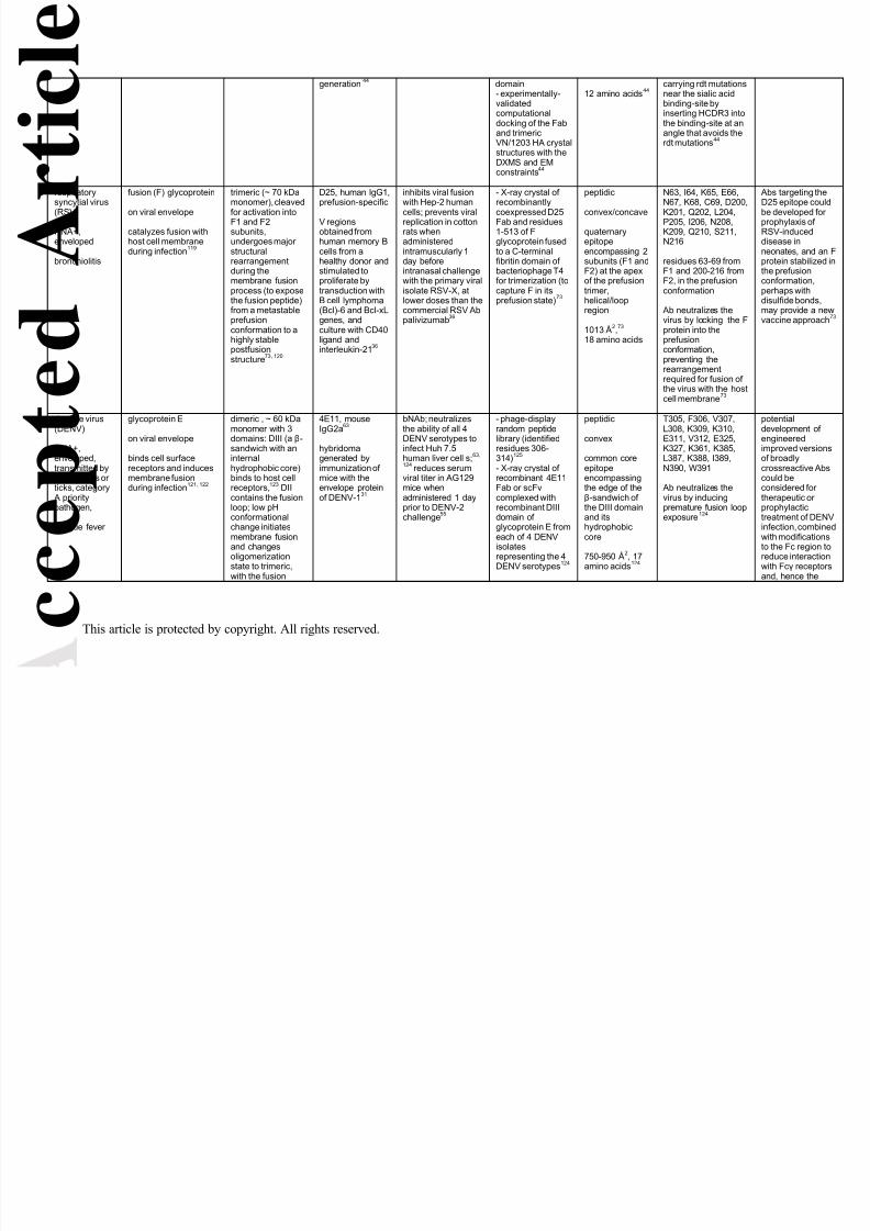
8/13/2019 imm12213
http://slidepdf.com/reader/full/imm12213 36/43
c c e p t
e d
A r t i c l e
This article is protected by copyright. All rights reserved.
generation 44 domain- experimentally-validatedcomputationaldocking of the Faband trimericVN/1203 HA crystalstructures with theDXMS and EMconstraints44
12 amino acids44 carrying rdt mutationsnear the sialic acidbinding-site byinserting HCDR3 intothe binding-site at anangle that avoids therdt mutations44
respiratorysyncytial virus
(RSV)
RNA+,enveloped
bronchiolitis
fusion (F) glycoprotein
on viral envelope
catalyzes fusion withhost cell membraneduring infection119
trimeric (~ 70 kDamonomer), cleaved
for activation intoF1 and F2subunits,undergoes majorstructuralrearrangementduring themembrane fusionprocess (to exposethe fusion peptide)from a metastableprefusionconformation to ahighly stablepostfusionstructure73, 120
D25, human IgG1,prefusion-specific
V regionsobtained fromhuman memory Bcells from ahealthy donor andstimulated toproliferate bytransduction withB cell lymphoma(Bcl)-6 and Bcl-xLgenes, andculture with CD40ligand andinterleukin-2136
inhibits viral fusionwith Hep-2 human
cells; prevents viralreplication in cottonrats whenadministeredintramuscularly 1day beforeintranasal challengewith the primary viralisolate RSV-X, atlower doses than thecommercial RSV Abpalivizumab36
- X-ray crystal ofrecombinantly
coexpressed D25Fab and residues1-513 of Fglycoprotein fusedto a C-terminalfibritin domain ofbacteriophage T4for trimerization (tocapture F in itsprefusion state)73
peptidic
convex/concave
quaternaryepitopeencompassing 2subunits (F1 andF2) at the apexof the prefusiontrimer,helical/loopregion
1013 Å2,73 18 amino acids
N63, I64, K65, E66,N67, K68, C69, D200,
K201, Q202, L204,P205, I206, N208,K209, Q210, S211,N216
residues 63-69 fromF1 and 200-216 fromF2, in the prefusionconformation
Ab neutralizes thevirus by locking the Fprotein into theprefusionconformation,preventing therearrangementrequired for fusion ofthe virus with the hostcell membrane73
Abs targetingD25 epitope c
be developedprophylaxis ofRSV-induceddisease inneonates, andprotein stabilizthe prefusionconformationperhaps withdisulfide bondmay provide avaccine appro
dengue virus
(DENV)
RNA+,enveloped,transmitted bymosquitoes orticks, category
A prioritypathogen,
dengue fever
glycoprotein E
on viral envelope
binds cell surfacereceptors and inducesmembrane fusionduring infection121, 122
dimeric , ~ 60 kDa
monomer with 3domains: DIII (a β-sandwich with aninternalhydrophobic core)binds to host cellreceptors,123 DIIcontains the fusionloop; low pHconformationalchange initiatesmembrane fusionand changesoligomerizationstate to trimeric,with the fusion
4E11, mouse
IgG2a63
hybridomagenerated byimmunization ofmice with theenvelope proteinof DENV-131
bNAb; neutralizes
the ability of all 4DENV serotypes toinfect Huh 7.5human liver cell s;63,
124 reduces serumviral titer in AG129mice whenadministered 1 dayprior to DENV-2challenge55
- phage-display
random peptidelibrary (identifiedresidues 306-314)125 - X-ray crystal ofrecombinant 4E11Fab or scFvcomplexed withrecombinant DIIIdomain ofglycoprotein E fromeach of 4 DENVisolatesrepresenting the 4DENV serotypes124
peptidic
convex
common coreepitopeencompassingthe edge of theβ-sandwich ofthe DIII domainand itshydrophobiccore
750-950 Å2, 17amino acids124
T305, F306, V307,
L308, K309, K310,E311, V312, E325,K327, K361, K385,L387, K388, I389,N390, W391
Ab neutralizes thevirus by inducingpremature fusion loopexposure124
potential
development engineeredimproved versof broadlycrossreactivecould beconsidered fotherapeutic orprophylactictreatment of Dinfection, comwith modificatto the Fc regioreduce interacwith Fcγ receand, hence th

8/13/2019 imm12213
http://slidepdf.com/reader/full/imm12213 37/43
c c e p t
e d
A r t i c l e
This article is protected by copyright. All rights reserved.
loops forming thetip of a trimericspike ; 4 serotypes(DENV-1-4)62, 122
1.6D, human IgG1
V region genesobtained by invitro stimulation ofperipheral blood Bcells from apatient that hadrecovered fromDengue virusinfection, followedby cloning andtransfection into
the humanembryonic kidneycell line HEK-29346
bNAb; neutralizesthe ability of all 4DENV serotypes toinvade monkeyepithelial LLC-MK2 cells, and enhancesinfection of Fcreceptor II –bearingK562 humanmacrophage-likecells at sub-neutralizingconcentrations46
- point mutationslibrary of DENV-3generated with aPCR mutagenesiskit46
peptidic
encompassesthe fusion loop ofthe DII domain
partial epitope, 2critical aminoacids46
W101, G109
overlaps with thefusion loop (residues98 to 109) – Abinterferes with fusionof glycoprotein E withthe endosomalmembrane of hostcells, preventingrelease of the virusinto the cytoplasm46
possibility of Adependentenhancementinfection55 46
Ebola virus
RNA-,enveloped,category Aprioritypathogen, tier 1select agent
hemorrhagicfever
GP glycoprotein
on viral envelope
mediates attachment tohost cells andmembrane fusion126, 127
trimeric, 150 kDamonomer 128 activated bycleavage into GP1and GP2;129 3 GP1viral attachmentsubunits assembleinto a chalice,cradled by the GP2fusion subunits,and a glycan capand mucin-likedomain restrictaccess to thereceptor-bindingsite in the chalicebowl;72
rearrangement andfurther cleavageGP occurs duringfusion130, 131
KZ52, humanIgG1
V region genesobtained from acombinatorial Fabphage displaylibrary generatedfrom the bonemarrow of ahuman survivor ofEbola infection34
neutralizes theability of (Zaire)Ebola virus to infectVero E6 monkeykidney epithelialcells, and confers orprolongs survival inguinea pigs againstlethal subcutaneouschallenge withguinea-pig-adaptedEbola virus whenadministeredintraperitoneallybefore or up to 1hour afterchallenge;56 does
not prolong survivalor reduce plasmaviremia in rhesusmacaques whenadministeredintravenously at 50mg/kg 1 day beforeand 4 days afterintramuscularchallenge with Ebolavirus58
- X-ray crystal ofrecombinant Fab incomplex with GPglycoproteinmutated to removethe mucin-like andtransmembranedomains and twoglycosylation sites(T42V and T230V)72
peptidic
convex
quaternaryepitope at thebase of thechalice formedby GP2 (mainly)and GP1,encompassing aβ-strand, a loop,and a helix/loop
894 Å2, 15 aminoacids72
V42, L43, V505, A507, Q508, P509,C511, P513, N514,H549, N550, Q551,D552, G553, C556
from GP1 till L43; restfrom GP272
Ab likely neutralizesby preventing GPrearrangement andblocking insertion ofthe internal fusionpeptides into the cellmembrane72
lack of protecby the Ab in thmonkey studydespite in vitrneutralizationprotective effiin the guinea model, suggethat a single mwill not sufficepost-exposureprophylactictreatment agaEbola virus58
humanimmuno-deficiency virustype 1 (HIV-1)
envelope spike (Env), A-K clades (geneticsubtypes), each withmany strains
heavilyglycosylated trimercomposed of 3gp120 molecules,
PG16, humanIgG1
V region genes
bNAb; neutralizes73% of 162 isolatesrepresenting majorHIV-1 clades at IC50
- alanine scanningand pointmutations38, 48 - X-ray crystal of
glycopeptidic
convex/concave
S158, N160 (GlcNAc560,GlcNAc561, Man562,Man564, Man565,
vaccine desigV1/V2-containimmunogens include both a

8/13/2019 imm12213
http://slidepdf.com/reader/full/imm12213 38/43
c c e p t
e d
A r t i c l e
This article is protected by copyright. All rights reserved.
RNA+,enveloped
acquiredimmuno-deficiencysyndrome
on viral envelope
binds to the primaryreceptor (CD4), and tothe coreceptor(chemokine receptorsCCR5 or CXCR4) onhost cells andmediates fusion ofvirus and host-cellmembranes132, 133
each noncovalentlyassociated with atransmembranegp41 molecule,which includes anN-terminal fusionpeptide and aconservedmembrane-proximal externalregion (MPER);gp120 consists of5 conserved (C1-
C5) and 5 variable(V1-V5) ‘loops’ (subdomains)comprising an innerand an outerdomain; thedomains areconnected by a 4-strand br idging (β)sheet, between aV1/V2 loop fromthe inner domainand a V3 loop fromthe outer domain;CD4 binds in adepressionbetween the innerand outer domainsand the bridgingsheet, and triggersa conformationalchange that
exposes the gp120coreceptor binding-site and disruptsthe gp120-gp41interaction, leadingto exposure of thegp41 fusionpeptide; N-linkeddiverse glycansshield gp120 from
Abs and consist ofboth highmannose-typeoligosaccharidesand complex-typeoligosaccharides95,
obtained from anactivated/expandedmemory B cell ofan HIV-1 clade A-infected donorselected for broadand potent serumneutralizingactivity againstHIV-138
< 50 µg/ml (infectionassay with thecoreceptor-transfected humanglioblastoma cell lineU87)38
Fab in complexwith the aglycosylatedscaffolded V1/V2loop, which wasselected by Ab-binding- site-directedmutagenesis of Ab- STD NMRcharacterization ofglycan recognitionby PG16 and site-
directed mutantsusing commerciallyavailable glycansincludingbiantennarycomplex-type andMan5GlcNAc248
encompassesboth highmannose- andcomplex-type N-linked glycans aswell as a β-strand in theV1/V2 loop ofgp120
1535 Å2, 11amino acids and10 sugars48
Man566), T163,K166, D167, R168,K169, Q170, K171,V172, N173 (GlcNAc573, Man578,Gal581, Neu5Ac582)
Ab may neutralize bypreventing Envconformations thatallow CD4 binding48
mannose-typglycan at amiacid 160 and sialic acid-containingcomplex-typeglycan at pos17348
PGT128, humanIgG1
V region genesobtained from anactivated memoryB cell of one of afew HIV-infecteddonors thatscored the highestamong 1,800donors on bothbreadth andpotency for serum
neutralization37
bNAb; neutralizes72% of 162 isolatesrepresenting majorHIV-1 clades at IC50< 50 µg/ml, and 50%at IC50 < 0.1 µg/ml(infection assay withthe coreceptor-transfected humanglioblastoma cell lineU87)37
- glycan arrayprobing- oligosaccharidecompetition withdefined-lengtholigomannosides- alanine shaving ingp12037 - X-ray crystal ofFab in complexwith a glycosylatedengineered gp120outer domain with amini V3 loop
(eODmV3)- X-ray crystal ofFab in complexwith a syntheticMan9 glycan- point mutations in
Ab (of glycan-recognizing aminoacids)- alanine shaving ofgp120 residues- EM of Fab incomplex withsoluble partiallydeglycosylated Envtrimer and manual
glycopeptidic
concave/convex
encompassing 2conservedglycans and a β-strand segmentat the C-terminus of theV3 loop
1081 Å2, 12amino acids and
15 sugars52
T297, R298, N300,N301 (GlcNAc300,GlcNAc301, Man302,Man303, Man304,Man307), R304,I322 A, I323, G324,D325, I326, R327,H330, N332* (GlcNAc200,GlcNAc201, Man232,Man233, Man234,Man237, Man238,Man214, Man215)
*N332 is not a contactresidue, but its N-linked glycan is
Ab recognizes andpenetrates the gp120glycan shield andpotentially crosslinksEnv trimers on theviral surface52 – mayneutralize bypreventing Envconformations thatallow CD4 binding
the PGT128epitope may bgood candidainclusion in anoligoepitope Abased vaccinecomponent37,
8/13/2019 imm12213
http://slidepdf.com/reader/full/imm12213 39/43
c c e p t
e d
A r t i c l e
This article is protected by copyright. All rights reserved.
132-134 fitting of the Fab-eODmV3 X-raycrystal structure52
VRC01, humanIgG1
V region genesobtained from amemory B cellisolated byfluorescence-activated cell
sorting forbinding to agp120 corecontaining theCD4 binding-siteand lack ofbinding to thegp120 core inwhich the CD40binding-site hadbeen deleted;memory B cellsderived from adonor who hasbeen infected withan HIV-1 clade Bvirus for morethan 15 years andhas not initiatedantiretroviraltreatment39
bNAb; neutralizes91% of 190 isolates,representing majorHIV-1 clades at IC50< 50 µg/ml and 72%at < 1 μg/ml (infection assay withCF2Th/synCCR5, adog thymocyte cell
line transfected withhuman CCR5)39
confers lifelongprotection tohumanized miceagainst systemic HIVchallenge whenadministered asDNA in an adeno-associated virusexpression vectoras a singleintramuscularinjection [vectoredimmunoprophylaxis(VIP)]59
- point mutationD368R (CD4binding-siteknockout) ofgp12039 - X ray crystal ofFab in complexwith a gp120 fromthe clade A/E
recombinant93TH057, consisting of itsinner domain-outerdomain core, withtruncations in theV1/V2 and V3loops as well as theN- and C-terminalregions that extendaway from the mainbody of gp120- point andinsertion mutationsin the Ab, includingreversion togermline sequence(with loss ofbinding)74
glycopeptidic
concave/convex
encompassing aloop in C1, thetruncated V1/V2loop, a loop inC2, a loop and a
helix in C3, aloop and a β-strand in C4, theV5 loop, and aloop and a β-strand in C5 inthe outer domainpart of the CD4binding-site,which isconformationallyinvariant, andextends past thefunctionally-constrained CD4binding site intothe conservedbase of the V5loop
1249 Å2, 34amino acids and
1 sugar 74
K97, T123, G124,G198, N276 (GlcNAc776), T278, N279,*N280 , A281, K282,S365 , G367 , D368 ,I371, W427 , Q428 ,G429,* T430,* T455 ,R456 , D457 , G458 ,G459, A460, N461,
T463, N465, E466,T467, R469,* G472 ,G473, D474,* K476
epitope overlaps withthe CD4 binding-siteand shares 21 of its27 residues [italic (*indicates sequencechanges from134)] –
Ab neutralizes byblocking the bindingof the CD4 receptor togp12074
vaccines couldeveloped thaguide the immsystem from Igenerearrangementhrough affinitmaturation toproduction of
VRC01-likebNAbs74
VRC01 alongbNAbs directeother conservEnv epitopes be developedvectoredimmunoprophagainst HIVinfection in higrisk individual
10E8, humanIgG3
obtained (after invitro activationand expansion)from a peripheral-blood memory Bcell of a patientwith neutralizingbreadth andpotency against a20 cross-cladepseudovirus
bNAb; neutralizes98% of 180 isolatesrepresenting majorHIV-1 clades at IC50< 50 µg/ml, and 72%at IC50 < 1 µg/ml(infection assay withthe human HeLa cellline TZM-bl, whichexpresses highlevels of CD4 andCCR5)21
- overlappingMPER peptidebinding- alanine scanningof MPER residues671-683- X ray crystal ofFab in complexwith a peptideencompassing theentire 28-residueMPER (residues656-683)- alanine shaving of
peptidic
convex/flat
one face of theMPER of gp41,which forms twohelices oriented100o relative toeach other
773 Å2, 15 aminoacids21
L661, D664, K665,S668, L669, W670,N671, W672, F673,T676, N677, L679,W680, I682, R683
neutralization by the Ab may be due tointerference withfusion of the viral andhost cellmembranes21
6 of 78 humafrom HIV-1-infdonors have 1like Abs, asassessed bybinding to HIV2/HIV-1 chimeviruses contaMPER subdomand peptideinhibition, shothat such Absnot rare and cbe induced w

8/13/2019 imm12213
http://slidepdf.com/reader/full/imm12213 40/43
c c e p t
e d
A r t i c l e
This article is protected by copyright. All rights reserved.
panel21 Ab contactresidues21
well-designedvaccine21
a Designations of microbes as group II re-emerging pathogen, or as (group III, agents with bioterrorism potential) category A, B or C priority pathogens are by the US, NIH, NationalInstitute of Allergy and Infectious Diseases (http://www.niaid.nih.gov/topics/emerging/Pages/list.aspx). Designation as a tier 1 select agent is by the US Centers for Disease Controland Prevention (http://www.selectagents.gov/Select%20Agents%20and%20Toxins%20List.html).
b Shapes of epitopes are designated as concave, convex, flat, or combinations thereof by visual inspection of published structures with DeepView (http://www.expasy.org/spdbv/) if notspecified in the primary reference.135 The surface area of the epitope is from the PISA database (http://www.ebi.ac.uk/pdbe/prot_int/pistart.html)136 when not given in the primaryreference. The surface areas (SA) of the F. tularensis O-Ag epitopes were calculated from the models using the formula [SA(Ab) + SA(Ag) – SA(complex)] / 2.
c Contact residues are from the primary reference when listed, otherwise from IEDB (http://www.iedb.org/) or, if not listed in IEDB, are residues within 4 Å of the Ab as determined by
DeepView (http://www.expasy.org/spdbv/ ).135
Amino acid numbering system is from the primary reference. Sugar numbering system is from the entry in the Protein Data Bank(http://www.rcsb.org/pdb/home/home.do) . Secondary structure is as determined by DeepView (http://www.expasy.org/spdbv/ ),135 unless otherwise indicated. The first discontinuous
residue in each epitope segment is underlined. Critical residues to Ag-Ab binding, determined by analysis of point mutants, are bolded. Secondary structure or sugar chain is indicatedby color code, with two alternating hues of the same color denoting independent structural elements: black/grey, β-strand; blue, α-helix; red/magenta, loop; green, sugar chain. Aminoacid (one-letter code) abbreviations: A, alanine; C, cysteine; D, aspartic acid; E, glutamic acid; F, phenyl alanine; G, glycine; H, histidine; I, isoleucine; K, lysine; L, leucine; M,methionine; N, asparagine; P, proline; Q, glutamine; R, arginine; S, serine; T, threonine; V, valine; W, tryptophan; Y, tyrosine. Sugar abbreviations: Gal, galactose; GlcNAc, N -acetylglucosamine; Man, mannose; Neu5Ac, 5-acetyl-neuraminic acid (sialic acid).
8/13/2019 imm12213
http://slidepdf.com/reader/full/imm12213 41/43
A c c
e p t e d A
r t i c l e
This article is protected by copyright. All rights reserved.
Figure Legends
Figure 1. Flow diagram of general strategies for identification of protective mAbs.
Figure 2. Western blot distinction between unique terminal and repeating internal epitopes of
microbial carbohydrate chains with repeating units. The N62 mAb, specific for a terminal epitope
in the O-Ag of the F. tularensis LPS (which consists of variable numbers of a tetrasaccharide
repeat, represented as ABCD) binds with equal intensity to short and long LPS chains (relative
to the abundance of each chain). But the binding intensity of the Ab52 mAb, specific for a
repeating internal epitope, increases with increasing chain length, as more mAb molecules are
bound. The basis for the differential binding pattern is illustrated schematically for the
(bracketed) lower part of the Western blot, with N62 represented by grey ovals and Ab52 by
cyan ovals. Western blot lanes from Lu et al.29
Figure 3. Linear representation of mapping results for a B-cell epitope in Neisseria
meningitides factor H binding protein, obtained using different methods. Figure adapted with
modifications from Malito et al.30 Epitope segments and contact residues are indicated by amino
acid numbers and represented in orange.
Figure 4. Examples of protective B-cell epitope structures. Ags are represented as solid
molecular surfaces (left panels and top right) and ribbon diagrams (right panels) rotated away
from the reader approximately 90° about the horizontal axis, colored with cyan carbons except
for contact residues which are colored orange. Ab V regions are shown as a wire-mesh
molecular surfaces colored grey (VH) and purple (VL). Images are clipped front and back to
more clearly show interactions of Abs and Ags. Selected contact residues are indicated in each
panel for reference. The four sugars of the tetrasaccharide repeat in F. tularensis O-Ag108 are
8/13/2019 imm12213
http://slidepdf.com/reader/full/imm12213 42/43
A c c
e p t e d A
r t i c l e
This article is protected by copyright. All rights reserved.
represented as ABCD for one and A’B’C’D’ for the other of the two repeats shown. S, serine; E,
glutamic acid; Q, glutamine; K, lysine; N, asparagine; D, aspartic acid; Man, mannose; Sia,
sialic acid. Images were generated using Maestro (version 9.3.5, Schrödinger, Inc.).

8/13/2019 imm12213
http://slidepdf.com/reader/full/imm12213 43/43
A c c
e p t e d A
r t i c l e