immunochemistry imc 03
TRANSCRIPT

IMMUNOGLOBULINS:
STRUCTURE AND
FUNCTION
Dr. Aga Syed SameerCSIR Lecturer (Demonstrator)
Department of Biochemistry,
Medical College,
Sher-I-Kashmir Institute of Medical Sciences,
Bemina, Srinagar, Kashmir, 190018. India.

IMMUNOGLOBULINS: STRUCTURE AND
FUNCTION
Glycoprotein molecules that are produced by plasma cells in response to an immunogen and which function as antibodies
Immune serum
Ag adsorbed serum
+ -
albumin
globulins
Mobility
Am
ou
nt
of
pro
tein

AntibodiesProteins that recognize and bind to a
particular antigen with very high specificity.
Made in response to exposure to the antigen.
One virus or microbe may have several
antigenic determinant sites, to which
different antibodies may bind.
Each antibody has at least two identical sites
that bind antigen: Antigen binding sites.
Valence of an antibody: Number of antigen
binding sites. Most are bivalent.
Belong to a group of serum proteins called
immunoglobulins (Igs).

Ag
Binding
Complement Binding Site
Placental
Transfer
Binding to Fc
Receptors
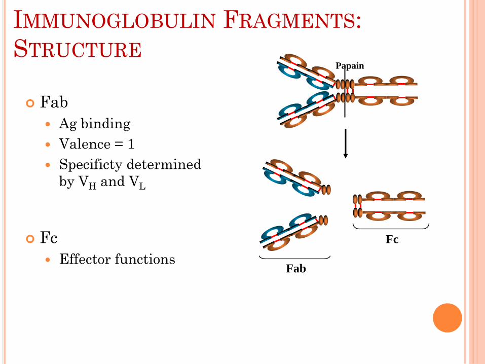
IMMUNOGLOBULIN FRAGMENTS:
STRUCTURE
IMMUNOGLOBULIN FRAGMENTS:
STRUCTURE
Fab
Ag binding
Valence = 1
Specificty determined
by VH and VL
Fc
Effector functions
Papain
Fc
Fab

Fab
Ag binding
Fc
Effector functions
F(ab’)2
Pepsin
Fc
Peptides
F(ab’)2
IMMUNOGLOBULIN FRAGMENTS:
STRUCTURE

IMMUNOGLOBULIN FRAGMENTS:
STRUCTURE

Monomer: A flexible Y-shaped molecule with
four protein chains:
2 identical light chains
2 identical heavy chains
Variable Regions: Two sections at the end of
Y’s arms. Contain the antigen binding sites
(Fab). Identical on the same antibody, but
vary from one antibody to another.
Constant Regions: Stem of monomer and
lower parts of Y arms.
Fc region: Stem of monomer only. Important
because they can bind to complement or cells.
IMMUNOGLOBULIN STRUCTURE

Variable & Constant
Regions
VL & CL
VH & CH
Hinge Region
CH1
VL
CL
VH
CH2 CH3
Hinge Region
Carbohydrate
Disulfide bond
IMMUNOGLOBULIN STRUCTURE
The four chains are linked by disulfide bonds.

L chains are one of two types:
Designated κ and λ
Only one type is found in particular Ig.
H chains are distinct for each of the five Ig
classes or isotypes:
Designated γ, α, μ, δ, ε for the respective classes of
Ig: IgG IgA, IgM, IgD & IgE.
IMMUNOGLOBULIN STRUCTURE

IMMUNOGLOBULIN STRUCTURE

IMMUNOGLOBULIN STRUCTURE

IGG
Structure
Monomer (7S)
IgG1, IgG2 and IgG4 IgG3

I. IgG
Structure: Monomer
Percentage serum antibodies: 80%
Location: Blood, lymph, intestine
Half-life in serum: 23 days
Complement Fixation: Yes
Placental Transfer: Yes
Known Functions: Enhances phagocytosis,
neutralizes toxins and viruses, protects fetus
and newborn.
IMMUNOGLOBULIN STRUCTURE

IGA Structure
Serum - monomer
Secretions (sIgA)
Dimer (11S), sIgA molecule consists of two H2L2 units plus one molecule each of J chain and secretory component(SC or SP)
J ChainSecretory Piece

III. IgA
Structure: Dimer
Percentage serum antibodies: 10-15%
Location: Secretions (tears, saliva, intestine,
milk), blood and lymph.
Half-life in serum: 6 days
Complement Fixation: No
Placental Transfer: No
Known Functions: Localized protection of
mucosal surfaces. Provides immunity to infant
digestive tract.
IMMUNOGLOBULIN STRUCTURE

IGM Structure
Pentamer (19S)
composed 5 H2L2 units plus one molecule of
J chain
Extra domain (CH4)
J chainCH4
J Chain

II. IgM
Structure: Pentamer
Percentage serum antibodies: 5-10%
Location: Blood, lymph, B cell surface
(monomer)
Half-life in serum: 5 days
Complement Fixation: Yes
Placental Transfer: No
Known Functions: First antibodies produced
during an infection. Effective against microbes
and agglutinating antigens.
IMMUNOGLOBULIN STRUCTURE

IGD
Structure
Monomer
Tail piece
Tail Piece

IV. IgD
Structure: Monomer
Percentage serum antibodies: 0.2%
Location: B-cell surface, blood, and lymph
Half-life in serum: 3 days
Complement Fixation: No
Placental Transfer: No
Known Functions: In serum function is
unknown. On B cell surface, initiate immune
response.
IMMUNOGLOBULIN STRUCTURE

IGE
Structure
Monomer
Extra domain (CH4)
CH4

V. IgE
Structure: Monomer
Percentage serum antibodies: 0.002%
Location: Bound to mast cells and basophils
throughout body. Blood.
Half-life in serum: 2 days
Complement Fixation: No
Placental Transfer: No
Known Functions: Allergic reactions.
Possibly lysis of worms.
IMMUNOGLOBULIN STRUCTURE

IMMUNOGLOBULIN STRUCTURE

IMMUNOGLOBULIN STRUCTURE

Isotypes:
Differ in the constant regions of the heavy
chains “γ, α, μ, δ, ε ” within the Y structure and
hence classified as such
Allotypes:
Differ in the constant regions of the light
chains “κ and λ” within the Y structure
Idiotypes:
Differ in unique amino acid sequence of the VH
and VL domains of a given antibody in an antigen-
binding site
IMMUNOGLOBULIN STRUCTURE

QUESTIONS?