immunoglobulin g4-related sclerosing disease … · abnormal findings were found in other organs....
TRANSCRIPT
147Copyright © 2016 The Korean Society of Radiology
INTRODUCTION
Immunoglobulin G4-related sclerosing disease (IgG4-SD) is a recently designated benign clinical entity that is histopathologically characterized by sclerosing inflammation and infiltration of numerous IgG4-positive plasma cells affecting multiple organs (1). IgG4-SD of the head and neck region was previously known as chronic sclerosing sialadenitis (Küttner tumor) or Mikulicz’s disease that involves mainly salivary and lacrimal glands (2).
Immunoglobulin G4-related sclerosing disease with involvement of palatine tonsils was reported in an earlier case study (3). However, to our knowledge, the imaging
Immunoglobulin G4-Related Sclerosing Disease Manifesting as Bilateral Tonsillar Hypertrophy on MR Images: A Case ReportMee Hyun Park, MD1, Ji Young Woo, MD1, Yul Lee, MD1, Dae Young Yoon, MD2, Hye Sook Hong, MD1, Min Eui Hong, MD3
Departments of 1Radiology and 3Pathology, Hallym University College of Medicine, Kangnam Sacred Heart Hospital, Seoul 07441, Korea; 2Department of Radiology, Hallym University College of Medicine, Kangdong Sacred Heart Hospital, Seoul 05355, Korea
Immunoglobulin G4-related sclerosing disease (IgG4-SD) is currently recognized as a distinct systemic disease involving various organs. We reported the imaging findings of a case of pathologically confirmed IgG4-SD involving bilateral palatine tonsils. CT and MRI showed diffuse enlargement of both palatine tonsils with homogeneous contrast enhancement. Focal contour bulging was noted in the right palatine tonsil. Lesions appeared as isointense on T1-weighted and slightly hyperintense on T2-weighted MRI images, as compared with muscle. The T2-weighted MRI image showed a striated pattern in both tonsils. Despite its rare occurrence, IgG4-SD should be included in the differential diagnoses of patients with symptomatic bilateral tonsillar hypertrophy that is non-responsive to medication.Index terms: IgG4-related sclerosing disease; Palatine tonsil; MRI
Korean J Radiol 2016;17(1):147-150
findings of this rare manifestation are unreported in the literature. Herein, we reported a case of IgG4-SD involving palatine tonsils, with computed tomography (CT) and magnetic resonance imaging (MRI).
This study was approved by the Institutional Review Board of our hospital. The requirement for patient informed consent was waived because of the retrospective nature of the study.
CASE REPORT
A 59-year-old woman presented with a 2-week history of sore throat. The patient had no remarkable past medical history. Physical examination revealed enlargement of bilateral palatine tonsils with a lobulated mass-like lesion and ulceration in the right tonsil. The laboratory tests at presentation showed upper-normal range of leukocytosis (9990/uL) with slightly increased neutrophil percentage (77%) and elevated C-reactive protein value (81.0 mg/L). The serum IgG4 level was not assessed at initial presentation.
Contrast-enhanced neck CT with a 64-row MDCT (Sensation 64, Siemens Medical Solutions, Malvern, PA,
http://dx.doi.org/10.3348/kjr.2016.17.1.147pISSN 1229-6929 · eISSN 2005-8330
Case Report | Neuroimaging and Head & Neck
Received September 2, 2015; accepted after revision October 14, 2015.Corresponding author: Ji Young Woo, MD, Department of Radiology, Hallym University College of Medicine, Kangnam Sacred Heart Hospital, 1 Singil-ro, Yeongdeungpo-gu, Seoul 07441, Korea.• Tel: (822) 829-5241 • Fax: (822) 832-1845• E-mail: [email protected] This is an Open Access article distributed under the terms of the Creative Commons Attribution Non-Commercial License (http://creativecommons.org/licenses/by-nc/3.0) which permits unrestricted non-commercial use, distribution, and reproduction in any medium, provided the original work is properly cited.
148
Park et al.
Korean J Radiol 17(1), Jan/Feb 2016 kjronline.org
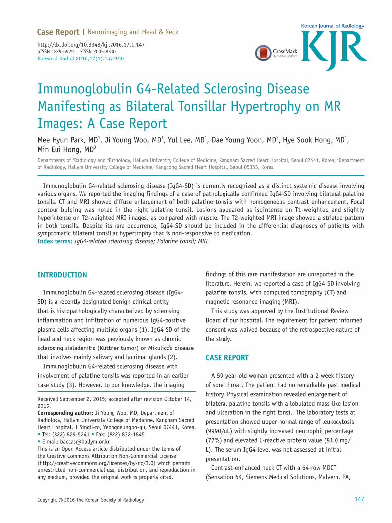
pattern on the fat suppressed T2-weighted image (Fig. 1C). 18F-fluorodeoxyglucose positron emission tomography (PET)-CT showed increased uptake at both palatine tonsils with maximum standardized uptake value of 7.9. On PET-CT, no abnormal findings were found in other organs. Considering the clinical and radiological findings, malignancy such as lymphoma or squamous cell carcinoma of right palatine tonsil associated with underlying bilateral tonsillar hypertrophy was initially suspected.
USA) and MRI with a 3.0 Tesla MR scanner (Skyra, Siemens Medical Solutions, Erlangen, Germany) showed diffuse enlargement of both palatine tonsils and homogenous contrast enhancement. There were several enlarged lymph nodes in the right level II. Lesions appeared as isointense on T1-weighted (Fig. 1A) and slightly hyperintense on T2-weighted MRI images (Fig. 1B), as compared with muscle. Asymmetric contour bulging was noted in the right palatine tonsil. Both palatine tonsils showed a striated
Fig. 1. MRI and histologic findings of 59-year-old woman with immunoglobulin G4 (IgG4)-related sclerosing disease.A, B. Coronal T1-weighted (A) and T2-weighted (B) MR images demonstrate diffuse enlargement of both palatine tonsils (arrows). Lesion appears isointense on T1-weighted- and slightly hyperintense on T2-weighted-images, as compared with muscle. Note focal asymmetric bulging in right palatine tonsil (arrowheads). C. Axial fat-saturated T2-weighted image reveals striated pattern in bilateral tonsils (arrows). D-G. Microscopic examinations show dense lymphoplasmacytic infiltrate (D, hematoxylin and eosin [H&E], x 40) and storiform fibrosis with lymphoplasmacytic inflammatory infiltration (E, H&E, x 400). Immunochemical studies demonstrate many IgG4-positive plasma cells (> 50/high power field) (F). Ratio of IgG4- (F, x 200) to total IgG- (G, x 200) expressing cells is > 40%.
A
D
G
B
E
C
F
149
IgG4-SD Involving Bilateral Tonsils
Korean J Radiol 17(1), Jan/Feb 2016kjronline.org
The lesion was diagnosed as IgG4-SD on subsequent excisional biopsy of the ulcerated mass-like lesion in the right palatine tonsil. Histological examination showed dense infiltration of plasma cells with storiform-type fibrosis (IgG4 positive cells > 50/high power field), and a ratio of IgG4-expressing to total IgG-expressing cells of > 40% on immunohistochemistry (Fig. 1D-G).
The patient was treated with corticosteroid therapy, which resulted in marked improvement of swelling of bilateral palatine tonsils within 1 month. Follow-up CT taken 7 weeks after initial CT revealed improvement of bilateral tonsillar hypertrophy.
DISCUSSION
The association of IgG4 with sclerosing diseases was firstly described in 2001 by Hamano et al. (4), who reported an elevated serum IgG4 level in patients with autoimmune pancreatitis. Since 2003, extra-pancreatic manifestations have been reported and the disease has been recognized as a systemic disease (5-11). Elevated serum IgG and IgG4 levels and the presence of autoantibodies (e.g., rheumatoid factor, antinuclear antibodies) may help in the diagnosis of IgG4-SD. Histologically, IgG4-SD is characterized by abundant infiltration of IgG4-positive plasma cells, storiform fibrosis, obliterative phlebitis, mild to moderate eosinophilia, and increased number of IgG4-positive cells on immunohistochemical staining (5-7). Patients usually show dramatic responses to corticosteroid.
In the head and neck regions, IgG4-SD most commonly involves the salivary and lacrimal glands. Recently, Khurram et al. (3) reported a case of IgG4-SD involving skin, tongue, and palatine tonsil. However, they focused on tongue lesions rather than tonsillar lesions and detailed imaging findings were not described.
According to previous reports, CT and MRI findings of IgG4-SD are usually nonspecific. IgG4-SD of the head and neck areas most commonly involves the salivary glands and lacrimal glands; furthermore, cross-sectional imaging usually shows bilateral symmetric swelling of the involved glands with homogeneous enhancement. In our case, imaging findings of IgG4-SD with tonsillar involvement were also nonspecific, but consistent with IgG4-SD involving other organs, with bilateral swelling and focal bulging of palatine tonsil. Although IgG4-SD often has nonspecific imaging findings and is difficult to differentiate from other disease with imaging alone, the radiologist should include
this disease in the differential diagnoses in an appropriate clinical setting because the disease responds well to corticosteroid therapy. Patients with IgG4-SD of palatine tonsil show clinical symptoms such as sore throat and swelling of bilateral tonsils, which are persistent despite antibiotics and analgesics. Diffuse bilateral enlargement of submandibular gland or lacrimal gland are suggestive of systemic IgG4-SD, because IgG4-SD commonly involves multiple organs.
In summary, we presented a case of IgG4-SD involving palatine tonsils with a review of its CT and MRI features. As seen in this case report, the imaging features of IgG4-SD in the palatine tonsils are nonspecific, but compatible with known imaging findings of IgG4-SD involving other organs. The radiologist should include IgG4-SD in the differential diagnoses and recommend that clinicians check serum IgG and IgG4 levels in patients with bilateral tonsillar hypertrophy and persistent clinical symptoms despite medication.
REFERENCES
1. Kamisawa T, Funata N, Hayashi Y, Eishi Y, Koike M, Tsuruta K, et al. A new clinicopathological entity of IgG4-related autoimmune disease. J Gastroenterol 2003;38:982-984
2. Katsura M, Mori H, Kunimatsu A, Sasaki H, Abe O, Machida T, et al. Radiological features of IgG4-related disease in the head, neck, and brain. Neuroradiology 2012;54:873-882
3. Khurram SA, Fernando M, Smith AT, Hunter KD. IgG4-related sclerosing disease clinically mimicking oral squamous cell carcinoma. Oral Surg Oral Med Oral Pathol Oral Radiol 2013;115:e48-e51
4. Hamano H, Kawa S, Horiuchi A, Unno H, Furuya N, Akamatsu T, et al. High serum IgG4 concentrations in patients with sclerosing pancreatitis. N Engl J Med 2001;344:732-738
5. Kamisawa T, Okamoto A. IgG4-related sclerosing disease. World J Gastroenterol 2008;14:3948-3955
6. Okazaki K, Uchida K, Koyabu M, Miyoshi H, Takaoka M. Recent advances in the concept and diagnosis of autoimmune pancreatitis and IgG4-related disease. J Gastroenterol 2011;46:277-288
7. Vlachou PA, Khalili K, Jang HJ, Fischer S, Hirschfield GM, Kim TK. IgG4-related sclerosing disease: autoimmune pancreatitis and extrapancreatic manifestations. Radiographics 2011;31:1379-1402
8. Ko Y, Woo JY, Kim JW, Hong HS, Yang I, Lee Y, et al. An immunoglobulin G4-related sclerosing disease of the small bowel: CT and small bowel series findings. Korean J Radiol 2013;14:776-780
9. Coulier B, Montfort L, Beniuga G, Pierard F, Gielen I. Small bowel obstruction caused by peritoneal immunoglobulin
150
Park et al.
Korean J Radiol 17(1), Jan/Feb 2016 kjronline.org
g4-related disease mimicking carcinomatosis: case report. Korean J Radiol 2014;15:66-71
10. Choi JW, Kim SY, Moon KC, Cho JY, Kim SH. Immunoglobulin G4-related sclerosing disease involving the urethra: case report. Korean J Radiol 2012;13:803-807
11. Rossi M, Virgilio E, Laurino F, Orgera G, Menè P, Pirozzi N, et al. Giant hepatic artery aneurysm associated with immunoglobulin G4-related disease successfully treated using a liquid embolic agent. Korean J Radiol 2015;16:953-954