immunoglobulin m immunoglobulin g responses to conjugated ... · movedfrom the plates, washed six...
TRANSCRIPT

Vol. 49, No. 3INFECTION AND IMMUNITY, Sept. 1985, p. 598-6080019-9567/85/090598-11$02.00/0Copyright ©) 1985, American Society for Microbiology
Immunoglobulin M and Immunoglobulin G Responses in BALB/cMice to Conjugated Outer Membrane Extracts of Four Salmonella
SerotypestDAVID A. KUDRNA, GEORGE W. TERESA,* JEANENE M. ARNZEN, AND K. SCOTT BEARD
Department of Bacteriology and Biochemistry, College of Agriculture, University of Idaho, Moscow, Idaho 83843
Received 28 February 1985/Accepted 28 May 1985
Outer membranes (OMs) of Salmonella enteritidis, S. anatum, S. typhimurium, and S. infantis were extractedand cross-linked with glutaraldehyde to form a large macromolecular antigen. The antigen consisted of OMproteins and lipopolysaccharide and was designated 4-OMP-LPS. Polyacrylamide gel electrophoresis ofextracted OMs from each serotype revealed differences in protein profiles. S. enteritidis and S. infantispossessed a greater variety of proteins than did S. anatum and S. typhimurium. Immunizations with4-OMP-LPS in phosphate-buffered saline (4-OMP-LPS-C) and 4-OMP-LPS emulsified with muramyl dipeptidein the oil phase of a hexadecane-water emulsion (4-OMP-LPS-MDP) revealed that BALB/c mice were capableof eliciting specific primary and secondary immunoglobulin M (IgM) and IgG responses. Both antigenpreparations were capable of eliciting IgM and IgG specific for the cell surfaces of each live Salmonellaserotype. Also, 4-OMP-LPS-MDP and 4-OMP-LPS-C were capable of evoking a substantial anamnesticresponse. Adsorption studies revealed that the combined serotypes had the antigenic capacity to adsorb up to94% of the antibodies, but 4-OMP-LPS-MDP antibodies were more effectively adsorbed than were 4-OMP-LPS-C antibodies. Adsorption of pooled antiserum with heterologous bacteria yielded a variety of adsorptionprofiles.
Outer membranes (OMs) of several gram-negative bacte-ria are capable of eliciting humoral antibody responses (7, 8,18, 21, 22, 28, 51). It has been found that outer membraneproteins (OMPs) prepared from Escherichia coli are potentB-cell mitogens capable of inducing a polyclonal antibodyresponse (28). Additionally, the E. coli OMP preparationwas mitogenic for lipopolysaccharide (LPS) nonresponderC3H/HeJ mice and could act as an adjuvant by enhancingantibody responses specific for sheep erythrocytes (28).Karch and Nixdorff (18) demonstrated that an OMP iso-
lated from Proteus mirabilis had the capacity to elicit anti-body responses typical of thymus-dependent antigens. Theimmunoglobulin G (IgG) response could be increased if theOMP was complexed with either LPS or vesicles ofphospholipids extracted from P. mirabilis. Conversely,OMP was capable of significantly increasing the humoralresponse to LPS by shifting the response from predomi-nantly IgM to predominantly IgG.Kuusi et al. (22), by using an OMP (porin) preparation
extracted from a rough strain of Salmonella typhimurium,were successful in eliciting high antiporin and high anti-LPStiters in both mice and rabbits. Both porins and rabbitantiserum to porins provided significant protection in micechallenged with smooth S. typhimurium. When the rabbitantiporin serum was adsorbed with an LPS immunosorbent,the protective capacity of the serum did not change, indicat-ing that antiporin antibodies were the protective compo-nents. After further purification of the porin preparation (21)to remove LPS and lipoproteins, it was found that theprotective capacity of the antiserum against the purifiedporin preparation was lost. However, on reconstitution of
* Corresponding author.t Research paper no. 8553 of the Idaho Experiment Station,
College of Agriculture, University of Idaho, Moscow.
the vaccine with homologous LPS, the protective capacitywas restored.
Winter et al. (51) investigated immune responses in cattlevaccinated with whole cells, OMs, and OMPs prepared fromBrucella abortus. These workers reported that OMP wascapable of inducing significant antibody and delayed hyper-sensitivity responses. Recently, a purified OMP-F (porin)preparation isolated from Pseudomonas aeruginosa wasshown to protect mice both actively and passively (7). Otherstudies have reported that mice infected with P. aeruginosaelicited antibodies specific for OMPs (8).The observation that OMPs complexed with LPS en-
hanced the synthesis of IgG (18) suggested that it may befeasible to prepare a novel immunizing agent by cross-linking OM components. Such a preparation would haveseveral advantages over antigens consisting of either singleor unconjugated OM components. (i) It would be moreeconomical; (ii) it would allow the construction of OMpolyvalent vaccines containing either homologous orheterologous OM antigens; (iii) it would allow the utilizationof many OM components that may have a greater enhancingvalue when cross-linked than if present as single compo-nents; (iv) it would present to the immune system surfaceantigens which may otherwise be hidden in the OM on theintact organism(s); and (v) such a complex molecule contain-ing increased protein may be able to elicit both humoral andcell-mediated immune responses. It is for these reasons thatwe designed experiments to: (i) extract OMs of four promi-nent Salmonella serotypes representing four serologicallydifferent serogroups and compare their OMP profiles, (ii)utilize as many of these extractable OMP components (in-cluding a small amount of LPS) as possible and conjugate theOMPs with glutaraldehyde to form a cross-linked antigen,and (iii) examine the primary and secondary IgM and IgGresponses specific for both the antigen and the live homolo-gous organisms.
598
on October 12, 2019 by guest
http://iai.asm.org/
Dow
nloaded from

IMMUNOGLOBULIN RESPONSE TO SALMONELLA OUTER MEMBRANES
MATERIALS AND METHODS
Bacterial strains and growth conditions for preparation ofcell walls. S. entieritidis NCTC 5694 serogroup D was obtainedfrom F. M. Collins (Trudeau Institute, Inc., Saranac Lake,N.Y.). S. ainautiim (83-4586) serogroup E1 and S. infaintis(83-4442) serogroup C1 were obtained from B. 0. Blackburn(National Veterinary Services Laboratories, Ames, Iowa). S.typhimiurilim ATCC 14028 serogroup B was obtained from theAmerican Type Culture Collection (Rockville, Md.). Allorganisms were serotyped by the method of Ewing (5) and,after identification, were maintained on Trypticase soy agar(BBL Microbiology Systems, Cockeysville, Md.) supple-mented with 5% sheep blood.Large quantities of cells were obtained from spread plates
of each organism on the same medium. After 18 h ofincubation at 37°C in increased humidity, cells were re-moved from the plates, washed six times in 0.9% NaCI andtwice with deionized water, and stored at -60°C.
Isolation and extraction of cell walls. OMs were extractedfrom each Salmonella serotype by a modification of themethod of Nixdorff et al. (30). Frozen cells were thawed,mechanically deflagellated, and washed three times withdeionized water and once with 0.02 M NaHCO3. Afterwashing, cells were suspended in 0.4% (wt/vol) 3-[(3-cholamidopropyl)-dimethylammonio]-1-propanesulfonateprepared by the procedure of Hjelmeland (10). Cells werebroken by releasing the slurry from a French pressure cell(Travenol Laboratories, Inc., Savage, Md.) operated at aninternal pressure of 1,406 kg/cm2 while maintaining an ap-proximate temperature of 4°C. The cell wall fraction wascollected by centrifuging for 60 min at 30,000 x g at 40C. Theresulting cell wall pellet was washed three times with 0.02 MNaHCO3 and three times with deionized water and thenextracted for 60 min with five volumes of 90% (vol/vol)acetic acid at room temperature. The mixture was centri-fuged for 15 min at 7,500 x g at 40C, and the supernatant wasdialyzed against deionized water at 4°C for 72 h. The OMmaterial precipitating during dialysis was collected by cen-trifugation for 40 min at 15,000 x g at 40C, and the pellet wasdissolved in 1.0 M NaOH and neutralized with 0.1 M sodiumacetate buffer (pH 4.5) containing 0.1 M NaCl. The solutionwas dialyzed against deionized water at 4°C for 48 h andcentrifuged for 15 min at 7,500 x g at 40C. The supernatantcontaining the OMP was lyophilized and stored at 4°C.Because it was the objective of this research to investigatethe humoral immune responses of a conjugated fraction ofOM containing a small amount of LPS, no further purifica-tion was performed.
Glutaraldehyde conjugation of OMPs. OMPs (500 mg) fromeach Salmonella serotype were combined and dissolved in400 ml of 0.1 M phosphate-buffered saline (PBS; pH 8.3) andcentrifuged for 15 min at 9,000 x g at room temperature. Thesupernatant was warmed to 37°C in a water bath, andglutaraldehyde (Aldrich Chemical Co., Inc., Milwaukee,Wis.) was added to a final concentration of 0.1% (37).Conjugation proceeded at 370C for 60 min with occasionalagitation, which resulted in a green-yellow solution. Thesolution was dialyzed against deionized water at 4°C for 24 hand lyophilized. The conjugate was rehydrated with deion-ized water, centrifuged for 20 min at 12,000 x g at 4°C, andwashed with deionized water in an ultrafiltration cell(Amicon Corp., Danvers, Mass.) equipped with a YM30membrane filter. After thorough washing, the solution wasconcentrated and lyophilized, and the final product (4-OMP-LPS) was stored under reduced pressure at -22°C.
Chemical analyses of 4-OMP-LPS. Total nitrogen contentwas estimated by the micro-Kjeldahl method of Kabat andMeyer (15) with bovine serum albumin as a standard. Proteincontent was determined by the Lowry method (24) withbovine serum albumin as a standard. 2-Keto-3-deoxyoc-tonate was estimated by the method of Karkhanis et al. (20)with 2-keto-3-deoxyoctonate (Sigma Chemical Co., St.Louis, Mo.) as a standard. Hexosamine was estimated asdescribed by Nowotny (31). Samples were hydrolyzed in 3 NHCI for 4 h at 100°C, evaporated to dryness over KOHpellets under reduced pressure, suspended in deionizedwater, neutralized with 1.0 N NaOH, and analyzed withD-(+)-glucosamine hydrochloride (Sigma) as a standard.SDS-PAGE of OMPs. OMPs from each Salmonella
serotype were separated by discontinuous sodium dodecylsulfate-polyacrylamide gel electrophoresis (SDS-PAGE)with the gel formulation of Lugtenberg et al. (26) in anelectrophoretic apparatus described by Matsudaira and Bur-gess (27). Gel slabs were 67 mm in width, 80 mm in length,and 1.0 mm in depth, with separating and stacking gelscontaining 11 and 3% acrylamide, respectively. Proteinmolecular weight standards, expressed as molecular weightX 103 followed by the letter K (1), were bovine serumalbumin (66K), ovalbumin (45K), and trypsinogen (24K)(Sigma). OMPs (15 p.g) were applied to each well. Electro-phoresis was carried out at a constant amperage of 9 mA pergel slab until the tracking dye was 5 mm from the bottomn ofthe gel. Gels were stained for 4 h at room temperature in asolution containing 0.1% (wt/vol) Brilliant Blue R (Sigma),50% absolute methanol, 10% acetic acid, and 40% deionizedwater. Gels were destained in a solution of 359 absolutemethanol-7% acetic acid-58% deionized water until thedesired clarity was obtained.
Densitometer scans of SDS-PAGE-separated OMPs. Sepa-rated OMPs from each Salmonella serotype were densityscanned with a model 2520 gel scanner (Gilford InstrumentLaboratories, Inc., Oberlin, Ohio). A Gilford model 252photometer and a model DU spectrophotometer (BeckmanInstruments, Inc., Fullerton, Calif.) were used as lightsource and wavelength control, respectively. Protein banddensities were recorded on a Gilford model 6051 recorder.Electrophoretic mobilities (relative mobilities) of proteinpeaks were calculated (1). For identification of shared pro-teins, peaks possessing the same relative mobilities wereassigned numbers.
Mice. Female inbred BALB/c mice (Harlan-Sprague-Dawley, Inc., Madison, Wis.) 6 weeks of age were used in allexperiments. Mice were housed five to an Econofilter cov-ered cage (Maryland Plastics, Inc., New York, N.Y.) withfree access to food and water.
Preparation of antigen emulsions. Emulsions containing 2.0mg of 4-OMP-LPS with 2.0 mg of muramyl dipeptide (MDP;Calbiochem-Behring, La Jolla, Calif.), 1.5% hexadecane(Sigma), 1.05% polyoxyethylene-sorbitan-mono-oleate(Tween 80; Sigma), and 0.45% sorbitan-mono-oleate(Emsorm 2500; Emery Industries, Mauldin, S.C.) weredesignated 4-OMP-LPS-MDP and were used for primary andsecondary immunizations. Emulsions excluding 4-OMP-LPSwere designated MDP-C and were used for inoculation ofprimary and secondary adjuvant control mice. Oil-wateremulsions were prepared as described elsewhere (L. F.Woodard and R. L. Jasman, Vaccine, in press). MDP (2 mg)in 160 LIl of absolute methanol was placed in a dry, sterile15-ml glass-Teflon homogenizer. 4-OMP-LPS (2 mg) wastransferred to homogenizers which were designated 4-OMP-LPS-MDP. After evaporation of methanol from all homoge-
VOL. 49, 1985 599
on October 12, 2019 by guest
http://iai.asm.org/
Dow
nloaded from

600 KUDRNA ET AL.
nizers, 60 of hexadecane, 42 of Tween 80, and 18 p.l ofEmsorm 2500 were added. Mixtures were emulsified for 60 s
with a motor-driven pestle operated at 1,500 rpm beforeaddition of sterile 0.1 M PBS (pH 7.3) to achieve a finalvolume of 4.0 ml. Mixtures were homogenized twice for 2min. Microscopic examination of antigen emulsions revealeduniform oil droplets with a shaded appearance (52).Immunization of mice. For primary immunization experi-
ments, 12 groups of five mice each were vaccinated subcu-taneously (s.c.) in the nape of the neck with 0.2 ml of4-OMP-LPS-MDP, 0.2 ml of MDP-C, or 100 p,g of 4-OMP-LPS in 0.2 ml of PBS (4-OMP-LPS-C). On postinoculationdays (PIDs) 7, 14, 21, and 28, mice were bled by cardiacpuncture after anesthetization. Individual sera were col-lected and stored at -60°C. Secondary immunization exper-
iments with mice and dosages as in primary experimentswere carried out by administering the primary dose on day 0and the secondary dose s.c. in the base of the tail on PID 20.Serum was collected, as previously described, on PIDs 25,30, 35, and 40.
Determination of 4-OMP-LPS-specific IgM and IgG byELISA. Immunoglobulin analyses were carried out by a
microplate modification of the enzyme-linked immunosor-bent assay (ELISA) technique described by Voller et al.(50). Briefly, polystyrene microtiter plates (no. 25860; Corn-ing Glass Works, Corning, N.Y.) were coated by overnightevaporation of 10 p.g of 4-OMP-LPS per 50 of deionizedwater per well. Wells were washed once with 0.2 M PBS (pH7.3). To reduce nonspecific binding, we added 100 p1I of a
solution containing 0.1% gelatin (Sigma) in PBS to each well.After incubation at 37°C for 30 min, wells were washed once
with PBS, and 100 p.l of a serially diluted serum sample(diluted in PBS) was added to each well. After 1 h ofincubation at 37°C, wells were washed three times with PBS,and 100 of specific anti-immunoglobulin-conjugated en-
zyme (peroxidase-conjugated goat anti-mouse IgG antibodyor peroxidase-conjugated goat anti-mouse IgM antibody[Tago Inc., Burlingame, Calif.]) was added to appropriatewells. Anti-immunoglobulin-conjugated enzyme for IgM as-
say was diluted 1:700, and anti-immunoglobulin-conjugatedenzyme for IgG assay was diluted 1:1,500. Both conjugateswere diluted in PBS containing 0.05% gelatin. After 1 h ofincubation at 37°C, wells were washed three times with PBSand twice with deionized water, and 100 of substratesolution was added to each well. Substrate solution con-sisted of 0.08% 5-aminosalicylic acid (pH 6.0) in deionizedwater with 0.05% H202 added before use. After 1 h ofincubation at 37°C in the dark, the optical density of eachwell was determined with a microplate reader (no. EL 307;Bioteck, Burlington, Vt.). ELISA controls consisted oftriplicate wells in which: (i) 4-OMP-LPS had not been driedto wells, (ii) PBS was added in place of serum, and (iii) PBSwas added in place of anti-immunoglobulin-conjugated en-zyme. Sera of untreated mice were included as negativeserum controls. Test serum was analyzed by triplicate anal-yses of each serially diluted serum sample. Antibody titerswere defined as the highest serum dilution giving an opticaldensity above the statistical mean of negative serum con-trols.
Determination ofIgM and IgG specific for live homologousSalmonella spp. Antibodies elicited by immunizations with4-OMP-LPS-MDP and 4-OMP-LPS-C were analyzed forspecificity to homologous live Salmonella spp. by ELISA asdescribed by Ison et al. (13). Briefly, S. enteritidis, S.anatum, S. typhimurium, and S. infantis were grown on 5%blood agar plates for 18 h at 37°C in increased humidity.
TABLE 1. Heterologous bacteria used in adsorption of antiseraBacterium Source"
Arizona hinshawii ......... University of Idaho reference cultureEnterobacter aerogenes .... ATCC 13048Escherichia co/i ............ATCC 25922Klebsiella pneumoniae ..... ATCC 13883Proteus mirabilis .......... University of Idaho reference culturePseudomonas aeruginosa . ATCC 27853Serratia marcescens ....... ATCC 8100Yersinia enterocolitica ..... University of Idaho reference cultureStaphylococcus epidermidis ATCC 12228
" ATCC, American Type Culture Collection.
Each serotype was washed once with 0.2 M PBS (pH 7.3),suspended in PBS, and adjusted to an optical density of 0.5at 700 nm.
Polystyrene microtiter plate (no. 25860; Corning) wellswere filled with poly-L-lysine (Sigma) at 50 ,ug/ml in PBS andincubated at room temperature for 30 min. Wells werewashed three times with PBS, and 100 p.l of bacterialsuspension was added to appropriate wells. After 30 min ofincubation at room temperature, nonadherent bacteria wereremoved by aspiration, and wells were washed three timeswith PBS. Plate wells coated with live bacteria were filledwith PBS containing 0.1 M glycine and stored in increasedhumidity at 4°C.
Before use, plates were allowed to reach room tempera-ture, and wells were washed twice with PBS and filled withPBS containing 0.1% gelatin (Sigma) to reduce nonspecificbinding. The ELISA procedure was then continued asdescribed previously, except that anti-immunoglobulin-conjugated enzyme for IgM assay was diluted 1:500 andanti-immunoglobulin-conjugated enzyme for IgG assay wasdiluted 1:800.
Adsorption of antisera with homologous and heterologousbacteria. We prepared antisera for adsorption studies byimmunizing mice s.c. in the nape of the neck with 0.2 ml of4-OMP-LPS-MDP or 4-OMP-LPS-C on day 0 and adminis-tering a secondary dose s.c. in the base of the tail on PID 20.Antisera were collected, as previously described, on PID 30.Homologous Salmonella strains used in these experiments
were described above. Heterologous bacteria used are listedin Table 1. All bacteria were grown on 5% sheep blood agarplates for 18 h at 37°C. Cells were washed once with 0.2 MPBS (pH 7.3), suspended in PBS, and adjusted to McFarlandnephelometer tube 9. Specified bacterial suspensions (500 p.leach) were centrifuged for 10 min at 8,500 x g, and thesupernatants (PBS) were removed. Antisera (80 ,ul) fromeither 4-OMP-LPS-MDP- or 4-OMP-LPS-C-immunized micewere mixed with bacterial cells and incubated in a 37°Cwater bath for 40 min. After incubation, tubes were sealedand refrigerated for 3 h. Cells were pelleted, and serum wassterilized by centrifugation through 0.2-p.m (pore size) cel-lulose acetate filters (Centrex DF122/1; Schleicher &Schuell, Inc., Keene, N.H.). After overnight storage at 4°C,sera were analyzed for 4-OMP-LPS-specific IgM and IgG byELISA as previously described.
Statistical analyses. Antibody titers, both IgM and IgG,determined from each mouse on each bleed day were con-verted to log2 + 1 and compared statistically. Either theFisher exact test (46) or the Student t test (34) was used tocalculate two-tailed P values for differences in titers. Statis-tical analyses were not performed on adsorption experi-
INFECT. IMMUN.
on October 12, 2019 by guest
http://iai.asm.org/
Dow
nloaded from

IMMUNOGLOBULIN RESPONSE TO SALMONELLA OUTER MEMBRANES
66K 45K 24K
Region I Region II Region III Region IV
1lt 120K-6OK GO6K-34K 1 34K-19K 19K- 12K
° 12 34 56 78 101112 14 151617 20 21 293031 32 33 34353637
FIG. 1. Densitometer scan of SDS-PAGE-separated OMPs obtained from S. enteritidis. Dominant protein regions are indicated, and majorprotein peaks are numbered. Positions of protein molecular weight standards are indicated, and arrows at the left and right sides indicate thetop of the separating gel and the position of the tracking dye, respectively. Absorbance was measured at 590 nm.
ments owing to inadequate quantities of antisera. Data wereeither significant (P < 0.05), highly significant (P < 0.01), ornot significant (P > 0.05).
RESULTS
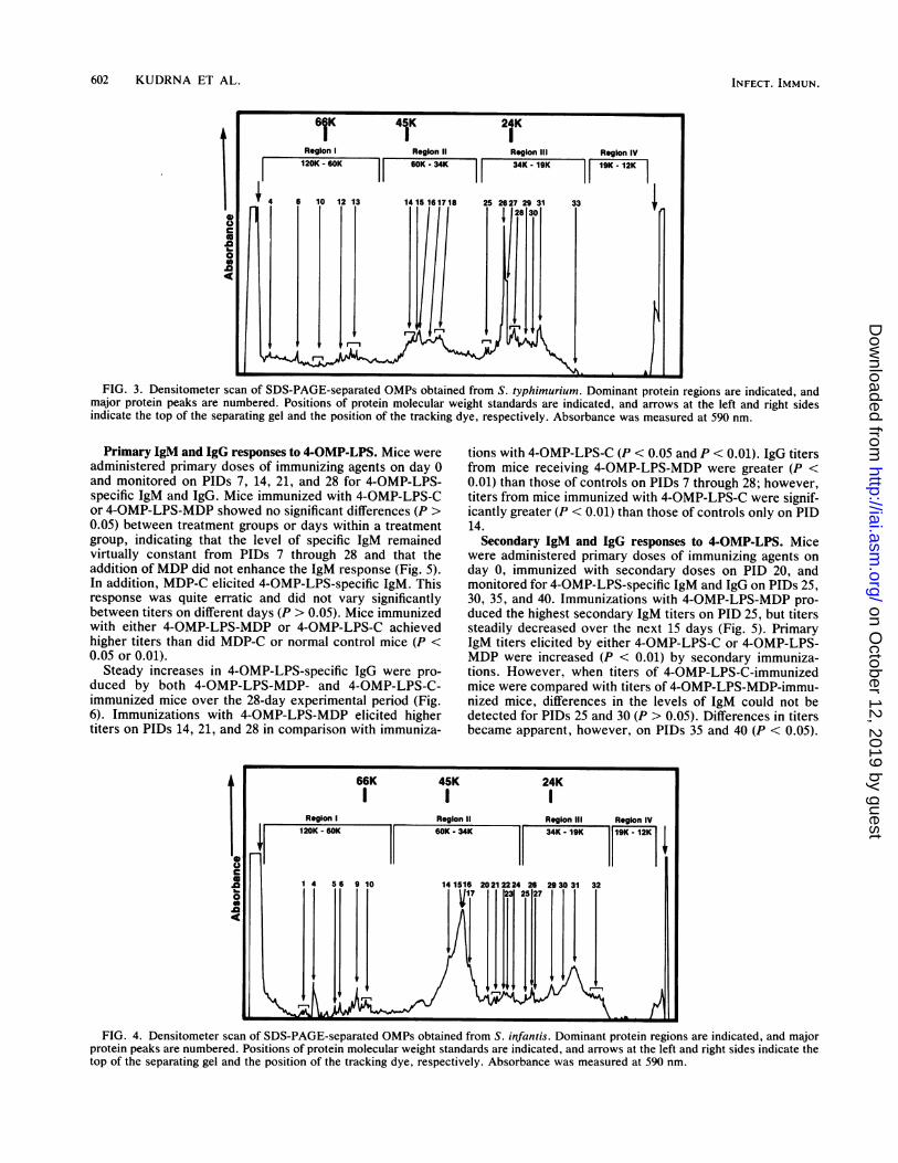
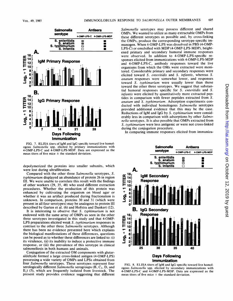
Comparison of OMPs from Salmonella serotypes by SDS-PAGE. OMPs from each Salmonella serotype were solubil-ized in 0.0625 M Tris hydrochloride (pH 6.8), separated bySDS-PAGE, and examined by densitometer scanning (Fig. 1through 4). Each serotype possessed OMPs with molecularweights ranging from 12K to 120K, which were arbitrarilyclassified into four electrophoretic regions.
Region I in each serotype provided a contrasting profile onthe uniqueness that characterized the OMPs of these orga-nisms. S. enteritidis displayed the greatest variety andquantity of OMPs. Proteins 4, 6, 10, and 12 were sharedamong S. enteritidis, S. infantis, and S. typhimurium,whereas S. anatum was largely deficient of OMPs. Onlyprotein 4 was shared by all serotypes.Region II in each serotype consisted of multiple protein
peaks and possessed the greatest amount of protein. Asindicated, proteins 14 through 17 migrated closely andaccounted for the majority of proteins for each serotype. S.typhimurium did not produce these proteins in quantity asdid the other serotypes. In addition, S. typhimurium OMswere deficient in proteins 19 through 24.Region III was a protein-rich region for each serotype,
with proteins 30 and 31 commonly shared. These twoproteins were quantitatively the most significant proteins foreach serotype except S. typhimurium, which produced largequantities of protein 26 and other major proteins that werenot apparent in the OMPs of the other serotypes.
Low-molecular-weight proteins occurring in region IVwere numerous and in high quantity in S. enteritidis, whichshared proteins 34 and 36 with S. anatum. S. infantis and S.typhimurium did not possess any major proteins in thisregion.Chemical analyses. Chemical analyses revealed that 4-
OMP-LPS contained 11.21% nitrogen, 71.10% protein,0.48% 2-keto-3-deoxyoctonate, and 3.63% hydrolyzablehexosamine.
FIG. 2. Densitometer scan of SDS-PAGE-separated OMPs obtained from S. anatum. Dominant protein regions are indicated, and majorprotein peaks are numbered. Positions of protein molecular weight standards are indicated, and arrows at the left and right sides indicate thetop of the separating gel and the position of the tracking dye, respectively. Absorbance was measured at 590 nm.
VOL. 49, 1985 601
on October 12, 2019 by guest
http://iai.asm.org/
Dow
nloaded from
602 KUDRNA ET AL.
FIG. 3. Densitometer scan of SDS-PAGE-separated OMPs obtained from S. typhimurium. Dominant protein regions are indicated, andmajor protein peaks are numbered. Positions of protein molecular weight standards are indicated, and arrows at the left and right sidesindicate the top of the separating gel and the position of the tracking dye, respectively. Absorbance was measured at 590 nm.
Primary IgM and IgG responses to 4-OMP-LPS. Mice wereadministered primary doses of immunizing agents on day 0and monitored on PIDs 7, 14, 21, and 28 for 4-OMP-LPS-specific IgM and IgG. Mice immunized with 4-OMP-LPS-Cor 4-OMP-LPS-MDP showed no significant differences (P >0.05) between treatment groups or days within a treatmentgroup, indicating that the level of specific IgM remainedvirtually constant from PIDs 7 through 28 and that theaddition of MDP did not enhance the IgM response (Fig. 5).In addition, MDP-C elicited 4-OMP-LPS-specific IgM. Thisresponse was quite erratic and did not vary significantlybetween titers on different days (P > 0.05). Mice immunizedwith either 4-OMP-LPS-MDP or 4-OMP-LPS-C achievedhigher titers than did MDP-C or normal control mice (P <0.05 or 0.01).
Steady increases in 4-OMP-LPS-specific IgG were pro-duced by both 4-OMP-LPS-MDP- and 4-OMP-LPS-C-immunized mice over the 28-day experimental period (Fig.6). Immunizations with 4-OMP-LPS-MDP elicited highertiters on PIDs 14, 21, and 28 in comparison with immuniza-
tions with 4-OMP-LPS-C (P < 0.05 and P < 0.01). IgG titersfrom mice receiving 4-OMP-LPS-MDP were greater (P <0.01) than those of controls on PIDs 7 through 28; however,titers from mice immunized with 4-OMP-LPS-C were signif-icantly greater (P < 0.01) than those of controls only on PID14.Secondary IgM and IgG responses to 4-OMP-LPS. Mice
were administered primary doses of immunizing agents onday 0, immunized with secondary doses on PID 20, andmonitored for 4-OMP-LPS-specific IgM and IgG on PIDs 25,30, 35, and 40. Immunizations with 4-OMP-LPS-MDP pro-duced the highest secondary IgM titers on PID 25, but titerssteadily decreased over the next 15 days (Fig. 5). PrimaryIgM titers elicited by either 4-OMP-LPS-C or 4-OMP-LPS-MDP were increased (P < 0.01) by secondary immuniza-tions. However, when titers of 4-OMP-LPS-C-immunizedmice were compared with titers of 4-OMP-LPS-MDP-immu-nized mice, differences in the levels of IgM could not bedetected for PIDs 25 and 30 (P > 0.05). Differences in titersbecame apparent, however, on PIDs 35 and 40 (P < 0.05).
FIG. 4. Densitometer scan of SDS-PAGE-separated OMPs obtained from S. infantis. Dominant protein regions are indicated, and majorprotein peaks are numbered. Positions of protein molecular weight standards are indicated, and arrows at the left and right sides indicate thetop of the separating gel and the position of the tracking dye, respectively. Absorbance was measured at 590 nm.
INFECT. IMMUN.
on October 12, 2019 by guest
http://iai.asm.org/
Dow
nloaded from

IMMUNOGLOBULIN RESPONSE TO SALMONELLA OUTER MEMBRANES
14
12
F+bo
0-J
10
8
6
4-I
2-
I2 , ,,6 , .I I .I .
0 7 14 21 25 28 30 35 40
Days Following ImmunizationFIG. 5. ELISA titers of 4-OMP-LPS-specific IgM primary and secondary antibody responses. Data are expressed as the mean titers of five
mice ± the standard deviation. Secondary immunizations given on day 20 are indicated by an arrow.
MDP-C-immunized mice did not respond to secondary im-munizations (P > 0.05). Mice receiving either 4-OMP-LPS-Cor 4-OMP-LPS-MDP elicited higher IgM titers than didMDP-C or normal control mice (P < 0.01).IgG titers (Fig. 6) were markedly increased (P < 0.01)
after secondary immunizations with either 4-OMP-LPS-MDP or 4-OMP-LPS-C. Mice immunized with 4-OMP-LPS-MDP responded with higher IgG titers on PIDs 25, 30, and 40than did 4-OMP-LPS-C-immunized mice (P < 0.05). Bothtreatment groups responded with significantly greater titersthan did control mice (P < 0.01).
Specificity of primary IgM and IgG toward live homologousSalmonella spp. Mice were administered primary doses ofimmunizing agents on day 0 and monitored on PIDs 7, 14, 21,and 28 for S. enteritidis-, S. anatum-, S. typhimurium-, andS. infantis-specific IgM and IgG. Immunizations with either4-OMP-LPS-MDP or 4-OMP-LPS-C elicited IgM and IgGdirected specifically toward the homologous live organismsfrom which the OMs were extracted (Fig. 7). Primaryimmunizations with both 4-OMP-LPS-MDP and 4-OMP-LPS-C induced high levels of IgM specific toward S. enteritidisand S. infantis (Fig. 7A). In contrast, IgM responses towardS. anatum were comparatively lower. Similarly, immuniza-tions with 4-OMP-LPS-MDP failed to induce S.typhimurium-specificIgM, whereas immunizations with 4-OMP-LPS-C elicited weak responses only on PIDs 7 and 14.
Primary immunizations with 4-OMP-LPS-MDP were ca-pable of evoking a specific IgG response toward S. infantis,S. enteritidis, and S. anatum throughout the 28-day experi-mental period, whereas the IgG response toward S.typhimurium was observed only on PIDs 21 and 28 (Fig. 7B).
Although immunizations with 4-OMP-LPS-C did not effect aspecific IgG response toward S. typhimurium, specific IgGresponses were observed for S. infantis, S. anatum, and S.enteritidis. Primary immunizations with 4-OMP-LPS-MDPinduced higher IgG titers than did immunizations with 4-OMP-LPS-C (P < 0.05 and P < 0.01).
Specificity of secondary IgM and IgG toward live homolo-gous Salmonella spp. Mice were administered primary dosesof immunizing agents on day 0, immunized with secondarydoses on PID 20, and monitored on PIDs 25, 30, 35, and 40for S. enteritidis-, S. anatum-, S. typhimurium-, and S.infantis-specific IgM and IgG. Secondary immunizationswith either 4-OMP-LPS-MDP or 4-OMP-LPS-C had thecapacity to stimulate high IgM titers toward S. enteritidis, S.anatum, and S. infantis, although 4-OMP-LPS-MDP-immu-nized mice responded with statistically higher levels (P <0.05 and P < 0.01) (Fig. 8A). Secondary IgM titers towardthe four serotypes were significantly increased above pri-mary IgM levels by immunizations with 4-OMP-LPS-MDP(P < 0.05), whereas secondary immunizations with 4-OMP-LPS-C did not increase IgM titers (P > 0.05). S.typhimurium IgM titers were lower in comparison with otherserotypes (P < 0.05). However, immunizations with 4-OMP-LPS-MDP generally elicited higher S. typhimuriumIgM levels than did immunizations with 4-OMP-LPS-C (P <0.05).
Elevated secondary IgG titers toward all serotypes wereobserved after secondary immunizations with both 4-OMP-LPS-MDP and 4-OMP-LPS-C (Fig. 8B). IgG titers toward allserotypes in the secondary response were greater than inprimary responses after immunizations with 4-OMP-LPS-
VOL. 49, 1985 603
on October 12, 2019 by guest
http://iai.asm.org/
Dow
nloaded from

604 KUDRNA ET AL.
14
12
10
I-
+
0.1
8
6
4
2
IgG
FIG. 6. ELISA titers of 4-OMP-LPS-specific IgG primary and secondary antibody responses. Data are expressed as the mean titers of fivemice ± the standard deviation. Secondary immunizations given on day 20 are indicated by an arrow.
MDP (P < 0.01). In contrast, mice immunized with 4-OMP-LPS-C had higher IgG responses to only S. enteritidis and S.infantis (P < 0.01). However, 4-OMP-LPS-MDP had thecapacity to elicit greater secondary IgG responses toward allserotypes than did 4-OMP-LPS-C (P < 0.05 and P < 0.01).When comparing responses toward all serotypes, it shouldbe noted that immunizations with either 4-OMP-LPS-MDPor 4-OMP-LPS-C induced lower levels of specific IgM andIgG directed toward S. typhimurium. Mice receiving bothprimary and secondary immunizations of MDP-C, and nor-mal mice, produced no detectable IgM or IgG specifictoward any of the four live homologous serotypes.
Reductions of 4-OMP-LPS-specific IgM and IgG titers byadsorption with homologous and heterologous bacteria. 4-OMP-LPS-specific IgM and IgG titers were reduced byadsorption of antisera with homologous Salmonella spp. andheterologous bacteria (Table 2). Combined homologous Sal-monella spp. were highly effective in adsorbing both IgMand IgG from both 4-OMP-LPS-MDP and 4-OMP-LPS-Cantisera. Adsorptions with both individual and combinedhomologous Salmonella spp. resulted in greater reductionsof specific IgM and IgG from 4-OMP-LPS-MDP antiserathan from 4-OMP-LPS-C antisera.With the exceptions of P. mirabilis and Staphylococcus
epidermidis, 4-OMP-LPS-C IgG was not reduced by any ofthe heterologous bacteria, whereas 4-OMP-LPS-MDP IgGwas reduced by all of the heterologous bacteria exceptYersinia enterocolitica. The abilities of heterologous bacte-ria to reduce the IgM titers of both antisera were compara-ble, although Arizona hinshawii, Enterobacter aerogenes,Serratia marcescens, and S. epidermidis reduced 4-OMP-LPS-MDP IgM more effectively than 4-OMP-LPS-C IgM.
DISCUSSION
Results described in this study provide substantial evi-dence that conjugated Salmonella OM extracts have thecapacity to elicit considerable IgM and IgG responses inBALB/c mice. These responses were specific toward the livehomologous Salmonella serotypes from which the OMs hadbeen extracted.
Within the OM complex, certain types of proteins wereshared by all four serotypes; each serotype could be char-acterized as possessing a unique complement of proteins inregions I and IV. Similar protein constituents in theseelectrophoretic regions have been reported for other mem-bers of the Enterobacteriaceae (12). It is worth noting,however, that possible differences in OMP profiles may bedue to either strain or serotype differences.Region II was primarily dominated by four major proteins,
which appeared to be analogous to the 33K, 34K, 35K, and36K OM complex described by Ames et al. (2) in S.typhimurium and identified by other investigators (16, 25, 26,32, 33, 49) in a variety of gram-negative bacteria. Aside fromminor quantitative differences, it appears that this complexof proteins can be readily extracted, either fully or partially,by several extraction procedures, whereas other proteins arepreferentially extracted (4). In contrast to the other threeserotypes, fewer proteins were found in region II fromextracts of S. typhimurium. Three possible explanations forthis finding are suggested. (i) S. typhimurium does notnormally produce large amounts of these major proteins. (ii)The proteins of S. typhimurium display different chemicaland physical properties and are not extractable by the aceticacid procedure. (iii) The fractionation process could have
INFECT. IMMUN.
on October 12, 2019 by guest
http://iai.asm.org/
Dow
nloaded from
IMMUNOGLOBULIN RESPONSE TO SALMONELLA OUTER MEMBRANES
An
SalmonellaserotvDe
Antisera-- 4-OMP-LPS-C 4-OMP-L-MDP
S. enteritidis 0 0S. anatum mn
S. typhimurium amS. infantis o a
cc16-CC 14- IgM Primary Response
1~10|-viCO 40
B.16
@ 14 IgG Primary Response-12
F>102 1 Mi
-8
co40
0 7 14 21 28Days FollowingImmunization
FIG. 7. ELISA titers of 1gM and IgG specific toward live homol-ogous Salmonella spp. elicited by primary immunizations with4-OMP-LPS-C and 4-OMP-LPS-MDP. Data are expressed as themean titers of five mice ± the standard deviation.
depolymerized the proteins into smaller subunits, whichwere lost during ultrafiltration.Compared with the other three Salmonella serotypes, S.
typhimurium displayed an abundance of protein 26 in regionIII. We were unable to correlate this result with the findingsof other workers (29, 35, 48) who used different extractionprocedures. Whether the production of this protein wasenhanced by cultivating the organism on blood agar orwhether it was an artifact produced during fractionation isunknown. In comparison, proteins 30 and 31 (which werepresent in all four serotypes) may be analogous to protein IIIdescribed by Garten et al. (6) and Hofstra and Dankert (12).
It is interesting to observe that S. typhimurium is notendowed with the same array of OMPs as seen in the otherthree serotypes investigated in this study and that 4-OMP-LPS preparations elicited weak S. typhimurium responses incontrast to the other three Salmonella serotypes. Althoughthere has been no evidence presented here which explainsthe biological manifestations of these differences, questionscan be posed as to whether these differences are linked to: (i)its virulence, (ii) its inability to induce a protective immuneresponse, or (iii) the prevalence of this serotype in clinicalsalmonellosis in both humans and animals.
Conjugation of the extracted OM components with glutar-aldehyde formed a large cross-linked antigen (4-OMP-LPS)possessing a wide variety of OMPs and LPSs obtained fromfour Salmonella serotypes. These serotypes represent fourserologically different Salmnonella serogroups (B, C1, D, andE1) (5), which are frequently isolated from livestock. Thepresent study provides evidence suggesting that different
Salmonella serotypes may possess different and sharedOMPs. We wanted to utilize as many extractable OMPs fromthese different serotypes as possible and, by cross-linkingthe OMPs, produce the corresponding serotype-specific im-munogen. When 4-OMP-LPS was dissolved in PBS (4-OMP-LPS-C) or emulsified with MDP (4-OMP-LPS-MDP), height-ened primary and secondary humoral immune responseswere observed. In addition to 4-OMP-LPS-specific re-sponses elicited from immunizations with 4-OMP-LPS-MDPand 4-OMP-LPS-C, antibody responses toward the liveorganisms from which the OMs were extracted were moni-tored. Considerable primary and secondary responses wereelicited toward S. enteritidis and S. infantis, whereas S.anatum responses were somewhat lower, and responsestoward S. typhimurium were usually lower than thosetoward the other three serotypes. We suggest that substan-tial humoral responses specific for S. enteritidis and S.infantis were elicited by quantitatively more extracted pep-tides in comparison with fewer peptides extracted from S.anatum and S. typhimurium. Adsorption experiments con-ducted with individual homologous Salmonella serotypesprovided additional evidence that this may be the case.Reductions of IgM and IgG by S. typhimurium were consid-erably less in comparison with adsorptions by other Salmo-nella serotypes. It is also possible that OMPs extracted fromS. typhimurium were less antigenic or were not cross-linkedduring the conjugation procedure.
In comparing immune responses elicited from immuniza-
m 16X 141- 12
10++> 6M
4
0..j2
0
cc 16
1- 12
Uj 107- 8+
O 40
Salmonella AntiseraserotPe G4-OMP-LPS-C 4-OMP-LPMDPS. enteritidis 0 aS. anatumn * 0
S. typhimurium mS. infantis o U
A.IgM SecondaryResponse timi25 30 35 40
Days FollowingImmunization
FIG. 8. ELISA titers of IgM and IgG specific toward live homol-ogous Salmonella spp. elicited by secondary immunizations with4-OMP-LPS-C and 4-OMP-LPS-MDP. Data are expressed as themean titers of five mice + the standard deviation.
605VOL. 49, 1985
I
on October 12, 2019 by guest
http://iai.asm.org/
Dow
nloaded from

606 KUDRNA ET AL.
TABLE 2. Reduction of 4-OMP-LPS-specific antibody titers afteradsorption by homologous and heterologous bacteria
S7 Reduction of antibody titers of:
Organism added to 4-OMP-LPS- 4-OMP-LPS-Cimmune sera MDP antisera antisera
IgM IgG tgM IgG
Control (no organism 0 0 0 0added)
Salmonella enteritidis 87.5 87.5 50.0 50.0Salmonella anatutin 75.0 87.5 50.0 50.0Salmonella tvphimuriuim 50.0 75.0 50.0 50.0Salmonella infantis 87.5 87.5 50.0 75.0Combined homologous 93.75 93.75 75.0 87.5Salmonella spp."
Arizona hinshawvii 75.0 75.0 50.0 0Enterobac-ter aerogenes 75.0 50.0 50.0 0Escherichia coli 50.0 50.0 50.0 0Klebsiella pneutmoniae 50.0 50.0 50.0 0Proteus mirabilis 50.0 50.0 50.0 50.0Pseudomonas aerulginosa 50.0 50.0 50.0 0Serratia marcescens 87.5 87.5 0 0Yersinia enterocolitica 50.0 0 NDb ND"'Staphylococcus epidermidis 75.0 50.0 50.0 50.0
" Equal amounts of cells of each of the above homologous Salmonella spp.incubated in one tube with antiserum.
b ND, Not determined.
tions with 4-OMP-LPS-C and 4-OMP-LPS-MDP, it is inter-esting to observe that emulsions of MDP, oil, and 4-OMP-LPS gave only slightly higher titers than 4-OMP-LPS inPBS. Although the adjuvanticity of MDP was expected toenhance humoral responses, the inherent antigenicity of4-OMP-LPS proved to be considerable, thus giving evidencethat OM preparations may contain all necessary ingredientsfor satisfactory immune enhancement. Specific secondaryIgG responses toward 4-OMP-LPS and individual Salmo-nella serotypes by 4-OMP-LPS-C suggest that IgG memorywas probably induced. Previous studies in our laboratory (9,11) and in others (17, 40, 43) showed that protein attached toLPS appeared to have changed the functional role of LPSfrom B-cell to T-cell dependency. In addition, data werepresented which suggested that immunological memory, aT-cell-dependent function (43), was induced by an LPS-protein complex. In contrast to these findings, OMPs ob-tained from E. coli have been shown to induce polyclonalactivation of murine lymphocytes but were unable to stimu-late nylon-wool-purified T-cells or thymocytes (28). Al-though immunizations with 4-OMP-LPS-C elicited substan-tial titers, adsorption studies comparing 4-OMP-LPS-C and4-OMP-LPS-MDP revealed that the latter antiserum wasadsorbed more effectively by individual or combined homol-ogous Salmonella spp. and heterologous bacteria. In addi-tion, 4-OMP-LPS-C IgM levels were weakly reduced byheterologous bacteria, and IgG was generally not reduced,whereas the opposite was true for 4-OMP-LPS-MDP IgMand IgG. From these findings, we suggest that the addition ofMDP promoted increased immune cell recognition and anti-body syntheses specific for OMPs present in lesser amounts.This specificity led to increased homologous and heterolog-ous adsorptions of 4-OMP-LPS-MDP antiserum by bacterialcell surfaces, whereas 4-OMP-LPS-C IgM adsorptions ap-peared uniform. To our knowledge, there have been nopublished reports that specifically describe the immuneresponses to conjugated OMPs of gram-negative bacteria.However, relevant to these studies are a number of reports
describing the immune responses to a variety of OM prepa-rations (3, 7, 8, 17, 19, 21, 22, 47, 51). These reports supportour findings to the extent that OMPs are capable of stimu-lating a humoral immune response.BALB/c mice have been shown to be genetically poor
responders to Salmonella antigens (38, 39, 41, 42), but theydo respond to bovine serum albumin in the presence of MDP(45). The present study provides evidence that 4-OMP-LPS,an antigen prepared from the OMs of four Salmonellaserotypes, is capable of stimulating BALB/c mice to produce4-OMP-LPS-specific IgM and IgG antibodies either by itselfor in the presence of MDP.
It appears that LPS is required in salmonella-derivedimmunizing preparations for the attainment of protection,although low levels of LPS are not sufficient to elicit anti-LPS (21). Chemical analyses of 4-OMP-LPS revealed thatsmall amounts of LPS were present, as indicated by themarkers 2-keto-3-deoxyoctonate and hexosamine. Althoughanti-LPS titers were not determined, high anti-LPS levelswere probably not involved, because the amount of LPS it4-OMP-LPS was low and immunopotentiation was achieved,whereas large amounts of LPS cause immunosuppression(36).Another point of interest in these studies is the observa-
tion that the adjuvant control (MDP-C) elicited smallamounts of 4-OMP-LPS-specific IgM after both primary andsecondary immunizations. Perhaps MDP-C activated thesyntheses of IgM antibodies directed against intestinal gram-negative surface antigens which cross-react with Salmonellaantigens. Since only IgM was elicited, and because IgM ispredominantly produced in response to 0-antigens (LPS)(14, 23, 44), this explanation seems plausible.
Current research in our laboratory is being conducted todetermine which of the many OM components of the 4-OMP-LPS conjugated complex contributed to the humoralresponses observed in these studies.
ACKNOWLEDGMENTS
This investigation was supported in part by research project H-515from the Agriculture Experiment Station, College of Agriculture,University of Idaho, and by projects K-389 and K-193 of the U.S.Department of Agriculture.
LITERATURE CITED
1. Ames, G. F.-L. 1974. Resolution of bacterial proteins by poly-acrylamide gel electrophoresis on slabs: membrane, soluble,and periplasmic fractions. J. Biol. Chem. 249:634-644.
2. Ames, G. F.-L., E. N. Spudich, and H. Nikaido. 1974. Proteincomposition of the outer membrane of Salmonella typhimurium:effect of lipopolysaccharide mutations. J. Bacteriol. 117: 406-416.
3. Bub, F., P. Bieker, H. Ii. Martin, and K. Nixdorff. 1980.Immunological characterization of two major proteins isolatedfrom the outer membrane of Proteus mirabilis. Infect. Immun.27:315-321.
4. Dassa, E., G. Frelat, and P.-L. Boquet. 1978. Protein analysis ofouter membranes prepared from Escherichia coli K12 by dif-ferent procedures. Biochem. Biophys. Res. Commun. 81:616-622.
5. Ewing, W. H. 1972. The genus Salmonella, p. 146-207. In P. R.Edwards and H. Ewing (ed.), Identification of Enterobacteria-ceae, 3rd ed. Burgess Publishing Co., Minneapolis, Minn.
6. Garten, W., I. Hindennach, and U. Henning. 1975. The majorproteins of the Escherichia coli outer cell envelope membrane:characterization of proteins II and III, comparison of all pro-teins. Eur. J. Biochem. 59:215-221.
7. Gilleland, H. E., Jr., M. G. Parker, J. M. Matthews, and R. D.
INFECT. IMMUN.
on October 12, 2019 by guest
http://iai.asm.org/
Dow
nloaded from

IMMUNOGLOBULIN RESPONSE TO SALMONELLA OUTER MEMBRANES
Berg. 1984. Use of a purified outer membrane protein F (porin)preparation of Pseudomonas aeruginosa as a protective vaccinein mice. Infect. Immun. 44:49-54.
8. Hedstrom, R. C., 0. R. Pavlovskis, and D. R. Galloway. 1984.Antibody response of infected mice to outer membrane proteinsof Pseudomonas aeruginosa. Infect. Immun. 43:49-53.
9. Hepper, K. P., R. D. Garman, M. F. Lyons, and G. W. Teresa.1979. Plaque-forming cell response in BALB/c mice to twopreparations of LPS extracted from Salmonella enteritidis. J.Immunol. 122:1290-1293.
10. Hjelmeland, L. M. 1980. A nondenaturing zwitterionic detergentfor membrane biochemistry: design and synthesis. Proc. Natl.Acad. Sci. U.S.A. 77:6368-6370.
11. Hodges, G. F., K. M. Regan, C. L. Foss, and G. W. Teresa. 1982.Induction of immunologic memory by a lipopolysaccharide-protein complex isolated from Fusobacterium necrophorum:cellular response. Am. J. Vet. Res. 43:117-121.
12. Hofstra, H., and J. Dankert. 1979. Antigenic cross-reactivity ofmajor outer membrane proteins in Enterobacteriaceae species.J. Gen. Microbiol. 111:293-302.
13. Ison, C. A., S. G. Hadfield, and A. A. Glynn. 1981. Enzyme-linked immunosorbent assay (ELISA) to detect antibodies ingonorrhoea using whole cells. J. Clin. Pathol. 34:1040-1043.
14. Jacobs, D. M. 1981. Immunomodulatory effects of bacteriallipopolysaccharide. J. Immunopharmacol. 3:119-132.
15. Kabat, E. A., and M. M. Meyer. 1961. Kjeldahl nitrogendetermination, p. 476-483. In E. Kabat and M. Meyer (ed.),Experimental immunochemistry. Thomas Publishing, Spring-field, Ill.
16. Kamio, Y., and H. Nikaido. 1977. Outer membrane of Salmo-nella typhimurium identification of proteins exposed on cellsurface. Biochim. Biophys. Acta 464:589-601.
17. Karch, H., J. Gmeiner, and K. Nixdorff. 1983. Alteration of theimmunoglobulin G subclass responses in mice to lipopolysac-charide: effects of nonbacterial proteins and bacterial membranephospholipids or outer membrane proteins of Proteus mirabilis.Infect. Immun. 40:157-165.
18. Karch, H., and K. Nixdorff. 1981. Antibody-producing cellresponses to an isolated outer membrane protein and to com-plexes of this antigen with lipopolysaccharide or with vesicles ofphospholipids from Proteus mirabilis. Infect. Immun. 31:862-867.
19. Karch, H., and K. Nixdorff. 1983. Modulation of the IgGsubclass responses to lipopolysaccharide by bacterial mem-brane components: differential adjuvant effects produced byprimary and secondary stimulation. J. Immunol. 131:6-8.
20. Karkhanis, Y. D., J. Y. Zeltner, J. J. Jackson, and D. J. Carlo.1978. A new and improved microassay to determine 2-keto-3-deoxyoctonate in lipopolysaccharide of gram-negative bacteria.Anal. Biochem. 85:595-601.
21. Kuusi, N., M. Nurminen, H. Saxen, and P. H. Makela. 1981.Immunization with major outer membrane protein (porin) prep-arations in experimental murine salmonellosis: effect of lipo-polysaccharide. Infect. Immun. 34:328-332.
22. Kuusi, N., M. Nurminen, H. Saxen, M. Valtonen, and P. H.Makela. 1979. Immunization with major outer membrane pro-teins in experimental salmonellosis of mice. Infect. Immun. 25:857-862.
23. Landy, M., R. P. Sanderson, and A. L. Jackson. 1965. Humoraland cellular aspects of the immune response to the somaticantigen of Salmonella enteritidis. J. Exp. Med. 122:483-504.
24. Lowry, 0. H., N. J. Rosebrough, A. L. Farr, and R. J. Randall.1951. Protein measurement with the Folin phenol reagent. J.Biol. Chem. 193:265-275.
25. Lugtenberg4 B., H. Bronstein, N. Van Selm, and R. Peters. 1977.Peptidoglycan-associated outer membrane proteins ingramnegative bacteria. Biochim. Biophys. Acta 465:571-578.
26. Lugtenberg, B., J. Meijers, R. Peters, P. van der Hoek, and L.van Alphen. 1975. Electrophoretic resolution of the 'major outermembrane protein' of Escherichia coli K12 into four bands.FEBS Lett. 58:254-258.
27. Matsudaira, P. T., and D. R. Burgess. 1978. SDS microslablinear gradient polyacrylamide gel electrophoresis. Anal.
Biochem. 87:386-396.28. Mohri, S., T. Watanabe, and H. Nariuchi. 1982. Studies of the
immunological activities of the outer membrane protein fromEscherichia coli. Immunology 46:271-280.
29. Nakae, T. 1976. Outer membrane of Salmonella: isolation ofprotein complex that produces transmembrane channels. J.Biol. Chem. 251:2176-2178.
30. Nixdorff, K., H. Fitzer, J. Gmeiner, and H. H. Martin. 1977.Reconstitution of model membranes from phospholipid andouter membrane proteins of Proteus mirabilis. Eur. J. Biochem.81:63-69.
31. Nowotny, A. 1979. Determination of amino sugars, p. 178-180.In A. Nowotny (ed.), Basic exercises in immunochemistry: alaboratory manual. Springer-Verlag, New York.
32. Nurminen, M. 1978. A mild procedure to isolate the 34K, 35K,and 36K porins of the outer membrane of Salmonellatyphimurium. FEMS Microbiol. Lett. 3:331-334.
33. Odumeru, J. A., A. R. Ronald, and W. L. Albritton. 1983.Characterization of cell proteins of Haemophilus ducreyi bypolyacrylamide gel electrophoresis. J. Infect. Dis. 148:710-714.
34. Ott, L. 1977. Inferences: small-sample results, p. 100-125. In C.Beal (ed.), An introduction to statistical methods and dataanalysis. Duxbury Press, North Scituate, Mass.
35. Palva, E. T. 1980. Protein neighborhoods in the outer membraneof Salmonella tvphimiurium. Biochim. Biophys. Acta 596:235-247.
36. Persson, U. 1977. Lipopolysaccharide-induced suppression ofthe primary immune response to a thymus-dependent antigen. J.Immunol. 118:789-796.
37. Peters, K., and F. M. Richards. 1977. Chemical cross-linking:reagents and problems in studies of membrane structure. Annu.Rev. Biochem. 46:523-551.
38. Plant, J., and A. A. Glynn. 1974. Natural resistance to Salmo-nella infection, delayed hypersensitivity and Ir genes in differentstains of mice. Nature (London) 248:345-347.
39. Plant, J., and A. A. Glynn. 1976. Genetics of resistance toinfection with Salmonella typhimurium in mice. J. Infect. Dis.133:72-78.
40. Plant, J. E., B. M. Wilson, and A. A. Glynn. 1982. Theprotein-lipopolysaccharide complex extracted with tri-chloracetic acid from Salmonella typhimnuritim effective in pro-tection of mice against S. typhimurium infection. Parasite Im-munol. 4:259-271.
41. Potter, M., A. D. O'Brien, E. Skamene, P. Gros, A. Forget,P. A. L. Kongshavn, and J. S. Wax. 1983. A BALB/c congenicstrain of mice that carries a genetic locus (Itvr) controllingresistance to intracellular parasites. Infect. Immun. 40:1234-1235.
42. Robson, H. G., and S. I. Vas. 1972. Resistance of inbred mice toSalmonella typhimiuriuim. J. Infect. Dis. 126:378-386.
43. Scibienski, R. J. 1979. Cellular parameters of the immunologicalmemory induced by lysozyme-LPS mixtures and complexes.Immunol. Commun. 8:325-336.
44. Skidmore, B. J., J. M. Chiller, D. C. Morrison, and W. 0.Weigle. 1975. Immunologic properties of bacterial lipopolysac-charide (LPS): correlation between the mitogenic, adjuvant, andimmunogenic activities. J. Immunol. 114:770-775.
45. Staruch, M. J., and D. D. Wood. 1982. Genetic influences on theadjuvanticity of muramyl dipeptide in vivo. J. Immunol. 128:155-160.
46. Steel, R. G. D., and J. H. Torrie. 1980. Enumeration data lI:contingency tables, p. 495-520. In R. Steel and J. Torrie (ed.),Principles and procedures of statistics: a biometrical approach,2nd ed. McGraw-Hill, N.Y.
47. Svenson, S. B., M. Nurminen, and A. A. Lindberg. 1979.Artificial Salmonella vaccines: 0-antigenic oligosaccharide-protein conjugates induce protection against infection withSalmonella typhimnurium. Infect. Immun. 25:863-872.
48. Tokunaga, M., H. Tokunaga, Y. Okajima, and T. Nakae. 1979.Characterization of porins from the outer membrane of Salmo-niella typhimurium. 2. Physical properties of the functionaloligomeric aggregates. Eur. J. Biochem. 95:441-448.
49. Verstreate, D. R., M. T. Creasy, N. T. Caveney, C. L. Baldwin,
607VOL. 49, 1985
on October 12, 2019 by guest
http://iai.asm.org/
Dow
nloaded from

INFECT. IMMUN.
M. W. Blab, and A. J. Winter. 1982. Outer membrane proteinsof Brucella abortus: isolation and characterization. Infect. Im-nmun. 35:979-989.
50. Voller, A., D. Bidwell, G. Huldt, and E. Engvall. 1974. Amicroplate method of enzyme-linked immunosorbent assay andits application to malaria. Bull. W.H.O. 51:209-211.
51. Winter, A. J., D. R. Verstreate, C. E. Hall, R. H. Jacobson,W. L. Castleman, M. P. Meredith, and C. A. McLaughlin. 1983.Immune response to porin in cattle immunized with whole cell,
outer membrane, and outer membrane protein antigens ofBrucella abortus combined with trehalose dimycolate andmuramyl dipeptide adjuvants. Infect. Immun. 42:1159-1167.
52. Woodard, L. F., H. W. Renshaw, and D. Burger. 1978. Cell-mediated immtinity in neonatal calves: delayed-type hypersen-sitivity and lymphocyte blastogenesis following immunizationwith a mycobacterial immunopotentiating glycolipid andtuberculoproteins of Mycobacterium bovis. Am. J. Vet. Res.39:579-584.
608 KUDRNA ET AL.
on October 12, 2019 by guest
http://iai.asm.org/
Dow
nloaded from