immunology and disease in the mojave desert tortoise ... · immunology and disease in the mojave...
TRANSCRIPT

University of Nevada, Reno
Immunology and Disease in the Mojave Desert Tortoise (Gopherus agassizii)
A dissertation submitted in partial fulfillment of the requirements for the degree of Doctor of Philosophy in Ecology, Evolution, and Conservation Biology
by
Franziska C. Sandmeier
Dr. C. Richard Tracy, Dissertation Advisor
December, 2009

© by Franziska C. Sandmeier All Rights Reserved

We recommend that the dissertation prepared under our supervision by
FRANZISKA SANDMEIER
entitled
Immunology And Disease In The Mojave Desert Tortoise (Gopherus Agassizii)
be accepted in partial fulfillment of the
requirements for the degree of
DOCTOR OF PHILOSOPHY
C. Richard Tracy, Ph.D., Advisor
Kenneth Hunter, Ph.D., Committee Member
Lynn Zimmerman, Ph.D., Committee Member
Jill Heaton, Ph.D., Committee Member
Anna Panorska, Ph.D., Graduate School Representative
Marsha H. Read, Ph. D., Associate Dean, Graduate School
December, 2009
THE GRADUATE SCHOOL

i
ABSTRACT
The motivation for this study was to gain a better understanding of the possible
effects of upper respiratory tract disease (URTD) and Mycoplasma agassizii (an
etiological agent of URTD) in the Mojave desert tortoise, a federally-threatened
population. We hope to influence future ecological studies as well as conservation
strategies in the management of wild populations of the desert tortoise. Specifically, we
(1) reviewed the entire published literature of URTD in Mojave desert tortoises, (2)
measured aspects of a generalized acquired immune response in desert tortoises in a
controlled environment, and (3) conducted a range-wide survey of URTD and the
seroprevalence of M. agassizii in Mojave desert tortoises. (1) In the review of the
literature we challenge the view that M. agassizii causes consistent levels of morbidity
and/or mortality across the Mojave Desert. Instead, URTD may be described more
accurately as a context-dependent disease. We summarize new evidence of relatively
high levels of natural antibodies to M. agassizii in desert tortoises, which suggests
possible problems of conventional diagnostic tests and a possible tortoise immune
mechanism of defense against M. agassizii. Partly because of the problems with
diagnostic testing, we recommend abandoning policies to euthanize tortoises that test
positive for an immune response to M. agassizii. Based on this review, we question
management strategies aimed solely at reducing Mycoplasma spp in desert tortoise
populations, and advocate a more careful consideration of extrinsic factors as an
additional, potential cause of disease. (2) We induced an acquired, humoral (antibody)
response in desert tortoises, via immunization with ovalbumin (OVA). We immunized

ii
tortoises both before and after hibernation, and observed a gender-by-season interaction
in the ability of desert tortoises to make an induced immune response. We observed
relatively high levels of pre-existing natural antibody to OVA in all tortoises, and levels
varied among individuals. There was a significant, negative relationship between an
animal’s natural antibody level and the maximum increase in acquired antibody levels.
There was a significant, positive relationship between the magnitude of long-term
elevations in OVA-specific antibody levels and maximum increase in acquired levels.
This experiment suggested that both natural and long-term elevations in acquired
antibody levels may be important elements of the tortoise immune system, with possible
influences on the ecology and evolution of host-pathogen interactions. (3) We focused
our range-wide survey on population-level analyses (n = 24), and tested for associations
among the prevalence of URTD, seroprevalence to M. agassizii, genetics of tortoise
populations, mean annual winter precipitation, and mean number of days below freezing.
We detected significant associations between mean number of days below freezing and
both the prevalence of URTD and the seroprevalence to M. agassizii. Furthermore, we
detected a significant association between mean levels of natural antibody and
seroprevalence to M. agassizii. Genetics of tortoise populations was associated with mean
levels of natural antibody. We propose hypotheses, concerning possible ecological and
evolutionary dynamics of the desert tortoise – M. agassizii system, based on these
associations and specific recommendations for future research to test these hypotheses.

iii
DEDICATION
To two desert tortoises, Poncho and L16, who were euthanized during the course of basic
research, which contributed to the immunological foundations of this study. Poncho was
a tortoise collected in Clark County, and deemed as “URTD-positive” and therefore
slated for euthanasia. L16 had been part of a previous experiment at another research
institution (UNLV), before he was moved to UNR. Their tissues have led to a better,
basic understanding of aspects of the immune system of desert tortoises. Their deaths
have helped save the lives of other tortoises in Nevada.
To the many live desert tortoises with whom I worked, both in captivity and in the wild.
To a three-toed box turtle, Lettuce, and his mostly silent, but unwavering, support
throughout the years.

iv
AKNOWLEDGEMENTS
I would like to thank my advisor, C. Richard Tracy, for his enduring encouragement,
advice, and support in all aspects of this research. In addition, the other members of my
committee - Lynn Zimmerman, Kenneth Hunter, Anna Panorska, and Jill Heaton – provided
much additional advice and direction.
Thank you for financial support of this research by both the Clark County Multi-
Species Habitat Conservation Plan and the U.S. Fish and Wildlife Service.
I greatly appreciate the help of the following individuals and agencies in allowing us
to collect tortoise blood samples: Roy Averill-Murray (U.S. Fish and Wildlife), Ann
McLuckie (Utah Department of Natural Resources), Polly Conrad and Julie Meadows
(Nevada Department of Wildlife), Dr. La Pre (California Bureau of Land Management),
Becky Jones (California Department of Fish and Game), Darren Riedle and Steve
Goodman (Arizona Game and Fish Department), and Linda Manning, Mel Essington,
Debra Hughson, and Ross Haley (National Park Service). In addition, Bob Williams, Roy
Averill-Murray, Kim Field, and Linda Allison from the U.S. Fish and Wildlife Service
provided support and assistance.
The Biological Resources Research Center (BRRC) and the BRRC Field Station
under the leadership of Ron Marlow in Las Vegas provided logistic support and resources.
Thank you also for the leadership provided by Tracy Kipke, Sonja Kokos, Matt McMillan,
Ryan Cody, Seth Cohen, Scott Sheldon, Walker Johnson, Kris Kenney, and Simone Brito.
Thank you for the immense quantities of blood sample samples and data
meticulously collected by employees of the BRRC/University of Nevada, Reno, the Student
Conservation Association, and Kiva Biolgical collected blood samples during tortoise
monitoring within Desert Wildlife Management Areas (2004-2006). Employees include:

v
Stanislav Cetkovsky, Damon Dunson, Kate Field, Talia Gebhard, Lauren Johnson, Holly
Kaplan, Davi Leite, David Lin, Rachel Mank, Kamille Potter, Elizabeth Ray, Erika Saenger,
Kristin Saletel, Aaron Switalski, Matt Davis, Lesley Hanson, Chelsea Beebe, Chris Herbst,
Suzanne Ankrum, Barrett Scurlock, Paul Brewer, Allison Peters, Deb Hill, Rob Vaghini,
Emily Barks, Jenny Ingarra, Sarah Jones, Melissa Scheele, Rachel Zach, Peter Woodman,
Mary Ann Hasskamp, William Hasskamp, Bryan Reiley, Kip Kermoian, Patty Kermoian,
Rachel Woodard, Brian Hasebe, Chandra Llewellyn, Charlie Jones, Lara McCluskey, Leslie
Backus, Sheri Scouten, Laura Pavliscak, Chereka Keaton, Eli Bernstein, Danna Hinderle,
Nancy Wiley, Daniel Kent, David Focardi, Cynthia Furman, Kelly Herbinson, Danna
Hinderele, Lars Holbek, Cheraka Keaton, Jessica Liberman, Michael Omana, Jacquelyn
Smith, Jennifer Weidensee, and Mary Weingarden.
Bridgette Hagerty and I shared all our field-work, and she organized and laid the
foundation for our sampling techniques and subsequent field seasons. I am immensely
grateful to have had someone so exceptionally organized, dedicated, hard-working, and fun
with whom to share our work in the Mojave. In addition, our field technician in 2005, Annie
Viniciguera, was a pleasure to work with. I am also grateful to her for the long days,
diligence, and good humor.
Kenneth Hunter, Sally duPré, and Hamid Mohmadapour conducted the novel
immunological research and created the immunological assays that underlies and were used
in this study. I am grateful both for their discoveries and for the all the immunological
knowledge that they taught me.
The Tracy lab provided valuable criticism, advice, and support throughout the years
(2003-2009), and include: Ken Nussear, Eric Simandle, Denise Jones, David Hyde, Natalie
Marioni, Chris Gienger, Rich Inman, Mike Pesa, Pete Noles, Stephanie Wakeling, Sarah
Snyder, Tia Pilikian, Nichole Maloney, Lee Lemanger, Amy Barber, Autumn Bahlman.

vi
Students and faculty in the EECB Program, and the Biology and NRES departments, also
provided valuable criticism and ideas.
In addition, although mentioned previously, Bridgette Hagerty, John Gray, Tracy
Kipke, Simone Brito, Walker Johnson, Annie Vinciguera, Tiffany Sharp, Stephanie
Wakeling, and Nicole Maloney all shaped this research through their work in the field and
the laboratory. I cannot thank you enough for all the thought, energy, and care, and for
picking up the slack during my various surgeries and temporary impairments. I am very
grateful, and could not have done it without you.

1
TABLE OF CONTENTS TORTOISE POPULATIONS: A REEVALUATION....................................................5
ABSTRACT ....................................................................................................................................................5 KEYWORDS...................................................................................................................................................6 INTRODUCTION.............................................................................................................................................6
Management of URTD ............................................................................................................................7 Objectives of this review .........................................................................................................................9
HOST-PATHOGEN SYSTEM AND DISEASE DIAGNOSIS BY ELISA TESTING ...................................................10 Mycoplasmosis in desert tortoises ........................................................................................................10 Definition of “mycoplasmosis” ............................................................................................................12 Diagnosis of mycoplasmosis .................................................................................................................14 ELISA ....................................................................................................................................................15
ALTERNATIVE HYPOTHESES .......................................................................................................................19 Hypotheses ............................................................................................................................................19 Desert tortoise declines attributed to URTD [Hypothesis (1)].............................................................21 Caveats in historic data ........................................................................................................................21 Support for Hypothesis (2)....................................................................................................................23 Potential evolution of virulence in Mycoplasma spp............................................................................27
“NATURAL” ANTIBODIES AS AN INNATE IMMUNE PARAMETER...................................................................27 Natural antibodies in the desert tortoise ..............................................................................................27 Necessity to reinterpret past studies of mycoplasmosis in the desert tortoise......................................31 Comparative importance of innate immune mechanisms across vertebrate taxa ................................32 Lack of knowledge of mechanisms of antibody production in the desert tortoise ................................32
EUTHANASIA AS A DISEASE MANAGEMENT PRACTICE ................................................................................34 Euthanasia policy of displaced seropostive tortoises ...........................................................................34 Culling/euthanasia in disease management..........................................................................................35 Cost and benefits unique to sensitive species .......................................................................................36 Problems with euthanasia/culling specific to URTD in desert tortoises ..............................................37
RESEARCH RECOMMENDATIONS ................................................................................................................40 SUMMARY ..................................................................................................................................................41 ACKNOWLEDGEMENTS ...............................................................................................................................41 REFERENCES...............................................................................................................................................41
CHAPTER 2. NATURAL AND INDUCED ANTIBODIES IN EXPERIMENTALLY IMMUNIZED DESERT TORTOISES (GOPHERUS
AGASSIZII): THE IMPORTANCE OF SEASON AND GENDER ...........................70 ABSTRACT ..................................................................................................................................................70 INTRODUCTION...........................................................................................................................................71 METHODS ...................................................................................................................................................73
Experimental design & treatment .........................................................................................................73 Animal husbandry .................................................................................................................................73 Blood sampling .....................................................................................................................................74 Polyclonal ELISA (OVA) ......................................................................................................................74 Titer calculations ..................................................................................................................................75 Statistical analyses................................................................................................................................76
RESULTS.....................................................................................................................................................76 General humoral response to OVA immunization ................................................................................76 Long-term antibody response ...............................................................................................................77 Natural antibodies ................................................................................................................................77
DISCUSSION................................................................................................................................................78 Lack of an antibody response in females immunized in spring ............................................................79 Natural antibodies to OVA....................................................................................................................80

2
Long-lived antibody response ...............................................................................................................82 IMPLICATIONS ............................................................................................................................................83 ACKNOWLEDGEMENTS ...............................................................................................................................84 REFERENCES...............................................................................................................................................85
CHAPTER 3. NATURAL AND ACQUIRED ANTIBODIES TO MYCOPLASMA AGASSIZII IN THE MOJAVE DESERT TORTOISE: IMPLICATIONS FOR
MANAGING A WILDLIFE DISEASE.......................................................................103 ABSTRACT ................................................................................................................................................103 KEYWORDS...............................................................................................................................................103 INTRODUCTION.........................................................................................................................................104 METHODS .................................................................................................................................................109
Sample collection ................................................................................................................................109 Serology ..............................................................................................................................................110 Statistical analyses..............................................................................................................................111
RESULTS...................................................................................................................................................115 Disease and immune status of individual tortoises.............................................................................115 Disease and seroprevalence in populations (n=24) ...........................................................................116
DISCUSSION..............................................................................................................................................118 Individual-level analyses ....................................................................................................................118 Population-level analyses ...................................................................................................................119 Hypotheses ..........................................................................................................................................120
MANAGEMENT RECOMMENDATIONS ........................................................................................................125 CONCLUSION ............................................................................................................................................126 ACKNOWLEDGEMENTS .............................................................................................................................126 REFERENCES.............................................................................................................................................127

3
LIST OF TABLES
CHAPTER 1 TABLE 1. SUMMARY OF NASAL-INOCULATION EXPERIMENTS SHOWING THAT TWO ISOLATES OF M.
AGASSIZII MOSTLY FULFILL KOCH'S POSTULATES (ADAPTED FROM BROWN ET AL. (1994) AND BROWN ET AL. (1999A)). DESERT TORTOISES WERE MONITORED 1, 3, AND 6 MONTHS POST-INOCULATION (BROWN ET AL., 1994). GOPHER TORTOISES WERE MONITORED 0, 4, 6, 8, 9, AND 12 WEEKS POST-INOCULATION (BROWN ET AL., 1999A). ...............................................................65
TABLE 2. DATA FROM A TECHNICAL REPORT TO THE US BUREAU OF LAND MANAGEMENT (BERRY 1990), DESCRIBING DECLINES IN TORTOISE DENSITY AND INCIDENCE OF URTD ON SMALL STUDY PLOTS (1-3 MI2) IN THE WESTERN MOJAVE OF CALIFORNIA. ALTHOUGH NEVER PUBLISHED IN THE PEER-REVIEWED LITERATURE, THIS REPORT ASSERTS AN ASSOCIATION BETWEEN INCIDENCES OF URTD AND POPULATION DECLINES OF DESERT TORTOISE. THE REPORT HAS BEEN REFERRED TO WIDELY IN THE PUBLISHED LITERATURE, AND ALSO IN THE DESERT TORTOISE RECOVERY PLAN (FWS 1994). THESE OBSERVATIONS HAVE BEEN USED TO SUPPORT THE HYPOTHESIS OF A CAUSAL RELATIONSHIP BETWEEN URTD AND POPULATION DECLINES OF DESERT TORTOISE EVERYWHERE IN THE MOJAVE DESERT. ONE ADDITIONAL STUDY PLOT CITED IN BERRY (1990), THE STODDARD VALLEY PLOT, IS NOT INCLUDED HERE DUE TO INSUFFICIENT SAMPLING TO CALCULATE POPULATION DENSITY DECLINES. ..............................................................................................................................................66
TABLE 3. RESEARCH QUESTIONS EMPHASIZING LACK OF BASIC KNOWLEDGE OF URTD IN DESERT TORTOISE POPULATIONS, THE PERTINENT PRELIMINARY EVIDENCE FOR EACH QUESTION, AND CORRESPONDING REFERENCE(S) FOR EACH QUESTION, WHERE AVAILABLE. THIS IS NOT AN EXHAUSTIVE LIST OF UNKNOWN PATHOLOGICAL AND EPIDEMIOLOGICAL QUESTIONS AND/OR PARAMETERS, BUT IT DOES REFLECT BASIC AREAS WHERE LACK OF KNOWLEDGE OF URTD AND MYCOPLASMOSIS CREATES A VOID FOR WILDLIFE MANAGERS AND CONSERVATION BIOLOGISTS............................................................................................................................................67
CHAPTER 3 TABLE 1. COMPLEXITY OF DESERT TORTOISE - M. AGASSIZII SYSTEM AT DIFFERENT SCALES
(REVIEWED IN SANDMEIER ET AL. 2009). ALSO INCLUDED ARE FACTORS THAT ARE THOUGHT TO IMPACT CLINICAL URTD, OTHER CAUSES OF MORTALITY IN THE DESERT TORTOISE, AS WELL AS FACTORS THAT COULD INFLUENCE APPARENT DECLINES IN TORTOISE POPULATIONS. REFERENCES NOT INCLUDED IN SANDMEIER ET AL. (2009) ARE IN PARENTHESES...............................133
TABLE 2. SAMPLING POPULATIONS AND ASSOCIATED ATTRIBUTES. (SEE TEXT FOR A FULL DESCRIPTION OF ATTRIBUTE VALUES.) ................................................................................................134
TABLE 4. POSSIBLE HYPOTHESES (NOT MUTUALLY EXCLUSIVE) OF EVOLUTIONARY DYNAMICS IN THE DESERT TORTOISE - M. AGASSIZII SYSTEM (SEE TEXT FOR A FULL EXPLANATION OF ASSUMPTIONS). ....................................................................................................................................136

4
LIST OF FIGURES CHAPTER 2 FIGURE LEGENDS ............................................................................................................................................98 FIGURE 1 .........................................................................................................................................................99 FIGURE 2 .......................................................................................................................................................100 FIGURE 3 .......................................................................................................................................................101 FIGURE 4 .......................................................................................................................................................102 CHAPTER 3 FIGURE LEGENDS...........................................................................................................................................139 FIGURE 1A .....................................................................................................................................................140 FIGURE 1B .....................................................................................................................................................141 FIGURE 2A .....................................................................................................................................................142 FIGURE 2B .....................................................................................................................................................143 FIGURE 3 .......................................................................................................................................................144 FIGURE 4 .......................................................................................................................................................145

5
Chapter 1. Upper Respiratory Tract Disease (URTD) as a Threat to Desert Tortoise Populations: A Reevaluation Abstract The relationships between Mycoplasma agassizii, a causative agent of upper respiratory
disease (URTD), and desert tortoise (Gopherus agassizii), generally illustrate the
complexities of disease dynamics in wild vertebrate populations. In this review, we
summarize current understanding of URTD in Mojave desert tortoise populations, we
illustrate how inadequate knowledge of tortoise immune systems may obfuscate
assessment of disease, and we suggest approaches to future management of URTD in
desert tortoise populations. We challenge the view that M. agassizii causes consistent
levels of morbidity and/or mortality across the Mojave Desert. Instead, URTD may be
described more accurately as a context-dependent disease. In addition, new evidence for
relatively high levels of natural antibodies to M. agassizii in desert tortoises suggests
possible problems in conventional diagnostic tests of disease in tortoises as well as a
possible tortoise immune mechanism to protect against M. agassizii. Partly because of the
problems in diagnostic testing, we recommend abandoning policies to euthanize tortoises
that test positive for an immune response to M. agassizii. Based on this review, we
question management strategies aimed solely at reducing Mycoplasma spp in desert
tortoise populations, and advocate a more careful consideration of extrinsic factors as a
cause of symptomatic disease.

6
Keywords natural antibody, wildlife disease, opportunistic, epidemiology, desert tortoise,
conservation
Introduction
The Mojave population of the desert tortoise, Gopherus agassizii, was listed as
threatened under the U.S. Endangered Species Act in 1990, in part due to observations of
“upper respiratory tract disease” (URTD) in wild populations (FWS, 1990, 1994). URTD
is a description of symptoms that include nasal exudate, edema around the eyes,
histological lesions in the nasal epithelium and in the mucosa of the upper respiratory
tract, and in severe cases, lethargy and death (Brown et al., 1994; Berry and Christopher,
2001). Although Mycoplasma agassizii has been experimentally shown to be one
causative agent (Brown et al., 1994) (see Table 1), URTD in the desert tortoise and
gopher tortoise (Gopherus polyphemus) has also been associated with other pathogens,
such as Pasteurella testudinis (desert tortoise: Snipes and Fowler, 1980; Jacobson et al.,
1991; Snipes et al., 1995; Dickenson et al., 2001), an iridiovirus (gopher tortoise:
Westhouse et al., 1996), and herpes virus infections (Pettan-Brewer et al., 1996; Johnson
et al., 2005; Jacobson, 2007).
URTD has been considered an important threat to persistence of desert tortoise
populations and as a threat that should be mitigated as part of the recovery of the species
(FWS, 1994; Tracy et al., 2004). The 1989 emergency listing of the Mojave population as
endangered was, in part, justified by initial observations of URTD in the Desert Tortoise
Natural Area (DTNA) in the western Mojave of California, and by the interpretation that

7
this disease was a possible novel epidemic with the potential to spread across desert
tortoise populations of the Mojave desert (FWS, 1989). However, one reason for the
subsequent down-listing of the Mojave desert tortoise from endangered to threatened
status stemmed from the recognition that the severity of URTD did not seem to be
similarly severe across the range for the Mojave desert tortoise (FWS, 1990). Roughly
concurrent with the listing of the Mojave desert tortoises as threatened in 1990, URTD
was implicated in population declines (Berry, 1990; FWS, 1990, 1994). In particular,
documented mortality among desert tortoise appeared to be severe at the Desert Tortoise
Natural Area in the Western Mojave of California, which correlated with concurrent high
incidences of symptoms of URTD in 1988-1990 (Table 2) (e.g. Berry, 1990). However,
plot-based surveys suggest that mortality was more severe at the DTNA plot than in other
plots in the Mojave (Corn, 1994). Although declines in some areas of the Western
Mojave have been corroborated by additional data (Corn, 1994; Tracy et al., 2004), there
are inadequate data documenting abnormal range-wide population declines (Corn, 1994;
Germano and Bury, 1994; Bury and Corn, 1995; Tracy et al. 2004)
Management of URTD
Despite recognized uncertainties in the extent and severity of URTD in natural
tortoise populations across the Mojave, URTD certainly could pose a serious threat to
desert tortoise populations, and conservation prescriptions have taken the spread of
URTD into consideration when management actions have involved handling and/or
moving wild tortoises (FWS, 1994; Tracy et al., 2004). In particular, urban development
projects have sometimes necessitated the translocation of wild animals into protected or
undeveloped areas. For example, as of 2006, the rapid expansion of Las Vegas and other

8
cities in Clark County, Nevada, had displaced 16,507 tortoises that have been collected
and moved to a temporary holding facility (Tracy, unpublished data). At the holding
facility, the tortoises are tested for exposure to M. agassizii by an enzyme-linked
immunosorbent assay (ELISA). That test detects levels of M. agassizii-specific antibodies
in tortoise blood serum. Tortoises that test sero-negative are typically translocated to a
“large scale translocation site” southwest of Las Vegas. Animals that are classified as
URTD “suspect” or “positive” by the ELISA have been euthanized. Euthanasia has been
considered as a conservative approach to protect potentially healthy wild desert tortoise
populations (Berry and Slone, 1989; Jacobson et al. 1995). Of the 16,507 tortoises that
have been collected in Clark County since 1990, 3,237 have been programmatically
euthanized (Tracy, unpublished data). Euthanasia was management policy from 1990-
2006.
Largely due to the recent recognition of uncertainty in the ability to diagnose
URTD in tortoises, apparent populational differences in manifestation of URTD across
the Mojave, and a realization that a disturbingly large number of tortoises have been
euthanized, tortoises labeled as being “suspect” and “positive” with respect to URTD are
currently no longer euthanized. Instead, they are maintained in separate pens at the Desert
Tortoise Conservation Center, Clark Co., NV. These tortoises will contribute to research
projects designed to further our understanding of this disease and its transmission (Draft
Recovery Plan for the Mojave Desert Tortoise, Nevada Field Office, FWS, 2007).
Despite an increasing interest in the epidemiology of wildlife diseases in
conservation biology (Cleaveland et al., 2002), there are apparent gaps between the
methods and research designs of immunologists and those of ecologists (Norris and

9
Evans, 2000; Salvante, 2006). In addition, the management of disease in non-mammalian
vertebrates, such as ectotherms, is complicated by the relative dearth of knowledge about
ectotherm immune systems (Manning, 1994; Horton, 1994; Jurd, 1994). The desert
tortoise-M. agassizii system is an important case study in what must be accomplished in
order to respond to mandates from conservation policy vis-a-vis wildlife disease. For the
desert tortoise, there is a pressing need to conduct basic immunological research on the
host species as well as a need to learn more about the epidemiology of pathogens across
the natural extent of host populations.
Objectives of this review
We review hypotheses about the desert tortoise-M. agassizii interactions and
existing diagnostic techniques to assess the presence of M. agassizii in individual
tortoises. This review has been influenced by recent immunological research suggesting
that desert tortoises produce natural antibodies to M. agassizii, which are considered to be
a component of the tortoise innate immune system (Hunter et al., 2008). High levels of
M. agassizii-specific natural antibodies suggest the possibility of a long evolutionary
relationship between desert tortoises and Mycoplasma spp. These antibodies also suggest
a biological reason for previously noted high “background” antibody levels in ELISA
tests, which can be a source of inaccuracy in current Mycoplasma-specific diagnostic
tests (Hunter et al., 2008; Schumacher et al., 1993).
We focus largely on roughly 18 years of research on URTD, including all peer-
reviewed publications, as well as influential “gray literature” cited in the Desert Tortoise
Recovery Plan (FWS, 1994). A re-interpretation of the literature shows that an
accumulation of relatively small-scale field studies has increased our understanding of

10
this host-pathogen system over space and time. In addition, new immunological research
is increasing our understanding of the mechanisms underpinning the desert tortoise
immune response. We present an evaluation of the literature and new immunological
knowledge to: (1) critically summarize this host-pathogen system and the efficacy of the
current disease diagnosis via ELISA, (2) challenge the hypothesis that URTD is an
epidemic phenomenon that has the intrinsic ability to cause widespread population
declines in desert tortoises, (3) discuss the implications of new evidence indicating that
natural antibodies in the desert tortoise account for apparent high “background” levels of
M. agassizii-specific antibodies, and (4) assess the appropriateness of management
practices involving euthanasia of ELISA-positive animals. More generally, we aim to
show the value to conservation biology in more fully understanding host-pathogen
ecological systems, including mechanisms of host immunology and the importance of
context-dependency of some wildlife diseases.
Host-pathogen system and disease diagnosis by ELISA testing Mycoplasmosis in desert tortoises
Five species of Mycoplasma, belonging to the bacterial class of Mollicutes (Barile
et al., 1985) have been identified in the desert tortoise. Three of these species have been
named (Brown et al., 1995, 2001, 2002). M. agassizii and M. testudineum both infect the
respiratory tract and can cause symptoms of URTD in desert and gopher tortoises (Brown
et al., 1994, 1999a, 2004). The third species, M. testudinis, has only been isolated from
the cloaca of desert tortoise, and it is not thought to cause symptoms of URTD (Brown et
al., 1995, 2002). M. testudineum has only recently been recognized as a distinct species,
but because of its close relationship to M. agassizii (Brown et al., 2004), it is assumed

11
that some of the diagnostic techniques used to detect M. agassizii in the desert tortoise
should also detect M. testudineum. For clarity, we will refer to M. agassizii explicitly
when citing those studies that only focused on this species. “Mycoplasma spp” will be
used to include all species of Mycoplasma that may be involved in respiratory
mycoplasmosis in desert tortoises.
Species of mycoplasmas have been discovered in a diversity of vertebrate hosts
(Simecka et al., 1992; Stipkovits and Kempf, 1996; Razin et al., 1998; Brown, 2002;
Rottem, 2003). Many of these pathogens are opportunistic, causing clinical symptoms of
disease only in conjunction with extrinsic factors, such as compromised host
immunocompetence or concomitant infection with other pathogens (Simecka et al., 1992;
Stipkovits and Kempf, 1996; Razin et al., 1998; Cassell et al., 1985; Waites and
Talkington, 2004). However, the focus of most research in desert tortoise populations has
been on the presence of antibodies to M. agassizii, and no studies have tried to quantify
the potential interaction of infection with Mycoplasma spp with extrinsic factors that may
alter pathogen prevalence or virulence or host susceptibility to infection and disease. In
addition, no study has examined whether high levels of Mycoplasma – specific antibodies
in wild tortoises actually protect against developing severe URTD. Such a relationship
might indicate that the most “sero-positive” individuals actually are the most resistant to
URTD. Many studies have suggested that the presence of M. agassizii is sufficient to
explain observed morbidity and mortality associated with symptoms of URTD in desert
tortoise populations (Jacobson et al., 1991, 1995; Schumacher et al., 1993, 1997; Brown
et al., 1994, 1999b; FWS, 1994; Christopher et al., 2003), but this hypothesis largely
remains untested.

12
Definition of “mycoplasmosis” In the tortoise literature, the terms “URTD”, disease, “mycoplasmosis”, and
infection often have not been clearly defined. This may be due to the current technical
inability to differentiate between all possible health states, which in the medical literature
are often referred to as “naïve”, “colonized”, “infected”, and “diseased” (sensu American
Heritage Medical Dictionary, 2007; Blood et al., 2007). Within this framework of
terminology, a naïve animal is unexposed to a particular microorganism. A colonized
animal’s tissues have adherent pathogens, or pathogens that have breached the epithelial
barrier without detectable local or systemic damage. Colonization can also be described
as a “latent infection”, or the persistence of the pathogen in the host without sufficient
replication and pathology to cause disease. An infected animal’s tissues have been
invaded, and the pathogen has caused either a local or systemic physiological response. A
diseased animal is an infected animal with observable symptomatic disease. Although
colonization may or may not produce a detectable adaptive immune response, infection
usually induces a clear adaptive immune response in vertebrates, except in severely
immunocompromised individuals. These are general terms used most often in the human
medical literature.
An “infection” is used slightly differently in epidemiological models within the
ecological literature. In the ecological literature, “infected” individuals include all those
that may transmit the disease to others, regardless of whether they are colonized or
infected and regardless of whether they manifest overt versus subclinical symptoms of
disease. Most diseases that are commonly modeled are highly infectious and pathogenic,
and in those cases a clear distinction between colonized and infected individuals may be

13
unnecessary. When this distinction does appear to affect disease dynamics, such models
may simply include a “lag time” to account for the time it takes for an exposed individual
to become infectious to others.
Differences in definitions of health status become important when diseases
involve opportunistic pathogens. Opportunistic pathogens are organisms that do not
ordinarily cause disease, but become pathogenic under certain circumstances, such as
when host immune function is impaired (e.g. Blood et al., 2007). Two of the most
commonly recognized causes of impaired immune function include inadequate nutrition
and concurrent infections with other pathogens (e.g. Wobeser, 2006). Therefore,
opportunistic pathogens may colonize hosts under certain conditions without causing
harm, yet they may cause harmful infectious disease under other conditions.
Measurements of the presence of M. agassizii in desert tortoises have relied largely on
indirect measurements, such as tortoise antibody production. However, relationships
among adaptive antibody production, the local abundance of M. agassizii present within
an individual host, and the degree of local or systemic damage caused by M. agassizii
remains unknown. Clear distinctions between naïve, colonized, and infected desert
tortoises are currently not possible.
Within this review, tortoises are described as “URTD-positive”, or “diseased” if
they show symptoms of URTD regardless of the cause. “Mycoplasmosis” as used refers
to tortoises mounting an induced, or adaptive, antibody response to M. agassizii and/or
related Mycoplasma spp. Although the term “mycoplasmosis” implies infection, it is
important to recognize that these tortoises have simply been exposed to Mycoplasma spp.
Without reliable diagnostic tests to measure Mycoplasma spp directly, we are currently
14
neither able to assess the presence and amount of mycoplasma, nor are we able to
estimate the degree of local or systemic harm associated with the presence of
mycoplasma in the respiratory tract of live desert tortoises.
In summary, as used in this review, a “mycoplasmosis-positive” tortoise has a
detectable current or recent colonization/infection with Mycoplasma spp (via an induced
antibody response), regardless of the degree of local tissue harm (e.g. lesions in
respiratory tract epithelia) or symptomatic URTD. Indeed, a tortoise diagnosed as
mycoplasmosis-positive also could be an animal that has recovered from an infection,
and which has cleared the pathogen from its body. On the other hand, if a tortoise is not
making an induced antibody response, several possibilities could be true. The tortoise
may be truly uninfected, never having been exposed to Mycoplasma spp. The tortoise
may be colonized by essentially “commensal” species or strains of Mycoplasma spp
which are not causing tissue damage and are not inducing an adaptive immune response.
The tortoise may be colonized (or infected) by Mycoplasma spp that are causing low
levels of local tissue damage but again, not inducing an immune response. This would be
an example of “subclinical” disease. Finally, the tortoise could be invaded with locally
and/or systemically pathogenic Mycoplasma spp, but the tortoise is immunosuppressed
and cannot make an antibody response. Due to the current lack of means to differentiate
among these different states, we refer to any tortoise without an induced antibody
response to Mycoplasma spp as “mycoplasmosis negative”.
Diagnosis of mycoplasmosis
A monoclonal ELISA test (Schumacher et al., 1993) has been used in both
management and research, and it is considered the standard method for determining both

15
(a) an individual animal’s exposure to M. agassizii, and (b) the seroprevalence in wild
tortoise populations (Jacobson et al., 1991, 1995; Lederle et al., 1997; Schumacher et al.,
1997; Brown et al., 1999b; Christopher et al., 2003; Dickinson et al., 2005). In addition to
the ELISA, and the less stringent description of the clinical symptoms of URTD, there
are several other procedures currently available to diagnose URTD. These include
detecting the presence of M. agassizii DNA via the polymerase chain reaction (PCR)
technique, or culture of microorganisms from nasal lavage samples (Brown et al., 2002).
Both of these techniques are logistically difficult to perform accurately on samples
collected from wild tortoises, and therefore are less sensitive than other diagnostic
techniques (Brown et al., 2002). Although not currently available, improvements in the
accurate direct detection of M. agassizii (e.g. via PCR or antigen assays) could be used to
confirm or re-evaluate serological assays (e.g. ELISA and Western blot techniques), as
well as to diagnose mycoplasmosis in individual tortoises. Histopathologic analysis of
tissues from the respiratory tract of necropsied animals (which requires autopsy of a dead
tortoise) has also been employed on a small scale (Brown et al., 2002; Wendland et al.,
2007). The relatively minor invasiveness, low cost, and ease of processing large
quantities of samples using ELISA have all led to a reliance on the monoclonal ELISA.
For example, in Clark County, the need to manage large numbers of diseased, displaced
tortoises has led to a sole reliance on ELISA results (Tracy, unpublished data).
ELISA
Due to its widespread application, it is important to recognize the inherent
limitations of the currently used monoclonal ELISA (Brown et al., 2002). An ELISA test
is an indirect measurement of pathogen exposure, and it only has the ability to detect the

16
current or past production of antibodies in the peripheral blood instead of assaying the
actual presence and/or abundance of pathogen at a discrete point in time (Brown et al.,
2002). Briefly, an ELISA measures serum or plasma antibody levels through a number of
procedures that culminate in an enzymatic reaction. The enzymatic reaction is detected as
a color change and measured as light absorbance in optical density units (OD). Results of
blood serum samples tested via ELISA are usually reported as either end-point titer
values (the serial dilution at which the OD approximates background absorbance) or as
the absorbance at a single, selected dilution. The particular monoclonal ELISA currently
in use for detecting mycoplasmosis measures absorbances at two dilutions to estimate an
end point titer from calibration curves using full dilution curves taken to the end-point
titer values (Brown et al., 2002). The ELISA used in the literature cited in this review had
a reported sensitivity of >90%, a specificity of >85%, mean positive and mean negative
predictive values of about 88%, and rates of false positives between 0-27% (Brown et al.,
2002). The ELISA was recently refined and has a sensitivity of 98%, a specificity of
99%, and positive and negative predictive values of 90% or greater (when the population
seroprevalence is between 9% and 85%) (Wendland et al., 2007). The ELISA is not
commercially available, and samples are run at the University of Florida at Gainesville,
FL (described in Wendland et al., 2007).
While a certain amount of subjectivity is inherent in any clinical assay, the
greatest problem of this particular ELISA test is that no true “gold standard” was used to
determine the test’s sensitivity and specificity, and to test its population-specific positive
and negative predictive values (Brown et al., 2002; Loong, 2003). A “gold standard” is an
independent standard used in determining whether an individual is truly positive or

17
negative, and can therefore be used in the validation of a separate assay (Loong, 2003). In
human biomedicine, Western blots are routinely used as a confirmatory test to designate
true positive and true negative subjects. Western blots can be used to verify that the
antibody binding measured in the ELISA is specific to certain pathogen proteins, or
antigens, resolved in the Western blot, and, for example, have been used in the validation
of an ELISA to detect antibodies to HIV (Gürtler, 1996; Mas et al., 1997; Kleinman et
al., 1998; Kassler et al., 1995; CDC, 1989, 1992). Although Schumacher et al. (1993)
presented Western blots of three individual desert tortoises, Western blots were not
subsequently used as an independent confirmatory test in ELISA validation. Schumacher
et al. (1993) recognized the problem of not having true positive and negative control
animals in their assessment of the ELISA, due to the current difficulties in accurate
detection of M. agassizii by culture or PCR. They used pathologic and histologic
evaluations of necropsy specimens to determine “true” health status (Schumacher et al.,
1993). However, determinations of truly infected animals have not been consistent
among subsequent studies. These subsequent studies approximated positive and negative
predictive values of the ELISA by using different combinations of the presence of
clinical signs and/or histopathological lesions (Brown et al., 1994, 1999b, 2002;
Schumacher et al., 1997).
Given the great reliance on the current monoclonal ELISA, and somewhat
incomplete knowledge of chelonian populations of antibody molecules (Benedict and
Pollard, 1972; Coe, 1972; Ambrosius, 1976; Herbst and Klein, 1995; Turchin and Hsu,
1996), it is surprising that there has been no discussion in the desert tortoise literature of
the different advantages of monoclonal ELISAs versus polyclonal ELISAs, both of which

18
have been used in studies in mammalian immunology (Janeway et al., 2005). In
particular, there is no published comparison between these two types of ELISAs in
measuring M. agassizii-specific tortoise antibodies (but see Schumacher and Klein,
unpublished data, cited in Schumacher et al., 1993). The current monoclonal ELISA was
created to recognize a light chain of desert tortoise antibody isotype IgY (Schumacher et
al., 1993). This monoclonal antibody is reported to recognize all M. agassizii-specific
IgM, IgY, and IgY(∆)Fc in the desert tortoise, because these antibody molecules are
expected to be made up of different heavy chains, but equivalent light chains
(Schumacher et al., 1993; Brown et al., 2002). However, many vertebrates have more
than one type of light chain, and the proportion of these light chains in antibodies varies
greatly from species-to-species (Pilström et al., 1998). Research is needed to quantify the
number, and relative proportion, of light chains in the desert tortoise to interpret data
correctly that is obtained through the use of the monoclonal ELISA described in
Schumacher et al. (1993). Polyclonal ELISAs recognize all heavy and light antibody
chains and could be used to assess the monoclonal ELISA (Schumacher et al., 1993) to
verify the assumption that the monoclonal ELISA is truly measuring all M. agassizii-
specific tortoise antibodies (but see Schumacher and Klein, unpublished data, cited in
Schumacher et al., 1993).
Current cutoff values of the M. agassizii-specific monoclonal ELISA reportedly
were chosen conservatively for the purpose of minimizing false negative results (Brown
et al., 2002; Wendland et al., 2007). Values reduced the chance of falsely declaring a
tortoise free of mycoplasmosis and of translocating a potentially sick tortoise into a new
tortoise population. However, this assay bias increases the chance of making false

19
positive errors, thereby increasing the chance of declaring an uninfected tortoise ill. In
Clark County, NV, this bias increased the chance of euthanizing uninfected animals, and
even a false positive error of only 10% could have resulted in more than 300 tortoises
being euthanized even though they were healthy (Tracy, unpublished data).
During the ELISA’s early use in the 1990’s by researchers and managers, the
ELISA assay was improved and the cut-off values used in differentiating between
positive and negative animals were changed (Lederle et al., 1997). This serves as an
example of the possible subjectivity in ELISA diagnoses. Lederle et al. (1997) calculated
that this change in assay interpretation actually decreased the proportion of seropositive
animals detected in their study from a putative 43% (using the “old” cut-off values) to
only 19% (reported in their publication) (Lederle et al., 1997). Such levels of change in
interpretation of ELISA results can affect the comparability of studies carried out before
and after the change in cut-off values.
Alternative hypotheses Hypotheses
A discussion of comprehensive, multiple alternative hypotheses concerning the
biological significance of Mycoplasma spp in natural populations is missing in the desert
tortoise literature. We propose possible alternative hypotheses, based on the wide range
of effects that infective micro-parasites may have on host populations (sensu Wobeser,
2006). Hypothesis (1) is overly simplistic, because it has been assumed to be true in most
of the literature on desert tortoise. We discuss the preponderance of Hypothesis (1) in the
literature, as well as the inadequacy of data clearly supporting either Hypotheses (1) or

20
(2). We provide evidence in support of Hypothesis (2), focusing on mechanisms (a) and
(b). Essentially no information exists pertaining to mechanism (c).
Significance of Mycoplasma spp in tortoise populations (mutually exclusive alternatives)
(Hypothesis 1) Mycoplasma spp have the intrinsic ability to cause consistently high rates of
morbidity and mortality in natural populations of the desert tortoise. As a correlate, we
should be able to predict URTD-induced morbidity and mortality relatively accurately
primarily by measuring the prevalence of Mycoplasma spp in tortoise populations.
(Hypothesis 2) Mycoplasma spp do not have the intrinsic ability to cause consistent rates of
morbidity and mortality in natural desert tortoise populations. The relationship between
the incidence of Mycoplasma spp and the incidence of clinical URTD should be
temporally and/or spatially heterogenous. As a correlate, we should be able to predict
URTD-induced morbidity and mortality only by measuring other factors in addition to
the prevalence of Mycoplasma spp in tortoise populations.
Possible mechanisms for hypothesis 2 (not mutually exclusive alternatives)
a) The ability of Mycoplasma spp to cause disease (pathogenicity) is influenced by
extrinsic factors affecting the host immune response and/or pathogen abundance and
virulence. These extrinsic factors may include drought, chronic stress, and other
pathogens. Thus, Mycoplasma spp should be regarded as opportunistic pathogens.
b) Different strains of Mycoplasma spp are not equally pathogenic, and genetic
differences result in varying rates of morbidity and mortality in the host (desert
tortoise).
21
c) Different desert tortoise populations have different levels of genetic resistance to
Mycoplasma spp.
Desert tortoise declines attributed to URTD [Hypothesis (1)]
Large declines in tortoise population density in the western Mojave were
originally attributed, in part, to the occurrence of URTD (Berry, 1990, 1997), and these
data have been widely referred to in the published literature (e.g. Brown et al., 1999a,
1999b; Berry and Christopher, 2001; Christopher et al., 2003), in the Federal Registers
for the emergency listing of the tortoise in 1989 (FWS, 1989), in its subsequent listing as
a threatened species (FWS, 1990), and in the Recovery Plan (FWS, 1994). Berry (1990)
estimated tortoise density declines from seven sufficiently sampled plots in the western
Mojave region over the course of approximately nine years (1979–1989) (Table 2). By
plot, estimated tortoise density declines ranged from no significant decline to a 68%
decline (Table 2). Incidence of clinical URTD ranged from 0% to 51% of surveyed
tortoises showing some symptoms of URTD (Table 2). At the time of federal listing of
the Mojave desert tortoise as threatened, observations of URTD-symptomatic animals
warranted attention, but the prevalence of URTD was neither homogenous across the
landscape, nor consistently linked to significant decreases in population density.
Caveats in historic data Even with correlation between URTD and population declines, past studies did
not control for common correlations with yet other variables such as site, year, climatic
factors, trends in other potential stressors, changes in predominant forage species due to
exotic invasions, or genetic traits of the host and pathogen populations.

22
For example, conditions caused by short-term drought have been shown to
produce patterns of mortality similar to those that are expected to result from the
occurrence of epidemic disease (Peterson, 1994; Longshore et al., 2003). A correlation
between drought and high adult tortoise mortality has been observed in several studies of
desert tortoise populations (Woodbury and Hardy, 1948; Peterson, 1994; reviewed in
Longshore et al., 2003). Because three distinct causes of desert tortoise mortality –
increased predation, starvation, and increased incidence of URTD – are all correlated
with periods of short-term drought (Peterson, 1994; Longshore et al., 2003), observations
of URTD and concurrent high mortality rates do not indicate a clear cause-and-effect
relationship.
The study plots used to conduct historic tortoise surveys were relatively small
(generally on the order of 2.6 km2) and did not cover an adequate extent of tortoise
habitat to draw conclusions that may be extrapolated to entire Mojave desert tortoise
populations (sensu Wiens, 1989; Corn, 1994). The original study plots also had been
selected non-randomly, in favor of areas with particularly high tortoise densities (Corn,
1994). Such non-random research design may be particularly important when considering
disease epidemiology, because the incidence of some diseases are known to increase with
increasing host density (Anderson and May, 1991; Hudson et al., 2002; Wobeser, 2006).
Thus, early observations of mortality and disease on desert tortoise study plots may not
have been representative of more widespread desert tortoise population dynamics (Corn,
1994), nor of disease prevalence.
Despite a lack of data demonstrating predictably high levels of morbidity and/or
mortality directly caused by M. agassizii over space and time, both peer-reviewed

23
(Schumacher et al., 1993; Brown et al., 1994, 1990b, 2001; Jacobson et al., 1995; Berry
et al., 2002; Christopher et al., 2003) and influential non-peer-reviewed (e.g. Berry, 1990;
Berry and Slone, 1989) literature assert a causal link between population declines and
URTD in natural populations. In addition, several publications refer to URTD as an
epidemic or epizootic, a claim that is similarly not based in evidence (Schumacher et al.,
1993; Jacobson et al., 1995; Christopher et al., 2003). Many of these assertions seem to
trace back to the initial observations of significant numbers of URTD symptomatic
animals and correlated declines in population density, predominantly in Fenner Valley
and the DTNA in the western Mojave (Table 2) (e.g. Berry, 1990, 1997; Brown et al.,
1999a, 1999b; Berry and Christopher, 2001; Christopher et al., 2003).
Support for Hypothesis (2)
Importantly, no direct causal relationship has been established that shows that
high rates of clinical cases of URTD in natural populations leads to higher mortality rates
than in populations without apparent cases of URTD. One reason that M. agassizii has
been treated as an inherently pathogenic organism and measurements of seroprevalence
have been relied upon so heavily in disease assessment of natural populations, may be
that Koch’s postulates mostly were fulfilled in two experimental inoculation studies
(Brown et al., 1994, 1999) (Table 1). These studies showed that M. agassizii strains PS6
and 723 were causative agents of URTD in the desert tortoise (Brown et al., 1994) and
gopher tortoise (Brown et al., 1999a), respectively, and caused seroconversion in infected
individuals. However, the fulfillment of Koch’s postulates neither determines the extent
of morbidity that a disease has on a typical individual, nor does it indicate the biological
or ecological significance of a disease.

24
Despite the reliance on serology in the study of URTD in natural populations,
much remains unknown about the progression from colonization through infection to
symptomatic disease for various strains of M. agassizii, and particularly about the length
of the tortoise antibody response (i.e. the persistence of antibody post-infection). Indeed,
no study has ever shown a consistent, unerring correspondence between results from
ELISA tests and diagnoses of URTD based upon clinical signs of disease, culture of nasal
lavage samples, PCR of nasal lavage samples, and/or histopathology in experimental (see
Table 1) (Schumacher et al., 1993; Brown et al., 1994), pet (Johnson et al., 2006), or wild
desert tortoises (Jacobson et al., 1991, 1995; Lederle et al., 1997; Schumacher et al.,
1997; Brown et al., 1999b; Christopher et al., 2003; Dickinson et al., 2005; Berry et al.,
2006). Without specific knowledge of the progression of disease post-exposure, it is
impossible to determine whether lack of consistent patterns in the data are due to time
lags between the presence of Mycoplasma spp and the antibody response in the tortoise,
or that they are due to extrinsic factors that may interact with Mycoplasma spp and/or the
tortoise immune system to cause (or not cause) symptomatic URTD.
Drought and the nutritional status of tortoises are tightly linked, and these factors
are thought to contribute to vulnerability to contract URTD (Jacobson et al., 1991;
Jacobson, 1994; Lederle et al., 1997; Schumacher et al., 1997; Brown et al., 1999a,
1999b, 2002; Christopher et al., 2003). Both reduced winter and summer precipitation has
been shown to have a negative impact on forage available to tortoises as well as on water
availability and the associated ability of tortoises to consume dry vegetation efficiently
and to discard nitrogenous waste products in urine (Nagy and Medica, 1986; Peterson,
1996; Longshore et al., 2003).

25
Most reports of high rates of clinical URTD, including observations in the
western Mojave in 1989, have been correlated with concurrent or immediately preceding
years of low rainfall or “short-term” drought conditions (e.g. Berry, 1990; Peterson,
1994; Christopher et al., 2003; Hereford et al., 2006). Reports of very low rates of clinical
cases of URTD appear to correlate with periods of average or above-average rainfall
and/or with geographic locations that receive relatively high average annual rainfall (e.g.
populations in the Eastern/Northeastern Mojave seem to have lower rates of clinical cases
of URTD in comparison to those in the Western Mojave) (e.g. Lederle et al., 1997;
Dickenson et al., 2005). Mechanisms through which environmental factors such as
rainfall, temperatures, seasonality, and vegetation type may affect the prevalence of
URTD in tortoise populations are unknown. For example increased prevalence of
Mycoplamsa spp in host populations and/or interaction between the presence of
Mycoplasma spp and host immunocompetence may influence the manifestation of
clinical disease.
In gopher tortoise populations, correlations between exposure to Mycoplasma spp
and apparent population declines appears to be variable among geographic locations and
often transient, when viewed over a time frame of roughly 10 years (McCoy et al., 2007).
Current data suggests a similar pattern of variability in the population-level presence and
affects of Mycoplasma spp in desert tortoise populations. Past studies have documented
geographic, and possibly temporal, differences in the relationship between
seroprevalence and prevalence of individuals with symptomatic URTD in desert tortoise
populations (Lederle et al., 1997; Schumacher et al., 1997; Brown et al., 1999b;
Christopher et al., 2003; Dickenson et al., 2005; Berry et al., 2006). However, sampling

26
methods have been shown to bias estimates of population exposure to Mycoplasma spp in
gopher tortoise populations (McCoy et al., 2007). For Mojave desert tortoise populations,
it is similarly difficult to draw general conclusions, in this case, about disease prevalence,
from disparate studies each focusing on only one or a few localities within the Mojave
desert.
There are also frequent, but untested, references in the literature that non-
mycoplasmal pathogens may interact with M. agassizii to cause URTD (desert tortoise:
Jacobson et al., 1991, 1995; Jacobson 1994; Brown et al., 1994, 2002; Snipes et al., 1995;
Christopher et al., 2003; Johnson et al., 2006; gopher tortoise: Brown et al., 1999a;
McLaughlin et al., 2000). For example, several studies have found that clinical symptoms
of URTD appear to correlate with the presence and/or the amount of the bacterium
species, Pasteurella testudinis, isolated from the respiratory tract (Snipes et al., 1995;
Jacobson et al., 1991; McLaughlin et al., 2000; Dickinson et al., 2001; Christopher et al.,
2003). Multiple species of gram-negative bacteria also have been isolated more
frequently from gopher tortoises with URTD than tortoises without URTD (McLaughlin
et al., 2000).
Strains of M. agassizii also appear to differ in pathogenicity, or their ability to
cause disease in the gopher tortoise (Brown et al., 1999a, 2002). Despite the recognition
of diversity in strains of M. agassizii, only two strains of M. agassizii (PS6 and 723), and
one strain of M. testudineum (BH29T) have been shown to be pathogenic in the desert
and/or gopher tortoise (Brown et al., 1994, 1999a, 2002, 2004). Experimental infection
with strains known to be pathogenic does not cause consistent morbidity and/or mortality
in all animals (Brown et al., 1999a, 2002; Rostal, unpublished data; Hunter et al., 2008).

27
Potential evolution of virulence in Mycoplasma spp Mycoplasmal infections in other vertebrate hosts are known to include strains of
varying virulence (e.g. Stipkovits and Kempf, 1996). Antigenic variation, including
variation in the expression of adhesion proteins, in mycoplasmas can involve both
reversible and non-reversible genetic changes that effect recognition by the host immune
system and virulence (Razin et al., 1998; Rosengarten et al., 2000). Inherent differences
in, and/or evolution of, virulence in species and strains of Mycoplasma may be important
for finding a pattern to the spatial and temporal heterogeneity of mycoplasmosis in
Mojave desert tortoises. In particular, different conditions of host densities, and of factors
that could influence average immune resistance in tortoise populations (e.g. forage and
water availability), may act as different selection pressures on the evolution of virulence
in Mycoplasma spp (sensu Ewald, 1994; Nesse and Williams, 1994). In addition, the
presence of competing strains of Mycoplasma spp within single tortoise hosts could also
affect the evolution of Mycoplasma spp virulence (sensu Read and Taylor, 2001).
“Natural” antibodies as an innate immune parameter Natural antibodies in the desert tortoise
Hunter et al. (2008) tested blood serum samples by polyclonal ELISA from 17
captive, egg-reared desert tortoises that had never been exposed to Mycoplasma spp, and
they found varying levels of M. agassizii-specific antibody. The antibody levels of these
known “negative” tortoises were sometimes as high as antibody levels of tortoises that
were diagnosed as “positive” by the monoclonal ELISA developed by Schumacher et al.
(1993) and by Western blot (Hunter et al., 2008). Furthermore, Hunter et al. (2008)
showed that Western blots of surmised “positive” and “negative” animals produce
28
antibodies with distinct antigen binding patterns. Negative tortoises may have relatively
high ELISA titers (previously described as “background” antibody levels), but produce
only a relatively small number of different types of antibodies specific to certain M.
agassizii proteins, or antigens (Hunter et al., 2008; Schumacher et al., 1993). In contrast,
naturally or experimentally infected tortoises mounting a true induced antibody response
to M. agassizii, produce antibodies specific to a much larger array of M. agassizii
proteins, or antigens (Hunter et al., 2008; Schumacher et al., 1993). Because of
considerable overlap in ELISA titers of mycoplasmosis-positive and mycoplasmosis-
negative animals, a Western blot may be used as a confirmatory test to an ELISA, to
measure whether a tortoise is mounting a true induced immune response to Mycoplasma
spp (Hunter et al., 2008).
M. agassizii-specific antibodies from the 17 uninfected tortoises were of the IgM
isotype (Hunter et al., 2008). which is consistent with what is known of natural antibodies
in humans, mice, and other vertebrate species (Avrameas, 1991; Casali and Schettino,
1996; Boes, 2000; Baumgarth et al., 2005). Natural antibodies found in other ectotherms
have also been exclusively of the IgM isotype (Gonzalez et al., 1988; Marchalonis et al.,
1993; Flajnik and Rumfelt, 2000; Morrison et al., 2005; Madsen et al., 2007). While
induced antibodies are made in an adaptive immune response to natural or experimental
infection, “natural” antibodies are produced by a separate lineage of B-cells (termed B-1
cells in mammals) and are not induced by infection (Avrameas, 1991; Casali and
Schettino, 1996; Boes, 2000; Baumgarth et al., 2005). Because they are constantly
present in the blood serum, they are often considered to be part of the innate (not
adaptive) immune system (Avrameas, 1991; Casali and Schettino, 1996; Boes, 2000;

29
Ochsenbein and Zinkernagel, 2000; Baumgarth et al., 2005). Unlike induced antibodies,
natural antibodies are encoded by germ line gene segments (Baccala et al., 1989; Casali
and Notkins, 1989; Kantor and Herzenberg, 1993; Boes, 2000; Baumgarth et al., 2005),
and genetic differences among individuals appear to influence the amount of these
antibodies produced (Hardy and Hayakawa, 1994; Ochsenbein and Zinkernagel, 2000;
Sinyakov et al., 2002; Paramentier et al., 2004; Kachamakova et al., 2006). Natural
antibodies have a restricted repertoire compared to the diversity of induced antibodies,
but natural antibodies tend to be polyreactive and often bind to molecules conserved
among classes of common pathogens (Gonzalez et al., 1988; Baccala et al., 1989; Casali
and Notkins, 1989; Boes, 2000; Flajnik and Rumfelt, 2000; Baumgarth et al., 2005).
Although several possible functions of natural antibodies have been hypothesized
within the literature (e.g. Avrameas, 1991; Flajnik and Rumfelt, 2000), natural antibodies
have been shown to be protective, or involved in protective immunity, in a wide range of
vertebrate species, including humans (Ben-Aissa-Fennira et al., 1998; suggested in
Kohler et al., 2003), mice (Briles et al., 1981; Szu et al., 1983; Ochsenbein et al., 1999;
Baumgarth et al., 2000, 2005), bony fish (Sinyakov et al., 2002; Magnadóttir, 2006), and
sharks (suggested in Marchalonis et al., 1993; Flajnik and Rumfelt, 2000). While natural
antibodies tend to be of lower affinity and more polyreactive (Boes, 2000; Baumgarth et
al., 2005), they also have been shown to augment adaptive immune responses (Ehrenstein
et al., 1998; Boes, 2000; Ochsenbein and Zinkernagel, 2000), and are considered to be an
important link between innate and adaptive immune responses (Ochsenbein and
Zinkernagel, 2000).

30
”Background” levels of M. agassizii-specific antibodies, interpreted as natural
antibody levels by Hunter et al. (2008), have been described in previous studies (Brown
et al., 1994; Schumacher et al., 1993). Specifically, Schumacher et al.’s (1993) Western
blot data showed that both a negative control tortoise and a pre-inoculation tortoise
(subsequently infected with M. agassizii), produced antibodies that bound to M. agassizii
proteins (Hunter et al., 2008). Furthermore, Schumacher et al. (1993) found that negative
animals had relatively high “background” antibody levels to other species of
Mycoplasma, especially to M. testudinis and M. gallisepticum. Surprisingly, the
background antibody levels to these two species of Mycoplasma were equal to, or higher
than, antibody levels to M. agassizii. This demonstration of relatively high levels of
antibodies that react with multiple pathogens is consistent with the general polyreactivity
of natural antibodies (Guilbert et al., 1982; Gonzalez et al., 1988; Marchalonis et al.,
1993; Boes, 2000; Flajnik and Rumfelt, 2000; Paramentier et al., 2004; Baumgarth et al.,
2005). Desert tortoise natural antibodies may bind to antigens conserved across species of
Mycoplasma.
Green sea turtles (Chelonia mydas) also appear to have high levels of
“background” IgM antibodies that bind to pathogens and/or protein antigens. One of two
individuals of C. mydas immunized with a common experimental antigen, 2,4-
dinitrophenylated bovine serum albumin (DNP-BSA), had relatively high titers of IgM
(possibly natural IgM), but not IgY, prior to immunization (Herbst and Klein, 1995).
Natural antibody levels to DNP in C. mydas had also been noted in an earlier study of
turtle antibodies (Benedict and Pollard, 1972). In still another experiment, individuals of
C. mydas with FPHV-specific antibodies (fibropapillomatosis-associated herpes virus)

31
did not test ELISA positive for IgY specific for another virus, LETV (lung-eye-trachea
disease-associated herpes virus) (Coberly et al., 2001). However, the same turtles tested
positive for antibody (including IgM) that reacted with LETV-infected cultured cells via
an immunohistochemistry assay (Coberly et al., 2001). This observation suggests the
possible presence of polyreactive IgM (either natural or induced) that binds multiple
types of virus.
Necessity to reinterpret past studies of mycoplasmosis in the desert tortoise
Because of the uncertainty in the interpretation of chelonian antibody levels as
indicators of current (or recent past) infection, research on mycoplasmosis in the desert
tortoise warrants reinterpretation. Past infection studies have selected tortoises with the
lowest “background” levels of antibodies as the “best” negative control specimens
(Schumacher et al., 1993; Brown et al., 1994), which may have introduced bias into the
research design. Therefore, no distinction has been made between the ability of M.
agassizii to cause disease in all experimental desert tortoises versus in tortoises with low
relative levels of natural antibodies. Tortoises with low levels of natural antibodies may
have low innate resistance towards M. agassizii.
Consistent with the hypothesis that low levels of natural antibody to M. agassizii
may increase a tortoise’s susceptibility to infection, Brown et al. (1994) found that two of
their tortoises, experimentally infected with exudates from clinically ill individuals, had
relatively high pre-inoculation levels of antibody and failed to show an induced antibody
response. This result suggests that tortoises with relatively low innate levels of M.
agassizii-specific antibody mount relatively large induced antibody responses to
experimental infection with M. agassizii. In domestic chickens, a positive correlation has

32
been found between high levels of natural antibody and the ability to produce high levels
of specific antibody following experimental immunization (Paramentier et al., 2004).
Comparative importance of innate immune mechanisms across vertebrate taxa
Previously overlooked, high levels of tortoise natural antibodies specific to M.
agassizii may be viewed as an example of immunologists’ general preoccupation with the
mammalian adaptive immune system and a failure to consider innate immune
mechanisms as equally important defense strategies (Turner, 1994b; Janeway et al.,
2005). Different components of adaptive and innate immunity may have varying
importance among taxa, and the relative importance of these immune mechanisms may
vary with such traits as metabolic strategies (e.g., homeothermy vs. ectothermy) or life-
history traits (e.g. Turner, 1994a; Hsu et al., 1998; Norris and Evans, 2000; Bayne and
Gerwick, 2001; Hangalapura et al., 2003). In contrast to mammals, ectotherms may rely
more heavily on innate immune mechanisms (ectotherms: Avtalion et al., 1976; fish:
Manning, 1994; Bayne and Gerwick, 2001; Magnadóttir, 2006; amphibians: Horton,
1994; reptiles: Jurd, 1994). In particular, high levels of natural antibodies have been
documented in sharks (up to 40 or 50% of total blood serum protein) (Marchalonis et al.,
1993; Flajnik and Rumfelt, 2000), bony fish (Gonzalez et al., 1988; Morrison et al.,
2005), and one reptile (the water python Liasis fuscus) (Madsen et al., 2007). In fish, high
“background” antibody levels to a wide variety of natural pathogens, but not to artificial
protein antigens, have long been recognized as a relatively common phenomenon
(Avtalion et al., 1976; Sinyakov et al., 2002).
Lack of knowledge of mechanisms of antibody production in the desert tortoise

33
The potential biological significance of “background” levels of M.
agassizii-specific antibody in desert tortoise blood sera emphasizes the need to
investigate the full diversity of possible innate and adaptive immune mechanisms
tortoises employ to combat common pathogens. As a first step, additional research
characterizing the relative production and possible protective function of both induced
and natural antibodies against Mycoplasma spp in the desert tortoise is necessary.
Mammalian B-1 cells, which produce natural antibodies, do not appear to undergo
affinity maturation, exhibit a memory response, or consistently exhibit isotype switching
according to the same mechanisms used by “conventional” B-2 cells, which produce
induced antibodies (Hardy and Hayakawa, 1994; Baumgarth et al., 2005). Separate B cell
lineages have not been discovered and/or adequately described in the desert tortoise or
any other chelonian (but see Andreas and Ambrosius, 1989).
The number and relative proportions of possible multiple light chains (Pilström et
al., 1998) in the induced and natural antibody repertoire of the desert tortoise needs to be
investigated and understood (but see Schumacher and Klein, unpublished data, cited in
Schumacher et al., 1993). However, the monoclonal antibody (“HL673”) to one tortoise
light chain used in the current ELISA (Schumacher et al., 1993) has been used widely in
basic chelonian immunology and disease research as well as in assessments of desert
tortoise “disease status” (Schumacher et al., 1993; Brown et al., 1994, 2004; Herbst and
Klein, 1995; Coberly et al., 2001). If chelonians generally, or some individual chelonian
species, produce more than one type of light chain, then ELISA results using “HL673”
may have been erroneously interpreted as reflecting total antibody production in these
studies of various chelonian species.

34
Assessing the individual-, and population- level variation in natural antibody
production, using a combination of Western blotting and ELISA techniques, will help
clarify the ecological and evolutionary importance of Mycoplasma spp in natural tortoise
populations. If different populations have different patterns of natural antibody
production, this could indicate different evolutionary relationships with species or strains
of Mycoplasma across the range of the desert tortoise. Such patterns need to be analyzed
in relation to different environmental conditions and/or the presence of other pathogens,
both of which may interact with Mycoplasma spp to exacerbate the ecological importance
of disease in certain tortoise populations. In addition, the degree of protection conferred
by both natural and induced antibodies needs elucidation to substantiate hypotheses
concerning the dynamics of the host-immune response, as well as hypotheses about the
prevalence of mycoplasmosis and disease.
Euthanasia as a disease management practice Euthanasia policy of displaced seropostive tortoises
Since approximately the time of the official listing of the Mojave population of
the desert tortoises, the practice of euthanasia of displaced, “diseased” animals has been
supported in both peer-reviewed and non-peer-reviewed publications. In a 1989
conference on URTD, cited in the Recovery Plan (FWS, 1994), the idea of removing sick
tortoises from natural populations was first discussed (Berry and Slone, 1989). As the
monoclonal ELISA test (Schumacher et al., 1993) became essentially the only measure of
diagnosing URTD, seropositive animals were viewed as potential carriers of disease
regardless of whether they exhibited symptoms of URTD. The treatment of large
numbers of seropositive, displaced tortoises became a complicated management problem.

35
Jacobson et al. (1995) wrote “The Desert Tortoise (Mojave Population) Recovery Plan . .
. fails to recommend what to do with ill or subclinically affected tortoises. Because there
is no known drug therapy for long-term improvement of tortoises with URTD, euthanasia
rather than relocation should be considered for such tortoises. Healthy appearing, sero-
positive tortoises from populations in which URTD has been seen should be considered
infectious, and should not be released into areas where URTD has not been observed.”
Managers took that advice and euthanasia became common in some areas of the Mojave.
(Tracy, unpublished data).
Culling/euthanasia in disease management
Recent reviews of disease management programs include a focused discussion of
the theoretical underpinnings of culling programs and their relationship to current
epidemiological models (e.g. Schauber and Woolf, 2003; Lloyd-Smith et al., 2005;
Sterner and Smith, 2006). Epidemiological models of URTD in the desert tortoise are
completely absent in the literature. Currently, the population- and individual-level data
necessary for the formulation of epidemiological models does not exist. Necessary data
should include the progression, possible recovery, and possible resistance of individuals
as well as the prevalence and spread of Mycoplasma spp within and among desert tortoise
populations. Additionally, culling has been used for economic reasons in domestic
animals, but it has never been used in humans, and it should be thoroughly considered
before use in rare or endangered species.
Epidemiological modeling of human, livestock, and wildlife diseases has been
based on what are now known as “SIR” (“susceptible-infected-recovered”) models of the
host population (Anderson and May, 1979, 1991; Heesterbeck and Roberts, 1995;

36
Swinton et al., 2002). Transmission of bacterial or viral pathogens can either be modeled
as density-dependent transmission or as frequency-dependent transmission (reviewed in
Swinton et al., 2002; Lloyd-Smith et al., 2005). Density-dependent disease models use
transmission rates that vary deterministically with host population size. Culling,
sterilization, and temporary birth control have been used in attempts to control density-
dependent diseases by reducing both infected (I) and susceptible (S) subpopulations of
the host population (Smith and Cheeseman, 2002; Lloyd-Smith et al., 2005; Swinton et
al., 2002; Wobeser, 2002). Frequency-dependent disease models use constant
transmission rates. Diseases consistent with this model may be managed by reducing only
the number of infected (I) animals, which has been less frequently employed in wildlife
management (sensu Lloyd-Smith et al., 2005).
None of these culling strategies considers the possible context-dependency (e.g.
the influence of environmental variables) in the manifestation of disease (sensu Wobeser,
2002, 2006). To manage a disease associated with context-dependent opportunistic
pathogens, the management of environmental parameters and not (solely) the size and
density of the host population may be the best strategy to reduce the incidence of disease.
Cost and benefits unique to sensitive species
The ethical and social concerns associated with euthanasia of domestic and wild
animal species are intrinsically hard to quantify, because objections to culling are largely
based on economics, and individual values pertaining to animal welfare, the value of
wildlife, and biodiversity (McCallum and Hocking, 2005; Kitching et al., 2006). Explicit
cost-benefit analyses are often used to justify culling programs of agricultural and
wildlife species (McCallum and Hocking, 2005; Hasonova and Pavlik, 2006). The

37
“costs” of culling should logically be less than the “costs” of the disease in any particular
population. Cost and benefit considerations of disease management via culling in species
of conservation concern require a longer time scale than the time scale used to calculate
immediate economic returns of an agricultural animal population. The cost of reducing
biodiversity due to the culling is considered to be much greater for threatened species
than for agricultural species, but there are no established ways to “calculate” the worth,
and the loss, of this type of biodiversity (Sterner and Smith, 2006). Brown et al. (2002)
emphasized the trade-off between the potential spread of disease, and the loss in
reproduction and genetic diversity in the desert tortoise. We propose that, in a threatened
species such as the desert tortoise, increases in mortality and decreases in potential
reproduction due to culling logically should be less than the population-wide increase in
mortality and/or decrease in reproduction and recruitment associated with a disease.
Importantly, no such data exist for the population-level effect of mycoplasmosis or
URTD in the desert tortoise.
Problems with euthanasia/culling specific to URTD in desert tortoises
The euthanasia policy in desert tortoises assumed that managers could accurately
distinguish between infected and susceptible individuals. This assumption is largely
undermined by the recognition of high natural antibody levels in desert tortoises coupled
with the current reliance on an ELISA to diagnose tortoise health status. Subclinical M.
agassizii infections in tortoises may be common (Brown et al., 2002). Subclinical
mycoplasmal infections in various vertebrate species, including the desert tortoise, may
be difficult to detect, even when using a combination of sensitive diagnostic techniques,

38
such as culture, PCR, and/or ELISA (Rottem, 2003; Baseman et al., 2004; Waites and
Talkington, 2004).
Similarly, inaccurate diagnoses have precluded the accurate measure of
transmission rates. Epidemiological models can only be applied to pathogens with
measurable transmission rates. In addition, such models assume that transmission is a
rate-limiting parameter in the manifestation of disease in the host population. Recent
reviews suggest many animal mycoplasmas may be commensals that become
opportunistic pathogens (Cassell et al., 1985; Razin et al., 1998; Simecka et al., 1992;
Blanchard and Bebear, 2002; Brown, 2002; Frey, 2002; Rottem, 2003). Depending on the
biology of the opportunistic pathogen, the manifestation of clinical symptoms is often
more heavily influenced by environmental conditions, host physiology, and the presence
of other pathogens than simply transmission of the pathogen (Cassell et al., 1985;
Simecka et al., 1992; Stipkovits and Kempf, 1996; Razin et al., 1998; Waites and
Talkington, 2004).
Euthanasia of tortoises testing positive for antibodies specific to Mycoplasma
assumes that the tortoises’ antibodies cannot protect them from URTD. If Mycoplasma
spp are relatively prevalent in desert tortoise populations, then tortoises with high levels
of natural and/or induced antibodies may be the individuals most resistant to the
development of severe URTD. In this case, a euthanasia policy actually selects for the
individuals most susceptible to URTD in the managed population.
A euthanasia policy also assumes that the native tortoise populations at
translocation sites are either naïve to mycoplasmosis, or have a significantly lower
proportion of infected animals than the population of translocated animals. However,
39
desert tortoises with URTD have been observed at the current Clark Co. translocation site
(pers. observation), and no data have ever been published on either serology or on other
measures of mycoplasmosis in the native tortoise population at the translocation site.
Regarding the possibility of future translocations of desert tortoises at various
locations in the Mojave desert, some evidence exists of study areas with few URTD
symptomatic (e.g. Lederle, 1997; Dickinson et al., 2005) and/or ELISA-positive tortoises
(e.g. Berry et al., 2006). There are no published accounts of an entire, discrete, Mojave
desert tortoise population that appears to be naïve to mycoplasmosis. Therefore, our
current knowledge of mycoplasmosis in natural desert tortoise populations should not
necessarily preclude the translocation of ELISA-positive animals into certain populations.
However, the possible introduction of different strains and/or species of Mycoplasma
with intentional or accidental translocations is a serious concern that should become a
focus of scientific research and possibly, management policy (see Table 3). While
establishing “disease free” herds or breeding stocks is sometimes stated as a management
objective in poultry and livestock (Stipkovits and Kempf, 1996; Collins and Socket,
1993), the feasibility and desirability of this objective in wild populations is questionable,
and currently this management strategy does not seem to be amenable to the desert
tortoise-Mycoplasma spp system (sensu Read and Taylor, 2001; Wobeser, 2006). There is
a current dearth of data and analyses concerning the individual- and population- level
deleterious effects of M. agassizii infection as well as the prevalence of M. agassizii,
which is exacerbated by concerns regarding the ethics and potential loss of biodiversity
due to culling a threatened species. A policy to euthanize displaced desert tortoises
testing ELISA-positive to M. agassizii cannot be justified.

40
To optimally manage both displaced and resident tortoise populations,
information on the health status of resident and displaced tortoise populations should
ideally include information beyond prevalence of mycoplasmosis, to include a range of
other potentially important pathogens and possibly other indices of stress, body
condition, reproduction, and estimates of longevity.
Research Recommendations
Research of URTD in the desert tortoise has pointed out a lack of adequate
knowledge about the mechanisms of the tortoise immune system. The relative importance
of both innate and adaptive mechanisms, in response to various infectious diseases,
remains a crucial but missing link in chelonian disease research. Recent immunological
research in other ectotherms, most notably in fish and amphibians, has demonstrated the
existence of protective mechanisms that are of less importance, or entirely absent, in
mammalian systems (Simmaco et al., 1998; Bayne and Gerwick, 2001; Plouffe et al.,
2005; Rollins-Smith and Conlon, 2005; Magnadóttir, 2006).
Future research on URTD in desert tortoise needs to focus both on the interaction
between the tortoise immune response and Mycoplasma spp at the level of the individual
tortoise, and on the epidemiology of mycoplasmosis in tortoise populations across space
and time (Table 3). Both these avenues of research need to address questions regarding
characteristics of the Mycoplasma spp (e.g. variations in strain virulence) and
characteristics of the tortoise immune response to Mycoplasma spp. The research
questions outlined in Table 3 are not comprehensive, but are ones which are of immediate
importance to forming new management policies that may better conserve current and
future generations of the Mojave desert tortoise.
41
Summary
Current evidence does not support: (1) the hypothesis that levels of morbidity
and/or mortality in natural populations of the desert tortoise can be predicted from
measuring the prevalence of tortoises testing positive for Mycoplasma spp-specific
antibodies, (2) the assertion that the current monoclonal ELISA test should be used as the
predominant diagnostic measure of a tortoise’s exposure to Mycoplasma spp, and (3) the
euthanasia of ELISA-seropositive, displaced tortoises to limit the potential spread of
Mycoplasma spp into natural populations. Due to our incomplete understanding of the
dynamics of ectothermic immune systems in general, and of chelonian immune systems
in particular, we propose more caution regarding measures of the desert tortoise immune
system as proxies for tortoise health status. The desert tortoise-Mycoplasma spp system is
a case study advocating the need to consider critically the immunological and ecological
complexities of host-pathogen systems in the management of wildlife disease.
Acknowledgements We thank Peter Hudson and an anonymous reviewer for insightful comments and
corrections on an earlier draft of this manuscript. We thank members of the Tracy lab,
and members of the USFWS Desert Tortoise Recovery Office for discussions of ideas as
this paper was being developed. This work was supported by Grants from the Clark
County Desert Conservation Program, and from the U.S. Fish and Wildlife Service.
References

42
Ambrosius, H., 1976. Immunoglobulins and antibody production in reptiles, in:
Marchalonis, J.J. (Ed.), Comparative Immunology. John Wiley & Sons, pp. 298-334.
American Heritage Medical Dictionary. 2007. Houghton Mifflin Company, Boston.
Andreas, E., Ambrosius, H., 1989. Surface immunoglobulin on lymphocytes of the
tortoise Agrionemys horsfieldii. Developmental and Comparative Immunology 13,
167-175.
Anderson, R.M., May, R.M., 1979. Population biology of infectious diseases, Part I.
Nature 280, 361-367.
Anderson, R.M., May, R.M., 1991. Infectious Diseases of Humans, Dynamics and
Control. Oxford University Press, New York.
Avrameas, S., 1991. Natural autoantibodies: from ‘horror autotoxicus’ to ‘gnothi
seauton’. Immunology Today 12, 154-159.
Avtalion, R.R., Weiss E., Moalem, T., 1976. Regulatory effects of temperature upon
immunity in ectothermic vertebrates, in: Marchalonis, J.J. (Ed.), Comparative
immunology. John Wiley & Sons, New York, pp 227-238.

43
Baccala, R., Quang, T.V., Gilbert, M., Ternynck, T., Avrameas, S., 1984. Two murine
neutral polyreactive autoantibodies are encoded by nonmutated germ-line genes.
Proc. Natl. Acad. Sci. 86, 4624-4628.
Barile, M.F., Razin, S., Tully, J.G., Whitcomb, R.F., (Ed.s), 1985. The Mycoplasmas.
Academic Press, Inc., Orlando.
Baseman, J.B., Cagle, M., Korte, J.E., Herrera, C., Rasmussen, W.G., Baseman, J.G.,
Shain, R., Piper, J.M., 2004. Diagnostic assessment of Mycoplasma genitalium in
culture-positive women. Journal of Clinical Microbiology 42, 203-211.
Baumgarth, N., Herman, O.C., Jager, G.C., Brown, L., Herzenberg, L.A., Herzenberg,
L.A., Chen, J., 2000. B-1 and B-2 cell-derived immunoglobulin M antibodies are
nonredundant components of the protective response to influenza virus infection.
Journal of Experimental Medicine 192, 271-280.
Baumgarth, N., Tung, J.W., and Herzenberg, L.A., 2005. Inherent specificities in natural
antibodies: a key to immune defense against pathogen invasion. Springer Semin.
Immun. 26, 347-362.
Bayne, C.J. and Gerwick, L., 2001. The acute phase response and innate immunity in
fish. Developmental and Comparative Immunology 25, 725-743.
44
Ben-Aissa-Fennira, F., Ammar-El Gaaied, A.B., Bouguerra, A., K. Dellagi, K. 1998.,
IgM antibodies to P1 cytoadhesin of Mycoplasma pneumoniae are part of the natural
antibody repertoire expressed early in life. Immunology Letters 63, 59-62.
Benedict, A.A., Pollard, L.W., 1972. Three classes of immunoglobulins found in the sea
turtle, Chelonia mydas. Folia Microbiol. 17, 75-78.
Berry, K.H., 1990. Status of the desert tortoise in 1989: incomplete draft report, with
marginal annotations for tables with 1990-1991 data sets for live tortoises. Bureau of
Land Management, Riverside.
Berry, K.H., 1997. Demographic consequences of disease in two desert tortoise
populations in California, USA., in: Van Abbema, J. (Ed.), Proceedings:
Conservation, Restoration, and Management of Tortoises and Turtles – an
International Conference. Wildlife Conservation Society Turtle Recovery Program
and the New York Turtle and Tortoise Society, Purchase, pp 91-97.
Berry, K.H., Bailey, T.Y., Anderson, K.M., 2006. Attributes of desert tortoise
populations at the National Training Center, Central Mojave Desert, California, USA.
Journal of Arid Environments 67, 165-191.
Berry, K.H., Christopher, M.M., 2001. Guidelines for the field evaluation of desert
tortoise health and disease. Journal of Wildlife Diseases 37, 427-450.

45
Berry, K.H., Slone, S., 1989. Final notes from meetings on upper respiratory tract disease
syndrome in desert tortoises. October 12-13, 1989. Bureau of Land Management,
Riverside, and Las Vegas.
Berry, K.H., Spanenberg, E.K., Homer, B.L., Jacobson, E.R., 2002. Deaths of desert
tortoises following periods of drought and research manipulation. Chelonian
Conservation and Biology 4, 436-448.
Blanchard, A., Bebear, C.M., 2002. Mycoplasmas in humans, in: Razin, S., Herrman R.,
(Ed.s), Molecular Biology and Pathogenicity of Mycoplasmas. Kluwer
Academic/Plenum Publishers, New York, pp 45-72.
Blood, D.C., Studdert, V.P, Gay, C.C. (Ed.s), 2007. Saunders Comprehensive Veterinary
Dictionary: with Veterinary Consult Access. Harcourt Publishers Limited, London.
Boes, M., 2000. Role of natural and immune IgM antibodies in immune responses.
Molecular Immunology 37, 1141-1149.
Briles, D.E., Nahm, M., Schroer, K., Davie, J., Baker, P., Kearney, J., Barletta, R., 1981.
Antiphosphocholine antibodies found in normal mouse serum are protective against
intravenous infection with type 3 Streptococcus pneumoniae. J. Exp. Med. 153, 694-
705.

46
Brown, D.R., 2002. Mycoplasmas and immunity of fish and reptiles. Frontiers in
Bioscience 7, 1338-1346.
Brown, D.R., Crenshaw, B.C., McLaughlin, G.S., Schumacher, I.M., McKenna, C.E.,
Klein, P.A., Jacobson, E.R., Brown, M.B., 1995. Taxonomic analysis of the tortoise
mycolasmas Mycoplasma agassizii and Mycoplasma testudinis by 16S rRNA gene
sequence comparison. International Journal of Systematic Bacteriology 45, 348-350.
Brown, D.R., Merritt, J.L., Jacobson, E.R., Klein, P.A., Tully, Brown, M.B., 2004.
Mycoplasma testudineum sp. nov., from a desert tortoise (Gopherus agassizii) with
upper respiratory tract disease. International Journal of Systematic and Evolutionary
Microbiology 54, 1527-1529.
Brown, D.R., Schumacher, I.M., McLaughlin, G.S., Wendland, L.D., Brown, M.B.,
Klein, P.A. Jacobson, E.R., 2002. Application of diagnostic tests for mycoplasmal
infections of desert and gopher tortoises, with management recommendations.
Chelonian Conservation and Biology 4, 497-507.
Brown, M.B., Berry, K.H., Schumacher, I.M., Nagy, K.A., Christopher, M.M., Klein,
P.A., 1999b. Seroepidemiology of upper respiratory tract disease in the desert tortoise
in the western Mojave desert of California. Journal of Wildlife Diseases 35, 716-727.
47
Brown, M.B., Brown, D.R., Klein, P.A., McLaughlin, G.S., Schumacher, I.M., Jacobson,
E.R., Adams, H.P., Tully, J.G., 2001. Mycoplasma agassizii sp. nov., isolated from the
upper respiratory tract of the desert tortoise (Gopherus agassizii) and the gopher tortoise
(Gopherus polyphemus). International Journal of Systematic and Evolutionary
Microbiology 51, 413-418.
Brown, M.B., McLaughlin, G.S., Klein, P.A., Crenshaw, B.C., Schumacher, I.M., Brown,
D.R. Jacobson, E.R., 1999a. Upper respiratory tract disease in the gopher tortoise is
caused by Mycoplasma agassizii. Journal of Clinical Microbiology 37, 2262-2269.
Brown, M.B., Schumacher, I.M., Klein, P.A., Harris, K., Correll, T., Jacobson, E.R.,
1994. Mycoplasma agassizii causes upper respiratory tract disease in the desert
tortoise. Infection and Immunity 62, 4580-4586.
Bury, R.B., Corn, P.S., 1995. Have desert tortoises undergone a long-term decline in
abundance? Wildlife Society Bulletin 23, 41-47.
Casali, P., Notkins, A.L., 1989. CD5+ B lymphocytes, polyreactive antibodies and the
human B-cell repertoire. Immunology Today 10, 364-368.
Casali, P., Schettino, E.W., 1996. Structure and function of natural antibodies. Current
Topics in Microbiology and Immunology 210, 167-169.

48
Cassell, G.H., Clyde, W.A., Davis, J.K., 1985. Mycoplasmal respiratory infections, in:
Razin, S., Barile, M.F.,(Ed.s), The Mycoplasmas. Vol IV. Academic Press, Orlando,
Florida, pp 69-107.
Centers for Disease Control, 1989. Interpretation and use of the western blot assay for
serodiagnosis of human immunodeficiency virus type 1 infections. Morbidity and
Mortality Weekly Report 38 (S-7), 1-7.
Centers for Disease Control, 1992. 1993 revised classification system for HIV infection
and expanded surveillance case definition for AIDS among adolescents and adults.
Morbidity and Mortality Weekly Report 41, 1-17.
Christopher, M.M., Berry, K.H., Henen, B.T., Nagy, K.A., 2003. Clinical disease and
laboratory abnormalities in free-ranging tortoises in California (1990-1995). Journal
of Wildlife Diseases 39, 35-56.
Cleaveland, S., Hess, G.R., Dobson, A.P., Laurenson, M.K., McCallum, H.I., Roberts,
M.G., Woodroffe, R., 2002. The role of pathogens in biological conservation, in:
Hudson, P.J., A. Rizzoli, A., Grenfell, B.T., Heesterbeek, H., Dobson, A.P. (Ed.s),
Ecology of Wildlife Diseases. Oxford University Press, New York, pp 139-150.
Coberley, S.S., Herbst, L.H., Brown, D.R., Ehrhart, L.M., Bagley, D.A., Schaf, S.A.,
Moretti, R.H., Jacobson, E.R., and Klein, P.A., 2001. Detection of antibodies to a

49
disease-associated herpesvirus of the green turtle, Chelonia mydas. Journal of Clinical
Microbiology 39, 3572-3577.
Coe, J.E., 1972. Immune response in the turtle (Chrysemys picta). Immunology 23, 45-
52.
Collins, M.T., Socket, D.C., 1993. Accuracy and economics of the USDA-licensed
enzyme-linked-immunosorbent-assay for bovine paratuberculosis. Journal of
American Veterinary Medical Association 203, 1456-1463.
Corn, P.S., 1994. Recent trends of desert tortoise populations in the Mojave desert, in:
Bury, R.B. Germano, D.J., (Ed.s), Biology of North American Tortoises. Fish and
wildlife research vol. 13. U.S. Department of the Interior, National Biological Survey,
Washington D.C., pp 85-93.
Dickinson, V.M., Duck, T., Schwalbe, C.R., Jarchow, J.L., Trueblood, M.H., 2001. Nasal
and cloacal bacteria in free-ranging desert tortoises from the western United States.
Journal of Wildlife Diseases 31, 252-257.
Dickinson, V.M., Schumacher, I.M., Jarchow, J.L, Duck, T., Schwalbe, C.R., 2005.
Mycoplasmosis in free-ranging desert tortoises in Utah and Arizona. Journal of
Wildlife Diseases 41, 839-842.
50
Ehrenstein, M.R., O’Keefe, T.L., Davies, S.L., Neuberger, M.S., 1998. Targeted gene
disruption reveals a role for natural secretory IgM in the maturation of the primary
immune response. Proc. Natl. Acad. Sci. 95, 10089-10093.
Ewald, P.W., 1994. Evolution of Infectious Disease. Oxford University Press, New York.
Fish and Wildlife Service, 1989. Endangered and threatened wildlife and plants;
emergency determination of endangered status for the Mojave population of the
desert tortoise. Federal Register 54(149), 32326.
Fish and Wildlife Service, 1990. Endangered and threatened wildlife and plants;
determination of threatened status for the Mojave population of the desert tortoise.
Federal Register 55(63)z, 12178-12191.
Fish and Wildlife Service, 1994. Desert Tortoise (Mojave Population) Recovery Plan.
U.S. Fish and Wildlife Service, Portland.
Flajnik, M. F., Rumfelt, L.L., 2000. Early and natural antibodies in non-mammalian
vertebrates. Current Topics in Microbiology and Immunology 252, 233-240.
Frey, J., 2002. Mycoplasmas of animals, in: Razin, S., Herrman, R. (Ed.s), Molecular
Biology and Pathogenicity of Mycoplasmas. Kluwer Academic/Plenum Publishers,
New York, pp 73-90.

51
Germano, D.J., Bury, R.B., 1994. Research on North American tortoises: a critique with
suggestions for the future, in: Bury, R.B., D.J. Germano, D.J. (Ed.s), Biology of
North American Tortoises. Fish and wildlife research vol. 13. U.S. Department of the
Interior, National Biological Survey, Washington D.C., pp 187-204.
Gonzalez, R., Charlemagne, J., Mahana, W., Avrameas, S., 1988. Specificity of natural
serum antibodies present in phylogenetically distinct fish species. Immunology 63,
31-36.
Gürtler, L., 1996. Difficulties and strategies of HIV diagnosis. The Lancet 348, 176-179.
Guilbert, B., Dighero, G., Avrameas, S., 1982. Naturally occurring antibodies against
common antigens in human sera. The Journal of Immunology 128, 2779-2787.
Hangalapura, B.N., Nieuwland, M.G.B., de Vries Reilingh, G., Heetkamp, M.J.W., van
den Brand, H., Kemp, B., Paramentier, H.K., 2003. Effects of cold stress on immune
responses and body weight of chicken lines divergently selected for antibody
responses to sheep red blood cells. Poultry Science 82, 1692-1700.
Hardy, R.R., Hayakawa, K., 1994. CD5 cells, a fetal B cell lineage. Advances in
Immunology 55, 297-339.

52
Hasonova, L., I. Pavlik, I. 2006. Economic impact of paratuberculosis in dairy herds: a
review. Veterinarni Medicina 51, 193-211.
Heesterbeek, J.A.P., Roberts, M.G., 1995. Mathematical models for microparsites of
wildlife, in: Grenfell, B.T., Dobson, A.P. (Ed.s), Ecology of Infectious Diseases in
Natural Populations. Cambridge University Press, Cambridge, pp 90-122.
Herbst, L.H., Klein, P.A., 1995. Monoclonal antibodies for the measurement of class-
specific antibody responses in the green sea turtle, Chelonia mydas. Veterinary
Immunology and Immunopathology 46, 317-335.
Hereford, R., Webb, R.H., Longpré, C.I., 2006. Precipitation history and ecosystem
response to multidecadal precipitation variability in the Mojave Desert region, 1893-
2001. Journal of Arid Environments 67, 13-34.
Horton, J.D., 1994. Amphibians, in: Turner, R.J. (Ed.), Immunology, a Comparative
Approach. John Wiley & Sons, Inc., New York, pp 101-136.
Hudson, P.J., Rizzoli, A., Grenfell, B.T., Heesterbeek, H., Dobson, A.P. (Ed.s), 2002.
Ecology of Wildlife Diseases. Oxford University Press, New York.
Hunter, K.W., Jr., duPré, S.A., Sharp, T., Sandmeier, F.C., Tracy, C.R., 2008. Western
blot can distinguish natural and acquired antibodies to Mycoplasma agassizii in the

53
desert tortoise (Gopherus agassizii). Journal of Microbiological Methods 75, 464-
471.
Hsu, E., 1998. Mutation, selection, and memory in B lymphocytes of exothermic
vertebrates. Immunological Reviews 162, 25-36.
Jacobson, E.R., 1994. Causes of mortality and diseases in tortoises: a review. Journal of
Zoo and Wildlife Medicine 25, 2-17.
Jacobson, E.R., 2007. Viruses and viral diseases of reptiles, in: Jacobson, E.R. (Ed.),
Infectious Diseases and Pathology of Reptiles. CRC Taylor and Francis Group, New
York, pp 395-460.
Jacobson, E.R., Brown, M.B., Schumacher, I.M., Collins, B.R., Harris, R.K., Klein, P.A.,
1995. Mycoplasmosis and the desert tortoise (Gopherus agassizii) in Las Vegas
Valley, Nevada. Chelonian Conservation and Biology 1, 279-284.
Jacobson, E.R., Gaskin, J.M., Brown, M.B., Harris, R.K., Gardiner, C.H., LaPointe, J.L.,
Adams, H.P., Reggiardo, C., 1991. Chronic upper respiratory tract disease of free-
ranging desert tortoises (Xerobates agassizii). Journal of Wildlife Diseases 27, 296-
316.

54
Janeway, C.A., Travers, P., Walport, M., Shlomick, M.J., (Ed.s), 2005. Immunobiology,
6th ed. Garland Science, New York.
Jarchow, J.L., 2004. A treatment protocol for upper respiratory tract disease of desert
tortoises, Gopherus agassizii. 7th Pathology and Medicine of Reptiles and
Amphibians Symposium, April 2004. Berlin, Germany.
Johnson, A.J., Morafka, D.J., Jacobson, E.R., 2006. Seroprevalence of Mycoplasma
agassizii and tortoise herpesvirus in captive desert tortoises (Gopherus agassizii)
from the greater Barstow area, Mojave Desert, California. Journal of Arid
Environments 67, 192-201.
Johnson, A.J., Pessier, A.P., Wellehan, J.F.X., Brown, R., and Jacobson, E.R., 2005.
Identification of a novel herpesvirus from a California desert tortoise (Gopherus
agassizii). Veterinary Microbiology 111, 107-116.
Jurd, R.D., 1994. Reptiles and birds, in: Turner, R.J. (Ed.), Immunology, a Comparative
Approach. John Wiley & Sons, Inc., New York, pp 137-172.
Kachamakova, N.M., Irnazarow, I., Paramentier, H.K., Savelkoul, H.F.J., Pilarcyk, A.,
Wiegertjes, G.F., 2006. Genetic differences in natural antibody levels in common
carp (Cyprinus carpio L.) Fish and Shellfish Immunology 21, 404-413.

55
Kantor, A.B., Herzenberg, L.A., 1993. Origin of murine B cell lineages. Annu. Rev.
Immunol. 11, 510-538.
Kassler, W.J., Haley, C., Jones, W.K., Gerber, A.R., Kennedy, E.J., George, J.R., 1995.
Performance of a rapid, on-site human immunodeficiency virus antibody assay in a
public health setting. Journal of Clinical Microbiology 33, 2899-2902.
Kitching, R.P., Thrusfield, M.V., Taylor, N.M., 2006. Use and abuse of mathematical
models: an illustration from the 2001 foot and mouth disease epidemic in the United
Kingdom. Rev. Sci. Tech. Off. Int. Epiz. 25, 293-311.
Kleinman, S., Busch, M.P., Hall, L., Thomson, R., Glynn, S., Gallahan, D., Ownby, H.E.,
Wiliam, A.E., 1998. False-positive HIV-1 test results in a low-risk screening setting
of voluntary blood donation. JAMA 280, 1080-1085.
Kohler, H., Bayry, J., Nicoletti, A., Kaveri, S.V., 2003. Natural antibodies as tools to
predict the outcome of immune response? Scandanavian Journal of Immunology 58,
285-289.
Lederle, P.E., Rautenstrauch, K.R., Rakestraw, D.L, Zander, K.Z., Boone, J.L., 1997.
Upper respiratory tract disease and mycoplasmosis in desert tortoises from Nevada.
Journal of Wildlife Diseases 33, 759-765.

56
Lloyd-Smith, J.O., Cross, P.C., Briggs, C.J., Daugherty, M., Getz, W.M., Latto, J.,
Sanchez, M.S., Smith, A.B., A. Swei, A., 2005. Should we expect population
thresholds for wildlife disease? TRENDS in Ecology and Evolution 20, 511-519.
Longshore, K.M., Jaeger, J.R., Sappington, J.M., 2003. Desert tortoise (Gopherus
agassizii) survival at two eastern Mojave Desert sites: death by short-term drought?
Journal of Herpetology 37, 169-177.
Loong, T., 2003. Understanding sensitivity and specificity with the right side of the brain.
British Medical Journal 327, 716-719.
Madsen, T., Ujvari, B., Nandakumar, K.S., Hasselquist, D., Holmdahl, R., 2007. Do
“infectious” prey select for high levels of natural antibodies in tropical python? Evol.
Ecol. 21, 271-279.
Magnadóttir, B., 2006. Innate immunity in fish (overview). Fish and Shellfish
Immunology 20, 137-151.
Manning, M.J., 1994. Fishes, in: Turner, R.J. (Ed.), Immunology, a Comparative
Approach. John Wiley & Sons, Inc., New York, pp 67-100.
57
Marchalonis, J.J., Hohman, V.S., Thomas, C., Schluter, S.F., 1993. Antibody production
in sharks and humans: a role for natural antibodies. Develomental and Comparative
Immunology 17, 41-53.
Mas, A., Soriano, V., Gutiérrez, M., Fumanal, F., Alonso, A., González-Lahoz, J., 1997.
Reliability of a new recombinant immunoblot assay (RIBA HIV-1/HIV-2 SIA) as a
supplemental (confirmatory) test for HIV-1 and HIV-2 infections. Transfus. Sci. 18,
63-69.
McCallum, H., Hocking, B.A., 2005. Reflecting on ethical and legal issues in wildlife
disease. Bioethics 19, 336-347.
McCoy, E.D., Mushinsky, H.R., Lindzey, J., 2007. Conservation strategies and emergent
diseases: the case of upper respiratory tract disease in the gopher tortoise. Chelonian
Conservation and Biology 6, 170-176.
McLaughlin, G.S., Jacobson, E.R., Brown, D.R., McKenna, C.E., Schumacher, I.M.,
Adams, H.P., Brown, M.B., Klein, P.A., 2000. Pathology of upper respiratory tract
disease of gopher tortoises in Florida. Journal of Wildlife Diseases 36, 272-283.
Morrison, R.N., Lyons, A.B., Nowak, B.F., Hayball, J.D., 2005. Assessment of snapper
(Pagrus auratus) natural IgM binding to bromelain treated sheep erythrocytes. Fish
and Shellfish Immunology 18, 91-99.

58
Nagy, K.A., Medica, P.A., 1986. Physiological ecology of desert tortoises in southern
Nevada. Herpetologica 42, 73-92.
Nesse, R.M., Williams, G.C., 1994. Why We Get Sick, the New Science of Darwinian
Medicine. Vintage Books, New York.
Norris, K., Evans, M.R., 2000. Ecological immunology: life history trade-offs and
immune defense in birds. Behavioral Ecology 11, 19-26.
Ochsenbein, A.F., Fehr, T., Lutz, C., Suter, M., Brombacher, F., Hensgartner, H.,
Zinkernagel, R.M., 1999. Control of early viral and bacterial distribution and disease
by natural antibodies. Science 286, 2156-2159.
Ochsenbein, A.F., Zinkernagel, R.M., 2000. Natural antibodies and complement link
innate and acquired immunity. Immunology Today 21, 624-630.
Paramentier, H.K., Lammers, L., Hoekman, J.J., de Vries Relingh, G., Zaanen, I.T.A.,
Savelkoul, H.F.J., 2004. Different levels of natural antibodies in chickens divergently
selected for specific antibody responses. Developmental and Comparative
Immunology 28, 39-49.

59
Peterson, C.C., 1994. Different rates and causes of high mortality in two populations of
the threatened desert tortoise Gopherus agassizii. Biological Conservation 70, 101-
108.
Peterson, C.C., 1996. Ecological energetics of the desert tortoise (Gopherus agassizii):
effects of rainfall and drought. Ecology 77, 1831-1844.
Pettan-Brewer, K.C.B., Drew, M.L., Ramsay, E., Mohr, F.C., Lowenstine, L.J., 1996.
Herpesvirus particles associated with oral and respiratory lesions in a California
desert tortoise (Gopherus agasszii). Journal of Wildlife Diseases 32, 521-526.
Pilström, L., Lundqvist, M.L., Wermentstam, N.E., 1998. The immunoglobulin light
chain in poikilothermic vertebrates. Immunological Reviews 166, 123-132.
Plouffe, D.A., Harrington, P.C., Walsh, J.G., Wilson, E.C., Belosevic, M., 2005.
Comparison of select innate immune mechanisms of fish and mammals.
Xenotransplantation 12, 266-277.
Razin, S., Yogev, D., Naot, Y., 1998. Molecular biology and pathogenicity of
mycoplasmas. Microbiology and Molecular Biology Reviews 62, 1094-1156.
Read, A.F., Taylor, L.H., 2001. The ecology of genetically diverse infections. Science
292, 1099-1102.

60
Rollins-Smith, L.A., Conlon, J.M., 2005. Antimicrobial peptide defenses against
chytridmycosis, an emerging infectious disease of amphibian populations.
Developmental and Comparative Immunology 29, 589-598.
Rosengarten, R., Citti, C., Glew, M., Lischewski, A., Droeße, M., Much, P., Winner, F.,
Brank, M., Spergser, J., 2000. Host-pathogen interactions in mycoplasma
pathogenesis: Virulence and survival strategies of minimalist prokaryotes. Int. J. Med.
Microbiol. 290, 15-25.
Rottem, S., 2003. Interaction of mycoplasmas with host cells. Physiol. Rev. 83, 417-432.
Salvante, K.G., 2006. Techniques for studying integrated immune function in birds. The
Auk 2, 575-586.
Schauber, E.M., Woolf, A., 2003. Chronic wasting disease in deer and elk: a critique of
current models and their application. Wildlife Society Bulletin 31, 610-616.
Schumacher, I.M., Brown, M.B., Jacobson, E.R., Collins, B.R., Klein, P.A., 1993.
Detection of antibodies to a pathogenic mycoplasma in desert tortoises (Gopherus
agassizii) with upper respiratory tract disease. Journal of Clinical Microbiology 31,
1454-1460.

61
Schumacher, I.M., Hardenbrook, D.B., Brown, M.B., Jacobson, E.R., Klein, P.A., 1997.
Relationship between clinical signs of upper respiratory tract disease and antibodies
to Mycoplasma agassizii in desert tortoises from Nevada. Journal of Wildlife
Diseases 33, 261-266.
Simecka, J.W., Davis, J.K., Davidson, M.K., Ross, S.E., Städtlander, C.T.K.-H., Cassell,
G.H., 1992. Mycoplasma Diseases of Animals. in: Maniloff, J., McElhaney, R.N.,
Finch, L.R., Baseman, J.B. (Ed.s), Mycoplasmas: molecular biology and
pathogenesis. American Society for Microbiology, Washington, D.C., pp 391-415.
Sinyakov, M.S., Dror, M., Zhevelev, H.M., Margel, S., Avtalion, R.R., 2002. Natural
antibodies and their significance in active immunization and protection against a
defined pathogen in fish. Vaccine 20, 3668-3674.
Smith, G.C., Cheeseman, C.L., 2002. A mathematical model for the control of diseases in
wildlife populations: culling, vaccination and fertility control. Ecological Modelling
150, 45-53.
Snipes, K.P., Fowler, M.E., 1980. A Pasteurella sp. associated with respiratory disease in
captive desert tortoises. Journal of American Veterinary Medical Association 177,
804-807.

62
Snipes, K.P., Kasten, R.W., Calagoan, J.M., Boothby, J.T., 1995. Molecular
characterization of Pasteurella testudinis isolated from desert tortoises (Gopherus
agassizii) with and without upper respiratory tract disease. Journal of Wildlife
Diseases 31, 22-29.
Sterner, R.T., Smith, G.C., 2006. Modelling wildlife rabies: Transmission, economics,
and conservation. Biological Conservation 131, 163-179.
Stipkovits, L, Kempf, I., 1996. Mycoplasmoses in poultry. Revue scientifique et
technique 15, 1495-1525.
Swinton, J., Woolhouse, M.E.J., Begon, M.E., Dobson, A.P., Ferroglio, E., Grenfell,
B.T., Guberti, V., Hails, R.S., Heesterbeek, J.A.P., Lavazza, A., Roberts, M.G.,
White, P.J., Wilson, K., 2002. Microparasite transmission and persistence, in:
Ecology of Wildlife Diseases. Hudson, P.J., Rizzoli, A., Grenfell, B.T., Heesterbeek,
H., Dobson, A.P. (Ed.s), Oxford University Press, New York, pp 83-101.
Szu, S.C., Clarke, S., Robbins, J.B., 1983. Protection against pneumococcal infection in
mice conferred by phosphocholine-binding antibodies: specificity of the
phosphocholine binding and relation to several types. Infection and Immunity 39,
993-999.

63
Tracy, C.R., Averill-Murray, R., Boarman, W.I., Delehanty, D., Heaton, J., Morafka, D.,
Nussear, K., Hagerty, B., Medica, P., 2004. Desert Tortoise Recovery Plan
Assessment. Biological Resources Ressearch Center Report. [available at:
http://www.fws.gov/Nevada/desert%5Ftortoise/documents/dtrpac/dtrpac_report.pdf]
Turchin, A., Hsu, E., 1996. The generation of antibody diversity in the turtle. Journal of
Immunology 156, 3797-3805.
Turner, R.J. (Ed.), 1994(a). Immunology, a Comparative Approach. John Wiley & Sons,
Inc., New York.
Turner, R.J. 1994(b). Mammals, in: Turner, R.J. (Ed.), Immunology, a Comparative
Approach. John Wiley & Sons, Inc., New York, pp 173-213.
Waites, K.B., Talkington, D.F., 2004. Mycoplasma pneumoniae and its role as a human
pathogen. Clinical Microbiology Reviews 17, 697-728.
Wendland, L.D., Zacher, L.A., Klein, P.A., Brown, D.R., Decomvitz, D., Littell, R.,
Brown, M.B., 2007. Improved enzyme-linked immunosorbent assay to reveal
Mycoplasma agassizii exposure: a valuable tool in the management of
environmentally sensitive tortoise populations. Clinical and Vaccine Immunology 14,
1190-1195.
64
Westhouse, R.A., Jacobson, E.R., Harris, R.K., Winter, K.R., Homer, B.L., 1996.
Respiratory and pharyngo-esophageal iridovirus infection in a gopher tortoise
(Gopherus polyphemus). Journal of Wildlife Diseases 32, 682-686.
Whiteman, N.K., Matson, K.D., Bollmer, J.L., Parker, P.G., 2006. Disease ecology in the
Galapagos Hawk (Buteo galapogoensis): host genetic diversity, parasite load and
natural antibodies. Proc. R. Soc. B 273, 797-804.
Wiens, J.A., 1989. Spatial scaling in ecology. Functional Ecology 3, 385-397.
Wimsatt, J., Johnson, J., Mangone, B.A., Tothill, Childs, J., Pelogquin, C.A., 1999.
Clarithromycin pharmacokinetics in the desert tortoise (Gopherus agassizii). Journal
of Zoo and Wildlife Medicine 30, 36-43.
Wobeser, G., 2002. Disease management strategies for wildlife. Rev. sci tech Off. int.
Epiz. 21, 159-178.
Wobeser, G., 2006. Essentials of disease in wild animals. Blackwell Publishing, Ames,
Iowa.
Woodbury, A.M., Hardy, R. 1948., Studies of the desert tortoise, Gopherus agassizii.
Ecological Monographs 18, 145-200.

- 65 –
- 1 -
TABLE 1. SUMMARY OF NASAL-INOCULATION EXPERIMENTS SHOWING THAT TWO ISOLATES OF M. AGASSIZII MOSTLY FULFILL KOCH'S POSTULATES (ADAPTED FROM BROWN ET AL. (1994) AND BROWN ET AL. (1999A)). DESERT TORTOISES WERE MONITORED 1, 3, AND 6 MONTHS POST-INOCULATION (BROWN ET AL., 1994). GOPHER TORTOISES WERE MONITORED 0, 4, 6, 8, 9, AND 12 WEEKS POST-INOCULATION (BROWN ET AL., 1999A).
Host species
M. agassizii isolate Sample size
Diagnosis of control group
Diagnosis of experimental group Reference(s)
Desert tortoise PS6 a
control (n=7) c 2/7 clinical sign 8/9 clinical sign
Brown et al., 1994
experimental (n=9) 1/7 seroconversion 9/9 seroconversion
Brown et al., 2001
0/7 culture positive 5/7 culture positive
Gopher tortoise 723 b
control (n=10) c 0/10 clinical sign 8/9 clinical sign d
Brown et al., 1999a
experimental (n=9) 0/10 seroconversion 9/9 seroconversion e
0/10 culture positive 8/9 culture positive f
0/10 PCR positive 3/9 g - 6/9 h PCR positive a. Strain PS6 was isolated from clinically ill desert tortoises. b. Strain 723 was isolated from one clinically ill gopher tortoise. c. Procedural controls received inoculation of culture media. d. clinical sign at weeks post-inoculation e. seroconverison at 12 weeks post-inoculation f. culture at 16 weeks post-inoculation g. PCR at 8 weeks post-inoculation h. PCR at 12 weeks post-inoculation

66
TABLE 2. DATA FROM A TECHNICAL REPORT TO THE US BUREAU OF LAND MANAGEMENT (BERRY 1990), DESCRIBING DECLINES IN TORTOISE DENSITY AND
INCIDENCE OF URTD ON SMALL STUDY PLOTS (1-3 MI2) IN THE WESTERN MOJAVE OF CALIFORNIA. ALTHOUGH NEVER PUBLISHED IN THE PEER-REVIEWED LITERATURE, THIS REPORT ASSERTS AN ASSOCIATION BETWEEN INCIDENCES OF URTD AND POPULATION DECLINES OF DESERT TORTOISE. THE REPORT HAS BEEN REFERRED TO WIDELY IN THE PUBLISHED LITERATURE, AND ALSO IN THE DESERT TORTOISE RECOVERY PLAN (FWS 1994). THESE OBSERVATIONS HAVE BEEN USED TO SUPPORT THE HYPOTHESIS OF A CAUSAL RELATIONSHIP BETWEEN URTD AND POPULATION DECLINES OF DESERT TORTOISE EVERYWHERE IN THE MOJAVE DESERT. ONE ADDITIONAL STUDY PLOT CITED IN BERRY (1990), THE STODDARD VALLEY PLOT, IS NOT INCLUDED HERE DUE TO INSUFFICIENT SAMPLING TO CALCULATE POPULATION DENSITY DECLINES.
Western Mojave Plots Dates of Estimated Date of Incidence
density data density decline URTD observations of URTD*
Fenner Valley and Desert Tortoise
Natural Area (DTNA)
Fremont Valley Plot 1979-1981 63% 1989 44% (7/16)
DTNA Interior Plot 1979-1988 50% 1989 52% (16/31)
DTNA Interpretive Center Plot 1979-1989 63% 1989 10.6% (23/217)
Fremont Peak Plot 1977-1989 68% 1989 0-6.9% (0/29-
2/29)**
Kramer Hills Plot 1980-1987 42-59% no formal survey none observed
Johnson Valley Plot 1980-1986 43-67% no formal survey none observed
Lucerne Valley Plot 1980-1986 no statistically 1989 37.5% (3/8)
significant decline (incidental observations)
*percent symptomatic (number symptomatic/total tortoises observed)
**2 tortoises with "potential sign" of URTD = dirt caked in nares
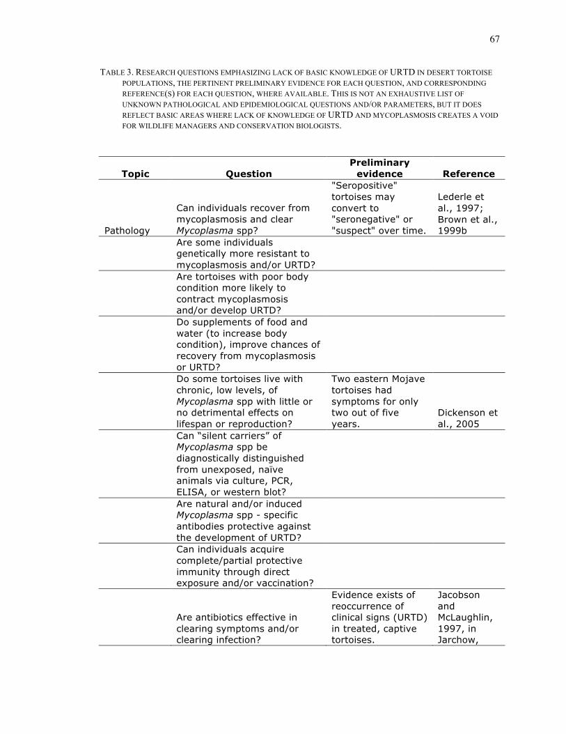
67
TABLE 3. RESEARCH QUESTIONS EMPHASIZING LACK OF BASIC KNOWLEDGE OF URTD IN DESERT TORTOISE POPULATIONS, THE PERTINENT PRELIMINARY EVIDENCE FOR EACH QUESTION, AND CORRESPONDING REFERENCE(S) FOR EACH QUESTION, WHERE AVAILABLE. THIS IS NOT AN EXHAUSTIVE LIST OF UNKNOWN PATHOLOGICAL AND EPIDEMIOLOGICAL QUESTIONS AND/OR PARAMETERS, BUT IT DOES REFLECT BASIC AREAS WHERE LACK OF KNOWLEDGE OF URTD AND MYCOPLASMOSIS CREATES A VOID FOR WILDLIFE MANAGERS AND CONSERVATION BIOLOGISTS.
Topic Question Preliminary
evidence Reference
Pathology
Can individuals recover from mycoplasmosis and clear Mycoplasma spp?
"Seropositive" tortoises may convert to "seronegative" or "suspect" over time.
Lederle et al., 1997; Brown et al., 1999b
Are some individuals genetically more resistant to mycoplasmosis and/or URTD?
Are tortoises with poor body condition more likely to contract mycoplasmosis and/or develop URTD?
Do supplements of food and water (to increase body condition), improve chances of recovery from mycoplasmosis or URTD?
Do some tortoises live with chronic, low levels, of Mycoplasma spp with little or no detrimental effects on lifespan or reproduction?
Two eastern Mojave tortoises had symptoms for only two out of five years.
Dickenson et al., 2005
Can “silent carriers” of Mycoplasma spp be diagnostically distinguished from unexposed, naïve animals via culture, PCR, ELISA, or western blot?
Are natural and/or induced Mycoplasma spp - specific antibodies protective against the development of URTD?
Can individuals acquire complete/partial protective immunity through direct exposure and/or vaccination?
Are antibiotics effective in clearing symptoms and/or clearing infection?
Evidence exists of reoccurrence of clinical signs (URTD) in treated, captive tortoises.
Jacobson and McLaughlin, 1997, in Jarchow,
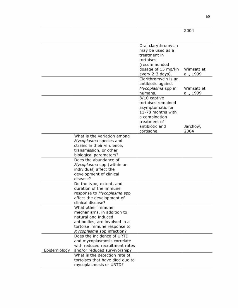
68
2004
Oral clarythromycin may be used as a treatment in tortoises (recommended dosage of 15 mg/kh every 2-3 days).
Wimsatt et al., 1999
Clarithromycin is an antibiotic against Mycoplasma spp in humans.
Wimsatt et al., 1999
8/10 captive tortoises remained asymptomatic for 11-78 months with a combination treatment of antibiotic and cortisone.
Jarchow, 2004
What is the variation among Mycoplasma species and strains in their virulence, transmission, or other biological parameters?
Does the abundance of Mycoplasma spp (within an individual) affect the development of clinical disease?
Do the type, extent, and duration of the immune response to Mycoplasma spp affect the development of clinical disease?
What other immune mechanisms, in addition to natural and induced antibodies, are involved in a tortoise immune response to Mycoplasma spp infection?
Epidemiology
Does the incidence of URTD and mycoplasmosis correlate with reduced recruitment rates and/or reduced survivorship?
What is the detection rate of tortoises that have died due to mycoplasmosis or URTD?

69
Do populations vary in their susceptibility/resistance to mycoplasmosis and/or URTD?
Does population-level variation in mycoplasmosis and/or URTD correlate with spatial and/or temporal patterns of environmental parameters (e.g. annual rainfall, annual temperatures, "short term drought")?
Do populations show variation in natural antibody production?
Inbred populations of the Galapagos hawk (Buteo galapoensis) had lower average levels and less variable natural antibodies than more outbred populations
Whiteman et al., 2006
Do population means of natural antibody production correlate with the respective population-wide prevalences of URTD?

70
Chapter 2. Natural and induced antibodies in experimentally immunized desert tortoises (Gopherus agassizii): the importance of
season and gender Abstract Captive desert tortoises were immunized with ovalbumin (OVA) in Ribi’s adjuvant to
induce a humoral immune response, both before and after hibernation. We observed a
significant mean increase in OVA-specific antibody, and a gender-by-season interaction
in the ability of desert tortoises to make an induced immune response. We observed
relatively high levels of pre-existing natural antibody to OVA in all tortoises, and levels
varied among individuals. There was a significant, negative relationship between an
animal’s natural antibody titer and the maximum increase in induced antibody titers, and
a significant, positive relationship between the magnitude of long-term elevations in
OVA-specific antibody titers and the maximum increase in induced titers. Both natural
and long-term elevations in induced antibody titers may be important elements of the
tortoise immune system, with possible influences on the ecology and evolution of host-
pathogen interactions. Reliance upon natural antibodies and the persistence of induced
antibodies may be an adaptation in reptiles to defend themselves from pathogens in spite
of their slow metabolic rates.
Keywords
natural antibody, induced antibody, ectothermic, reptile

71
Introduction While research on reptilian immune systems gained momentum in the 1970s and
1980s, interest in reptiles within the field of immunology subsequently waned (Origgi
2007) even though a more complete understanding of the reptilian immune system is
recognized as being important to the study of comparative immunology of vertebrates
(Origgi 2007). There is a dearth of knowledge of the diversity and relative importance of
immunological mechanisms in reptiles (Stacy & Pessier 2007). Most immunological
studies of chelonians have focused on adaptive immune processes (i.e., induced antibody
responses and lymphoproliferation assays; Ambrosius 1976; Leceta & Zapata 1985;
Muñoz & De La Fuente 2001, 2003; Herbst & Klein 1995; Work et al. 2001; Origgi
2007).
Recent studies in ecological immunology stress the importance of “innate”, or
constitutive, immune parameters (e.g. Matson 2006; Salvante 2006; Martin et al. 2008).
Constitutive immunity is a more precise term for “innate” immune mechanisms, and it
refers to mechanisms that are not induced by direct pathogen exposure (Schmid-Hempel
& Ebert 2003; Martin et al. 2008). It has been hypothesized that constitutive immune
mechanisms may be more important to ectothermic, versus endothermic, vertebrates due
to their slow metabolic rates of ectotherms and slow induced immune responses of
relatively low magnitude (Bayne & Gerwick 2001; Chen et al. 2007).
Research of chelonian immune responses largely has focused on a relatively small
number of species (e.g. Chrysemys picta, Testudo hermanni, T. graeca, Chelonia mydas,
Chelydra serpentina, Mauremys caspica, Gopherus agassizii; Ambrosius 1976; Herbst &
72
Klein 1995; Mead et al. 1983; Schumacher et al. 1993; Muñoz & De la Fuente 2001,
2003; Origgi 2007). Species studied to date have the ability to produce three isotypes of
antibodies (IgM, IgY, and IgYΔFc) in response to infection or artificial immunization,
with the production of IgY and IgYΔFc following the initial production of IgM
(Chartrand et al. 1971; Benedict & Pollard 1972; Coe 1972; Ambrosius 1976; Herbst &
Klein 1995). The temporal progression of an induced humoral, or antibody, response has
not been quantified in most species, including tortoises in the genus Gopherus (but see
Ambrosius 1976). However humoral responses in chelonians have been used as an
indicator of disease status and exposure to certain antigens (e.g. Coberley et al. 2001;
Herbst et al. 1998, 2008; Jacobson & Origgi 2007; Sandmeier et al. 2009).
We focus on two possible constitutive immune parameters in desert tortoises
(Gopherus agassizii), natural antibodies and long-term elevations of induced antibodies.
We describe a generalized humoral response in the desert tortoise. We immunized
tortoises with an intradermal injection of chicken egg protein (ovalbumin or OVA) in
Ribi’s adjuvant to induce OVA-specific antibody responses in a group of 16 captive
tortoises, all of which had not previously been exposed to OVA. OVA is an immunogenic
protein of moderate size, known to stimulate B-cells and produce strong humoral
responses in vertebrates (Parslow 2001).
We address three aspects of production of OVA-specific antibodies in the desert
tortoise. First, we determined whether immunization resulted in increased OVA-specific
induced antibody production, and assessed influences of gender and season on antibody
production. Second, we quantified “long-term” antibody titers one year after
immunizations, and determined whether the magnitude of an individual’s antibody

73
response affects the magnitude of “long-term” antibody titers. Third, we assessed
whether the levels of natural OVA antibodies, prior to antigen exposure, affect the
magnitude of the induced antibody response, and whether season and gender influence
natural antibody titers.
Methods Experimental design & treatment Twenty captive adult tortoises, housed at the University of Nevada, Reno, were
immunized intradermally either with OVA in Ribi’s adjuvant (n = 16), or with sterile
saline solution (n = 4). Each animal received two 0.5 ml intradermal injections (one in
each forelimb) of OVA (Sigma Chemical Co., St. Louis, MO) in Ribi’s adjuvant (Ribi
ImmunoChem Research, Inc., Hamilton, MT) or sterile saline (Ribi’s adjuvant promotes
humoral responses in experimental immunizations; Roitt & Delves 2001). Ten (eight
experimental and two control) tortoises were immunized in November 2005 (called the
winter-immunized group), prior to hibernation, and an equivalent group was immunized
in April 2006 (called the spring-immunized group), after hibernation. Treatment and
control groups had equal numbers of females and males. A blood sample was taken from
each animal prior to treatment, to serve as an individual-specific control. All animals
were “boosted” twice with OVA/Ribi’s adjuvant or sterile saline solution two and five
months after the initial immunization.
Animal husbandry All tortoises had been in captivity for at least 10 years prior to the start of the
experiment. They were housed in an approved animal care facility at the University of
Nevada, Reno, and maintained at a temperature range of 19º to 32º C in ambient day
74
length in Reno, Nevada. Tortoises were fed a diet of alfalfa and green vegetables several
times weekly, and once weekly each individual was placed in a tub with water where they
could take a drink and become well hydrated. Hibernation (at 13º C) was gradually
induced in December, and animals were allowed to hibernate for 1.5 months. One adult
female, in the spring-immunization group, was a replacement for a deceased animal, and
this animal did not hibernate.
One spring-immunized female was removed from the experiment during the
course of treatments, and did not receive a third immunization. Two additional animals
were not available for blood collection one year later.
Blood sampling Blood samples (2ml) were drawn subcarapacially (Hernandez-Divers et al. 2002)
prior to treatment in November 2005. After treatment, 0.5-1.0 ml of blood was taken
every two weeks until September 2006 (for the winter-immunized group) and until
November 2006 (for the spring-immunized group). Finally, a blood sample was taken
from all animals approximately one year after the last immunization treatment, in August
2007. All blood samples were centrifuged , and plasma was frozen at – 30º C.
Polyclonal ELISA (OVA) OVA-specific antibodies present in frozen blood plasma samples were assayed
using a polyclonal ELISA (Hunter et al. 2008) with the following modifications.
Immulon IB 96-well plates (Thermo Fisher Scientific, Inc., Waltham, MA) were coated
with 50 µl per well of OVA antigen in PBS (10 µg/µl). ELISAs were run at four dilutions
of tortoise blood plasma (1:10, 1:100, 1:1000, 1:10,000). To calibrate ELISA plates were
calibrated against a standard sample, run on each plate. The standard sample consisted of

75
plasma pooled from a separate group of 21 captive desert tortoises. Blood samples from
this group were collected in August/September 2007.
Titer calculations For each sample, absorbances were plotted (dilution vs. absorbance) on a log10
scale, and a best-fit line was used to approximate the linear portion of each resultant
curve. Because all tortoise samples had relatively large OVA-specific ELISA titers before
immunization (natural OVA antibodies), we did not have a “negative” sample to use as a
null, or “background”, absorbance. To calculate antibody titers, we instead chose a “cut-
off” absorbance of one that intersected each best-fit line. Therefore, titers are (log10-1 -
transformed) dilution values at which the corresponding absorbance, on the best-fit line,
equals one.
We calculated averages based on the bi-weekly antibody titers in order to
minimize the influence of non-directional, weekly fluctuations in antibody titer, the
immunization schedule, a minimum lag time of four weeks in the tortoise humoral
response, and individual variation in the gradual increase in antibody titers. Titer values,
per individual animal, were averaged across consecutive blood sampling dates (spanning
4-10 weeks) as follows: “pre-bleed” titers = average of first three sampling dates (prior to
an increase in antibody titers); post-1st immunization titer = average of sampling dates 4-6
(during which there was an average increase in antibody in response to the first
immunization); post-2nd immunization titer = average of sampling dates 7-12 (during
which there was an average increase in antibody in response to the second
immunization); post-3rd immunization titer = average of dates 13-17 for winter group, =

76
average of dates 13-15 for spring group (during which there was an average increase in
antibody in response to the third immunization) (Fig. 1, 2).
Animals produced the maximum antibody response after the third immunization,
and the average of these maximum antibody titers were used in all statistical analyses.
Similarly, “maximum increases in antibody titer” were obtained by calculating the ratio
of the average antibody titer after the third immunization to the average of pre-
immunization antibody titers.
Statistical analyses Mann-Whitney U tests were used to compare the effect of two treatment groups.
One-tailed tests were used to detect increases in antibody titers, due to immunization
treatment. Two-tailed tests were used to detect differences between season and gender.
Kruskal-Wallis tests were used to compare three or more groups, and Spearman Rank-
Correlations were used to test for correlations between two variables. We used a
significance level of α = 0.05, and all statistical analyses were run in Aabel (Aabel 3,
Gigawiz Ltd. Co.).
Results General humoral response to OVA immunization Before immunization tortoises had relatively high levels of natural antibodies to
OVA (Fig. 1). A Western blot was used to confirm that tortoise antibodies were
specifically binding OVA (data not shown). While variability among individual tortoises
in natural antibody levels was large (pre-immunization titers in Fig. 1), natural antibody
titers of the four control animals, on a per-animal basis, tended to be relatively constant
over the course of an active season (Fig. 1c).

77
Maximum titers (n = 15) of induced antibodies in experimentally immunized
animals were significantly elevated over pre-immunization natural antibody titers (n =
16) (Z = -3.558; p < 0.001) (Fig. 1). There was also a significant difference in the
maximum increase in OVA-specific antibody titers between experimental (n=15) and
control (n = 4) animals (Z = -2.2; p = 0.014) (see Fig. 2).
We found that gender (females: n = 7, males: n = 8) alone did not significantly
affect the maximum increase in antibody titers (Z = -1.157; p = 0.247). Similarly, the
season of the immunization treatment (winter: n = 8, spring: n = 7) alone did not affect
the maximum increase in antibody titers (Z = -0.926; p = 0.355). However, while gender
(females: n = 4, males: n = 4) did not affect the maximum increase in antibody titers in
the winter-immunized group (Z = -0.5774, p > 0.5), in the spring-immunized group males
(n = 4) had a significantly greater increase in antibody titers in comparison to females (n
= 3) (Z = -2.121, p = 0.034) (Fig. 2).
Long-term antibody response Antibody titers approximately one year post-treatment (n = 14) were significantly
higher than pre-immunization antibody titers (Z = -2.895; p = 0.002) (Fig. 2). Maximum
increases in antibody titer (n = 14) were positively correlated with antibody titers one
year post-treatment (rs = 0.9076; t = 7.493; p < 0.01) (Fig. 3), suggesting that
immunization-induced antibodies in G. agassizii are long-lived or that antibody
production is sustained.
Natural antibodies Winter natural antibody levels (n = 10) were significantly lower than spring
natural antibody levels (n = 10) (Z = -1.890; p = 0.029]. Gender (females: n = 10, males:

78
n = 10) did not explain the lower natural antibody titers in the winter (Z = -0.302; p >
0.05), and there was no significant gender by season interaction (winter-immunized
animals (females: n = 5, males: n = 5): Z = -1.149; p = 0.251; spring-immunized animals
(females: n = 5, males: n = 5): Z = -0.522; p > 0.5). Thus, gender generally, and gender in
different seasons did not explain the lower levels of antibodies seen in winter.
Discussion
We discovered that G. agassizii have variable levels natural antibodies to OVA.
Unlike conventional antibodies, natural antibodies are coded for in the genome (Baccala
et al. 1989; Casali & Notkins 1989; Kantor & Herzenberg 1993; Baumgarth et al. 2005),
are mainly of the IgM isotype (Casali & Schettino 1996; Baumgarth et al. 2005), and are
thought to be an important component of innate immunity against infectious agents
(Briles et al. 1981; Szu et al. 1983; Ochsenbein et al. 1999; Baumgarth et al. 2000,
2005). The existence of such antibodies reactive with the non-infectious OVA probably
results from their inherent polyspecificity (Gonzalez et al. 1988; Flajnik & Rumfelt 2000;
Baumgarth et al. 2005). Natural antibodies to a wide variety of antigens (including OVA)
are present in non-mammalian vertebrates (e.g. Gonzalez et al. 1988; Flajnik & Rumfelt
2000; Sinyakov et al. 2002, 2006; Hangalapura et al. 2003; Paramentier et al. 2004;
Madsen et al. 2007).
While immunization with OVA induced an antibody response, the maximum
response (20- to 30-fold) was not as high as the maximum responses often measured in
mammals (e.g. 100-fold titer increases; Leishman et al. 1998; Parslow 2001).
Nonetheless, immunization induced a significant increase in OVA-specific antibodies,
elevated above both individual pre-immunization titers, and above changes in antibody
79
titer of the procedural control animals (Fig. 1, 2). Additionally, we found three
prominent, unexpected patterns. These included: (1) the failure of an antibody response in
females that were immunized in the spring (Fig. 1b, 2b); (2) relatively high, individually-
variable, levels of natural antibody to OVA, with a negative relationship between natural
and induced antibody titers (Fig. 1c, 4); and (3) a long time-period across which antibody
levels remained elevated (Fig. 1, 2, 3).
Lack of an antibody response in females immunized in spring An effect of the season in which the animal is exposed to antigen has been
observed in other immunization experiments in chelonians (reviewed in Ambrosius 1967;
Leceta and Zapata 1986). However, those studies did not report an influence of gender on
the humoral response, and the effects of season appear not to be consistent among species
and among immune mechanisms. For example, Testudo hermanni exhibits delayed
immunological reactivity to immunization in the fall, and not in the spring (Ambrosius
1967). Mauremys caspica has significant season-by-gender interactions in some but not
all functions of spleen leukocytes, and regulation appears to be somewhat, but not solely,
influenced by hormone levels (e.g., testosterone, estradiol, and corticosterone) (Muñoz &
De la Fuente 2001).
In female desert tortoises, reproductive hormones (viz., estradiol, testosterone,
and corticosterone) may modulate a physiological trade-off between the expenses of
reproduction (egg production) and the expenses of a humoral immune response (sensu
Saad et al. 1991; Zapata et al. 1992; Burnham et al. 2003; Origgi 2007). Estradiol has
received much less attention than have testosterone and corticosteroids, but all three
hormones may affect changes in the reptilian immune system (Burnham et al. 2003;

80
Origgi 2007). In addition to a bimodal spike in estradiol production in early spring and
fall (Lance et al. 2001, 2002), female desert tortoises also exhibit their highest relative
levels in both testosterone and corticosterone in spring (Rostal et al. 1994; Lance et al.
2001). Additionally, the direct energetic cost of egg production could influence induction
of humoral responses in female tortoises in the spring. By measuring only two immune
parameters (i.e. natural and induced antibody levels), we are unable to conclude that
females are “immunosuppressed”, or to quantify a possible shift in immune parameters
(sensu Norris & Evans 2000; Adamo 2004 ; Salvante 2006).
Tortoises immunized in spring also did not have a delayed response to OVA.
Upon second and a third exposure to OVA in summer and early fall, anti-OVA antibody
levels did not increase, suggesting that females may have become tolerant to OVA (Fig.
2b). The mechanisms by which the responsiveness of the immune system to a T cell-
dependent immunogen, like OVA, is possibly seasonally repressed in female tortoises are
not known. Female tortoises may experience seasonal shifts in immune function that have
long-term effects on immune responsiveness, making them more-or-less susceptible to
certain classes of pathogen, with possibly important effects on the epidemiology of
disease in wild populations.
Natural antibodies to OVA Variation in natural antibody levels to OVA in desert tortoises may indicate
variation in natural antibodies to various pathogens, and therefore, those variations could
well be of ecological and evolutionary significance. Supporting this hypothesis, natural
antibodies are known to be cross-reactive (Guilbert et al. 1982; Gonzalez et al. 1988),
titers are thought to be genetically determined (Paramentier et al. 2004), and large

81
individual variation in titers have been reported in humans, mice, birds, and fish (e.g.
Ben-Aissa-Fennira et al. 1998; Sinyakov et al. 2002; Paramentier et al. 2004;
Kachamakova et al. 2006). Chickens with high levels of natural antibody to sheep red
blood cells, usually also had high titers to a variety of other antigens (Parmentier et al.
2004). Natural antibodies are protective against pathogenic organisms in fish (Carassius
auratus, Sinyakov et al. 2002) and mice (Briles et al. 1981; Cartner et al. 1998;
Baumgarth et al. 2000).
It is possible that the diversity and quantity of natural antibody levels in natural
host populations – and especially those of ectothermic vertebrates – may be indicators of
pathogens of ecological and evolutionary importance. For example, when specific
reactivity to a pathogen molecular motifs (sensu Briles et al. 1981; Baumgarth et al.
2005) is shown to exist in a host species’ natural antibody repertoire, the diversity and
quantity of natural antibody may have evolved as a response to past pathogen pressure
(sensu Baumgarth et al. 2005; Madsen et al. 2007). However, with the exclusion of M.
agassizii (Hunter et al. 2008), little is known about the reactivity of the desert tortoise
natural antibody repertoire to a variety of possible pathogens.
The negative relationship between natural antibody titers and the magnitude of the
induced antibody response (Fig. 4), suggests that natural antibodies may have a
significant biological effect through epitope masking. Epitope masking refers to the
binding of antigen by constitutive molecules, such as natural antibodies, which then
reduce the amount of “free” antigen and the subsequent magnitude of the induced
response (Janeway et al. 2005). For instance, there is a general, negative relationship
between levels of natural antibody and levels of acquired antibody, produced in response

82
to vaccination with a particular antigen, in fish (Sinyakov et al. 2006; Sinyakov &
Avtalion 2009). Madsen et al. (2007) also interpreted the failure of water pythons (Liasis
fuscus) to make an induced antibody response to immunizations to such an effect of
natural antibodies.
Although other mechanisms of the desert tortoise immune system may be
enhanced in winter (Christopher et al. 1999), reduced levels of natural antibody in winter
may alter host immunology and host-pathogen interactions in the desert tortoise.
Changes in host susceptibility could also interact with seasonal changes in pathogen
growth rates and pathogen transmission patterns (sensu Woodhams et al. 2008).
Long-lived antibody response Long-term production of induced antibodies is mechanistically different from
“classical” immunological memory, which relies on secondary exposure to a specific
pathogen or antigen that activates a discrete group of memory B-cells (Slifka & Ahmed
1996). In mammals, immunoglobulin molecules have relative short half-lives (Janeway et
al. 2005), so protracted elevation in antibody titers to pathogens have been attributed to
several mechanisms including: (1) re-exposure to the pathogen or chronic infection, (2)
persisting antigen-antibody complexes on follicular dentritic cells, (3) cross-reactivity
with environmental or self antigens, (4) idiotypic networks, and (5) the activation of a
unique group of B-cells (long-lived plasma cells) that continuously produce antibodies in
the absence of additional stimulation (Slifka & Ahmed 1996).
It is possible that desert tortoises possess long-lived plasma cells that produce
antibodies for extended periods. However, the half-lives of desert tortoise
immunoglobulins have not been reported, so it is also possible that the persistence of high
83
anti-OVA titers may be due to long-lived antibody molecules. It could well be
advantageous to ectothermic vertebrates with low metabolic rates and slow humoral
immune responses to have persistent antibodies to ensure some resistance to previously
encountered pathogens and to minimize the energetic cost of producing another round of
antibodies. Herbst et al. (2008) observed a long-lived antibody response in two
individuals of green sea turtle (Chelonia mydas) with elevated IgY levels for at least 20
months after experimental infection with chelonid fibropapillomatosis-associated herpes
virus. In both mammals and fish, long-lived antibody-secreting cells have been observed
in the bone marrow (of mammals) and the anterior kidney (of fish) and can produce
antibody for months to years (Slifka et al. 1995; Slifka & Ahmed 1996; Manz et al. 1998;
Kaattari et al. 2005). Kaattari et al. (2005) suggest that long-lived, antibody-secreting,
plasma cells may be responsible for the arithmetic increases in antibody titer often
observed upon secondary exposure to antigens in fish. Arithmetic increases in antibody
titer are also commonly observed in reptiles (Ambrosius 1976; Jacobson & Origgi 2007;
Origgi 2007). Long-lived B-cells in rainbow trout (Oncorhyncus mykiss) are activated by
antigen, migrate to the anterior kidney, and product all or most of the circulating levels of
antibody present in the late humoral response (Bromage et al. 2004; Kaattari et al. 2005).
Implications
Both high natural antibody titers and long-term elevations in induced antibody
titers suggest that constitutive immune parameters may be of relatively great importance
to the desert tortoise. Understanding the interactions between constitutive and induced
immunity, as well as the seasonal variations in both, could lead to a better understanding
of the ecology of host-pathogen interactions in the desert tortoise, and in other reptilian

84
species as well. Furthermore, a more complete understanding of the full array of
constitutive and induced parameters in the reptilian immune system would allow for
increased accuracy in diagnosing levels of pathogen exposure and host resistance in
natural populations. The evolutionary significance of a possible greater reliance on
constitutive versus induced immune parameters in reptiles has not been adequately
explored, but could provide insights into current and past pathogen pressure, life history
trade-offs in reptiles, and the evolution of the vertebrate immune system.
Acknowledgements We thank John Gray for his invaluable assistance, both with the collection of blood
samples and various technical and logistic aspects of this project. We also thank the UNR
EECB peer review group for revisions on an earlier draft of this manuscript. This work
was funded by a grant from the Desert Conservation Program of Clark County, Nevada.
The work was conducted under a protocol approved by the Animal Care and Use
Committee (IACUC) at the University of Nevada, Reno (IACUC A06/07-49).

85
References Adamo, S. A. 2004 How should behavioural ecologists interpret measurements of
immunity? Anim. Behav., 68, 1443-1449.
(DOI 10.1016/j.anbehav.2004.05.005)
Ambrosius, H. 1976 Immunoglobulins and antibody production in reptiles. In
Comparative Immunology (ed. J. J. Marchalonis), pp. 298-334. New York: John Wiley
and Sons.
Baccala, R., Quang, T. V., Gilbert, M., Ternyck, T. & Avrameas, S. 1989 Two murine
neutral polyreactive autoantibodies are encoded by nonmutated germ-line genes. Proc. of
the Nat. Acad. of Sci., 84, 4624-4628.
Bayne, C. J. & Gerwick, L. 2001 The acute phase response and innate immunity in fish.
Dev. and Comp. Immun., 25, 725-743.
(DOI 1016/S0145-305X(01)00033-7)
Baumgarth, N., Herman, O. C., Jager, G. C., Brown, L. E., Herzenberg, L. A. & Chen, J.
2000 B-1 and B-2 cell-derived immunoglobulin M antibodies are nonredundat
components of the protective response to influenza virus infection. J. Exp. Med., 192,
271-80.
(DOI 10.1084/jem.192.2.27)

86
Baumgarth, N., Tung, J. W. & Herzenberg, L. A. 2005 Inherent specificities in natural
antibodies: a key to immune defense against pathogen invasion. Springer Semin. Immun.,
26, 347-362.
(DOI 10.1007/s00281-004-0182-2)
Ben-Aissa-Fennira, F., Ammar-El Gaaied, A. B., Bouguerra, A. & Dellagi K. 1998 IgM
antibodies to P1 cytoadhesin of Mycoplasma pneumoniae are part of the natural antibody
repertoire expressed early in life. Immun. Lett., 63, 59-62.
(DOI 10.1016/S0165-2478(98)00053-4)
Benedict, A. A. & Pollard, L. W. 1972 Three classes of immunglobulins found in the sea
turtle, Chelonia mydas. Folia Microbiol., 17, 75-78.
(DOI 10.1007/BF02872256)
Briles, D. E., Nahm, M., Schroer, K., Davie, J., Baker, P., Kearney, J. & Barletta, R. 1981
Antiphosphocholine antibodies found in normal mouse serum are protective against
intravenous infection with type 3 Streptococcus pneumoniae. J. Exp. Med., 153, 694-705.
(DOI 10.1084/jem.153.3.694)
Bromage, E. S., Kaattari, I. M., Zwolla, P. & Kaattari, S. L. 2004 Plasmablast and plasma
cell production and distribution in trout immune tissues. J. of Immun., 173, 7313-7323.

87
Burnham, D.K., Lackey, A., Manering, M., Jaensson, E., Pearson, J., Tyler, D.O.,
Melson, D. & Talent, L. 2003 Effects of 17α-ethinylestradiol on immune parameters in
the lizard Sceloporus occidentalis. Environ. Toxicol., 18, 211-218.
(DOI 10.1002/tox.10117)
Cartner, S. C., Lindsey, J. R., Gibbs-Erwin, J., Cassell, G. H. & Simecka, J. W. 1998
Roles of innate and adaptive immunity in respiratory mycoplasmosis. Infect. and Immun.,
66, 3485-3491.
Casali, P. & Notkins, A. L. 1989 CD5+ B lymphocytes, polyreactive antibodies and the
human B-cell repertoire. Immun. Today, 10, 364-368.
(DOI 10.1016/0165-5699(89)90268-5)
Casali, P. & Schettino, E. W. 1996 Structure and function of natural antibodies. Current
Topics in Microbiol. and Immun., 210, 167-169.
Chartrand, S. L., Litman, G. W., LaPointe, N., Good, R. A. & Frommell, D. 1971 The
evolution of the immune response XII. The immunoglobulins of the turtle. Molecular
requirements for the biologic activity of 5.7S immunoglobulin. The J. of Immunol., 107,
1-10.

88
Chen, X., Cuijuan, N. & Pu, L. 2007 Effects of stocking density on growth and non-
specific immune responses in juvenile soft-shelled turtle Pelodiscus sinensis. Aquacult.
Res., 38, 1380-1386.
(DOI 10.1111/j.1365-2109.2007.01813.x)
Christopher, M. M., Berry, K. H., Wallis, I. R., Nagy, K. A., Henen, B. T. & Peterson, C.
C. 1999 Reference intervals and physiologic alterations in hematologic and biochemical
values of free-ranging desert tortoises in the Mojave Desert. J. of Wild. Dis., 35, 212-238.
Coberley, S. S., Herbst, L. H., Ehrhart, L. M., Bagley, D. A., Hirama, S., Jacobson, E. R.
& Klein, P. A. 2001 Survey of Florida green turtles for exposure to a disease-associated
herpesvirus. Dis. of Aquat. Org., 47, 159-167.
(DOI 10.3354/dao047159)
Coe, J. E. 1972 Immune resonse in the turtle (Chrysemys picta). Immun., 23, 45-52.
Flajnik, M. F. & Rumfelt, L. L. 2000 Early and natural antibodies in non-mammalian
vertbrates. Current Topics in Microbio. and Immun., 252, 233-240.
Gonzalez, R., Charlemagne, J., Mahana, W. & Avrameas, S. 1988 Specificity of natural
serum antibodies present in phylogenetically distinct fish species. Immun., 63, 31-36.

89
Guilbert, B., Dighiero, G. & Avrameas, S. 1982 Naturally occurring antibodies against
nine common antigens in human sera. I. Detection, isolation, and characterization. J. of
Immun., 128, 2779-2787.
Hangalapura, B. N. , Nieuwland, M. G. B., de Vries Reilingh, G., Heetkamp, M. J. W.,
van den Brand, H., Kemp, B. & Paramentier, H. K. 2003 Effects of cold stress on
immune responses and body weight of chicken lines divergently selected for antibody
responses to sheep red blood cells. Poultry Sci., 82, 1692-1700.
Herbst, L. H., Greiner, E. C., Ehrhart, L., Bagley, D. A. & Klein, P. A. 1998 Serological
association between spirorchidiasis, herpesvirus infection, and fibropapillomatosis in
green turtles from Florida. J. of Wild. Dis., 34, 496-507.
Herbst, L. H. & Klein, P. A. 1995 Monoclonal antibodies for the measurement of class-
specific antibody responses in the green sea turtle, Chelonia mydas. Vet. Immunol. and
Immunopathol., 46, 317-335.
(DOI 10.1016/0165-2427(94)05360-5)
Herbst, L. H., Lemaire, S., Ene, A. R., Heslin, D. J., Llewellyn, M. E., Bagley, D. A.,
Klein, P. A. & Lenz, J. 2008 Use of baculovirus-expressed glycoprotein H in an enzyme-
linked immunosorbent assay developed to assess exposure to chelonid
fibropapillomatosis-associated herpesvirus and its relationship to the prevalence of
fibropapillomatosis in sea turtles. Clin. and Vacc. Immun., 15, 843-851.

90
(DOI 10.1128/CVI.00438-07)
Hernandez-Divers, S. M., Hernandez-Divers, S.J. & Wyneken, J. 2002 Angiographic,
anatomic and clinical technique descriptions of a subcarapacial venipuncture site for
chelonians. J. of Herp. Med. and Surgery, 12, 32-37.
Hunter, K. W. Jr., DuPré, S. A., Sharp, T., Sandmeier, F. C. & Tracy, C. R. 2008
Western blot can distinguish natural and acquired antibodies to Mycoplasma agassizii in
the desert tortoise (Gopherus agassizii). J. of Microbiol. Meth., 75, 464-471.
(DOI 10.1016/jmimet.2008.07.022)
Jacobson, E. R. & Origgi, F. C. 2007 Serodiagnostics. In Infectious diseases and
pathology of reptiles (ed. E.R. Jacobson). pp. 381-394. New York: Taylor & Francis.
Janeway, C. A. Jr., Travers, P., Walport, M. & Schlomick M. J. (eds.) 2005
Immunobiology, 6th edn. New York: Garland Science.
Kaattari, S., Bromage, E. & Kaattari, I. 2005 Analysis of long-lived plasma cell
production and regulation: Implications for vaccine design for aquaculture. Aquacult.,
246, 1-9.
(DOI 10.1016/j.aquaculture.2004.12.024)

91
Kachamakova, N. M., Irnazarow, I., Parmentier, H. K., Savelkoul, H. F. J., Pilarczyk, A.
& Wiegertjes, G. F. 2006 Genetic differences in natural antibody levels in common carp
(Cyprinus carpio L.). Fish and Shellfish Immun., 21, 404-413.
(DOI 10.1016/j.fsi.2006.01.005)
Kantor, A. B. & Herzenberg, L. A. 1993 Origin of murine B cell lineages. Ann. Rev. of
Immun., 11, 510-538.
(DOI 10.1146/annurev.iy.11.040193.002441)
Lance, V. A., Grumbles, J. S. & Rostal, D. C. 2001 Sex differences in plasma
corticosterone in desert tortoises, Gopherus agassizii, during the reproductive cycle. J. of
Exp. Zool., 289, 285-289.
(DOI 10.1002/1097-010X(20010415/30)289::AID-JEZ2>3.0.CO;2-B)
Lance, V. A., Place, A. R., Grumbles, J. S., & Rostal, D. C. 2002 Variation in plasma
lipids during the reproductive cycle of male and female desert tortoises, Gopherus
agassizii. J. of Exp. Zool., 293, 703-711.
(DOI 10.1002/jez.10160)
Leceta, J. & Zapata, A. 1986 Seasonal variations in the immune response of the tortoise
Mauremys caspica. Immunol., 57, 483-487.
92
Leishman, A. J., Garside, P. & Mowat, A. M. 1998 Immunological consequences of
intervention in established immune responses by feeding protein antigens. Cell.
Immunol., 10, 1703-1711.
(DOI 10.1006/cimm.1998.1242)
Madsen, T., Ujvari, B., Nandakumar, K. S., Hasselquist, D. & Homdahl, R. 2007 Do
“infectious” prey select for high levels of natural antibodies in tropical python? Evol.
Ecol., 21, 271-279.
(DOI 10.1007/s10682-006-9004-4)
Manz, R. A., Löhning, M., Cassese, G., Thiel, A. & Radbruch, A. 1998 Survival of long-
lived plasma cells is independent of antigen. Internat. Immun., 10, 1703-1711.
(DOI 10.1093/intimm/10.11.1703)
Martin, L. B., Weil, Z. M. & Nelson, R. J. 2008 Seasonal changes in vertebrate immune
activity: mediation by physiological trade-offs. Phil. Trans. R. Soc. B, 363, 321-339.
(DOI 10.1098/rstb.2007.2142)
Matson, K. D. 2006 Are there differences in immune function between continental and
insular birds? Proc. R. Soc. B, 273, 2267-2274.
(DOI 10.1098/rspb.2006.3590)

93
Mead, K. F., Borysenko, M. & Findlay, S. R. 1983 Naturally abundant basophils in the
snapping turtle, Chelydra serpentina, possess cytophilic surface antibody with reaginic
function. The J. of Immunol., 130, 334-340.
Muñoz, F. J. & De la Fuente, N. 2001 The effect of the seasonal cycle on the splenic
leukocyte functions in the turtle Mauremys caspica. J. Comp. Physiol. B, 171, 27-42.
(DOI 10.1086/323033)
Muñoz, F. J. & De la Fuente, N. 2003 In vitro proliferation of blood, spleen, and thymus
leukocytes from the turtle Mauremys caspica. Comp. Biochem. and Physiol. C, 134, 303-
309.
(DOI 10.1016/S1532-0456(03)00004-8)
Norris, K. & Evans, M. R. 2000 Ecological immunology: life history trade-offs and
immune defense in birds. Behav. Ecol., 11, 19-26.
(DOI 10.1093/beheco/11.1.19)
Ochsenbein, A. F., Fehr, T., Lutz, C., Suter, M., Brombacher, F., Hensgartner, H. &
Zinkernagel, R. M. 1999 Control of early viral and bacterial distribution and disease by
natural antibodies. Sci., 286, 2156-2159.
(DOI 10.1126/science.286.5447.2156)
94
Origgi, F. C. 2007 Reptile immunology. In Infectious diseases and pathology of reptiles
(ed. E. R. Jacobson), pp. 131-218. New York: Taylor and Francis.
Parmentier, H. K., Lammers, A., Hoekman, J. J., De Vries Reiling, Zaanen, I. T. A. &
Savelkoul, H. F. J. 2004 Different levels of natural antibodies in chickens divergently
selected for specific antibody responses. Dev. and Comp. Immun., 28, 39-49.
(DOI 10.1016/S0145-305X(03)00087-9)
Parslow, T. G. 2001 Immunogens, antigens, and vaccines. In Medical Immunology 10th
edn. ( ed. T. G. Parslow, D. P. Stites, A.I. Terr & J. B. Imboden JB), pp. 72-81. New
York: Lange Medical Books/McGraw-Hill.
Roitt, I. M. & Delves, P. J. 2001 Roitt’s essential immunology 10th edn. Malden:
Blackwell Science.
Rostal, D. C., Lance, V. A., Grumbles, J. S. & Alberts, A. C. 1994 Seasonal reproductive
cycle of the desert tortoise (Gopherus agassizii) in the eastern Mojave desert. Herp.
Monographs, 8, 72-82.
(DOI 10.2307/1467071)
Saad, A. H., Torroba, M., Varas, A. & Zapata, A. 1991 Testosterone induces
lymphopenia in turtles. Veter. Immun. and Immunopath., 28, 173-180.
(DOI 10.1016/0165-2427(91)90139-4)

95
Salvante, K. G. 2006 Techniques for studying integrated immune function in birds. The
Auk, 123, 575-586.
(DOI 10.1642/0004-8038(2006)123[575:TFSIIF]2.0.C0;2)
Sandmeier, F. C., Tracy, C. R., DuPré, S. & Hunter, K. 2009 Upper respiratory tract
disease (URTD) as a threat to desert tortoise populations: a reevaluation. Biol.Conserv.,
142, 1255-1268.
(DOI 10.1016/j.biocon.2009.02.001)
Schumacher, I. M., Brown, M. B., Jacobson, E. R., Collins, B. R. & Klein, P. A. 1993
Detection of antibodies to a pathogenic mycoplasma in desert tortoises (Gopherus
agassizii) with upper respiratory tract disease. J. of Clin. Microbiol., 31, 1454-1460.
Schmid-Hempel, P. & Ebert, D. 2003 On the evolutionary ecology of specific immune
defence TRENDS in Ecol. and Evol., 18, 27-32.
(DOI 10.1016/S0169-5347(02)00013-7)
Sinyakov, M.S. & Avtalion, R. R. 2009. Vaccines and natural antibodies: A link to be
considered. Vaccine, 27, 1985-1986.
(DOI 10.1016/j.vaccine.2009.01.121)

96
Sinyakov, M.S., Dror, M., Lublin-Tennenbaum, T., Salzberg, S., Margel, S. & Avtalion,
R.R. 2006. Nano- and microparticles as adjuvants in vaccine design: Success and failure
is related to host natural antibodies. Vaccine, 24, 6534-6541.
(DOI 10.1016/j.vaccine.2006.06.021)
Sinyakov, M. S., Dror, M., Zhevelev, H. M., Margel, S. & Avatalion, R.R. 2002 Natural
antibodies and their significance in active immunization and protection against a defined
pathogen in fish. Vaccine, 20, 3668-3674.
(DOI 10.1016/S0264-410X(02)00379-1)
Slifka, M. K. & Ahmed, R. 1996 Long-term humoral immunity against viruses: revisiting
the issue of plasma cell longevity. Trends in Microbiol., 4, 394-400.
(DOI 10.1016/0966-842X(96)10059-7)
Slifka, M. K., Matloubian, M. & Ahmed, R. 1995 Bone marrow is a major site of long-
term antibody production after acute viral infection. J. of Virol., 69, 1895-1902.
Stacy, B. A. & Pessier, A. P. 2007 Host response to infectious agents and identification
of pathogens in tissue section. In Infectious diseases and pathology of reptiles (ed. E. R.
Jacobson), pp. 257-298. New York: Francis and Taylor.

97
Szu, S. C., Clarke, S. & Robbins, J. B. 1983 Protection against pneumococcal infection in
mice conferred by phosphocholine-binding antibodies: specficity of the phosphocholine
binding and relation to several types. Infect. and Immun., 39, 993-999.
Woodhams, D. C., Alford, R. A., Briggs, C. J., Johnson, M. & Rollins-Smith, L. A. 2008
Life-history trade-offs influence disease in changing climates: strategies of an amphibian
pathogen. Ecol., 89, 1627-1639.
(DOI 10.1890/06-1842.1)
Work, T. M., Rameyer, R. A., Balazs, G. H., Cray, C. & Chang, S. P. 2001 Immune
status of free-ranging green turtles with fibropapillomatosis from Hawaii. J. of Wild. Dis.,
37, 574-581.
Zapata, A. G, Varas, A. & Torroba, M. 1992 Seasonal variations in the immune system of
lower vertebrates. Immunol. Today, 13, 142-147.
(DOI 10.1016/0167-5699(92)90112-K)

98
FIGURE LEGENDS
Fig. 1. Average OVA-specific antibody titers by immunization treatment. For all animals,
the second and third immunizations followed the preliminary immunization by
two and five months, respectively. A single sample was taken one year later, in
August 2007, for both groups of animals. (a) Winter-immunized animals, (b)
Spring-immunized animals, (c) Procedural control animals.
Fig. 2. Average increases in OVA-specific antibody titers by immunization treatment.
Increases in antibody titers were calculated as the ratio of the average titer after an
immunization in relation to the average pre-immunization antibody titer: (a)
Winter-immunized animals, (b) Spring-immunized animals, (c) Procedural
control animals.
Fig. 3. Relationships between each tortoise’s maximum increase in OVA-specific
antibody titers, and the long-term elevation in OVA-specific antibodies
approximately one year after the last immunization treatment.
Fig. 4. Relationships between each experimental animal’s OVA-specific natural antibody
titer, and the maximum increase in antibody titers in response to OVA
immunization.

99
FIGURE 1

100
FIGURE 2

101
FIGURE 3

102
FIGURE 4

103
Chapter 3. Natural and acquired antibodies to Mycoplasma agassizii in the Mojave desert tortoise: Implications for managing a wildlife disease
Abstract This is the first range-wide analysis of the prevalence of upper respiratory tract disease
(URTD) and seroprevalence of a known, etiological agent, Mycoplasma agassizii, in the
Mojave desert tortoise (Gopherus agassizii). We analyze this host-pathogen system from
the viewpoint that this is a potentially complex disease, with varying dynamics over both
space and time. We focus on population-level analyses (n = 24), and test for associations
among prevalence of URTD, seroprevalence to M. agassizii, mean and standard
deviations of levels of natural antibody to M. agassizii, genetics of tortoise populations,
mean annual winter precipitation, and mean number of days below freezing. We detected
significant associations between mean number of days below freezing and both
prevalence of URTD and seroprevalence to M. agassizii. Furthermore, we detected a
significant association between mean levels of natural antibody and seroprevalence to M.
agassizii. Genetics of tortoise populations was associated with mean levels of natural
antibody. We propose hypotheses, concerning possible ecological and evolutionary
dynamics of the desert tortoise – M. agassizii system, based on these associations. We
present recommendations for future research to address tests of these hypotheses.
Keywords context-dependent, climate, wildlife disease, innate immunity, reptile

104
Introduction
Pathogens and parasites can pose risks to the persistence of endangered
populations or species (Altizer et al. 2003, Plowright et al. 2008). However, pathogens
and parasites also may be important in influencing host population dynamics,
interspecific competition, levels of biodiversity, and the flow of energy in ecosystems
(McCallum and Dobson 1995, Altizer et al. 2003, Hudson et al. 2006). Therefore,
preserving co-evolving host-parasite populations may be a sound conservation strategy
both for protecting host populations of conservation concern and conserving biodiversity
in general (Altizer et al. 2003). Research aimed at understanding the impacts of
anthropogenic changes – including climate change, habitat fragmentation, and expanding
urban development – on these co-evolving dynamics is expected to become increasingly
relevant to applied as well as basic ecological studies (sensu Altizer et al. 2003, Plowright
et al. 2008). Unfortunately, the study of large-scale ecological factors (e.g. climatic
patterns) and complex host-pathogen ecological and evolutionary dynamics are often not
directly amenable to cause-and-effect experiments (Plowright et al. 2008). Plowright et
al. (2008) recommend a combination of laboratory, field, and modeling techniques, a
reconsideration of Hill’s criteria (Hill 1965), and the consideration of multiple,
competing hypotheses to investigate complex dynamics of human and wildlife diseases.
Recent epidemiological models address ways to include realistic, complex disease
dynamics in natural systems. Two types of complexity in such models include the effects
of population heterogeneity (in host and pathogen populations) and the relation of
phenomena among different scales (reviewed in Levin et al. 1997). Therefore, new

105
approaches increasingly are linking within- and between-host dynamics of infectious
diseases (Mideo et al. 2008), and these approaches can be used to examine effects of
multiple within-host defense strategies, such as tolerance of a pathogen as well as
resistance to pathogens (Schneider and Ayres 2008). For example, host-pathogen models
have found that the inclusion of heterogeneity in the mechanisms of host defense can
significantly change epidemiological predictions (e.g. Nath et al. 2008, Kramer-Schadt et
al. 2009).
Here, we present analyses of data from the first, range-wide, serological survey of
a potentially complex, infectious disease system in the Mojave desert tortoise (Gopherus
agassizii). Upper respiratory tract disease (URTD) is thought to affect rates of morbidity
and mortality in natural populations of the federally-threatened Mojave desert tortoise
(FWS 1990, 1994, Sandmeier et al. 2009) (Table 1). The term “URTD” has been used to
refer to visible signs of respiratory disease, regardless of etiological agent (Sandmeier et
al. 2009), and we treat it as such throughout this paper. Concordant with important
recommendations for such surveys (Plowright et al. 2008), our survey was based upon,
and complements, past and current laboratory experiments (Schumacher et al. 1993,
Brown et al. 1994, Hunter et al. 2008, Sandmeier et al. submitted). Mechanistic,
epidemiological models of URTD in desert tortoise populations are absent from the
literature. Our analyses were, therefore, designed to be concordant with some of the
predictions of increasingly-realistic epidemiological models. We incorporate parameters
deemed to be important in controlled experiments, namely serological measures of innate
and adaptive host-defense and relatively high individual variation in these parameters
(Table 1). Specifically, we measure one measure of the host innate immune system

106
(natural antibody levels), as well as population-level means and standard deviations (i.e.
population-level heterogeneity) of these natural antibody levels. Desert tortoises exhibit
high variability in natural antibody titers (Hunter et al. 2008, Sandmeier et al. 2009,
Sandmeier et al. submitted). Natural antibodies have been shown to confer both/either
tolerance and resistance to certain pathogens in a variety of vertebrate species, including
humans (Ben-Aissa-Fennira et al. 1998), mice (Briles et al. 1981, Szu et al. 1983,
Ochsenbein et al. 1999, Baumgarth et al. 2000, 2005), and fish (Sinyakov et al. 2002,
Magnadóttir 2006).
We consider two climatic factors that have the potential to interact with infectious
agents and/or the host immune system to exacerbate susceptibility to, or progression of,
clinical disease. We investigate possible associations between mean measures of rainfall
and thermal environment (viz., annual number of days below freezing, which correlates
with duration of seasonal hibernation (sensu Nussear et al. 2007)) and the prevalence of
URTD and seroprevalence of an identified etiological agent, Mycoplasma agassizii
(strain PS6) (Brown et al. 1994, 2001, reviewed in Sandmeier et al. 2001). This survey is,
in essence, a “snapshot” in time. All samples were collected over three years (2004-
2006), which were roughly comparable, based upon climatic conditions and activity
levels of desert tortoises (F. Sandmeier unpublished data, sensu Hereford et al. 2006,
FWS 2009).
Similar to some other complex diseases of wildlife (e.g. chytridiomycosis in
amphibians; Blaustein and Kiesecker 2002, Briggs et al. 2005, McCallum 2005) and to
some other mycoplasma diseases of mammals (reviewed in Jones and Simecka 2003),
many variables may influence rates morbidity in desert tortoises, due to infection with M.

107
agassizii (Table 1). Many of these variables in the desert tortoise – M. agassizii system
are currently not quantified (Table 1). One caveat that is central to the interpretation of
analyses in this study concerns the reliance upon serology to measure the exposure to M.
agassizii in desert tortoises. Serology has been the most common approach to detect M.
agassizii in wild desert tortoises (Sandmeier et al. 2009). However, seropositivity of an
individual tortoise (presence of acquired antibodies to M. agassizii-PS6) does not
necessarily indicate a current infection (Sandmeier et al. 2009, Sandmeier et al. in press,
K. Hunter unpublished data). Gender and season of exposure, as well as innate levels of
natural antibody, have been shown to influence the occurrence, and magnitude, of an
acquired antibody response (Sandmeier et al. in press). In addition, desert tortoises have
been shown to exhibit long-term increases in acquired antibody titers after exposure to a
non-replicating, artificial antigen (ovalbumin) (Sandmeier et al. in press). Serology may
detect past, but not necessarily current, infections, and it may fail to detect some
instances of exposure to M. agassizii.
Both the ELISA and Western blot serological techniques used in this study are
based upon the one strain of M. agassizii that has been shown to be pathogenic in desert
tortoises and that is available through the American Type Culture Collection (Rockville,
MD; ATCC 700616) (Brown et al. 1994, 2001). However, by using this one strain, it is
possible that we could miss detecting significantly different strains of M. agassizii, or
possible closely related species that may also cause URTD (sensu Brown unpubl.ed data).
Avirulent strains may fail to produce URTD, and they also may fail to produce an
acquired humoral response in the tortoise. In this circumstance, serology will not detect
these strains. A commonly used, monoclonal ELISA to M. agassizii strain PS6 (described

108
in Schumacher et al. 1993, Wendland et al. 2007) is thought to measure antibody levels to
other, known strains of M. agassizii (M. Brown, unpublished data). Similarly, and for
brevity, we refer to levels of antibody measured via our polyclonal ELISA and Western
blot, using M. agassizii strain PS6 as the antigen, as being specific to “M. agassizii.” In
the future, direct, accurate detection of pathogen(s) (e.g. via nasal lavages and
PCR/quantitative PCR) should be available as a preferable alternative to detect pathogens
in natural, tortoise populations (pers. comm. K. Hunter, J. Simecka).
The goal of our study was to increase understanding of the factors involved in the
seroprevalence of M. agassizii, and factors involved in the prevalence of URTD in
natural populations of the desert tortoise in the Mojave Desert. Our specific aims were
five-fold. We sought: (1) to examine patterns among gender, genetics of the tortoise,
disease status, serological status, and natural antibody levels of individual tortoises, (2) to
determine the relationship between a population’s levels of seroprevalence to M. agassizii
and the prevalence of URTD, (3) to examine how the mean and standard deviation
(measure of heterogeneity) of a population’s natural antibody levels, as well as genetic
identity, mean winter rainfall, and mean annual days below freezing are associated with
the seroprevalence to M. agassizii and the prevalence of URTD in tortoise populations,
(4) to examine how genetics of the tortoise populations, mean winter rainfall, and mean
annual days below freezing are associated with the mean and standard deviation of a
population’s natural antibody levels, and (5) to form hypotheses pertaining to the ecology
and evolution of the desert tortoise – M. agassizii system. We use these hypotheses to
suggest fruitful avenues for future research. We propose that our approach (sensu
Plowright et al. 2008) encompasses the suspected complexity of this wildlife disease and
109
may serve as a useful example for others involved in research of diseases with complex
etiologies.
Methods
Sample collection Tortoise blood samples were collected both by crews of workers hired by the U.S.
Fish and Wildlife Service to monitor tortoises in designated critical habitat called Desert
Wildlife Management Areas (DWMAs) (March-June 2004, 2005) (FWS 2009) and by
our crews of two to eight people (March-October 2004-2006). Our crews walked
systematically-placed transects during the active season for the desert tortoise,
predominantly outside of DWMAs, in order to cover adequately the entire, occupied
range of the Mojave desert tortoise (sensu Wiens 1989) (Hagerty and Tracy, in press).
Our study area was divided into 24 relatively discrete “sampling groups”, based on the
contiguity of habitat (Fig. 1b, Table 2). An attempt was made to sample approximately 20
tortoises in each sampling group (Table 2). Some migration is assumed to occur among
these 24 valleys/inter-connected valleys. However, for brevity, we refer to these 24
sampling groups as tortoise “population” throughout.
Due to permitting restrictions, blood was collected from toenail clips in 2004,
from toenail clips (NV sites) and via brachial venipuncture (CA sites) in 2005, and via
subcarapacial venipuncture (Hernandez-Divers et al. 2002) in 2006. Methods of blood
sampling did not appear to effect serological results, quantitatively nor qualitatively (F.
Sandmeier unpublished data). All blood samples were kept on ice, centrifuged as soon as

110
possible later that day, and blood plasma was frozen at -30˚ F for long-term storage at the
University of Nevada, Reno. At the time a blood sample was taken, geographic data
(UTM coordinates and elevation) and tortoise-specific data, including body
measurements and clinical signs of URTD (exudates in/around the nares, edema of the
eyes, wheezing breath, abnormally lethargic behavior) (sensu Brown et al. 1994, Berry
and Christopher 2001) were recorded.
Serology ELISA. We followed the procedures for the polyclonal ELISA and Western blot,
described in Hunter et al. (2008), with the following modifications. Plasma samples run
on separate ELISA plates were calibrated to a standard, M. agassizii-negative plasma
sample. This standard consisted of pooled, blood plasma, collected in August/September
2007 from a group of 21 captive desert tortoises that were never exposed to M. agassizii.
These tortoises were raised from eggs at the University of Nevada, Las Vegas. They were
transferred to the University of Nevada, Reno in 2004 and have remained unexposed to
diseased tortoises. For each ELISA, absorbances were plotted (dilution vs. absorbance)
on a log10 scale, and a best-fit line was used to approximate the linear portion of each
resultant curve. Due to levels of natural antibodies (Hunter et al. 2008), we did not have a
“negative” sample to use as a null absorbance. To calculate antibody titers, we instead
chose a “cut-off” absorbance of 0.5, a value that intersected each best-fit line. Titers are
dilution values at which the corresponding absorbance, on the best-fit line, equals 0.5.
Titer values were log10 –transformed to approximate a normal distribution.

111
Western blot. The same standard sample was used to calibrate each set of Western blots.
Banding patterns of antibodies from the plasma samples were compared to the natural
antibody pattern of the standard, M. agassizii-negative sample. Completed Western blots
were photographed with a digital camera, and Canvas (version X; ACD Systems of
America 2005) was used to calibrate the exposure of all photographs. Photographs were
then used to count bands and score the Western blot by one researcher (FS). Tortoise
samples were assigned to one of five categories, according to the following criteria: 0 =
same number of bands as the natural antibody pattern of the negative standard; 1 = one to
three additional bands compared to the natural antibody pattern; 2 = five to seven
additional bands, or more than seven, including very weak additional bands; 3 = more
than 8 additional, strong bands; 4 = many additional bands that form a “smear” and are
not possible to count individually. Categories 0 and 1 were considered to be “negative”
for evidence of acquired antibodies to M. agassizii (i.e. exhibiting a natural antibody
pattern.) Category 2 was considered to be “suspect”, and to indicate evidence of an early,
late, or weak acquired antibody response specific to M. agassizii. Categories 3 and 4 were
considered to be “positive”, and to indicate evidence of a strong, acquired antibody
response specific to M. agassizii.
Statistical analyses Individual-level analyses. Relationships among gender, genetics of tortoises, URTD,
seropositivity (suspect/positive Western blot), and natural antibody levels were assessed
using the following nonparametric statistical tests: Spearman’s rank correlation (URTD
and seropositivity), contingency table analyses (gender and URTD; genetics of tortoises
and URTD; gender and seropositivity; genetics of tortoises and seropostitivity), Kruskal

112
Wallis tests (genetics of tortoises and natural antibody levels), and Mann-Whitney U tests
(genetics of tortoises and seropositivity; genetics of tortoises and natural antibody levels).
We also analyzed the distribution of natural antibody levels among genetics of tortoises,
using a box-and-whisker plot (Fig. 3). We used α = 0.05, and analyses were performed in
Aable 3 (Gigawiz Ltd. Co. 2002).
Population-level analyses. Generalized linear models (multiple linear regression) were
used to assess relationships among the population-level characteristics (n = 24). Four
different measures of disease and host defense were modeled as dependent variables: (1)
the proportion of the population with URTD, (2) the seroprevalence to M. agassizii
within the population, (3) the mean level of natural antibodies, and (4) the standard
deviation around mean levels of natural antibodies. Model variables used as dependent
and independent variables are described below. We followed the recommendation of Zar
(1950) (Freeman and Tukey 1950, cited in Zar 1999) to normalize population-proportions
of animals with URTD and seropositive Western blots according to the formula:
p’ = ½[arcsin(sqrt(x/(n+1)) +arcsin(sqrt((x+1)/(n+1))))]
Significant models were determined by backward selection. In addition, all possible
univariate models were also considered. We used α = 0.05, analyses were performed in
JMP version 5 (SAS Institute, Inc. 2002).
Model variables:

113
1) Genetics of tortoises: Each individual, and population of tortoises, was assigned
to one of the three main, distinct, genetic clusters, based on microsatellite
markers, occurring in the Mojave desert (“California”, “Las Vegas”, and “North
Mojave”) (Fig. 1a) (Hagerty and Tracy in press).
2) Mean amount of winter rainfall: Mean winter rainfall (Nov. – April) was
averaged over the 15 years preceding this study (1989-2003) from National
Oceanographic and Atmospheric Administration (NOAA) weather stations with
sufficient data (http://cdo.ncdc.noaa.gov/cgi-bin/cdo/cdostnserach.pl), situated
across the Mojave desert. Thiessen polygons were created in ArcMap (version
9.2; ESRI 2006) to assign climatic values (winter rainfall, annual freezing days;
see below) to individual tortoises and tortoise populations. Where tortoise
populations fell within more than one Thiessen polygon, the respective climatic
values were averaged. Climatic values were averaged over the 15 years as an
approximation of the amount of winter rainfall experienced by an adult tortoise
throughout its lifetime, or its recent history. Winter rainfall is known to affect the
availability of spring-time forage available to desert tortoises (Hereford et al.
2006, Nussear et al. 2009). Hence, winter rainfall affects aspects of a tortoise’s
nutritional status during the spring active season, and we hypothesized that
nutritional status might affect a tortoise’s level of immunocompetence to infection
with M. agassizii (Table 1).
3) Mean of the annual number of days below freezing: Similarly to mean winter
rainfall, the annual number of freezing days were averaged over the same
time-span (15 years), and obtained from the same weather stations. Thermal

114
differences among regions in the Mojave are known to affect the average length
of hibernation of tortoises (Nussear et al. 2007). Both temperature and season are
known to affect the reptilian immune system (Origgi 2007), and may be important
in the desert tortoise-M. agassizii system.
4) Proportion of animals with URTD: We defined symptomatic disease as the
presence of fresh or dried exudate in or around the nares, or on the face, of the
tortoise. Due to the large number of observers conducting visual examinations of
tortoises in the field, we deemed the presence/absence of exudate to the most
objective and accurate diagnosis of URTD. Statistical analyses, which also
included other recorded signs of URTD, did not change the qualitative
conclusions drawn from these analyses (F. Sandmeier unpublished data).
5) Seroprevalence (proportion of the sample with a suspect/positive Western blot): A
suspect/positive Western blot is an indication of the past production of acquired
antibodies to M. agassizii, or a closely related species of Mycoplasma. This is an
indication of past exposure to M. agassizii, however a negative Western blot does
not necessarily indicate a lack of exposure to M. agassizii (sensu Hunter et al.
2008, Sandmeier et al. submitted). In addition, desert tortoises may produce
“long-term” elevation of acquired antibodies (e.g. Sandmeier et al. submitted),
positive serology does not necessarily indicate current infection.
6) Mean level of natural antibody (ELISA of Western-blot-negative tortoises):
Although natural antibodies may confer tolerance or resistance against M.
agassizii infection (sensu Schneider and Ayres 2008, Hunter et al. 2008), the
effect of natural antibodies to M. agassizii are currently unknown.

115
7) Standard deviation of natural antibody level: Heterogeneity in host populations
has, theoretically, been shown to affect epidemiological patterns (e.g. Nath et al.
2008, Kramer-Schadt et al. 2009).
Results
Disease and immune status of individual tortoises In total, plasma samples from 664 tortoises were tested by ELISA and/or Western
blot. Most, but not all, samples had a sufficient volume of plasma to test by both
serological techniques. Six hundred samples were tested by Western blot, and 584
samples were tested by ELISA. Complete information on signs of URTD (including an
unobstructed view of the nares) was available for 648 animals. Twenty-seven out of 648
tortoises showed signs of recent discharge of exudate in or around the nares. Tortoises
with URTD correlated seropositivity to M. agassizii among individual animals
(Spearman’s rs = 0.262, p < 0.001).
URTD. Gender did not have a significant affect on an individual’s expression of URTD
(χ2 = 1.764, p = 0.184), but genetics of tortoises did have a significant effect (χ2 = 8.445,
p = 0.015). Compared to tortoises in the Las Vegas and North Mojave genetic groups
tortoises in the California genetic group were less likely to have URTD, but this
relationship was not significant by nonparametric comparisons (Mann-Whitney U:
compared to the Las Vegas group: z = -0.342, p (two-tailed) > 0.5; compared to the North
Mojave group: z = -1.004, p (two-tailed) = 0.315).

116
Seropositivity. Females were more likely to be seropositive to M. agassizii than were
males (χ2 = 6.6449, p = 0.006). Genetics of tortoises also had a significant effect on an
individual’s probability of being seropositive to M. agassizii (χ2 = 58.013, p < 0.001).
Tortoises in the California genetic group had a lower probability of being seropositive
than did those of the Las Vegas and the North Mojave groups (Mann-Whitney U:
compared to the Las Vegas group: z = -2.587, p (two-tailed) = 0.01; compared to the
North Mojave group: z = -3.945, p < 0.001). The Las Vegas and North Mojave cluster
did not differ from each other in levels of seropositivity to M. agassizii (Mann-Whitney
U: z = -1.310, p = 0.190).
Natural antibody levels. Females had lower natural antibody levels (Mann-Whitney U: z
= -2.437, p (two-tailed) = 0.015). Natural antibody titers also differed by genetic cluster
(Kruskal Wallis: χ2 = 5.991, p < 0.001). The California cluster had significantly lower
natural antibody levels than the tortoises in the Las Vegas and the North Mojave cluster
(Mann-Whitney U: Las Vegas: z = -4.717, p (two-tailed) < 0.001; North Mojave: z = -
5.503, p < 0.001). The Las Vegas and North Mojave cluster did not differ from each other
in natural antibody levels (Mann-Whitney U: z = -1.222, p = 0.222).
The lowest natural antibody levels of the California genetic group did not overlap
with the lowest levels of the Las Vegas and North Mojave genetic groups (Fig. 3).
Similarly, the highest levels of natural antibody in the Las Vegas and North Mojave
genetic group did not overlap with the highest levels of the California genetic group (Fig.
3).
Disease and seroprevalence in populations (n=24)

117
Within each population (Table 2), the proportion of animals with URTD ranged
from 0 – 22.2%. The proportion of animals seropositive to M. agassizii ranged from 0 –
73.3% (Fig. 2a). The proportion of animals with URTD and seroprevalence to M.
agassizii were positively associated with each other, among populations (adjusted R2 =
0.588, p < 0.0001) (Fig. 3). Mean levels of natural antibody titers (log10 – transformed)
ranged from 3.67 – 4.41 (Fig. 2b).
Generalized linear models for populations
Proportion with URTD. Mean annual days below freezing (R2 = 0.434, p < 0.0005) was
significantly associated with the proportion of the population with URTD (Table 3).
Seroprevalence to M. agassizii). Mean annual days below freezing was significantly
associated with seroprevalence to M. agassizii (R2 = 0.422, p < 0.0006) (Table 3). In a
separate model, mean natural antibody levels were also positively associated with
seroprevalence to M agassizii (R2 = 0.232, p < 0.0173) (Table 3).
Mean natural antibody titer. Genetics of tortoises (R2 = 0.594, p < 0.001) was
significantly associated with a population’s mean natural antibody level (Table 3). In
addition, a model including mean annual days below freezing was also significantly
associated with mean natural antibody levels (R2 = 0.295, p < 0.0061), but indicators of a
lack of fit were also significant (Lack of Fit: F-ratio = 23.160, p = 0.012). A model
including both genetics of tortoises and mean days below freezing was significant
118
(adjusted R2 = 0.593, p < 0.001), but mean annual days below freezing was no longer a
significant variable (p = 0.102).
Discussion
Individual-level analyses Females had lower levels of natural antibody, and they had a higher probability of
exhibiting an acquired antibody response to M. agassizii. A negative association between
the level of pre-existing natural antibody and the production of acquired antibodies has
been detected in desert tortoises exposed to an artificial antigen (ovalbumin) (Sandmeier
et al. submitted). This same kind of relationship between levels of natural and acquired
antibodies is possibly a common phenomenon in fish (Sinyakov et al. 2006, Sinyakov
and Avtalion 2009). Because we detected no gender bias in URTD, it seems unlikely that
female tortoises would have higher rates of colonization or infection with M. agassizii.
Instead, females simply may be more likely than males to make an acquired immune
response to sufficient exposure to M. agassizii. Diagnostic tools to detect M. agassizii
directly in the respiratory tract are needed to test this hypothesis (Table 5). In addition,
only tortoises that belong to the California genetic group showed lower levels of
seroprevalence to M. agassizii, as well as lower levels of natural antibody. The California
genetic group comprises the western- and southern-most portions of the Mojave Desert
(Fig. 1a). This pattern of reduced detection of disease was unexpected due to previous
reports of seemingly more-pronounced incidence of URTD in this portion of the Mojave
(Sandmeier et al. 2009). However, URTD was not absent from this region, and patterns

119
of disease are not expected to be temporally static. Additionally, it may be that evidence
of disease should be less among individuals from populations that have gone through a
recent epidemic-driven reduction in population size. Population-level analyses reflect
similar patterns, and hypotheses pertaining to these patterns are discussed in more detail
in the following section.
Population-level analyses We focus on population-level analyses for a number of reasons inherent both to
this system (Table 1) and to the sampling protocol. Namely, signs of URTD are
intermittent, acquired antibodies may remain elevated for a long periods of time (sensu
Sandmeier et al. submitted), there may be a negative relationship between levels of
natural and acquired antibodies (sensu Sandmeier et al. submitted), and there is
considerable variability in mean levels of natural antibodies in these populations. In
addition, tortoises occur in different densities across the Mojave (FWS 2009). Different
movement patterns may occur in various regions, due to different topography and levels
of anthropogenic land-use and development (FWS 1994). The landscape was not sampled
evenly with respect to season and year, due to fiscal limitations on the number of people
employed, and the time required to sample such a large number of tortoises across the
Mojave Desert. Analyses of population-level proportions of URTD and seroprevalence,
as well as means and variabilities of natural antibody levels, tends to aggregate and
smooth much of the variance inherent in samples of individual tortoises over three years.
In addition, population-level summary statistics of disease and immune status fit spatially
and temporally with available weather data which also is scaled broadly in both space and
time.

120
As expected by the positive association between the population seroprevalence to
M. agassizii and the proportion of tortoises with URTD (Fig. 3), similar relationships
with independent variables were detected for both these variables (Table 3). Mean annual
days below freezing was positively associated with a population’s seroprevalence to M.
agassizii and with its proportion of animals with URTD (R2 ~ 0.42 and ~ 0.43,
respectively.) Mean levels of natural antibodies were also positively associated with the
seroprevalence to M. agassizii (R2 ~ 0.23).
Mean natural antibody levels of populations were most closely associated with the
genetics of the tortoise population (R2 ~ 0.60). Mean natural antibody levels may also be
associated with the mean annual days below freezing, but the significance of this analysis
was inconclusive (Table 3). The genetic history and distribution of tortoise populations
may constrain production of natural antibodies. Thus, the California genetic group
historically may have lower mean levels of natural antibody than do the Las Vegas and
North Mojave genetic groups (sensu Hagerty and Tracy in press). Taken together, these
results suggest that the mean annual number of days below freezing (indicating the length
of hibernation; sensu Nussear et al. 2007), may be involved in disease dynamics.
Mean winter rainfall, and the associated average hydration and nutritional status
of tortoises in a given population, was relatively less important than length of hibernation
as an indicator of disease and mean levels of natural antibodies. However, levels of
precipitation in the Mojave vary dramatically over time (Hereford et al. 2006), and he
relationships that we detected in this survey may not be temporally consistent.
Hypotheses

121
We use the term “virulence” to refer to the degree of morbidity, or signs of
URTD, caused by M. agassizii. The general fitness effects of a desert tortoise’s infection
with M. agassizii have not been quantified (Table 1). Of the variables that we considered
most likely to be associated with the incidence of disease in natural populations, only
mean number of days below freezing experienced by a population were significantly
associated with the prevalence of URTD and with the seroprevalence to M. agassizii. In
addition, mean annual days below freezing may be associated with mean natural antibody
levels (although the association was inconclusive), and mean natural antibody levels were
also associated with levels of seroprevalence. Numerous interpretations of these data are
plausible, and here we attempt to suggest the full breadth of hypotheses that are currently
consistent with our findings (sensu Plowright et al. 2008). We organize these multiple
hypotheses according to assumptions that must be made in their formulation. We focus
on the first hypothesis, in order to expand on the possible mechanisms underlying it.
However, we recognize the importance of the assumptions underlying this first
hypothesis, and relax these assumptions in the subsequent alternative hypotheses. We
conclude with recommendations for future research, aimed at testing these assumptions,
and, ultimately, the hypotheses themselves.
1. Immune function (mean natural antibody levels) and prevalence of M. agassizii
are functionally related, and this current relationship has been influenced by
evolution of the desert tortoise, M. agassizii, or both (i.e. co-evolution) (Table 4).
[Assumptions: (1) URTD and seroprevalence measure prevalence (or possibly
prevalence and virulence; sensu Van Baalen 1998) of M. agassizii. (2) Natural
antibodies provide defense against deleterious effects of exposure to M. agassizii.

122
(3) Evolutionary dynamics of the desert tortoise and/or M. agassizii influence the
epidemiology observed in wild populations.]
2. Evolutionary dynamics have not been important in shaping the desert tortoise-M.
agassizii system. Instead ecological interactions (e.g. host densities, tortoise
movement among local populations, current climatic conditions, etc.) are the
dominant force leading to the observed epidemiological/immunological pattern.
3. M. agassizii may not cause URTD, but is an indicator of other pathogens (which
do cause URTD). Similarly, natural antibody levels may not be involved in
defense against M. agassizii, but they may be an indicator of other (possibly more
important) defense mechanisms of the immune system. Because natural
antibodies are known to be cross-reactive (Gonzalez et al. 1988, Flajnik and
Rumfelt 2000, Baumgarth et al. 2005), they could also be an evolutionary
adaptation to other pathogens that may be involved in URTD. The mechanisms
proposed in hypothesis #1 may still be involved in driving evolution of the host
and/or pathogen, despite some of the assumptions being incorrect.
4. We neither actually measure the presence of M. agassizii in tortoises, nor a
combination of the presence and relative virulence of M. agassizii. The
serological tests employed do not detect all strains (or all virulent strains) of M.
agassizii. Furthermore, natural antibody levels do not provide an indication of
resistance or tolerance to M. agassizii, or to other pathogens involved in URTD.
This is essentially a null hypothesis that requires testing via mechanistic
experiments to verify the assumptions underlying the first hypothesis.

123
Expansion of Hypothesis 1. The possible mechanisms driving the associations between
mean natural antibody levels, mean annual days below freezing, and prevalence of
URTD/seroprevalence of M. agassizii are not mutually exclusive (Table 4). We briefly
expand on the four major ways in which evolutionary dynamics may drive this
host-pathogen system (Table 4, dynamics 1-4).
1. Extrinsic factors (e.g. thermal regime) influence the selective pressures
imposed on the tortoise population, the M. agassizii population, or the qualitative
outcomes of their ecological and evolutionary interactions (sensu Galvani 2003). While
thermal regime is thought to influence tortoise activity across the Mojave Desert (FWS
2009), it has not been included explicitly in models of extant, “preferred”, tortoise habitat
(Nussear et al. 2009). However, length of hibernation, or possibly a variable associated
with length of hibernation, appear to affect the desert tortoise-M. agassizii system. While
length of hibernation may affect aspects of the tortoise immune system (e.g. Origgi 2007,
Sandmeier et al. submitted), thermal regime also may affect aspects of M. agassizii (e.g.
growth rate, behavior affecting contact rate among tortoises, transmissibility, etc.) (sensu
Woodhams et al. 2008, Cattadori et al. 2005, Grassly and Fraser 2006). Length of
hibernation also may affect emergent properties of the host-pathogen system. For
example, stressful hibernation affects the fitness consequences, and hence the
epidemiology, of parasitic infection in a bumblebee (Bombus terrestris) (Brown et al.
2003).
2. M. agassizii may be the dominant selective pressure, sufficient to drive the
evolution of higher mean levels of natural antibodies in the host populations where it is
present, or where especially virulent strains are present. Pathogens and parasites are

124
recognized as agents of selective pressure to drive various mechanisms of defense in
natural host populations (e.g. Altizer et al. 2003, Schneider and Ayres 2008). In addition,
trade-offs between the costs of defense mechanisms and other life history traits (e.g.
reproduction, longevity, etc.) may lead to heterogeneity among individuals in these traits
of defense against pathogenic agents within a host population (Kaitala et al. 1997).
3. Defense-traits against pathogens in the desert tortoise are driving the evolution
of M. agassizii. In particular, host defense within a population, may result in the
evolution of either increasing or decreasing virulence of the pathogen, and the direction
of evolution may depend on variables such as the mechanisms of host defense,
heterogeneity in defense in the host population, the specificity of pathogen strains to host
types, and the levels of pathogen-induced mortality in the host (Ganusov et al. 2002,
Altizer et al. 2003, Galvani et al. 2003, Mackinnon et al. 2008, Alizon et al. 2009). Host
heterogeneity in defense (Fig. 3) may also result in “super-spreaders” within the host
population (sensu Lloyd-Smith et al. 2005). Such “super-spreaders” (e.g. tortoises with
the highest levels of natural antibodies in the northeastern Mojave) may be individuals
that are more tolerant of pathogenic infection (Lloyd-Smith et al. 2005). “Super-
spreaders” may allow for disease persistence, and they may affect the probability and
severity of epidemics (Lloyd-Smith et al. 2005). Therefore, dynamics related to defense
in individual hosts can interact with between-host dynamics and fundamentally affect
large-scale epidemiological patterns (Cross et al. 2007, Alizon et al. 2009).
4. Co-evolutionary dynamics may lead to iterations over time of the mechanisms
described above (sensu Janzen 1980). They may also result in more complex dynamics
emerging from co-evolutionary dynamics between desert tortoise and M. agassizii

125
populations. For example, multiple, evolutionarily-stable strategies have been predicted
by co-evolutionary models of host-pathogen evolution (Van Baalen 1998, Koella and
Restif 2001). A relatively simple model predicts two evolutionarily-stable states: hosts
with little defense infected with a prevalent, relatively avirulent pathogen, and hosts with
high levels of defense infected with a rare, but virulent, pathogens (Van Baalen 1998). In
addition to these multiple stable-state outcomes in host-pathogen systems with co-
evolutionary dynamics, cyclic and chaotic dynamics of the system have also been
predicted (Kaitala et al. 1997).
Management recommendations Without additional diagnostic tools, controlled experiments, and large-scale field
(Table 5) (sensu Plowright et al. 2008), our hypotheses cannot be tested accurately. For
example, an understanding of effective immune mechanisms against M. agassizii in the
desert tortoise, mean levels of defense among local populations, and interactions of
immunocompetence with external variables (e.g. climatic conditions) will allow for
predictions of disease-risk in different host populations. Similarly, improved methods to
detect all strains of M. agassizii will allow for assessment of its historic distributions and
factors affecting its prevalence and persistence.
Without needed basic information, management aimed at reducing disease may
actually exacerbate disease. For example, if mean levels of defense in host populations
select for greater persistence and/or virulence of M. agassizii, the current policy of only
translocating tortoises with low levels of natural antibody (Sandmeier et al. 2009) may
have variable epidemiological effects. Effects may depend on the natural antibody levels
of the resident population. In populations with similarly low natural antibody levels,
126
extant levels of M. agassizii may not be affected. However, adding individuals with low
natural antibody levels into populations with higher levels of natural antibody could,
theoretically increase the persistence and/or the virulence of M. agassizii (e.g. Altizer et
al. 2003, Read and Mackinnon 2008, Alizon et al. 2009).
Conclusion The complexity of this host-pathogen system (Table 2), and complex interactions
suggested by this study (Table 3), necessitate further research in order to predict the
effect of extant levels of M. agassizii in natural populations. Additional knowledge will
allow managers to assess the effect of management strategies, aimed at reducing the
deleterious effects of M. agassizii and URTD. In addition, due to this host-pathogen
system’s complexity - in terms of tortoise immunology, Mycoplasma biology, and
interaction(s) with extrinsic factors - this system may provide an empirical example of
little-tested hypotheses (e.g. Table 4) concerning the ecology and evolution of dynamic,
context-dependent wildlife diseases.
Acknowledgements Funding was provided by through the U.S. Fish and Wildlife Service and the Clark
County Multi-Species Conservation Plan. This research was conducted under permits
issued by USFWS (TE-076710), NDOW 9S 24403), CADFG (SC-007374), and UDWR
((5BAND6646), and was in compliance with the Institutional Animal Care and Use
Committee at the University of Nevada, Reno (protocol # A03/04-12, A05/06-23, and
subsequent renewals). We thank the many field technicians hired by the Biological
Resources Research Center at the University of Nevada, Reno, Student Conservation
Association, and Kiva Biological, who were crucial to sample collection over such a
127
large spatial extent. We thank Annie Vinciguerra, Tracy Kipke, Simone Brito, and
Walker Johnson for their exceptional support in the second and third field seasons of this
project. We also thank Tiffany Sharp, Stephanie Wakeling, and Nicole Maloney for their
help with immunological assays of the plasma samples.
References Alizon, S., A. Hurford, N. Mideo, and M. Van Baalen. 2008. Virulence evolution and the trade-off hypothesis: history, current state of affairs and the future. J. Evol. Biol. 22:245-259. Altizer, S., D. Harvell, and E. Friedle. 2003. Rapid evolutionary dynamics and disease threats to biodiversity. TRENDS in Ecology and Evolution 18:589-596. Baumgarth, N., O. C. Herman, G. C. Jager, L. Brown, L. A. Herzenberg, L. A. Herzenberg, and J. Chen. 2000. B-1 and B-2 cell-derived immunoglobulin M antibodies are nonredundant components of the protective response to influenza virus infection. Journal of Experimental Medicine 192:271-280. Baumgarth, N., J. W. Tung, and L. A. Herzenberg. 2005. Inherent specificities in natural antibodies: a key to immune defense against pathogen invasion. Springer Seminars in Immunopathology 26:347-362. Ben-Aissa-Fennira, F. A. B. Ammar-El Gaaied, A. Bouguerra, and K. Dellagi. 1998. IgM antibodies to P1 cytoadhesin of Mycoplasma pneumoniae are part of the natural antibody repertoire expressed early in life. Immunological Letters 63:59-62. Berry, K. H., and M. M. Christopher. 2001. Guidelines for the field evaluation of desert tortoise health and disease. Journal of Wildlife Diseases 37:427-450. Blaustein, A. R. and J. M. Kiesecker. 2002. Complexity in conservation: lessons from the global decline of amphibian populations. Ecology Letters 5:597-608. Briggs, C. J., V. T. Vredenburg, R. A. Knapp, and L. J. Rachowicz. 2005. Investigating the population-level effects of chytridiomycosis: an emerging infectious disease of amphibians. Ecology 86:3149-3159. Briles, D. E., M. Nahm, K. Schroer, J. Davie, P. Baker, J. Kearney, and R. Barletta. 1981. Antiphosphocholine antibodies found in normal mouse serum are protective against

128
intravenous infection with type 3 Streptocococcus pneumoniae. Journal of Experimental Medicine 153:694-705. Brown, M. B., D. R. Brown, P. A. Klein, G. S. McLaughlin, I. M. Schumacher, E. R. Jacobson, H. P. Adams, and J. G. Tully. 2001. Mycoplasma agassizii sp. nov., isolated from the upper respiratory tract of the desert tortoise (Gopherus agassizii) and the gopher tortoise (Gopherus polyphemus). International Journal of Systemic and Evolutionary Microbiology 51:413-418. Brown, M. B. I. M. Schumacher, P. A. Klein, K. Harris, T. Correll, and E. R. Jacobson. 1994. Mycoplasma agassizii causes upper respiratory tract disease in the desert tortoise. Infection and Immunity 62:4580-4568. Brown, M. J. F., R. Schmid-Hempel, and P. Schmid-Hempel. 2003. Strong context-dependent virulence in a host-parasite system: reconciling genetic evidence with theory. Journal of Animal Ecology 72:994-1002. Cattadori, I. M., T. H. Haydon, and P. J. Hudson. 2005. Parasites and climate synchronize red grouse populations. Nature 433:737-741. Cross, P. C., P. L. F. Johnson, J. O. Lloyd-Smith, and W. M. Getz. 2007. Utility of R0 as a predictor of disease invasion in structured populations. J. R. Soc. Interface 4:315-324. Fish and Wildlife Service. 1990. Endangered and threatened wildlife and plants; determination of threatened status for the Mojave population of the desert tortoise. Federal Register 55(63):12178-12191. Fish and Wildlife Service. 1994. Desert tortoise (Mojave population) recovery plan. US Fish and Wildlife Service, Portland, OR, USA. Fish and Wildlife Service. 2009. Range-wide monitoring of the Mojave population of the desert tortoise: 2007 annual report. Desert Tortoise Recovery Office, U.S. Fish and Wildlife Service. Reno, NV, USA. Flajnik, M. F. and L. L. Rumfelt. 2000. Early and natural antibodies in non-mammalian vertebrates. Current Topics in Microbiology and Immunology 252:233-240. Freeman, M.F. and J.W. Tukey. 1950. Transformations related to the angular and the square root. Ann. Math. Statist. 21:607-611. Galvani, A. P. 2003. Epidemiology meets evolutionary ecology. TRENDS in Ecol. and Evol. 18:132-139.

129
Ganusov, V. V., C. T. Bergstrom, and R. Antia. 2002. Within-host population dynamics and the evolution of microparasites in a heterogeneous host population. Evolution 56:213-223. Gonzalez, R., J. Charlemagne, W. Mahama, and S. Avrameas. 1988. Specificity of natural serum antibodies present in phylogenetically distinct fish species. Immunology 128:2779-2787. Grassly, N. C. and C. Fraser. 2006. Seasonal infectious disease epidemiology. Proc. R. Soc. B. 273:2541-2550. geneous host population. Oikos 115:495-503. Hagerty B.E., K. Nussear, T. Esque, C.R. Tracy. Submitted. Making molehills out of mountains: a landscape genetic approach to connectivity for desert tortoises. Landscape Ecology Hagerty, B. and C.R. Tracy. in press. Defining population structure for the Mojave desert tortoise. Hereford, R., R. H. Webb, and C. I. Longpré. 2006. Precipitation history and ecosystem response to multidecadal precipitation variability in the Mojave desert region, 1893-2001. Journal of Arid Environments 67:13-34. Hernandez-Divers, S. M., S. J. Hernandez-Divers, and J. Wyneken. 2002. Angiographic, anatomic and clinical technique descriptions of a subcarapacial venipuncture site for chelonians. Journal of Herpetological Medicine and Surgery 12:32-37. Hill, A. B. 1965. The environment and disease: association or causation? Proceedings of the Royal Society of Medicine 58:295-300. Hudson, P. J., A. P. Dobson, and K. D. Lafferty. 2006. Is a healthy ecosystem one that is rich in parasites? TRENDS in Ecology and Evolution 21:381-385. Hunter, K. W. Jr., S. A. DuPré, T. Sharp, F. C. Sandmeier, and C. R. Tracy. 2008. Western blot can distinguish natural and acquired antibodies to Mycoplasma agassizii in the desert tortoise (Gopherus agassizii). Journal of Microbiological Methods 75:464-471. Janzen, D. H. 1980. When is it coevolution? Evolution 34:611-612. Jones, H. P. and J. W. Simecka. 2003. T lymphocyte responses are critical determinants in the pathogenesis and resistance to mycoplasma respiratory disease. Frontiers in Bioscience 8:930-945.

130
Kaitala, V., M. Heino, and W. M. Getz. 1997. Host-parasite dynamics and the evolution of host immunity and parasite fecundity strategies. Bulletin of Mathematical Biology 3:427-450. Koella, J. C. and O. Restif. 2001. Coevolution of parasite virulence and host life history. Ecology Letters 4:207-214. Kramer-Schadt, S., N. Fernández, D. Eisinger, V. Grimm, and H. Thulke. 2009. Individual variations in infectiousness explain long-term disease persistence in wildlife populations. Oikos 118:199-208. Levin, S. A., B. Grenfell, A. Hastings, and A. S. Perelson. 1997. Mathematical and computational challenges in population biology and ecosystems science. Science 275:334-343. Lloyd-Smith, J. O., S. J. Schreiber, P. E. Kopp, and W. M. Getz. 2005. Superspreading and the effect of individual variation on disease emergence. Nature 438:355-359. Mackinnon, M. J., S. Gandon, and A. F. Read. 2008. Virulence evolution in response to vaccination: The case of malaria. Vaccine 26:C42-C52. Magnadóttir, B. 2006. Innate immunity in fish (overview). Fish and Shellfish Immunology 20:137-151. McCallum, H. 2005. Inconclusiveness of chytridiomycosis as the agent in widespread frog declines. Conservation Biology 19:1421-1430. McCallum, H. and A. Dobson. 1995. Detecting disease and parasite threates to endangered species and ecosystems. TRENDS in Ecology and Evolution 10:190-194. Mideo, N., S. Alizon, and T. Day. 2008. Linking within- and between-host dynamics in the evolutionary epidemiology of infectious diseases. TRENDS in Ecology and Evolution 23:511-517. Nath, M., J. A. Woolliams, and S. C. Bishop. 2008. Assessment of the dynamics of microparasite infections in genetically homogeneous and heterogeneous populations using a stochastic epidemic model. J. Anim. Sci. 86:1747-1757. Nussear, K. E., T. C. Esque, D. F. Hanes, and C. R. Tracy. 2007. Desert tortoise hibernation: temperatures, timing, and environment. Copeia 2:378-386. Nussear, K. E., T. C. Esque, R. D. Inman, L. Gass, K. A. Thomas, C. S. A. Wallace, J. B. Blainey, D. M. Miller, and R. H. Webb. 2009. Modeling habitat of the desert tortoise (Gopherus agassizii) in the Mojave and parts of the Sonoran Deserts of California,

131
Nevada, Utah, and Arizona. U.S. Geological Survey Open-File Report 2009-1102, 18 p. Henderson, Nevada, USA. Ochsenbein, A. F., T. Fehr, C. Lutz, M. Suter, F. Brombacher, H. Hensgartener, and R. M. Zinkernagel. 1999. Control of early viral and bacterial distribution and disease by natural antibodies. Science 286:2156-2159. Origgi, F. C. 2007. Reptile immunology. Pages 131-218. In Jacobson, E. R. Infectious diseases and pathology of reptiles. Taylor and Francis, New York, New York, USA. Plowright, R. K., S. H. Sokolow, M. E. Gorman, P. Daszak, and J. E. Foley. 2008. Causal inference in disease ecology: investigating ecological drivers of disease emergence. Frontiers in Ecology and the Environment 6:420-429. Pxytycz, B. and A. Jzkowicz. 1994. Differential effects on temperature on macrophages of ectothermic vertebrates. Journal of Leukocyte Biology 56:729-731. Read, A. F. and M. J. Mackinnon. 2008. Pathogen evolution in a vaccinated world. Pages 139-152. In Stearns, S. C. and J. C. Koella. Evolution in health and disease. Oxford University Press, Oxford, UK. Sandmeier, F. C., C. R. Tracy, S. A. DuPré, and K. W. Hunter, Jr. submitted. Natural and induced antibodies in experimentally immunized desert tortoises (Gopherus agassizii): the importance of season and gender. Schneider, D. S. and J. S. Ayres. 2008. Two ways to survive infection: what resistance and tolerance can teach us about treating infectious diseases. Nature Reviews Immunology 8:889-895. Schumacher, I. M., M. B. Brown, E. R. Jacobson, B. R. Collins, and P. A. Klein. 1993. Detection of antibodies to a pathogenic mycoplasma in desert tortoises (Gopherus agassizii) with upper respiratory tract disease. Journal of Clinical Microbiology 31:1454-1460. Sieve, A. N., K. D. Meeks, S. Bodhankar, S. Lee, J. K. Kolls, J. W. Simecka, and R. E. Berg. 2009. A novel IL-17-dependent mechanism of cross protection: Respiratory infection with mycoplasma protects against a secondary listeria infection. Eur. J. Immunol. 39:426-438. Sinyakov, M. S. and R. R. Avtalion. 2009. Vaccines and natural antibodies: A link to be considered. Vaccine 27:1985-1986. Sinyakov, M. S., M. Dror, T. Lublin-Tennenbaum, S. Salzberg, S. Margel and R. R. Avtalion. 2006. Nano- and microparticles as adjuvants in vaccine design: Success and failure is related to host natural antibodies. Vaccine 24:6534-6541.

132
Sinyakov, M. S., M. Dror, H. M. Zhevelev, S. Margel, and R. R. Avtalion. 2002. Natural antibodies and their significance in active immunization and protection against a defined pathogen in fish. Vaccine 20:3668-3674. Szu, S. C., S. Clarke, and J. B. Robbins. 1983. Protection against pneumococcal infection in mice conferred by phosphocholine-binding antibodies: specificity of the phosphocholine binding and relation to several types. Infection and Immunity 39:993-999. Van Baalen, M. 1998. Coevolution of recovery ability and virulence. Proc. R. Soc. Lond. B 265: 317-325. Weins, J. A. 1989. Spatial scaling in ecology. Functional Ecology 3:385-397. Wendland, L. D., L. A. Zacher, P. A. Klein, D. R. Brown, D. Demcovitz, R. Littell, and
M. B. Brown. 2007. Improved enzyme-linked immunosorbent assay to reveal Mycoplasma agassizii exposure: a valuable tool in the management of environmentally sensitive tortoise populations. Clinical and Vaccine Immunology 14:1190-1195.
Woodhams, D. C., R. A. Alford, C. J. Briggs, M. Johnson, and L. A. Rollins-Smith.
2008. Life-hisory trade-offs influence disease in changing climates: strategies of an amphibian pathogen. Ecology 6:1627-1639.
Zar, J. H. 1999. Biostatistical analysis, 4th ed. Prentice Hall, Upper Saddle River, NJ, USA.

133
TABLE 1. COMPLEXITY OF DESERT TORTOISE - M. AGASSIZII SYSTEM AT DIFFERENT SCALES (REVIEWED IN SANDMEIER ET AL. 2009). ALSO INCLUDED ARE FACTORS THAT ARE THOUGHT TO IMPACT CLINICAL URTD, OTHER CAUSES OF MORTALITY IN THE DESERT TORTOISE, AS WELL AS FACTORS THAT COULD INFLUENCE APPARENT DECLINES IN TORTOISE POPULATIONS. REFERENCES NOT INCLUDED IN SANDMEIER ET AL. (2009) ARE IN PARENTHESES.
Scale Characteristics Mycoplasma spp - high rates of antigenic variation (in vertebrates) - infections are often chronic - immune responses can limit dissemination through body (Jones and Simecka 2003 - immune responses are often inefficient at clearing Mycoplasma (Jones and Simecka 2003) - immune responses can lead to immunopathology (Jones and Simecka 2003) - often "oppurtunistic"; can cause disease in hosts with compromised immune systems and/or have other
infections - multiple strains of M. agassizii, with varying virulence, have been identified Desert tortoise - possible individual variability in tolerance/resistence to M. agassizii (via natural antibodies) - individual variablity in magnitude of acquired antibody responses (Sandmeier et al. in press) - immune responses fluctuate seasonally - seasonal fluctuations may vary by gender (Sandmeier et al. in press) - hibernation/winter season affects distribution of immune cells - nutritional/physiological state may influence immunocompetence - unquantified rates of morbidity, mortality, and/or changes in reproductive output due to M. agassizii - URTD is often intermittent in individual tortoises - other pathogens (e.g. herpes virus, Pasteurella testudins) may contribute to/cause URTD Tortoise population - possible metapopulation dynamics (and local, cyclic population fluctuations mitigated by migration
among local populations) - genetic variability among Mojave populations (Hagerty and Tracy in press) - population declines are thought to have varied, across the Mojave, in recent times Environmental conditions
- large temporal and spatial fluctuations in climatic conditions (e.g. rainfall, vegetation growth rates, temperature)
- predator populations have varying effect on desert tortoise populations across space and time (Nussear and Esque in press)
- topography, development, other anthropogenic disturbance vary across the landscape

134
TABLE 2. SAMPLING POPULATIONS AND ASSOCIATED ATTRIBUTES. (SEE TEXT FOR A FULL DESCRIPTION OF ATTRIBUTE VALUES.)
Sampling population * Genetic group
Mean annual freezing days
Mean winter rainfall (mm) Seroprevalence n †
Proportion URTD n
Mean NAb ‡
st. dev. NAbs n
Cuckwalla Valley (CK) CA 19.3 269.7 0 44 0 65 3.98 0.13 41 Chemehuevi Valley (CM) CA 2.5 310.3 0 45 0 54 3.82 0.14 35 E Providence Mtns (EP) CA 12 498.5 0 33 0.16 36 3.79 0.17 27 Fremont-Kramer (FK) CA 45.7 470.7 0.059 17 0.1 19 3.9 0.23 14 Ord-Rodman (OR) CA 35 370.5 0 15 0 15 3.67 0.24 13 Pinot Mtns (PM) CA 27 242 0 24 0 23 4.07 0.21 17 Superior-Cronese (SC) CA 35 370.5 0.069 31 0 33 3.74 0.2 19 W Providence Mtns. (WP) CA 24 524.7 0 13 0 12 3.8 0.24 13 Amargosa Valley (AM) Las Vegas 66.5 376.5 0 11 0 11 4.34 0.3 8 Eldorado Valley (EL) Las Vegas 10.7 381 0.021 46 0 45 4.02 0.13 41 Ivanpah Valley (IV) Las Vegas 51 497 0 13 0 13 4.04 0.24 13 Pahrump Valley (PA) Las Vegas 81.5 352 0.125 8 0.12 8 4.41 0.32 7 Piute Valley (PI) Las Vegas 13 356.5 0.014 73 0 72 3.95 0.1 68 Shadow Valley (SH) Las Vegas 67 410 0 15 0 16 4.02 0.27 10 South I-15 Corridor (SI) Las Vegas 49.3 502.3 0.273 22 0 22 4.29 0.22 15 S Las Vegas Valley (SL) Las Vegas 34.7 454.3 0.367 30 0.12 34 4.28 0.21 16 NW Las Vegas Valley (NWL) Las_Vegas 45 315.5 0.389 18 0.11 18 4.17 0.26 11 Beaver Dam Slope (BD) N Mojave 67 424.3 0.125 8 0 9 4.04 0.32 7 Coyote Springs (CS) N Mojave 28.7 389 0 21 0 32 4.28 0.21 17 Gold Butte (GB) N Mojave 54 257.5 0.155 13 0 13 4.28 0.27 10 Muddy Mountains (MD) N Mojave 18.2 373.5 0 18 0.06 16 4.09 0.22 15 Mormon Mesa (MM) N Mojave 54 275.5 0 35 0.03 38 4.21 0.16 29 NE Las Vegas Valley (NEL) N Mojave 18 345 0.526 19 0.22 19 4.14 0.28 9 Red Cliffs Desert Reserve (RC) N Mojave 60 498 0.733 30 0.19 35 4.26 0.3 8
* site abbreviations correspond to those on Fig. 1b † "n" refers to the sample size on which the previous column's statistics are based

135
‡ NAb = "natural antibody" Table 3. Regression models of population-level measures of disease and natural antibody levels in tortoise populations (n = 24). Proportions were normalized (see text), and all significant models were univariate. Response variable Predictor Variable R2 p
Proportion with URTD mean annual days below freezing 0.422 < 0.0006
Seroprevalence mean annual days below freezing 0.434 < 0.0005
mean natural antibody levels 0.232 < 0.0173
Mean natural antibody levels genetic group 0.594 < 0.0001

136
TABLE 4. POSSIBLE HYPOTHESES (NOT MUTUALLY EXCLUSIVE) OF EVOLUTIONARY DYNAMICS IN THE DESERT TORTOISE - M. AGASSIZII SYSTEM (SEE TEXT FOR A FULL EXPLANATION OF ASSUMPTIONS).
Evolutionary dynamic Possible mechanisms References (reviews and models of
similar mechanisms)
1. Extrinsic factors driving evolution of host and pathogen separately
A. Historic distributions may affect: prevalence of M. agassizii (or of strains) and/or diversity of natural antibody levels in tortoise populations.
B. Extrinsic factors (e.g. climatic variables) may affect: traits (e.g. growth rate, virulence) of M. agassizii and/or natural antibody levels (and trade-offs with other physiological traits) within tortoise populations.
Cattadori et al. 2005, Grassly and Fraser 2006
2. Pathogen is driving evolution of host defense
Prevalence of M. agassizii (or virulent strains) is a sufficient selective pressure for:
A. the evolution of higher mean levels of natural antibody Altizer et al. 2003, Schneider and Ayres 2008
B. the maintanence of heterogeneity in levels of natural antibody in tortoise populations, via (tortoise) life-history trade-offs
Kaitala et al.1997
3. Host is driving evolution of pathogen
Higher levels of natural antibodies tortoise populations in the northeastern Mojave is a selective pressure for:
A. an increase in the persistence of M. agassizii Altizer et al. 2003, Lloyd-Smith et al. 2005, Read and Mackinnon 2008, Kramer-Schadt et al. 2009
B. an increase in the virulence of M. agassizii Altizer et al. 2003, Galvani et al. 2003, Mackinnon et al. 2008, Read and Mackinnon 2008, Alizon et al. 2009

137
Table 5. Aims of basic research necessary for testing assumptions of hypotheses presented herein, and for elucidating possible evolutionary dynamics of desert tortoise-M. agassizii system.
Species Development of tools Controlled experiments Surveys of natural populations Mycoplasma spp
Create tools to detect multiple species/strains of Mycoplasma in tortoise respiratory tract
Quantify levels at which Mycoplasma can be detected in the respiratory tract
Describe spatial/temporal distribution of species/strains of Mycoplasma across the Mojave
Measure growth rates of strains/spp in various conditions (e.g. various temperatures)
Correlate trends in tortoise population size with the distribution of URTD and strains/spp of Mycoplasma
Assess fitness effects (morbidity, mortality, fecundity) of strains/spp of on individual tortoises (with respect to gender, genetic population)
Identify constrainsts to evolution of virulence
Assess interactions of strains/spp with other pathogens, nutritional status of tortoise, season, temperature, physiological stress, etc.
Quantify possible specificity of strains/spp to the genetics of tortoises
Assess the ability to persist in other hosts spp
Assess the possible influence of infection with Mycoplasma on the disease outcome of a tortoise's infection with other pathogens (sensu Sieve et al. 2009)
Desert tortoise
Create assays of pertinent innate and acquired immune mechanisms
Identify of mechanisms of tolerance/resistence
Describe spatial/temporal variation in immune parameters across natural Mojave desert tortoise populations
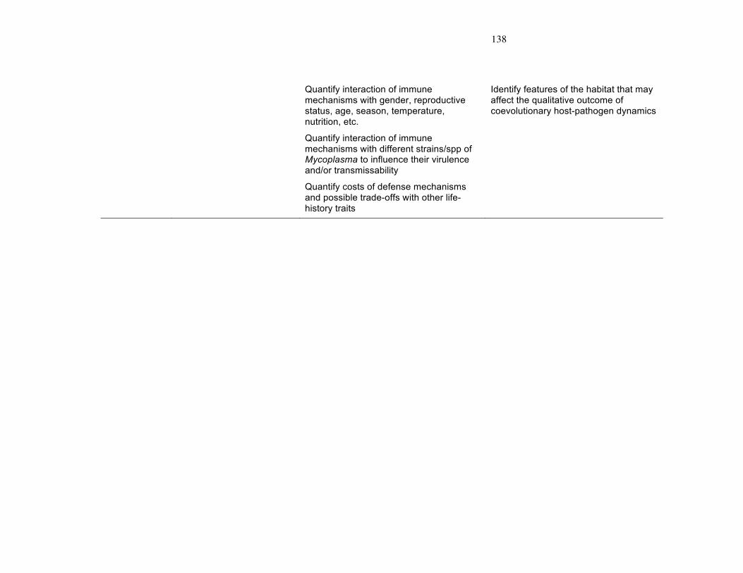
138
Quantify interaction of immune mechanisms with gender, reproductive status, age, season, temperature, nutrition, etc.
Identify features of the habitat that may affect the qualitative outcome of coevolutionary host-pathogen dynamics
Quantify interaction of immune mechanisms with different strains/spp of Mycoplasma to influence their virulence and/or transmissability
Quantify costs of defense mechanisms and possible trade-offs with other life-history traits
- 139 –
- 1 -
FIGURE LEGENDS
Figure 1. Mojave desert, including southern California, southern Nevada, southeastern
Utah, and northwestern Arizona.
a) Tortoise point locations, and three, distinct genetic groups (Hagerty and Tracy in
press). From left to right (west to east), the genetic groups are “California”, “Las Vegas”,
and “North Mojave” (sensu Hagerty and Tracy in press).
b) Twenty-four desert tortoise sampling populations (modified from Hagerty et al.
submitted). Points designate centroids of areas in which tortoises were sampled.
Abbreviations correspond to those used in Table 2.
Figure 2. Overlapping points are desert tortoise locations, and seroprevalence and mean
values were assigned according to the calculated population (n = 24) values (see text for
more detail).
a) Seroprevalence to M. agassizii (i.e. the proportion of the population seropositive to M.
agassizii). Seroprevalence was transformed to approximate a normal distribution (see text
for detail).
b) Mean natural antibody levels of populations.
Figure 3. Box-and-whisker diagram of natural antibody levels of individual tortoises
belonging to the three main genetic groups in the Mojave desert (California, Las Vegas,
and North Mojave). Natural antibody levels are log10-transformed.
Figure 4. Population-levels (n = 24) of seroprevalence to M. agassizii versus proportion
with URTD. Both the population proportions of animals with a seropositive Western blot
and with URTD were transformed to approximate a normal distribution (see text for more
detail).

140
FIGURE 1A

141
FIGURE 1B

142
FIGURE 2A

143
FIGURE 2B

144
FIGURE 3

145
FIGURE 4