impact of xylose and mannose on central...
TRANSCRIPT

Helsinki University of Technology, Department of Chemical Technology Technical Biochemistry Report 1/2005 Espoo 2005 TKK-BE-10
IMPACT OF XYLOSE AND MANNOSE ON CENTRAL METABOLISM OF YEAST Saccharomyces cerevisiae Juha-Pekka Pitkänen
GAP
BPG
Pyruvate
E4P
S7P
AcCoA
SucCoA
OAA
DHAP
Acetaldehyde
Acetate
Ethanol
CO2
CO2
CO2
AcCoA
OAACO2
CO2
Glycerol
NADH
NAD(P)H
NADPH
NADH
NADH
NAD(P)H
NAD(P)H
NADH
GTPNADPH
6PG
FUM
SUC
NAD(P)HCO2
M1P
GDP-mannose
M6P
F6P
G6P
GO3P
RU5P
R5P
3PG
PEPMAL
AKG
ICIT
CIT
X5P
Glyoxylate
ICIT
CIT
MAL SUCNADH
CO2
�������������������
���������
���������
����
������������
��������������
GLUCOSEMANNOSE
�����������
XYLOSE
Xylitol
Xylulose
NADH
NAD(P)H
������
����
F16P F26P
AB TEKNILLINEN KORKEAKOULUTEKNISKA HÖGSKOLANHELSINKI UNIVERSITY OF TECHNOLOGYTECHNISCHE UNIVERSITÄT HELSINKIUNIVERSITE DE TECHNOLOGIE D’HELSINKI
Helsinki University of Technology, Department of Chemical Technology Technical Biochemistry Report 1/2005 Espoo 2005 TKK-BE-10
IMPACT OF XYLOSE AND MANNOSE ON CENTRAL METABOLISM OF YEAST Saccharomyces cerevisiae Juha-Pekka Pitkänen Dissertation for the degree of Doctor of Science in Technology to be presented with due permission of the Department of Chemical Technology for public examination and debate in Auditorium AS 1 (in TUAS house) at Helsinki University of Technology (Espoo, Finland) on the 18th of November, 2005, at 12 noon. Helsinki University of Technology Department of Chemical Technology Laboratory of Bioprocess Engineering Teknillinen korkeakoulu Kemian tekniikan osasto Bioprosessitekniikan laboratorio
Distribution: Helsinki University of Technology Laboratory of Bioprocess Engineering P.O. Box 6100 FIN-02015 HUT Tel +358-9-4512541 Fax +358-9-462373 E-mail [email protected] � Juha-Pekka Pitkänen ISBN 951-22-7893-6 (printed) ISBN 951-22-7894-4 (pdf) ISSN 0359-6621 This thesis is online: http://lib.tkk.fi/Diss/2005/isbn9512278944 Yliopistopaino Espoo 2005
1
Pitkänen, Juha-Pekka. Impact of xylose and mannose on central metabolism of yeast Saccharomyces cerevisiae. Espoo 2005, Helsinki University of Technology. Keywords: Saccharomyces cerevisiae, yeast, metabolism, D-xylose, D-mannose, ethanol, GDP-mannose, flux analysis, metabolite analysis, transcript analysis, protein analysis
���������
In this study, understanding of the central metabolism was improved by quantification of metabolite concentrations, enzyme activities, protein abundances, and gene transcript concentrations. Intracellular fluxes were estimated by applying stoichiometric models of metabolism. The methods were applied in the study of yeast Saccharomyces cerevisiae in two separate projects. A xylose project aimed at improved utilization of D-xylose as a substrate for, e.g., producing biomaterial-based fuel ethanol. A mannose project studied the production of GDP-mannose from D-mannose in a strain lacking the gene for phosphomannose isomerase (PMI40 deletion). Hexose, D-glucose is the only sugar more abundant than pentose D-xylose. D-xylose is common in hardwoods (e.g. birch) and crop residues (ca. 25% of dry weight). However, S. cerevisiae is unable to utilize D-xylose without a recombinant pathway where D-xylose is converted to D-xylulose. In this study D-xylose was converted in two steps via xylitol: by D-xylose reductase and xylitol dehydrogenase encoded by XYL1 and XYL2 from Pichia stipitis, respectively. Additionally, endogenous xylulokinase (XKS1) was overexpressed in order to increase the consumption of D-xylose by enhancing the phosphorylation of D-xylulose. Despite of the functional recombinant pathway the utilization rates of D-xylose still remained low. This study proposes a set of limitations that are responsible for the low utilization rates of D-xylose under microaerobic conditions. Cells compensated for the cofactor imbalance, caused by the conversion of D-xylose to D-xylulose, by increasing the flux through the oxidative pentose phosphate pathway and by shuttling NADH redox potential to mitochondrion to be oxidized in oxidative phosphorylation. However, mitochondrial NADH inhibits citrate synthase in citric acid cycle, and consequently lower flux through citric acid cycle limits oxidative phosphorylation. Further, limitations in the uptake of D-xylose, in the pentose phosphate pathway, and in the citric acid cycle were alleviated in xylose chemostat isolates with three-fold improved xylose utilization rates. Uptake rate of D-xylose, assayed in vitro with radioactive D-xylose, was improved by 60% in the chemostat isolates grown under aerobic conditions on D-xylose. In the pentose phosphate pathway activities of transketolase and transaldolase were increased two-fold, and consequently concentrations of their substrates were decreased two-fold in the chemostat isolates. Finally, less pyruvate and citrate, but more malate accumulated in the chemostat isolates than in the original strain grown on D-xylose under aerobic conditions. In a S. cerevisiae strain with PMI40 deletion, growth on media without D-mannose and D-glucose is disabled. Phosphomannose isomerase encoded by PMI40 connects D-mannose to glycolysis, which is the main pathway for D-glucose utilization. Hypothetically, a PMI40 deletion strain would direct all its D-mannose into the biosynthesis of GDP-mannose. However, in the PMI40 deletion strain increased initial D-mannose concentrations led to increased intracellular mannose 6-phosphate concentrations. Mannose 6-phosphate inhibited activity of phosphoglucose isomerase (encoded by PGI1) in glycolysis, which in essence is equivalent to suppressed expression of PGI1. Subsequently, reduced availability of glycolysis intermediates, due to inhibition of phosphoglucose isomerase, led to a decrease in the glycolytic flux. Eventually, increased initial D-mannose concentrations resulted in a starvation response, which was accompanied by slower cell cycle and slower growth rate.
2
�����
This work was carried out in VTT Biotechnology (Technical Research Centre of Finland), in Medicel Oy, and in Rational Drug Design Program, Biomedicum, University of Helsinki during the years 2000-2004. I am deeply indebted to Dr. Aristos Aristidou for getting me started and excited in the field of metabolic engineering and Dr. Laura Ruohonen for kindly guiding me with my writing and thus getting my publications finished. I also thank Dr. Heikki Ojamo for his tutoring review and comments of this work. Of the managers, I wish to thank my supervisor Prof. Matti Leisola for lighting my initial interest in bioprocess engineering, Prof. Merja Penttilä, Dos. Risto Renkonen, and Timo Lehtonen for being able to build up such respectful research environments and giving me an opportunity to work as a part of them. Then I gratefully acknowledge other co-authors and colleagues, Laura Salusjärvi, Eija Rintala, Susanne Alff, Anssi Törmä, Dr. Laura Huopaniemi and Dr. Pirkko Mattila for their efforts in this work. I am grateful also for Eila Leino, Tarja Hakkarainen, Seija Rissanen, Tuija Toivikko, Satu Bruun, Sirkka-Liisa (Kikka Kauranen) Holm, Kati Venäläinen, and Aki Aittola for their indispensable efforts in the laboratory. For the scientific discussions e.g. about yeast physiology and analytical techniques I wish to thank Dr. Hannu Maaheimo, Dr. John Londesborough, Dr. Peter Richard, Mervi Toivari, Dr. Kari Koivuranta, Dr. Helena Simolin, Juha Kokkonen, Dr. Tapani Suortti, Dr. Tapio Kotiaho, Ismo Mattila, Sakari Joenväärä, Ilja Ritamo, Dr. Jarkko Heinonen, Dr. Christophe Roos, and Dr. Meelis Kolmer. For their participation in the automated sampling device project I am thankful for Mikko Lehmusto, Kalevi Puukko, Pertteli Varpela, Ilkka Pekkarinen, Dr. Antti Vuolanto, Mikko Tiainen, Peter Lindroos, Markku Suomela, Jari Kauhaniemi, and Pekka Edlund. In addition to people above, I warmly thank my other fellow workers both at VTT Biotechnology and Medicel Oy for creating friendly working environments and providing their occasional assistance, Anu Tamminen, Jonni Hurme, Michael Bailey, Juha Tähtiharju, Merja Aarnio, Matti Siika-Aho, Reetta Kuokka, Mikko Putkonen, Henna Jalovaara, Suvi Laukkanen, Jouni Ahtinen, Tuula Kallioinen, Ritva Javanainen, Marika Hedberg, Riika Penttilä, Leena Penttilä, Harri Tapanainen, Jarno Mäkinen, Tuomas Raitila, Ville Parviainen, Dr. Marko Nykänen, Dr. Minna Mäki, Nina Järvinen, and the software team of Medicel Oy in Menuetto building, especially Dennis Diggin for proofreading my thesis. I wish to thank everyone who has been involved in my work over the years, even if his or her name is not specifically mentioned here. The funding of the National Technology Agency of Finland (TEKES) and the Academy of Finland through the Graduate School in Chemical Engineering (GSCE) is gratefully acknowledged. Tästä työstä huolimatta on elämässäni ollut juuri ja juuri muutakin, josta saan kiittää ainakin vanhempiani, isovanhempiani, appivanhempiani, veljeäni Mattia, muita sukulaisiani sekä kavereitani, mutta ennenkaikkea vaimoani Hannaa ja rakkaita poikiamme Nikoa ja Pyryä. Helsinki, April 2005 Juha-Pekka Pitkänen
3
� ���������� ��� ����
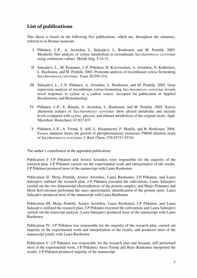
This thesis is based on the following five publications, which are, throughout the summary, referred to as Roman numerals.
I Pitkänen, J.-P., A. Aristidou, L. Salusjärvi, L. Ruohonen, and M. Penttilä. 2003. Metabolic flux analysis of xylose metabolism in recombinant Saccharomyces cerevisiae using continuous culture. Metab. Eng. 5:16-31.
II Salusjärvi, L., M. Poutanen, J.-P. Pitkänen, H. Koivistoinen, A. Aristidou, N. Kalkkinen,
L. Ruohonen, and M. Penttilä. 2003. Proteome analysis of recombinant xylose-fermenting Saccharomyces cerevisiae. Yeast 20:295-314.
III Salusjärvi, L., J.-P. Pitkänen, A. Aristidou, L. Ruohonen, and M. Penttilä. 2005. Gene
expression analysis of recombinant xylose-fermenting Saccharomyces cerevisiae reveals novel responses to xylose as a carbon source. Accepted for publication in Applied Biochemistry and Biotechnology
IV Pitkänen, J.-P., E. Rintala, A. Aristidou, L. Ruohonen, and M. Penttilä. 2005. Xylose
chemostat isolates of Saccharomyces cerevisiae show altered metabolite and enzyme levels compared with xylose, glucose, and ethanol metabolism of the original strain. Appl. Microbiol. Biotechnol. 67:827-837
V Pitkänen, J.-P., A. Törmä, S. Alff, L. Huopaniemi, P. Mattila, and R. Renkonen. 2004.
Excess mannose limits the growth of phosphomannose isomerase PMI40 deletion strain of Saccharomyces cerevisiae. J. Biol. Chem. 279:55737-55743.
The author’s contribution in the appended publications Publication I: J-P Pitkänen and Aristos Aristidou were responsible for the majority of the research plan. J-P Pitkänen carried out the experimental work and interpretation of the results. J-P Pitkänen produced most of the manuscript with Laura Ruohonen. Publication II: Merja Penttilä, Aristos Aristidou, Laura Ruohonen, J-P Pitkänen, and Laura Salusjärvi outlined the research plan. J-P Pitkänen executed the cultivations, Laura Salusjärvi carried out the two dimensional electrophoresis of the protein samples, and Marjo Poutanen and Heini Koivistoinen performed the mass spectrometric identification of the protein spots. Laura Salusjärvi produced most of the manuscript with Laura Ruohonen. Publication III: Merja Penttilä, Aristos Aristidou, Laura Ruohonen, J-P Pitkänen, and Laura Salusjärvi outlined the research plan. J-P Pitkänen executed the cultivations and Laura Salusjärvi carried out the transcript analysis. Laura Salusjärvi produced most of the manuscript with Laura Ruohonen. Publication IV: J-P Pitkänen was responsible for the majority of the research plan, carried out majority of the experimental work and interpretation of the results, and produced most of the manuscript jointly with Laura Ruohonen. Publication V: J-P Pitkänen was responsible for the research plan and Susanne Alff performed most of the experimental work. J-P Pitkänen, Anssi Törmä and Risto Renkonen interpreted the results. J-P Pitkänen produced majority of the manuscript.

4
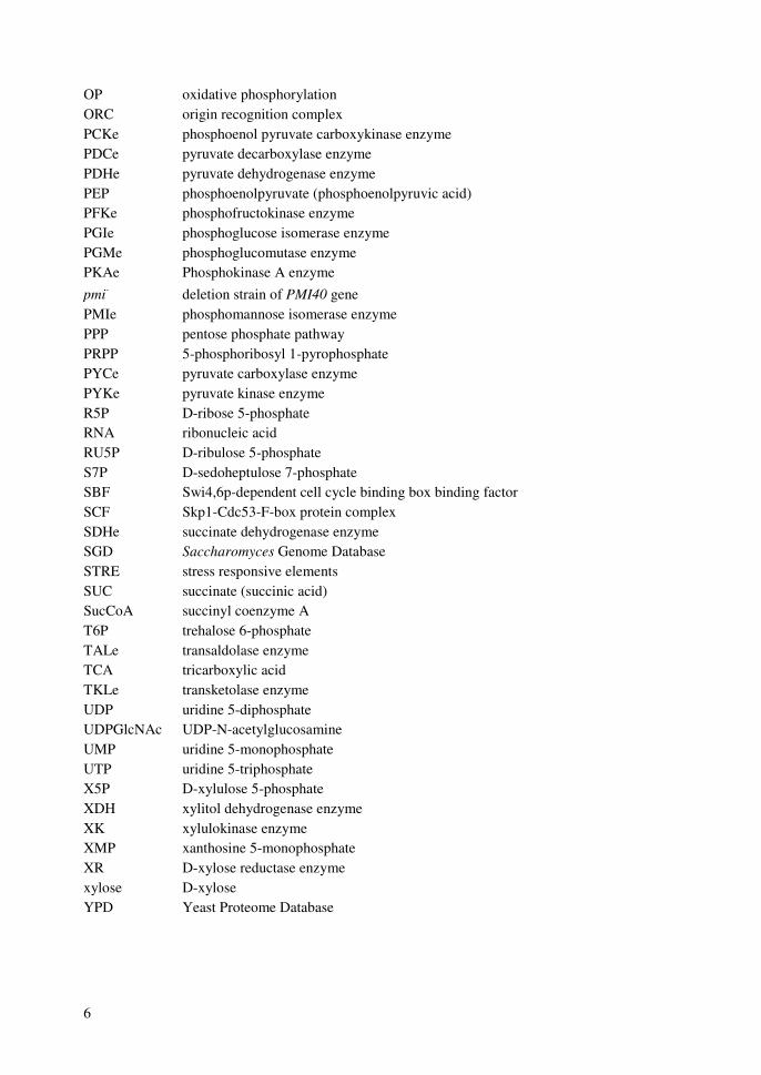
����� �� ����
Gene names (e.g. PMI40) and protein names (e.g. Pmi40p) are abbreviated according to Saccharomyces Genome Database (SGD). Enzyme names (e.g. PMIe) are also abbreviated according to the corresponding gene names in SGD, but without the gene numbers. 2-DE two-dimensional gel electrophoresis 2PG 2-phospho-D-glycerate 3-IPM 3-isopropylmalate 3PG 3-phospho-D-glycerate 6PG 6-phospho-D-gluconate AcCoA acetyl coenzyme A ACOe aconitase enzyme ADHe alcohol dehydrogenase enzyme ADP adenosine 5-diphosphate AICAR 5-aminoimidazole-4-carboxamide ribotide AIR Aminoimidazole ribotide AKG alpha-ketoglutarate (alpha-ketoglutaric acid) ALDe acetaldehyde dehydrogenase enzyme AMP adenosine 5-monophosphate APC anaphase promoting complex APC-P Phosphorylated anaphase promoting complex ASA L-aspartate semialdehyde ATP adenosine 5-triphosphate BPG 1,3-bisphosphoglycerate cAMP 3,5-cyclic adenosine monophosphate CDGS carbohydrate-deficient glycoprotein syndrome CDK cyclin dependent kinase cDNA Complementary deoxyribonucleic acid CDP cytidine 5-diphosphate CIT citrate (citric acid) CITe citrate synthase enzyme CMP cytidine 5-monophosphate CRE cAMP responsive element cRNA Complementary ribonucleic acid CTP cytidine 5-triphosphate DHAP dihydroxyacetone phosphate DNA deoxyribonucleic acid Dol-P dolichol phosphate E4P D-erythrose 4-phosphate EC Enzyme Classification ER endoplasmic reticulum F16P D-fructose 1,6-bisphosphate F26P D-fructose 2,6-bisphosphate F6P D-fructose 6-phosphate

5
FAD flavin adenine dinucleotide (oxidized) FADH2 flavin adenine dinucleotide (reduced) FBA flux balancing analysis FBPe fructose 1,6-bisphosphatase enzyme FUM fumarate (fumaric acid) G1P D-glucose 1-phosphate G6P D-glucose 6-phosphate GAP glyceraldehyde 3-phosphate GDP guanosine 5-diphosphate glucose D-glucose GMD GDP-mannose dehydratase enzyme GMER GDP-4-keto-6-deoxy-mannose-3,5-epimerase/4-reductase enzyme GMP guanosine 5-monophosphate GO3P glycerol 3-phosphate GTP guanosine 5-triphosphate HE-TPP 2-hydroxyethyl thiamine pyrophosphate H-ICIT homoisocitrate HPLC high performance liquid chromatography H-SER L-homoserine HXKe Hexokinase enzyme ICIT isocitrate (isocitric acid) ICLe isocitrate lyase enzyme IMP inosine 5-monophosphate KEGG Kyoto Encyclopedia of Genes and Genomes LC-MS liquid chromatographic mass spectrometry M1P D-mannose 1-phosphate M6P D-mannose 6-phosphate MAL L-malate (L-malic acid) MalCoA malonyl coenzyme A MALDI-TOF matrix assisted laser desorption/ionization time of flight mannose D-mannose MAP kinase mitogen activated protein kinase MBF Mlu1p-dependent cell cycle binding box binding factor MCA metabolic control analysis MCM mini-chromosome maintenance complex MDHe malate dehydrogenase enzyme MEN mitotic exit network MLSe malate synthase enzyme mRNA messenger RNA
NAD+ nicotinamide adenine dinucleotide (oxidized) NADH nicotinamide adenine dinucleotide (reduced)
NADP+ nicotinamide adenine dinucleotide phosphate (oxidized) NADPH nicotinamide adenine dinucleotide phosphate (reduced) NMR nuclear magnetic resonance spectroscopy OAA oxaloacetate (oxaloacetic acid)
6
OP oxidative phosphorylation ORC origin recognition complex PCKe phosphoenol pyruvate carboxykinase enzyme PDCe pyruvate decarboxylase enzyme PDHe pyruvate dehydrogenase enzyme PEP phosphoenolpyruvate (phosphoenolpyruvic acid) PFKe phosphofructokinase enzyme PGIe phosphoglucose isomerase enzyme PGMe phosphoglucomutase enzyme PKAe Phosphokinase A enzyme pmi- deletion strain of PMI40 gene PMIe phosphomannose isomerase enzyme PPP pentose phosphate pathway PRPP 5-phosphoribosyl 1-pyrophosphate PYCe pyruvate carboxylase enzyme PYKe pyruvate kinase enzyme R5P D-ribose 5-phosphate RNA ribonucleic acid RU5P D-ribulose 5-phosphate S7P D-sedoheptulose 7-phosphate SBF Swi4,6p-dependent cell cycle binding box binding factor SCF Skp1-Cdc53-F-box protein complex SDHe succinate dehydrogenase enzyme SGD Saccharomyces Genome Database STRE stress responsive elements SUC succinate (succinic acid) SucCoA succinyl coenzyme A T6P trehalose 6-phosphate TALe transaldolase enzyme TCA tricarboxylic acid TKLe transketolase enzyme UDP uridine 5-diphosphate UDPGlcNAc UDP-N-acetylglucosamine UMP uridine 5-monophosphate UTP uridine 5-triphosphate X5P D-xylulose 5-phosphate XDH xylitol dehydrogenase enzyme XK xylulokinase enzyme XMP xanthosine 5-monophosphate XR D-xylose reductase enzyme xylose D-xylose YPD Yeast Proteome Database
7
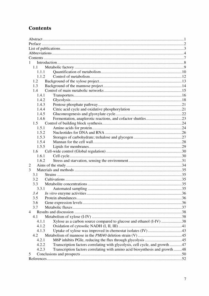
��������
Abstract ..........................................................................................................................................1 Preface ...........................................................................................................................................2 List of publications.........................................................................................................................3 Abbreviations .................................................................................................................................4 Contents .........................................................................................................................................7 1 Introduction............................................................................................................................8
1.1 Metabolic factory ...........................................................................................................9 1.1.1 Quantification of metabolism...............................................................................10 1.1.2 Control of metabolism..........................................................................................12
1.2 Background of the xylose project.................................................................................13 1.3 Background of the mannose project.............................................................................14 1.4 Control of main metabolic networks ............................................................................15
1.4.1 Transporters..........................................................................................................16 1.4.2 Glycolysis.............................................................................................................18 1.4.3 Pentose phosphate pathway..................................................................................21 1.4.4 Citric acid cycle and oxidative phosphorylation ..................................................21 1.4.5 Gluconeogenesis and glyoxylate cycle.................................................................22 1.4.6 Fermentation, anaplerotic reactions, and cofactor shuttles...................................23
1.5 Control of building block synthesis..............................................................................24 1.5.1 Amino acids for protein........................................................................................24 1.5.2 Nucleotides for DNA and RNA ...........................................................................26 1.5.3 Storages of carbohydrate; trehalose and glycogen ...............................................27 1.5.4 Mannan for the cell wall.......................................................................................28 1.5.5 Lipids for membranes...........................................................................................29
1.6 Cell-wide control (Global regulation) ..........................................................................30 1.6.1 Cell cycle..............................................................................................................30 1.6.2 Stress and starvation, sensing the environment ....................................................31
2 Aims of the study .................................................................................................................34 3 Materials and methods .........................................................................................................35
3.1 Strains ..........................................................................................................................35 3.2 Cultivations ..................................................................................................................35 3.3 Metabolite concentrations ............................................................................................35
3.3.1 Automated sampling ............................................................................................35 3.4 In vitro enzyme activities .............................................................................................36 3.5 Protein abundances.......................................................................................................36 3.6 Gene expression levels .................................................................................................36 3.7 Metabolic fluxes...........................................................................................................37
4 Results and discussion .........................................................................................................38 4.1 Metabolism of xylose (I-IV) ........................................................................................38
4.1.1 Xylose as a carbon source compared to glucose and ethanol (I-IV) ....................38 4.1.2 Oxidation of cytosolic NADH (I, II, III) ..............................................................41 4.1.3 Uptake of xylose was improved in chemostat isolates (IV) .................................43
4.2 Metabolism of mannose in the PMI40 deletion strain (V) ...........................................45 4.2.1 M6P inhibits PGIe, reducing the flux through glycolysis ....................................45 4.2.2 Transcription factors correlating with glycolysis, cell cycle, and growth ............47 4.2.3 Transcription factors correlating with amino acid biosynthesis and growth ........48
5 Conclusions and prospects ...................................................................................................50 References....................................................................................................................................52

8
�� ��������� ���
In bioreactor cultivations (fermentations) microbiological cells are the catalysts performing reactions that lead to products. We aim to utilize these products, ethanol as an example, and build processes around them, or alternatively the cells themselves use these products as building material for their offspring. In essence, growth of microbes is a self-catalyzing reaction. Understanding how the catalyst works and application of that understanding to control the catalyst’s activity are crucial in order to develop profitable processes. Use of microbes instead of chemical synthesis benefits from their biological selectivity. Due to biological selectivity raw materials can be cruder and less purified in a bioprocess than in a process based on chemical synthesis since microbes are, in principle, able to utilize only what they require from complex mixtures. Biological selectivity also reduces down-stream purification steps since microbes are able to produce molecules e.g. with exact stereochemical conformation. Overall, thanks to biological selectivity, the bioprocesses have earned an environmentally friendly status and are considered a reasonable alternative when developing sustainable processes for the future. Chemoorganotrophic organisms such as yeasts and humans cleave organic chemical compounds often produced by phototrophic organisms (Madigan et al., 1997). The compounds are cleaved in biochemical reactions inside a cellular membrane in order to provide energy in catabolic reactions and building blocks in biosynthetic reactions for generating new cells (Berg et al., 2002). The compounds involved in intracellular, biochemical reactions (catabolic or biosynthetic) are called metabolites. Reactions between metabolites are catalyzed by enzymes, which decrease the activation energy of the reactions. Instructions for constructing the enzymes are stored in double-stranded DNA as genes (Lodish et al., 2001). Upon request, the information from genes is transcribed to messenger RNA (mRNA) and further translated into protein. After translation most proteins undergo post-translational modifications to form active enzymes. During post-translational modifications proteins can be folded and amino acid side chains of proteins can be modified covalently e.g. by phosphorylation or acetylation, and by adding carbohydrate chains in glycosylation (Lodish et al., 2001). Some proteins participate e.g. in scaffolding or signaling of the cellular functions (Lodish et al., 2001). In order to unveil cellular behavior we need to be knowledgeable of biological connections and the methods by which we can quantify them. Metabolic engineering is an array of methods building on the foundations of genetic engineering in modification of the genomes of the organisms, and in various analytical and mathematical methods in trying to measure and model the behavior of organisms (Bailey, 1991; Stephanopoulos et al., 1998; Stephanopoulos, 1999; Wiechert, 2002). Metabolic engineering aims at the modification and measurement of metabolic fluxes in order to improve yields and rates of the known products or to produce altogether new products or to utilize novel substrates (Bailey, 1991; Stephanopoulos, 1994). The yeast Saccharomyces cerevisiae is one of the most utilized model organisms of eukaryotic cell systems, and is in scientific popularity close to the prokaryotic model organism, bacterium Escherichia coli. There are several reasons for the popularity of this yeast. To begin with, yeasts must have been the first household organism people took as a companion – although unknowingly. The obvious reason was the production of ethanol for recreational purposes (McGovern et al., 2004) or carbon dioxide in baking. In order to attain successful synergy with humans, production of other compounds is minimal and ethanol and carbon dioxide are the two main products of the yeast, besides itself of course. Over the millennia, alongside humans, yeast has gathered properties that enable it to prevail in fermentation tanks over competing organisms. Yeast tolerates environments that contain high ethanol, acid, or salt concentrations. Further, it can adapt to variation in osmotic pressure or temperature. Certainly there are microorganisms that are more robust in extremes of some of the conditions mentioned above, but yeast has a good combination of properties for tolerating various conditions (Olsson and Hahn-Hägerdal,

9
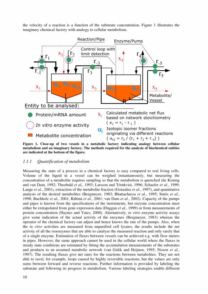
1996; Gasch et al., 2000). But above all, yeast cell is a eukaryote like our human cells. It has several cellular compartments including nucleus and mitochondria that give their characteristics to transcription and respiration, respectively. The yeast genome, which was the first eukaryotic gene sequence decoded (Goffeau et al., 1997), has introns making for example mRNA processing an important task. Compared to human (IHGSC, 2001), and to other eukaryotic model organisms like house mouse Mus musculus (Gregory et al., 2002), fruit fly Drosophila melanogaster (Adams et al., 2000), a soil nematode Caenorhabditis elegans (CESC, 1998), or a flowering plant Arabidopsis thaliana (AGI, 2000) the life cycle of yeast is clearly the shortest and it is the easiest and cheapest to maintain and engineer. As a simple, unicellular organism yeast is best suited for studying events concerning the basic functions of a single cell, such as metabolism (Berg et al., 2002), membrane trafficking, DNA replication, transcription, mRNA cleavage, translation (Lodish et al., 2001), post-translational modifications of proteins (Freedman, 1995), etc. As a unicellular organism yeast is not applicable for example in studies of morphogenesis or developmental biology in general. The genome of S. cerevisiae has over 6000 genes in 16 chromosomes (Goffeau et al., 1997). In nature S. cerevisiae exists as a polyploid, but in laboratory the genetic engineering and genetic stability of the strains are best achieved with haploid yeast strains (Entian and Kötter, 1998). The fact that S. cerevisiae is one of the model organisms of modern biotechnology for eukaryotic systems makes it’s genetic modification and characterization of rate limiting steps feasible tasks (Ostergaard et al., 2000). Thus, it is possible to add otherwise foreign substrates or unfamiliar products to the repertoire of S. cerevisiae. Taken together, the properties of yeast have made it a popular production organism for novel bio-based products. In this study, understanding of the central metabolism was improved by quantification of metabolite concentrations, enzyme activities, protein abundances, and gene transcript concentrations. Intracellular fluxes were estimated by applying stoichiometric models of metabolism in pseudo steady state conditions. Methods of metabolic engineering were harnessed in order to elucidate and improve the utilization of an unfamiliar substrate D-xylose (xylose) and production of an intracellular precursor GDP-mannose in S. cerevisiae. In essence, this study examines, in two separate projects, how xylose in one hand and D-mannose (mannose) in the other affect the growth of genetically modified S. cerevisiae. 1.1 Metabolic factory Intracellular reaction network has some analogy to a chemical factory. Let us imagine a chemical factory that contains various vessels and numerous pipes with pumps connecting vessels to each other. The vessels represent metabolites in a biological cell, i.e. the compounds produced or consumed in biological reactions. An amount of liquid in a vessel has an analogy to a concentration of a metabolite in a cellular compartment. Similarly, pipes transferring liquid from vessel to vessel can be thought as reactions converting one metabolite to another. Thus, a single vessel represents only one metabolite unlike in real cells where several metabolites are together in a single compartment, in a single vessel. Pumps in this imaginary chemical factory represent enzymes, increasing the flow between pools. Actually, pumps and pipes form together an entity that can be compared to an enzyme-catalyzed reaction. A vertical position of a vessel or a path of pipes in a vertical axis has analogy to thermodynamics in chemistry. If an initial vessel (substrate) is situated higher than a final vessel (product) the process is energetically favorable. Depth of the pipe inlet in a vessel corresponds to properties of an enzyme, namely affinity of an enzyme. In principle, a pipe that reaches close to the bottom of the vessel resembles an enzyme with a low Km value and high affinity. Thus, one pipe might be deeper in a same vessel than another pipe since different enzymes, using the same metabolite as their substrate, have different affinities for the metabolite. Similarly, diameter of a pipe is analogous to maximal velocity (Vmax) of an enzyme-catalyzed reaction. Further, liquid flow rate in a pipe is higher when there is more liquid in the vessel due to higher hygroscopic pressure, in analogy;
10
the velocity of a reaction is a function of the substrate concentration. Figure 1 illustrates the imaginary chemical factory with analogy to cellular metabolism.
r2r-3
r-1
r1
r3
x1
x2
x3
������������ �������
����������� ���!��
�������� ��"��� ���#���
$���������������������
���!������������������%�!���������������&�������������'��( )��( � ��( *
+�������������� %����������,�����,�#����%%��������������'�α�- )��- ��'�( .��-�.���/*�*
ααααr2
ααααi
xi
ααααr1
r4r-5
r5
x4
x5ααααr3
�������������������������������
��"�����!��������������
$���������0�����
ααααr5ααααr3
Figure 1. Close-up of two vessels in a metabolic factory indicating analogy between cellular metabolism and an imaginary factory. The methods required for the analysis of biochemical entities are indicated at the bottom of the figure. 1.1.1 Quantification of metabolism Measuring the state of a process in a chemical factory is easy compared to real living cells. Volume of the liquid in a vessel can be weighed instantaneously, but measuring the concentration of a metabolite requires sampling so that the metabolism is quenched (de Koning and van Dam, 1992; Theobald et al., 1993; Larsson and Törnkvist, 1996; Schaefer et al., 1999; Lange et al., 2001), extraction of the metabolite fraction (Gonzalez et al., 1997), and quantitative analysis of the desired metabolites (Bergmeyer, 1983; Bhattacharya et al., 1995; Smits et al., 1998; Buchholz et al., 2001; Räbinä et al., 2001; van Dam et al., 2002). Capacity of the pumps and pipes is known from the specifications of the instruments, but enzyme concentration must either be extrapolated from gene expression data (Duggan et al., 1999) or from measurements of protein concentration (Haynes and Yates, 2000). Alternatively, in vitro enzyme activity assays give some indication of the actual activity of the enzymes (Bergmeyer, 1983) whereas the operator of the chemical factory can adjust and hence knows the rate of the pumps. Also, when the in vitro activities are measured from unpurified cell lysates, the results include the net activity of all the isoenzymes that are able to catalyze the measured reaction and only rarely that of a single enzyme. Estimation of fluxes between vessels can be achieved e.g. with flow meters in pipes. However, the same approach cannot be used in the cellular world where the fluxes in steady state conditions are estimated by fitting the accumulation measurements of the substrates and products to an assumed metabolic network (van Gulik and Heijnen, 1995; Nissen et al., 1997). The resulting fluxes give net rates for the reactions between metabolites. They are not able to ravel, for example, loops caused by highly reversible reactions, but the values are only sums between forward and reverse reactions. Further information is provided by labeling the substrate and following its progress in metabolism. Various labeling strategies enable different

11
possibilities to monitor ratios between intracellular pathways (Szyperski, 1998). The basic idea is that different metabolic routes cleave the backbone of the substrates differently. Resulting labeling patterns of the isotopic isomers can be measured from the products or from the amino acids hydrolyzed from cellular protein. The amino acids lead us to central metabolism as the eight precursors of amino acids situate in the main metabolic networks (Szyperski, 1998; Berg et al., 2002). The methods mentioned above, measurement of metabolites, enzyme activities in vitro, proteins, gene expression, metabolic fluxes, and isotopic isomers after labeling, are the backbone of quantifying cellular behavior. Figure 1 illustrates how the measurements of biochemical entities relate to the imaginary chemical factory. In dynamic situations the flux estimation can be achieved by monitoring the weights of the vessels; some vessels lose content and some gain. The principle is the same both in cells and in a factory, but tremendously more difficult in cells where as many metabolites as possible need to be quantified, but taken together from publications describing methods for the analysis of intracellular metabolites, hitherto only roughly 10% can be measured feasibly (Bergmeyer, 1983; Smits et al., 1998; Buchholz et al., 2001; Räbinä et al., 2001; van Dam et al., 2002). After measuring the in vivo concentrations of metabolites in dynamic situations an estimation of a dynamic model containing the metabolic reactions and regulatory connections affecting the rate of enzymatic reactions are fitted to the measured information (Rizzi et al., 1997). The fitted results include dynamic properties of the enzymes such as affinities or power-law parameters (Voit, 2000). The same, or corresponding, values can, in principle, be assayed and have been assayed from purified enzymes with in vitro assays, but those values do not necessarily reflect the actual in vivo values since the concentration ratio between metabolites and proteins is higher in vitro (Visser et al., 2000). More importantly, the concentrations of factors affecting the activity or mainly the three dimensional structure of enzymes is different in vitro than in vivo. However, in vitro measurements do give indications what factors can affect activity of a specific enzyme, and in vitro measurements may indicate the mechanism of the reaction (Cornish-Bowden, 1999). Another aspect for dynamic studies include non-invasive in vivo NMR methods, which enable the monitoring of the metabolic reactions in the cells incubated in the NMR device (Brindle et al., 1993). Thus, no sampling of the system is required for monitoring. However, with NMR methods the sensitivity may be poor and the incubation conditions may be compromised inside the instrument. In some respect the analogies between the actual cells and the imaginary factory seem far-fetched, but the purpose here is to underline how difficult it is to estimate how a biological system operates. First, we do not necessarily even know what to look for. If for example yeast has over 6000 genes (Goffeau et al., 1997) which encode for roughly same number of proteins, which in addition undergo various modifications before catalyzing reactions between around 600 metabolites (Forster et al., 2003), it creates a huge factory and bear in mind that this factory makes the pumps and actuators by itself. So in this context, how do we know what is relevant information and what can be ignored until we are able to process the whole system? Gene expression analysis is basically the only real analysis that can be performed without being forced to make targeted selections of what to look for on the whole system-wide level (top-down), but measurements of proteins and metabolites require at least some initial knowledge of the entities we want to measure (bottom-up). Second, we are dealing with a constantly changing situation, so we will have to gather snap-shots either from an assumed steady state or in a suitable time series of a dynamic situation. Basically, we will have to quench the cellular events either immediately after sampling or we will have to create on-line measurement methodologies. Third, getting from a pellet of cells to samples of mRNA, protein or metabolites requires long, qualitative steps of purification. Each sample type requires basically its own samples and own extraction methods. Fourth, after extraction the actual quantification of the molecules of mRNA, proteins, and metabolites requires novel and extensive instrumentation and methods.
12
1.1.2 Control of metabolism Both factories and cells require the control of the state and capacity of their components so that they can be kept close to their optimal working values, i.e., homeostasis (Saldanha et al., 2004). In cells the control signal is achieved by changing the three-dimensional structure of the proteins when either other proteins, metabolites, DNA or RNA attach to them (Lodish et al., 2001). Overall, we are studying a three-dimensional puzzle in its utmost complexity. The changed conformation of a protein might be able to bind to other proteins, metabolites, or DNA, changing e.g. the transcription rates of genes. In some instances the effect is seen as allosteric control of flux through glycolysis (see section 1.4.2 below). In the factory the control loop starts most probably from the measurement of the volumes in vessels. The information is carried to a pump as an electronic signal and that should cause the pump either to increase or decrease the pump speed (Figure 1). The rates of enzymatic reactions can be controlled in a variety of ways at different levels of the flow of information from a gene to an enzyme. First, varying the expression of the genes encoding the enzyme, controls the amount of the enzyme. Additionally, there are a number of steps where the flow of information from a gene to an enzyme is controlled, including transcription of gene to mRNA, post-transcriptional processing of mRNA, transfer of mRNA from nucleus to ribosomes in cytosol, translation of mRNA to a protein, and post-translational processing of a protein to an enzyme (Lodish et al., 2001). Second, the activity of an enzyme can be adjusted by covalent modifications (e.g. phosphorylation), or by allosteric binding, which both affect the three-dimensional structure of the enzyme and hence its activity (Cornish-Bowden, 1999). Third, availability of the substrates affects the reaction rate according to thermodynamics. The effect of the availability of the substrate can often be seen as accumulation of a substrate, which basically results in an increase of the forward reaction rate. Also the protein complexes are a way to increase the net flux through the pathway, as the substrates are not allowed to diffuse freely around an enzyme or even in the cellular compartment, hence increasing the availability of the substrates. However, protein complexes also affect the activity of the enzymes as being a member of a complex might influence the three-dimensional structures of the complex members (Ho et al., 2002). Various modes of control are possible also in biological systems, including feed-back, feed-forward, sniffing, buzzing etc. mechanisms (Tyson et al., 2003). Like indicated above, all ions and molecules (metabolites, proteins, nucleic acids) that bind to proteins changing their conformation can carry control signals in cells. Bearing in mind that overall we want to increase yields and production rates of some products from the substrates we are able to purchase. We basically want to increase fluxes from our substrates to valuable products and keep other fluxes minimal. Thus, we need to examine the results of gene expression levels, protein abundances, enzyme activities, metabolite concentrations, and metabolic fluxes parallel in the context of metabolic networks in order to find possible limitations in the metabolism. Incoherent metabolic situations where the parallel measurements at the five levels mentioned above can be considered as indications of metabolic bottlenecks. Possible incoherent metabolic situations can be, for instance, increased metabolite concentration with increased expression of genes downstream, increased metabolite concentration with increased fluxes downstream, increased gene expression with decreased fluxes, and decreased metabolite concentration with increased gene expression upstream. In the examples the phrase “gene expression” can be replaced with “protein abundance” or “enzyme activity”. However, as there are several steps of controlling the actual in vivo activity of the enzymes that catalyze the reactions, we are not necessarily able to tell, what would be the actual mechanism how cells control the flux. When talking about the control of flux, a mathematical method called metabolic control analysis (MCA) enables the calculation of actual distribution of control imposed by separate steps on a metabolic pathway (Fell, 1997). According to MCA the flux control coefficient of an enzyme on a pathway is defined as a ratio of the normalized change in enzyme activity and the normalized change in steady state flux. Values of the flux control
13
coefficients for individual enzymes on a pathway vary between 0 and 1. MCA defines also elasticity coefficients, which are ratios of the normalized change in enzyme’s rate and the normalized change in metabolite concentration. Thus, elasticity coefficients are properties of individual enzymes related to the kinetic properties of the enzymes (Fell, 1997). 1.2 Background of the xylose project Xylose is the most common pentose sugar in the hemicellulose (ca. 25% of dry weight) of hardwoods and crop residues (Hartley, 1981). Overall in nature, glucose is the only sugar more abundant than xylose. Thus, efficient utilization of the xylose component of hemicellulose, in addition to the hexoses present in lignocellulose, offers an opportunity to reduce the cost of bio-ethanol production (Hahn-Hägerdal et al., 1991; Olsson and Hahn-Hägerdal, 1996). S. cerevisiae, which is one of the most prominent ethanol-producing organisms from hexose sugars, is unable to utilize xylose (Barnett, 1976). S. cerevisiae cells take up xylose with the same sugar permeases it uses for the uptake of D-glucose (glucose) (Kötter and Ciriacy, 1993; Hamacher et al., 2002). However, xylose uptake is very inefficient compared to that of glucose (Kötter and Ciriacy, 1993; Singh and Mishra, 1995; Lee et al., 2002b) and especially at low concentrations it limits utilization of xylose (Gardonyi et al., 2003). S. cerevisiae can catabolize D-xylulose (xylulose) (Wang and Schneider, 1980; Jeffries, 1981; Ueng et al., 1981), however, it cannot utilize xylose due to the inability to convert xylose to xylulose efficiently (Jeffries, 1990). In naturally xylose utilizing fungi xylose is first reduced to xylitol by D-xylose reductase (XR) that prefers NADPH over NADH. Xylitol is then oxidized to xylulose with NAD+ by xylitol dehydrogenase (XDH) as presented in Figure 2. S. cerevisiae has the genes for XR and XDH endogenously; YHR104w (Träff et al., 2002) and YLR070c (Richard et al., 1999), respectively. However, the endogenous genes are not expressed to a level that would sustain growth on xylose even under aerobic conditions (Toivari et al., 2004). Further, different cofactor specificities would lead to a serious cofactor imbalance during xylose consumption via the XR-XDH pathway, unless the cells were able to compensate for it elsewhere in the metabolism. This cofactor imbalance is especially problematic under anaerobic conditions (Bruinenberg et al., 1983a). Before entering the pentose phosphate pathway (PPP) xylulose is phosphorylated to D-xylulose 5-phosphate (X5P) by xylulokinase (XK). Non-oxidative reactions of PPP convert X5P to glyceraldehyde 3-phosphate (GAP) and D-fructose 6-phosphate (F6P), which link PPP to glycolysis (Berg et al., 2002). Non-oxidative PPP includes seven metabolites that are interconverted by just two enzymes operating close to equilibrium. Also accumulation of PPP intermediates has been indicated in cells grown on xylose (Zaldivar et al., 2002). Activity of the PPP enzymes may be too low for efficient xylose utilization (Kötter and Ciriacy, 1993). Further, the response to xylose at the metabolic level of S. cerevisiae seems to be a mixture of the response to glucose and ethanol. Some reports state that xylose is a respiratory carbon source like ethanol (Jin et al., 2004) while others claim that xylose is a fermentative carbon source causing a response for catabolite repression, although, the response is not as strict as that caused by glucose (Roca et al., 2003a). Efficient utilization of xylose seems to be requiring small amounts of oxygen, which may be related to oxygen requirement of xylose uptake (Hahn-Hägerdal et al., 2001) or to respiration having a significant role in xylose utilization (Jeffries and Jin, 2004; Jin et al., 2004). The most common approach to construct xylose-utilizing recombinant S. cerevisiae strains has been the expression of XR- and XDH-encoding genes XYL1 and XYL2, respectively, from Pichia stipitis (Kötter et al., 1990; Walfridsson et al., 1995). Further, overexpression of the endogenous XK encoding gene (XKS1) improves xylose utilization, as has been demonstrated in recent studies (Eliasson et al., 2000; Richard et al., 2000; Toivari et al., 2001). These efforts have resulted in fermentation of xylose by S. cerevisiae, but the ethanol yield over the carbon utilized is clearly lower than with hexose sugars, and a significant fraction of xylose ends up into xylitol. Three steps mentioned above, xylose uptake, conversion of xylose to xylulose, and PPP
14
reactions, have hitherto been considered as the steps, which impose the most severe limitations to the metabolism of xylose (Jeffries and Jin, 2004). Various approaches have been taken to address these problems. There have been efforts to improve simultaneous uptake of xylose and glucose in order to improve uptake rates of xylose (Hamacher et al., 2002; Sedlak and Ho, 2004). Especially, a separate transport system for xylose would be beneficial (Hamacher et al., 2002) since even small glucose amounts competitively inhibit xylose uptake (Lagunas, 1993) facilitated by HXT-encoded transporters (Lee et al., 2002b). Inefficient xylose fermentation has been ascribed also to the different cofactor preference of XR and XDH. Strategies to balance the cellular redox include efforts to increase the intracellular NADPH availability by metabolic engineering of ammonium assimilation pathways (Roca et al., 2003b) and various transhydrogenase systems (Aristidou et al., 1998; Aristidou and Penttilä, 2000) including NADPH-dependent glyceraldehyde 3-phosphate dehydrogenase (Verho et al., 2002). To circumvent the redox issue of xylose conversion by the oxidoreductive pathway, heterologous expression of xylose isomerases has been tried, however, only recently, this approach met with significant success with functional fungal isomerase (Kuyper et al., 2003; Kuyper et al., 2004b). Reactions of PPP have been studied by overexpression of genes encoding enzymes of the non-oxidative PPP (Walfridsson et al., 1995; Kuyper et al., 2004a), and deletion of genes encoding enzymes of the oxidative PPP to reduce NADPH production (Jeppsson et al., 2002). However, increased activity of PPP enzymes displayed a significant improvement in xylose utilization only in a strain with functional xylose isomerase (Kuyper et al., 2004a). Additionally, by-pass of the majority of the non-oxidative PPP reactions has been attempted by expressing a phosphoketolase pathway from bacteria in S. cerevisiae (Sonderegger et al., 2004b). Recently, a few groups have presented recovery of more efficient xylose utilizing S. cerevisiae strains following a directed evolution approach with mutation and selection for improved growth (Sonderegger and Sauer, 2003; Wahlbom et al., 2003b). These mutants show altered properties at the known xylose bottlenecks discussed above (Wahlbom et al., 2003a; Sonderegger et al., 2004a). 1.3 Background of the mannose project Phosphomannose isomerase enzyme (PMIe) catalyzes the interconversion of fructose 6-phosphate (F6P) in glycolysis to mannose 6-phosphate (M6P) in a mannose pathway as presented in Figure 2. In the eukaryotic model organism, S. cerevisiae, PMIe is encoded by PMI40 gene (Smith et al., 1992). In a PMI40 deletion strain (pmi-), synthesis of M6P from F6P is not possible, disabling the growth of such a strain on media without mannose and glucose. The inability to grow, caused by defective glycosylation of a temperature-sensitive pmi40 mutant of S. cerevisiae, and repairing the defects by addition of mannose to the growth media have been previously described (Payton et al., 1991). In humans PMIe deficiency is the cause of carbohydrate-deficient glycoprotein syndrome (CDGS) type Ib, but the condition can be successfully treated by mannose administration (Niehues et al., 1998). M6P produced either from F6P or mannose serves as a precursor for the de novo biosynthesis of nucleotide sugar GDP-mannose. M6P is converted to mannose 1-phosphate (M1P) by phosphomannomutase encoded by SEC53 (Kepes and Schekman, 1988). Subsequently, M1P is ligated with guanosine 5-triphosphate molecule (GTP) to form GDP-mannose by M1P guanylyltransferase encoded by PSA1 (Hashimoto et al., 1997). The de novo formation of the purine ring of GTP, required for the biosynthesis of GDP-mannose (Shimma et al., 1997), starts from ribose 5-phosphate (R5P) in the pentose phosphate pathway and requires also 3-phosphoglycerate (3PG) in the glycolysis as a precursor. Taken together, the biosynthesis of GTP is more complex than the mannose pathway (Berg et al., 2002). GDP-mannose is needed in S. cerevisiae for mannosylation of various structures such as lipopolysaccharides and glycoproteins (Berg et al., 2002). In S. cerevisiae GDP-mannose biosynthesis, as nucleotide sugar biosynthesis generally, is carried out in the cytosol (Gao et al., 2001). Additionally, GDP-mannose is necessary as a precursor for
15
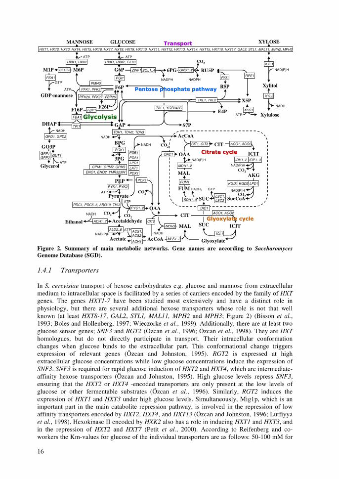
other nucleotide sugars including GDP-fucose, GDP-rhamnose, and GDP-talose (Mattila et al., 2000; Järvinen et al., 2001; Mäki et al., 2002; Wu et al., 2002; Mäki et al., 2003). However, GDP-fucose or GDP-rhamnose does not exist endogenously in S. cerevisiae, but recombinant strains can be used e.g. for production of GDP-fucose with genes encoding GMD (GDP-mannose dehydratase) and GMER (GDP-4-keto-6-deoxy-mannose-3,5-epimerase/4-reductase) (Mattila et al., 2000; Järvinen et al., 2001). With appropriate glycosyl transferase enzymes various nucleotide sugars can be applied in in vitro oligosaccharide synthesis and protein glycosylation (Renkonen et al., 1992). Oligosaccharides with correct conformation are important in immunoresponse (Satomaa et al., 2002). Hypothetically, a strain lacking the gene for PMIe would direct all its mannose into the biosynthesis of GDP-mannose. The effects of various mannose concentrations on GDP-production or on growth of the PMI40 deletion strain have not been studied previously. 1.4 Control of main metabolic networks In this study the central metabolism and main metabolic networks are referred to as a set of reactions that carry most of the carbon flux. They are catabolic reactions that lead to the production of energy and precursors. The reactions and control of transport, glycolysis, pentose phosphate pathway, pyruvate utilization, citric acid cycle, gluconeogenesis, and glyoxylate cycle are discussed. The roles of xylose and mannose utilization are discussed along each pathway. The main metabolic networks are displayed in Figure 2 with metabolites and genes encoding the enzymes that catalyze the reactions. The gene names are according to the Saccharomyces Genome Database (SGD) (Cherry et al., 1998). The main source for constructing maps of the central metabolic pathways was a basic biochemistry textbook (Berg et al., 2002) and the Kyoto Encyclopedia of Genes and Genomes (KEGG) database (Kanehisa and Goto, 2000). Figure 2 presents also the reactions involving redox and energy cofactors. The connections controlling the central metabolic pathways are discussed at three levels; regulation of gene expression, protein-protein interactions, and conformational (allosteric and covalent) modifications. Information for the regulation of gene expression is gathered from three sources, Yeast Proteome Database’s (YPD) regulator reports (Costanzo et al., 2000), Transcription factor binding network by Young-lab (Lee et al., 2002a), and functional genomics co-expression results from YPD (Costanzo et al., 2000). Information on protein-protein interactions is from Bind-database (Bader et al., 2003), and for allosteric modifications from Brenda database (Schomburg et al., 2000) and from YPD effector reports (Costanzo et al., 2000). The purpose of this review is to illustrate what parts of metabolism are interconnected and are thus expected to behave similarly after changes in the environment. Several of the figures are crowded with connections, but in several cases they are only simplifications of the situation. Thus, the figures below underline how important the development of interactive pathway software is; the amount of present data in the databases and in the publications is immense. Figure 3 summarizes the control of expression of genes encoding enzymes in central metabolic pathways according to YPD. Figure 4 presents what transcription factors bind to regulatory sequences of genes encoding enzymes in central metabolic pathways according to results of Young-lab (Lee et al., 2002a). Figure 5 introduces some protein complexes, which have members in proteins of central metabolic pathways according to YPD and Bind databases. Finally, Figure 6 summarizes the allosteric and covalent connections affecting activity of enzymes in central metabolic pathways according to Brenda and YPD databases. Figure 6 presents the enzyme classification (EC) numbers of the enzymes encoded by genes in Figure 2 according to Brenda and Nomenclature Committee of the International Union of Biochemistry and Molecular Biology (NC-IUBMB, 1992).
16
GLUCOSE
GAP
BPG
E4P
S7P
AcCoA
SucCoA
OAA
DHAP
Acetaldehyde
Acetate
Ethanol
CO2
CO2
CO2
AcCoA
OAACO2
CO2
Glycerol
NADH
NAD(P)H
NADPH
NADH
NADH
NAD(P)H
NAD(P)H
NADH
TKL1, TKL2
TAL1, YGR043C
ZWF1
TDH1, TDH2, TDH3
PGI1
PYK1, PYK2KGD1
MDH1..2
SDH1..4
GTP
MANNOSE
PMI40
SEC53
PSA1 RKI1NADPH
6PG
FUM
ACO1, ACO2
FUM1
SUC
FBP1
NAD(P)H
IDH1..2
LSC1, LSC2
CIT1, CIT3
CO2
GND1..2
RPE1
M1P
GDP-mannose
M6P
F6P
G6P
F16P
GO3P
RU5P
R5P
3PG
PEPMAL AKG
ICIT
CIT
PDC1, PDC5..6, ARO10, THI3
X5P
GPD1, GPD2
GPP1GPP2
ACS1, ACS2
ALD2..6
ADH1..7
PYC1..2
ICIT
CIT
MAL SUC
ICL1MLS1..2
MDH3CIT2
TPI1
PCK1
PDB1
NADH
CO2Pyruvate
HXK1, HXK2
Glyoxylate
ENO1, ENO2, YMR323WGPM1, GPM2, GPM3
PFK1, PFK2
PGK1
KGD2 LPD1
LPD1LAT1
IDP1..3
SOL1..4
PDX1
FBA1
F26P
FBP26PFK26, PFK27
HXK1, HXK2, GLK1
HXT1, HXT2, HXT3, HXT4, HXT5, HXT6, HXT7, HXT8, HXT9, HXT10, HXT11, HXT12, HXT13, HXT14, HXT15, HXT16, HXT17, GAL2, STL1, MAL11, MPH2, MPH3
PDA1
XYLOSE
Xylitol
Xylulose
XYL1
XYL2
NAD(P)H
NADHXKS1
��������������������
����� ��� ���� ����������� ��� ���� ������
����������������������
����������������������������
���� ������� ���
ATP
ATP
ATP
ATP
GTP
ATP
ATP
ATP
ATPFADH2
ACH1
OAC1
DIC1ACO1, ACO2
GUT1
Figure 2. Summary of main metabolic networks. Gene names are according to Saccharomyces Genome Database (SGD). 1.4.1 Transporters In S. cerevisiae transport of hexose carbohydrates e.g. glucose and mannose from extracellular medium to intracellular space is facilitated by a series of carriers encoded by the family of HXT genes. The genes HXT1-7 have been studied most extensively and have a distinct role in physiology, but there are several additional hexose transporters whose role is not that well known (at least HXT8-17, GAL2, STL1, MAL11, MPH2 and MPH3; Figure 2) (Bisson et al., 1993; Boles and Hollenberg, 1997; Wieczorke et al., 1999). Additionally, there are at least two glucose sensor genes; SNF3 and RGT2 (Özcan et al., 1996; Özcan et al., 1998). They are HXT homologues, but do not directly participate in transport. Their intracellular conformation changes when glucose binds to the extracellular part. This conformational change triggers expression of relevant genes (Özcan and Johnston, 1995). RGT2 is expressed at high extracellular glucose concentrations while low glucose concentrations induce the expression of SNF3. SNF3 is required for rapid glucose induction of HXT2 and HXT4, which are intermediate-affinity hexose transporters (Özcan and Johnston, 1995). High glucose levels repress SNF3, ensuring that the HXT2 or HXT4 -encoded transporters are only present at the low levels of glucose or other fermentable substrates (Özcan et al., 1996). Similarly, RGT2 induces the expression of HXT1 and HXT3 under high glucose levels. Simultaneously, Mig1p, which is an important part in the main catabolite repression pathway, is involved in the repression of low affinity transporters encoded by HXT2, HXT4, and HXT13 (Özcan and Johnston, 1996; Lutfiyya et al., 1998). Hexokinase II encoded by HXK2 also has a role in inducing HXT1 and HXT3, and in the repression of HXT2 and HXT7 (Petit et al., 2000). According to Reifenberg and co-workers the Km-values for glucose of the individual transporters are as follows: 50-100 mM for

17
Hxt1p and Hxt3p, 10 mM for Hxt2p and Hxt4p, 1-2 mM for Hxt6p and Hxt7p (Reifenberger et al., 1997). Further, they suggest that at low glucose levels HXT2 encodes for transporters with both high (Km 1.5 mM) and low affinity (Km 60 mM) for glucose (Reifenberger et al., 1997). There is only limited information available about the expression or kinetic properties of the remaining HXT genes (HXT8-17) (Özcan and Johnston, 1999). Uptake of xylose is also mediated through the HXT transporter family members (HXT1-17, GAL2, STL1, MAL11, MPH2 and MPH3) (Kötter and Ciriacy, 1993; Hamacher et al., 2002), but affinities of the transporters to xylose are so low compared to glucose that co-utilization of glucose and xylose is almost impossible (Lee et al., 2002b). Reported Km values for xylose transport vary between 130 mM and 1.5 M (Kötter and Ciriacy, 1993; Singh and Mishra, 1995) and references therein), which are at least 5- to 200-fold higher vs. that for glucose. According to Hamacher and co-workers, transporters encoded by HXT7, HXT4, GAL2, and HXT5, presented in the order of efficiency, were able to transport xylose in significant amounts. (Hamacher et al., 2002). On the other hand, HXT1, HXT3, HXT8, and HXT17 encoded poor xylose transporters (Hamacher et al., 2002). Further, according to Sedlak and Ho, efficient xylose transporters on a mixture of glucose and xylose were HXT7, HXT5, GAL2, HXT1, and HXT4, in the order of efficiency (Sedlak and Ho, 2004).
OAC1
ACO1, ACO2
GLUCOSE
GAP
BPG
E4P
S7P
AcCoA
SucCoA
OAA
DHAP
Acetaldehyde
Acetate
Ethanol
CO2
CO2
CO2
AcCoA
OAACO2
CO2
Glycerol
TKL1, TKL2
TAL1, YGR043C
ZWF1
TDH1, TDH2, TDH3
PGI1
PYK1, PYK2KGD1
MDH1
GTP
MANNOSE
PMI40
SEC53
PSA1 RKI1
6PG
FUM
ACO1, ACO2
FUM1
SUC
FBP1
IDH1..2
LSC1, LSC2
CIT1, CIT3
CO2
GND1..2
RPE1
M1P
GDP-mannose
M6P
F6P
G6P
F16P
GO3P
RU5P
R5P
3PG
PEPMAL AKG
ICIT
CIT
PDC1, PDC5..6, ARO10, THI3
X5P
GPD1, GPD2
GPP1GPP2
ACS1, ACS2
ALD2..6
ADH1..7
PYC1..2
ICIT
CIT
MAL SUCICL1
MLS1..2
MDH2..3CIT2
TPI1
PCK1
PDB1
CO2Pyruvate
HXK1, HXK2
Glyoxylate
GPM1, GPM2, GPM3
PFK1, PFK2
PGK1
KGD2 LPD1
LPD1LAT1
IDP1..2
SOL1..4
PDX1
FBA1
F26P
FBP26PFK26, PFK27
HXK1, HXK2, GLK1
HXT1, HXT2, HXT3, HXT4, HXT5, HXT6, HXT7, HXT8, HXT9, HXT10, HXT11, HXT12, HXT13, HXT14, HXT15, HXT16, HXT17, GAL2, STL1, MAL11, MPH2, MPH3
PDA1
XYLOSE
Xylitol
Xylulose
XYL1
XKS1
XYL2
ENO1, ENO2
SDH1..4
GCR2 GCR1
RAP1
MIG1
PHO85
HOG1
YAP1
MSN2
HAP4
RTG1
CAT8
SSN6
ACH1
DIC1
GUT1
REB1
HAP2
Figure 3. Summary of control of gene expression in central metabolism according to Yeast Proteome Database (YPD). Genes in colored boxes present transcription factors affecting the expression of the enzyme-encoding genes in white boxes. Regulatory connections are indicated with the colored arrows. Gene names are according to SGD.

18
GLUCOSE
GAP
BPG
E4P
S7P
AcCoA
SucCoA
OAA
DHAP
Acetaldehyde
Acetate
Ethanol
CO2
CO2
CO2
AcCoA
OAACO2
CO2
Glycerol
PGI1
PYK1, PYK2KGD1
MDH1
SDH1..4
GTP
MANNOSE
PMI40
SEC53
PSA1 RKI1
6PG
FUM
ACO1, ACO2
FUM1
SUC
FBP1
IDH1..2
LSC1, LSC2
CIT1, CIT3
CO2
GND1..2
RPE1
M1P
GDP-mannose
M6P
F6P
G6P
F16P
GO3P
RU5P
R5P
3PG
PEPMAL AKG
ICIT
CIT
PDC1, PDC5..6, ARO10, THI3
X5P
GPD1, GPD2
GPP1GPP2
ACS1, ACS2
ALD2..6
ADH1..7ICIT
CIT
MAL SUCICL1
MLS1..2
CIT2
TPI1
PDB1
CO2Pyruvate
HXK1, HXK2
Glyoxylate
PFK1, PFK2
KGD2 LPD1
LPD1LAT1
IDP1..2
SOL1..4
PDX1
FBA1
F26P
PFK26, PFK27
HXT1, HXT2, HXT3, HXT4, HXT5, HXT6, HXT7, HXT8, HXT9, HXT10, HXT11, HXT12, HXT13, HXT14, HXT15, HXT16, HXT17, GAL2, STL1, MAL11, MPH2, MPH3
PDA1
XYLOSE
Xylitol
Xylulose
XYL1
XKS1
XYL2
SOK2
CIN5
PHD1
ZWF1
HXK1, HXK2, GLK1
PCK1
TDH1, TDH2, TDH3
FBP26
HSF1
TKL1, TKL2
TAL1, YGR043C
GCN4
ABF1
SIP4
GCR2 GCR1
PYC1..2
GPM1, GPM2, GPM3ENO1, ENO2
PGK1
GAT1 FHL1
RAP1
REB1
MDH2..3
ACH1
OAC1
DIC1ACO1, ACO2
GUT1
Figure 4. Summary of transcription factors affecting gene expression in central metabolism (Lee et al., 2002a). Genes in colored boxes present transcription factors affecting the expression of the enzyme-encoding genes in white boxes. Regulatory connections are indicated with the colored arrows. Gene names are according to SGD. 1.4.2 Glycolysis Glycolysis “sweet dissolution” is the main reaction network for the utilization of hexoses. Glycolysis and especially lower glycolysis is the best-conserved metabolic network in practically all organisms (Berg et al., 2002). Glycolysis starts from phosphorylation of glucose by hexokinases to form G6P. Phosphorylation gives the metabolites a negative charge, which attracts a cloud i.e. oriented solvent shells of water molecules that increases the size of the metabolites enabling it to stay inside the cell membrane (Berg et al., 2002). G6P is further isomerized and phosphorylated in the upper glycolysis to form fructose 1,6-bisphosphate (F16P). F16P is cleaved to two inter-convertible triose phosphates, dihydroxyacetone phosphate (DHAP) and glyceraldehyde 3-phosphate (GAP). In the lower glycolysis, GAP is processed further to pyruvate, which is the end product of glycolysis (Figure 2). The expression of the genes of glycolysis is controlled mainly at the lower glycolysis by transcription factors GCR1, GCR2 (Uemura and Jigami, 1992), and RAP1 (Shore, 1994; Svetlov and Cooper, 1995) as shown in Figure 3. Further, the expression of HXK1 encoding for hexokinase I is controlled together with HXT2, HXT4, and HXT13 genes encoding for transporters. According to the regulatory connections in Figure 3 the upper glycolysis has basically no regulation at the expression level, indicating that the genes would be constitutively expressed. However, the transcription factor binding network in Figure 4 illustrates that all steps in glycolysis are at some stage connected to one transcription factor, RAP1 (Lee et al., 2002a). Further, protein complex memberships i.e.
19
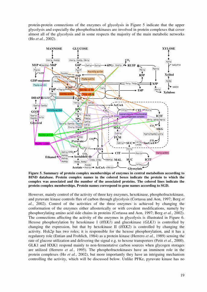
protein-protein connections of the enzymes of glycolysis in Figure 5 indicate that the upper glycolysis and especially the phosphofructokinases are involved in protein complexes that cover almost all of the glycolysis and in some respects the majority of the main metabolic networks (Ho et al., 2002).
Oac1pGut1p
GLUCOSE
GAP
BPG
E4P
S7P
AcCoA
SucCoA
OAA
DHAP
Acetaldehyde
Acetate
Ethanol
CO2
CO2
CO2
AcCoA
OAACO2
CO2
Glycerol
Tkl1p, Tkl2p
Tal1p
Zwf1p
Tdh1p, Tdh2p, Tdh3p
Pgi1P
Pyk1p, Pyk2pKgd1p
Mdh1p
Sdh1p
GTP
MANNOSE
Pmi40p
Sec53p
Psa1p Rki1p
6PG
FUM
Aco1p
Fum1p
SUC
Fbp1p
Idh1p, Idh2p
Lsc1pLsc2p
Cit1p
CO2
Gnd1p
Rpe1p
M1P
GDP-mannose
M6P
F6P
G6P
F16P
GO3P
RU5P
R5P
3PG
PEPMAL AKG
ICIT
CIT
Pdc1p,
X5P
Gpd1p, Gpd2p
Gpp1pGpp2p
Acs2p Acs1p
Ald2p, Ald4p
Adh1p
Pyc1p
ICIT
CIT
MAL SUCIcl1p
Mls1p
Mdh3pCit2p, Cit3p
Tpi1p
Pck1p
Pdb1p
CO2Pyruvate
Hxk1p, Hxk2p
Glyoxylate
Eno1p, Eno2p
Gpm1p, Gpm2p, Gpm3p
Pfk1p, Pfk2p
Pgk1p
Kgd2p Lpd1p
Lpd1pLat1p
Idp1p
Sol1p
Pdx1p
Fba1p
F26P
Fbp26pPfk26p, Pfk27p
Hxk1p, Hxk2p, Glk1p
Pda1p
XYLOSE
Xylitol
Xylulose
Xyl1p
Xyl2p
NAD(P)H
NADHXks1pSen15p-ac55m
Pbp2p-ac12m
Prp6p-ac47m
Rvs167p-ac13m
supramolecular mitochondrialdehydrogenase complex
Cdc55p-ac46m Cft1p-ac36m
Ynl127p-ac13m
Rpc40p-ac51m
Hat1p-ac50mAdh2pAdh4p
Adh6p
Rad26p-ac22m
Aco1p
Dic1p
Figure 5. Summary of protein complex memberships of enzymes in central metabolism according to BIND database. Protein complex names in the colored boxes indicate the protein to which the complex was associated and the number of the associated proteins. The colored lines indicate the protein complex memberships. Protein names correspond to gene names according to SGD. However, mainly control of the activity of three key enzymes, hexokinase, phosphofructokinase, and pyruvate kinase controls flux of carbon through glycolysis (Cortassa and Aon, 1997; Berg et al., 2002). Control of the activities of the three enzymes is achieved by changing the conformation of the enzymes either allosterically or with covalent modifications, namely by phosphorylating amino acid side chains in proteins (Cortassa and Aon, 1997; Berg et al., 2002). The connections affecting the activity of the enzymes in glycolysis is illustrated in Figure 6. Hexose phosphorylation by hexokinase I (HXK1) and glucokinase (GLK1) is controlled by changing the expression, but that by hexokinase II (HXK2) is controlled by changing the activity. Hxk2p has two roles; it is responsible for the hexose phosphorylation, and it has a regulatory role (Entian and Frohlich, 1984) as a protein kinase (Herrero et al., 1989) sensing the rate of glucose utilization and delivering the signal e.g. to hexose transporters (Petit et al., 2000). GLK1 and HXK1 respond mainly to non-fermentative carbon sources when glycogen storages are utilized (Herrero et al., 1995). The phosphofructokinases have an imminent role in the protein complexes (Ho et al., 2002), but more importantly they have an intriguing mechanism controlling the activity, which will be discussed below. Unlike PFKe, pyruvate kinase has no
20
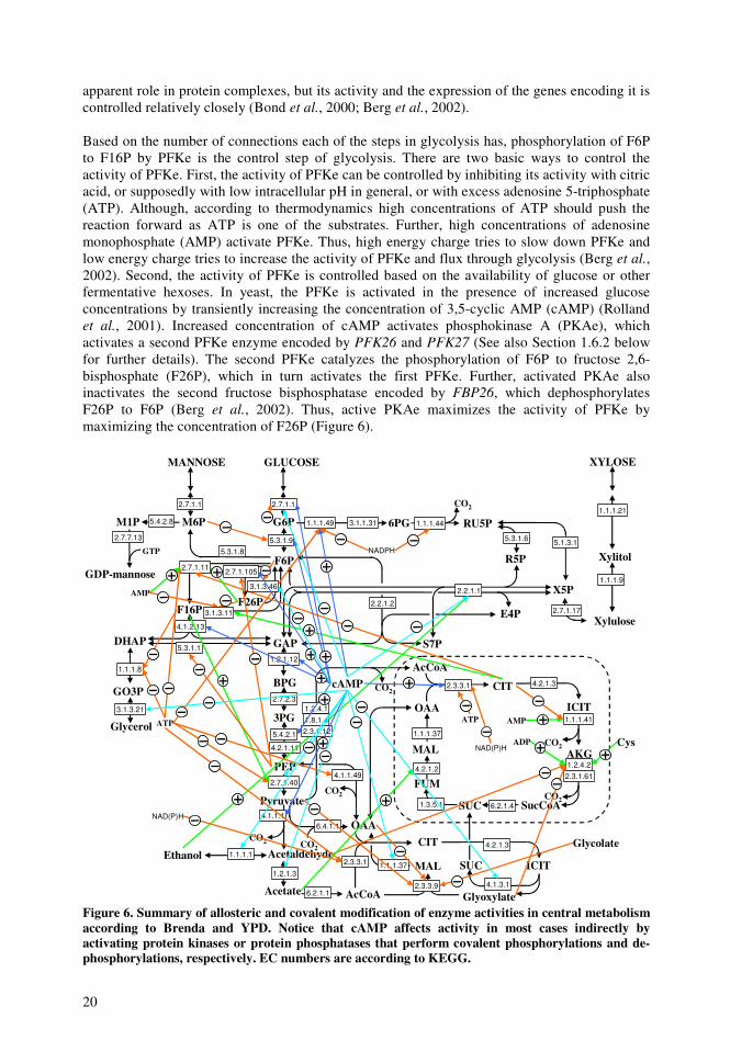
apparent role in protein complexes, but its activity and the expression of the genes encoding it is controlled relatively closely (Bond et al., 2000; Berg et al., 2002). Based on the number of connections each of the steps in glycolysis has, phosphorylation of F6P to F16P by PFKe is the control step of glycolysis. There are two basic ways to control the activity of PFKe. First, the activity of PFKe can be controlled by inhibiting its activity with citric acid, or supposedly with low intracellular pH in general, or with excess adenosine 5-triphosphate (ATP). Although, according to thermodynamics high concentrations of ATP should push the reaction forward as ATP is one of the substrates. Further, high concentrations of adenosine monophosphate (AMP) activate PFKe. Thus, high energy charge tries to slow down PFKe and low energy charge tries to increase the activity of PFKe and flux through glycolysis (Berg et al., 2002). Second, the activity of PFKe is controlled based on the availability of glucose or other fermentative hexoses. In yeast, the PFKe is activated in the presence of increased glucose concentrations by transiently increasing the concentration of 3,5-cyclic AMP (cAMP) (Rolland et al., 2001). Increased concentration of cAMP activates phosphokinase A (PKAe), which activates a second PFKe enzyme encoded by PFK26 and PFK27 (See also Section 1.6.2 below for further details). The second PFKe catalyzes the phosphorylation of F6P to fructose 2,6-bisphosphate (F26P), which in turn activates the first PFKe. Further, activated PKAe also inactivates the second fructose bisphosphatase encoded by FBP26, which dephosphorylates F26P to F6P (Berg et al., 2002). Thus, active PKAe maximizes the activity of PFKe by maximizing the concentration of F26P (Figure 6).
GLUCOSE
GAP
BPG
E4P
S7P
AcCoA
SucCoA
OAA
DHAP
Acetaldehyde
Acetate
Ethanol
CO2
CO2
CO2
AcCoA
OAA
CO2CO2
Glycerol
NADPHGTP
MANNOSE
6PG
FUM
SUC
NAD(P)HCO2
M1P
GDP-mannose
M6P
F6P
G6P
F16P
GO3P
RU5P
R5P
3PG
PEPMAL AKG
ICIT
CIT
X5P
ICIT
CIT
MAL SUC
CO2Pyruvate
Glyoxylate
F26P
XYLOSE
Xylitol
Xylulose2.7.1.17
5.1.3.15.3.1.6
2.2.1.1
2.2.1.2
1.1.1.443.1.1.311.1.1.49
2.7.1.1
5.3.1.95.3.1.8
2.7.1.1
5.4.2.8
2.7.7.13
2.7.1.1052.7.1.11
4.1.2.13
5.3.1.1
1.1.1.8
3.1.3.21
1.1.1.9
1.1.1.21
4.2.1.3
1.1.1.41
1.2.4.2
6.2.1.4
2.3.3.1
1.3.5.1
4.2.1.2
1.1.1.37
1.2.1.12
2.7.2.3
5.4.2.1
4.2.1.11
1.2.4.1
2.7.1.40
4.2.1.3
4.1.3.12.3.3.9
1.1.1.372.3.3.1
6.2.1.1
6.4.1.14.1.1.1
1.2.1.3
1.1.1.1
4.1.1.49
ATP
AMP
3.1.3.11
2.3.1.61
1.8.1.42.3.1.12
3.1.3.46
AMPATP
ADP Cys
Glycolate
NAD(P)H
cAMP
Figure 6. Summary of allosteric and covalent modification of enzyme activities in central metabolism according to Brenda and YPD. Notice that cAMP affects activity in most cases indirectly by activating protein kinases or protein phosphatases that perform covalent phosphorylations and de-phosphorylations, respectively. EC numbers are according to KEGG.
21
1.4.3 Pentose phosphate pathway Pentose phosphate pathway (PPP) is a sequence of several rearranging reactions (Figure 2). Namely, enzymes transketolase (TKLe) and transaldolase (TALe) interconvert the phosphates of trioses, tetroses, pentoses, hexoses, and heptoses (Berg et al., 2002). TKLe transfers glycolaldehyde groups and TALe dihydroxyacetone groups constituting non-oxidative PPP. Oxidative PPP is a linear pathway from G6P to RU5P (ribulose 5-phosphate) producing NADPH reducing equivalents and carbon dioxide. Pentoses D-xylose and L-arabinose enter main metabolism in PPP after they have been finally converted to xylulose 5-phosphate (X5P). Epimerase and isomerase reactions convey the interconversion between RU5P, X5P, and ribose 5-phosphate (R5P). R5P is an essential precursor for biosynthesis of purines, pyrimidines, and L-histidine. Very little is known about the regulation of the PPP reactions (Figure 3, Figure 4, Figure 5, Figure 6). The oxidative PPP seems to have two controlling points; ZWF1 and GND1/GND2. GNDe-reaction cleaves the carbon dioxide molecule, which is practically irreversible, but ZWFe-reaction has more regulatory connections and is the committed step as it is the first step in the oxidative PPP (Lee et al., 1999). Further, NADPH inhibits both NADPH-producing reactions. The non-oxidative reactions of PPP seem to be more idling as they operate close to equilibrium with low free Gibbs energies. However, the second gene of TKLe, TKL2 is controlled e.g. by MSN2 (Figure 3) relating the expression of TKL2 to general stress response (Boy-Marcotte et al., 1998). Congruently, inhibition of the TKLe activity can be transmitted via cAMP in nutrient rich conditions (Boy-Marcotte et al., 1998). Rap1p may be involved also in the regulation of TKL2 (Bar-Joseph et al., 2003). Overall, TKL2 and the second gene of TALe, YGR043c are expressed in diauxic shift (Holstege et al., 1998) and in various classical stress conditions (Rep et al., 2000). It has been postulated that expression of TKL2 and YGR043c is required for delivering carbon up to precursor R5P when the cells are grown on non-fermentable carbon sources such as ethanol (Daran-Lapujade et al., 2004). 1.4.4 Citric acid cycle and oxidative phosphorylation Citric acid cycle a.k.a. tri-carboxylic acid (TCA) cycle a.k.a. Krebs cycle in yeast and other eukarya operates inside mitochondria (Berg et al., 2002). The main purpose of the TCA cycle is to produce NADH, which is then used in oxidative phosphorylation to regenerate ATP under aerobic conditions. In addition, the TCA cycle metabolites alpha-ketoglutarate (AKG) and oxaloacetate (OAA) are important precursors in amino acid biosynthesis. Starting from pyruvate, the pyruvate dehydrogenase (PDHe) complex (Gavin et al., 2002) consisting of three enzyme activities encoded by at least five genes, converts pyruvate to carbon dioxide, NADH, and acetyl transferred to coenzyme A yielding acetyl coenzyme A (AcCoA). Acetyl is transferred further to OAA by citrate synthase (CITe) to yield citric acid (CIT); a tri-carboxylic acid containing six carbon atoms. This first step of the TCA cycle is the main controlling point of the cycle. Transcription factors RTG1, HAP2, and HAP4 induce the expression of CIT-genes (Svetlov and Cooper, 1995) (Figure 3), and ATP and NADH inhibit (Kispal and Srere, 1991), and cAMP causes activation (Dejean et al., 2002) of the CITe activity (Figure 6). Further, in the TCA cycle CIT is oxidized and cleaved to form AKG, carbon dioxide, and NADH via isocitric acid (ICIT). These two steps are also induced by RTG1 (Svetlov and Cooper, 1995). Again, another carbon dioxide is cleaved from AKG accompanied with NADH generation, when AKG is converted to succinyl coenzyme A (SucCoA). Expression of genes encoding the enzymes of this step is induced by HAP2 and HAP4 (Svetlov and Cooper, 1995) and the activity is inhibited by acetaldehyde and glyoxylate (Hirabayashi and Harada, 1972). SucCoA is converted to succinate (SUC), which generates GTP. SUC is further oxidized by succinate dehydrogenase (SDHe) complex to fumarate (FUM) generating FADH2 reducing equivalents. SDHe complex is involved also in the oxidative phosphorylation (OP) discussed below (Berg et al., 2002). SDH-genes are induced by HAP2 and HAP4. FUM is converted to malate (MAL), which is oxidized
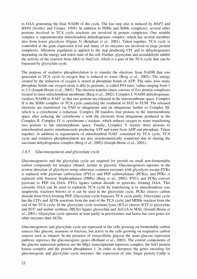
22
to OAA generating the final NADH of the cycle. The last step also is induced by HAP2 and HAP4 (Svetlov and Cooper, 1995). In addition to PDHe and SDHe complexes, several other proteins involved in TCA cycle reactions are involved in protein complexes. One notable complex is supramolecular mitochondrial dehydrogenase complex, which has several members also from lower glycolysis (Figure 5) (Belghazi et al., 2001). Taken together, TCA cycle is controlled at the gene expression level and many of its enzymes are involved in large protein complexes. Allosteric regulation is applied to the step producing CIT and to dehydrogenases depending on the energy and redox state of the cell. Further, glyoxylate and acetaldehyde inhibit the activity of the reaction from AKG to SucCoA, which is a part of the TCA cycle that can be bypassed by glyoxylate cycle. The purpose of oxidative phosphorylation is to transfer the electrons from NADH that was generated in TCA cycle to oxygen that is reduced to water (Berg et al., 2002). The energy created by the reduction of oxygen is stored in phosphate bonds of ATP. The ratio, how many phosphate bonds one oxygen atom is able to generate, is called P/O-ratio, values ranging from 1 to 2.5 (Joseph-Horne et al., 2001). The electron transfer chain consists of five protein complexes located in inner mitochondrial membrane (Berg et al., 2002). Complex I, NADH dehydrogenase, oxidizes NADH to NAD+ so that two protons are released in the intermembrane space. Complex II is the SDHe complex of TCA cycle catalyzing the oxidation of SUC to FUM. The released electrons are transferred via FAD to ubiquinone and on ubiquinone further to Complex III, which is a cytochrome c reductase. Complex III transfers four protons to the intermembrane space after reducing the cytochrome c with the electrons from ubiquinone produced in the Complex II. Complex IV is cytochrome c oxidase, which reduces oxygen to water transferring two protons to the intermembrane space. Finally, Complex V returns three protons to mitochondrial matrix simultaneously producing ATP and water from ADP and phosphate. Taken together, in addition to regeneration of mitochondrial NAD+ consumed by TCA cycle, TCA cycle and oxidative phosphorylation are also stoichiometrically connected due to sharing the succinate dehydrogenase complex (Berg et al., 2002) (Joseph-Horne et al., 2001). 1.4.5 Gluconeogenesis and glyoxylate cycle Gluconeogenesis and the glyoxylate cycle are required for growth on small non-fermentable carbon compounds for instance ethanol, acetate or glycerol. Gluconeogenesis operates in the reverse direction of glycolysis using otherwise common enzymes with glycolysis except PYKe is replaced with pyruvate carboxylase (PYCe) and PEP carboxykinase (PCKe), and PFKe is replaced with fructose bisphosphatase (FBPe) (Berg et al., 2002). PYCe and PCKe convert pyruvate to PEP via OAA. PYCe ligates carbon dioxide to pyruvate, forming OAA. This cytosolic OAA can be used to replenish TCA cycle by transferring it to mitochondrion (see anaplerotic reactions below) or it can be used in the glyoxylate cycle. PCKe cleaves carbon dioxide from OAA forming PEP. Glyoxylate cycle bypasses TCA cycle partly. Glyoxylate cycle has the CITe and ACOe reactions from the start of the TCA cycle, and MDHe reaction from the end of the TCA cycle. In the glyoxylate cycle isocitrate lyase (ICLe) cleaves ICIT to glyoxylate and SUC and malate synthase (MLSe) ligates glyoxylate and AcCoA to MAL (Joseph-Horne et al., 2001). Glyoxylate cycle operates at least partly in peroxisomes and hence has own genes for other enzymes than ACOe. Gluconeogenesis and glyoxylate cycle are repressed in the cells growing on fermentable carbon sources like glucose, mannose or fructose, but active in the cells growing on respirative carbon sources such as ethanol. In the presence of extracellular glucose the main glucose repression pathway represses the gluconeogenic genes (Rolland et al., 2002). The central components of the glucose repression pathway are the Mig1 transcriptional repressor complex, the Snf1 protein kinase complex and the protein phosphatase 1. In order to derepress the genes encoding for gluconeogenic and glyoxylate cycle enzymes, the expression of zinc finger protein Cat8p is
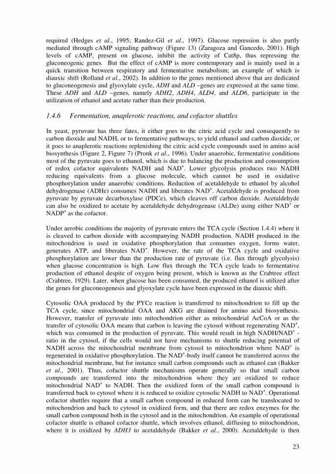
23
required (Hedges et al., 1995; Randez-Gil et al., 1997). Glucose repression is also partly mediated through cAMP signaling pathway (Figure 13) (Zaragoza and Gancedo, 2001). High levels of cAMP, present on glucose, inhibit the activity of Cat8p, thus repressing the gluconeogenic genes. But the effect of cAMP is more contemporary and is mainly used in a quick transition between respiratory and fermentative metabolism; an example of which is diauxic shift (Rolland et al., 2002). In addition to the genes mentioned above that are dedicated to gluconeogenesis and glyoxylate cycle, ADH and ALD –genes are expressed at the same time. These ADH and ALD –genes, namely ADH2, ADH4, ALD4, and ALD6, participate in the utilization of ethanol and acetate rather than their production. 1.4.6 Fermentation, anaplerotic reactions, and cofactor shuttles In yeast, pyruvate has three fates, it either goes to the citric acid cycle and consequently to carbon dioxide and NADH, or to fermentative pathways, to yield ethanol and carbon dioxide, or it goes to anaplerotic reactions replenishing the citric acid cycle compounds used in amino acid biosynthesis (Figure 2, Figure 7) (Pronk et al., 1996). Under anaerobic, fermentative conditions most of the pyruvate goes to ethanol, which is due to balancing the production and consumption of redox cofactor equivalents NADH and NAD+. Lower glycolysis produces two NADH reducing equivalents from a glucose molecule, which cannot be used in oxidative phosphorylation under anaerobic conditions. Reduction of acetaldehyde to ethanol by alcohol dehydrogenase (ADHe) consumes NADH and liberates NAD+. Acetaldehyde is produced from pyruvate by pyruvate decarboxylase (PDCe), which cleaves off carbon dioxide. Acetaldehyde can also be oxidized to acetate by acetaldehyde dehydrogenase (ALDe) using either NAD+ or NADP+ as the cofactor. Under aerobic conditions the majority of pyruvate enters the TCA cycle (Section 1.4.4) where it is cleaved to carbon dioxide with accompanying NADH production. NADH produced in the mitochondrion is used in oxidative phosphorylation that consumes oxygen, forms water, generates ATP, and liberates NAD+. However, the rate of the TCA cycle and oxidative phosphorylation are lower than the production rate of pyruvate (i.e. flux through glycolysis) when glucose concentration is high. Low flux through the TCA cycle leads to fermentative production of ethanol despite of oxygen being present, which is known as the Crabtree effect (Crabtree, 1929). Later, when glucose has been consumed, the produced ethanol is utilized after the genes for gluconeogenesis and glyoxylate cycle have been expressed in the diauxic shift. Cytosolic OAA produced by the PYCe reaction is transferred to mitochondrion to fill up the TCA cycle, since mitochondrial OAA and AKG are drained for amino acid biosynthesis. However, transfer of pyruvate into mitochondrion either as mitochondrial AcCoA or as the transfer of cytosolic OAA means that carbon is leaving the cytosol without regenerating NAD+, which was consumed in the production of pyruvate. This would result in high NADH/NAD+ -ratio in the cytosol, if the cells would not have mechanisms to shuttle reducing potential of NADH across the mitochondrial membrane from cytosol to mitochondrion where NAD+ is regenerated in oxidative phosphorylation. The NAD+-body itself cannot be transferred across the mitochondrial membrane, but for instance small carbon compounds such as ethanol can (Bakker et al., 2001). Thus, cofactor shuttle mechanisms operate generally so that small carbon compounds are transferred into the mitochondrion where they are oxidized to reduce mitochondrial NAD+ to NADH. Then the oxidized form of the small carbon compound is transferred back to cytosol where it is reduced to oxidize cytosolic NADH to NAD+. Operational cofactor shuttles require that a small carbon compound in reduced form can be translocated to mitochondrion and back to cytosol in oxidized form, and that there are redox enzymes for the small carbon compound both in the cytosol and in the mitochondrion. An example of operational cofactor shuttle is ethanol cofactor shuttle, which involves ethanol, diffusing to mitochondrion, where it is oxidized by ADH3 to acetaldehyde (Bakker et al., 2000). Acetaldehyde is then
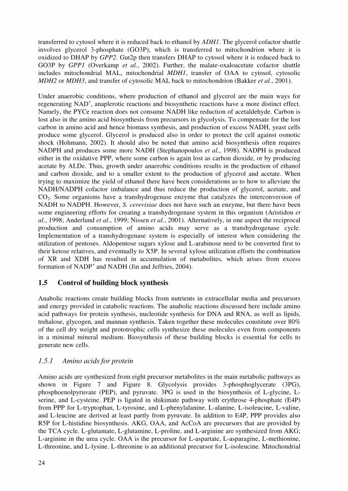
24
transferred to cytosol where it is reduced back to ethanol by ADH1. The glycerol cofactor shuttle involves glycerol 3-phosphate (GO3P), which is transferred to mitochondrion where it is oxidized to DHAP by GPP2. Gut2p then transfers DHAP to cytosol where it is reduced back to GO3P by GPP1 (Overkamp et al., 2002). Further, the malate-oxaloacetate cofactor shuttle includes mitochondrial MAL, mitochondrial MDH1, transfer of OAA to cytosol, cytosolic MDH2 or MDH3, and transfer of cytosolic MAL back to mitochondrion (Bakker et al., 2001). Under anaerobic conditions, where production of ethanol and glycerol are the main ways for regenerating NAD+, anaplerotic reactions and biosynthetic reactions have a more distinct effect. Namely, the PYCe reaction does not consume NADH like reduction of acetaldehyde. Carbon is lost also in the amino acid biosynthesis from precursors in glycolysis. To compensate for the lost carbon in amino acid and hence biomass synthesis, and production of excess NADH, yeast cells produce some glycerol. Glycerol is produced also in order to protect the cell against osmotic shock (Hohmann, 2002). It should also be noted that amino acid biosynthesis often requires NADPH and produces some more NADH (Stephanopoulos et al., 1998). NADPH is produced either in the oxidative PPP, where some carbon is again lost as carbon dioxide, or by producing acetate by ALDe. Thus, growth under anaerobic conditions results in the production of ethanol and carbon dioxide, and to a smaller extent to the production of glycerol and acetate. When trying to maximize the yield of ethanol there have been considerations as to how to alleviate the NADH/NADPH cofactor imbalance and thus reduce the production of glycerol, acetate, and CO2. Some organisms have a transhydrogenase enzyme that catalyzes the interconversion of NADH to NADPH. However, S. cerevisiae does not have such an enzyme, but there have been some engineering efforts for creating a transhydrogenase system in this organism (Aristidou et al., 1998; Anderlund et al., 1999; Nissen et al., 2001). Alternatively, in one aspect the reciprocal production and consumption of amino acids may serve as a transhydrogenase cycle. Implementation of a transhydrogenase system is especially of interest when considering the utilization of pentoses. Aldopentose sugars xylose and L-arabinose need to be converted first to their ketose relatives, and eventually to X5P. In several xylose utilization efforts the combination of XR and XDH has resulted in accumulation of metabolites, which arises from excess formation of NADP+ and NADH (Jin and Jeffries, 2004). 1.5 Control of building block synthesis Anabolic reactions create building blocks from nutrients in extracellular media and precursors and energy provided in catabolic reactions. The anabolic reactions discussed here include amino acid pathways for protein synthesis, nucleotide synthesis for DNA and RNA, as well as lipids, trehalose, glycogen, and mannan synthesis. Taken together these molecules constitute over 80% of the cell dry weight and prototrophic cells synthesize these molecules even from components in a minimal mineral medium. Biosynthesis of these building blocks is essential for cells to generate new cells. 1.5.1 Amino acids for protein Amino acids are synthesized from eight precursor metabolites in the main metabolic pathways as shown in Figure 7 and Figure 8. Glycolysis provides 3-phosphoglycerate (3PG), phosphoenolpyruvate (PEP), and pyruvate. 3PG is used in the biosynthesis of L-glycine, L-serine, and L-cysteine. PEP is ligated in shikimate pathway with erythrose 4-phosphate (E4P) from PPP for L-tryptophan, L-tyrosine, and L-phenylalanine. L-alanine, L-isoleucine, L-valine, and L-leucine are derived at least partly from pyruvate. In addition to E4P, PPP provides also R5P for L-histidine biosynthesis. AKG, OAA, and AcCoA are precursors that are provided by the TCA cycle. L-glutamate, L-glutamine, L-proline, and L-arginine are synthesized from AKG; L-arginine in the urea cycle. OAA is the precursor for L-aspartate, L-asparagine, L-methionine, L-threonine, and L-lysine. L-threonine is an additional precursor for L-isoleucine. Mitochondrial

25
AcCoA is also used as an additional precursor for the biosynthesis of L-lysine and L-leucine (Kanehisa and Goto, 2000; Berg et al., 2002). Biosynthesis of L-glycine provides a methylene group, and that of L-methionine requires a methylene group that is transferred by tetrahydrofolate. In labeling experiments the pathway of the labeled substrate to amino acid precursors can be back calculated from labeling patterns of amino acids since most of the pathways cleave the substrates differently. Links between carbon positions in the amino acid precursors and the amino acids have been presented e.g. by Szyperski (Szyperski, 1998). Operation of several pathways or operation of several reversible reactions produces ratios of these differently labeled isotopic isomers. The ratios of isotopic isomers can be measured e.g. with NMR (nuclear magnetic resonance spectroscopy). The cost in the currency of energy, reducing equivalents, and precursors of producing each amino acid is presented in Publication I in the metabolic network model.
OAA
LYS
LYS20
LYS4
H-ICITLYS12
LYS2
LYS9
SaccharopineLYS1
E4P
AcCoA
R5P
3PG
PEPAKG
PyruvateGLU GLNPRO
ARG
GLT1GLT1
PUT2 GLN1PRO3
PUT2
CAR2
OrnithineCitrulline
Arg-Suc
ASP
FUM
ARG1
ARG4
UreaCAR1
ARG3
Carbamoyl-P
ALA
ASN
CYS
GLY
HIS
ILE
LEU
MET
PHE
SER
THR
TYR
VAL
TRP
AAT1
ASN1, ASN2
YDR111C
SER3SER1SER2SHM1
CYS4
CYS3
HOM3HOM2H-SERTHR1THR4
MET2
MET17
MET6Oxobutanoate
ILV1, CHA1
HE-TPP PDB1
OxoisovalerateILV2, ILV6
AcetolactateILV5ILV3
BAT1
LEU1
LEU23-IPM BAT1
BAT1 ILV5ILV3 ILV2,ILV6
LEU4
ASAHOM6
ARO4ARO1ENO1ShikimateChorismate ARO1ARO2PrephenatePHA2AAT1
TYR1
AAT1
ARO7TRP2
TRP4
TRP1
TRP3
TRP5 PRS4PRPPHIS1HIS4HIS6HIS7AICAR
Imidazole-GO3P HIS3 HIS5 HIS2 HIS4
GCN4
CBF1
RAP1
MET4
LEU3
GLN3
LYS14
ARG80
BAS1
Figure 7. Summary of control of gene expression in amino acid biosynthesis according to Yeast Proteome Database. Genes in colored boxes present transcription factors affecting the expression of the enzyme-encoding genes in white boxes. Regulatory connections are indicated with the colored arrows. Gene names are according to SGD. The main transcription factor that controls the expression of genes in the majority of the amino acid biosynthesis pathways is GCN4 (Natarajan et al., 2001). In Figure 7 it has connections to 27 steps of over 70 steps in the Figure. Hence, GCN4 is an important component in the regulation of amino acid biosynthesis. Even the YPD regulator report (Figure 7) and transcription factor binding data (Figure 8) agree about the role of GCN4 (Lee et al., 2002a). Additional transcription factors regulating the biosynthesis of some amino acids, are GLN3, LEU3, MET4, LYS14, and ARG80. GLN3 regulates expression of genes encoding for enzymes in the amino acid biosynthesis pathways starting from AKG (Mitchell and Magasanik, 1984). LEU3 regulates biosynthesis of L-leucine, L-isoleucine, and L-valine (Brisco et al., 1987). MET4 (Mountain et al., 1993) and LYS14 (Feller et al., 1994) control the biosynthesis of L-methionine and L-lysine, respectively. ARG80 has a role in controlling the urea cycle and thus biosynthesis of L-arginine (Svetlov and Cooper, 1995). As indicated in Figure 7, several amino acid biosynthesis pathways have a feedback control mechanism, inhibiting early steps of the pathway when concentration of
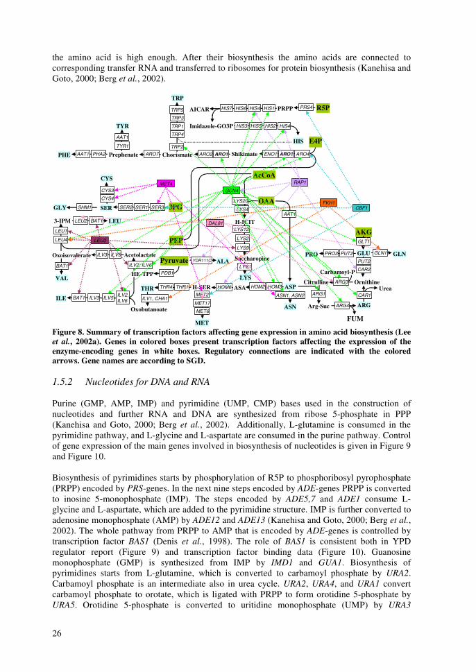
26
the amino acid is high enough. After their biosynthesis the amino acids are connected to corresponding transfer RNA and transferred to ribosomes for protein biosynthesis (Kanehisa and Goto, 2000; Berg et al., 2002).
LYS
LYS20
LYS4
H-ICITLYS12
LYS2
LYS9
SaccharopineLYS1
E4P
AcCoA
OAA
R5P
3PG
PEPAKG
PyruvateGLU GLNPRO
ARG
GLT1GLT1
PUT2 GLN1PRO3
PUT2
CAR2
OrnithineCitrulline
Arg-Suc
ASP
FUM
ARG1
ARG4
UreaCAR1
ARG3
Carbamoyl-P
ALA
ASN
CYS
GLY
HIS
ILE
LEU
MET
PHE
SER
THR
TYR
VAL
TRP
AAT1
ASN1, ASN2
YDR111C
SER3SER1SER2SHM1
CYS4
CYS3
HOM3HOM2H-SERTHR1THR4
MET2
MET17
MET6Oxobutanoate
ILV1, CHA1
HE-TPP PDB1
OxoisovalerateILV2, ILV6
AcetolactateILV5ILV3
BAT1
LEU1
LEU23-IPM BAT1
BAT1 ILV5ILV3 ILV2,ILV6
LEU4
ASAHOM6
ARO4ARO1ENO1ShikimateChorismate ARO1ARO2PrephenatePHA2AAT1
TYR1
AAT1
ARO7TRP2
TRP4
TRP1
TRP3
TRP5 PRS4PRPPHIS1HIS4HIS6HIS7AICAR
Imidazole-GO3P HIS3 HIS5 HIS2 HIS4
GCN4
CBF1
RAP1MET4
LEU3
DAL81
FKH1
Figure 8. Summary of transcription factors affecting gene expression in amino acid biosynthesis (Lee et al., 2002a). Genes in colored boxes present transcription factors affecting the expression of the enzyme-encoding genes in white boxes. Regulatory connections are indicated with the colored arrows. Gene names are according to SGD. 1.5.2 Nucleotides for DNA and RNA Purine (GMP, AMP, IMP) and pyrimidine (UMP, CMP) bases used in the construction of nucleotides and further RNA and DNA are synthesized from ribose 5-phosphate in PPP (Kanehisa and Goto, 2000; Berg et al., 2002). Additionally, L-glutamine is consumed in the pyrimidine pathway, and L-glycine and L-aspartate are consumed in the purine pathway. Control of gene expression of the main genes involved in biosynthesis of nucleotides is given in Figure 9 and Figure 10. Biosynthesis of pyrimidines starts by phosphorylation of R5P to phosphoribosyl pyrophosphate (PRPP) encoded by PRS-genes. In the next nine steps encoded by ADE-genes PRPP is converted to inosine 5-monophosphate (IMP). The steps encoded by ADE5,7 and ADE1 consume L-glycine and L-aspartate, which are added to the pyrimidine structure. IMP is further converted to adenosine monophosphate (AMP) by ADE12 and ADE13 (Kanehisa and Goto, 2000; Berg et al., 2002). The whole pathway from PRPP to AMP that is encoded by ADE-genes is controlled by transcription factor BAS1 (Denis et al., 1998). The role of BAS1 is consistent both in YPD regulator report (Figure 9) and transcription factor binding data (Figure 10). Guanosine monophosphate (GMP) is synthesized from IMP by IMD1 and GUA1. Biosynthesis of pyrimidines starts from L-glutamine, which is converted to carbamoyl phosphate by URA2. Carbamoyl phosphate is an intermediate also in urea cycle. URA2, URA4, and URA1 convert carbamoyl phosphate to orotate, which is ligated with PRPP to form orotidine 5-phosphate by URA5. Orotidine 5-phosphate is converted to uritidine monophosphate (UMP) by URA3
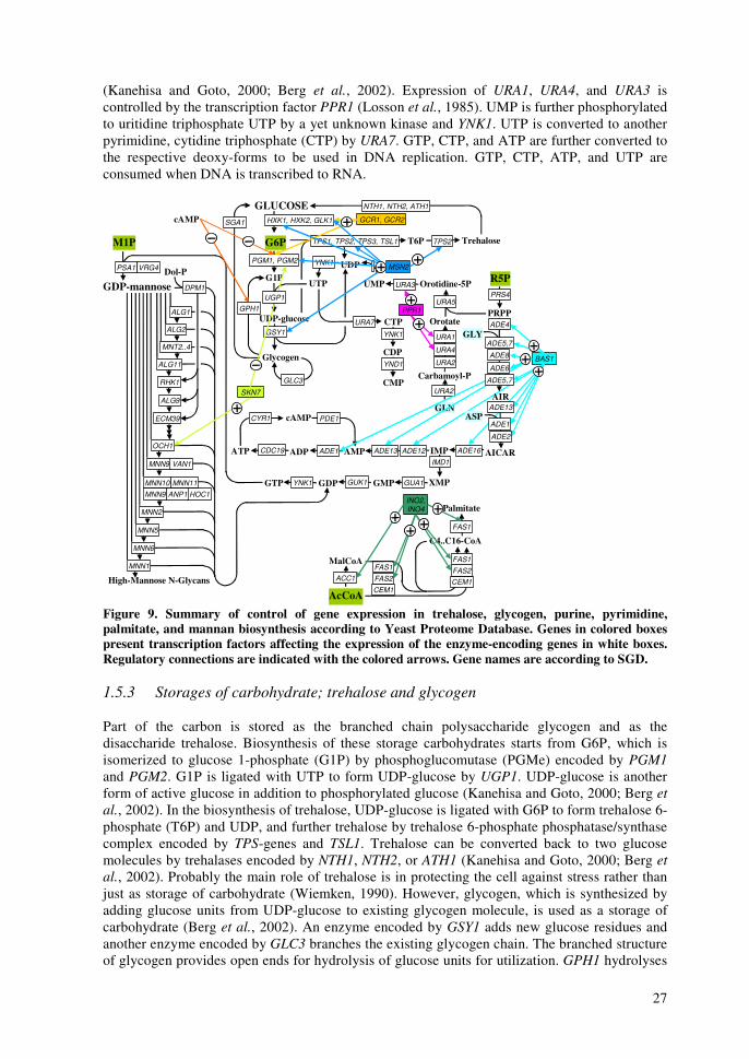
27
(Kanehisa and Goto, 2000; Berg et al., 2002). Expression of URA1, URA4, and URA3 is controlled by the transcription factor PPR1 (Losson et al., 1985). UMP is further phosphorylated to uritidine triphosphate UTP by a yet unknown kinase and YNK1. UTP is converted to another pyrimidine, cytidine triphosphate (CTP) by URA7. GTP, CTP, and ATP are further converted to the respective deoxy-forms to be used in DNA replication. GTP, CTP, ATP, and UTP are consumed when DNA is transcribed to RNA.
AcCoA
M1P G6P
R5PG1P
UDP-glucose
UTP
T6P TrehaloseTPS2
PRPP
IMP
GTP
IMD1
ADE16
GLN
Carbamoyl-PURA2
URA4
URA1
Orotate
Orotidine-5PURA3UMP
PGM1, PGM2
PRS4
URA2
GMP XMPGUA1
ATP CDC19 AMP
URA5
CMP
CTP
Glycogen
High-Mannose N-Glycans
ALG1
ALG2
MNT2..4
ALG11
TPS1, TPS2, TPS3, TSL1
UDPYNK1
CDPYND1
GPH1
YNK1
GLUCOSESGA1
GSY1URA7
GDP GUK1YNK1
ADE1ADP
cAMPCYR1
RHK1
ALG9
ECM39
MNN11MNN10
Dol-P
MNN9
OCH1
HOC1ANP1MNN9
MNN2
MNN5
MNN6
MNN1
VRG4
GDP-mannose
PSA1
MalCoA
C4..C16-CoA
CEM1FAS2
FAS1
FAS1
Palmitate
ACC1
CEM1
FAS2
FAS1
SKN7
GCR1, GCR2
PPR1
BAS1
INO2, INO4
PDE1
ADE13 ADE12
UGP1DPM1
VAN1
GLC3
HXK1, HXK2, GLK1cAMP
MSN2
NTH1, NTH2, ATH1
AICAR
AIR
ADE4
ADE8
ADE6
ADE5,7
ADE2
ADE13ASP
GLYADE5,7
ADE1
Figure 9. Summary of control of gene expression in trehalose, glycogen, purine, pyrimidine, palmitate, and mannan biosynthesis according to Yeast Proteome Database. Genes in colored boxes present transcription factors affecting the expression of the enzyme-encoding genes in white boxes. Regulatory connections are indicated with the colored arrows. Gene names are according to SGD. 1.5.3 Storages of carbohydrate; trehalose and glycogen Part of the carbon is stored as the branched chain polysaccharide glycogen and as the disaccharide trehalose. Biosynthesis of these storage carbohydrates starts from G6P, which is isomerized to glucose 1-phosphate (G1P) by phosphoglucomutase (PGMe) encoded by PGM1 and PGM2. G1P is ligated with UTP to form UDP-glucose by UGP1. UDP-glucose is another form of active glucose in addition to phosphorylated glucose (Kanehisa and Goto, 2000; Berg et al., 2002). In the biosynthesis of trehalose, UDP-glucose is ligated with G6P to form trehalose 6-phosphate (T6P) and UDP, and further trehalose by trehalose 6-phosphate phosphatase/synthase complex encoded by TPS-genes and TSL1. Trehalose can be converted back to two glucose molecules by trehalases encoded by NTH1, NTH2, or ATH1 (Kanehisa and Goto, 2000; Berg et al., 2002). Probably the main role of trehalose is in protecting the cell against stress rather than just as storage of carbohydrate (Wiemken, 1990). However, glycogen, which is synthesized by adding glucose units from UDP-glucose to existing glycogen molecule, is used as a storage of carbohydrate (Berg et al., 2002). An enzyme encoded by GSY1 adds new glucose residues and another enzyme encoded by GLC3 branches the existing glycogen chain. The branched structure of glycogen provides open ends for hydrolysis of glucose units for utilization. GPH1 hydrolyses
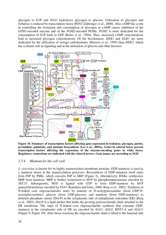
28
glycogen to G1P and SGA1 hydrolyses glycogen to glucose. Utilization of glycogen and trehalose is induced by transcription factor MSN2 (Zahringer et al., 2000). Also cAMP has a role in controlling the formation and consumption of glycogen as cAMP causes inhibition of the GPH1-encoded enzyme and of the PGM2-encoded PGMe. PGM2 is more dedicated for the consumption of G1P back to G6P (Boles et al., 1994). Thus, increased cAMP concentrations lead to increased glycogen concentrations. Of the hexokinases, HXK1 and GLK1 are more dedicated for the utilization of storage carbohydrates (Herrero et al., 1995) than HXK2, which has a clearer role in signaling and in the utilization of glucose and other hexoses.
AcCoA
M1P G6P
R5PG1P
UDP-glucose
UTP
T6P TrehaloseTPS2
PRPP
IMP
GTP
IMD1
ADE16
GLN
Carbamoyl-PURA2
URA4
URA1
Orotate
Orotidine-5PURA3UMP
PGM1, PGM2
PRS4
URA2
GMP XMPGUA1
ATP CDC19 AMP
URA5
CMP
CTP
Glycogen
High-Mannose N-Glycans
ALG1
ALG2
MNT2..4
ALG11
TPS1, TPS2, TPS3, TSL1
UDPYNK1
CDPYND1
GPH1
YNK1
GLUCOSE NTH1, NTH2, ATH1
SGA1
GSY1URA7
GDP GUK1YNK1
ADE1ADP
cAMPCYR1
RHK1
ALG9
ECM39
MNN11MNN10
Dol-P
MNN9
OCH1
HOC1ANP1MNN9
MNN2
MNN5
MNN6
MNN1
VRG4
GDP-mannose
PSA1
MalCoA
C4..C16-CoA
CEM1FAS2
FAS1
FAS1
Palmitate
ACC1
CEM1
FAS2
FAS1
RAP1
SWI5
MBP1
SKN7
SWI4
FKH1
CIN5
ABF1
BAS1
INO2, INO4
PDE1
ADE13 ADE12
UGP1DPM1
VAN1
GLC3
HXK1, HXK2, GLK1
AICAR
AIR
ADE4
ADE8
ADE6
ADE5,7
ADE2
ADE13ASP
GLYADE5,7
ADE1
Figure 10. Summary of transcription factors affecting gene expression in trehalose, glycogen, purine, pyrimidine, palmitate, and mannan biosynthesis (Lee et al., 2002a). Genes in colored boxes present transcription factors affecting the expression of the enzyme-encoding genes in white boxes. Regulatory connections are indicated with the colored arrows. Gene names are according to SGD. 1.5.4 Mannan for the cell wall S. cerevisiae is known for its highly mannosylated membrane proteins. GDP-mannose is used as a mannose donor in the mannosylation processes. Biosynthesis of GDP-mannose itself starts from F6P by PMIe, which converts F6P to M6P (Figure 2). Alternatively, HXKe synthesizes M6P from mannose. M6P is further isomerized to M1P by phosphomannomutase encoded by SEC53. Subsequently, M1P is ligated with GTP to form GDP-mannose by M1P guanylyltransferase encoded by PSA1 (Kanehisa and Goto, 2000; Berg et al., 2002). Synthesis of N-linked core oligosaccharides starts by transfer of N-acetylglucosamine (from UDP-N-acetylglucosamine), glucose (from UDP-glucose), and mannose (from GDP-mannose) to dolichol phosphate carrier (Dol-P) at the cytoplasmic side of endoplasmic reticulum (ER) (Berg et al., 2002). Dol-P is a lipid anchor that holds the growing polysaccharide chain attached to the ER membrane. The steps of N-linked core oligosaccharide synthesis that consume GDP-mannose at the cytoplasmic side of ER are encoded by ALG1, ALG2, MNT2-4, and ALG11 (Figure 9, Figure 10). After those reactions the oligosaccharide chain is tilted to the lumenal side
29
of ER. At the lumenal side of ER, mannose is donated by Dol-P-mannose, which is synthesized from GDP-mannose and Dol-P by DPM1-encoded enzyme at the cytoplasmic side of ER. In the reactions encoded by RHK1, ALG9, and ECM39, Dol-P-mannose is consumed to lengthen and branch the oligosaccharide chain by four mannoses. Subsequently, three quality control glucoses are added before the oligosaccharide chain is transferred from Dol-P to asparagine residue in the nascent polypeptide chain. Quality control enzymes encoded by CWH41, ROT2, and MNS1 verify the structure of the N-linked core oligosaccharide and cleave off the three glucoses and one mannose to confirm the successful quality control (Parodi, 1999). At this point the novel glycoprotein is transported to Golgi for further mannosylation. The mannose donor in Golgi is GDP-mannose, where a VRG4-encoded transporter transfers it. As the only known GDP-mannose transporter, Vrg4p is in a good position to control the mannosylation processes in Golgi (Dean et al., 1997). The first mannosylation step in Golgi is encoded by OCH1 after which the oligosaccharide either remains as a core-type oligosaccharide or becomes a highly mannosylated cell wall oligosaccharide, mannan. Also Och1p is a good controlling point as is indicated by its connection to transcription factor Skn7p (Li et al., 2002), involved in regulation of stress responses (Figure 9, Figure 10). The first step in mannan biosynthesis is catalyzed by mannan polymerase I, which is encoded by MNN9 and VAN1. Further elongation of the mannan chain by as many as 50-200 mannoses is catalyzed by mannan polymerase II, which is encoded by MNN9-11, HOC1, and ANP1 (Aloy et al., 2004). MNN2-encoded enzyme starts to form a net-like structure with �1,2-bonds, and MNN5- and MNN6-encoded enzymes continue the formation of the highly branched structure. The mannosylation is terminated by an enzyme encoded by MNN1, which adds the last �1,3-mannoses (Klis, 1994). Except for the transcription factor Skn7p controlling the expression of OCH1, there does not seem to be very many regulatory connections in the mannan biosynthesis. Transcription factors involved in the regulation of cell cycle, Swi4p and Swi5p bind to a few genes encoding enzymes at the start of mannosylation pathway (PSA1 and DPM1) and at the end of it (MNN9 and MNN5) (Lee et al., 2002a). Thus, regulation of mannan biosynthesis seems to be generally growth-related and further induced if the integrity of the cell wall is compromised in cell wall damaging conditions. The process of mannosylation results in high-mannose N-glycans, which are found in proteins on outer layers of cell membranes. Together with chitin and notably shorter O-glycans, N-glycans form the cell wall of S. cerevisiae (Lipke and Ovalle, 1998). Chitin biosynthesis starts by conversion of F6P to N-acetyl-glucosamine 6-phosphate by enzymes encoded by GFA1 and GNA1. PCM1 and QRI1 -encoding enzymes convert N-acetylglucosamine 6-phosphate further to UDP-N-acetylglucosamine (UDPGlcNAc). Chitin synthase (encoded by CHS1-3) finally adds new UDPGlcNAc to existing chitin molecules, which are N-acetylglucosaminyl –polymers (Kanehisa and Goto, 2000; Berg et al., 2002). Especially expression of CHS3 increases vividly in cell wall damaging conditions (Osmond et al., 1999). 1.5.5 Lipids for membranes Fatty acids are required for the biosynthesis of the lipid bilayer of cell membranes. The carbon skeleton of fatty acids is synthesized from cytosolic AcCoA. Malonyl coenzyme A (MalCoA) is formed from AcCoA by AcCoA carboxylase encoded by ACC1 with the aid of biotin-carboxyl carrier protein (Figure 9, Figure 10). Malonyl and acetyl groups are ligated to acyl carrier protein and subsequently to each other to form a four-carbon acetoacetyl group bound to acyl carrier protein (Kanehisa and Goto, 2000; Berg et al., 2002). Round by round the carbon chain is lengthened by transferring two carbons each round from malonyl, bound to acyl carrier protein. In each round the newly added group is oxidized, dehydrated, and a formed enoyl group is reduced to get a straight carbon chain. After seven rounds the fatty acid chain is 18 carbons long palmitate. The majority of the reactions mentioned above from MalCoA and AcCoA to
30
palmitate are catalyzed by eight enzyme activities, which are encoded by three genes, FAS1, FAS2, and CEM1 (Schweizer et al., 1978; Kuziora et al., 1983). Expression of these genes as well as that of ACC1 is controlled by the transcription factors INO2 and INO4 (Figure 9, Figure 10) (Lee et al., 2002a). Membranes also contain various other components, balancing lipid structures, such as ergosterol, which is a close relative to mammalian cholesterol (Nurminen et al., 1975; Zinser et al., 1993; Eisenkolb et al., 2002). Starting from cytosolic AcCoA, ergosterol synthesis of 20 steps is encoded mainly by the ERG-genes (Paltauf et al., 1992). 1.6 Cell-wide control (Global regulation) Cell-wide control such as regulation of cell cycle or responses to external stimuli such as heat, salt, high osmotic pressure, or starvation have widespread mechanisms controlling several aspects of cellular behavior. In principle, the individual parts of metabolism discussed above are also under a global control of cell proliferation, response to stress or changes in nutrient concentrations, e.g. starvation. 1.6.1 Cell cycle Cell cycle controls when and how a daughter cell is created from a mother cell, and verifies that this proliferative process is successful. Between cell divisions all essential cell components must be duplicated. Replication and segregation of the genetic material stored in DNA molecules is an especially delicate task in order to maintain the properties of the population through generations. Replication and separation of DNA have their own distinct phases in cell cycle, synthesis (S-phase) and mitosis (M-phase), respectively. They are temporarily separated by two growth phases, G1-phase (Growth 1) precedes the S-phase and G2-phase (Growth 2) precedes the M-phase like presented in Figure 11. The majority of the cell cycle –related information presented here was gathered from reviews, web pages, and models of Dr. Tyson’s group (Tyson et al., 2002; Chen et al., 2004) and to some extent from KEGG (Kanehisa and Goto, 2000). In S. cerevisiae the cell cycle has two control points, in G1- and in M-phase. In the G1-phase a decision is made whether to undergo Start transition and to proceed through S-, G2-, and M-phases to replicate the DNA (Chen et al., 2004). The Start transition is reached if a recently divided cell has reached a certain size in order to have resources to proceed through DNA replication and segregation (Lorinz and Carter, 1979; Jörgensen et al., 2002). DNA damage, mating pheromone, or a small cell size, often caused by low nutrient concentrations signaled as low cAMP concentration prolong or halt the G1-phase. In M-phase a decision is made whether to undergo Finish transition so that the sister chromatids are segregated, the cell divides, and the cell cycle returns to G1-phase (Chen et al., 2004). The Finish transition is reached if the replicated DNA is not damaged, the DNA has been replicated completely, and the chromosomes are aligned properly in the metaphase. If defects are observed in any of the checkpoints above, the cell cycle arrests in the M-phase (Tyson et al., 2002). CDK/Cyclin complexes, which belong to a family of protein kinases, control the cell cycle. Cdk-unit (cyclin dependent kinase) is the catalytic subunit, which is encoded in S. cerevisiae only by CDC28. Nine regulatory cyclin units are encoded by CLN1..3, and CLB1..6. Proper progression through the cell cycle requires the successive activation and inactivation of these Cdc28p/cyclin dimers (Spellman et al., 1998; Chen et al., 2004). There are several different mechanisms for regulating Cdc28p activity in the cell: through the synthesis of cyclins by various transcription factors (SBF (Swi4,6p-dependent cell cycle binding box Binding Factor), MBF (Mlu1p-dependent cell cycle binding box Binding Factor) and Mcm1p), through the degradation of cyclins (promoted by Cdc20p/APC (anaphase promoting complex), Cdh1p/APC, and Grr1p/SCF), through association with stoichiometric CDK inhibitors (Sic1p and Cdc6p, and Far1p), and through phosphorylation and dephosphorylation of Cdc28p by Swe1p and Mih1p, respectively (Figure 11). Cyclin Cln3p (Dirick et al., 1995) has the main role in giving the Start
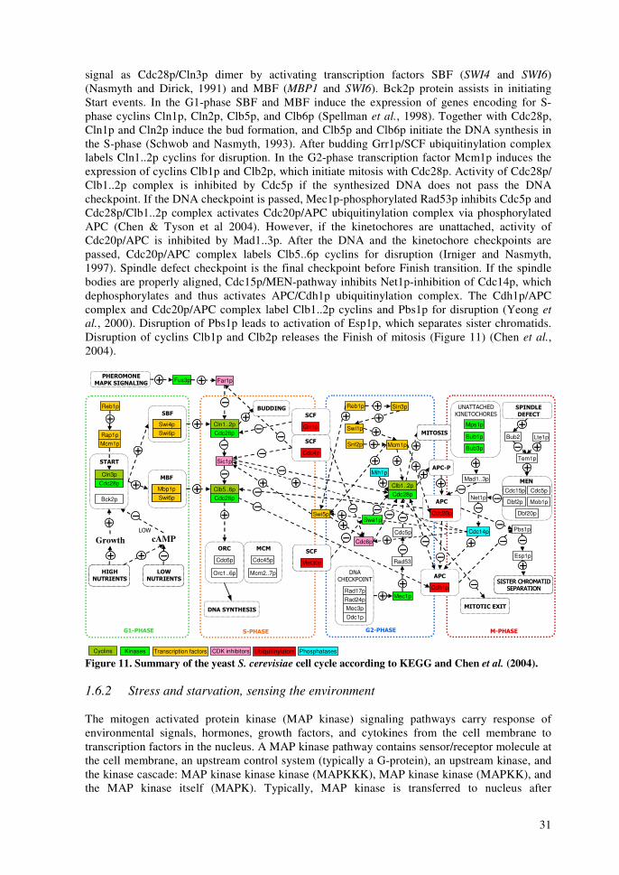
31
signal as Cdc28p/Cln3p dimer by activating transcription factors SBF (SWI4 and SWI6) (Nasmyth and Dirick, 1991) and MBF (MBP1 and SWI6). Bck2p protein assists in initiating Start events. In the G1-phase SBF and MBF induce the expression of genes encoding for S-phase cyclins Cln1p, Cln2p, Clb5p, and Clb6p (Spellman et al., 1998). Together with Cdc28p, Cln1p and Cln2p induce the bud formation, and Clb5p and Clb6p initiate the DNA synthesis in the S-phase (Schwob and Nasmyth, 1993). After budding Grr1p/SCF ubiquitinylation complex labels Cln1..2p cyclins for disruption. In the G2-phase transcription factor Mcm1p induces the expression of cyclins Clb1p and Clb2p, which initiate mitosis with Cdc28p. Activity of Cdc28p/ Clb1..2p complex is inhibited by Cdc5p if the synthesized DNA does not pass the DNA checkpoint. If the DNA checkpoint is passed, Mec1p-phosphorylated Rad53p inhibits Cdc5p and Cdc28p/Clb1..2p complex activates Cdc20p/APC ubiquitinylation complex via phosphorylated APC (Chen & Tyson et al 2004). However, if the kinetochores are unattached, activity of Cdc20p/APC is inhibited by Mad1..3p. After the DNA and the kinetochore checkpoints are passed, Cdc20p/APC complex labels Clb5..6p cyclins for disruption (Irniger and Nasmyth, 1997). Spindle defect checkpoint is the final checkpoint before Finish transition. If the spindle bodies are properly aligned, Cdc15p/MEN-pathway inhibits Net1p-inhibition of Cdc14p, which dephosphorylates and thus activates APC/Cdh1p ubiquitinylation complex. The Cdh1p/APC complex and Cdc20p/APC complex label Clb1..2p cyclins and Pbs1p for disruption (Yeong et al., 2000). Disruption of Pbs1p leads to activation of Esp1p, which separates sister chromatids. Disruption of cyclins Clb1p and Clb2p releases the Finish of mitosis (Figure 11) (Chen et al., 2004).
Sic1p
Far1pFus3p
Cdc28p
Cdc28pCln1..2p
Swi6pSwi4pSwi6pSwi4p
Swi6pMbp1pSwi6pMbp1p
Cdc28pClb5..6p
Cdc28pClb1..2p
Bck2p
���
���
������
���� �������
Cdc6p
Orc1..6p
Cdc45p
Mcm2..7p
�� ��������
������
�������� ������� �������� �������
Mcm1p
Cdc14p
Mad1..3p
����������
���������
������������
��������
������������
��������
���
Cdc20p
���
Cdh1p
Swi5p
�����
������
������
Pbs1p
Esp1p
Swi1p
Snf2p
Reb1p Sin3p
���
Cdc4p
���
Grr1p
���
Met30p
���
Met30p
�� ���
������ ���������� ����
Cdc6p
Mih1p
Transcription factorsCyclins CDK inhibitors UbiquitinylatorsKinases
Mcm1p
Cln3p
Rap1p
�����
Reb1p
Mps1p
Bub1p
���
��������
Rad17pRad24pMec3pDdc1p
Mec1p
Cdc5p
Rad53
Bub3p
��
Cdc15p
Bub2
Cdc5p
Dbf2p Mob1p
Dbf20p
Net1p
Lte1p
Tem1p
������������
Swe1p
Phosphatases
����
�������
����
�������
��
�������
��
�������
cAMPGrowth���
Figure 11. Summary of the yeast S. cerevisiae cell cycle according to KEGG and Chen et al. (2004). 1.6.2 Stress and starvation, sensing the environment The mitogen activated protein kinase (MAP kinase) signaling pathways carry response of environmental signals, hormones, growth factors, and cytokines from the cell membrane to transcription factors in the nucleus. A MAP kinase pathway contains sensor/receptor molecule at the cell membrane, an upstream control system (typically a G-protein), an upstream kinase, and the kinase cascade: MAP kinase kinase kinase (MAPKKK), MAP kinase kinase (MAPKK), and the MAP kinase itself (MAPK). Typically, MAP kinase is transferred to nucleus after
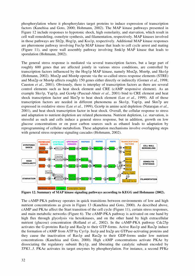
32
phosphorylation where it phosphorylates target proteins to induce expression of transcription factors (Kanehisa and Goto, 2000; Hohmann, 2002). The MAP kinase pathways presented in Figure 12 include responses to hypotonic shock, high osmolarity, and starvation, which result in cell wall remodeling, osmolyte synthesis, and filamentation, respectively. MAP kinases involved in those pathways are Slt2p, Hog1p, and Kss1p, respectively. Additional MAP kinase responses are pheromone pathway involving Fus3p MAP kinase that leads to cell cycle arrest and mating (Figure 11), and spore wall assembly pathway involving Smk1p MAP kinase that leads to sporulation (Hohmann, 2002). The general stress response is mediated via several transcription factors, but a large part of roughly 600 genes that are affected jointly in various stress conditions, are controlled by transcription factors influenced by the Hog1p MAP kinase, namely Msn2p, Msn4p, and Sko1p (Hohmann, 2002). Msn2p and Msn4p operate via the so-called stress response elements (STRE) and Msn2p or Msn4p affects roughly 150 genes either directly or indirectly (Gorner et al., 1998; Causton et al., 2001). Obviously, there is interplay of transcription factors as there are several control elements such as heat shock element and CRE (cAMP responsive element). As an example Sko1p, Yap1p, and Gcn4p (Pascual-Ahuir et al., 2001) bind to CRE element and heat shock transcription factor and Skn7p to heat shock element (Lee et al., 1999). All of those transcription factors are needed in different phenomena as Sko1p, Yap1p, and Skn7p are expressed in oxidative stress (Lee et al., 1999), Gcn4p in amino acid depletion (Natarajan et al., 2001), and heat shock transcription factor in heat shock. Overall, the cellular response to stress and adaptation to nutrient depletion are related phenomena. Nutrient depletion, i.e. starvation, is stressful as such and cells induce a general stress response, but in addition, growth on low glucose concentrations or on poor carbon sources such as ethanol leads to adaptation by reprogramming of cellular metabolism. These adaptation mechanisms involve overlapping steps with general stress response signaling cascades (Hohmann, 2002).
Slt2pMkk1..2pBck1pPkc1p
Wsc1..3p
Mid2p
Sho1p
Sln1p
Sho1p
Rho1p Rlm1p
Hog1pPbs2pSsk2pSsk1pYpd1p
Msn2pMsn4p
Ste11pSte20p
Kss1pSte7pSte11pSte20pCdc42p
Ras2p
Swi4pSwi6p
Dig1p, Dig2p
Ste12p Tec1p��������
����
��������
����
��������
������
����
������
����
����������
������
��������
������
��������
���� ����
��������
���� ����
��������
Ste50p
Ste50p
Cdc42pCdc24p
Cdc24p
�����������������������
������
��������
����
����������
������
�������
����
Hot1p
Sko1p
Transcription factorsKinasesMembrane sensors Figure 12. Summary of MAP kinase signaling pathways according to KEGG and Hohmann (2002). The cAMP-PKA pathway operates in quick transitions between environments of low and high nutrient concentrations as given in Figure 13 (Kanehisa and Goto, 2000). As described above, cAMP and PKAe affect the Start transition of the cell cycle (Figure 11), certain stress responses, and main metabolic networks (Figure 6). The cAMP-PKA pathway is activated on one hand by high flux through glycolysis via hexokinases, and on the other hand by high extracellular nutrient (glucose) concentration (Rolland et al., 2002). In the cAMP-PKA pathway Cdc25p activates the G-proteins Ras1p and Ras2p to their GTP-forms. Active Ras1p and Ras2p induce the formation of cAMP from ATP by Cyr1p. Ira1p and Ira2p are GTPase-activating proteins and they cause the inactivation of Ras1p and Ras2p to their GDP-forms under low nutrient concentrations (Kanehisa and Goto, 2000). High cAMP concentrations activate PKAe by dissociating the regulatory subunit Bcy1p, and liberating the catalytic subunit encoded by TPK1..3. PKAe activates its target enzymes by phosphorylation. For instance, a second PFKe
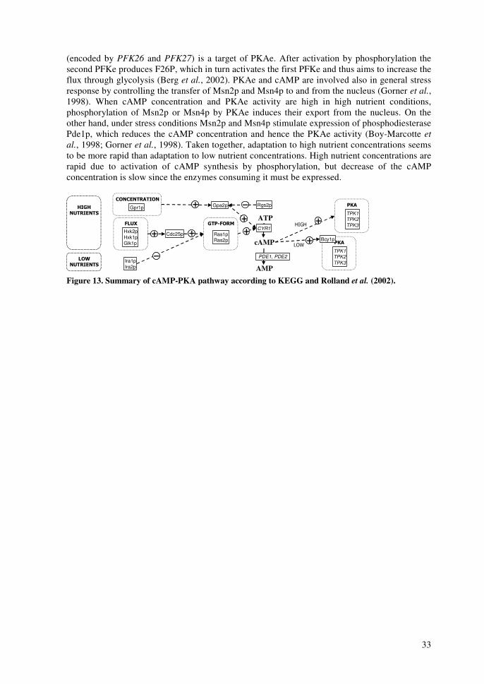
33
(encoded by PFK26 and PFK27) is a target of PKAe. After activation by phosphorylation the second PFKe produces F26P, which in turn activates the first PFKe and thus aims to increase the flux through glycolysis (Berg et al., 2002). PKAe and cAMP are involved also in general stress response by controlling the transfer of Msn2p and Msn4p to and from the nucleus (Gorner et al., 1998). When cAMP concentration and PKAe activity are high in high nutrient conditions, phosphorylation of Msn2p or Msn4p by PKAe induces their export from the nucleus. On the other hand, under stress conditions Msn2p and Msn4p stimulate expression of phosphodiesterase Pde1p, which reduces the cAMP concentration and hence the PKAe activity (Boy-Marcotte et al., 1998; Gorner et al., 1998). Taken together, adaptation to high nutrient concentrations seems to be more rapid than adaptation to low nutrient concentrations. High nutrient concentrations are rapid due to activation of cAMP synthesis by phosphorylation, but decrease of the cAMP concentration is slow since the enzymes consuming it must be expressed.
cAMP
ATP
AMP
PDE1, PDE2
TPK1TPK2TPK3
Ira1pIra2p
Cdc25p Ras1pRas2p
CYR1���
���
����
�������
����
�������
��
�������
��
�������
�������
���
TPK1TPK2TPK3
���Bcy1p
Hxk2pHxk1pGlk1p
����
Gpr1p
��������
Gpa2p Rgs2p
Figure 13. Summary of cAMP-PKA pathway according to KEGG and Rolland et al. (2002).

34
�� � ���������������
This study can be divided in two parts. The first objective was to implement and improve methods for metabolic engineering. Namely, development of methodology for the measurement of intracellular and extracellular metabolite concentrations was pursued during this study. Similarly, in vitro enzyme activity assays were implemented and further developed. Additionally, metabolic network models of the steady state situations were implemented and constructed further. The methods for measuring gene expression levels, protein abundances, and ratios of isotopic isomers after labeling were implemented and developed by co-authors of the appended articles. Further development goals included implementation of dynamic models that would have utilized time-series data of e.g. intracellular metabolites, but that goal has not been reached yet. The products of the method development projects were used to fuel the second objective. Overall, as the second objective of this study aimed at the elucidation of metabolic network activities by measuring and modeling intracellular entities and their connections as widely as possible. The second objective was divided further into two projects. In the first project at VTT Biotechnology, the goal was to study the metabolism of a xylose-utilizing S. cerevisiae strain in an attempt to identify possible limitations that cause slow growth, high yield of xylitol, and poor yield of ethanol from xylose. In the second project at Medicel Oy, the goal was to study the metabolism of a PMI40 deletion strain in an effort of improving yield of an intracellular metabolite and a protein mannosylation precursor GDP-mannose from mannose. Common denominators in the two projects were the organism, yeast, poor growth on the corresponding substrates (xylose or mannose), and the methods applied for studying cellular phenomena. Although being so different by nature, utilization of the substrates demonstrated also common effects in metabolism; xylose in the recombinant xylose strain and mannose in the recombinant PMI40 deletion strain.
35
�� ���� ���������������
3.1 Strains S. cerevisiae yeast strains H2217 (IV; Richard et al., 2002), H2446 (I), H2490 (I, II, III, IV), H2490-3 (IV), H2490-4 (IV), Y13 (V), and Y5; PMI40 deletion strain, (V) were employed in this work. They all are based on the CEN.PK2 strain (VW-1B; MATα, leu2-3/112, ura3-52, trp1-289, his3∆1, MAL2-8c, SUC2) (Boles et al., 1996). Table 1 in publication IV summarizes the important xylose strains. Laura Ruohonen oversaw the construction of original xylose utilization strains. Strain denoted here Y13 is the prototrophic control strain CEN.PK113-7D (MATa URA3 TRP1 LEU2 HIS3 MAL2-8C SUC2) obtained from Euroscarf (Germany) (Entian and Kötter, 1998). Pirkko Mattila oversaw the construction of strain Y5. 3.2 Cultivations Yeast cells were cultured in three distinctively different cultivation modes. In publications I-III continuous chemostat cultivations both under aerobic and anaerobic conditions were set up for studying the metabolism of xylose in well-controlled bioreactors (I). In publication IV xylose chemostats were harnessed to obtain chemostat isolates with improved xylose metabolism. The chemostat isolates were studied further in aerobic shake flask cultures and xylose chemostat cultivations (IV). The cultivation media in publications I-IV were based on yeast nitrogen base without amino acids (Difco, USA). In publication V the PMI40-deletion strain Y5 was studied in mid-exponential phase of aerobic batch cultivations in well-controlled bioreactors (V). The cultivation media in publication V were based on the minimal mineral medium (Verduyn et al., 1992). In all cultivations, samples were analyzed for their cell density optically and gravimetrically, and for the concentration of main extracellular metabolites in liquid phase by HPLC and for those in gas phase by mass spectrometer (I, V). 3.3 Metabolite concentrations Concentrations of several intracellular metabolites were measured in publications IV and V. The liquid chromatographic mass spectrometric (LC-MS) method was based on a method developed by van Dam and co-workers (van Dam et al., 2002) after widening the scope of measured metabolites. However, the quantification itself is only part of delivering quantitative metabolite concentrations. For obtaining reliable metabolite concentrations metabolism was quenched by sampling at least 20 mg cell dry weight of cells to cold methanol (de Koning and van Dam, 1992). The cell pellets were collected after centrifugation. The pellets were then treated with ethanol extraction and lyophilized to dryness (Gonzalez et al., 1997). Re-suspended samples were quantified for over a dozen metabolites in central metabolic pathways using the LC-MS method. The LC-MS method applied Dionex anion exchange liquid chromatography and Micromass triple quadrupole mass spectrometer (van Dam et al., 2002). 3.3.1 Automated sampling In order to provide reproducible quenching of the cultivation samples we designed and constructed a Medicel Sampling Unit for automated rapid sampling (not published). This allowed automated sampling at the intervals as short as 20 seconds. Depending on the sample flow rate and the volume of the sampling line tubing, residence time in the sample tubing line was only 2 s. Samples were introduced into methanol tubes for metabolite samples and into empty tubes for enzyme samples. Sample tubes were stored in a cold bath, maintained at –40 °C throughout the sampling process. Thus, samples taken into the empty tubes were allowed to freeze, but after thawing they could be used for the analysis of extracellular metabolites, cell
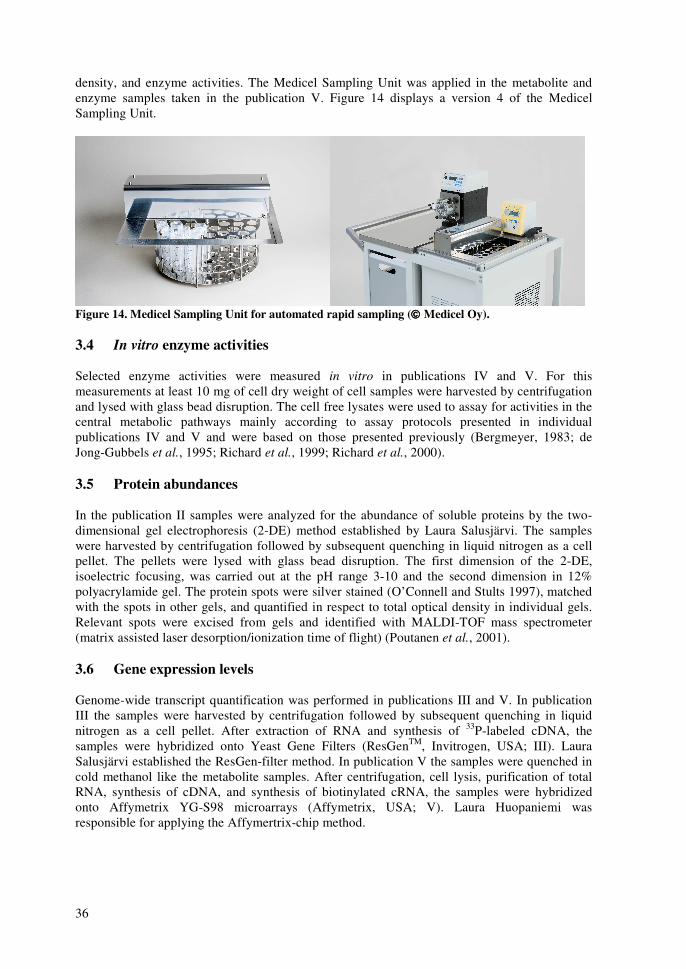
36
density, and enzyme activities. The Medicel Sampling Unit was applied in the metabolite and enzyme samples taken in the publication V. Figure 14 displays a version 4 of the Medicel Sampling Unit.
Figure 14. Medicel Sampling Unit for automated rapid sampling (���� Medicel Oy). 3.4 In vitro enzyme activities Selected enzyme activities were measured in vitro in publications IV and V. For this measurements at least 10 mg of cell dry weight of cell samples were harvested by centrifugation and lysed with glass bead disruption. The cell free lysates were used to assay for activities in the central metabolic pathways mainly according to assay protocols presented in individual publications IV and V and were based on those presented previously (Bergmeyer, 1983; de Jong-Gubbels et al., 1995; Richard et al., 1999; Richard et al., 2000). 3.5 Protein abundances In the publication II samples were analyzed for the abundance of soluble proteins by the two-dimensional gel electrophoresis (2-DE) method established by Laura Salusjärvi. The samples were harvested by centrifugation followed by subsequent quenching in liquid nitrogen as a cell pellet. The pellets were lysed with glass bead disruption. The first dimension of the 2-DE, isoelectric focusing, was carried out at the pH range 3-10 and the second dimension in 12% polyacrylamide gel. The protein spots were silver stained (O’Connell and Stults 1997), matched with the spots in other gels, and quantified in respect to total optical density in individual gels. Relevant spots were excised from gels and identified with MALDI-TOF mass spectrometer (matrix assisted laser desorption/ionization time of flight) (Poutanen et al., 2001). 3.6 Gene expression levels Genome-wide transcript quantification was performed in publications III and V. In publication III the samples were harvested by centrifugation followed by subsequent quenching in liquid nitrogen as a cell pellet. After extraction of RNA and synthesis of 33P-labeled cDNA, the samples were hybridized onto Yeast Gene Filters (ResGenTM, Invitrogen, USA; III). Laura Salusjärvi established the ResGen-filter method. In publication V the samples were quenched in cold methanol like the metabolite samples. After centrifugation, cell lysis, purification of total RNA, synthesis of cDNA, and synthesis of biotinylated cRNA, the samples were hybridized onto Affymetrix YG-S98 microarrays (Affymetrix, USA; V). Laura Huopaniemi was responsible for applying the Affymertrix-chip method.

37
3.7 Metabolic fluxes Publication I applied flux balancing analysis (FBA) with over-determined stoichiometric matrix topology (van Gulik and Heijnen, 1995; Nissen et al., 1997; Granström et al., 2002) to estimation of fluxes in xylose metabolism. The stoichiometric matrix included 71 metabolites and 38 or 39 metabolic fluxes for anaerobic and aerobic conditions respectively. Extracellular accumulation rates were measured in pseudo steady states of the chemostat cultivations. Additionally, constraints were imposed to certain metabolic fluxes to set them one-directional. The set of stoichiometric matrix, accumulation rate vector, and constraint matrix and vector were used to solve values of the flux vector in a constrained linear least squares problem (LSQLIN in MatlabTM).

38
�� ������������ ����� ���
4.1 Metabolism of xylose (I-IV) Publications I-III compared the metabolism of xylose with a small glucose addition to the metabolism of glucose in continuous cultivations. Publication I presented the flux analysis of xylose metabolism when the amount of glucose in the xylose feed was reduced from 3 to 0.1 g/liter under aerobic conditions. Publication II presented a 2-DE quantification of soluble proteins in the same cultivations. Additionally, publication II displayed the protein abundances in the transition from aerobic to anaerobic conditions. Further, publication III presented gene transcript quantification of the same chemostat cultivations under aerobic conditions. Publication IV, on the other hand, described recovery of efficiently xylose utilizing chemostat isolates from similar, but longer xylose chemostat cultivations. Publication IV applied measurements of intracellular metabolite concentrations and in vitro enzyme activity assays for comparing the metabolism of xylose in the chemostat isolates to metabolism of xylose, glucose, or ethanol in the original strains. Thus, publications I-III compared the metabolism of xylose to metabolism of glucose in chemostat cultivations, and publication IV compared the metabolism of xylose to metabolism of glucose and metabolism of ethanol in batch cultivations. The majority of the cultivations were performed under aerobic or actually close-microaerobic conditions. 4.1.1 Xylose as a carbon source compared to glucose and ethanol (I-IV) As already discussed in the introduction, xylose is a poor carbon source compared to glucose. Although only 10% of carbon in the feed medium was glucose in the highest glucose addition, 30% of the biomass in this chemostat cultivation still originated from glucose (I-III). Considering the three proposed bottlenecks of xylose metabolism, the publications I-III highlight mainly the effects caused by the redox cofactor imbalance arising from the conversion of xylose to xylulose by the recombinant pathway of XR and XDH. The combined results of publications I-III are presented in Figure 15 comparing xylose (with 3 g/liter glucose) and glucose as carbon sources under aerobic conditions. The flux of futile cycle through the upper glycolysis and oxidative PPP increased on xylose, possibly in order to improve the production of NADPH for the XR reaction (I). Congruently, up-regulation of GND1 (Sinha and Maitra, 1992), encoding for an NADPH-generating dehydrogenase in the oxidative PPP, was observed in the transcript analysis (III). As the oxidative PPP also consumes carbon as the emission of carbon dioxide, the flux of carbon to pyruvate was reduced (I). Possibly, to counterbalance for a decreased flux through lower glycolysis to pyruvate, the abundance of several proteins in the lower glycolysis pathway was elevated (II). In addition, a transcription factor TYE7, which operates to induce the expression of the enolase genes in the lower glycolysis, was up-regulated. TYE7 can operate in the place of transcription factors GCR1/GCR2 (Sato et al., 1999), which are the main transcription factors controlling the lower glycolysis (Figure 3) (Uemura and Fraenkel, 1990). Consistently in our experimental results of flux, protein, and transcript analysis (I-III), TCA cycle activity was clearly reduced on xylose. The relative carbon flux through TCA cycle on xylose was only roughly 60% of that on glucose (I), and the protein abundances (II), and transcript concentrations (III) were decreased congruently. Both protein abundances and transcript concentrations displayed further effects of balancing the intracellular redox state. Namely, several enzymes catalyzing oxidoreductive reactions were up-regulated such as those involved in the metabolism of glycerol, ethanol, and acetate. Transcript concentrations of the xylose-grown cells revealed further that in continuous chemostat cultivation, a response to starvation was triggered despite the fact that there is abundant xylose available (III). ATO1, ATO2, and ATO3 that increase in order to increase ammonia secretion as a response to starvation (Palkova et al., 2002) were not repressed on
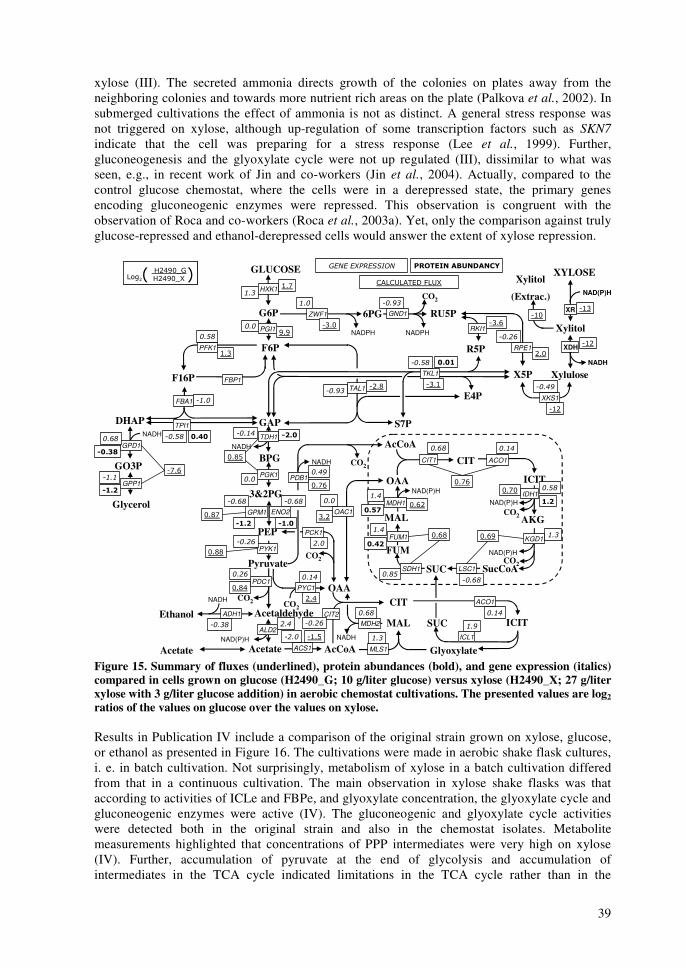
39
xylose (III). The secreted ammonia directs growth of the colonies on plates away from the neighboring colonies and towards more nutrient rich areas on the plate (Palkova et al., 2002). In submerged cultivations the effect of ammonia is not as distinct. A general stress response was not triggered on xylose, although up-regulation of some transcription factors such as SKN7 indicate that the cell was preparing for a stress response (Lee et al., 1999). Further, gluconeogenesis and the glyoxylate cycle were not up regulated (III), dissimilar to what was seen, e.g., in recent work of Jin and co-workers (Jin et al., 2004). Actually, compared to the control glucose chemostat, where the cells were in a derepressed state, the primary genes encoding gluconeogenic enzymes were repressed. This observation is congruent with the observation of Roca and co-workers (Roca et al., 2003a). Yet, only the comparison against truly glucose-repressed and ethanol-derepressed cells would answer the extent of xylose repression.
GLUCOSE
GAP
BPG
E4P
AcCoA
SucCoA
OAA
DHAP
Acetaldehyde
CO2
CO2
CO2
AcCoA
OAACO2
CO2NADH
NAD(P)H
NADPH
NADH
NADH
NAD(P)H
NAD(P)H
NADH
TKL1
TAL1
ZWF1
PFK1
FBA1
TDH1
PGI1
PYK1
PGK1
KGD1
MDH1
SDH1
RKI1NADPH
6PG
FUM
ACO1
FUM1
SUC
FBP1
NAD(P)HIDH1
LSC1
CIT1
CO2
GND1
RPE1
�����������������
F6P
G6P
F16P
GO3P
RU5P
R5P
3&2PG
PEPMAL
ICIT
CIT
PDC1
X5P
GPD1
GPP1
ACS1
ALD2
ADH1
PYC1
Glyoxylate
ICIT
CIT
MAL SUCICL1
MLS1
MDH2CIT2
TPI1
PCK1
PDB1
NADH
CO2Pyruvate
S7P
�
AKG
XR
Xylitol
Xylulose
XDH
NAD(P)H
NADH
XKS1
Glycerol
Acetate
Ethanol
Xylitol
(Extrac.)
Acetate
GPM1 ENO2
1�,-'�������*2-3456 2-3456�1�,-'�������*2-3456 2-3456�
XYLOSE� � � ��� �����
� 1�71 8����17�HXK1
� !�
(9:
�
���
��
��
����
494
(9/
OAC1
��� "# �
59;<
59;:
��
����
"$ # "$ �
����
59;;
����
�:9=
���
"� %&
�
"$ #
�/95
����
-95
����
�/9=
����
�(-
�/9(
���� � �$
�-9;����
�(-
�(/�(5
���
59:=59:=
���
59:5
��
���
$ #
59=459=; �
�������
�
� !#
59=-
�
� '(/9-
��
59;3
���
-93
��
��
��� �(9<
���� �� �����
�
���
ACO1
��
Figure 15. Summary of fluxes (underlined), protein abundances (bold), and gene expression (italics) compared in cells grown on glucose (H2490_G; 10 g/liter glucose) versus xylose (H2490_X; 27 g/liter xylose with 3 g/liter glucose addition) in aerobic chemostat cultivations. The presented values are log2 ratios of the values on glucose over the values on xylose. Results in Publication IV include a comparison of the original strain grown on xylose, glucose, or ethanol as presented in Figure 16. The cultivations were made in aerobic shake flask cultures, i. e. in batch cultivation. Not surprisingly, metabolism of xylose in a batch cultivation differed from that in a continuous cultivation. The main observation in xylose shake flasks was that according to activities of ICLe and FBPe, and glyoxylate concentration, the glyoxylate cycle and gluconeogenic enzymes were active (IV). The gluconeogenic and glyoxylate cycle activities were detected both in the original strain and also in the chemostat isolates. Metabolite measurements highlighted that concentrations of PPP intermediates were very high on xylose (IV). Further, accumulation of pyruvate at the end of glycolysis and accumulation of intermediates in the TCA cycle indicated limitations in the TCA cycle rather than in the
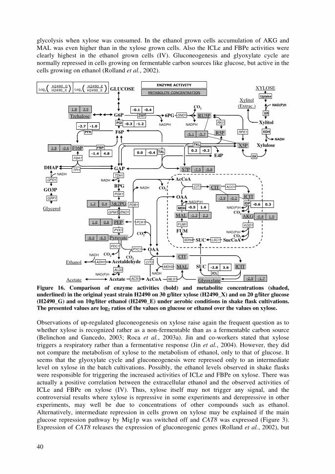
40
glycolysis when xylose was consumed. In the ethanol grown cells accumulation of AKG and MAL was even higher than in the xylose grown cells. Also the ICLe and FBPe activities were clearly highest in the ethanol grown cells (IV). Gluconeogenesis and glyoxylate cycle are normally repressed in cells growing on fermentable carbon sources like glucose, but active in the cells growing on ethanol (Rolland et al., 2002).
GLUCOSE
GAP
BPG
E4P
AcCoA
SucCoA
OAA
DHAP
Acetaldehyde
CO2
CO2
CO2
AcCoA
OAACO2
CO2NADH
NAD(P)H
NADPH
NADH
NADH
NAD(P)H
NAD(P)H
NADH
TKL
TAL
ZWF
PFK
FBA1
TDH1
PGI
PYK1
PGK1
KGD1
MDH
SDH4
RKI1NADPH
6PG
FUM
ACO1
FUM1
SUC
FBP
NAD(P)HIDP
LSC1
CIT1
CO2
GND1
RPE1
��)�*������+���
$�8 >?1+8���?����8� 8+?�
� � "� !� � "� !
F6P
G6P
F16P
GO3P
RU5P
R5P
3&2PG
PEPMAL
ICIT
CIT
PDC1
X5P
GPD1
GPP1
ACS1
ALD2
ADH1
PYC1
Glyoxylate
ICIT
CIT
MAL SUCICL
MLS1
MDH3CIT2
TPI1
PCK1
PDB1
NADH
CO2Pyruvate
S7P
(9; /9<(9; /9< "� $ "� !"� $ "� !
� # "� #� # "� #
"� % "$ #"� % "$ #"# ( "$ �"# ( "$ �
"$ ! ! &"$ ! ! &
"# & % ,"# & % ,
"� ' $ ,"� ' $ ,"� , � %"� , � %(9- 593(9- 593
�<9( �<9:�<9( �<9:
�:9< �<9;�:9< �<9;
(95 59;(95 59;
�=95 �=9<�=95 �=9<
-9; �59=-9; �59=
�(9- -9-�(9- -9-
�/94 �59-�/94 �59-
�-95 �(9:�-95 �(9:
AKG �59; (95�59; (95
Uptake
XR
Xylitol
Xylulose
XDH
NAD(P)H
NADH
XK
Trehalose
Glycerol
Acetate
Ethanol
Xylitol (Extrac.)
XYLOSE
Acetate
GPM1 ENO1
1�,-'�������* 1�,-'�������*2-3456 2-3456�2-3456� 2-3456�1�,-'�������* 1�,-'�������*1�,-'�������* 1�,-'�������*2-3456 2-3456�2-3456� 2-3456�
Figure 16. Comparison of enzyme activities (bold) and metabolite concentrations (shaded, underlined) in the original yeast strain H2490 on 30 g/liter xylose (H2490_X) and on 20 g/liter glucose (H2490_G) and on 10g/liter ethanol (H2490_E) under aerobic conditions in shake flask cultivations. The presented values are log2 ratios of the values on glucose or ethanol over the values on xylose. Observations of up-regulated gluconeogenesis on xylose raise again the frequent question as to whether xylose is recognized rather as a non-fermentable than as a fermentable carbon source (Belinchon and Gancedo, 2003; Roca et al., 2003a). Jin and co-workers stated that xylose triggers a respiratory rather than a fermentative response (Jin et al., 2004). However, they did not compare the metabolism of xylose to the metabolism of ethanol, only to that of glucose. It seems that the glyoxylate cycle and gluconeogenesis were repressed only to an intermediate level on xylose in the batch cultivations. Possibly, the ethanol levels observed in shake flasks were responsible for triggering the increased activities of ICLe and FBPe on xylose. There was actually a positive correlation between the extracellular ethanol and the observed activities of ICLe and FBPe on xylose (IV). Thus, xylose itself may not trigger any signal, and the controversial results where xylose is repressive in some experiments and derepressive in other experiments, may well be due to concentrations of other compounds such as ethanol. Alternatively, intermediate repression in cells grown on xylose may be explained if the main glucose repression pathway by Mig1p was switched off and CAT8 was expressed (Figure 3). Expression of CAT8 releases the expression of gluconeogenic genes (Rolland et al., 2002), but

41
the activity of Cat8p might be blocked partly by cAMP thus preventing the full-scale expression of the gluconeogenic genes (Zaragoza and Gancedo, 2001). Concentrations of cAMP are influenced both by extracellular glucose concentrations and by intracellular metabolic fluxes (Figure 13). Intracellular fluxes may not thus be related to glucose as such, but they are related rather to the energy and redox status of the cell (Zaragoza and Gancedo, 2001). One of the sensors of glycolytic flux is PFKe, whose regulation involves the energy state and concentration of cAMP. However, present knowledge states that HXKe's have a bigger role in controlling the cAMP concentrations than the PFK (Rolland et al., 2002). Furthermore, HXKe's are not needed in the metabolism of xylose. If indeed cAMP concentrations would convey the repression signal in cells grown on xylose, it would require that the levels of cAMP on xylose were somewhere between the levels on glucose and ethanol. Thus, concentrations of cAMP may be worth measuring in the future studies of xylose metabolism. 4.1.2 Oxidation of cytosolic NADH (I, II, III) The different cofactor specificities of the two-step oxidoreductase reaction of xylose create a futile cycle between pentose phosphates, F6P and G6P with serious consequences (I). At the upstream of main carbon metabolism there is a route for NADPH generation; oxidative pentose phosphate pathway (Bruinenberg et al., 1983a), but it consumes carbon in the form of evolved carbon dioxide reducing the flow of carbon downstream to pyruvate (I). Figure 17 displays how back-flux from F6P to G6P increased (rPGI; negative) when the concentration of glucose addition in the chemostat feed was decreased from initial, the 3 g/liter (I). Hence, NADPH generation is not a problem for the cell but it causes problems further downstream since the main NAD+ generating routes start from pyruvate (Bruinenberg et al., 1983b). The flow of carbon is preferentially (and too easily even) used for the generation of NADPH, leaving a lesser amount for the generation of NAD+. In addition to evolution of carbon as the carbon dioxide, good NADPH availability increases also the production of xylitol (Jeppsson et al., 2002). Thus, some carbon is lost in the excreted xylitol. The generation and availability of cytosolic NAD+ becomes a bottleneck for xylose metabolism; especially in a strain without a functional xylose isomerase (Kuyper et al., 2003) or a functional transhydrogenase cycle. Supposedly, under aerobic conditions NADH is oxidized to NAD+ in the oxidative phosphorylation. However, the reducing potential of cytosolic NADH must be shuttled to mitochondrion, as NADH cannot be transferred across the mitochondrial membrane (Overkamp et al., 2000). Shuttling mechanisms consist generally of mitochondrial and cytosolic isoenzymes, catalyzing oxidoreductive reactions of metabolites that can be transferred between the cytosol and mitochondrion. Concentrations of some of these genes (ALD4, ALD6) and proteins (Gpd1p, Ald4p, Ald6p) increased in the xylose cultivation as an indication of attempts to get cytosolic NADH oxidized in mitochondrion (III, II). Thus, allegedly mitochondrial NADH concentrations increased when the cells were consuming xylose and oxidative phosphorylation should have been able to consume the excess NADH. As the TCA cycle produces also NADH, its activities decreased as discussed above (Figure 15). When the concentration of the glucose addition was decreased further the flux of pyruvate to mitochondrion (rAcCoA) decreased at the expense of flux to acetaldehyde (rACD; Figure 17; I). In fact, mitochondrial NADH inhibits the initial steps of TCA cycle, namely the enzymes citrate synthase (Kispal and Srere, 1991) and isocitrate dehydrogenase (Satrustegui et al., 1983) (Figure 6). The main reason why oxidative phosphorylation was not able to consume the excess NADH must have been the fact that the TCA cycle and oxidative phosphorylation share the succinate dehydrogenase complex, which reduces succinate to fumarate in the TCA cycle and gives the electrons to ubiquinone via FADH2 in the oxidative phosphorylation (Berg et al., 2002). Thus, as oxidation of succinate to fumarate is required for functional oxidative phosphorylation, it causes a stoichiometric limit in the TCA cycle, since ratio between consumed succinate and produced NADH is fixed. However, glyoxylate cycle bypasses two thirds of the NADH-producing reactions of the TCA cycle

42
producing succinate, which can then be oxidized to fumarate by the succinate dehydrogenase complex (Berg et al., 2002). Indicative of the reduced oxidative phosphorylation was also the increasing respiratory quotient when the glucose addition in the feed was decreased (I). Increased respiratory quotient was caused by both increasing carbon evolution rate and by decreasing oxygen uptake rate. Decreased regeneration of NAD+ was seen also as a decrease in the flux of xylose to xylulose 5-phosphate (rX5P; Figure 17; I) when glucose addition in the feed was dropped below 0.5 g/liter. As the dilution rate in the chemostat cultivations was so low also the energy requirements of the maintenance became more demanding. Thus, when the amount of glucose in the feed was reduced the cells were forced to reconsider the utilization rate of xylose. Hypothetically, if the functional xylose isomerase was not available, metabolic engineering of xylose metabolism would benefit on finding a mechanism for production of NADPH upstream in metabolism through a route, which would not result in loss of carbon. For example simultaneous deletion of G6P dehydrogenase (Jeppsson et al., 2002) and e.g. expression of a partly NADPH-dependent GAP dehydrogenase (Verho et al., 2002; Verho et al., 2003) would direct the flow of carbon directly towards pyruvate and ethanol without the loss of carbon.
0
20
40
60
80
100
120
0.0 0.5 1.0 1.5 2.0 2.5 3.0
Glucose in the feed (g/l)
r PG
I, r A
CD, a
nd r
AcC
oA
(% o
f the
tota
l car
bon)
-60
-40
-20
0
20
40
60
80
r PG
I
(% o
f the
tota
l car
bon)
r X5P
r PGI
r ACD r AcCoA
Figure 17. Intracellular fluxes based on metabolic flux analysis that are related to consumption and generation of NADH are presented as a function of initial glucose concentration (0.1, 0.5, 1 and 3 g/liter) with 30 g/liter xylose as the main carbon source (I). The selected central carbon fluxes were calculated from results of continuous chemostat cultivations at the dilution rate of 0.05 h-1. Fluxes 30 (XUL to X5P, ����), 2 (G6P to F6P, ����), 8 (PYR to AcCoA, ����) and 9 (PYR to ACD, ××××) are presented. Values are presented in % of the total carbon utilization (mmol / mmol). There are also other indications that the metabolism of xylose requires the presence of mitochondrion as petite mutants, lacking the mitochondrion were not able to grow on xylose (Jin et al., 2004). Is the phenomenon related to some other oxidoreductive reactions other than those consuming NADH. One possibility of other oxidoreductive reactions is cytochrome-c dependent lactate dehydrogenase encoded by CYB2, which was up-regulated in the xylose chemostat cultivation both at the transcript and protein level (II, III). Cyb2p catalyzes the oxidation of lactate to pyruvate with ferrocytochrome-c as the reduced cofactor. Could Cyb2p be involved in decoupling of respiration from TCA cycle by replacing succinate dehydrogenase complex or more likely assisting it? Alternatively, the requirement of oxygen may be related to the glyoxylate cycle; will the glyoxylate cycle become dysfunctional when the mitochondrion is removed? Then one can ask why the glyoxylate cycle activity would be required? Could the
43
cells not just produce ethanol and thus regenerate their redox state? However, as xylose does not seem to repress the gluconeogenic enzymes and also respiration is operational, the possibly produced ethanol is subsequently also consumed under aerobic conditions. A possibility to tackle the active glyoxylate cycle would be to conditionally knock out, e.g., gene encoding isocitrate lyase (ICL1). This might improve ethanol production under microaerobic conditions. 4.1.3 Uptake of xylose was improved in chemostat isolates (IV) Characterization of the chemostat isolates with improved xylose metabolism gives us a possibility to learn from the improvements made in the isolates (IV). The time required for the recovery of the isolates was greatly shortened by using a strain with an empty multicopy plasmid containing a LEU2 marker. The number of plasmids was greatly reduced in all isolates, but totally lost in isolate H2490-3. Thus, combination of an empty multicopy plasmid and chemostat cultivations proved to be efficient in providing spontaneous mutations. Here benefit of chemostat cultivation was that inevitably there co-exists several populations some of who have different growth modes in terms of the nutrient source (some cells may be consuming remains of other cells) and especially in terms of the phase in cell cycle. Eventually, there co-exists populations who have altered genomes. Figure 18 displays the comparison of two chemostat isolates to the original strain. Xylose uptake, which is one possible rate-limiting step in xylose metabolism, in addition to low availability of cytosolic NAD+ proposed in the previous chapter, increased significantly by 60% in both isolates (IV). In S. cerevisiae most of the major hexose-transporters, encoded by HXT1-HXT7 and GAL2, have also been shown to be capable of xylose transport (Hamacher et al., 2002; Sedlak and Ho, 2004). The uptake rate measured in the original strain grown in the presence of a low glucose level was comparable to the rate in the chemostat isolates on xylose alone. The positive effect of low glucose levels has been reported before despite the inhibitory effect of glucose (Meinander and Hahn-Hägerdal, 1997). What is noteworthy is that all isolates had been grown in the presence of low glucose levels at some point of the chemostat cultivation, and the isolate H2490-3 was even recovered from a xylose/glucose mixture (IV). Further, the improved xylose uptake may also require some oxygen as under anaerobic conditions the specific xylose consumption rate was reduced to the same level as in the original strain under aerobic conditions (IV). Do the uptake systems applied in the isolates require ATP, and thus require oxygen for producing more ATP? The observed poor xylose uptake rate under anaerobic conditions could be the reason for the inability to grow under anaerobic conditions (Hahn-Hägerdal et al., 2001) and would thus be the main rate-limiting step of xylose metabolism. The transporters in the absence of glucose and in the presence of xylose would be Hxt5p, Hxt6p and Hxt7p, since HXT5 is expressed under starvation (Buziol et al., 2002) and HXT6 and HXT7 on non-fermentable carbon sources (Dlugai et al., 2001). Hxt5p might be the transporter taking care of xylose expression in the isolates as reported by Wahlbom and co-workers (Wahlbom et al., 2003a). Thus, Hxt5p is involved in starvation and uptake of hexoses at the same time, as the cells are ready for the utilization of respirative substrates. Hxt5p might link uptake of xylose to oxygen availability since the utilization of ethanol, glycerol, and acetate requires oxygen as well. Another possibility for the transporter of xylose might be Hxt2p, which was induced in the chemostat cultivation on xylose. The result that the HXT2-encoded transporter is less active under anaerobic conditions (Reifenberger et al., 1997) could partly explain why the growth properties of the isolates were not improved under anaerobic conditions. The reports describing xylose uptake facilitated by separate transporters have not included Hxt2p in the discussions (Hamacher et al., 2002; Sedlak and Ho, 2004), which may be due to problems in its expression (Hamacher et al., 2002).
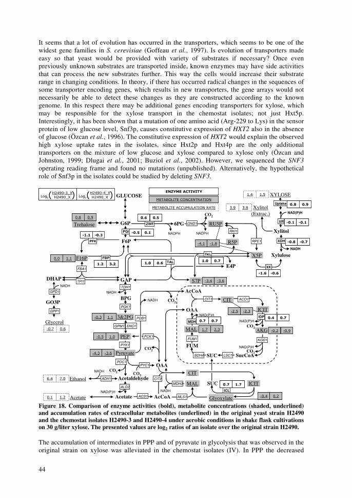
44
It seems that a lot of evolution has occurred in the transporters, which seems to be one of the widest gene families in S. cerevisiae (Goffeau et al., 1997). Is evolution of transporters made easy so that yeast would be provided with variety of substrates if necessary? Once even previously unknown substrates are transported inside, known enzymes may have side activities that can process the new substrates further. This way the cells would increase their substrate range in changing conditions. In theory, if there has occurred radical changes in the sequences of some transporter encoding genes, which results in new transporters, the gene arrays would not necessarily be able to detect these changes as they are constructed according to the known genome. In this respect there may be additional genes encoding transporters for xylose, which may be responsible for the xylose transport in the chemostat isolates; not just Hxt5p. Interestingly, it has been shown that a mutation of one amino acid (Arg-229 to Lys) in the sensor protein of low glucose level, Snf3p, causes constitutive expression of HXT2 also in the absence of glucose (Özcan et al., 1996). The constitutive expression of HXT2 would explain the observed high xylose uptake rates in the isolates, since Hxt2p and Hxt4p are the only additional transporters on the mixture of low glucose and xylose compared to xylose only (Özcan and Johnston, 1999; Dlugai et al., 2001; Buziol et al., 2002). However, we sequenced the SNF3 operating reading frame and found no mutations (unpublished). Alternatively, the hypothetical role of Snf3p in the isolates could be studied by deleting SNF3.
GLUCOSE
GAP
BPG
E4P
AcCoA
SucCoA
OAA
DHAP
Acetaldehyde
CO2
CO2
CO2
AcCoA
OAACO2
CO2NADH
NAD(P)H
NADPH
NADH
NADH
NAD(P)H
NAD(P)H
NADH
TKL
TAL
ZWF
PFK
FBA1
TDH1
PGI
PYK1
PGK1
KGD1
MDH
SDH4
RKI1NADPH
6PG
FUM
ACO1
FUM1
SUC
FBP
NAD(P)HIDP
LSC1
CIT1
CO2
GND1
RPE1
��)�*������+���
$�8 >?1+8���?����8� 8+?�
$ � � ,$ � � ,
F6P
G6P
F16P
GO3P
RU5P
R5P
3&2PG
PEPMAL
ICIT
CIT
PDC1
X5P
GPD1
GPP1
ACS1
ALD2
ADH1
PYC1
Glyoxylate
ICIT
CIT
MAL SUCICL
MLS1
MDH3CIT2
TPI1
PCK1
PDB1
NADH
CO2Pyruvate
S7P
59; 59459; 594 � , � '� , � '
$ � � ($ � � (
"� ' � $"� ' � $"$ $ "� %"$ $ "� %
$ # % #$ # % #
� ( $ (� ( $ (
� ( � (� ( � (� ! � (� ! � (�59/ (9(�59/ (9(
�39( �(9;�39( �(9;
�/93 �/9=�/93 �/9=
�59< (95�59< (95
�39/ �/9=�39/ �/9=
595 (9(595 (9(
(9: -9/(9: -9/
�-9< �-9/�-9< �-9/
�593 59-�593 59-
AKG �59- �594�59- �594
Uptake
XR
Xylitol
Xylulose
XDH
NAD(P)H
NADH
XK
"$ , "� ,"$ , "� ,
"� $ "� $"� $ "� $
"� & "� ("� & "� (
� & � -� & � -
Trehalose
Glycerol�59: 59=�59: 59=
Acetate59( (9-59( (9-
Ethanol=93 :95=93 :95
Xylitol (Extrac.)
/94 /9;/94 /9;
XYLOSE(93 (9<(93 (9<
Acetate
$�8 >?1+8�� ��7$71 8+?��� 8�
GPM1 ENO1
1�,-'�������* 1�,-'�������*2-345�/6� 2-345�36�2-3456� 2-3456�1�,-'�������* 1�,-'�������*1�,-'�������* 1�,-'�������*2-345�/6� 2-345�36�2-3456� 2-3456�
Figure 18. Comparison of enzyme activities (bold), metabolite concentrations (shaded, underlined) and accumulation rates of extracellular metabolites (underlined) in the original yeast strain H2490 and the chemostat isolates H2490-3 and H2490-4 under aerobic conditions in shake flask cultivations on 30 g/liter xylose. The presented values are log2 ratios of an isolate over the original strain H2490. The accumulation of intermediates in PPP and of pyruvate in glycolysis that was observed in the original strain on xylose was alleviated in the chemostat isolates (IV). In PPP the decreased
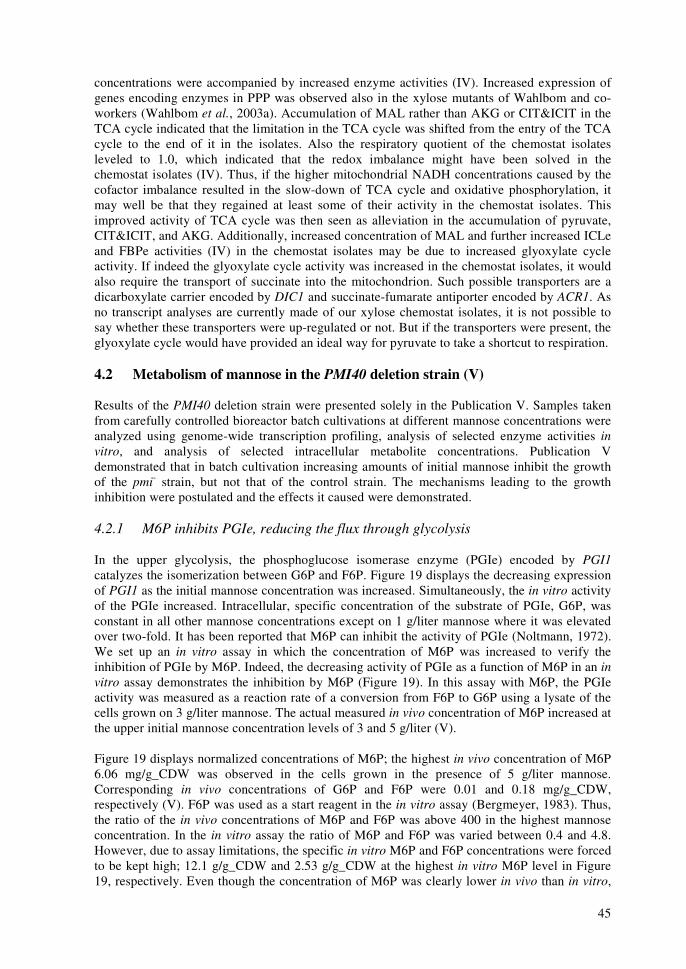
45
concentrations were accompanied by increased enzyme activities (IV). Increased expression of genes encoding enzymes in PPP was observed also in the xylose mutants of Wahlbom and co-workers (Wahlbom et al., 2003a). Accumulation of MAL rather than AKG or CIT&ICIT in the TCA cycle indicated that the limitation in the TCA cycle was shifted from the entry of the TCA cycle to the end of it in the isolates. Also the respiratory quotient of the chemostat isolates leveled to 1.0, which indicated that the redox imbalance might have been solved in the chemostat isolates (IV). Thus, if the higher mitochondrial NADH concentrations caused by the cofactor imbalance resulted in the slow-down of TCA cycle and oxidative phosphorylation, it may well be that they regained at least some of their activity in the chemostat isolates. This improved activity of TCA cycle was then seen as alleviation in the accumulation of pyruvate, CIT&ICIT, and AKG. Additionally, increased concentration of MAL and further increased ICLe and FBPe activities (IV) in the chemostat isolates may be due to increased glyoxylate cycle activity. If indeed the glyoxylate cycle activity was increased in the chemostat isolates, it would also require the transport of succinate into the mitochondrion. Such possible transporters are a dicarboxylate carrier encoded by DIC1 and succinate-fumarate antiporter encoded by ACR1. As no transcript analyses are currently made of our xylose chemostat isolates, it is not possible to say whether these transporters were up-regulated or not. But if the transporters were present, the glyoxylate cycle would have provided an ideal way for pyruvate to take a shortcut to respiration. 4.2 Metabolism of mannose in the PMI40 deletion strain (V) Results of the PMI40 deletion strain were presented solely in the Publication V. Samples taken from carefully controlled bioreactor batch cultivations at different mannose concentrations were analyzed using genome-wide transcription profiling, analysis of selected enzyme activities in vitro, and analysis of selected intracellular metabolite concentrations. Publication V demonstrated that in batch cultivation increasing amounts of initial mannose inhibit the growth of the pmi- strain, but not that of the control strain. The mechanisms leading to the growth inhibition were postulated and the effects it caused were demonstrated. 4.2.1 M6P inhibits PGIe, reducing the flux through glycolysis In the upper glycolysis, the phosphoglucose isomerase enzyme (PGIe) encoded by PGI1 catalyzes the isomerization between G6P and F6P. Figure 19 displays the decreasing expression of PGI1 as the initial mannose concentration was increased. Simultaneously, the in vitro activity of the PGIe increased. Intracellular, specific concentration of the substrate of PGIe, G6P, was constant in all other mannose concentrations except on 1 g/liter mannose where it was elevated over two-fold. It has been reported that M6P can inhibit the activity of PGIe (Noltmann, 1972). We set up an in vitro assay in which the concentration of M6P was increased to verify the inhibition of PGIe by M6P. Indeed, the decreasing activity of PGIe as a function of M6P in an in vitro assay demonstrates the inhibition by M6P (Figure 19). In this assay with M6P, the PGIe activity was measured as a reaction rate of a conversion from F6P to G6P using a lysate of the cells grown on 3 g/liter mannose. The actual measured in vivo concentration of M6P increased at the upper initial mannose concentration levels of 3 and 5 g/liter (V). Figure 19 displays normalized concentrations of M6P; the highest in vivo concentration of M6P 6.06 mg/g_CDW was observed in the cells grown in the presence of 5 g/liter mannose. Corresponding in vivo concentrations of G6P and F6P were 0.01 and 0.18 mg/g_CDW, respectively (V). F6P was used as a start reagent in the in vitro assay (Bergmeyer, 1983). Thus, the ratio of the in vivo concentrations of M6P and F6P was above 400 in the highest mannose concentration. In the in vitro assay the ratio of M6P and F6P was varied between 0.4 and 4.8. However, due to assay limitations, the specific in vitro M6P and F6P concentrations were forced to be kept high; 12.1 g/g_CDW and 2.53 g/g_CDW at the highest in vitro M6P level in Figure 19, respectively. Even though the concentration of M6P was clearly lower in vivo than in vitro,
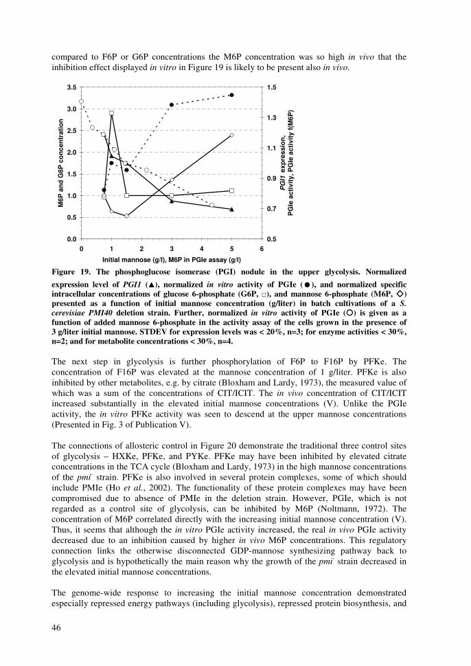
46
compared to F6P or G6P concentrations the M6P concentration was so high in vivo that the inhibition effect displayed in vitro in Figure 19 is likely to be present also in vivo.
0.0
0.5
1.0
1.5
2.0
2.5
3.0
3.5
0 1 2 3 4 5 6
Initial mannose (g/l), M6P in PGIe assay (g/l)
M6P
and
G6P
con
cent
ratio
n
0.5
0.7
0.9
1.1
1.3
1.5
PG
I1 e
xpre
ssio
n,P
GIe
act
ivity
, PG
Ie a
ctiv
ity f(
M6P
)
Figure 19. The phosphoglucose isomerase (PGI) nodule in the upper glycolysis. Normalized expression level of PGI1 (����), normalized in vitro activity of PGIe (����), and normalized specific intracellular concentrations of glucose 6-phosphate (G6P, �), and mannose 6-phosphate (M6P, ����) presented as a function of initial mannose concentration (g/liter) in batch cultivations of a S. cerevisiae PMI40 deletion strain. Further, normalized in vitro activity of PGIe (����) is given as a function of added mannose 6-phosphate in the activity assay of the cells grown in the presence of 3 g/liter initial mannose. STDEV for expression levels was < 20%, n=3; for enzyme activities < 30%, n=2; and for metabolite concentrations < 30%, n=4. The next step in glycolysis is further phosphorylation of F6P to F16P by PFKe. The concentration of F16P was elevated at the mannose concentration of 1 g/liter. PFKe is also inhibited by other metabolites, e.g. by citrate (Bloxham and Lardy, 1973), the measured value of which was a sum of the concentrations of CIT/ICIT. The in vivo concentration of CIT/ICIT increased substantially in the elevated initial mannose concentrations (V). Unlike the PGIe activity, the in vitro PFKe activity was seen to descend at the upper mannose concentrations (Presented in Fig. 3 of Publication V). The connections of allosteric control in Figure 20 demonstrate the traditional three control sites of glycolysis – HXKe, PFKe, and PYKe. PFKe may have been inhibited by elevated citrate concentrations in the TCA cycle (Bloxham and Lardy, 1973) in the high mannose concentrations of the pmi- strain. PFKe is also involved in several protein complexes, some of which should include PMIe (Ho et al., 2002). The functionality of these protein complexes may have been compromised due to absence of PMIe in the deletion strain. However, PGIe, which is not regarded as a control site of glycolysis, can be inhibited by M6P (Noltmann, 1972). The concentration of M6P correlated directly with the increasing initial mannose concentration (V). Thus, it seems that although the in vitro PGIe activity increased, the real in vivo PGIe activity decreased due to an inhibition caused by higher in vivo M6P concentrations. This regulatory connection links the otherwise disconnected GDP-mannose synthesizing pathway back to glycolysis and is hypothetically the main reason why the growth of the pmi- strain decreased in the elevated initial mannose concentrations. The genome-wide response to increasing the initial mannose concentration demonstrated especially repressed energy pathways (including glycolysis), repressed protein biosynthesis, and

47
repressed cell wall biogenesis. Similarly, stress responses, vitamin, coenzyme and amino acid biosynthesis were induced in the pmi- strain as a function of the increasing initial mannose concentration. The expression of ribosome biogenesis and assembly were relatively constant under the mannose concentrations studied. The observed gene patterns were similar to those characterized in starved or stressed cells (Gasch et al., 2000; Hohmann, 2002). As discussed above in the case of xylose cultivations, expression of ATO1, ATO2 and ATO3 is induced as a response to starvation (Palkova et al., 2002). Expression of especially ATO3 was strongly up-regulated with positive correlation to elevated initial mannose concentration, thus demonstrating common features between metabolism of non-preferred carbon substrates xylose and mannose.
GLUCOSE
GAP
BPG
DHAP
NADH
TDH1, TDH2, TDH3
PGI1
PYK1, PYK2
MANNOSE
PMI40
SEC53
PSA1
ENO1, ENO2
M1P
GDP-mannose
M6P
F6P
G6P
F16P
3PG
PEP
Pyruvate
GPM1, GPM2, GPM3
2PG
PFK1, PFK2
CIT
cAMP
F26P
ATP
ADP
ATP
ADP
GTP
ATP
ADPPGK1
ADP
ATP
ADP
ATP
AMP
PDE1, PDE2
PFK26, PFK27FBP26
H+
TPK1, TPK2, TPK3 (PKA)
TPI1
Alanine
FBP1
FBA1
CYR1
YDR111C
��������������%�������
��#���������%�������-1.9, -1.0, -0.6HXK1, HXK2, GLK1
-0.5
-0.3, -0.4
-1.1-1.3
-0.3, 0.2
0.3
-1.42.3
3.0
-1.4
-0.9-0.3
0.1
1.2, 0.3
-0.5
-0.4, -0.5, -0.7
-0.6
0.0
-0.1
0.2
-0.4, -0.5, -0.6
-0.3, 0.5, -0.8
-0.4, -0.4
-0.1, -0.5
-0.3
0.5
0.6
-1.3
0.5
1.9
-0.20.5�
Figure 20. Post-translational regulation of S. cerevisiae glycolysis including connections for allosteric and covalent modifications. Selected gene expression levels (italics), enzyme activities (bold) and intracellular metabolite concentrations (underlined) are compared as log2-ratios of 5 and 1 g/liter initial mannose concentrations in batch cultivations of a S. cerevisiae PMI40 deletion strain. For clarity, the positive values (elevated at 5 g/liter mannose) are indicated with green color and the negative values (reduced at 5 g/liter mannose) with red. 4.2.2 Transcription factors correlating with glycolysis, cell cycle, and growth As mentioned in Publication V, transcription factor Rap1p is involved in expression of several genes encoding enzymes in glycolysis (Lee et al., 2002a). Further, it also has a role in the expression of cell cycle regulating genes (Lee et al., 2002a) e.g. CLN3 and CDC28, which also respond to substrate availability (Newcomb et al., 2003). Figure 21 demonstrates how well expression of RAP1 correlates with the median expression of genes encoding enzymes in glycolysis and median expression of genes encoding proteins in different phases of cell cycle all given as a function of growth rate (unpublished).

48
Positive correlation suggests an interesting link between glycolysis, cell cycle, and growth rate. Cell cycle progression in S. cerevisiae is controlled by the transition from phase G1 to phase S (Chen et al., 2004). The size of the cell defines when the Start-signal is given to progress to the S-phase (Lorinz and Carter, 1979). It is not exactly known what is the level of interplay between the activity of glycolysis and the cell cycle, and how does the cell size or cell size related mechanism connect to the Start-signal. However, it is certain that the cell must be increasing in size throughout the G1-phase in order to gain proper cell size (Jörgensen et al., 2002). Thus, glycolysis must be active in order to enable growth during G1-phase and hence glycolysis is affecting the expression levels of cell cycle. If the transcription factor Rap1p is a link between glycolysis and cell cycle, what does Rap1p do? It is known that Rap1p affects telomere structuring (Liu et al., 1994; Shore, 1994) and that it never works alone (Lee et al., 2002a). Rap1p is required for repression of genes encoding ribosomal RNA and protein (Miyoshi et al., 2001). It may well be that Rap1p also controls how transcription factors access the metabolism-related genes they are supposed to control, in the chromosome structure. In this way Rap1p would be able to regulate several genes affecting growth, but not too tightly. Other relevant, high-level transcription factors are Reb1p and Mcm1p. However, of those two only Reb1p has targets in the central metabolic pathways (Lee et al., 2002a) as Mcm1p is more focused on regulation of the cell cycle (Simon et al., 2001).
0.80
0.85
0.90
0.95
1.00
1.05
1.10
1.15
1.20
0.05 0.10 0.15 0.20 0.25 0.30 0.35
Growth rate (1/h)
Med
ian
expr
essi
on
0.8
0.9
1.0
1.1
1.2
1.3
1.4R
AP
1 e
xpre
ssio
n
Figure 21. Expression of RAP1 (����), median expression of glycolysis (����), and median expression of genes involved in cell cycle phases G1 (����), S (××××), G2 (����), and M (����), in batch cultivations of a S. cerevisiae PMI40 deletion strain grown on various initial mannose concentrations (5-0.5 g/liter). 4.2.3 Transcription factors correlating with amino acid biosynthesis and growth Expression of several genes involved in the biosynthesis of amino acids increased at low growth rates of the elevated initial mannose concentrations as given in Figure 22. This is interesting considering that expression of main pathways, especially glycolysis and PPP, simultaneously decreased (Figure 21). Obviously, as the growth rates increase, more amino acids are required for the protein synthesis. In our setup, genes encoding enzymes in the amino acid biosynthesis pathways were up-regulated at the low growth rates, which may be due to an attempt to deliver more amino acids for growth. Thus, the observed expression levels of genes encoding enzymes in the amino acid biosynthesis pathways appear rather as an effect than a cause. The correlation between transcription factors and their targets is important for future studies as we might want to

49
control the expression of several genes by only minor changes. Expression of GCN4, the main transcription factor that controls biosynthesis of several amino acids (Natarajan et al., 2001), increased. Congruently, median expression of the targets of GCN4 increased (unpublished). On the other hand, expression of transcription factors MET4 (Mountain et al., 1993), GLN3 (Mitchell and Magasanik, 1984), and LEU3 (Brisco et al., 1987) decreased towards lower growth rates, but median expression levels of their target genes all had different responses. Median expression of MET4 targets increased towards lower growth rates and that of GLN3 targets stayed practically constant. Median expression of LEU3 targets was lower at low growth rates (unpublished). These comparisons give indications if the previously reported correlation between the transcription factors and their target genes is valid also in our set-ups. In case of direct correlation like those between GCN4 or LEU3 and their targets it can be postulated that previous statements about their regulatory connections hold true. Furthermore, it can be stated that there are no additional regulation steps between those two transcription factors and their target genes. In comparison, expression of MET4 and its targets had a negative correlation although it has been reported that MET4 induces the genes included in the median in Figure 22 (Mountain et al., 1993). Also several other transcription factors whose targets response does not correlate with that of the transcription factors, may have additional regulation steps between transcription and functional protein. Such regulation steps may be phosphorylation or de-phosphorylation of the transcription factors in order to change their binding affinity (Berg et al., 2002). Also localization of the transcription factors is of utmost importance (Durchschlag et al., 2004).
0.80
0.85
0.90
0.95
1.00
1.05
1.10
1.15
1.20
0.05 0.10 0.15 0.20 0.25 0.30 0.35
Growth rate (1/h)
Med
ian
expr
essi
on
0.6
0.8
1.0
1.2
1.4
1.6
1.8G
CN
4, G
LN3,
LE
U3,
ME
T4ex
pres
sion
Figure 22. Expression of transcription factors GCN4 (����), GLN3 (����), LEU3 (����), and MET4 (����) and median expressions of their target genes (symbols for target genes’ medians: ����, ����, ����, and ����, respectively), in batch cultivations of a S. cerevisiae PMI40 deletion strain grown on various initial mannose concentrations (5-0.5 g/liter).
50
� ������� �����������������
Uptake of xylose was improved in the xylose chemostat isolates in aerobic conditions, but not under anaerobic conditions. The improved xylose uptake might have evolved from the same uptake system cells use for the transport of low glucose concentrations. Uptake of xylose is induced by minor amounts of glucose if the glucose concentration is kept so low that it does not compete with xylose. Additionally, the uptake of xylose might also require minor amounts of oxygen, as in the xylose chemostat isolates grown under anaerobic conditions the consumption rate of xylose was comparable to that in the original strains grown under aerobic conditions. Hxt5p might be responsible for the transport of xylose in the chemostat isolates as reported by Wahlbom and co-workers for their isolates (Wahlbom et al., 2003a). Another possibility for the transporter of xylose might be Hxt2p, which was induced in our chemostat cultivations with a xylose feed that contained some glucose (3 g/liter). Based on the large number of the HXT family genes that encode the hexose transporters, a lot of evolution has occurred in the transporters. Is evolution of transporters made easy so that yeast would be provided with a variety of substrates if necessary? Thus, it is not known if it was the original Hxt5p responsible for the improved xylose uptake or if there were other, modified transporters present in the isolates. Increased enzyme activities and decreased pentose concentrations in PPP of the chemostat isolates and over two-fold higher pentose concentrations in the original strain were indicative of the bottleneck in the early steps of xylose utilization in the original xylose-consuming strain (Figure 18). The next bottleneck was formed after pyruvate as pyruvate concentration was increased in the original strain grown on xylose. In the chemostat isolates the pyruvate concentrations were decreased (Figure 18). Thus, why does pyruvate accumulate in the original strain, and why does it not go to ethanol like desired? Obviously, there are limitations in the TCA cycle as so much NADH is shuttled from cytosol to mitochondrion that the mitochondrial NADH concentrations inhibit the beginning of the TCA cycle. Under aerobic conditions oxidative phosphorylation should be able to consume the mitochondrial NADH. However, as TCA cycle and oxidative phosphorylation share the step catalyzed by succinate dehydrogenase complex, the rate of oxidative phosphorylation is limited to that of the TCA cycle. Thus, it seems that under aerobic conditions pyruvate produced in the xylose grown cells will preferably end up into the glyoxylate cycle via acetate and cytosolic acetyl coenzyme A. A benefit of the glyoxylate cycle is that two moles less mitochondrial NADH is generated per round in the TCA cycle, which may be crucial when thinking about the cytosolic source of NADH. Growth on xylose in aerobic batch culture resembles growth on ethanol as the produced ethanol can be consumed under aerobic conditions. In a chemostat cultivation in low dilution rates the xylose seems to be able to repress some of the main genes encoding enzymes in the glyoxylate cycle in the absence of ethanol (Figure 15). Thus, pyruvate may be converted to ethanol in the cells grown on xylose under aerobic conditions, but active glyoxylate cycle enzymes consume it subsequently. Obviously, xylose does not repress the glyoxylate cycle genes to the extent that would allow accumulation of ethanol under aerobic conditions like expected in Crabtree-positive yeasts grown on glucose. NMR studies of batch and chemostat cultivations on labeled ethanol, glucose, or xylose substrates should be able to finalize the question if the glyoxylate cycle really is active in cells growing on xylose. Taken together, what could be done so that S. cerevisiae would recognize xylose as a fermentative carbon source rather than respiratory like currently? Deletion of a gene encoding, e.g., isocitrate lyase in the glyoxylate cycle would improve ethanol production under microaerobic conditions. However, the behavior of the cells under anaerobic, natural ethanol-producing conditions is more important than aerobic growth on xylose when considering that production of ethanol was the objective. Although there are at least two groups who have xylose isolates with improved xylose utilization, it would still benefit to study and

51
evolve our isolates further to see what are the reasons why our isolates are incapable of growth under anaerobic conditions. It is still unknown what enables anaerobic growth on xylose. In the pmi- strain increased initial mannose concentrations led to increased intracellular M6P concentrations, M6P inhibits PGIe activity, which in essence equaled to suppressed expression of PGI1. Eventually, decreased availability of F6P and F16P due to inhibited PGIe led to a decrease in the glycolytic flux. Subsequently, increased initial mannose concentrations caused a starvation response, which was accompanied by a slower cell cycle and growth rate. Transcription factor RAP1 is a hypothetical link between glycolysis and the cell cycle having a role in controlling the expression of genes involved in both processes. Role of RAP1 should be studied further, possibly in a conditional knock out strain where different expression levels of RAP1 could be accomplished. Further, in order to increase yields of GDP-mannose, a different feed strategy of mannose should be applied so that the initial concentration of mannose would be low enough in order to avoid inhibition of PGIe. A simple feed strategy would be to feed D-fructose instead of glucose and see if by that we could simply bypass the PGIe reaction. PGIe inhibition could be avoidable also by protein engineering so that PGIe would not bind M6P. The reactions consuming M6P, phosphomannomutase (SEC53) and subsequently M1P guanylyltransferase (PSA1) should also be enforced. Further, the number of reactions that consume GDP-mannose should be limited so that less GDP-mannose would be consumed in the construction of the cell wall. Additionally, transport of GDP-mannose to extracellular medium would reduce possible other limitations caused by increasing GDP-mannose concentrations. Further, when increased GDP-mannose accumulation is achieved, it will result in limitations in the availability of GTP (Shimma et al., 1997). Despite the xylose and mannose projects seemed very different at a first glance, there were some common nominators in the two projects. In essence, poor growth rates and a response to starvation were observed eventually in both strains. Obviously, signaling of the poor situations was important both in xylose-grown cells and in the pmi- cells grown on high initial mannose concentrations. How the signals were transmitted will remain of interest for future studies. When considering the experimental setups and experimental data to aid planning future experiments, the most beneficial and most trustworthy data was obtained from the experiments where more than two points were examined. Curve plots of at least four points gave an opportunity to consider the observed phenomena more reliably than what was possible when considering just two extremes.

52
�������
Adams, M., Celniker, S., Holt, R., Evans, C., Gocayne, J., Amanatides, P., Scherer, S., Li, P., Hoskins, R. and Galle, R., The genome sequence of Drosophila melanogaster., Science 287 (2000) 2185 - 2195. AGI, The Arabidopsis Genome Initiative, Analysis of the genome sequence of the flowering plant Arabidopsis thaliana, Nature 408 (2000) 796-815. Aloy, P., Bottcher, B., Ceulemans, H., Leutwein, C., Mellwig, C., Fischer, S., Gavin, A.-C., Bork, P., Superti-Furga, G., Serrano, L. and Russell, R. B., Structure-based assembly of protein complexes in yeast, Science 303 (2004) 2026-2029. Anderlund, M., Nissen, T. L., Nielsen, J., Villadsen, J., Rydström, J., Hahn-Hägerdal, B. and Kielland-Brand, M. C., Expression of the Escherichia coli pntA and pntB genes, encoding nicotinamide nucleotide transhydrogenase, in Saccharomyces cerevisiae and its effect on product formation during anaerobic glucose fermentation, Appl. Environ. Microbiol. 65 (1999) 2333-2340. Aristidou, A., Londesborough, J., Penttilä, M., Richard, P., Ruohonen, L., Söderlund, H., Teleman, A. and Toivari, M., Transformed micro-organisms with improved properties, (1998) 99/46363 PCT/FI99/00185 Aristidou, A. and Penttilä, M., Metabolic engineering applications to renewable resource utilization, Curr. Opin. Biotechnol. 11 (2000) 187-98. Bader, G. D., Betel, D. and Hogue, C. W., BIND: the Biomolecular Interaction Network Database, Nucleic Acids Res. 31 (2003) 248-250. Bailey, J. E., Toward a science of metabolic engineering, Science 252 (1991) 1668-1675. Bakker, B. M., Bro, C., Kötter, P., Luttik, M. A., van Dijken, J. P. and Pronk, J. T., The mitochondrial alcohol dehydrogenase Adh3p is involved in a redox shuttle in Saccharomyces cerevisiae, J. Bacteriol. 182 (2000) 4730-4737. Bakker, B. M., Overkamp, K. M., van Maris, A. J., Kötter, P., Luttik, M. A., van Dijken, J. P. and Pronk, J. T., Stoichiometry and compartmentation of NADH metabolism in Saccharomyces cerevisiae, FEMS Microbiol. Rev. 25 (2001) 15-37. Bar-Joseph, Z., Gerber, G. K., Lee, T. I., Rinaldi, N. J., Yoo, J. Y., Robert, F., Gordon, D. B., Fraenkel, E., Jaakkola, T. S., Young, R. A. and Gifford, D. K., Computational discovery of gene modules and regulatory networks, Nat. Biotechnol. 21 (2003) 1337-1342. Barnett, J. A., The utilization of sugars by yeasts, Adv. Carbohydr. Chem. Biochem. 32 (1976) 126-228. Belghazi, M., Bathany, K., Hountondji, C., Grandier-Vazeille, X., Manon, S. and Schmitter, J. M., Analysis of protein sequences and protein complexes by matrix-assisted laser desorption/ionization mass spectrometry, Proteomics 1 (2001) 946-954. Belinchon, M. M. and Gancedo, J. M., Xylose and some non-sugar carbon sources cause catabolite repression in Saccharomyces cerevisiae, Arch. Microbiol. 180 (2003) 293-297.
53
Berg, J. M., Tymoczko, J. L. and Stryer, L., Biochemistry, W. H. Freeman & Co., New York 2002, 974 p. Bergmeyer, H. U., Ed. Methods of Enzymatic Analysis, John Wiley & Sons, New York 1983, 2300 p. Bhattacharya, M., Fuhrman, L., Ingram, A., Nickerson, K. W. and Conway, T., Single-run separation and detection of multiple metabolic intermediates by anion-exchange high-performance liquid chromatography and application to cell pool extracts prepared from Escherichia coli, Anal. Biochem. 232 (1995) 98-106. Bisson, L. F., Coons, D. M., Kruckeberg, A. L. and Lewis, D. A., Yeast sugar transporters, Crit. Rev. Biochem. Mol. Biol. 28 (1993) 259-308. Bloxham, D. P. and Lardy, H. A. (1973). Phosphofructokinase. The enzymes. P. D. Boyer. New York, Academic Press. 8: 239-278 pp. Boles, E., Gohlmann, H. W. and Zimmermann, F. K., Cloning of a second gene encoding 5-phosphofructo-2-kinase in yeast, and characterization of mutant strains without fructose 2,6-bisphosphate, Mol. Microbiol. 20 (1996) 65-76. Boles, E. and Hollenberg, C. P., The molecular genetics of hexose transport in yeasts, FEMS Microbiol. Rev. 21 (1997) 85-111. Boles, E., Liebetrau, W., Hofmann, M. and Zimmermann, F. K., A family of hexosephosphate mutases in Saccharomyces cerevisiae, Eur. J. Biochem. 220 (1994) 83-96. Bond, C. J., Jurica, M. S., Mesecar, A. and Stoddard, B. L., Determinants of allosteric activation of yeast pyruvate kinase and identification of novel effectors using computational screening, Biochemistry 39 (2000) 15333-15343. Boy-Marcotte, E., Perrot, M., Bussereau, F., Boucherie, H. and Jacquet, M., Msn2p and Msn4p control a large number of genes induced at the diauxic transition which are repressed by cyclic AMP in Saccharomyces cerevisiae, J. Bacteriol. 180 (1998) 1044-1052. Brindle, K. M., Fulton, A. M. and Williams, S. P., Enzymology in vivo using NMR and molecular genetics, J. Mol. Recognit. 4 (1993) 159-165. Brisco, P. R., Cunningham, T. S. and Kohlhaw, G. B., Cloning, disruption and chromosomal mapping of yeast LEU3, a putative regulatory gene, Genetics 115 (1987) 91-99. Bruinenberg, P. M., Debot, P. H. M., van Dijken, J. P. and Scheffers, W. A., The role of redox balances in the anaerobic fermentation of xylose by yeasts, Eur. J. Appl. Microbiol. Biotechnol. 18 (1983a) 287-292. Bruinenberg, P. M., van Dijken, J. P. and Scheffers, W. A., A theoretical analysis of NADPH production and consumption in yeasts, J. Gen. Microbiol. 129 (1983b) 953-964. Buchholz, A., Takors, R. and Wandrey, C., Quantification of intracellular metabolites in Escherichia coli K12 using liquid chromatographic-electrospray ionization tandem mass spectrometric techniques, Anal. Biochem. 295 (2001) 129-137.

54
Buziol, S., Becker, J., Baumeister, A., Jung, S., Mauch, K., Reuss, M. and Boles, E., Determination of in vivo kinetics of the starvation-induced Hxt5 glucose transporter, FEMS Yeast Res. 2 (2002) 283-291. Causton, H. C., Ren, B., Koh, S. S., Harbison, C. T., Kanin, E., Jennings, E. G., Lee, T. I., True, H. L., Lander, E. S. and Young, R. A., Remodeling of yeast genome expression in response to environmental changes, Mol. Biol. Cell 12 (2001) 323-337. CESC, The C. elegans Sequencing Consortium, Genome sequence of the nematode C. elegans: A platform for investigating biology, Science 282 (1998) 2012-2018. Chen, K. C., Calzone, L., Csikasz-Nagy, A., Cross, F. R., Novak, B. and Tyson, J. J., Integrative analysis of cell cycle control in budding yeast, Mol. Biol. Cell 15 (2004) 3841-3862. Cherry, J. M., Adler, C., Ball, C., Chervitz, S. A., Dwight, S. S., Hester, E. T., Jia, Y., Juvik, G., Roe, T., Schroeder, M., Weng, S. and Botstein, D., SGD: Saccharomyces Genome Database, Nucleic Acids Res. 26 (1998) 73-79. Cornish-Bowden, A., Fundamentals of enzyme kinetics, Portland Press, London 1999, 343 p. Cortassa, S. and Aon, M. A., Distributed control of the glycolytic flux in wild-type cells and catabolite repression mutants of Saccharomyces cerevisiae growing in carbon-limited chemostat cultures, Enzyme Microb. Technol. 21 (1997) 596-602. Costanzo, M. C., Hogan, J. D., Cusick, M. E., Davis, B. P., Fancher, A. M., Hodges, P. E., Kondu, P., Lengieza, C., Lew-Smith, J. E., Lingner, C., Roberg-Perez, K. J., Tillberg, M., Brooks, J. E. and Garrels, J. I., The yeast proteome database (YPDTM) and Caenorhabditis elegans proteome database (WormPDTM): comprehensive resources for the organization and comparison of model organism protein information, Nucleic Acids Res. 28 (2000) 73-76. Crabtree, H. G., Observations on the carbohydrate metabolism of tumours, Biochem. J. 23 (1929) 536-545. Daran-Lapujade, P., Jansen, M. L. A., Daran, J.-M., van Gulik, W., de Winde, J. H. and Pronk, J. T., Role of transcriptional regulation in controlling fluxes in central carbon metabolism of Saccharomyces cerevisiae: A chemostat culture study, J. Biol. Chem. 279 (2004) 9125-9138. de Jong-Gubbels, P., Vanrolleghem, P., Heijnen, S., van Dijken, J. P. and Pronk, J. T., Regulation of carbon metabolism in chemostat cultures of Saccharomyces cerevisiae grown on mixtures of glucose and ethanol, Yeast 11 (1995) 407-18. de Koning, W. and van Dam, K., A method for the determination of changes of glycolytic metabolites in yeast on a subsecond time scale using extraction at neutral pH., Anal. Biochem. 204 (1992) 118-123. Dean, N., Zhang, Y. B. and Poster, J. B., The VRG4 gene is required for GDP-mannose transport into the lumen of the Golgi in the yeast, Saccharomyces cerevisiae, J. Biol. Chem. 272 (1997) 31908-31914. Dejean, L., Beauvoit, B., Bunoust, O., Guerin, B. and Rigoulet, M., Activation of Ras cascade increases the mitochondrial enzyme content of respiratory competent yeast, Biochem. Biophys. Res. Commun. 293 (2002) 1383-1388.

55
Denis, V., Boucherie, H., Monribot, C. and Daignan-Fornier, B., Role of the Myb-like protein Bas1p in Saccharomyces cerevisiae: a proteome analysis, Mol. Microbiol. 30 (1998) 557-566. Dirick, L., Bohm, T. and Nasmyth, K., Roles and regulation of Cln/Cdc28 kinases at the start of the cell cycle of Saccharomyces cerevisiae, EMBO J. 14 (1995) 4803-4813. Dlugai, S., Hippler, S., Wieczorke, R. and Boles, E., Glucose-dependent and -independent signalling functions of the yeast glucose sensor Snf3, FEBS Lett. 505 (2001) 389-392. Duggan, D. J., Bittner, M., Chen, Y., Meltzer, P. and Trent, J. M., Expression profiling using cDNA microarrays, Nature Genet. 21 (1999) 10-14. Durchschlag, E., Reiter, W., Ammerer, G. and Schuller, C., Nuclear localization destabilizes the stress-regulated transcription factor Msn2, J. Biol. Chem. 279 (2004) 55425-55432. Eisenkolb, M., Zenzmaier, C., Leitner, E. and Schneiter, R., A specific structural requirement for ergosterol in long-chain fatty acid synthesis mutants important for maintaining raft domains in yeast, Mol. Biol. Cell 13 (2002) 4414-4428. Eliasson, A., Christensson, C., Wahlbom, C. F. and Hahn-Hägerdal, B., Anaerobic xylose fermentation by recombinant Saccharomyces cerevisiae carrying XYL1, XYL2, and XKS1 in mineral medium chemostat cultures, Appl. Environ. Microbiol. 66 (2000) 3381-3386. Entian, K.-D. and Frohlich, K.-U., Saccharomyces cerevisiae mutants provide evidence of hexokinase Pll as a bifunctional enzyme with catalytic and regulatory domains for triggering catabolite repression, J. Bacteriol. 158 (1984) 29-35. Entian, K. D. and Kötter, P. (1998). Yeast mutant and plasmid collections. Methods in Microbiology. J. P. Brown and M. F. Tuite. San Diego, Academic Press Ltd. 26: 431-449 pp. Fell, D. A., Understanding the control of metabolism, Portland Press, London 1997, 301 p. Feller, A., Dubois, E., Ramos, F. and Pierard, A., Repression of the genes for lysine biosynthesis in Saccharomyces cerevisiae is caused by limitation of Lys14-dependent transcriptional activation, Mol. Cell. Biol. 14 (1994) 6411-6418. Forster, J., Famili, I., Fu, P., Palsson, B. O. and Nielsen, J., Genome-scale reconstruction of the Saccharomyces cerevisiae metabolic network, Genome Res. 13 (2003) 244-253. Freedman, R. B., The formation of protein disulphide bonds, Curr. Opin. Struct. Biol. 5 (1995) 85-91. Gao, X. D., Nishikawa, A. and Dean, N., Identification of a conserved motif in the yeast golgi GDP-mannose transporter required for binding to nucleotide sugar, J. Biol. Chem. 276 (2001) 4424-32. Gardonyi, M., Jeppsson, M., Liden, G., Gorwa-Grauslund, M. F. and Hahn-Hägerdal, B., Control of xylose consumption by xylose transport in recombinant Saccharomyces cerevisiae, Biotechnol. Bioeng. 82 (2003) 818-824. Gasch, A. P., Spellman, P. T., Kao, C. M., Carmel-Harel, O., Eisen, M. B., Storz, G., Botstein, D. and Brown, P. O., Genomic expression programs in the response of yeast cells to environmental changes, Mol. Biol. Cell 11 (2000) 4241-4257.

56
Gavin, A. C., Bosche, M., Krause, R., Grandi, P., Marzioch, M., Bauer, A., Schultz, J., Rick, J. M., Michon, A. M., Cruciat, C. M., et al., Functional organization of the yeast proteome by systematic analysis of protein complexes, Nature 415 (2002) 141-147. Goffeau, A., Aert, R., Agostini-Carbone, M., Ahmed, A., Aigle, M., Alberghina, L., Albermann, K., Albers, M., Aldea, M. and Alexandraki, D., The yeast genome directory, Nature 387 (1997) 1 - 105. Gonzalez, B., Francois, J. and Renaud, M., A rapid and reliable method for metabolite extraction in yeast using boiling buffered ethanol, Yeast 13 (1997) 1347-1355. Gorner, W., Durchschlag, E., Martinez-Pastor, M. T., Estruch, F., Ammerer, G., Hamilton, B., Ruis, H. and Schuller, C., Nuclear localization of the C2H2 zinc finger protein Msn2p is regulated by stress and protein kinase A activity, Genes Dev. 12 (1998) 586-597. Granström, T. B., Aristidou, A. A. and Leisola, M., Metabolic flux analysis of Candida tropicalis growing on xylose in an oxygen limited chemostat, Metab. Eng. 4 (2002) 248-256. Gregory, S. G., Sekhon, M., Schein, J., Zhao, S., Osoegawa, K., Scott, C. E., Evans, R. S., Burridge, P. W., Cox, T. V., Fox, C. A., et al., A physical map of the mouse genome, Nature 418 (2002) 743-750. Hahn-Hägerdal, B., Linden, T., Senac, T. and Skoog, K., Ethanolic fermentation of pentoses in lignocellulose hydrolysates, Appl. Biochem. Biotechnol. 28-29 (1991) 131-144. Hahn-Hägerdal, B., Wahlbom, C. F., Gardonyi, M., van Zyl, W. H., Cordero Otero, R. R. and Jonsson, L. J., Metabolic engineering of Saccharomyces cerevisiae for xylose utilization, Adv. Biochem. Eng. Biotechnol. 73 (2001) 53-84. Hamacher, T., Becker, J., Gardonyi, M., Hahn-Hägerdal, B. and Boles, E., Characterization of the xylose-transporting properties of yeast hexose transporters and their influence on xylose utilization, Microbiology 148 (2002) 2783-2788. Hartley, R. D., Chemical constitution, properties and processing of lignocellulosic wastes in relation to nutritional quality for animals, Agric. Environ. 6 (1981) 91-113. Hashimoto, H., Sakakibara, A., Yamasaki, M. and Yoda, K., Saccharomyces cerevisiae VIG9 encodes GDP-mannose pyrophosphorylase, which is essential for protein glycosylation, J. Biol. Chem. 272 (1997) 16308-16314. Haynes, P. A. and Yates, J. R., Proteome profiling - pitfalls and progress, Yeast 17 (2000) 81-87. Hedges, D., Proft, M. and Entian, K. D., CAT8, a new zinc cluster-encoding gene necessary for derepression of gluconeogenic enzymes in the yeast Saccharomyces cerevisiae, Mol. Cell. Biol. 15 (1995) 1915-1922. Herrero, P., Fernandez, R. and Moreno, F., The hexokinase isoenzyme PII of Saccharomyces cerevisiae is a protein kinase, J. Gen. Microbiol. 135 (1989) 1209-1216. Herrero, P., Galindez, J., Ruiz, N., Martinez-Campa, C. and Moreno, F., Transcriptional regulation of the Saccharomyces cerevisiae HXK1, HXK2 and GLK1 genes, Yeast 11 (1995) 137-144.
57
Hirabayashi, T. and Harada, T., Inhibition of the alpha-ketoglutarate dehydrogenase complex from baker's yeast by acetaldehyde and glyoxylate, Agric. Biol. Chem. 36 (1972) 1249-1251. Ho, Y., Gruhler, A., Heilbut, A., Bader, G., Moore, L., Adams, S., Millar, A., Taylor, P., Bennett, K., Boutilier, K., et al., Systematic identification of protein complexes in Saccharomyces cerevisiae by mass spectrometry, Nature 415 (2002) 180-183. Hohmann, S., Osmotic stress signaling and osmoadaptation in yeasts, Microbiol. Mol. Biol. Rev. 66 (2002) 300-372. Holstege, F. C., Jennings, E. G., Wyrick, J. J., Lee, T. I., Hengartner, C. J., Green, M. R., Golub, T. R., Lander, E. S. and Young, R. A., Dissecting the regulatory circuitry of a eukaryotic genome, Cell 95 (1998) 717-728. IHGSC, International Human Genome Sequencing Consortium, Initial sequencing and analysis of the human genome, Nature 409 (2001) 860-921. Irniger, S. and Nasmyth, K., The anaphase-promoting complex is required in G1 arrested yeast cells to inhibit B-type cyclin accumulation and to prevent uncontrolled entry into S-phase, J. Cell Sci. 110 (1997) 1523-1531. Jeffries, T. W., Fermentation of xylulose to ethanol using xylose isomerase and yeasts. Presented at the Biotechnol. Bioeng. Symp., Symp. Biotechnol. Energy Prod. Conserv (1981) 315-324. Jeffries, T. W. (1990). Fermentation of D-Xylose and Cellobiose. Yeast - Biotechnology and Biocatalysis. H. Verachert and R. De Mot. New York, Marcel Dekker. 5 pp. Jeffries, T. W. and Jin, Y.-S., Metabolic engineering for improved fermentation of pentoses by yeasts, Appl Microbiol Biotechnol 63 (2004) 495-509. Jeppsson, M., Johansson, B., Hahn-Hägerdal, B. and Gorwa-Grauslund, M., Reduced oxidative pentose phosphate pathway flux in recombinant xylose-utilizing Saccharomyces cerevisiae strains improves the ethanol yield from xylose, Appl. Environ. Microbiol. 68 (2002) 1604-1609. Jin, Y.-S. and Jeffries, T. W., Stoichiometric network constraints on xylose metabolism by recombinant Saccharomyces cerevisiae, Metab. Eng. 6 (2004) 229-238. Jin, Y.-S., Laplaza, J. M. and Jeffries, T. W., Saccharomyces cerevisiae engineered for xylose metabolism exhibits a respiratory response, Appl. Environ. Microbiol. 70 (2004) 6816-6825. Joseph-Horne, T., Hollomon, D. W. and Wood, P. M., Fungal respiration: a fusion of standard and alternative components, Biochim. Biophys. Acta 1504 (2001) 179-195. Järvinen, N., Mäki, M., Räbinä, J., Roos, C., Mattila, P. and Renkonen, R., Cloning and expression of Helicobacter pylori GDP-L-fucose synthesizing enzymes (GMD and GMER), Eur. J. Biochem. 268 (2001) 6458-6464. Jörgensen, P., Nishikawa, J. L., Breitkreutz, B.-J. and Tyers, M., Systematic identification of pathways that couple cell growth and division in yeast, Science 297 (2002) 395-400. Kanehisa, M. and Goto, S., KEGG: Kyoto Encyclopedia of Genes and Genomes, Nucleic Acids Res. 28 (2000)
58
Kepes, F. and Schekman, R., The yeast SEC53 gene encodes phosphomannomutase, J. Biol. Chem. 263 (1988) 9155-9161. Kispal, G. and Srere, P. A., Studies on yeast peroxisomal citrate synthase, Arch. Biochem. Biophys. 286 (1991) 132-137. Klis, F. M., Review: cell wall assembly in yeast, Yeast 10 (1994) 851-869. Kuyper, M., Harhangi, H. R., Stave, A. K., Winkler, A. A., Jetten, M. S. M., de Laat, W. T. A. M., den Ridder, J. J. J., Op den Camp, H. J. M., van Dijken, J. P. and Pronk, J. T., High-level functional expression of a fungal xylose isomerase: the key to efficient ethanolic fermentation of xylose by Saccharomyces cerevisiae?, FEMS Yeast Res. 4 (2003) 69-78. Kuyper, M., Hartog, M. M. P., Toirkens, M. J., Almering, M. J. H., Winkler, A. A., van Dijken, J. P. and Pronk, J. T., Metabolic engineering of a xylose-isomerase-expressing Saccharomyces cerevisiae strain for rapid anaerobic xylose fermentation, FEMS Yeast Res. (2004a) doi:10.1016/j.femsyr.2004.09.010. Kuyper, M., Winkler, A. A., van Dijken, J. P. and Pronk, J. T., Minimal metabolic engineering of Saccharomyces cerevisiae for efficient anaerobic xylose fermentation: a proof of principle, FEMS Yeast Res 4 (2004b) 655-664. Kuziora, M. A., Chalmers, J. H., Douglas, M. G., Hitzeman, R. A., Mattick, J. S. and Wakil, S. J., Molecular cloning of fatty acid synthetase genes from Saccharomyces cerevisiae, J. Biol. Chem. 258 (1983) 11648-11653. Kötter, P., Amore, R., Hollenberg, C. P. and Ciriacy, M., Isolation and characterization of the Pichia stipitis xylitol dehydrogenase gene, XYL2, and construction of a xylose-utilizing Saccharomyces cerevisiae transformant, Curr. Genet. 18 (1990) 493-500. Kötter, P. and Ciriacy, M., Xylose fermentation by Saccharomyces cerevisiae, Appl. Microbiol. Biotechnol. 38 (1993) 776-783. Lagunas, R., Sugar transport in Saccharomyces cerevisiae, FEMS Microbiol. Rev. 104 (1993) 229-242. Lange, H. C., Eman, M., Van Zuijlen, G., Visser, D., Van Dam, J. C., Frank, J., Teixeira de Mattos, M. J. and Heijnen, J. J., Improved rapid sampling for in vivo kinetics of intracellular metabolites in Saccharomyces cerevisiae, Biotechnol. Bioeng. 75 (2001) 406-415. Larsson, G. and Törnkvist, M., Rapid sampling, cell inactivation and evaluation of low glucose concentrations during fed-batch cultivation, J. Biotechnol. 49 (1996) 69-82. Lee, J., Godon, C., Lagniel, G., Spector, D., Garin, J., Labarre, J. and Toledano, M. B., Yap1 and Skn7 control two specialized oxidative stress response regulons in yeast, J. Biol. Chem. 274 (1999) 16040-16046. Lee, T. I., Rinaldi, N. J., Robert, F., Odom, D. T., Bar-Joseph, Z., Gerber, G. K., Hannett, N. M., Harbison, C. R., Thompson, C. M., I., S., et al., Transcriptional regulatory networks in Saccharomyces cerevisiae, Science 298 (2002a) 799-804.
59
Lee, W. J., Kim, M. D., Ryu, Y. W., Bisson, L. F. and Seo, J. H., Kinetic studies on glucose and xylose transport in Saccharomyces cerevisiae, Appl. Microbiol. Biotechnol. 60 (2002b) 186-191. Li, S., Dean, S., Li, Z., Horecka, J., Deschenes, R. J. and Fassler, J. S., The eukaryotic two-component histidine kinase Sln1p regulates OCH1 via the transcription factor, Skn7p, Mol. Biol. Cell 13 (2002) 412-424. Lipke, P. N. and Ovalle, R., Cell wall architecture in yeast: New structure and new challenges, J. Bacteriol. 180 (1998) 3735-3740. Liu, C., Mao, X. and Lustig, A. J., Mutational Analysis Defines a C-Terminal Tail Domain of RAP1 Essential for Telomeric Silencing in Saccharomyces cerevisiae, Genetics 138 (1994) 1025-1040. Lodish, H., Berk, A., Zipursky, S. L., Matsudaira, P., Baltimore, D. and Darnell, J., Molecular cell biology, W. H. Freeman & Co., New York 2001, 1084 p. Lorinz, A. and Carter, B. L. A., Control of cell size at bud initiation in Saccharomyces cerevisiae, J. Gen. Microbiol. 113 (1979) 287-295. Losson, R., Fuchs, R. P. and Lacroute, F., Yeast promoters URA1 and URA3. Examples of positive control, J. Mol. Biol. 185 (1985) 65-81. Lutfiyya, L. L., Iyer, V. R., DeRisi, J., DeVit, M. J., Brown, P. O. and Johnston, M., Characterization of three related glucose repressors and genes they regulate in Saccharomyces cerevisiae, Genetics 150 (1998) 1377-1391. Madigan, M. T., Martinko, J. M. and Parker, J., Brock Biology of Microorganisms, Prentice Hall International, New Jersey 1997, 986 p. Mattila, P., Rabina, J., Hortling, S., Helin, J. and Renkonen, R., Functional expression of Escherichia coli enzymes synthesizing GDP-L-fucose from inherent GDP-D-mannose in Saccharomyces cerevisiae, Glycobiology 10 (2000) 1041-1047. McGovern, P. E., Zhang, J., Tang, J., Zhang, Z., Hall, G. R., Moreau, R. A., Nunez, A., Butrym, E. D., Richards, M. P., Wang, C.-S., Cheng, G., Zhao, Z. and Wang, C., Fermented beverages of pre- and proto-historic China, PNAS 101 (2004) 17593-17598. Meinander, N. Q. and Hahn-Hägerdal, B., Influence of cosubstrate concentration on xylose conversion by recombinant, XYL1-expressing Saccharomyces cerevisiae: a comparison of different sugars and ethanol as cosubstrates, Appl. Environ. Microbiol. 63 (1997) 1959-1964. Mitchell, A. P. and Magasanik, B., Regulation of glutamine-repressible gene products by the GLN3 function in Saccharomyces cerevisiae, Mol. Cell. Biol. 4 (1984) 2758-2766. Miyoshi, K., Miyakawa, T. and Mizuta, K., Repression of rRNA synthesis due to a secretory defect requires the C-terminal silencing domain of Rap1p in Saccharomyces cerevisiae, Nucl. Acids Res. 29 (2001) 3297-3303. Mountain, H. A., Bystrom, A. S. and Korch, C., The general amino acid control regulates MET4, which encodes a methionine-pathway-specific transcriptional activator of Saccharomyces cerevisiae, Mol. Microbiol. 7 (1993) 215-228.
60
Mäki, M., Järvinen, N., Räbinä, J., Maaheimo, H., Mattila, P. and Renkonen, R., Cloning and functional expression of a novel GDP-6-deoxy-D-talose synthetase from Actinobacillus actinomycetemcomitans, Glycobiology 13 (2003) 295-303. Mäki, M., Järvinen, N., Räbinä, J., Roos, C., Maaheimo, H., Mattila, P. and Renkonen, R., Functional expression of Pseudomonas aeruginosa GDP-4-keto-6-deoxy-D-mannose reductase (RMD) which synthesizes GDR-D-rhamnose, Eur. J. Biochem. 269 (2002) 593-601. Nasmyth, K. and Dirick, L., The Role of SWI4 and SWI6 in the activity of G1 cyclins in yeast, Cell 66 (1991) 995-1013. Natarajan, K., Meyer, M. R., Jackson, B. M., Slade, D., Roberts, C., Hinnebusch, A. G. and Marton, M. J., Transcriptional profiling shows that Gcn4p is a master regulator of gene expression during amino acid starvation in yeast, Mol. Cell. Biol. 21 (2001) 4347-4368. NC-IUBMB, Nomenclature Committee of the International Union of Biochemistry and Molecular Biology, Enzyme Nomenclature, Academic Press, San Diego 1992, p. Newcomb, L. L., Diderich, J. A., Slattery, M. G. and Heideman, W., Glucose regulation of Saccharomyces cerevisiae cell cycle genes, Euk. Cell 2 (2003) 143-149. Niehues, R., Hasilik, M., Alton, G., Korner, C., Schiebe-Sukumar, M., Koch, H. G., Zimmer, K. P., Wu, R., Harms, E., Reiter, K., von Figura, K., Freeze, H. H., Harms, H. K. and Marquardt, T., Carbohydrate-deficient glycoprotein syndrome type Ib. Phosphomannose isomerase deficiency and mannose therapy, J. Clin. Invest. 101 (1998) 1414-1420. Nissen, T. L., Anderlund, M., Nielsen, J., Villadsen, J. and Kielland-Brandt, M. C., Expression of a cytoplasmic transhydrogenase in Saccharomyces cerevisiae results in formation of 2-oxoglutarate due to depletion of the NADPH pool, Yeast 18 (2001) 19-32. Nissen, T. L., Schulze, U., Nielsen, J. and Villadsen, J., Flux distributions in anaerobic, glucose-limited continuous cultures of Saccharomyces cerevisiae, Microbiology 143 (1997) 203-218. Noltmann, E. A. (1972). Aldose-ketose isomerases. The enzymes. P. D. Boyer. New York, Academic Press. 6: 271-354 pp. Nurminen, T., Konttinen, K. and Suomalainen, H., Neutral lipids in the cells and cell envelope fractions of aerobic baker’s yeast and anaerobic brewer’s yeast, Chem. Physiol. Lipids 14 (1975) 15-32. Olsson, L. and Hahn-Hägerdal, B., Fermentation of lignocellulosic hydrolysates for ethanol production, Enzyme Microb. Technol. 18 (1996) 312-331. Osmond, B. C., Specht, C. A. and Robbins, P. W., Chitin synthase III: Synthetic lethal mutants and ''stress related'' chitin synthesis that bypasses the CSD3/CHS6 localization pathway, Proc. Nat. Acad. Sci. Usa 96 (1999) 11206-11210. Ostergaard, S., Olsson, L. and Nielsen, J., Metabolic engineering of Saccharomyces cerevisiae, Microbiol. Mol. Biol. Rev. 64 (2000) 34-50. Overkamp, K. M., Bakker, B. M., Kötter, P., Luttik, M. A., Van Dijken, J. P. and Pronk, J. T., Metabolic engineering of glycerol production in Saccharomyces cerevisiae, Appl. Environ. Microbiol. 68 (2002) 2814-2821.
61
Özcan, S., Dover, J. and Johnston, M., Glucose sensing and signaling by two glucose receptors in the yeast Saccharomyces cerevisiae, EMBO J. 17 (1998) 2566-2573. Özcan, S., Dover, J., Rosenwald, A. G., Wolfl, S. and Johnston, M., Two glucose transporters in Saccharomyces cerevisiae are glucose sensors that generate a signal for induction of gene expression, Proc. Natl. Acad. Sci. USA 93 (1996) 12428-12432. Özcan, S. and Johnston, M., Three different regulatory mechanisms enable yeast hexose transporter (HXT) genes to be induced by different levels of glucose, Mol. Cell. Biol. 15 (1995) 1564-1572. Özcan, S. and Johnston, M., Two different repressors collaborate to restrict expression of the yeast glucose transporter genes HXT2 and HXT4 to low levels of glucose, Mol. Cell. Biol. 16 (1996) 5536-5545. Özcan, S. and Johnston, M., Function and regulation of yeast hexose transporters, Microbiol. Mol. Biol. Rev. 63 (1999) 554-569. Overkamp, K. M., Bakker, B. M., Kötter, P., van Tuijl, A., de Vries, S., van Dijken, J. P. and Pronk, J. T., In vivo analysis of the mechanisms for oxidation of cytosolic NADH by Saccharomyces cerevisiae mitochondria, J. Bacteriol. 182 (2000) 2823-2830. Palkova, Z., Devaux, F., Ricicova, M., Minarikova, L., Le Crom, S. and Jacq, C., Ammonia pulses and metabolic oscillations guide yeast colony development, Mol. Biol. Cell 13 (2002) 3901-3914. Paltauf, F., Kohlwein, S. and Henry, S. A. (1992). Regulation and compartmentalization of lipid synthesis in yeast. Molecular and Cellular Biology of the Yeast Saccharomyces: Gene Expression. E. W. Jones, J. R. Pringle and J. R. Broach. Cold Spring Harbor, NY, Cold Spring Harbor Laboratory Press: 415-500 pp. Parodi, A. J., Reglucosylation of glycoproteins and quality control of glycoprotein folding in the endoplasmic reticulum of yeast cells, Biochim. Biophys. Acta 1426 (1999) 287-295. Pascual-Ahuir, A., Serrano, R. and Proft, M., The Sko1p repressor and Gcn4p activator antagonistically modulate stress-regulated transcription in Saccharomyces cerevisiae, Mol. Cell. Biol. 21 (2001) 16-25. Payton, M. A., Rheinnecker, M., Klig, L. S., DeTiani, M. and Bowden, E., A novel Saccharomyces cerevisiae secretory mutant possesses a thermolabile phosphomannose isomerase, J. Bacteriol. 173 (1991) 2006-2010. Petit, T., Diderich, J. A., Kruckeberg, A. L., Gancedo, C. and Van Dam, K., Hexokinase regulates kinetics of glucose transport and expression of genes encoding hexose transporters in Saccharomyces cerevisiae, J. Bacteriol. 182 (2000) 6815-6818. Poutanen, M., Salusjärvi, L., Ruohonen, L., Penttilä, M. and Kalkkinen, N., Use of matrix-assisted laser desorption/ionization time-of-flight mass mapping and nanospray liquid chromatography/electrospray ionization tandem mass spectrometry sequence tag analysis for high sensitivity identification of yeast proteins separated by two-dimensional gel electrophoresis, Rapid Commun. Mass Spectrom. 15 (2001) 1685-92.
62
Pronk, J. T., Steensma, H. Y. and van Dijken, J. P., Pyruvate Metabolism in Saccharomyces cerevisiae, Yeast 12 (1996) 1607-1633. Randez-Gil, F., Bojunga, N., Proft, M. and Entian, K. D., Glucose derepression of gluconeogenic enzymes in Saccharomyces cerevisiae correlates with phosphorylation of the gene activator Cat8p, Mol. Cell. Biol. 17 (1997) 2502-2510. Reifenberger, E., Boles, E. and Ciriacy, M., Kinetic characterization of individual hexose transporters of Saccharomyces cerevisiae and their relation to the triggering mechanisms of glucose repression, Eur. J. Biochem. 245 (1997) 324-333. Renkonen, O., Leppänen, A., Niemelä, R., Vilkman, A., Helin, J., Penttilä, L., Maaheimo, H., Seppo, A. and Suopanki, J., Enzymatic in vitro synthesis of radiolabeled pentasaccharides GlcNAc beta 1-3(Gal beta 1-4GlcNAc beta 1-6)Gal beta 1-4GlcNAc/Glc and the isomeric Gal beta 1-4GlcNAc beta 1-3(GlcNAc beta 1-6)Gal beta 1-4GlcNAc/Glc, Biochem. Cell Biol. 70 (1992) 86-89. Rep, M., Krantz, M., Thevelein, J. M. and Hohmann, S., The transcriptional response of Saccharomyces cerevisiae to osmotic shock. Hot1p and Msn2p/Msn4p are required for the induction of subsets of high osmolarity glycerol pathway-dependent genes, J. Biol. Chem. 275 (2000) 8290-8300. Richard, P., Putkonen, M., Väänänen, R., Londesborough, J. and Penttilä, M., The missing link in the fungal L-arabinose catabolic pathway, identification of the L-xylulose reductase gene., Biochemistry 41 (2002) 6432-6437. Richard, P., Toivari, M. H. and Penttilä, M., Evidence that the gene YLR070c of Saccharomyces cerevisiae encodes a xylitol dehydrogenase, FEBS Lett. 457 (1999) 135-138. Richard, P., Toivari, M. H. and Penttilä, M., The role of xylulokinase in Saccharomyces cerevisiae xylulose catabolism, FEMS Microbiol. Lett. 190 (2000) 39-43. Rizzi, M., Baltes, M., Theobald, U. and Reuss, M., In vivo analysis of metabolic dynamics in Saccharomyces cerevisiae: II. Mathematical model, Biotechnol. Bioeng. 55 (1997) 592-608. Roca, C., Haack, M. B. and Olsson, L., Engineering of carbon catabolite repression in recombinant xylose fermenting Saccharomyces cerevisiae, Appl. Microbiol. Biotechnol. 63 (2003a) 578-583. Roca, C., Nielsen, J. and Olsson, L., Metabolic engineering of ammonium assimilation in xylose-fermenting Saccharomyces cerevisiae improves ethanol production, Appl. Environ. Microbiol. 69 (2003b) 4732-4736. Rolland, F., Wanke, V., Cauwenberg, L., Ma, P., Boles, E., Vanoni, M., de Winde, J. H., Thevelein, J. M. and Winderickx, J., The role of hexose transport and phosphorylation in cAMP signalling in the yeast Saccharomyces cerevisiae, FEMS Yeast Res. 1 (2001) 33-45. Rolland, F., Winderickx, J. and Thevelein, J. M., Glucose-sensing and -signalling mechanisms in yeast, FEMS Yeast Res. 2 (2002) 183-201. Räbinä, J., Mäki, M., Savilahti, E. M., Järvinen, N., Penttilä, L. and Renkonen, R., Analysis of nucleotide sugars from cell lysates by ion-pair solid-phase extraction and reversed-phase high-performance liquid chromatography, Glycoconjugate J. 18 (2001) 799-805.
63
Saldanha, A. J., Brauer, M. J. and Botstein, D., Nutritional homeostasis in batch and steady-state culture of yeast, Mol. Biol. Cell 15 (2004) 4089-4104. Sato, T., Lopez, M. C., Sugioka, S., Jigami, Y., Baker, H. V. and Uemura, H., The E-box DNA binding protein Sgc1p suppresses the gcr2 mutation, which is involved in transcriptional activation of glycolytic genes in Saccharomyces cerevisiae, FEBS Lett. 463 (1999) 307-311. Satomaa, T., Renkonen, O., Helin, J., Kirveskari, J., Mäkitie, A. and Renkonen, R., O-glycans on human high endothelial CD34 putatively participating in L-selectin recognition, Blood 99 (2002) 2609-2611. Satrustegui, J., Bautista, J. and Machado, A., NADPH/NADP+ ratio: regulatory implications in yeast glyoxylic acid cycle, Mol. Cell. Biochem. 51 (1983) 123-127. Schaefer, U., Boos, W., Takors, R. and Weuster-Botz, D., Automated sampling device for monitoring intracellular metabolite dynamics, Anal. Biochem. 270 (1999) 88-96. Schomburg, I., Hofmann, O., Bänsch, C., Chang, A. and Schomburg, D., Enzyme data and metabolic information: BRENDA, a resource for research in biology, biochemistry, and medicine, Gene Funct. Dis. 3-4 (2000) 109-118. Schweizer, E., Werkmeister, K. and Jain, M. K., Fatty acid biosynthesis in yeast., Mol. Cell. Biochem. 21 (1978) 95-107. Schwob, E. and Nasmyth, K., CLB5 and CLB6, a new pair of B cyclins involved in DNA replication in Saccharomyces cerevisiae, Genes Dev. 7 (1993) 1160-1175. Sedlak, M. and Ho, N. W., Characterization of the effectiveness of hexose transporters for transporting xylose during glucose and xylose co-fermentation by a recombinant Saccharomyces yeast, Yeast 21 (2004) 671-684. Shimma, Y., Nishikawa, A., bin Kassim, B., Eto, A. and Jigami, Y., A defect in GTP synthesis affects mannose outer chain elongation in Saccharomyces cerevisiae, Mol. Gen. Genet. 256 (1997) 469-480. Shore, D., RAP1: a protein regulator in yeast, Trends Genet. 10 (1994) 408-412. Simon, I., Barnett, J., Hannett, N., Harbison, C. T., Rinaldi, N. J., Volkert, T. L., Wyrick, J. J., Zeitlinger, J., Gifford, D. K., Jaakkola, T. S. and Young, R. A., Serial regulation of transcriptional regulators in the yeast cell cycle, Cell 106 (2001) 697-708. Singh, A. and Mishra, P., Microbial Pentose Utilization - Current Applications in Biotechnology, Elsevier, Amsterdam 1995, 416 p. Sinha, A. and Maitra, P. K., Induction of specific enzymes of the oxidative pentose phosphate pathway by glucono-delta-lactone in Saccharomyces cerevisiae, J. Gen. Microbiol. 138 (1992) 1865-1873. Smith, D. J., Proudfoot, A., Friedli, L., Klig, L. S., Paravicini, G. and Payton, M. A., PMI40, an intron-containing gene required for early steps in yeast mannosylation, Mol. Cell. Biol. 12 (1992) 2924-2930.
64
Smits, H. P., Cohen, A., Buttler, T., Nielsen, J. and Olsson, L., Cleanup and analysis of sugar phosphates in biological extracts by using solid-phase extraction and anion-exchange chromatography with pulsed amperometric detection, Anal. Biochem. 261 (1998) 36-42. Sonderegger, M., Jeppsson, M., Hahn-Hagerdal, B. and Sauer, U., Molecular basis for anaerobic growth of Saccharomyces cerevisiae on xylose, investigated by global gene expression and metabolic flux analysis, Appl. Environ. Microbiol. 70 (2004a) 2307-2317. Sonderegger, M. and Sauer, U., Evolutionary engineering of Saccharomyces cerevisiae for anaerobic growth on xylose, Appl. Environ. Microbiol. 69 (2003) 1990-1998. Sonderegger, M., Schumperli, M. and Sauer, U., Metabolic engineering of a phosphoketolase pathway for pentose catabolism in Saccharomyces cerevisiae, Appl. Environ. Microbiol. 70 (2004b) 2892-2897. Spellman, P. T., Sherlock, G., Zhang, M. Q., Iyer, V. R., Anders, K., Eisen, M. B., Brown, P. O., Botstein, D. and Futcher, B., Comprehensive identification of cell cycle-regulated genes of the yeast Saccharomyces cerevisiae by microarray hybridization, Mol. Biol. Cell 9 (1998) 3273-3297. Stephanopoulos, G., Metabolic engineering, Curr Opin Biotechnol 5 (1994) 196-200. Stephanopoulos, G., Metabolic fluxes and metabolic engineering, Met. Eng. 1 (1999) 1-11. Stephanopoulos, G., Aristidou, A. and Nielsen, J., Metabolic Engineering: Principles & Methodologies, Academic Press, San Diego 1998, 669 p. Svetlov, V. V. and Cooper, T. G., Review: compilation and characteristics of dedicated transcription factors in Saccharomyces cerevisiae, Yeast 11 (1995) 1439-1484. Szyperski, T., 13C-NMR, MS and metabolic flux balancing in biotechnology research, Quart. Rev. Biophys. 31 (1998) 41-106. Theobald, U., Mailinger, W., Reuss, M. and Rizzi, M., In vivo analysis of glucose-induced fast changes in yeast adenine nucleotide pool applying a rapid sampling technique, Anal. Biochem. 214 (1993) 31-37. Toivari, M. H., Aristidou, A., Ruohonen, L. and Penttilä, M., Conversion of xylose to ethanol by recombinant Saccharomyces cerevisiae: Importance of xylulokinase (XKS1) and oxygen availability, Metab. Eng. 3 (2001) 236-249. Toivari, M. H., Salusjarvi, L., Ruohonen, L. and Penttila, M., Endogenous xylose pathway in Saccharomyces cerevisiae, Appl. Environ. Microbiol. 70 (2004) 3681-3686. Träff, K. L., Jönsson, L. J. and Hahn-Hägerdal, B., Putative xylose and arabinose reductases in Saccharomyces cerevisiae, Yeast 19 (2002) 1233-1241. Tyson, J. J., Chen, K. C. and Novak, B., Sniffers, buzzers, toggles and blinkers: dynamics of regulatory and signaling pathways in the cell, Curr. Opin. Cell Biol. 15 (2003) 221-231. Tyson, J. J., Csikasz-Nagy, A. and Novak, B., The dynamics of cell cycle regulation., Bioessays 24 (2002) 1095-1109.
65
Uemura, H. and Fraenkel, D. G., gcr2, a new mutation affecting glycolytic gene expression in Saccharomyces cerevisiae, Mol. Cell. Biol. 10 (1990) 6389-6396. Uemura, H. and Jigami, Y., Role of GCR2 in transcriptional activation of yeast glycolytic genes, Mol. Cell. Biol. 12 (1992) 3834-3842. Ueng, P. P., Hunter, C. A., Gong, C.-S. and Tsao, G. T., D-xylulose fermentation in yeasts, Biotechnol. Lettr. 3 (1981) 315-320. Wahlbom, C. F., Cordero Otero, R. R., van Zyl, W. H., Hahn-Hägerdal, B. and Jonsson, L. J., Molecular analysis of a Saccharomyces cerevisiae mutant with improved ability to utilize xylose shows enhanced expression of proteins involved in transport, initial xylose metabolism, and the pentose phosphate pathway, Appl. Environ. Microbiol. 69 (2003a) 740-746. Wahlbom, C. F., van Zyl, W. H., Jönsson, L. J., Hahn-Hägerdal, B. and Cordero-Otero, R. R., Generation of the improved recombinant xylose-utilizing Saccharomyces cerevisiae TMB 3400 by random mutagenesis and physiological comparison with Pichia stipitis CBS 6054, FEMS Yeast Res. 1543 (2003b) 1-8. Walfridsson, M., Hallborn, J., Penttilä, M., Keränen, S. and Hahn-Hägerdal, B., Xylose-metabolizing Saccharomyces cerevisiae strains overexpressing the TKL1 and TAL1 genes encoding the pentose phosphate pathway enzymes transketolase and transaldolase, Appl. Environ. Microbiol. 61 (1995) 4184-4190. van Dam, J. C., Eman, M. R., Frank, J., Lange, H. C., van Dedem, G. W. K. and Heijnen, S. J., Analysis of glycolytic intermediates in Saccharomyces cerevisiae using anion exchange chromatography and electrospray ionization with tandem mass spectrometric detection, Anal. Chim. Acta 460 (2002) 209-218. van Gulik, W. M. and Heijnen, J. J., A metabolic network stoichiometry analysis of microbial growth and product formation, Biotechnol. Bioeng. 48 (1995) 681-698. Wang, P. Y. and Schneider, H., Growth of yeasts on D-xylulose, Can. J. Microbiol. 26 (1980) 1165-1168. Verduyn, C., Postma, E., Scheffers, W. A. and Van Dijken, J. P., Effect of benzoic acid on metabolic fluxes in yeasts: A continuous-culture study on the regulation of respiration and alcoholic fermentation, Yeast 8 (1992) 501-517. Verho, R., Londesborough, J., Penttilä, M. and Richard, P., Engineering redox cofactor regeneration for improved pentose fermentation in Saccharomyces cerevisiae, Appl. Environ. Microbiol. 69 (2003) 5892-5897. Verho, R., Richard, P., Jonson, P. H., Sundqvist, L., Londesborough, J. and Penttilä, M., Identification of the first fungal NADP-GAPDH from Kluyveromyces lactis, Biochemistry 41 (2002) 13833-13838. Wiechert, W., Modeling and simulation: tools for metabolic engineering, J. Biotechnol. 94 (2002) 37-63. Wieczorke, R., Krampe, S., Weierstall, T., Freidel, K., Hollenberg, C. P. and Boles, E., Concurrent knock-out of at least 20 transporter genes is required to block uptake of hexoses in Saccharomyces cerevisiae, FEBS Lett. 464 (1999) 123-128.
66
Wiemken, A. J., Trehalose in yeast, stress protectant rather than reserve carbohydrate, Antonie Van Leeuwenhoek 58 (1990) 209-217. Visser, D., van der Heijden, R., Mauch, K., Reuss, M. and Heijnen, S., Tendency modeling: A new approach to obtain simplified kinetic models of metabolism applied to Saccharomyces cerevisiae, Metab. Eng. 2 (2000) 252-275. Voit, E. O., Computational Analysis of Biochemical Systems. A Practical Guide for Biochemists and Molecular Biologists, Cambridge University Press, Cambridge, U.K 2000, 531 p. Wu, B., Zhang, Y., Zheng, R., Guo, C. and Wang, P. G., Bifunctional phosphomannose isomerase/GDP-D-mannose pyrophosphorylase is the point of control for GDP-D-mannose biosynthesis in Helicobacter pylori, FEBS Lett. 519 (2002) 87-92. Yeong, F. M., Lim, H. H., Padmashree, C. G. and Surana, U., Exit of mitosis in budding yeast: biphasic inactivation of the Cdc28-Clb2 mitotic kinase and the role of Cdc20., Mol. Cell. Biochem. 5 (2000) 501-511. Zahringer, H., Thevelein, J. M. and Nwaka, S., Induction of neutral trehalase Nth1 by heat and osmotic stress is controlled by STRE elements and Msn2/Msn4 transcription factors: variations of PKA effect during stress and growth, Mol Microbiol 35 (2000) 397-406. Zaldivar, J., Borges, A., Johansson, B., Smits, H. P., Villas-Boas, S. G., Nielsen, J. and Olsson, L., Fermentation performance and intracellular metabolite patterns in laboratory and industrial xylose-fermenting Saccharomyces cerevisiae, Appl. Microbiol. Biotechnol. 59 (2002) 436-442. Zaragoza, O. and Gancedo, J. M., Elements from the cAMP signaling pathway are involved in the control of expression of the yeast gluconeogenic gene FBP1, FEBS Lett. 506 (2001) 262-266. Zinser, E., Paltauf, F. and Daum, G., Sterol composition of yeast organelle membranes and subcellular distribution of enzymes involved in sterol metabolism, J. Bacteriol. 175 (1993) 2853–2858.
HELSINKI UNIVERSITY OF TECHNOLOGY TECHNICAL BIOCHEMISTRY REPORT 1/2005, TKK-BE-10
TKK-BE-1 Wu, X.Y., Characterization and utilization of commercial lipases for ester and polyester synthesis; 1998
TKK-BE-2 Pastinen, O., Xylose isomerase from Streptomyces rubiginosus: stability, novel reactions and applications; 2000
TKK-BE-3 Herrgård, S., Computational analysis of protein function –
lipases and A-kinase anchoring proteins; 2001 TKK-BE-4 Nyyssölä, A., Pathways of glycine betaine synthesis in two
extremely halophilic bacteria, Actinopolyspora halophila and Ectothiorhodospira halochloris; 2001
TKK-BE-5 von Weymarn, N., Process development for mannitol
production by lactic acid bacteria; 2002 TKK-BE-6 Granström, T., Biotechnical production of xylitol with Candida
yeasts; 2002 TKK-BE-7 Huhtala, A., Corneal epithelial and retinal pigment epithelial
cell culture assays as potential alternatives to animal experimentation for the evaluation of ocular toxicity; 2003
TKK-BE-8 Vuolanto, A., Cross-linked protein crystal technology in
bioseparation and biocatalytic applications; 2004 TKK-BE-9 Xiong, H., Production and characterization of Trichoderma
reesei and Thermomyces lanuginosus xylanases; 2004 TKK-BE-10 Pitkänen, J.-P., Impact of xylose and mannose on central
metabolism of yeast Saccharomyces cerevisiae; 2005 ISBN 951-22-7893-6 (printed) ISBN 951-22-7894-4 (pdf) ISSN 0359-6621 Online: http://lib.tkk.fi/Diss/2005/isbn9512278944