improvement of glucose oxidase immobilization using sol ...jere.unimap.edu.my/images/artikel/jere...
TRANSCRIPT
Improvement of Glucose Oxidase Immobilization Using Sol-Gel Technique
MOHO NASIR, N.F/ ,YANG, Y. 2
1 School rf Mechatronics, Universiti Malqysia Per/is, Malqysia. 2Institute for Science and Technology in Medicine, School rf Medicine, Keele U niver.riry, UK
ABSTRACT A sol gel based needle rype biosensor using 3-gjycido:>rypropyl dimetf(yletho:>ry silane, as silane agent was det;eloped to improve the immobilization ifjiciency rf glucose oxidase (GOD). The glucose sensor was coated with 3Ccopofymer as outmost lqyer to increase sensor's biocompatibzli(:y. The biosensor's peifmmance fabricated f?y this technique was compared with the 'conventional sensors tvhich uses gltttaraldef(yde as the cross link agent for immobilization rf GOD. Reliable results were obtained from various characterization tests, including sensor response to glucose, the sensitivity and stability tests. In the stabzlity test, both types rf the sensors were immersed in 20 111M glucose solution for more than 24 hours and the sol gel based sen.ror showed a better stability. This indicates that the sol gel immobilization technique applied is able to retain eniJme for longer time rf period compared to the conventional method. Furthermore, the studies revealed when the sensors u;ere tested in a glucose solution containing 1% bovine serum albumin in a long term stability test, the peiformance rf the sol gel based sensor coated with 3 C-copofymer tJJas not cif.Jected considerablY f?y protein adsorption, which is. a vital character for an in vivo biosensor.
Keywords: Biosensor, Glucose sensor, Immobilization Technique, Sol Gel Technique, Biocompatibility
INTRODUCTION The field ofbiosensors had been given to birth by the first successful work on glucose sensors by Clark and Lyons in 1962. Clark eta/. had ingeniously trapped glucose oxidase physically against the platinum electrode by simply using a piece of dialysis membrane (1 ). Since then, the main issue of creating reliable biosensors lies on the immobilization technique~ The immobilization techniques could be generally categorized as adsorption, microencapsulation, entrapment, cross linking, covalent bonding and conducting polymers (1-6).
Sol gel technique can be categorized as a combination of both microencapsulation and cross linking methods. Basically, this technique applies wet chemistry reactions and the inorganic polymerization of molecular precursors. It involves low molecular weight metal alkoxide precursors M (OR)z, where R is an alkyl groups i.e. CH3- and CH3CH2~ groups (7). Examples of these precursors are tetramethoxysilane (TMOS; Si (OCH3)4) or tetraethoxysilane (TEOS; Si (OC2Hs)4) (8). These precursors can be mixed at molecular level and multi component material will be formed at low temperature i.e. room temperature (3, 7, 9). The oxide network is formed by hydrolysis and condensation process of the precursors as follows (7):
Si-OR + H20->Si-OH + ROH (hydrolysis) Si-OH + RO-Si->Si-0-Si + ROH (condensation) We could write the whole the interaction as: Si(OR)4 + 2H20->Si02 + 4ROH
Hydrolysis results in the formation of silanol groups (Si-OH). These silanol moieties react further to form siloxane (Si-0-Si) polymers in a condensation reaction.This occurs at localized region, which would forms colloidal particle later, named as sol. When the network is formed between particles, a gel like solution would be formed, which enables it to encapsulate the enzyme due to the porous property of the gel (1 0).
The attractive properties of sol gel materials include the simplicity of preparation, tunable porosity, low temperature encapsulation, chemical inertness, optical transparency, negligible swelling, and mechanical stability (11 ). The early application of the sol gel for biosensor was reported by Braun et al. (1990), which proved that the enzyme trapped in the porous sol gel matrix still maintain its activity and no indication of denaturation (12). Other groups have also applied sol gel technique for sensor fabrication (13-16).
In addition, it was found that adding cross link agent improved the immobilization of the enzyme (3 ). Wu et al. (1999) reported the application of 3-glycidoxy-propyldimethylethoxy as a cross link agent. It is reported that the enzyme was covalently coupled to the sol gel matrix and showed a linear response after the sensor was coated with cellulose acetate as diffusion membrane (4).
For in vivo based biosensor, biocompatibility is a critical issue. Sensors need to be coated or treated by certain methods to prevent biofouling from occurring. The reported coating materials include chitin, hydrogel, nafion, and diamond like carbon (11, 17-21 ).
Development of biomimetic coating has been reported, which was based on mimicking cell membrane structure, i.e. outer lipid layer. Ishihara et al. had confirmed that biomimetic material has the ability to prevent thrombus formation (22}, reduce protein adsorption (23) and platelet adhesion (24). Further development of biomimetic membrane is by the synthesis of 3C-copolymer by Yang et al. (25). This material is haemocompatible and at the same time would be able to attach itself to mechanically stronger substance such as polyurethane (25).
In this article, we report the results of the improvement of needle type glucose biosensors by combining techniques, i.e. the application of sol gel technique to immobilization of GOD and utilizing 3C- copolymer for its biocompatible layer in comparison to the conventional immobilization technique using glutaraldehyde.
MATERIALS AND METHODS Construction of Sensors The sensors were constructed based on a hydrogen peroxide sensor where uninsulated platinum wire (diameter, 0.125 mm) (Goodfellow®) was passed along a stainless steel tube (O.D., 1.0 mm; wall thickness, 0.72 mm) and fixed in the tube with epoxy resin (Promatech Ltd.). The tip of tube and then was polished and cleaned carefully before negative charged Nafion film was deposited on it as the inner membrane. The electrode tip was left to dry for an hour.

After the drying process completed, glucose oxidase (GOD) (Sigma-Aldrich) was immobilized using either conventional cross linking method or sol-gel technique. In conventional cross linking method, GOD was mixed with bovine serum albumin (BSA) (Sigma-Aldrich) to form a gel like solution. Then, glutaraldehyde (GTD) was added and the solution was carefully mixed. The sensors' tip was then tipped in the mixture and left for dry at room temperature for approximately three hours.
In the sol gel method, the reaction agents were divided and mixed in two parts. Part A contained the silane agent and the part B consisted of GOD. In detail, part A consisted ofTetramethyl orthosilicate (TMOS) which was mixed with Phosphate buffer saline (PBS) and the silane agent, 3-Giycidoxypropyl dimethylethoxysilane, (GOP). Part B consisted of GOD and bovine serum albumin (BSA) diluted in PBS. Part A and Part B were thoroughly mixed. The sensor tip was dipped into the mixture and left dry overnight.
After the GOD immobilizing either by conventional cross linking technique or the sol gel method, the sensor tips were dipped into the PU (Tecoflex®, SG-600)/THF solution twice to form a layer of diffusion-limiting membrane, which was allowed to dry completely before being dipped in 3(-copolymer/methanol solution which served as the biocompatible layer. Figure 1 shows the constructed miniature sensor in reference to a wire and a five pence coin.
Figure 1. The sensor constructed compared to a wire and a 5 pence coin.
Synthesis of 3C-copolymers The 3C-copolymerwas synthesis following the methods by Yang et ai.The detail description can be found elaborate (15).
Characterization of the Sensor In these experiments, current generated in the sensors were measured in every test based on amperometric principles.The potential applied between the working and the reference electrode was 650 mV.The sensors were immersed in a PBS solution 30 minutes to stabilize the membrane system before the beginning of measurement. Then, an amperometer (produced by Lancaster University) with 0.01 nA resolution was used to record the current. All the measurements were performed at room temperature, average at 22° C.
Sensor Response to Glucose The glucose concentrations of the test solutions varied from 3.15 mM to 31.5 mM by adding concentrated glucose solution to the test solution. Another set of data were obtained by utilizing three different glucose concentrations: 1 OmM, 20mM and 40mM. The glucose biosensors fabricated from both immobilization methods were dipped into the glucose solution separately and readings from amperometer were taken. The result. was then compared.
Sensitivity and Stability Test This test was done in by adding 3.15 mM to the PBS solution. The sensitivity ofthe sensor was measured by measuring the increase of current when certain amount of glucose was added to the solution in particular time. A good sensor should have a rapid increase in response to the addition of the glucose.The sensor's current reading should be maintained until another addition. The drifts of the current value should be minimized.
Long Term Stability Test The sensor was left for 24 hours in a PBS solution containing 20mM of glucose. This was intended to determine whether a sensor is able to detect a fixed amount of glucose in a solution for certain time, i.e. a stable current output.
Biocompatibility of the Sensors The procedure was followed as the same as in long term stability test. The only difference was that the sensors were tested in the glucose solution with the presence of protein for 70 hours as described by Yang et al. (25). For the purpose of this study, 1% bovine serum albumin in PBS was applied.
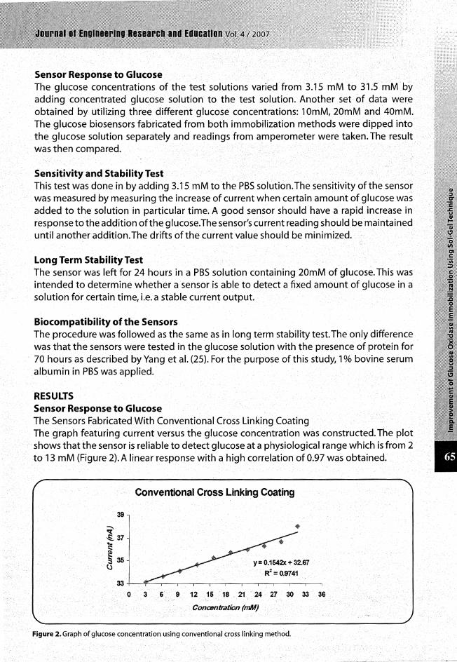
RESULTS Sensor Response to Glucose The Sensors Fabricated With Conventional Cross Linking Coating The graph featuring current versus the glucose concentration was constructed. The plot shows that the sensor is reliable to detect glucose at a physiological range which is from 2 to 13 mM (Figure 2). A linear response with a high correlation of 0.97 was obtained.
39
~ ~ 37
~ !Sss ~
33
0
Conventional Cross Linking Coating
3 G
y = 0.1542x + 32.67
~=0.9741
9 12 15 18 21 24 27 30 33 36
Concehtration (mM}
Figure 2. Graph of glucose concentration using conventional cross linking method.

Another set of experiments using 10 mM, 20 mM and 40 mM were conducted. The second graph for these experiments, shows a steady increase when high glucose concentrations were added to the PBS solution (Figure 3). A linear response with a high correlation of 0.99 was obtained.
11...,------·--------~---------.
-· 1i < E--15 -c e !:::: 14 ... 'U
13
y = 0.1021x + 12..06 R2 =0.9963
12+-------.-------.-------.------,,-----~
0
Figure 3. Graph of 10 mM, 20 mM and 40 mM glucose concentration using conventional cross linking method.
The Sensors Fabricated With Sol Gel Coating Figure 4 is the sensor fabricated by sol gel technique response to glucose concentration. The sensors were reliable to detect particular glucose concentrations with a linear response with a high correlation of 0.99.
13·
1?
1' lt
11
a 10
9
Sol G·el Technique Coating
y : 0;.0<811X "''8.731;3
R 2 : =•0•.993-'11
Figure 4. Graph of glucose concentration using sol gel method utilizing high volume of cross link agent.
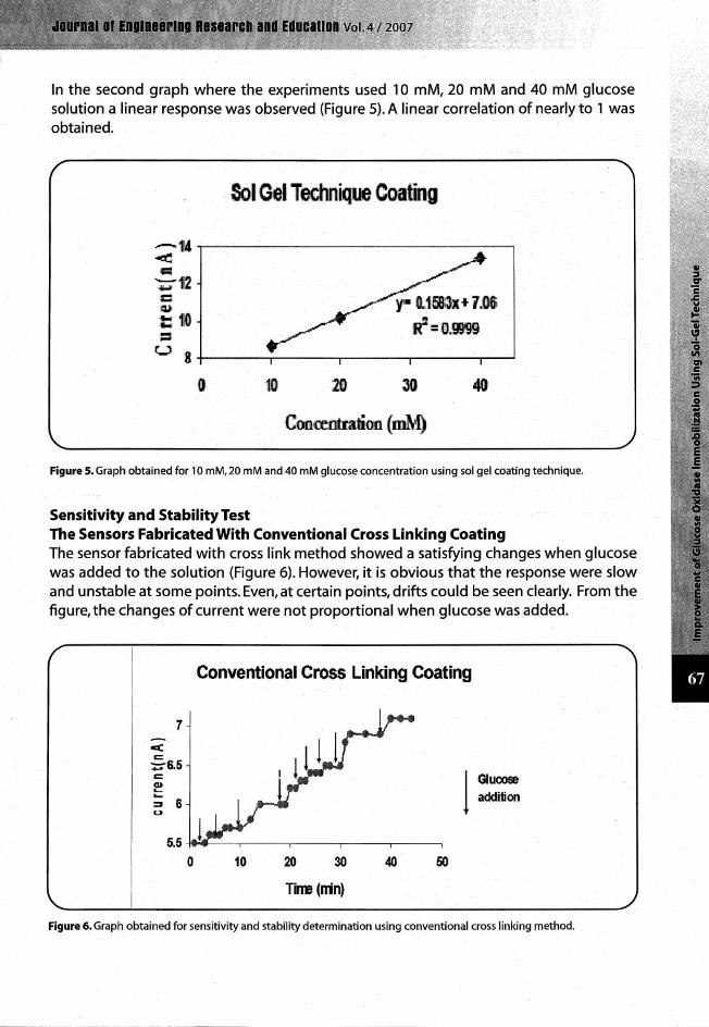
In the second graph where the experiments used 10 mM, 20 mM and 40 mM glucose solution a linear response was observed (Figure 5). A linear correlation of nearly to 1 was obtained.
. Sol Gel Technique Coating
CootCmtratioo (mM)
Figure 5. Graph obtained for 10 mM, 20 mM and 40 mM glucose concentration using sol gel coating technique.
Sensitivity and Stability Test The Sensors Fabricated With Conventional Cross Linking Coating The sensor fabricated with cross link method showed a satisfying changes when glucose was added to the solution (Figure 6). However, it is obvious that the response were slow and unstable at some points. Even, at certain points, drifts could be seen clearly. From the figure, the changes of current were not proportional when glucose was added.
c( c
7
~6.5 c ~ ;; 6 u
Conventional Cross Linking Coating
I Glucose addition
5.5 -f'<IIIOIIIJ----,-----,--------,-----,--------,
0 10 20 30 40 50
Tirre (rrin}
Figure 6. Graph obtained for sensitivity and stability determination using conventional cross linking method.

The Sensors Fabricated With Sol Gel Coating The sensor produced by sol gel technique showed a desirable change when addition of glucose to the test solution. The graph shows that the changes of current due to the change of concentration is nearly linear to be uniform and could be seen as a similar pattern compared to the previous technique (Figure 7).
Sol Get Temmque Cooting
"'c 11 s -= CD - 10 -= u
9 I
Figure 7. Graph obtained for sensitivity and stability determination using sol gel technique coating.
Long Term Stability Test The graph shows a relative response of the system compared to the first reading made (Figure 8). The measurement was performed for 24 hours to find out the long term stability of the sensor when a certain amount of glucose present in the test solution. The dotted line represents the data for the sol gel sensor, while the solid line represents the conventional cross link sensor. From the graph, the sol gel coated sensor showed a lesser decrease compared to conventional cross link coating. After 1 0 hours of measurement, the sol gel system maintained the measurement at the same reading, while the reading values for conventional cross link coating continued to decrease.
/ 1.5 --------------------------------------------
1 ~- /SOl~ ~~----·
conlmJOnaiCrOSSIII1<1{19
0+----.----.----.----~--~--~
0 5 10 15 20 25
Time (hour) 1 , ________ _, __________________________________ ~--------------~/ Figure B. Graphs which shown the relative response of sol gel coating and conventional cross linking coating throughout 24 hours ofthe biosensor in 20 mM glucose concentration.

Long Term Stability Test in Protein Soluti9n-Biocompatibility Test The stability results in protein solutiohwere compared with the results from the previous section for the same period of time i.e. without protein solution. From the first graph, the current detected in the sensor coated by 3C-copolymer for 24 hours did not decrease significantly and the relative response was maintained for more than 90% of its first reading (Figure 9)~
1.2
Cii 1 1: 0
0.8 Sl. U)
e 0.6 ~ ;::1 OA CIS
! 0.2
0 0
Sol Gel +3CCopolymer\
-- -- •' - - ._ - .. -- -
---1-:-------- 'sol Gel
Conventional Crossllnking
5 10 15 20 25 30
Time (hours)
Figure 9. The relative response of sol gel coating with biocompatible layer, sol gel coating alone and conventional cross lirikihg coating throughout iong term stability test (24 hours) ofthe biosensor in 20 mM glucose concentration.
The second graph shows the extended version of the test which lasted for 70 hours. The result is significant where the reading was maintained around 80% of the first reading after the sensor was left for 70 hours (Figure 1 0).
1.2
I 1 c: g_ 0.1 • • .... 0.6 J i OA
• ~ 0.2
0 0
- ........... - .. ~ ....... -f ...,.,___Sal Gel
Oomlenlianal Ctaslllinking
40
nme ~fhoun)
~ .f -. Sal Gel-ti3C-. ~I!'
Figure 10. The relative response of sol gel coating With biocompatible layer, (which is extended for the another 70 hours) sol gel coating alone and conventional cross linking coating throughout long term stability test of the biosensor in 20 mM glucose concentration. ·
-----~---~ ·---------~-------- --- --------,-~---"·---.. -~~-----·-··-----------------, ---
DISCUSSION The results from the sensor response to glucose concentration test exhibit the reliability of both techniques to detect glucose concentration with high linear response. The data obtained from the study also reaffirmed the discovery by Yang et al. 2000. Previously, in which the sensor fabrication was based on the application of glutaraldehyde as the cross link agent. Thus, these sensors' performances have met the basic clinical requirements which are to sense glucose level in physiological ranges.
The sensitivity and stability test had proved that the immobilization of the enzyme was better by sol gel technique compared to the conventional method. We suggested that the enzymes have more freedom to interact with the glucose molecules. Thus, we could predict the increment of current produced proportional to amount of glucose in certain time with more accuracy. And we hope by using this technique, drifts could be minimized if not totally eliminated.
In long term stability test, sensors with sol gel immobilization technique exhibited less decrement in measurement compared with the conventional cross linking method. This happened due to the ability of the technique to retain enzymes. Wu et al. 1999 suggested that the decrement of current measured is more likely due to the denaturation of the enzyme rather than enzyme leaching (4). Thus, the application of sol gel technique might produce a desirable result to lessen the decrement. The bonding between glucose oxidase and silicate through covalent bonding is stronger than cross linking between glucose oxidase molecules, which contribute to the better attachment of the enzyme to the electrode. For the sensors produced by conventional cross link agent, both situations also happened but enzyme leaching is a considerable factor which contributes to the decrease of reading obtained more than the sensors produced by sol gel technique coating. We suggested that the enzyme bonding in this technique is weaker than in the sol gel technique.
The final test is very significant to the aim of this study, which showed an improvement of the glucose biosensor through immobilization method. The sol gel technique combined with 3C-copolymer as biocompatible layer proved to be functionally reliable in protein solution. If the biocompatible layer does not function, the protein adsorption on the membrane's surface would later prevent glucose from penetrating and interacting with glucose oxidase. From the result, the measurement deterioration is nearly negligible for both test, thus, the biocompatible layer had functioned well. This reaffirms the proposal by Yang et al. (2000) that 3C-copolymer is able to suppress protein adsorption and maintain good membrane permeability to glucose for certain period of time (25).
Our discovery shows the combination of a biocompatible layer with sol gel technique to fabricate glucose sensor can lead the sensors to have a high reliability and a good stability up to 70 hours. Enzyme leaching and protein adsorption are two barriers faced by researches in developing in vivo biosensor. Thus, the sol gel technique provides a better enzyme attachment while 3C-copolymer prevents bio-fouling phenomenon from occurring. The improvement of the biosensor performance using sol gel technique and biocompatible layer had been a success if compared to the conventional technique using cross linking method combined with polyurethane layer as the outermost layer.
CONCLUSION The results obtained in this project has confirmed that the sol gel technique is reliable for immobilizing enzymes in biosensor application. The sensitivity and the stability of the sensors improved due to the enhancement of the GOD's ability to interact with glucose while maintaining their immobility in the sol gel matrices. Furthermore, the sol gel technique had proven to lessen the denaturation effect and further suppressed leaching from occurring in glucose solution for 24 hours of time. The sol gel sensors coated with biocompatible layer showed outstanding stability even in protein solution for 70 hours of period. This is due to. the better attachment of the enzyme provided by the sol gel matrices and also the capability of 3C-copolymer to prevent protein adsorption on the membranes surface which would affect the measurement of the sensor.
REFERENCES
1. Egg ins, B.R., Biosensors {1996): an Introduction .John Wilry and Sons, Chichester.
2. Harwood, G.W. J., & Pouton, C.W. {1996): Amperometric enzyme biosensors for the analysis of drugs and metabolites.Advanced Drug Delivery Reviews, 18: 163-191.
"
3. Yao, T., & Takashima, K., {1997): Amperometric biosensor with a composite membrane of sol gel derived enzyme film and electrochemically generated poly {1,2-diaminobenzene) film. Biosensors & Bioelectronics, 13:67-73.
4. Wu, J., Suls, J., & Sansen, W., {1999): Amerometric glucose sensor with enzyme covalently immobilized by sol gel technology.Anafytical Sciences, 15:1029-1032.
5. D'Orazio, P., {2003): Biosensors in clinical chemistry, Clinica Chimica Acta, 334:41-69.
6. Gerard, M., Chaubey, A., and Malhotra, B.D., {2002): Application Of Conducting Polymers To Biosensors, Biosensors and Bioelectronics, 17:345-359.
7. Livage,J., {1997): Sol-gel processes, Current Opinion in Solid State & Matetials Science, 2:132-136.
8. Ratner, B. D., {1995): Surface modification of polymers: chemical, biological and surface analytical challenges, Biosensors & Bioelectronics, 10:797-804.
9. Ramanathan, K., Jonsson B. R., & Danielsson, B., (2001 ): Sol-gel based thermal biosensor for glucose,Anafytica Chimica Acta, 427:1-10.
10. Wang, J., (1999): Sol-gel materials for electrochemical biosensors,Anafytica Chimica Acta, 399:21-27.
11. Kim, M.A., & Lee, W.Y., (2003): Amperometric phenol biosensor based on sol-gel silicate/Nafion composite film,Anafytica Chimica Acta ,479:143-150.
·'~· ~--. -··-------~----·---,--------~-..--.~.-. ------:----·--
12. Braun, S., Shtelzer, S., Rappoport, S., Avnir, D., & Ottolenghi, M., (1992): Biocatalysis by sol-gel entrapped enzymes. journal of Non-C1ystallin~ Solids 147 & 146:739-743.
13. Yamanaka, S.A., Nishida, F., Ellerby, C.R., Dunn, B., Valentine J.S., & Zink, J.l., (1992): Enzymatic activity of glucose oxidase in transparent glass by the sol gel method. Ch~micall\!Iaterials 4:467-500.
14. Tatsu, Y., Yamashita, K., Yamaguchi, M., Yamamura, S., Yamamoto, H., & Yoshikawa, S., (1992): Entrapment of glucose oxidase in silica gel by the sol gel method and its application to glucose sensor. Chemistry Letters 1992: 1615-1618.
15. Narang, U., Prasad, P.N., Bright, F.V., Ramanathan, K., Kumar, N.D., Malhorry, B.D., Kanalasanan, M.N., & Chaodri, S., (1994): Glucose biosensor based on a sol-gel derived platform.Anafytical Chemistry 66:3139-3144.
16. Kunzelmann, U., & Bottcher, H., (1997): Biosensor properties of glucose oxidase immobilized within Si02 gels. Sensors and Actuators B. 38-39:222-228.
17. Ohashi, E., & Karube, 1., (1995): Development of a thin membrane glucose sensor
using P'-type crystalline chitin for implantable biosensor.]oumal of Biotechnology. 40:13-19.
18. Wisniewski, N., Reichert, M., (2000): Methods for reducing biosensor membrane biofouling, Colloids and Suifaces B: Biointeifaces, 18:197~219.
19. Turner, R. F. B., Harrison D. J., & Rojotte, R. V., (1991 ): Preliminary in vivo biocompatibility studies on perfluorosulphonic acid polymer membranes for biosensor applications, Biomaterials, 12:361-368.
20. Moussy, F., Harrison, D.J., O'Brien, D.W., & Rajotte, R.V., (1993): Performance of subcutaneously implanted needle-type glucose sensors employing a noveltrilayer coating.Anafytical Chemistry. 65:2072-2077.
21. Higson, S. P. J., & Vadgama, P.M., (1993): Diamond-like carbon coated microporous polycarbonate as a composite barrier for a glucose enzyme electrode,Anafytica ChimicaActa, 271:1125-133.
22. Ishihara, K., Aragaki, R., Ueda, T., Watenabe, A., & Nakabayashi, N., (1990): Reduced thrombogenicity of polymers having phospholipids polar groups. journal of Biomedical Materials Research 24: 1069-1077.
23. Ishihara, K., Oshida, H., En do, Y., Ueda, T., Watenabe, A., & Nakabayashi, N., (1992): Haemocompatibility of human whole blood on polymers with a phospholipid polar group and its mechanism.journal of Biomedical Materials Research 26: 1543-1552.
24. Ishihara/ K.I Ziatsi N.P.I Tierney/ B.P.I & Nakabayashi1 N.1 (1991 ): Protein adsorption from human plasma is reduced on phospholipid polymers. journal of Biomedical Materials Research 25:1397-1407.
25. Yang/ Y.l Zhang/ S.F.I Kingston/ M.A./ Jones/ G.I Wright/ G.I Spencer, S.A.I (2000): Glucose sensor with improve haemocompatibility.Biosensors & Bioelectronics, 15:221-227.
--------,...-... --~---------- .. -~-------~