improving protein-small molecule structure predictions ......but they played a big role in keeping...
TRANSCRIPT
Improving Protein-Small Molecule Structure Predictions with Ensemble Methods, or
Using Computers to Guess How Tiny Things Fit Together
By
Darwin Yu Fu
Dissertation
Submitted to the Faculty of the
Graduate School of Vanderbilt University
in partial fulfillment of the requirements
for the degree of
DOCTOR OF PHILOSOPHY
in
Chemistry
August 31, 2018
Nashville, Tennessee
Approved:
Professor Jens Meiler
Professor Terry Lybrand
Professor Andes Hess
Professor Tony Capra
DEDICATIONS
I would like to dedicate this work to my family for providing me with this opportunity
to delve into a fascinating world of science. To my parents, Cary and Yao, without whose
sacrifices and risk-taking I would not be here in this country. To my brother, Daniel, for
challenging me and giving me the chance to pass on the lessons I have learned. The three
of you have been incredible supportive and keep me reminded of where I came from and
who I am.
ii

ACKNOWLEDGMENTS
Research is a team effort both inside and outside of the lab. Though my name is on the
front page of this dissertation, the research presented here would not be possible without the
help of a significant number of people. To start, I would like to thank my advisor, Dr. Jens
Meiler, for his mentorship over the course of my graduate career. I remember the first day
in lab when he offered me the choice of working on ”small molecule docking” or ”protein
secondary structure modeling”, and I just picked one and ran with it for six years. I admire
his ability to make deep insightful comments on a project while maintaining the overall
wide picture of research goals. He pushed me to test the limits of existing methodologies,
and this research truly would not be possible without his support and guidance. I would also
like to thank the rest of my thesis committee: Dr. Terry Lybrand, Dr. Andes Hess, and Dr.
Tony Capra for their excellent advice and constructive critique on my work. My qualifying
committee members Dr. Jarrod Smith and Dr. Corey Hopkins were tremendous in making
sure I had a critical understanding of previous literature. I also like to acknowledge my
high school chemistry teacher, Mr. Camacho, for being one of the first to pique my interest
in Chemistry, and my undergraduate research mentor, Dr. Dong Xu, for giving me the
opportunity to play with protein models on the computer.
Much of this work is funded by a PhRMA Foundation Pre-Doctoral Informatics Fel-
lowship and the Molecular Biophysics Training Grant. I like to thank Dr. Walter Chazin for
his organization of the Molecular Biophysics Training Program that introduced me to the
breadth of structural biology, and for his helpful pointers on the basketball court. The in-
clude collaborative projects would not be have been possible without medicinal chemistry
and pharmacology parternships. In particular, I like to thank Dr. Craig Lindsley, Dr. Heidi
Hamm, Dr. Thorsten Berg, Dr. Cody Wenthur, Dr. Karl Voigtritter, Dr. Kayla Temple, Dr.
Matthew Duvernay, and Dr. Shaun Stauffer for their contributions to applications projects
contained in this thesis.
One community I want to acknowledge expressly is RosettaCommons. My first scien-
iii

tific conferences in graduate school was RosettaCon in 2012 and since then I have learned
a great deal about programming and modeling from the community. In particular, Andrew
Leaver-Fay, Steven Lewis, and Jared Adolf-Bryfogle have taught me quite a few insider
tips to Rosetta.
My Meiler lab colleagues, past and present, have been in the trenches with me through
the years. Gordon Lemmon, Sam DeLuca, Rocco Moretti, and Steven Combs helped me
work through many of my RosettaLigand questions. Jeff Mendenhall, Axel Fischer, David
Nannemann, and Jonathan Sheehan helped me with programming tips and scientific dis-
cussions. Amanda Duran, Alberto Cisneros, and Alyssa Lokits have been great colleagues
in lab and good friends outside lab. I thank you for making the lab such a welcoming place,
and helping me celebrate the ups and move past the downs.
The rigors of graduate school would have been tougher if it were not for my friends
outside the lab. My soccer/basketball teammates, and tennis partners may not realize it
but they played a big role in keeping me sane. I like to acknowledge Mackenzie, Rose,
Monika, Carl, Leslie, and Sherri for being great friends, trivia teammates, and graduate
school sounding boards. I also wish to express my gratitude to my non-graduate school
friends, Abigail, Sherry, and Ari, for confirming that there is indeed life outside of the lab.
This seems as good place as any to acknowledge Lucky Bamboo and Corner Asian for
keeping me fed with homestyle Chinese food while I’m in Nashville.
Lastly, a few more thank yous for people whose love and support goes way beyond the
thesis pages. To Megan, my adventuring partner in life, for all the encouragement until
every last word of this dissertation was done. To my brother, for all the things you have
taught me about being patient and kind. To my dad, for all the computer knowledge you
have given me since the days of when I was just playing with floppy disks in your office. To
my mom, for all the times you drove me, both literally and figuratively, to learn about the
world. This would not have been possible without you all and I am excited for the future
both in life and in science.
iv

TABLE OF CONTENTS
Page
LIST OF FIGURES . . . . . . . . . . . . . . . . . . . . . . . . . . . . . . . . . . . ix
SUMMARY . . . . . . . . . . . . . . . . . . . . . . . . . . . . . . . . . . . . . . . xi
1 Introduction . . . . . . . . . . . . . . . . . . . . . . . . . . . . . . . . . . . . . 1
1.1 Summary . . . . . . . . . . . . . . . . . . . . . . . . . . . . . . . . . . . . 1
1.2 Proteins and their small molecule partners . . . . . . . . . . . . . . . . . . 1
1.3 Computational protein - small molecule ligand docking . . . . . . . . . . . 2
1.4 Ligand docking software . . . . . . . . . . . . . . . . . . . . . . . . . . . . 3
1.5 RosettaLigand . . . . . . . . . . . . . . . . . . . . . . . . . . . . . . . . . 4
1.6 Community assessments of docking . . . . . . . . . . . . . . . . . . . . . . 5
1.7 The use of experimental data in docking . . . . . . . . . . . . . . . . . . . 7
1.8 The power of different restraint types . . . . . . . . . . . . . . . . . . . . . 8
1.9 Protein receptor structure-based data . . . . . . . . . . . . . . . . . . . . . 10
1.10 Small molecule ligand-based data . . . . . . . . . . . . . . . . . . . . . . . 12
1.11 Protein-ligand interface-based data . . . . . . . . . . . . . . . . . . . . . . 14
1.12 Similar binding of similar ligands . . . . . . . . . . . . . . . . . . . . . . . 16
1.13 Significantly different binding modes observed in similar ligands . . . . . . 18
1.14 The use of structural ensembles . . . . . . . . . . . . . . . . . . . . . . . . 20
2 RosettaLigandEnsemble . . . . . . . . . . . . . . . . . . . . . . . . . . . . . . . 22
2.1 Summary . . . . . . . . . . . . . . . . . . . . . . . . . . . . . . . . . . . . 22
2.2 Introduction . . . . . . . . . . . . . . . . . . . . . . . . . . . . . . . . . . 22
2.2.1 Ligand docking and structure-based drug discovery . . . . . . . . . . 22
2.2.2 Inconsistent performance of existing protein-ligand docking tools . . . 23
2.2.3 Use of structure ensembles in docking . . . . . . . . . . . . . . . . . 24
v

2.2.4 Incorporating ligand ensemble docking into RosettaLigand . . . . . . 25
2.3 Experimental Methods . . . . . . . . . . . . . . . . . . . . . . . . . . . . . 26
2.3.1 RosettaLigandEnsemble algorithm . . . . . . . . . . . . . . . . . . . 26
2.3.2 Experimental model generation . . . . . . . . . . . . . . . . . . . . . 28
2.4 Results and Discussion . . . . . . . . . . . . . . . . . . . . . . . . . . . . 29
2.4.1 RLE improves sampling and scoring among top models . . . . . . . . 30
2.4.2 RLE eliminates alternate binding modes . . . . . . . . . . . . . . . . 31
2.4.3 Illustrative examples of success and failure . . . . . . . . . . . . . . . 34
2.4.4 Higher chemical similarity promotes higher sampling efficiency up to
a limit . . . . . . . . . . . . . . . . . . . . . . . . . . . . . . . . . . 36
2.4.5 Identifying favorable binding poses corresponding with SAR data . . 38
2.4.6 Comparing RosettaLigandEnsemble with protein-ligand docking tools 39
2.5 Conclusions and Future Directions . . . . . . . . . . . . . . . . . . . . . . 42
2.5.1 Needed improvements in decoy discrimination . . . . . . . . . . . . . 42
2.5.2 Consideration of alternate binding modes among congeneric ligands . 42
2.5.3 Ensemble approaches from protein structure based direction . . . . . 43
3 ROSIE Ligand Docking . . . . . . . . . . . . . . . . . . . . . . . . . . . . . . . 44
3.1 Summary . . . . . . . . . . . . . . . . . . . . . . . . . . . . . . . . . . . . 44
3.2 Introduction . . . . . . . . . . . . . . . . . . . . . . . . . . . . . . . . . . 44
3.3 Experimental Methods . . . . . . . . . . . . . . . . . . . . . . . . . . . . . 46
3.4 Results and Discussion . . . . . . . . . . . . . . . . . . . . . . . . . . . . 47
3.4.1 Inputs for ROSIE ligand docking . . . . . . . . . . . . . . . . . . . . 47
3.4.2 Outputs for ROSIE ligand docking . . . . . . . . . . . . . . . . . . . 49
3.4.3 Information about ROSIE server . . . . . . . . . . . . . . . . . . . . 51
3.4.4 Validation of RosettaLigand algorithm . . . . . . . . . . . . . . . . . 51
3.5 Conclusions and Future Directions . . . . . . . . . . . . . . . . . . . . . . 52
vi

4 Applications of RosettaLigand and RosettaLigandEnsemble . . . . . . . . . . . . 54
4.1 Summary . . . . . . . . . . . . . . . . . . . . . . . . . . . . . . . . . . . . 54
4.2 Introduction . . . . . . . . . . . . . . . . . . . . . . . . . . . . . . . . . . 55
4.2.1 The necessity of comparative models . . . . . . . . . . . . . . . . . . 55
4.2.2 Comparative modeling and docking with Rosetta . . . . . . . . . . . 55
4.2.3 Multi-template comparative modeling in Rosetta . . . . . . . . . . . . 56
4.2.4 STAT proteins . . . . . . . . . . . . . . . . . . . . . . . . . . . . . . 57
4.2.4.1 Structure of erasin and STAT proteins . . . . . . . . . . . . . 57
4.2.5 G-protein coupled receptors . . . . . . . . . . . . . . . . . . . . . . . 58
4.2.5.1 Metabotropic gluatamate receptors . . . . . . . . . . . . . . . 59
4.2.5.2 Allosteric modulation of group II mGluRs . . . . . . . . . . . 60
4.2.5.3 Crystal structures for mGlu receptors . . . . . . . . . . . . . 60
4.2.5.4 Protease activated receptors . . . . . . . . . . . . . . . . . . 61
4.3 Experimental Methods . . . . . . . . . . . . . . . . . . . . . . . . . . . . . 62
4.3.1 Modeling of target proteins . . . . . . . . . . . . . . . . . . . . . . . 62
4.3.1.1 STAT . . . . . . . . . . . . . . . . . . . . . . . . . . . . . . 62
4.3.1.2 mGlu3 . . . . . . . . . . . . . . . . . . . . . . . . . . . . . 63
4.3.1.3 PAR4 . . . . . . . . . . . . . . . . . . . . . . . . . . . . . . 63
4.3.2 Preparing and docking ligands . . . . . . . . . . . . . . . . . . . . . 63
4.3.2.1 STAT . . . . . . . . . . . . . . . . . . . . . . . . . . . . . . 64
4.3.2.2 mGlu3 . . . . . . . . . . . . . . . . . . . . . . . . . . . . . 64
4.3.2.3 PAR4 . . . . . . . . . . . . . . . . . . . . . . . . . . . . . . 65
4.4 Results and Discussion . . . . . . . . . . . . . . . . . . . . . . . . . . . . 65
4.4.1 STAT . . . . . . . . . . . . . . . . . . . . . . . . . . . . . . . . . . 65
4.4.2 mGlu3 . . . . . . . . . . . . . . . . . . . . . . . . . . . . . . . . . . 68
4.4.3 PAR4 . . . . . . . . . . . . . . . . . . . . . . . . . . . . . . . . . . 70
vii
4.5 Conclusion . . . . . . . . . . . . . . . . . . . . . . . . . . . . . . . . . . . 71
4.5.1 STAT . . . . . . . . . . . . . . . . . . . . . . . . . . . . . . . . . . 71
4.5.2 mGlu3 . . . . . . . . . . . . . . . . . . . . . . . . . . . . . . . . . . 71
4.5.3 PAR4 . . . . . . . . . . . . . . . . . . . . . . . . . . . . . . . . . . 72
5 Conclusion . . . . . . . . . . . . . . . . . . . . . . . . . . . . . . . . . . . . . . 73
5.1 Summary . . . . . . . . . . . . . . . . . . . . . . . . . . . . . . . . . . . . 73
5.2 Key findings . . . . . . . . . . . . . . . . . . . . . . . . . . . . . . . . . . 74
5.3 Future Outlook . . . . . . . . . . . . . . . . . . . . . . . . . . . . . . . . . 76
5.3.1 The challenge of small molecule scoring . . . . . . . . . . . . . . . . 76
5.3.2 Applications to virtual screening . . . . . . . . . . . . . . . . . . . . 77
5.3.3 The accessibility of computational predictions . . . . . . . . . . . . . 78
APPENDIX . . . . . . . . . . . . . . . . . . . . . . . . . . . . . . . . . . . . . . . 80
A Description of Datasets . . . . . . . . . . . . . . . . . . . . . . . . . . . . . . . 80
B Protocol Capture for RosettaLigandEnsemble . . . . . . . . . . . . . . . . . . . . 87
C Additional Developments of Ensemble Docking . . . . . . . . . . . . . . . . . . 91
BIBLIOGRAPHY . . . . . . . . . . . . . . . . . . . . . . . . . . . . . . . . . . . 106
viii

LIST OF FIGURES
Figure Page
1.1 Simulated restrained docking sampling efficiency . . . . . . . . . . . . . . 9
1.2 Examples of different types of experimental restraints . . . . . . . . . . . . 10
1.3 Pairwise Small Molecule RMSD vs Tanimoto Similarity . . . . . . . . . . 17
1.4 Binding similarity exception observed in transthyretin . . . . . . . . . . . . 18
1.5 Binding similarity exception observed in CmeR regulator . . . . . . . . . . 19
2.1 Hypothesized mechanism of RLEs sampling advantage . . . . . . . . . . . 26
2.2 Illustration of RLE algorithm . . . . . . . . . . . . . . . . . . . . . . . . . 28
2.3 RLE Sampling and scoring benchmark comparisons for top models . . . . . 30
2.4 RLE and RosettaLigand ligand RMSD distributions . . . . . . . . . . . . . 32
2.5 Illustrative examples of RLE success and failure in recovering a native-like
best scoring model . . . . . . . . . . . . . . . . . . . . . . . . . . . . . . 34
2.6 RLE and RosettaLigand RMSD distributions for HSP90 . . . . . . . . . . . 35
2.7 Tanimoto Similarity versus Property Similarity for RLE dataset . . . . . . . 36
2.8 Sampling efficiency versus PropertySimilarity for top 10 percent scoring
models . . . . . . . . . . . . . . . . . . . . . . . . . . . . . . . . . . . . . 37
2.9 RLE Spearman correction analysis . . . . . . . . . . . . . . . . . . . . . . 39
2.10 Comparison of docking from RLE and AutoDock . . . . . . . . . . . . . . 40
2.11 RLE performance on 18 failure cases from Wang et. al. . . . . . . . . . . . 41
3.1 Sample ROSIE ligand docking input . . . . . . . . . . . . . . . . . . . . . 48
3.2 Sample ROSIE ligand docking output . . . . . . . . . . . . . . . . . . . . 50
4.1 Erasin inhibitory activity . . . . . . . . . . . . . . . . . . . . . . . . . . . 58
4.2 Derivatives of mGlu3 selective NAM VU0463597 . . . . . . . . . . . . . . 61
ix

4.3 Erasin binding assay with STAT3 . . . . . . . . . . . . . . . . . . . . . . . 66
4.4 Comparison of STAT1,3,5 binding . . . . . . . . . . . . . . . . . . . . . . 67
4.5 Ensemble of selective NAMs docked into mGlu3 model . . . . . . . . . . . 69
4.6 Sequence alignment of mGlu proteins . . . . . . . . . . . . . . . . . . . . 70
4.7 Ensemble of antagonists docked into PAR4 model . . . . . . . . . . . . . . 71
A.1 Protein-Ligand systems used to benchmark RosettaLigandEnsemble . . . . 82
A.2 RLE dataset scaffold size versus RMSD . . . . . . . . . . . . . . . . . . . 83
A.3 Sequence and structural similarity of GPCR templates . . . . . . . . . . . . 86
C.1 Datasets for various SAR-guided ensemble docking approaches . . . . . . . 93
C.2 Suggested workflows for ensemble ligand docking approaches in Rosetta . . 95
C.3 A2A receptor dataset . . . . . . . . . . . . . . . . . . . . . . . . . . . . . 96
C.4 Neuropeptide Y1 receptor dataset . . . . . . . . . . . . . . . . . . . . . . . 97
C.5 A2A double ensemble docking results . . . . . . . . . . . . . . . . . . . . 98
C.6 A2A docked ligands in grid view . . . . . . . . . . . . . . . . . . . . . . . 99
C.7 Y1 double ensemble docking results . . . . . . . . . . . . . . . . . . . . . 100
x

SUMMARY
Understanding protein-small molecule interactions is a critical part of exploring pro-
tein structure and function. Protein-ligand docking is the computational method aimed at
predicting if and how a protein and a small molecule interacts. The work presented in this
thesis examines ways to improve upon existing docking methodology through the use of
experimental structure-activity relationships and structural ensembles. The algorithms are
then applied in collaborative efforts towards structure-based drug discovery.
Chapter 1 is an introductory chapter discussing the background of incorporating ex-
perimental information and structural ensembles into protein ligand docking. It contains
material from Bender et. al. ”Protocols for molecular modeling with Rosetta3 and Roset-
taScripts”, a protocols manuscript for which I was an equally contributing first author. I
was responsible for the protein-ligand docking section as well as developing a workshop
tutorial on RosettaLigand docking. There is also material from Fu & Meiler ”The Predic-
tive Power of Different Types of Experimental Restraints in Small Molecule Docking: A
Review”, a review for which I was the sole first author. The datasets analyzed are discussed
in Appendix A.
Chapter 2 details the development of RosettaLigandEnsemble. It contains materials
from Fu & Meiler ”RosettaLigandEnsemble: A Small Molecule Ensemble Driven Dock-
ing Approach” for which I am the sole first author. I programmed and benchmarked Roset-
taLigandEnsemble and wrote the manuscript. Appendix B contains the protocol capture
accompanying this chapter. The congeneric ligand dataset is discussed in Appendix A.
Chapter 2 is based on an in-prep manuscript regarding the improvement to an auto-
mated protein ligand docking server. Further feature additions to the server, located at
http://rosie.rosettacommons.org/ligand docking, is ongoing. I contributed to the server,
drafted the manuscript, and will be an equally contributing first author on the manuscript.
Chapter 4 covers the applications of RosettaLigand and RosettaLigandEnsemble in col-
laborative drug discovery efforts. I made protein modeling and ligand docking models to
xi

support experimental data. The STAT collaboration resulted in a publication ”Development
of Erasin: A Chromone-Based STAT3 Inhibitor Which Induces Apoptosis in Erlotinib-
Resistant Lung Cancer Cells” for which I was a co-author. An additional manuscript from
the STAT project is presently under review. The mGlu3 collaboration is no longer being
pursued in its existing form. The PAR collaboration is an ongoing collaboration likely to
result in a publication at a future time.
Appendix A contains the datasets analyzed in Chapter 1 and 2. There are additional
datasets generated as a common lab resources for future benchmark studies.
Appendix C is an ongoing project extending RosettaLigandEnsemble to include en-
semble docking with protein mutations. The primary algorithm development and proof
of concept study will be submitted as an Arxiv preprint with a peer reviewed manuscript
to follow once an extensive benchmark is complete. Additional features added to Rosetta
will enable future developments in multitarget virtual screening and ensemble docking for
highly dynamic proteins.
xii

Chapter 1
Introduction
1.1 Summary
This chapter introduces protein small molecule docking and provides an overview of its
applications in drug discovery. The feedback loop between computational predictions and
experimental data is critical step in the modeling pipeline. This chapter discusses methods
for feeding wet-lab data into docking simulations, and ways for computational results to
inform further wet-lab experimentation.
This chapter contains material published as Bender et. al. ”Protocols for molecular
modeling with Rosetta3 and RosettaScripts”[1] for which I am an equally contributing
co-first author, and Fu & Meiler ”Predictive Power of Different Types of Experimental
Restraints in Small Molecule Docking: A Review”[2] for which I am the sole first author.
1.2 Proteins and their small molecule partners
It is a truth universally acknowledged, that a single protein in possession of a good
binding surface, must be in want of a ligand. A critical aspect of many proteins’ functions
in nature is the binding of ligands. Ligands range in size from individual ions such as
the magnesium in chlorophyll to large biopolymers such as DNA or RNA. Proteins may
modify a bound ligand, such as in the case of enzymes, or may need it to function, such
as in the case of cofactors or coenzymes. One prominent example of the latter is the role
ascorbic acid plays in making collagen, a structural protein that makes up tissue such as
tendons, ligaments, and skin. A lack of ascorbic acid, or vitamin C, impairs a hydroxylation
step in collagen synthesis resulting in scurvy, a disease marked by joint weakness and skin
bleeding [3]. Conversely, some ligands specifically inhibit protein function. Penicillin
1

binds a protein involved in bacterial cell wall peptide synthesis, resulting in a damaged cell
wall and bacterial cell destruction. A whole class of penicillin derivatives have been created
with anti-microbial properties via protein inhibition [4]. Noted chemist George Scatchard
mused there is such wide variety in the ways proteins interact with ligands that it would be
impossible to try to categorize all the forms of protein-ligand binding[5].
As one might imagine, controlled promotion and inhibition of particular proteins have
tremendous use in medicine and in correcting aberrant protein function. One class of
these protein binding ligands are small molecules, organic compounds larger than ions
but smaller than biologics. According to DrugBank, roughly 70% of approved drugs are
small molecules that bind reversibly to proteins as biological regulators. Lipinkski’s rule
of five provides additional general guidelines to these ”drug-like” molecules including a
molecular weight below 500 Daltons and a limited number of hydrogen bond acceptors
and donors. Generally, it is much easier to experimentally determine ”if” a small molecule
binds to a given protein, then to determine the ”how, where, and why”. The latter questions
require an understanding of molecular structure and the biological process the compound
aids or disrupts. The challenge of elucidating protein-ligand structural questions is one of
the central aims of computational docking.
1.3 Computational protein - small molecule ligand docking
Protein-ligand docking aims to predict computationally the binding interactions be-
tween a protein and a small molecule ligand. This requires a combination of recapitulating
the binding pose and quantifying the interaction strength. Docking is an important step in
the pipeline for structure based design and discovery of small molecule drugs. Successful
prediction of binding position is necessary to delineate critical interactions for improving
selectivity and/or efficacy. Docking interrogates how and why a ligand binds to a pro-
tein. Tangentially, virtual screening and ligand ranking asks if and how strongly a ligand
will bind. Together these techniques have aided advances in lead compound hit discovery
2

and optimization[6]. One of the earliest examples of ligand ”docking” involved building a
scaled model of hemoglobin by hand[7]. A first actual computational docking used shape
complementarity to dock protein surfaces by describing them as knobs and holes. The
study successfully matched two dimers of hemologin but could not dock a trypsin and
an inhibitor because the potential combinations of the two required too much computing
capacity[8].
Computational ligand docking exist in conjunction with traditional wet-lab experi-
ments. As Richard Hamming once noted, ”The purpose of computing is insight, not num-
bers”. Computer models provide hypotheses that must be validated experimentally. The
results of these tests are then fed back to create an iterative cycle of model improvement.
The attraction of computer modeling is the direction it provides towards the efficient use of
wet-lab resources.
1.4 Ligand docking software
Popular docking algorithms such as AutoDOCK[9], DOCK[10], GLIDE[11], GOLD[12],
and RosettaLigand[13], have diverse methods for representing, sampling, and scoring the
molecular interface. Each sampling and scoring setup has its own advantages and disad-
vantages with regards to computational cost and accuracy. Many of these pros and cons are
system or use-case dependent. RosettaLigand is the core algorithm used in this thesis and
will be detailed separately.
For sampling, AutoDOCK[9] and GOLD[12] uses genetic algorithms that generate a
series of trial conformations. Each set of trial conformations are allowed to change before
the ones with the best binding energy is selected in a manner akin to natural selection.
Incremental growth algorithms such as DOCK[10] and GLIDE[11] break the ligand down
into fragments and start placement at an ”anchor”. The molecule is then reconstructed
piece by piece to fill the binding pocket. Monte Carlo methods such as RosettaLigand[13]
make a random perturbation to the protein-ligand model in each step and then accepts the
3

move if it has a more favorable score, or resets it if the move generates a less favorable
score. To avoid being trapped in a local minimum, there is a slight probability that a less
favorable move will be accepted based on the Metropolis criterion.
Scoring functions are divided into three categories: force-field, empirical, and knowledge-
based[14]. Force-field scoring is often referred to as ”physics based” but in reality, docking
algorithms generally simplify calculations or use empirical parameters to reduce the com-
putational cost. Force-field based methods such as MM-PBSA and MM-GBSA may be
used to rescore select docking models previously produced with other score terms[15].
AutoDOCK[9], DOCK[10] both began using an AMBER force field before adding empiri-
cal scoring terms. Empirical score functions generally sum up individual scoring terms over
pairs of specific interactions. For example, the ChemScore function used in GOLD[12] and
GLIDE[11] evaluates all hydrogen bonds and assign favorable scores to ones within a cer-
tain distance and angle range. RosettaLigand[13] uses a knowledge-based scoring function
for many aspects of its protein structure scoring. The scoring function is based on statistics
from the Protein Data Bank regarding the most common distances and angles for protein
features, and awards the best scores to models that match previous data.
1.5 RosettaLigand
RosettaLigand is a protein-small molecule docking application within the Rosetta Macro-
molecular Modeling framework. It is a Metropolis Monte Carlo sampling method paired
with a knowledge based scoring function designed to consider both protein and ligand
flexibility[16, 17]. RosettaLigand uses a two phase docking approach: a low resolution
phase of rapid sampling based on shape complementarity followed by a high resolution
phase of sidechain repacking and backbone minimizing. Ligand flexibility is modeled
through pre-generated conformations[18]. Using default settings, RosettaLigand accounts
for significantly more protein flexibility than comparable methods. As such, the two phase
method allows for many ligand placements to be sampled without being bogged down in
4

the computationally intensive task of optimizing a fully flexible protein structure.
The low resolution phase scores binding poses based on a pre-generated Van der Waals
attraction-repulsion grid. A band of unfavorable repulsive interactions and a band of favor-
able attractive interactions are placed at set distances around a rigid receptor model. Ligand
atoms outside of this scoring shell are considered neutral scoring. Scoring grids can also be
pre-computed based on hydrogen bonding and other non-Van der Waals interactions[18].
The high resolution phase scores models using the Rosetta energy function. As previously
discussed, the energy function uses PDB-derived knowledge-based potentials for scoring
protein features such as Ramachandran angles and sidechain rotamers. Pairwise interac-
tions such as Van der Waals attraction-repulsion, electrostatic interactions, hydrogen bond-
ing, and disulfide bridges are weighted to units of kilocalories per mole. Many of Rosetta’s
interaction potentials are derived from the CHARMM forcefield. Further details of the
Rosetta energy function can be found in Alford et. al.[19]
Features for RosettaLigand are selected and customized through an XML interface[20],
making it easier to add new docking capabilities. A sample of previous RosettaLigand
development include docking with explicit interface water molecules[21], protein-ligand
interface design [22], and rapid screening of ligand libraries[18]. These protocols have
driven an improvement in Rosetta’s docking accuracy in simpler cases as well as expand
RosettaLigand’s applicability in more complex systems. For example, explicit water dock-
ing demonstrated 56 percent recovery for failed docking cases across a diverse benchmark
of 341 structures [21]. RosettaLigand’s performance in community docking benchmarks is
discussed in the following section.
1.6 Community assessments of docking
Ligand docking is commonly assessed by determining if docking programs can pre-
dict the binding mode given an interacting pair of protein and ligand. Depending on the
availability of data, structures of existing homologs or a priori knowledge of the binding
5

site may also be provided. Two related challenges are whether or not programs can rank a
series of binding ligands with a mutual target, or determine if a particular ligand binds to
the given target.
Community assessments of docking software have generally displayed success in re-
covering near-native binding poses. Davis et. al. found that accurate binding poses were
found for all targets in a GlaxoSmithKline compound collection, but the overall success
rate varied dramatically among systems. Furthermore, no algorithm consistently outper-
formed the others across all systems. RosettaLigand generated native-like poses for at least
forty percent of ligands in each target set. RosettaLigand had difficulty in cases with tight
binding deep pockets as the pre-computed ligand conformers could not account for the
precise binding geometry needed[23].
The CSAR 2012 benchmark demonstrated features such as protein structure minimiza-
tion, histidine tautomeric states correction, pre-generated small molecule conformations,
native small molecule training, and substructure based restraints correlated positively with
docking success. However, binding affinity prediction and relative ranking of active small
molecules remains the most challenging aspect in the field and during this experiment in
particular[24]. In the 2015 D3R Grand Challenge involving blinded docking of HSP90 and
MAP4K4 ligands, roughly half of submitted predictions had a near-native model as the best
prediction. Not surprisingly, the most successful workflows superpositioned targets onto
similar ligands instead of sampling large, unrestricted binding space. One observeration
of note was that the same docking software used by different groups produced varied re-
sults depending on the exact parameters for ligand placement and scoring. RosettaLigand
was such a case where performance differed significantly based on the initial alignment
method and the allowed sampling volume [25]. The second iteration of the D3R Grand
Challenge affirmed the broad results of the 2015 benchmark and highlighted the need for
regular blinded assessments to evaluate development[26].
6

1.7 The use of experimental data in docking
A common theme of the previously discussed docking assessments was the benefit af-
forded by relevant experimental data. Experimental data may be straight forward in appli-
cation such as in the case of receptor structures determined by X-ray crystallography; these
structures may be used directly as docking targets. Some experimental data may be more
”fuzzy” as in the case of protein-ligand contact points determined via NMR spectroscopy.
With the proper integrative docking algorithms, these interacting points serve as distance
restraints that can be incorporated as part of the sampling and/or scoring process. Restraints
limit the ways a protein-ligand interface can be constructed, hence reducing the sampling
the sampling complexity and improving the scoring discrimination[24, 16, 25, 27]. The
potential for such soft restraints in hybrid/integrative methods have already been reported
for other aspects of protein modeling, such as the use of cryo-electron microscopy[28] or
electron paramagnetic resonance[29] restraints in protein structure prediction.
Experimental information for guiding ligand docking can be classified as protein recep-
tor structure-based, small molecule ligand-based, or protein-ligand interface-based. Protein
receptor structure-based data provides information about the protein target in the form of an
observed conformation, or knowledge regarding the ligand binding site. Conversely, small
molecule ligand-based indicate the ligand components responsible for interacting with the
target. Protein-ligand interface-based measurements are a combination of the two and di-
rectly identifies a specific protein-small molecule interaction. Although the same experi-
mental technique (ex: NMR spectroscopy) may be used to generate any of the three kinds
of restraints, the computational guidance provided by each data type differs significantly.
The following sections discusses the overall value of experimental restraints, the strengths
and drawbacks of each form of experimental data, and examples of programs/methods that
apply each data type.
7
1.8 The power of different restraint types
In order to demonstrate the different power of the various restraint types, a simulated re-
strained docking benchmark was conducted using RosettaLigand[20] on the PDBBind Core
Subset described in Appendix A.2. Protein receptor structure-based data were represented
by restraining three randomly chosen binding pocket residues contact any small molecule
heavy atom. This is analogous to experimental data that identifies residues whose shifts
change in an NMR experiment upon ligand addition, or whose mutations abolish ligand
binding. Small molecule ligand-based restraints are simulated by requiring three particular
small molecule heavy atoms contact the protein. This is akin to structure-activity rela-
tionships that show binding affinity changes when certain functional groups are swapped
out. Protein-ligand interface-based restraints are created by enforcing contacts on three
randomly chosen pairs of interacting atoms between the receptor and the small molecule.
These types of restraints are derived from experiments such as a double mutant cycle that
correlate a specific protein-ligand contact point. A contact was defined as an interatomic
distance less than 4 , which includes most commonly observed molecular interactions[30].
For each test, the small molecule was initially subject to a random reorientation and transla-
tion within a 5 sphere. An additional test was conducted using minimal initial perturbation
as a representation of using the binding mode of a homologous protein-ligand complex as a
guide to initial placement. This case arises in applications where the a ligand highly similar
to the ligand of interest has a known binding mode with the given protein receptor. 2500
docking trajectories were completed for each protein-small molecule test case under each
restraint condition. The models were analyzed for percentage of native-like small molecule
binding modes using a 2.0 RMSD cutoff.
Figure 1.1 shows the distribution of sampling success rates across test systems for each
of the restraint conditions. The largest improvements are seen in docking with interface-
based restraints and in using molecular similarity to restrict the starting position. This
makes intuitive sense as both restricts the small molecule rotational orientation in additional
8

Figure 1.1: Boxplots of simulated restrained docking sampling efficiency. Each boxplotsshow distribution of percent native-like binding modes observed across the PDBBind Coresubset for each restraint condition.
to its translational location in the binding pocket. Certain interface-based techniques, such
as INPHARMA, specifically work by determining the relative orientation of two similar
small molecules. Reinforcing interatomic distances with a protein point and a ligand point
restricts both the protein side-chain and small molecule conformational flexibility. Figure
1.2 provides examples of each type of restraint. Molecular similarity is discussed later in
this chapter as it is a general assumption about protein-ligand families.
9

Figure 1.2: Receptor-based (blue), small molecule-based (red), and interface-based (green)experimental data. Example programs for particular methods are given in parentheses.
1.9 Protein receptor structure-based data
Structure-based, also referred to as receptor-based data, are derived from observed
changes or effects on the protein alone. Protein structures in the absence of (apo) or in
complex with the small molecule (holo) determined via X-ray crystallography are the most
straightforward form of structure-based data. Small molecules can be directly docked into
receptor crystal structures or, if such structures are unavailable, into homology models.
Docking into holo crystal structures is generally more accurate than docking into apo crys-
tal structures or comparative models[31]. In testing a nitroreductase protein-ligand target
from CASP 11, Huwe et. al. found few dockings to comparative model structures that
were superior to docking to the experimental crystal structure. However, the comparative
10

model docking managed to capture specific contacts 72.7% of the time[32]. Bordogna et.
al. found a Spearmans correlation coefficient of 0.66 between RMSD accuracy of the com-
parative model and the accuracy of the docking simulation for a diverse test set[33]. In
high-throughput docking, or virtual screening, applications, comparative models are capa-
ble of similar enrichment rates as their crystal structure template counterparts[34, 35]. A
common theme across the assessments is that traditional measures of comparative model-
ing ease, such as sequence similarity between template and target, does not correlate with
subsequent docking success. The success rate for docking into homology models can be
improved by up to 70 percent by using holo experimental templates crystallized with small
molecules of similar chemotypes[36]. Careful validation of the input crystal structure, par-
ticularly in regards to proper orientation and placement of the small molecule, should be
performed prior to using the structure in computational drug discovery efforts. Any modi-
fications to the target protein in the crystallization process, including biologically irrelevant
mutations or inserted constructs, should also be considered[37].
Receptor structures may also be derived from nuclear magnetic resonance (NMR) spec-
troscopy. An ensemble of conformations is generally provided to capture the flexibility
observed in structures obtained by NMR spectroscopy. Alternatively, NMR spectroscopy
may be utilized to obtain information on protein-small molecule interface contacts. Chem-
ical shift perturbations[38] are observed for specific residues upon small molecule binding,
while intra protein Nuclear Overhauser Effect (NOE)[39, 40, 41] reflect structural changes
within the protein. Distance restraints derived from these two sources are based on the
assumption that changes are due to interactions with the small molecule. Protein-focused
methods can help define the receptor binding pocket but do not necessarily give informa-
tion on the small molecule binding mode. This type of information generally translate to
a restraint favoring small molecule positons that are within a certain distance of the con-
tact residue[42]. Orts et. al. demonstrated the use of protein-mediated NOE data for two
competitively binding small molecules as a post-docking scoring filter that can improve ac-
11
curacy by two orders of magnitude[40]. Onila et. al. extended this method to directly use
NMR data during docking by simultaneously optimizing poses for both small molecules,
which improved docking in a test set of weakly bound cAMP-dependent protein kinase
complexes. However, the results were highly dependent on obtaining proper orientation
of the protein side chains[39]. Cala et. al. reviews further experimental details for NMR
characterization of protein-small molecule contacts[42].
Other methods for studying localizing small molecule binding site interactions in-
clude hydrogen/deuterium exchange mass spectrometry (HDX-MS) and isothermal titra-
tion calorimetry (ITC) used in conjunction with mutagenesis. HDX-MS relies on the dif-
ferent exchange rates for exposed versus buried amide hydrogen atoms to identify pro-
tein residues covered up by small molecule binders[43]. Mouchlis et. al. uses HDX-MS
on protein backbone amides in conjunction with docking to determine binding modes of
phospholipase A2 inhibitors[44]. ITC is used to measure the thermodynamic components
of binding affinity: enthalpy and entropy. Structural information is inferred from binding
affinity changes following protein mutagenesis or small molecule modification. It is gen-
erally assumed that such changes is due to the impact of the alteration on small molecule
binding[45].
1.10 Small molecule ligand-based data
Small molecule-based information takes advantage of binding data through quantitative
structure-activity relationships (QSAR). QSAR is traditionally part of virtual screening ap-
plications when no receptor information is available. These models generally make use of
2D small molecule property descriptors or 3D small molecule shape fitting without con-
structing a receptor model. Certain docking algorithms can incorporate comparisons of
known small molecule binders in generating putative binding modes. SABRE is a method
that generates a consensus molecular shape density function from multiple bioactive small
molecules. Candidate molecules are then shape fitted using chemical substructures as op-
12

posed to the entire molecule at once[46]. Adherence to experimental SAR can also be used
as a filtering step, though this is tricky as scoring functions generally do not rank order
compounds well. DoMCoSAR is a docking algorithm that selects the most commonly ob-
served binding modes and utilizes correlation with SAR to guide final model selection[47].
Small molecule conformational shape fitting can also be achieved with transfer NOE as
demonstrated in the design of a flexible macrocyclic inhibitor[48].
Ligand based pharmacophore models are an extension of molecular shape fitting by
identifying chemical commonalities among binders of a given receptor. Known ligand
binders of a receptor are aligned as flexible conformers and common features are identified
in 3D space to identify the pharmacophore. This approach can also be performed with pro-
tein side chains to create receptor side pharmacophores[49].One such method PharmDock
converts the receptor and ligand to hydrogen bonding and hydrophobic pharmacophores
before using an alignment algorithm to match pairs. On a test of the PDBBind Core Set,
PharmDock identified a native-like top model 56% of the time when native conformations
were used but only 37% when using Omega generated conformers, signifying the impor-
tance of input pharmacophore conformations[50]. Pharmacophores can be generated in
combination with molecular dynamics to generate an ensemble of binding pocket models
as shown in a development study of ligands for a highly flexible sulfotransferase binding
pocket[51]. Yang et. al. further discuss both ligand and structure-based pharmacophore
modeling along with potential challenges such as dataset construction, molecular align-
ment, and feature selection[49].
Use of SAR and multiple active small molecules in docking simulations is an exciting
area of research. In particular, activity cliffs, highly similar compounds with orders-of-
magnitude differences in potency, provide powerful SAR information. New methodologies
that utilize the experimental knowledge provided by SARs around activity cliffs can guide
the creation of additional structural analogues[52]. The current release of the ChEMBL
database contains over 13 million activities recorded against over 10,000 targets[53]. Fur-
13

thermore, there are system specific small molecule SAR databases for common drug dis-
covery targets such as GLASS for GPCRs[54] and KLIFS for kinases[55]. One potential
way to incorporate this data is to use an ensemble docking method that can simultaneously
optimize multiple protein-small molecule complexes and correlate their calculated scores.
MLSD is an extension of AutoDock4 that allows the simultaneous optimization of multiple
small molecule fragments, though it is restricted to molecules that concurrently bind to the
same target[56]. Mass spectrometry of protein unfolding can also be used to examine mul-
tiple ligand bindings and their combined interactions on protein stability in the gas phase,
though further methods are necessary to translate this to binding modes[57].
In a reverse modality, small molecule information can also be used to generate distance-
dependent pair potentials for protein comparative modeling. The MOBILE program docks
small molecules into a starting ensemble average of homology models, and then gen-
erates restraints for subsequent rounds of model refinement. When used in combina-
tion with MODELLER, MOBILE produced native-like binding pocket geometries in 70%
of test cases, improving results in 60% of cases compared to restraint free comparative
modeling[58].
1.11 Protein-ligand interface-based data
Protein-small molecule interactions are the most powerful combination of the protein
and small molecule information as it can identify specific contact points that can be used in
determining both location and orientation. In testing small molecule docking into G-protein
coupled receptors, Nguyen et. al. demonstrated that sampling efficiency can increase by
an order of magnitude for every ten known protein-small molecule contacts. The gain was
even greater when utilizing more detailed information such as a specific ionic interaction
translated as a 3.0 distance restraint[59].
A protein-small molecule double mutant cycle analysis identifies interactions by com-
paring ITC binding data of a single protein mutation, a single small molecule functional
14

group substitution, and both simultaneously. A substantial non-additive interaction en-
ergy change is evidence for a direct interaction. A collection of these pairwise inter-
actions can be used to derive protein-small molecule distance constraints to incorporate
into the energy gradient docking grid. Roisman et. al. probed 100 potential pairwise
interactions in an interferon-receptor complex and identified five significant interactions.
Docking simulations were run until a converged model was generated satisfying the five
restraints[60]. Blum et. al. utilized double mutant cycle analysis with a nicotine ana-
logue and an acetylcholine receptor backbone amide substitution to show a hydrogen bond
interaction[61]. Similar success have been obtained with double mutant cycle alanine
scanning for a yeast Ste2p GPCR[62], and with an allosteric binding site on a hM1 mus-
carinic receptor complex[63]. Compared to the traditional single site-directed mutagenesis
method, this approach has the potential to differentiate the impact on small molecule bind-
ing from disrupting favorable interactions vs. protein stability.
Another method of directly determining intermolecular distance restraints is with protein-
ligand NOEs. However, this is generally limited by the need to assign resonances for
the protein-small molecule complex. One alternative that relies on matching only small
molecule resonance assignments was developed by Constantine et. al [64]. NMR NOE
experiments can also be used to derive relative orientations between two weakly binding,
competitive small molecules. The INPHARMA technique relies on the transfer of NOEs
between the two small molecules and a common receptor target. This in combination
with a crystal structure of one protein-small molecule complex can be used in determin-
ing the binding of a related small molecule series without assuming binding in a similar
fashion[65].
Small molecule similarity is a type of interface restraint based on the known binding
mode between a related small molecule and the given protein target. The binding mode of
the related molecule can be used as a guide in placement and orientation during docking
simulations. HybridDock is one approach that augments docking with molecular similarity
15

by generating possible binding modes using existing co-crystallized molecules. This sig-
nificantly improved both the binding energy correlation and native binding mode recovery
in a CSAR 2013-2014 test[66]. LigBEnD is a similar approach that uses the co-crystallized
molecule to generate an atomic property force field. Scoring models with this ligand-based
force field correctly predicted 30 out of 36 compounds in the D3R docking challenge[67].
Related binding pockets may be found even among proteins of distinct global folds and
evolutionary history. An analysis of potential enzyme drug targets and evolutionarily dis-
tant proteins in the PDB found similar binding pockets with different global folds in 61%,
10%, and 61% of kinases, phosphatases, and proteases respectively[68].
1.12 Similar binding of similar ligands
One potent form of experimental restraint as shown in figure 1.1 was molecular sim-
ilarity, the assumption that similar ligands will bind in similar fashion to a given target.
Although the relative orientation can be experimentally tested, it is often assumed as a
starting point. The common molecular scaffold is presumed to make similar interactions
with the binding pocket, while peripheral functional group modifications create different
contacts that explain SARs. Previous analysis of 206 protein-small molecule structure pairs
observed similar small molecule binding modes in ninety percent of related structures.
Binding similarity was defined as having an optimized small molecule shape Tanimoto of
greater than 0.8. The receptors in those cases exhibited very similar backbone structures
with the primary differences due to side chain conformation or water architecture[69]. An
examination of scaffold building pairs found that in 41 out of 297 cases the binding mode
changed upon chemical elaboration of a scaffold[70].
Using a subset of the PDBBind Refined Set, described in Appendix A.3, an analysis is
done to show the feasibility of the similarity approach across a broad dataset. The compari-
son of 7298 ligand pairs from 366 targets, shown in Figure fig:Review-Fig3, demonstrated a
significant decrease in RMSD of the common scaffold as the Tanimoto similarity increased.
16

Disparities in binding mode decreases significantly for Tanimoto similarities above 0.6.
Figure 1.3: Pairwise small molecule scaffold nRMSD vs. Tanimoto similarity Inset: Num-ber of pairs and percentage under nRMSD cutoff for each Tanimoto range.
The dataset included 548 pairs where the common substructure is equivalent in size
to the smaller molecule. A small molecule binding mode change (nRMSD >2.0 ) was
observed in 52 cases (9.5%), slightly lower than the percent changed based on volume
overlap comparison used by Malhotra and Karanicolas[70]. Although these cases present
a challenge to using molecular similarity docking, it is generally possible to recover the
correct binding mode in these scenarios. In particular, a number of properties such as
pocket volume and molecular weight of the smaller molecule can be used as predictors of
when these exceptions occur. The much more challenging systems contain similar ligands
presenting in completely opposite binding modes. These exceptions are likely to lead to
docking failures when assuming molecular similarity and are discussed in more detail in
17
the following sections.
1.13 Significantly different binding modes observed in similar ligands
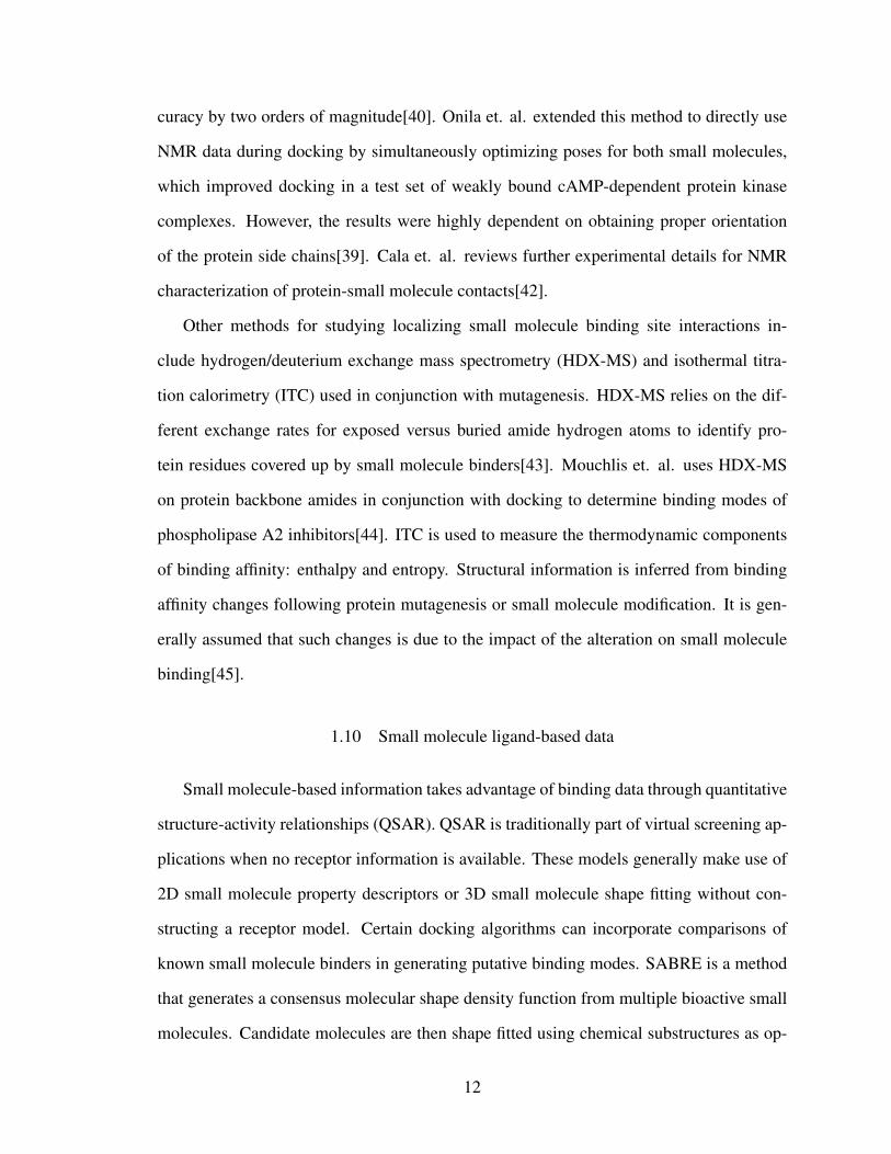
In eleven small molecule pairs, highly similar small molecules (Tanimoto >0.7) ex-
hibited significantly different binding modes (nRMSD >5.0 ). In one particular exam-
ple, a series of diflunisal derivatives exhibited two opposite orientations when bound to
transthyretin, a protein involved in amyloidogenesis. The lead compound diflunisal was
found to bind in a forward and a reverse orientation. More interestingly, a meta-difluoro
derivative (Figure 1.4, left) was found exclusively in the reverse binding mode while an
ortho-difluoro derivative (Figure 1.4, right) was found exclusively in the forward binding
mode[71].
Figure 1.4: meta-difluoro diflunisal derivative (left, PDB: 2B9A) and ortho difluoro diflu-nisal derivative (right, PDB: 2F7I) bound to transthyretin.
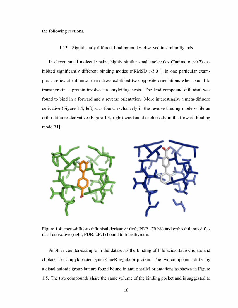
Another counter-example in the dataset is the binding of bile acids, taurocholate and
cholate, to Campylobacter jejuni CmeR regulator protein. The two compounds differ by
a distal anionic group but are found bound in anti-parallel orientations as shown in Figure
1.5. The two compounds share the same volume of the binding pocket and is suggested to
18
interact similarly with a previously identified glycerol binding site. Furthermore, the pocket
is highlighted by a large hydrophobic tunnel with numerous mini-pockets, suggesting a
reason as to why it is capable of binding diverse small molecules in diverse fashions[72].
Figure 1.5: cholate (left, PDB: 3QPS) and taurocholate (right, PDB: 3QQA) bound toCmeR regulator protein.
A number of notable exceptions can be found in literature as well. Structure-based
design of an influenza neuraminidase inhibitor series showed up to 180 degree variation
in the orientation of a central five member ring. Potent analogues were only found for
the congeneric series that bound in the same orientation with consistent SAR[73]. A
study of dipeptidyl peptidase IV inhibitors showed chemically similar small molecules
with different distal aromatic substitutions and placements bind in distinct orientations.
The substituted phenyl ring made pi-pi interactions but with distinct residues in the dif-
ferent cases[74]. Another common exception in systems such as HIV-1-Protease involve
inhibitors bound in two approximately symmetrical orientations[75]. Kim et. al. dis-
cusses a number of other exceptions, such as dihydrofolate reductase and cytochrome c
peroxidase small molecules, that were identified through examination of outliers left out
19

when constructed QSAR datasets. It should be noted that some of the exceptions involve
conformational changes in the distal parts of the small molecule while the main chemical
scaffold remains aligned[76]. It may also be possible in these QSAR datasets that similar
small molecules reside in different conformations of a flexible protein binding pocket. In
such cases, the small molecule orientations remain constant but the protein-small molecule
contacts change[77].
Based on the PDBBind refined set survey, these exceptions are fairly uncommon. Some
features frequently seen in these exceptions include nearly symmetrical molecules, large
binding pockets allowing multiple orientations, and distal groups capable of making favor-
able interactions with different residues in the binding pocket. Unfortunately, there are no
currently known small molecule or receptor structure factors to distinguish exceptions from
regular binders.
Although a similarity based approach towards docking or screening with atoms aligned
by identity may not work in these particular cases, there may be remedies using molecu-
lar properties. A docking method utilizing pharmacophores with properties such as partial
charge or hydrogen bond donor/acceptors can alleviate this problem. Ph4Dock is an ex-
ample where atoms are represented as electrical charge centers without consideration for
identity[78]. Furthermore, similar contact residues are often observed in these situations
allowing for productive suggestions of pairwise interaction validation experiments such as
double mutant cycles.
1.14 The use of structural ensembles
Structural ensembles take advantage of binding similarities by the simultaneous con-
sideration of multiple structures for the protein and/or ligand. An ensemble can be used to
represent different conformations of the same molecule, or a group of related molecules.
Protein receptor ensembles are frequently used to account for receptor flexibility. Rueda
et. al. demonstrated improvement of cross-docking results using binding site ensembles to
20

represent protein flexibility[79]. In particular, ensembles of two or more proteins, enhanced
for proteins co-crystallized with chemically similar small molecules, performed better on
average than single docking or randomly enumerated ensembles[80]. These conformational
ensembles can also be derived computationally using the relaxed complex scheme (RCS),
a series of molecular dynamics simulations to pre-generate low energy conformations[81].
Experimental data can then filter the conformations to avoid docking efficiency decrease
stemming from having a large number of ensembles structures[82]. Sinko et. al. and Feixas
et. al. further discuss RCS and other experimental methods to account for protein flexibility
in drug design applications[83, 84].
One area of further development would be ensemble methods to work with protein
mutants rather than just protein conformations. Such an algorithm would allow for simul-
taneous docking or screening against multiple targets of biological relevance. This could
be beneficial in targeting multiple mutants with a single compound, or in targeting dual
receptors as a replacement for combination therapy. Anighoro et. al. demonstrated the
applicability of dual inhibitor screening for Hsp90 and B-Raf inhibitors, though the com-
putation was performed independently rather than in conjunction[85]. The related multi-
ple ligand simultaneous docking strategy, where fragments are docked individually before
chemically linked, was used to find an inhibitor of STAT3[86]. An ensemble screening
method would score the potential ligands against multiple targets at the same time rather
than as a post-screening analysis.
The work in the remaining chapters develop a number of algorithms necessary to use
molecular similarity and structural ensembles as an aid to protein-ligand docking. Chap-
ter 2 covers the RosettaLigandEnsemble algorithm that emphasizes overlapping binding
modes of similar ligands. Appendix C shows a new docking modality allowing for the
simultaneous consideration of protein and ligand structural ensembles. Chapter 4 applies
these methods for small molecule discovery applications.
21

Chapter 2
RosettaLigandEnsemble
2.1 Summary
This chapter discusses the creation and testing of a new docking algorithm within the
RosettaLigand framework. RosettaLigandEnsemble simultaneously docks an ensemble of
small molecules into a single flexible target receptor. The new method shows a significant
improvement in sampling efficiency over the previously existing RosettaLigand method.
Ligand ensemble docking is a novel approach not previously available in RosettaLigand
or any other popular ligand docking program. The text of this chapter contains material
published as Fu & Meiler ”RosettaLigandEnsemble: A Small Molecule Ensemble Driven
Docking Approach” for which I am the sole first author. I was responsible for developing
the algorithm, writing the code, and benchmarking the new docking features.
2.2 Introduction
2.2.1 Ligand docking and structure-based drug discovery
Structure-based drug discovery and optimization is a critical technique at the intersec-
tion of pharmacology and structural biology. Structure-based computer-aided drug discov-
ery is a powerful way to create hypothesis on ligand binding poses and specific critical
protein/ligand interactions that guide the design of improved small molecules[87]. These
hypothesis can be tested by a variety of experimental approaches including fluorescence
binding studies, calorimetric measurements, NMR spectroscopic studies, or X-ray crys-
tallography. Experimental validation often compares multiple ligands with the wild-type
protein or a mutant target[88]. For computer aided drug discovery to maximize its im-
22

pact on drug discovery, it is necessary for computational ligand docking methodologies to
effectively identify correct protein-ligand binding positions.
Structure-activity relationships (SARs) refer to differences in binding affinity or bio-
logical efficacy following chemical scaffold derivatizations. Medicinal chemistry makes
use of such minor modifications to optimize lead compounds for desired affinity and other
pharmacological properties. This creates a massive wealth of SAR data on related ligands
for a single protein target. The PubChem database alone contains over 200 million bio-
logical activities measurements on approximately 10,000 protein targets[89]. BindingDB
specifically organizes a portion of its database into collections of congeneric ligands with
at least one co-crystallized with the common protein target[90]. It is generally expected
that highly similar ligands form similar interactions when binding to the same target[2].
We hypothesize that a docking algorithm that leverages this information can eliminate a
portion of false positive binding poses, i.e. poses that score well but are incorrect.
2.2.2 Inconsistent performance of existing protein-ligand docking tools
RosettaLigand[17, 16], a small docking tool within the Rosetta structural biology mod-
eling software suite[1], is one of several algorithms developed for this purpose in the last
few decades. AutoDock[9], DOCK[10], and Glide[11] are other popular methods, all of
which differ on both sampling and scoring technique. Performance of these docking tools
are not always consistent across systems. A 2013 docking study using the PDBBind dataset
evaluated scoring functions for decoy discrimination and scoring correlation. The success
rate for identifying correct binding modes from decoys was significantly higher than for
discerning weak, middle, and strong binders within a related ligand series[27]. Similar
results were obtained in the 2012 Community Structure Activity Resource (CSAR) evalu-
ation, which found that even when docking software was able to recover correct binding
poses for a given ligand, few could consistently rank order active ligands[24]. The recent
D3R Grand Challenge reaffirmed these findings and noted that docking performance var-
23

ied even within the same congeneric series. In addition, the overall success of a docking
method was dependent on its preparatory workflow[25]. This performance gap between
docking and ranking is likely due to the steep energy landscape observed near native bind-
ing modes for high affinity protein-ligand complexes. Small perturbations in these regions
generally resulted in drastic scoring changes[91].
2.2.3 Use of structure ensembles in docking
Ensemble methods have traditionally been independently approached from the pro-
tein and ligand sides. Protein ensembles are a common way of capturing conformational
diversity during rigid receptor docking simulations. This need for a structure ensemble
can be due to the inherent flexibility of the protein (conformational selection) and/or due
to an induced fit effect upon ligand binding. Protein structural ensembles can be gener-
ated from experimental determination such as NMR, or through computational methods
such as molecular dynamics. One such preparation is the relaxed complex scheme that
generates a set of receptor targets for docking[81]. To emulate induced fit with ligand
binding, Glide docking can be used to convert all interface residues to alanine to allow
for sampling the binding pocket without bias from initial sidechain orientations[92]. For
scoring purposes, protein ensembles can be handled by an average energy grid that scores
over the ensemble[93], or by using a selection method to identify a single template mid-
simulation[94]. Feixas et. al. and Sinko et. al. further reviews the use of multiple receptor
structures in drug discovery and design[84, 83].
Ligand structural ensembles are used to represent both ligand conformations and phar-
macophore information from multiple ligands. Molecular mechanics or fragment based
sampling can be used to generate conformations prior to docking[95]. Hybrid methods in-
corporate information from multiple ligands to better position a given target. For example,
HybridDock performs pre-docking alignment via pharmacophore matching with similar
molecules[66]. However, these methods require related co-crystal structures to be readily
24
applicable.
It has been observed that using well-chosen structural ensembles is advantageous over
docking with a single structure, particularly when ensemble proteins are co-crystallized
with molecules of similar chemical structure[96, 97]. In this chapter, we developed a two-
stage algorithm for ensemble docking of multiple related ligands into a single protein struc-
ture.
2.2.4 Incorporating ligand ensemble docking into RosettaLigand
RosettaLigand models protein-ligand interactions with full ligand and protein binding
pocket flexibility. This is achieved with pre-generated ligand conformations and protein
side-chain rotamer libraries[17, 16]. RosettaLigand is currently capable of docking mul-
tiple ligands simultaneously, but only in the sense that they bind the protein jointly (e.g.
a small molecule together with a key bridging water molecule or a co-factor with metal
ion bound)[21]. Here, we have extended RosettaLigand to RosettaLigandEnsemble (RLE),
an algorithm that can identify a binding mode favorable to a superimposed ensemble of
congeneric ligands. This allows users to simultaneously dock a series of ligands in unison
instead of individually as single ligands. We hypothesize that this will increase the effi-
ciency and accuracy of sampling. We illustrate RLEs hypothesized sampling advantage in
Figure 2.1.
Due to the presence of functional groups of varying sizes found within a SAR series,
there may be binding modes available to certain molecules but not others. RLE is capable
of eliminating binding orientations not available to the ensemble as whole. Furthermore,
highly similar ligands are expected to bind in similar fashions with common interactions to
the chemical core[69, 2]. The RLE scoring function emphasizes favorable positioning for
the common scaffold, shown by the red outline. The greater number of molecules that share
a common substructure, the greater the scoring emphasis on that particular substructure. It
is not anticipated that RLE will significantly improve docking for congeneric ligands that
25

Figure 2.1: Hypothesized mechanism of RLEs sampling advantage. Top: Three smallmolecules (green) are independently docked by RosettaLigand into the protein bindingpocket (blue). There are multiple docked orientations possible for each small molecule.Bottom: The same three molecules are first aligned using their common scaffold (red).Docking in concert using RLE then yields a single, unambiguous binding orientation.
exhibit significantly different binding modes. Malhotra et. al. reviews receptor and ligand
characteristics that tend to exhibit these alternate binding modes[70].
2.3 Experimental Methods
The validation dataset of 89 protein-ligand cocrystal structures curated across twenty
systems is described in Appendix .
2.3.1 RosettaLigandEnsemble algorithm
Figure 2.2 illustrates the two-stage RLE algorithm. RLE takes as input a single protein
structure and a congeneric series of molecules superimposed by chemical scaffold. In
the low resolution TransformEnsemble phase, the same 3D translations and rotations are
applied to all molecules in order to maintain the superposition and find a common binding
mode. Step sizes and direction for both translation and rotation are taken from a Gaussian
26
distribution centered on a user provided value. Scoring is done using a pre-generated shape
complementarity energy grid and moves are accepted/rejected by a Metropolis Monte Carlo
criterion based on the sum of scores for all ligands in the ensemble. The protein structure
remains static but ligand conformers are changed by swapping out individual ligands with
alternate conformations from pre-generated libraries. The benchmark used the fragment
based BCL::Conf small molecule conformer generator[95]. During the high resolution
HighResEnsemble phase, only small perturbations to the ligand are applied with the focus
on optimizing the protein-ligand interface. Since side-chain orientation differences are
observed even for binding of related ligands, each protein-ligand interface is optimized
independently. In a single simulation run, RLE generates x models where x is the number
of ligands in the ensemble. Over the course of n simulation runs, RLE generates n*x total
models, the same quantity as x independent RosettaLigand runs of n trajectories each.
The bulk of the computation time in both RosettaLigand and RLE is due to protein side-
chain rotamer sampling during the high resolution docking phase. Since RLE generates
individual protein-ligand models for the high resolution stage, computation time is not
significantly altered.
27

Figure 2.2: Illustration of RLE algorithm. The algorithm is separated into the low reso-lution TransformEnsemble step and the high resolution HighResEnsemble step. Curvedarrows represent repeated moves accepted or rejected based on Metropolis Monte Carlocriterion. Individual model of each protein-ligand pair are outputted from a single proteinstructure and superimposed congeneric ligands as input.
2.3.2 Experimental model generation
Initial parameters for RLE are derived from the latest features of RosettaLigand algorithm[18,
20] and optimized for sampling efficiency. Additional sampling cycles and a decreased ro-
tational barrier was necessary to counteract the increased sampling space involved in find-
ing an optimal position for all molecules simultaneously. The exact number of sampling
steps was calculated on-the-fly based on difference between the current step score and the
maximum possible score assuming all atoms formed favorable interactions. Meanwhile,
28

the repulsive score term was halved to allow the entire ensemble to rotate through clashes.
Ligand atoms are forbidden from moving outside of the defined docking sphere as was the
case in RosettaLigand.
Following optimization, docking was performed with both RosettaLigand and RLE and
evaluated for native ligand pose recovery. For each system, individual molecules were
docked independently and as an ensemble into the same receptor structure. For each run,
2500 models were produced and the top ten percent were selected based on ligand interface
energy for subsequent analysis.
In order to make the docking simulation resemble actual use, a uniform volume ran-
dom translation within a 5 sphere and a random full rotational orientation is performed
prior to docking. A random conformer is selected from the ligand conformer library. This
avoids biasing the starting position and orientation to that observed in the crystallographic
complex. An example of how to generate models for one system is provided in Appendix
B.
2.4 Results and Discussion
We examine the top ten percent of scoring models by ligand interface score for each
ligand cross-docking case. The top 250 models are analyzed for both sampling efficiency
and scoring discrimination of native-like models. Native-like models are defined as having
a ligand root mean squared deviation (RMSD) of less than 2 compared to the co-crystal
structure. Sampling efficiency is represented as the percentage of models that are native-
like, while scoring discrimination is represented as the scoring rank of the first native-like
model. A higher sampling percentage of native-like models and a lower scoring rank for
the best scored native-like model indicate improvement.
29

2.4.1 RLE improves sampling and scoring among top models
Among the top ten percent of models by score, RLE improved both the percentage
of native-like models and the scoring rank of the first native-like model when compared
to RosettaLigand. The increased sampling efficiency was observed in 62 out of 89 cases
while the improved scoring rank was seen in 22 out of 89 cases as shown in Figure 2.3. In
three cases, RLE produced a native like model while RosettaLigand did not. In ten cases,
neither RLE nor RosettaLigand were able to find a native-like model in the top ten percent.
Figure 2.3: Comparison of sampling efficiency and scoring discrimination among top tenpercent of models by score from individual RosettaLigand docking versus ensemble RLEdocking. Overlapping dots are indicated by number of overlapped points below it. Bluediagonal line shows when RosettaLigand and RLE performance are identical. A: Percent-age of native-like models from single and ensemble docking B: Scoring rank of the bestscored native-like model from single and ensemble docking C: Small molecule RMSD ofthe top ranked model from single and ensemble docking. The 2.0 angstrom success cutoffis marked out in black lines.
30
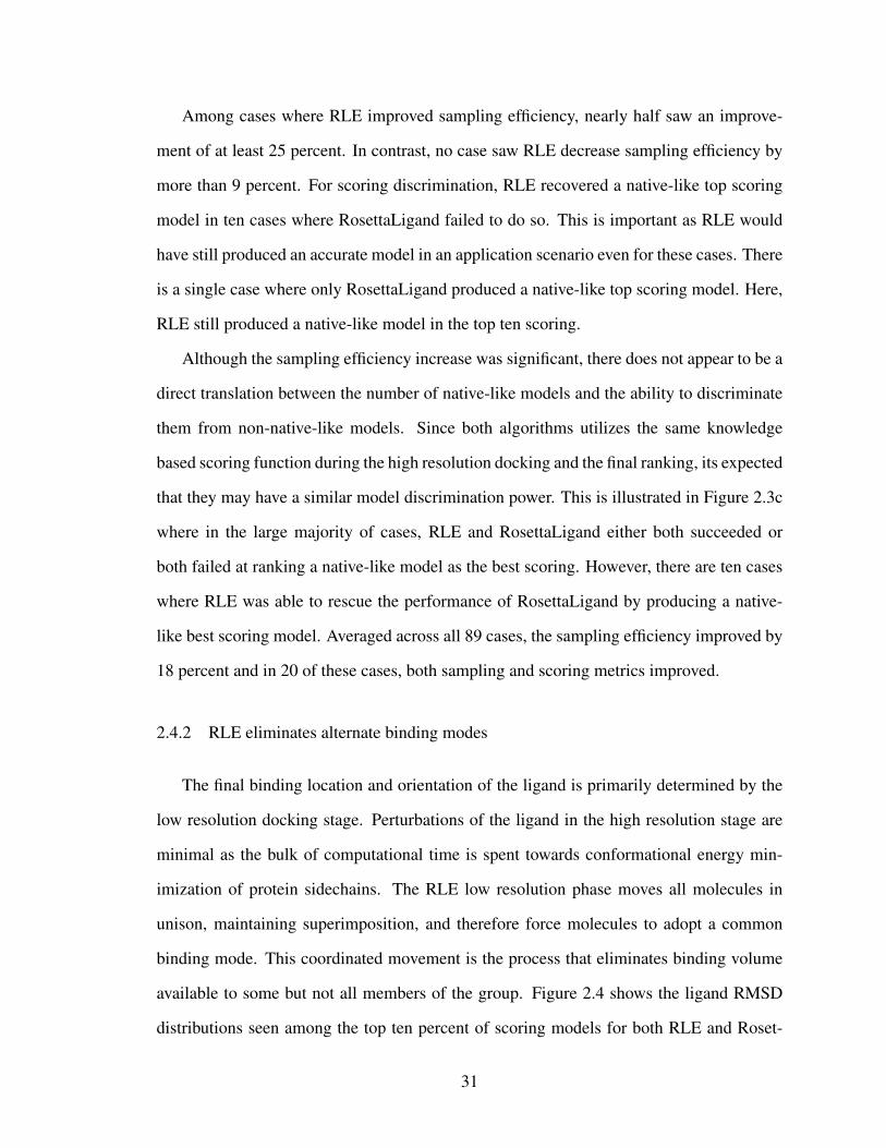
Among cases where RLE improved sampling efficiency, nearly half saw an improve-
ment of at least 25 percent. In contrast, no case saw RLE decrease sampling efficiency by
more than 9 percent. For scoring discrimination, RLE recovered a native-like top scoring
model in ten cases where RosettaLigand failed to do so. This is important as RLE would
have still produced an accurate model in an application scenario even for these cases. There
is a single case where only RosettaLigand produced a native-like top scoring model. Here,
RLE still produced a native-like model in the top ten scoring.
Although the sampling efficiency increase was significant, there does not appear to be a
direct translation between the number of native-like models and the ability to discriminate
them from non-native-like models. Since both algorithms utilizes the same knowledge
based scoring function during the high resolution docking and the final ranking, its expected
that they may have a similar model discrimination power. This is illustrated in Figure 2.3c
where in the large majority of cases, RLE and RosettaLigand either both succeeded or
both failed at ranking a native-like model as the best scoring. However, there are ten cases
where RLE was able to rescue the performance of RosettaLigand by producing a native-
like best scoring model. Averaged across all 89 cases, the sampling efficiency improved by
18 percent and in 20 of these cases, both sampling and scoring metrics improved.
2.4.2 RLE eliminates alternate binding modes
The final binding location and orientation of the ligand is primarily determined by the
low resolution docking stage. Perturbations of the ligand in the high resolution stage are
minimal as the bulk of computational time is spent towards conformational energy min-
imization of protein sidechains. The RLE low resolution phase moves all molecules in
unison, maintaining superimposition, and therefore force molecules to adopt a common
binding mode. This coordinated movement is the process that eliminates binding volume
available to some but not all members of the group. Figure 2.4 shows the ligand RMSD
distributions seen among the top ten percent of scoring models for both RLE and Roset-
31
taLigand docking. Each protein-ligand pair of the system is plotted separately so effects
across the system can be observed. Higher density at the low RMSD end of distribution
indicates success. The red line in each subplot shows the 2 ligand RMSD cutoff for native-
like binding modes. The systems have been sorted qualitatively into broad categories based
on whether or not RLE generally improved both the sampling efficiency and the scoring
discrimination. The RMSD distribution pattern for RLE is much more consistent within
a system than the RosettaLigand distribution patterns for the same system. This is the
aforementioned forced common binding mode effect. However, there remains individual
protein-ligand pairs within a system where the distribution was not significantly improved.
Figure 2.4: Ligand RMSD distribution observed among top 10 percent of models for Roset-taLigand and RLE. Nine example systems have been separated into four qualitative cate-gories of sampling and scoring change. For each system, data is split by individual protein-ligand pair with RLE docking on the left and RosettaLigand on the right. Black line showsthe median and red line shows the 2 cutoff. A: RLE improved docking sampling and scor-ing for CTAP, HCV, and TPPHO (left, mid, right). B: RLE improvement varied from ligandto ligand within system for CDK2 and P38 (left, right). C: RosettaLigand performed betterfor LPXC and THROM (left, right). D: Both methods failed to perform well for CATB andTHERM (top, bottom.
In the systems where RLE drives both a sampling efficiency and a scoring discrimi-
32

nation improvement (green), RLE eliminated a significant number of high RMSD binding
modes seen in the RosettaLigand results. In the CTAP example, RLE ligand RMSDs are
all within a similar range while the outliers produced by RosettaLigand are eliminated. It
remains possible for ensemble docking to be more successful for certain ligands within
group than others. Ligand C for CTAP has a smaller second peak that is not consistently
eliminated by ensemble docking. One reason for this is because the high resolution stage
considers ligand conformers in additional to protein conformers. For larger, more flexible
molecules, RMSD may be relatively high even if the correct binding location and orien-
tation is recovered. This is due to ligand conformational flexibility in the distal regions.
Alternatively, in the HCV example, the majority of models from both RosettaLigand and
RLE are not native like but only RLE generates a batch of native-like models. This is the
aforementioned rescue scenario in which RLE is able to produce a correct model when
RosettaLigand cannot.
A limitation to the RLE algorithm occurs when the alternate, high RMSD binding mode
is available to all molecules within a system, as seen in the P38 system with mixed results
(orange). RLE does not provide a significant advantage in scoring discrimination when
both methods have a similar sampling efficiency. The emphasis on placement of the com-
mon scaffold means that an incorrectly identified common binding mode will result in poor
performance across the system as seen in THROM (blue). RosettaLigand was able to pro-
duce good results for two members of this system because its docking runs are independent.
Whether or not the different binding modes will be correctly consolidated in an actual ap-
plication depends on the particular post-hoc analysis chosen. This incorrect placement of
the common scaffold is repeated in the CATB and THERM systems where the distribution
peaks fall out of the native-like RMSD range (red). One reason for this is the lack of chem-
ical diversity in the functional group modifications within the group. These systems are
difficult cases that neither algorithm can dock well.
33
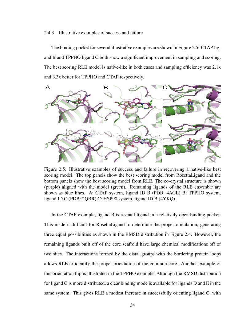
2.4.3 Illustrative examples of success and failure
The binding pocket for several illustrative examples are shown in Figure 2.5. CTAP lig-
and B and TPPHO ligand C both show a significant improvement in sampling and scoring.
The best scoring RLE model is native-like in both cases and sampling efficiency was 2.1x
and 3.3x better for TPPHO and CTAP respectively.
Figure 2.5: Illustrative examples of success and failure in recovering a native-like bestscoring model. The top panels show the best scoring model from RosettaLigand and thebottom panels show the best scoring model from RLE. The co-crystal structure is shown(purple) aligned with the model (green). Remaining ligands of the RLE ensemble areshown as blue lines. A: CTAP system, ligand ID B (PDB: 4AGL) B: TPPHO system,ligand ID C (PDB: 2QBR) C: HSP90 system, ligand ID B (4YKQ).
In the CTAP example, ligand B is a small ligand in a relatively open binding pocket.
This made it difficult for RosettaLigand to determine the proper orientation, generating
three equal possibilities as shown in the RMSD distribution in Figure 2.4. However, the
remaining ligands built off of the core scaffold have large chemical modifications off of
two sites. The interactions formed by the distal groups with the bordering protein loops
allows RLE to identify the proper orientation of the common core. Another example of
this orientation flip is illustrated in the TPPHO example. Although the RMSD distribution
for ligand C is more distributed, a clear binding mode is available for ligands D and E in the
same system. This gives RLE a modest increase in successfully orienting ligand C, with
34

the major deviation due to conformation rather than orientation.
Figure 2.6: Ligand RMSD distribution observed among top 10 percent of HSP90 modelsfor RosettaLigand and RLE. RLE docking distribution is on the left and RosettaLigand onthe right. Five line summary shows highest, lowest, median, and quartiles.
Although RLE showed a slight sampling improvement for HSP90, the system proved
to be a relatively challenging case for ensemble docking due to the small size of the ligand
system. RosettaLigand produced a native-like best scoring pose while RLE generated a
flipped conformation. The RMSD distribution however favors RLE with more native-like
models. Across the system, there is a persistent alternate binding mode suggested by RLE,
as seen in Figure 2.6, due to the fact that the binding pocket is much larger than the ligand.
RLE is unable to rule out alternative binding modes of the common scaffold without distal
groups that can eliminate conformational space.
35

2.4.4 Higher chemical similarity promotes higher sampling efficiency up to a limit
In order to better understand indicators of successful and unsuccessful systems, we
sought to characterize the similarity of the cross-dock small molecules compared to the
co-crystallized molecule. The traditional Tanimoto similarity coefficient is not particularly
robust for more complex substitutions as it focuses on atom identity within a common sub-
structure. We compared molecules using the in-house BioChemicalLibrary to calculate a
PropertySimilarity[59]. PropertySimilarity measures similarity based on atomic charges,
Van der Waals volume, bond types, and the presence of hydrogen bond donors/acceptors.
For this dataset, PropertySimilarity has a general positive correlation with Tanimoto simi-
larity as shown in Figure 2.7.
Figure 2.7: Tanimoto Similarity versus Property Similarity for 89 cross-docking test cases.Each molecule is compared to the ligand co-crystallized with the receptor structure for thesystem it belongs to.
The relationship between sampling and scoring improvement to property similarity is
shown in Figure 2.8. Ligands are classified based on whether both sampling and scoring im-
proved (red), both worsened (blue), or a mix of the two (white). There is a general tendency
for molecules docked with high sampling efficiency to have a high chemical similarity to
36

the co-crystallized molecule. However, there are a number of highly related molecules in
Figure 2.8 that are poorly sampled, suggesting that chemical similarity is a necessary but
not sufficient condition of docking success. This is in agreement with previous studies that
have shown docking success increases with chemical similarity[80, 96].
Figure 2.8: Sampling efficiency versus PropertySimilarity for top 10 percent scoring mod-els. Protein-Ligand pairs are divided into cases where RLE improved both sampling andscoring (red), worsened both sampling and scoring (white), or improved one but not theother (blue). A: PropertySimilarity of each test ligand to the ligand co-crystallized with re-ceptor structure vs the improvement in RLE docking as calculated by RLE percent native-like minus RosettaLigand percent native-like. B: The mean PropertySimilarity for eachprotein system vs. the improvement in RLE docking. Each vertical set of dots represents asingle protein-ligand system.
In order to predict performance on a system level, we computed the mean PropertySim-
ilarity for each system and plotted this value against each ligands improvement in sampling
efficiency when docked with RLE. This is shown in Figure 2.8 with each vertical line com-
prising of a congeneric set of protein-ligand pairs.
The largest improvement falls around a mean PropertySimilarity measure of 0.8, sug-
gesting that there is a sweet spot for improvement. Systems that are too different (P38,
mean=0.37) or too similar (THERM, mean=0.97) exhibit limited benefits from ensemble
docking. In particular, the THERM system consists of a chemical scaffold to which the
primary modification is the switching of various hydrocarbon groups. Furthermore, the
molecule interacted with two separate hydrophobic ends and a network of water molecules
37

in the binding account, which makes orientation determination difficult[98].
2.4.5 Identifying favorable binding poses corresponding with SAR data
The inaccuracy of ranking despite accurate docking remains problematic. One post-
hoc solution is to select sets of binding modes that correlated with experimental data. We
sought to address this deficiency during docking by adding a corrective factor to drive
high resolution docking towards binding modes. Following each cycle of optimization,
we modified the scoring difference based on the Spearman correlation to experimental
data as an adjustment prior to applying the Metropolis criterion. The adjustment provides
an additional bonus to perturbations that improved the score of stronger binding or more
active ligands, and to perturbations that worsened the score of weaker binding or less active
ligands. The low resolution docking stage remains the same and does not account for
experimental correlations. The adjusted co-dependent algorithm is shown in Figure 2.9a
with the score adjustment being applied in the highlighted step.
Although the corrective factor does improve correlation with experimental affinity, it
does not improve the sampling efficiency or docking accuracy. This is in part due to the fact
that the binding orientation is primarily determined in the low resolution phase that does
not account for correlation. The Spearman correlation coefficient is defined as the Pearson
correlation based on only the ranks of the models. Therefore, the Spearman correlation
has a discrete distribution with limited values available for a small data set. This makes it
difficult to significantly improve the correlation in many cases. The results are in agreement
with previous results showing that improvements in the Pearson scoring correlation using
machine learning based scoring functions only translated to a moderate increase in accurate
ranking[99].
One additional hindrance to a more successful corrective method is the dilemma in
selecting models illustrated in Figure 2.9b. In order to maintain the experimental correla-
tion, entire ensembles of ligand models must be selected. However, since the Monte Carlo
38

Figure 2.9: RLE Spearman correction during high resolution docking to favor bindingmodes that correlate with experimental data. A: Spearman corrected RLE high resolutiondocking steps shown in red B: Model selection dilemma resulting from inaccuracies indocking scoring function C,D: Distribution of Spearman correlation in generated ensemblesfor each system with a corrective factor (left, purple) and without (right, blue). The meanline is shown in black.
sampling method is stochastic, it is unlikely that each ensemble will contain low energy
conformations of every protein-ligand interface. Selecting models by a mean metric across
the entire ensemble may select the best scoring models for one ligand but not for others.
Even with improvements in scoring functions, this selection dilemma may prevent RLE
from simultaneously selecting the best models for each ligand.
2.4.6 Comparing RosettaLigandEnsemble with protein-ligand docking tools
We used AutoDock[9] with a Lamarckian Genetic Algorithm to test its performance
in the 89 cross-docking systems in the benchmark set. Standard protocol settings and
39

a docking volume comparable to RLE docking were used to generate the models. The
AutoDock simulations were performed using a rigid receptor model.
Figure 2.10: Small molecule RMSD of the top ranked model from RLE and AutoDockdocking. The 2.0 angstrom success cutoff is marked out in black lines. Equal performanceof the two software is indicated by the blue line.
Figure 2.10 shows the small molecule RMSD of the top scoring model from RLE and
AutoDock docking. RLE recovered a native-like small molecule pose in 23 cases where
AutoDock did not. By contrast, there were only 13 cases where AutoDock had a native-like
best scoring model when RLE did not.
Wang et. al. evaluated ten docking software across the PDBBind dataset, including 18
cross-docking cases from the present benchmark in which at least one tested method did
not recover a native-like top scoring model[100]. RLE rescued the performance in 15 out
of 18 of the cases in Figure 2.11.
40

Figure 2.11: RosettaLigandEnsemble performance on 18 failure cases from Wang et. al.The table shows small molecule RMSD of the top ranked models from RLE and ten otherdocking software. All results except for RLE were pulled from Wang et. al.
Most notably, RLE was able to generate native-like models across the calcium-dependent
protein kinase CDPK1 (CALM) and Helicobacter pylori nucleosidase (MTAN) systems.
However, the 2B07 test case from a series of protein tyrosine phosphatase inhibitors (TP-
PHO) remains challenging. This is likely related to the orientation flip discussed with
regards to Figure 2.5. RLE was also able to recover native like top scoring models for all
five spleen tyrosine kinase (SYK) compounds also tested in CSAR 2014, matching the per-
formance of the best available docking tools.[101] However, RLE performed worse on an
deacetylase (LPXC) test system that was part of CSAR 2012[24], only recovering a near
native model in 1 out of 4 cases. The generated best scoring models had a RMSD of 2.23
in the worst test case, suggesting only a minor performance decrease in the LPXC system.
It should be noted that these comparisons do not account for additional protein flexibili-
ties accounted for by RLE, nor does it include the effects of differences in starting ligand
conformation. However, there does not appear to be a strong induced fit or conformational
selection component in these structures.
41

2.5 Conclusions and Future Directions
2.5.1 Needed improvements in decoy discrimination
The improved sampling efficiency did not directly translate into improved scoring rank-
ing partly due to the inaccuracies in discriminating between native-like and non native-like
models. Better decoy discrimination in conjunction with the more efficient sampling will
allow for fewer models to be produced before converging on a native like binding mode.
The reduced number of models will greatly reduce the time and computational resources
necessary for docking. Furthermore, the SAR correlated docking would benefit greatly
from a more accurate scoring function capable of ranking ligands. RLE in combination
with such a method would generate binding modes in accordance with SAR data without
the need for post-hoc filtering.
2.5.2 Consideration of alternate binding modes among congeneric ligands
RLE docking is generally designed for docking in cases where similar ligands exhibit
a common binding mode. This is the case for the vast majority of known protein-ligand
crystallographic complexes[2, 70]. Presently, a priori assumptions are made for a given
system, even if single ligand docking is used as initial placement is often based on previ-
ously seen binding modes. A future development of RLE docking would allow for minor
shifts in the binding mode while maintaining the general placement and orientation, a sort
of soft ensemble docking. Furthermore, the use of a property based alignment method such
as PropertySimilarity will allow for common scaffolds based on chemical similarity as op-
posed to identity. Cases wherein similar ligands bind in completely different pockets or to
different protein conformations will remain challenging for ensemble based methods.
42

2.5.3 Ensemble approaches from protein structure based direction
A similar approach can be used to drive ensemble docking improvements in the use
of protein mutation data. Current approaches to protein ensembles generally focus on
accounting for conformational diversity. Mutational data on proteins is used to identify
potential protein-ligand interaction sites as a distance restraint to docking. An alternate
ensemble approach would utilize SARs based on multiple protein mutants to determine
how the ligands may bind to each mutant within the series. A further step would be in
combining protein ensemble and ligand ensemble methods to improve docking accuracy
by considering how ligand modifications fit into the different pockets of protein mutants.
Multi-target virtual screening, in particular with biologically relevant mutants, can be per-
formed with such an algorithm. The Platinum database of small molecule interactions with
protein mutants[102] provides an excellent source of data for training an algorithm in this
approach.
43

Chapter 3
ROSIE Ligand Docking
3.1 Summary
This chapter discusses the upgrade of protein-small molecule docking server for use by
non-computational focused researchers interested in modeling a protein-ligand interface.
The server was updated to use the improved RosettaLigand docking algorithm described in
DeLuca et. al. [18]. Further additions such as the inclusion of a small molecule conformer
generator were made with the goal of reducing manual preparation steps necessary for a
docking submission.
The work in this chapter is ongoing but the bulk portion will be submitted as Fu et. al.
”ROSIE Ligand Docking: A Protein-Small Molecule Docking Server” for which I am an
equally contributing first author.
3.2 Introduction
Many proteins function by interaction with endogenous small molecule ligands. Small
molecule therapeutics account also for the majority number of FDA-approved medications[103].
Therefore, understanding the function of proteins as well as developing of novel drug can-
didates requires a structural understanding of binding interactions between a protein and
a small molecule. Atomic level insights regarding these interactions from structures of
protein-ligand complexes determined by X-ray crystallography are ideal for this purpose,
but experimental structures can be challenging and costly to obtain. Computational protein-
ligand docking aims to provide a full-atom model of the bound protein-small molecule
complex using existing structures of the individual partners as the starting point. Recent
advances and applications in small molecule docking and drug discovery have been dis-
44

cussed extensively[104, 87].
A number of protein-ligand docking servers are presently available. SwissDock[105]
is a server based on EADock DSS, an evolutionary algorithm, while MEDock[106] uses a
novel optimization algorithm that works well with more rugged energy landscapes. PatchDock[107]
is a geometry based method focused on shape complementarity. There are also standalone
docking tools such as NRGsuite, which integrates with PyMOL to allow users to setup
real-time simulations on their own machines[108]. While some structure-based virtual
screening servers can also perform docking, these tend to focus on speed for docking large
libraries rather than accuracy for recapitulating a single protein-ligand interface. Other
small molecule docking methods generally require the user to provide computational re-
sources (e.g. AutoDock[9]) or a fee for usage (e.g. DockingServer[109]).
RosettaLigand[16, 17] is a well-established small molecule docking protocol within
the Rosetta biochemical modeling software suite[1, 110]. RosettaLigand uses shape com-
plementarity to sample reasonable binding modes and then performs high resolution re-
finement using a knowledge based scoring function. The algorithm treats the ligand and
surrounding protein residues, both backbone and side-chain, as fully flexible during the
refinement process. Hundreds of independent simulations can be quickly generated by
RosettaLigand running on a cluster.
There are a few limitations of the existing implementation of the RosettaLigand ap-
plication: 1) Rosetta primarily utilizes a command line interface, 2) Rosetta has specific
input formats for protocol scripts and ligand parameter files, 3) Rosetta has a large variety
of options for customizing the docking process, and 4) analysis of outputs often require
additional scripting. While the ability to customize protocols is an advantage to the experi-
enced user, it may be overwhelming for newcomers to protein-ligand docking. In order to
overcome these challenges, we have developed the ligand docking protocol for the Rosetta
Online Server that Includes Everyone (ROSIE)[111].
45
3.3 Experimental Methods
ROSIE Ligand Docking utilizes a two stage approach for predicting the binding mode
between a small molecule ligand and the protein target. A low resolution first stage iden-
tifies likely binding modes based on geometric complementarity of the ligand with the
protein binding pocket. A rigid protein model is used to generate an attraction/repulsion
grid based on hard spheres. The ligand is then allowed to translate, rotate, and change con-
formations in the docking volume with moves accepted or rejected based on a Monte Carlo
Metropolis (MCM) criterion[18]. By default, the grid is 15 wide but users may increase
the search space up to 30 .
The preliminary model from the low resolution stage is passed on to a high resolution
MCM stage with a full-atom knowledge-based scoring function. Small perturbations are
made to the ligand position followed by rotamer library sampling of local protein residues.
Local residues are dynamically defined as residues within 7 of the ligand in addition to
sequentially adjacent residues to allow for backbone flexibility. The restriction of protein
flexibility to interface residues makes modeling of large proteins tractable. A soft-repulsive
energy minimization allows residues to sample conformations beyond the rotamer library.
Following six cycles of MCM docking, a final hard-repulsive gradient minimization is
used to optimize ligand and receptor torsion angles, both backbone and sidechain. The
score difference between the docked protein-ligand complex and the separated protein-
ligand pair is reported in Rosetta Energy Units as the interface delta score, an emulation
of binding free energy change. The models with the top ten most negative interface delta
scores are presented as the best models[16].
The detailed terms of the high resolution scoring function have been covered elsewhere[112,
17, 113]. Key terms included in the ROSIE Ligand Docking score function are 1) Lennard-
Jones attraction/repulsion potentials, 2) Lazardis-Karplus implicit solvation, 3) hydrogen
bonding potential, and 4) empirically derived rotamer and phi-psi angle probability scores.
In addition, the ligand docking specific scoring function includes the above terms and elec-
46

trostatic terms for the ligand with adjusted weights benchmarked on 100 native protein-
ligand complexes[17].
3.4 Results and Discussion
The following describes the ROSIE Ligand Docking server setup including a descrip-
tion of the input and output process. Previous validation of the underlying RosettaLigand
algorithm is also discussed.
3.4.1 Inputs for ROSIE ligand docking
A structure of the protein target in PDB format is required. This structure can be derived
from experimental or computational methods. The ligand molecule should be provided as
a single SDF-formatted file. The user may choose to supply within this file all ligand
conformers to be considered during docking, or select for ROSIE to generate conformers
using the BioChemicalLibrarys knowledge based rotamer library sampling algorithm[95].
ROSIE Ligand Docking does not perform binding site detection and thus an approximate
starting location for the ligand is necessary. The starting location can be provided by setting
the ligand in the binding pocket prior to uploading the SDF file and checking the Use the
starting coordinates in the SDF option. Alternatively, the user can provide the starting point
as X, Y, Z coordinates. In this case, the server will automatically translate the centroid, cal-
culated by averaging the coordinates of all non-hydrogen ligand atoms, to the provided
coordinates. The automated conformer generation and use of SDF coordinates were not
available in previous implementations of the server. For more information on X, Y, Z coor-
dinates in the PDB format, consult the HETATM description on page 190 of the PDB format
guide (ftp://ftp.wwpdb.org/pub/pdb/doc/format descriptions/Format v33 Letter.pdf). Ad-
ditional information on the SDF file, an extension of the MDL Molfile format, can be found
at https://en.wikipedia.org/wiki/Chemical table file. In particular, the first block provides
the coordinates used by the server to define the ligand position.
47

In cases where the approximate binding site is unknown, SiteHound-web[114] can
be used to identify potential ligand binding pockets. The center coordinates output by
SiteHound-web can directly be used as the starting X, Y, Z coordinates for ROSIE Ligand
Docking.
Figure 3.1: Screenshot of input screen for submitting a new ROSIE Ligand Docking job
Default sampling parameters that work well for most cases are provided, but may be
adjusted under Advanced Settings to modify search radius, step size, or step count. Users
do not have to modify any parameters under Advanced Settings. For a larger binding
pocket, users may wish to perform more extensive sampling by increasing the Maximum
48

radius to search, Size of the low-resolution grid, Size of the low-resolution translation,
Number of Monte Carlo sampling steps, and Randomize the initial position parameters.
Conversely, if the likely binding location is better defined through experimental data, the
user may wish to decrease these parameters for a limited refinement docking. There are
further options for customizing the number of high resolution protein backbone and side-
chain optimization cycles. Optional parameters allow the user to specify job title, job
description, and email address for notifications. Users should consult the documentation
for additional details and usage examples. Figure 3.1 shows an example input screen for
docking eticlopride to human dopamine receptor 3 (PDB: 3PBL).
3.4.2 Outputs for ROSIE ligand docking
Figure 3.2 illustrates the typical output from a single run of ROSIE Ligand Docking.
The output is displayed in the Results section in the form of images and file links for the top
ten best scoring models. An interface score versus total score plot is available as a quick
visualization of scored models. A score table is provided in the standard Rosetta score
file formatting, where each row represents a single, independent model and each column
represents a score term.
The primary scoring term used to evaluate ligand docking models is interface delta,
which is located in the second column. This value is calculated in Rosetta Energy Units and
is meant to emulate the binding energy of the ligand, with more negative values being better.
In contrast, the total score evaluates the protein-ligand complex in its entirety, including the
unmodified protein residues outside of 7 protein-ligand interface region. If the user wishes
to sort by alternative scoring terms, the user may simply click on the column header for the
desired scoring term. The server enables downloading of all input and output as a single
TAR archive or individually through custom web-links. Summaries are also provided in
JSON format for use with database processing. The job submission parameters used to
generate results are also available.
49

Figure 3.2: ROSIE Ligand Docking sample results page generated by docking eticloprideto human dopamine receptor 3. The output page includes the top ten scoring models gen-erated, a score plot, a sortable score table, and download links to all files.
50
3.4.3 Information about ROSIE server
ROSIE Ligand Docking has completed more than 2000 ligand docking jobs since Jan-
uary 2014, which amounts to over 90,000 CPU hours. Roughly 1 CPU hour is needed to
generate 100 structures with exact time dependent on ligand size and sampling parame-
ters. Each docking model is an independent simulation allowing ROSIE Ligand Docking
to generate all models on the order of real time minutes. The current ROSIE computing
cluster is shared among all protocols and hence completion time from submission depends
on present server load. Previous implementation of the ROSIE Ligand Docking utilizes the
docking protocol described in Meiler & Baker (2006)[17] and Davis & Baker (2009)[16].
The present version combines the low resolution docking protocol from DeLuca (2015)[18]
with the high resolution docking protocol previously used. The updated low resolution
docking algorithm demonstrated a 10-15 percent increase in docking accuracy and a 30-
fold speed increase over the previous method. ROSIE Ligand Docking has a Python front-
end for user interface and records docking tasks into a MySQL database following input
validation. Unseen to the user, the back-end converts user requests into Rosetta command-
line scripts for the computer cluster. The results are stored back into the database for easy
user access. This setup allows for developers to modify the modeling protocol without af-
fecting user interactions[115]. ROSIE Ligand Docking is maintained and tested alongside
the Rosetta Software Suite to ensure underlying protocol changes do not impact the server
in unexpected ways.
3.4.4 Validation of RosettaLigand algorithm
The RosettaLigand protocol that serve as the basis for ROSIE Ligand Docking has been
validated in a number of independent studies. Davis et. al. found the best scoring model
to be within 2 of the native structure in 54/85 benchmark cases[16]. In a separate test
on a pharmacologically relevant GlaxoSmithKline dataset, RosettaLigand obtained ¿40
51

percent docking success rate for 4 out of 7 systems. The current docking protocol was
further refined in DeLuca et. al., which demonstrated an effective 30-fold speed increase
and a 15 percent accuracy increase against 43 protein-ligand complexes[18]. These results
were comparable to other docking software, all of which showed system-dependent suc-
cess rates[16]. Absolute binding energy prediction on an HIV-1 protease/inhibitor dataset
obtained a correlation coefficient R of 0.71[20].
The RosettaLigand algorithm can also utilize protein targets generated via compara-
tive modeling. This is important as many pharmaceutically relevant targets do not have
experimentally determined structures. Docking into comparative models is a more chal-
lenging task but success can be significantly improved by utilizing holo protein templates
co-crystallized with chemically similar ligands. Kaufmann et. al. used RosettaLigand
to recover a native-like binding mode in the top ten scoring for 21 out of 30 benchmark
cases[36]. Nguyen et. al. demonstrated the applicability of this concept to G-Protein Cou-
pled Receptors (GPCRs), a major protein family for drug discovery applications[59]. A
broadly applicable comparative modeling method is not yet available on ROSIE but inter-
ested users are encouraged to look into the RosettaCM algorithm[116] or the combined
Rosetta homology modeling plus docking protocol[117].
3.5 Conclusions and Future Directions
We have developed ROSIE Ligand Docking as part of the ROSIE set of easy to use,
automated protocols for computational structural biology. Other available ROSIE proto-
cols include protein-protein docking, peptide docking, and antibody modeling. Additional
protocols can be developed via the directions provided in [115] and will be incorporated
into ROSIE once server back-ends become available.
ROSIE Ligand Docking provides a lower barrier to Rosetta for users less familiar with
scripting languages or computing clusters. Basic inputs of protein and small molecule
structures is sufficient to generate models of the interaction complex. Furthermore, users
52

do not need to pre-compute conformations of protein or small molecules. More advanced
users may customize docking options or use ROSIE Ligand Docking in conjunction with
other computational methods. The standardized file formats of PDB and SDF for protein
and small molecule respectively can used with other structural biology software with ease.
In the future, we anticipate improvements in the ligand docking protocol are necessary
to keep up with drug discovery efforts geared towards more challenging targets. In particu-
lar, G-Protein Coupled Receptors and other membrane proteins remain difficult targets for
computational ligand docking. The difficulty observed in scoring these membrane protein-
ligand complexes may be improved by a better scoring function parameterized specifically
for these ligands. The current ROSIE Ligand Docking implementation do not allow for cus-
tomization of the scoring function. An iterative comparative modeling and ligand docking
pipeline is also in development. Furthermore, ROSIE Ligand Docking does not currently
allow for experimental restraints, a feature available in the command line version of Roset-
taLigand. The inclusion of protein-ligand interface restraints may help ROSIE Ligand
Docking better identify binding modes that correspond to experimental data. The current
workflow may also be upgraded to allow for use in virtual screening application.
53

Chapter 4
Applications of RosettaLigand and RosettaLigandEnsemble
4.1 Summary
This chapter discusses the application of protein-ligand structure prediction with Rosetta
to small molecule discovery applications of pharmacological importance. Beyond Roset-
taLigandEnsemble docking, complementary techniques such as comparative modeling and
loop closure are utilized to model protein receptors. The computational predictions in this
chapter are checked in collaboration by research groups at the Vanderbilt Center for Neu-
roscience Drug Discovery and at Leipzig University.
The STAT ligands research discussed in this chapter contains material published as
Lis et. al. ”Development of Erasin: A Chromone-Based STAT3 Inhibitor Which Induces
Apoptosis in Erlotinib-Resistant Lung Cancer Cells.”[118] for which I am a middle author,
and material from an additional co-author manuscript currently under review. The work on
mGluR modulators is unpublished but contains a graphic reprinted with permission from
Wenthur, C., & Morrison, R. (2013), ”Discovery of (R)-(2-fluoro-4-((-4-methoxyphenyl)
ethynyl) phenyl)(3-hydroxypiperidin-1-yl) methanone (ML337), an mGlu3 selective and
CNS penetrant negative allosteric modulator”, Journal of Medicinal Chemistry, 1(56), pp.
52085212, Copyright 2013, American Chemical Society[119]. Finally, the PAR binders
research covers ongoing unpublished work.
54
4.2 Introduction
4.2.1 The necessity of comparative models
Research questions in pharmacology often focus on understanding how a particular
small molecule ligand binds to a given protein target. With advances in cell-based assays
for high throughput screening, the number of known protein-ligand interactions are being
uncovered at a rapid pace[120]. However, Structure-based drug design requires detailed
knowledge of molecular interactions beyond a simple measure of binding affinity. Exper-
imental structure determination methods are improving rapidly and nearly 70 percent of
the human proteome either have a known structure or have a homolog (>30% sequence
identity) with a known structure[121]. In particular, in a structural coverage study of 667
human proteins targeted by 1194 approved drugs, roughly half had a determined structure
and almost all had a homolog with a determined structure[122]. Comparative modeling
is the computational technique for generating a protein’s unknown structure, based on its
sequence, from fragments of related homologs. Even in cases of when the target protein
has a known structure, a variation of comparative modeling may be necessary to generate
alternative conformations to capture the inherent dynamics of the protein. Protein structure
prediction is especially handy with membrane proteins that are more difficult to character-
ize than their soluble counterpart. The addition of modeling enables structural insights for
entire protein families when only a few members have experimentally determined struc-
tures. A common workflow in these studies is to generate an ensemble of comparative
models to represent a target receptor, and then docking a small molecule to the ensemble
to identify possible binding modes.
4.2.2 Comparative modeling and docking with Rosetta
The Rosetta software suite, with its protein modeling and design foundation, is able
to perform both comparative modeling and ligand docking. With the RosettaScripts ap-
55

plication system, the two protocols can be linked seamlessly. A full case example using
T4-lysozyme is explored by Combs et. al[117]. In one benchmark Rosetta docking to
comparative models, Kaufmann and Meiler observed a native-like binding pose among the
top ten scoring for 21 out of 30 test cases. Furthermore, docking results were significantly
better in cases when utilizing protein templates containing a ligand of similar chemotype
compared to templates with dissimilar ligands or in the apo state[36]. In the case of G-
protein coupled receptors, integral membrane proteins of great pharmaceutical interest,
Nguyen et. al. found that RosettaLigand sampled near-native poses in docking into com-
parative models of 14 GPCRs, but it was challenging to select correct binding modes by
score alone. The use of high sequence identity templates, knowledge-based binding pocket
filters, and experimental contact points all served to improve accuracy[59].
4.2.3 Multi-template comparative modeling in Rosetta
Comparative modeling in Rosetta traditionally used a ”copy and refine” approach. A
given target sequence is matched with known structures from the Protein Data Bank (PDB),
the matching segments are copied over, and short sequence-based fragment pieces are used
to fill in the unmatched regions. Performance relied on having homologous proteins with at
least 30% sequence identity. Proteins that maintained its tertiary fold and had well ordered
structure were the easiest to model[1]. Proteins from less well characterized families were
more challenging to predict. Furthermore, docking to comparative models becomes more
difficult if the template protein structures were determined with no ligand or a chemically
distinct ligand bound. This can be an issue even when the homolog template has high
overall sequence identity with the target as the binding interface may not be conserved.
One way to better comparative modeling in these cases is with RosettaCM, an improved
sampling algorithm and scoring function that utilizes multiple templates with high local
sequence identity.
RosettaCM[116] is a comparative modeling algorithm that assembles structures from
56

multiple aligned fragments. This allows for more accurate construction of a target protein
even when there are no close homologs. In the CASP10 study, RosettaCM improved side-
chain and backbone conformations when sequence identity was over 15%. A dedicated
scoring function for an initial low-resolution fragment recombination stage favors compact
structures with buried hydrophobic residues and paired beta strands. This initial stage
generated structures of correct topology overall but are often distorted where fragments are
joined. The second stage uses local fragment replacements to minimize individual areas
before a third stage adds the full side chain representation of the protein. One particular
area of improvement was in the modeling of loops connecting helices. This is particularly
important to ligand docking as there is often an induced fit effect when a flexible loop is
present near a ligand binding site.
4.2.4 STAT proteins
STAT, or signal transducer and activator of transcription, proteins are transcription fac-
tors that are activated by membrane associated receptors. All seven STAT proteins have
been shown to play a role in disease with STAT3 and STAT5 being popular targets for small
molecule inhibitors of human tumor growth[123, 124]. Berg et. al. previously discovered
a potent STAT5 inhibitor with a naturally occurring chromone scaffold[125]. The question
of whether a STAT5 inhibitor can be altered for activity against other STAT members was
explored and tested using a fluorescence polarization assay[126].
4.2.4.1 Structure of erasin and STAT proteins
A derivative of the originally reported STAT5 inhibitor was found to have significant ac-
tivity against STAT3 with lesser inhibition against STAT1. The compound, dubbed erasin,
and its activity is shown in Figure 4.1. A major structural question is understanding why
this compound exhibited different activities against the various STAT subtypes and if the
compound can be rationally designed for desired selectivity.
57

Figure 4.1: Activity of erasin in fluorescence polization assay against SH2 domains ofSTAT1, STAT3, and STAT5b.
Three crystal structures of relevance were identified in the PDB. a structure of unphos-
phorylated STAT1 complexed with a phosphopeptide, structure of the STAT3 homodimer
bound to its DNA recognition site, and a structure of STAT5A that was a close homolog
of the STAT5B structure. These structure was truncated to consider only the SH2 domain
containing the ligand binding site, which is based on the peptide binding position observed
in the STAT3 structure.
4.2.5 G-protein coupled receptors
GPCRs are transmembrane signal transduction proteins that comprise the largest human
superfamily of receptors. They are characterized by seven transmembrane domains, and
generally interact with either the cAMP or phosphatidylinositol signal pathways. A recent
analysis shows 475 drugs, over a third of alll drugs approved by the FDA, target GPCRs.
Existing drugs interact with 108 unique GPCRs with over 300 potential drugs in clinical
trials that target either established or novel GPCR targets[127]. There are over 350 potential
druggable GPCRs, i.e. GPCRs where one expects to be able to design a small molecule
binder and the binding of the small molecule might alter the function with a therapeutic
benefit. The discovery of new, selective GPCR ligands is also beneficial to studying GPCR
function in biological systems[128]. This makes GPCRs one of the most critical targets for
drug discovery[129, 130].
Only a fraction of GPCR-small molecule crystal structures have been solved across all
four major classes (A,B,C,F). There remains over 100 orphan GPCRs, receptors for which
58

the endogenous small molecule is not yet known[129]. The GPCR Dock assessments re-
ported near experimental accuracy for docking rigid orthosteric small molecules into close
homologs, but docking flexible small molecules into binding pockets of distant homologs
with flexible loops remains challenging. As existing structures cover only a fraction of
druggable GPCRs, docking into comparative models will often be a necessary part of the
modeling pipeline[131, 132]. The existing structures and associated structure activity rela-
tionship data provide excellent templates in driving modeling efforts[133].
4.2.5.1 Metabotropic gluatamate receptors
Metabotropic glutamate receptors (mGluRs) are a family of eight class-C mammalian
GPCRs critical in excitatory activity in the CNS. The wide distribution of glutamater-
gic synapses suggests regulation of particular subtypes can be useful in treating a wide
range of CNS diseases[134] including Fragile X syndrome[135], schizophrenia[136] and
Parkinsons[137]. The eight mGluRs are further divided into groups I, II, and III based on
their localization and function in the synapse.
Metabotropic glutamate receptor 3 is a group II member of the mGlu GPCR receptor
family. Upon agonist binding, mGlu3 exerts cellular effects via inhibition of adenylyl cy-
clase. Activation of mGluR3 in turn modulates glutamate N-methyl-D-aspartate receptors,
whose inhibition is known to induce schizophrenic behavior in otherwise healthy individ-
uals. An mGluR3 agonist, LY354740, was shown to alleviate schizophrenia symptoms
in an animal model[138]. Furthermore, genomic studies have identified single nucleotide
polymorphisms in the mGluR3 gene associated with decreased pre-frontal cortex function,
cognition, memory, as well as an increased risk of schizophrenia. In addition, the local-
ization of group II mGlus in the forebrain and the differential distribution in presynaptic,
postsynaptic, and glial compartments make them promising therapeutic targets if selective
ligands can be developed.
59
4.2.5.2 Allosteric modulation of group II mGluRs
Previous development of allosteric modulators for group II receptors has been hindered
by lack of selectivity between mGlu2 and mGlu3[139]. For instance, a previously dis-
closed NAM, LY2389575C[140], was shown to have a mere four-fold selectivity prefer-
ence for mGlu3 over mGlu2[141] in a GPCR-potassium channel coupling thallium influx
assay[142] . This intra-group preference is critical for therapeutic and probe development
as the two subtypes have distinct mechanisms in neurodegeneration/neuroprotection as-
sociated with schizophrenia[143]. However, recent progress within the Vanderbilt Center
for Neuroscience Drug Discovery has generated a novel mGlu3 selective NAM from a re-
lated mGlu5 PAM. The CNS-penetrant NAM, VU0463597, exhibits potent yet selective
binding to mGlu3 (>15x over mGluR2) and has an SAR profile suitable for molecular
docking experiments[141]. A library optimization strategy to improve upon the selectiv-
ity and DMPK profile of VU0463597 has yielded 410 total compounds, 137 of which are
active in a calcium mobilization assay[119].
Figure 4.2 shows VU0463597 and examples of currently known structure-activity re-
lationships. Two regions of particular interest are the MeO entity (green) implicated in
selectivity over mGlu5, and the amide entity (teal) implicated in selectivity over mGlu2.
Despite these advances, the underlying biological mechanism of mGlu3 allosteric modula-
tion and selectivity over mGlu2/5 remains mysterious.
4.2.5.3 Crystal structures for mGlu receptors
There are two crystal structures for the transmembrane domain of mGluRs, mGlu1 and
mGlu5, both in complex with a negative allosteric modulator[144, 145]. The mGlu1 struc-
ture, bound to the FITM ligand, was observed in a closed conformation. A N-terminus
fusion with a thermostablized apocytochrome was performed for crystallization[145]. The
mGlu5 structure was crystallized in complex with the mavoglurant negative allosteric mod-
60

Figure 4.2: mGlu3 selective NAM VU0463597 is shown with critical chemical entities andexamples of previously tested structural derivatives. Graphic reprinted from Wenthur et.al. 2013
ulator. A T4 lysozyme construct was inserted in the intercellular loop 2 to aid crystalliza-
tion. Relative to the mGlu1 ligand binding site, the mavoglurant site was much deeper in
the central helical core[144].
4.2.5.4 Protease activated receptors
Protease-activated receptors, or PAR, are a family of four (PAR 1-4) of GPCRs highly
expressed in platelet cells. They are activated by thrombin signaling, a critical component
of blood coagulation and thrombosis. Thrombin is known to cleave the N-terminus to
reveal ligand tether region that binds to the second extracellular loop to initiate signaling.
Several PAR4 antagonists are known to bind and block this binding site as a potential
61

antithrombotic[146]. However limited physiological understanding of PAR4 function have
made drug discovery efforts challenging[147].
The structure of PAR1 in complex with the antagonist vorapaxar is available. Vorapaxar
binding is highly specific but virtually irreversible, leading to its clinical limitations. The
binding is very close to the extracellular surface but solvent exposure is limited due to the
closure of the extracellular loop[148]. Recently, the structure of PAR2 in complex with two
distinct antagonists have also been published. One of the antagonists exhibits slow binding
kinetics and competed against the orthsteric tethered ligand, making it ideal for potential
pharmaceutical development. The other antagonist bound in an allosteric pocket outside
of the transmembrane domain, exerting its inhibitory influence by preventing the structural
rearrangement necessary for PAR2 activation[149].
4.3 Experimental Methods
4.3.1 Modeling of target proteins
Rosetta comparative modeling was performed in each case using either a single tem-
plate or multiple templates when available. Templates were restricted to proteins of the
same family as at least one close homolog was available in each case. The template struc-
tures were available in the holo state with a ligand in the same putative binding pocket
targeted by subsequent docking runs. For the GPCRs, a membrane protein specific scoring
framework[150] was used in generating models.
4.3.1.1 STAT
The existing structures of STAT1 and STAT3 were energy minimized using the Rosetta
FastRelax to generate an ensemble of models. STAT5b was constructed from the highly
similar STAT5a homolog using single template ”copy and refine” Rosetta comparative
modeling.
62

4.3.1.2 mGlu3
An mGlu3 comparative model was onstructed using the RosettaCM protocol[116]. A
sequence analysis of the 7TM domain shows mGlu3 is 45% identical to both mGlu1 and
mGlu5. The mGlu1 and mGlu5 crystal structures exhibit 72% identity and 83% similarity
over the 7TM domain to each other. The critical transmembrane helices and allosteric bind-
ing site are fairly well-resolved in the crystal structures. Gap regions in the crystal structure
template are restricted to the intracellular and extracellular loops, which are relatively far
away and unlikely to impact docking calculations. The same is true of the crystallization
constructs observed in the mGlu1 and mGlu5 templates. Missing residues in the align-
ment can be filled in via cyclic coordinate descent[151]. In order to reduce bias, multiple
top-scoring comparative models will be used for docking and the resulting scores will be
averaged to select the best protein-ligand complex models.
4.3.1.3 PAR4
The existing structure of PAR1 in complex with vorapaxar is used to generate compar-
ative models of PAR4 with single template modeling. This work is completed primarily
by Alyssa Lokits but is unpublished. The top comparative models were used for docking.
An effort to improve these models using RosettaCM with the PAR1 and PAR2 structures
as multiple templates is ongoing.
4.3.2 Preparing and docking ligands
Ligand conformations are generated using the Molecular Operating Environment. Roset-
taLigand and RosettaLigandEnsemble docking was used with a low resolution shape com-
plementarity stage and a high resolution knowledge based energy function to generate 1000
models. Models are selected based on ”interface energy”, an approximation for ligand bind-
ing energy calculated as the difference between the Rosetta score of the bound complex and
63

the separated protein/ligand molecules.
4.3.2.1 STAT
Erasin docking to STAT3 was performed allowing a full ligand reorientation. Prior
to interface energy evaluation, generated models were clustered using bcl::cluster with an
RMSD cutoff of 3 . The largest clusters were analyzed qualitatively and quantitatively to
determine possible binding interactions. To understand erasin’s selectivity profile, STAT3
>STAT1 >STAT5b, models for erasin binding to STAT1 and STAT5b were also generated.
The position of erasin was aligned with pair fitting to the hypothesized binding mode from
STAT3 and a limited refinement with ligand and side-chain flexibility was performed.
4.3.2.2 mGlu3
Ligands are filtered to include only those with a defined biological effect (positive or
negative modulation) and IC50 values within detection limit. Compounds may be further
focused by evaluating their Tanimoto similarities to each other, and clustering molecules
that share the largest common scaffold. This will increase the likelihood that the selected
ligands adopt a common pose upon binding. A correlation weight of 500 was used with
RosettaLigandEnsemble to promote the generation of binding modes that match structure
activity relationships for the ligands. It is hypothesized that the mGlu3 ligands will bind in
a parallel position at the center of the transmembrane helices, similar to the pose adopted
by FITM and mavoglurant in the mGlu1 and mGlu5 structures. Prior to interface score av-
eraging, visual inspection was used to eliminate models where the ligand ensemble adopted
a perpendicular binding mode that protruded into the surrounding membrane. These likely
arose because the low resolution stage does not consider membrane nature of the protein
surroundings.
64
4.3.2.3 PAR4
A series of three BMS derivatives were docked with RosettaLigandEnsemble with a
correlation weight of 500. It should be noted that experimental correlation is challenging
in cases with few ligands since rank correlation takes on very discrete values. The binding
mode of vorapaxar in PAR1 is used to make the initial placement of the BMS derivatives.
4.4 Results and Discussion
4.4.1 STAT
The fluorescence polarization assay to generate binding data is illustrated in Figure
4.3AB. The putative binding mode for erasin to STAT3 with key residues is shown in Figure
4.3C. Comparing the predicted STAT3 binding mode and STAT3 bound to a phosphotyrosine-
containing peptide motif in Figure 4.3D shows significant conformation changes for STAT3
binding to erasin.
The sequence and structural ensemble used for docking are shown in Figure 4.4AB
respectively. The STAT1 structure contains a highly flexible loop region away from the
ligand binding site. The STAT5b structure was much more compact compared to STAT1
and STAT3. The binding modes of the three STAT proteins with erasin are shown in Figure
4.4C. Significant clashes exist between erasin and the STAT5b structure, and to a lesser ex-
tent, with the STAT1 structure as shown in Figure 4.4D. These clashes, shown in red and or-
ange, are due primarily to intermolecular repulsive interactions scored on a Lennard-Jones
potential and unfavorable protein dihedral angles scored by a knowledge-based potential.
The acyl hydrazone part of erasin is located in a narrow channel. This is in accordance with
SAR studies that have shown substitutions in this region has significant impact on binding.
65

Figure 4.3: A: Principle of fluorescence polarization (FP)-based competitive binding as-says. The fluorophore attached to the STAT SH2 domain binding peptide is indicated by agreen star. B: Activity of erasin in fluorescence polarization assays against STAT1, STAT3,STAT5b and Lck. C: Docking pose of erasin. D) Overlay of the docking pose of erasin(STAT3 backbone shown in light grey, highlighted amino acid side chains shown in green)and the crystal structure of phosphorylated STAT3 (protein backbone shown in ochre, high-lighted amino acid side-chains shown in orange, PDB 1BG1).
66

Figure 4.4: A) Amino acid sequence of the SH2 domains of STAT1, STAT3, and STAT5b.B) Structural models of the SH2 of STAT1, STAT3, and STAT5b in the absence of a lig-and show regions of protein flexibility. C) Representative model of a cluster 3 bindingpose of erasin bound to the STAT3 SH2 domain, and similar poses for STAT1 and STAT5bobtained by docking starting from the pose of erasin as observed in STAT3. D) Surface rep-resentation for models in panel C showing per-residue Rosetta energy units using rainbowcolor codes (red-orange-yellow-green-blue), with the least favorable interactions shown inred and the most favorable interactions shown in blue. Clashes (indicated in orange andyellow) do not occur in the binding pocket of erasin in STAT3, but to a small extent, in theequivalent region of STAT1, and to a large extent, in STAT5b.
67

4.4.2 mGlu3
Seven models were then selected out of the top twenty comparative models to account
for conformational diversity. The selected models have up to 2.25 A of RMSD from the
best scoring model in all TM residues and up to 1.5 A of RMSD in key residues. Key
residues are defined as residues with at least one atom within 7 A of the allosteric binding
site as suggested by the mGlu1 crystal structure.
RosettaLigandEnsemble was used to dock eight congeneric NAMs into the top com-
parative models for mGlu3. The congeneric NAMs were discovered by collaborators as
derivatives of the mGlu3-selective lead VU0463597 shown in Figure 4.2. All modifications
were made only to the amide scaffold in order to maintain selectivity for mGlu3/mGlu3.
The selected compounds exhibited a ten-fold affinity range based on IC50 in a calcium flux
assay. The best scoring model exhibited a rank correlation of 0.6 with experimental binding
data and is shown in Figure 4.5.
Clustering analysis shows that the top scoring models generally adopted a binding mode
wherein the MeO moiety in the compound faced towards the extracellular surface. The ex-
act placement of the MeO group varied but interactions are frequently observed with the
residues colored in yellow/teal. This is significant as the sequence alignment in 4.6 shows
that the yellow residues are different in mGlu2/3 when compared with mGlu5. Previously
published SAR studies on the ligand scaffold have indicated the MeO is necessary to erad-
icate mGlu5 binding. This model suggests that the R68Q change in mGlu5 may disrupt a
key electrostatic interaction between the arginine and MeO, thus eliminating binding.
Furthermore, SARs indicate that substitutions at the amide moiety are critical in de-
termining selectivity between mGlu2/mGlu3. However, it has been difficult to completely
eradicate binding at one or the other. The orange residues shown interact with this critical
amide end and are conserved between mGlu2 and mGlu3. The degree of conservation in
the putative binding pocket explains why it remains challenging to select between the two
group II mGlus. The model also identifies an important residue (D/N 168) that may be
68

Figure 4.5: Top scoring model from RosettaLigandEnsemble docking of eight NAMs intoseven mGlu3 comparative models. The view is from the extracellular side and key residuesare color-coded according to the alignment in Figure 4.6.
specifically targeted in future ligand optimizations.
69

Figure 4.6: Sequence alignment of mGlu2, mGlu3, and mGlu5 crystal structures with theTM domain indicated by red helices. Key residues in the interface are colored as fol-lows: residues different between mGlu3 and mGlu5 (yellow), different between mGlu3 andmGlu2 (green), same between mGlu3 and mGlu2 (orange), same among all three mGlus(teal).
4.4.3 PAR4
Figure 4.7 shows the docking of three derivatives with different sized alkyloxy sub-
stitutes into the comparative model of PAR4. The top scoring ensemble docking results
show the alkyloxy group pointing into the center of the helical core. The docked ensem-
ble matches the experimental data that the largest group, the benzyloxy, had the strongest
biological inhibition. This suggests some sort of anchoring mechanism for the ligand. The
conservation coloring shows a limited number of possible mutations for explaining the
70

PAR1 vs PAR4 selectivity of these compounds.
Figure 4.7: Docked model of hydroxyl (left), methoxy(center), and benzyloxy (right)derivatives are shown in blue. Vorapaxar position in PAR1 is shown in orange. Proteinresidues are colored by sequence similarity to PAR1 with white being identical and muta-tions are colored from least conserved (red) to most conserved (purple).
4.5 Conclusion
4.5.1 STAT
Rosetta docking of erasin to STAT1, STAT3, and STAT5b generated a possible binding
mode that explains the protein and ligand structure activity relationships. Further studies
are needed to examine the protein structural cause of the binding selectivity. One hypoth-
esis is the lack of a key interaction on STAT5b that causes a loop to close over the narrow
channel observed in STAT3. Experimental mutagenesis studies would be needed to confirm
this hypothesis. A work currently under review examines the flexibility exhibited by these
loops, and their impact on the binding site.
4.5.2 mGlu3
Rosetta was used to generate mGlu3 models from mGlu1 and mGlu5 templates. Roset-
taLigandEnsemble was successfully used to dock a congeneric series of NAMs into these
71
comparative models to find a binding mode that matches the structure activity relation-
ships. These models are used to create hypotheses that could be tested using a double mu-
tant cycle. In particular, mutation of critical residues in mGlu3 to their mGlu1 or mGlu5
counterpart should eliminate binding for a mGlu3 selective ligand.
4.5.3 PAR4
RosettaLigandEnsemble was used to dock BMS antagonists into a comparative model
of PAR4. Docking generated vorapaxar like binding modes with a portion of the molecule
extending deep into the helical core. Possible mutagenesis sites were selected for testing
but have not yet been experimentally validated. The project is ongoing and has a focus on
updating PAR models and docking a new chemical class of inhibitors.
72

Chapter 5
Conclusion
5.1 Summary
Chapter 1 discussed the general background of protein-ligand docking. The central
theme of this dissertation is the improvement of protein-ligand docking tools by the ad-
dition of structural ensembles and structure-activity relationships. The chapter discusses
existing methods for incorporating experimental data and critically examines the assump-
tions behind molecular similarity. The included analysis showed that it is generally sound
to presume that congeneric small molecules bound in highly similar fashions to a given
protein target.
Chapter 2 presented a new docking method, RosettaLigandEnsemble, that allows for
the use of a multiple ligand overlay to simultaneously find binding modes for related small
molecules. It is designed to work in conjunction with SAR studies that provide many mod-
ifications to a central scaffold. There was a significant improvement in sampling efficiency
over the existing RosettaLigand algorithm, particularly for ligand series with significant
modifications to certain members. However, generating binding scores in correlation with
the SAR remained challenging due to the stochastic nature of the docking algorithm and
the steepness of the energy well around the native ligand binding mode. This chapter is
paired with Appendix B which contains a tutorial for running RosettaLigandEnsemble.
Chapter 3 covered the upgrades to an automated ligand docking server. The goal of
this project was to improve the accessibility of Rosetta docking methods. In particular,
it would be beneficial for those without any computational biology training to be able to
use Rosetta for docking and modeling. However, there are still additional features to be
added to ROSIE ligand docking such as a simplification of input file formats and a clearer
73

interpretation of output results.
Chapter 4 applied RosettaLigand and RosettaLigandEnsemble to collaborative small
molecule discovery projects. Docking was used in conjunction with Rosetta comparative
modeling, a critical protocol for use cases without a readily available crystallographic struc-
ture of the protein target. It also touched on the pharmacological importance of G-protein
coupled receptors and the computational techniques necessary to approach GPCR ligand
docking. The STAT, mGlu3, and PAR applications all involved exploring the question of
how a small molecule binds and the structural factors that explain its selectivity. Although
only one of the projects was successfully completed and published at the present time,
the applications show the potential of RosettaLigandEnsemble docking as part of the drug
discovery pipeline.
Method development would not be possible without the proper benchmarking datasets.
Appendix A covers the datasets used to optimize the protocols within this thesis. These
datasets are made available as a lab resource for future research.
A proof of concept study considering structural ensembles from the protein side is
demonstrated in Appendix . This was a significant method development towards protein-
ligand docking incorporating structure-activity data from both ligand modifications and
protein mutations. The new method, ProtLigEnsemble, will require an extensive bench-
mark, beyond the scope of this dissertation, to fully understand its strengths and weak-
nesses. The protocol for running ProtLigEnsemble is included in this appendix.
5.2 Key findings
The introduction of biochemical data in the modeling process is a strong promoter of
protein-small molecule modeling success and accuracy. Spectroscopic methods such as
NMR and mass spectrometry have been adapted to interrogate receptor-small molecule in-
teractions. One can expect improvements in both sampling and scoring when incorporating
experimental contacts or structure activity relationships. Structure activity relationships de-
74

rive from energetic changes upon modifications to either the receptor or the small molecule.
Docking improvements are particularly significant when experimental restraints limit both
the translational and the rotational modes of the small molecule. Such restraints can be de-
rived from interface-based experiments that identifies specific protein-ligand interactions,
or by relying on binding modes of similar small molecules. These methods would rely on
the assumption that similarities in structure translates to similarities in binding. A pairwise
comparison across the PDBBind database shows that this is largely true among congeneric
small molecules. Exceptions exist wherein typically highly symmetric molecules bind in
inverted orientations. These situations may be addressed by further development of phar-
macophore, or molecular property, based alignment methods.
RosettaLigandEnsemble docking significantly improved the sampling efficiency in a
benchmark set of congeneric ligand structures. However, additional pre and post processing
is necessary to use the algorithm. Correlation weighting is capable of producing sets of
structures that match the SAR data, however current scoring functions are not accurate
enough for this to be a significant benefit. Applying the existing scoring function to the
native-like binding mode is unlikely to rank the ligands in the correct order.
Applications of RosettaLigand and RosettaligandEnsemble to mGlu3, PAR, and STAT
proteins generate reasonable models of protein-ligand interactions. The predicted binding
mode identifies several key contacts to explain observations of ligand SARs and protein
mutant selectivity. Further experimentation is necessary to validate the proposed binding
mode. One issue is the resource commitment necessary to make mutagenesis studies to
test the binding mode. This can be challenging if the goals of the computational approach
do not match those of the experimental approach. In particular, an exhaustive synthesis
approach with readily available ligand fragments may be preferred over limited synthesis
based on probabilistic computational predictions.
75

5.3 Future Outlook
Although the research advanced the central theme of accurate protein-ligand modeling
in the context of structure-based drug discovery, there are a number of considerations that
need to be addressed further. Two in particular are how to address the consistently more
challenging task of ranking small molecules, and to bridge the gap between computation
and traditional ”wet-lab” chemistry.
5.3.1 The challenge of small molecule scoring
Although the work in thesis focused primarily on docking to recover binding modes,
the major challenge remains the ability to accurately score, or rank, small molecule ligands.
Docking assessments have consistently found ranking to be the harder of the two tasks. An
improvement to scoring is likely to carry with it an improvement to native pose recovery. As
the D3R benchmark illustrated with its MAP4K4 docking benchmark, many submissions
had a native like binding prediction among its top 5 scoring models but only half identified
it as the most accurate model. Furthermore, the best correlation with experimental affinity
obtained for ranking was 0.48[25].
One potential way to improve ligand scoring is through the use of system specific scor-
ing functions. This is particularly useful for protein systems such as kinases where there
is an ever growing wealth of data. Ross et. al. demonstrated that an universal scoring
function, trained across a diverse data set, is less accurate than a target-specific scoring
function when applied to a single system. A score function that captures experimental
affinities across the entire dataset has varying accuracy on single protein datasets[152].
Thus, protein target dependence remains a significant challenge in choosing diverse bench-
mark targets for scoring function development, and in selecting a score function for use
in a particular application system. Current approaches to overcome this bias are to test
a multitude of scoring protocols before selecting one, or to utilize rescoring algorithms
76
such as NNScore that can be suited to a specific receptor for screening applications[153].
Trained scoring functions such as the random forest based SFCscore or the support vector
machine based SVRR may be fitted to a given target by careful choice of training data
and/or descriptors[154, 155]. One major advantage of tuned scoring functions is the abil-
ity to reproduce experimental activity data. AutoShim, for example, uses provided IC50
activity data and partial least squares regression to parameterize the scoring function. This
empirical correction improved experimental SAR correlation from an all-purpose scoring
function best of 0.32 to 0.5 across a GSK docking set[156]. However, care must be taken to
avoid overfitting as having the same protein families in training and validation can produce
unrealistically high prediction accuracy[157].
5.3.2 Applications to virtual screening
Ligand scoring and ranking is also fundamental to the task of screening. This applies
both in a broad high throughput virtual screening situation where an algorithm needs to
score interactions across a variety of molecular scaffold, and in a local optimization sce-
nario where an algorithm need to be able to rank small modifications to a functional group.
Although this work focused on binding mode recovery, an natural extension would be to
the task of virtual screening. DeLuca et. al. [18] created tools for using the low resolu-
tion shape complementarity function for doing rapid virtual screening. Rosetta has also
been used in a raycasting docking approach called DARC for targeting protein-protein in-
teraction sites[158]. Ensemble docking can be used to perform virtual screening against
multiple receptor targets. Although a number of methods currently use this approach for
screening against multiple conformations of a flexible protein, a new application would be
to screen against multiple protein mutants.
Computational approaches for multi-target screening is useful for addressing at least
two basic research problems. First, certain mutations will invalidate the activity of an
existent drug. This is the case for many target proteins with a high mutation rate such as
77

HIV proteins. The problem of binding erasing mutations is addressed from the protein side
with multi-target antibodies. A similar approach could be done from the small molecule
approach. Secondly, screening against multiple targets can be used to find multi-target
drugs as a replacement for combination therapy. In these cases, targets may not necessary
be the same protein binding target but rather a different target altogether[159]. It has been
suggested though that the diversity of protein binding pockets may be represented by a
thousand shapes[160], making it entirely possible to find multi-target drugs by screening
against multiple protein targets aligned by pocket similarities.
One additional improvment in this area is the advent of big data. Machine learning is
already a part of other aspects of the drug discovery process such as predicting organic
synthesis reaction routes and outcomes[161]. As previously discussed, machine learning is
also contributing to ligand docking in the development of system specific scoring functions
to bolster ranking capabilities. Big data driven approachs can be applied to target discovery,
particularly in the context of a limited number of binding pockets. This can be used for drug
repurposing existing compounds, saving time and effort on the front of safety and synthesis
testing [162]. This is particularly true given the ever growing size of compound bioactivity
databases. One challenge however is that the ever growing database of activity data do not
all reflect the same level of confidence in the bioassay[163]. There is certainly still a need
for structural based machine learning approaches to drug discovery.
5.3.3 The accessibility of computational predictions
Docking as a predictive tool is only useful if it can be coupled with traditional wet-lab
experimental verification. A binding mode is merely a hypothesis until it can be matched
to experimental data. Receptor or ligand side data can bolster support for a model but
coupled interface data such as double mutant cycles can provide stronger validation. One
gap to bridge is the mismatch in goals between wet-lab benchwork and dry-lab in silico
work. Computational approaches often come with the idea that no question is too basic.
78

The ability of docking to successfully predict a protein-ligand complex structure is not a
given. However, experimental groups often have much further goals for a computational
collaboration. This came into play during the mGlu3 project. My work generally focused
on trying to determine the binding of presently available compounds while our collabo-
rators were interested in the development of DREDDs, or designer receptors exclusively
activated by designer drugs. Docking to GPCRs, in particular a yet to be crystallized class
C GPCR, was still a challenging problem and interface design was many steps away.
Part of this gap is due to a fundamental misunderstanding regarding the capabilities and
the timescale of computation. Computational methods are often described in collabora-
tions without the subtle nuances needed to understand their limitations. This is partially
due to poor communication between the computational and benchwork sides. One way
to foster better communication is increasing the availability of computational methods for
non-computational scientists. Many collaborations feature computational and wet-lab bi-
ologist working independently but coming together to share data. It could be beneficial to
develop easy to use tools for those without computational backgrounds. In particular, most
researchers are familiar with ”point-and-click” user interfaces in other scientific software.
It should be an standard in the computational field to offer graphical ways to operate the
methods. Furthermore, visualization of structural data is not always straight forward and
often require its own dedicated software tools or plugins. An easier way to share graphical
data and its interpretations could go a long way towards understanding what a computa-
tional prediction entails and means.
79

Appendix A
Description of Datasets
This appendix describes the various datasets constructed for the research presented in
this thesis. The datasets can be accessed in the Meiler Lab’s internal storage.
A.1 RosettaLigandEnsemble Congeneric Ligands Validation Set
A dataset of 109 protein-ligand complexes across twenty systems A.1 are curated from
the combination of the Community Structure-Activity Resource [164], BindingDB Protein-
Ligand Validation Sets[90], PDBBind[165], D3R docking resource, and individual crystal-
lographic studies[166, 167, 98].
Each dataset consisted of at least four chemically related ligands with experimental data
and X-ray crystallography determined structures against a common protein target. A single
receptor structure was selected from each dataset as the primary docking target on the
basis of crystallographic resolution, density in the ligand binding pocket, and experimental
affinity/activity. In order to test the potential of an ensemble docking approach, the dataset
favors cases wherein congeneric ligands bind in a similar fashion and an improvement using
RLE docking is expected. Figure A.2 shows the distribution of congeneric ligand RMSDs
and common scaffold sizes seen in the dataset.
The selected protein receptor structure is energy minimized using the Rosetta FastRelax
protocol with a knowledge based all-atom energy function[170]. The details of the Rosetta
energy function has been covered extensively by Alford et. al[19]. This minimization is
performed in the apo state to remove bias of side-chain positioning for the co-crystallized
ligand. All other molecules in the series are cross-docked to the energy minimized tar-
get using either traditional RosettaLigand docking or simultaneous RLE docking. Ligand
80

conformations are generated using the in-house BioChemicalLibrary fragment-based con-
former sampling methodology[95]. The co-crystallized ligand is excluded from docking
with RosettaLigand and RLE to avoid any bias, leaving a total of 89 test cases across all
systems.
81
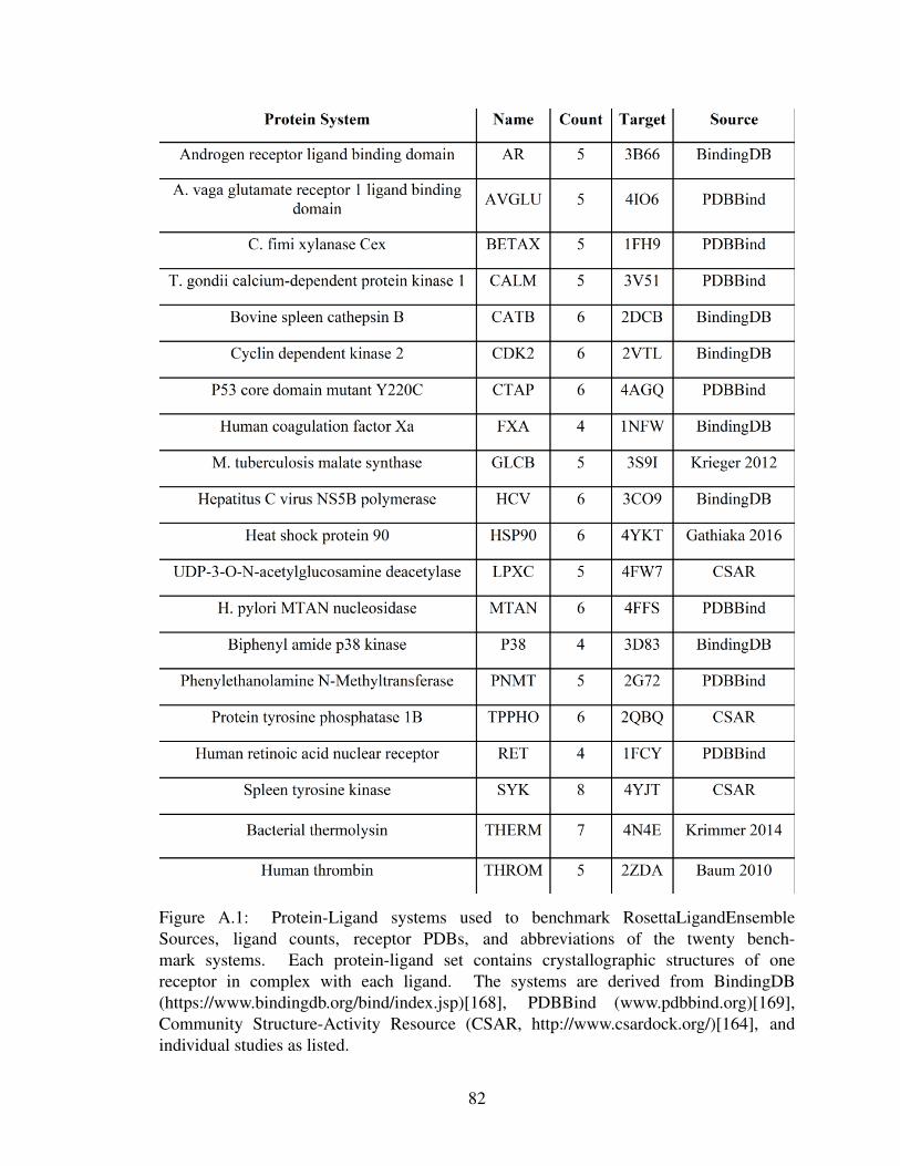
Figure A.1: Protein-Ligand systems used to benchmark RosettaLigandEnsembleSources, ligand counts, receptor PDBs, and abbreviations of the twenty bench-mark systems. Each protein-ligand set contains crystallographic structures of onereceptor in complex with each ligand. The systems are derived from BindingDB(https://www.bindingdb.org/bind/index.jsp)[168], PDBBind (www.pdbbind.org)[169],Community Structure-Activity Resource (CSAR, http://www.csardock.org/)[164], andindividual studies as listed.
82
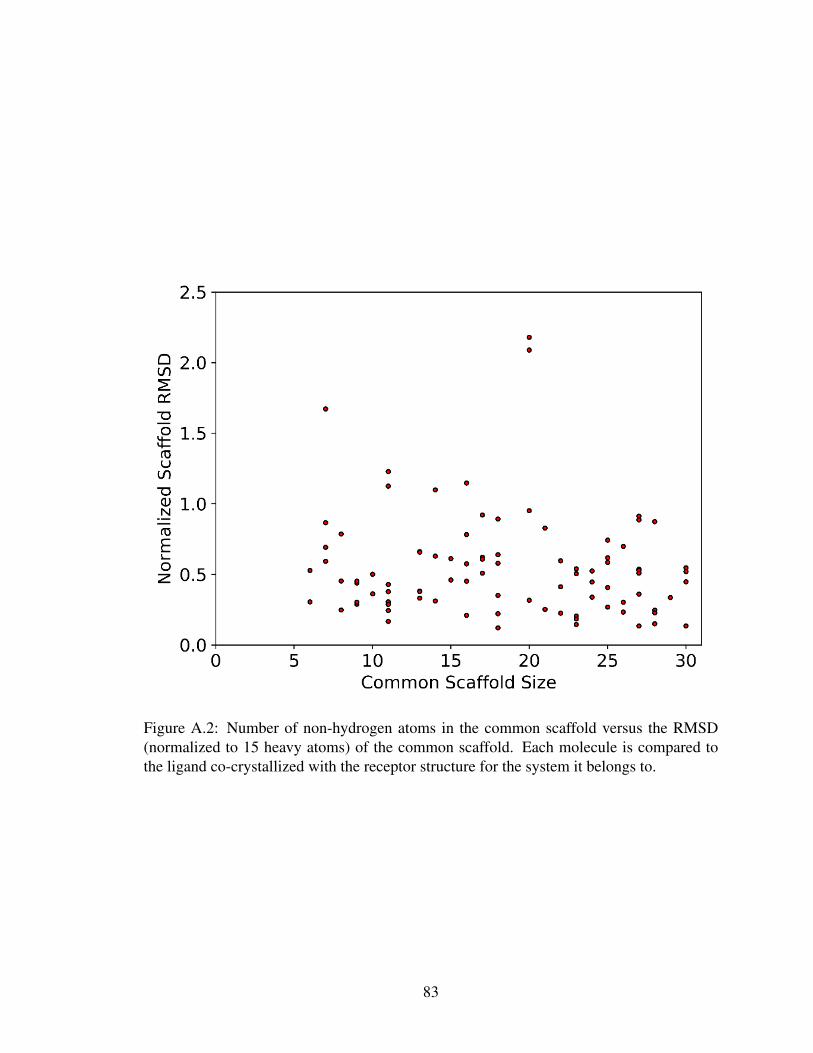
Figure A.2: Number of non-hydrogen atoms in the common scaffold versus the RMSD(normalized to 15 heavy atoms) of the common scaffold. Each molecule is compared tothe ligand co-crystallized with the receptor structure for the system it belongs to.
83

A.2 PDBBind Core Set
The PDBBind Core Set is a subset of the PDB Refined Set that contains 65 targets with
3 ligands each. Each triplet contains a low affinity binder, a medium affinity binder, and
a high affinity binder. A subset of 65 protein-ligand complexes is constructed using only
the high affinity binding molecules[27]. For benchmark purposes, the protein receptors are
relaxed using Rosetta FastRelax[170] in the apo state to reduce bias due to crystallographic
side-chain positioning.
A.3 PDBBind Refined Set
The PDBBind refined set contains 3446 structures controlled for protein structure qual-
ity, accurate binding data, small molecule properties, and non-surface interactions. Each
protein-small molecule co-crystal structure is paired with experimentally measured Kd, Ki,
or IC50[27, 169]. In order to analyze similarities in congeneric ligand binding, systems
with only one crystal structure were fitlered out. This produced 2443 structures across
441 targets. All complexes within a system were then aligned based on binding pocket
residues within 15 of the small molecule. The in-house BioChemicalLibrary (BCL) soft-
ware suite, available at http://www.meilerlab.org/bclcommons, is used to calculate small
molecule properties and make all possible intrasystem pairwise comparisons. The pairwise
normalized RMSD (nRMSD) is calculated based on the heavy atom RMSD of the largest
common connected substructure and normalized to ten heavy atoms using a small molecule
analog of RMSD-100[171]. The pairwise Tanimoto similarity coefficient is computed as
the number of atoms in the largest common connected substructure divided by the total
number of unique atoms.
All 34461 pairwise comparisons were then filtered to eliminate identical small molecule
pairs (Tanimoto = 1), trivial common scaffolds (scaffold heavy atoms <5), and different
binding pockets (small molecule center distance >3.0). A large number of the remaining
84

comparisons were from the HIV-1-Protease system, which dominates the PDBBind refined
set. In order to avoid biasing the results towards any particular system, only a randomly
chosen small subset of HIV-1-Protease comparisons are included. Small molecule sym-
metry was factored in when calculating nRMSDs for pairs where symmetrical molecules
are flipped. The final subset included a total of 7298 comparisons across 366 targets. The
median Tanimoto similarity across the dataset was 0.333 and the median nRMSD of the
common scaffold was 1.071 .
A.4 GPCR Modeling and Docking Set
A GPCR modeling and docking set was constructed in order to test the effectiveness
of docking ligands into comparative models of GPCRs. A set of sixteen GPCR structures
were collected from the PDB. Each GPCR would be modeled with RosettaCM[116] using
the remaining fifteen as templates. Comparative modeling would focus primarily on the
transmembrane regions with loop building used to reconstruct intracellular and extracellu-
lar loops. Figure A.3 shows the transmembrane domain sequence identity (lower triangle)
and C-alpha RMSDs (upper triangle). This dataset need to be updated as newer GPCR
protein-ligand complex structures become available.
85

Figure A.3: Transmembrane domain sequence similarity among the sixteen benchmarkGPCRs is shown in the lower triangle. Aligned C-alpha RMSDs over the same transmem-brane region is shown in the upper triangle. Red indicates a higher similarity while blueindicates greater differences.
86

Appendix B
Protocol Capture for RosettaLigandEnsemble
B.1 Obtaining files
Rosetta can be obtained through www.rosettacommons.org All files associated with this
protocol capture is provided in the demos/protocol capture/rosettaligand ensemble/ direc-
tory of the Rosetta distribution. This protocol has been tested to work with Rosetta version
d978e6f, released August 22nd, 2017. Examples commands for this protocol are numbered
in the commands file of the protocol capture folder and referenced as (1), (2), (3)etc.
B.2 Starting files
The raw starting files are a single target protein receptor structure in PDB format, and a
series of ligands in SDF format. The protein structure can be prepared from an experimen-
tally determined structure or from homology modeling. The receptor structure used in this
example is the p53 core domain bound to a stabilizing small molecule (PDB: 4AGQ). This
file can be found in /inputs/ as protein.pdb. The ligand series should share a core scaffold
by which the ligands can be aligned. This example contains five congeneric ligands, but
any number between three and eight is a reasonable use case.
B.3 Ligand preparation
PyMol pair fitting is an easy way to manually align ligands by minimizing the distance
between core scaffold atoms. Automated ligand alignment tools may also be used but
generally do not perform as well compared to manual inspection. Examples of aligned
ligands can be found in the /prep/aligned ligands/ directory.
87

Each ligand must have its own conformational library generated prior to using Roset-
taLigandEnsemble. Conformer generation was done using the BioChemicalLibrary. The
BCL (http://www.meilerlab.org/servers/bcl-academic-license) is a suite of software tools
readily available for academic users. Other software for conformer generation may also be
used but the outputs need to be converted to SDF files.
To generate conformers using the default settings, use command (1). Conformer gener-
ation can also be customized to use the PDB or CSD libraries, a greater range of rotamers,
or a structural comparison filter to remove similar conformers. For a full list of these op-
tions, see the help menu with command (2). Examples of generated conformers files are in
/prep/conformers/
B.4 Param file preparation
Rosetta requires params file to properly handle small molecule ligands. Prior to this
step, join the aligned ligand structure with the corresponding conformers into a single SDF
file such that the aligned structure is first in the file. This will insure that the inputs will
maintain the core scaffold alignment when generating the conformers. Examples of these
joined files are in /prep/make params/
Since PDB files use three digit residue codes and single digit chain designations, it is
helpful to assign a code for each ligand file. The example uses the ligands.list file to label
each ligand as residues 00B through 00F and corresponding chains B through F. This file
also contains pK values for each ligand binding to the target receptor.
To make params files for each ligand, use command (3). The molfile to params python
script is included in the /main/source/scripts/python/public/ directory of the Rosetta distri-
bution. Running the script without any input prints out the help menu.
For each ligand, command (3) generates a PDB file containing the single aligned lig-
and structure, a PDB file containing the remaining ligand conformers, and a params file
containing connectivity and charge information for the ligand. Examples of these files can
88
be found in the /prep/rosetta inputs/ directory using the previously discussed letter des-
ignations. If you wish to incorporate SAR during docking, then use a text editor to add
NUMERIC PROPERTY AFFINITY YOURVALUE to the end of the params file, where
YOURVALUE represents an SAR measurement of the users choice. Rank correlation is
used in SAR mode and hence units only need to be self-consistent. RosettaLigandEnsem-
ble assumes that smaller affinities are more favorable just as smaller Rosetta scores are
considered more favorable. The behavior can be adjusted for cases where larger affin-
ity values is more favorable by either taking the negative of the provided affinities in the
params file, or by making the correlation weight negative.
B.5 Input file organization
For RLE runs, it is convenient to prepare a single PDB file containing the aligned lig-
ands by concatenating the individual ligand PDB files. The conformer and params do not
need to be joined. This is done as ligands.pdb in the /inputs/ folder. Youll also find the
previously prepared protein receptor PDB in the same directory.
In addition to structural files, a RosettaScripts XML file and a Rosetta options file. The
XML file describes the custom protocol to be used by Rosetta. Details of how to setup
an XML file and the meaning of the individual tags can be found by searching the doc-
umentation website https://www.rosettacommons.org/docs/latest/. The example dock.xml
provided uses the settings from the benchmark. Actual application use may require the user
to alter these values according to biological context. The defined scoring function is based
on the existing RosettaLigand scoring function, but may be substituted in the XML script.
The provided options file defines Rosetta input and output directories along with a number
of sampling parameters. A full options list is available on the documentation website. The
ligand ensemble option is necessary to use RLE; a weight of 0 can be used to run RLE
without taking SAR data into consideration.
Run command (4) to perform a single simulation and generate a set of RLE models.
89

Each simulation will produce X models, where X is the number of input ligands. These
example output models are in the /outputs/ directory along with a score.sc scorefile.
B.6 Output and analysis
Individual protein-ligand predicted structures are labeled by a chain and a number des-
ignation, B 1.pdb through F 1.pdb. Structures with the same numeric label are based on
the same docking simulation and have a common binding pose. The protein interface con-
tacting each ligand are optimized independently. The score.sc file contains all score terms
for each simulation across a single row. Generally, individual ligand interface scores are
used to rank models, with a negative score indicating a better model. These ligand in-
terface scores are listed as interface delta *, where * is the single letter ligand chain ID.
The values are appended at the end of each output PDB, and also in the scorefile for each
protein-ligand pair. One suggestion is for the end user to examine the top ten percent of
models for each pair.
90
Appendix C
Additional Developments of Ensemble Docking
C.1 Summary
This appendix describes ongoing research on the development and benchmark of en-
semble docking approaches that merge structure-activity relationships derived from ligand
modifications and protein mutations. A new algorithm, ProtLigEnsemble, was created to
use such SAR data and a proof of concept study was completed. The basis of the algorithm
and the proof of concept will be submitted as a Rxiv preprint for which I will be the sole
first author. A full peer reviewed manuscript is expected once a more extensive benchmark
has been completed. This appendix includes a protocol capture illustrating how the new
Rosetta based ensemble features should be used.
C.2 Ensemble approaches from the protein perspective
RosettaLigandEnsemble was developed to dock a series of congeneric ligands to a sin-
gle protein target with the option to include structure-activity relationship (SARs) for ligand
modifications. As one might imagine, this can be extended to utilize the SARs associated
with protein changes rather than ligand changes. As discussed in Chapter 1, ligand bind-
ing changes upon protein residue mutagenesis is often utilized as a way to localize the
interaction site. We have extended ligand docking in Rosetta to process mutation data
automatically and to use the SARs to guide modeling.
Existing methods make use of protein ensembles for working with challenging protein
targets that are not well represented by a single static structure. This often occurs with
protein targets with highly flexible binding sites and/or a significant induced fit effect. In
one study, Ellingson et. al. used molecular dynamics snapshots to improve decoy dis-
91

crimination over docking against a single crystal structure[172]. This strategy can also be
used to generate the holo protein conformation when starting with an apo structure [173].
Although Rosetta does not perform molecular dynamics, we have now enabled the abil-
ity to use protein ensembles with an ”average-grid” scoring method in RosettaLigand and
RosettaLigandEnsemble. This greatly increases the amount of protein flexibility Rosetta
can process on top of the backbone and side-chain degrees of freedom typically allowed.
One interesting feature is that Rosetta protein ensembles do not have to be comprised of
the same protein variant. In other words, Rosetta protein ensembles can be used to dock or
screen against multiple mutants simultaneously.
C.3 Combining protein and ligand SARs
A further combination for ensemble docking would be the use of protein-based and
ligand-based SAR. This modality is geared towards cases where a ligand panel is tested
against several protein mutants. Figure C.1 shows the distinction among the datasets for
each of the ensemble docking approaches.
RosettaLigandEnsemble is discussed in Chapter 2 while protein ensemble docking and
protein ligand ”double” ensemble docking is new to this appendix. Protein ensemble dock-
ing can be performed by enabling additional options in the existing Rosetta ligand docking
movers. Double ensemble docking represents a novel method, ProtLigEnsemble, which
will be detailed further in this appendix. ProtLigEnsemble only requires a single input
structure either from experimental determination or comparative modeling. The remaining
structures will be generated by residue subsitution and energy minimization as the general
overall fold should remain the same. The presence of misfolding mutations can be experi-
mentally determined without the need for a fully solved structure. Missing binding data in
the table would be ignored by the algorithm.
92
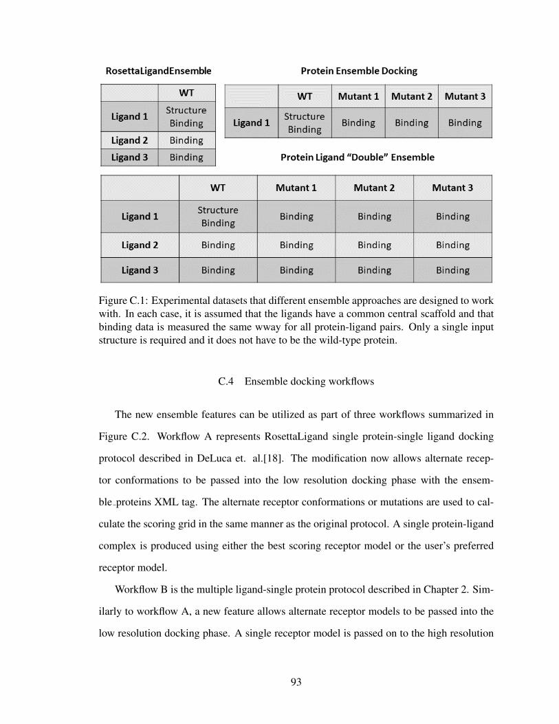
Figure C.1: Experimental datasets that different ensemble approaches are designed to workwith. In each case, it is assumed that the ligands have a common central scaffold and thatbinding data is measured the same wway for all protein-ligand pairs. Only a single inputstructure is required and it does not have to be the wild-type protein.
C.4 Ensemble docking workflows
The new ensemble features can be utilized as part of three workflows summarized in
Figure C.2. Workflow A represents RosettaLigand single protein-single ligand docking
protocol described in DeLuca et. al.[18]. The modification now allows alternate recep-
tor conformations to be passed into the low resolution docking phase with the ensem-
ble proteins XML tag. The alternate receptor conformations or mutations are used to cal-
culate the scoring grid in the same manner as the original protocol. A single protein-ligand
complex is produced using either the best scoring receptor model or the user’s preferred
receptor model.
Workflow B is the multiple ligand-single protein protocol described in Chapter 2. Sim-
ilarly to workflow A, a new feature allows alternate receptor models to be passed into the
low resolution docking phase. A single receptor model is passed on to the high resolution
93

docking phase where SAR guided ligand ensemble docking takes place. Users can specify
whether they wish to use the best scoring model or to use the primary input model with the
use main model XML tag. The final output contains one protein receptor in complex with
each of the input ligands.
Workflow C is the new double ensemble method for multiple proteins-multiple lig-
ands docking. The approach in docking is similar to that of RosettaLigandEnsemble in the
low resolution phase. Alternate conformations may also be used in this stage. The addi-
tional protein mutants are generated in the high resolution ProtLigEnsemble stage. Similar
to RosettaLigandEnsemble, an experimental dta rank correlation will be used to promote
docking modes that generate the most favorable Rosetta scores for the strongest binders.
Compounds without binding data to a given receptor will not be factored into the Spear-
man’s coefficient. The final output contains a structure for every possible pair of input
ligands and input proteins.
Additional changes in ProtLigEnsemble are the optimization order and the docking ra-
dius. The RosettaLigandEnsemble SAR guidance optimized one protein-ligand pair after
another in iterative cycles. This has been adjusted to complete all optimization cycles for
the protein-ligand pair with the highest experimental affinity first. This should generate the
best scoring structures for the strongest binders, improving correlation with experimental
data and reducing the stochastic nature of the output model scores. Furthermore, a docking
radius has been introduced as a simple way of defining the binding pocket. Previous iter-
ations required individual definitions for each ligand, leading to increasingly bulky XML
scripts. The new version automatically defines the flexible portion of the protein structure
based on a single setting and applies it to all protein-ligand pairs.
94
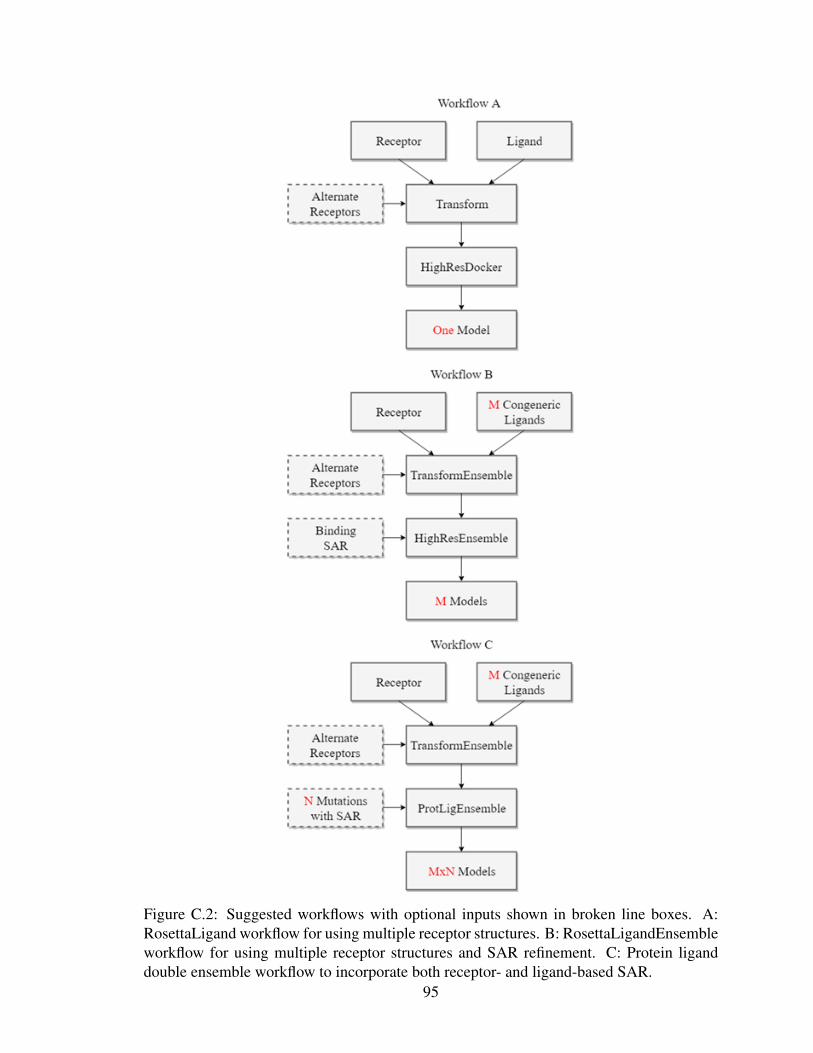
Figure C.2: Suggested workflows with optional inputs shown in broken line boxes. A:RosettaLigand workflow for using multiple receptor structures. B: RosettaLigandEnsembleworkflow for using multiple receptor structures and SAR refinement. C: Protein liganddouble ensemble workflow to incorporate both receptor- and ligand-based SAR.
95

C.5 ProtLigEnsemble proof of concept dataset
Two test cases are derived from individual studies on two GPCRs, the adenosine A2A
receptor and the neuropeptide Y1 receptor. The A2A dataset focus on work by Zhukov
et. al. [174] in which a panel of antagonists are tested against point mutants of a ther-
mostablized receptor.
Figure C.3: Experimental dataset derived from Zhukov et. al. containing binding data(pKd) for two ligands against seven receptor variants. Crystal structure of wild type recep-tor in complex with ZM241385 (PDB: 3EML) is shown in green, the ligand in orange, andthe mutation sites marked out in purple.
96

Figure C.3 shows the binding pocket of the A2A-ZM241385 complex with available
mutational data denoted in purple. SCH420814 is a ligand with a significant addition to the
extracellular binding end of the ligand. The crystal structure was published separately by
Jaakola et. al. (PDB: 3EML)[175].
The Y1 dataset, shown in Figure C.4 is derived from Yang et. al., which contains
a crystal structure of the Y1 receptor in complex with the antagonist UR-MK299 (PDB:
5ZBQ)[176]. The additional antagonists feature the addition of various linear amide groups
to the central scaffold of UR-MK299.
Figure C.4: Experimental dataset derived from Yang et. al. containing binding data (nMKd) for four ligands against five receptor variants. Crystal structure of wild type receptorin complex with UR-MK299 (PDB: 5ZBQ) is shown in green, the ligand in orange, andthe mutation sites marked out in purple.
97

C.6 ProtLigEnsemble docking results
Figure C.5 summarizes the docking results for the A2A test case. The docked mod-
els capture the general orientation of the binding mode observed in the crystal structure as
indicated by the significant portion of docked models around 4 RMSD. However, the com-
pact conformation of the ZM241385 ligand with the wildtype receptor is not well captured.
This may be an issue with the pregenerated conformations rather than the docking method.
One issue with the RMSD analysis is that only one protein-ligand pair can be compared for
native-like binding modes. Alternative measures may be necessary to evaluate the efficacy
of the method across the entire dataset. A significant enrichment of binding modes that
correlate positively with the SAR is observed for the A2A test.
Figure C.5: Summary of docking results with top right graph showing the RMSD distribu-tion of wildtype-ZM241385 models, and the bottom right graph showing the experimentalrank correlation. A representative output is shown overlaid with the 3EML crystal struc-ture. The crystal receptor structure and bound ligand is in red, docked ligands are in green,and mutation sites are shown in yellow.
Figure C.6 shows the docked models from a single simulation in grid format. One ben-
efit of double ensemble docking over individual docking runs is the consistency of docking
modes across the entire system. Rather than performing ad-hoc analysis to distinguish pu-
98

tative binding modes generated from individual runs, ProtLigEnsemble identifies binding
modes consistent with the entire dataset. The interfaces consists of individually optimized
ligand conformations and protein side-chain positioning. One limitation of this dataset is
that the side-chain mutations consisted of alanine scanning, which in some cases simply
represent a reduction in side-chain steric volume. A more extensive dataset including sig-
nificant electrostatic based mutations could be useful in testing how well ProtLigEnsemble
captures the SAR of substantial changes.
Figure C.6: Docking output from a single run of double ensemble docking. Top left cornershows the crystal structure of wild-type protein in complex with ZM241385. Remainingpanels show each of 14 possible combination of 2 ligands with 7 protein variants. Thecrystal structure is in red, docked ligands are in green, and mutation sites are shown inyellow.
Figure C.7 shows the Y1 docking results summary. As indicated by the RMSD dis-
tribution, the native like binding mode was captured for a significant number of wildtype-
99

UR-MK299 models generated. One issue that continues to arise is the band of high RMSD
models. Like RosettaLigandEnsemble, ProtLigEnsemble does not take into account SAR
during the low resolution docking phase. Future development could focus on a way to
improve model selection with low resolution scoring grids without adding substantial com-
putational cost. The Spearman distribution in the Y1 test case is also less than ideal. This
is likely due to the larger number of protein-ligand pairs being modeled. ProtLigEnsem-
ble optimizes binding pairs in order of affinity and it is possible that the SAR correlation
becomes too restrictive when docking the weaker binding pairs. This is analogous to the
difficulties of negative design when using an algorithm designed to find more and more
favorable scores.
Figure C.7: Summary of docking results with top right graph showing the RMSD distribu-tion of wildtype-UR-MK299 models, and the bottom right graph showing the experimentalrank correlation. A representative output is shown overlaid with the 5ZBQ crystal struc-ture. The crystal receptor structure and bound ligand is in red, docked ligands are in green,and mutation sites are shown in yellow.
100

C.7 Conclusion and future directions
Protein ensemble and double ensemble docking have been incorporated into the Rosetta
ligand docking suite. The proof of concept shows that ProtLigEnsemble is capable of gen-
erate a consistent set of docked models with protein-ligand SAR guidance. Additional
benchmarking is necessary to explore the full potential and limitations of the new algo-
rithms. One suggestion for benchmarking the addition of protein ensembles to the Roset-
taLigand and RosettaLigandEnsemble workflows is via PSCDB. PSCDB is a database that
tracks different types of receptor motion upon ligand binding[177]. This may be particu-
larly helpful in assessing the use of ensemble methods for ligand binding to highly flexible
loop regions.
To augment the double ensemble proof of study benchmark, the GPCR dataset de-
scribed in Appendix A may be a good starting point. Many of these receptor studies in-
clude mutational data in their supplemental and there may be additional ligand compounds
related to the co-crystallized ligand. It is unlikely a binding value for each protein mutant -
ligand pair will be found, but ProtLigEnsemble can work around this limitation by ignoring
missing SAR values. One other concern for dataset building is that different assays may
be used for different protein-ligand combinations. In particular, some of the ligand data
may be a measure of biological response, which can be due to allosteric effects as opposed
to binding affinity changes. Careful curation will be necessary to generate a proper and
diverse benchmark dataset.
C.8 ProtLigEnsemble protocol capture
The following section provides an example for using the double ensemble docking
protocol. Rosetta can be obtained through www.rosettacommons.org
All files associated with this protocol capture is provided in the demos/protocol capture/rosettaligand ProtLigEnsemble/
directory of the Rosetta distribution. This protocol has been tested to work with Rosetta
101
version 9820fea, released July 12, 2018. Examples commands for this protocol are num-
bered in the commands file of the protocol capture folder and referenced as (1), (2), (3)etc.
C.8.1 Starting files
The raw starting files are a single target protein receptor structure in PDB format, and a
series of ligands in SDF format. The receptor structure used in this example is neuropeptide
Y1 receptor bound to the ligand UR-MK299 (PDB: 5ZBQ). This file can be found in /in-
puts/ as protein.pdb. The four congeneric ligands in /prep/aligned ligands/ directory have
been aligned by their core scaffold. The reasonable number of ligands depends on the num-
ber of protein variants considered as each run generates all possible pairs. We generated up
to 30 models per docking run without issue though this number may change depending on
your computational setup.
C.8.2 Ligand preparation
Ligand preparation can be performed in the same fashion as the procedure in Appendix
B. In particular, commands 1, 2, and 3 cover the process of generating conformations with
the BCL conformer generator and creating Rosetta ligand param files. Example ligand con-
formers have been created for you in /prep/conformers and the necessary param generation
files are provided in /prep/make params/
The final Rosetta input ligand files are provided in /prep/rosetta inputs/. The ligands
have been designated with the letters B,C,D,E though you are free to use any chain des-
ignation as long as they are different from each other and the protein receptor. The corre-
spondence between published ligand designations and the Rosetta lettering is provided in
ligands.list. The params file process is the same as those for RosettaLigandEnsemble but
you can skip adding SAR data to the param files as SAR data will be provided in a separate
dedicated file.
102

C.8.3 Setting up the QSAR file
The QSAR file, an example of which is provided as inputs/qsar.txt, will provide protein-
ligand pairs of interest to the ProtLigEnsemble mover. Each line is organized as a protein
identifier, a ligand identifier, and an optional binding value. To provide a ligand binding
value to a wild-type protein, enter:
WT B 0.17
where WT indicates wildtype, B is the single letter chain of the ligand, and 0.17 is the
optional affinity value. Note that the affinity value can be any measure as long as they are
self consistent. Rosetta assumes the lower values indicates a more favorable binding. This
can be changed by setting a negative correlation weight in the -docking:ligand:ligand ensemble
option. To provide a binding value to a mutant protein, enter:
107 A B 7.5
where 107 A indicates a mutation at residue 107 to alanine, B is the single letter chain
of the ligand, and 7.5 is the optional affinity value. This will cause Rosetta to generate
a 107A mutant regardless of what the wildtype residue at position 107 is. Note that this
numbering system must correspond to Rosetta pose numbering, where the first residue is
numbered 1, the second is numbered 2...and so on. For the time being, ProtLigEnsemble
is designed to work with single mutants. This QSAR file will be provided to Rosetta in the
XML script as the qsar file tag for the ProtLigEnsemble mover.
C.8.4 Input file organization
For ProtLigEnsemble runs, it is preferred to combine all aligned ligand PDBs into a
single PDB file. This is provided as ligands.pdb in the /inputs/ folder. Note that the params
and conformers files are not combined, just the single ligand PDB inputs.
In addition to structural files, a RosettaScripts XML file and a Rosetta options file. The
XML file describes the custom protocol to be used by Rosetta. Details of how to setup
103

an XML file and the meaning of the individual tags can be found by searching the doc-
umentation website https://www.rosettacommons.org/docs/latest/. The example dock.xml
provided uses the settings from the benchmark. Actual application use may require the user
to alter these values according to biological context. The defined scoring function is based
on the existing RosettaLigand scoring function, but may be substituted in the XML script.
The provided options file defines Rosetta input and output directories along with a number
of sampling parameters. A full options list is available on the documentation website. The
ligand ensemble option is necessary to use ProtLigEnsemble; a weight of 0 can be used to
run ProtLigEnsemble without taking SAR data into consideration.
A few XML tags in the ProtLigEnsemble mover are newer features to this mover. The
aforementioned qsar file tag tells Rosetta where to find the QSAR file. The distance tag
defines the radius, in angstroms, of the sphere around the ligand considered to be the bind-
ing pocket. All residues in this sphere are considered to be flexible. This tag replaces
the LigandArea, InterfaceBuilder, and MoveMap tags RosettaLigand users may be familiar
with. The ignore correlation option tells ProtLigEnsemble to avoid calculating the rank
correlation until there are at least 4 protein-ligand pairs in the dataset. This is because
ProtLigEnsemble optimizes binding pairs in order of binding affinity. Considering the rank
correlation with only a few protein-ligand pairs is not particularly useful. This option may
be adjusted based on the number of ligands in your particular dataset.
Run command (4) to perform a single simulation and generate a set of ProtLigEnsemble
models. Each simulation will produce X models, where X is the number of protein-ligand
pairs listed in the SAR file. These example output models are in the /outputs/ directory
along with a score.sc scorefile.
C.8.5 Output and analysis
Individual protein-ligand predicted structures are labeled by a protein-ligand pair desig-
nation. Wildtype proteins and ligand combinations will be tagged as WT B 1.pdb through
104

WT E 1.pdb where the 1 indicates the docking run it came from. Mutant receptor-ligand
pairs will be tagged as 107 A B 1.pdb through 107 A E 1.pdb. The first two parts indicate
the mutant residue number and the mutant residue identity respectively. These are followed
by the ligand chain designation and the docking run number.
Structures with the same numeric label are based on the same docking simulation and
have a common binding pose. The protein interface contacting each ligand are optimized
independently. The score.sc file contains all score terms for each simulation across a single
row. Generally, individual ligand interface scores are used to rank models, with a negative
score indicating a better model. These ligand interface scores are listed as interface delta *,
where * corresponds to the protein-ligand prefix tag seen in the PDB files. The values are
appended at the end of each output PDB, and also in the scorefile for each protein-ligand
pair. One suggestion is for the end user to examine the top ten percent of models for each
pair.
105

BIBLIOGRAPHY
[1] Brian J. Bender, Alberto Cisneros, Amanda M. Duran, Jessica A. Finn, Darwin Fu,
Alyssa D. Lokits, Benjamin K. Mueller, Amandeep Kaur Sangha, Marion F. Sauer,
Alexander M. Sevy, Gregory Sliwoski, Jonathan H. Sheehan, Frank DiMaio, Jens
Meiler, and Rocco Moretti. Protocols for Molecular Modeling with Rosetta3 and
RosettaScripts. Biochemistry, 55(34):acs.biochem.6b00444, 2016.
[2] Darwin Y. Fu and Jens Meiler. Predictive Power of Different Types of Experimental
Restraints in Small Molecule Docking: A Review. Journal of Chemical Information
and Modeling, 58(2):225–233, feb 2018.
[3] M J Barnes. FUNCTION OF ASCORBIC ACID IN COLLAGEN METABOLISM.
Annals of the New York Academy of Sciences, 258(1):264–277, sep 1975.
[4] E. M. Wise and James T Park. Penicillin: its basic site of action as an inhibitor of
a peptide cross-linking reaction in cell wall mucopeptide synthesis. Proceedings of
the National Academy of Sciences, 54(1):75–81, jul 1965.
[5] George Scatchard. the Attractions of Proteins for Small Molecules and Ions. Annals
of the New York Academy of Sciences, 51(4):660–672, 1949.
[6] Subha Kalyaanamoorthy and Yi-Ping Phoebe Chen. Structure-based drug design to
augment hit discovery. Drug Discovery Today, 16(17-18):831–839, 2011.
[7] C R Beddell, P J Goodford, F E Norrington, S Wilkinson, and R Wootton. Com-
pounds Designed to Fit a Site of Known Structure in Human Hemoglobin. British J.
Pharm., 57(2):201–209, jun 1976.
[8] Michael L. Connolly. Shape complementarity at the hemoglobin α1β1 subunit in-
terface. Biopolymers, 25(7):1229–1247, 1986.
106
[9] Garrett M Morris, Ruth Huey, William Lindstrom, Michel F Sanner, Richard K
Belew, David S Goodsell, and Arthur J Olson. AutoDock4 and AutoDockTools4:
Automated Docking with Selective Receptor Flexibility. Journal of computational
chemistry, 30(16):2785–2791, 2010.
[10] T J Ewing, S Makino, A G Skillman, and I D Kuntz. DOCK 4.0: search strategies
for automated molecular docking of flexible molecule databases. J Comput Aided
Mol Des, 15(5):411–428, 2001.
[11] Richard A. Friesner, Jay L. Banks, Robert B. Murphy, Thomas A. Halgren, Jasna J.
Klicic, Daniel T. Mainz, Matthew P. Repasky, Eric H. Knoll, Mee Shelley, Jason K.
Perry, David E. Shaw, Perry Francis, and Peter S. Shenkin. Glide: A New Approach
for Rapid, Accurate Docking and Scoring. 1. Method and Assessment of Docking
Accuracy. Journal of Medicinal Chemistry, 47(7):1739–1749, mar 2004.
[12] Peter Willett, Robert C Glen, Andrew R Leach, Robin Taylor, and Gareth Jones.
Development and Validation of a Genetic Algorithm for Flexible Docking. J. Mol.
Biol., 267:727–748, 1997.
[13] Gordon Lemmon, Kristian Kaufmann, and Jens Meiler. Prediction of HIV-1 Pro-
tease/Inhibitor Affinity using RosettaLigand. Chemical Biology & Drug Design,
79(6):888–896, jun 2012.
[14] Jie Liu and Renxiao Wang. Classification of current scoring functions. Journal of
Chemical Information and Modeling, 55(3):475–482, 2015.
[15] Michael Berry, Burtram Fielding, and Junaid Gamieldien. Practical Considerations
in Virtual Screening and Molecular Docking. In Emerging Trends in Computational
Biology, Bioinformatics, and Systems Biology, number AUGUST, pages 487–502.
Elsevier, 2015.
107

[16] Ian W Davis and David Baker. RosettaLigand docking with full ligand and receptor
flexibility. J Mol Biol, 385(2):381–392, jan 2009.
[17] J Meiler and D Baker. ROSETTALIGAND: Protein-small molecule docking with
full side-chain flexibility. Proteins, 65(3):538–548, 2006.
[18] Samuel DeLuca, Karen Khar, and Jens Meiler. Fully Flexible Docking of Medium
Sized Ligand Libraries with RosettaLigand. Plos One, 10(7):e0132508, 2015.
[19] Rebecca F. Alford, Andrew Leaver-Fay, Jeliazko R. Jeliazkov, Matthew J. O’Meara,
Frank P. DiMaio, Hahnbeom Park, Maxim V. Shapovalov, P. Douglas Renfrew,
Vikram K. Mulligan, Kalli Kappel, Jason W. Labonte, Michael S. Pacella, Richard
Bonneau, Philip Bradley, Roland L. Dunbrack, Rhiju Das, David Baker, Brian
Kuhlman, Tanja Kortemme, and Jeffrey J. Gray. The Rosetta All-Atom Energy
Function for Macromolecular Modeling and Design. Journal of Chemical Theory
and Computation, 13(6):3031–3048, 2017.
[20] Gordon H Lemmon and Jens Meiler. RosettaLigand docking with flexible XML pro-
tocols, volume 819 of Methods in Molecular Biology. Springer New York, New
York, NY, 2012.
[21] Gordon Lemmon and Jens Meiler. Towards Ligand Docking Including Explicit In-
terface Water Molecules. PLoS ONE, 8(6):e67536, jun 2013.
[22] Brittany Allison, Steven Combs, Sam DeLuca, Gordon Lemmon, Laura Mizoue, and
Jens Meiler. Computational design of protein-small molecule interfaces. Journal of
Structural Biology, 185(2):193–202, feb 2014.
[23] Ian W. Davis, Kaushik Raha, Martha S. Head, and David Baker. Blind docking
of pharmaceutically relevant compounds using RosettaLigand. Protein Science,
18(9):1998–2002, sep 2009.
108

[24] Kelly L. Damm-Ganamet, Richard D. Smith, James B. Dunbar, Jeanne A. Stuckey,
and Heather A. Carlson. CSAR benchmark exercise 2011-2012: evaluation of results
from docking and relative ranking of blinded congeneric series. Journal of chemical
information and modeling, 53(8):1853–70, aug 2013.
[25] Symon Gathiaka, Shuai Liu, Michael Chiu, Huanwang Yang, Jeanne A. Stuckey,
You Na Kang, Jim Delproposto, Ginger Kubish, James B. Dunbar, Heather A. Carl-
son, Stephen K. Burley, W. Patrick Walters, Rommie E. Amaro, Victoria A. Feher,
and Michael K. Gilson. D3R grand challenge 2015: Evaluation of proteinligand pose
and affinity predictions. Journal of Computer-Aided Molecular Design, 30(9):1–18,
sep 2016.
[26] Zied Gaieb, Shuai Liu, Symon Gathiaka, Michael Chiu, Huanwang Yang, Chenghua
Shao, Victoria A. Feher, W. Patrick Walters, Bernd Kuhn, Markus G. Rudolph,
Stephen K. Burley, Michael K. Gilson, and Rommie E. Amaro. D3R Grand Chal-
lenge 2: blind prediction of proteinligand poses, affinity rankings, and relative bind-
ing free energies. Journal of Computer-Aided Molecular Design, 32(1):1–20, 2018.
[27] Yan Li, Li Han, Jie Liu, Zhihai Liu, Zhixiong Zhao, Renxiao Wang, Jie Li, Li Han,
Jie Liu, Zhixiong Zhao, and Renxiao Wang. Comparative Assessment of Scoring
Functions on an Updated Benchmark: 1. Compilation of the Test Set. Journal of
Chemical Information and Modeling, 54(6):1700–1716, 2014.
[28] Frank DiMaio, Yifan Song, Xueming Li, Matthias J Brunner, Chunfu Xu, Vincent
Conticello, Edward Egelman, Thomas C Marlovits, Yifan Cheng, and David Baker.
Atomic-accuracy models from 4.5-A cryo-electron microscopy data with density-
guided iterative local refinement. Nature methods, 12(4):361–5, 2015.
[29] Axel W. Fischer, Nathan S. Alexander, Nils Woetzel, Mert Karakas, Brian E. Weiner,
and Jens Meiler. BCL::MP-fold: Membrane protein structure prediction guided by
109
EPR restraints. Proteins: Structure, Function, and Bioinformatics, 83(11):1947–
1962, nov 2015.
[30] Caterina Bissantz, Bernd Kuhn, and Martin Stahl. A medicinal chemist’s guide to
molecular interactions. Journal of Medicinal Chemistry, 53(14):5061–5084, 2010.
[31] S L McGovern and B K Shoichet. Information decay in molecular docking
screens against holo, apo, and modeled conformations of enzymes. J Med Chem,
46(14):2895–907., 2003.
[32] Peter J. Huwe, Qifang Xu, Maxim V. Shapovalov, Vivek Modi, Mark D. Andrake,
and Roland L. Dunbrack. Biological function derived from predicted structures in
CASP11. Proteins: Structure, Function, and Bioinformatics, 84(July 2015):370–
391, sep 2016.
[33] Annalisa Bordogna, Alessandro Pandini, and Laura Bonati. Predicting the accuracy
of protein-ligand docking on homology models. Journal of Computational Chem-
istry, 32(1):81–98, jan 2011.
[34] Philippe Ferrara and Edgar Jacoby. Evaluation of the utility of homology models in
high throughput docking. Journal of Molecular Modeling, 13(8):897–905, 2007.
[35] Visvaldas Kairys, Miguel X. Fernandes, and Michael K. Gilson. Screening drug-
like compounds by docking to homology models: A systematic study. Journal of
Chemical Information and Modeling, 46(1):365–379, 2006.
[36] Kristian W Kaufmann and Jens Meiler. Using RosettaLigand for small molecule
docking into comparative models. PloS one, 7(12), jan 2012.
[37] Dr Cooper. X-Ray crystallography: Assessments and validation of protein-small
molecule complexes for drug discovery. Expert Opin Drug Discov, 6(8):771–782,
2012.
110

[38] Tim Ten Brink, Clementine Aguirre, Thomas E. Exner, and Isabelle Krimm. Perfor-
mance of protein-ligand docking with simulated chemical shift perturbations. Jour-
nal of Chemical Information and Modeling, 55(2):275–283, 2015.
[39] Ionut Onila, Tim ten Brink, Kai Fredriksson, Luca Codutti, Adam Mazur, Christian
Griesinger, Teresa Carlomagno, and Thomas E. Exner. On-the-Fly Integration of
Data from a Spin-Diffusion-Based NMR Experiment into ProteinLigand Docking.
Journal of Chemical Information and Modeling, page 150814163616007, 2015.
[40] Julien Orts, Stefan Bartoschek, Christian Griesinger, Peter Monecke, and Teresa
Carlomagno. An NMR-based scoring function improves the accuracy of binding
pose predictions by docking by two orders of magnitude. Journal of Biomolecular
NMR, 52(1):23–30, 2012.
[41] Lars Skjærven, Luca Codutti, Andrea Angelini, Manuela Grimaldi, Dorota Latek,
Peter Monecke, Matthias K. Dreyer, and Teresa Carlomagno. Accounting for con-
formational variability in protein-ligand docking with nmr-guided rescoring. Journal
of the American Chemical Society, 135(15):5819–5827, 2013.
[42] Olivier Cala, Florence Guilliere, and Isabelle Krimm. NMR-based analysis of
protein-ligand interactions. Analytical and Bioanalytical Chemistry, 406(4):943–
956, 2014.
[43] Kentaro Ishii, Masanori Noda, and Susumu Uchiyama. Mass spectrometric analysis
of proteinligand interactions. Biophysics and Physicobiology, 13:87–95, 2016.
[44] Varnavas D. Mouchlis, Christophe Morisseau, Bruce D. Hammock, Sheng Li, J. An-
drew McCammon, and Edward A. Dennis. Computer-aided drug design guided by
hydrogen/deuterium exchange mass spectrometry: A powerful combination for the
development of potent and selective inhibitors of Group VIA calcium-independent
phospholipase A2. Bioorganic and Medicinal Chemistry, 24(20):4801–4811, 2016.
111

[45] Stephanie Leavitt and Ernesto Freire. Direct measurement of protein binding ener-
getics by isothermal titration calorimetry. Current Opinion in Structural Biology,
11(5):560–566, 2001.
[46] Ning-Ning Wei and Adel Hamza. SABRE: ligand/structure-based virtual screening
approach using consensus molecular-shape pattern recognition. Journal of chemical
information and modeling, 54(1):338–46, jan 2014.
[47] M Vieth and D J Cummins. DoMCoSAR: a novel approach for establishing the
docking mode that is consistent with the structure-activity relationship. Application
to HIV-1 protease inhibitors and VEGF receptor tyrosine kinase inhibitors. Journal
of medicinal chemistry, 43(16):3020–32, aug 2000.
[48] C. J. Dinsmore, M. J. Bogusky, J. C. Culberson, J. M. Bergman, C. F. Homnick,
C. B. Zartman, S. D. Mosser, M. D. Schaber, R. G. Robinson, K. S. Koblan, H. E.
Huber, S. L. Graham, G. D. Hartman, J. R. Huff, and T. M. Williams. Confor-
mational restriction of flexible ligands guided by the transferred NOE experiment:
Potent macrocyclic inhibitors of farnesyltransferase [25]. Journal of the American
Chemical Society, 123(9):2107–2108, 2001.
[49] Sheng-Yong Yang. Pharmacophore modeling and applications in drug discovery:
challenges and recent advances. Drug Discovery Today, 15(11-12):444–450, 2010.
[50] Bingjie Hu and Markus A Lill. PharmDock: a pharmacophore-based docking pro-
gram. Journal of cheminformatics, 6(1):14, 2014.
[51] Christin Rakers, Fabian Schumacher, Walter Meinl, Hansruedi Glatt, Burkhard
Kleuser, and Gerhard Wolber. In silico prediction of human sulfotransferase 1E1
activity guided by pharmacophores from molecular dynamics simulations. Journal
of Biological Chemistry, 291(1):58–71, 2016.
112

[52] Dilyana Dimova, Kathrin Heikamp, Dagmar Stumpfe, and Jurgen Bajorath. Do
medicinal chemists learn from activity cliffs? A systematic evaluation of cliff
progression in evolving compound data sets. Journal of Medicinal Chemistry,
56(8):3339–3345, 2013.
[53] A. Patrıcia Bento, Anna Gaulton, Anne Hersey, Louisa J. Bellis, Jon Chambers,
Mark Davies, Felix A. Kruger, Yvonne Light, Lora Mak, Shaun McGlinchey, Michal
Nowotka, George Papadatos, Rita Santos, and John P. Overington. The ChEMBL
bioactivity database: an update. Nucleic Acids Research, 42(D1):D1083–D1090, jan
2014.
[54] Wallace K B Chan, Hongjiu Zhang, Jianyi Yang, Jeffrey R Brender, Junguk
Hur, Arzucan Ozgur, and Yang Zhang. GLASS: a comprehensive database for
experimentally-validated GPCR-ligand associations. Bioinformatics (Oxford, Eng-
land), 31(May):btv302–, 2015.
[55] Oscar P J Van Linden, Albert J. Kooistra, Rob Leurs, Iwan J P De Esch, and Chris
De Graaf. KLIFS: A knowledge-based structural database to navigate kinase-ligand
interaction space. Journal of Medicinal Chemistry, 57(2):249–277, 2014.
[56] Huameng Li and Chenglong Li. Multiple Ligand Simultaneous Docking: Orches-
trated Dancing of Ligands in Binding Sites of Protein. Journal of computational
chemistry, 31(16):2014–2022, 2010.
[57] Timothy M Allison, Eamonn Reading, Idlir Liko, Andrew J Baldwin, Arthur
Laganowsky, and Carol V Robinson. Quantifying the stabilizing effects of protein-
ligand interactions in the gas phase. Nature Communications, 6:1–10, 2015.
[58] Andreas Evers, Holger Gohlke, and Gerhard Klebe. Ligand-supported Homology
Modelling of Protein Binding-sites using Knowledge-based Potentials. Journal of
Molecular Biology, 334(2):327–345, 2003.
113

[59] Elizabeth Dong Nguyen, Christoffer Norn, Thomas M Frimurer, and Jens Meiler.
Assessment and Challenges of Ligand Docking into Comparative Models of G-
Protein Coupled Receptors. PLoS ONE, 8(7):e67302, jul 2013.
[60] L C Roisman, J Piehler, J.-Y. Trosset, H A Scheraga, and G Schreiber. Structure
of the interferon-receptor complex determined by distance constraints from double-
mutant cycles and flexible docking. Proceedings of the National Academy of Sci-
ences, 98(23):13231–13236, nov 2001.
[61] A. P. Blum, H. A. Lester, and D. A. Dougherty. Nicotinic pharmacophore: The
pyridine N of nicotine and carbonyl of acetylcholine hydrogen bond across a subunit
interface to a backbone NH. Proceedings of the National Academy of Sciences,
107(30):13206–13211, 2010.
[62] Fred Naider, Jeffrey M. Becker, Yong Hun Lee, and Amnon Horovitz. Double-
mutant cycle scanning of the interaction of a peptide ligand and its G protein-coupled
receptor. Biochemistry, 46(11):3476–3481, 2007.
[63] Catherine Marquer, Carole Fruchart-Gaillard, Guillaume Letellier, Elodie Marcon,
Gilles Mourier, Sophie Zinn-Justin, Andre Menez, Denis Servent, and Bernard
Gilquin. Structural model of ligand-G protein-coupled receptor (GPCR) complex
based on experimental double mutant cycle data: MT7 snake toxin bound to dimeric
hM1 muscarinic receptor. Journal of Biological Chemistry, 286(36):31661–31675,
2011.
[64] Keith L. Constantine, Malcolm E. Davis, William J. Metzler, Luciano Mueller, and
Brian L. Claus. Protein-ligand NOE matching: A high-throughput method for bind-
ing pose evaluation that does not require protein NMR resonance assignments. Jour-
nal of the American Chemical Society, 128(22):7252–7263, 2006.
[65] Julien Orts, Jennifer Tuma, Marcel Reese, S. Kaspar Grimm, Peter Monecke, Stefan
114

Bartoschek, Alexander Schiffer, K. Ulrich Wendt, Christian Griesinger, and Teresa
Carlomagno. Crystallography-independent determination of ligand binding modes.
Angewandte Chemie - International Edition, 47(40):7736–7740, 2008.
[66] Sheng-You Huang, Min Li, Jianxin Wang, and Yi Pan. HybridDock: A Hybrid Pro-
teinLigand Docking Protocol Integrating Protein- and Ligand-Based Approaches.
Journal of Chemical Information and Modeling, 56(6):1078–1087, jun 2016.
[67] Polo C.-H. Lam, Ruben Abagyan, and Maxim Totrov. Ligand-biased ensemble re-
ceptor docking (LigBEnD): a hybrid ligand/receptor structure-based approach. Jour-
nal of Computer-Aided Molecular Design, 32(1):187–198, jan 2018.
[68] Jeffrey Skolnick, Mu Gao, Ambrish Roy, Bharath Srinivasan, and Hongyi Zhou. Im-
plications of the small number of distinct ligand binding pockets in proteins for drug
discovery, evolution and biochemical function. Bioorganic and Medicinal Chemistry
Letters, 25(6):1163–1170, 2015.
[69] Jonas Bostrom, Anders Hogner, and Stefan Schmitt. Do structurally similar ligands
bind in a similar fashion? Journal of medicinal chemistry, 49(23):6716–25, nov
2006.
[70] Shipra Malhotra and John Karanicolas. When Does Chemical Elaboration Induce a
Ligand To Change Its Binding Mode? Journal of Medicinal Chemistry, 60(1):128–
145, jan 2017.
[71] Sara L. Adamski-Werner, Satheesh K. Palaninathan, James C. Sacchettini, and Jef-
fery W. Kelly. Diflunisal Analogues Stabilize the Native State of Transthyretin.
Potent Inhibition of Amyloidogenesis. Journal of Medicinal Chemistry, 47(2):355–
374, 2004.
[72] Hsiang Ting Lei, Zhangqi Shen, Priyanka Surana, Mathew D. Routh, Chih Chia Su,
115

Qijing Zhang, and Edward W. Yu. Crystal structures of CmeR-bile acid complexes
from Campylobacter jejuni. Protein Science, 20(4):712–723, 2011.
[73] Vincent Stoll, Kent D Stewart, Clarence J Maring, Steven Muchmore, Vincent Gi-
randa, Yu-gui Y Gu, Gary Wang, Yuanwei Chen, Minghua Sun, Chen Zhao, April L
Kennedy, Darold L Madigan, Yibo Xu, Ayda Saldivar, Warren Kati, Graeme Laver,
Thomas Sowin, Hing L Sham, Jonathan Greer, and Dale Kempf. Influenza neu-
raminidase inhibitors: structure-based design of a novel inhibitor series. Biochem-
istry, 42(3):718–27, 2003.
[74] Zhonghua Pei, Xiaofeng Li, Kenton Longenecker, Thomas W von Geldern, Paul E
Wiedeman, Thomas H Lubben, Bradley A Zinker, Kent Stewart, Stephen J Ballaron,
Michael a Stashko, Amanda K Mika, David W A Beno, Michelle Long, Heidi Wells,
Anita J Kempf-Grote, David J Madar, Todd S McDermott, Lakshmi Bhagavatula,
Michael G Fickes, Daisy Pireh, Larry R Solomon, Marc R Lake, Rohinton Edalji,
Elizabeth H Fry, Hing L Sham, and James M Trevillyan. Discovery, StructureActiv-
ity Relationship, and Pharmacological Evaluation of (5-Substituted-pyrrolidinyl-2-
carbonyl)-2-cyanopyrrolidines as Potent Dipeptidyl Peptidase IV Inhibitors. Journal
of Medicinal Chemistry, 49(12):3520–3535, jun 2006.
[75] P M Fitzgerald, B M McKeever, J F VanMiddlesworth, J P Springer, J C Heim-
bach, C T Leu, W K Herber, R A Dixon, and P L Darke. Crystallographic analysis
of a complex between human immunodeficiency virus type 1 protease and acetyl-
pepstatin at 2.0-A resolution. The Journal of biological chemistry, 265(24):14209–
14219, 1990.
[76] Ki Hwan Kim. Outliers in SAR and QSAR: 2. Is a flexible binding site a possible
source of outliers? Journal of computer-aided molecular design, 21(8):421–35,
2007.
116

[77] Ki Hwan Kim. Outliers in SAR and QSAR: Is unusual binding mode a possible
source of outliers? Journal of computer-aided molecular design, 21(8):63–86, 2007.
[78] Junichi Goto, Ryoichi Kataoka, and Noriaki Hirayama. Ph4Dock: Pharmacophore-
based protein - Ligand docking. Journal of Medicinal Chemistry, 47(27):6804–6811,
dec 2004.
[79] Manuel Rueda, Giovanni Bottegoni, and Ruben Abagyan. Consistent improvement
of cross-docking results using binding site ensembles generated with elastic network
normal modes. Journal of Chemical Information and Modeling, 49:716–725, 2009.
[80] Oliver Korb, Tim Ten Brink, Fredrick Robin Devadoss Victor Paul Raj, Matthias
Keil, and Thomas E Exner. Are predefined decoy sets of ligand poses able to quantify
scoring function accuracy? Journal of computer-aided molecular design, 26(2):185–
97, mar 2012.
[81] Rommie E. Amaro, Riccardo Baron, and J. Andrew McCammon. An improved re-
laxed complex scheme for receptor flexibility in computer-aided drug design. Jour-
nal of Computer-Aided Molecular Design, 22(9):693–705, 2008.
[82] Mengang Xu and Markus A. Lill. Utilizing experimental data for reducing ensemble
size in flexible-protein docking. Journal of Chemical Information and Modeling,
52(1):187–198, 2012.
[83] Ferran Feixas, Steffen Lindert, William Sinko, and J. Andrew McCammon. Explor-
ing the role of receptor flexibility in structure-based drug discovery. Biophysical
Chemistry, 186:31–45, 2014.
[84] William Sinko, Steffen Lindert, and J Andrew McCammon. Accounting for receptor
flexibility and enhanced sampling methods in computer-aided drug design. Chemical
biology & drug design, 81(1):41–9, jan 2013.
117

[85] Andrew Anighoro, Luca Pinzi, Giulio Rastelli, and Jurgen Bajorath. Virtual Screen-
ing for Dual Hsp90/B-Raf Inhibitors. In Methods in Pharmacology and Toxicology,
number January 2015, pages 167–188. 2017.
[86] Wenying Yu, Chenglong Li, Wenda Zhang, Yuanzheng Xia, Shanshan Li, Jia yuh
Lin, Keqin Yu, Mu Liu, Lei Yang, Jianguang Luo, Yijun Chen, Hongbin Sun, and
Lingyi Kong. Discovery of an Orally Selective Inhibitor of Signal Transducer and
Activator of Transcription 3 Using Advanced Multiple Ligand Simultaneous Dock-
ing. Journal of Medicinal Chemistry, 60(7):2718–2731, 2017.
[87] Gregory Sliwoski, Sandeepkumar Kothiwale, Jens Meiler, and Edward W
Lowe. Computational Methods in Drug Discovery. Pharmacological Reviews,
66(January):334–395, 2014.
[88] William L Jorgensen. Efficient Drug Lead Discovery and Optimizatiion. Acc Chem
Res, 42(6):724–733, 2009.
[89] Sunghwan Kim, Paul A. Thiessen, Evan E. Bolton, Jie Chen, Gang Fu, Asta Gindu-
lyte, Lianyi Han, Jane He, Siqian He, Benjamin A. Shoemaker, Jiyao Wang, Bo Yu,
Jian Zhang, and Stephen H. Bryant. PubChem substance and compound databases.
Nucleic Acids Research, 44(D1):D1202–D1213, 2016.
[90] Qian Liu, Brian Masek, Karl Smith, and Julian Smith. Tagged fragment method for
evolutionary structure-based de novo lead generation and optimization. Journal of
medicinal chemistry, 50(22):5392–5402, 2007.
[91] S Wang, E S Humphreys, S Y Chung, D F Delduco, S R Lustig, H Wang, K N
Parker, N W Rizzo, S Subramoney, Y M Chiang, and A Jagota. Peptides with
selective affinity for carbon nanotubes. Nat Mater, 2(3):196–200., 2003.
[92] Woody Sherman, Tyler Day, Matthew P. Jacobson, Richard A. Friesner, and Ramy
118

Farid. Novel procedure for modeling ligand/receptor induced fit effects. Journal of
Medicinal Chemistry, 49(2):534–553, 2006.
[93] F Osterberg, G M Morris, M F Sanner, A J Olson, and D S Goodsell. Automated
docking to multiple target structures: incorporation of protein mobility and structural
water heterogeneity in AutoDock. Proteins, 46(1):34–40, 2002.
[94] Sheng Y. Huang and Xiaoqin Zou. Ensemble docking of multiple protein structures:
Considering protein structural variations in molecular docking. Proteins: Structure,
Function and Genetics, 66:399–421, 2007.
[95] Sandeepkumar Kothiwale, Jeffrey L. Mendenhall, and Jens Meiler. BCL::Conf:
Small molecule conformational sampling using a knowledge based rotamer library.
Journal of Cheminformatics, 7(1):1–15, 2015.
[96] Marcel L. Verdonk, Paul N. Mortenson, Richard J. Hall, Michael J. Hartshorn, and
Christopher W. Murray. Protein-ligand docking against non-native protein conform-
ers. Journal of Chemical Information and Modeling, 48(11):2214–2225, 2008.
[97] Oliver Korb, Tjelvar S G Olsson, Simon J Bowden, Richard J Hall, Marcel L Ver-
donk, John W Liebeschuetz, and Jason C Cole. Potential and limitations of ensemble
docking. Journal of chemical information and modeling, 52(5):1262–74, may 2012.
[98] Stefan G. Krimmer, Michael Betz, Andreas Heine, and Gerhard Klebe. Methyl,
ethyl, propyl, butyl: Futile but not for water, as the correlation of structure and
thermodynamic signature shows in a congeneric series of thermolysin inhibitors.
ChemMedChem, 9(4):833–846, 2014.
[99] HM Ashtawy and NR Mahapatra. Does Accurate Scoring of Ligands against Protein
Targets Mean Accurate Ranking? Bioinformatics Research and Applications, pages
298–310, 2013.
119

[100] Zhe Wang, Huiyong Sun, Xiaojun Yao, Dan Li, Lei Xu, Youyong Li, Sheng Tian,
and Tingjun Hou. Comprehensive evaluation of ten docking programs on a diverse
set of proteinligand complexes: the prediction accuracy of sampling power and scor-
ing power. Phys. Chem. Chem. Phys., 18(18):12964–12975, 2016.
[101] Heather A. Carlson, Richard D. Smith, Kelly L. Damm-Ganamet, Jeanne A. Stuckey,
Aqeel Ahmed, Maire A. Convery, Donald O. Somers, Michael Kranz, Patricia A.
Elkins, Guanglei Cui, Catherine E. Peishoff, Millard H. Lambert, and James B. Dun-
bar. CSAR 2014: A Benchmark Exercise Using Unpublished Data from Pharma.
Journal of Chemical Information and Modeling, 56(6):1063–1077, 2016.
[102] D. E. V. Pires, T. L. Blundell, and D. B. Ascher. Platinum: a database of experimen-
tally measured effects of mutations on structurally defined protein-ligand complexes.
Nucleic Acids Research, 43(D1):D387–D391, 2014.
[103] Vivian Law, Craig Knox, Yannick Djoumbou, Tim Jewison, An Chi Guo, Yifeng
Liu, Adam MacIejewski, David Arndt, Michael Wilson, Vanessa Neveu, Alexandra
Tang, Geraldine Gabriel, Carol Ly, Sakina Adamjee, Zerihun T. Dame, Beomsoo
Han, You Zhou, and David S. Wishart. DrugBank 4.0: Shedding new light on drug
metabolism. Nucleic Acids Research, 42(D1):1091–1097, 2014.
[104] Si-Sheng Ou-Yang, Jun-Yan Lu, Xiang-Qian Kong, Zhong-Jie Liang, Cheng Luo,
and Hualiang Jiang. Computational drug discovery. Acta pharmacologica Sinica,
33(9):1131–40, 2012.
[105] Aurelien Grosdidier, Vincent Zoete, and Olivier Michielin. SwissDock, a protein-
small molecule docking web service based on EADock DSS. Nucleic Acids Re-
search, 39(SUPPL. 2):270–277, 2011.
[106] S.-L. Chang and N Tjandra. Temperature dependence of protein backbone motion
120

from carbonyl 13C and amide 15N NMR relaxation. J. Magn. Res., 174:43–53,
2005.
[107] Dina Schneidman-Duhovny, Yuval Inbar, Ruth Nussinov, and Haim J. Wolfson.
PatchDock and SymmDock: Servers for rigid and symmetric docking. Nucleic Acids
Research, 33(SUPPL. 2):363–367, 2005.
[108] Francis Gaudreault, Louis Philippe Morency, and Rafael J. Najmanovich. NRGsuite:
A PyMOL plugin to perform docking simulations in real time using FlexAID. Bioin-
formatics, 31(23):3856–3858, 2015.
[109] Zsolt Bikadi and Eszter Hazai. Application of the PM6 semi-empirical method to
modeling proteins enhances docking accuracy of AutoDock. Journal of Cheminfor-
matics, 1(1):1–16, 2009.
[110] Kristian W. Kaufmann, Gordon H. Lemmon, Samuel L. DeLuca, Jonathan H. Shee-
han, and Jens Meiler. Practically Useful: What the R <scp>osetta</scp> Protein
Modeling Suite Can Do for You. Biochemistry, 49(14):2987–2998, 2010.
[111] Rocco Moretti, Sergey Lyskov, Rhiju Das, Jens Meiler, and Jeffrey J. Gray. Web-
accessible molecular modeling with Rosetta: The Rosetta Online Server that In-
cludes Everyone (ROSIE). Protein Science, 27(1):259–268, jan 2018.
[112] Andrew Leaver-Fay, Matthew J. O’Meara, Mike Tyka, Ron Jacak, Yifan Song,
Elizabeth H. Kellogg, James Thompson, Ian W. Davis, Roland A. Pache, Sergey
Lyskov, Jeffrey J. Gray, Tanja Kortemme, Jane S. Richardson, James J. Havranek,
Jack Snoeyink, David Baker, and Brian Kuhlman. Scientific benchmarks for guiding
macromolecular energy function improvement, volume 523. Elsevier Inc., 1 edition,
2013.
[113] Matthew J. O’Meara, Andrew Leaver-Fay, Michael D. Tyka, Amelie Stein, Kevin
Houlihan, Frank Dimaio, Philip Bradley, Tanja Kortemme, David Baker, Jack
121

Snoeyink, and Brian Kuhlman. Combined covalent-electrostatic model of hydrogen
bonding improves structure prediction with Rosetta. Journal of Chemical Theory
and Computation, 11(2):609–622, 2015.
[114] Marylens Hernandez, Dario Ghersi, and Roberto Sanchez. SITEHOUND-web: A
server for ligand binding site identification in protein structures. Nucleic Acids Re-
search, 37(SUPPL. 2):413–416, 2009.
[115] Sergey Lyskov, Fang Chieh Chou, Shane O. Conchuir, Bryan S. Der, Kevin Drew,
Daisuke Kuroda, Jianqing Xu, Brian D. Weitzner, P. Douglas Renfrew, Parin Sri-
pakdeevong, Benjamin Borgo, James J. Havranek, Brian Kuhlman, Tanja Ko-
rtemme, Richard Bonneau, Jeffrey J. Gray, and Rhiju Das. Serverification of Molec-
ular Modeling Applications: The Rosetta Online Server That Includes Everyone
(ROSIE). PLoS ONE, 8(5):5–7, 2013.
[116] Yifan Song, Frank DiMaio, Ray Yu-Ruei Wang, David Kim, Chris Miles,
Tj Brunette, James Thompson, and David Baker. High-resolution comparative mod-
eling with RosettaCM. Structure (London, England : 1993), 21(10):1735–42, oct
2013.
[117] Steven a Combs, Samuel L DeLuca, Stephanie H DeLuca, Gordon H Lemmon,
David P Nannemann, Elizabeth D Nguyen, Jordan R Willis, Jonathan H Shee-
han, and Jens Meiler. Small-molecule ligand docking into comparative models with
Rosetta. Nature Protocols, 8(7):1277–1298, jun 2013.
[118] Christian Lis, Stefan Rubner, Martin Roatsch, Angela Berg, Tyler Gilcrest, Dar-
win Fu, Elizabeth Nguyen, Anne-Marie Schmidt, Harald Krautscheid, Jens Meiler,
and Thorsten Berg. Development of Erasin: a chromone-based STAT3 inhibitor
which induces apoptosis in Erlotinib-resistant lung cancer cells. Scientific Reports,
7(1):17390, dec 2017.
122

[119] CJ Wenthur and Ryan Morrison. Discovery of (R)-(2-fluoro-4-((-4-methoxyphenyl)
ethynyl) phenyl)(3-hydroxypiperidin-1-yl) methanone (ML337), an mGlu3 selec-
tive and CNS penetrant negative allosteric modulator. Journal of medicinal . . . ,
1(56):5208–5212, 2013.
[120] W. Frank An and Nicola Tolliday. Cell-based assays for high-throughput screening.
Molecular Biotechnology, 45(2):180–186, 2010.
[121] Joseph C. Somody, Stephen S. MacKinnon, and Andreas Windemuth. Structural
coverage of the proteome for pharmaceutical applications. Drug Discovery Today,
22(12):1792–1799, dec 2017.
[122] Rita Santos, Oleg Ursu, Anna Gaulton, A Patrıcia Bento, Ramesh S Donadi, Cris-
tian G Bologa, Anneli Karlsson, Bissan Al-Lazikani, Anne Hersey, Tudor I Oprea,
and John P Overington. A comprehensive map of molecular drug targets. Nature
Reviews Drug Discovery, 16(1):19–34, jan 2017.
[123] Thorsten Berg. Signal Transducers and Activators of Transcription as Targets for
Small Organic Molecules. ChemBioChem, 9(13):2039–2044, 2008.
[124] Hua Yu and Richard Jove. The STATs of cancer new molecular targets come of age.
Nature Reviews Cancer, 4(2):97–105, 2004.
[125] Judith Muller, Bianca Sperl, Wolfgang Reindll, Anke Kiessling, and Thorsten Berg.
Discovery of chromone-based inhibitors of the transcription factor STAT5. Chem-
BioChem, 9(5):723–727, 2008.
[126] Jochen Schust and Thorsten Berg. A high-throughput fluorescence polarization as-
say for signal transducer and activator of transcription 3. Analytical Biochemistry,
330(1):114–118, 2004.
123

[127] Alexander S. Hauser, Misty M. Attwood, Mathias Rask-Andersen, Helgi B. Schioth,
and David E. Gloriam. Trends in GPCR drug discovery: New agents, targets and
indications. Nature Reviews Drug Discovery, 16(12):829–842, 2017.
[128] Bryan L. Roth, John J. Irwin, and Brian K. Shoichet. Discovery of new GPCR
ligands to illuminate new biology. Nature Chemical Biology, 13(11):1143–1151,
2017.
[129] Vsevolod Katritch, Vadim Cherezov, and Raymond C. Stevens. Structure-Function
of the G ProteinCoupled Receptor Superfamily. Annual Review of Pharmacology
and Toxicology, 53(1):531–556, jan 2013.
[130] Christofer S. Tautermann. GPCR structures in drug design, emerging opportunities
with new structures. Bioorganic & Medicinal Chemistry Letters, 24(17):4073–4079,
2014.
[131] Irina Kufareva, Manuel Rueda, and Vsevolod Katritch. Status of GPCR modeling
and docking as reflected by community-wide GPCR Dock 2010 assessment. Struc-
ture, 19(8):1108–1126, 2011.
[132] Irina Kufareva, Vsevolod Katritch, RaymondC. Stevens, and Ruben Abagyan. Ad-
vances in GPCR Modeling Evaluated by the GPCR Dock 2013 Assessment: Meeting
New Challenges. Structure, 22(8):1120–1139, 2014.
[133] Jeremy Shonberg, Ralf C. Kling, Peter Gmeiner, and Stefan Lober. GPCR crystal
structures: Medicinal chemistry in the pocket. Bioorganic & Medicinal Chemistry,
2015.
[134] P J Conn and J P Pin. Pharmacology and functions of metabotropic glutamate re-
ceptors. Annual review of pharmacology and toxicology, 37:205–37, jan 1997.
124

[135] Q J Yan, M Rammal, M Tranfaglia, and R P Bauchwitz. Suppression of two major
Fragile X Syndrome mouse model phenotypes by the mGluR5 antagonist MPEP.
Neuropharmacology, 49(7):1053–66, dec 2005.
[136] Jason M Uslaner, Sophie Parmentier-Batteur, Rosemarie B Flick, Nathaniel O
Surles, June S H Lam, Caitlyn H McNaughton, Marlene a Jacobson, and Pete H
Hutson. Dose-dependent effect of CDPPB, the mGluR5 positive allosteric modula-
tor, on recognition memory is associated with GluR1 and CREB phosphorylation in
the prefrontal cortex and hippocampus. Neuropharmacology, 57(5-6):531–8, 2009.
[137] H Awad, G W Hubert, Y Smith, A I Levey, and P J Conn. Activation of metabotropic
glutamate receptor 5 has direct excitatory effects and potentiates NMDA receptor
currents in neurons of the subthalamic nucleus. J Neurosci, 20(21):7871–7879, nov
2000.
[138] P Jeffrey Conn, Craig W Lindsley, and Carrie K Jones. Activation of metabotropic
glutamate receptors as a novel approach for the treatment of schizophrenia. Trends
in pharmacological sciences, 30(1):25–31, jan 2009.
[139] Kamondanai Hemstapat, Herve Da Costa, and Yi Nong. A novel family of potent
negative allosteric modulators of group II metabotropic glutamate receptors. . . . of
Pharmacology and . . . , 322(1):254–264, 2007.
[140] Filippo Caraci, Gemma Molinaro, and Giuseppe Battaglia. II Metabotropic Gluta-
mate (mGlu) Receptors for the Treatment of Psychosis Associated with Alzheimer’s
Disease: Selective Activation of mGlu2 Receptors Amplifies β -. Molecular . . . ,
79(3):618–626, 2011.
[141] DJ Sheffler and CJ Wenthur. Development of a novel, CNS-penetrant, metabotropic
glutamate receptor 3 (mGlu 3) NAM probe (ML289) derived from a closely related
mGlu 5. Bioorganic & medicinal . . . , 22(12):3921–3925, 2012.
125

[142] Colleen M CM Niswender, Kari A Johnson, Qingwei Luo, Jennifer E Ayala, Car-
oline Kim, P Jeffrey Conn, and C David Weaver. Gi/o-linked G protein-coupled
receptor coupling to potassium channels provides new insights into the pharmacol-
ogy of the group III metabotropic glutamate receptors. Molecular pharmacology,
73(4):1213–1224, 2008.
[143] Corrado Corti, Giuseppe Battaglia, Gemma Molinaro, Barbara Riozzi, Anna
Pittaluga, Mauro Corsi, Manolo Mugnaini, Ferdinando Nicoletti, and Valeria
Bruno. The use of knock-out mice unravels distinct roles for mGlu2 and mGlu3
metabotropic glutamate receptors in mechanisms of neurodegeneration/neuropro-
tection. The Journal of neuroscience : the official journal of the Society for Neuro-
science, 27(31):8297–308, aug 2007.
[144] Andrew S. Dore, Krzysztof Okrasa, Jayesh C. Patel, Maria Serrano-Vega, Kirstie
Bennett, Robert M. Cooke, James C. Errey, Ali Jazayeri, Samir Khan, Ben Tehan,
Malcolm Weir, Giselle R. Wiggin, and Fiona H. Marshall. Structure of class C GPCR
metabotropic glutamate receptor 5 transmembrane domain. Nature, 511:557–62, jul
2014.
[145] Huixian Wu, Karen J Gregory, P Cho, Colleen M Niswender, Jens Meiler, Vadim
Cherezov, P Jeffrey Conn, and Raymond C Stevens. Structure of a Class C GPCR
Metabotropic Glutamate Receptor 1 Bound to an Allosteric Modulator. Science,
March, 2014.
[146] Gamariel Rwibasira Rudinga, Ghulam Jilany Khan, and Yi Kong. Protease-
Activated Receptor 4 (PAR4): A Promising Target for Antiplatelet Therapy. In-
ternational Journal of Molecular Sciences, 19(2):573, 2018.
[147] Justin R. Hamilton and JoAnn Trejo. Challenges and Opportunities in Protease-
126

Activated Receptor Drug Development. Annual Review of Pharmacology and Toxi-
cology, 57(1):349–373, 2017.
[148] Cheng Zhang, Yoga Srinivasan, Daniel H. Arlow, Juan Jose Fung, Daniel Palmer,
Yaowu Zheng, Hillary F. Green, Anjali Pandey, Ron O. Dror, David E. Shaw,
I William, Shaun R. Coughlin, Brian K. Kobilka, William I. Weis, Shaun R. Cough-
lin, and Brian K. Kobilka. High-resolution crystal structure of human Protease-
Activated Receptor 1 bound to the antagonist vorapaxar. Nature, 492(7429):387–
392, 2013.
[149] Robert K. Y. Cheng, Cedric Fiez-Vandal, Oliver Schlenker, Karl Edman, Birte
Aggeler, Dean G. Brown, Giles A. Brown, Robert M. Cooke, Christoph E. Dumelin,
Andrew S. Dore, Stefan Geschwindner, Christoph Grebner, Nils-Olov Hermansson,
Ali Jazayeri, Patrik Johansson, Louis Leong, Rudi Prihandoko, Mathieu Rappas,
Holly Soutter, Arjan Snijder, Linda Sundstrom, Benjamin Tehan, Peter Thornton,
Dawn Troast, Giselle Wiggin, Andrei Zhukov, Fiona H. Marshall, and Niek Dekker.
Structural insight into allosteric modulation of protease-activated receptor 2. Nature,
545(7652):112–115, apr 2017.
[150] Rebecca F. Alford, Julia Koehler Leman, Brian D. Weitzner, Amanda M. Duran,
Drew C. Tilley, Assaf Elazar, and Jeffrey J. Gray. An Integrated Framework Ad-
vancing Membrane Protein Modeling and Design. PLoS Computational Biology,
11(9):1–23, 2015.
[151] A A Canutescu, A A Shelenkov, and R L Dunbrack Jr. A graph-theory algorithm for
rapid protein side-chain prediction. Protein Sci, 12(9):2001–14., 2003.
[152] Gregory a Ross, Garrett M Morris, and Philip C Biggin. One Size Does Not Fit
All: The Limits of Structure-Based Models in Drug Discovery. Journal of chemical
theory and computation, 9(9):4266–4274, 2013.
127

[153] Jacob D. Durrant, Aaron J. Friedman, Kathleen E. Rogers, and J. Andrew McCam-
mon. Comparing neural-network scoring functions and the state of the art: Applica-
tions to common library screening. Journal of Chemical Information and Modeling,
53(7):1726–1735, 2013.
[154] Wei Wang, Wanlin He, Xi Zhou, and Xin Chen. Optimization of molecular docking
scores with support vector rank regression. Proteins, 81(8):1386–98, aug 2013.
[155] David Zilian and Christoph a Sotriffer. SFCscore(RF): a random forest-based scor-
ing function for improved affinity prediction of protein-ligand complexes. Journal
of chemical information and modeling, 53(8):1923–33, aug 2013.
[156] R S Martin, R A Henningsen, A Suen, S Apparsundaram, B Leung, Z Jia, R K Kon-
dru, and M E Milla. Kinetic and thermodynamic assessment of binding of serotonin
transporter inhibitors. J Pharmacol Exp Ther, 327(3):991–1000, 2008.
[157] Christian Kramer and Peter Gedeck. Leave-cluster-out cross-validation is appropri-
ate for scoring functions derived from diverse protein data sets. Journal of Chemical
Information and Modeling, 50(11):1961–1969, 2010.
[158] Ragul Gowthaman, Sven a Miller, Steven a Rogers, Jittasak Khowsathit, Lan Lan,
Nan Bai, David K. Johnson, Chunjing Liu, Liang Xu, Asokan Anbanandam, Jeffrey
Aube, Anuradha Roy, and John Karanicolas. DARC: mapping surface topography
by ray-casting for effective virtual screening at protein interaction sites. Journal of
Medicinal Chemistry, page 150630114820003, 2015.
[159] Rona R. Ramsay, Marija R. Popovic-Nikolic, Katarina Nikolic, Elisa Uliassi, and
Maria Laura Bolognesi. A perspective on multi-target drug discovery and design for
complex diseases. Clinical and Translational Medicine, 7(1):3, 2018.
[160] Mu Gao and Jeffrey Skolnick. A Comprehensive Survey of Small-Molecule Binding
Pockets in Proteins. PLoS Computational Biology, 9(10):e1003302, oct 2013.
128
[161] Connor W. Coley, Regina Barzilay, Tommi S. Jaakkola, William H. Green, and
Klavs F. Jensen. Prediction of Organic Reaction Outcomes Using Machine Learning.
ACS Central Science, 3(5):434–443, 2017.
[162] Rosa S. Kim, Nicolas Goossens, and Yujin Hoshida. Use of big data in drug de-
velopment for precision medicine. Expert Review of Precision Medicine and Drug
Development, 1(3):245–253, 2016.
[163] Ye Hu and Jurgen Bajorath. Entering the ’big data’ era in medicinal chemistry:
molecular promiscuity analysis revisited. Future science OA, 3(2):FSO179, 2017.
[164] James B. Dunbar, Richard D. Smith, Kelly L. Damm-Ganamet, Aqeel Ahmed,
Emilio Xavier Esposito, James Delproposto, Krishnapriya Chinnaswamy, You-
Na Na Kang, Ginger Kubish, Jason E. Gestwicki, Jeanne A. Stuckey, and Heather A.
Carlson. CSAR Data Set Release 2012: Ligands, Affinities, Complexes, and Dock-
ing Decoys. Journal of chemical information and modeling, 50:1842–1852, may
2013.
[165] Renxiao Wang, Xueliang Fang, Yipin Lu, Chao-Yie Yang, and Shaomeng Wang.
The PDBbind database: methodologies and updates. Journal of medicinal chemistry,
48(12):4111–9, jun 2005.
[166] Bernhard Baum, Laveena Muley, Michael Smolinski, Andreas Heine, David
Hangauer, and Gerhard Klebe. Non-additivity of functional group contributions in
protein-ligand binding: a comprehensive study by crystallography and isothermal
titration calorimetry. Journal of molecular biology, 397(4):1042–54, apr 2010.
[167] Inna V. Krieger, Joel S. Freundlich, Vijay B. Gawandi, Justin P. Roberts, Vidyad-
har B. Gawandi, Qingan Sun, Joshua L. Owen, Maria T. Fraile, Sofia I. Huss,
Jose Luis Lavandera, Thomas R. Ioerger, and James C. Sacchettini. Structure-
129
Guided Discovery of Phenyl-diketo Acids as Potent Inhibitors of M. Tuberculosis
Malate Synthase. Chemistry and Biology, 19(12):1556–1567, 2012.
[168] Michael K. Gilson, Tiqing Liu, Michael Baitaluk, George Nicola, Linda Hwang,
and Jenny Chong. BindingDB in 2015: A public database for medicinal chem-
istry, computational chemistry and systems pharmacology. Nucleic Acids Research,
44(D1):D1045–D1053, 2016.
[169] Z. Liu, Y. Li, L. Han, J. Li, J. Liu, Z. Zhao, W. Nie, Y. Liu, and R. Wang. PDB-wide
collection of binding data: current status of the PDBbind database. Bioinformatics,
31(3):405–412, 2014.
[170] Patrick Conway, Michael D. Tyka, Frank DiMaio, David E. Konerding, and David
Baker. Relaxation of backbone bond geometry improves protein energy landscape
modeling. Protein Science, 23(1):47–55, 2014.
[171] Karen J. Gregory, Elizabeth D. Nguyen, Chrysa Malosh, Jeffrey L. Mendenhall, Jes-
sica Z. Zic, Brittney S. Bates, Meredith J. Noetzel, Emma F. Squire, Eric M. Turner,
Jerri M. Rook, Kyle a. Emmitte, Shaun R. Stauffer, Craig W. Lindsley, Jens Meiler,
and P. Jeffrey Conn. Identification of specific ligand-receptor interactions that govern
binding and cooperativity of diverse modulators to a common metabotropic gluta-
mate receptor 5 allosteric site. ACS Chemical Neuroscience, 5(4):282–295, 2014.
[172] Sally R. Ellingson, Yinglong Miao, Jerome Baudry, and Jeremy C. Smith. Multi-
conformer ensemble docking to difficult protein targets. Journal of Physical Chem-
istry B, 119(3):1026–1034, 2015.
[173] Stefano Motta and Laura Bonati. Modeling Binding with Large Conformational
Changes: Key Points in Ensemble-Docking Approaches. Journal of Chemical In-
formation and Modeling, 57(7):1563–1578, 2017.
130

[174] Andrei Zhukov, Stephen P. Andrews, James C. Errey, Nathan Robertson, Benjamin
Tehan, Jonathan S. Mason, Fiona H. Marshall, Malcolm Weir, and Miles Congreve.
Biophysical mapping of the adenosine A2A receptor. Journal of medicinal chem-
istry, 54(13):4312–23, jul 2011.
[175] VP Jaakola, MT Griffith, and MA Hanson. The 2.6 angstrom crystal structure of a
human A2A adenosine receptor bound to an antagonist. Science, 322(5905):1211–
1217, 2008.
[176] Zhenlin Yang, Shuo Han, Max Keller, Anette Kaiser, Brian J Bender, Mathias Bosse,
Kerstin Burkert, Lisa M Kogler, David Wifling, Guenther Bernhardt, Nicole Plank,
Timo Littmann, Peter Schmidt, Cuiying Yi, Beibei Li, Sheng Ye, Rongguang Zhang,
Bo Xu, Dan Larhammar, Raymond C Stevens, Daniel Huster, Jens Meiler, Qiang
Zhao, Annette G Beck-Sickinger, Armin Buschauer, and Beili Wu. Structural basis
of ligand binding modes at the neuropeptide YY1 receptor. Nature, 556(7702):520–
524, 2018.
[177] Takayuki Amemiya, Ryotaro Koike, Akinori Kidera, and Motonori Ota. PSCDB: A
database for protein structural change upon ligand binding. Nucleic Acids Research,
40(D1):554–558, jan 2012.
131