imtek powerpoint template 2008: version 2 of the … · imtek powerpoint template 2008: version 2...
TRANSCRIPT
IMTEK powerpoint template 2008: Version 2 of the first slide
Biochip-Technologies
T. Brandstetter
T. Brandstetter/ 09.05.2014 / slide 2 www.imtek.de/cpi
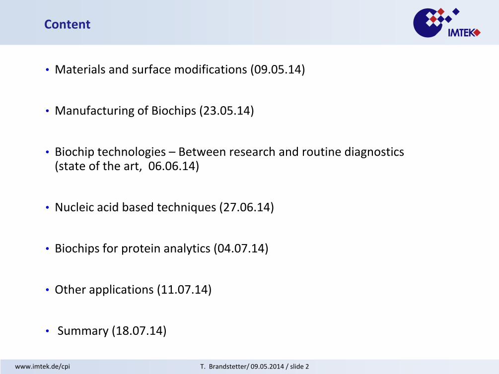
• Materials and surface modifications (09.05.14)
• Manufacturing of Biochips (23.05.14)
• Biochip technologies – Between research and routine diagnostics (state of the art, 06.06.14)
• Nucleic acid based techniques (27.06.14)
• Biochips for protein analytics (04.07.14)
• Other applications (11.07.14)
• Summary (18.07.14)
Content

T. Brandstetter/ 09.05.2014 / slide 3 www.imtek.de/cpi
Our profile
Research and teaching
• 22 faculties
• 300 researchers and technicians
• highly interdisciplinary world of
microsystem technology
IMTEK and industry
• Many industrial cooperations
• MSTBw
Core competences of CPI
• Preparation of surfaces with tailor-made
properties
• Topological and chemical micro
structuring of surfaces
• AFM
• Biochip-technologies
T. Brandstetter/ 09.05.2014 / slide 4 www.imtek.de/cpi
Biochip-technologies http://portal.uni-freiburg.de/cpi/biochip-group-dr-brandstetter

T. Brandstetter/ 09.05.2014 / slide 5 www.imtek.de/cpi
Biochips – what are they?(1)
• devices that can contain anywhere from tens to tens of millions of individual sensor elements (or biosensors)
• The sensors are packed together into a package typically the size of a microscope slide. Because so many sensors can be put into such a small area, a huge number of distinct tests can be done very rapidly.
• Biochips are often made using the same microfabrication technology used to make microchips. Unlike microchips, however, biochips are generally not electronic (although they can be).
• The key premise behind biochips is, that they can do chemistry on a small scale. Each biosensor can be thought of as a "microreactor“, which does chemistry designed to sense a specific analyte.

T. Brandstetter/ 09.05.2014 / slide 6 www.imtek.de/cpi
Biochips – what are they?(2)
• Biosensors can be made to sense a wide variety of analytes, including DNA, protein, antibodies, and small biological molecules.
• Fluorescence is often used to indicate a sensing event. Automated microscopy systems can be used to "read" the chip, i.e. determine which sensors are fluorescing
• Most biochips are 2D arrays of sensors placed carefully in a grid arrangement. The position of the sensor on the chip determines its function.
• To place the sensors in precise coordinates, sophisticated and expensive microdeposition techniques are used. The sensors are essentially placed one at a time, or serially, on the chip.
T. Brandstetter/ 09.05.2014 / slide 7 www.imtek.de/cpi
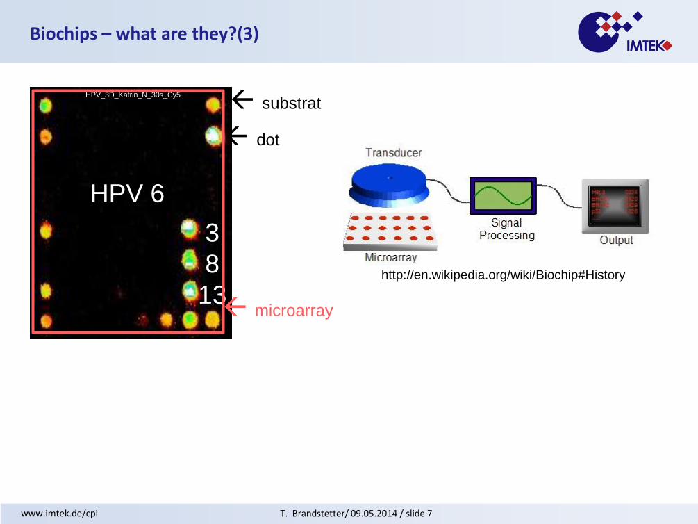
Biochips – what are they?(3)
3
8
13
HPV 6
HPV_3D_Katrin_N_30s_Cy5
substrat
dot
microarray
http://en.wikipedia.org/wiki/Biochip#History

T. Brandstetter/ 09.05.2014 / slide 8 www.imtek.de/cpi
Manufacturing of biochips – in general(1)
3. Immobilisation
2. Microarray printing
1. Untreated slide
mixed analyte solution

T. Brandstetter/ 09.05.2014 / slide 9 www.imtek.de/cpi
step1:
print polymer mixed with DNA
step 2:
photocrosslinking
via UV-irradiation
step 3:
hybridisation
and
readout
C OH
Manufacturing of biochips – in general(2)
T. Brandstetter/ 09.05.2014 / slide 10 www.imtek.de/cpi
Materials and surface modifications
T. Brandstetter/ 09.05.2014 / slide 11 www.imtek.de/cpi
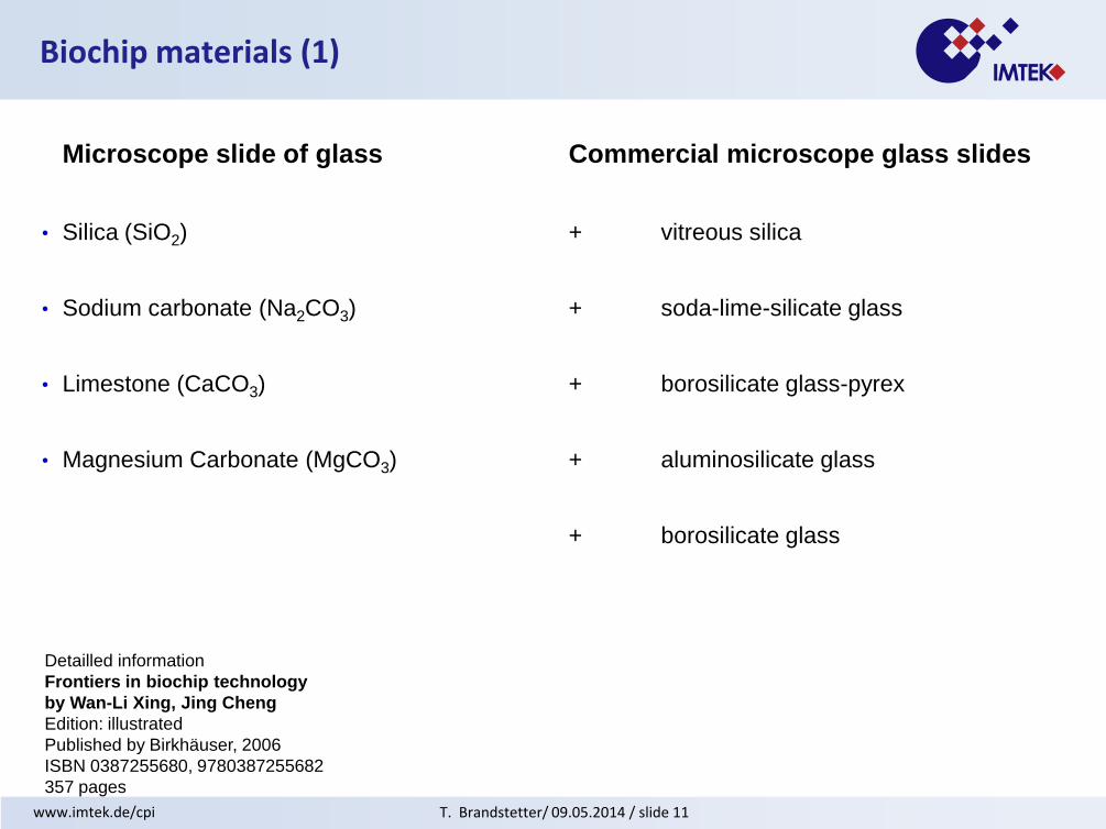
Biochip materials (1)
Microscope slide of glass Commercial microscope glass slides
• Silica (SiO2) + vitreous silica
• Sodium carbonate (Na2CO3) + soda-lime-silicate glass
• Limestone (CaCO3) + borosilicate glass-pyrex
• Magnesium Carbonate (MgCO3) + aluminosilicate glass
+ borosilicate glass
Detailled information
Frontiers in biochip technology
by Wan-Li Xing, Jing Cheng
Edition: illustrated
Published by Birkhäuser, 2006
ISBN 0387255680, 9780387255682
357 pages
T. Brandstetter/ 09.05.2014 / slide 12 www.imtek.de/cpi
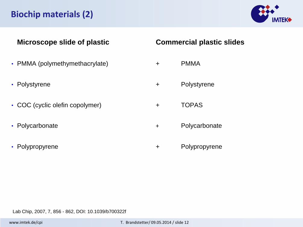
Biochip materials (2)
Microscope slide of plastic Commercial plastic slides
• PMMA (polymethymethacrylate) + PMMA
• Polystyrene + Polystyrene
• COC (cyclic olefin copolymer) + TOPAS
• Polycarbonate + Polycarbonate
• Polypropyrene + Polypropyrene
Lab Chip, 2007, 7, 856 - 862, DOI: 10.1039/b700322f
T. Brandstetter/ 09.05.2014 / slide 13 www.imtek.de/cpi
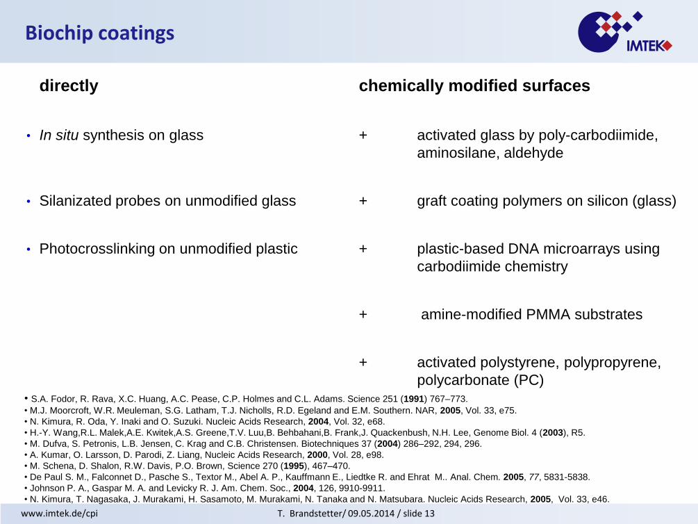
Biochip coatings
directly chemically modified surfaces
• In situ synthesis on glass + activated glass by poly-carbodiimide,
aminosilane, aldehyde
• Silanizated probes on unmodified glass + graft coating polymers on silicon (glass)
• Photocrosslinking on unmodified plastic + plastic-based DNA microarrays using
carbodiimide chemistry
+ amine-modified PMMA substrates
+ activated polystyrene, polypropyrene,
polycarbonate (PC)
• S.A. Fodor, R. Rava, X.C. Huang, A.C. Pease, C.P. Holmes and C.L. Adams. Science 251 (1991) 767–773.
• M.J. Moorcroft, W.R. Meuleman, S.G. Latham, T.J. Nicholls, R.D. Egeland and E.M. Southern. NAR, 2005, Vol. 33, e75.
• N. Kimura, R. Oda, Y. Inaki and O. Suzuki. Nucleic Acids Research, 2004, Vol. 32, e68.
• H.-Y. Wang,R.L. Malek,A.E. Kwitek,A.S. Greene,T.V. Luu,B. Behbahani,B. Frank,J. Quackenbush, N.H. Lee, Genome Biol. 4 (2003), R5.
• M. Dufva, S. Petronis, L.B. Jensen, C. Krag and C.B. Christensen. Biotechniques 37 (2004) 286–292, 294, 296.
• A. Kumar, O. Larsson, D. Parodi, Z. Liang, Nucleic Acids Research, 2000, Vol. 28, e98.
• M. Schena, D. Shalon, R.W. Davis, P.O. Brown, Science 270 (1995), 467–470.
• De Paul S. M., Falconnet D., Pasche S., Textor M., Abel A. P., Kauffmann E., Liedtke R. and Ehrat M.. Anal. Chem. 2005, 77, 5831-5838.
• Johnson P. A., Gaspar M. A. and Levicky R. J. Am. Chem. Soc., 2004, 126, 9910-9911.
• N. Kimura, T. Nagasaka, J. Murakami, H. Sasamoto, M. Murakami, N. Tanaka and N. Matsubara. Nucleic Acids Research, 2005, Vol. 33, e46.

T. Brandstetter/ 09.05.2014 / slide 14 www.imtek.de/cpi
2D chips using SAMs (self assembled monolayers)
typical DNA-chip design:
+ sensitivity
adapted from: E. Southern, K. Mir, M. Shchepinov, Nature Gen., 27 (1999) 5
+ surface properties
+ reproducibility (why is acceptance of microarrays below expectations in non-research areas?
sequence of the probe
polyT(thymine) tailer
Weakness:
T. Brandstetter/ 09.05.2014 / slide 15 www.imtek.de/cpi
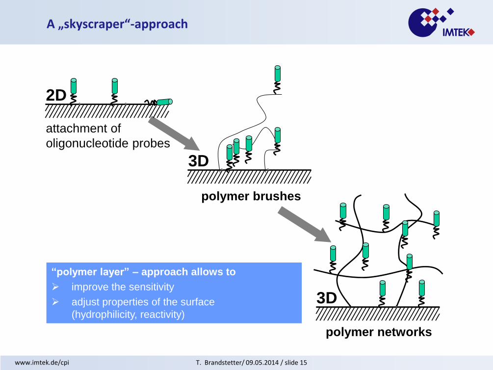
A „skyscraper“-approach
“polymer layer” – approach allows to
improve the sensitivity
adjust properties of the surface
(hydrophilicity, reactivity)
polymer brushes
attachment of
oligonucleotide probes
2D
3D
polymer networks
3D

T. Brandstetter/ 09.05.2014 / slide 16 www.imtek.de/cpi
chemisorption of polymers
grafting of polymers on
plasma modified
surfaces
photochemical attachment
of polymers
blockcopolymers
via macroinitators
growth of polymers
on surfaces
surface-attached
polymer networks „grafting in between“
Functional polymer monolayers
T. Brandstetter/ 09.05.2014 / slide 17 www.imtek.de/cpi
C O C O C
CCH
350 nmOH
CC
OH
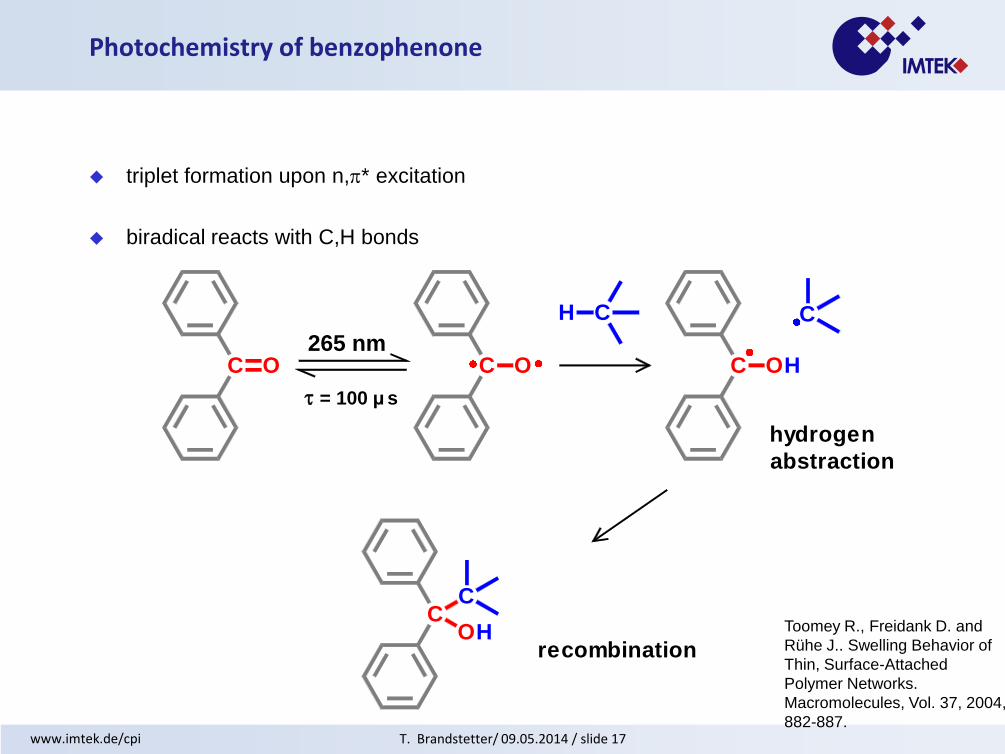
hydrogen
abstraction
recombination
= 100 µ s
Photochemistry of benzophenone
265 nm
triplet formation upon n,* excitation
biradical reacts with C,H bonds
Toomey R., Freidank D. and
Rühe J.. Swelling Behavior of
Thin, Surface-Attached
Polymer Networks.
Macromolecules, Vol. 37, 2004,
882-887.

T. Brandstetter/ 09.05.2014 / slide 18 www.imtek.de/cpi
Me
O O
O
ON
Me
Me
swelling in
water (2h)
polymeric substrate
(e.g. polyurethane)
photocrosslinkableovercoat
ca. 20 µm
~ 1 mm
simultaneous crosslinking
and surface attachment
via pendant benzophenone
units
Polymer networks attached to polymeric substrates
T. Brandstetter/ 09.05.2014 / slide 19 www.imtek.de/cpi
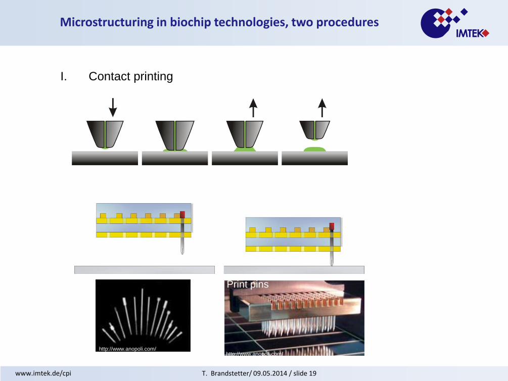
Microstructuring in biochip technologies, two procedures
I. Contact printing
http://www.anopoli.com/ http://www.anopoli.com/
Print pins Printhead

T. Brandstetter/ 09.05.2014 / slide 20 www.imtek.de/cpi
Microstructuring in biochip technologies, contact printing
Omnigrid from GeneMachine®
Contact printing procedure
65% humidity, RT
Steel or tungsten needle with reservoir
droplet volume 400 – 600 pl
droplet diameter 140 – 200 µm
Process variance > 10%
T. Brandstetter/ 09.05.2014 / slide 21 www.imtek.de/cpi
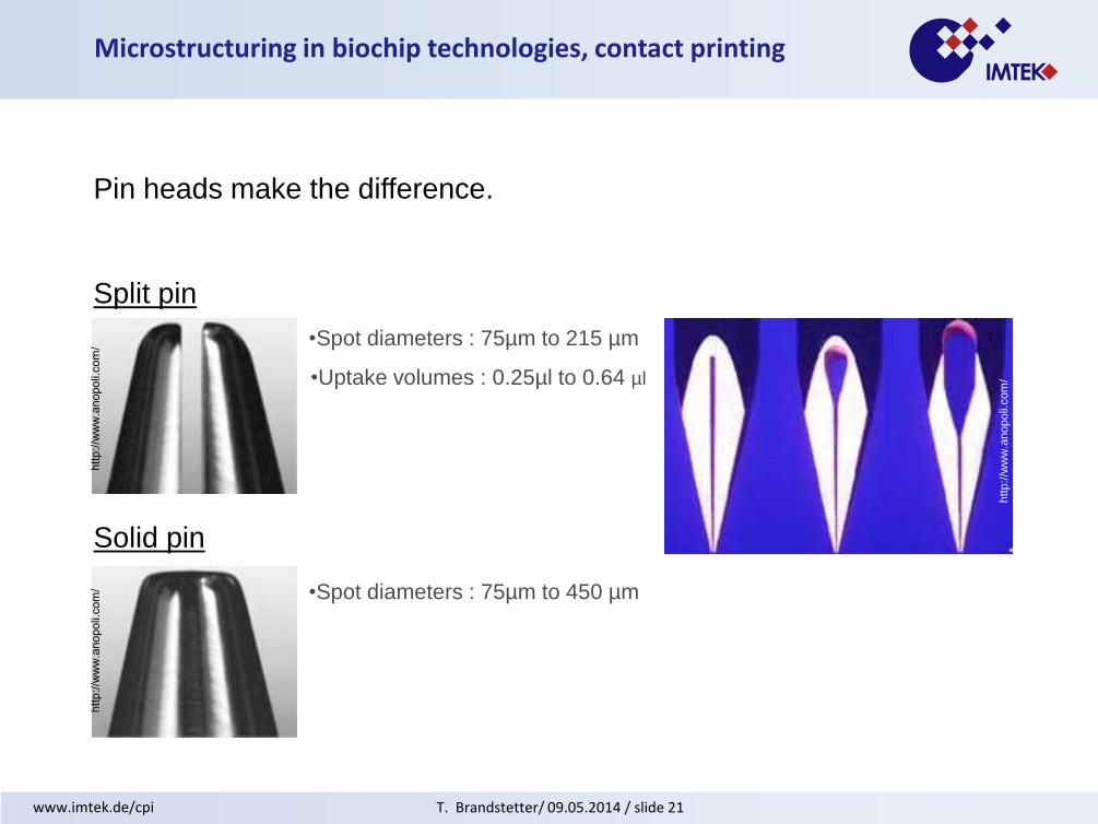
Microstructuring in biochip technologies, contact printing
Pin heads make the difference.
Split pin
Solid pin
•Spot diameters : 75µm to 215 µm
•Uptake volumes : 0.25µl to 0.64 µl
•Spot diameters : 75µm to 450 µm
htt
p:/
/ww
w.a
nopoli.
com
/
T. Brandstetter/ 09.05.2014 / slide 22 www.imtek.de/cpi
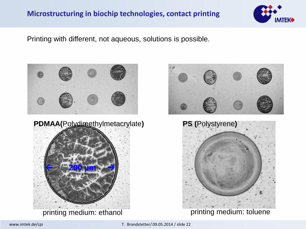
PDMAA(Polydimethylmetacrylate) PS (Polystyrene)
Microstructuring in biochip technologies, contact printing
Printing with different, not aqueous, solutions is possible.
printing medium: ethanol printing medium: toluene
200 µm

T. Brandstetter/ 09.05.2014 / slide 23 www.imtek.de/cpi
Microstructuring in biochip technologies, contact printing
Spot diameter is not really controllable.
Split pin
Solid pin
Printing of 0.25 µm Cy5-labelled oligo-DNA in 400
mM Napi and 1mg/ml PDMAA-co-5%MABP-co-
2,5%VPA

T. Brandstetter/ 09.05.2014 / slide 24 www.imtek.de/cpi
Microstructuring in biochip technologies, contact printing
scale lining
PDMAA layer
PMMA (5 mg/ml) lining
Printing medium toluene
exposure after
photocrosslinkage
T. Brandstetter/ 09.05.2014 / slide 25 www.imtek.de/cpi
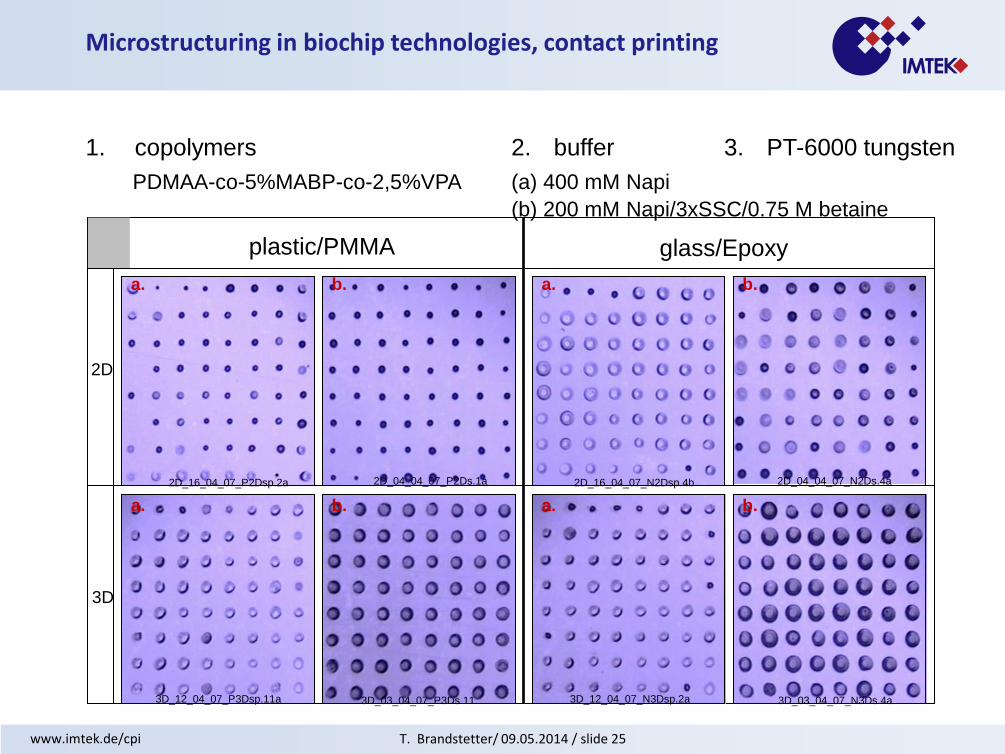
1. copolymers 2. buffer 3. PT-6000 tungsten
2D
3D
plastic/PMMA glass/Epoxy
PDMAA-co-5%MABP-co-2,5%VPA (a) 400 mM Napi
(b) 200 mM Napi/3xSSC/0.75 M betaine
2D_16_04_07_P2Dsp.2a 2D_16_04_07_N2Dsp.4b
3D_12_04_07_P3Dsp.11a
a. a.
3D_12_04_07_N3Dsp.2a
a. a. b.
b.
2D_04_04_07_N2Ds.4a
3D_03_04_07_N3Ds.4a
b.
2D_04_04_07_P2Ds.1a
3D_03_04_07_P3Ds.11
b.
Microstructuring in biochip technologies, contact printing
3D
T. Brandstetter/ 09.05.2014 / slide 26 www.imtek.de/cpi
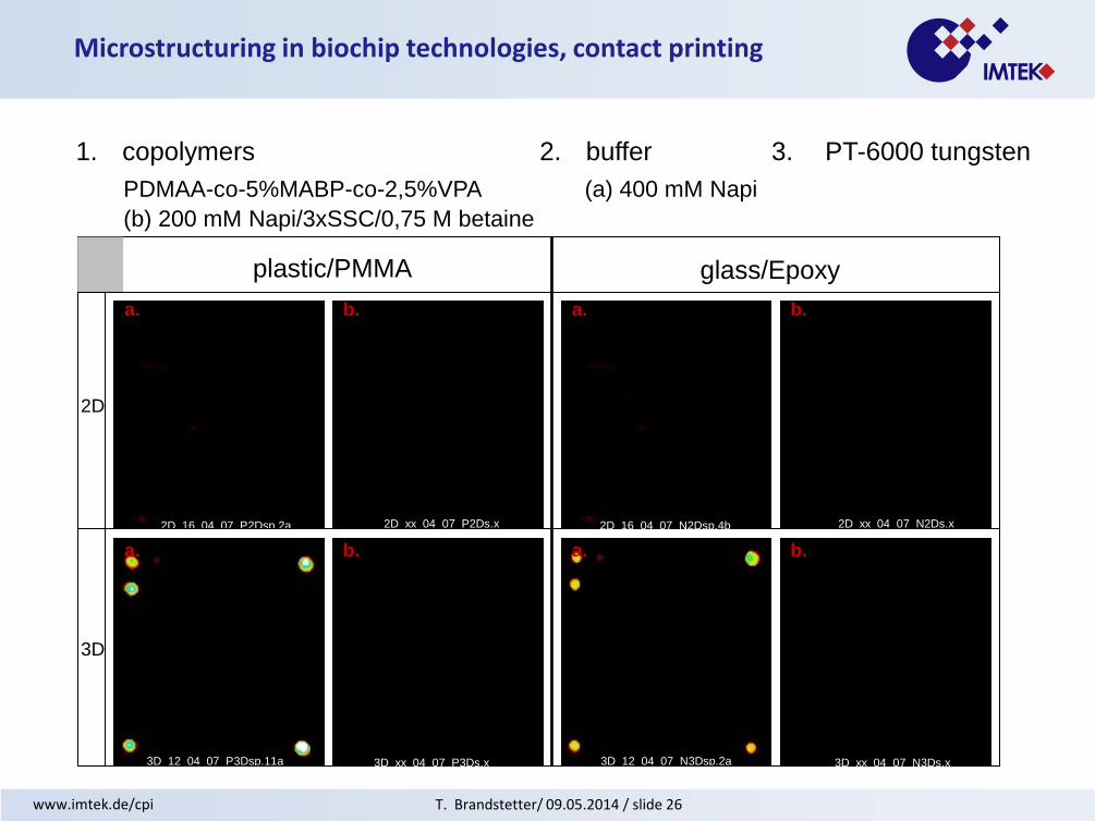
1. copolymers 2. buffer 3. PT-6000 tungsten
2D
3D
plastic/PMMA glass/Epoxy
PDMAA-co-5%MABP-co-2,5%VPA (a) 400 mM Napi
(b) 200 mM Napi/3xSSC/0,75 M betaine
2D_16_04_07_P2Dsp.2a
3D_12_04_07_P3Dsp.11a
a. a.
3D_12_04_07_N3Dsp.2a
a. a. b.
b.
2D_xx_04_07_N2Ds.x
3D_xx_04_07_N3Ds.x
b.
2D_xx_04_07_P2Ds.x
3D_xx_04_07_P3Ds.x
b.
2D_16_04_07_N2Dsp.4b
Microstructuring in biochip technologies, contact printing
2D
3D
T. Brandstetter/ 09.05.2014 / slide 27 www.imtek.de/cpi
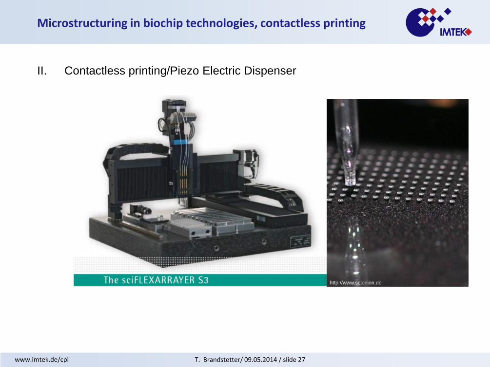
Microstructuring in biochip technologies, contactless printing
II. Contactless printing/Piezo Electric Dispenser
http://www.scienion.de

T. Brandstetter/ 09.05.2014 / slide 28 www.imtek.de/cpi
Microstructuring in biochip technologies, contactless printing
II. Piezo Electric dispenser
Piezo Electric dispenser(Scienion AG®)
Contactless printing procedure
65% humidity, RT
droplet volume 410 pl,
droplet diameter 175 µm
droplet volume and diameter is
adjustable
Process variance < 10%
T. Brandstetter/ 09.05.2014 / slide 29 www.imtek.de/cpi
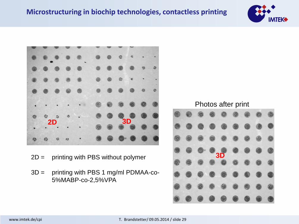
Photos after print
Microstructuring in biochip technologies, contactless printing
2D
3D
3D
2D = printing with PBS without polymer
3D = printing with PBS 1 mg/ml PDMAA-co-
5%MABP-co-2,5%VPA

T. Brandstetter/ 09.05.2014 / slide 30 www.imtek.de/cpi
Microstructuring in biochip technologies, contactless printing
Droplet stacking
1mg/ml polymer in distilled water
PSS = Polystyrenesulfanit
PMMA = Polymethylmetacrylate
Small droplet with 10x
Large droplets with 20x
Photo after print
PSS PMMA
T. Brandstetter/ 09.05.2014 / slide 31 www.imtek.de/cpi
Microstructuring in biochip technologies, contactless printing
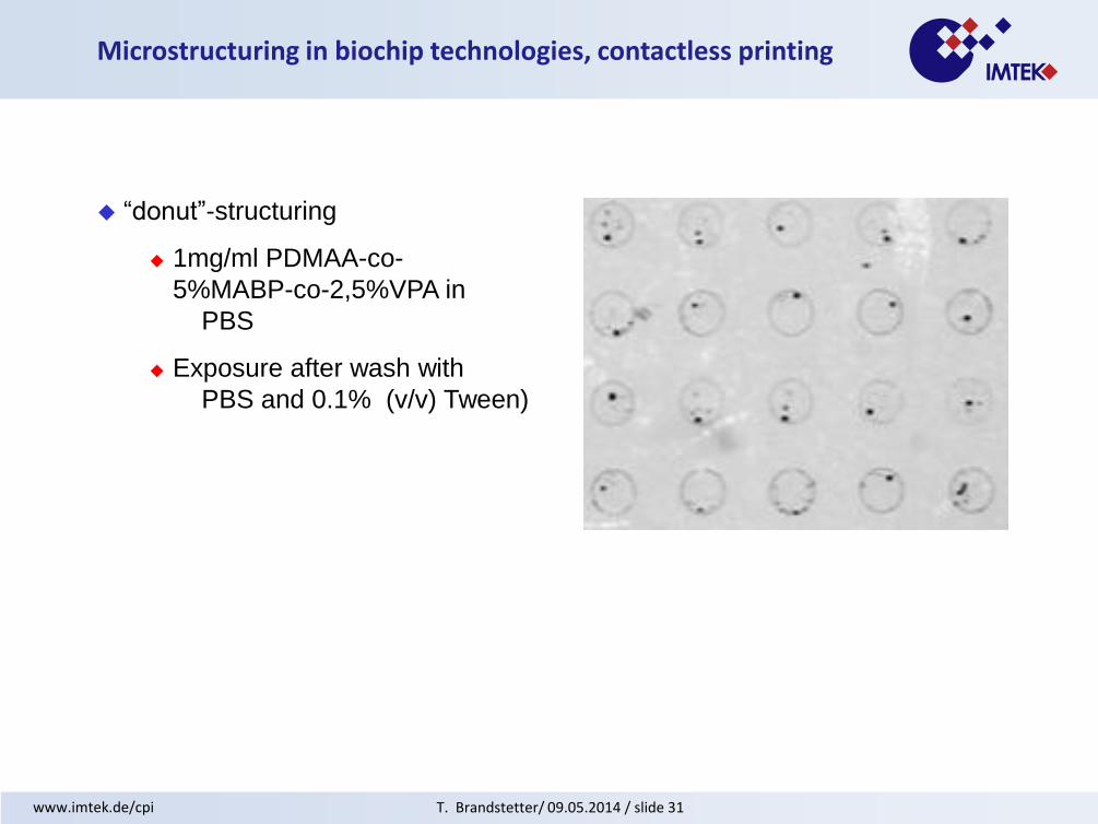
“donut”-structuring
1mg/ml PDMAA-co-
5%MABP-co-2,5%VPA in
PBS
Exposure after wash with
PBS and 0.1% (v/v) Tween)

T. Brandstetter/ 09.05.2014 / slide 32 www.imtek.de/cpi
Dot morphology, how to analyze?
Dot morphology, depending on
surface properties
print solution contact angle
analyte concentration
Dot morphology, analyzed by
AFM
Fluorescence microscope
Raster electron microscope
T. Brandstetter/ 09.05.2014 / slide 33 www.imtek.de/cpi
Microstructuring in biochip technologies, contactless printing
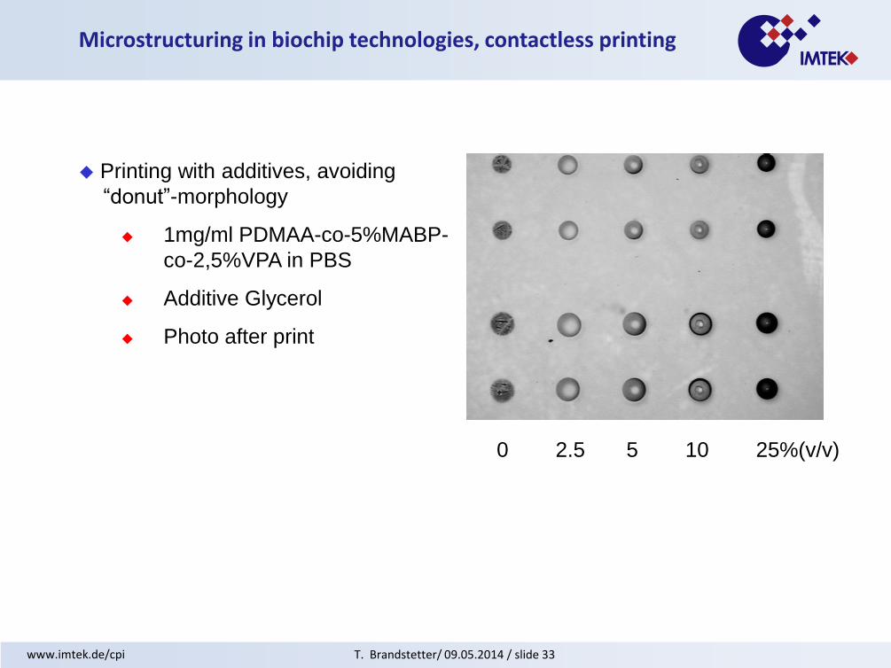
Printing with additives, avoiding
“donut”-morphology
1mg/ml PDMAA-co-5%MABP-
co-2,5%VPA in PBS
Additive Glycerol
Photo after print
0 2.5 5 10 25%(v/v)

T. Brandstetter/ 09.05.2014 / slide 34 www.imtek.de/cpi
Microstructuring in biochip technologies, contactless printing
Printing with/withoutTrehalose
1mg/ml PDMAA-co-5%MABP
-co-2,5%VPA in PBS
125 mg/ml Trehalose (T) in PBS
“Donut”-structure without
Trehalose
Homogeneity in the dot
morphology, using Trehalose
-T
-T
+T
+T
α-D-glucopyranosyl α-D-
glucopyranoside(α,α‐Trehalose)
http://en.wikipedia.org/wiki/Trehalose
Exposure with a fluorescence microscope
T. Brandstetter/ 09.05.2014 / slide 35 www.imtek.de/cpi
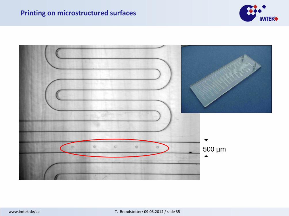
Printing on microstructured surfaces
500 µm

T. Brandstetter/ 09.05.2014 / slide 36 www.imtek.de/cpi
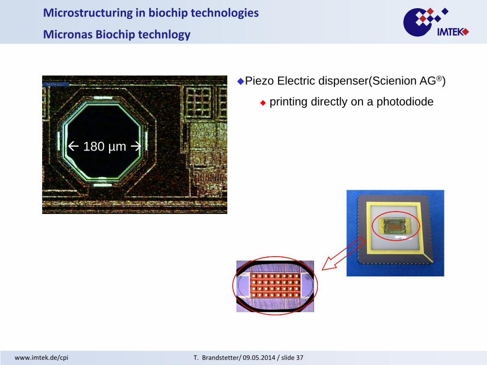
Microstructuring in biochip technologies
Micronas Biochip technlogy
Piezo Electric dispenser(Scienion AG®)
Contactless printing procedure
80% humidity, RT
droplet volume 390 pl,
photodiode diameter 180 µm
printing on structured surfaces
Process variance < 10%
T. Brandstetter/ 09.05.2014 / slide 37 www.imtek.de/cpi
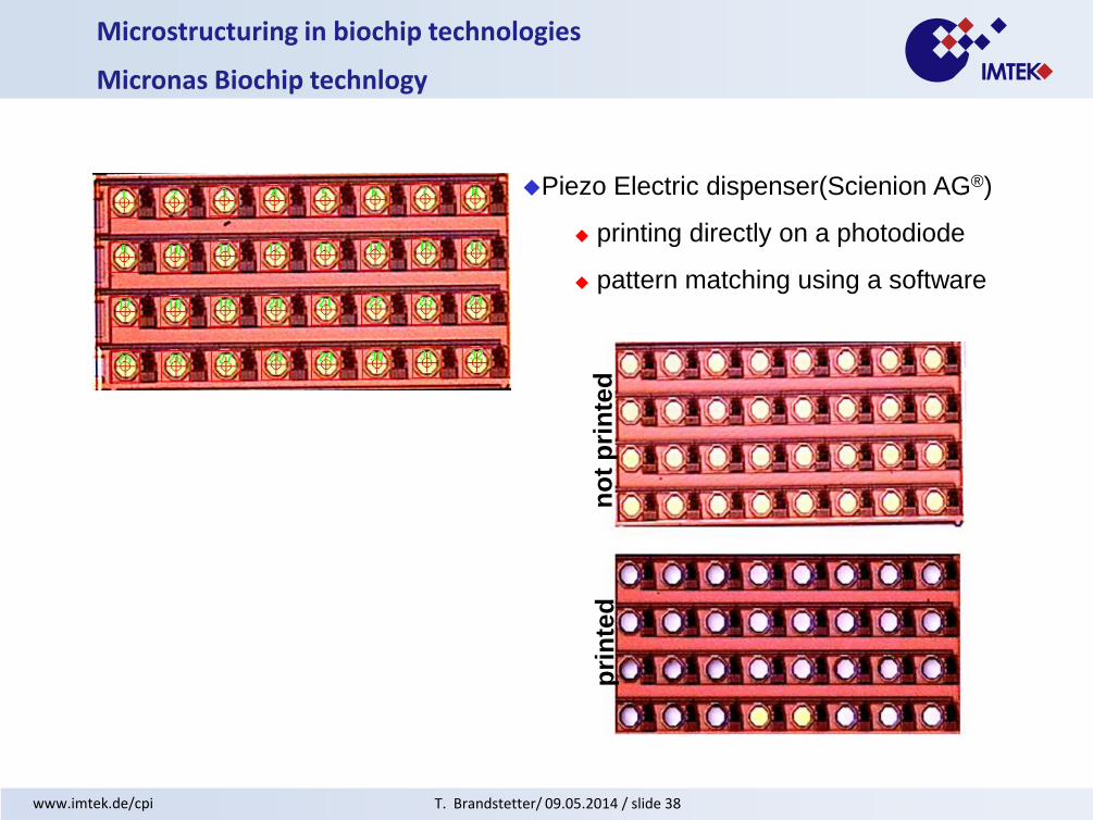
Microstructuring in biochip technologies
Micronas Biochip technlogy
Piezo Electric dispenser(Scienion AG®)
printing directly on a photodiode
180 µm
T. Brandstetter/ 09.05.2014 / slide 38 www.imtek.de/cpi
Microstructuring in biochip technologies
Micronas Biochip technlogy
Piezo Electric dispenser(Scienion AG®)
printing directly on a photodiode
pattern matching using a software
no
t p
rin
ted
pri
nte
d

T. Brandstetter/ 09.05.2014 / slide 39 www.imtek.de/cpi
Piezo Electric dispenser (Scienion AG®)
Contactless printing procedure
Droplet volume control
Droplet diameter tunable (>100µm)
Printing only with aqueous solutions
1mg/ml polymer
Process variance < 10%
Omnigrid from GeneMachine®
Contact printing procedure
Steel or tungsten needle with reservoir
droplet volume 400 – 600 pl
droplet diameter approx. 200 µm
Printing of different solutions
> 1mg/ml polymer possible
Process variance > 10%
Microstructuring in biochip technologies, summary

T. Brandstetter/ 09.05.2014 / slide 40 www.imtek.de/cpi
Thank you for your attention!
http://www.bilder-welten.net/de/produkt_detail.php?id=23019&catid=1623
T. Brandstetter/ 09.05.2014 / slide 41 www.imtek.de/cpi
Literature
• E. Southern, K. Mir, M. Shchepinov, Nature Gen., 27 (1999) 5
• Frontiers in biochip technology, by Wan-Li Xing, Jing Cheng, Edition: illustrated, published by
Birkhäuser, 2006, ISBN 0387255680, 9780387255682, 357 pages
• Lab Chip, 2007, 7, 856 - 862, DOI: 10.1039/b700322f
• S.A. Fodor, R. Rava, X.C. Huang, A.C. Pease, C.P. Holmes and C.L. Adams. Science 251
(1991) 767–773.
• M.J. Moorcroft, W.R. Meuleman, S.G. Latham, T.J. Nicholls, R.D. Egeland and E.M. Southern.
NAR, 2005, Vol. 33, e75.
• N. Kimura, R. Oda, Y. Inaki and O. Suzuki. Nucleic Acids Research, 2004, Vol. 32, e68.
• H.-Y. Wang,R.L. Malek,A.E. Kwitek,A.S. Greene,T.V. Luu,B. Behbahani,B. Frank,J.
Quackenbush, N.H. Lee, Genome Biol. 4 (2003), R5.
• M. Dufva, S. Petronis, L.B. Jensen, C. Krag and C.B. Christensen. Biotechniques 37 (2004)
286–292, 294, 296.
• A. Kumar, O. Larsson, D. Parodi, Z. Liang, Nucleic Acids Research, 2000, Vol. 28, e98.
• M. Schena, D. Shalon, R.W. Davis, P.O. Brown, Science 270 (1995), 467–470.
• De Paul S. M., Falconnet D., Pasche S., Textor M., Abel A. P., Kauffmann E., Liedtke R. and
Ehrat M.. Anal. Chem. 2005, 77, 5831-5838.
• Johnson P. A., Gaspar M. A. and Levicky R. J. Am. Chem. Soc., 2004, 126, 9910-9911.
• N. Kimura, T. Nagasaka, J. Murakami, H. Sasamoto, M. Murakami, N. Tanaka and N.
Matsubara. Nucleic Acids Research, 2005, Vol. 33, e46.
• Toomey R., Freidank D. and Rühe J.. Swelling Behavior of Thin, Surface-Attached Polymer
Networks. Macromolecules, Vol. 37, 2004, 882-887.