in 1675, dutch microscopist anton van leeuwenhoek … · 24 asu research— f all/winter 1995 in...
TRANSCRIPT

24 A S U R E S E A R C H — F A L L / W I N T E R 1 9 9 5
In 1675, Dutch microscopist Anton van Leeuwenhoek had to grind
his own lenses. Today, scientists use innovative technology
and techniques to inspect the microscopic world.
b y A l a n a M i k k e l s e n
Below–Different microscopic techniques produce different views of the same structure.The top micrograph was made with a standard light microscope using differentialinterference contrast (DIC) optics. The imagedepicts the late stages of zoospore formationin the zoosporangium, the spore-formingstructure of the fungi Allomyces macrogynus.The bottom image was made using a scan-ning transmission electron microscope afterthe specimen was prepared by high-pressurefreezing and freeze-substitution methods. Top right–a video-enhanced digital lightmicrograph shows a zoospore being releasedfrom the zoosporangium. Under normal conditions, each zoospore has a single nucleus and a flagellum, a long whiplikestructure used for swimming.

It sounds like a straight-forward process, butthe machine that carried out the freezing is anhpm 010, a $170,000 example of engineeringprecision. Only nine American institutionshave one of the high pressure freezers.Researchers at Arizona State University arethe first scientists in the West to have accessto such a device.
The high-pressure freezing device repre-sents one of the most sophisticated ways toprepare organisms for viewing under the elec-tron microscope. As part of the Life SciencesElectron Micrsoscopy Facility, it is the latestaddition to a wealth of high-tech tools asubiologists are using to unlock the secretswithin the microscopic underworld.
The tools range from computer-aidedobservation to innovative methods of prepa-ration, and they encompass both light andelectron microscopes. All are part of a currentbrand of technologies that, within the lastdecade or so, have made it increasingly pos-sible for life scientists to study their minus-cule subjects in almost undisturbed andincreasingly accurate detail.
Using rapid freezing methods for electronmicroscope studies allows researchers tosee and study their specimens in a moretrue-to-life form.
“Theoretically, we could thaw the cellsafter we freeze them and they would stillbe alive,” explains Roberson, director ofthe electron microscopy facility and asso-
ciate professor of botany.Roberson’s gold-rimmed glasses rest on
his forehead above his eyebrows, where he isapt to lift them anytime he is about to peer intoa microscope. He’s been doing this kind ofwork for more than a decade,mostly studyingfungi. He’s convinced that the high pressure
freezer is one of the best methods available forpreserving large samples of living tissue.
Tinkering With The Tiny Preparing a living thingfor viewing under an electron microscope isnot a simple task. Electron microscopes pro-duce their images by transmitting beams ofelectrons through a sample and, much likecasting a shadow, an image of the specimen isgenerated on a special screen.
For the procedure to work, the specimenmust be placed in a vacuum; otherwise, airand other molecules would deflect the electronbeam into a useless scatter. Under normal cir-cumstances, such an evacuated chamber wouldblow a cell apart. To compensate, scientistsmust stabilize their cells and tissue specimens.
Traditionally, electron microscopists pre-pared specimens by submerging them inchemicals which formed stable links betweenthe molecules of a cell’s internal structures, ororganelles. The process, called fixation, pre-serves the subcellular architecture so the tis-sue can be prepared for viewing. Samples arethen usually embedded in an epoxy resin,sliced ultra-thin with a diamond knife, andplaced on a metal grid.
Despite its pivotal role in microscopicstudies, fixation also comes with a trade-off.The same chemicals that immobilize cellularcomponents sometimes interact with them aswell, causing abnormalities in their shape andposition.
“With chemical fixation, depending onhow thick the tissue is, it takes anywhere from30 seconds to five minutes for a fixative topenetrate the tissue,” explains Mike Harding,a graduate student in Roberson’s lab. “Mean-while, the cell reacts to the slow rate of fixa-tion in an adverse way, and probably is doing
all kinds of crazy things. So what you see maynot be representative of what the specimenlooked like prior to being fixed,” Harding addsas he prepares to study root fungi with theelectron microscope and the hpm 010.
The high-pressure freezer solves some ofthese problems and makes it much more likelythat the way a cell looks after it is prepared isconsistent with its prefixation state.
Indeed, since asu acquired the hpm 010 inAugust 1994, Roberson and master’s studentDavid Lowry conducted experiments that indi-cated some molecules involved in the processof maintaining cytoplasmic order were muchmore numerous in cells fixed with the rapidhigh-pressure freezer than in cells fixed usingconventional chemical methods. Robersonattributes the difference to chemical degrada-tion in the traditionally-prepared samples.
The high-pressure freezer blasts cells witha stream of liquid nitrogen which, at 197degrees below zero Celsius, stops lifeprocesses within miliseconds. In the life of acell, that translates to an almost instantaneousarrest of biological activity.
The result: a cell’s metabolism is essen-tially suspended, but the cell has not yet died.By using a process called “freeze substitu-tion,” scientists can then use chemicals tolock the frozen molecules in place. But unlikewith chemical fixation, they are not relying onthe chemicals to stop the biological activity inthe first place.
If this description conjures up futuristicimages of preserving whole corpses so than anindividual might be revived to meet his or hergrand-relatives at some far-distant date, nipthose dreams right now. Roberson says thehigh-pressure freezer–and cryogenics in gen-eral–work only for very small pieces of tissue.
A S U R E S E A R C H — F A L L / W I N T E R 1 9 9 5 25
FUZZY WHITE BLOBS waver in Robby Roberson’s
view as he turns a knob on the side of his microscope.
He selects the organisms as they come into focus.
Using forceps, he then selects and sets each sample into
the depression of a tiny gold disc. One disc gets clamped
onto the end of a heavy, nine-inch-long silver cylinder,
which Roberson carefully places in the top of a chest high
rectangular machine. With the push of a button, the silence
of the laboratory is broken as a blast of liquid nitrogen
slams into the specimens. An icy fog begins to waft from
the machine’s inner chamber. The specimen, a fungus
called Allomyces, has just been frozen alive.
Robby Roberson
MIC
RO
GR
AP
HS
CO
UR
TES
Y R
OB
BY
RO
BE
RS
ON
, PH
.D.
JOHN C. PHILLIPS PHOTO

“You can’t take a pocket gopher, freeze it,and still have a viable pocket gopher,” Robersonsays. “But you can take simpler organisms,freeze them and still have viable organisms.”
Freezing requires that heat be removedfrom a sample. The larger the sample, themore heat it contains, and the more time isrequired to freeze it. And time, as it turns out,is the ally of a cryogenic researcher’s greatestenemy: ice.
The longer it takes to freeze a sample, themore likely that the water in a cell will solid-ify into large crystals, which poke holes in acell’s interior and rip apart its internal struc-ture. So scientists get a ravaged landscaperather than an image of a cell’s true form.They end up back at square one.
One solution is to use antifreeze-like sub-stances called cryoprotectentsto prevent crystalformation. But cryoprotectents, like chemicalfixatives, can distort cellular structure. So sci-entists turn to methods that take the heat out ofcells so fast that ice crystals have little chanceto grow from their tiny, pellet-like precursors.
asu developmental biologistDougChandlerwas one of the first to use quick-freeze tech-niques in the late 1980s. He remembers thecontroversy surrounding findings which wereobtained using different preparation methods.The findings were related to a process calledexocytosis, which allows a cell to move sub-stances from its interior to the space outside itssurface membrane.
Exocytosis happens like the reverse ofsticking one’s finger into a balloon and pinch-ing off the mini bubble that forms. Small vesi-cles, or circles of lipid, encompass cellularsubstances packaged inside the cell. The sub-stances, usually proteins, are released whentheir vesicles fuse with the cellular membrane.
Some scientists saw a loop or “bleb” onthe outside of the cell’s membrane during thisprocess; while others observed that proteinsembedded in the cell’s surface membrane dis-appeared just prior to the exocytosis of under-lying vessicles. The “blebs” were thought tobe an intermediate step in the exocytosisprocess, but were later shown to appear onlyif the cryoprotectant glycerin was used.
Chandler and his colleagues showed thatthe second phenomenon, called protein clear-ing, appeared in chemically fixed cells, butnot in cells that were frozen Both phenomenahad been observed so many times, and wereso vivid, that Chandler’s peers were skepticalof his conclusions at first. Today, he thinksthat their results, not his, were what scientistscall “artifact”– a misleading result attributedto experimental manipulation rather than agenuine phenomenon.
Importantly, Chandler notes that whatevercaused the artifacts in the first place must havesome biochemical cause, but he’s not pursuingthe meaning of that process at the moment.
“We think we’ve dispelled 80 percent ofthe doubt (that his results were artifact). Butprotein clearing was thought to be an impor-tant part of the exocytosis process for about10 years,” Chandler says.
When he began his career, Chandler stud-ied the cortical reaction, a classic example ofexocytosis which occurs in recently-fertilizedegg cells. A split second after the sperm pen-etrates the egg, vesicles lying below the egg’ssurface membrane fuse and release proteinsinto the extracellular space.
The reaction was long known to cause theonce-receptive egg to become impenetrable tosperm (preventing more than one sperm frominserting its genetic material into the egg–a sit-uation fatal to the new embryo in most cases).
Part of the way the reaction accomplishedthis was by causing a thin membrane that encir-cled the egg surface to lift away from the cell.The membrane, called the vitelline envelope,appeared in micrographs of thin sections as awispy, fairly uninteresting layer bounding theoutside of the cell. No one paid much attentionto it. Until one day, when Chandler viewed thevitelline envelope using a new technique.
Chandler was then a post-doctoral studentstudying sea urchins. At the time, his mentorwas developing a process called deep etching.Prior to that, samples were commonly freeze-
fractured, meaning frozen and then crackedopen. The fracture usually happened alongthe line between the two layers of a cell’s sur-face membrane. The outer half of the mem-brane–and the vitelline envelope that sur-rounded it–was usually washed away toreveal the detail of the membrane’s interior.
In most cases, the “freeze” part of freeze-fracture was accomplished by dipping thesamples into liquid freon, liquid propane, orsome other cryogen. But “plunge freezing,” asit is called, is fairly slow, so glycerin is usuallyadded to prevent ice crystals from forming.
The deep etching technique hinges on aprocess called “sublimation,” which cannotoccur in the presence of glycerin. So, insteadof plunging samples into cryogen, Chandlerslammed them against a super-cooled mirroror block of copper, which sucked the heat outin a matter of miliseconds.
Then water was sublimed, or evaporated,by going directly from the solid to the gaseousphase. Gradual sublimation exposes the three-dimensional surface of a sample from the out-side inward, like an ice block melting away toreveal a solid sculpture within.
The sample is then sprayed with metalfrom all sides, and the original specimen iswashed away to reveal a three-dimensional,platinum “replica” of the specimen that canbe viewed under the microscope.
Because the whole specimen was intact,Chandler was able to view the vitelline enve-lope in detail. What he saw was a vast networkof interwoven proteins above the cellularmembrane that seemed intricately complex.More importantly, it drastically changed itsappearance and structure in response to therelease of proteins during the cortical reaction.
The network, the vitelline envelope, wasan example of extracellular matrix–proteinsthat exist outside of cells and link themtogether or perform other important functions.
The metamorphosis Chandler observed inthe extracellular matrix of the sea urchin eggchanged the direction of his career and others’.People began to study exacly how the vitellineenvelope changed to perform the two com-pletely different functions of enticing sperminto and keeping sperm out of the egg.
“It was a surprise finding in the sense thatI wasn’t really interested in the extracelluarmatrix,” Chandler recalls. “People talkedabout it. But I just routinely decided to deepetch my exocytosis stuff. I can’t rememberwhy exactly–probably just to get a differentview of the exocytosis process.
“Suddenly we saw the pictures, and I said,‘Holy cow, what is that?’ Then it dawned on usand we said,‘Of course.That’s the stuff you canhardly see in thin sections that people think is
26 A S U R E S E A R C H — F A L L / W I N T E R 1 9 9 5
Electron microscopy combined with freezefracture techniques provide scientists withextremely detailed images of cellular struc-tures. An oblique freeze fracture through the surface of a frog’s egg magnified 60,000times. The fibrous-looking structures areextracellular matrix material found outsidethe egg’s cell membrane.
Electron microscopy combined with freezefracture techniques provide scientists withextremely detailed images of cellular struc-tures. An oblique freeze fracture through the surface of a frog’s egg magnified 60,000times. The fibrous-looking structures areextracellular matrix material found outsidethe egg’s cell membrane.
MIC
RO
GR
AP
HS
CO
UR
TES
Y D
OU
GLA
S C
HA
NLE
R, P
H.D
./ J
OH
N C
. PH
ILLI
PS
PH
OTO
important for sperm reception.’ We immedi-ately knew something fantastic was there.”
Chandler has since become expert at theslam-freeze/deep-etch process, which fewpeople in the world bother to tackle.
“It’s enough of a pain that not many peopledo it routinely,’ he explains, “even though it’swidely recognized as one of the best ways toget the best possible preservation.”
Chandler also has pioneered the under-standing of egg extracellular matrices andcharacterized the precise biochemical changesof the vitelline envelope in both sea urchin andfrog eggs.
Using microscopy and biochemical studies,he has shown that the vitelline envelopechanges from a loose network of proteins intoan armor-like coating that protects the egg, bothfrom further fertilization and probably frominfectious agents such as viruses and bacteria.
Most of Chandler’s work has been doneon pieces of egg. Now he wants to use thehigh-pressure freezer to study a different kindof extracellular matrix. This one resides onthe inside of early embryos and guides thecontinuously dividing sheets of primordialnerve, muscle, and gut cells into their finalpositions in the developing fetus.
For this work, he wants to view whole seaurchin eggs which, at a diameter of 0.5milimeters, are too big to slam freeze withoutbeing littered with ice crystals.
“The high pressure freezer is the onlytechnique (other than chemical fixation) thatcan freeze a sample that big for electronmicroscopy,” Chandler explains.
Indeed, the ability to effectively preparethick specimens without chemical artifact isone of the greatest advantages of the high-
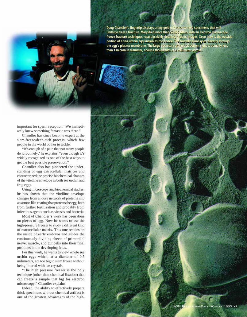
Doug Chandler’s fingertip displays a tiny gold grid used to hold specimens that will undergo freeze fracture. Magnified more than 50,000 times with an electron microscope,freeze fracture techniques result in richly detailed, artistic images. Seen here is the outsideportion of a sea urchin egg known as the cortex. The fracture plane goes directly throughthe egg’s plasma membrane. The large secretory granule at bottom right is actually lessthan 1 micron in diameter, about a-thousandth of a millimeter across.
Doug Chandler’s fingertip displays a tiny gold grid used to hold specimens that will undergo freeze fracture. Magnified more than 50,000 times with an electron microscope,freeze fracture techniques result in richly detailed, artistic images. Seen here is the outsideportion of a sea urchin egg known as the cortex. The fracture plane goes directly throughthe egg’s plasma membrane. The large secretory granule at bottom right is actually lessthan 1 micron in diameter, about a thousandth of a millimeter across.
mag
es c
ourt
esy
Dou
glas
Cha
ndle
r, P
h.D
.
A S U R E S E A R C H — F A L L / W I N T E R 1 9 9 5 27
28 A S U R E S E A R C H — F A L L / W I N T E R 1 9 9 5
pressure freezer. It’s partly what drew graduatestudent Mike Harding to asu from Canada.
Harding wants to study a fungal-plantassociation called vam, or vesicular arbuscu-lar mycorrhizal fungus. Like all mycorrhizalfungi, vam grow inside or near plant roots.Although micorrizal relationships are not fullyunderstood, scientists generally believe thatthe pairing is a mutually beneficial one, withthe fungus providing hard-to-get-at nutrientslike phosphorous to the plant in exchange forthe photosynthetic byproduct carbon, whichthe fungus uses as food.
More than 90 percent of the Earth’s plantsform associations with mycorrhizal fungi,including virtually all forest species andalmostevery agriculturally important plant. Still, agreat deal remains to be discovered about thedynamics of the fungus-plant relationship.
Some unanswered questions include: Do both parties always benefit from the asso-ciation? Who benefits the most? Does therelationship change from mutually beneficialto exploitative during periods of stress andfood shortage? How might such a shift affectcrop production or forest growth during, forexample, times of drought?
To answersome of these questions,Hardingintends to study vam arbuscules, convolutedstructures of membrane that serve as nutrientexchange organs. He will start by growingvam in the roots of bell peppers and charac-terizing the appearance of the root-arbusculeinterface. Then he will subject the plant-fungus system to various stressors, includingheat, to see how the relationship changes.
The magnitude of his task is monumental.To study the arbuscules, he must first findthem. Although vam can grow in about 80percent of a plant’s root, arbuscules only formin a portion of the fungal mass.
Moreover, each arbuscule is a complexthree-dimensional structure that is only pre-sent for between eight and 10 days, and onlyone part of the arbuscule is the focal point fornutrient transfer. Harding says that if he had
to search through root tissue in search ofarbuscules using specimens one or two celllayers thick, it would be impossible.
“I wouldn’t even try it,” he says.But because the high-pressure freezer
allows him to freeze samples up to half amilimeter thick, Harding says he can realis-tically use cryotechniques to visualize the theroot system in search of the appropriate struc-tures. He can then use thin sections to recon-struct the structures in three dimensions.
The secret to the hpm 010’s success atfreezing that all-elusive thick sample?Pressure. And timing.
The liquid nitrogen is fired at the sampleusing a blast of pressure equivalent to 300,000pounds per square inch–the same force as theblast from the barrel of a 30.06 rifle.
At that pressure, a living sample would beblown apart. But the extra-cold nitrogenreaches the sample a fraction of a secondbefore the pressure, and the specimen isfrozen solid before it can be harmed.
The high-pressure freezer is also effectivebecause it outsmarts water. At that pressure,water is frozen in a vitreous, or glass-likestate. Thus, the absence of ice crystals.
The process is not fool-proof, however. Infact, it takes considerable practice to carry outany of thesehighly-sensitive fixationprocesses.
Quing Fang He, an asu doctoral candidatein botany, knows this all too well. His objectof interest is a bacterium called Synchocystis.
Under Roberson’s guidance, He has beentrying to fix samples of Synchocystis so thathe can study how the cyanobacterium mobi-lizes proteins that regulate the expression ofthe photosynthetic pigment chlorophyll inresponse to changes in light. But he keepsgetting ice crystals.
“It’s not simplicity that counts. It’s theresults,” says He’s advisor, Willem Vermaas,asu professor of botany.
One of the most powerful advantages ofthe high-pressure freezer is that it maintainsthe chemical and structural integrity of most
biological molecules in a specimen. For He,that carries the promise of a good view ofcomplex photosynthetic membranes that aredifficult to see in chemically fixed tissue.
It also means that scientists can combinetraditional, structural studies of cellular spacewith state-of-the-art functional techniques.That kind of advance extends the utility of themicroscope, an instrument whosebasicphysicalandopticalprincipleshaveremainedessentiallyunchanged since the middle of this century.
“Methodology to a great extent drives anyscience,” says Allan Bieber, director of ASU’smolecular and cellular biology program.“Whenever you get a significant advancementin methodology, you usually see a corre-sponding jump in knowledge.”
The jump can be astounding when inves-tigators combine multiple methods, as RobbyRoberson does. Using light microscopy, con-ventional and freeze-prepared electronmicroscopy, computer-aided viewers thatenhance contrast, and video microscopy,Roberson is piecing together the process ofapical growth in the fungus Allomyces.
Apical growth occurs at the ends of fungalhyphae, but it is analagous to other growthprocesses as well.
“Apical growth is fairly unique in eukary-otes (non-bacterial organisms). But nerve cellsand root hairs do it as well,” says Roberson.
In 1992, Roberson and graduate studentMargie Vargas happened upon an unexpectedsight that triggered his interest in Allomyces.
“It was one of those late nights in the lab,and I saw something no one had ever seenbefore. Textbooks are going to be rewritten nowbecause of our discovery of that little thing rightthere,” says Roberson, pointing to a starburst-like structure at the end of a fungal filament.
The structure, called a Spitzenkörper, orapical body, had been described in variousfungi. But never before had it been seen inAllomyces, which was always characterized asbelonging to a family of fungi that lacked thestructure.
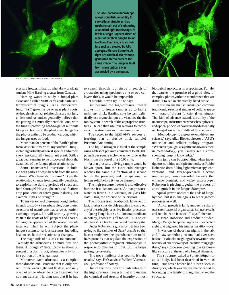
The laser confocal microscopeallows scientists an ability tosee cellular structures thatmight remain out of sight usinga standard light microscope. Atleft is a single “optical slice” ofa pair of cerebral ganglia foundin Clione limacina, a tiny shell-less mollusc studied by ASUzoologist Richard Satterlie. Atright are confocal-microscope-generated stereo pairs of thesame image. The image is builtfrom many optical slicesassembled by a computer.
IMAGES COURTESY CHARLES KAZELIK / JOHN C. PHILLIPS PHOTO

Lasers have long been on the cutting edge of technology in music, communications, and medical science. Now, they are making
important contributions for researchers whostudy microscopic worlds.
Richard Satterlie knows the usefulness oflaser technology. Lasers helped him find a newcell in an organism he has been studying formore than a decade. The organism, known asClione limacina, is an inch-long, almost trans-parent bag of seawater with devil's horns andangel's wings that beat continuously. A nastylittle carnivore, Clione is a shell-less molluscthat lives at depths of up to 100 meters in coldnorthern waters of the Pacific Ocean.
Satterlie discovered a cell–actually a pair ofcells–that expresses the chemical serotonin, aneurohormone that acts on muscles and nerves.In the past, Satterlie had shown that such cellsgenerate a beating of Clione’s wings and help it“fly” about in its aqueous environment.
Normally, Satterlie locates the nerve cells bytagging them with antibodies that are linked tofluorophores, compounds which fluoresce whenilluminated with a certain wavelength of light.
“We’ve done this immunohistochemistrymany times before using a standard fluorescentmicroscope,” Satterlie says. “We thought wehad mapped out all of the cells that react toserotonin.”
But this time, Satterlie viewed Clione underthe discriminating eye of ASU’s new laser confo-cal microscope, housed in the GoldwaterTechnology Center. The instrument sports fluo-rescent capabilities like that of a conventional
microscope, which illuminates a fluorescently-labeled specimen and detects the fluorescentlight it emits.
Most microscopes collect light from thewhole sample, although only one part of thespecimen is usually in focus at a time. Lightarising from other planes of the sample usuallyimpinges upon the focal point and somewhatdistorts the resulting image. The confocalmicroscope, however, blocks out all light exceptthat coming from the plane of focus. Theresearcher can then focus up and down withinthe sample, store virtually perfect images in theconnected computer, and reassemble them intoa three-dimensional reconstruction of a cell.
“We’re optically sectioning the specimen,not physically sectioning it,” explains CharlesKazilek, associate research professional in thedepartment of zoology. Kazilek manages ASU’sLife Sciences Visualization Group, which over-sees the confocal microscope along with otherimaging instruments housed in the GoldwaterTechnology Center.
The result is a clearer, purer image com-prised of anywhere from one to several hundredoptical “slices” of a specimen. In Satterlie’ scase, using the new technology meant finding acell that was probably obscured by light fromthe cells in front of and behind it.
“We’re not sure exactly how the cell fits inwith the other cells that influence accelerationin Clione,” Satterlie explains. With the help ofthe confocal microscope, he plans to find out.— A L A N A M I K K E L S E N
C o n f o c a l M i c r o s c o p y
The discovery lead Roberson to look for acertain protein, called tubulin, in Allomyces.Using light microscopy, and cells labelledwith fluorescent molecules, Roberson andVargas have shown that what they think istubulin appears around the border and near thetip of growing Allomyces filaments. Whengrown in drugs that disrupt tubulin, the fluo-rescent organization disbands and the cellsstop growing, suggesting that this proteinmight play a role in apical growth.
Roberson and graduate student DavidLowry then looked at the cells under the elec-tron microscope to see in detail what anotherimportant protein called actin was doing. Todistinguish actin from other molecules in thecell, they incubated the cells with antibodiesjoined to gold particles and that were specificfor actin.
The resulting images revealed gold parti-cles clustered in the vicinity of structurescalled coated vesicles–which are in factanalagous to the vesicles of the cortical reac-tion. In this case, Roberson assumes that thevesicles interact with the plasma membraneand help the cells grow.
Using the cryo-fixation techniques,Roberson’s group hopes to follow the fungus’lead and more precisely characterize actin’srole. Eventually, Roberson says, he hopes tocome full circle and use the light microscopeto obtain pictures of actin’s movement in ani-mated, growing, pure living cells.
Sometimes our simplest instruments giveus the most integrated picture.
ASU research on fungi, developmental biology, and other life sciences is sponsoredby the National Science Foundation. Formore information on electron microscopyfacilties at ASU, contact Robert Roberson,Ph.D., Department of Botany, 602.965.8618.For details on the laser confocal and otherlight microscopes, contact Charles Kazilek,Associate Research Professional, Depart-ment of Zoology 602.965.1710.
Charles Kazilek is the master of ASU’slaser-confocal microscope system.
Charles Kazilek is the master of ASU’slaser-confocal microscope system.
A S U R E S E A R C H — F A L L / W I N T E R 1 9 9 5 29