in castrated and non-castrated male syrian golden hamsters
TRANSCRIPT

781
THE EFFECT OF OESTROGEN ON 9,10-DIMETHYL-1,2-BENZAN-THRACENE (DMBA)-INDUCED CHEEK POUCH CARCINOMAIN CASTRATED AND NON-CASTRATED MALE SYRIANGOLDEN HAMSTERS
A. POLLIACK, I. CHARUZY AND I. S. LEVIJFrom the Department of Pathology, Hadassah University Hospital, Jerusalem, Israel
Received for publication August 21, 1969
OESTROGENS influence epithelial differentiation and keratinization, and theprolonged administration of these compounds leads to extensive squamousmetaplasia in the rat endometrium (Selye et al., 1935; McCuen, 1936; Gitlin,1954; Moore, 1957; Reiter, 1965). Furthermore, oestrogens stimulate mitoticactivity in the epidermis both in vivo and in vitro (Bullough, 1955, 1962; Allen,1956) and are known as membrane and lysosome labilizers, causing an increasein the permeability of these structures (Bangham et al., 1965).
In previous studies we have demonstrated the co-carcinogenic effect of anothermembrane labilizer and mitogenic agent, vitamin A palmitate, using DMBA-induced hamster cheek pouch carcinoma (Levij and Polliack, 1968; Polliack andLevij, 1969). The present study was designed to determine the influence of anoestrogen compound in the same experimental model, using castrated andnon-castrated male hamsters. Castration was performed in order to assess theeffect of an oestrogen in the absence of naturally occurring testicular hormones.The dose of oestrogen used was much higher than that needed to produce aphysiological action.
MATERIAL AND METHODS
Forty-eight male Syrian golden hamsters of a local strain, 15-2 months of ageand weighing 55-65 g., were used. Of these, 24 animals were castrated, andone week thereafter the following treatment was started:
The right cheek pouches of all animals were painted three times per week witha 0.5% (weight/volume) solution ofDMBA in liquid paraffin, using a No. 4 camel'shair brush. The brush was dipped into the solution, excess was allowed to dripoff, and the pouch was then stroked firmly several times along its entire length.
Two groups of six non-castrated animals were treated in the above mannerduring 9 weeks (Group 1) and 12 weeks (Group 2) respectively. Another twogroups of six non-castrated animals were treated identically for 9 weeks (Group 3)and 12 weeks (Group 4), but throughout these periods the animals received bi-weekly intramuscular injections of 1*5 mg. stilboestrol diphosphate (15 mg./ml.in normal saline).
Two groups of six castrated animals were treated with DMBA alone during9 weeks (Group 5) and 12 weeks (Group 6). The remaining two groups of sixcastrated animals received topical DMBA and bi-weekly intramuscular injectionsof stilboestrol diphosphate as above during 9 weeks (Group 7) and 12 weeks(Group 8).

A. POLLIACK, I. CHARUZY AND I. S. LEVIJ
An additional six male hamsters (Group 9), three of which were castrated,received intramuscular stilboestrol diphosphate bi-weekly during 12 weeks,without topical DMBA.
During the experiment all animals received Purina Laboratory chow and tapwater ad libitum. After completion of the above treatments, the animals werekilled, autopsies were performed, and both cheek pouches were examined. Theorgans were fixed in 4% formalin and stained with haematoxylin and eosin.During the macroscopical examination of the cheek pouches, the tumours werecounted, and the nature of these lesions was subsequently determined histologically.In addition, multiple sections were taken from the macroscopically non-tumorousareas of each pouch.
One animal of Group 3 and one of Group 4 died duriilg the experiment fromintercurrent pneumonia.
RESULTS
In order to avoid repetition, a short description of the most frequently encoun-tered lesions will be given:
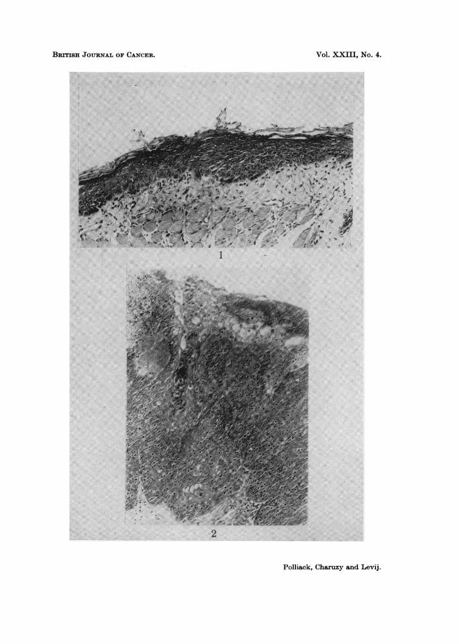
Benign epithelial hyperplasia.-Changes characterized by diffuse hyperkeratosis,focal regular acanthosis, occasional mild epithelial atypia, and varying degrees ofchronic inflammation in the upper lamina propria (Fig. 1).
Intra-epithelial carcinoma.-Focal acanthosis with marked epithelial atypiaand dyskeratosis involving all layers, with loss of polarity, but with an intactbasal layer, and without invasion of the lamina propria (Fig. 2).
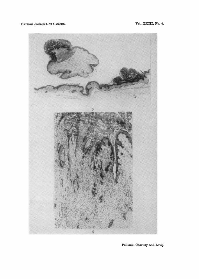
Atypical papilloma.-Premalignant papillomatous lesion with acanthosis,characterized by varying degrees of epithelial atypia, ranging from mild nuclearpleomorphism, hyperchromasia and slight loss of polarity to the changes describedas intra-epithelial carcinoma (Fig. 3).
Squamous cell carcinoma.-Cellular and nuclear changes as described in intra-epithelial carcinoma, but with invasion of the lamina propria (Fig. 4).
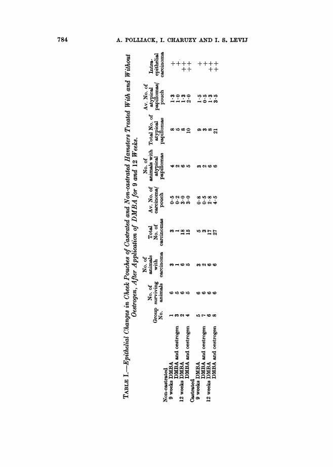
It was possible to record accurately the number of squamous cell carcinomasand atypical papillomas in each pouch. However, it was impossible to determineexactly the number of intra-epithelial carcinomas, since these lesions did notpresent as tumours macroscopically. Their frequency was estimated by examina-tion of many sections, and the impression gained in this way was recorded as++ when many foci were present in each animal and as + when in each animalonly a small number of these lesions were found.
Non-castrated animalsGroup 1 (DMBA 9 weeks).-One squamous cell carcinoma, 1-4 mm. in diameter,
was present in each of three animals. Four animals had a total of eight atypicalpapillomas. In all animals, a small number of intra-epithelial carcinomas wasfound.
EXPLANATION OF PLATES
FiG. 1.-Benign epithelial hyperplasia (H. and E. x 140).FIG. 2.-Intra-epithelial carcinoma (H. and E. x 140).FIG. 3.-Atypical papilloma (H. and E. x 52).FIG. 4.-Infiltrating squamous cell carcinoma (H. and E. x 52).
782
BRITISH JOURNAL OF CANCER.
A:
I
z
Polliack, Charuzy and Levij.
VOl. XXIII, NO. 4.
BRmsH JOuRNAL OF CANCER.
Polliack, Charuzy and Levij.
Vol. JXM NO. 4.
13

EFFECT OF OESTROGEN ON DMBA CARCINOMA
Group 2 (DMBA 12 weeks). A total of 18 squamous cell carcinomas, 3-8 mm.in diameter, and eight atypical papillomas, were found in this group, and allanimals had tumours. Many intra-epithelial carcinomas were present in eachanimal.
Group 3 (DMBA and oestrogen 9 weeks). One of the five surviving animalsshowed a 2 mm. squamous cell carcinoma, and in two animals a total of fiveatypical papillomas was present. Some foci of intra-epithelial carcinoma werefound in all animals.
Group 4 (DMBA and oestrogen 12 weeks). All five surviving animals showedsquamous cell carcinomas, 2-8 mm. in diameter, and a total of 15 of these tumourswas present. A total of ten atypical papillomas was found, and multiple intra-epithelial carcinomas were present in all animals.
Castrated animalsGroup 5 (DMBA 9 weeks). Three of the animals had a total of five squamous
cell carcinomas, 3-15 mm. in diameter, and three animals showed a total of nineatypical papillomas. In all animals, some intra-epithelial carcinomas were present.
Group 6 (DMBA 12 weeks).-All animals had squamous cell carcinomas,1-15 mm. in diameter, and a total of 17 of these tumours was present. A totalof eight atypical papillomas was found, and all animals had multiple foci ofintra-epithelial carcinoma.
Group 7 (DMBA and oestrogen 9 weeks). In two animals, a total of threesquamous cell carcinomnas, 2-7 mm. in diameter, was found, and the same animalsalso had a total of three atypical papillomas. The remaining four animalsrevealed no tumours. A small number of intra-epithelial carcinomas was presentin all animals.
Group 8 (DMBA and oestrogen 12 weeks). All animals had tumours, and atotal of 27 squamous cell carcinomas, 2-15 mm. in diameter, and 21 atypicalpapillomas were present. Multiple foci of intra-epithelial carcinoma were foundin all animals.
In the three castrated and three non-castrated animals treated with oestrogenonly for 12 weeks (Group 9) no macroscopic tumours were present, and histo-logically no epithelial changes were found.
The findings in the various groups of animals are summarized in Table I.Table II gives a comparison of the tumour incidence after 12 weeks in castratedand non-castrated animals treated with DMBA alone or with DMBA and oestrogen.
Benign epithelial hyperplasia was present in the non-tumorous epithelium inall animals of Groups 1-8.
DISCUSSION
The results of the present study indicate that oestrogen, when administeredintramuscularly to castrated male hamsters during topical application of DMBAto the cheek pouches of these animals, enhances the development of malignancy.This promoting effect of the hormone was marked after 12 weeks, but it was notpresent after 9 weeks. In non-castrated animals, oestrogen did not promotecarcinoma formation at any stage of the experiment. Naturally occurringtesticular hormones apparently do not have a direct effect on male hamstercheek pouch carcinogenesis, since the incidence of carcinoma was similar in
783
A. POLLIACK, I. CHARUZY AND I. S. LEVIJ
++++ ++++++ ++
O '0
Z g¢oCOI C
,*Q
"-I
S Z.> OOOC OO0w
Z-2oS now
u i t-E CO O ' OO m
u 0 .2 0
00OCOt 001O
0'ea,oOO 0t 0tMt .§- .S '?
C 0)c : C) 0 0*
*o Pt co 0 o
1. t XX XX
EH 0 m > Lo 0
784

EFFECT OF OESTROGEN ON DMBA CARCINOMA
TABLE II. CoMparison of Incidence of Cheek Pouch Tumrours after Local Applica-tion of DMIBA for 12 Weeks in Castrated and Non-castrated Hamsters TreatedWith and Without Oestrogen.
Squamous cell carcinoma Atypical papilloma
Castrated Non-castrated Castrated Non-castratedDMBA 17 (2 8) 18 (3.0) 8 (1-3) 8 (1-3)DMIBA and oestrogen 27 (4 -5) 15 (3.0) . :1(3 5) 10 (2 0)
Non-bracketed numbers represent total number of tumours in each group; bracketed( numbersrepresent average number of tumours per pouch in each group.
castrated and non-castrated animals after 12 weeks of DMBA application, whenno oestrogen was administered.
It is known that the prolonged administration of oestrogens produces extensivesquamous metaplasia in the rat endometrium (Selye et al., 1935; McCuen, 1936;Gitlin, 1954). However, Gitlin (1957) was unable to demonstrate squamousmetaplasia in extragenital organs of the rat following prolonged oestrogen adminis-tration. Moore (1957) and Reiter (1965) have also succeeded to induce uterinesquamous metaplasia with oestrogen, even in the presence of small amounts ofvitamin A, which is known to reduce keratinization. Bullough (1955, 1962) hasshown that the epidermis is particularly sensitive to oestrogen, and that thishormone increases the epidermal mitotic rate. This effect was claimed to be dueto stimulation of the glucokinase system. Allen (1956) has also demonstrated amitogenic effect of oestradiol benzoate in vivo. The oestradiol is apparentlyconcentrated in cell nuclei, and bound to the chromatin to a much greater degreethan other hormones, e.g. testosterone. There appears to be a considerabledegree of stereospecificity in the binding of oestrogen to endometrial nuclei(Maurer and Chalkley, 1967).
Bangham et al. (1965) showed that low concentrations of female sex hormonesand their analogue, diethylstilboestrol, increase the permeability of cell membranesand lysosomes more than other steroid hormones, and that this results from adirect interaction with cell membrane lipids and is independent of cell metabolism.Pretreatment with cortisone inhibited this membrane effect of oestrogen.
In the present study a tumour-enhancing effect was obtained in male animalsreceiving oestrogen after castration, which suggests that large amounts of oestrogenin the absence of endogenous male hormones enhance malignancy in our experi-mental model. This phenomenon could be due to the effect of the hormone onmembrane permeability, resulting in more effective penetration of DMBA intothe cell. On the other hand, the promotion of carcinoma formation may be dueto the additive effect of the carcinogenic action of DMBA and the mitogenic actionof oestrogen.
It is feasible to suggest that the underlying mechanism of the epidermalmitogenic effect of oestrogen is due to its influence on lysosomal membranes, withsubsequent release of enzymes, e.g. DNAse, which may alter nuclear metabolismand result in cell division (Allison and Malluci, 1964; Allison, 1968). This isapplicable to the present findings as well as to the results of previous studieswhere it was shown that another membrane labilizer, vitamin A palmitate,potentiated DMBA carcinogenesis in the hamster cheek pouch (Levij and Polliack,1968; Polliack and Levij, 1969).
64
785
786 A. POLLIACK, I. CHARUZY AND I. S. LEVIJ
SUMMARY
Castrated and non-castrated male Syrian golden hamsters received localtreatment of the right cheek pouch three times weekly during 9 or 12 weeks with0.5 /0 DMBA in liquid paraffin. Half of both groups received in addition bi-weekly intramuscular injections of 1-5 mg. stilboestrol diphosphate in salineduring the same period. After 9 weeks the incidence of cheek pouch tumourswas similar in castrated and intact animals, treated with or without oestrogen.However, after 12 weeks of DMBA application, castrated animals treated withoestrogen had more cheek pouch carcinomas than castrated animals treated withDMBA only. In the latter group the tumour incidence was similar to that innon-castrated animals treated with or without oestrogen. Thus, in the absence ofnaturally occurring testicular hormones, oestrogen potentiated the carcinogenicaction of DMBA. This effect of oestrogen may have been due to better penetra-tion of the carcinogen into the cells as a result of increased permeability of cellularmembranes induced by oestrogen. Another possible explanation is that tumourformation was promoted due to the additive effect of the carcinogenic action ofDMBA and the mitogenic action of oestrogen.
This study was financially supported by a grant from the Jani Dekkerstichtingand the Dr. Ludgardine Bouwmanstichting, Holland.
The authors wish to thank Miss Lidia Scalozub and Mr. Gad Ganem for theirtechnical assistance.
REFERENCESALLEN, J. M.-(1956) Expl Cell Res., 10, 523.ALLISON, A. C. (1968) Eur. J. Cancer, 3, 481.ALLISON, A. C. AND MALLUCI, L.-(1964) Nature, Lond., 203, 1024.BANGHAM, A. D., STANDISH, M. M. AND WEISSMAN, J.-(1965) J. exp. Biol., 13, 253.BULLOUGH, W. S.-(1955) Vitams Horm., 13, 261. (1962) Biol. Rev., 37, 307.GITLIN, G. (1954) Anat. Rec., 120, 637.-(1957) Endocrinology, 60, 571.LEVIJ, I. S. AND POLLIACK, A. (1968) Cancer, N. Y., 22, 300.MCCUEN, C. S.-(1936) Am. J. Cancer, 27, 91.MAURER, H. R. AND CHALKLEY, G. R. (1967) J. molec. Biol., 27, 431.MOORE, T.-(1957) In 'Vitamin A'. Amsterdam (Elsevier Publishing Co.).POLLIACK, A. AND LEVIJ, I. S.-(1969) Cancer Res., 29, 327.REITER, R. J.-(1965) Experientia, 21, 207.SELYE, H., THOMSON, P. L. AND COLLIP, J. B.-(1935) Nature, Lond., 135, 65.