indian journal of tuberculosis - tbassnindia.orgtbassnindia.org/forms/ijt_6_3.pdf · benefits of...
TRANSCRIPT
Vol. 56 : No. 3 July 2009
Registered with the Registrar of Newspapers of India under No. 655/57
Indian Journal of TuberculosisPublished quarterly by the Tuberculosis Association of India
Contents
EDITORIAL
Benefits of early anti-retroviral therapy in patients withHIV-TB co-infection - S.Rajasekaran 113
ORIGINAL ARTICLES
Association of 22 cytokine gene polymorphisms withtuberculosis in Macedonians - Dejan Trajkov, Mirjana Trajchevska, Todor Arsov,
Aleksandar Petlichkovski, Ana Strezova, Olivija Efinska-Mladenovska, Aleksandar Sandevski and Mirko Spiroski117
Assessment of long term status of sputum positive pulmonarytuberculosis patients successfully treated with Short CourseChemotherapy
- V.V. Banu Rekha, Rajeswari Ramachandran, K.V. Kuppu Rao, Fathima Rahman, A.R. Adhilakshmi, D. Kalaiselvi P. Murugesan, V. Sundaram and P.R. Narayanan 132
Mycobacterial ES-31 Serine Protease - A Biomarker forMycobacterium tuberculosis - A preliminary Report - M. Anindita, V. Upadhye, D. Thamke, D. Mendiratta
and B.C. Harinath 141
Comparing outcomes in new pulmonary sputum positiveand sputum negative cases under RNTCP in rural India - Abhijit Mukherjee, Rupak Singla and Indrani Saha 144
Status Report on RNTCP 151
CASE REPORTS
Sternal tuberculous osteomyelitis presenting as a pulsatileswelling - Hari Kishan Boorugu, Anugrah Chrispal and Elsa Mary
Thomas 154
Primary multi-drug resistant tubercular lymphadenitis in anHIV infected patient - Jagdish Rawat, Girish Sindhwani and Ruchi Dua 157
Tuberculosis of the middle ear with post auricular abscess- Manoj Arya, Ramakant Dixit, A.R. Paramez, Sidharth Sharma and Dilip Singh Rathore 160
Abstracts 164
Guidelines for Contributors 167
Editor-in-ChiefR.K. SrivastavaEditorsM.M. SinghLalit KantV.K. AroraJoint EditorsG.R. KhatriD. BeheraAssociate EditorsS.K. SharmaL.S. ChauhanAshok ShahJ.C. SuriV.K. DhingraAssistant EditorK.K. ChopraMembersBanerji, D.Gupta, K.B.Katiyar, S.K.Katoch, V.M.Kumar, PrahladNarang, P.Narayanan, P.R.Nishi AgarwalParamasivan, C.N.Puri, M.M.Radhakrishna, S.Raghunath, D.Rai, S.P.Rajendra PrasadSarin, RohitVijayan, V.K.Wares, D.F.Journal CoordinatorsKanwaljit SinghR. Varadarajan
SubscriptionInlandAnnual Rs.800Single Copy Rs.200ForeignFor SAARC countries US $ 30For South East Asian andEastern countries US $ 35For other countries US $ 40
Cheques/D.Ds. should be drawn in favourof "Tuberculosis Association of India, NewDelhi"The statements and opinions contained inthis journal are solely those of the authors/advertisers. The Publisher, Editor-in-Chiefand its Editorial Board Members andemployees disown all responsibility for anyinjury to persons or property resulting fromany ideas or products referred to in thearticles or advertisements contained in thisjournal.
Reproduction of any article, or part thereof, published in the Indian Journal of Tuberculosis, without prior permission of theTuberculosis Association of India is prohibited.Bibliographic details of the journal available in ICMR-NIC Centre's IndMED data base (http://indmed.nic.in). Full-text ofarticles from 2000 onwards are available online in medIND data base (http://medind.nic.in). IJT is indexed in MEDLINEof National Library of Medicine, USA.Published and printed by S.C. Goyal, on behalf of the Tuberculosis Association of India, 3, Red Cross Road, NewDelhi-110001 Phone: 011-23711303; 23715217 and printed at Cambridge Printing Works, B-85, Naraina IndustrialArea-II, New Delhi-110 028 Phone : 25893439.

Indian Journal of Tuberculosis
[Indian J Tuberc 2009; 56:113-116]
Editorial
BENEFITS OF EARLY ANTI-RETROVIRAL THERAPY IN PATIENTS WITH HIV-TBCO-INFECTION
Indian Journal of TuberculosisVol. 56 New Delhi, July, 2009 No. 3
Globally, 33 million people were estimated to be living with HIV/AIDS in 20071. The number ofHIV-positive TB cases and deaths were estimated at 1.39 million cases (15% of all incident cases) and0.48 million deaths, which was 24% of the estimated two million HIV deaths in 20072. In India, the 2006estimates suggested that national adult HIV prevalence in India was approximately 0.36 per cent, amountingto 2.34 million (ranging between 2 and 3.1 million) people living with HIV and AIDS3. Even going by theconventional figure of 40% of the Indian population infected with Mycobacterium tuberculosis, it is estimatedthat not less than one million persons with HIV are co-infected with TB. Considering the fact that thelifetime risk of developing TB disease is between 50-60%, India is likely to have not less than one lakhpatients, needing treatment for both HIV and TB simultaneously at any given point of time.
Free Anti-Retroviral Therapy (ART) was introduced in India in April, 2004, as a component ofcare, support and treatment, in National AIDS Control Programme (NACP). The concept of managingHIV disease in India till that time was to treat the opportunistic infections, as and when these wereidentified. The most common opportunistic infection, tuberculosis, was being treated by the physicianswith their own selective drug schedules with their own preferential rhythm and duration of administration.Prevailing stigma and discrimination, tagged with HIV/AIDS and the absence of a robust referral andlinkage system between Revised National Control Programme (RNTCP) and NACP during those timesdenied a large number of patients to the benefits of effective anti TB treatment protocol, available throughDirectly Observed Treatment Strategy even those times.
The first ever published work4 on the effectiveness of RNTCP treatment schedule was carriedout at Government Hospital of Thoracic Medicine, Tambaram, the largest TB and HIV care centre inIndia, by Tuberculosis Research Institute through a collaborative initiative in 1999-2000. This prospectiveobservational feasibility study of 71 patients with HIV and tuberculosis, who were treated with categoryI regimen showed a favourable response in 72% of patients5. Even though the early bacteriologicalresponse to RNTCP regimen was satisfactory, the overall outcome was adversely affected by the highmortality (37% during treatment and 24 months of follow-up) and high recurrence rate 37%. This wasessentially due to the deterioration of immune system during anti-tuberculosis treatment, with the baselevel mean CD4% seen falling down from 12.6 (5.9) to 8.9 (4.9) at the end of treatment (p < 0.001). Thisfinding highlighted the need for the initiation of ART in addition to anti-tuberculosis treatment to improvethe immune system and thereby the long term treatment outcome.
Indian Journal of Tuberculosis
EDITORIAL
Initiation of ART reduces risk of further HIV-related morbidity and mortality. The impact ofART on HIV-infected tuberculosis TB patients was amply demonstrated in South Africa and SouthEast Asia. HAART reduced the incidence of HIV-1-associated tuberculosis by more than 80% (95%CI 62–91) in an area endemic with tuberculosis and HIV-1 in a study conducted at Cape Town, SouthAfrica6. In an observational study in Taiwan, ART was found to decrease the incidence rate of newHIV-TB co-infection cases and increased the survival rate of HIV-TB co-infection cases. The survivalrate of HIV-TB co-infection cases was 62.16% during the period 1993–1996 (pre-free HAART era)and increased to 86.60% during the period 1998–2006, a post-free ART era (P < 0.0001)7. ART hadbrought a substantial reduction in deaths during TB treatment for HIV infected TB patients in Thailandas well. Of the patients with known outcomes, death during TB treatment occurred in 5 (7%) of 71who received ART as against 94 (43%) of 219 who did not8. The impact of ART in public healthprogramme in Thailand was in evidence, as patients who received ART had one sixth the risk of deathof those not receiving ART9. The survival benefit persisted even for those with a very low CD4 countwas a significant observation.
While access to anti-retroviral therapy is rapidly expanding in resource-limited settings,tuberculosis continues to be the most challenging opportunistic disease in HIV infected patients evenafter initiating anti-retroviral therapy. India had successfully rolled out first line ART to just over 220thousand patients during the last five years, ever since its initiation in 2004. There is an increasingneed for the co-administration of anti-tuberculosis and anti-retroviral treatment in patients with HIV-TB co-infection. However, the ground reality has been disturbingly indifferent. The unpublishedobservations available to Central TB Division from Trichy and Mysore districts of Tamilnadu andKarnataka respectively, suggested that only 26% of 396 HIV-TB co infected patients, eligible to getART, actually received ART. Apart from the issues of self-imposed stigma, illiteracy, inadequateinformation and knowledge and the programmatic issues, the most contentious issue is the decisionmaking process of ART medical officers on “when to start ART safely in HIV patients receiving anti-tuberculosis treatment”. ART physicians’ reluctance to initiate ART to HIV-infected TB patients remainsa global issue, in spite of the existing national guidelines and international recommendations. They arereally concerned about overlapping toxicity, drug-drug interactions, pill burden and immunereconstitution inflammatory syndrome (IRIS).
The underlying mechanisms in the development of TB after initiation of ART are indeedcomplex10. IRIS is one of the manifestations of ‘‘ART-associated TB’’ and this refers to the severeand overtly exaggerated inflammatory effects of TB. IRIS is characterised by worsening of systemicsymptoms, transient enlargement of pre-existing lesions, onset of new lesions includinglymphadenopathy and worsening of radiographic changes. The frequency of IRIS in cohort studiesvaried markedly between 8% and 43%7,8 The mean interval to IRIS after ART initiation also variedwidely (1 - 180 days) with most cases occurring within the first 28 days11. The risk of mortalityassociated with delays in ART initiation has to essentially outweigh the concerns for the possibleIRIS and its outcome. The optimal timing of ART initiation may therefore be earlier in the course ofTB treatment for patients in resource-limited settings with high prevalence of TB. Early ART wasfavoured, even in the settings of South Africa, with the highest reported rates of IRIS (70%) andsevere drug toxicity (56%). ART can be deferred in settings, where IRIS-related mortality rate was
114
Indian Journal of Tuberculosis
EDITORIAL
found to exceed 4.6%. These results support early initiation of ART in patients with AIDS, exceptwhen IRIS-related mortality rates are high12. In a larger cohort of 2330 patients with HIV initiated onART in India, tuberculosis-associated immune reconstitution disease was found in 81 of them (3.5%)only. Even though the risk of developing IRIS was expected to be high, on being initiated with ARTin those with low baseline CD4 cell counts, manifestations of IRIS and risk of mortality in Indianpatients were found to be self-limiting, favouring early initiation of ART in patients with HIV-TB co-infection13.
ART reduces the incidence of TB in treated cohorts even in high TB prevalence countries.Two hundred and sixty-two patients (5.1%) of 5099 patients with as many with 88% patients hadbase level CD4 count less than 200/mm3, followed-up for one to four years were found to have PostART TB with 100-person year risk of 2.83 in the largest Indian cohort14. In a Cape Town cohort of346 patients receiving HAART between 1996 and 2005, TB incidence rate was observed to be 3.5/100 person-years in the first year and significantly decreased during follow-up, reaching 1.01/100person-years in the fifth year (P = 0.002 for trend) 15. In spite of beneficial effect of ART, incidenceof post-ART TB continues to be on the higher side than those among HIV-negative individuals.Current data suggest that ART can achieve suboptimal restoration of MTB-specific immune responsesonly. Hence, the contribution of ART is limited to patients alone and not to the community, as asignificant number patients receiving ART live much longer and yet would maintain a chronicallyheightened risk of TB16.
HIV associated TB is a major public health problem. Tuberculosis services are an importantentry point for identifying ART eligible patients. Given that dually infected patients identified throughtuberculosis services contributed to 10% of the HIV-infected adult population with a CD4 cell countbelow 350 cells/mm3 in the 18 sub-Saharan African countries17. The burden of HIV among tuberculosispatients varies widely in India, from 1% in Koch Bihar, West Bengal, to 13.8% in Guntur, AndhraPradesh18. Programme efforts to implement comprehensive TB-HIV services should be targeted toareas with the highest HIV burden districts through the Intensified TB Screening mechanism. Asimple diagnostic tool, evaluating the common signs and symptoms like oral thrush, diarrhoea, itchingin 25-45 year old adults indulging in high risk behaviour could be used to screen patients for HIV atDOTS centres to Integrated Counselling and Testing Centres for early detection of HIV-TB co-infection19. Simultaneously, the number of HIV infected persons to have been screened for TB has tobe strengthened through Intensified TB Screening. The existing policy of NACP to screen all thepatients with HIV sero-positivity for identifying CD4 counts augurs well for identifying the eligiblepatients with HIV-TB co-infection for early ART initiation. The revised ART guidelines are strongly infavour of initiation ART early in all TB patients with HIV, having CD4 count less than 350 cells/mm3
after two weeks of anti-tuberculosis patients. Patients with extra-pulmonary TB, being a WHO clinicalstage IV disease, have the additional advantage of getting ART initiated irrespective of CD4 cellcount.
Having realised the need for early identification of HIV-TB co-infection and for early initiationof ART in them, it is the moment to prioritise the course of action. Dissemination of information,knowledge and national guidelines among the concerned health care workers through a well planned
115
Indian Journal of Tuberculosis
training programme is the need of the hour. All the health care professionals connected with RNTCPand NACP should join together and strengthen the collaborative activities, referrals, linkages and monitoringmechanism to translate the light of wisdom into fruit of benefit for the needy.
Dr. S. RajasekaranNACO National Consultant (ART Quality Management)
ChennaiREFERENCES
1. United Nations Acquired Immune Deficiency Syndrome. The Joint United Nations Programme on HIV/AIDS: 2008 Reporton Global Epidemic. http://www.unaids. org/en/KnowledgeCentre/ HIVData/ GlobalReport/2008/2008_Global_report.asp,accessed on May 31, 2009.
2. National AIDS Control Organization. Technical Report on HIV estimates, 2006. Available at http://www.nacoonline.org/Quick_Links/HIV_Data/, accessed on May 31, 2009, accessed on May 31, 2009.
3. World Health Organisation. Global tuberculosis control - epidemiology, strategy, financing: Programmes and Projects:WHO Report 2009 WHO/HTM/TB/2009.411: http://www.who.int/tb/publications/global_report/2009/en/index.html,accessed on May 31, 2009.
4. Swaminathan S, Sangeetha M, Arunkumar N, Menon PA, Beena Thomas, Shibi K, Ponnuraja and Rajasekaran S. PulmonaryTuberculosis in HIV positive individuals: Preliminary report on clinical features and response to treatment. Indian JTuberc 2002; 49:189-93.
5. Swaminathan S, Deivanayagam CN, Rajasekaran S, Venkatesan P, Padmapriyadarsini C, Menon PA, Ponnuraja C, Dilip M.Long term follow up of HIV-infected patients with tuberculosis treated with 6-month intermittent short course chemotherapy.Natl Med J India 2008; 21:3-8.
6. Badri M, Wilson D, Wood R. Effect of highly active antiretroviral therapy on incidence of tuberculosis in South Africa: acohort study. Lancet 2002; 359:2059-64
7. Tseng SH, Jiang DD, Hoi HS, Yang SL, Hwang KP. Impact of HAART Therapy on Co-Infection of Tuberculosis and HIVCases for 9 Years in Taiwan. Am J Trop Med Hyg 2009; 80:675-7
8. Akksilp S, Karnkawinpong O, Wattanaamornkiat W, Viriyakitja D, Monkongdee P, Sitti W, Rienthong D, Siraprapasiri T,Wells CD, Tappero JW, Varma JK. Antiretroviral therapy during tuberculosis treatment and marked reduction in death rateof HIV-infected patients, Thailand. Emerg Infect Dis 2007; 13:1001-7
9. Sanguanwongse N, Cain KP, Suriya P, Nateniyom S, Yamada N. Antiretroviral therapy for HIV-infected tuberculosis patientssaves lives but needs to be used more frequently in Thailand. JAIDS 2008; 48:181-9
10. Lawn SD, Wilkinson RJ, Lipman MC, Wood R. Immune reconstitution and “unmasking” of tuberculosis during antiretroviraltherapy. Am J Respir Crit Care Med 2008; 177:680-5
11. Lawn SD, Bekker LG, Wood R. How effectively does HAART restore immune responses to Mycobacterium tuberculosis?Implications for tuberculosis control. AIDS 2005; 19:1113-24
12. Schiffer JT, Joshua T. Timing of antiretroviral therapy initiation in tuberculosis patients with AIDS: A decision analysis.JAIDS 2007; 44: 229-34
13. Rajasekaran S, Vijila, Ravichandran N. Immune Reconstitution Tuberculosis in HIV Patients after Antiretroviral Therapy.JK Science 2006; 8:205-8
14. Rajasekaran, S, Raja K, Jeyaseelan L, Vijila S, Krithiga Priya, Kuralmozhi Mohan, Anwar Parvez, Mahilmaran A, ChandrasekarC. Post-HAART Tuberculosis in adults and adolescents with HIV in India. Indian J Tuberc 2009; 56:69-76
15. Lawn SD, Badri M, Wood R. Tuberculosis among HIV-infected patients receiving HAART: long term incidence and riskfactors in a South African cohort. AIDS 2005; 19:2109-16
16. Lawn SD, Bekker LG, Wood R. How effectively does HAART restore immune responses to Mycobacterium tuberculosis?Implications for tuberculosis control. AIDS 2005; 19:1113-24
17. Bwire R, Nagelkerke NJ, Borgdorff MW. Finding patients eligible for antiretroviral therapy using TB services as entry pointfor HIV treatment. Trop Med Int Hlth 2006; 11:1567-75
18. Raizada N, Chauhan LS, Khera A, Sokhey J, Wares DF, Sahu R, Thakur R, Dewan PK. HIV Seroprevalence amongTuberculosis Patients in India, 2006–2007. PLoS One 2008; 3: e2970
19. Rajasekaran S, Jeyaseelan L, Mahilmaran A, Krishnarajasekhar OR, Kumar S, Annadurai S. A diagnostic tool to screen forHIV co-infection at the TB DOTS centre in India. SAARC J Tuberc Lung Dis HIV/AIDS 2007; 4:1-7
EDITORIAL116
Indian Journal of Tuberculosis
117Original article
(Received on 25.3.2008. Accepted after revision on 19.6.2009)
[Indian J Tuberc 2009; 56:117-131]
ASSOCIATION OF 22 CYTOKINE GENE POLYMORPHISMS WITHTUBERCULOSIS IN MACEDONIANS
Dejan Trajkov 1, Mirjana Trajchevska 2, Todor Arsov1, Aleksandar Petlichkovski1, Ana Strezova1, OlivijaEfinska-Mladenovska1, Aleksandar Sandevski1 and Mirko Spiroski 1
SummaryObjective: To examine the possible role of 22 cytokine gene polymorphisms in host susceptibility to or protectionagainst tuberculosis (TB) in Macedonians.Method: 301 healthy unrelated individuals and 75 patients with pulmonary TB were studied. Cytokine genotyping wasperformed by PCR with sequence-specific priming (PCR-SSP) (Heidelberg kit).Results: TNF-Ü -238/G, IL-1R psti1970/C, IL-1â +3962/T:T, IL-4 -1098/ T:T, IFNã utr5644/A:A, IL-10 -1082/G:G, IL-4 -590/C:C, IL-10/ATC, IL-4/TCT, IL-4/TCC, IL-10/ATC:GCC, IL-4/TCT:TTT, IL-4/TCC:TTC, IL-10/GCC:GCC andIL-4/TCC:TCC were positively associated with TB, while protective association was identified for IL-4 -1098/G, IL-1â+3962/C, IFNã utr5644/T, IL-1â +3962/C:T, IL-4 -1098/G:T, IL-4 -590/C:T, IFNã utr5644/A:T, IL-4/GCC, IL-4/TTCand IL-4/GCC:TTC.Conclusion: These results suggest that some cytokine polymorphisms are significantly associated and affect hostsusceptibility/resistance to TB in Macedonians.
Key words: TB, Cytokine polymorphism, Macedonians.
1. Institute of Immunobiology and Human Genetics, Faculty of Medicine, University “St. Kiril and Metodij”, Skopje, Republic of Macedonia 2.Institute for Tuberculosis, Skopje, Republic of MacedoniaCorrespondence: Mirko Spiroski, MD, PhD., Institute of Immunobiology and Human Genetics, Faculty of Medicine, University “Ss. Kiril andMetodij”, 1109 Skopje, PO Box 60, Republic of Macedonia; Tel.: +389-2-3110556; Fax: +389-2-3110558; URL: http://www.immunology.edu.mk;E-mail: [email protected]
INTRODUCTION
Tuberculosis (TB) represents itself asa major health problem globally. 1 The incidenceof d isease has increased in developedcountries2 and, according to World HealthOrganization’s estimations, with four millionsdeaths annually, TB is one of the leading deathcausing diseases.3, 4 Although one third of thewor ld ’s populat ion is in fected wi th M.tuberculosis, the fact that only 10% of thosewho are infected develop tuberculosis pointsout the ro le of genet ic factors in thepathogenesis of the disease.5 The best evidencethat genetic factors are very important insusceptibility and resistance to tuberculosiscomes from the familial clustering, familialdifferences in incidence, and twin studiesshowing that concordance among identicaltwins was 65-85% and 25-35% for non-identical ones.6-9
Each stage of the host response to M.tuberculosis is under genetic control, including theinit ial encounter with mycobacteria bymacrophages, epithelial cells and dendritic cellsin the lung, induction of the inductive T cellresponse, and killing by activated macrophageswithin granulomas.10, 11 Although environmentalfactors are important determinants of progressionto disease, there is a genetic componentunderlying susceptibility to tuberculosis (TB), thebasis of which may vary in different populations.12
Crucial factors in resistance to M. tuberculosisl ie in macrophage activation and Th1 typelymphocyte response.13 Cytokines that areproduced at the site of infection and influenceactivation of these cells, highly participate in thepathogenesis of TB.14 Manifestation of clinical TBdepends on balance between T helper 1 (Th1)cytokines associated with resistance to infection,and Th2 cytokines associated with progressivedisease.15 Factors that influence the nature of
Indian Journal of Tuberculosis
118 DEJAN TRAJKOV ET AL
cytokine response, such as polymorphisms ofcytokine genes, will lead to modification of hostimmunological response. Although mechanisms ofaltered gene expression associated withpolymorphisms are still poorly understood, thereis more and more evidence that sequence changesin cytokine genes may result in alteredtranscription factor recognition sites, affectingtranscriptional activation and influence productionof the corresponding peptide solely or due tolinkage with another marker directly affectinggene expression.16,18
We have previously published data for thecytokine polymorphisms in healthy Macedonianpopulation.19 The aim of this study was toinvestigate the existence of possible associationsbetween 22 cytokine genes polymorphisms andTB in Macedonians.
MATERIAL AND METHODS
Groups
The total studied sample consisted of 376examinees, divided into two different groups asfollows: healthy individuals, and patients withtuberculosis.
Healthy individuals. There were 301unrelated individuals, born in different parts ofMacedonia. They were age and sex non-matched healthy individuals who attended theInst i tute of Immunobio logy and HumanGenetics for DNA donation between May 1,2001 and April 25, 2002 and agreed to take partin this study as a control group. Individualswith fami ly h istory of tuberculosis wereexcluded from the investigation.
Tuberculosis. There were 75 patientswith tuberculosis fulf i l l ing the criteria ofd iagnost ic a lgor i thm for TB diagnosisrecommended by WHO.20 They were 20-59years old consecutive patients who attended theInstitute for Tuberculosis, Skopje, Republic ofMacedonia for treatment between January 10,2003 and April 25, 2004.
All individuals were of Macedonian originand nationality, and residents of different regionsof the Republic of Macedonia. Each individual wasinterviewed on a one-to-one basis, his/hergenealogy was recorded for the last threegenerations, and a signed consent was obtained.Admixture, if any, was recorded for eachindividual. Individuals with only one Macedonianparent was excluded from the study.
All the patients and healthy individualsincluded in this study signed a written consent toparticipate in the study which was approved bythe Committee of the Ministry of Education andScience from Republic of Macedonia (No13-874/3-05), and Ethical Committee of the MedicalFaculty in Skopje.
Genomic DNA Isolation and Storage
DNA was isolated from peripheral bloodleukocytes by phenol-chloroform extractionmethod or with BioRobot EZ1 workstation(QIAGEN).21 The quality and quantity of DNAwas analyzed by GeneQuant (Pharmacia Biotech,Uppsala, Sweden). Isolated DNA samples werestored in the Macedonian Human DNA Bank.22
Typing Methods
Cytokine genotyping was performed byPCR-SSP (Heidelberg kit). Fourteen cytokinegenes with 22 single nucleotide polymorphisms(SNP) were typed: IL-1Ü -889, IL-1â -511, IL-1â +3962, IL-1R psti1970, IL-1RA mspa11100,IL-4RÜ +1902, IL-12 -1188, IFNã utr5644, TGF-â1 cdn10, TGF-â1 cdn25, TNF-Ü -308, TNF-Ü -238, IL-2 -330, IL-2 +166, IL-4 -1098, IL-4 -590, IL-4 -33, IL-6 -174, IL-6 565, IL-10 -1082,IL-10 -819, and IL-10 -592. Briefly, PCR-SSPtyping Heidelberg kit consists of 48 PCR primermixes aliquotted in 96 well PCR trays (twotypings per tray). Master mix, which was suppliedalong with the reagents and consisted of MgCL2,buffer, dNTP’s, and glycerol was mixed with 1.2- 3.0 µg DNA and 20 U Taq polymerase anddispensed in the 48 wells.23 Agarose gelelectrophoresis on a 2% gel revealed a positive or

Indian Journal of Tuberculosis
119TWENTY-TWO CYTOKINE GENE POLYMORPHISMS IN TB
negative signal for specific amplification in eachwell. Subsequently, the results were analyzedaccording to the interpretation scheme providedwith the kit.24
Statistical Methods
The population genetics analysis package,PyPop, developed by the Biostatistics Core forthe Workshop,25-27 was used for analysis of thecytokine data in this study. Allele frequencies andexpected Hardy Weinberg proportions (HWP) foreach SNP were determined.28 The exact test forgenotype frequency deviation from HWP wascalculated using the Arlequin implementationaccessed via PyPop.29 Those SNPs that did notfit HWP were evaluated to determine whetherthere was an excess of homozygotes orheterozygotes, or if any particular genotypefrequencies were significantly different from theexpected frequencies. Comparisons of differentgenotypes for two groups were tested by the χ2
test. Crude odds ratios (OR), as estimates of therelative risk, were calculated within 95% CI.
RESULTS
Cytokine Alleles
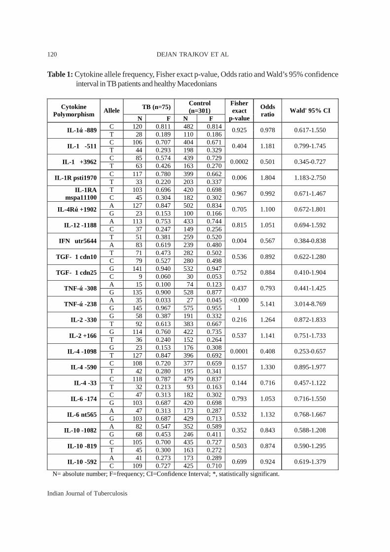
In Table 1, frequencies of polymorphiccytokine alleles, Fisher exact p-value, Odds ratioand Wald’s 95% confidence interval in TB patientsand healthy Macedonians are shown.
For the members of IL-1 gene cluster,we found positive association for IL-1R psti1970/T allele (p=0.006, OR=1.804, Wald’s 95% CIbetween 1.183-2.750), while IL-1â +3962/C alleleshowed negative (protective) association(p=0.0002, OR=0.501, Wald’s 95% CI between0.345-0.727) for TB. In the IL-12/IFNã axis,protective association was obtained for the IFNãutr5644/T allele (p=0.004, OR=0.567, Wald’s95% CI between 0.384-0.838). Results showedthat people with TNF-Ü -238 /G allele have 5.141fold risk to develop TB (p<0.0001, Wald’s 95%CI between 3.014-8.769) in comparison to otherswith TNF-Ü -238 /A allele. Analysis of the IL-4
polymorphisms showed that IL4 -1098/G allelewas associated with TB (p=0.0001, OR=0.408,Wald’s 95% CI between 0.253-0.657) (Table 1).
Cytokine genotypes
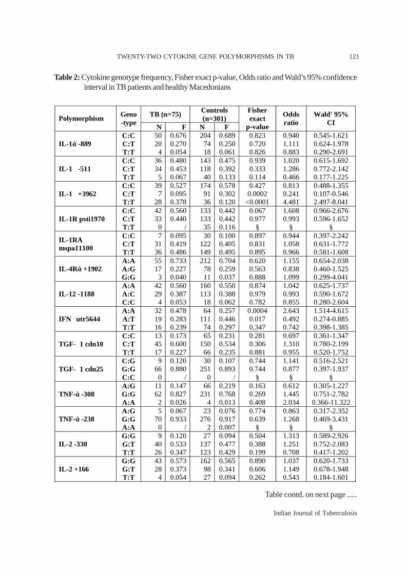
Table 2 contains summarized results fordifferent cytokine genotypes found in our study.
Analysis of all possible IL-1 gene clustergenotypes showed that only heterozygousgenotype of IL-1â +3962 polymorphism wasprotectively associated with TB (p=0.0002,OR=0.241, Wald’s 95% CI between 0.107-0.546),while homozygous /T:T genotype of the samepolymorphism showed susceptible effect(p<0.001, OR=4.481, Wald’s 95% CI between2.497-8.041). In the IL-12/IFNã axis twogenotypes of IFNã utr5644 polymorphism showedassociation with TB. While /A:A genotype waspositively associated (p=0.0004, OR=2.643,Wald’s 95% CI between 1.514-4.615), /A:Tgenotype showed negative association with TB(p=0.017, OR=0.492, Wald’s 95% CI between0.274-0.885). Two observed genotypes of IL-4polymorphisms were positively associated withTB (IL-4 -1098/T:T, p<0.0001, OR=3.564,Wald’s 95% CI between 2.066-6.150; and IL-4 -590/C:C, p=0.018, OR=1.856, Wald’s 95% CIbetween 1.108-3.108), while heterozygousgenotypes of the same IL-4 polymorphismsshowed protective association: IL-4 -1098/G:T(p<0.0001, OR=0.285, Wald’s 95% CI between0.165-0.491) and IL-4 -590/C:T genotype (p=0.006,OR=0.489, Wald’s 95% CI between 0.292-0.817).We found that patients with IL-10 -1082/G:Ggenotype have 2.5 higher risk to develop TB(p<0.022, OR=2.552, Wald’s 95% CI between1.117-5.831). Homozygous genotypes IL-1Rpsti1970/T:T, TNF-Ü -238/A:A and IL-4 -1098/G:Gwere present only in patients with TB. Neither healthyMacedonian population nor patients with TB haveTGF-â1 cdn25/C:C genotype (Table 2).
Cytokine Haplotypes
For several genes with multiple SNPs pergene (TGF-â1, TNF-Ü, IL-2, IL-4, IL-6, IL-10)
Indian Journal of Tuberculosis
120
Table 1: Cytokine allele frequency, Fisher exact p-value, Odds ratio and Wald’s 95% confidenceinterval in TB patients and healthy Macedonians
TB (n=75) Control (n=301)
Cytokine Polymorphism
Allele N F N F
Fisher exact
p-value
Odds ratio
Wald' 95% CI
C 120 0.811 482 0.814 IL-1 ά -889
T 28 0.189 110 0.186 0.925 0.978 0.617-1.550
C 106 0.707 404 0.671 IL-1 � -511
T 44 0.293 198 0.329 0.404 1.181 0.799-1.745
C 85 0.574 439 0.729 IL-1 � +3962
T 63 0.426 163 0.270 0.0002 0.501 0.345-0.727
C 117 0.780 399 0.662 IL-1R psti1970
T 33 0.220 203 0.337 0.006 1.804 1.183-2.750
T 103 0.696 420 0.698 IL-1RA mspa11100 C 45 0.304 182 0.302
0.967 0.992 0.671-1.467
A 127 0.847 502 0.834 IL-4Rά +1902
G 23 0.153 100 0.166 0.705 1.100 0.672-1.801
A 113 0.753 433 0.744 IL-12 -1188
C 37 0.247 149 0.256 0.815 1.051 0.694-1.592
T 51 0.381 259 0.520 IFN � utr5644
A 83 0.619 239 0.480 0.004 0.567 0.384-0.838
T 71 0.473 282 0.502 TGF- � 1 cdn10
C 79 0.527 280 0.498 0.536 0.892 0.622-1.280
G 141 0.940 532 0.947 TGF- � 1 cdn25
C 9 0.060 30 0.053 0.752 0.884 0.410-1.904
A 15 0.100 74 0.123 TNF-ά -308
G 135 0.900 528 0.877 0.437 0.793 0.441-1.425
A 35 0.033 27 0.045 TNF-ά -238
G 145 0.967 575 0.955 <0.000
1 5.141 3.014-8.769
G 58 0.387 191 0.332 IL-2 -330
T 92 0.613 383 0.667 0.216 1.264 0.872-1.833
G 114 0.760 422 0.735 IL-2 +166
T 36 0.240 152 0.264 0.537 1.141 0.751-1.733
G 23 0.153 176 0.308 IL-4 -1098
T 127 0.847 396 0.692 0.0001 0.408 0.253-0.657
C 108 0.720 377 0.659 IL-4 -590
T 42 0.280 195 0.341 0.157 1.330 0.895-1.977
C 118 0.787 479 0.837 IL-4 -33
T 32 0.213 93 0.163 0.144 0.716 0.457-1.122
C 47 0.313 182 0.302 IL-6 -174
G 103 0.687 420 0.698 0.793 1.053 0.716-1.550
A 47 0.313 173 0.287 IL-6 nt565
G 103 0.687 429 0.713 0.532 1.132 0.768-1.667
A 82 0.547 352 0.589 IL-10 -1082
G 68 0.453 246 0.411 0.352 0.843 0.588-1.208
C 105 0.700 435 0.727 IL-10 -819
T 45 0.300 163 0.272 0.503 0.874 0.590-1.295
A 41 0.273 173 0.289 IL-10 -592
C 109 0.727 425 0.710 0.699 0.924 0.619-1.379
N= absolute number; F=frequency; CI=Confidence Interval; *, statistically significant.
DEJAN TRAJKOV ET AL
Indian Journal of Tuberculosis
121
Table 2: Cytokine genotype frequency, Fisher exact p-value, Odds ratio and Wald’s 95% confidenceinterval in TB patients and healthy Macedonians
TWENTY-TWO CYTOKINE GENE POLYMORPHISMS IN TB
TB (n=75) Controls (n=301) Polymorphism Geno
-type N F N F
Fisher exact
p-value
Odds ratio
Wald' 95% CI
IL-1ά -889 C:C C:T T:T
50 20 4
0.676 0.270 0.054
204 74 18
0.689 0.250 0.061
0.823 0.720 0.826
0.940 1.111 0.883
0.545-1.621 0.624-1.978 0.290-2.691
IL-1 � -511 C:C C:T T:T
36 34 5
0.480 0.453 0.067
143 118 40
0.475 0.392 0.133
0.939 0.333 0.114
1.020 1.286 0.466
0.615-1.692 0.772-2.142 0.177-1.225
IL-1 � +3962 C:C C:T T:T
39 7
28
0.527 0.095 0.378
174 91 36
0.578 0.302 0.120
0.427 0.0002 <0.0001
0.813 0.241 4.481
0.488-1.355 0.107-0.546 2.497-8.041
IL-1R psti1970 C:C C:T T:T
42 33 0
0.560 0.440
/
133 133 35
0.442 0.442 0.116
0.067 0.977
§
1.608 0.993
§
0.966-2.676 0.596-1.652
§
IL-1RA mspa11100
C:C C:T T:T
7 31 36
0.095 0.419 0.486
30 122 149
0.100 0.405 0.495
0.897 0.831 0.895
0.944 1.058 0.966
0.397-2.242 0.631-1.772 0.581-1.608
IL-4Rά +1902 A:A A:G G:G
55 17 3
0.733 0.227 0.040
212 78 11
0.704 0.259 0.037
0.620 0.563 0.888
1.155 0.838 1.099
0.654-2.038 0.460-1.525 0.299-4.041
IL-12 -1188 A:A A:C C:C
42 29 4
0.560 0.387 0.053
160 113 18
0.550 0.388 0.062
0.874 0.979 0.782
1.042 0.993 0.855
0.625-1.737 0.590-1.672 0.280-2.604
IFN � utr5644 A:A A:T T:T
32 19 16
0.478 0.283 0.239
64 111 74
0.257 0.446 0.297
0.0004 0.017 0.347
2.643 0.492 0.742
1.514-4.615 0.274-0.885 0.398-1.385
TGF- � 1 cdn10 C:C C:T T:T
13 45 17
0.173 0.600 0.227
65 150 66
0.231 0.534 0.235
0.281 0.306 0.881
0.697 1.310 0.955
0.361-1.347 0.780-2.199 0.520-1.752
TGF- � 1 cdn25 C:G G:G C:C
9 66 0
0.120 0.880
/
30 251
0
0.107 0.893
/
0.744 0.744
§
1.141 0.877
§
0.516-2.521 0.397-1.937
§
TNF-ά -308 A:G G:G A:A
11 62 2
0.147 0.827 0.026
66 231
4
0.219 0.768 0.013
0.163 0.269 0.408
0.612 1.445 2.034
0.305-1.227 0.751-2.782 0.366-11.322
TNF-ά -238 A:G G:G A:A
5 70 0
0.067 0.933
/
23 276
2
0.076 0.917 0.007
0.774 0.639
§
0.863 1.268
§
0.317-2.352 0.469-3.431
§
IL-2 -330 G:G G:T T:T
9 40 26
0.120 0.533 0.347
27 137 123
0.094 0.477 0.429
0.504 0.388 0.199
1.313 1.251 0.708
0.589-2.926 0.752-2.083 0.417-1.202
IL-2 +166 G:G G:T T:T
43 28 4
0.573 0.373 0.054
162 98 27
0.565 0.341 0.094
0.890 0.606 0.262
1.037 1.149 0.543
0.620-1.733 0.678-1.948 0.184-1.601
Table contd. on next page .....
Indian Journal of Tuberculosis
122 DEJAN TRAJKOV ET AL
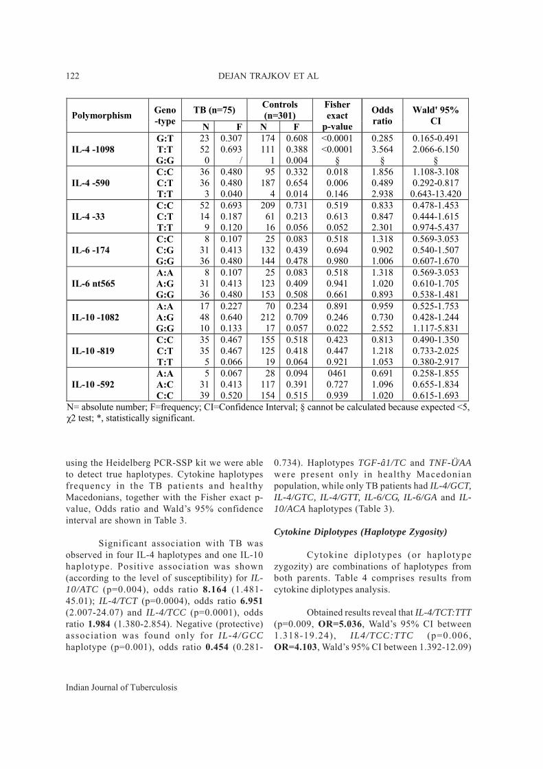
using the Heidelberg PCR-SSP kit we were ableto detect true haplotypes. Cytokine haplotypesfrequency in the TB patients and healthyMacedonians, together with the Fisher exact p-value, Odds ratio and Wald�s 95% confidenceinterval are shown in Table 3.
Significant association with TB wasobserved in four IL-4 haplotypes and one IL-10haplotype. Positive association was shown(according to the level of susceptibility) for IL-10/ATC (p=0.004), odds ratio 8.164 (1.481-45.01); IL-4/TCT (p=0.0004), odds ratio 6.951(2.007-24.07) and IL-4/TCC (p=0.0001), oddsratio 1.984 (1.380-2.854). Negative (protective)association was found only for IL-4/GCChaplotype (p=0.001), odds ratio 0.454 (0.281-
0.734). Haplotypes TGF-â1/TC and TNF-Ü/AAwere present only in healthy Macedonianpopulation, while only TB patients had IL-4/GCT,IL-4/GTC, IL-4/GTT, IL-6/CG, IL-6/GA and IL-10/ACA haplotypes (Table 3).
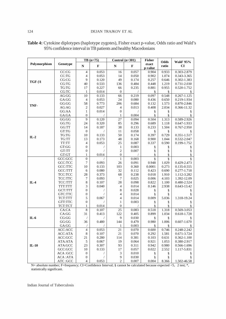
Cytokine Diplotypes (Haplotype Zygosity)
Cytokine diplotypes (or haplotypezygozity) are combinations of haplotypes fromboth parents. Table 4 comprises results fromcytokine diplotypes analysis.
Obtained results reveal that IL-4/TCT:TTT(p=0.009, OR=5.036, Wald�s 95% CI between1.318-19.24), IL4/TCC:TTC (p=0.006,OR=4.103, Wald�s 95% CI between 1.392-12.09)
TB (n=75) Controls (n=301) Polymorphism Geno
-type N F N F
Fisher exact
p-value
Odds ratio
Wald' 95% CI
IL-4 -1098 G:T T:T G:G
23 52
0
0.307 0.693
/
174 111
1
0.608 0.388 0.004
<0.0001 <0.0001
§
0.285 3.564
§
0.165-0.491 2.066-6.150
§
IL-4 -590 C:C C:T T:T
36 36
3
0.480 0.480 0.040
95 187
4
0.332 0.654 0.014
0.018 0.006 0.146
1.856 0.489 2.938
1.108-3.108 0.292-0.817
0.643-13.420
IL-4 -33 C:C C:T T:T
52 14
9
0.693 0.187 0.120
209 61 16
0.731 0.213 0.056
0.519 0.613 0.052
0.833 0.847 2.301
0.478-1.453 0.444-1.615 0.974-5.437
IL-6 -174 C:C C:G G:G
8 31 36
0.107 0.413 0.480
25 132 144
0.083 0.439 0.478
0.518 0.694 0.980
1.318 0.902 1.006
0.569-3.053 0.540-1.507 0.607-1.670
IL-6 nt565 A:A A:G G:G
8 31 36
0.107 0.413 0.480
25 123 153
0.083 0.409 0.508
0.518 0.941 0.661
1.318 1.020 0.893
0.569-3.053 0.610-1.705 0.538-1.481
IL-10 -1082 A:A A:G G:G
17 48 10
0.227 0.640 0.133
70 212
17
0.234 0.709 0.057
0.891 0.246 0.022
0.959 0.730 2.552
0.525-1.753 0.428-1.244 1.117-5.831
IL-10 -819 C:C C:T T:T
35 35
5
0.467 0.467 0.066
155 125
19
0.518 0.418 0.064
0.423 0.447 0.921
0.813 1.218 1.053
0.490-1.350 0.733-2.025 0.380-2.917
IL-10 -592 A:A A:C C:C
5 31 39
0.067 0.413 0.520
28 117 154
0.094 0.391 0.515
0461 0.727 0.939
0.691 1.096 1.020
0.258-1.855 0.655-1.834 0.615-1.693
N= absolute number; F=frequency; CI=Confidence Interval; § cannot be calculated because expected <5, χ2 test; *, statistically significant.

Indian Journal of Tuberculosis
123
TB (n=75) Control (n=301) Polymorphism Haplotype N F N F
Fisher exact
p-value
Odds ratio
Wald' 95% CI
TGF-β1
CC CG TG TC
8 63 78 1
0.053 0.420 0.520 0.007
30 250 282
/
0.053 0.445 0.502
/
0.998 0.586 0.692
§
0.999 0.904 1.076
§
0.448-2.227 0.628-1.301 0.750-1.543
§
TNF-ά
AG GA GG AA
14 4
131 1
0.093 0.027 0.873 0.007
74 26
502 /
0.123 0.043 0.834
/
0.313 0.355 0.236
§
0.735 0.607 1.374
§
0.403-1.340 0.209-1.766 0.811-2.326
§
IL-2
GG GT TG TT
56 2
57 35
0.373 0.014 0.380 0.233
178 14
244 138
0.310 0.024 0.425 0.240
0.140 0.412 0.318 0.856
1.325 0.541 0.829 0.962
0.911-1.929 0.122-2.405 0.573-1.199 0.629-1.469
IL-4
GCC GCT GTC GTT TCC TCT TTC TTT
23 / / /
78 7
17 25
0.153 / / /
0.520 0.047 0.113 0.167
163 8 4 1
202 4
110 80
0.285 0.014 0.007 0.002 0.353 0.007 0.192 0.140
0.001 § § §
0.0001 0.0004 0.024 0.407
0.454 § § §
1.984 6.951 0.537
1.23
0.281-0.734 § § §
1.380-2.854 2.007-24.07 0.311-0.927 0.753-2.008
IL-6
CA CG GG GA
47 /
103 /
0.313 /
0.687 /
172 9
420 1
0.286 0.150 0.698 0.002
0.505 §
0.793 §
1.141 §
0.950 §
0.774-1.681 §
0.645-1.398 §
IL-10
ACA ACC ATA ATC GCC
/ 37 41 4
68
/ 0.247 0.273 0.027 0.453
12 177 161
2 246
0.020 0.296 0.269 0.003 0.411
§ 0.232 0.919 0.004 0.352
§ 0.779 1.021 8.164 1.187
§ 0.517-1.174 0.683-1.526 1.481-45.01 0.828-1.701
N= absolute number; F=frequency; CI=Confidence Interval; § cannot be calculated because expected <5, χ2 test;*, statistically significant.
Table 3: Haplotype frequency of cytokine polymorphism, Fisher exact p-value, Odds ratio andWald�s 95% confidence interval in TB patients and healthy Macedonians
and IL-4/TCC:TCC (p=0.018, OR=1.910, Wald�s95% CI between 1.112-3.282) combination ofhaplotypes have susceptible association, whileonly IL-4/GCC:TTC (p=0.0001, OR=0.273,Wald�s 95% CI between 0.135-0.555) hasprotective association with TB. Concerning IL-10 combination of haplotypes, obtained resultsshowed that /ATC:GCC (p=0.004, OR=8.366,Wald�s 95% CI between 1.502-46.58) and /GCC:GCC (p=0.022, OR=2.552, Wald�s 95% CIbetween 1.117-5.831) diplotypes have positiveassociation with TB. We found that TGF-â1/CG:TC, TNF-Ü/GG:AA, IL-2/GT:GT and IL-4/TCT:TCT combination of haplotypes were present
only in healthy Macedonian population. On theother hand, TNF-Ü/GA:GA; IL-2/GT:TG, /GT:GGand /GT:TT; IL-4/GCT:TTT, /GTC:TTC and /GTT:TTC; IL-6/CG:GG and /GA:GG; and IL-10/ACA:GCC and /ACA:ATA diplotypes were foundonly in patients with TB (Table 4).
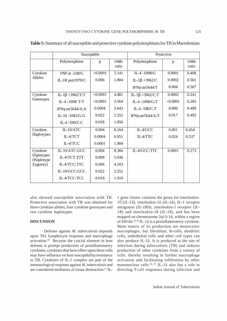
In Table 5, we can see the summary ofall susceptible and protective cytokinepolymorphisms obtained in our study. We can seethat majority of cytokine diplotypes were positivelyassociated with TB, while only one showedprotective association. Two cytokine alleles, fivecytokine genotypes and three cytokine haplotypes
TWENTY-TWO CYTOKINE GENE POLYMORPHISMS IN TB
Indian Journal of Tuberculosis
124 DEJAN TRAJKOV ET AL
Table 4: Cytokine diplotypes (haplotype zygotes), Fisher exact p-value, Odds ratio and Wald’s95% confidence interval in TB patients and healthy Macedonians
TB (n=75) Control (n=301) Polymorphism Genotype
N F N F
Fisher exact
p-value
Odds ratio
Wald' 95% CI
TGF-β1
CC:CG CC:TG CG:CG CG:TG TG:TG CG:TC
4 4 9
40 17 1
0.053 0.053 0.120 0.533 0.227 0.014
16 14 49
136 66 0
0.057 0.050 0.174 0.484 0.235
/
0.904 0.902 0.257 0.448 0.881
§
0.933 1.074 0.646 1.219 0.955
§
0.303-2.879 0.343-3.365 0.302-1.383 0.731-2.030 0.520-1.752
§
TNF- �
AG:GG GA:GG GG:GG AG:AG GG:AA GA:GA
10 4
58 2 1 0
0.133 0.053 0.773 0.027 0.014
/
66 24
206 4 0 1
0.219 0.080 0.684 0.013
/ 0.004
0.097 0.436 0.132 0.408
§ §
0.548 0.650 1.573 2.034
§ §
0.267-1.125 0.219-1.934 0.870-2.846 0.366-11.32
§ §
IL-2
GG:GG GG:TG GG:TT GT:TG TG:TG TG:TT TT:TT GT:GG GT:TT GT:GT
9 24 14 0
10 13 4 0 0 1
0.120 0.320 0.187
/ 0.133 0.173 0.053
/ /
0.014
27 85 38 11 50 48 25 1 2 0
0.094 0.296 0.133 0.058 0.174 0.168 0.087 0.003 0.007
/
0.504 0.689 0.233
§ 0.397 0.900 0.337
§ § §
1.313 1.118 1.504
§ 0.729 1.044 0.590
§ § §
0.589-2.926 0.647-1.933 0.767-2.950
§ 0.351-1.517 0.532-2.047 0.199-1.752
§ § §
IL-4
GCC:GCC GCC:TCC GCC:TTC GCC:TTT TCC:TCC TCC:TTC TCC:TTT TTT:TTT GCT:TTT GTC:TTC TCT:TTT GTT:TTC TCT:TCT
0 7
10 6
28 7 8 3 0 0 5 0 1
/ 0.093 0.133 0.080 0.373 0.093 0.107 0.040
/ /
0.067 /
0.014
1 26
103 32 68 7
28 4 8 4 4 1 0
0.003 0.091 0.360 0.112 0.238 0.025 0.098 0.014 0.028 0.014 0.014 0.003
/
§ 0.948
0.0001 0.423 0.018 0.006 0.822 0.146
§ §
0.009 § §
§ 1.029 0.273 0.690 1.910 4.103 1.100 2.938
§ §
5.036 § §
§ 0.429-2.473 0.135-0.555 0.277-1.718 1.112-3.282 1.392-12.09 0.480-2.524 0.643-13.42
§ §
1.318-19.24 § §
IL-6
CA:CA CA:GG CG:GG GG:GG GA:GG
8 31
/ 36
/
0.107 0.413
/ 0.480
/
25 122
9 144
1
0.083 0.405 0.030 0.479 0.003
0.518 0.899
§ 0.980
§
1.318 1.034
§ 1.006
§
0.569-3.053 0.618-1.728
§ 0.607-1.670
§
IL-10
ACC:ACC ACC:ATA ACC:GCC ATA:ATA ATA:GCC GCC:GCC ACA :GCC ACA :ATA ATC :GCC
4 8
21 5
23 10 0 0 4
0.053 0.107 0.280 0.067 0.307 0.133
/ /
0.053
21 21
114 19 93 17 3 9 2
0.070 0.070 0.381 0.064 0.311 0.057 0.010 0.030 0.007
0.600 0.292 0.103 0.921 0.942 0.022
§ §
0.004
0.746 1.581 0.631 1.053 0.980 2.552
§ §
8.366
0.248-2.242 0.671-3.724 0.362-1.100 0.380-2.917 0.566-1.696 1.117-5.831
§ §
1.502-46.58 N= absolute number; F=frequency; CI=Confidence Interval; § cannot be calculated because expected <5, � 2 test; *, statistically significant.
Indian Journal of Tuberculosis
125
also showed susceptible association with TB.Protective association with TB was obtained forthree cytokine alleles, four cytokine genotypes andtwo cytokine haplotypes.
DISCUSSION
Defense against M. tuberculosis dependsupon Th1 lymphocyte response and macrophageactivation.30 Because the crucial element in hostdefense is prompt production of proinflammatorycytokines, cytokines that have effect upon these cellsmay have influence on host susceptibility/resistanceto TB. Cytokines of IL-1 complex are part of theimmunological response against M. tuberculosis andare considered mediators of tissue destruction.31 IL-
1 gene cluster contains the genes for interleukin-1Ü (IL-1Ü), interleukin-1â (IL-1â), IL-1 receptorantagonist (IL-1RA), interleukin-1 receptor (IL-1R) and interleukin-18 (IL-18), and has beenmapped on chromosome 2q13-14, within a regionof 450 kb.32,33 IL-1â is a proinflammatory cytokine.Main source of its production are monocytes/macrophages, but fibroblast, B-cells, dendriticcells, endothelial cells and other cell types canalso produce IL-1â. It is produced at the site ofinfection during tuberculosis (TB) and inducesproduction of other cytokines from a variety ofcells, thereby resulting in further macrophageactivation and facilitating infiltration by othermononuclear cells.34, 35 IL-1â also has a role indirecting T-cell responses during infection and
Susceptible Protective
Polymorphism p Odds ratio
Polymorphism p Odds ratio
Cytokine Alleles
TNF-α -238/G
IL-1R psti1970/C
<0.0001
0.006
5.141
1.804
IL-4 -1098/G
IL-1β +3962/C
IFNγ utr5644/T
0.0001
0.0002
0.004
0.408
0.501
0.567
Cytokine Genotypes
IL-1β +3962/T:T
IL-4 -1098/ T:T
IFNγ utr5644/A:A
IL-10 -1082/G:G
IL-4 -590/C:C
<0.0001
<0.0001
0.0004
0.022
0.018
4.481
3.564
2.643
2.552
1.856
IL-1β +3962/C:T
IL-4 -1098/G:T
IL-4 -590/C:T
IFNγ utr5644/A:T
0.0002
<0.0001
0.006
0.017
0.241
0.285
0.489
0.492
Cytokine Haplotypes
IL-10/ATC
IL-4/TCT
IL-4/TCC
0.004
0.0004
0.0001
8.164
6.951
1.984
IL-4/GCC
IL-4/TTC
0.001
0.024
0.454
0.537
Cytokine Diplotypes (Haplotype Zygosity)
IL-10/ATC:GCC
IL-4/TCT:TTT
IL-4/TCC:TTC
IL-10/GCC:GCC
IL-4/TCC:TCC
0.004
0.009
0.006
0.022
0.018
8.366
5.036
4.103
2.552
1.910
IL-4/GCC:TTC 0.0001 0.273
Table 5: Summary of all susceptible and protective cytokine polymorphisms for TB in Macedonians
TWENTY-TWO CYTOKINE GENE POLYMORPHISMS IN TB
Indian Journal of Tuberculosis
126
granuloma formation.36 While IL-1Ü and IL-1âare potent proinflammatory cytokines involved inthe early recruitment of inflammatory cells to M.tuberculosis induced granulomas,37,38 IL-1RA isan anti-inflammatory cytokine. All three moleculesbind to the same interleukin-1 receptor (IL-1R)39
and compete among each other. In our study weevaluated the role of two SNPs in IL-1â gene (-511 C/T and +3962 C/T), one polymorphism inIL-1Ü gene (-889 C/T), one polymorphism in IL-1R gene (psti1970 C/T) and one polymorphism inIL-1RA gene (mspa11100 T/C) in the pathogenesisof TB. Our results showed that peoples with IL-1R psti1970/C allele are nearly twice moresusceptible toTB than those with IL-1R psti1970/T allele. We could not confirm that IL-1Rpsti1970/C:C genotype also is posit ivelyassociated with TB.40 On the other hand IL-1â+3962/C allele showed protective association.Unlike the result reported for Colombianpopulation who showed strong protectiveassociation of IL-1â +3953/T allele carryinggenotypes and TB,41 and contradictory to theresults obtained for Iranian,42 Cambodian,43 andpopulation from western region of Gambia44 whocould not confirm any association, we found thatpeople with IL-1â +3692/T:T genotype have 4.4times higher risk to develop TB. Contradictory,IL-1â +3692/C:T genotype showed negative(protective) association for TB. Unlike the resultsof several authors,40,43 our investigation seems torefute any association between IL-1Ü and IL-1RApolymorphisms (alleles or genotypes) and TB.
Interleukin 12 (IL-12) as a part of IL-12/IFN-ã axis plays an important role in the host defenseagainst intracellular pathogens like Mycobacteriumtuberculosis.45 Although several authors investigatedthe role of IL-12 polymorphisms and their associationwith TB, results are still obscure. It has been reportedthat besides the polymorphism in the promoterregion, polymorphism in 3’UTR correlate with theprotein secretion.46 We investigated the possible roleof the polymorphisms in the IL-12B gene 3'untranslated region (UTR) in the pathogenesis ofTB. Our results showed no significant differencesof frequency at alleles and genotypes level,suggesting that polymorphism in 3’UTR has no or
has negligible effect on the pathogenesis of TB inthe population of ethnic Macedonians. These resultscorrelate with others.47-50
IFNã is one of the participants in the IL12/IFN-ã axis, besides IL-12, IL-12R and IFNãR. It isthe most important Th1 type cytokine involved inthe host defense against M. tuberculosis51 viamacrophage activation,52 and plays a major role indefense against viruses and intracellular agents.Many studies indicate that one of the IFN-ã genepolymorphisms, the polymorphism +874 T/A locatedin the first intron of the IFN-ã gene, correlates withthe IFN-ã production. The /A allele of thispolymorphism correlates with low level of IFN-ãafter stimulation.53 We found significant differencesnot only in IFNã utr5644 T/A allele frequency, butin IFNã utr5644 genotypes containing /A allele (/A:A, /A:T) as well. The IFNã utr5644/A:A genotypeis positively associated with TB patients with oddsratio of 2.443, meaning that people with IFNãutr5644/A:A genotype have 2.4 times bigger risk todevelop TB in comparison to people with othergenotypes. On the contrary, /A:T genotype wasfound to be protective with odds ratio 0.492.Concerning the allele level, negative (protective)association for TB was found for the IFNã utr5644/T. This polymorphism was also found to beassociated with TB in Spanish,54 Colombian,55
Sicilian,56 South African,57 Croatian,58 Hong KongChinese59 and Brazilian,60 but not in Indonesian,50
Caucasian American61 Indian,62 and West Africanpopulation.63,64
Contradictory with data reported by someauthors, who found significant negative associationat codon 10 TGFâ1 polymorphism, /T allele andpredomination of /C allele and /C:C genotype amongpatients with pulmonary tuberculosis,40 in our studywe did not find any significant association betweenTB and TGF-â1 polymorphisms in codon 10 andcodon 25. Similar findings for TGF-â1 obtain otherauthors.55, 65, 66
Patients with advanced TB have higherlevels of serum TNF-Ü than patients with mildtuberculosis or healthy people.67 Although results ofassociations between TNF-Ü polymorphisms and
DEJAN TRAJKOV ET AL

Indian Journal of Tuberculosis
127
susceptibility/resistance to TB have been alreadystudied68,69 and showed ethnic-specific pattern, theyare still questionable. Our results for associationbetween TNF-Ü -308 A/G polymorphism and TBcorrelate with results obtained for Thais,70
Cambodian,42, 71 Colombian55 and population fromIndia,72 but are apparently in disagreement with thosedata which find significant association betweencertain TNF-Ü-308 genotypes and TB in three ethnicgroup from Bashkorstan73 and in Sicilian patients. 74
In contrast to the previously reported results,70, 72
we could confirm the positive (susceptible)association of TNF-Ü -238/G allele for TB. However,we could not confirm the findings of an earlier studyfrom Iran that TNF -238 genotypes were associatedwith TB.40 Only a few authors have investigated therole of other TNF-Ü polymorphisms and TB, andfound no association between them and TB.42,70 Itremains questionable whether this observedassociation is solely effect of TNF-Ü polymorphismsor association with HLA-Al, B17, B21 and DR7 mayplay a significant role in pathogenesis of TB.72
To our knowledge, this is the first studyanalyzing the association between IL-2polymorphisms and TB. In our study we did notfind any significant association in IL-2 -330 and IL-2 +160 frequencies of alleles, genotypes, haplotypes,or diplotypes.
IL-4 is generally elevated in advanced stagesof tuberculosis75 and by down regulatingmacrophage activation may determine whether theinfection becomes latent or progressive.76, 77 It alsoinduces production of mucus and hyperplasia ofgoblet cells78 in bronchial submucosa. Its genetogether with the gene of the IL-13 are located onthe chromosome 5q31, only 20 kilobase apart, inthe region associated with airway hyper-responsiveness.79 IL-4 mediates its activity throughIL-4 receptor (IL-4RÜ). In this study, weinvestigated alleles and genotypes of threepolymorphisms of IL-4 (at positions -1098, -590,and -33), as well as haplotypes and diplotypes ofinvestigated polymorphisms. It has been shown thatpolymorphism at position -590 in the IL-4 promoterregion is associated with enhanced IL-4 promoterstrength and altered IL-4 level and activity, and
thereby influences the IL-4 dependent events whichdetermine disease progression.80,81 Our resultsshowed that IL-4 -1098/G allele was protective forTB. Frequency analysis of genotypes showed thathomozygous genotypes of two IL-4 polymorphisms(IL-4 -098/T:T, and IL-4 -590/C:C) were associatedwith susceptibility for TB, while heterozygousgenotypes of the same IL-4 polymorphisms (IL-4 -1098/G:T, and IL-4 -590/C:T) showed protectiveassociation. Our results obtained for IL-4 -590genotypes are not in agreement with those gainedfor Iranian population40 and seem to be inverse withthe results gained for population of India.62 We alsofound that two of the IL-4 haplotypes (IL-4/TCTand IL-4/TCC) have susceptible association for TB,as well as protective association with other twohaplotypes (IL-4/GCC and IL-4/TTC). From thediplotypes analysis, we can see that combination ofIL-4 haplotypes IL-4/TCT:TTT, IL-4/TCC:TTC andIL-4/TCC:TCC were positively associated with TB,and only one IL-4 dilpotype (IL-4/GCC:TTC) wasnegatively associated with TB. We did not find anysignificant association between IL-3 -33 and IL-4RÜ+1902 frequencies of alleles and genotypes and TB.
Although it is known that IL-6 is importantin the initial innate response to M. tuberculosis,82,83
its role in the pathogenesis of TB is still obscure.Human macrophages in response to M. tuberculosisinfection secrete IL-6,84 but it is not vital in generationof specific protective immunity.83 Only a few studieshave analyzed the possible relationship between IL-6 polymorphisms and TB. In this study, the IL-6 -174 C/G and IL-6 nt565 A/G gene polymorphismsshowed no association with tuberculosis. Theseresults are in agreement with other studies,55 sincesignificant association between TB and IL-6 -174genotypes was shown only in Iranian patients.40
IL-10 belongs to the group of macrophage-deactivating cytokines, that down regulates Th1-induced response to M. tuberculosis and reactivateschronic pulmonary TB in mice.85, 86 IL10 inhibitsIFN-ã production by T cells87 and counter balancedpro-inflammatory effects of TNF-Ü88 It has beenalso shown that IL-10 converts human dendritic cellsinto macrophage-like cells with increased anti-mycobacterial activity.89 Hence, IL-10 may play an
TWENTY-TWO CYTOKINE GENE POLYMORPHISMS IN TB
Indian Journal of Tuberculosis
128
important role in the pathogenesis of TB. Weinvestigated the associations between three SNPsin the IL-10 gene promoter region (-1082 A/G, -819C/T, and -592 A/C) and TB. When studiedindependently there was no significant associationbetween all three investigated IL-10 alleles and TB.However, analysis of genotypes showed that /G:Ggenotype (homozygous G allele) was susceptible toTB. Patients with this genotype have 2.5 times biggerrisk to develop TB. The same risk for developingTB have the patients with IL-10/GCC:GCCdiplotype. This risk became much higher in patientswith IL-10/ATC haplotype (odds ratio=8.164) andIL-10/ATC:GCC combination of haplotypes(diplotype or haplotype zygozity) (odds ratio 8.366).Data showed that IL-10 -1082/GG haplotype90 andIL-10/GCC haplotype correlates with higher levelof IL-10 after stimulation, which leads to thesuppression of IFN-ã, and hence favoursdevelopment/relapse of TB.59,90 It was found that /GCC haplotype was associated more with thedevelopment of relapse/extra-pulmonary TB, thanwith the susceptibility to TB.59, 91 Previousinvestigations have showed that IL-10 1082 A/Gpolymorphism was associated with TB in the HongKong Chinese,59 Colombian,55 Spanish,92 Turkish,93
Cambodian,42 but not in the Gambian43 and Spanishpopulation.92 In Korean populations not IL-10 -1082A/G polymorphism, but IL-10 -592 A/Cpromoters polymorphism was found to havesignificant association with TB.94 Some authorsclaimed that not solely effect of IL-10polymorphisms, but the combination of certain IL-10 and TNF-Ü genotypes might influence theoutcome of inflammation response against TB.74
Although mechanisms associated withpolymorphisms that lead to the changes in geneexpression are still poorly understood, it is well-known that TB is partly under polygenic control.The genetic components that play role in hostdefense to TB encompass not only multiple alleleslocated on different genes and even on differentchromosomes, but also gene-environmentinteraction as well. Still, it is not very well knownwhether polymorphisms itself contribute tosusceptibility/protection to TB or their linkagedisequilibrium with an unknown disease
susceptibility allele. Many of these polymorphismsoccur in ethnic-specific patterns. Identification ofthese genes and these kinds of interactions are ofgreat importance in clarifying the role of geneticfactors in pathogenesis of TB and may lead to newways of treatment or prophylaxis.
In conclusion, we confirm that somecytokine polymorphisms contribute tosusceptibility/protection to tuberculosis inMacedonians.
ACKNOWLEDGEMENTS
This research is part of the project“Molecular analysis of cytokine gene polymorphismsin the Republic of Macedonia” supported by theMinistry of Education and Science from Republicof Macedonia (Project No. 13-874/3-05). We wouldlike to gratefully acknowledge Prof. G. Opelz andDr. J. Mytilineos from the Institute of Immunology,Department of Transplantation Immunology,University of Heidelberg, Heidelberg, Germany forkindly supplying the Heidelberg PCR-SSP kitreagents in this project. For sample collection,technical support, and laboratory direction, we thankElena Zaharieva.
REFERENCES
1. Nunn P. The global control of tuberculosis: what are theprospects?. Scand J Infect Dis 2001;33:329–32.
2. Fatkenheuer G, Taelman H, Lepage P, Schwenk A, WenzelR. The return of tuberculosis. Diagn Microbiol Infect Dis1999;34:139 –46.
3. Dolin PJ, Raviglione MC, Kochi A. Global tuberculosisand mortality during 1990–2000. Bull World HealthOrgan 1994;72: 213–220.
4. World Health Organization. The World Health Report2004—changing history, 2004.
5. Schluger NW. Recent advances in our understanding ofhuman host responses to tuberculosis. Respir Res2001;2:157–63.
6. Comstock GW. Tuberculosis in twins: a re-analysisof the Prophi t survey. Am Rev Respi r Dis1978;117:621-4.
7. Fine PE. Immunogenetics of susceptibility to leprosy,tuberculosis and leishmaniasis. An epidemiologicalperspective. Int J Lepr Other Mycobact Dis 1981;49:437-54.
8. Bellamy R. Genetic susceptibility to tuberculosis. ClinChest Med 2005;26(2): 233-46.
DEJAN TRAJKOV ET AL

Indian Journal of Tuberculosis
129
9. Kallmann FJ, Reisner D. Twin studies on the significanceof genetic factors in tuberculosis. Am Rev Tuberc 1942;47:549–574.
10. Hoal EG. Human genetic susceptibility to tuberculosis andother mycobacterial diseases. IUBMB Life 2002;53(4-5):225-9.
11. Malik S, Schurr E. Genetic susceptibility to tuberculosis.Clin Chem Lab Med 2002;40(9): 863–8.
12. Hill AV. The immunogenetics of human infectious diseases.Annu Rev Immunol 1998;16: 593–617.
13. Fenton MJ, Vermeulen MW. Immunopathology oftuberculosis: roles of macrophages and monocytes. InfectImmun 1996;64: 683–690.
14. Sher A, Coffman RL. Regulation of immunity to parasitesby T cells and T cell-derived cytokines. Annu Rev Immunol1992;10:385 –409.
15. Wallis RS, Ellner JJ. Cytokines and tuberculosis. J LeukocBiol 1994;55:676 –81.
16. Pravica V, Asderakis A, Perrey C, Hajeer A, Sinnott PJ,
Hutchinson IV. In vitro production of IFN-ã correlates
with CA repeat polymorphism in the human IFN-ã gene.Eur J Immunogenet 1999;28:1–3.
17. Kilpinen S, Huhtala H, Hurme M. The combination of theinterleukin-1alpha (IL-1alpha-889) genotype and theinterleukin-10 (IL-10 ATA) haplotype is associated withincreased interleukin-10 (IL-10) plasma levels in healthyindividuals. Eur Cytokine Netw 2002;13(1):66-71.
18. Warle MC, Farhan A, Metselaar HJ, Hop WC, Perrey C,Zondervan PE, Kap M, Kwekkeboom J, Ijzermans JN,Tilanus HW, Pravica V, Hutchinson IV, Bouma GJ. Arecytokine gene polymorphisms related to in vitro cytokineproduction profiles? Liver Transpl 2003;9(2):170-81.
19. Trajkov D, Arsov T, Petlichkovski A, Strezova A, Efinska-Mladenovska O, Spiroski M. Cytokine genepolymorphisms in population of ethnic Macedonians. CroatMed J 2005;46(4):685-92.
20. World Health Organization. Treatment of tuberculosis.Guidelines for national programmes. 2nd ed, 1997. WHO/TB/97.220. Geneva: WHO, 1997.
21. Towner P. Purification of DNA. Essential MolecularBiology (ed.T.A.Brown). Oxford University Press, Oxford.1995:47-54.
22. Spiroski M, Arsov T, Petlichkovski A, Strezova A, TrajkovD, Efinska-Mladenovska O, Zaharieva E. Case Study:Macedonian Human DNA Bank (hDNAMKD) as a sourcefor public health Genetics. In: Health Determinants in theScope of New Public Health. Ed. by Georgieva L, BurazeriG. Hans Jacobs Company: Sofia, 2005:33-44.
23. Tseng LH, Chen PJ, Lin MT, Singleton K, Martin EG, YenAH, Martin PJ, Hansen JA. Simultaneous genotyping ofsingle nucleotide polymorphisms in the IL-1 gene complexby multiplex polymerase chain reaction-restrictionfragment length polymorphism. J Immunol Methods2002;267: 151-6.
24. Helmberg W, Lanzer G, Zahn R, Weinmayr B, Wagner T,Albert E. Virtual DNA analysis – a new tool for combinationand standardised evaluation of SSO, SSP and sequencing-based typing results. Tissue Antigens1998; 51:587-92.
25. Lancaster A, Nelson MP, Meyer D, Thomson G, SingleRM. PyPop: a software framework for population
genomics: analyzing large-scale multi-locus genotype data.Pac Symp Biocomput 2003;:514-25.
26. Lancaster AK, Single RM, Solberg OD, Nelson MP,Thomson G. PyPop update—a software pipeline for large-scale multilocus population genomics. Tissue Antigens2007;69(Suppl 1):192-7.
27. Single RM, Meyer D, Mack SJ, Lancaster A, Erlich HA,Thomson G. 14th International HLA and ImmunogeneticsWorkshop: report of progress in methodology, datacollection, and analyses. Tissue Antigens 2007;69(Suppl1):185-7.
28. Guo S, Thomson E. Performing the exact test of HardyWeinberg proportion for multiple alleles. Biometrics1992;48:361.
29. Schneider S, Roessli D, Excoffier L. Arlequin version 2.000:a software for population genetics data analysis. Geneva(Switzerland): Genetics and Biometry Laboratory,University of Geneva; 2000.
30. Fenton MJ, Vermeulen MW. Immunopathology oftuberculosis: roles of macrophages and monocytes. InfectImmun 1996;64: 683–690.
31. Dinarello CA. Interleukin-1 and interleukin-1 antagonism.Blood 1991;77: 1627–1652.
32. Nicklin MJH, Weith A, Duff GW. A physical map of theregion encompassing the human inteleukin-1a, interleukin-1, and interleukin-1 receptor antagonist genes. Genomics1994;19: 382-384.
33. Nicklin MJ, Barton JL, Nguyen M, FitzGerald MG, DuffGW, Kornman K. A sequence-based map of the nine genesof the human interleukin-1 cluster. Genomes 2002;79:718-725.
34. Sica A, Wang JM, Colotta F, Dejana E, Mantovani A,Oppenheim JJ, Larsen CG, Zachariae CD, Matsushima K.Monocyte chemotactic and activating factor geneexpression induced in endothelial cells by IL-1 and tumornecrosis factor. J Immunol 1990;144(8): 3034-3038.
35. Hunter CA, Chizzonite R, Remington JS. IL-1 beta isrequired for IL-12 to induce production of IFN-gamma byNK cells. A role for IL-1 beta in the T cell-independentmechanism of resistance against intracellular pathogens. JImmunol 1995;155: 4347-4354.
36. Tsao TCY, Hong J, Li LF, Hsieh MJ, Liao SK, Chang KSS.Imbalances between tumor necrosis factor-Ü and its solublereceptor forms, and interleukin-1 and interleukin-1receptor antagonist in BAL fluid of cavitary pulmonarytuberculosis. Chest 2000;117: 103-109.
37. Marshall BG, Wangoo A, Cook HT, Shaw RJ. Increasedinflammatory cytokines and new collagen formation incutaneous tuberculosis and sarcoidosis. Thorax 1996;51:1253–1261.
38. Bergeron A, Bonay M, Kambouchner M, Lecossier D,Riquet M, Soler P, Hance A, Tazi A. Cytokine patterns intuberculous and sarcoid granulomas: correlations withhistopathologic features of the granulomatous response. JImmunol 1997;159: 3034–3043.
39. Arend WP, Malyak M, Guthridge CJ, Gabay C. Interleukin-1 receptor antagonist: role in biology. Annu Rev Immunol1998;16: 27-55.
40. Amirzargar AA, Rezaei N, Jabbari H, Danesh AA, KhosraviF, Hajabdolbaghi M, Yalda A, Nikbin B. Cytokine single
TWENTY-TWO CYTOKINE GENE POLYMORPHISMS IN TB
Indian Journal of Tuberculosis
130
nucleotide polymorphisms in Iranian patients with pulmonarytuberculosis. Eur Cytokine Netw 2006;17(2):84-9.
41. Gomez LM, Camargo, JF, Castiblanco J, Ruiz-Narvaez EA,Cadena J, Anaya JM. Analysis of IL1B, TAP1, TAP2 andIKBL polymorphisms on susceptibility to tuberculosis.Tissue Antigens 2006;67(4): 290-6.
42. Delgado JC, Baena A, Thim S, Goldfeld AE. Ethnic-specificgenetic associations with pulmonary tuberculosis. J InfectDis 2002;186: 1463–1468.
43. Bellamy R, Ruwende C, Corrah T, McAdam KP, WhittleHC, Hill AV. Assessment of the interleukin 1 gene clusterand other candidate gene polymorphisms in hostsusceptibility to tuberculosis. Tuberi Lung Dis 1998;79:83–89.
44. Casanova JL, Abel L. Genetic dissection of immunity tomycobacteria: the human model. Annu Rev Immunol2002;20: 581-620.
45. Schluger NW, Rom WN. The host immune response totuberculosis. Am J Respir Crit Care Med 1998;157: 679-91.
46. Oppmann B, Lesley R, Blom B, Timans JC, Xu Y, HunteB, Vega F, Yu N, Wang J, Singh K, Zonin F, Vaisberg E,Churakova T, Liu M, Gorman D, Wagner J, Zurawski S,Liu Y, Abrams JS, Moore KW, Rennick D, deWaal-MalefytR, Hannum C, Bazan JF, Kastelein RA. Novel p19 proteinengages IL-12p40 to form a cytokine, IL-23, withbiological activities similar as well as distinct from IL-12.Immunity 2000;13: 715-25.
47. Ma X, Reich RA, Gonzalez O, Pan X, Fothergill AK, StarkeJR, Teeter LD, Musser JM, Graviss EA. No evidence forassociation between polymorphism in the 3’untranslatedregion of interleukin-12B and human susceptibility totuberculosis. J Infect Dis 2003;188: 1116-8.
48. Puzyrev VP, Freidin MB, Rudko AA, Strelis AK,Kolokolova OV. Polymorphisms of tuberculosissusceptibility candidate genes in the Slavonic populationof Siberia: A pilot study. Mol Biol 2002;36 (5): 634-636.
49. Tso HW, Lau YL, Tam CM, Wong HS, Chiang AKS.Associations between IL12B polymorphisms andtuberculosis in the Hong Kong Chinese population. J InfectDis 2004;190: 913–9.
50. Sahiratmadja E, Baak-Pablo R, de Visser AW, AlisjahbanaB, Adnan I, van Crevel R, Marzuki S, van Dissel JT,Ottenhoff THM, van de Vosse E. Association ofpolymorphisms in IL-12/IFN-ã pathway genes withsusceptibility to pulmonary tuberculosis in Indonesia.Tuberculosis 2007;87: 303-311.
51. Condos R, Rom W N, Liu Y M, Schluger N W. Localimmune responses correlate with presentation and outcomein tuberculosis. Am J Respir Crit Care Med 1998;157:729–735.
52. Dorman SE, Holland SM. Interferon-g and interleukin-12pathway defects and human disease. Cytokine GrowthFactor Rev 2000;11:321–33.
53. Pravica V, Asderakis A, Perrey C, Hajeer A, Sinnott PJ,Huychinson IV. In vitro production of IFN-g correlateswith CA repeat polymorphism in the human IFN-gammagene. Eur J Immunogenet 1999;26(1):1–3.
54. Lopez-Maderuelo D, Arnalich F, Serantes R, Gonzales A,Codoceo R, Madero R, Vazquez JJ, Montiel C. Interferon-
gamma and interleukin-10 gene polymorphisms inpulmonary tuberculosis. Am J Respir Crit Care Med2003;167(7): 970-5.
55. Henao MI, Montes C, Paris SC, Garcia LF. Cytokine genepolymorphisms in Colombian patients with differentclinical presentations of tuberculosis. Tuberculosis 2006;86:11-19.
56. Lio D, Marino V, Serauto A, Gioia V, Scola L, Crivello A,Forte GI, Coloona-Romano G, Candore G, Caruso C.Genotype frequencies of the +874T—>A single nucleotidepolymorphism in the first intron of the interferon-gammagene in a sample of Sicilian patients affected by tuberculosis.Eur J Immunogenet 2002;29(5): 371-4.
57. Rossouw M, Nel HJ, Cooke GS, van Helden PD, Hoal EG.Association between tuberculosis and a polymorphic NFKB
binding site in the interferon ã gene. Lancet 2003;361:1871-72.
58. Etokebe GE, Bulat-Kardum L, Johansen MS, Knezevic J,Balen S, Matakovic-Mileusnic N, Matanic D, Flego V,
Pavelic J, Beg-Zec Z, Dembic Z. Interferon-ã gene (T874Aand G2109A) polymorphisms are associated withmicroscopy-positive tuberculosis. Scand J Immunol2006;63: 136-41.
59. Tso HW, Ip WK, Chong WP, Tam CM, Chiang AKS, LauYL. Association of interferon gamma and interleukin 10genes with tuberculosis in Hong Kong Chinese. GenesImmun 2005;6: 358-363.
60. Amim LH, Pacheco AG, Fonseca-Costa J, Loredo CS, RabahiMF, Melo MH, Ribeiro FC, Mello FC, Oliveira MM, LapaE Silva JR, Ottenhoff TH, Kritski AL, Santos AR. Role ofIFN-gamma +874 T/A single nucleotide polymorphism inthe tuberculosis outcome among Brazilians subjects. MolBiol Rep 2007 Aug 8; [Epub ahead of print].
61. Moran A, Ma X, Reich RA, Graviss EA. No associationbetween the 874T/A single nucleotide polymorphism inthe IFN-gene and susceptibility to TB. Int J Tuberc LungDis 2007;11(1): 113-115.
62. Vidyarani M, Selvaraj P, Prabhu Anand S, Jawahar MS,Adhilakshmi AR, Narayanan PR. Interferon gamma (IFNã)& interleukin-4 (IL-4) gene variants & cytokine levels inpulmonary tuberculosis. Indian J Med Res 2006;124: 403-410.
63. Cooke GS, Campbell SJ, Sillah J, Gustafson P, Bah B,Sirugo G, Bennett S, McAdam KPWJ, Sow O, LienhardtC, Hill AVS. Polymorphism within the Interferon-ã/Receptor complex is associated with pulmonarytuberculosis. Am J Respir Crit Care Med 2006;174:339–343.
64. Sallakci N, Coskun M, Berber Z, Gurkan F, Kocamaz H,Uysal G, Bhuju S, Yavuzer U, Singh M, Yegin O. Interferon-ã gene +874T-A polymorphism is associated withtuberculosis and gamma interferon response. Tuberculosis2007;87: 225-30.
65. Blobe GC, Schiemann WP, Lodish HF. Role of transforminggrowth factor beta in human disease. N Engl J Med2000;342: 1350–1358.
66. Niimi T, Sato S, Sugiura Y, Yoshinouchi T, Akita K, MaedaH, Achiva H, Ninomiya S, Akita Y, Suzuki M, Nishio M,Yoshikawa K, Morishita M, Shimizu S, Uede R.Transforming growth factor-beta gene polymorphism in
DEJAN TRAJKOV ET AL
Indian Journal of Tuberculosis
131
sarcoidosis and tuberculosis patients. Int J Tuberc LungDis 2002;6(6): 510-5.
67. Fiorenza G, Rateni L, Farroni MA, Bogue C, DlugovitzkyDG. TNF-alpha, TGF-beta and NO relationship in serafrom tuberculosis (TB) patients of different severity.Immunol Lett 2005;98: 45–8.
68. Hajeer AH, Hutchison IV. Influence of TNF Ü genepolymorphisms on TNF Ü production and diseases. HumImmunol 2001;62: 1191–9.
69. Allen RD. Polymorphisms of the human TNF-alphapromoter - random variation or functional diversity? MolImmunol 1999;36: 1017–27.
70. Vejbaesya S, Chierakul N, Luangtrakool P,Sermduangprateep C. NRAMP1 and TNF-apolymorphisms and susceptibility to tuberculosis in Thais.Respirology 2007;12: 202-206.
71. Goldfeld AE, Delgado JC, Thim S, Bozon MV, UglialoroAM, Turbay D, Cohen C, Yunis EJ. Association of anHLA-DQ allele with clinical tuberculosis. JAMA1998;279(3): 226-8.
72. Selvaraj P, Sriram U, Mathan Kurian S, Reetha AM,Narayanan PR. Tumor necrosis factor aqlpha (-238 and -308) and beta gene polymorphisms in pulmonarytuberculosis: haplotype analysis with HLA-A, -B and DRgenes. Tuberculosis 2001;81: 335–41.
73. Bikmaeva AR, Sibiriak SV, Valiakhmetova DKh,Khusnutdinova EK. Polymorphism of the tumor necrosisalpha gene in patients with infiltrative tuberculosis andfrom the Bashkorstan populations. Mol Biol 2002;36(5):784-7.
74. Scola L, Crivello A, Marino V, Gioia V, Serauto A, CandoreG, Coloona-Romano G, Caruso C, Lio D. IL-10 and TNF-alpha polymorphism in a sample of Sicilian patientsaffected by tuberculosis: implication for ageing and lifespan expectancy. Mech Ageing Dev 2003;124(4): 569-72.
75. Biedermann T, Zimmermann S, Himmelrich H, Gumv A,Egeter O, Sakrauski AK, et al. IL-4 instructs TH1 responsesand resistance to Leishmania major in susceptible BALB/c mice. Nat Immunol 2001;2: 1054-60.
76. Bogdan C, Vodovotz Y, Paik J, Xie QW, Nathan C.Mechanism of suppression of nitric oxide synthaseexpression by interleukin-4 in primary mouse macrophages.J Leukoc Biol 1994;55: 227-33.
77. Gordon S. Alternative activation of macrophages. Nat RevImmunol 2003;3: 23-35.
78. Dabbagh K, Takeyama K, Lee HM, Ufki IF, Lausier JA,Nadel JA. IL-4 induces mucin gene expression and gobletcell metaplasia in vitro and in vivo. J Immunol 1999;162:6233-37.
79. Doull IJ, Lawrence S, Watson M, Begishvili T, BeasleyRW, Lampe F, Holgate T, Morton NE. Allelic associationof gene markers on chromosome 5q and 11q with atopyand bronchial hyperresponsiveness. Am J Respir Crit CareMed 1996;153: 1280-4.
80. Rosenwasser LJ, Klemm DJ, Dresback JK, Inamura H,Mascali JJ, Klinnert M, Borish L. Promoter polymorphisms
in the chromosome 5 gene cluster in asthma and atopy.Clin Exp Allergy 1995;25(Suppl 2): 74-8.
81. Luoni G, Verra F, Arca B, Sirima BS, Troye-Blomberg M,Coluzzi M, Kwiatkowski D, Modiano D.Antimalarialantibody levels and IL-4 polymorphism inthe Fulani of West Africa. Genes Immun 2001;2: 411-4.
82. Saunders BM, Frank AA, Orme IM, Cooper AM.Interleukin-6 induces early gamma interferon productionin the infected lung but is not required for generation ofspecific immunity to Mycobacterium tuberculosisinfection. Infect Immun 2000;68 (6): 3322–6.
83. Ladel CH, Blum C, Dreher A, Reifenberg K, Kopf M,Kaufmann SH. Lethal tuberculosis in interleukin-6-deficientmutant mice. Infect Immun 1997;65(11): 4843–9.
84. Giacomini E, Iona E, Ferroni L, Miettinen M, Fattorini L,Orefici G, Julkunen I, Coccia EM. Infection of humanmacrophages and dendritic cells with Mycobacteriumtuberculosis induces a differential cytokine gene expressionthat modulates T cell response. J Immunol2001;166(12):7033 –41.
85. Murray PJ, Young RA. Increased antimycobacterialimmunity in interleukin-10-deficient mice. Infect Immun1999;67: 3087–95.
86. Gong JH, Zhang M, Modlin RL, et al. Interleukin-10 down-regulates Mycobacterium tuberculosis-induced Th1responses and CTLA-4 expression. Infect Immun 1996;64:913–8.
87. Bogdan C, Vodovotz Y, Nathan C. Macrophagedeactivation by interleukin 10. J Exp Med 1991;174:1549–55.
88. Moore KW, de Waal Malefyt R, Coffman RL, O’Garra A.Interleukin-10 and the interleukin-10 receptor. Annu RevImmunol 2001;19: 683–765.
89. Fortsch D, Rollinghoff M, Stenger S. IL-10 converts humandendritic cells into macrophage-like cells with increasedantibacterial activity against virulent Mycobacteriumtuberculosis. J Immunol 2000;165: 978–987.
90. Turner DM, Williams DM, Sankaran D, Lazarus M, SinnotPJ, Hutchinson IV. An investigation of polymorphism in theIL-10 gene promoter. Eur J Immunogen 1997;24: 1-8.
91. Turner J, Gonzalez-Juarrero M, Ellis DL, Basaraba RJ,Kipnis A, Orme IM, Cooper AM. In vivo IL-10 productionreactivates chronic pulmonary tuberculosis in C57BL/6mice. J Immunol 2002;169(11): 6343–6351.
92. Lopez-Maderuelo D, Arnalich F, Serantes R, Gonzalez A,Codoceo R, Madero R, Vazquez JJ, Montiel C.Interferongamma and interleukin-10 gene polymorphismsin pulmonary tuberculosis. Am J Respir Crit Care Med2003;167(7): 970–975.
93. Oral HB, Budak F, Uzaslan EK, Basturk B, Bekar A, AkalinH, Ege E, Ener B, Goral G. Interleukin-10 (IL-10) genepolymorphism as a potential host susceptibility factorin tuberculosis. Cytokine 2006;35: 143-147.
94. Shin HD, Park BL, Kim LH, Cheong HS, Lee IH, ParkSK. Common interleukin 10 polymorphism associatedwith decreased risk of tuberculosis. Exp Mol Med2005;37(2): 128-132.
TWENTY-TWO CYTOKINE GENE POLYMORPHISMS IN TB
Indian Journal of Tuberculosis
Original Article
ASSESSMENT OF LONG TERM STATUS OF SPUTUM POSITIVE PULMONARY TBPATIENTS SUCCESSFULLY TREATED WITH SHORT COURSE CHEMOTHERAPY
V.V. Banu Rekha, Rajeswari Ramachandran, K.V. Kuppu Rao, Fathima Rahman, A.R. Adhilakshmi, D.Kalaiselvi, P. Murugesan, V. Sundaram and P.R. Narayanan
(Received on 17.4.2008; Accepted after revision on 26.5.2009)
SummaryBackground: Long term status of pulmonary tuberculosis (PTB) patients treated with short course chemotherapy (SCC)regimens remains unknown.Objective: To assess the clinical, bacteriological, radiological status and health related quality of life (HRQoL) of PTBpatients 14 -18 years after successful treatment with SCC.Methodology: In a cross-sectional study, cured PTB patients treated during 1986 – 1990 at the Tuberculosis Research Centre(TRC) were investigated for their current health status including pulmonary function tests (PFT). The St Georges respiratoryquestionnaire (SGRQ) was used to assess the HRQoLResults: The mean period after treatment completion for the 363 eligible participants was 16.5yrs (range 14-18 yrs, 84%coverage) ; 25 (7 %) had been re-treated and 52 (14%) died. Among the investigated, 58 (29%) had persistent respiratorysymptoms; 170(86%) had radiological sequelae but none had active disease. Abnormal PFT was observed in 96 (65%) withpredominantly restrictive type of disease in 66(45%). The SGRQ scores for activity and impact were high implyingimpairment in HRQoL.Conclusion: Assessment of long term status of cured PTB patients showed an impairment of lung functions and HRQoLhighlighting the need to address these issues in the management of TB that may provide added value to patient care.[Indian J Tuberc 2009; 56:132-140]
Key Words: Long term assessment. SCC, PTB, PFT, SGRQ
INTRODUCTION
Standardised and directly observed sixmonth regimen is currently recommended for thetreatment of Pulmonary Tuberculosis (PTB). Themain objective of treatment in PTB is to achievehigh bacteriological cure rates. Though the patientsare cured of the disease, the information on the longterm status of the patients successfully treated withshort course chemotherapy (SCC) regimens remainslargely unknown. Studies have documented thatabout 1/3rd of the cured PTB patients do haverespiratory complaints at the end of treatmentseeking medical care1,2. The morbidity in PTBpatients while on treatment or at the end of treatmenthas been reported using subjective (quality of life)as well as objective (pulmonary function tests)measurements1-7. However, the long term status ofcured PTB patients has not been studied. Thisinformation is essential to quantify the impact of
disease for appropriate early interventions in themanagement of patients with PTB. Hence theTuberculosis Research Centre (TRC) undertook astudy to assess the clinical, bacteriological andradiological status of PTB patients14 -18 years aftersuccessful completion of treatment with SCC .Theaim of the present communication is to analyze themorbidity in these patients.
METHODOLOGY
Setting
TRC has been undertaking randomizedcontrolled clinical trials in the treatment of pulmonaryand extra-pulmonary forms of TB since 1956 with theobjective of evolving suitable chemotherapeuticregimens in patients suffering from TB. The patientsenrolled in the trial are followed up bacteriologicallyand radiologically for a maximum period of five years.
Tuberculosis Research Centre, Chennai.Correspondence: Dr. Rajaswari Ramachandran, Formerly Deputy Director, Senior Grade, Tuberculosis Research Centre, Mayor V.R. Ramanathan Road, Chetput, Chennai-600 031.Phone: 91 44 29369613; Mobile 9444057486; e-mail: [email protected]
Indian Journal of Tuberculosis
Study population
This cross-sectional study was conductedat TRC, Chennai, during August 2004 – 2005. Thestudy population included patients enrolled during1986-1990 and had bacteriological quiescence (ie.culture negative) at the 60th month of follow-up.These were new PTB patients (<1 month of previousanti-tuberculosis drugs) who were treated with firstline anti-tuberculosis drugs namely isoniazid (H),rifampicin (R), pyrazinamide (Z) and ethambutol (E)in one of the following SCC regimens whichincluded six month intermittent (2EHRZ
2/4EHR
2,
2HRZ2/4RH2) or nine month daily (2EHRZ/ 6EH)regimens.
All efforts were made to interview thepatients during home visits or over the telephone.In case of reported death of the patient, a doctor’svisit was made to probe the reasons for death (verbalautopsy)
When the patient attended the centre, adetailed history which included re-treatment duringthe follow-up period was elicited, after obtaininginformed consent. Information was also obtainedon habits which included smoking and alcoholism,respiratory symptoms and co-morbid conditions likediabetes, hypertension, bronchial asthma and cardiacproblem. The blood pressure, weight and height wererecorded and a general and systemic examinationwas performed. Other investigations includedpostero-anterior chest radiography, two sputumexamination by smear and culture for tubercle bacilli,12 lead Electrocardiogram (ECG) and Pulmonaryfunction test (PFT) by Spirometry. The quality oflife was assessed by a trained social worker usingthe St. Georges respiratory questionnaire (SGRQ).
Bacteriology: Sputum smears were examined forAFB by fluorescence microscopy.8 and thespecimens were processed by modified Petroff’smethod and cultured on the LJ medium.9 Allpositive cultures were subjected to identificationtests for Mycobacterium tuberculosis and to drugsusceptibility tests using the minimal inhibitoryconcentration method for isoniazid andrifampicin10,11.
Radiology: The chest x-rays were read by a panelof three doctors. The following observations wererecorded namely a) the extent of lung involvementwhich was documented as unilateral or bilateral, b)the number of zones involved (minimum one-zoneto maximum six -zones and c) presence or absenceof cavitation12.
Electrocardiogam (ECG): A 12 lead ECG wasrecorded for the patients and independently read bythe cardiologist. The criteria laid down by WHO(1961) were used for the ECG based diagnosis ofcor-pulmonale13.
Pulmonary function testing (PFT) – PFT wasperformed according to the techniques mentionedin the operating manual of the spirometry(sensormedix) with special reference to AmericanThoracic society of Standardization of spirometry.14
The parameters measured in spirometry includeForced Vital Capacity (FVC), Forced expiratoryvolume in 1 second (FEV
1), ratio of FEV
1 to FVC
(FEV1%). For assessing chronic changes, theobserved values of FVC, FEV
1, and FEV
1% were
expressed as a percentage of the predicted valuesby using the regression equation developed byVijayan15. Lung function impairments were classifiedaccording to the above mentioned parameters asnormal/restrictive disease/obstructive disease/combined disease14 .
St Georges respiratory questionnaire (SGRQ):In the present study. SGRQ, a standardized, airwaysdisease specific respiratory questionnaire, was usedto measure health related quality of life (HRQoL)16,
17 This health status instrument consisting of 50 itemsis grouped under three components namelysymptoms (8 items) which measures respiratorysymptoms, activity (16 items) which measuresimpairment of mobility or physical activity andimpact (26 items) which measures the psychosocialimpact of disease). This tool has been validated foruse in chronic pulmonary diseases including TB inmany countries including India18-23. SGRQ scoreswere calculated using score calculation algorithmsand missing data imputation recommended by itsdeveloper (P.W. Jones, St Georges Hospital MedicalSchool, London, UK) Scores ranging from 0 to
V. V. BANU REKHA ET AL 133
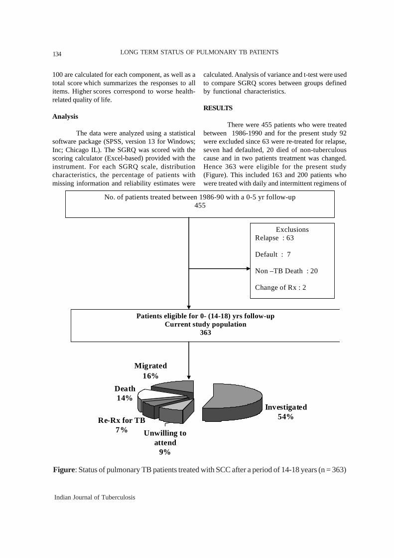
Indian Journal of Tuberculosis
100 are calculated for each component, as well as atotal score which summarizes the responses to allitems. Higher scores correspond to worse health-related quality of life.
Analysis
The data were analyzed using a statisticalsoftware package (SPSS, version 13 for Windows;Inc; Chicago IL). The SGRQ was scored with thescoring calculator (Excel-based) provided with theinstrument. For each SGRQ scale, distributioncharacteristics, the percentage of patients withmissing information and reliability estimates were
calculated. Analysis of variance and t-test were usedto compare SGRQ scores between groups definedby functional characteristics.
RESULTS
There were 455 patients who were treatedbetween 1986-1990 and for the present study 92were excluded since 63 were re-treated for relapse,seven had defaulted, 20 died of non-tuberculouscause and in two patients treatment was changed.Hence 363 were eligible for the present study(Figure). This included 163 and 200 patients whowere treated with daily and intermittent regimens of
������������������������������������������������������������������������������������������������������
���������������������������������������������������������������������������������������������������������
������
���������������������������������������������������������������������������������������������������������������
�������������������������������������������������������������������������������������������������������������������������������
��������������������
����������������������������������������������������������������������������������������������������������������
��������������������������������������������������������������������������
���������������������������������������������������������
��������������������������������������������������
������������������������������������������������������������������������������������
������������������������������������������������������������������������������������������������������������������������������������������������������������������������������������������������
������������������������������������������������������������������������������������������������������������������������������������������������������������������������������������������������������������������������������������������������ Investigated
54%Re-Rx for TB7%
Death14%
Migrated16%
Unwilling to attend
9%
Exclusions Relapse : 63 Default : 7 Non –TB Death : 20 Change of Rx : 2
Patients eligible for 0- (14-18) yrs follow-up Current study population
363
No. of patients treated between 1986-90 with a 0-5 yr follow-up 455
Figure: Status of pulmonary TB patients treated with SCC after a period of 14-18 years (n = 363)
LONG TERM STATUS OF PULMONARY TB PATIENTS134
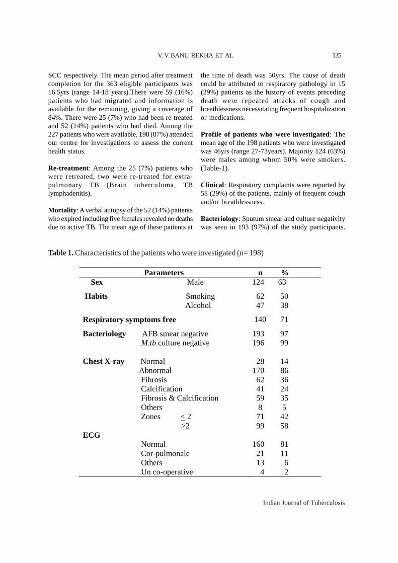
Indian Journal of Tuberculosis
SCC respectively. The mean period after treatmentcompletion for the 363 eligible participants was16.5yrs (range 14-18 years).There were 59 (16%)patients who had migrated and information isavailable for the remaining, giving a coverage of84%. There were 25 (7%) who had been re-treatedand 52 (14%) patients who had died. Among the227 patients who were available, 198 (87%) attendedour centre for investigations to assess the currenthealth status.
Re-treatment: Among the 25 (7%) patients whowere retreated, two were re-treated for extra-pulmonary TB (Brain tuberculoma, TBlymphadenitis).
Mortality : A verbal autopsy of the 52 (14%) patientswho expired including five females revealed no deathsdue to active TB. The mean age of these patients at
the time of death was 50yrs. The cause of deathcould be attributed to respiratory pathology in 15(29%) patients as the history of events precedingdeath were repeated attacks of cough andbreathlessness necessitating frequent hospitalizationor medications.
Profile of patients who were investigated: Themean age of the 198 patients who were investigatedwas 46yrs (range 27-73years). Majority 124 (63%)were males among whom 50% were smokers.(Table-1).
Clinical : Respiratory complaints were reported by58 (29%) of the patients, mainly of frequent coughand/or breathlessness.
Bacteriology: Sputum smear and culture negativitywas seen in 193 (97%) of the study participants.
Table 1. Characteristics of the patients who were investigated (n= 198)
Parameters n % Sex Male 124 63
Habits Smoking Alcohol
62 50 47 38
Respiratory symptoms free 140 71
Bacteriology AFB smear negative M.tb culture negative
193 97 196 99
Chest X-ray Normal Abnormal Fibrosis Calcification Fibrosis & Calcification Others Zones < 2 >2
28 14 170 86 62 36 41 24 59 35 8 5 71 42 99 58
ECG Normal Cor-pulmonale Others Un co-operative
160 81 21 11 13 6 4 2
V. V. BANU REKHA ET AL 135

Indian Journal of Tuberculosis
Among the five patients who had positive smears,three patients were culture negative while twopatients’ cultures grew Mycobacterium kansasii.
Radiology: There were 28 (14%) patients withnormal chest radiography while others hadabnormality predominantly of fibrosis and orcalcification.
Spirometry: The pulmonary function tests wereperformed in 148 (75%) patients who were willingto participate which included 98 males and 50females (Table-2). The spirometry was normal for52 (35%) of the participants. The predominant lungfunction impairment was restrictive type of diseasein 66 (45%) patients and this was observed more in
females - 32 (64%). Obstructive and combined typeof lung function impairments were predominant inmales. The difference in lung function impairmentsbetween the smokers and non-smokers was notstatistically significant.
Electrocardiogram (ECG): Features suggestive ofcor-pulmonale was observed in 21(11%) of thepatients. The abnormal ECG changes observed inthe remaining 14(7%) patients were that of leftventricular hypertrophy or heart rate disturbanceslike sinus bradycardia or tachycardia.
SGRQ: Tables 3 and 4 describe mean SGRQ scalescores in study population according to sex, smokingbehaviour, ECG, chest X-ray and FEV1% predicted.
Table 2: Types of abnormality in lung function in 148 investigated patients treated 14-18 years agowith SCC
Table 3: Distribution characteristics and reliability estimates SGRQ scales in the study populationweighted sample (n=198)
Symptoms Activity Impact Overall
Mean Normal values*
12 9
2
6
Mean observed values 16.46 28.95 14.11 19.60
SD 18.63 23.19 23.96 20.65
Observed range 0-79.53 0-100 0-92.5 0-83.65
% with missing data 1.5 3.1 4.6 5.1
* Reference No: 26
LONG TERM STATUS OF PULMONARY TB PATIENTS
Males Females Total Type of disease n % n % n %
Normal
Restrictive disease
Obstructive disease
Combined disease
37 38
34 35
7 7
20 20
15 30
32 64
0 0
3 6
52 35
66 45
7 5
23 16
Total 98 100 50 100 148 100
136
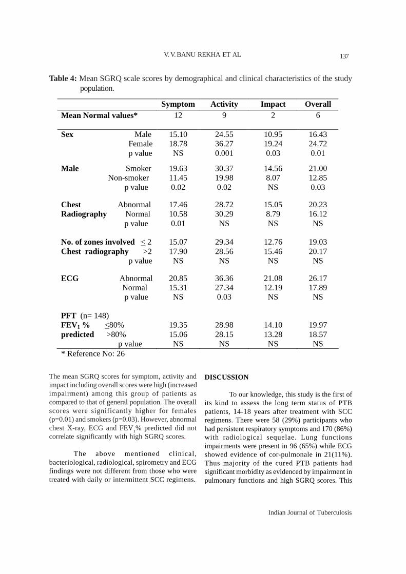
Indian Journal of Tuberculosis
The mean SGRQ scores for symptom, activity andimpact including overall scores were high (increasedimpairment) among this group of patients ascompared to that of general population. The overallscores were significantly higher for females(p=0.01) and smokers (p=0.03). However, abnormalchest X-ray, ECG and FEV1% predicted did notcorrelate significantly with high SGRQ scores.
The above mentioned clinical,bacteriological, radiological, spirometry and ECGfindings were not different from those who weretreated with daily or intermittent SCC regimens.
DISCUSSION
To our knowledge, this study is the first ofits kind to assess the long term status of PTBpatients, 14-18 years after treatment with SCCregimens. There were 58 (29%) participants whohad persistent respiratory symptoms and 170 (86%)with radiological sequelae. Lung functionsimpairments were present in 96 (65%) while ECGshowed evidence of cor-pulmonale in 21(11%).Thus majority of the cured PTB patients hadsignificant morbidity as evidenced by impairment inpulmonary functions and high SGRQ scores. This
Table 4: Mean SGRQ scale scores by demographical and clinical characteristics of the studypopulation.
Symptom Activity Impact Overall Mean Normal values*
12 9
2
6
Sex Male Female p value
15.10 18.78 NS
24.55 36.27 0.001
10.95 19.24 0.03
16.43 24.72 0.01
Male Smoker Non-smoker p value
19.63 11.45 0.02
30.37 19.98 0.02
14.56 8.07 NS
21.00 12.85 0.03
Chest Abnormal Radiography Normal p value
17.46 10.58 0.01
28.72 30.29 NS
15.05 8.79 NS
20.23 16.12 NS
No. of zones involved < 2 Chest radiography >2 p value
15.07 17.90 NS
29.34 28.56 NS
12.76 15.46 NS
19.03 20.17 NS
ECG Abnormal Normal p value
20.85 15.31 NS
36.36 27.34 0.03
21.08 12.19 NS
26.17 17.89 NS
PFT (n= 148) FEV1 % <80% predicted >80% p value
19.35 15.06 NS
28.98 28.15 NS
14.10 13.28 NS
19.97 18.57 NS
* Reference No: 26
V. V. BANU REKHA ET AL 137
Indian Journal of Tuberculosis
study highlights the presence of respiratory systemrelated morbidity among the microbiologically curedPTB patients.
Persistent respiratory symptoms observedin approximately 1/3rd of the patients in the presentstudy is an important cause of concern. Studies havedocumented that cured PTB patients continue to haverespiratory symptoms (30-47%) at the end oftreatment, (40%) after one year of treatment and(15.9%) after two and a half years aftertreatment1,2,5,24 In another study 14% of treated PTBpatients continued to attend the out-patientdepartment for more than five years for respiratorycomplaints.25 Since all the investigated patients in thepresent study were culture negative forMycobacterium tuberculosis, there is a need toeducate the patients that persistence of symptomsis not synonymous with reactivation of the disease.
The respiratory disease specific SGRQscores were higher in this cohort of patients whichimplies an impairment in the quality of life which isin conformity with other studies done among PTBpatients18,19. As data pertaining to SGRQ scores ingeneral population in India is not available, thenormative values for a general population studied inSpain recommended in SGRQ manual is used forcomparison. The symptom, activity and impactscores observed in our study after a mean period of16.5 years after successful treatment for TB were16, 28 and 14 respectively as compared to generalpopulation scores of 12, nine and two on a scale of10026. These findings suggest that the scores of thetreated PTB patients were high (indicating worsestatus) when compared to the general population.Similar to earlier SGRQ studies in PTB patients, thescore for impact was lower than that of symptomand activity18,19 The scores were less whencompared to patients with other respiratory diseaseslike interstitial lung disease, COPD orbronchiectasis20,21,22. These findings suggest thatHRQoL among treated PTB patients was suboptimalwhen compared to the general population, but betterwhen compared to other respiratory diseases.
Generally the observed SGRQ scores arehigher for females27. In this cohort the femaleparticipants had a significantly higher scores in
SGRQ suggestive of impairment of HRQoL withreference to activity and impact of disease ascompared to males. An earlier study done on PTBpatients while on treatment showed no genderdifferences in the SGRQ scores18. Among themales, in smokers SGRQ showed significantimpairment of quality of life emphasizing the needto create an awareness about smoking cessationamong treated TB patients.
Lung function impairments after TB is anunrecognized cause of chronic lung diseaseworldwide7. In the present study PFT was donein 75% of the patients. The predominant lungfunction patterns observed in these patients wererestrictive and combined type similar to otherpublished studies6,7. Our findings, similar to aprevious reports have shown that females hadmore restrictive type of disease compared tomales3. A previous study among treated PTBpatients from our centre showed that the mainventilatory type of abnormality was restrictive innature where as a study from Pretoria concludedthat equal number of patients had restrictive/obstructive type of disease6,28. However, a studyfrom South Africa which was done at an averageof 16 years after TB treatment showed that 68%of the patients had chronic obstructive lungdisease29. Impairments in lung function in thepresent study were not significantly high amongsmokers. The published studies on smoking andlung function impairments in PTB patients arevaried19,30. The patients with a FEV1% Pred. of<80% had high SGRQ scores implying poorquality of life though it did not attain statisticalsignificance
We observed a mortality of 14% in thiscohort of patients. A study done among PTBpatients irrespective of being treated has shown amortality rate of 24% and lung functionimpairment as the probable cause of death in 15%after a duration of 10 years30. Although 15 patientsin our study had frequent respiratory complaintsor hospitalization among those who expired, it wasnot possible to conclude that TB sequelae wasthe probable cause of death.
LONG TERM STATUS OF PULMONARY TB PATIENTS138
Indian Journal of Tuberculosis
The high coverage observed in our studycould be attributed to the stringent efforts inscreening for domiciliary stability for enrollment ofpatients to our controlled clinical trials. We observedno difference in the profile of patients treated withdaily or intermittent anti-tuberculosis therapy. Thishighlights the fact that intermittent chemotherapy isas effective as daily pertaining to the long termimpact of pulmonary TB.
The major limitation of this study is thatthe observations made need not be confined to TBsequelae alone and could be due to the aging and orhealth related factors of the study population. Thelack of concurrent age-sex matched controls coupledwith the lack of baseline investigations for pulmonaryfunctions and assessment of quality of life is alsoanother major limitation.
Despite the above mentioned limitations,this study has shown that treated and cured PTBpatients do suffer from long term morbidity. Indiais a high burden country for TB and there is anactive and well structured Revised National TBControl Programme (RNTCP) operatingsuccessfully covering the entire population sinceMarch 2006. The RNTCP has treated more than8.1 million patients, and thereby prevented almost1.46 million TB deaths.31 As the programme matures,its level of sophistication will undoubtedly increase.1
From the findings of this study it appears thatthe providers and programme managers have aresponsibility to recognize and address the longterm impact of TB even after patients are cured.Addressing issues relevant to the quality of lifeand considering measures for pulmonaryrehabilitation will provide added value to theprogramme.
ACKNOWLEDGEMENTS
The authors thank the staff of Bacteriologyand Statistics Departments of TRC for their technicalassistance and nursing staff for their help in takingcare of the patients. The authors acknowledge Ms.Sampoornam (STA) and the drivers of TRC for theirvaluable help in tracing the patients. We thank the
patients who co-operated for the investigations andinterviews.
REFERENCES
1. Rajeswari R, Muniyandi M, Balasubramanian R,Narayanan PR. Perceptions of tuberculosis patientsabout their physical, mental and social well-being: afield report from South India. Soc Sci Med 2005; 60:1845-1853
2. Kuppu Rao K V, Swaminathan S, Venkatesan P.Residual lung function impairment in patients treatedfor pulmonary tuberculosis. Proceedings of thenational conference on pulmonary diseases,NAPCON’99. New Delhi
3. Dhingra VK and Rajpal S. Health related quality oflife (HRQL) scoring in tuberculosis. Indian J Tuberc2003; 50:99-104
4. Marra CA, Marra F, Cox VC, Palepu A, FitzgeraldJM. Factors influencing quality of life in patientswith active tuberculosis. Health Qual Life Outcomes2004 ;2:58-68
5. Muniyandi M, Rajeswari R, Balasubramanian R,Nirupa C, Gopi PG, K. Jaggarajamma K et al.Evaluation of post-treatment health-related qualityof life (HRQoL) among tuberculosis patients. Int JTuberc Lung Dis 2007;11:887–892
6. Vijayan V K, Chakravarthy Rajkumar, Kailash N,Prabakar R, Tripathy S P. Cardio-pulmonary statusof treated cases of pulmonary tuberculosis. LungIndia 1982:1:21-23
7. Pasipanodya JG, Miller T L, Vecino M, Munguia G,Garmon R, Bae S et al. Pulmonary impairment aftertuberculosis. Chest 2007; 131: 1817-1823
8. Holst E, Mitchison DA, Radhakrishna S.Examination of smears for tubercle bacilli byfluorescence microscopy. Indian J Med Res 1959;47: 495-499.
9. Kent PT, Kubica GP. Isolation Procedures. In: PublicHealth Mycobacteriology. A guide for Level 3laboratory. US Dept. of Health and Human services.Public Health CDC service. Atlanta, Georgia. 1985
10. Allen B, Baker FJ. Mycobacteria: isolation,identification and sensitivity testing. London:Butterworth, 1968.
11. Canetti G, Fox W, Khomenko A, Mahler HT, MenonNK, Mitchison DA et al. Advances in techniques oftesting mycobacterial drug sensitivity, and the use ofsensitivity tests in tuberculosis control programmes.Bull WHO 1969; 41: 21–43
12. Arthur JA, Wrightman. Diagnostic Imaging. In:Seaton A, Seaton D, Leitch A G, editors. Crofton andDouglas’s Respiratory diseases.5th edition, Oxford:Blackwell Science Pvt. Ltd; 2000.119-147.
13. W.H.O. (1961) Tech.Report Series No.21314. American Thoracic Society. Evaluation of
impairment/disability secondary to respiratory
V. V. BANU REKHA ET AL 139
Indian Journal of Tuberculosis
disorders. Am Rev Respir Dis 1986; 133: 1205-120915. Vijayan VK, Kuppurao KV, Venkatesan P, Sankaran
K, Prabhakar R. Pulmonary function in healthy youngadult Indians in Madras. Thorax 1990;45:611-615
16. World Health Organization. The World HealthOrganization Quality of Life assessment(WHOQOL): position paper from the World HealthOrganization. Soc Sci Med 1995; 41: 1403– 1409
17. Jones PW, Quirk FH, Baveystock CM. The StGeorge’s Respiratory Questionnaire. Respir Med1991;85(Suppl B):25-31
18. Jotam G. Pasipanodya, Thaddeus L. Miller, MauricioVecino, Guadalupe, Munguia, Sejong Bae, GerryDrewyer and Stephen E. Weis. Using the St. GeorgeRespiratory Questionnaire To Ascertain HealthQuality in Persons With Treated PulmonaryTuberculosis. Chest 2007;132: 1591–1598
19. Byoung H. Lee, Jae Hyung Lee, Kyung Chan Kim andSang-hoon Kim. Post-tuberculosis destroyed lung:clinical characteristics and health-related quality oflife measurement. Chest 2007;132 :6398
20. Domingo-Salvany A, Lamarca R, Ferrer M, Garcia-Aymerich J, Alonso J, Félez M, et al. Health-relatedquality of life and mortality in male patients withchronic obstructive pulmonary disease. Am J RespirCrit Care Med 2002;166: 680-685.
21. Chang JA, Curtis JR, Patrick DL and Raghu G.Assessment of Health-Related Quality of Life inPatients with Interstitial Lung Disease. Chest1999;116:1175-1182.
22. Wilson CB, Jones PW, O’Leary CJ, Cole PJ, WilsonR. Validation of the St George’s RespiratoryQuestionnaire in Bronchiectasis. Am J Respir CritCare Med 1997;156:536-541
23. Ashutosh N. Aggarwal, Gupta D, Kumar T, Singh Nand Jindal SK.. Validation of Hindi Translation of St.George’s Respiratory Questionnaire in Indian Patientswith Chronic Obstructive Pulmonary Disease. IndianJ Chest Dis Allied Sci 2007; 49: 87-92
24. Sophia Vijay, Balasangameswara VH., Jagannatha PS,Saroja VN, Kumar P. Treatment outcome and two &half years follow-up status of new smear positivepatients treated under RNTCP. Indian J Tuberc 2004;51:199-208
25. Arora VK, Amit Johri, Ramesh Varma, Palani. Posttreatment adjustment problems and copingmechanisms in pulmonary tuberculosis patients.Indian J Tuberc 1992; 39: 181-184
26. Paul W Jones, Sally Spencer, Sue Adie. The St George’srespiratory questionnaire Manual. Version 2.1. Dated29.5.2003
27. Ferrer M, Villasante C, Alonso J, Sobradillo V, GabrielR, Vilagut G et al. Interpretation of quality of lifescores from the St. George’s RespiratoryQuestionnaire. Eur Respir J 2002; 19: 405-413
28. Plit ML, Anderson R, Van Rensburg CEG, Page ShippL, Blott JA, Fresen JL, Feldman C. Influence ofantimicrobial chemotherapy on spirometric parametersand pro-inflammatory indices in severe pulmonarytuberculosis. Eur Respir J 1998;12: 351-356
29. Willcox PA, Ferguson AD. Chronic obstructiveairways disease following treated pulmonarytuberculosis. Respir Med 1989; 83: 195-198
30. Krishna K, Bond S, Arvinli M, et al. Pulmonaryfunction in treated tuberculosis; a long term follow-up. Am Rev Respir Dis 1977; 115:402
31. Central TB Division. http://www.tbcindia.org/(Accessed on 24.1.08)
LONG TERM STATUS OF PULMONARY TB PATIENTS140
Indian Journal of Tuberculosis
141Original article
(Received on 26.5.2009. Accepted after revision on 2.7.2009)
[Indian J Tuberc 2009; 56:141-143]
MYCOBACTERIAL ES-31 SERINE PROTEASE – A BIOMARKER FORMYCOBACTERIUM TUBERCULOSIS – A PRELIMINARY REPORT*
M. Anindita 1, V. Upadhye1, D. Thamke2, D.K. Mendiratta 2 and B.C. Harinath 1
Summary: There is a need for simple and reliable method to identify Mycobacterium tuberculosis from AFB smearpositive cases. Utility of mycobacterial ES-31 serine protease as a marker to detect Mycobacterium tuberculosis bacilliwas explored using Fluorescein isothiocyanate conjugated anti-ES-31 serine protease antibody. The presence of ES-31serine protease in bacilli was indicated by green fluorescence on the cell surface. Green fluorescence was observed withM.tb. H
37Ra bacilli and M.tb. H
37Rv bacilli while no Fluorescence was observed with M. chelonae, Nocardia farcinicum as
well as in E. coli showing the usefulness of ES-31 serine protease as a marker for identification of mycobacterium tuberclebacilli in cultures.
Key words: Mycobacterial ES-31 serine protease, Immunofluorescence, Non-tuberculous mycobacteria
INTRODUCTION
Detection of presence of acid fast bacilli inclinical specimens is not sufficient and requireslabour intensive biochemical methods for furtheridentification of Mycobacterial species. Many peopleharbour NTM in their respiratory secretions withoutany symptoms. The immunosuppressed individualsinfected by human immunodeficiency virus (HIV)infection have become most significant risk factorfor disseminated NTM disease and of these, 95%are due to Mycobacterium avium complex (MAC).Clinical differentiation of M. tb. infection and othermycobacteriosis is difficult due to overlappingdomain of symptoms. Differentiation of M.tb. fromNTM depends upon use of time consumingconventional morphological and biochemical testsor expensive genotypic methods1. Thus, there is aneed for exploration of newer simple and rapidmethods to distinguish M. tb. from NTM.
In earlier studies from our laboratory, wehave reported diagnostic importance of ES-31 proteinantigen, a serine protease in the diagnosis ofpulmonary tuberculosis and TB in HIV-TB co-infection2,3. In this study we explored the utility of
mycobacterial ES-31 serine protease as a marker, ifany, to identify tubercle bacilli using Fluoresceinisothiocyanate(FITC) conjugated anti – ES-31 serineprotease antibody.
Bacilli used in the present study include M.tb. H37Ra bacilli and M. tb. H37Rv bacilli; NTM suchas Mycobacterium chelonae (M. chelonae), Nocardiafarcinicum and Escherichia coli (E. coli).
Isolation of anti ES-31 antigen and its antibodyand conjugation with Fluorescein isothiocyanate(FITC):
M. tb. H37Ra detergent soluble sonicate (DSS)antigen, was prepared from M.tb. H
37Ra bacilli.
Briefly, bacilli were 5% phenol inactivated in 0.5Mphosphate buffer (PBS, pH7.2) and incubated withsodium dodecyl sulphate (SDS) extraction buffer.The supernatant was dialysed against 0.01M PBS,pH 7.2 and used as an antigen source. . Anti-DSSIgG antibodies were raised in goat by immunizingintramuscularly with 500 µg protein/mL DSS antigenwith 1 ml Freund’s incomplete adjuvant on days 0,20, 33 and 45. Immune sera were collected on days32, 44, 57, 60 and thereafter fortnightly and anti-
1. Jamnalal Bajaj Tropical Disease Research Centre, Sevagram, Wardha2. Department of Microbiology, Mahatma Gandhi Institute of Medical Sciences, Sevagram, Wardha* As a part of the research project financially supported by Tuberculosis Association of India.Correspondence: Dr. B. C. Harinath, Director, JB Tropical Disease Research Centre and Department of Biochemistry, Mahatma Gandhi Instituteof Medical Sciences, Sevagram - 442 102 Wardha (Maharashtra); Tele Fax: (07152) 284038; E-mail: [email protected], [email protected]
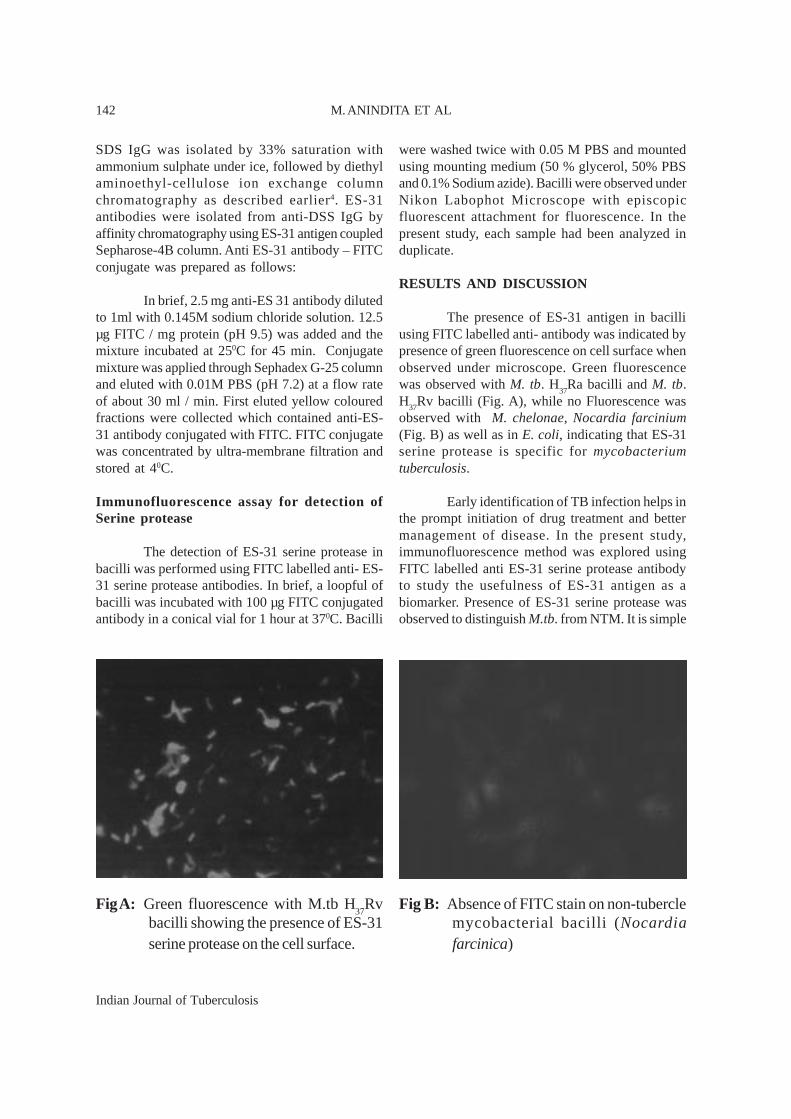
Indian Journal of Tuberculosis
142
SDS IgG was isolated by 33% saturation withammonium sulphate under ice, followed by diethylaminoethyl-cellulose ion exchange columnchromatography as described earlier4. ES-31antibodies were isolated from anti-DSS IgG byaffinity chromatography using ES-31 antigen coupledSepharose-4B column. Anti ES-31 antibody – FITCconjugate was prepared as follows:
In brief, 2.5 mg anti-ES 31 antibody dilutedto 1ml with 0.145M sodium chloride solution. 12.5µg FITC / mg protein (pH 9.5) was added and themixture incubated at 250C for 45 min. Conjugatemixture was applied through Sephadex G-25 columnand eluted with 0.01M PBS (pH 7.2) at a flow rateof about 30 ml / min. First eluted yellow colouredfractions were collected which contained anti-ES-31 antibody conjugated with FITC. FITC conjugatewas concentrated by ultra-membrane filtration andstored at 40C.
Immunofluorescence assay for detection ofSerine protease
The detection of ES-31 serine protease inbacilli was performed using FITC labelled anti- ES-31 serine protease antibodies. In brief, a loopful ofbacilli was incubated with 100 µg FITC conjugatedantibody in a conical vial for 1 hour at 370C. Bacilli
were washed twice with 0.05 M PBS and mountedusing mounting medium (50 % glycerol, 50% PBSand 0.1% Sodium azide). Bacilli were observed underNikon Labophot Microscope with episcopicfluorescent attachment for fluorescence. In thepresent study, each sample had been analyzed induplicate.
RESULTS AND DISCUSSION
The presence of ES-31 antigen in bacilliusing FITC labelled anti- antibody was indicated bypresence of green fluorescence on cell surface whenobserved under microscope. Green fluorescencewas observed with M. tb. H
37Ra bacilli and M. tb.
H37Rv bacilli (Fig. A), while no Fluorescence wasobserved with M. chelonae, Nocardia farcinium(Fig. B) as well as in E. coli, indicating that ES-31serine protease is specific for mycobacteriumtuberculosis.
Early identification of TB infection helps inthe prompt initiation of drug treatment and bettermanagement of disease. In the present study,immunofluorescence method was explored usingFITC labelled anti ES-31 serine protease antibodyto study the usefulness of ES-31 antigen as abiomarker. Presence of ES-31 serine protease wasobserved to distinguish M.tb. from NTM. It is simple
Fig A: Green fluorescence with M.tb H37
Rvbacilli showing the presence of ES-31serine protease on the cell surface.
Fig B: Absence of FITC stain on non-tuberclemycobacterial bacilli (Nocardiafarcinica)
M. ANINDITA ET AL
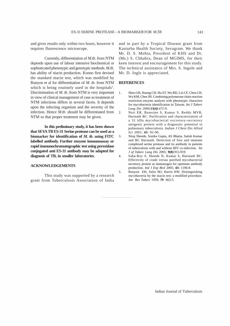
Indian Journal of Tuberculosis
143
and gives results only within two hours, however itrequires fluorescence microscope.
Currently, differentiation of M.tb. from NTMdepends upon use of labour intensive biochemical orsophisticated phenotypic and genotypic methods. M.tb.has ability of niacin production. Konno first devisedthe standard niacin test, which was modified byRunyon et al for differentiation of M. tb. from NTMwhich is being routinely used in the hospitals5.Discrimination of M. tb. from NTM is very importantin view of clinical management of case as treatment ofNTM infections differs in several facets. It dependsupon the infecting organism and the severity of theinfection. Hence M.tb. should be differentiated fromNTM so that proper treatment may be given.
In this preliminary study, it has been shownthat SEVA TB ES-31 Serine protease can be used as abiomarker for identification of M. tb. using FITClabelled antibody. Further enzyme immunoassay orrapid immunochromatographic test using peroxidaseconjugated anti ES-31 antibody may be adapted fordiagnosis of TB, in smaller laboratories.
ACKNOWLEDGEMENTS
This study was supported by a researchgrant from Tuberculosis Association of India
and in part by a Tropical Disease grant fromKasturba Health Society, Sevagram. We thankMr. D. S. Mehta, President of KHS and Dr.(Ms.) S. Chhabra, Dean of MGIMS, for theirkeen interest and encouragement for this study.The technical assistance of Mrs. S. Ingole andMr. D. Ingle is appreciated.
REFERENCES
1. Shen GH, Hunng CH, Hu ST, Wu BD, Lin CF, Chen CH,Wu KM, Chen JH. Combining polymerase chain reactionrestriction enzyme analysis with phenotypic charactersfor mycobacteria identification in Taiwan. Int J TubercLung Dis 2009;13(4):472-9.
2. Nair ER, Banerjee S, Kumar S, Reddy MVR,Harinath BC. Purification and characterization ofa 31 kDa mycobacter ia l excretory-secretoryantigenic protein with a diagnostic potential inpulmonary tuberculosis. Indian J Chest Dis AlliedSci 2001; 43: 81-90.
3. Niraj Shende, Sonika Gupta, AS Bhatia, Satish Kumarand BC Harinath. Detection of free and immunecomplexed serine protease and its antibody in patientsof tuberculosis with and without HIV co-infection. IntJ of Tuberc Lung Dis 2005; 9(8):915-919.
4. Saha-Roy S, Shende N, Kumar S, Harinath BC.Effectivity of crude versus purified mycobacterialsecretory protein as immunogen for optimum antibodyproduction. Ind J Exp Biol 2005; 43: 1196-8.
5. Runyon EH, Selin MJ, Harris HW. Distinguishingmycobacteria by the niacin test: a modified procedure.Am Rev Tuberc 1959; 79: 663-5.
ES-31 SERINE PROTEASE – A BIOMARKER FOR M.TB
Indian Journal of Tuberculosis
SummarySetting: The study was carried out at the Bagula TU, Nadia, West Bengal, India.Objective: To find out the treatment outcomes of new smear negative cases, in low HIV prevalence population, and tocompare the results with new smear positive cases in the same population.Design: It was a retrospective record based study. All patients registered between January 1999 and June 2005 were dividedinto new smear positive and new smear negative groups and the difference in the outcomes analysed.Results: Favourable outcome was less in new smear negative cases, compared to new smear positive (84% vs. 86%, p=0.002).Death and default were more in new smear negative cases, compared to new smear positive (death: 6.8% vs. 3.7%;default: 6.02% vs. 4.18%), (p < 0.05). Failure and transferred out were non- significantly higher in new smear positivegroup.Conclusions: Smear negative patients had a worse treatment outcome compared to smear positive patients including lowerfavourable outcomes and higher deaths and defaults. The possible reasons need to be explored and corrective actions need
to be taken accordingly. [Indian J Tuberc 2009; 56:144-150 ]
Key Words: Tuberculosis, Treatment outcome, RNTCP
Original Article
COMPARING OUTCOMES IN NEW PULMONARY SPUTUM POSITIVE ANDSPUTUM NEGATIVE CASES UNDER RNTCP IN RURAL INDIA
Abhijit Mukherjee 1, Rupak Singla2 and Indranil Saha3
(Received on 2.12.2008; Accepted on 18.6.2009)
INTRODUCTION
Under the RNTCP, new pulmonarytuberculosis patients are classified on the basis ofthe presence of acid fast bacilli in their smears. Thedemonstration of AFB in the smear of patients withtuberculosis depends on the presence of 10 4 bacilliper ml of sputum. Smear negative patients, havinga lesser bacterial load in their sputum, are also startedon ATD depending on the presence of clinical andradiological evidence of tuberculosis.
During the initial years following theimplementation of the Revised National TuberculosisControl Programme (RNTCP), more stress wasgiven to the areas of sputum positive case findingand cure of 85% of the detected cases. With thesuccess achievement of the initial goals, the timehas come to focus on another important sub-groupof pulmonary tuberculosis patients, the sputumsmear negatives.
Although studies on smear positivetuberculosis are abundant in the literature, there isvery little work on smear negative cases in areaswith low prevalence of HIV, the areas that are evennow more common in India.
The present study was undertaken to findout the different treatment outcomes of new sputumsmear negative cases, in a low HIV prevalencepopulation and to compare the results with new sputumsmear positive cases in the same population .
MATERIAL AND METHODS
It was a retrospective record based study,carried out at the Bagula Tuberculosis Unit (TU),Nadia, West Bengal. The TU caters to a populationof approximately 0.5 million; where most of thepeople belong to the lower socio-economic status.The Bagula TU has five Designated Microscopy
State Tuberculosis Demonstration and Training Centre, Medical College, Kolkata.1. Medical Officer, State Tuberculosis Demonstration and Training Cengtre, Medical College, Kolkata.2. Head, Department of TB & Respiratory Diseases, LRS Institute of TB & Respiratory Diseases, New Delhi.3. Assistant Professor, Department of Community Medicine, R.G. Kar Medical College & Hospital, Kolkata.Correspondence: Dr. Abhijit Mukherjee, 34, S.N. Banerjee Road, New Barrackpore, Kolkata-700 131. e-mail: [email protected]. Tel No. +91 9433187412,
Indian Journal of Tuberculosis
Centres (DMC) manned by five trained laboratorytechnicians, supervised by a Senior TuberculosisLaboratory Supervisor (STLS). A total of 2884patients registered between January 1999 and June2005 were evaluated for the study. 14 patients withincomplete records were excluded.
Diagnosis, classification and chemotherapywere done and the outcomes following treatment werenoted as per the under mentioned RNTCP guidelines1.
(a) New: A patient who has never had treatment fortuberculosis or has taken anti- tuberculosis drugsfor less than one month.
(b) Relapse: A patient declared cured of TB by aphysician, but who reports back to the health serviceand is found to be bacteriologically positive.
(c) Failure: Smear-positive patient who is smear-positive at five months or more after startingtreatment. Failure also includes a patient who wasinitially smear negative but who becomes smearpositive during treatment.
(d)Treatment after default (TAD): A patient whoreceived anti-tuberculosis treatment for one monthor more from any source and who returns totreatment after having defaulted, i.e., not taken anti-TB drugs consecutively for two months or more.
(e) Cured: Initially smear-positive patient who hascompleted treatment and had negative sputum smearresults, on at least two occasions, one of whichwas at completion of treatment.
(f) Treatment Completed: Sputum smear-positivecase who has completed treatment, with negativesmears at the end of the initial phase but none at theend of treatment.
Or : Sputum smear-negative TB patient who hasreceived a full course of treatment and has notbecome smear-positive during or at the end oftreatment.
(g) Chronic: A patient who remains smear-positiveafter completing a re-treatment regimen.
(h) Died: Patient who died during treatment,regardless of cause.
(i) Default: A patient who, at any time afterregistration, has not taken anti-TB drugs for twomonths or more consecutively.
(j) Transferred Out : A patient who has beentransferred to another Tuberculosis Unit/District andhis/her treatment results are not known.
(k) Favourable Outcome: Favourable outcomewas defined as cured and treatment completedcombined.
(l) Unfavourable Outcome: Died, default, failureand chronic cases together are taken together asunfavourable outcome.
Difference between two means and twoproportions were tested by student t test and z testfor proportion respectively.
RESULTS
At first, total patients were divided into twogroups i.e. new sputum smear positive and newsputum smear negative. A total of 1458 new smearpositive and 1412 new smear negative cases wereregistered. In both the cases proportion of malepatients were more, compared to female. Proportionof males in new smear positive cases weresignificantly higher (72.8% vs. 67.0%; z=3.37, p <0.01) than new smear negative cases. Mean age ofnew smear negative cases was significantly lowerthan new smear positive cases (39.6 ±17.0 yrs vs.44.4 ± 18.5 yrs) (Table 1).
Favourable outcome was more in newsputum positive cases, compared to new sputumnegative cases (88.06% vs. 84.13%), that too wasstatistically significant (p = 0.002). Out ofunfavourable outcomes, death and default weremore in new smear negative cases, compared tonew smear positive (death: 6.8% vs. 3.7%; default:6.02% vs. 4.18%) and in both the situations thedifference was statistically significant (p < 0.05).However, failure and transferred out were non-
ABHIJIT MUKHERJEE ET AL 145
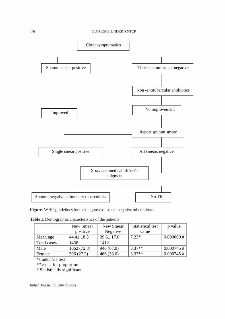
Indian Journal of Tuberculosis
Three sputum smear negative Sputum smear positive
Non -antitubercular antibiotics
No improvement Improved
Repeat sputum smear
Single smear positive All smears negative
X ray and medical officer’s judgment
No TB Sputum negative pulmonary tuberculosis
Chest symptomatics
Figure: WHO guidelines for the diagnosis of smear negative tuberculosis.
Table 1. Demographic characteristics of the patients
New Smear positive
New Smear Negative
Statistical test value
p value
Mean age 44.4± 18.5 39.6± 17.0 7.23* 0.000000 # Total cases 1458 1412 Male 1062 (72.8) 946 (67.0) 3.37** 0.000745 # Female 396 (27.2) 466 (33.0) 3.37** 0.000745 # *student’s t test ** z test for proportion # Statistically significant
OUTCOME UNDER RNTCP146
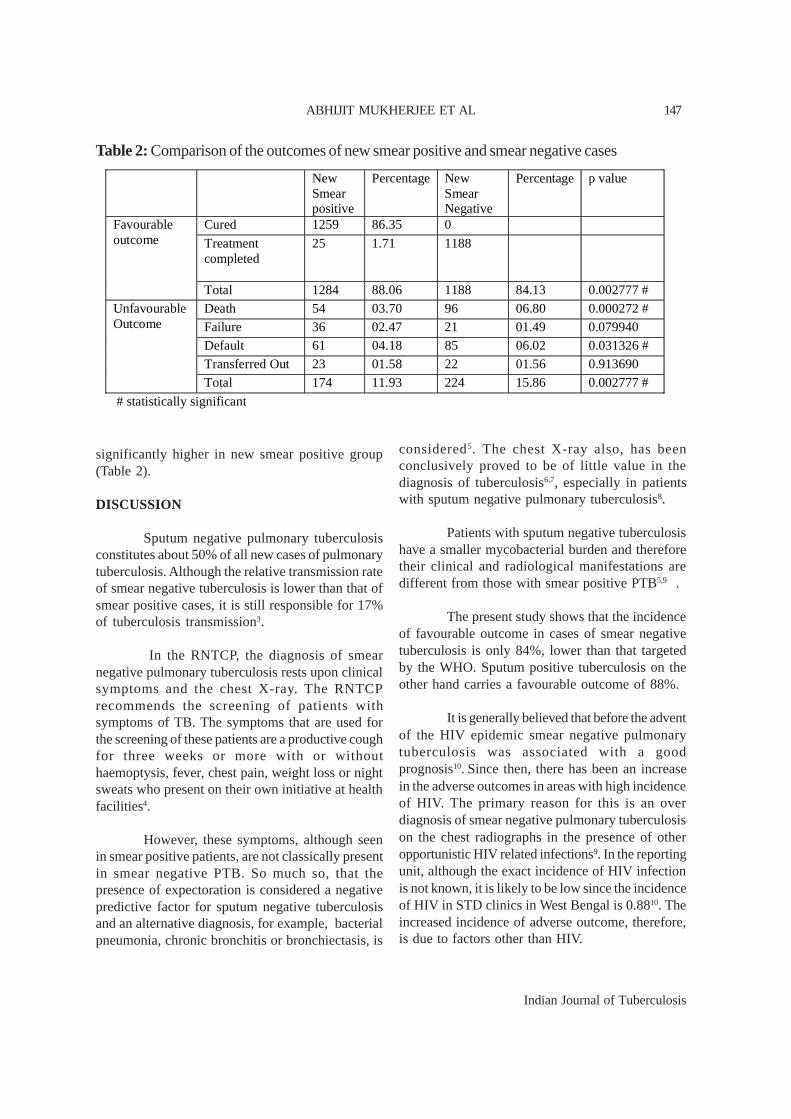
Indian Journal of Tuberculosis
significantly higher in new smear positive group(Table 2).
DISCUSSION
Sputum negative pulmonary tuberculosisconstitutes about 50% of all new cases of pulmonarytuberculosis. Although the relative transmission rateof smear negative tuberculosis is lower than that ofsmear positive cases, it is still responsible for 17%of tuberculosis transmission3.
In the RNTCP, the diagnosis of smearnegative pulmonary tuberculosis rests upon clinicalsymptoms and the chest X-ray. The RNTCPrecommends the screening of patients withsymptoms of TB. The symptoms that are used forthe screening of these patients are a productive coughfor three weeks or more with or withouthaemoptysis, fever, chest pain, weight loss or nightsweats who present on their own initiative at healthfacilities4.
However, these symptoms, although seenin smear positive patients, are not classically presentin smear negative PTB. So much so, that thepresence of expectoration is considered a negativepredictive factor for sputum negative tuberculosisand an alternative diagnosis, for example, bacterialpneumonia, chronic bronchitis or bronchiectasis, is
considered5. The chest X-ray also, has beenconclusively proved to be of little value in thediagnosis of tuberculosis6,7, especially in patientswith sputum negative pulmonary tuberculosis8.
Patients with sputum negative tuberculosishave a smaller mycobacterial burden and thereforetheir clinical and radiological manifestations aredifferent from those with smear positive PTB5,9 .
The present study shows that the incidenceof favourable outcome in cases of smear negativetuberculosis is only 84%, lower than that targetedby the WHO. Sputum positive tuberculosis on theother hand carries a favourable outcome of 88%.
It is generally believed that before the adventof the HIV epidemic smear negative pulmonarytuberculosis was associated with a goodprognosis10. Since then, there has been an increasein the adverse outcomes in areas with high incidenceof HIV. The primary reason for this is an overdiagnosis of smear negative pulmonary tuberculosison the chest radiographs in the presence of otheropportunistic HIV related infections9. In the reportingunit, although the exact incidence of HIV infectionis not known, it is likely to be low since the incidenceof HIV in STD clinics in West Bengal is 0.8810. Theincreased incidence of adverse outcome, therefore,is due to factors other than HIV.
Table 2: Comparison of the outcomes of new smear positive and smear negative cases New
Smear positive
Percentage New Smear Negative
Percentage p value
Cured 1259 86.35 0
Treatment completed
25 1.71 1188
Favourable outcome
Total 1284 88.06 1188 84.13 0.002777 #
Death 54 03.70 96 06.80 0.000272 #
Failure 36 02.47 21 01.49 0.079940
Default 61 04.18 85 06.02 0.031326 #
Transferred Out 23 01.58 22 01.56 0.913690
Unfavourable Outcome
Total 174 11.93 224 15.86 0.002777 #
# statistically significant
ABHIJIT MUKHERJEE ET AL 147
Indian Journal of Tuberculosis
The decreased incidence of favourableoutcome in smear negative cases was seen to bedue to the differences in the incidence of defaultsand deaths among these groups.
In their study in Pakistan, Kamran Siddiquiet al11 found that 6.1% patients declared AFB smearnegative from a tuberculosis programme laboratorywere AFB smear positive when re-examined in areference laboratory. Increased incidence of failureamong smear negative pulmonary tuberculosis couldbe the result of improper categorization resulting inthe prescription of inappropriate drugs. However,further studies to identify causes of failure must beundertaken.
Default is significantly higher in patientswith sputum negative PTB. Several socio-economic,demographic, drug and occupational factors, thathave been found to be associated with an increasedincidence of default from anti-tubercular therapy12,are likely to be present equally in both the groupssince they belong to the same population. Duringthe initial years of the implementation of the RNTCP,more stress was given to the identification and cureof sputum positive patients. This lack of attentionof the tuberculosis programme workers towards thesputum negative group resulted in more defaults13.This study includes patients since 1999, when theDOTS programme was initially started in the area.
The present study observed that smearnegative patients had significantly higher deaths.Patients with sputum negative PTB, seen in thisstudy, have a significantly lower mean age than theother group and hence a decreased physiologicalchance of death. The other important reasons forthe increased incidence of death in smear negativePTB can be i) prevalence of HIV, ii) delay in thediagnosis of patients in this group and iii) inaccuratediagnosis under operational conditions.
In areas with low incidence of HIV, delayin diagnosis is an important cause of increasedincidence of death in patients with smear negativepulmonary tuberculosis. This delay from theappearance of symptoms to the time of diagnosisof the disease can be divided into two intervals.
Patient delay defined as the time interval from theappearance of the major pulmonary symptoms ofthe disease until the first visit to the medical facilityand health service delay defined as the time intervalfrom the first consultation until the date ofdiagnosis14. Studies have shown that the mean timeinterval in the delay in seeking medical attention thatis the patient delay is higher in patients with smearnegative pulmonary tuberculosis15. This suggests thatthese patients seem to stay at home until they noticean alarming symptom or are grossly incapacitatedby the disease.
Radiological facilities under programmeconditions are available at the higher centres oftreatment, which in most cases are located far fromthe patients’ residence. Distance from the nearbyhealth care facility has been shown to be linked tothe increase in the delay in seeking medical treatment.In the study population access to radiologicalfacilities and often economic constraints ofradiological investigations result in the delay indiagnosis of smear negative tuberculosis.
This delay in the diagnosis causes patientsto be more seriously ill at the time of presentation.In the current study approximately 25% of patientswere seriously ill at the time of diagnosis. Analysisof all deaths in the smear negative group shows thatabout 35% of patients who died during therapy wereseriously ill at the time of presentation.
Diagnosis of smear negative tuberculosisshould take at least 15 days under programmeconditions, which should be the optimum healthservice delay. However, in their study in Ethiopia,Demissie M. et al15 found that 85% of patients werediagnosed in less than 15 days which indicates thatthe correct diagnostic procedure for the diagnosisof these cases is not being followed. This will resultin several false positive cases and an over-diagnosisof smear negative pulmonary tuberculosis.
Bacterial pneumonias are the commonestcause of misdiagnosis on the radiographs and mayoccur in 14.0 to 41.2 % of cases15-19. Other lungdiseases mimicking tuberculosis are interstitialpneumonitis, carcinoma, lymphoma, chronic
OUTCOME UNDER RNTCP148

Indian Journal of Tuberculosis
obstructive pulmonary diseases, Interstitial lungdiseases, occupational lung diseases like silicosis,etc.
The use of clear cut radiological criteria canhelp in better detection of pulmonary tuberculosis.In their study in Kenya, van Cleeff MR et al8 hasshown that the introduction of a four point scoringsystem, improved the diagnosis of smear negativecases by reducing over-diagnosis up to 67%, whileonly 8% fewer culture positive cases would startimmediate treatment. In the absence of clear cutradiological criteria under the RNTCP, difficultiesin the interpretation of chest radiographs arecommon.
The training given to the medical studentsin the undergraduate programmes or the medicalofficers under the RNTCP is not adequate in theinterpretation of radiological reports, especially inthe detection of smear negative tuberculosis.Moreover, although the interpretation of the chestradiograph is more difficult than a sputum smearresult, quality control is hardly practised.
Follow-up of sputum smear negativepatients, at the end of the intensive phase, under theRNTCP is by sputum microscopy. Radiologicalfollow-up at the end of intensive phase will give anopportunity for the re-assessment of the diagnosis,and plan the use of ancillary investigations likesputum culture in patients without significantradiological improvement at the end of the intensivephase.
The limitations of our paper are that we havenot done HIV, X-ray, diabetes status, and durationof symptoms analysis as it is not available underprogramme conditions. A more detailed studyincorporating all these would be helpful.
To conclude, sputum negativetuberculosis constitutes almost half the casesof pulmonary tuberculosis. We believe thatdelayed presentation, delay in the diagnosis andimproper diagnosis under field conditions insmear negative cases are responsible for thedecreased incidence of favourable outcome in
low HIV areas. We recommend a more intensetraining of medical students during theirundergraduate courses and health servicedoctors involved in the RNTCP, formulation ofclear cut radiological guidelines for the detectionof pulmonary tuberculosis, and theestablishment of quality control in theinterpretation of chest X rays in the diagnosisof smear negative tuberculosis. We alsorecommend the routine use of chest radiographsalong with sputum smear examination at theend of the intensive phase for follow up of smearnegative cases.
REFERENCES
1. Revised National Tuberculosis Control Programme.Technical Guidelines For Tuberculosis Control. CentralTB Division. Directorate General of Health Services.Nirman Bhavan, New Delhi, India. May 2000, pp4-23.
2. Treatment of tuberculosis: Guidelines for nationalprogrammes. Third Edition.WHO, Geneva. WHO/CDS/TB/2003.313.
3. Behr MA, Warren SA, Salamon H, Hopewell PC, Poncede Leon A, Daley CL, Small PM. Transmission ofMycobacterium tuberculosis from patients smear negativefor acid-fast bacilli. Lancet 1999; 353: 444-9.
4. Revised National Tuberculosis Control Programme.Technical Guidelines For Tuberculosis Control.
5. Identifying Pulmonary Tuberculosis in Patients withNegative Sputum Smear Results. Alka M. Kanaya,MD,David V. Glidden, PhD and Henry F. Chambers, MD.Chest. 2001; 120: 349-355.
6. Garland LH. Studies on the accuracy of diagnosticprocedures. American Journal of Roentgenology andRadium Therapeutic Nuclear Medicine, 1959; 82: 25-38.
7. Springett VH. Results of the study on x-ray classification.Conclusions. Bulletin of the International Union againstTuberculosis. 1968; 41: 125-129.
8. van Cleeff MR, Kivihya-Ndugga LE, Meme H, OdhiamboJA, Klatser PR. The role and performance of chest X-rayfor the diagnosis of tuberculosis: a cost-effectiveness analysisin Nairobi, Kenya. BMC Infect Dis. 2005 Dec 12; 5:111.
9. Hargreaves N, Phiri S, Kadzakumanja CO, Salaniponi FM,Harries AD, Squire SB. Int Conf AIDS. 2000 Jul 9-14; 13:abstract no. WePeC4439.
10. National Aids Control Organisation, Ministry of Health& Family Welfare, Government of India, Facts and Figures.Observed HIV Prevalence levels State wise: 1998 – 2004.
11. Siddiqi K. Clinical and X-ray diagnosis of smear-negativepulmonary tuberculosis in low-income countries: Thecurrent evidence Nuffield Centre for International Healthand Development, University of Leeds. http://www.kaisernetwork.org/health_cast.
12. Chandrasekaran V, Gopi PG,Subramani R,ThomasA,Jaggarajamma A, Narayanan PR. Default During the
ABHIJIT MUKHERJEE ET AL 149
Indian Journal of Tuberculosis
Intensive Phase of Treatment under DOTS Programme.Indian J Tuberc 2005; 52: 197-202.
13. Harries AD, Nyirendra TE, Banerjee A, Boeree MJ,Salaniponi FM. Treatment outcomes of patients with smearnegative and sputum positive pulmonary tuberculosis inthe National Tuberculosis Control Programme, Malawi.Trans R Soc Trop Med Hyg. 1999; 93(4): 443-6.
14. Demissie M, Lindtjorn B, Berhane Y. Patient and healthservice delay in the diagnosis of pulmonary tuberculosisin Ethiopia. BMC Public Health 2002, 2: 1471-2458-2-23.
15. Daley C L, Mugusi F, Chen L L, et al. Pulmonarycomplications of HIV infection in Dar es Salaam, Tanzania:role of bronchoscopy and bronchoalveolar lavage. Am JRespir Crit Care Med 1996; 154: 105–110.
16. Batungwanayo J, Taelman H, Lucas S, et al. Pulmonarydisease associated with the human immunodeficiency virusin Kigali, Rwanda: a fiberoptic bronchoscopic study of111 cases of undetermined etiology. Am J Respir CritCare Med 1994; 149: 1591–1596.
17. Abouya Y L, Beaumel A, Lucas S. et al. Pneunocytiscariniipneumonia: an uncommon cause of death in Africanpatients with acquired immunodeficiency syndrome. AmRev Respir Dis 1992; 145: 617-620.
18. Kamanfu G. Mlika-Cabanne N, Giraad P.M. et al.Pulmonary complications of human immuno deficiencyvirus infection in Bujumbura, Burundi. Am Rev Respir Dis1993; 147: 658-663.
19. Lucas S.B., Honnou A, Peacock C, et al. The morality andpathollgy of HIV infection in a West African city. AIDS1993; 7: 1569-1579.
OUTCOME UNDER RNTCP150
Indian Journal of Tuberculosis
151
STATUS REPORT ON RNTCP*
* Dr. L. S Chauhan, DDG (TB), Directorate General of Health Services, Ministry of Health and Family Welfare, Government of India, New Delhi
The RNTCP has continued to achieve thetwin objectives of NSP case detection and treatmentsuccess rate at the national level during the firstquarter, 2009.
RNTCP performance during first quarter 2009
During the quarter, over 1.79 millionsuspects were examined, 228,754 sputum positivecases were diagnosed, and a total of 372,619 TBcases (which include sputum smear negative andextra-pulmonary TB patients) were registered fortreatment. The treatment success rate amongstthe new smear positive PTB cases registered inthe first quarter 2008 is 87% and the sputumconversion rate of patients registered during fourthquarter 2008 is 90%.
Major Activities during the quarter
TB-HIV collaborative activities
All the nine states implementingIntensified TB/HIV Package services have startedreporting on the status of routine referral of allTB patients for HIV counselling and testing. Withthe expansion of Intensified TB/HIV package ofservices to Delhi and Gujarat during 2009, thenumber of states implementing this package hasincreased to 11 states. It was decided to expandthe package in a phased manner, to cover the entirenation by 2012.
The National Laboratory Committee meeting
The 16th meeting of National LabCommittee was held in March, 2009. The laboratoryscale-up plan for the diagnosis and management ofMDR-TB which includes establishment of 43 solidculture and LPA units and 33 liquid culture unitswas endorsed by the committee. The laboratoryperformance indicator and generic standardoperating procedures for C&DST labs were also
approved by the committee. Six Institutions underthe ICMR, Government of India were identified forparticipating in the DOTS-plus diagnosis services,after undergoing accreditation from the TuberculosisResearch Centre, Chennai.
Progress in accreditation of Intermediate ReferenceLaboratories (IRL)
The IRLs of Tamil Nadu and Rajasthan wereaccredited under RNTCP during the quarter and itis currently undertaking culture and DST servicesfor the MDR TB suspects from the state. Anothersix IRLs (Haryana, West Bengal, Uttarakhand,Chattisgarh, Jharkhand and Orissa), three medicalcollege labs and three private labs are under theaccreditation process and are expected to beaccredited in 2009.
Progress in DOTS-Plus services for MDR-TB cases
During this quarter, DOTS Plus treatmentservices have been initiated in Tamil Nadu, thusbringing the total number of states implementingDOTS Plus to eight. By the end of this quarter, over400 MDR-TB cases were on RNTCP Category IVtreatment in the country. The state of Rajasthan hasstarted the identification of MDR suspects and willbegin the treatment services shortly. The states ofOrissa and Uttar Pradesh are in advance stage ofpreparation and are expected to roll out the servicesshortly.
Progress in the involvement of NGOs and PPs
The involvement of health institutionsunder the Catholic Bishop’s Conference of India(CBCI) began during the third quarter 2008. Oneremaining state level workshop was conducted,thus completing this activity in all 11 identifiedstates. In addition, 59 new NGO-PP Memorandaof Understanding were signed between the state/districts and the Catholic Health Facilities.
Indian J Tuberc 2009; 56:151-153
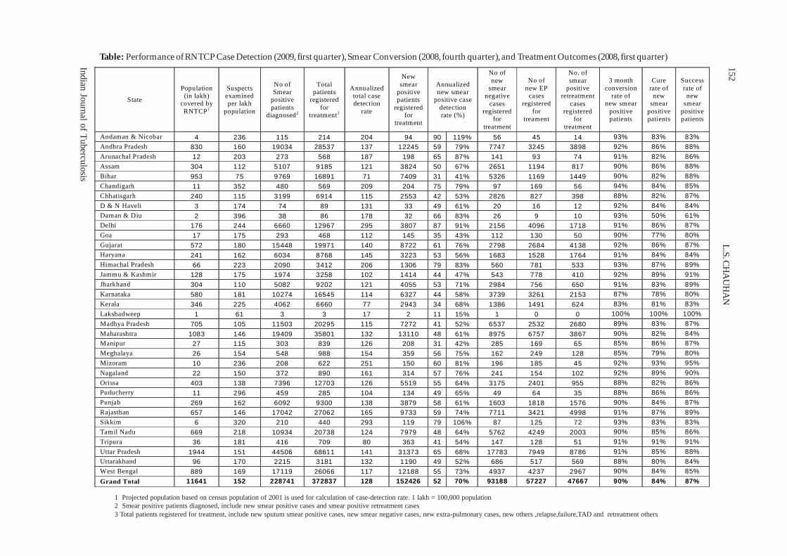
Indian Journal of Tuberculosis
152L.S
. CH
AU
HA
NTable: Performance of RNTCP Case Detection (2009, first quarter), Smear Conversion (2008, fourth quarter), and Treatment Outcomes (2008, first quarter)
1 Projected population based on census population of 2001 is used for calculation of case-detection rate. 1 lakh = 100,000 population2 Smear positive patients diagnosed, include new smear positive cases and smear positive retreatment cases3 Total patients registered for treatment, include new sputum smear positive cases, new smear negative cases, new extra-pulmonary cases, new others ,relapse,failure,TAD and retreatment others
State
Population (in lakh)
covered by RNTCP1
Suspects examined per lakh
population
No of Smear
positive patients
diagnosed2
Total patients
registered for
treatment3
Annualized total case detection
rate
New smear
positive patients
registered for
treatment
Annualized new smear
positive case detection rate (%)
No of new
smear negative
cases registered
for treatment
No of new EP cases
registered for
treament
No. of smear
positive retreatment
cases registered
for treatment
3 month conversion
rate of new smear
positive patients
Cure rate of new
smear positive patients
Success rate of new
smear positive patients
Andaman & Nicobar 4 236 115 214 204 94 90 119% 56 45 14 93% 83% 83% Andhra Pradesh 830 160 19034 28537 137 12245 59 79% 7747 3245 3898 92% 86% 88% Arunachal Pradesh 12 203 273 568 187 198 65 87% 141 93 74 91% 82% 86% Assam 304 112 5107 9185 121 3824 50 67% 2651 1194 817 90% 86% 88%
Bihar 953 75 9769 16891 71 7409 31 41% 5326 1169 1449 90% 82% 88%
Chandigarh 11 352 480 569 209 204 75 79% 97 169 56 94% 84% 85%
Chhatisgarh 240 115 3199 6914 115 2553 42 53% 2826 827 398 88% 82% 87%
D & N Haveli 3 174 74 89 131 33 49 61% 20 16 12 92% 84% 84% Daman & Diu 2 396 38 86 178 32 66 83% 26 9 10 93% 50% 61% Delhi 176 244 6660 12967 295 3807 87 91% 2156 4096 1718 91% 86% 87%
Goa 17 175 293 468 112 145 35 43% 112 130 50 90% 77% 80%
Gujarat 572 180 15448 19971 140 8722 61 76% 2798 2684 4138 92% 86% 87% Haryana 241 162 6034 8768 145 3223 53 56% 1683 1528 1764 91% 84% 84% Himachal Pradesh 66 223 2090 3412 206 1306 79 83% 560 781 533 93% 87% 89% Jammu & Kashmir 128 175 1974 3258 102 1414 44 47% 543 778 410 92% 89% 91% Jharkhand 304 110 5082 9202 121 4055 53 71% 2984 756 650 91% 83% 89% Karnataka 580 181 10274 16545 114 6327 44 58% 3739 3261 2153 87% 78% 80%
Kerala 346 225 4062 6660 77 2943 34 68% 1386 1491 624 83% 81% 83%
Lakshadweep 1 61 3 3 17 2 11 15% 1 0 0 100% 100% 100%
Madhya Pradesh 705 105 11503 20295 115 7272 41 52% 6537 2532 2680 89% 83% 87%
Maharashtra 1083 146 19409 35801 132 13110 48 61% 8975 6757 3867 90% 82% 84% Manipur 27 115 303 839 126 208 31 42% 285 169 65 85% 86% 87%
Meghalaya 26 154 548 988 154 359 56 75% 162 249 128 85% 79% 80%
Mizoram 10 236 208 622 251 150 60 81% 196 185 45 92% 93% 95%
Nagaland 22 150 372 890 161 314 57 76% 241 154 102 92% 89% 90%
Orissa 403 138 7396 12703 126 5519 55 64% 3175 2401 955 88% 82% 86% Puducherry 11 296 459 285 104 134 49 65% 49 64 35 88% 86% 86% Punjab 269 162 6092 9300 138 3879 58 61% 1603 1818 1576 90% 84% 87% Rajasthan 657 146 17042 27062 165 9733 59 74% 7711 3421 4998 91% 87% 89% Sikkim 6 320 210 440 293 119 79 106% 87 125 72 93% 83% 83% Tamil Nadu 669 218 10934 20738 124 7979 48 64% 5762 4249 2003 90% 85% 86%
Tripura 36 181 416 709 80 363 41 54% 147 128 51 91% 91% 91%
Uttar Pradesh 1944 151 44506 68611 141 31373 65 68% 17783 7949 8786 91% 85% 88% Uttarakhand 96 170 2215 3181 132 1190 49 52% 686 517 569 88% 80% 84% West Bengal 889 169 17119 26066 117 12188 55 73% 4937 4237 2967 90% 84% 85%
Grand Total 11641 152 228741 372837 128 152426 52 70% 93188 57227 47667 90% 84% 87%
Indian Journal of Tuberculosis
153STATUS REPORT ON RNTCP
Under the Global Fund round, six RNTCPIMA PPM projects and 118 CMEs were held and4888 PPs were sensitized during this quarter. 47DTPs were held and 1005 PPs were trained.
Progress under NRHM and GFATM
The appraisal of NRHM PIPs for all statesand UTs was done during February-March’09 bythe NRHM Division and Programme Officers.
Under GFATM, the first Rolling ContinuationChannel (RCC) TB Grant has been approved by theGF Board. The Project Implementation Plan (PIP) for
six years has been prepared and submitted to theExpenditure Finance Committee (EFC) for approval.
Progress in Advocacy, Communication and SocialMobilization
World TB Day was observed on 24th March,2009 across the country in all the states and districts.On this day, intensive efforts were made with allthe stake holders to highlight the burden of TB andthe progress made by RNTCP for reducing thisburden. Central TB Division released TB-India,2009, the annual report on the progress made underRNTCP.
Indian Journal of Tuberculosis
Summary: Primary sternal tuberculous osteomyelitis is a rare form of tuberculous osteomyelitis. We report a case of ayoung adult with primary tuberculous osteomyelitis of the sternum who presented with a pulsatile anterior chest wallswelling. Computed Tomography of the thorax revealed a hypodense lytic lesion in the body of the sternum that had erodedinto the anterior mediastinum where it lay in close contact with the right ventricle, resulting in the clinically evidenttransmitted pulsations. Among the protean manifestations of tuberculosis this case illustrates a unique presentation as apulsatile chest wall mass. [Indian J Tuberc 2009; 56:154-156 ]
Key Words: Sternal osteomyelitis, Tuberculosis, Pulsatile sternal swelling
STERNAL TUBERCULOUS OSTEOMYELITIS PRESENTING AS A PULSATILESWELLING
Hari Kishan Boorugu*, Anugrah Chrispal*and Elsa Mary Thomas*
(Received on 18.11.2008; Accepted after revision on 23.4.2009)
INTRODUCTION
Sternal osteomyelitis may be of primary andsecondary forms. The more commonly seensecondary sternal osteomyelitis is usually acomplication of sternotomy following proceduresas coronary artery bypass grafts, and may be causedby atypical mycobacteria and other pyogenicbacteria1. Sternal osteomyelitis per se is a rare diseaseaccounting for 0.3 to 1.8% of all cases ofosteomyelitis2. Isolated tuberculous sternalosteomyelitis comprises less than 1% ofmusculoskeletal tuberculosis3. Two studies fromIndia have reported 14 and 19 cases of sternaltuberculosis between 2000 and 2006 highlightingthe relative rarity of this condition4,5. Sternaltuberculosis may mimic a variety of conditions ofthe sternum including pyogenic osteomyelitis,actinomycosis, fungal infections, sarcoidosis andmalignancies6. Tuberculous sternal osteomyelitisusually occurs in young adults who present withinsidious onset pain, discomfort and swelling in thesternal region4. We present a young adult with apainless pulsatile sternal swelling which was foundto be of tuberculous origin.
CASE HISTORY
A 28-year old man from Tamil Nadu,presented with a gradually increasing, pulsatile,painless swelling over the mid sternal region for threemonths. During this period, he had low grade,intermittent fever, anorexia and significant weightloss. He denied a history of high risk behaviour orblood transfusions in the past. On examination hewas emaciated and pale. He had no peripherallymphadenopathy. He had a 3X3 centimetre, welldefined, non-tender, pulsatile swelling in the midsternal region which was smooth and fluctuant withno evidence of inflammation. These pulsationsappeared to be ‘transmitted’ and not ‘expansile’ onclinical examination. Per abdominal examinationrevealed an ill-defined mass in the left hypochondriumwhich moved well with respiration, whichsubsequently was found to be a lymph node masson abdominal ultrasonography.
Investigations revealed a dimorphicanaemia ( Haemoglobin 8.4 gram %) and erythrocytesedimentation rate (ESR) of 140 millimetres at onehour. Liver function tests revealed hypoalbuminemia
* Assistant Professor, Departments of Medicine 2 and Radiology, Christian Medical College and Hospital, Vellore.Correspondence: Dr. Hari Kishan Boorugu, Assistant Professor, Department of Mexicine 2, Christian Medical College and Hospital, Vellore-632 004, (Tamil Nadu). Fax number: 0416 2232035; Phone numbers: Landline -04162282031; Mobile: 09994068911; E-mail: [email protected]
Case Report

Indian Journal of Tuberculosis
(Serum Albumin – 2.6 gram %). HIV ELISA wasnegative. Chest radiography showed significantmediastinal widening and there were no infiltratesin the lung fields to suggest parenchymalinvolvement. Abdominal Ultrasonography revealedmultiple enlarged mesenteric and omental lymphnodes.
A Computerized Tomogram (CT) of thechest revealed a hypodense lytic lesion eroding thebody of the sternum, with chest wall and intra-thoracic extension, abutting the right ventricularoutflow tract (Figure), suggestive of osteomyelitisof the sternum with cold abscess formation. Therewere multiple enlarged, necrotic, matted mediastinaland hilar lymph nodes. Subcarinal nodes had erodedinto the oesophagus forming a fistulous tract. Therewas no parenchymal involvement evident on CT.White, cheesy material was aspirated from the chestwall swelling, a smear of which revealed numerousacid fast bacilli on Ziehl-Neelsen stain. Aspiratesfrom intra abdominal lymph nodes and the chestwall swelling subsequently grew Mycobacteriumtuberculosis on culture. The patient was thus
diagnosed to have disseminated tuberculosis withsternal tuberculous osteomyelitis and cold abscess.He was initiated on anti-tuberculous therapy withRifampicin (450 mg once daily), Isoniazid (300 mgonce daily with Pyridoxine), Ethambutol (800 mgonce daily) and Pyrazinamide (1250 mg daily). Hewas advised to take all four drugs for the initial twomonths and then switch to continuation phase withRifampicin and Isoniazid for the next 10 months.He was advised surgical repair of the sternal defectat a subsequent date. He, however, was lost to followup.
DISCUSSION
This patient had a sternal tuberculousosteomyelitis with a cold abscess that had erodedinto the thorax and was pulsatile by virtue of itsproximity to the anteriorly placed right ventricle andits outflow tract. Tuberculous sternal osteomyelitisis a rare presentation of tuberculosis. This case,however, was unique due to its presentation as apulsatile sternal swelling which, to the best of ourknowledge, has not been described previously.
Usually, tuberculous sternal osteomyelitis iscaused by reactivation of latent foci of primarytuberculosis due to lymphatic or haematogenousdissemination. Occasionally, it may be due to directextension from contiguous mediastinal lymphnodes3. Tuberculous sternal osteomyelitis tends toinvolve the body of the sternum more often thanthe manubrium. The indolent nature of the swellinghelps distinguish tuberculous sternal osteomyelitisfrom other pyogenic osteomyelitis caused byStaphylococcus aureus and Gram Negative Bacilli.On occasion sternal tuberculous osteomyelitis haspresented with sternal fracture and draining sinuses7.Complications of sternal osteomyelitis includesecondary infection, fracture, fistula formation,compression of trachea, compression and erosionof underlying blood vessels, extension of thetubercular abscess into surrounding tissues includingthe mediastinum, pleural cavity and subcutaneousplanes3.
Diagnosis rests on histopathological andmicrobiological confirmation from the sternal tissue
Figure: Axial contrast-enhanced CT scan of thethorax showing a hypodense lytic lesionin the body of the sternum, with corticalbreach, extending anteriorly into the chestwall and posteriorly into the anteriormediastinum, abutting the right ventricularoutflow tract
HARI KISHAN BOORUGU ET AL 155
Indian Journal of Tuberculosis
or pus from the cold abscess6. Radiographicabnormalities tend to lag behind the clinicalmanifestations, and may mimic other benign andmalignant conditions8. Computed Tomography of thechest is excellent for delineation of extent of sternaldestruction as well as soft tissue extension.Magnetic Resonance Imaging (MRI) is probablysuperior for detecting early marrow oedema and softtissue involvement3.
Treatment is based on anti-tuberculouschemotherapy in combination with drainage anddebridement of necrotic material, although a numberof cases have been treated with anti-tuberculousagents alone. There is no consensus on the role ofadjuvant surgical interventions in patients with sternaltuberculosis. Debridement is the mainstay of surgicaltreatment along with sternal defect closure by flaprepair (pectoralis major, latissimus dorsi, rectusabdominis or omental flap closure) with or withoutchest wall reconstruction4. Vacuum assisted closurehas been tried successfully7.
Presentation of a pulsatile chest wallmass should alert the clinician to possibledifferential diagnoses including vascularmalformations, aneurysms and vascular
metastatic lesions. Among the proteanmanifestations of tuberculosis this caseillustrates a unique presentation as a pulsatilechest wall mass.
REFERENCES
1. Gill EA, Steven DL. Primary Sternal Osteomyelitis. WestJ Med 1989 Aug;151(2):199-203.
2. Prakash A, Hira HS, Tuberculous Osteomyelitis of Sternumin a Diabetic. Indian J Tuberc 2001;48:35-36.
3. Jain V K, Singh Y, Shukla A, Mittal D. TuberculousOsteomyelitis of Sternum: A Case Report. Journal ofClinical and Diagnostic Research 2007 June;1:163-167.
4. Khan SA, Varshney MK, Hasan AS, Kumar A, TrikhaV.Ali WM, Beg MH, Tuberculosis of the sternum: a clinicalstudy. J Bone Joint Surg Br 2007 Jun;89(6):817-20.
5. Rashid M, Zafar U, Abbas SN. An experience with a rarediagnosis of isolated tuberculosis of sternum at JNMCHospital, Aligarh, India. Saudi Med J 2008 Apr;29(4):580-3.
6. Sharma S, Juneja M, Garg A. Primary tubercularosteomyelitis of the sternum. Indian J Pediatr 2005Aug;72(8):709-10.
7. Ford SJ, Rathinam S, King JE, Vaughan R. Tuberculousosteomyelitis of the sternum: successful management withdebridement and vacuum assisted closure. Eur JCardiothorac Surg 2005 Oct;28(4):645-7.
8. Gopal K, Raj A, Rajesh MR, Prabhu SK, Geothe J. Sternaltuberculosis after sternotomy for coronary artery bypasssurgery: a case report and review of the literature. J ThoracCardiovasc Surg 2007 May;133(5):1365-6.
STERNAL TUBERCULOSIS156
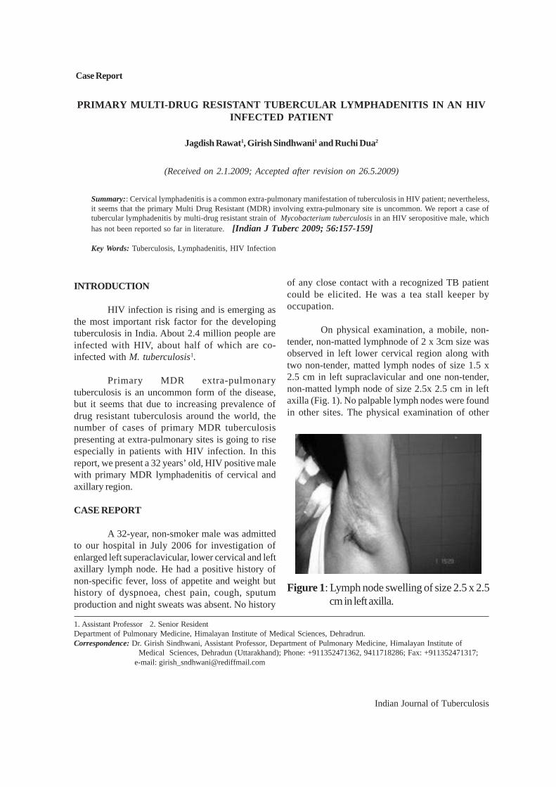
Indian Journal of Tuberculosis
Summary:: Cervical lymphadenitis is a common extra-pulmonary manifestation of tuberculosis in HIV patient; nevertheless,it seems that the primary Multi Drug Resistant (MDR) involving extra-pulmonary site is uncommon. We report a case oftubercular lymphadenitis by multi-drug resistant strain of Mycobacterium tuberculosis in an HIV seropositive male, whichhas not been reported so far in literature. [Indian J Tuberc 2009; 56:157-159]
Key Words: Tuberculosis, Lymphadenitis, HIV Infection
Case Report
PRIMARY MULTI-DRUG RESISTANT TUBERCULAR LYMPHADENITIS IN AN HIVINFECTED PATIENT
Jagdish Rawat1, Girish Sindhwani1 and Ruchi Dua2
(Received on 2.1.2009; Accepted after revision on 26.5.2009)
INTRODUCTION
HIV infection is rising and is emerging asthe most important risk factor for the developingtuberculosis in India. About 2.4 million people areinfected with HIV, about half of which are co-infected with M. tuberculosis1.
Primary MDR extra-pulmonarytuberculosis is an uncommon form of the disease,but it seems that due to increasing prevalence ofdrug resistant tuberculosis around the world, thenumber of cases of primary MDR tuberculosispresenting at extra-pulmonary sites is going to riseespecially in patients with HIV infection. In thisreport, we present a 32 years’ old, HIV positive malewith primary MDR lymphadenitis of cervical andaxillary region.
CASE REPORT
A 32-year, non-smoker male was admittedto our hospital in July 2006 for investigation ofenlarged left superaclavicular, lower cervical and leftaxillary lymph node. He had a positive history ofnon-specific fever, loss of appetite and weight buthistory of dyspnoea, chest pain, cough, sputumproduction and night sweats was absent. No history
of any close contact with a recognized TB patientcould be elicited. He was a tea stall keeper byoccupation.
On physical examination, a mobile, non-tender, non-matted lymphnode of 2 x 3cm size wasobserved in left lower cervical region along withtwo non-tender, matted lymph nodes of size 1.5 x2.5 cm in left supraclavicular and one non-tender,non-matted lymph node of size 2.5x 2.5 cm in leftaxilla (Fig. 1). No palpable lymph nodes were foundin other sites. The physical examination of other
1. Assistant Professor 2. Senior ResidentDepartment of Pulmonary Medicine, Himalayan Institute of Medical Sciences, Dehradrun.Correspondence: Dr. Girish Sindhwani, Assistant Professor, Department of Pulmonary Medicine, Himalayan Institute of Medical Sciences, Dehradun (Uttarakhand); Phone: +911352471362, 9411718286; Fax: +911352471317; e-mail: [email protected]
Figure 1: Lymph node swelling of size 2.5 x 2.5cm in left axilla.

Indian Journal of Tuberculosis
organ systems was normal. The haematological andbiochemical parameters were normal and atuberculosis skin test for M.tuberculosis showed7mm.induration. X-ray chest PA view wasunremarkable. Serum samples were positive forantibodies against HIV using HIV combimmunoassay (J. Mitra & Co. Ltd.), followed byHIV microlisa kit (J. Mitra & co. Ltd.). Patient gavehistory of blood transfusion in past, that might bethe possible source of infection. CD4+ cell countwas 139/MicroL. Ultrasound examination of wholeabdomen showed retroperitoneal lymphadenopathyand mild hepatomegaly. FNAC of left cervical andsupraclavicular lymph node showed chronicgranulomatous inflammation consistent withtuberculosis. The acid fast bacilli of the specimenwere negative. For confirmation of diagnosis, puswas also sent for culture and sensitivity fortuberculosis by Lowenstein – Jensen method at anintermediate reference laboratory (New Delhi TBCentre, NRL).
Patient was put on anti-tubercular treatment(ATT) under DOTS, CAT-I. Pus aspirated from thecold abscess in the neck grew M.tuberculosiscomplex, resistant to Streptomycin, Rifampicin andIsoniazid. . During this three month period till theavailability of culture and sensitivity report, therewas no significant improvement in lymph node size.
His ATT regimen was modified accordingto sensitivity report to Kanamycin, Ethionamide,Ethambutol, Pyrazinamide and Levofloxacin according
to body weight. He was also put on anti-retroviraltherapy (ART) (Lamivudine+stavudine+Efavirenz).Kanamycin was stopped after three months andremaining drugs continued (3 months of intensive phaseand 15 months of continuation phase). At the end ofeighteen months, complete resolution of lymph nodesoccurred with some residual scarring (Figs. 2 &3). ATT was stopped with continuation of ART. Atthe end of treatment, CD4+ cell count was 251/microL.
DISCUSSION
Drug resistant tuberculosis has become amajor public health problem since early 19902. Theprevalence of primary multi drug resistanttuberculosis in India was estimated around 2-3%;however, this prevalence is 15-50% amongpreviously treated cases.
In HIV seronegative and immuno-competent patients, pulmonary tuberculosis is thecommonest mode of presentation, while EPTBaccounts for only 20% of cases but in HIVpositive patients it accounts for nearly 50-55 %3.Among extra-pulmonary tuberculosis, cervicallymphadenitis is the commonest presentation;nevertheless, it seems that the primary MDR ofEPTB is an uncommon form of the disease inpatients of HIV infection. The reported patient isthe first HIV infected patient with primary MDRtubercular lymphadenopathy in India to the bestof our knowledge.
Figure 2: Axillary lymph node disappearedafter treatment.
Figure 3: Cervical lymph node healed byscarring.
JAGDISH RAWAT ET AL158
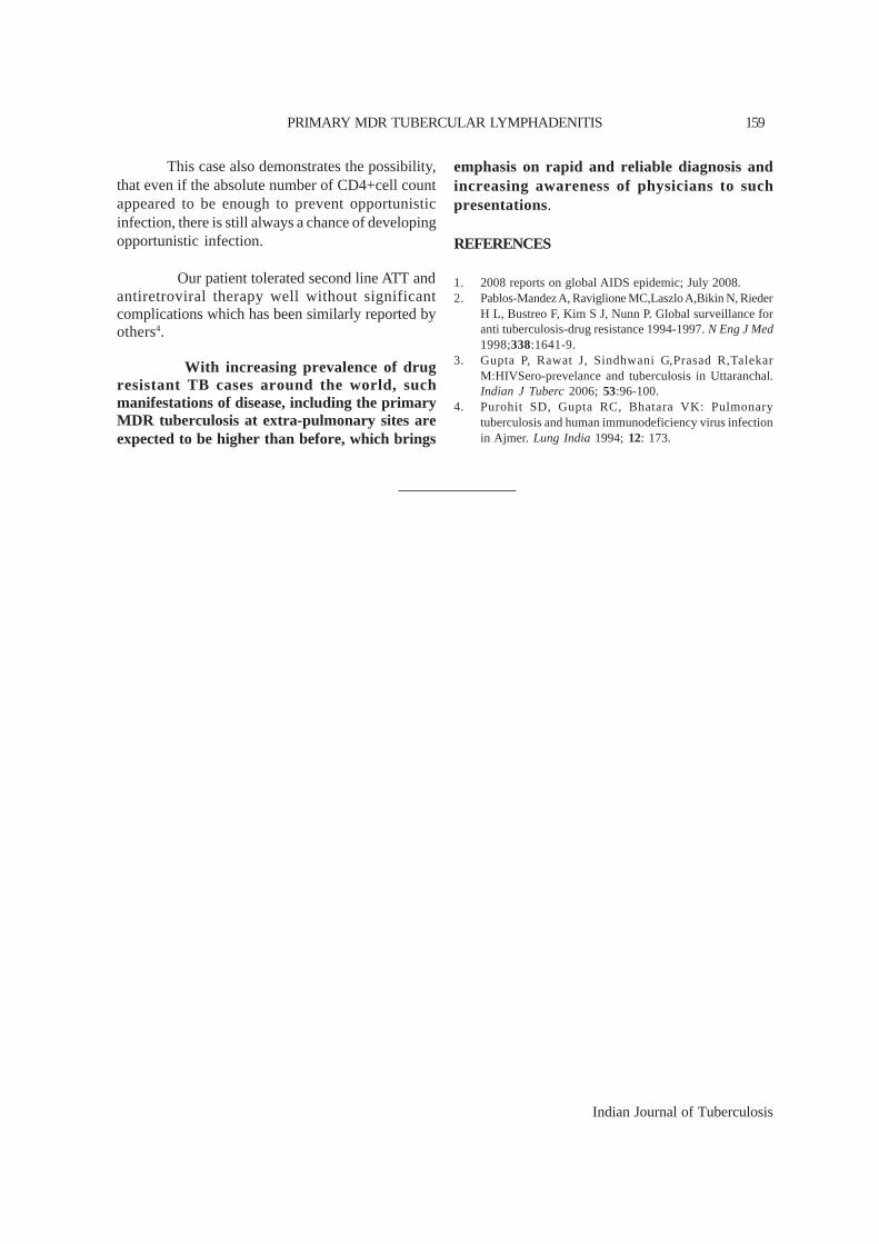
Indian Journal of Tuberculosis
This case also demonstrates the possibility,that even if the absolute number of CD4+cell countappeared to be enough to prevent opportunisticinfection, there is still always a chance of developingopportunistic infection.
Our patient tolerated second line ATT andantiretroviral therapy well without significantcomplications which has been similarly reported byothers4.
With increasing prevalence of drugresistant TB cases around the world, suchmanifestations of disease, including the primaryMDR tuberculosis at extra-pulmonary sites areexpected to be higher than before, which brings
emphasis on rapid and reliable diagnosis andincreasing awareness of physicians to suchpresentations.
REFERENCES
1. 2008 reports on global AIDS epidemic; July 2008.2. Pablos-Mandez A, Raviglione MC,Laszlo A,Bikin N, Rieder
H L, Bustreo F, Kim S J, Nunn P. Global surveillance foranti tuberculosis-drug resistance 1994-1997. N Eng J Med1998;338:1641-9.
3. Gupta P, Rawat J, Sindhwani G,Prasad R,TalekarM:HIVSero-prevelance and tuberculosis in Uttaranchal.Indian J Tuberc 2006; 53:96-100.
4. Purohit SD, Gupta RC, Bhatara VK: Pulmonarytuberculosis and human immunodeficiency virus infectionin Ajmer. Lung India 1994; 12: 173.
PRIMARY MDR TUBERCULAR LYMPHADENITIS 159
Indian Journal of Tuberculosis
Summary: A case of tuberculous otitis media with post auricular abscess is being described in a 14 year old female patientin view of its rare occurrence. The diagnosis was made on demonstration of acid fast bacilli (AFB) in the ear discharge andcharacteristic cytological features of post auricular abscess aspirate. [Indian J Tuberc 2009; 56:160-163]
Key words: Tuberculosis, Middle ear, Post auricular abscess
Case Report
TUBERCULOSIS OF THE MIDDLE EAR WITH POST AURICULAR ABSCESS
Manoj Arya, Ramakant Dixit, A.R. Paramez, Sidharth Sharma and Dilip Singh Rathore
(Received on 1.5.2009; Accepted after revision on 18.6.2009)
INTRODUCTION
Tuberculosis of the middle ear is a rareclinical entity that is usually seen in association withor secondary to pulmonary tuberculosis1. The trueincidence of this condition is difficult to assess,however in children, tuberculous otitis media (TOM)has been accounted for 0.04% of’ all cases ofchronic suppurative otitis media2. These patientsusually present with painless otorrhoea that fails torespond to the usual antimicrobial treatment inpatients with evidence of tuberculosis elsewhere inthe body3.
This report describes an apparently healthy14-year-old female child having painless eardischarge with hearing impairment and postauricular abscess on left side due to tuberculosis.
CASE REPORT
A 14-year-old female child presented withpainless discharge from left ear for four monthswith decreased hearing from the same and swellingbehind the affected ear for the last two months.She had no history of fever or any other complaint.There was no history of previous illness and nohistory of tuberculosis in the family. She was treatedby several courses of broad spectrum antibioticsboth systemic and local.
On physical examination, she was a healthyfemale child with body mass index 25. There wasno anaemia, lymphadenopathy or any otherabnormality on systemic examination. On otoscopy,there was thick pus seen in left external auditorycanal which after removal showed three perforationsin the tympanic membrane. There was a softfluctuant, slightly tender swelling at left post auriculararea measuring 4 x 5 cm with crusty scaling externalsurface (Figure 1). Local examination of right ear,nose, oral cavity and pharynx were normal.
Her laboratory investigations revealedhaemoglobin 13 gm%, total leucocyte count 7900
Department of Respiratory Medicine & Tuberculosis, JLN Medical College, Ajmer, (Rajasthan)Correspondence: Dr. Ramakant Dixit, 381/26, Ramganj, Ajmer-305 001, Rajasthan. Tel: 91-145-2691542. E-mail: [email protected]
Figure 1: Photograph of patient showing thickpurulent ear discharge with postauricular abscess.
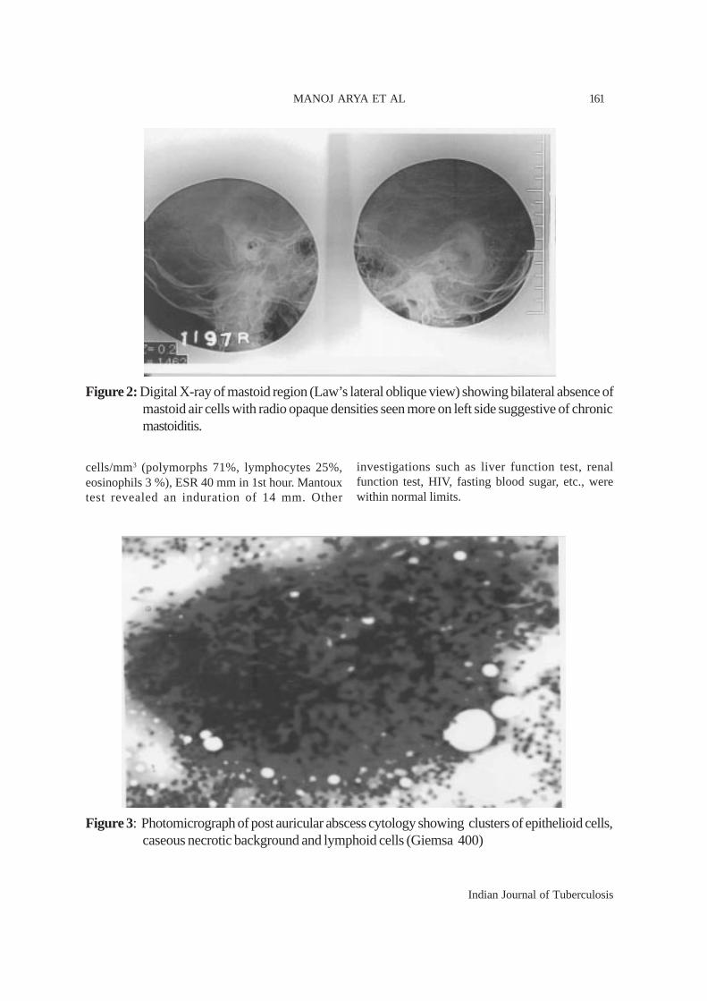
Indian Journal of Tuberculosis
cells/mm3 (polymorphs 71%, lymphocytes 25%,eosinophils 3 %), ESR 40 mm in 1st hour. Mantouxtest revealed an induration of 14 mm. Other
investigations such as liver function test, renalfunction test, HIV, fasting blood sugar, etc., werewithin normal limits.
Figure 2: Digital X-ray of mastoid region (Law’s lateral oblique view) showing bilateral absence ofmastoid air cells with radio opaque densities seen more on left side suggestive of chronicmastoiditis.
Figure 3: Photomicrograph of post auricular abscess cytology showing clusters of epithelioid cells,caseous necrotic background and lymphoid cells (Giemsa 400)
MANOJ ARYA ET AL 161
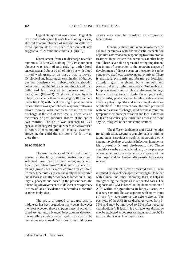
Indian Journal of Tuberculosis
Digital X-ray chest was normal. Digital X-ray of mastoids region (Law’s lateral oblique view)showed bilateral absence of mastoid air cells withradio opaque densities seen more on left sidesuggestive of chronic mastoiditis (Figure 2).
Direct smear from ear discharge revealednumerous AFB on ZN staining (3+). Post auricularabscess was drained anti-gravity, under localanaesthesia and about 10 ml of thick pus with bloodmixed with granulation tissue was removed.Cytological and histological examination of drainedpus was consistent with tuberculosis i.e. showingcollection of epithelioid cells, multinucleated giantcells and lymphocytes in caseous necroticbackground (Figure 3). Child was managed by anti-tuberculosis chemotherapy as category III treatmentunder RNTCP, with local dressing of post auricularlesion. There was good clinical response followingabove therapy with complete cessation of eardischarge at the end of two weeks. There was norecurrence of post auricular abscess at the end oftwo months. The child was referred to ENTspecialist for surgical opinion where she was advisedto report after completion of medical treatment.However, the child did not come for follow-upthereafter.
DISCUSSION
The true incidence of TOM is difficult toassess, as the large reported series have beenselected from hospitalized sub-groups withestablished tuberculosis4-5. It is known to occur inall age groups but is more common in children.Primary tuberculosis of ear has rarely been reportedand disease is usually secondary to infection in lung,larynx, pharynx and nose6. In the present case, thetuberculous involvement of middle ear seems primaryin view of lack of evidence of tuberculosis infectionat other body sites.
The route of spread of tuberculosis tomiddle ear has been argued for many years; howeverthe most accepted theory support entry of organismvia pharyngotympanic tube4. Infection can also reachthe middle ear via external auditory canal or byhematogenous spread. Very rarely the middle ear
cavity may also be involved in congenitaltuberculosis7.
Generally, there is unilateral involvement ofear in tuberculosis with characteristic presentationof painless otorrhoea not responding to antimicrobialtreatment in patients with tuberculosis at other bodysite. There is variable degree of hearing impairmentthat is out of proportion to the apparent degree ofdevelopment of disease seen on otoscopy. It can beconductive deafness, sensory neural or mixed. Thereis multiple tympanic membrane perforation,abundant granular tissue, bone necrosis andpreauricular lymphadenopathy. Periauricularlymphadenopathy and fistula are infrequent findings.Late complications include facial paralysis,labyrinthitis, post auricular fistulae, subperiostealabscess petrous apicitis and intra cranial extensionof infection8. In the present case, the child presentedwith painless ear discharge, mild deafness, multipletympanic membrane perforation and local extensionof lesion to cause post auricular abscess withoutany neurological or serious complications.
The differential diagnosis of TOM includesfungal infection, wegner’s granulomatosis, midlinegranulomas, sarcoidosis, syphilis, necrotizing otitisextema, atypical mycobacterial infection, lymphoma,histiocytosis- X and cholesteatoma9. Theseconditions can be excluded clinically by the presenceof ear ache, and the type and consistency of thedischarge and by further diagnostic laboratoryworkup.
The role of X-ray of mastoid and CT scanis limited in view of non-specific finding but togetherwith clinical and other laboratory tests, it helps instrengthening the diagnosis in suspected cases. Thediagnosis of TOM is based on the demonstration ofAFB within the granuloma in biopsy tissue, eardischarge or middle ear aspirate with or withoutculture for Mycobacterium tuberculosis. Thepositivity of the AFB in ear discharge varies from 5-35% and may be improved to 50% after repeatedexaminations10. If facility is available, ear dischargemay be subjected to polymerase chain reaction (PCR)test for Mycobacterium tuberculosis.
TUBERCULOSIS OF THE MIDDLE EAR162
Indian Journal of Tuberculosis
The treatment of choice for TOM is anti-tuberculosis chemotherapy using currently availablecombinations. Surgery may be required in somecases to remove sequestra and to improve drainage.In view of effective anti-tuberculosis drugs available,the role of surgery in today’s practice is limited andshould be reserved for decompression of facialnerve, for removal of necrotic material which mightprovide a nidus for the organism to remain out ofreach of anti-tuberculosis drugs. When surgery iscombined with adequate chemotherapy, there isbetter healing with good prognosis. When anti-tuberculosis therapy is started early, the mortality isnegligible11.
In conclusion, TOM is a rare conditionwhich if untreated can damage middle ear andsurrounding structures. It should always beconsidered in the differential diagnosis ofchronic ear discharge not responding to usualantimicrobial therapy. A high level of clinicalsuspicion is needed for early diagnosis and anti-tuberculosis therapy should be started as soonas possible to prevent possible complications.
REFERENCES
1. Mahajan M, Agrawal DS, Singh NP, Gadre DJ. Tuberculosisof middle ear- A case report. Indian J Tuberc 1995; 42:55.
2. Weiner GM, O Connell JE, Pahor AL. The role of surgeryin tuberculosis mastoiditis: Appropriate chemotherapy isnot always enough. J Laryngol Otol 1977; 111: 752-3.
3. Gupta KB, Tandon S, Mathur SK, Kalra R. Tuberculosis ofmiddle ear -a case report. Indian J Tuberc 2000; 47 : 45-46.
4. Adams JG. Tuberculosis otitis media: A complication ofthoracoplasty. Ann Otol 1942; 51: 209.
5. Windle Taylor, PC Bailey CM. Tuberculosis otitis media.Laryngoscope 1980; 90: 1039-44.
6. Sharan R, Issar DK. Primary tuberculosis of the middleear cleft. Practitioner 1979; 222: 93-95.
7. Procotr B, Windsat JR. Tuberculosis of the ear. ArchOtolaryngol 1942; 35: 221-49.
8. Yaniv E. Tuberculous otitis media: A clinical record.Laryngoscope 1987; 97: 1303-6.
9. M Cart HW. Tuberculosis disease of the middle ear. JLaryngol Otol 1925; 40: 456-66.
10. Chaturvedi VN, Chaturvedi P. Tuberculosis of the middleear. Indian Pediatr 1986; 23: 199-204.
11. Chyo YS, Lee HS, Kim SW, et al. Tuberculous otitis media:A clinical and radiological analysis of 52 patients.Laryngoscope 2006; 116: 921-27.
MANOJ ARYA ET AL 163
Indian Journal of Tuberculosis
Indian J Tuberc 2009; 56:164-166
ABSTRACTS
Predictive factors for Mortality among non-HIV-infected patients with Pulmonary Tuberculosisand Respiratory Failure
S.M. Lin, T.Y. Wang, W.T. Liu, C.C. Chand, H.C.Lin et al. Int J Tuberc Lung Dis 2009; 13(3): 335-349
The objective was to determine predictivefactors for mortality among pulmonarytuberculosis (PTB) patients without humanimmunodeficiency virus (HIV) infection and inneed of mechanical ventilation (TBMV). From July2004 to December 2005, 612 respiratory failurepatients requiring mechanical ventilation wereadmitted to the intensive care unit (ICU) of ChangGung Memorial Hospital, Taipei, Taiwan. Of these,59 non-HIV-infected patients had active PTB asthe primary cause. Mortality rates were measuredin TBMV patients and predictors wereinvestigated. Incidence of treatment delay fornosocomial pneumonia was compared betweensurvivors and fatalities. Of the 59 patients withTBMV, 40 (67.8%) died in the ICU. Multi-organfailure syndrome (OR 8.59, 95%CI 1.85-101.27)and nosocomial pneumonia (OR 5.77, 95%CI1.33-44.36) were independently associated within-hospital mortality. Treatment delay of morethan 24 hours for nosocomial pneumonia wassignificantly more frequent among fatalities thanamong survivors (19/26, 73.1% vs. 0/3, 0%; P =0.033). Nosocomial pneumonia in TB patients withrespiratory failure is associated with a poorprognosis; this appears to be further aggravatedby delays in appropriate treatment. Measures toprevent nosocomial pneumonia should be carefullyinstituted and treatment for nosocomial pneumoniashould be started promptly among such patients.
Radiographic manifestations of culture-positivePulmonary Tuberculosis: Cavitary or non-cavitary?
J.A. Al-Tawfiq and B.M. Saadeh. Int J Tuberc LungDis 2009; 13(3): 367-370.
The objective was to examine theradiographic pattern of patients with pulmonarytuberculosis (PTB) in Saudi Arabia and the relationof these findings to demographic andmicrobiological data. It was a retrospectivehospital-based series of patients with culture-positive PTB. Among 168 cases of culture-positivePTB identified, 97 (57.7%) were males and 71females (42.3%); 136 (81%) were Saudis and19% were non-Saudis. The mean age was 52.3 ±19.2 years: nine (5.4%) were children aged d”18years and 64 (38.1%) were adults aged >60 years.Overall, 121 (78%) had upper lobe infiltrates, 35(19.7%) had cavitary lesions and 33 (19.6%) hadboth upper lobe infi l trate and cavitation.Lymphadenopathy and pleural effusion wereeach present in 11.3% of the patients. Patientsaged >60 years were less likely to have upperlobe infiltrate (38/64, 59.4%) compared tochildren (7/9, 77.8%) and adults aged 19-60years (76/95, 80%, P = 0.001). Diabetes mellituswas documented in 57/135 (42.2%) patients.There was no difference in the presence of upperlobe infiltrate and the presence of cavitation inpatients with and without diabetes mellitus.Cavitary or upper lobe infiltrate remains acommon presentation of PTB. As patients aged>60 years often present with no cavitation andwithout upper lobe infiltrate, it is important tokeep in mind the possibility of tuberculosis inthis group of patients.

Indian Journal of Tuberculosis
ABSTRACTS
Analysis of discordance between the TuberculinSkin Test and the Interferon-Gamma ReleaseAssay
A. Machado Jr., K. Emode, I. Takenami, B.C.Finkmoore, T. Barbosa, J. Carvalho et al. Int J TubercLung Dis 2009; 13(4): 446-453.
The objective was to analyze factorsassociated with discordance between tuberculin skintest (TST) and interferon- gamma release assay(IGRA) results among household contacts ofpulmonary tuberculosis (PTB) patients. TST(purified protein derivative) and IGRA(QuantiFERON®-TB Gold) were performed onhouse- hold contacts of PTB patients diagnosedbetween 2006 and 2007 in Salvador, Brazil.Discordant test groups were compared with theTST-/IGRA- group. Of 261 household contactssatisfactorily tested by TST, 145 (55.6%) hadpositive TST results; of 298 satisfactorily tested byIGRA, 127 (43.1%) had positive results. The testagreement was 0.76 (• = 0.53, 95%CI 0.43-0.63).Sixty-one (24%) were discordant: 44 (72%) withTST+/IGRA- and 17 (28%) with TST-/ IGRA+results. Compared to the TST-/IGRA- group, theTST+/IGRA- and TST+/IGRA+ groups weresignificantly more likely to have a chest X-rayshowing old lung scars (OR = 6.8, 95%CI 1.3-35.0;OR = 7.4, 95%CI 2.2-24.4, respectively). The TST-/IGRA + group was exposed to their index casesfor significantly longer than the TST-/IGRA- group(OR = 7.2,95%CI 1.7-29.3). The TST+/IGRA- andTST+/IGRA+ groups shared more similarcharacteristics with each other than with the TST-/IGRA- group. In a setting endemic for TB, TSTresults appear to be more suitable in the decision totreat latent TB infection.
Risk factors for new pulmonary tuberculosispatients failing treatment under the RevisedNational Tuberculosis Control Programme, India
R. Singla, D. Srinath, S. Gupta, P. Visalakshi, U.K.Khalid, N. Singla et al. Int J Tuberc Lung Dis 2009;13(4): 521-526.
The objective was to study the risk factorsfor new pulmonary TB (PTB) patients failing
treatment. It was a prospective case-control study.The profile of new PTB patients failing treatment(i.e., sputum smear- positive at 5 months oftreatment) and responders under the RevisedNational Tuberculosis Control Programme (RNTCP)were compared and risk factors associated withtreatment failure were analysed. A total of 42treatment failure cases and 76 controls were enrolledin the study. The presence of cavity on chest X-ray(CXR), sputum acid-fast bacilli (AFB) smearpositivity at 2 months of treatment and the numberof interruptions in treatment were independentlyassociated with failures. Among failure patients at 5months, 17 (40.5%) had negative sputum culturefor Mycobacterium tuberculosis, and only six(14.3%) had multi- drug-resistant TB (MDR- TB).When put on re-treatment, patients with smear-positive, culture-negative sputum had cure rates of88.2% compared to 28.6% among culture-positivepatients. The presence of cavity on CXR, sputumsmear positivity at 2 months of treatment and thenumber of interruptions of treatment are risk factorsfor failure. Among failures based on smearexamination, the prevalence of MDR- TB is low andmany patients have negative cultures for M.tuberculosis. Smear positivity at the end of treatmentmay not be a reliable indicator of treatment failure.
Effect of Tripod Position on Objective Parametersof Respiratory Function in Stable ChronicObstructive Pulmonary Disease
S.P. Bhatt, R. Guleria, T.K. Luqman-Arafathl, A.K.Gupta, A. Mohan, S. Nanda1 and I.C. Stoltzfus.Indian J Chest Dis Allied Sci 2009; 51(2): 83-85.
The objective was to examine changes inrespiratory dynamics in patients with chronicobstructive pulmonary disease (COPD) sittingleaning forward with hands supported on the knees(tripod position), a posture frequently assumed bypatients in respiratory distress. Spirometry, maximalinspiratory and expiratory pressures (MIP and MEP)generated at the mouth, and diaphragmatic excursionduring tidal and vital capacity maneuver breathingmeasured by B-mode ultrasonography were studiedin 13 patients with stable COPD in sitting, supineand tripod positions. Mean±SD age of patients was
165
Indian Journal of Tuberculosis
ABSTRACTS
52.2±6.8 years. Median disease duration was threeyears. There was no statistically significantdifference in spirometry for sitting, supine and tripodpositions (FEY1: 1.11±0.4L, 1.14±0.5L and1.11±0.4L p=0.99), respectively, (FEY
1/FVC:
49.2±11.0, 53.7±8.5 and 48.5±11.3, p=0.37), mouthpressures (MIP: 102.9±28.9, 90.6±29.1 and99.2±32.9 cm H2O, p=0.61 and MEP: 100.8±29.9,
100.4±34.4 and 90.6±32.6 cm H2O, p=0.74) and
diaphragmatic movements during tidal (16.1±5.9,20.1±6.8 and 16.6±6.2 mm, p=0.22) and forcedbreathing (33.9±11.0, 43.1±19.6 and 37.4±17.1 mm,p=0.35). Commonly measured indices ofrespiratory function were not different in thetripod compared to sitting and supine positions.
166
Errata
Name of the author Names of the authorsprinted as to read as
April, 2009 issuePage 108 K. Gowrinath K. Gowrinath and Rahul Magazine
Indian Journal of Tuberculosis
GUIDELINES FOR CONTRIBUTORS
GENERAL
The Indian Journal of Tuberculosis (IJT)is published four times in a year; January, April,July and October. It publishes original articles ontuberculosis, respiratory diseases, case reports,review articles, and abstracts of articles publishedin other medical journals and book reviews. Everyissue contains editorial sections on contemporarysubjects, radiology forum and a forum for readersto express their opinions on published articles andraise questions on subjects appearing in the journal.
SUBMISSION OF ARTICLES
All correspondence relating to the IJT shouldbe addressed to: The Editor, Indian Journal ofTuberculosis, Tuberculosis Association of India, 3Red Cross Road, New Delhi - 110 001.
Articles are published on the understandingthat every author confirms his participation in thestudy concerned and approves its content, and anaffirmation that the article is original and has notbeen published/submitted for publication elsewhereand will not be so submitted, if accepted forpublication in the IJT. A letter to this effect signedby the author should accompany the article.
All received articles are published, if foundsuitable, after completion of basic formalities.Notification of acceptance or rejection will be sentwithin three months of receipt. The decision of theEditor is final who reserves the right to make editorialcorrections.
PREPARATION OF MANUSCRIPTS
Manuscripts should conform to the UniformRequirements for Manuscripts submitted to theBiomedical Journals (for further details see Ann InternMed 1997; 126: 36-47). Articles on clinical researchshould conform to the standards defined in theHelsinki Declaration.
Three copies of the manuscripts, includingdiagrams and photographs, typed on one side of thepage with double spacing and wide margins shouldbe submitted. To facilitate referral, it would beappreciated if compact diskettes are also enclosed.The preferred package is MS Word. The authorshould mention e-mail address, telephone and faxnumbers apart from complete postal address withPIN code. Articles can also be sent by e-mail [email protected].
All submitted manuscripts should have adefinite format comprising the following sections:Title page, Summary, Introduction, Material andMethods, Results, Discussion, Acknowledgementsand References.
Title page
This should contain: (1) A conciseinformative title; (2) The name of the principa1author followed by names of other authors withoutgiving qualification or position held, except numera1on top of last letter of name; (3) A running titleusually not exceeding five words; (4) A word countof the text, excluding references, tables and figures;(5) In the case of original articles, a few key wordsfor indexing purposes, using where possible, termsof medical subjects headings list from index medicus.The position held by each author in any institution shouldbe indicated at the bottom of the title page along withthe name and address of the author to whomcorrespondence regarding the manuscript has to besent. Fax and telephone numbers (both landline andmobile) and e-mail ID should also be given.
Summary
An informative summary of not more than250 words should be provided that can be understoodwithout reference to the text (see Ann Intern Med1990; 113: 69-76). The summary should be as perVancouver format as follows: Background, Aims,Methods, Results and Conclusions. Unstructured
Indian J Tuberc 2009; 56:167-170
Indian Journal of Tuberculosis
summaries may be submitted for review articles, casereports and short communications (100 words).
Text
Heading should conform to the text of thearticle. Normally only two categories of heading areused. Major headings shou1d be in capital lettersand minor in upper lower case letters at the left-hand margin. The sub-titles should not be numberedin figures or alphabetically
The text should be written as lucidly aspossible.
Numerals should be spelt out from one tonine (except measurement) and when beginning asentence.
l. Research and experimental manuscripts shouldfollow the usual conventions, as fo11ows:
Introduction: Setting forth clearly the aimof the study or the main hypothesis, withreference to previous studies and indicatingthe method used.Material and Methods: used in the study.Results: Presented in logical sequence in thetext, with tables and illustrations. All theresults of the tables should not be repeatedin the text; only important results shouldbe emphasized.Discussion should be related to the aims,objects and results of the study.
Care should be taken that language isgrammatically correct and fluent, that all relevantinformation is included, irrelevant details omitted andrepetitions, especially from section to section,avoided.
In case reports, the sections on “Materialand Methods” and “Results” are replaced by thesection “Clinical Record’, and all other sections areappropriately shortened.
2. Other papers can be sub-divided, as the authorsdesire: the use of headings enhances readability.
References
References cited in the text and given at theend of the manuscript should conform to theVancouver style. The authenticity of the referencesis the responsibility of the author. They must benumbered in the order in which they are cited in thetext, and should be numbered in Arabic numerals insuperscript. References that are cited more than onceshould retain the same number for each citation. Thetruly scientifically acceptable references are thoseof publications that can be consulted. Permissionfrom the source(s) of information for citing theirwork must be obtained beforehand. All the numberedreferences in the text should be typed out in detail atthe end of the manuscript, in the same numericalorder as they appear in the text.
Journal: References to an article in aperiodical should include the authors’ names (list allauthors when six or fewer, when there are more,list only the first three authors and add “et al”), thefull title of the article, the name of the cited journalin its usual abbreviated form according to the IndexMedicus, year of publication, tome or volume number,first and last page numbers in full:
e.g. Jain NK, Chopra KK, Prasad G. Initialand Acquired drug resistance to Isoniazid andRifampicin and its implications for treatment.Indian J Tuberc 2002; 39: 121-124.
Book References to a piece of work (bookor monograph) should include the authors’ names,the title of the piece of work, the place and year ofpublication:
e.g. Crofton, J. and Douglas, A. RespiratoryDiseases, 1st Edition. Edinburgh: BlackwellScientific Publications Ltd, 1969.
Chapter in a book: Reference to a chapterin a book should include the authors’ names, the titleof the chapter with the word “In” preceding thereference of the work:
e.g. Fraser RS, Muller NL, Colman N, ParePD. Upper airway obstruction. In: Fraser
GUIDELINES FOR CONTRIBUTORS168
Indian Journal of Tuberculosis
RS, Muller NL, Colman N, Pare PD, BralowL, ed Fraser and Pare’s Diagnosis ofDiseases of Chest; 4th Ed; Vol III.Philadephia: W.B. Saunders Co, 1999: pp2021-2048.
Reference to electronic material: Ifreferences are made to electronically publishedmaterial, as much of the information as for otherreference sources should be provided, the htmladdress and the date last accessed.
Personal communication: References topersonal communications should be given in the textwith the name of the individual cited and with his/her consent.
Acknowledgements
Acknowledgements should be brief (notmore than six lines). Acknowledge only thosepersons who made substantial contribution to thestudy and all sources of support in the form of grants.
Tables
Tables should be referred to consecutivelyin the text, placed after the list of references onseparate sheets of paper, and should be numbered inArabic numerals which are used for reference in thetext. A short descriptive title should appear abovethe table, each column should have a short orabbreviated title. All abbreviations and necessaryexplanatory notes should be given below the table.The number of tables should be kept to a basicminimum to explain the most significant results.
Figures
Figures should be referred to consecutivelyin the text, placed after the list of references onseparate sheets of paper, and should be numbered inArabic numerals which are used for reference in thetext. A short descriptive title should appear abovethe figure. Figures can be inserted into the worddocument for submission or uploaded separately asimage files (.jpg, .gif, or .tif). If this is not possible,good quality (camera ready) prints of the figures
must be provided.
Line drawings (curves, diagrams,histograms) should be provided in black and white.For optimal clarity, avoid shading.
Half-tone figures should be clear and highlycontrasted in black and white. Photo- micrographsshould have internal scale where appropriate. X-rayfilms should be careful1y made to bring out the detailsto be illustrated with an overlay indicating the areaof importance.
Illustration: Legends for photographsshould be typed separately with appropriate indicationregarding the photograph to which a legend pertains.Photographs (black and white prints) should be clear,glossy and unmounted. Facilities for printingphotographs in four colours as illustrations in casereports are available. Contributors are requested topreferably send colour photographs of their clinicalmaterial. Each photograph should carry, on itsreverse, the title of the paper, and an arrow indicatingthe top edge of the photograph in pencil. It shouldbe put in an envelope and properly labelled on theoutside and attached to the article.
Patient confidentiality: Where illustrationsshow recognisable individuals, consent must beobtained for publication. If not essential to theillustration, authors should indicate where it can becropped, or mask the eyes.
Permission to reproduce illustrations ortables should be obtained from the original publishersand authors, and submitted with the article by emailor fax. They should be acknowledged in the legendsas follows:
“Reproduced with the kind permission of(publishers) from (reference)”
Abbreviations and units
Avoid abbreviations in the title or summary.All abbreviations or acronyms used in the text mustbe defined at the first mention, and should be kept toa minimum. Symbols and units of measure must
GUIDELINES FOR CONTRIBUTORS 169
Indian Journal of Tuberculosis
conform to recognized scientific use i.e. SI units.
LENGTH OF TEXT
Editorial text can be up to 500 words withfive references
Review articles are from those especiallyrequested persons, who have acknowledgedcompetence in given subjects. Text can be up to 4500words, a structured or unstructured summary ofmaximum 250 words, 10 tables/figures and 50references. Leading articles are by those who haveexpertise in selected aspect of a subject.
Original articles deal with planned studiesthat have been duly completed and convey definiteconclusions from the data presented in the text. Textcan be up to 2500 words, a structured summary ofmaximum 250 words, seven tables/figures and 35references. Preliminary communications fromresearch still in progress could be submittedexceptionally, if the topic is important and the interimresults could be of interest.
Short communications can be of a textup to 1000 words, a summary of 100 words, twotables/figures and 10 references.
Case reports present problems ofunusual clinical interest which have beensystematically and fully investigated and where afirm diagnosis has been established withreasonable certainty, or the result of therapeuticmanagement is of significance. Text can be up to1000 words, a summary of 100 words, two tables/figures and 10 references.
Workers in the field of Tuberculosis andRespiratory Diseases are invited to contribute to theRadiology Forum by submitting brief reports of
patients with interesting clinical and radiologicalfeatures for publication. These will be published,provided that:
(a) the condition is of clinical andradiological interest;(b) photographs (10 cm x 8 cm) are ofsuitable quality for printing;(c) the diagnosis in each case has beenconfirmed;(d) the chest radiograph is accompaniedby brief clinical account, not exceeding500 words, and five references
Forum , in the form of letters to theEditor, provides a platform to readers forexpressing their opinions and is a channel ofcommunication with the journal and its readers.It could be used for making suggestions, scientificcritique on published articles or for reachingindependent conclusions, for asking questions onsubjects covered by the journal and for providingsupplementary information, either confirming orcontradicting the conclusions reached in thearticle. Such letters can be up to a text of 1000words with two tables/figures and 10 references.Only the most important agreements,disagreements/suggestions may be chosen forcommenting. It is usual to send a copy of suchletters to the authors for obtaining a response, ifany, after editorial changes. The response,similarly, has to be brief and relevant.
Correspondence can be up to 500 wordswithout tables or figures and five references.
IJT has been indexed in MEDLINE ofNational Library of Medicine, USA
The journal is also available online at thewebsite http://medind.nic.in.
GUIDELINES FOR CONTRIBUTORS170