infections associated with medical devices
TRANSCRIPT

Drugs 2005; 65 (2): 179-214REVIEW ARTICLE 0012-6667/05/0002-0179/$39.95/0
2005 Adis Data Information BV. All rights reserved.
Infections Associated withMedical DevicesPathogenesis, Management and Prophylaxis
Christof von Eiff,1 Bernd Jansen,2 Wolfgang Kohnen2 and Karsten Becker1
1 Institute of Medical Microbiology, University of Munster Hospital and Clinics,Munster, Germany
2 Department of Hygiene and Environmental Medicine, Johannes Gutenberg University Mainz,Mainz, Germany
Contents
Abstract . . . . . . . . . . . . . . . . . . . . . . . . . . . . . . . . . . . . . . . . . . . . . . . . . . . . . . . . . . . . . . . . . . . . . . . . . . . . . . . . . . . . 1801. Impact of Medical Device-Associated Infections . . . . . . . . . . . . . . . . . . . . . . . . . . . . . . . . . . . . . . . . . . . . 1802. Pathogenesis of Medical Device-Associated Infections . . . . . . . . . . . . . . . . . . . . . . . . . . . . . . . . . . . . . . 182
2.1 Attachment of the Micro-organisms to the Surface of the Implanted Device . . . . . . . . . . . . . . 1832.2 Cell Proliferation and Intercellular Adhesion . . . . . . . . . . . . . . . . . . . . . . . . . . . . . . . . . . . . . . . . . . . . 184
3. Management of Medical Device-Associated Infections . . . . . . . . . . . . . . . . . . . . . . . . . . . . . . . . . . . . . . 1853.1 General Considerations . . . . . . . . . . . . . . . . . . . . . . . . . . . . . . . . . . . . . . . . . . . . . . . . . . . . . . . . . . . . . . 1853.2 Removal of the Device . . . . . . . . . . . . . . . . . . . . . . . . . . . . . . . . . . . . . . . . . . . . . . . . . . . . . . . . . . . . . . . 1863.3 Salvage of the Device and Treatment with Antimicrobial Agents . . . . . . . . . . . . . . . . . . . . . . . . . 186
3.3.1 Use of Lock Solutions for Intraluminal Therapy (‘Antibiotic-Lock’ Technique) . . . . . . . . . . 1883.3.2 Recommendations for Calculated Antimicrobial Therapy . . . . . . . . . . . . . . . . . . . . . . . . . . 1893.3.3 Recommendations for Aetiologically Guided Antimicrobial Therapy . . . . . . . . . . . . . . . . 1903.3.4 Use of Alternative Substances and Approaches . . . . . . . . . . . . . . . . . . . . . . . . . . . . . . . . . . . 194
4. Prevention Strategies . . . . . . . . . . . . . . . . . . . . . . . . . . . . . . . . . . . . . . . . . . . . . . . . . . . . . . . . . . . . . . . . . . . . . 1954.1 Nontechnological Recommendations to Reduce the Incidence of Intravascular Catheter-
Related Bloodstream Infections . . . . . . . . . . . . . . . . . . . . . . . . . . . . . . . . . . . . . . . . . . . . . . . . . . . . . . . 1954.1.1 Standardisation of Aseptic Care . . . . . . . . . . . . . . . . . . . . . . . . . . . . . . . . . . . . . . . . . . . . . . . . . 1954.1.2 Choice of Catheter Insertion Site . . . . . . . . . . . . . . . . . . . . . . . . . . . . . . . . . . . . . . . . . . . . . . . . 1954.1.3 Hand Hygiene and Aseptic Technique . . . . . . . . . . . . . . . . . . . . . . . . . . . . . . . . . . . . . . . . . . . 1974.1.4 Skin Antisepsis and Catheter Site Dressing Regimens . . . . . . . . . . . . . . . . . . . . . . . . . . . . . . . 1984.1.5 Catheter Material and In-Line Filters . . . . . . . . . . . . . . . . . . . . . . . . . . . . . . . . . . . . . . . . . . . . . . 198
4.2 Prevention by Material Modification . . . . . . . . . . . . . . . . . . . . . . . . . . . . . . . . . . . . . . . . . . . . . . . . . . . 1994.2.1 Basic Considerations for the Prevention of Device-Related Infections Through
Development of New Devices . . . . . . . . . . . . . . . . . . . . . . . . . . . . . . . . . . . . . . . . . . . . . . . . . . . 1994.2.2 Development of Antiadhesive Polymer Materials by Physico-Chemical Modification 2004.2.3 Incorporation of Antimicrobial Agents in Medical Devices . . . . . . . . . . . . . . . . . . . . . . . . . . 2014.2.4 Neurological Prostheses . . . . . . . . . . . . . . . . . . . . . . . . . . . . . . . . . . . . . . . . . . . . . . . . . . . . . . . . 2044.2.5 Innovative Approaches for the Prevention of Device-Related Infections . . . . . . . . . . . . . 205
5. Conclusion . . . . . . . . . . . . . . . . . . . . . . . . . . . . . . . . . . . . . . . . . . . . . . . . . . . . . . . . . . . . . . . . . . . . . . . . . . . . . . 205
180 von Eiff et al.
The insertion or implantation of foreign bodies has become an indispensableAbstractpart in almost all fields of medicine. However, medical devices are associatedwith a definitive risk of bacterial and fungal infections. Foreign body-relatedinfections (FBRIs), particularly catheter-related infections, significantly contrib-ute to the increasing problem of nosocomial infections. While a variety of micro-organisms may be involved as pathogens, staphylococci account for the majorityof FBRIs. Their ability to adhere to materials and to promote formation of abiofilm is the most important feature of their pathogenicity. This biofilm on thesurface of colonised foreign bodies is regarded as the biological correlative for theclinical experience with FBRI, that is, that the host defence mechanisms oftenseem to be unable to handle the infection and, in particular, to eliminate the micro-organisms from the infected device. Since antibacterial chemotherapy is alsofrequently not able to cure these infections despite the use of antibacterials withproven in vitro activity, removal of implanted devices is often inevitable and hasbeen standard clinical practice. However, in specific circumstances, such asinfections of implanted medical devices with coagulase-negative staphylococci, atrial of salvage of the device may be justified. All FBRIs should be treated withantibacterials to which the pathogens have been shown to be susceptible. Inaddition to systemic antibacterial therapy, an intraluminal application of antibac-terial agents, referred to as the ‘antibiotic-lock’ technique, should be considered tocircumvent the need for removal, especially in patients with implanted long-termcatheters.
To reduce the incidence of intravascular catheter-related bloodstream infec-tions, specific guidelines comprising both technological and nontechnologicalstrategies for prevention have been established. Quality assurance, continuingeducation, choice of the catheter insertion site, hand hygiene and aseptic tech-niques are aspects of particular interest. Furthermore, all steps in the pathogenesisof biofilm formation may represent targets against which prevention strategiesmay be directed. Alteration of the foreign body material surface may lead to achange in specific and nonspecific interactions with micro-organisms and, thus, toa reduced microbial adherence. Medical devices made out of a material that wouldbe antiadhesive or at least colonisation resistant would be the most suitablecandidates to avoid colonisation and subsequent infection. Another concept forthe prevention of FBRIs involves the impregnation of devices with varioussubstances such as antibacterials, antiseptics and/or metals. Finally, further stud-ies are needed to translate the knowledge on the mechanisms of biofilm formationinto applicable therapeutic and preventive strategies.
1. Impact of Medical Device- joint replacements or other orthopaedic devices, asAssociated Infections well as intravascular catheters, renal dialysis shunts,
cerebrospinal fluid (CSF) shunts or continuous am-The insertion of indwelling or implanted foreign bulatory peritoneal dialysis catheters, has become an
polymer bodies, such as prosthetic heart valves, indispensable part of modern medical care. Whilecardiac pacemakers, total artificial hearts and total medical devices are increasingly used in almost all
2005 Adis Data Information BV. All rights reserved. Drugs 2005; 65 (2)

Infections Associated with Medical Devices 181
fields of medicine for diagnostic and/or therapeutic exposure to CVCs by all patients in the selectedprocedures (see table I for an overview), they are population during the selected time period) occur inparticularly necessary for managing the care of criti- intensive care units (ICUs) each year.[2] However,cally ill patients. the use of foreign material has led to special compli-
cations associated with the presence of such materialIn general, the placement of a vascular accessbecause insertion or implantation of medical deviceswith increasingly sophisticated catheters is widelyis associated with a definitive risk of bacterial andused and a considerable number of patients have onefungal infections, that is, foreign body-related infec-or more vascular catheters in place during theirtions (FBRIs).hospital stay.[1] In the US, 15 million central venous
catheter (CVC) days (i.e. the total number of days of FBRIs comprise all entities with respect to localand bloodstream infections (BSI) associated withinserted or implanted medical devices. On the sub-ject of catheter-related infections (CRIs), the con-fusing terminology and varying definitions in theliterature have historically been a barrier to effectivecommunication.[3,4] The definitions of CRIs andtheir microbiological criteria are given in table II.CRIs include colonisation of the device, localisedcatheter infections (exit site, pocket, tunnel infec-tion) and catheter-related bloodstream infection(CRBI).[5-8] In the absence of a standard referencetechnique (‘gold’ standard), microbiological diag-nostics of FBRIs are still a matter of debate (fordetailed information, see table II and respectivereviews[9-13]).
The contamination of the medical device mostlikely occurs by inoculation with only a few micro-organisms from the patient’s skin or mucous mem-branes during implantation. Sometimes, the patho-gens may also be acquired from the hands of thesurgical or clinical staff. According to the underly-ing patient characteristics, the micro-organisms thatare implicated, and the type of the device, morbidityand mortality of device-associated infections mayvary, but FBRIs, particularly CRIs, significantlycontribute to the increasing problem of nosocomialinfections.[1,18] It has been reported that rates forCRBIs range between 5.0 per 1000 central-line daysin medical/surgical ICUs (major teaching hospitals)and 8.5 per 1000 central-line days in burn units.[1]
The attributable cost per infection is an estimated$US34 508–$US56 000,[19,20] and the annual cost ofcaring for patients with CVC-associated BSIs
Table I. Implanted medical devices
Intravascular
Peripheral catheters (venous, arterial)
Midline catheters
Central venous catheters
non-tunnelled catheters (Cook, Arrow)
tunnelled catheters (Hickman, Broviac, Groshong)
Pulmonary artery catheters
Totally implanted ports (Port-a-Cath, MediPort, Infusaport)
Cardiovascular
Mechanical heart valves
Implantable defibrillators and related devices
Vascular grafts
Ventricular assist devices
Coronary stents
Implantable patient monitors
Neurosurgical
Ventricular shunts
Ommaya reservoirs
Intracranial pressure devices
Implantable neurological stimulators
Orthopaedic
Joint prostheses and other reconstructive orthopaedic implants
Spinal implants
Fracture-fixation devices
Urological
Inflatable penile implants
Gynaecological
Breast implants
Otolaryngological
Cochlear implants
Middle ear implants
Ophthalmological
Intra-ocular lenses
Glaucoma tubes
Dental
Dental implants
2005 Adis Data Information BV. All rights reserved. Drugs 2005; 65 (2)

182 von Eiff et al.
Table II. Definition of intravascular catheter-related infections and their diagnosis[5-8]
Type of infection Definition Microbiologically documented Clinically documented
Catheter Cultured catheter segment yields a Quantitative technique (sonication,colonisation significant number of bacteria according to vortexing technique): ≥1000 cfu
the culture methods used (in the absence (sensitivity 94%; specificity 92%)[14]
of any clinical signs of infection at the Semiquantitative technique (roll-plateinsertion site) method): ≥15 cfu (sensitivity 85%;
specificity 85%)[15]
Localised catheter Infection at the insertion site: periorificial Positive (semi)quantitative catheter Clinical infectioninfection (exit site cellulitis, purulence, tunnelitis and pocket culture in the presence of clinical signs (erythema, tenderness,infection) infections (for totally implantable devices) of infection at the insertion site induration, purulence) at
the insertion site
Catheter-related Isolation of the same micro-organismb from Standard blood cultures: two sets with Defervescence afterbloodstream a (semi)quantitative culture of the distal at least one drawn percutaneously removal of an implicatedinfectiona catheter segment and from the blood of a (sensitivity 91%; specificity 86%)[16] catheter from a patient
patient with clinical symptoms of sepsis Quantitative blood culture: differential with primary blood streamand no other apparent source of infection quantitative cultures of two sets with at infection (indirect evidence
least one drawn percutaneously in the absence of catheter(sensitivity 79%; specificity 94%)[16] culture)Differential time blood culture: differentialtime of two sets drawn simultaneouslyone drawn percutaneously and the otherfrom the suspected vascular access(sensitivity 91%; specificity 94%)[17]
a This term is preferred to the term ‘catheter-related sepsis’ because ‘sepsis’ does not imply the presence of bacteraemia andbecause this term is used to define the systemic inflammatory response syndrome associated with a septic focus. ‘Catheter-relatedbacteraemia’ is less accurate as blood cultures may grow fungal species (fungaemia).[5]
b That is, clonally identical isolates of the same species (ideally proven by genotyping techniques, practically at least by antibiogram).
cfu = colony-forming units.
ranges from $US296 million to $US2.3 billion.[21] A siderable hospital expenditures, morbidity and alsoan increased mortality rate.[25]total of 250 000 cases of CVC-associated BSIs have
This article focuses on therapy and prevention ofbeen estimated to occur annually if entire hospitalsinfections associated with medical devices with em-are assessed rather than ICUs exclusively. In thisphasis on the development of new devices designedcase, attributable mortality is an estimated 12–25%to prevent infection. In the first part, this overviewfor each infection, and the marginal cost to thedeals with the current knowledge on the pathogene-healthcare system is $US25 000 per episode.[18,22]
sis of FBRIs, particularly due to CoNS, because theWhile a variety of Gram-positive and -negative basic understanding of the pathogenesis is crucial
bacteria as well as fungi have been involved as for both the management and the prevention of thosecausative organisms in FBRIs, staphylococci, infections. Nevertheless, further studies are war-particularly Staphylococcus epidermidis and other ranted to translate the knowledge on the mechan-
isms of biofilm formation into applicable therapeu-coagulase-negative staphylococci (CoNS) accounttic and preventive strategies.for the majority of infections, both of temporarily
inserted and of permanently implanted materi-2. Pathogenesis of Medical Device-al.[23,24] Normally, these bacteria are found as nor-Associated Infectionsmal inhabitants of human skin and mucous mem-
branes. However, in the appropriate clinical setting, The ability to adhere to materials and promotespecifically when there is a possible infection of a formation of a biofilm is an important feature of themedical device, CoNS may be associated with con- pathogenicity of bacteria involved in foreign body
2005 Adis Data Information BV. All rights reserved. Drugs 2005; 65 (2)
Infections Associated with Medical Devices 183
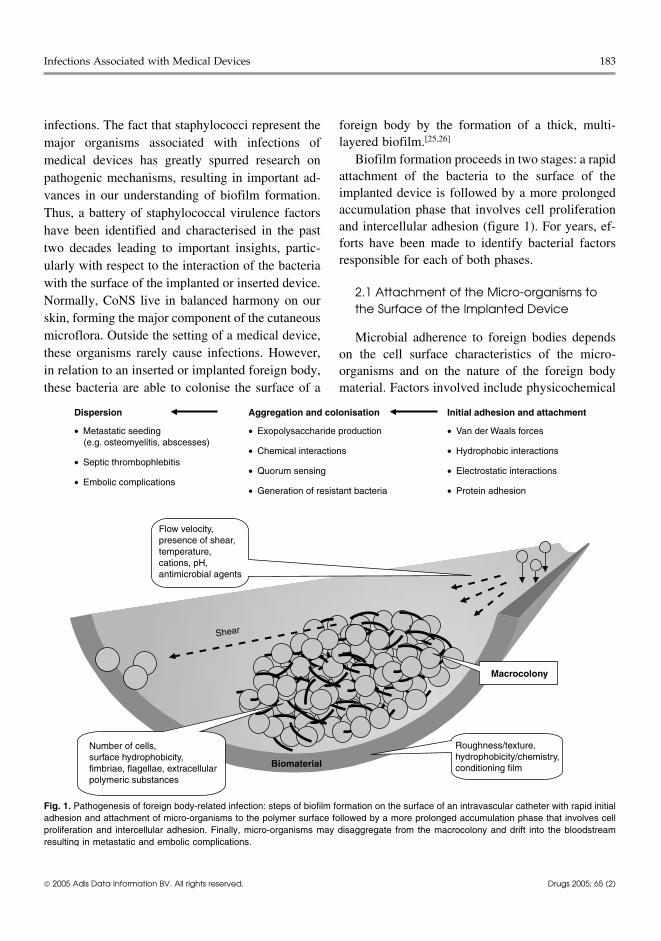
infections. The fact that staphylococci represent the foreign body by the formation of a thick, multi-layered biofilm.[25,26]major organisms associated with infections of
Biofilm formation proceeds in two stages: a rapidmedical devices has greatly spurred research onattachment of the bacteria to the surface of thepathogenic mechanisms, resulting in important ad-implanted device is followed by a more prolongedvances in our understanding of biofilm formation.accumulation phase that involves cell proliferationThus, a battery of staphylococcal virulence factorsand intercellular adhesion (figure 1). For years, ef-have been identified and characterised in the pastforts have been made to identify bacterial factorstwo decades leading to important insights, partic-responsible for each of both phases.ularly with respect to the interaction of the bacteria
with the surface of the implanted or inserted device.2.1 Attachment of the Micro-organisms to
Normally, CoNS live in balanced harmony on ourthe Surface of the Implanted Device
skin, forming the major component of the cutaneousmicroflora. Outside the setting of a medical device, Microbial adherence to foreign bodies dependsthese organisms rarely cause infections. However, on the cell surface characteristics of the micro-in relation to an inserted or implanted foreign body, organisms and on the nature of the foreign bodythese bacteria are able to colonise the surface of a material. Factors involved include physicochemical
Fig. 1. Pathogenesis of foreign body-related infection: steps of biofilm formation on the surface of an intravascular catheter with rapid initialadhesion and attachment of micro-organisms to the polymer surface followed by a more prolonged accumulation phase that involves cellproliferation and intercellular adhesion. Finally, micro-organisms may disaggregate from the macrocolony and drift into the bloodstreamresulting in metastatic and embolic complications.
2005 Adis Data Information BV. All rights reserved. Drugs 2005; 65 (2)
184 von Eiff et al.
forces such as polarity, London-van der Waal’s S. epidermidis autolysin AtlE which mediates pri-mary attachment to a polymer surface (see section 2)forces and hydrophobic interactions.[27] Cell surfacewas also found to exhibit vitronectin-binding ac-hydrophobicity and initial adherence oftivity, suggesting not only a function in the earlyS. epidermidis to polystyrene have been attributed tostages of adherence but also a contribution to latertwo different bacterial surface-associated proteins,stages of adherence involving specific interactionsdesignated SSP-1 and SSP-2.[28] Initial attachmentwith plasma proteins deposited on the polymer sur-of S. epidermidis to a polymer surface may be alsoface.[29] Aside from proteins, teichoic acid was sug-mediated at least in part by AtlE, a surface-asso-gested to function as a bridging molecule betweenciated autolysin.[29] The biofilm-associated proteinthe bacteria and fibronectin-coated polymer.[37]Bap was reported to contribute to both phases of
S. aureus biofilm formation, adhesion and accumu-2.2 Cell Proliferation andlation,[30] while Bhp, a Bap-homologous protein,Intercellular Adhesionmay contribute to S. epidermidis biofilm forma-
tion.[31] Aside from proteins, a polysaccharide struc-Once adhered to the surface of the foreign body,ture called capsular polysaccharide/adhesin (PS/A)
micro-organisms multiply and accumulate in multi-has been associated with initial adherence and slimelayered cell clusters, which requires intercellular
production.[32] In a rabbit model of endocarditis, PS/adhesion. A specific polysaccharide antigen termed
A-deficient mutants were less virulent and im-polysaccharide intercellular adhesin (PIA), which is
munisation with PS/A resulted in protection againstinvolved in intercellular adhesion and biofilm ac-
infection.[33]
cumulation and is chemically related to PS/A, hasWhile the direct interaction between bacteria on been detected and analysed in staphylococci.[38]
one side and the unmodified and naked surface of Tn917 mutants lacking PIA were not able to accu-the foreign body on the other side plays a crucial mulate in multilayered cell clusters. The icaADBCrole in the early stages of the adherence process in operon that mediates cell clustering and the intercel-vitro and probably also in vivo, additional factors lular adhesin synthesis in S. epidermidis has beenmay be important in later stages of adherence in cloned and sequenced.[39,40] Later, three other genevivo. Implanted devices rapidly become coated with loci were identified, which have a direct or indirectplasma and connective tissue proteins, such as regulatory influence on expression of the syntheticfibronectin, fibrinogen, vitronectin, thrombos- genes for PIA and biofilm formation.[41] In a mousepondin, laminin, collagen and von Willebrand factor model of subcutaneous foreign body infection as(vWf), which subsequently may serve as specific well as in a rat model of CVC-associated infection, areceptors for colonising micro-organisms.[34-36] In PIA-negative mutant was shown to be significantlythe vascular system at sites of increased flow, vWf less virulent than the isogenic wild-type strain.[42,43]
may also play an important role in adhesion of A PIA/haemagglutinin-positive S. epidermidisstaphylococcal cells to polymer surfaces because strain was significantly more likely to cause a sub-under high shear rates platelets do not appreciably cutaneous abscess than its isogenic PIA/haemagglu-bind to extracellular matrix proteins other than tinin-negative mutant and was significantly lessvWF.[35] Several host factor-binding proteins from likely to be eradicated from the inoculation site byS. aureus (e.g. the fibrinogen receptor ClfA and the host defence. Furthermore, the wild-type strain wasfibronectin-binding proteins FbpA and FbpB) and found to adhere to the implanted catheters morefrom CoNS (e.g. the fibrinogen-binding protein Fbe abundantly than the PIA/haemagglutinin-negativeand the fibronectin-binding autolysin Aas) have mutant.[43] In an investigation designed to study thebeen identified and characterised. The pathogenic properties of S. epidermidis strains ob-
2005 Adis Data Information BV. All rights reserved. Drugs 2005; 65 (2)
Infections Associated with Medical Devices 185
tained from blood of patients with FBRI, a strong form a highly differentiated biofilm structure.[48]
association was detected between pathogenesis and The S. aureus quorum-sensing system is encoded byboth biofilm formation and presence of the ica gene the accessory gene regulator (agr) locus that con-cluster.[44] Most recently, it was shown that induc- tributes to virulence in model biofilm-associatedtion of biofilm formation could be completely inhib- infections. Most recently, it was shown that, underited by chloramphenicol, which – given at a later some conditions, disruption of agr expression hadstage of biofilm accumulation – also inhibited fur- no discernible influence on biofilm formation, whilether development of preformed biofilm. This indi- under others it either inhibited or enhanced biofilmcates that continuous translation of an additional, formation. Under those conditions where agr ex-icaADBC-independent factor is required for the ex- pression enhanced biofilm formation (tested in apression of a biofilm-positive phenotype.[45] rotating-disk reactor), biofilms of an agr signalling
mutant were particularly sensitive to rifampicin butOther factors such as the 140 kDa extracellularnot to oxacillin.[49]
protein AAP (accumulation-associated protein) alsoThe clinical experience with polymer-associatedseem to be necessary for accumulation and biofilm
infections reveals that the host defence mechanismsformation.[46] AAP, which is lacking in an accumu-often seem to be unable to handle the infection and,lation-negative mutant and detectable only in ex-in particular, to eliminate the micro-organisms fromtracellular products from bacteria grown under ses-the infected polymer device. In addition, antibacteri-sile conditions, was shown to be essential for accu-al chemotherapy is frequently not able to cure thesemulative growth in certain S. epidermidis strains oninfections despite the use of antibacterials withpolymer surfaces. Of 58 CoNS studied, 55% wereproven in vitro activity (see section 3.3). Thus, the140 kDa antigen-positive and produced significant-biofilm may protect the embedded bacteria againstly larger amounts of biofilm than the other strainshost response mechanisms as well as against anti-that were 140 kDa antigen-negative. An antiserumbacterial agents.[50,51]
specific for AAP inhibited accumulation by up to98% of the wild-type strain.[46]
3. Management of MedicalTaken together, the factors described here lead toDevice-Associated Infectionsthe consequence that bacteria, particularly staphylo-
cocci, are able to adhere rapidly to the surface of aforeign body. During the following accumulation
3.1 General Considerationsphase, the bacteria proliferate to form multilayeredcell clusters on the surface of a medical device. Thepresence of such large adherent biofilms on the Whenever an infection of an indwelling or im-surfaces of foreign bodies, particularly on explanted planted foreign body is suspected, a general decisionintravascular catheters, has been demonstrated by has to be addressed: whether to remove the foreignscanning electron microscopy.[26,47]
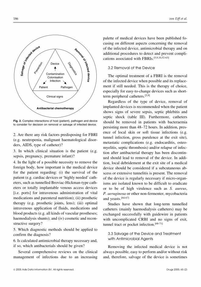
body and/or whether to initiate calculated anti-microbial treatment (figure 2). Answering the fol-Intercellular signalling, often referred to aslowing key questions may help the physician toquorum sensing, has been shown to be involved inmanage these infections adequately based on a ratio-biofilm development by several Gram-positivenale approach.and -negative bacteria such as Streptococcus
mutans, Burkholderia cepacia and Pseudomonas 1. Is an FBRI a plausible explanation for the pa-aeruginosa. For example, under certain conditions, tient’s signs (e.g. fever, skin inflammation at the exita quorum-sensing–defective mutant of P. aerugi- site, soft tissue inflammation along the tunnel of annosa is – in contrast to its parent strain – unable to implanted catheter, septic thrombophlebitis)?
2005 Adis Data Information BV. All rights reserved. Drugs 2005; 65 (2)
186 von Eiff et al.
palette of medical devices have been published fo-cusing on different aspects concerning the removalof the infected device, antimicrobial therapy and onadditional procedures to detect and prevent compli-cations associated with FBRIs.[5,9,10,52-63]
3.2 Removal of the Device
The optimal treatment of a FBRI is the removalof the infected device when possible and its replace-ment if still needed. This is the therapy of choice,especially for easy-to-change devices such as short-term peripheral catheters.[5,9]
Regardless of the type of device, removal ofimplanted devices is recommended when the patientshows signs of severe sepsis, septic phlebitis andseptic shock (table III). Furthermore, cathetersshould be removed in patients with bacteraemia
Device
Patient Pathogen
Clinical signs
DiagnosticsContamination
ColonisationInfection
Prev
entio
n
Sal
vag
eof
infe
cted
deviceRem
oval of infectedd
evice
Antibacterial chemotherapy
Fig. 2. Complex interactions of host (patient), pathogen and deviceto consider for decision on removal or salvage of infected device.
persisting more than 48–72 hours. In addition, pres-ence of local skin or soft tissue infections (e.g.
2. Are there any risk factors predisposing for FBRItunnel infection, gross purulence at the exit site),
(e.g. neutropenia, malignant haematological disor-metastatic complications (e.g. endocarditis, osteo-
ders, AIDS, type of catheter)?myelitis, septic thrombosis) and/or relapse of infec-
3. In which clinical situation is the patient (e.g. tion after antibacterial therapy has been discontin-sepsis, pregnancy, premature infant)? ued should lead to removal of the device. In addi-4. In the light of a possible necessity to remove the tion, local debridement at the exit site of a medicalforeign body, how important is the medical device device should be considered if a subcutaneous ab-for the patient regarding: (i) the survival of the scess or extensive tunnelitis is present. The removalpatient (e.g. cardiac devices or ‘highly needed’ cath- of the device is regularly necessary if micro-organ-eters, such as tunnelled Broviac-Hickman-type cath- isms are isolated known to be difficult to eradicateeters or totally implantable venous access devices or to be of high virulence such as S. aureus,[i.e. ports] for intravenous administration of vital P. aeruginosa or other non-fermenter, mycobacteriamedications and parenteral nutrition); (ii) prosthetic and yeasts.[64-67]
therapy (e.g. prosthetic joints, lens); (iii) optimal Studies have shown that long-term tunnelledintravenous application of fluids, medications and catheters (mainly haemodialysis catheters) may beblood products (e.g. all kinds of vascular prostheses; exchanged successfully with guidewire in patientshaemodialysis shunts); and (iv) cosmetic and recon- with uncomplicated CRBI and no signs of exit,structive surgery? tunnel tract or pocket infection.[68-71]
5. Which diagnostic methods should be applied to3.3 Salvage of the Device and Treatmentconfirm the diagnosis?with Antimicrobial Agents6. Is calculated antimicrobial therapy necessary and,
if so, which antibacterials should be given? Removing the infected medical device is notSeveral comprehensive reviews on the clinical always possible, easy to perform and/or without risk
management of infections due to an increasing and, therefore, salvage of the device is sometimes
2005 Adis Data Information BV. All rights reserved. Drugs 2005; 65 (2)
Infections Associated
with M
edical D
evices187
2005 A
dis D
ata
Info
rma
tion
BV. A
ll righ
ts rese
rved
.D
rug
s 2005; 65 (2)
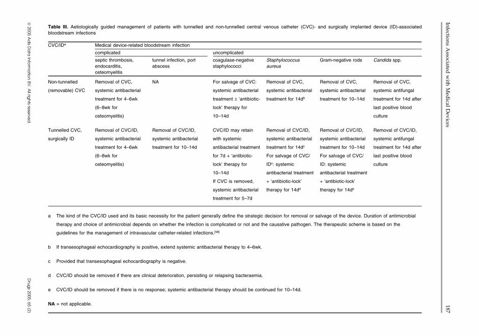
Table III. Aetiologically guided management of patients with tunnelled and non-tunnelled central venous catheter (CVC)- and surgically implanted device (ID)-associatedbloodstream infections
CVC/IDa Medical device-related bloodstream infection
complicated uncomplicated
septic thrombosis, tunnel infection, port coagulase-negative Staphylococcus Gram-negative rods Candida spp.endocarditis, abscess staphylococci aureusosteomyelitis
Non-tunnelled Removal of CVC, NA For salvage of CVC: Removal of CVC, Removal of CVC, Removal of CVC,
(removable) CVC systemic antibacterial systemic antibacterial systemic antibacterial systemic antibacterial systemic antifungal
treatment for 4–6wk treatment ± ‘antibiotic- treatment for 14db treatment for 10–14d treatment for 14d after
(6–8wk for lock’ therapy for last positive blood
osteomyelitis) 10–14d culture
Tunnelled CVC, Removal of CVC/ID, Removal of CVC/ID, CVC/ID may retain Removal of CVC/ID, Removal of CVC/ID, Removal of CVC/ID,
surgically ID systemic antibacterial systemic antibacterial with systemic systemic antibacterial systemic antibacterial systemic antifungal
treatment for 4–6wk treatment for 10–14d antibacterial treatment treatment for 14dc treatment for 10–14d treatment for 14d after
(6–8wk for for 7d + ‘antibiotic- For salvage of CVC/ For salvage of CVC/ last positive blood
osteomyelitis) lock’ therapy for IDc: systemic ID: systemic culture
10–14d antibacterial treatment antibacterial treatment
If CVC is removed, + ‘antibiotic-lock’ + ‘antibiotic-lock’
systemic antibacterial therapy for 14dd therapy for 14de
treatment for 5–7d
a The kind of the CVC/ID used and its basic necessity for the patient generally define the strategic decision for removal or salvage of the device. Duration of antimicrobial
therapy and choice of antimicrobial depends on whether the infection is complicated or not and the causative pathogen. The therapeutic scheme is based on the
guidelines for the management of intravascular catheter-related infections.[58]
b If transesophageal echocardiography is positive, extend systemic antibacterial therapy to 4–6wk.
c Provided that transesophageal echocardiography is negative.
d CVC/ID should be removed if there are clinical deterioration, persisting or relapsing bacteraemia.
e CVC/ID should be removed if there is no response; systemic antibacterial therapy should be continued for 10–14d.
NA = not applicable.
188 von Eiff et al.
the preferred option. In particular, FBRIs associated tion or to increased expression of fibronectin-bind-ing proteins.[77-79]with long-term or permanent catheters (such as
The special conditions surrounding a foreignHickman-type catheter or Port-a-Cath) are frequent-body have guided the search for alternative applica-ly treated successfully ‘through the line’ (tabletions of antibacterials such as lipid-based sustained-III).[4,72,73]
release formulations. Roehrborn et al.[80] describeOnce a biofilm has formed on an implantedthe use of such biodegradable, locally injectablemedical device it is difficult to treat such infectionsformulation of amikacin in a mouse model in whichbecause of significantly decreased levels of suscep-Teflon 1 tubes were subcutaneously implanted and
tibility to antimicrobial agents (some 10 to 1000challenged by inoculation of S. aureus. Whereas
times less) and lower levels of phagocytosis relativetreatment with local or systemic free amikacin had
to the levels of resistance and phagocytosis for theirno effect, the number of infected foreign bodies was
planktonic counterparts (see pathogenesis, sectionreduced from 86% to 25% (p = 0.02) following
2).[74] Thus, supraphysiological concentrations of treatment with encapsulated amikacin formulation,antibacterial agents may be required to eliminate the and log cfu (colony forming units) per gram of tissuemicro-organisms embedded in biofilms.[75] As was significantly decreased from 4.8 ± 0.9 toshown in a number of experimental FBRIs, the 1.3 ± 0.6.pharmacokinetic parameters are modified and do Typically, initial treatment of catheter-relatednot correspond to the efficacy of antibacterial treat- bacteraemia is administration of systemic antibac-ment in vivo when a foreign body is implanted. terials. Additionally, when a catheter-related infec-These changes are obvious if mouse model-based tion is documented and a specific pathogen is identi-results of S. aureus-caused intra-abdominal abscess fied, ‘antibiotic-lock’ therapy should be consideredsurrounding intraperitoneally placed silicone cathe- if salvage of the catheter is necessary. It is notewor-ter treated by meticillin and gentamicin are thy that recommendations for the treatment ofanalysed.[76] Whereas both agents showed strong medical device-associated infections are based al-effects in vitro in time-kill studies on bacteria most exclusively on observational studies, animalcolonising catheters taken out of infected mice and models, case reports and expert opinion rather thanon catheters contaminated in vitro, only poor results on the results of appropriate clinical trials.were observed in vivo, despite high local concentra-
3.3.1 Use of Lock Solutions for Intraluminal Therapytions (greater than minimum inhibitory concentra-(‘Antibiotic-Lock’ Technique)
tion [MIC] for at least 72 hours) of meticillin andA technique of filling and closing a catheter
high peak concentrations of gentamicin (>13 µg/lumen with a lock solution may prevent or cure
mL). The failure was not caused by development ofcatheter-related infections, as active ingredients can
antibacterial resistance or influenced by protein con- be maintained in direct contact with the internalcentration, pH or local presence of inhibitors of surface of the device for prolonged periods of timeantibacterials in the pus. (hours to days). Thus, to circumvent the need for
Of importance, antibacterials administered in catheter withdrawal, Messing et al.[81] were the firstsubinhibitory concentrations may influence the to describe the intraluminal application of antibac-mechanisms of adherence and slime production, es- terial agents, referred to as antibiotic-lock tech-pecially in staphylococci, for example, leading to nique. Avoiding systemic adverse effects, thishigher polysaccharide intracellular adhesin produc- method allows the delivery of a high concentration
1 The use of trade names is for product identification purposes only and does not imply endorsement.
2005 Adis Data Information BV. All rights reserved. Drugs 2005; 65 (2)
Infections Associated with Medical Devices 189
of antibacterials (or, rarely, disinfectants) in the with antiadherence properties, was shown to be ac-catheter in order to decontaminate the intraluminal tive against a broad range of bacteria as well assurface of the catheter in situ. In an analysis of 14 fungi.[90,91] The findings of Shah et al.[92] evaluatingopen-label trials of standard parenteral therapy for taurolidine-citrate (Neutrolin, Biolink Corp.,the treatment of CRBI and the salvage of tunnelled Norwell, MA, USA) for its antimicrobial and bi-catheters, a salvage in 342 (66.5%) of 514 episodes ofilm eradication activity in a catheter model sug-was documented.[58] gested that this lock solution is a promising combi-
nation agent for the prevention and treatment ofCurrently, the antibiotic-lock technique is recom-intravascular catheter-related infections.[92] Alterna-mended for the treatment of uncomplicated catheter-tively, ethanol (alcohol)-lock technique was intro-related bacteraemia by several medical societies,duced for the treatment of BSIs in patients withsuch as the Infectious Diseases Society of America,tunnelled CVCs and proven to be a safely used, wellthe Society of Critical Care Medicine and thetolerated and effective way to treat central venousSociety for Healthcare Epidemiology of America.[58]
line infections.[93] However, further studies areHowever, several parameters of intraluminal anti-needed to ascertain whether ethanol or taurolidinebacterial therapy are not clearly defined, for exam-locks might be equal or superior to the antibiotic-ple the duration of the antibiotic-lock therapy is notlock technique. Since its effect does not depend onestablished. In most studies, this technique was ad-sensitivity to antibacterial agents, this approach mayministered for 7–14 days. Furthermore, the useful-be of particular value for infections with multiresis-ness of different types of antibacterial agents, theirtant micro-organisms. Furthermore, highly antibac-optimum concentration and the necessity of simulta-terial-resistant micro-organisms will not be selectedneous systemic treatment remain to be defined.[82]
by the use of disinfectants or other alternative agentsGlycopeptides, aminoglycosides and ciprofloxacinand, in principle, its use could reduce the consump-have been shown to be suitable agents.[67,81,83,84]
tion of broad-spectrum antibacterials, especiallySome studies have used the antibiotic-lock tech-
vancomycin.nique in conjunction with the administration of sys-temic antibacterials and/or thrombolytic/anticoagu- 3.3.2 Recommendations for Calculatedlant agents.[83,85-87] However, bacteria such as Antimicrobial Therapystaphylococci may survive and grow in heparin- Because of the high risk of complications, CVC-locked catheters.[88] The drawback of using lock related and surgically implanted venous access in-solutions containing antibacterials used for systemic fections should be treated with parenteral drugs,therapy is that it may lead to the emergence of using high doses and short courses (approximatelyantibacterial resistance. In particular, the prophylac- 7–10 days), irrespective of the removal of the de-tic and therapeutic long-term application of vanco- vice.[94,95] Antimicrobial therapy for the time periodmycin could be of high risk for the development of prior to a microbiological diagnosis should be initi-staphylococcal subpopulations with reduced suscep- ated on a calculated basis considering the spectrumtibility against glycopeptides as a result of the exis- of expected pathogens and their local/regional resis-tence of more or less ‘occult’ device-related infec- tance situation. However, treatment should be de-tion sites.[89]
escalated to narrow-spectrum drugs on the basis ofsusceptibility tests as soon as test results are avail-To meet concerns regarding a selection of highlyable.resistant bacteria and an insufficient clearance of the
device, the antimicrobial activity of alternative Taking into account that staphylococci (especial-agents such as catheter lock solutions were investi- ly CoNS, such as S. epidermidis and S. haemolyti-gated. Taurolidine, known as a nontoxic substance cus) are by far the most frequent pathogens isolated
2005 Adis Data Information BV. All rights reserved. Drugs 2005; 65 (2)
190 von Eiff et al.
in FBRIs, calculated antimicrobial therapy should action of rifampicin compared with vancomycininclude the administration of a glycopeptide (espe- was noted in a mouse model of intraperitoneallycially vancomycin) with an aminoglycoside (e.g. implanted preformed bacterial biofilm catheter seg-gentamicin) or rifampicin because a significant per- ments.[99] While simultaneous use of antibacterialscentage of staphylococci recovered from hospital- of the cell wall-active class (including vancomycin)ised patients are meticillin resistant.[58,63,96] In criti- and rifampicin was shown to act synergistically,cally ill patients, coverage against Gram-negative other antibacterials (including aminoglycosides)bacteria, including P. aeruginosa, and even fungi antagonised rifampicin activity.[100] However, com-may be considered until definitive data from micro- bination of antibacterials is not generally recom-biological diagnostics are available. mended for CRBI due to CoNS.[58] Recently, two
oxazolidinones, linezolid and eperezolid, were3.3.3 Recommendations for Aetiologically Guided
shown to achieve eradication of S. epidermidis bio-Antimicrobial Therapyfilms more rapidly than vancomycin and gentamicinAetiologically guided antimicrobial therapyin an in vitro model using polyurethane coupons in ashould be initiated as soon as possible on the basis ofmodified Robbins device.[101]
appropriate microbiological diagnostics. Choice andThe duration of parenteral therapy may be quiteduration of this therapy depends mainly on the iso-
short (5–7 days) when treating uncomplicated FBRIlated causative micro-organism, the resistance pat-due to CoNS if the catheter is removed. If an intralu-tern and the presence of complications, especiallyminal infection is suspected and an intravenousdeep-seated soft tissue infections (table III).catheter or a surgically implanted device is retained,
Coagulase-Negative Staphylococci systemic antibacterial therapy and antibiotic-lockImplant infections due to CoNS remain a thera- therapy for 10–14 days are recommended (table
peutic challenge since they frequently result in fail- III).[58,83,102,103] It should be noted that persistent orure of conservative therapy and often require with- relapsing fever and other signs of treatment failuredrawal of the foreign body. Although cure rates are are clear indications for removal of the device.[58]
not affected by removal, investigations on the im- The widespread use of vancomycin for the treat-pact of CVC removal on the recurrence of catheter- ment of FBRIs is of concern because of the emer-related CoNS bacteraemia have shown that there is a gence of vancomycin-resistant enterococci and of20% chance of recurrence of bacteraemia when the staphylococci with reduced sensitivity to glycopep-CVC is not removed.[97,98] In contrast, the risk is tides (vancomycin-/glycopeptide-intermediate S.significantly reduced to 3% if the catheter is re- aureus). Moreover, the most recent recovery of truemoved.[97] This risk is especially high if the catheter vancomycin-resistant S. aureus strains underscoresstays in place for >3 weeks after bacteraemia. the need of control regarding the use of vancomycin
Most CoNS isolates causing FBRI are meticillin in healthcare settings.[104,105]
resistant as a result of the possession of the mecAStaphylococcus aureusgene. As a result, these isolates are resistant to all β-
lactam antibacterials. Thus, most CoNS infections FBRIs caused by S. aureus infections are dreadedrequire treatment with glycopeptides, in particular because of possible accompaniment by serious in-vancomycin. In addition, teicoplanin has potential fectious complications such as severe sepsis, septicfor use as an alternative in the treatment of infec- thrombosis and/or several deep-seated infectionstions due to CoNS.[55] Notably, glycopeptides are (endocarditis, osteomyelitis and other metastatic in-poorly bactericidal against staphylococci. If an iso- fections). Thus, it is generally accepted that thelate is susceptible, replacement of vancomycin by a colonised foreign body, especially in the case ofsemisynthetic penicillin is advisable. Superior rapid non-tunnelled CVC, must be removed.[58,106,107]
2005 Adis Data Information BV. All rights reserved. Drugs 2005; 65 (2)
Infections Associated with Medical Devices 191
Tunnelled CVCs should be removed if there is evi- phylaxis or angio-oedema.[58] For patients with seri-dence of exit-site infection as well as tunnel or ous allergy to β-lactams and for those infected withpocket infections.[108,109] Only in selected cases of meticillin-resistant S. aureus (MRSA), vancomycinuncomplicated infections (table III) may tunnelled is the drug of choice.[58,118] However, vancomycinCVCs or medical devices be retained and treated has higher failure rates than have penicillinase-resis-with appropriate systemic antibacterial therapy ac- tant penicillins and some complications are difficultcompanied by antibiotic-lock therapy (see section to treat with glycopeptide monotherapy for pharma-3.3.1).[58,110,111] cological reasons.[119,120] In the case of MRSA,
lincosamide antibacterials (clindamycin) and newerSince metastatic infections may occur in thefluoroquinolones as well as combinations with ri-course of S. aureus infections, it is clinically impor-fampicin, fusidic acid, cotrimoxazole and fosfo-tant to rule out at least their most devastating conse-mycin may be included into the therapeutic regimenquence, that is, acute endocarditis. Transesophagealif isolates are sensitive.[119] New antimicrobials suchechocardiography, which has been shown to be aas the oxazolidinones, streptogramins and newerhighly sensitive method to diagnose endocarditis,glycopeptides exhibit high activity against MRSAshould be performed in each patient with S. aureus(and other multiresistant Gram-positive pathogens),BSI unless contraindications are present.[112,113]
but resistance to some of these agents has alreadyClinical symptoms of bone infections should lead tooccurred. In a recent study encompassing childrenscintigraphic and radiographic examinations.[54] Awith hospital-acquired pneumonia or bactaeremiarecently published scoring system based on the pres-due to multiresistant Gram-positive bacteria, linezo-ence or absence of four risk factors (communitylid was well tolerated. No significant difference wasacquisition, skin examination findings suggestingdetected in clinical cure rates in the clinically evalu-acute systemic infection, persistent fever at 72 hoursable population between the linezolid and vancomy-and positive follow-up blood culture results atcin groups for patients with catheter-related bacter-48–96 hours) accurately identified complicatedaemia.[121] However, the potential of these alterna-S. aureus bacteraemia.[114]
tive agents for the treatment of CRBIs should beIn contrast to CoNS, most experts favour paren-
analysed in further trials.teral treatment for CRBI caused by S. aureus with a
Several animal models of FBRIs were developedminimum duration of 10–14 days of parenteral anti-in order to investigate the effects of antibacterialbacterials.[115,116] Some authors recommend a subse-treatment (table IV).[122-126] In one study, Chuard etquent additional treatment with oral anti-al.[123] showed that two- or three-drug combinationsstaphylococcal antibacterials over a period of 1–2such as fleroxacin and rifampicin (and vancomycin),weeks.[117] If persisting bacteraemia or complica-respectively, were highly effective and superior totions such as prolonged fever, metastatic or deep-single drugs in treating chronic staphylococcalseated infection are occurring, much longer periodsFBRIs.[123] Applying different fluoroquinolones,(4–6 weeks for endocarditis, 6–8 weeks for osteo-partly in comparison with vancomycin, in two dif-myelitis) of parenteral anti-staphylococcal therapyferent experimental models (rat and guinea pig), itare recommended.[4,58] The first choice for treatmentwas shown that the newer fluoroquinolones,of CRBIs caused by S. aureus should be the paren-temafloxacin and sparfloxacin, were significantlyteral application of β-lactam antibacterials (penicil-more active than ciprofloxacin for the prophylaxislinase-resistant penicillins, e.g. flucloxacillin, ox-or treatment of FBRIs caused by a fluoroquinolone-acillin) when the isolate is susceptible.[58] First-gen-susceptible MRSA strain. As with temafloxacin anderation cephalosporins, such as cefazolin, may be
used for patients with penicillin allergy without ana- sparfloxacin, vancomycin was also significantly
2005 Adis Data Information BV. All rights reserved. Drugs 2005; 65 (2)

192 von Eiff et al.
Table IV. Experimental animal models of foreign body infection to study effects of treatment with antibacterials
Study (year) Animal Foreign body model Pathogen Antibacterial treatment
Chuard et al.[123] (1991) Rat Subcutaneous tissue cages Staphylococcus aureus Vancomycin, fleroxacin,(MRSA) rifampicin
Gagnon et al.[99] (1992) Mouse Intraperitoneal catheter S. epidermidis Rifampicin, vancomycinsegments
Espersen et al.[76] (1994) Mouse Intraperitoneal silicone S. aureus Meticillin, gentamicincatheter
Schaad et al.[125] (1994) Guinea pig Subcutaneous tissue cages S. aureus (MRSA) Teicoplanin, rifampicin
Schaad et al.[126] (1994) Rat Subcutaneous tissue cages S. aureus (MSSA, MRSA) Imipenem, oxacillin,vancomycin
Cagni et al.[124] (1995) Guinea pig Subcutaneous tissue cages S. aureus (MRSA) Sparfloxacin, temafloxacin,ciprofloxacin, vancomycin
Roehrborn et al.[80] (1995) Mouse Subcutaneous Teflon tubes S. aureus Amikacin (lipid-based, slow-release)
Van Wijngaerden et al.[129] Rat Subcutaneous catheter S. epidermidis Teicoplanin, rifampicin(1999)
Rupp et al.[130] (2001) Rat Central venous catheter Enterococcus faecium (VRE) Oritavancin
Vaudaux et al.[122] (2002) Guinea pig Subcutaneous tissue cages S. aureus (MRSA) Levofloxacin, alatrofloxacin,vancomycin
Kuklin et al.[131] (2003) Mouse Subcutaneous Teflon S. aureus (bioluminescent Linezolidcatheter mutant)
Vaudaux et al.[128] (2003) Rat Subcutaneous tissue cages S. aureus Daptomycin, vancomycinMRSA = meticillin-resistant Staphylococcus aureus; MSSA = meticillin-susceptible S. aureus; VRE = vancomycin-resistant enterococci.
more active than ciprofloxacin in decreasing the require catheter withdrawal. Vancomycin has beenviable counts of MRSA in tissue cage fluids in the widely used to treat infections caused by theserat model.[124] A further comparison of fluoroquino- bacteria, although treatment should be de-escalatedlones with vancomycin for treatment of experimen- based on the results of susceptibility testing. Cathe-tal FBRI by MRSA showed levofloxacin was signif- ter removal is essential for successful treatment oficantly more active than vancomycin in decreasing CVC-related infections due to rapidly growing my-the viable counts of MRSA.[122] A second-genera- cobacteria of the Mycobacterium fortuitum com-tion glycopeptide, oritavancin (LY 333328), was plex.[132] Since these mycobacteria exhibit variable,shown to be effective against S. aureus in a rat CVC species-specific susceptibility to traditional an-infection model.[127] The therapeutic activity of timycobacterial drugs and other antibacterials (in-daptomycin was compared with that of vancomycin cluding cefoxitin, imipenem/cilastatin, aminoglyco-in a rat model of subcutaneously implanted tissue sides, tetracyclines, macrolides and co-trimoxazolecages chronically infected with S. aureus.[128] The [trimethoprim/sulfamethoxazole]), therapy shouldauthors concluded that a low-dose regimen of dapto- be based on culture and susceptibility results.[133]
mycin was at least equivalent to vancomycin; how-ever, three of four cages implanted in daptomycin- Gram-Negative Rods
treated rats yielded subpopulations with reduced Gram-negative rods are commonly associatedsusceptibility to daptomycin. with contaminated infusate and are usually found to
be the cause of BSIs in immunocompromised pa-Gram-Positive Rods (Including Rapidly tients with indwelling devices. Controlled studiesGrowing Mycobacteria) regarding withdrawal of the infected device or theThe majority of intravenous line infections choice of optimal antibacterial agents and the dura-
caused by Corynebacterium spp. and Bacillus spp. tion of therapy are missing. However, patients with
2005 Adis Data Information BV. All rights reserved. Drugs 2005; 65 (2)
Infections Associated with Medical Devices 193
catheter-related infections due to Gram-negative include withdrawal of the catheter.[144-146] The evi-rods should have the catheter removed, if possible, dence for these recommendations is strongest in theand should receive appropriate antibacterial therapy. non-neutropenic patient population.[147] In neutro-Patients with devices that cannot be removed should penic patients it is difficult to determine whether thebe treated for 2 weeks with systemic and antibiotic- gut or a catheter may act as the primary source oflock therapy provided that the Gram-negative fungaemia. Management of Candida infection bybacteraemia is not associated with organ dysfunc- catheter removal alone is not sufficient because oftion, hypoperfusion or hypotension.[58,134,135] In an increased risk of disseminated and/or metastaticcases of catheter-related bacteraemia with non-fer- fungal infections.[148,149] Thus, it is recommended tomenter species other than P. aeruginosa, B. cepacia, treat catheter-related Candida infections with appro-Acinetobacter baumannii and Stenotrophomonas priate antifungal agents for a minimum duration of 2maltophilia, some reports have demonstrated that or 3 weeks after the last positive blood culture.[61]
catheter withdrawal reduces the rate of treatment Infections due to Malassezia spp. should includefailure and improves survival.[136] Approximately discontinuation of intravenous lipids.[150]
10–14 days of parenteral therapy is recommendedSince its introduction to the pharmaceutical mar-
when treating CRBIs caused by Gram-negativeket in the 1950s, amphotericin B has been the gold
rods. However, a longer duration (4–6 weeks) ofstandard antifungal agent for life-threatening inva-
antibacterial therapy should be performed if pro-sive fungal infections. However, its use is considera-
longed bacteraemia occurs despite catheter remov-bly hampered by the high rate of toxicity, which has
al.[137]
led to the development of lipid-based formulationsof amphotericin B with their superior safety profiles.YeastsThese lipid formulations can be considered as suita-Since several Candida species readily form bio-ble replacements for amphotericin B for primaryfilms, they are frequently isolated from patients withtherapy for many invasive fungal infections.[151] C.FBRIs.[138] C. albicans represents the predominantalbicans is generally susceptible to all antifungaland most virulent species. However, the importanceagents; however, its potential to develop azole resis-of infections caused by non-albicans Candida spp.tance has been documented. In a randomised trial inand other unusual yeasts (e.g. Malassezia spp.,patients without neutropenia and major immuno-Rhodotorula spp., Hansenula anomala) has emer-deficiency, high-dose fluconazole appeared to beged over the last decade.[139] Notably, current rou-effective as amphotericin B, with less toxicity.[152] Intine methods for yeast identification may be insuffi-contrast, some Candida spp. other than C. albicanscient to identify isolates of lipophilic Malasseziaare characterised by decreased susceptibility againstspp., which have been found to be associated to aazoles. Thus, knowledge of the species is increas-low but not negligible extent with infections ofingly important for the choice of the specific anti-CVCs for parenteral nutrition-bearing lipid emul-fungal treatment and, especially in the setting ofsions.[140] In particular, infections due to C. parap-infections due to non-albicans Candida spp., sus-silosis have been shown to correlate strongly withceptibility testing by standardised methods is mostthe presence of an intravascular device and the usehelpful. Whereas C. krusei and C. glabrata areof total parenteral nutrition due to the slime-forming
ability of this species.[141] intrinsically/innately more resistant to fluconazole,C. tropicalis, C. guillermondii and C. dubliniensisIn the case of CRBIs due to yeasts, removal of allare generally susceptible to azoles; however, fluco-existing intravascular catheters is desirable, if feasi-nazole may be less active against these yeasts. Inble.[142,143] Following isolation of C. parapsilosispatients infected by these yeasts or in institutionsand C. glabrata in blood, initial management must
2005 Adis Data Information BV. All rights reserved. Drugs 2005; 65 (2)

194 von Eiff et al.
where isolates of these Candida spp. are more fre- utes and is also effective against S. epidermidis atquent, the prescription of amphotericin B or the higher concentrations (MIC90: 12.5–64 µg/mL).[159]
administration of higher doses of fluconazole should Using biofilm plate assays, Wu et al.[159] demonstra-be the preferred treatment until the susceptibility ted that lysostaphin is also effective against sessiledata are available.[144] Of note, the azole-sensitive staphylococci associated with biofilms. Once estab-species C. lusitaniae has innately higher MICs to lished, staphylococcal biofilms are very difficult toamphotericin B. disrupt. Therefore, the fact that lysostaphin is specif-
The first of the second-generation triazole agents ically able to disrupt the extracellular matrix ofto receive regulatory approval is voriconazole, S. aureus biofilms in vitro on plastic and glass sur-which has shown an expanded in vitro activity faces (confirmed by scanning electron microscopy)against a wide variety of yeasts and moulds. In has to be regarded as a major progress in the man-addition, caspofungin, a new echinocandin anti- agement of FBRIs. Various other enzymes havefungal agent with broad-spectrum activity against been studied for the removal and disinfection ofCandida and Aspergillus spp., was shown to be bacterial biofilms; however, they are hampered byhighly active against Candida isolates exhibiting the fact that these procedures require two or morehigh-level resistance to fluconazole and itracona- compounds – one enzyme for removal of the adher-zole.[153] In a recent study designed to compare the ent bacteria in the biofilms and a further agent withefficacy of caspofungin with that of amphotericin B, antibacterial activity.[160]
caspofungin was shown to be at least as effective asUltrasound, defined as acoustic energy or sound
amphotericin B for the treatment of invasive candi-waves with frequencies >20 kHz, is commonly used
diasis and, more specifically, candidaemia.[154] Re-to remove bacterial cells from the surface of foreign
garding C. glabrata, C. krusei and C. albicans,bodies, especially if applied as high-intensity ultra-
voriconazole and caspofungin appear to have en-sound (>10 W/cm2).[161] This intensity is known tohanced activity; however, the clinical relevance oflyse bacterial and eucaryotic cells on surfaces and inthese findings should be studied in treatmentsuspension. The application of low-frequency ultra-trials.[153,155,156]
sound to enhance the activity of vancomycin againstTherapy of patients with FBRIs due to Candidaimplanted S. epidermidis biofilms was examinedspp. should be accompanied by ophthalmoscopicusing polyethylene disks covered with a biofilm ofexamination to rule out metastatic endophthalmitis.S. epidermidis and implanted subcutaneously in rab-It should be kept in mind that candidal endocarditisbits on both sides of their spine.[162] Carmen et al.[162]
has also been observed following FBRIs.reported that S. epidermidis biofilms responded fa-vourably to combinations of ultrasound and vanco-3.3.4 Use of Alternative Substancesmycin at 48 hours of insonation. In addition, pulsedand Approachesultrasound enhances the killing of Escherichia coliWith the emergence of antibacterial-resistantbiofilms by aminoglycosides in a rabbit model withstaphylococci, the antibacterial enzyme lysostaphinsubcutaneously implanted polyethylene disks. Here,has, in the past few years, gained renewed interest asthe ultrasound significantly reduced bacterial viabil-an antistaphylococcal therapeutic agent.[157,158] Thisity below that of nontreated biofilms without dam-glycylglycine endopeptidase is specifically capableage to the skin.[163] However, Pitt and Ross[164] foundof cleaving the cross-linking pentaglycine bridges inthat low-frequency ultrasound (70 kHz) of lowthe cell wall of staphylococci, making it highlyacoustic intensity (<2 W/cm2) increased the growthactive against both actively growing and quiescentrate of the cells compared with growth withoutbacteria. With a MIC90 of 0.001–0.064 µg/mL,ultrasound when S. epidermidis, P. aeruginosa andlysostaphin kills planktonic S. aureus within min-
2005 Adis Data Information BV. All rights reserved. Drugs 2005; 65 (2)
Infections Associated with Medical Devices 195
E. coli cells adhered to and grew on a polyethylene ters, dressings, administration sets and fluids (ap-surface. pendix B) are also given. Finally, recommendations
regarding intravascular devices use in paediatricpatients are provided in these guidelines.[22]4. Prevention Strategies
In the following subsections, some of the mostimportant strategies for prevention of catheter-relat-
4.1 Nontechnological Recommendations to ed infections are summarised, including those mostReduce the Incidence of Intravascular recently published.Catheter-Related Bloodstream Infections
4.1.1 Standardisation of Aseptic CareGuidelines providing evidence-based recommen-
Quality assurance and continuing education aredations for preventing catheter-related infectionsaspects of particular interest. Several studies havehave been prepared by a working group comprisingshown that the risk for intravascular device-asso-members from professional organisations represent-ciated BSIs declines following standardisation ofing several clinical disciplines led by the Society ofaseptic care.[22,165-167] While insertion and mainten-Critical Care Medicine.[22] Published in 2002, theseance of intravascular catheters by inexperiencedguidelines provide healthcare practitioners who in-staff (as well as nursing staff reductions) mightsert catheters and those responsible for surveillanceincrease the risk for catheter colonisation and CRBI,and control of infections with background informa-specialised ‘IV (intravenous) teams’ have showntion and specific recommendations to reduce theeffectiveness in reducing the incidence of infectionsincidence of intravascular CRBIs.and associated complications and costs.[167-169]
The recommendations given in table V and tableVI should be considered in the context of the institu-
4.1.2 Choice of Catheter Insertion Sitetion’s experience and availability of personnelThe density of local skin flora and, thus, also theskilled in the placement of intravascular devices. In
site of catheter insertion, influences the subsequentboth tables, only those recommendations that arerisk for CRI.[170-172] In adult patients, a subclavianstrongly recommended for implementation andsite is preferred for infection control purposes, al-strongly supported by well designed experimental,though other factors (e.g. the potential for mechani-clinical or epidemiological studies (category IA) arecal complications or risk for subclavian vein steno-included. For other categories, according to the sys-sis) should be considered when deciding where totem for categorising recommendations issued by theplace the catheter.[173-175] Consideration of comfort,Centers for Disease Control and Prevention/Hospi-security and maintenance of asepsis as well as pa-tal Infection Control Practices Advisory Committee,tient-specific factors (e.g. anatomic deformity andthat is, category IB (strongly recommended for im-bleeding diathesis), relative risk (RR) of mechanicalplementation and supported by some experimental,complications, the availability of bedside ultrasoundclinical or epidemiological studies, and a strongand the risk for infection should guide site selec-theoretical rationale), category IC (required by statetion.[172]or federal regulations, rules or standards) and cate-
gory II (suggested for implementation and sup- In addition, phlebitis has long been recognised asported by suggestive clinical or epidemiological a risk for infection. Lower extremity insertion sitesstudies or a theoretical rationale), see O’Grady et are associated with a higher risk for phlebitis thanal.[22] In these guidelines, examples of clinical defi- are upper extremity sites (for adults), and hand veinsnitions for CRI (appendix A) and a summary of have a lower risk for infection than do veins on therecommended frequency of replacements for cathe- wrist or upper arm.[176]
2005 Adis Data Information BV. All rights reserved. Drugs 2005; 65 (2)
196 von Eiff et al.
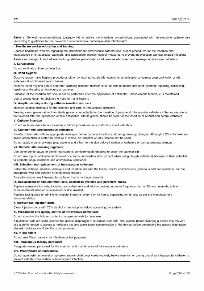
Table V. General recommendations (category IA) to reduce the infectious complications associated with intravascular catheter use(according to guidelines for the prevention of intravascular catheter-related infections)[22]
I. Healthcare worker education and training
Educate healthcare workers regarding the indications for intravascular catheter use, proper procedures for the insertion andmaintenance of intravascular catheters, and appropriate infection-control measures to prevent intravascular catheter-related infections
Assess knowledge of, and adherence to, guidelines periodically for all persons who insert and manage intravascular catheters
II. Surveillance
Do not routinely culture catheter tips
III. Hand hygiene
Observe proper hand hygiene procedures either by washing hands with conventional antiseptic-containing soap and water or withwaterless alcohol-based gels or foams
Observe hand hygiene before and after palpating catheter insertion sites, as well as before and after inserting, replacing, accessing,repairing or dressing an intravascular catheter
Palpation of the insertion site should not be performed after the application of antiseptic, unless aseptic technique is maintained
Use of gloves does not obviate the need for hand hygiene
IV. Aseptic technique during catheter insertion and care
Maintain aseptic technique for the insertion and care of intravascular catheters
Wearing clean gloves rather than sterile gloves is acceptable for the insertion of peripheral intravascular catheters if the access site isnot touched after the application of skin antiseptics. Sterile gloves should be worn for the insertion of arterial and central catheters
V. Catheter insertion
Do not routinely use arterial or venous cutdown procedures as a method to insert catheters
VI. Catheter site care/cutaneous antisepsis
Disinfect clean skin with an appropriate antiseptic before catheter insertion and during dressing changes. Although a 2% chlorhexidine-based preparation is preferred, tincture of iodine, an iodophor or 70% alcohol can be used
Do not apply organic solvents (e.g. acetone and ether) to the skin before insertion of catheters or during dressing changes
VII. Catheter-site dressing regimens
Use either sterile gauze or sterile, transparent, semipermeable dressing to cover the catheter site
Do not use topical antibacterial ointment or creams on insertion sites (except when using dialysis catheters) because of their potentialto promote fungal infections and antimicrobial resistance
VIII. Selection and replacement of intravascular catheters
Select the catheter, insertion technique and insertion site with the lowest risk for complications (infectious and non-infectious) for theanticipated type and duration of intravenous therapy
Promptly remove any intravascular catheter that is no longer essential
IX. Replacement of administration sets, needleless systems and parenteral fluids
Replace administration sets, including secondary sets and add-on devices, no more frequently than at 72-hour intervals, unlesscatheter-related infection is suspected or documented
Replace tubing used to administer propofol infusions every 6 or 12 hours, depending on its use, as per the manufacturer’srecommendation
X. Intravenous injection ports
Clean injection ports with 70% alcohol or an iodophor before accessing the system
XI. Preparation and quality control of intravenous admixtures
Do not combine the leftover content of single-use vials for later use
If multidose vials are used: cleanse the access diaphragm of multidose vials with 70% alcohol before inserting a device into the vial;use a sterile device to access a multidose vial and avoid touch contamination of the device before penetrating the access diaphragm;discard multidose vial if sterility is compromised
XII. In-line filters
Do not use filters routinely for infection-control purposes
XIII. Intravenous therapy personnel
Designate trained personnel for the insertion and maintenance of intravascular catheters
XIV. Prophylactic antimicrobials
Do not administer intranasal or systemic antimicrobial prophylaxis routinely before insertion or during use of an intravascular catheter toprevent catheter colonisation or bloodstream infection
2005 Adis Data Information BV. All rights reserved. Drugs 2005; 65 (2)
Infections Associated with Medical Devices 197
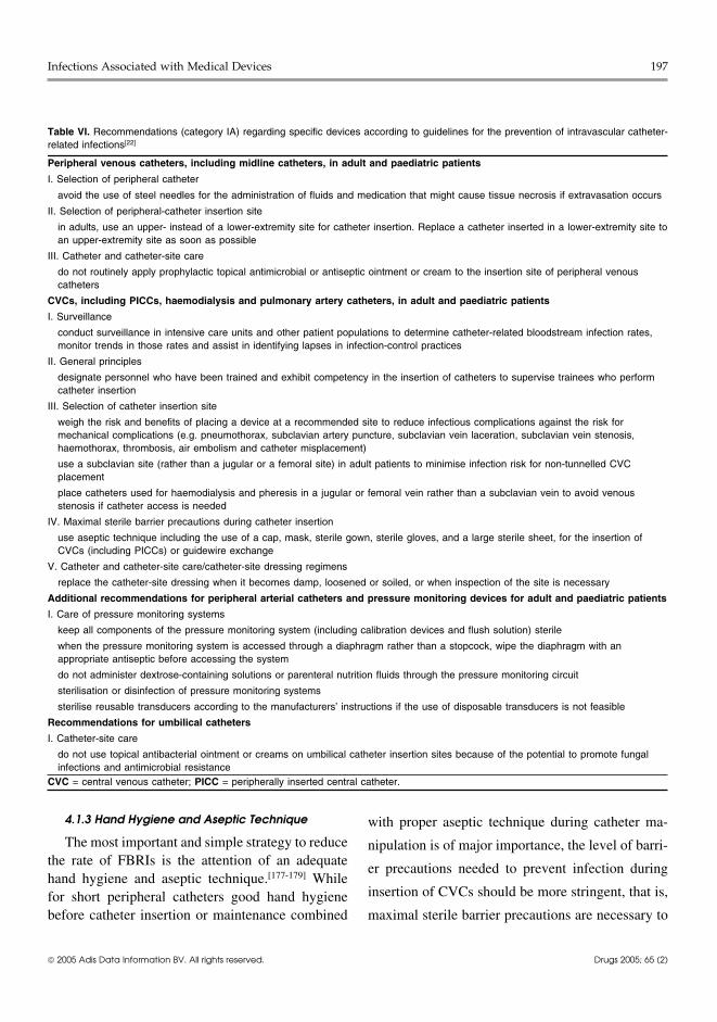
Table VI. Recommendations (category IA) regarding specific devices according to guidelines for the prevention of intravascular catheter-related infections[22]
Peripheral venous catheters, including midline catheters, in adult and paediatric patients
I. Selection of peripheral catheter
avoid the use of steel needles for the administration of fluids and medication that might cause tissue necrosis if extravasation occurs
II. Selection of peripheral-catheter insertion site
in adults, use an upper- instead of a lower-extremity site for catheter insertion. Replace a catheter inserted in a lower-extremity site toan upper-extremity site as soon as possible
III. Catheter and catheter-site care
do not routinely apply prophylactic topical antimicrobial or antiseptic ointment or cream to the insertion site of peripheral venouscatheters
CVCs, including PICCs, haemodialysis and pulmonary artery catheters, in adult and paediatric patients
I. Surveillance
conduct surveillance in intensive care units and other patient populations to determine catheter-related bloodstream infection rates,monitor trends in those rates and assist in identifying lapses in infection-control practices
II. General principles
designate personnel who have been trained and exhibit competency in the insertion of catheters to supervise trainees who performcatheter insertion
III. Selection of catheter insertion site
weigh the risk and benefits of placing a device at a recommended site to reduce infectious complications against the risk formechanical complications (e.g. pneumothorax, subclavian artery puncture, subclavian vein laceration, subclavian vein stenosis,haemothorax, thrombosis, air embolism and catheter misplacement)
use a subclavian site (rather than a jugular or a femoral site) in adult patients to minimise infection risk for non-tunnelled CVCplacement
place catheters used for haemodialysis and pheresis in a jugular or femoral vein rather than a subclavian vein to avoid venousstenosis if catheter access is needed
IV. Maximal sterile barrier precautions during catheter insertion
use aseptic technique including the use of a cap, mask, sterile gown, sterile gloves, and a large sterile sheet, for the insertion ofCVCs (including PICCs) or guidewire exchange
V. Catheter and catheter-site care/catheter-site dressing regimens
replace the catheter-site dressing when it becomes damp, loosened or soiled, or when inspection of the site is necessary
Additional recommendations for peripheral arterial catheters and pressure monitoring devices for adult and paediatric patients
I. Care of pressure monitoring systems
keep all components of the pressure monitoring system (including calibration devices and flush solution) sterile
when the pressure monitoring system is accessed through a diaphragm rather than a stopcock, wipe the diaphragm with anappropriate antiseptic before accessing the system
do not administer dextrose-containing solutions or parenteral nutrition fluids through the pressure monitoring circuit
sterilisation or disinfection of pressure monitoring systems
sterilise reusable transducers according to the manufacturers’ instructions if the use of disposable transducers is not feasible
Recommendations for umbilical catheters
I. Catheter-site care
do not use topical antibacterial ointment or creams on umbilical catheter insertion sites because of the potential to promote fungalinfections and antimicrobial resistance
CVC = central venous catheter; PICC = peripherally inserted central catheter.
4.1.3 Hand Hygiene and Aseptic Technique with proper aseptic technique during catheter ma-
The most important and simple strategy to reduce nipulation is of major importance, the level of barri-the rate of FBRIs is the attention of an adequate
er precautions needed to prevent infection duringhand hygiene and aseptic technique.[177-179] While
insertion of CVCs should be more stringent, that is,for short peripheral catheters good hand hygienebefore catheter insertion or maintenance combined maximal sterile barrier precautions are necessary to
2005 Adis Data Information BV. All rights reserved. Drugs 2005; 65 (2)
198 von Eiff et al.
reduce the incidence of CRBI in patients with al intravenous colonisation compared with povidoneiodine.[186]CVCs.
Different dressing regimens have also been com-Good hand hygiene comprises the use of either apared. In the largest controlled trial of dressingwaterless, alcohol-based product or an antibacterialregimens on 2000 peripheral catheters, the rate ofsoap and water with adequate rinsing.[178] Maximalcolonisation among catheters dressed with transpar-sterile barrier precautions should be achievedent dressings (5.7%) was shown to be comparablethrough the use of a cap, mask, sterile gown, sterilewith that of those dressed with gauze (4.6%).[187] Nogloves and a large sterile drape.[179,180] For the inser-clinically substantial differences in either the inci-tion of peripheral venous catheters, a new pair ofdences of catheter site colonisation or phlebitis weredisposable nonsterile gloves can be used in conjunc-observed. In a meta-analysis assessing studies thattion with a ‘no-touch’ technique, thus, appropriatecompared the risk for CRBIs for groups using trans-aseptic technique does not necessarily require sterileparent dressings versus groups using gauze dressing,gloves.[22]
the risk was found not to differ between theA review of data regarding handwashing and groups.[188] A chlorhexidine-impregnated sponge
hand antisepsis in healthcare settings and recom- placed over the site of short-term arterial and CVCsmendations to promote improved hand hygiene reduced the risk for catheter colonisation andpractices and reduce transmission of pathogenic CRBI.[189] Concerning catheter securement devices,micro-organisms to patients and personnel in health- a study – comparing a sutureless device with suturecare settings is given in the “Guideline for Hand for the securement of peripherally inserted centralHygiene in Health-Care Settings” by Boyce and catheters – revealed that CRBI was reduced in thePittet.[181]
group of patients who received the sutureless de-vice.[190]
4.1.4 Skin Antisepsis and Catheter SiteDressing Regimens 4.1.5 Catheter Material and In-Line Filters
The type of catheter material used is also ofMost recently, it was shown that most CRBIsimportance regarding the risk for subsequent infec-with short-term percutaneously inserted, noncuffedtions. For example, several studies showed thatCVCs were extraluminally acquired and derivedTeflon or polyurethane catheters are associatedfrom the cutaneous microflora. It was concludedwith fewer infectious complications than cathetersthat strategies achieving successful suppression ofmade of polyvinyl chloride or polyethylene.[187,191]cutaneous colonisation can substantially reduce theSteel needles have the same rate of infectious com-risk of CRBI with short-term CVCs.[182] In the past,plications as do Teflon catheters; however, theira number of different commercially available prod-use is frequently complicated by infiltration of intra-ucts for cleansing arterial catheter and CVC inser-venous fluids into the subcutaneous tissues[192] (pre-tion sites have been studied.[183-186] Preparation ofvention by material modification [section 4.2] or bycentral venous and arterial sites with 2% aqueousincorporation of antimicrobial agents [sectionchlorhexidine gluconate lowered BSI rates com-4.2.3]).pared with site preparation with 10% povidone io-
dine or 70% alcohol.[185] In another prospective, The routine use of intravenous in-line filters onrandomised study of adults, a tincture of 0.5% infusion lines has been controversial for many yearschlorhexidine was shown to be as or less effective in and is still under debate.[193,194] So far, no data havepreventing CRBI or CVC colonisation than 10% been published that support the efficacy of in-linepovidone iodine.[184] In contrast, in a study compris- filters in preventing infections associated with in-ing neonates, 0.5% chlorhexidine reduced peripher- travascular catheters and infusion systems; however,
2005 Adis Data Information BV. All rights reserved. Drugs 2005; 65 (2)
Infections Associated with Medical Devices 199
they reduce the incidence of infusion-related phlebi- pecially because it is still unknown if a specifictis.[194] While these filters may reduce the risk for adhesin (e.g. protein, polysaccharide) is genus- orinfection from contaminated infusate or proximal species-specific or merely strain-specific. There-contamination (i.e. introduced proximal to the filter) fore, most of the recently developed strategies haveor may reduce the risk for phlebitis in patients who focused on the modification of medical devices,require high doses of medication or in those in especially of catheters.whom infusion-related phlebitis has already oc-
Alteration of the material surface (e.g. of a poly-curred, no strong recommendation can be made in
meric catheter) leads to a change in specific andfavour of using in-line filters because infusate-relat-nonspecific interactions with micro-organisms.ed BSI are rare and in-line filters might becomeSuch a surface modification of polymeric medicalblocked, especially with certain solutions (e.g. dex-devices may lead to a reduced microbial adherencetran and lipids).via altered interactions with proteins and platelets.
The development of so-called antimicrobial4.2 Prevention by Material Modificationpolymers is aimed predominantly at the preventionof microbial colonisation rather than microbial ad-
4.2.1 Basic Considerations for the Prevention ofherence. Devices containing antibacterials, disinfec-Device-Related Infections Through Developmenttants or metals have been evaluated experimentallyof New Devicesor in clinical trials and are, in part, commerciallyAs microbial adherence is an essential step in theavailable and already used in clinical applications,pathogenesis of FBRI, inhibition of adherence ap-for example intravascular catheters. Destruction ofpears to be a very attractive approach for prevention.
All important steps in the pathogenesis, such as the biofilm embedding surface-adherent micro-or-adhesion, accumulation and biofilm formation, re- ganisms by enzymes or ultrasound plus subsequentpresent possible targets against which prevention antibacterial therapy as well as the electrical en-strategies may be directed (table VII). Although hancement of antibacterial penetration through bio-there is now a more detailed insight into the molecu- films[163,195-197] are all therapeutic strategies ratherlar pathogenesis of device-related infection, as out- than preventive measures.lined in section 2, this has not yet led to strategies
Therefore, it seems obvious that, because of thedirected against specific adherence mechanisms, es-particular pathogenesis of FBRI, approaches that aredirected against bacterial colonisation of a deviceare very promising. Medical devices made out of amaterial that would be antiadhesive or at leastcolonisation-resistant in vivo would be the mostsuitable candidates to avoid colonisation and subse-quent infection. In the last 15–20 years there havebeen a large number of studies dealing with thisproblem, in part using different strategies. A generaloverview is given in table VIII; most of the studieshave been performed with intravascular cathetersbecause of their widespread use. Thus, the mainfocus of this section is on the discussion of modifiedcatheter materials.
Table VII. Possible strategies directed against specific factors inthe pathogenesis of catheter-related infection
Step in Possible preventive strategypathogenesis
Adhesion Antiadhesive surfaces by polymer surfacemodification
Inhibition of specific adherence mechanisms
Antimicrobial devices?
Accumulation Inhibition of specific factors involved inaccumulation (e.g. antibodies againstpolysaccharide/adhesin, accumulation-associatedprotein)
Antimicrobial devices
Biofilm Antimicrobial devices
formation Interference with quorum sensing
Electrical current, ultrasound + antimicrobials
2005 Adis Data Information BV. All rights reserved. Drugs 2005; 65 (2)
200 von Eiff et al.
polymers that were surface-modified with poly(eth-ylene oxide). They observed reductions in adherentbacteria of between 70% and 95% compared withthe untreated polymer. A photochemical coating ofpolymers was used by Dunkirk et al.,[200] demon-strating that the coating reduced adhesion of a varie-ty of bacterial strains. Tebbs et al.[201] compared theadherence of five S. epidermidis strains to a poly-urethane catheter and to a commercial hydrophilic,coated polyurethane catheter (Hydrocath). Adhe-sion of three strains to the coated catheters wasconsiderably reduced. Bacterial colonisation wasfurther reduced by the addition of benzalkoniumchloride to a hydrophilic polyurethane catheter(Hydrocath).[202] Our own approaches to develop
Table VIII. Prevention strategies of device-related infections bymaterial modification
Catheters and devices used in modification processes
Intravascular catheters
Urinary catheters
Ventricular catheters
Continuous ambulatory peritoneal dialysis catheters
Catheter hubs
Cuffs
Dressings
Tubing systems
Process of modification
Modification of basic polymers (‘antiadhesive polymers’)
Incorporation or superficial bonding of antimicrobial substances(‘antimicrobial polymers’)
antibacterials
antiseptics
Metals with antimicrobial activityanti-infective materials comprised the modificationof polymer surfaces by radiation or glow discharge
4.2.2 Development of Antiadhesive Polymertechniques. By means of radiation grafting leadingMaterials by Physico-Chemical Modificationto a reduced in vitro adhesion of S. epidermidis,
The most attractive approach to obviate FBRIs 2-hydroxymethylmethacrylate was covalently bond-would be to develop a medical material that proves ed to a polyurethane surface.[203,204]
to be resistant against microbial adherence, evenMore recent work on surface modification ofafter insertion into the bloodstream and despite the
polymer materials to prevent bacterial adhesion in-ever-occurring interactions of the device surfacevolved the use of sulfonated poly(ethylene oxide) aswith host factors such as proteins and cells. There issurfactant in a polyurethane[205] or the introductionevidence that the intrinsic properties of a materialof glycerophosphorylcholine as a chain extender inmight be of advantage regarding resistance to infec-polyurethane.[206] Both approaches lead to increasedtion. Thus, improvement of the surface texture, tai-water uptake and to lower bacterial adhesion. Anloring the protein adsorption characteristics and im-overview mainly on experimental research on theproving the antithrombogenicity of a given materialsurface modification of polymers and on bindingwould be key factors in the development of innova-macromolecules such as albumin to surfaces in or-tive, infection-resistant materials. However, thisder to prevent bacterial adherence can be foundgoal has not yet been reached satisfactorily.elsewhere.[207,208]
Several research groups have tried to developSo far, the only such modified polymer used inpolymers with new surface properties that would
clinical applications is a hydrophilic, polyvinylpyr-lead to a reduction of bacterial adhesion. Bridgett etrolidone-coated catheter (Hydrocath) based onal.[198] studied the adherence of three isolates ofpolyurethane. Its relatively low thrombogenicityS. epidermidis to polystyrene surfaces that wereand low in vitro bacterial adherence should also bemodified with a copolymer of poly(ethylene oxide)of benefit regarding infection resistance; however,and poly(propylene oxide). In vitro, a substantialthis has not yet been demonstrated in a clinical trial.reduction in bacterial adhesion was achieved with
all surfactants tested. Similar results were found by A major disadvantage of all of the previouslyDesai et al.,[199] who investigated the adhesion of described approaches, which aim primarily at theS. epidermidis, S. aureus and P. aeruginosa to modification of the surface properties of basic
2005 Adis Data Information BV. All rights reserved. Drugs 2005; 65 (2)
Infections Associated with Medical Devices 201
materials such as catheters or other devices, is the bound superficially to a catheter – either directly orfact that – for thermodynamic reasons – the creation by means of a carrier – or incorporated into theof surfaces that show a ‘zero’ adhesion is probably interior of the polymer. If such a device comes intonot feasible. In an experimental study that investi- contact with an aqueous environment, release of thegated the relationship between bacterial adhesion drug into the near vicinity occurs. The amount of theand the free surface enthalpy of adhesion of a large antimicrobial substance released is influenced bynumber of differently modified polymers, we dem- the processing parameters, loading dose, appliedonstrated that it is impossible to develop a polymer technique, molecular size of the drug and thesurface that shows an absolute bacterial ‘zero’ ad- physico-chemical properties of the polymeric de-herence in vitro.[209] In particular, adherence of vice. A high antimicrobial concentration is reachedS. epidermidis to a variety of polymers with differ- (at least initially) in the very near vicinity of theent surface properties, generated by means of the device surface, mostly exceeding the MIC and mini-glow discharge technique, was investigated.[209] We mum bactericidal concentration of susceptible or-found that adhesion of the bacteria to the modified ganisms. Most such materials exhibit a release pat-materials decreased with increasing negative free tern according to first-order kinetics, with an initial-enthalpy values. A certain minimum number of ad- ly high drug release and subsequent exponentialherent S. epidermidis cells could be proved at posi- decrease of the released drug; however, more so-tive free enthalpy values at which adhesion should phisticated drug-release systems with defined re-be thermodynamically impeded. Hence, it seems lease kinetics have also been developed.impossible to design an absolute antiadhesive mate- As yet, it is unclear whether such a device isrial that retains its properties even in the more com- capable of inhibition of microbial adherence per se,plex in vivo situation, in which the native surface or if its activity is more directed against colonisa-properties are masked by adsorption of bacterial and tion; however, at least an elimination of alreadyhost components. adherent micro-organisms should be achieved for
the duration that the antimicrobial compound is re-4.2.3 Incorporation of Antimicrobial Agents in leased in the necessary concentrations. Thus, suchMedical Devices materials are especially suitable to prevent, for ex-The loading of medical polymers with anti- ample, infection in short-term catheters, which
microbial substances either for therapeutic or pre- originates from contamination during the insertionventive purposes has a long tradition. The best or from hub contamination.known anti-infective, polymeric drug delivery sys-
Antibacterialstems are the polymethylmethacrylate-gentamicinbone cement and the polymethylmethacrylate- There are a large number of studies on the bond-gentamicin beads (Septopal) used for treatment of ing of antibacterials to biomaterials. Solovskij etbone and soft tissue infections.[210,211] Vascular pros- al.[215] prepared polymers to which ampicillin andtheses made from Dacron have been treated with 6-aminopenicillanic acid were covalently bondedvarious antibacterial agents to create infection-resis- and which inhibited the in vitro growth of S. aureus.tant grafts but without routine clinical application to However, most studies have focused on the incorpo-date.[212-214] In recent years, catheters or parts of the ration or superficial coating of antimicrobials rathercatheter system have been coated with antimicrobial than on covalent bonding by chemical reaction.drugs, and some of these antimicrobial devices are Sherertz et al.[216] used a rabbit model to investigatealready commercially available. The main principle intravascular catheters coated with several anti-of such devices is that an antimicrobial substance microbial substances (dicloxacillin, clindamycin,(e.g. an antibacterial, disinfectant or metal ion) is fusidic acid and chlorhexidine). The frequency of
2005 Adis Data Information BV. All rights reserved. Drugs 2005; 65 (2)
202 von Eiff et al.
catheter infections was significantly reduced com- spective, randomised clinical trial,[224] the mino-cycline-rifampicin catheter was compared with anpared with the control group when the dicloxacillin-uncoated control catheter and demonstrated a statis-coated catheter was used. We have investigated thetically significant decrease in catheter colonisationincorporation of flucloxacillin, clindamycin and(8% vs 26% for the control catheter, p < 0.001) andciprofloxacin into polyurethane polymers and dem-in CRBI (0% vs 5%, p < 0.01). In a large multicentreonstrated a considerable reduction of the in vitrotrial, the minocycline-rifampicin catheter was com-adherence of S. epidermidis.[204,217] In another ap-pared with another commercially available catheterproach, a commercially available central venous,containing chlorhexidine and silver sulfadiazine (thehydrophilic-coated polyurethane catheter (Hydro-CHSS catheter), which is described further in thecath) was loaded with the glycopeptide teico-Antiseptics section.[225] It was found that the mino-planin.[218] In in vitro studies as well as in a mousecycline-rifampicin catheter was 3-fold less likely tomodel, the capability of this catheter to preventbe colonised (7.8% vs 22.6% for the CHSS catheter,colonisation with S. epidermidis and S. aureus, re-p < 0.0001) and 12-fold less likely to lead to CRBIspectively, was proven for a period of at least 48(0.3% vs 3.4%, p < 0.002). This difference has beenhours, rendering the catheter suitable to preventexplained by the fact that minocycline-rifampicinearly-onset infection.[218,219] To extend the anti-catheters are coated internally and externally (inmicrobial spectrum of such a catheter to includecontrast with the first-generation CHSS catheter),Gram-negative bacteria and fungi, a combination ofthe combination of minocycline and rifampicinteicoplanin with silver was incorporated into Hydro-showing superior surface activity than chlorhexidinecath catheters, which exhibited considerable ac-and, finally, that the minocycline-rifampicin cathe-tivity against S. epidermidis, E. coli and C. albi-ters retain surface antimicrobial activity longer incans.[209]
situ.[226] Although resistance against minocyclineKamal et al.[220] have evaluated the efficacy of a
and rifampicin could not be detected in clinicalcefazolin-containing catheter (in which cefazolin
trials, this remains of concern as in vitro develop-was bound to benzalkonium chloride) in a prospec-
ment of resistance has been demonstrated.[227]tive, randomised trial. There was a significant de-
More recent work on antibacterial-containingcrease in catheter colonisation (7-fold) as deter-catheters included, for example, the adsorption ofmined by the semiquantitative tip culture method;[15]
cefamandole nafate on functionalised urethane cath-no CRBI was observed in this study. In a moreeters that were then used to coat a commercialrecent comparative study before and after the rou-CVC[228] or the use of a combination of an antibac-
tine use of cefazolin catheters in the ICU, the au-terial substance (rifampicin) in combination with an
thors described a marked reduction in the rate ofantifungal substance (miconazole) in a polyurethane
CRBI from 11.5 to 5.1 infections per 1000 cathetercatheter.[229]
days.[221]
A disadvantage of all of these approaches mightRaad et al.[222,223] reported on the broad-spectrum result from the risk for development of resistance
activity against Gram-negative and -positive organ- against the antimicrobial agents, especially if anti-isms and C. albicans of a minocycline-rifampicin bacterials considered as first-line drugs in the ther-catheter based on in vitro and animal data. This apy of infections are used as an active part of thecatheter has been marketed as the Cook Spectrum modified catheters.catheter (Cook Critical Care, Bloomington, IN,
AntisepticsUSA) and is coated on the inner and outer surfacewith minocycline and rifampicin, which have a syn- Antimicrobial substances that differ from anti-ergistic or additive action in combination. In a pro- bacterials, such as antiseptics, have also been used
2005 Adis Data Information BV. All rights reserved. Drugs 2005; 65 (2)
Infections Associated with Medical Devices 203
to develop new catheter materials. The disinfectant colonisation of 0.44 (95% CI 0.36, 0.54; p < 0.001)and of 0.56 for CRBI (95% CI 0.37, 0.84;Irgasan was incorporated into several polymerp = 0.005). From an analysis of six prospective stud-catheters, showing a reduction of infections in rab-ies, Mermel[2] also concluded that short-term use ofbits.[230] We used the hydrophilic Hydrocath cathe-CHSS catheters reduces the risk for catheter-relatedter to incorporate iodine, leading to a polyvinylpyr-BSI. In one study with longer catheter dwellingrolidone-iodine-complex on the inner and outertimes (mean duration 20 days), no such difference incatheter surface.[218] In vitro adherence of variousthe incidence of CRBI was observed, which prob-micro-organisms (Staphylococcus spp., E. coli,ably reflects less antimicrobial efficacy over timeCandida spp., Pseudomonas spp.) was completelydue to a loss of activity to 25% of the baseline valueinhibited for the time of iodine release. After iodineafter 10 days in situ.[237] As the ‘first-generation’exhaustion, re-loading of the catheter was possible.CHSS catheters are coated only externally, colonisa-Tebbs and Elliott[202] incorporated benzalkoniumtion of the inner lumen as a result of hub contamina-chloride into triple-lumen Hydrocath catheters andtion might also be of greater relevance with longerdemonstrated a long-lasting antimicrobial activity ofduration of placement. For these reasons, a newthe catheters against staphylococci and a somewhatsecond-generation CHSS catheter has recently beenlesser activity against Gram-negative bacteria anddeveloped that is coated both internally and exter-C. albicans.nally, and that exhibits enhanced chlorhexidine ac-
The most promising development in this field intivity (ArrowGard Plus, Arrow International, Read-
the last few years was a catheter using a combinationing, PA, USA). Clinical trials with this new type of
of an antiseptic (chlorhexidine) and silver sulfadia- catheter are currently being performed. In one al-zine (CHSS catheter). This catheter became avail- ready published study, a significant reduction inable ~10 years ago, is polyurethane-based and im- catheter colonisation and a trend to reduction ofpregnated with minute amounts of chlorhexidine infection episodes was associated with the anti-and silver sulfadiazine (ArrowGard, Arrow Interna- microbial catheter.[247]
tional, Reading, PA, USA). A synergistic effect of Development of resistance to chlorhexidine haschlorhexidine and sulfadiazine has been shown in been demonstrated in vitro.[248] However, in vivovitro.[231] This ‘first-generation’ CHSS catheter is resistance to either chlorhexidine or silver sulfadia-coated only on the exterior surface and exhibits zine associated with the use of the antimicrobialantimicrobial properties for ~15 days. Since its in- catheter has not yet been observed. Anaphylactoidtroduction >8 million catheters have been sold reactions, probably due to chlorhexidine, have beenworldwide and a considerable number of random- reported from Japan and UK, but have not beenised clinical trials have been performed with this observed in the US so far.[249]
type of catheter.[173,232-241] In the study with theMetalsgreatest patient numbers, which also used molecular
methods for the confirmation of CRBI, the CHSS Among metals with antimicrobial activity, silvercatheter was associated with a 2-fold reduction in has raised the interest of many investigators because
of its good antimicrobial action and low toxicity.[250]the incidence of catheter colonisation and a 5-foldSilver has also extensively been used for the devel-reduction of CRBI (RR 0.21, 95% CI 0.03, 0.95;opment of infection-resistant urinary catheters.p = 0.03).[238] As of 2004, seven meta-analyses or
systematic reviews have been published.[2,226,242-246] Sioshansi[251] used ion implantation to depositVeenstra et al.[243] investigated randomised clinical silver-based coatings on a silicone rubber, whichtrials with CHSS versus control catheters up to 1998 thereafter demonstrated antimicrobial activity. Sil-and found summary odds ratios for catheter ver-copper surface films, sputter-coated onto cathe-
2005 Adis Data Information BV. All rights reserved. Drugs 2005; 65 (2)
204 von Eiff et al.
ter materials, also showed antibacterial activity Several clinical studies have been performedwith silver-containing intravascular catheters. In aagainst P. aeruginosa biofilm formation.[252] In arandomised, prospective study in haemato-onco-more recent piece of research, an ion beam tech-logical patients, a silver sulfate-polyurethane cathe-nique applying low implantation energy has beenter (Fresenius AG, Bad Homburg, Germany UK)used for the formation of silver nano-particles on thewas associated with a significantly lower rate ofsurface of polymers that exhibited an improved ef-CRBI compared with the control group (10.2% vsfect on bacterial adhesion.[253] We developed an22.5%, p = 0.01).[260] In three trials, the ‘Erlanger’antimicrobial polymer by binding silver ions to acid-silver catheter in which the silver is microdispersedmodified, negatively charged polyurethane sur-was evaluated.[256,261,262] In the adult population, aface.[209] Another approach is loading of a hydro-reduction in catheter colonisation and in ‘catheter-
philic polyurethane catheter with silver nitrate.[254]
associated sepsis’ was observed; however, the au-In addition, surface-coated polyurethane catheters
thors used criteria for determining CRBI that differ-with a silver surface thickness of 15–20A have been ed from most other studies. A more recent clinicalinvestigated with regards to their biocompatibil- investigation failed to show a statistically significantity and antimicrobial efficacy, showing markedly difference in the colonisation rate of the silver cathe-decreased adherence of Gram-positive and -negative ter compared with a control catheter.[262] Ranucci etmicro-organisms in vitro.[255] Further interest has al.[257] compared the Oligon Vantex catheter, com-been raised regarding devices in which silver is posed of silver, carbon and platinum with adistributed in form of nano-particles or in combina- benzalkonium chloride-treated catheter (Multi-Med,tion with other elements such as carbon and plati- Edwards Life Sciences, Irvine, CA, USA) in a pro-num. The ‘Erlanger’ silver catheter uses a microdis- spective randomised trial. Use of the Oligonpersed silver technology to increase the quantity of Vantex catheter decreased the rate of catheter
colonisation by 11%, while the rate for CRBI did notavailable ionised silver.[256] The ‘Oligon’ cathetersdiffer significantly between the Oligon Vantex andare composed of polyurethane in which carbon,control groups.silver and platinum particles are incorporated, which
leads to an electrochemically driven release of silver4.2.4 Neurological Prostheses
ions in the outer and inner vicinity of the catheterInfection of CNS (hydrocephalus) shunts is a
surface. However, a peripherally implanted centralmajor problem in patients with ventricular drainage.
catheter based on this technology (Olimpicc,Therefore, efforts have also been made to develop
Vygon, Cirencester, UK) has been withdrawn frominfection-resistant hydrocephalus shunts or other
the market at least in Germany because of mechani- neurological prostheses. Bridgett et al.[263] reportedcal problems associated with this type of catheter. A on the reduced staphylococcal adherence tomore recent development is the Oligon Vantex
Hydromer-coated and, thus, hydrophilic CSFcatheter (Edwards Life Sciences, Irvine, CA, shunts; however, there were technical difficulties inUSA).[257] Other approaches are catheters with ‘ac- achieving a uniform Hydromer layer on the sili-tive iontophoresis’ technology in which micro-or- cone rubber.[263] Bayston et al.[264-267] have publishedganisms are repelled by electrical current generated a considerable amount of experimental work onfrom a carbon-impregnated catheter[258] or where impregnation of silicone shunt catheters with vari-low-amperage current is produced by two electrical- ous antimicrobials. In particular, a combination ofly charged parallel silver wires helically wrapped rifampicin and clindamycin proved to be clearlyaround the proximal segment of silicone cathe- superior to other agents tested. In a newer study, itters.[259] was shown in vitro that the rifampicin-clindamycin-
2005 Adis Data Information BV. All rights reserved. Drugs 2005; 65 (2)
Infections Associated with Medical Devices 205
4.2.5 Innovative Approaches for the Prevention ofimpregnated catheters are able to kill adheredDevice-Related Infectionsstaphylococci completely within 48–52A very interesting approach to prevent biofilm
hours.[268,269]
formation as a prerequisite for CRI has been sug-We have also developed an incorporation method gested by Khoury et al.[196] They found that by
for rifampicin and other hydrophobic antibacterials application of an external stimulus, for example aninto silicone ventricular catheters.[270] In an animal electric field together with antibacterials, the killing
of biofilm-embedded bacteria is dramatically en-model using New Zealand white rabbits, rifampicin-hanced (e.g. killing of P. aeruginosa by to-loaded catheters were implanted into the ventricularbramycin). Although it is more of a therapeuticspace and infection was induced by inoculation ofstrategy than a preventive measure, the use of ancertain dosages of S. epidermidis or S. aureus.[271]
electric current might be useful for the prevention ofNone of the animals that received the rifampicin-CRI, as was previously pointed out. Related ap-
loaded catheter showed clinical signs of infection,proaches aimed at eradication of biofilms include
nor could the infecting strain be recovered from thethe combined use of ultrasound together with anti-
catheter, brain tissue or CSF. In contrast, all animals bacterials[163,197] and, possibly, the bactericidal ef-with the uncoated catheters showed signs of severe fect of extracorporeal shock waves on S. aureus.[276]
meningitis or ventriculitis, and the infecting strains Another approach to create anti-infective sur-were cultivated in each case from the catheter and faces has been developed on the basis of so-calledfrom surrounding tissue. As an improvement of the ‘intelligent polymers’. Because a disadvantage of
the currently available antimicrobial catheters is thecatheter, especially to prevent development of resis-continuous leaching out of active substances oncetance of staphylococci to rifampicin, a combinationthe device is in contact with blood (or other bodyof rifampicin and trimethoprim was used for thefluids), a system was developed in which an anti-impregnation process.[272] Recently, two cases weremicrobial compound is released on demand. Thisreported in which the rifampicin catheter was suc-could be achieved by binding iron to a chemically
cessfully used for the treatment of patients with amodified polymer surface; then in a second step, a
complicated course of shunt infection.[273]fluoroquinolone was coupled. In vitro experiments
Furthermore, a silicone catheter with a combina- with P. aeruginosa demonstrated liberation of thetion of three antimicrobials (rifampicin, fusidic acid antibacterial only in the presence of the bacteria.[277]
and mupirocin) has been described with a long-lasting drug release of up to ~100 days; however, no 5. Conclusionanimal or clinical data are available so far for this
The increasing use of indwelling foreign bodiestype of catheter.[274] Zabramski et al.[275] performedhas become essential in modern day clinical prac-a prospective, randomised clinical trial with an ex-tice; however, their use is associated with a multi-ternal ventricular drain catheter coated with mino-tude of complications, the most common being in-
cycline and rifampicin. The antibacterial-impregnat-fection. Nowadays, it is generally accepted that the
ed catheters were one-half as likely to become consequences of FRBIs are substantial, both incolonised as the control catheters (17.9% vs 36.7%, terms of morbidity and mortality as well as in termsp < 0.0012), and CSF cultures were seven times less of financial resources expended. The causativefrequently positive in patients with the modified pathogens attach to surfaces of the medical devicescatheters than in the control group (1.3% vs 9.4%, and develop biofilms, leading to an evasion of hostp = 0.002). defence mechanisms and to a phenotypic resistance
2005 Adis Data Information BV. All rights reserved. Drugs 2005; 65 (2)
206 von Eiff et al.
to antimicrobial agents. Thus, conservative manage- removal of, in particular, highly needed and/or diffi-cult to replace medical devices.ment of FBRIs is frequently unsuccessful and can be
dangerous for the patient. Antimicrobial agents haveAcknowledgementsa role in preventing medical device-related compli-
cations, such as metastatic sepsis, yet in most situa- This work was supported by grants from BMBF (Pathoge-nomic Network) and from the Interdisciplinary Clinical Re-tions it is impossible to eradicate the primary focussearch Center (project Hei2/042/04). The authors have noof infection on the implant. In the light of theconflicts of interest that are directly relevant to the content ofemergence of multiresistant micro-organisms, suchthis review.
as meticillin-resistant staphylococci with reducedsusceptibility or with resistance towards glycopep- Referencestides, prudent use of antimicrobials is advised. Inno- 1. NNIS System. National Nosocomial Infections Surveillance
(NNIS) System Report, data summary from January 1992vative and multidisciplinary approaches for prophy-through June 2003, issued August 2003. Am J Infect Control
laxis and management should result in novel, more 2003 Dec; 31 (8): 481-982. Mermel LA. Prevention of intravascular catheter-related infec-effective control strategies.
tions. Ann Intern Med 2000 Mar 7; 132 (5): 391-402In the future, the development of medical devices 3. Mermel LA. Defining intravascular catheter-related infections:
a plea for uniformity. Nutrition 1997 Apr; 13 (4 Suppl.): 2S-4Sbased on modified anti-infective materials will lead4. Samore MH, Burke JP. Infections of long intravenous lines: new
to a further reduction of the incidence of FRBIs. developments and controversies. Curr Clin Top Infect Dis2000; 20: 256-70However, even the best technology will fail if
5. Sitges-Serra A, Girvent M. Catheter-related bloodstream infec-standard hygienic procedures, with their often easy-tions. World J Surg 1999 Jun; 23 (6): 589-95
to-perform preventive techniques based on recom- 6. Garner JS, Jarvis WR, Emori TG, et al. CDC definitions fornosocomial infections, 1988. Am J Infect Control 1988 Jun; 16mendations of the respective national guidelines, are(3): 128-40
not implemented. 7. Eggimann P, Pittet D. Overview of catheter-related infectionswith special emphasis on prevention based on educationalSome of the new developments, such as anti- programs. Clin Microbiol Infect 2002 May; 8 (5): 295-309
microbial catheters, have already been adopted, but 8. Pearson ML. Guideline for prevention of intravascular device-related infections: the Hospital Infection Control Practicesmore, good quality clinical studies are needed toAdvisory Committee. Am J Infect Control 1996 Aug; 24 (4):
better define their impact on reducing FRBIs, pa- 262-939. Bouza E, Burillo A, Munoz P. Catheter-related infections: diag-tient morbidity and mortality, and their cost effec-
nosis and intravascular treatment. Clin Microbiol Infect 2002tiveness. These studies are needed before recom- May; 8 (5): 265-74
10. Hodge D, Puntis JW. Diagnosis, prevention, and management ofmending a broader use of these devices. Althoughcatheter related bloodstream infection during long term paren-antimicrobial materials obviously have the potentialteral nutrition. Arch Dis Child Fetal Neonatal Ed 2002 Jul; 87
to decrease infections, there is a major point of (1): F21-411. Raad II, Hanna HA. Intravascular catheter-related infections:criticism/concern: that of development of anti-
new horizons and recent advances. Arch Intern Med 2002 Aprmicrobial resistance against the agents used. This 22; 162 (8): 871-8
12. Kristinsson KG. Diagnosis of catheter-related infections. In:should still be carefully monitored when using suchSeifert H, Jansen B, Farr BM, editors. Catheter-related infec-
devices and should be an important issue in forth- tions. New York: Marcel Dekker Inc., 1997: 31-5713. von Eiff C, Peters G. Pathogenesis and detection of biofilmcoming clinical studies.
formation on medical implants. In: Jass J, Surman S, Walker J,The most important challenge will be to imple- editors. Medical biofilms: detection, prevention and control.
Chichester: Wiley, 2003: 51-72ment all of the current knowledge in daily practice.14. Sherertz RJ, Raad II, Belani A, et al. Three-year experience
In addition, translating the recent data on the mech- with sonicated vascular catheter cultures in a clinical microbi-ology laboratory. J Clin Microbiol 1990 Jan; 28 (1): 76-82anisms of biofilm formation and bacterial inter-
15. Maki DG, Weise CE, Sarafin HW. A semiquantitative cultureference into applicable strategies and innovative method for identifying intravenous-catheter-related infection.materials may avoid the unnecessary and expensive N Engl J Med 1977 Jun 9; 296 (23): 1305-9
2005 Adis Data Information BV. All rights reserved. Drugs 2005; 65 (2)
Infections Associated with Medical Devices 207
16. Siegman-Igra Y, Anglim AM, Shapiro DE, et al. Diagnosis of 33. Shiro H, Muller E, Gutierrez N, et al. Transposon mutants ofvascular catheter-related bloodstream infection: a meta-analy- Staphylococcus epidermidis deficient in elaboration of capsu-sis. J Clin Microbiol 1997 Apr; 35 (4): 928-36 lar polysaccharide/adhesin and slime are avirulent in a rabbit
model of endocarditis. J Infect Dis 1994 May; 169 (5): 1042-917. Blot F, Nitenberg G, Chachaty E, et al. Diagnosis of catheter-related bacteraemia: a prospective comparison of the time to 34. Herrmann M, Vaudaux PE, Pittet D, et al. Fibronectin, fibrino-positivity of hub-blood versus peripheral-blood cultures. Lan- gen, and laminin act as mediators of adherence of clinicalcet 1999 Sep 25; 354 (9184): 1071-7 staphylococcal isolates to foreign material. J Infect Dis 1988
18. Safdar N, Kluger DM, Maki DG. A review of risk factors for Oct; 158 (4): 693-701catheter-related bloodstream infection caused by percutan- 35. Herrmann M, Hartleib J, Kehrel B, et al. Interaction of voneously inserted, noncuffed central venous catheters: implica- Willebrand factor with Staphylococcus aureus. J Infect Distions for preventive strategies. Medicine (Baltimore) 2002 1997 Oct; 176 (4): 984-91Nov; 81 (6): 466-79 36. Dickinson GM, Bisno AL. Infections associated with indwelling
19. Rello J, Ochagavia A, Sabanes E, et al. Evaluation of outcome devices: concepts of pathogenesis; infections associated withof intravenous catheter-related infections in critically ill pa- intravascular devices. Antimicrob Agents Chemother 1989tients. Am J Respir Crit Care Med 2000 Sep; 162 (3 Pt 1): May; 33 (5): 597-6011027-30 37. Hussain M, Heilmann C, Peters G, et al. Teichoic acid enhances
20. Dimick JB, Pelz RK, Consunji R, et al. Increased resource use adhesion of Staphylococcus epidermidis to immobilizedassociated with catheter-related bloodstream infection in the fibronectin. Microb Pathog 2001 Dec; 31 (6): 261-70surgical intensive care unit. Arch Surg 2001 Feb; 136 (2): 229- 38. Mack D, Fischer W, Krokotsch A, et al. The intercellular34 adhesin involved in biofilm accumulation of Staphylococcus
21. Mermel LA. Correction: catheter catheter-related blood stream epidermidis is a linear beta-1,6-linked glucosaminoglycan:infections. Ann Intern Med 2000; 133 (5): 395 purification and structural analysis. J Bacteriol 1996 Jan; 178
22. O’Grady NP, Alexander M, Dellinger EP, et al. Guidelines for (1): 175-83the prevention of intravascular catheter-related infections:
39. Heilmann C, Schweitzer O, Gerke C, et al. Molecular basis ofCenters for Disease Control and Prevention. MMWR Recomm
intercellular adhesion in the biofilm-forming StaphylococcusRep 2002 Aug 9; 51 (RR-10): 1-29
epidermidis. Mol Microbiol 1996 Jun; 20 (5): 1083-9123. Donlan RM. Biofilms and device-associated infections. Emerg
40. Gerke C, Kraft A, Suβmuth R, et al. Characterization of the N-Infect Dis 2001 Mar; 7 (2): 277-81acetylglucosaminyltransferase activity involved in the biosyn-
24. Hugonnet S, Sax H, Eggimann P, et al. Nosocomial blood-thesis of the Staphylococcus epidermidis polysaccharide inter-
stream infection and clinical sepsis. Emerg Infect Dis 2004; 10cellular adhesin. J Biol Chem 1998 Jul 17; 273 (29): 18586-93
(1): 76-8141. Mack D, Rohde H, Dobinsky S, et al. Identification of three25. von Eiff C, Peters G, Heilmann C. Pathogenesis of infections
essential regulatory gene loci governing expression ofdue to coagulase-negative staphylococci. Lancet Infect DisStaphylococcus epidermidis polysaccharide intercellular ad-2002 Nov; 2 (11): 677-85hesin and biofilm formation. Infect Immun 2000 Jul; 68 (7):
26. Peters G, Locci R, Pulverer G. Adherence and growth of coagu-3799-807
lase-negative staphylococci on surfaces of intravenous cathe-42. Rupp ME, Ulphani JS, Fey PD, et al. Characterization ofters. J Infect Dis 1982 Oct; 146 (4): 479-82
Staphylococcus epidermidis polysaccharide intercellular ad-27. Dickinson GM, Bisno AL. Infections associated with indwellinghesin/hemagglutinin in the pathogenesis of intravascular cath-devices: infections related to extravascular devices. Antimi-eter-associated infection in a rat model. Infect Immun 1999crob Agents Chemother 1989 May; 33 (5): 602-7May; 67 (5): 2656-9
28. Veenstra GJ, Cremers FF, van Dijk H, et al. Ultrastructural43. Rupp ME, Ulphani JS, Fey PD, et al. Characterization of theorganization and regulation of a biomaterial adhesin of
importance of polysaccharide intercellular adhesin/hemagglu-Staphylococcus epidermidis. J Bacteriol 1996 Jan; 178 (2):tinin of Staphylococcus epidermidis in the pathogenesis of537-41biomaterial-based infection in a mouse foreign body infection29. Heilmann C, Hussain M, Peters G, et al. Evidence for autolysin-model. Infect Immun 1999 May; 67 (5): 2627-32mediated primary attachment of Staphylococcus epidermidis
44. Ziebuhr W, Heilmann C, Gotz F, et al. Detection of theto a polystyrene surface. Mol Microbiol 1997 Jun; 24 (5):intercellular adhesion gene cluster (ica) and phase variation in1013-24Staphylococcus epidermidis blood culture strains and mucosal30. Cucarella C, Solano C, Valle J, et al. Bap, a Staphylococcusisolates. Infect Immun 1997 Mar; 65 (3): 890-6aureus surface protein involved in biofilm formation. J
45. Dobinsky S, Kiel K, Rohde H, et al. Glucose-related dissocia-Bacteriol 2001 May; 183 (9): 2888-96tion between icaADBC transcription and biofilm expression31. Tormo MA, Cucarella C, Amorena B, et al. The Bap homologby Staphylococcus epidermidis: evidence for an additionalprotein of Staphylococcus epidermis RP62A promotes biofilmfactor required for polysaccharide intercellular adhesin synthe-formation. EMBL accession number AY028618 [online].sis. J Bacteriol 2003 May; 185 (9): 2879-86Available from URL: http://www.ncbi.nlm.nih.gov/entrez/
46. Hussain M, Herrmann M, von Eiff C, et al. A 140-kilodaltonviewer.fcgi?.db=nucleotide&val=13661836 [Accessed 2004extracellular protein is essential for the accumulation ofOct 4]Staphylococcus epidermidis strains on surfaces. Infect Immun32. Muller E, Hubner J, Gutierrez N, et al. Isolation and characteri-1997 Feb; 65 (2): 519-24zation of transposon mutants of Staphylococcus epidermidis
deficient in capsular polysaccharide/adhesin and slime. Infect 47. Peters G, Locci R, Pulverer G. Microbial colonization of pros-Immun 1993 Feb; 61 (2): 551-8 thetic devices. II: scanning electron microscopy of naturally
2005 Adis Data Information BV. All rights reserved. Drugs 2005; 65 (2)
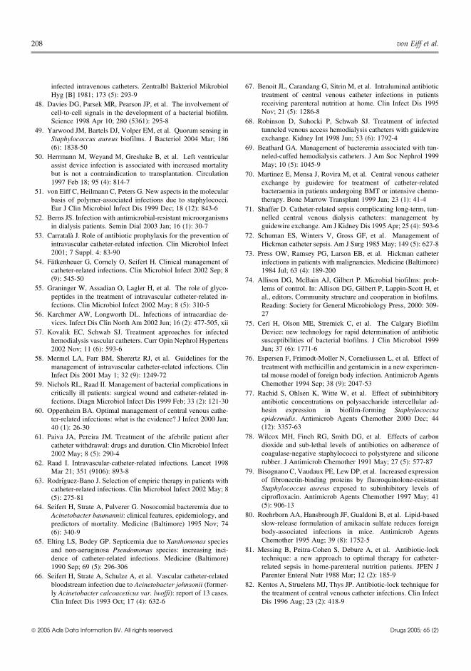
208 von Eiff et al.
infected intravenous catheters. Zentralbl Bakteriol Mikrobiol 67. Benoit JL, Carandang G, Sitrin M, et al. Intraluminal antibioticHyg [B] 1981; 173 (5): 293-9 treatment of central venous catheter infections in patients
receiving parenteral nutrition at home. Clin Infect Dis 199548. Davies DG, Parsek MR, Pearson JP, et al. The involvement ofNov; 21 (5): 1286-8cell-to-cell signals in the development of a bacterial biofilm.
Science 1998 Apr 10; 280 (5361): 295-8 68. Robinson D, Suhocki P, Schwab SJ. Treatment of infectedtunneled venous access hemodialysis catheters with guidewire49. Yarwood JM, Bartels DJ, Volper EM, et al. Quorum sensing inexchange. Kidney Int 1998 Jun; 53 (6): 1792-4Staphylococcus aureus biofilms. J Bacteriol 2004 Mar; 186
(6): 1838-50 69. Beathard GA. Management of bacteremia associated with tun-neled-cuffed hemodialysis catheters. J Am Soc Nephrol 199950. Herrmann M, Weyand M, Greshake B, et al. Left ventricularMay; 10 (5): 1045-9assist device infection is associated with increased mortality
but is not a contraindication to transplantation. Circulation 70. Martinez E, Mensa J, Rovira M, et al. Central venous catheter1997 Feb 18; 95 (4): 814-7 exchange by guidewire for treatment of catheter-related
bacteraemia in patients undergoing BMT or intensive chemo-51. von Eiff C, Heilmann C, Peters G. New aspects in the moleculartherapy. Bone Marrow Transplant 1999 Jan; 23 (1): 41-4basis of polymer-associated infections due to staphylococci.
Eur J Clin Microbiol Infect Dis 1999 Dec; 18 (12): 843-6 71. Shaffer D. Catheter-related sepsis complicating long-term, tun-nelled central venous dialysis catheters: management by52. Berns JS. Infection with antimicrobial-resistant microorganismsguidewire exchange. Am J Kidney Dis 1995 Apr; 25 (4): 593-6in dialysis patients. Semin Dial 2003 Jan; 16 (1): 30-7
53. Carratala J. Role of antibiotic prophylaxis for the prevention of 72. Schuman ES, Winters V, Gross GF, et al. Management ofintravascular catheter-related infection. Clin Microbiol Infect Hickman catheter sepsis. Am J Surg 1985 May; 149 (5): 627-82001; 7 Suppl. 4: 83-90 73. Press OW, Ramsey PG, Larson EB, et al. Hickman catheter
54. Fatkenheuer G, Cornely O, Seifert H. Clinical management of infections in patients with malignancies. Medicine (Baltimore)catheter-related infections. Clin Microbiol Infect 2002 Sep; 8 1984 Jul; 63 (4): 189-200(9): 545-50 74. Allison DG, McBain AJ, Gilbert P. Microbial biofilms: prob-
55. Graninger W, Assadian O, Lagler H, et al. The role of glyco- lems of control. In: Allison DG, Gilbert P, Lappin-Scott H, etpeptides in the treatment of intravascular catheter-related in- al., editors. Community structure and cooperation in biofilms.fections. Clin Microbiol Infect 2002 May; 8 (5): 310-5 Reading: Society for General Microbiology Press, 2000: 309-
2756. Karchmer AW, Longworth DL. Infections of intracardiac de-vices. Infect Dis Clin North Am 2002 Jun; 16 (2): 477-505, xii 75. Ceri H, Olson ME, Stremick C, et al. The Calgary Biofilm
Device: new technology for rapid determination of antibiotic57. Kovalik EC, Schwab SJ. Treatment approaches for infectedsusceptibilities of bacterial biofilms. J Clin Microbiol 1999hemodialysis vascular catheters. Curr Opin Nephrol HypertensJun; 37 (6): 1771-62002 Nov; 11 (6): 593-6
58. Mermel LA, Farr BM, Sherertz RJ, et al. Guidelines for the 76. Espersen F, Frimodt-Moller N, Corneliussen L, et al. Effect ofmanagement of intravascular catheter-related infections. Clin treatment with methicillin and gentamicin in a new experimen-Infect Dis 2001 May 1; 32 (9): 1249-72 tal mouse model of foreign body infection. Antimicrob Agents
Chemother 1994 Sep; 38 (9): 2047-5359. Nichols RL, Raad II. Management of bacterial complications incritically ill patients: surgical wound and catheter-related in- 77. Rachid S, Ohlsen K, Witte W, et al. Effect of subinhibitoryfections. Diagn Microbiol Infect Dis 1999 Feb; 33 (2): 121-30 antibiotic concentrations on polysaccharide intercellular ad-
hesin expression in biofilm-forming Staphylococcus60. Oppenheim BA. Optimal management of central venous cathe-epidermidis. Antimicrob Agents Chemother 2000 Dec; 44ter-related infections: what is the evidence? J Infect 2000 Jan;(12): 3357-6340 (1): 26-30
78. Wilcox MH, Finch RG, Smith DG, et al. Effects of carbon61. Paiva JA, Pereira JM. Treatment of the afebrile patient afterdioxide and sub-lethal levels of antibiotics on adherence ofcatheter withdrawal: drugs and duration. Clin Microbiol Infectcoagulase-negative staphylococci to polystyrene and silicone2002 May; 8 (5): 290-4rubber. J Antimicrob Chemother 1991 May; 27 (5): 577-8762. Raad I. Intravascular-catheter-related infections. Lancet 1998
Mar 21; 351 (9106): 893-8 79. Bisognano C, Vaudaux PE, Lew DP, et al. Increased expressionof fibronectin-binding proteins by fluoroquinolone-resistant63. Rodriguez-Bano J. Selection of empiric therapy in patients withStaphylococcus aureus exposed to subinhibitory levels ofcatheter-related infections. Clin Microbiol Infect 2002 May; 8ciprofloxacin. Antimicrob Agents Chemother 1997 May; 41(5): 275-81(5): 906-1364. Seifert H, Strate A, Pulverer G. Nosocomial bacteremia due to
80. Roehrborn AA, Hansbrough JF, Gualdoni B, et al. Lipid-basedAcinetobacter baumannii: clinical features, epidemiology, andslow-release formulation of amikacin sulfate reduces foreignpredictors of mortality. Medicine (Baltimore) 1995 Nov; 74body-associated infections in mice. Antimicrob Agents(6): 340-9Chemother 1995 Aug; 39 (8): 1752-565. Elting LS, Bodey GP. Septicemia due to Xanthomonas species
81. Messing B, Peitra-Cohen S, Debure A, et al. Antibiotic-lockand non-aeruginosa Pseudomonas species: increasing inci-technique: a new approach to optimal therapy for catheter-dence of catheter-related infections. Medicine (Baltimore)related sepsis in home-parenteral nutrition patients. JPEN J1990 Sep; 69 (5): 296-306Parenter Enteral Nutr 1988 Mar; 12 (2): 185-966. Seifert H, Strate A, Schulze A, et al. Vascular catheter-related
bloodstream infection due to Acinetobacter johnsonii (former- 82. Kentos A, Struelens MJ, Thys JP. Antibiotic-lock technique forly Acinetobacter calcoaceticus var. lwoffi): report of 13 cases. the treatment of central venous catheter infections. Clin InfectClin Infect Dis 1993 Oct; 17 (4): 632-6 Dis 1996 Aug; 23 (2): 418-9
2005 Adis Data Information BV. All rights reserved. Drugs 2005; 65 (2)
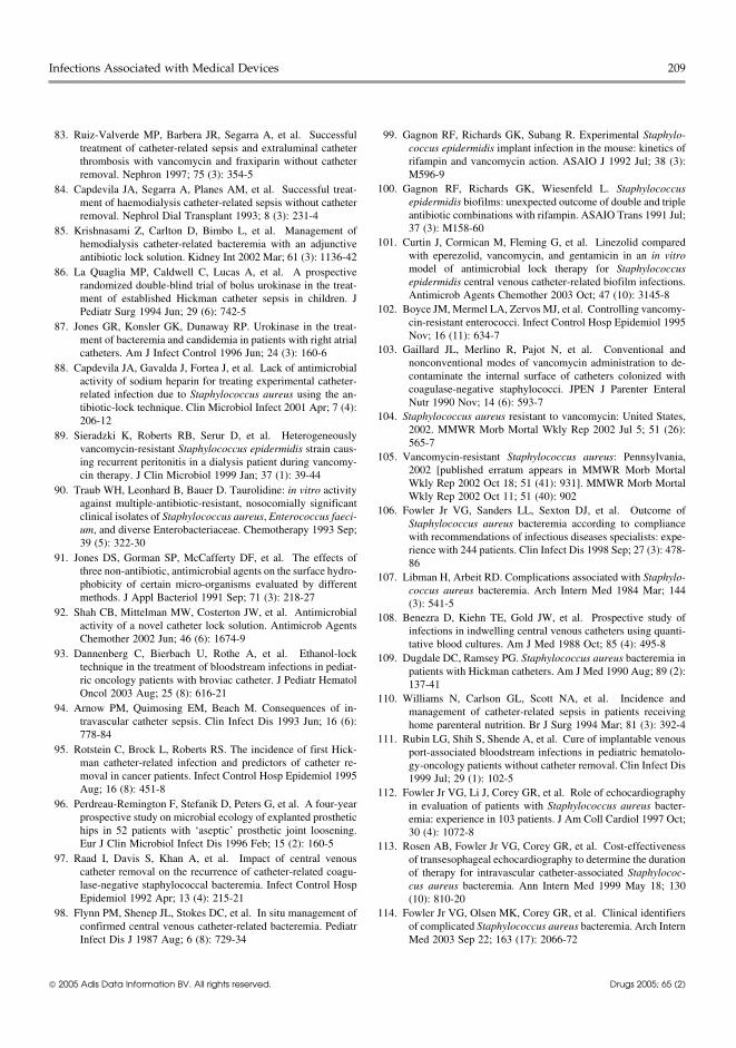
Infections Associated with Medical Devices 209
83. Ruiz-Valverde MP, Barbera JR, Segarra A, et al. Successful 99. Gagnon RF, Richards GK, Subang R. Experimental Staphylo-treatment of catheter-related sepsis and extraluminal catheter coccus epidermidis implant infection in the mouse: kinetics ofthrombosis with vancomycin and fraxiparin without catheter rifampin and vancomycin action. ASAIO J 1992 Jul; 38 (3):removal. Nephron 1997; 75 (3): 354-5 M596-9
100. Gagnon RF, Richards GK, Wiesenfeld L. Staphylococcus84. Capdevila JA, Segarra A, Planes AM, et al. Successful treat-epidermidis biofilms: unexpected outcome of double and triplement of haemodialysis catheter-related sepsis without catheterantibiotic combinations with rifampin. ASAIO Trans 1991 Jul;removal. Nephrol Dial Transplant 1993; 8 (3): 231-437 (3): M158-6085. Krishnasami Z, Carlton D, Bimbo L, et al. Management of
101. Curtin J, Cormican M, Fleming G, et al. Linezolid comparedhemodialysis catheter-related bacteremia with an adjunctivewith eperezolid, vancomycin, and gentamicin in an in vitroantibiotic lock solution. Kidney Int 2002 Mar; 61 (3): 1136-42model of antimicrobial lock therapy for Staphylococcus86. La Quaglia MP, Caldwell C, Lucas A, et al. A prospectiveepidermidis central venous catheter-related biofilm infections.randomized double-blind trial of bolus urokinase in the treat-Antimicrob Agents Chemother 2003 Oct; 47 (10): 3145-8ment of established Hickman catheter sepsis in children. J
102. Boyce JM, Mermel LA, Zervos MJ, et al. Controlling vancomy-Pediatr Surg 1994 Jun; 29 (6): 742-5cin-resistant enterococci. Infect Control Hosp Epidemiol 199587. Jones GR, Konsler GK, Dunaway RP. Urokinase in the treat-Nov; 16 (11): 634-7ment of bacteremia and candidemia in patients with right atrial
103. Gaillard JL, Merlino R, Pajot N, et al. Conventional andcatheters. Am J Infect Control 1996 Jun; 24 (3): 160-6nonconventional modes of vancomycin administration to de-88. Capdevila JA, Gavalda J, Fortea J, et al. Lack of antimicrobialcontaminate the internal surface of catheters colonized withactivity of sodium heparin for treating experimental catheter-coagulase-negative staphylococci. JPEN J Parenter Enteralrelated infection due to Staphylococcus aureus using the an-Nutr 1990 Nov; 14 (6): 593-7tibiotic-lock technique. Clin Microbiol Infect 2001 Apr; 7 (4):
104. Staphylococcus aureus resistant to vancomycin: United States,206-122002. MMWR Morb Mortal Wkly Rep 2002 Jul 5; 51 (26):89. Sieradzki K, Roberts RB, Serur D, et al. Heterogeneously565-7vancomycin-resistant Staphylococcus epidermidis strain caus-
105. Vancomycin-resistant Staphylococcus aureus: Pennsylvania,ing recurrent peritonitis in a dialysis patient during vancomy-2002 [published erratum appears in MMWR Morb Mortalcin therapy. J Clin Microbiol 1999 Jan; 37 (1): 39-44Wkly Rep 2002 Oct 18; 51 (41): 931]. MMWR Morb Mortal
90. Traub WH, Leonhard B, Bauer D. Taurolidine: in vitro activityWkly Rep 2002 Oct 11; 51 (40): 902
against multiple-antibiotic-resistant, nosocomially significant106. Fowler Jr VG, Sanders LL, Sexton DJ, et al. Outcome ofclinical isolates of Staphylococcus aureus, Enterococcus faeci-
Staphylococcus aureus bacteremia according to complianceum, and diverse Enterobacteriaceae. Chemotherapy 1993 Sep;with recommendations of infectious diseases specialists: expe-39 (5): 322-30rience with 244 patients. Clin Infect Dis 1998 Sep; 27 (3): 478-
91. Jones DS, Gorman SP, McCafferty DF, et al. The effects of 86three non-antibiotic, antimicrobial agents on the surface hydro-
107. Libman H, Arbeit RD. Complications associated with Staphylo-phobicity of certain micro-organisms evaluated by different
coccus aureus bacteremia. Arch Intern Med 1984 Mar; 144methods. J Appl Bacteriol 1991 Sep; 71 (3): 218-27
(3): 541-592. Shah CB, Mittelman MW, Costerton JW, et al. Antimicrobial 108. Benezra D, Kiehn TE, Gold JW, et al. Prospective study of
activity of a novel catheter lock solution. Antimicrob Agents infections in indwelling central venous catheters using quanti-Chemother 2002 Jun; 46 (6): 1674-9 tative blood cultures. Am J Med 1988 Oct; 85 (4): 495-8
93. Dannenberg C, Bierbach U, Rothe A, et al. Ethanol-lock 109. Dugdale DC, Ramsey PG. Staphylococcus aureus bacteremia intechnique in the treatment of bloodstream infections in pediat- patients with Hickman catheters. Am J Med 1990 Aug; 89 (2):ric oncology patients with broviac catheter. J Pediatr Hematol 137-41Oncol 2003 Aug; 25 (8): 616-21 110. Williams N, Carlson GL, Scott NA, et al. Incidence and
94. Arnow PM, Quimosing EM, Beach M. Consequences of in- management of catheter-related sepsis in patients receivingtravascular catheter sepsis. Clin Infect Dis 1993 Jun; 16 (6): home parenteral nutrition. Br J Surg 1994 Mar; 81 (3): 392-4778-84 111. Rubin LG, Shih S, Shende A, et al. Cure of implantable venous
95. Rotstein C, Brock L, Roberts RS. The incidence of first Hick- port-associated bloodstream infections in pediatric hematolo-man catheter-related infection and predictors of catheter re- gy-oncology patients without catheter removal. Clin Infect Dismoval in cancer patients. Infect Control Hosp Epidemiol 1995 1999 Jul; 29 (1): 102-5Aug; 16 (8): 451-8 112. Fowler Jr VG, Li J, Corey GR, et al. Role of echocardiography
96. Perdreau-Remington F, Stefanik D, Peters G, et al. A four-year in evaluation of patients with Staphylococcus aureus bacter-prospective study on microbial ecology of explanted prosthetic emia: experience in 103 patients. J Am Coll Cardiol 1997 Oct;hips in 52 patients with ‘aseptic’ prosthetic joint loosening. 30 (4): 1072-8Eur J Clin Microbiol Infect Dis 1996 Feb; 15 (2): 160-5 113. Rosen AB, Fowler Jr VG, Corey GR, et al. Cost-effectiveness
97. Raad I, Davis S, Khan A, et al. Impact of central venous of transesophageal echocardiography to determine the durationcatheter removal on the recurrence of catheter-related coagu- of therapy for intravascular catheter-associated Staphylococ-lase-negative staphylococcal bacteremia. Infect Control Hosp cus aureus bacteremia. Ann Intern Med 1999 May 18; 130Epidemiol 1992 Apr; 13 (4): 215-21 (10): 810-20
98. Flynn PM, Shenep JL, Stokes DC, et al. In situ management of 114. Fowler Jr VG, Olsen MK, Corey GR, et al. Clinical identifiersconfirmed central venous catheter-related bacteremia. Pediatr of complicated Staphylococcus aureus bacteremia. Arch InternInfect Dis J 1987 Aug; 6 (8): 729-34 Med 2003 Sep 22; 163 (17): 2066-72
2005 Adis Data Information BV. All rights reserved. Drugs 2005; 65 (2)
210 von Eiff et al.
115. Raad II, Sabbagh MF. Optimal duration of therapy for catheter- 129. Van Wijngaerden E, Peetermans WE, Vandersmissen J, et al.related Staphylococcus aureus bacteremia: a study of 55 cases Foreign body infection: a new rat model for prophylaxis andand review. Clin Infect Dis 1992 Jan; 14 (1): 75-82 treatment. J Antimicrob Chemother 1999 Nov; 44 (5): 669-74
116. Malanoski GJ, Samore MH, Pefanis A, et al. Staphylococcus 130. Rupp ME, Fey PD, Longo GM. Effect of LY333328 againstaureus catheter-associated bacteremia: minimal effective ther- vancomycin-resistant Enterococcus faecium in a rat centralapy and unusual infectious complications associated with arte- venous catheter-associated infection model. J Antimicrobrial sheath catheters. Arch Intern Med 1995 Jun 12; 155 (11): Chemother 2001 May; 47 (5): 705-71161-6 131. Kuklin NA, Pancari GD, Tobery TW, et al. Real-time monitor-
117. Rahal JJ. Preventing second-generation complications due to ing of bacterial infection in vivo: development of biolumines-Staphylococcus aureus. Arch Intern Med 1989 Mar; 149 (3): cent staphylococcal foreign-body and deep-thigh-wound503-4 mouse infection models. Antimicrob Agents Chemother 2003
118. Hospital Infection Control Practices Advisory Committee Sep; 47 (9): 2740-8(HICPAC). Recommendations for preventing the spread of 132. Swanson DS. Central venous catheter-related infections due tovancomycin resistance. Infect Control Hosp Epidemiol 1995 nontuberculous Mycobacterium species. Pediatr Infect Dis JFeb; 16 (2): 105-13 1998 Dec; 17 (12): 1163-4
119. Peters G, Becker K. Epidemiology, control and treatment of 133. Guay DR. Nontuberculous mycobacterial infections. Annmethicillin-resistant Staphylococcus aureus. Drugs 1996; 52 Pharmacother 1996 Jul; 30 (7-8): 819-30Suppl. 2: 50-4
134. Ishida H, Ishida Y, Kurosaka Y, et al. In vitro and in vivo120. Levine DP, Fromm BS, Reddy BR. Slow response to vancomy- activities of levofloxacin against biofilm-producing Pseudo-
cin or vancomycin plus rifampin in methicillin-resistant monas aeruginosa. Antimicrob Agents Chemother 1998 Jul;Staphylococcus aureus endocarditis. Ann Intern Med 1991 42 (7): 1641-5Nov 1; 115 (9): 674-80
135. Ashby MJ, Neale JE, Knott SJ, et al. Effect of antibiotics on121. Jantausch BA, Deville J, Adler S, et al. Linezolid for thenon-growing planktonic cells and biofilms of Escherichia coli.treatment of children with bacteremia or nosocomial pneumo-J Antimicrob Chemother 1994 Mar; 33 (3): 443-52nia caused by resistant gram-positive bacterial pathogens.
136. Gill MV, Ly H, Mueenuddin M, et al. Intravenous line infectionPediatr Infect Dis J 2003 Sep; 22 (9 Suppl.): S164-71due to Ochrobactrum anthropi (CDC Group Vd) in a normal122. Vaudaux P, Francois P, Bisognano C, et al. Comparison ofhost. Heart Lung 1997 Jul; 26 (4): 335-6levofloxacin, alatrofloxacin, and vancomycin for prophylaxis
137. Seifert H. Catheter-related infections due to gram-negative ba-and treatment of experimental foreign-body-associated infec-cilli. In: Seifert H, Jansen B, Farr BM, editors. Catheter-relatedtion by methicillin-resistant Staphylococcus aureus. Antimi-infections. New York: Marcel Dekker, 1997: 111-38crob Agents Chemother 2002 May; 46 (5): 1503-9
138. Douglas LJ. Candida biofilms and their role in infection. Trends123. Chuard C, Herrmann M, Vaudaux P, et al. Successful therapy ofMicrobiol 2003 Jan; 11 (1): 30-6experimental chronic foreign-body infection due to methicil-
lin-resistant Staphylococcus aureus by antimicrobial combina- 139. Hazen KC. New and emerging yeast pathogens. Clin Microbioltions. Antimicrob Agents Chemother 1991 Dec; 35 (12): 2611- Rev 1995 Oct; 8 (4): 462-786 140. Sizun J, Karangwa A, Giroux JD, et al. Malassezia furfur-
124. Cagni A, Chuard C, Vaudaux PE, et al. Comparison of sparflox- related colonization and infection of central venous catheters: aacin, temafloxacin, and ciprofloxacin for prophylaxis and prospective study in a pediatric intensive care unit. Intensivetreatment of experimental foreign-body infection by methicil- Care Med 1994 Aug; 20 (7): 496-9lin-resistant Staphylococcus aureus. Antimicrob Agents 141. Anaissie EJ, Rex JH, Uzun O, et al. Predictors of adverseChemother 1995 Aug; 39 (8): 1655-60 outcome in cancer patients with candidemia. Am J Med 1998
125. Schaad HJ, Chuard C, Vaudaux P, et al. Teicoplanin alone or Mar; 104 (3): 238-45combined with rifampin compared with vancomycin for pro- 142. Pappas PG, Rex JH, Sobel JD, et al. Guidelines for treatment ofphylaxis and treatment of experimental foreign body infection candidiasis. Clin Infect Dis 2004 Jan 15; 38 (2): 161-89by methicillin-resistant Staphylococcus aureus. Antimicrob
143. Karlowicz MG, Hashimoto LN, Kelly Jr RE, et al. ShouldAgents Chemother 1994 Aug; 38 (8): 1703-10central venous catheters be removed as soon as candidemia is
126. Schaad HJ, Chuard C, Vaudaux P, et al. Comparative efficaciesdetected in neonates? Pediatrics 2000 Nov; 106 (5): E63
of imipenem, oxacillin and vancomycin for therapy of144. Munoz P, Burillo A, Bouza E. Criteria used when initiatingchronic foreign body infection due to methicillin-susceptible
antifungal therapy against Candida spp. in the intensive careand -resistant Staphylococcus aureus. J Antimicrobunit. Int J Antimicrob Agents 2000 Jul; 15 (2): 83-90Chemother 1994 Jun; 33 (6): 1191-200
145. Rex JH, Walsh TJ, Sobel JD, et al. Practice guidelines for the127. Rupp ME, Ulphani J. Efficacy of LY333328 in a rat model oftreatment of candidiasis: Infectious Diseases Society ofStaphylococcus aureus central venous catheter-associated in-America. Clin Infect Dis 2000 Apr; 30 (4): 662-78fection [abstract no. F111]. Programs and Abstracts of the
146. Kibbler CC, Seaton S, Barnes RA, et al. Management andThirty-Eighth Interscience Conference on Antimicrobialoutcome of bloodstream infections due to Candida species inAgents and Chemotherapy, San Diego, CA, 1998. Washing-England and Wales. J Hosp Infect 2003 May; 54 (1): 18-24ton, DC: American Society for Microbiology, 1998: 260
128. Vaudaux P, Francois P, Bisognano C, et al. Comparative 147. Nguyen MH, Peacock Jr JE, Tanner DC, et al. Therapeuticefficacy of daptomycin and vancomycin in the therapy of approaches in patients with candidemia: evaluation in a multi-experimental foreign body infection due to Staphylococcus center, prospective, observational study. Arch Intern Medaureus. J Antimicrob Chemother 2003 Jul; 52 (1): 89-95 1995 Dec 11; 155 (22): 2429-35
2005 Adis Data Information BV. All rights reserved. Drugs 2005; 65 (2)
Infections Associated with Medical Devices 211
148. Lecciones JA, Lee JW, Navarro EE, et al. Vascular catheter- 165. Rosenthal VD, Guzman S, Pezzotto SM, et al. Effect of anassociated fungemia in patients with cancer: analysis of 155 infection control program using education and performanceepisodes. Clin Infect Dis 1992 Apr; 14 (4): 875-83 feedback on rates of intravascular device-associated blood-
stream infections in intensive care units in Argentina. Am J149. Rose HD. Venous catheter-associated candidemia. Am J MedInfect Control 2003 Nov; 31 (7): 405-9Sci 1978 May; 275 (3): 265-9
150. Barber GR, Brown AE, Kiehn TE, et al. Catheter-related 166. Sherertz RJ, Ely EW, Westbrook DM, et al. Education ofMalassezia furfur fungemia in immunocompromised patients. physicians-in-training can decrease the risk for vascular cathe-Am J Med 1993 Oct; 95 (4): 365-70 ter infection. Ann Intern Med 2000 Apr 18; 132 (8): 641-8
151. Ostrosky-Zeichner L, Marr KA, Rex JH, et al. Amphotericin B: 167. Eggimann P, Harbarth S, Constantin MN, et al. Impact of atime for a new ‘gold standard’. Clin Infect Dis 2003 Aug 1; 37 prevention strategy targeted at vascular-access care on inci-(3): 415-25 dence of infections acquired in intensive care. Lancet 2000
152. Rex JH, Bennett JE, Sugar AM, et al. A randomized trial May 27; 355 (9218): 1864-8comparing fluconazole with amphotericin B for the treatment 168. Soifer NE, Borzak S, Edlin BR, et al. Prevention of peripheralof candidemia in patients without neutropenia: Candidemia venous catheter complications with an intravenous therapyStudy Group and the National Institute. N Engl J Med 1994 team: a randomized controlled trial. Arch Intern Med 1998Nov 17; 331 (20): 1325-30 Mar 9; 158 (5): 473-7
153. Diekema DJ, Messer SA, Hollis RJ, et al. Activities of cas-169. Fridkin SK, Pear SM, Williamson TH, et al. The role ofpofungin, itraconazole, posaconazole, ravuconazole, vori-
understaffing in central venous catheter-associated blood-conazole, and amphotericin B against 448 recent clinical iso-stream infections. Infect Control Hosp Epidemiol 1996 Mar;lates of filamentous fungi. J Clin Microbiol 2003 Aug; 41 (8):17 (3): 150-83623-6
170. Bertone SA, Fisher MC, Mortensen JE. Quantitative skin cul-154. Mora-Duarte J, Betts R, Rotstein C, et al. Comparison oftures at potential catheter sites in neonates. Infect Control Hospcaspofungin and amphotericin B for invasive candidiasis. NEpidemiol 1994 May; 15 (5): 315-8Engl J Med 2002 Dec 19; 347 (25): 2020-9
171. Goetz AM, Wagener MM, Miller JM, et al. Risk of infection155. Kauffman CA, Zarins LT. In vitro activity of voriconazoledue to central venous catheters: effect of site of placement andagainst Candida species. Diagn Microbiol Infect Dis 1998catheter type. Infect Control Hosp Epidemiol 1998 Nov; 19May; 31 (1): 297-300(11): 842-5156. Fegeler W, Bille J, Dupont B, et al. Distribution of yeast species
172. Randolph AG, Cook DJ, Gonzales CA, et al. Ultrasoundin clinical routine diagnostic specimen and their susceptibilityguidance for placement of central venous catheters: a meta-to fluconazole, voriconazole and flucytosine. Mycoses 1999;analysis of the literature. Crit Care Med 1996 Dec; 24 (12):42: 174-52053-8157. Patron RL, Climo MW, Goldstein BP, et al. Lysostaphin
treatment of experimental aortic valve endocarditis caused by a 173. Heard SO, Wagle M, Vijayakumar E, et al. Influence of triple-Staphylococcus aureus isolate with reduced susceptibility to lumen central venous catheters coated with chlorhexidine andvancomycin. Antimicrob Agents Chemother 1999 Jul; 43 (7): silver sulfadiazine on the incidence of catheter-related bacter-1754-5 emia. Arch Intern Med 1998 Jan 12; 158 (1): 81-7
158. von Eiff C, Kokai-Kun JF, Becker K, et al. In vitro activity of 174. Richet H, Hubert B, Nitemberg G, et al. Prospective multicenterrecombinant lysostaphin against Staphylococcus aureus iso- study of vascular-catheter-related complications and risk fac-lates from anterior nares and blood. Antimicrob Agents tors for positive central-catheter cultures in intensive care unitChemother 2003 Nov; 47 (11): 3613-5 patients. J Clin Microbiol 1990 Nov; 28 (11): 2520-5
159. Wu JA, Kusuma C, Mond JJ, et al. Lysostaphin disrupts 175. Merrer J, De Jonghe B, Golliot F, et al. Complications ofStaphylococcus aureus and Staphylococcus epidermidis bio- femoral and subclavian venous catheterization in critically illfilms on artificial surfaces. Antimicrob Agents Chemother patients: a randomized controlled trial. JAMA 2001 Aug 8;2003 Nov; 47 (11): 3407-14 286 (6): 700-7
160. Johansen C, Falholt P, Gram L. Enzymatic removal and disin-176. Maki DG, Mermel LA. Infections due to infusion therapy. In:fection of bacterial biofilms. Appl Environ Microbiol 1997
Bennett JV, Brachmann PS, editors. Hospital infections. Phila-Sep; 63 (9): 3724-8delphia (PA): Lippincott-Raven, 1998: 689-724
161. Dewhurst E, Rawson DM, Steele GC. The use of a model177. MacDonald A, Dinah F, Mackenzie D, et al. Performancesystem to compare the efficiency of ultrasound and agitation in
feedback of hand hygiene, using alcohol gel as the skin decon-the recovery of Bacillus subtilis spores from polymer surfaces.taminant, reduces the number of inpatients newly affected byJ Appl Bacteriol 1986 Oct; 61 (4): 357-63MRSA and antibiotic costs. J Hosp Infect 2004 Jan; 56 (1): 56-162. Carmen JC, Roeder BL, Nelson JL, et al. Ultrasonically en-63hanced vancomycin activity against Staphylococcus
178. Pittet D, Hugonnet S, Harbarth S, et al. Effectiveness of aepidermidis biofilms in vivo. J Biomater Appl 2004 Apr; 18hospital-wide programme to improve compliance with hand(4): 237-45hygiene: Infection Control Programme. Lancet 2000 Oct 14;163. Rediske AM, Roeder BL, Nelson JL, et al. Pulsed ultrasound356 (9238): 1307-12enhances the killing of Escherichia coli biofilms by aminogly-
coside antibiotics in vivo. Antimicrob Agents Chemother 2000 179. Raad II, Hohn DC, Gilbreath BJ, et al. Prevention of centralMar; 44 (3): 771-2 venous catheter-related infections by using maximal sterile
164. Pitt WG, Ross SA. Ultrasound increases the rate of bacterial cell barrier precautions during insertion. Infect Control Hospgrowth. Biotechnol Prog 2003 May; 19 (3): 1038-44 Epidemiol 1994 Apr; 15 (4 Pt 1): 231-8
2005 Adis Data Information BV. All rights reserved. Drugs 2005; 65 (2)
212 von Eiff et al.
180. Galway R, Harrod ME, Crisp J, et al. Central venous access and 195. Ascher DP, Shoupe BA, Maybee D, et al. Persistent catheter-handwashing: variability in policies and practices. Paediatr related bacteremia: clearance with antibiotics and urokinase. JNurs 2003 Dec; 15 (10): 14-8 Pediatr Surg 1993 Apr; 28 (4): 627-9
196. Khoury AE, Lam K, Ellis B, et al. Prevention and control of181. Boyce JM, Pittet D. Guideline for Hand Hygiene in Health-Carebacterial infections associated with medical devices. ASAIO JSettings: recommendations of the Healthcare Infection Control1992 Jul; 38 (3): M174-8Practices Advisory Committee and the HICPAC/SHEA/APIC/
IDSA Hand Hygiene Task Force. Society for Healthcare Epi- 197. Rediske AM, Roeder BL, Brown MK, et al. Ultrasonic en-demiology of America/Association for Professionals in Infec- hancement of antibiotic action on Escherichia coli biofilms: antion Control/Infectious Diseases Society of America. MMWR in vivo model. Antimicrob Agents Chemother 1999 May; 43Recomm Rep 2002 Oct 25; 51 (RR-16): 1-45 (5): 1211-4
182. Safdar N, Maki DG. The pathogenesis of catheter-related blood- 198. Bridgett MJ, Davies MC, Denyer SP. Control of staphylococcalstream infection with noncuffed short-term central venous adhesion to polystyrene surfaces by polymer surface modifica-catheters. Intensive Care Med 2004 Jan; 30 (1): 62-7 tion with surfactants. Biomaterials 1992; 13 (7): 411-6
183. Clemence MA, Walker D, Farr BM. Central venous catheter 199. Desai NP, Hossainy SF, Hubbell JA. Surface-immobilized poly-practices: results of a survey. Am J Infect Control 1995 Feb; 23 ethylene oxide for bacterial repellence. Biomaterials 1992; 13(1): 5-12 (7): 417-20
200. Dunkirk SG, Gregg SL, Duran LW, et al. Photochemical184. Humar A, Ostromecki A, Direnfeld J, et al. Prospective ran-coatings for the prevention of bacterial colonization. J Bio-domized trial of 10% povidone-iodine versus 0.5% tincture ofmater Appl 1991 Oct; 6 (2): 131-56chlorhexidine as cutaneous antisepsis for prevention of central
venous catheter infection. Clin Infect Dis 2000 Oct; 31 (4): 201. Tebbs SE, Sawyer A, Elliott TS. Influence of surface morpholo-1001-7 gy on in vitro bacterial adherence to central venous catheters.
Br J Anaesth 1994 May; 72 (5): 587-91185. Maki DG, Ringer M, Alvarado CJ. Prospective randomised trialof povidone-iodine, alcohol, and chlorhexidine for prevention 202. Tebbs SE, Elliott TS. Modification of central venous catheterof infection associated with central venous and arterial cathe- polymers to prevent in vitro microbial colonisation. Eur J Clinters. Lancet 1991 Aug 10; 338 (8763): 339-43 Microbiol Infect Dis 1994 Feb; 13 (2): 111-7
186. Garland JS, Buck RK, Maloney P, et al. Comparison of 10% 203. Jansen B. New concepts in the prevention of polymer-associatedpovidone-iodine and 0.5% chlorhexidine gluconate for the foreign body infections. Zentralbl Bakteriol 1990 Apr; 272 (4):prevention of peripheral intravenous catheter colonization in 401-10neonates: a prospective trial. Pediatr Infect Dis J 1995 Jun; 14 204. Jansen B, Schareina S, Steinhauser H, et al. Development of(6): 510-6 polymers with antiinfective properties. Polym Mater Sci Eng
1987; 57: 43-6187. Maki DG, Ringer M. Evaluation of dressing regimens for pre-vention of infection with peripheral intravenous catheters: 205. Han DK, Park KD, Kim YH. Sulfonated poly (ethylene oxide)-gauze, a transparent polyurethane dressing, and an iodophor- grafted polyurethane copolymer for biomedical applications. Jtransparent dressing. JAMA 1987 Nov 6; 258 (17): 2396-403 Biomater Sci Polym Ed 1998; 9 (2): 163-74
188. Hoffmann KK, Weber DJ, Samsa GP, et al. Transparent poly- 206. Baumgartner JN, Yang CZ, Cooper SL. Physical property ana-urethane film as an intravenous catheter dressing: a meta- lysis and bacterial adhesion on a series of phosphonated poly-analysis of the infection risks. JAMA 1992 Apr 15; 267 (15): urethanes. Biomaterials 1997 Jun; 18 (12): 831-72072-6 207. Kohnen W, Jansen B. Changing material surface chemistry for
189. Maki DG, Mermel LA, Kluger DM. The efficacy of a chlorhex- preventing bacterial adhesion. In: An YH, Friedman RJ, edi-idine-impregnated sponge (biopatch) for the prevention of tors. Handbook of bacterial adhesion. Totowa (NJ): Humanaintravascular catheter-related infection: a prospective, random- Press, 2000: 581-9ized, controlled, multicenter trial [abstract no. 1430]. 40th 208. An YH, Blair BK, Martin KL, et al. Macromolecule surfaceInterscience Conference on Antimicrobial Agents and Chemo- coating for preventing bacterial adhesion. In: YH An, Fried-therapy of the American Society for Microbiology; 2000 Sep man RJ, editors. Handbook of bacterial adhesion. Totowa17-20; Toronto (NJ): Humana Press, 2000: 609-25
190. Yamamoto AJ, Solomon JA, Soulen MC, et al. Sutureless 209. Jansen B, Kohnen W. Prevention of biofilm formation by poly-securement device reduces complications of peripherally in- mer modification. J Ind Microbiol 1995 Oct; 15 (4): 391-6serted central venous catheters. J Vasc Interv Radiol 2002 Jan; 210. Marcinko DE. Gentamicin-impregnated PMMA beads: an intro-13 (1): 77-81 duction and review. J Foot Surg 1985 Mar; 24 (2): 116-21
191. Maki DG, Ringer M. Risk factors for infusion-related phlebitis 211. Welch A. Antibiotics in acrylic bone cement: in vitro studies. Jwith small peripheral venous catheters: a randomized control- Biomed Mater Res 1978 Sep; 12 (5): 679-700led trial. Ann Intern Med 1991 May 15; 114 (10): 845-54 212. Moore WS, Chvapil M, Seiffert G, et al. Development of an
192. Tully JL, Friedland GH, Baldini LM, et al. Complications of infection-resistant vascular prosthesis. Arch Surg 1981 Nov;intravenous therapy with steel needles and Teflon catheters: a 116 (11): 1403-7comparative study. Am J Med 1981 Mar; 70 (3): 702-6 213. Powell TW, Burnham SJ, Johnson Jr G. A passive system using
193. Ball PA. Intravenous in-line filters: filtering the evidence. Curr rifampin to create an infection-resistant vascular prosthesis.Opin Clin Nutr Metab Care 2003 May; 6 (3): 319-25 Surgery 1983 Nov; 94 (5): 765-9
194. Maddox RR, John Jr JF, Brown LL, et al. Effect of inline 214. McDougal EG, Burnham SJ, Johnson Jr G. Rifampin protectionfiltration on postinfusion phlebitis. Clin Pharm 1983 Jan; 2 (1): against experimental graft sepsis. J Vasc Surg 1986 Jul; 4 (1):58-61 5-7
2005 Adis Data Information BV. All rights reserved. Drugs 2005; 65 (2)
Infections Associated with Medical Devices 213
215. Solovskij MV, Ulbrich K, Kopecek J. Synthesis of N-(2- 231. Quesnel LB, Al Najjar AR, Buddhavudhikrai P. Synergismhydroxypropyl)methacrylamide copolymers with anti- between chlorhexidine and sulphadiazine. J Appl Bacteriolmicrobial activity. Biomaterials 1983 Jan; 4 (1): 44-8 1978 Dec; 45 (3): 397-405
232. Bach A, Schmidt H, Bottiger B, et al. Retention of antibacterial216. Sherertz RJ, Carruth WA, Hampton AA, et al. Efficacy ofactivity and bacterial colonization of antiseptic-bonded centralantibiotic-coated catheters in preventing subcutaneousvenous catheters. J Antimicrob Chemother 1996 Feb; 37 (2):Staphylococcus aureus infection in rabbits. J Infect Dis 1993315-22Jan; 167 (1): 98-106
233. Hannan M, Juste RN, Umasanker S, et al. Antiseptic-bonded217. Jansen B, Peters G. Modern strategies in the prevention ofcentral venous catheters and bacterial colonisation. Anaesthe-polymer-associated infections. J Hosp Infect 1991 Oct; 19 (2):sia 1999 Sep; 54 (9): 868-7283-8
234. Ciresi DL, Albrecht RM, Volkers PA, et al. Failure of antiseptic218. Jansen B, Jansen S, Peters G, et al. In-vitro efficacy of a centralbonding to prevent central venous catheter-related infectionvenous catheter (‘Hydrocath’) loaded with teicoplanin to pre-and sepsis. Am Surg 1996 Aug; 62 (8): 641-6vent bacterial colonization. J Hosp Infect 1992 Oct; 22 (2): 93-
235. Pemberton LB, Ross V, Cuddy P, et al. No difference in107catheter sepsis between standard and antiseptic central venous219. Romano G, Berti M, Goldstein BP, et al. Efficacy of a centralcatheters: a prospective randomized trial. Arch Surg 1996 Sep;venous catheter (Hydrocath) loaded with teicoplanin in131 (9): 986-9preventing subcutaneous staphylococcal infection in the
236. George SJ, Vuddamalay P, Boscoe MJ. Antiseptic-impregnatedmouse. Zentralbl Bakteriol 1993 Aug; 279 (3): 426-33central venous catheters reduce the incidence of bacterial220. Kamal GD, Pfaller MA, Rempe LE, et al. Reduced intravascu-colonization and associated infection in immunocompromisedlar catheter infection by antibiotic bonding: a prospective,transplant patients. Eur J Anaesthesiol 1997 Jul; 14 (4): 428-31randomized, controlled trial. JAMA 1991 May 8; 265 (18):
237. Logghe C, Van Ossel C, D’Hoore W, et al. Evaluation of2364-8chlorhexidine and silver-sulfadiazine impregnated central ve-221. Kamal GD, Divishek D, Kumar GC, et al. Reduced intravascu-nous catheters for the prevention of bloodstream infection inlar catheter-related infection by routine use of antibiotic-bond-leukaemic patients: a randomized controlled trial. J Hosp In-ed catheters in a surgical intensive care unit. Diagn Microbiolfect 1997 Oct; 37 (2): 145-56Infect Dis 1998 Mar; 30 (3): 145-52
238. Maki DG, Stolz SM, Wheeler S, et al. Prevention of central222. Raad I, Darouiche R, Hachem R, et al. Antibiotics and preven-
venous catheter-related bloodstream infection by use of antion of microbial colonization of catheters. Antimicrob Agents
antiseptic-impregnated catheter: a randomized, controlledChemother 1995 Nov; 39 (11): 2397-400
trial. Ann Intern Med 1997 Aug 15; 127 (4): 257-66223. Raad I, Darouiche R, Hachem R, et al. The broad-spectrum 239. Tennenberg S, Lieser M, McCurdy B, et al. A prospective
activity and efficacy of catheters coated with minocycline and randomized trial of an antibiotic- and antiseptic-coated centralrifampin. J Infect Dis 1996 Feb; 173 (2): 418-24 venous catheter in the prevention of catheter-related infections.
224. Raad I, Darouiche R, Dupuis J, et al. Central venous catheters Arch Surg 1997 Dec; 132 (12): 1348-51coated with minocycline and rifampin for the prevention of 240. Collin GR. Decreasing catheter colonization through the use ofcatheter-related colonization and bloodstream infections: a an antiseptic-impregnated catheter: a continuous quality im-randomized, double-blind trial. The Texas Medical Center provement project. Chest 1999 Jun; 115 (6): 1632-40Catheter Study Group. Ann Intern Med 1997 Aug 15; 127 (4): 241. Sheng WH, Ko WJ, Wang JT, et al. Evaluation of antiseptic-267-74 impregnated central venous catheters for prevention of cathe-
225. Darouiche RO, Raad II, Heard SO, et al. A comparison of two ter-related infection in intensive care unit patients. Diagnantimicrobial-impregnated central venous catheters: Catheter Microbiol Infect Dis 2000 Sep; 38 (1): 1-5Study Group. N Engl J Med 1999 Jan 7; 340 (1): 1-8 242. Haxhe JJ, D’Hoore W. A meta-analysis dealing with the effec-
226. Crnich CJ, Maki DG. The promise of novel technology for the tiveness of chlorhexidine and silver-sufhadiazine impregnatedprevention of intravascular device-related bloodstream infec- central venous catheters. J Hosp Infect 1998 Oct; 40 (2): 166-8tion. I: pathogenesis and short-term devices. Clin Infect Dis 243. Veenstra DL, Saint S, Saha S, et al. Efficacy of antiseptic-2002 May 1; 34 (9): 1232-42 impregnated central venous catheters in preventing catheter-
227. Tambe SM, Sampath L, Modak SM. In vitro evaluation of the related bloodstream infection: a meta-analysis. JAMA 1999risk of developing bacterial resistance to antiseptics and anti- Jan 20; 281 (3): 261-7biotics used in medical devices. J Antimicrob Chemother 2001 244. Marin MG, Lee JC, Skurnick JH. Prevention of nosocomialMay; 47 (5): 589-98 bloodstream infections: effectiveness of antimicrobial-impreg-
228. Donelli G, Francolini I, Piozzi A, et al. New polymer-antibiotic nated and heparin-bonded central venous catheters. Crit Caresystems to inhibit bacterial biofilm formation: a suitable ap- Med 2000 Sep; 28 (9): 3332-8proach to prevent central venous catheter-associated infec- 245. Pai MP, Pendland SL, Danziger LH. Antimicrobial-coated/tions. J Chemother 2002 Oct; 14 (5): 501-7 bonded and -impregnated intravascular catheters. Ann
229. Schierholz JM, Fleck C, Beuth J, et al. The antimicrobial Pharmacother 2001 Oct; 35 (10): 1255-63efficacy of a new central venous catheter with long-term 246. McConnell SA, Gubbins PO, Anaissie EJ. Do antimicrobial-broad-spectrum activity. J Antimicrob Chemother 2000 Jul; 46 impregnated central venous catheters prevent catheter-related(1): 45-50 bloodstream infection? Clin Infect Dis 2003 Jul 1; 37 (1): 65-
72230. Kingston D, Seal DV, Hill ID. Self-disinfecting plastics forintravenous catheters and prosthetic inserts. J Hyg (Lond) 247. Brun-Buisson C, Doyon F, Sollet JP, et al. Prevention of in-1986 Apr; 96 (2): 185-98 travascular catheter-related infection with newer chlorhex-
2005 Adis Data Information BV. All rights reserved. Drugs 2005; 65 (2)
214 von Eiff et al.
idine-silver-sulfadiazine-coated catheters. Intensive Care Med 264. Bayston R, Zdroyewski V, Barsham S. Use of an in vitro model2004; 30: 837-43 for studying the eradication of catheter colonisation by
Staphylococcus epidermidis. J Infect 1988 Mar; 16 (2): 141-6248. Tattawasart U, Maillard JY, Furr JR, et al. Development ofresistance to chlorhexidine diacetate and cetylpyridinium chlo- 265. Bayston R, Barsham S. Catheter colonisation: a laboratoryride in Pseudomonas stutzeri and changes in antibiotic suscep- model suitable for aetiological, therapeutic and preventivetibility. J Hosp Infect 1999 Jul; 42 (3): 219-29 studies. Med Lab Sci 1988 Jul; 45 (3): 235-9
249. Oda T, Hamasaki J, Kanda N, et al. Anaphylactic shock induced 266. Bayston R, Grove N, Siegel J, et al. Prevention of hydrocepha-by an antiseptic-coated central venous [correction of nervous] lus shunt catheter colonisation in vitro by impregnation withcatheter [published erratum appears in Anesthesiology 1998 antimicrobials. J Neurol Neurosurg Psychiatry 1989 May; 52Feb; 88 (2): 560]. Anesthesiology 1997 Nov; 87 (5): 1242-4 (5): 605-9
250. Gosheger G, Hardes J, Ahrends H, et al. Silver-coated 267. Bayston R, Milner RD. Antimicrobial activity of silicone rubbermegaendoprostheses in a rabbit model: an analysis of the used in hydrocephalus shunts, after impregnation with anti-infection rate and toxicological side effects. Biomaterials microbial substances. J Clin Pathol 1981 Sep; 34 (9): 1057-622004; 25 (24): 5547-56
268. Bayston R, Lambert E. Duration of protective activity of cere-251. Sioshansi P. New processes for surface treatment of catheters. brospinal fluid shunt catheters impregnated with antimicrobial
Artif Organs 1994 Apr; 18 (4): 266-71 agents to prevent bacterial catheter-related infection. JNeurosurg 1997 Aug; 87 (2): 247-51252. McLean RJ, Hussain AA, Sayer M, et al. Antibacterial activity
of multilayer silver-copper surface films on catheter material. 269. Bayston R, Ashraf W, Bhundia C. Mode of action of an anti-Can J Microbiol 1993 Sep; 39 (9): 895-9 microbial biomaterial for use in hydrocephalus shunts. J An-
253. Davenas J, Thevenard P, Philippe F, et al. Surface implantation timicrob Chemother 2004; 53: 778-82treatments to prevent infection complications in short term
270. Schierholz J, Jansen B, Jaenicke L, et al. In-vitro efficacy of andevices. Biomol Eng 2002 Aug; 19 (2-6): 263-8antibiotic releasing silicone ventricle catheter to prevent shunt
254. Gatter N, Kohnen W, Jansen B. In vitro efficacy of a hydrophilic infection. Biomaterials 1994 Oct; 15 (12): 996-1000central venous catheter loaded with silver to prevent microbial
271. Hampl J, Schierholz J, Jansen B, et al. In vitro and in vivocolonization. Zentralbl Bakteriol 1998 Jan; 287 (1-2): 157-69efficacy of a rifampin-loaded silicone catheter for the preven-
255. Jansen B, Rinck M, Wolbring P, et al. In vitro evaluation of the tion of CSF shunt infections. Acta Neurochir (Wien) 1995; 133antimicrobial efficacy and biocompatibility of a silver-coated (3-4): 147-52central venous catheter. J Biomater Appl 1994 Jul; 9 (1): 55-70
272. Kohnen W, Schaper J, Klein O, et al. A silicone ventricular256. Boswald M, Lugauer S, Regenfus A, et al. Reduced rates of catheter coated with a combination of rifampin and
catheter-associated infection by use of a new silver-impregnat- trimethoprim for the prevention of catheter-related infections.ed central venous catheter. Infection 1999; 27 Suppl. 1: S56-60 Zentralbl Bakteriol 1998 Jan; 287 (1-2): 147-56
257. Ranucci M, Isgro G, Giomarelli PP, et al. Impact of oligon 273. Hampl JA, Weitzel A, Bonk C, et al. Rifampin-impregnatedcentral venous catheters on catheter colonization and catheter- silicone catheters: a potential tool for prevention and treatmentrelated bloodstream infection. Crit Care Med 2003 Jan; 31 (1): of CSF shunt infections. Infection 2003 Mar; 31 (2): 109-1152-9
274. Schierholz JM, Pulverer G. Investigation of a rifampin, fusidic-258. Liu WK, Tebbs SE, Byrne PO, et al. The effects of electric
acid and mupirocin releasing silicone catheter. Biomaterialscurrent on bacteria colonising intravenous catheters. J Infect
1998 Nov; 19 (22): 2065-741993 Nov; 27 (3): 261-9
275. Zabramski JM, Whiting D, Darouiche RO, et al. Efficacy of259. Raad I, Hachem R, Zermeno A, et al. In vitro antimicrobialantimicrobial-impregnated external ventricular drain catheters:efficacy of silver iontophoretic catheter. Biomaterials 1996a prospective, randomized, controlled trial. J Neurosurg 2003Jun; 17 (11): 1055-9Apr; 98 (4): 725-30
260. Goldschmidt H, Hahn U, Salwender HJ, et al. Prevention of276. von Eiff C, Overbeck J, Haupt G, et al. Bactericidal effect ofcatheter-related infections by silver coated central venous
extracorporeal shock waves on Staphylococcus aureus. J Medcatheters in oncological patients. Zentralbl Bakteriol 1995Microbiol 2000 Aug; 49 (8): 709-12Dec; 283 (2): 215-23
277. Kohnen W, Bletz C, Hampl J, et al. Intelligent polymers for the261. Carbon RT, Lugauer S, Geitner U, et al. Reducing catheter-prevention of device-related infection [poster no. S5]. 9thassociated infections with silver-impregnated catheters inAnnual Meeting of the Society of Healthcare Epidemiology oflong-term therapy of children. Infection 1999; 27 Suppl. 1:America; 1999 Apr 18-20; San FranciscoS69-73
262. Stoiser B, Kofler J, Staudinger T, et al. Contamination ofcentral venous catheters in immunocompromised patients: a
Correspondence and offprints: Prof. Dr Christof von Eiff,comparison between two different types of central venousInstitute of Medical Microbiology, University of Munstercatheters. J Hosp Infect 2002 Mar; 50 (3): 202-6Hospital and Clinics, Domagkstraβe 10, 48149 Munster,263. Bridgett MJ, Davies MC, Denyer SP, et al. In vitro assessmentGermany.of bacterial adhesion to Hydromer-coated cerebrospinal fluid
shunts. Biomaterials 1993 Feb; 14 (3): 184-8 E-mail: [email protected]
2005 Adis Data Information BV. All rights reserved. Drugs 2005; 65 (2)