inhibitory effects of metformin at low concentration on
TRANSCRIPT
RESEARCH Open Access
Inhibitory effects of metformin at lowconcentration on epithelial–mesenchymaltransition of CD44+CD117+ ovarian cancerstem cellsRongrong Zhang1†, Ping Zhang1†, Hong Wang1†, Dongming Hou2, Wentao Li3*, Guishan Xiao4 and Chenwei Li5*
Abstract
Background: Although metformin, a first-line drug for treating diabetes, may play an important role in inhibition ofepithelial ovarian cancer cell growth and cancer stem cells (CSCs), metformin at low dose showed less effect on theproliferation of ovarian cancer cells. In this study, we evaluated the effect of metformin at low dose on ovarianCSCs in order to understand the molecular mechanisms underlying.
Methods: The inhibitory effects of metformin at los dose on proliferation and population of ovarian cancer cellsincluding SKOV3 and A2780 were assessed by cell proliferation assay and flow cytometry. Quantitative real-timePCR assay on expression of Bcl-2, Survivin and Bax was performed to determine the effect of metformin at lowdose on epithelial-mesenchymal transition (EMT) of cancer cells and CSCs. Tumor sphere formation assay was alsoperformed to evaluate the effect of metformin on spheres forming ability of CSCs. The therapeutic efficacy and theanti-CSC effects of metformin at low dose were investigated by using both SKOV3 cells and primary tumorxenografts. In addition, the CSC frequency and EMT in tumor xenograft models were also assessed by flowcytometry and quantitative real-time PCR.
Results: Metformin at low dose did not affect the proliferation of ovarian cancer cells. However, it inhibitedpopulation of CD44+CD117+ selectively, neither CD133+ nor ALDH+ cells. It suppressed expression of snail2,twist and vimentin significantly in cancer cells and CD44+CD117+ CSCs in vitro. Low dose of metformin reducedsurvivin expression in CSCs. Low concentrations of metformin inhibited the secondary and the tertiary tumorsphere formation, decreased SKOV3 and primary ovarian tumor xenograft growth, enhanced the anticancer effectof cisplatin, and lowered the proportion of CD44+CD117+ CSCs in the xenograft tissue. Metformin was alsoassociated with a reduction of snail2, twist, and vimentin in CD44+CD117+ ovarian CSCs in vivo.
Conclusions: Our results implicate that metformin at low dose inhibits selectively CD44+CD117+ ovarian CSCsthrough inhibition of EMT and potentiates the effect of cisplatin.
Keywords: Metformin, Epithelial–mesenchymal transition, Ovarian cancer stem cells
* Correspondence: [email protected]; [email protected]†Equal contributors3Department of Interventional Radiology, Cancer Hospital, Fudan University,Shanghai 200032, China5Sunstem Biotechnology Co., Ltd, Shanghai 200439, ChinaFull list of author information is available at the end of the article
© 2015 Zhang et al. Open Access This article is distributed under the terms of the Creative Commons Attribution 4.0International License (http://creativecommons.org/licenses/by/4.0/), which permits unrestricted use, distribution, andreproduction in any medium, provided you give appropriate credit to the original author(s) and the source, provide a link tothe Creative Commons license, and indicate if changes were made. The Creative Commons Public Domain Dedication waiver(http://creativecommons.org/publicdomain/zero/1.0/) applies to the data made available in this article, unless otherwise stated.
Zhang et al. Stem Cell Research & Therapy (2015) 6:262 DOI 10.1186/s13287-015-0249-0
BackgroundOvarian cancer is the fifth most common type of can-cer in females and the leading cause of mortality forgynecological malignancies. Epithelial ovarian cancer(EOC) accounts for more than 90 % of all ovarianneoplasms [1, 2].To understand the exact biological features of ovarian
carcinoma, series of new studies are being focused on can-cer stem cells (CSCs). Study of CSCs suggests that theseare a rare population of cancer cells with inherent che-moresistance, capable of regeneration of the various celltypes within tumors, which then leads to relapse of therapy[3, 4]. Drugs targeting CSCs may offer great promise forthe development of novel anticancer drugs. Highly diversi-fied phenotypes in ovarian CSCs have been found.Well-studied cell surface biomarkers including CD44/CD117, CD133, and aldehyde dehydrogenase (ALDH)are used extensively to identify ovarian CSCs [5–11].Metformin, a traditional medication for type 2 dia-
betes, has been demonstrated to have antiproliferativeand proapoptotic effects on ovarian cancer [12], and hasbeen used to target CSCs in ovarian cancer [6]. Recentstudies indicated that metformin can inhibit the growthand the proliferation of ovarian CSCs in vitro and in vivo.These results provide a rationale for using metformin totreat patients with ovarian cancer [6].The epithelial–mesenchymal transition (EMT) is a
process by which an epithelial cancer cell loses its cellpolarity and cell–cell adhesion and acquires the capacityto migrate and metastasize. Epithelial cancer cells throughEMTcan undergo a phenotypic switch, which allows thesepolarized and immobile epithelial cells to become motilemesenchymal cells [13–15]. EMT has been linked to theability for self-renewal and generation of multiple lineages[14]. Although the latest studies showed that EMTcan initiate regeneration of CSCs from immortalizedhuman mammary epithelia [14, 16, 17], the molecularmechanism underlying regeneration of CSCs throughEMT is unknown.A series of studies has shown that metformin may re-
press the EMT transcriptionally and repress a cell pheno-type associated with CSCs in breast cancer [18, 19]. Italso inhibited cellular transformation and killed breastCSCs selectively in vitro and in vivo [19]. Snail1 andsnail2 are key transcriptional factors to regulate progres-sion, functionality, and survival of metastatic ovarianstem cancer cells. Activation of Snail1 and Snail2 re-sults in regeneration of stem-like cells through EMT,causing cellular resistance to the therapy and failureof p53-mediated apoptosis, and leading to activationof a self-renewal program [20].Although the dose of metformin used in the clinic is
much lower than that used in most research, metforminat a dose less than 1 mM shows less effect on
proliferation of ovarian cancer cells in vitro [6]. How-ever, a recent study of pancreatic cancer showed thatmetformin at low concentrations selectively inhibitedthe proliferation and the invasive capacities of CD133+
cells in vitro, and of pancreatic cancer xenograft growthin vivo [21]. Recently, data from a study suggested thatmetformin at low dose might transform ovarian cancercells or CSCs into noncancerous cells by reprogrammingCSCs, leading to benefit in patients who had these can-cers with minimal side effects [22].In this study, we hypothesized that metformin at low
dose inhibits growth of ovarian cancer cells, and foundthat metformin at low dose inhibited proliferation ofovarian CSCs both in vitro and in vivo, suggesting anovel mechanism underlying inhibitory effects of met-formin on ovarian CSCs.
MethodsCell cultureThe human ovarian cancer cell lines SKOV3 and A2780were obtained from the Cell Bank of the Shanghai Insti-tute of Biochemistry & Cell Biology, Shanghai Institutefor Biological Sciences, Chinese Academy of Sciences,Shanghai, China (http://www.cellbank.org.cn), and werecultured in RPMI-1640 medium supplemented with 10 %fetal bovine serum (Gibco, Grand Island, NY, USA). Allcell lines were maintained in a humidified atmosphere at37 °C with 5 % CO2. For tumor spheres culture in vitro,cells were plated in ultralow attachment six-well plates(Corning Inc., Corning, NY, USA) at a density of 10,000cells per well in serum-free endothelial basal medium-2(Lonza, Basel, Switzerland) with 10 ng/ml leukemia in-hibitory factor (Sigma. St. Louis, MO, USA), 20 ng/mlhuman fibroblast growth factor-2 (Sigma. St. Louis,MO, USA), or 20 ng/ml epidermal growth factor(Gibco, Grand Island, NY, USA). Sphere cultures werepassaged every 7–10 days. To passage spheres, media wereremoved and spheres were incubated at room temperaturefor 5 minutes in 0.05 % trypsin (Gibco, Grand Island, NY,USA). Spheres were observed under the microscope toverify dissociation. Cells were then washed with Hanks’buffered salt solution (HBSS; Gibco) and filteredthrough a 40 nm strainer before replanting.
Cell proliferation assaysCells were cultured at a density of 3 × 10³ cells per wellin 96-well plates in RPMI-1640 media with 10 % fetalbovine serum (FBS). After 24 hours, Cell Titer 96H Aque-ous One Solution Reagent (Promega, Madison, WI, USA)was added to each well according to the manufacturer’sinstructions. After culture for 2 hours, cell viability wasdetermined by measuring the absorbance at 490 nm usinga Bio-Rad plate-reader (Hercules, California, CA, USA).
Zhang et al. Stem Cell Research & Therapy (2015) 6:262 Page 2 of 12
Quantitative real-time PCRcDNA was first synthesized using equivalent amounts oftotal RNA (0.5-1 μg) with random primers in a 20 μlreverse-transcriptase reaction mixture (Promega). Real-time quantitative RT-PCR (Taqman) primers for snail1,snail2, twist, vimentin, E-cadherin, and β-actin weredesigned and purchased from Applied Biosystems asAssay-on-Demand™ Gene Expression Products. Real-time RT-PCR was performed following the supplier’sdirections. The 20 μl PCR mixture contained 10 μl of2× Taqman universal PCR master mixes, 1 μl of 20×working stock of expression assay mix, and 50 ng RNAconverted DNA. Real-time PCR was performed in an ABIPRISM 7900HT sequencing detection system (AppliedBiosystems, Foster City, CA, USA). The assay for eachsample was performed in triplicate. Fluorescence of thePCR products was detected using same apparatus. Thenumber of cycles required for the amplification plotto reach the threshold limit, the Ct value, was usedfor quantification.
Western blotting assayImmunoblot analysis was performed using antibodiesagainst snail1, snail2, twist, vimentin, E-cadherin, andβ-actin (Cell Signaling Technology, Beverly, MA, USA)diluted according to the manufacturers’ instructions. Afteranalysis, the blots were stripped, washed, and reprobedwith β-actin antibody to serve as a loading control. Pro-tein expression was quantified using a Kodak Gel Docu-mentation System (model 1D 3.6; Kodak, Rochester, NY,USA).
Preparation for single cell suspensions of tumor cellsBefore digestion with collagenase, xenograft tumors werecut up into small pieces with scissors, and then mincedcompletely using sterile scalpel blades. To obtain singlecell suspensions, the resultant minced tumor pieces weremixed with ultra-pure collagenase IV (WorthingtonBiochemical, Lakewood, NJ, USA) in medium 199 (200units of collagenase per ml) and allowed to incubate at37 °C for 2.5–3 hours for enzymatic dissociation. Thespecimens were further mechanically dissociated every15–20 minutes by pipetting with a 5 ml pipette. At theend of the incubation, cells were filtered through a 40 μmnylon mesh and washed with HBSS/20 % FBS, and thenwashed twice with HBSS.
Flow cytometryDissociated cells were counted and transferred to a 5 mltube, washed twice, and resuspended in HBSS with 2 %FBS at concentrations of 106 cells per 100–200 μl.Sandoglobin solution (1 mg/ml) was then added to thesample at a dilution of 1:20 and the sample was thenincubated on ice for 20 minutes. The sample was then
washed twice and resuspended with HBSS containing2 % FBS. Antibodies were incubated for 20 minutes onice, and the sample was washed twice with HBSS contain-ing 2 % FBS. When needed, a secondary antibody wasfollowed by a 20-minute incubation. After another wash-ing, cells were resuspended in HBSS buffer containing4′,6-diamidino-2-phenylindole (DAPI; 1 μg/ml). Theantibodies used were anti-CD44 allophycocyanin (APC),anti-CD117 (PE), anti-CD133 (FITC), and anti-H2K(PharMingen, Franklin Lakes, NJ, USA), each at a dilutionof 1:40. In all experiments using human xenograft tissue,infiltrating mouse cells were eliminated by discardingH2K (mouse histocompatibility class I) cells during flowcytometry. Dead cells were eliminated using the viabilitydye DAPI. ALDH enzymatic activity was assessed usingthe ALDEFLUOR kit as per the manufacturer’s protocol(Stem Cell Technologies, Vancouver, BC, Canada). Cellstreated with ALDH inhibitor diethylaminobenzaldehyde(DEAB; 50 mM/l) were used as control. Flow cytometrywas performed on a FACS Aria machine (BD Immunocy-tometry Systems, Franklin Lakes, NJ, USA). Side scatterand forward scatter profiles were used to eliminate celldoublets. Cells were routinely sorted twice, and the cellswere reanalyzed for purity, which typically was greaterthan 97 %.
Analysis of cell cycle analysisFor cell cycle analysis by flow cytometry, cells were fixedwith 70 % ethanol overnight at 4 °C. Cell pellets werethen suspended in 300 μl phosphate-buffered saline (PBS)containing 100 μg/ml RNase for 30 minutes at roomtemperature, and then 10 μg/ml propidium iodide (Cal-biochem, San Diego, CA, USA) were added. The distribu-tion of cells in the different phases of the cell cycle wasanalyzed from DNA histograms using BD Cell Quest soft-ware (Becton Dickinson Biosciences, San Diego, CA, USA).
Subcutaneous implantation of cells in NOD-SCID miceSingle cell suspensions (5 × 10⁵ cells) were made withserum-free RPMI/Matrigel (BD Bioscience, San Jose,CA) mixture (1:1 volume) and injected subcutaneouslyinto the right and left mid abdominal area of 8-week-oldmale nude mice using a 27-gauge needle. Mice obtainedfrom Shanghai SLAC Lab. Animal Co., Ltd (Shanghai,China) were monitored daily for tumor growth. The treat-ments were started when the tumor size reached approxi-mately 150 mm3, and then mice received no treatment,metformin 20 mg/kg intraperitoneally (i.p.) daily, cisplatin250 μg/kg i.p. for 3 days, or cisplatin 250 μg/kg i.p. plusmetformin 20 mg/kg i.p. daily. Tumor growth wasmeasured using calipers, and animals underwent autopsyat 4 and 9 weeks after treatment and tumor growth wasassessed. All experiments in this study were approved bythe local institutional review board and ethics committee
Zhang et al. Stem Cell Research & Therapy (2015) 6:262 Page 3 of 12
at the authors’ affiliated institutions and animal experi-ments were carried out in accordance with the estab-lished institutional guidelines for animal care and useof Laboratory Animals of the Shanghai Jiao Tong Univer-sity (Shanghai, China).
Statistical analysisData are expressed as the mean ± standard error. Sta-tistically significant differences were determined by theunpaired Student’s t test between two groups and one-way analysis of variance followed by Dunnett’s testbetween multiple comparisons. P <0.05 was consideredsignificant.
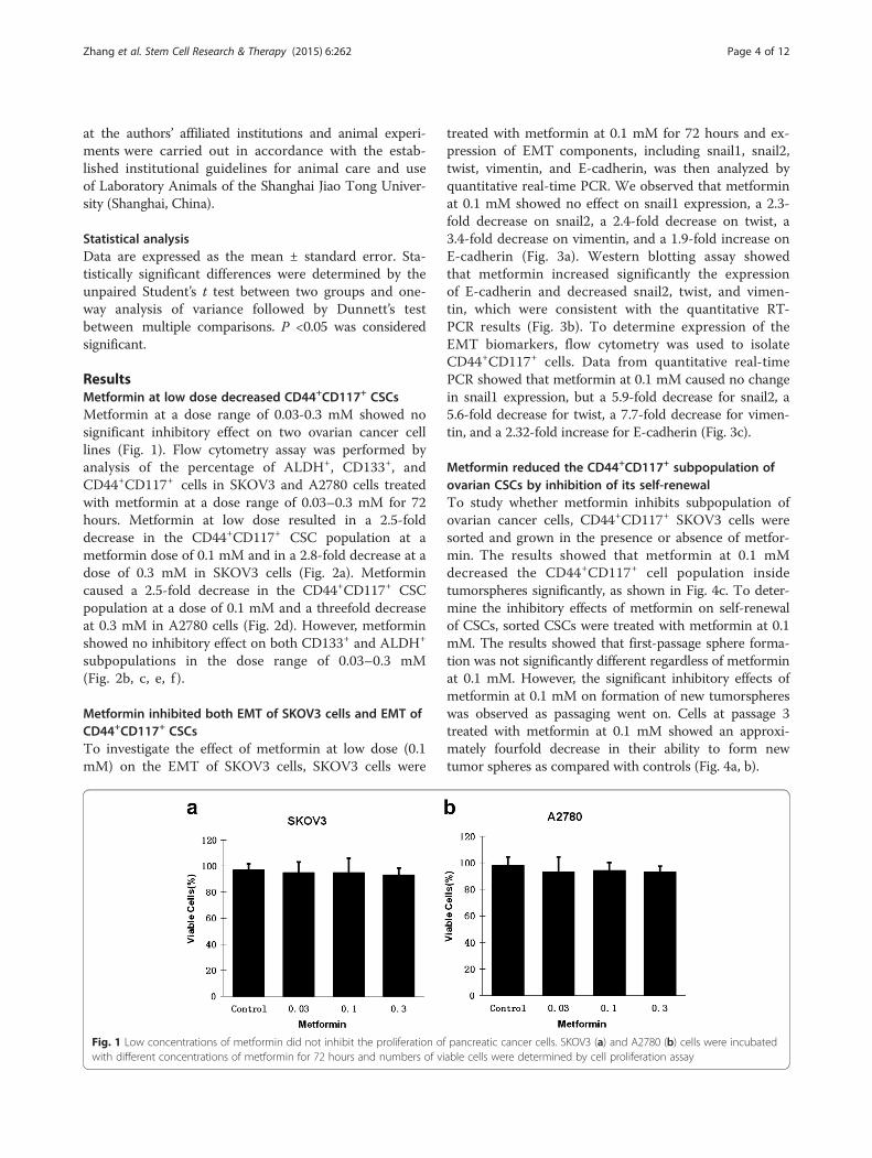
ResultsMetformin at low dose decreased CD44+CD117+ CSCsMetformin at a dose range of 0.03-0.3 mM showed nosignificant inhibitory effect on two ovarian cancer celllines (Fig. 1). Flow cytometry assay was performed byanalysis of the percentage of ALDH+, CD133+, andCD44+CD117+ cells in SKOV3 and A2780 cells treatedwith metformin at a dose range of 0.03–0.3 mM for 72hours. Metformin at low dose resulted in a 2.5-folddecrease in the CD44+CD117+ CSC population at ametformin dose of 0.1 mM and in a 2.8-fold decrease at adose of 0.3 mM in SKOV3 cells (Fig. 2a). Metformincaused a 2.5-fold decrease in the CD44+CD117+ CSCpopulation at a dose of 0.1 mM and a threefold decreaseat 0.3 mM in A2780 cells (Fig. 2d). However, metforminshowed no inhibitory effect on both CD133+ and ALDH+
subpopulations in the dose range of 0.03–0.3 mM(Fig. 2b, c, e, f ).
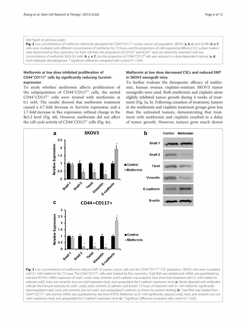
Metformin inhibited both EMT of SKOV3 cells and EMT ofCD44+CD117+ CSCsTo investigate the effect of metformin at low dose (0.1mM) on the EMT of SKOV3 cells, SKOV3 cells were
treated with metformin at 0.1 mM for 72 hours and ex-pression of EMT components, including snail1, snail2,twist, vimentin, and E-cadherin, was then analyzed byquantitative real-time PCR. We observed that metforminat 0.1 mM showed no effect on snail1 expression, a 2.3-fold decrease on snail2, a 2.4-fold decrease on twist, a3.4-fold decrease on vimentin, and a 1.9-fold increase onE-cadherin (Fig. 3a). Western blotting assay showedthat metformin increased significantly the expressionof E-cadherin and decreased snail2, twist, and vimen-tin, which were consistent with the quantitative RT-PCR results (Fig. 3b). To determine expression of theEMT biomarkers, flow cytometry was used to isolateCD44+CD117+ cells. Data from quantitative real-timePCR showed that metformin at 0.1 mM caused no changein snail1 expression, but a 5.9-fold decrease for snail2, a5.6-fold decrease for twist, a 7.7-fold decrease for vimen-tin, and a 2.32-fold increase for E-cadherin (Fig. 3c).
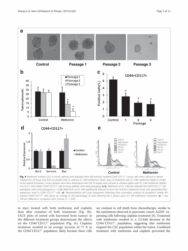
Metformin reduced the CD44+CD117+ subpopulation ofovarian CSCs by inhibition of its self-renewalTo study whether metformin inhibits subpopulation ofovarian cancer cells, CD44+CD117+ SKOV3 cells weresorted and grown in the presence or absence of metfor-min. The results showed that metformin at 0.1 mMdecreased the CD44+CD117+ cell population insidetumorspheres significantly, as shown in Fig. 4c. To deter-mine the inhibitory effects of metformin on self-renewalof CSCs, sorted CSCs were treated with metformin at 0.1mM. The results showed that first-passage sphere forma-tion was not significantly different regardless of metforminat 0.1 mM. However, the significant inhibitory effects ofmetformin at 0.1 mM on formation of new tumorsphereswas observed as passaging went on. Cells at passage 3treated with metformin at 0.1 mM showed an approxi-mately fourfold decrease in their ability to form newtumor spheres as compared with controls (Fig. 4a, b).
Fig. 1 Low concentrations of metformin did not inhibit the proliferation of pancreatic cancer cells. SKOV3 (a) and A2780 (b) cells were incubatedwith different concentrations of metformin for 72 hours and numbers of viable cells were determined by cell proliferation assay
Zhang et al. Stem Cell Research & Therapy (2015) 6:262 Page 4 of 12
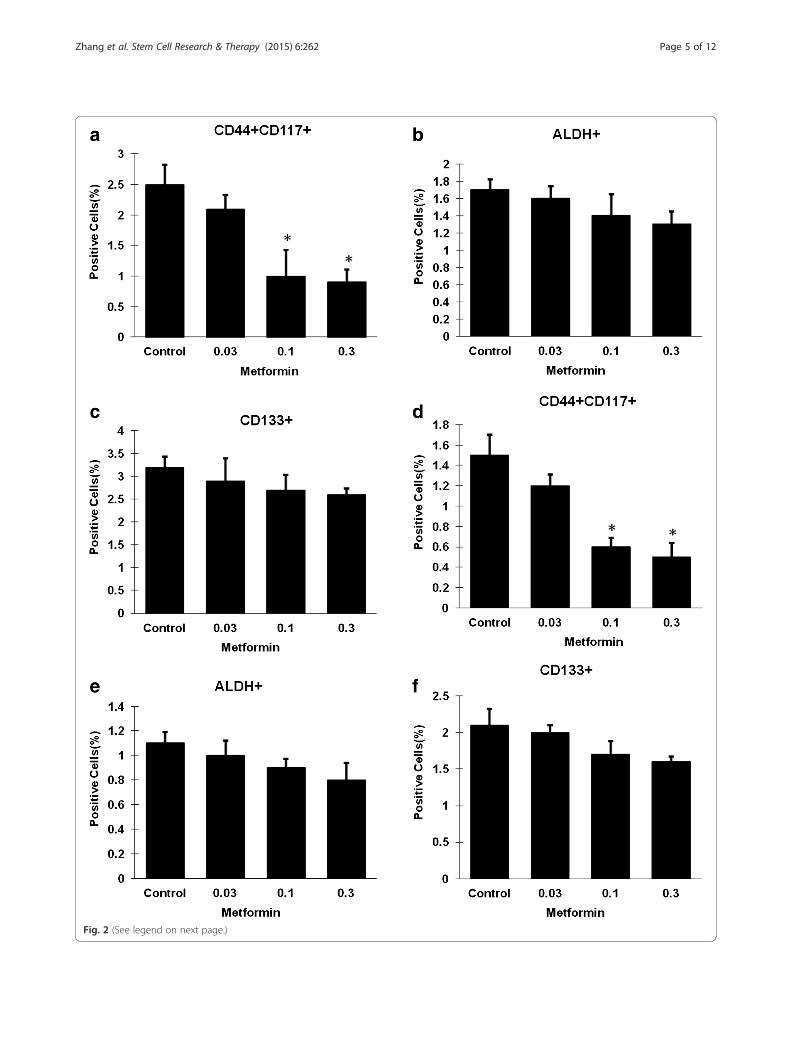
Fig. 2 (See legend on next page.)
Zhang et al. Stem Cell Research & Therapy (2015) 6:262 Page 5 of 12
Metformin at low dose inhibited proliferation ofCD44+CD117+ cells by significantly reducing SurvivinexpressionTo study whether metformin affects proliferation ofthe subpopulation of CD44+CD117+ cells, the sortedCD44+CD117+ cells were treated with metformin at0.1 mM. The results showed that metformin treatmentcaused a 6.7-fold decrease in Survivin expression, and a1.7-fold increase in Bax expression, without change in theBcl-2 level (Fig. 4d). However, metformin did not affectthe cell cycle activity of CD44+CD117+ cells (Fig. 4e).
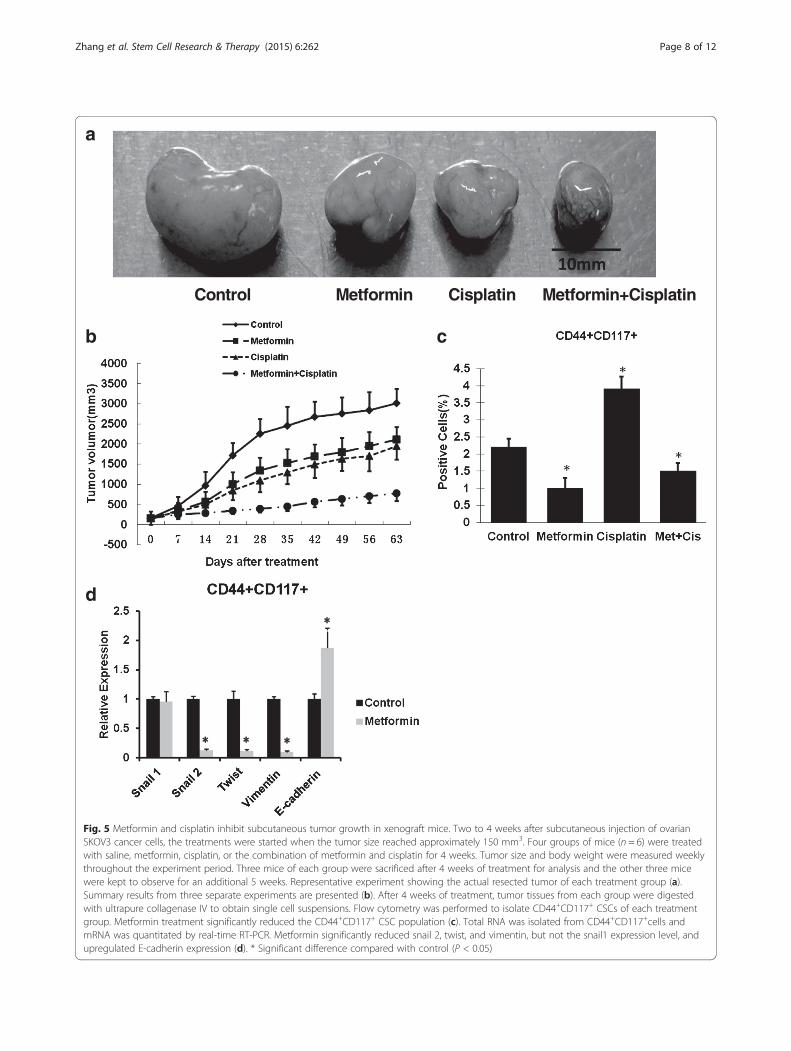
Metformin at low dose decreased CSCs and reduced EMTin SKOV3 xenograft miceTo further evaluate the therapeutic efficacy of metfor-min, human ovarian cisplatin-resistant SKOV3 tumorxenografts were used. Both metformin and cisplatin aloneslightly inhibited tumor growth during 4 weeks of treat-ment (Fig. 5a, b). Following cessation of treatment, tumorsin the metformin and cisplatin treatment groups grew lessthan the untreated tumors, demonstrating that treat-ment with metformin and cisplatin resulted in a delayof tumor growth. However, tumors grew much slower
Fig. 3 Low concentrations of metformin reduced EMT of ovarian cancer cells and the CD44+CD117+ CSC population. SKOV3 cells were incubatedwith 0.1 mM metformin for 72 hours. The CD44+CD117+ cells were isolated by flow cytometry. Total RNA was isolated and mRNA was quantitated byreal-time RT-PCR. mRNA expression of snail1, snail2, twist, vimentin, and E-cadherin was analyzed. Data show that treatment with 0.1 mM metforminreduced snail2, twist, and vimentin, but not snail1expression level, and upregulated the E-cadherin expression level (a). Bands detected with antibodiesindicate the immune reactivity for snail1, snail2, twist, vimentin, E-cadherin, and β-actin: 72 hours of treatment with 0.1 nM metformin significantlydownregulated snail2, twist, and vimentin, but not snail1, and upregulated E-cadherin as shown by western blotting (b). Total RNA was isolated fromCD44+CD117+ cells and the mRNA was quantitated by real-time RT-PCR. Metformin at 0.1 mM significantly reduced snail2, twist, and vimentin, but notsnail1 expression level, and upregulated the E-cadherin expression level (c). * Significant difference compared with control (P < 0.05)
(See figure on previous page.)Fig. 2 Low concentrations of metformin selectively decreased the CD44+CD117+ ovarian cancer cell population. SKOV3 (a, b, c) and A2780 (d, e, f)cells were incubated with different concentrations of metformin for 72 hours, and the proportions of cells expressing different CSC surface markerswere determined by flow cytometry. For both cell lines, the proportions of CD133+ and ALDH+ were not altered by treatment with lowconcentrations of metformin (0.03–0.3 mM) (b, c, e, f), but the proportion of CD44+CD117+cells was reduced in a dose-dependent manner (a, d).ALDH aldehyde dehydrogenase. * Significant difference compared with control (P < 0.05)
Zhang et al. Stem Cell Research & Therapy (2015) 6:262 Page 6 of 12
in mice treated with both metformin and cisplatinthan after cessation of both treatments (Fig. 5b).FACS plots of sorted cells harvested from tumors inthe different treatment groups demonstrate the effectson the CD44+CD117+ population (Fig. 5c). Cisplatintreatment resulted in an average increase of 77 % inthe CD44+CD117+ population, likely because these cells
are resistant to cell death from chemotherapy, similar tothe enrichment observed in pancreatic cancer ALDH+-ex-pressing cells following cisplatin treatment [6]. Treatmentwith metformin resulted in a 2.2-fold decrease in theCD44+CD117+ population, suggesting that metformintargeted the CSC population within the tumor. Combinedtreatment with metformin and cisplatin prevented the
Fig. 4 Metformin inhibits CSCs in tumor spheres and regulates their self-renewal. Isolated CD44+CD117+ cancer cells were cultured in spheremedium for 24 hours and then incubated with or without 0.1 mM metformin. Seven days of treatment with 0.1 mM metformin failed to inhibittumor sphere formation. Tumor spheres were then dissociated with 0.05 % trypsin and cultured in ultralow plates with 0.1 mM metformin. Metfor-min at 0.1 mM inhibits CD44+CD117+ cells forming spheres with serial passaging (a, b). Metformin at 0.1 mM also reduced the CD44+CD117+ cellpopulation with serial passaging (n = 3) (c). Metformin at 0.1 mM significantly reduced Survivin but not Bcl-2 expression level, and upregulated Baxexpression level in CD44+CD117+ cells (d). Representative cell cycle histograms following flow cytometric analysis of propidium iodide (PI)-stained CD44+CD117+ cells show no change in the percentage of cells entering the S phase upon 0.1 nM metformin treatment (e). * Sig-nificant difference compared with control (P < 0.05)
Zhang et al. Stem Cell Research & Therapy (2015) 6:262 Page 7 of 12
a
b
d
c
Fig. 5 Metformin and cisplatin inhibit subcutaneous tumor growth in xenograft mice. Two to 4 weeks after subcutaneous injection of ovarianSKOV3 cancer cells, the treatments were started when the tumor size reached approximately 150 mm3. Four groups of mice (n = 6) were treatedwith saline, metformin, cisplatin, or the combination of metformin and cisplatin for 4 weeks. Tumor size and body weight were measured weeklythroughout the experiment period. Three mice of each group were sacrificed after 4 weeks of treatment for analysis and the other three micewere kept to observe for an additional 5 weeks. Representative experiment showing the actual resected tumor of each treatment group (a).Summary results from three separate experiments are presented (b). After 4 weeks of treatment, tumor tissues from each group were digestedwith ultrapure collagenase IV to obtain single cell suspensions. Flow cytometry was performed to isolate CD44+CD117+ CSCs of each treatmentgroup. Metformin treatment significantly reduced the CD44+CD117+ CSC population (c). Total RNA was isolated from CD44+CD117+cells andmRNA was quantitated by real-time RT-PCR. Metformin significantly reduced snail 2, twist, and vimentin, but not the snail1 expression level, andupregulated E-cadherin expression (d). * Significant difference compared with control (P < 0.05)
Zhang et al. Stem Cell Research & Therapy (2015) 6:262 Page 8 of 12
increase in the CSC population observed with cisplatintreatment alone, resulting in a 2.6-fold decrease in theCD44+CD117+ population compared with cisplatin treat-ment alone, which increased in the CSC population, fur-ther demonstrating that metformin is capable of targetingthe CSC population (Fig. 5c). The effect of metformin on
EMT markers’ expression of CD44+CD117+ cells wasanalyzed by real-time RT-PCR. Data demonstrated that0.1 mM metformin had no effect on snail1 expression, butled to a 7.7-fold decrease for snail2, an 8.3-fold decreasefor twist, a 10-fold decrease for vimentin, and a 1.9-foldincrease for E-cadherin (Fig. 5d).
a
c
b
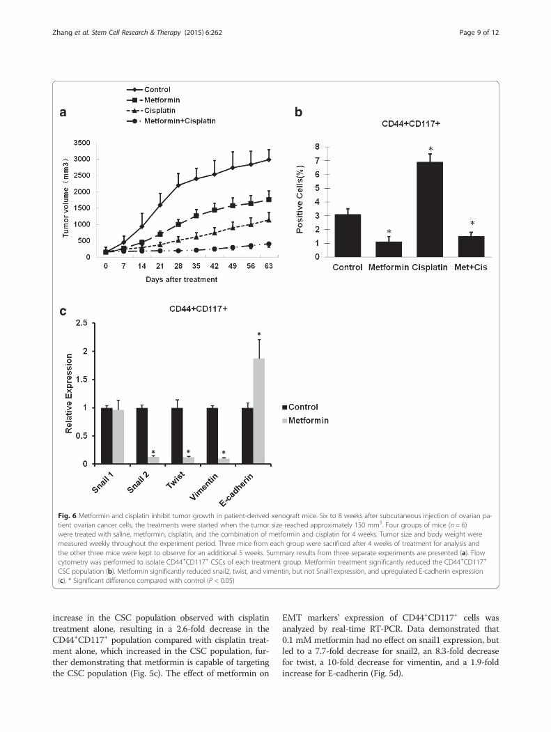
Fig. 6 Metformin and cisplatin inhibit tumor growth in patient-derived xenograft mice. Six to 8 weeks after subcutaneous injection of ovarian pa-tient ovarian cancer cells, the treatments were started when the tumor size reached approximately 150 mm3. Four groups of mice (n = 6)were treated with saline, metformin, cisplatin, and the combination of metformin and cisplatin for 4 weeks. Tumor size and body weight weremeasured weekly throughout the experiment period. Three mice from each group were sacrificed after 4 weeks of treatment for analysis andthe other three mice were kept to observe for an additional 5 weeks. Summary results from three separate experiments are presented (a). Flowcytometry was performed to isolate CD44+CD117+ CSCs of each treatment group. Metformin treatment significantly reduced the CD44+CD117+
CSC population (b). Metformin significantly reduced snail2, twist, and vimentin, but not Snail1expression, and upregulated E-cadherin expression(c). * Significant difference compared with control (P < 0.05)
Zhang et al. Stem Cell Research & Therapy (2015) 6:262 Page 9 of 12
Metformin at low dose decreased CSCs and reduced EMTin primary ovarian cancer xenograft miceData indicated that treatment with metformin or cisplatinalone resulted in a delay of tumor growth. However, in thegroup treated with metformin and cisplatin, tumor growthwas extremely slow, even near the cessation of treatment,showing a significant enhanced anti-tumor effect (Fig. 6a).The effect of metformin on expression of EMT markerCD44+CD117+ was analyzed by real-time RT-PCR. Weobtained similar results when measuring the effect ofmetformin, used alone or in combination with cisplatin,on the CD44+CD117+ CSC population (Fig. 6b). Treat-ment with metformin resulted in a 2.8-fold decrease inthe CD44+CD117+ population, and an increase of 2.2-fold for cisplatin, and combined treatment with met-formin and cisplatin resulted in a 4.6-fold decrease inthe CD44+CD117+ population compared with cisplatinalone, and even a 2.1-fold decrease in the CD44+CD117+
population compared with control, further demonstratingthat metformin is capable of targeting the CSC population(Fig. 6b). Real-time RT-PCR experiment demonstratedthat metformin had no effect on Snail 1 expression, butled to a 11.1-fold decrease in Snail 2, a 10-fold decrease inTwist, a 11.1-fold decrease in Vimentin expression, and a2.9-fold increase in E-cadherin expression (Fig. 6c).
DiscussionIn this study, we demonstrated that low concentrationsof metformin selectively inhibited CD44+CD117+ ovar-ian CSCs through downregulation of EMT. Moreover,we showed that low concentrations of metformin down-regulated snail2, twist, and vimentin, leading to inhib-ition of EMT, and downregulated Survivin expression toincrease apoptosis in the CD44+CD117+ cell population.The efficacy study indicated that metformin also po-tentiated the effect of cisplatin. Therefore, low concen-trations of metformin may be used as a therapeuticagent for killing ovarian CSCs.Of clinical importance, CSCs have been hypothesized
to be resistant to conventional chemotherapy and radi-ation therapy and to be responsible for cancer metastasisand recurrence after clinical remission. For ovarian can-cer, CSCs are also believed to be resistant to chemother-apy and radiation therapy [23–25]. Metformin was activeagainst ovarian cancer cells in vitro and in vivo. Also,metformin inhibited the growth of metastatic nodules inthe lung and significantly potentiated cisplatin-inducedcytotoxicity, resulting in approximately 90 % reductionin tumor growth in ovarian cancers [26]. Shank et al. [6]showed that metformin acted on ovarian CSCs, reducingthe percentage of ALDH+ CSCs in vitro and in vivo,and inhibiting the growth of ovarian tumor spheres. Inaddition, metformin therapy alone slowed the growthof ovarian CSCs in vivo. Targeting Notch pathway of
ovarian CSCs sensitized tumors to cisplatin therapy[27]. Low-dose metformin may reprogram ovarian can-cer cells or CSCs into noncancerous cells [22]. Thesestudies indicate that cisplatin treatment enriches theovarian CSC population and that current treatmentsfor ovarian cancer do not address the unique survivalmechanisms of ovarian CSCs. It is therefore essentialto better understand CSC so that specific therapiescan be devised to target this population of cancercells. Our in vivo data are consistent with these earlyfindings, highlighting the significance of low-dose met-formin in targeting CSCs.Ovarian CSCs possess mesenchymal characteristics
and EMT ability [28]. Ovarian CSCs could be a sourceof ovarian cancer metastasis through EMT and regula-tion of TWIST-1 expression, and function is a criticalstep in this process [29]. Ovarian CSCs that undergo theEMT have demonstrated that the tumor cells are ingeneral less differentiable, more invasive, more chemore-sistant, and result in poor clinical outcomes [29–31].Chen et al. [9] demonstrated that the overexpression ofmiR-200c significantly reduced the CD117+CD44+ CSCxenograft growth and lung metastasis in vivo, partiallythrough the reversal of the EMT phenotype. Snail1 andsnail2 have also been associated with chemoresistanceand activation of stemness-associated pathways [32, 33].Craveiro et al. [34] showed that snail2 is one of the moresignificantly upregulated genes in paclitaxel-survivingCSCs and snail2 overexpression was sufficient to yieldmore aggressive and more chemoresistant tumors. In-hibition of the EMT process by snail1 silencing reducedthe side population cell frequency, and affected their in-vasive capacity and engraftment [35]. We therefore in-vestigated the effect of metformin on EMT in CSCs,focusing on snail1, snail2, and twist, and established theconnection between metformin and EMT of ovarianCSCs.We firstly studied the effect of metformin at low
concentration on the CSC population and found thatlow concentrations of metformin selectively inhibitedCD44+CD117+ cells, but not CD133+ or ALDH+ cells,likely through downregulated EMT. We found thatcisplatin could only kill the rapidly increased ovariantumor cells, while it could not inhibit the populationof CSCs. This is one of the reasons why many tumorsare recurring and resistant to chemotherapy drugs,such as cisplatin. To study the effect of low concen-trations of metformin on the function of CSCs, weutilized an in vitro sphere-forming assay. We found thatusing low concentrations of metformin decreased the CSCpopulation and EMT. Our results are consistent with pre-vious work indicating that low-dose metformin inhibitsCSCs in other cancer types, such as pancreatic cancer andbreast cancer [18, 21].
Zhang et al. Stem Cell Research & Therapy (2015) 6:262 Page 10 of 12
The underlying molecular mechanisms of metformininhibiting EMT in CSCs are not well understood. Lee etal. [36] reported that induction of EMT in human retinalpigment epithelial cells upregulates Survivin, leading toSurvivin-dependent inhibition of cell cycle arrest andapoptosis. It has been reported that drug resistance ofovarian CSCs were linked with Survivin [37]. Anotherstudy indicated that metformin induced apoptosis inovarian cancer cells without significant cytotoxicity [38].We found that metformin treatment resulted in apop-tosis of ovarian CSCs by downregulating Survivin andupregulating Bax. These results suggest that low concen-trations of metformin reduce survival of ovarian CSCs.We analyzed the efficacy of targeting ovarian CSCs in
xenografts of established ovarian cancers in nude miceand found that treatment with low-dose metformin inboth SKOV3 and primary xenograft models resultedin a marked decrease in the ovarian CSC population.Both treatments with low-dose metformin alone orcisplatin alone did not considerably inhibit tumorgrowth, while treatment with both agents significantlyblocked tumor growth, even up to 5 weeks after ces-sation of treatment. Low-dose metformin significantlyinhibited CD44+CD177+ CSCs and EMT in vivo, which isconsistent with our in vitro data. These data suggest thatusing a combined therapy that targets both the ovarianCSC population and the nontumorigenic bulk populationof ovarian cancer cells may be most efficacious in treatingpatient symptoms associated with tumor mass and resultin eradication of the CSC population.As evidence mounts to support the paradigm that a
small subset of cancer cells dictate the biologic behaviorof malignant disease, approaches in designing more effect-ive therapeutic strategies must consider a new target—theCSCs. Data have shown that ovarian cancers contain asmall population of CSCs responsible for tumor initiationand propagation and resistance to conventional chemo-therapy and radiation. Here we found that targeting lowconcentrations of metformin may be useful in eradicatingCSCs and potentiate conventional chemotherapy. Thisstudy provides data to support further exploration of lowconcentrations of metformin, as a therapeutic target toeradicate CSCs from human ovarian tumors, which mayprevent ovarian cancer recurrence and improve long-termsurvival.
ConclusionsThis research indicates that low concentrations of met-formin selectively inhibit CD44+CD117+ ovarian CSCsthrough inhibitions of EMT and potentiate the effect ofcisplatin. The present study may support the new clinicalapplications of metformin, which will prevent ovariancancer recurrence and will improve long-term survival.
AbbreviationsALDH: Aldehyde dehydrogenase; APC: Allophycocyanin; CSC: Cancer stemcell; DAPI: 4′,6-Diamidino-2-phenylindole; DEAB: Diethylaminobenzaldehyde;EMT: Epithelial–mesenchymal transition; EOC: Epithelial ovarian cancer;FBS: Fetal bovine serum; HBSS: Hanks’ buffered salt solution;i.p.: Intraperitoneally; PBS: Phosphate-buffered saline.
Competing interestsThe authors declare that they have no competing interests.
Authors’ contributionsRZ carried out all cell-culture experiments, PCR, and western blotting ana-lyses, and performed some of the statistical analyses. PZ and HW carried outthe in vivo experiments and performed some of the statistical analyses. RZ,PZ, HW, and DH participated in the design and coordination of the studyand helped to draft and revise the manuscript. WL and CL conceived of, de-signed, and coordinated the study, helped with the statistical analyses, andrevised the manuscript. GX revised the manuscript thoroughly. All authorsread and approved the final manuscript.
AcknowledgementsThe work was supported by Natural Science Foundation of China (81402819).
Author details1Department of Gynecology, Xinhua Hospital, School of Medicine, ShanghaiJiao Tong University, Shanghai 200092, China. 2Department ofOtolaryngology & Head and Neck Surgery, Xinhua Hospital, School ofMedicine, Shanghai Jiao Tong University, Shanghai 200092, China.3Department of Interventional Radiology, Cancer Hospital, Fudan University,Shanghai 200032, China. 4School of Pharmaceutical Science and Technology,Dalian University of Technology, Dalian 116024, China. 5SunstemBiotechnology Co., Ltd, Shanghai 200439, China.
Received: 16 February 2015 Revised: 21 March 2015Accepted: 30 November 2015
References1. Siegel R, Ward E, Brawley O. Cancer statistics, 2011. CA Cancer J Clin.
2011;61:212–36.2. Bast Jr RC, Hennessy B, Mills GB. The biology of ovarian cancer: new
opportunities for translation. Nat Rev Cancer. 2009;9:415–28.3. Boman BM, Wicha MS. Cancer stem cells: a step toward the cure. J Clin
Oncol. 2008;26:2795–9.4. Burgos-Ojeda D, Rueda BR, Buckanovich RJ. Ovarian cancer stem cell
markers: prognostic and therapeutic implications. Cancer Lett. 2012;322:1–7.5. Silva IA, Bai S, McLean K, Yang K, Griffith K, Thomas D. Aldehyde
dehydrogenase in combination with CD133 defines angiogenicovarian cancer stem cells that portend poor patient survival. CancerRes. 2011;71:3991–4001.
6. Shank JJ, Yang K, Ghannam J. Metformin targets ovarian cancer stem cellsin vitro and in vivo. Gynecol Oncol. 2012;127(2):390–7.
7. Deng S, Yang X, Lassus H, Liang S, Kaur S, Ye Q, et al. Distinct expressionlevels and patterns of stem cell marker, aldehyde dehydrogenase isoform 1(ALDH1), in human epithelial cancers. PLoS One. 2010;5(4):e10277.
8. Zhang S, Balch C, Chan MW, Lai HC, Matei D, Schilder JM, et al.Identification and characterization of ovarian cancer-initiating cells fromprimary human tumors. Cancer Res. 2008;68(11):4311–20.
9. Chen D, Zhang Y, Wang J. MicroRNA-200c over expression inhibits tumorigenicity and metastasis of CD117+CD44+ ovarian cancer stem cells byregulating epithelial-mesenchymal transition. J Ovarian Res. 2013;6(1):50.
10. Ferrandina G, Bonanno G, Pierelli L. Expression of CD133-1 and CD133-2 inovarian cancer. Int J Gynecol Cancer. 2008;18:506–14.
11. Curley MD, Therrien VA, Cummings CL. CD133 expression defines atumorinitiating cell population in primary human ovarian cancer. Stem Cells.2009;27(12):2875–83.
12. Gotlieb WH, Saumet J, Beauchamp MC, Gu J, Lau S, Pollak MN. In vitrometformin antineoplasticactivity in epithelial ovarian cancer. Gynecol Oncol.2008;110:246–50.
13. Thiery JP, Sleeman JP. Complex networks orchestrate epithelial-mesenchymal transitions. Nat Rev Mol Cell Biol. 2006;7(2):131–42.
Zhang et al. Stem Cell Research & Therapy (2015) 6:262 Page 11 of 12
14. Mani SA, Guo W, Liao MJ, Eaton EN, Ayyanan A, Zhou AY. The epithelial-mesenchymal transition generates cells with properties of stem cells. Cell.2008;133(4):704–15.
15. Thiery JP. Epithelial-mesenchymal transitions in development andpathologies. Curr Opin Cell Biol. 2003;15(6):740–6.
16. Sarrio D, Rodriguez-Piniella SM, Harrison D. Epithelial-mesenchymaltransition in breast cancer relates to the basal-like phenotype. Cancer Res.2008;68:989–97.
17. Morel AP, Lievre M, Thomas C. Generation of breast cancerstem cellsthrough epithelial-mesenchymal transition. PLoS One. 2008;3:e2888.
18. Vazquez-Martin A, Oliveras-Ferraros C, Cufi S. Metformin regulates breastcancer stem cell ontogeny by transcriptional regulation of the epithelial-mesenchymal transition (EMT) status. Cell Cycle. 2010;9:3807–14.
19. Hirsch HA, Iliopoulos D, Tsichlis PN, Struhl K. Metformin selectively targetscancer stem cells, and acts together with chemotherapy to block tumorgrowth and prolong remission. Cancer Res. 2009;69:7507–11.
20. Kurrey NK, Jalgaonkar SP, Joglekar AV. Snail and slug mediateradioresistance and chemoresistance by antagonizing p53-mediatedapoptosis and acquiring a stem-like phenotype in ovarian cancer cells. StemCells. 2009;27(9):2059–68.
21. Gou S, Cui P, Li X. Low concentrations of metformin selectively inhibitCD133+cell proliferation in pancreatic cancer and have anticancer action.PLoS One. 2013;8(5):e63969.
22. Hu T, Chung YM, Guan M. Reprogramming ovarian and breast cancer cellsinto non-cancerous cells by low-dose metformin or SN-38 through FOXO3activation. Sci Rep. 2014;4:5810.
23. Tomao F, Papa A, Rossi L. Current status of bevacizumab inadvancedovarian cancer. Onco Targets Ther. 2013;6:889–99.
24. Ishii H, Iwatsuki M, Ieta K. Cancer stem cells and chemoradiation resistance.Cancer Sci. 2008;99:1871–7.
25. Jordan CT, Guzman ML, Noble M. Cancer stem cells. N Engl J Med.2006;355:1253–61.
26. Rattan R, Graham RP, Maguire JL. Metformin suppresses ovarian cancergrowth and metastasis with enhancement of cisplatin cytotoxicity in vivo.Neoplasia. 2011;13(5):483–91.
27. McAuliffe SM, Morgan SL, Wyant GA. Targeting Notch, a key pathway forovarian cancer stem cells, sensitizes tumors to platinum therapy. Proc NatlAcad Sci U S A. 2012;109(43):E2939–48.
28. Jiao J, Huang L, Ye F. Cyclin D1 affects epithelial-mesenchymaltransition in epithelial ovarian cancer stem cell-like cells. Onco TargetsTher. 2013;6:667–77.
29. Yin G, Alvero AB, Craveiro V. Constitutive proteasomal degradation ofTWIST-1 in epithelial-ovarian cancer stem cells impacts differentiation andmetastatic potential. Oncogene. 2013;32(1):39–49.
30. Cao L, Shao M, Schilder J. Tissue transglutaminase links TGF-β, epithelial tomesenchymal transition and a stem cell phenotype in ovarian cancer.Oncogene. 2012;31(20):2521–34.
31. Cittelly DM, Dimitrova I, Howe EN. Restoration of miR-200c toovarian cancerreduces tumor burden and increases sensitivity topaclitaxel. Mol CancerTher. 2012;11:2556–65.
32. Peinado H, Olmeda D, Cano A. Snail, Zeb and bHLH factors in tumorprogression: an alliance against the epithelial phenotype? Nat Rev Cancer.2007;7:415–28.
33. Singh A, Settleman J. EMT, cancer stem cells and drug resistance: anemerging axis of evil in the war oncancer. Oncogene. 2010;29:4741–51.
34. Craveiro V, Yang-Hartwich Y, Holmberg JC. Phenotypic modifications inovarian cancer stem cells following paclitaxel treatment. Cancer Med.2013;2(6):751–62.
35. Jiang H, Lin X, Liu Y. Transformation of epithelial ovarian cancer stem likecells into mesenchymal lineage via EMT results in cellular heterogeneity andsupports tumor engraftment. Mol Med. 2012;18:1197–208.
36. Lee J, Choi JH, Joo CK. TGF-β1 regulates cell fate during epithelial-mesenchymal transition by upregulating Survivin. Cell Death Dis.2013;4:e714.
37. Dong Z, Yang L, Lai D. KLF5 strengthens drug resistance of ovariancancer stem-like cells by regulating Survivin expression. Cell Proliferat.2013;46(4):425–35.
38. Yasmeen A, Beauchamp MC, Piura E. Induction of apoptosis bymetformin inepithelial ovarian cancer: involvement of the Bcl-2 family proteins. GynecolOncol. 2011;121:492–8.
• We accept pre-submission inquiries
• Our selector tool helps you to find the most relevant journal
• We provide round the clock customer support
• Convenient online submission
• Thorough peer review
• Inclusion in PubMed and all major indexing services
• Maximum visibility for your research
Submit your manuscript atwww.biomedcentral.com/submit
Submit your next manuscript to BioMed Central and we will help you at every step:
Zhang et al. Stem Cell Research & Therapy (2015) 6:262 Page 12 of 12