innovative diagnostic tools for early detection of ... dementia 2014.pdf · review article...
TRANSCRIPT

Alzheimer’s & Dementia - (2014) 1-18
Review Article
Innovative diagnostic tools for early detection of Alzheimer’s disease
Christoph Laskea,b,*, Hamid R. Sohrabic,d, Shaun M. Froste,f, Karmele L�opez-de-Ipi~nag,Peter Garrardh, Massimo Buscemai,j, Justin Dauwelsk, Surjo R. Soekadarl, Stephan Muellerl,
Christoph Linnemannl, Stephanie A. Bridenbaughm, Yogesan Kanagasingame,f,Ralph N. Martinsc,d, Sid E. O’Bryantn
aSection for Dementia Research, Hertie-Institute of Clinical Brain Research and Department of Psychiatry and Psychotherapy, University of T€ubingen,
T€ubingen, GermanybGerman Center for Neurodegenerative Diseases (DZNE), T€ubingen, Germany
cCenter of Excellence for Alzheimer’s Disease Research and Care, School of Medical Sciences, Edith Cowan University, Perth, WA, AustraliadMcCusker Alzheimer’s Research Foundation, School of Psychiatry and Clinical Neurosciences, University of Western Australia, Perth, WA, Australia
eCommonwealth Scientific and Industrial Research Organisation (CSIRO), Perth, AustraliafAustralian e-Health Research Centre, Perth, WA, Australia
gSystems Engineering and Automation Department, University of the Basque Country UPV/EHU, Donostia, SpainhNeuroscience Research Center, Cardiovascular and Cell Sciences Research Institute, St George’s University of London, London, United Kingdom
iSemeion Research Centre of Sciences of Communication, Rome, ItalyjDepartment of Mathematical and Statistical Sciences, Center for Computational and Mathematical Biology, University of Colorado, Denver, CO, USA
kSchool of Electrical and Electronic Engineering, Nanyang Technological University, SingaporelDepartment of Psychiatry and Psychotherapy, University of T€ubingen, T€ubingen, Germany
mFelix Platter Hospital, University Center for Medicine of Aging Basel, Basel Mobility Center, Basel, SwitzerlandnDepartment of Internal Medicine & Institute for Aging and Alzheimer’s Disease Research, University of North Texas Health Science Center,
Fort Worth, TX, USA
Abstract Current state-of-the-art diagnostic measures of Alzheimer’s disease (AD) are invasive (cerebro-
The authors have
*Correspondingau
E-mail address: ch
1552-5260/$ - see fro
http://dx.doi.org/10.10
spinal fluid analysis), expensive (neuroimaging) and time-consuming (neuropsychological assess-ment) and thus have limited accessibility as frontline screening and diagnostic tools for AD. Thus,there is an increasing need for additional noninvasive and/or cost-effective tools, allowing identifica-tion of subjects in the preclinical or early clinical stages of AD who could be suitable for furthercognitive evaluation and dementia diagnostics. Implementation of such tests may facilitate earlyand potentially more effective therapeutic and preventative strategies for AD. Before applyingthem in clinical practice, these tools should be examined in ongoing large clinical trials. This reviewwill summarize and highlight the most promising screening tools including neuropsychometric, clin-ical, blood, and neurophysiological tests.� 2014 The Alzheimer’s Association. All rights reserved.
Keywords: Alzheimer’s disease; Diagnostic tools; Screening tests; Noninvasive tests; Early detection
1. Introduction
Alzheimer’s disease (AD) is the leading cause of demen-tia in the elderly, affecting more than 35 million peopleworldwide [1]. Aging populations in developed countries
no potential conflicts of interest to declare.
thor.Tel.:149-7071-2983444;Fax:149-7071-294141.
nt matter � 2014 The Alzheimer’s Association. All rights r
16/j.jalz.2014.06.004
ensure that AD will reach epidemic proportions unless ther-apies are developed to cure or prevent it [2]. Unfortunately,to date nearly all “disease-modifying” experimental inter-ventions for AD have failed to demonstrate clinical benefitsin individuals with symptomatic AD. The most likely expla-nation for these failures is that the drugs were administeredtoo late in the course of the AD neuropathological processes[3]. It is plausible to assume that these therapies will bemore effective when applied before major brain damage
eserved.

C. Laske et al. / Alzheimer’s & Dementia - (2014) 1-182
has occurred which makes the identification of biomarkerssensitive to preclinical or early clinical stages of AD crucial[4]. Whether an earlier treatment start in the preclinicalstage of AD is associated with a better outcome is still un-known and is actually examined in ongoing treatment trials[3]. These trials include the Dominantly Inherited Alz-heimer Network Trial (DIAN-TU; ClinicalTrials.gov num-ber, NCT01760005), Alzheimer’s Prevention Initiative(NCT01998841), and the Anti-Amyloid Treatment inAsymptomatic Alzheimer’s Disease study (A4 Study;NCT02008357). Early-stage identification may also helpto develop new treatments that are more effective at thisstage as it can facilitate monitoring of the response to theintervention. In addition, a positive early diagnosis givesthe patients and their family the necessary time to under-stand the disease, to decide on the life and financial burdensof the disease, and to arrange for the future needs and care ofthe patients.
The current state-of-the-art clinical diagnosis of AD re-quires a specialty clinic and includes a medical examination,neuropsychological testing, neuroimaging, cerebrospinalfluid (CSF) analysis and blood examination. This processis neither time nor cost-effective. Additionally, given therapidly aging global population with an expected dramaticincrease of AD cases, there are insufficient numbers of spe-cialty clinics to meet the growing needs.While CSF and neu-roimaging markers are gold standards for the in vivoassessment of the patients, they are invasive and expensiveand, therefore, have limited utility as frontline screeningand diagnostic tools. In addition, prior work has shownthat nonspecialist clinicians are inaccurate at identifyingearly AD and mild cognitive impairment (MCI) [5], whichis a major impetus to the search for clinically-usefulscreening and diagnostic tools.
Thus, there is an increasing need for additional noninva-sive and/or cost-effective tools, allowing frontline identifica-tion of subjects in the preclinical or early clinical stages ofAD. Further examination of patients with conspicuousnoninvasive cognitive and noncognitive measures could beperformed in a next step by established clinical, CSF and/or neuroimaging analyses in a specialty clinic. The identifi-cation of methods to predict the risk for developing ADwould be of great value for healthcare systems. Identifica-tion of AD risk markers could help to identify individuals
Table 1
US estimates of population age 65 and above for 2009 and 2030 along with estim
Physicians by specialty and MRI
machines in the United States
Number of physicians by specialty
and of MRI machines
Physicians certified in geriatrics [8] 7162
Neurologists [10] 10,154
Psychiatrists [9] 39,457
MRI machines in the United States [11] 10,000
Abbreviation: MRI, magnetic resonance imaging.
who might benefit from early intensive lifestyle consulta-tions and pharmacological interventions. The relevance ofearly diagnosis of AD is supported by recent neuropatholog-ical, biochemical and neuroimaging findings showing thatbiomarkers of AD can be detected in the brains and CSFof approximately 20% to 30% of cognitively healthy elderlyindividuals [6–8].
This review will summarize and highlight the most prom-ising novel noninvasive and/or inexpensive screening anddiagnostic tools such as neuropsychometric, clinical, blood,and neurophysiological tests for early detection of ADbeyond the established clinical, CSF and neuroimaging de-mentia diagnostics.
2. Socioeconomic aspects of dementia diagnostics
While many have argued the need for screening methodsthat are accessible and time- and cost-effective, few haveempirically demonstrated this point. To empirically illus-trate the need for noninvasive and inexpensive screening/diagnostic tools, we use the U.S. numbers of geriatrician,neurology, and psychiatry physician providers and availablemagnetic resonance imaging (MRI) machines below. In theUnited States there were an estimated 7162 physicians certi-fied in geriatrics in 2011 [9]. This translates to 5585 patientsaged 65 years old and above to be seen per specialist per yearbased on 2009 census estimates if all geriatrics were toreceive an annual screening that included cognitive exami-nation. This is particularly problematic when geriatric spe-cialists are becoming less and less available [9]. Table 1outlines the situation for the fields of neurology and psychi-atry as well.
The situation is worse considering that not all geriatri-cians, neurologists or psychiatrists are dementia specialists.Furthermore, psychiatrists and neurologists are aging andworking fewer hours than in the past [10,11], therefore thisis likely a significant overestimation of capacity in thatfield. If one considers MRI as frontline screening tool, thesituation does not improve. There are an estimated 11,000MRI machines within the United States currently [12].This would mean that each MRI machine should be usedfor 3636 US elders with current estimates and for over6000 US elders by the year 2030, based on projected ageestimates. These numbers assume that all MRI machines
ates of physician availability
Population age .65 years in 2009,
n 5 40 million
Population age .65 years in 2030,
n 5 70 million
Patients per physician Patients per physician
5585 9773
3939 6893
1014 1774
3636 6364

C. Laske et al. / Alzheimer’s & Dementia - (2014) 1-18 3
would be exclusively used for dementia screening. If oneconsiders using positron emission tomography (PET) imag-ing machines (e.g. Amyvid�, Elli Lilly), the situation be-comes even more difficult given that there are only anestimated 2000 PET/computed tomography scanners in theUnited States. Again, this does not take into account otherindications for the use of these machines. With both clinicaland imaging modalities, the above estimates do not take intoaccount the need for repeated (i.e., annual) examinations orcost burden. The high costs of MRI and PET machinesfurther limits their use as frontline screeners. Thus, there isneed for inexpensive and/or noninvasive diagnostic toolsthat do not require a specialist. If, for example, a blood-based assessment was available for $200 per person (costsfar above this limit the economic benefit), the cost savingswould be substantial. For example, if PET amyloid beta(Ab) imaging were made available at $1000 per examinationand only 1 million elders were screened annually, the costwould be US $1 billion annually, whereas the cost of a bloodtest would be $200 million annually. If 15% screened posi-tive, and went on to PET Ab imaging, the cost savings ofthis screen–follow-up procedure would be $650 million dol-lars annually. Given that there are approximately 40 millionAmericans age 65 and older, then the examples listed abovesubstantially underestimate the current need and explainwhy many have suggested that AD alone could bankruptmany medical systems if nothing is done immediately con-cerning the development of inexpensive and/or noninvasivescreening tools that do not require a specialist.
3. Neuropsychometric tests
A previous retrospective cohort study examining de-scendants of carriers of the PSEN1 E280 A mutation iden-tified three predementia clinical stages according toneuropsychological assessment: (1) asymptomatic pre-MCI, (2) symptomatic pre-MCI, and (3) MCI [13]. The firstidentifiable clinical stage, called asymptomatic pre-MCI,was detected 11 to 15 years before onset of dementia andwas characterized by neuropsychological test scores twoSD or more away from the mean normal value score fornoncarriers in at least one test on any cognitive domainand the absence of memory complaints and no effect on ac-tivities of daily living. The second clinical stage, calledsymptomatic pre-MCI, was timed 5-11 years before de-mentia and was characterized by additional subjectivememory complaints. The third clinical stage, called MCI,was timed 1 to 5 years before reaching dementia and wascharacterized by higher scores in subjective memory com-plaints without or with minimal impairment in complexinstrumental functions. Thus, subtle cognitive changescould be detected in these subjects even 15 years beforeclinical manifestation of dementia. However, this staginghas been reported in PSEN1 E280 A mutation carriers.The situation in familial AD may be different from thatin sporadic AD where other, age-related factors may
contribute to the preclinical and clinical outcome. There-fore, results from this study may not be directly transfer-able to those in sporadic AD.
3.1. Episodic memory tests
Episodic memory is the first and most severely affectedcognitive domain in AD and in prodromal stages includingamnestic MCI (aMCI) [14]. Several tests can be used toassess episodic memory such as the Logical Memory sub-test from the Wechsler Memory Scale [15], the CaliforniaVerbal Learning Test, now in its second revision (CVLT-II) [16], and the Free and Cued Selective Reminding Test(FCSRT) [17]. A comparison of CVLT and Consortiumto Establish a Registry for Alzheimer’s Disease-Neuropsychological Assessment Battery has shown thatCVLT is more sensitive to preclinical changes in theepisodic memory [18]. The FCSRT free recall was morepredictive than the Wechsler Logical Memory immediaterecall for identifying individuals with memory complaintswho developed incident AD over 2 to 4 years [19]. Among astandardized neuropsychological battery, the FCSRT wasalso the most sensitive and specific test for diagnosis of pro-dromal AD in another study [20]. In addition, the FCSRTbetter predicted the likelihood of an AD-like CSF profileamong MCI subjects than the Wechsler Logical Memorydelayed recall [21].
A large study examining members of families with domi-nantly inherited AD was called the DIAN study. In thisstudy, significant differences between mutation carriersand noncarriers were detected in the Mini-Mental State Ex-amination [22] and the Clinical Dementia Rating-Sum ofBoxes [23] scores at assessments performed five yearsbefore expected symptom onset and thus in the preclinicalstage [24]. In the delayed-recall portion of the Logical Mem-ory test [25], however, significant cognitive impairment wasfound in mutation carriers, as compared with noncarriers,even ten years before expected symptom onset. The LogicalMemory test has also been used in prospective studies forsporadic AD and has predicted AD 10 years before its clin-ical diagnosis [26]. Thus, use of an episodic memory testsuch as the Wechsler Logical Memory test or the FCSRT al-lows early detection of subtle cognitive deficits in both, fa-milial AD and sporadic AD, favoring inclusion of one ofthese tests in a screening battery for detection of preclinicaland early symptomatic AD.
4. Clinical tests
4.1. Assessment of subjective memory complaints
With regard to cognitive decline, objective deficits asmeasurable with specific neuropsychological tests need tobe distinguished from subjective memory complaints(SMC) as reported by the individual or an informant (familymembers, care-givers, or clinician). Currently, there isincreasing interest in SMC and a debate as to whether they

C. Laske et al. / Alzheimer’s & Dementia - (2014) 1-184
aremeaningful or not with respect to the diagnosis of preclin-ical AD. Recent studies have demonstrated that SMCs areassociated with pathological brain amyloid burden in cogni-tively normal older individuals and with increased risk fordevelopment of late-onset AD even before any measurablecognitive decline [27,28]. In addition, a study with carriersof the PSEN1 E280 A mutation identified a pre-MCI stagewith already existing SMC [13]. A recent study demonstratedthat one-third of the adults aged�50 years attending primarycare centers with SMC were already affected by MCI [29].Cognitive impairment as reported by informants (familymembers) was associated with an even higher prevalenceof MCI. Thus, SMC may serve as an indicator of preclinicaland early symptomatic AD and information concerningcognitive impairment in screening for AD and other demen-tias should be obtained from both, the individuals and infor-mants (familymembers or caregivers). Apart from the effectsof depression and personality factors, SMC report could bepotentially a very good time point to start any preventive trial.Diagnostic tools to assess SMC such as the SubjectiveCogni-tive Failures Questionnaire [30,31] or simple questionsconcerning the presence of memory impairment and abouttheir concerns [27,28] are noninvasive and inexpensive andmight therefore be suitable as additional parameters for abroader screening of putative amyloid positive but stillcognitively healthy individuals. The definition of SMCcontinues to be a work in process, especially given thecurrent lack of a single standardized test.
4.2. Assessment of late-onset depression
Depression is common across the lifespan with one in fiveindividuals experiencing a depressive episode during theirlifetime [32]. Dementia is also very common in late lifewith the risk doubling every 5 years after age 65, increasingup to 50% among those greater than 90 years old [33].Although studies have shown that depression and late onsetdementia frequently coexist [34], causality remains contro-versial. Depression has been reported in women carryingpresenilin-1 mutation at preclinical phase of familial AD,supporting the hypothesis that AD neuropathology may beinvolved in depression occurrence at least in this group offemales [35]. Depression or depressive symptoms mayreflect (1) a risk factor for dementia, (2) a prodromal phaseof dementia or (3) a consequence of the dementia neurode-generative processes.
A personal history of depression seems to be a risk factorfor later development of dementia [36–41]. The type ofdementia seems to depend on the time point whendepression occurs during life. In a longitudinal study,Barnes and colleagues [42] compared the risk of AD orvascular dementia (VaD) in those with depressive symptomsat mid- and late-life and found that subjects with late-lifedepressive symptoms had a twofold increase in AD risk,whereas subjects with midlife and late-life symptoms hadmore than a threefold increase in VaD risk.
Depression that presents for the first time in late life mayalso reflect an early symptom of dementia, particularly ofAD. It is assumed that the combination of late-onset depres-sion with currently elevated depressive symptoms mayrepresent an active neurodegenerative process (i.e., a prodro-mal state of dementia), whereas an association between alate-onset depression that remitted and currently shows noelevation of depressive symptoms and subsequent dementiamay represent an indirect effect (i.e., a risk factor for subse-quent dementia) [43]. In line with this, the vast majority oflongitudinal and cross-sectional studies proposed that late-life depression seems to be a prodromal stage of AD[34,43–47]. Further studies demonstrated that the number[36] and severity [37] of depressive symptoms or even a clin-ical diagnosis of depression at baseline in close temporalproximity to dementia [40,44,48] or together withapolipoprotein E (APOE) ε4 status [39] predicted develop-ment of dementia during follow-up with increased riskof AD.
In a recent longitudinal study, Heser and colleagues [43]investigated whether late-onset depression is a risk factorfor a prodrome of AD or for dementia of other etiologiesin a cohort of elderly patients (n 5 2,663, meanage 5 81.2 years). The authors showed that depression pa-rameters and subjective memory impairment predicted ADindependently of objective cognition [43]. The authorsconcluded that late-onset depression with currently elevateddepressive symptoms accompanied by worrisome subjectivememory impairment in the elderly best suggested an AD pa-thology requiring close neuropsychological monitoring.Thus, an assessment for depressive symptoms shouldbe included in a screening battery for the early diagnosisof AD.
4.3. Speech testing
Verbal communication is a complex process, whichdraws on a wide range of cognitive abilities including shortterm memory, knowledge of phonological structure andgrammatical convention, and word meaning. Language isproduced spontaneously in large quantities by all humanson a more or less daily basis, and its recordability (in spokenor written format) makes it one of the easiest biological sam-ples to collect. In addition, the multitude of dimensionsavailable for analysis means that recorded speech is poten-tially one of the most informative biological samples toassay. The deterioration of spoken language immediately af-fects the patient’s ability to interact naturally with his or hersocial environment, and is usually also accompanied by al-terations in emotional responses. Both of these changesappear early in the progress of AD and both can be measuredusing automatic speech analysis techniques. These diag-nostic procedures can (after proper training) be performedby anyone in the patient’s habitual environment, withoutaltering or blocking the patient’s abilities [49,50]. Thesemethodologies also help to estimate the severity of AD in
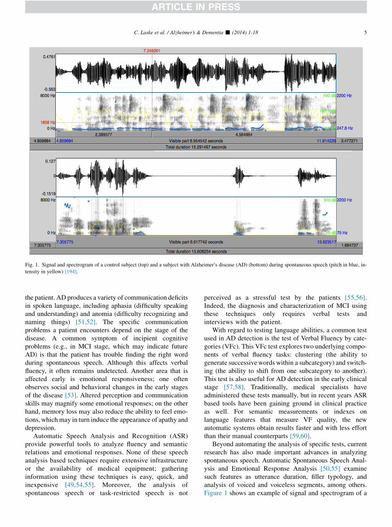
Fig. 1. Signal and spectrogram of a control subject (top) and a subject with Alzheimer’s disease (AD) (bottom) during spontaneous speech (pitch in blue, in-
tensity in yellow) [194].
C. Laske et al. / Alzheimer’s & Dementia - (2014) 1-18 5
the patient. AD produces a variety of communication deficitsin spoken language, including aphasia (difficulty speakingand understanding) and anomia (difficulty recognizing andnaming things) [51,52]. The specific communicationproblems a patient encounters depend on the stage of thedisease. A common symptom of incipient cognitiveproblems (e.g., in MCI stage, which may indicate futureAD) is that the patient has trouble finding the right wordduring spontaneous speech. Although this affects verbalfluency, it often remains undetected. Another area that isaffected early is emotional responsiveness; one oftenobserves social and behavioral changes in the early stagesof the disease [53]. Altered perception and communicationskills may magnify some emotional responses; on the otherhand, memory loss may also reduce the ability to feel emo-tions, which may in turn induce the appearance of apathy anddepression.
Automatic Speech Analysis and Recognition (ASR)provide powerful tools to analyze fluency and semanticrelations and emotional responses. None of these speechanalysis based techniques require extensive infrastructureor the availability of medical equipment; gatheringinformation using these techniques is easy, quick, andinexpensive [49,54,55]. Moreover, the analysis ofspontaneous speech or task-restricted speech is not
perceived as a stressful test by the patients [55,56].Indeed, the diagnosis and characterization of MCI usingthese techniques only requires verbal tests andinterviews with the patient.
With regard to testing language abilities, a common testused in AD detection is the test of Verbal Fluency by cate-gories (VFc). This VFc test explores two underlying compo-nents of verbal fluency tasks: clustering (the ability togenerate successivewords within a subcategory) and switch-ing (the ability to shift from one subcategory to another).This test is also useful for AD detection in the early clinicalstage [57,58]. Traditionally, medical specialists haveadministered these tests manually, but in recent years ASRbased tools have been gaining ground in clinical practiceas well. For semantic measurements or indexes onlanguage features that measure VF quality, the newautomatic systems obtain results faster and with less effortthan their manual counterparts [59,60].
Beyond automating the analysis of specific tests, currentresearch has also made important advances in analyzingspontaneous speech. Automatic Spontaneous Speech Anal-ysis and Emotional Response Analysis [50,55] examinesuch features as utterance duration, filler typology, andanalysis of voiced and voiceless segments, among others.Figure 1 shows an example of signal and spectrogram of a

C. Laske et al. / Alzheimer’s & Dementia - (2014) 1-186
control subject and a subject with AD during spontaneousspeech. Use of spontaneous speech fluency test integratedwith a fractal dimension set allowed discrimination betweenAD patients and healthy controls with a clinically relevantaccuracy of .80% [50]. The analysis of emotional re-sponses includes classical features like pitch, intensity,and variation of frequency components, and, most recently,of Emotional Temperature. This last feature is based on theanalysis of a number of prosodic and paralinguistic features[50]. Emotional response test including emotional tempera-ture allowed discrimination between AD patients andhealthy controls with a high accuracy of .97.7% [50]. Ina similar vein, some researchers have recently proposedthat slight cognitive changes in the early and preclinicalstages could be detected using nonlinear speech parameterssuch as fractals [50].
Differences in aspects of language production andcomprehension embody the distinctive neurodegenerativesyndromes of primary progressive aphasia (PPA), whichare normally underpinned by degenerative pathologies otherthan AD. Three broad subtypes of PPA are recognized: pa-tients with the fluent subtype (also referred to as semanticdementia) produce well-formed speech dominated bygeneric words such as “thing” or “bit”, resulting in minimalinformational content, and have difficulty understanding themeanings of single words [61]; in the nonfluent variety (pro-gressive nonfluent aphasia) there is phonologically and/orgrammatically distorted speech output and preservedsingle-word comprehension [62]; the third variant (logo-penic progressive aphasia) is marked by a slow rate ofspeech production, marked word-finding pauses, occasionalphonological errors and difficulty with sentence (but not sin-gle word) repetition [63].
Wilson et al. (2010) [64] have shown that the connectedspeech of these three subtypes are associated with distinctprofiles using the quantitative production analysis (QPA)scoring protocol [65]. Scoring of speech samples usingQPA relies on manual scoring and is therefore both labo-rious and subject to variation between raters. Garrardet al. (2013) [66] and Fraser et al. (2012) [67] have describedmore rapid and reproducible automated approaches,applying them to distinctions between individual syn-dromes, and between patients and controls. A comparativestudy of spontaneous speech in PPA and AD, however,remains to be conducted.
4.4. Olfactory testing
A number of studies have reported olfactory impairmentsin AD [68–70], MCI [71], and individuals positive for APOE34 allele, the main genetic risk factor for AD [72]. Interest-ingly, olfactory dysfunction in MCI patients may conferpoorer prognosis and greater risk of conversion to AD[73]. Additionally, olfactory deficits have been reported insubjective memory complaints [74] and presymptomaticAD [75]. Olfactory impairment is a significant clinical pre-
dictor of memory decline [76]. In a previous study, a100% classification rate of AD patients was achieved usingan olfactory identification score combined with olfactoryevent-related potentials [77].
The neuropathological hallmarks of AD, i.e. neurofibril-lary tangles (NFTs) and amyloid plaques (APs), can be seenin the neocortex and associated brain regions such as ento-rhinal and transentorhinal areas that are also closelyinvolved in olfactory processing. In fact, the olfactory sys-tem has direct projections to the piriform lobe and hippo-campal formation that are associated with a number ofcognitive and behavioral functions including emotions,perception, and memory [78,79]. Furthermore, NFTs andAPs have been found in olfactory epithelium, olfactorybulb, and olfactory cortex of AD patients [80,81]. It iswidely accepted that these neuropathological featuresprovide a specific and sensitive set of criteria for ADdiagnosis. For example, in one study, a 93% diagnosticaccuracy was achieved using the NFT counts per sectionin the olfactory bulb of AD and control brains [82]. In atransgenic mouse model, olfactory dysfunction was signifi-cantly associated with increased Ab load in the olfactorybulb, which preceded all other brain regions [83].
There are various psychophysical, psycho-physiological,and electrophysiological methods available to measure odormemory, threshold, identification, and discrimination[84,85]. Currently, the University of Pennsylvania SmellIdentification Test, a scratch-and-sniff test using cards con-taining odors to be identified, and Sniffin’ Sticks-containing pen-like odor dispensing sticks measuringthreshold, identification and discrimination of odors arewell established olfactory tests available. Olfactory memoryrefers to both the ability of memorizing odors, and the mem-ories that are evoked by a specific odor [86].
Recently, the olfactory stress test was introduced as a newway of assessing olfaction to detect individuals at higher riskfor AD [87]. In this line of research, intranasal atropine isadministered and the olfactory function is assessed. Test re-sults have been significantly associated with memory perfor-mance. This method still in its early stages may provide aninexpensive way of screening those in the preclinical phaseof AD. However, Schofield et al. [87] findings should beexamined in longitudinal and cross-sectional studies to bevalidated for further applications.
In spite of the high sensitivity for AD, olfactory assess-ment has a disappointingly low specificity and is seen inother neurological and psychiatric disorders [88–91].Olfactory imperviousness to time [92,93] may dilute itsdiagnostic utility in late onset AD diagnosis. However, inearly onset or familial AD cases (where the symptoms canbe detected at an earlier age and usually do not presentwith age-related, comorbid conditions) olfactory tests maybecome a useful screening measure. A better understandingof olfactory impairment in AD will help to elucidate the un-derlying mechanisms and the pathogenesis of the diseaseand its progression and prognosis [94–96].

C. Laske et al. / Alzheimer’s & Dementia - (2014) 1-18 7
4.5. Eye testing
Ocular imaging may provide a noninvasive method forearly detection and monitoring of neurodegenerative dis-eases including AD. The retina is an extension of the brainthat is more accessible for imaging. Additionally, theresponse of the pupil to light is largely driven by the cholin-ergic system, which is impaired in the AD brain [97].Furthermore, visual disturbance is often an early complaintof AD patients [98,99] and studies have reported reducedvisual performance on tests of visual field [100], color vision[101], contrast sensitivity [102], backward masking [103],visual attention, motion perception, shape-from motion, vi-suospatial construction, and also visual memory [104].Both retinal morphology [105–107] and a suppressed pupillight response [108–110] have previously been reportedin AD.
Retinal morphology reported in AD involves changes tothe vasculature [105] and optic nerve head [111], retinalcell loss [112,113] and thinning of the retinal nerve fiberlayer (RNFL) [106]. A key study by Berisha et al. [105]found that AD participants had a specific pattern of RNFLthinning, narrower retinal blood column diameter anddecreased retinal blood flow. While this study was limitedby its small size, other studies have supported and expandedon the existence of retinal vascular abnormalities in AD[106,107,111]. The retinal vascular changes can besummarized as vascular narrowing, reduced complexity ofthe branching pattern, reduced optimality of the branchinggeometry, and less tortuous venules [107]. A suppressed pu-pil light response has also previously been reported in AD,with the pupil responding to a bright flash of light withslower velocity and acceleration and a reduced amplitudeof response [108,110,114].
Importantly, some retinal vascular and pupil responsechanges were also found to be present in cognitively healthyindividuals with high brain amyloid plaque burden, suggest-ing that eye testing may facilitate early detection of ADneuropathology while in the prodromal stage [107,110].The retinal changes also opposed those previously reportedin vascular dementia [115], indicating that these measureshave the potential to reduce the misdiagnosis rate for thesemost common forms of dementia.
A recent study investigated both the retina and pupil in fa-milial AD, demonstrating that cognitively healthy carriers ofthe APPGlu693Gln mutation exhibit slower recovery frompupil flash response, with 100% separation between muta-tion carriers and noncarriers [116]. Despite the known cere-bral vascular effects of the APPGlu693Gln mutation, noretinal vascular abnormalities were observed in the mutationcarriers.
Amyloid plaques have also been reported in the postmor-tem retinas of AD patients at early stages [117], and in theocular lens as an unusual form of cataract [118]. Studies inanimal AD models have also found Amyloid plaque burdenin retina and brain correlate [117].
4.6. Gait testing
Cognitive impairment due to AD is characterized notonly by memory loss, but also by functional impairment[119]. Gait impairments are often associated with cognitiveimpairments. Both slow and irregular gait during normal,self-paced walking are risk factors for cognitive impairmentand dementia [120,121]. Verghese et al. [122] have intro-duced the motor cognitive risk syndrome, proposing thatthe presence of both MCI and slow gait in an individual isa better predictor of developing dementia than either MCIor slow gait alone [122,123].
There was a previously held view that gait difficultiesonly occurred in advanced AD and that gait disturbancesearly in the disease were considered an exclusion criterion[124]. Yet quantitative gait analysis studies have shownthat many gait changes are present in the early stages ofAD [124,125]. Some gait changes may even appear beforeAD is clinically symptomatic and before cognitive declinecan be detected by neuropsychological assessments [119].
Gait difficulties at such early stages of cognitive impair-ment are often neither subjectively present nor visible to thenaked eye (even that of a trained specialist) yet can bemeasured by quantitative gait analysis [126]. Comparedwith the gait of healthy seniors, the gait of older individualswith cognitive decline is slower with a shorter stride length,lower cadence and an increased stride-to-stride variability[124,125].
Gait has long been considered an automatic motor activ-ity. It is, however, a complex activity, controlled by corticalprocesses [126,127]. It is currently thought that theneurodegenerative changes in AD affect commonpathways needed for both cognition and the neuromotorcontrol of walking [128–130]. Particularly affected areexecutive functions, which are needed for planning andallocating attention to simultaneously performed tasks[124,126,130,131].
Gait analysis with dual task paradigms—walking whilesimultaneously performing a second task (either cognitiveor motor)—challenge available attention reserves and exec-utive functions. Dual task test paradigms assess the effects ofdivided attention on motor performance and gait control.Dual tasking permits detection of gait deficits, which, underthe single-task condition of walking alone, may otherwiseremain undetected [126,132]. Increased stride variability,particularly under dual task conditions, is currently one ofthe most sensitive markers for underlying gait deficits andis associated with not only an increased fall risk but alsowith cognitive deficits. Recent evidence from quantitativegait analyses using dual task paradigms shows that gaitworsens (becomes slower and more variable from stride tostride) as cognitive decline progresses [119] (Figure 2).
Quantitative gait analysis, particularly when dual taskparadigms are used, may be able to aid diagnosis of thosein the earliest stages of cognitive impairment. Early detec-tion allows the timely implementation of interventions

Fig. 2. Task-related gait speed: The greater the cognitive impairment, the
slower the gait speed. For all cognitive groups, gait was slower during
dual tasks than during the single task of normal walking [119].
C. Laske et al. / Alzheimer’s & Dementia - (2014) 1-188
with the ultimate goal of improving or maintaining mobilityand functional independence for as long as possible. Earlydetection of gait and cognitive impairments may also pro-vide a better understanding of the pathophysiology and pro-gression of dementia.
5. Blood tests
The molecular changes underlying the neurodegenerativepathology in AD may take place up to 20 years before itsclinical appearance. In this context, the discovery of bio-markers in biological fluids enabling an early diagnosis ofAD in the pre-clinical stage is eagerly awaited. Blood is apotential source of biomarkers for neurodegenerativechanges in the brain, because w500 ml of CSF is absorbedinto the blood every day. Moreover, the common finding ofblood-brain barrier damage in AD may facilitate movementof proteins from brain to blood [133,134].
Blood-based biomarkers may have utility in predictingrisk for AD, which is an area requiring more investigation.For example, Kivipelto and colleagues created a 20-year de-mentia risk score that included total cholesterol, APOE ε4genotype, blood pressure readings, smoking status, gender,education, age, and physical activity level that was a signif-icant predictor of future risk [135]. Graff-Radford et al. fol-lowed 563 cognitively normal elders over an average of3.7 years. Baseline plasma Ab42/Ab40 ratios in the lowerquartile indicated a greater risk of developing MCI or ADover time [136]. More recently, Yaffe and colleagues foundthat a low Ab42/Ab40 ratio significantly increased risk ofcognitive decline over nine years in individuals who weredementia free at baseline [137]. When looking at C-reactiveprotein (CRP), data from the Honolulu-Aging Study sug-gests that midlife elevations are a significant risk for ADin late life [138]. Van Oijen et al. [139] found increasinglevels of fibrinogen among cognitively normal elders to bea significant risk for later development of dementia (ADand Vascular dementia) in the Rotterdam Study. Tan et al.(2007) [140] analyzed data from the Framingham Study
and found that increased expression of inflammatory cyto-kines from peripheral blood mononuclear cells increasedrisk for incident AD over a seven-year period. Van Exelet al. [141] studied offspring with and without family his-tories of AD to examine patterns of vascular factors andinflammation. These authors found that offspring withparental history of AD were more likely to carry theAPOE ε4 genotype, have higher systolic and diastolic bloodpressure, and express higher levels of proinflammatory cyto-kines IL-1b, IL-1b/IL-1ra ratio, TNF-a, IL-6, and IFN-g.Mielke et al. demonstrated in their population-based pro-spective study a strong relationship between increasedserum levels of particular ceramide species at baseline andthe subsequent risk of developing all-cause dementia andAD over a 9-year period [141]. In a recent study, a plasmapanel of ten phospholipids showed a high accuracy in pre-dicting development of memory impairment (aMCI orAD) within a 2- to 3-year timeframe in older adults [142].However, the analysis was performed with mass spectrom-etry not widely available in many labs and this biomarkerpanel needs external validation in independent cohortsbefore further development for clinical use. Taken together,the above results suggest that those subjects at greatest riskfor AD show already in the preclinical stage characteristicalterations of several biomarkers in blood. Thus, blood-based biomarkers could play a key role in predicting futureAD risk. Additional work is now needed to determine ifblood markers can be combined with clinically-relevant in-formation, to create risk profiles for later development ofAD similar to the work that has been done with cardiovascu-lar risk scores (e.g., Framingham scores).
Several recent studies have demonstrated that blood-based biomarker panels could be a useful diagnostic toolfor identification of AD patients. Ray and colleagues(2007) [143] assessed 120 plasma proteins in an effort toidentify a profile of multiple biomarkers indicative of AD.They identified a panel of 18 proteins that were effectiveto distinguish AD patients from healthy controls with anoverall classification accuracy of 89%. The algorithm alsoaccurately identified 81% of MCI patients who progressedto AD within a 2- to 6-year follow-up period. In thefollowing years, several research groups identified differentblood-based biomarker panels in serum and plasma able todiscriminate AD patients from healthy controls or MCI pa-tients progressing to AD from stable MCI individuals witha clinically relevant accuracy [144–148]. Table 2 gives anoverview of recent blood-based biomarker panels for diag-nostic use in AD.
Taken together, these studies suggest that a blood-basedscreening tool for AD is on the horizon. Although greatprogress is being made in this research field, blood-basedbiomarkers are not yet ready for clinical implementationdue to a lack of standardization concerning preanalytical,analytical and postanalytical methods, which would benecessary to foster cross-validation across cohorts and labo-ratories.

Table 2
Recent blood-based biomarker panels for diagnostic use in AD
Working groups Sample mediums Number of biomarkers in the panels Diagnostic accuracy training set/test set
Ray et al. [143] Plasma 18 Biomarkers alone: accuracy 5 89%/89%
O’Bryant et al. [144] Serum 108 Biomarkers alone: AUC 5 0.91
Biomarkers1 clinicalx1 demographic parametersy:
AUC 5 0.95
Laske et al. [145] Serum 3 (cortisol, vWF, OLAB) Biomarkers alone: accuracy 5 82%/87%
Soares et al. [146] Plasma 7 Test set:
Sensitivity 5 80% to 90%
Specificity 5 70% to 80%
Doecke et al. [147] Plasma 18 AIBL cohort:
AUC 5 0.87
ADNI cohort:
AUC 5 0.85%
Hu et al. [148] Plasma 4 (APOE, B-type natriuretic peptide,
C-reactive protein, pancreatic polypeptide)
No data provided
Abbreviations: AUC5 area under the receiver operating characteristic curve; x 5 i.e. cholesterol, triglycerides, high density lipoproteins, low density lipo-
proteins, lipoprotein-associated phospholipase, homocysteine, and C-peptide; y 5 i.e. age, gender, education, and APOE status; APOE, apolipoprotein; ADNI,
Alzheimer’s Disease Neuroimaging Initiative; AIBL, Australian Imaging, Biomarker & Lifestyle Flagship Study of Ageing.
C. Laske et al. / Alzheimer’s & Dementia - (2014) 1-18 9
6. Neurophysiological tests
6.1. Novel EEG biomarkers
Recent MRI studies have demonstrated that amyloid pa-thology is linked to neural dysfunction including alteredresting state connectivity in a distributed network of brain re-gions supporting memory function in subjects with preclin-ical AD [149,150]. It is widely accepted that the cerebralEEG rhythms reflect underlying brain network activity[151]. As a consequence, modifications in EEG rhythmscould be an early sign of AD.
EEG biomarkers would provide a noninvasive and rela-tively inexpensive screening tool for early diagnosis of ADand could be automatically analyzed in just a few minutes.However, traditional EEG biomarkers have not been consid-ered accurate enough to be useful in clinical practice[152,153]. The capability to extract useful informationfrom a rough EEG track, using only mathematicalalgorithms, is a challenging but promising task. NovelEEG biomarkers and their combination into a diagnosticclassification index may be able to discriminate ADpatients in different clinical stages from normal subjectswith an even higher accuracy.
In the last twenty years, many powerful learning ma-chines and algorithms were proposed to face this hardproblem with different and interesting results [154].Computerized EEG analysis in aged people has been en-riched by a number of modern techniques capable of exploit-ing the large amount of information on time-frequencyprocesses in single recording channels and on spatial local-ization of these processes [155–158]. Recent studies haveconvincingly demonstrated that several novel measures ofEEG analysis could be useful for predicting MCIconversion to AD and for identification of early AD with aclinically useful accuracy. These novel measures includethe use of high EEG upper/low alpha frequency power
ratio [159], the combination of multiple EEG biomarkersmainly related to activity in the beta-frequency range (14–30 Hz) into a diagnostic index in the eyes-closed resting state[160], optimized EEG frequency bands [161] and the alpha(8–13.9 Hz)/theta (4–7.9 Hz) ratio [162].
An alternative and promising attempt to make the EEGanalysis suitable for clinical applications in aging has beenaccomplished through the use of neural networks, capableof extracting specific and smooth characteristics from enor-mous amounts of data. Some authors [163] developed a sys-tem based on recurring neural nets processing spectral datain EEG. They succeeded in classifying AD and non-AD pa-tients with a sensitivity of 80% and a specificity of 100% in asmall study cohort. In other studies, classifiers based on arti-ficial neural networks, wavelets, and blind source separationachieved promising results [164–168]. In recent years, acompletely new approach to EEG analysis has emerged,called I-FAST (Implicit Function as Squashing Time)[169]. I-FAST is composed of three steps (see Figure 3):(1) The transformation of the N EEG channels of each sub-ject into a vector of features (Squashing Phase). (2) The dy-namic elimination of the noisy features from the vectorrepresenting each subject (Noise Elimination Phase). (3)The intelligent classification, with the support of MachineLearning, of the features of each subject (classificationphase). I-FASTapproach has shown to be able to distinguishelderly AD, MCI, and control elderly subjects in a blindmanner with an accuracy of over 94% [169,170]. Recently,I-FAST methodology was also applied to a consistentsample of MCI subjects (n 5 143), where a subsample(n 5 51) converted to AD within three to five years [171].I-FAST succeed in predicting which subjects were MCI sta-ble and which ones were MCI converted with an accuracy ofover 92%, using only data coming from EEG signal.
The next milestone of the EEG analysis using complexartificial adaptive systems aims to be able to extract from
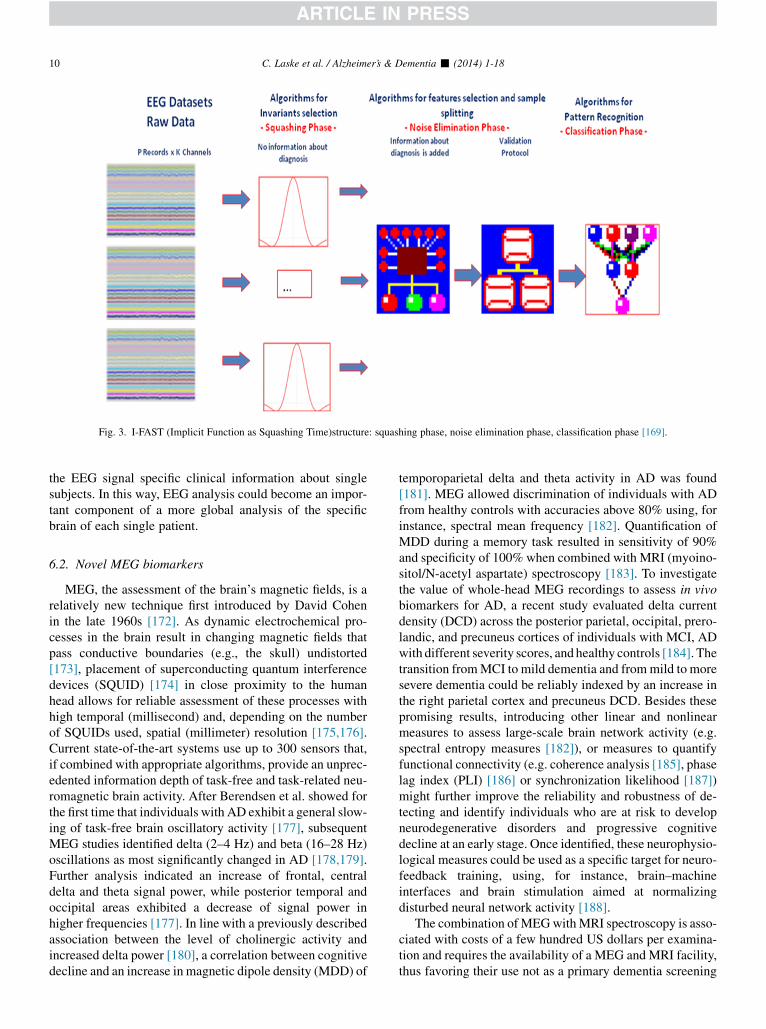
Fig. 3. I-FAST (Implicit Function as Squashing Time)structure: squashing phase, noise elimination phase, classification phase [169].
C. Laske et al. / Alzheimer’s & Dementia - (2014) 1-1810
the EEG signal specific clinical information about singlesubjects. In this way, EEG analysis could become an impor-tant component of a more global analysis of the specificbrain of each single patient.
6.2. Novel MEG biomarkers
MEG, the assessment of the brain’s magnetic fields, is arelatively new technique first introduced by David Cohenin the late 1960s [172]. As dynamic electrochemical pro-cesses in the brain result in changing magnetic fields thatpass conductive boundaries (e.g., the skull) undistorted[173], placement of superconducting quantum interferencedevices (SQUID) [174] in close proximity to the humanhead allows for reliable assessment of these processes withhigh temporal (millisecond) and, depending on the numberof SQUIDs used, spatial (millimeter) resolution [175,176].Current state-of-the-art systems use up to 300 sensors that,if combined with appropriate algorithms, provide an unprec-edented information depth of task-free and task-related neu-romagnetic brain activity. After Berendsen et al. showed forthe first time that individuals with AD exhibit a general slow-ing of task-free brain oscillatory activity [177], subsequentMEG studies identified delta (2–4 Hz) and beta (16–28 Hz)oscillations as most significantly changed in AD [178,179].Further analysis indicated an increase of frontal, centraldelta and theta signal power, while posterior temporal andoccipital areas exhibited a decrease of signal power inhigher frequencies [177]. In line with a previously describedassociation between the level of cholinergic activity andincreased delta power [180], a correlation between cognitivedecline and an increase in magnetic dipole density (MDD) of
temporoparietal delta and theta activity in AD was found[181]. MEG allowed discrimination of individuals with ADfrom healthy controls with accuracies above 80% using, forinstance, spectral mean frequency [182]. Quantification ofMDD during a memory task resulted in sensitivity of 90%and specificity of 100% when combined with MRI (myoino-sitol/N-acetyl aspartate) spectroscopy [183]. To investigatethe value of whole-head MEG recordings to assess in vivobiomarkers for AD, a recent study evaluated delta currentdensity (DCD) across the posterior parietal, occipital, prero-landic, and precuneus cortices of individuals with MCI, ADwith different severity scores, and healthy controls [184]. Thetransition fromMCI to mild dementia and frommild to moresevere dementia could be reliably indexed by an increase inthe right parietal cortex and precuneus DCD. Besides thesepromising results, introducing other linear and nonlinearmeasures to assess large-scale brain network activity (e.g.spectral entropy measures [182]), or measures to quantifyfunctional connectivity (e.g. coherence analysis [185], phaselag index (PLI) [186] or synchronization likelihood [187])might further improve the reliability and robustness of de-tecting and identify individuals who are at risk to developneurodegenerative disorders and progressive cognitivedecline at an early stage. Once identified, these neurophysio-logical measures could be used as a specific target for neuro-feedback training, using, for instance, brain–machineinterfaces and brain stimulation aimed at normalizingdisturbed neural network activity [188].
The combination ofMEGwithMRI spectroscopy is asso-ciated with costs of a few hundred US dollars per examina-tion and requires the availability of a MEG and MRI facility,thus favoring their use not as a primary dementia screening

C. Laske et al. / Alzheimer’s & Dementia - (2014) 1-18 11
instrument but rather as a promising alternative diagnosticoption to already established diagnostic measures. Recenttechnical advances, e.g. development of ultra low-field mi-croteslaMRI duringMEG recordings [189] and atomic mag-netometers [190] operating at room temperature, suggestthat associated costs will significantly decrease and avail-ability of such combined MEG/MRI recordings improve innear future.
7. Conclusions
Current state-of-the-art diagnostic measures of AD areinvasive (CSF analysis), expensive (neuroimaging), andtime-consuming (neuropsychological assessment). Further-more, these measures are limited to specialty clinics andthus have limited accessibility as high-throughput, or front-line, screening and diagnostic tools for AD. More impor-tantly, nonspecialists are often inaccurate at identifyingearly AD and MCI [5]. Thus, there is an increasing needfor additional noninvasive and/or cost-effective tools, allow-ing frontline identification of subjects in the preclinical orearly clinical stages of AD who could be suitable for moni-toring in specialty clinics and for early treatment. Implemen-tation of effective screening instruments will allow diagnosisearlier in the course of dementia, even at the point whenmemory function is still essentially within the normal range.This strategy would enable an earlier, and potentially moreeffective, prevention and treatment of AD with a specialfocus to preserve cognitive functions.
Early AD development is clinically characterized not onlyby progressive memory loss presented in subjective cognitivecomplaints [27,28] and objective psychometric testing [14],but also by noncognitive symptoms such as late-onset depres-sion [43] and progressive functional impairment of speech(language use and emotional responses) [50,55], olfaction[75], pupil light response [108,110], retinal vasculature[105–107], and gait [124,125] with a gradual increase alongthe continuum of AD from preclinical via MCI to thedementia stage. The recognition that several noncognitivesymptoms, such as olfactory dysfunction [75] and gait impair-ment [191,192], occur very early in the disease course and canpredict the subsequent development of AD suggests thatnoncognitive functions may serve as phenotypic markers ofpreclinical AD. Increasing knowledge of affected systems inAD development furthers our understanding of thepathophysiology of AD and allows us to identify novelcandidate biomarkers for diagnosis of AD.
Assessment of subjective cognitive complaints, late-onsetdepression, speech (language use and emotional responses),olfactory function, pupil light response, retinal vasculature,and gait may have potential utility as clinical tools for detec-tion of preclinical and early clinical AD. These measures arenoninvasive and inexpensive and thus suitable for a broaderscreening of individuals with preclinical or early clinicalAD. In addition, use of measures such as EEG and MEGand use of blood-based biomarkersmay enlarge the spectrum
of AD diagnostics. Several measures (e.g., speech, gait,EEG, and MEG) can be automatically analyzed, allowing amultidimensional, objective and reliable diagnostic proce-dure. These novelmeasures will not replace a comprehensiveclinical and neuropsychological assessment and standardtests with CSF analysis and neuroimaging, but rather willenhance these modalities by offering primary care providersa means for determining who needs referrals for comprehen-sive assessment for diagnostic confirmation.
Given the complex nature of AD pathophysiology [1], itis likely that the optimal prediction models for future devel-opment of MCI and/or AD, and risk for progression fromMCI to AD, will come from algorithmic approaches thatcombine multiple diagnostic methods. Actually, we do notknow which of the described tests in the present reviewmanuscript work best for screening of preclinical and earlysymptomatic AD. As an example, one potential ScreeningApproach could include a quick but reliable measure of sub-jective cognitive complaints (either patient, informant orboth), a brief cognitive assessment (e.g., episodic memory)and an assessment for depressive symptoms plus one ormore of the following measures chosen for their availabilityincluding olfactory, speech, eye test, gait, blood biomarkers(e.g. APOE, serum Ab load, etc.) and EEG (Figure 4). Asthe average time spent in primary care settings with geriatricpatients is usually less than 20 minutes, it seems reasonableto allocate these tests on two or more consecutive visits(Figure 4). Inclusion of cognitive and noncognitive ap-proaches may aid in discrimination across neurodegenera-tive disease states to aid in appropriate referrals. Apotential diagnostic approach may utilize a comprehensivebattery, adding neuropsychological assessment plus brainimaging, MEG or CSF analysis to the screening approachbattery (Figure 4). Future research may indicate the utilityof these two approaches or the need for revisions. Large pro-spective cohort studies of patient performance and correla-tion with brain imaging modalities and/or biochemicalmarkers of AD will, however, be required before the bestcombination of selected biomarkers to optimize diagnosticsensitivity and specificity is identified. Thus, beforeapplying them in clinical practice, these tools should befurther examined, as some are now, in ongoing large clinicaltrials such as ADNI (Alzheimer’s Disease NeuroimagingInitiative), AIBL (Australian Imaging, Biomarker & Life-style Flagship Study of Ageing) or DIAN. Despite the dif-ference in underlying cause and age of onset, familial ADand the more common sporadic AD have similar neuropath-ological hallmarks and clinical features [193]. Furtherinvestigation of the reported novel candidate biomarkersin familial AD, and utilization of familial AD cohorts infuture biomarker studies, provides a powerful opportunityto investigate the temporal sequence of different ADbiomarker changes during disease progression. Studyingpre-symptomatic individuals with autosomal dominant in-heritance alleviates many problems inherent in studies ofpre-symptomatic sporadic AD, including uncertainty about
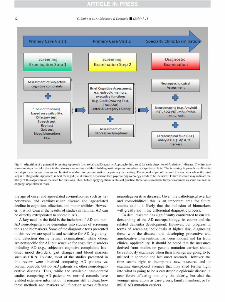
Fig. 4. Algorithm of a potential Screening Approach (two steps) and Diagnostic Approach (third step) for early detection of Alzheimer’s disease. The first two
screening steps can take place in the primary care setting and the third diagnostic step can take place in a specialty clinic. The Screening Approach is splitted in
two steps for economic reasons and limited available time per one visit in the primary care setting. The second step could be used to even tailor where the third
step (i.e. Diagnostic Approach) is best managed (i.e. if clinical depression then psychiatry/psychology needs to be included). Future research may indicate the
utility of this algorithm or the need for revisions. Thus, before applying them in clinical practice, these tools should be further examined, as some are now, in
ongoing large clinical trials.
C. Laske et al. / Alzheimer’s & Dementia - (2014) 1-1812
the age of onset and age-related co-morbidities such as hy-pertension and cardiovascular disease and age-relateddecline in cognition, olfaction, and motor abilities. Howev-er, it is not clear if the results of studies in familial AD canbe directly extrapolated to sporadic AD.
A key need in the field is the inclusion of AD and non-AD neurodegenerative dementias into studies of screeningtools and biomarkers. Some of the diagnostic tests presentedin this review are specific and sensitive for AD (e.g., amy-loid detection during retinal examination), while othersare nonspecific for AD but sensitive for cognitive disordersincluding AD (e.g., subjective cognitive complaints, late-onset mood disorders, gait changes and blood markerssuch as CRP). To date, most of the studies presented inthis review were obtained comparing AD patients vs.normal controls, but not AD patients vs. other neurodegen-erative diseases. Thus, while the available case-controlstudies comparing AD patients vs. normal controls haveyielded extensive information, it remains still unclear, howthese methods and markers will function across different
neurodegenerative diseases. Given the pathological overlapand comorbidities, this is an important area for futurestudies and it is likely that the inclusion of biomarkerswill greatly aid in the differential diagnostic process.
To date, research has significantly contributed to our un-derstanding of the AD neuropathology, its course and therelated dementia development. However, our progress interms of screening individuals at higher risk, diagnosingthose with the disease, and developing preventive andameliorative interventions has been modest and far fromclinical applicability. It should be noted that the measuresderived from studies on genetic mutation carriers shouldbe cautiously examined when their findings are going to beutilized in sporadic and late onset research. However, thetime seems right to incorporate new measures and toexamine unexplored avenues that may shed further lightinto what is going to be a catastrophic epidemic disease innear future affecting not only the elderly, but also theyounger generations as care-givers, family members, or fa-milial AD mutation carriers.
C. Laske et al. / Alzheimer’s & Dementia - (2014) 1-18 13
Acknowledgments
This work was supported by the German Federal Ministry ofEducation and Research (BMBF, 01GQ0831, 16SV5840),the Deutsche Forschungsgemeinschaft (DFG SO932-2)and the European Union (FP7-ICT-2011-288551). RNM isthe founder and owns stock in Alzhyme. HRS is workingas a Site Lead neuropsychologist or Neuropsychologicalrater for Takeda, Wyeth, and Pfizer. None of the authors ofthis manuscript have any biomedical financial interests orpotential conflicts of interest.
RESEARCH IN CONTEXT
1. Systematic review: We reviewed the literature onnoninvasive and inexpensive cognitive and noncog-nitive diagnostic measures for early detection of Alz-heimer’s disease (AD) beyond established dementiadiagnostics with cerebrospinal fluid analysis, neuro-imaging and neuropsychometric testing.
2. Interpretation: Assessment of subjective cognitivecomplaints, late-onset depression, speech, olfactoryfunction, pupil light response, retinal vasculature andgait may have potential utility as noninvasive andinexpensive clinical tools for detection of preclinicaland early clinical AD. In addition, use of measuressuch as electroencephalography and magnetoen-cephalography and use of blood-based biomarkersmay enlarge the spectrum of AD diagnostics.
3. Future directions: Before applying the presentedtests in clinical practice, these tools should be exam-ined now in ongoing large clinical trials.
References
[1] Querfurth HW, LaFerla FM. Alzheimer’s disease. N Engl J Med
2010;362:329–44.
[2] Hebert LE, Scherr PA, Bienias JL, Bennett DA, Evans DA. Alz-
heimer disease in the US population: prevalence estimates using
the 2000 census. Arch Neurol 2003;60:1119–22.
[3] Laske C. Phase 3 trials of solanezumab and bapineuzumab for Alz-
heimer’s disease. N Engl J Med 2014;370:1459.
[4] Jack CR, Albert MS, Knopman DS, McKhann GM, Sperling RA,
Carrillo MC, et al. Introduction to the recommendations from the Na-
tional Institute on Aging-Alzheimer’s Association workgroups on
diagnostic guidelines for Alzheimer’s disease. Alzheimers Dement
2011;7:257–62.
[5] Connolly A, Gaehl E, Martin H, Morris J, Purandare N. Underdiag-
nosis of dementia in primary care: variations in the observed preva-
lence and comparisons to the expected prevalence. Aging Ment
Health 2011;15:978–84.
[6] Aizenstein HJ, Nebes RD, Saxton JA, Price JC, Mathis CA,
Tsopelas ND, et al. Frequent amyloid deposition without significant
cognitive impairment among the elderly. Arch Neurol 2008;
65:1509–17.
[7] Price JL, McKeel DW, Buckles VD, Roe CM, Xiong C,
Grundman M, et al. Neuropathology of nondemented aging: pre-
sumptive evidence for preclinical Alzheimer disease. Neurobiol Ag-
ing 2009;30:1026–36.
[8] De Meyer G, Shapiro F, Vanderstichele H, Vanmechelen E,
Engelborghs S, De Deyn PP, et al. Diagnosis-independent Alzheimer
disease biomarker signature in cognitively normal elderly people.
Arch Neurol 2010;67:949–56.
[9] American Geriatrics Society Geriatrics Workforce Policy Studies
Center. http://www.adgapstudy.uc.edu/index.cfm/Accessed March
9, 2012.
[10] Scully JH, Wilk JE. Selected characteristics and data of psychiatrists
in the United States, 2001–2002. Academic Psychiatry; 2003.
[11] American Academy of Neurology. http://www.aan.com/go/about/
statistics/Accessed May 5, 2011.
[12] Rinck P. Magnetic resonance in medicine. The basic textbook of the
EuropeanMagnetic Resonance Forum. 7th ed. Electronic version 7.1;
2013. 1 October 2013.
[13] Acosta-Baena N, Sepulveda-Falla D, Lopera-G�omez CM, Jaramillo-
Elorza MC, Moreno S, Aguirre-Acevedo DC, et al. Pre-dementia
clinical stages in presenilin 1 E280A familial early-onset Alz-
heimer’s disease: a retrospective cohort study. Lancet Neurol 2011;
10:213–20.
[14] B€ackman L, Jones S, Berger A-K, Laukka EJ, Small BJ. Cognitive
impairment in preclinical Alzheimer’s disease: a meta-analysis.
Neuropsychology 2005;19:520–31.
[15] Abikoff H, Alvir J, Hong G, Sukoff R, Orazio J, Solomon S, et al.
Logical memory subtest of the Wechsler Memory Scale: age and ed-
ucation norms and alternate-form reliability of two scoring systems. J
Clin Exp Neuropsychol 1987;9:435–48.
[16] Delis DC, Kramer JH, Kaplan E, Ober BA. California Verbal
Learning Test – Second Edition (CVLT�-II). TX: Psychological
Cooperation; 2000.
[17] Grober E, Lipton RB, Hall C, Crystal H. Memory impairment on free
and cued selective reminding predicts dementia. Neurology 2000;
54:827–32.
[18] Beck IR, Gagneux-Zurbriggen A, Berres M, Taylor KI, Monsch AU.
Comparison of verbal episodic memory measures: consortium to
establish a registry for Alzheimer’s disease -Neuropsychological
Assessment Battery (CERAD-NAB) versus California Verbal
Learning Test (CVLT). Arch Clin Neuropsychol 2012;27:510–9.
[19] Derby CA, Burns LC, Wang C, Katz MJ, Zimmerman ME,
L’Italien G, et al. Screening for predementia AD: time-dependent
operating characteristics of episodic memory tests. Neurology
2013;80:1307–14.
[20] Sarazin M, Berr C, De Rotrou J, Fabrigoule C, Pasquier F,
Legrain S, et al. Amnestic syndrome of the medial temporal type
identifies prodromal AD: a longitudinal study. Neurology 2007;
69:1859–67.
[21] Wagner M, Wolf S, Reischies FM, Daerr M, Wolfsgruber S, Jessen F,
et al. Biomarker validation of a cued recall memory deficit in prodro-
mal Alzheimer disease. Neurology 2012;78:379–86.
[22] Folstein MF, Folstein SE, McHugh PR. Mini-Mental State; 1975.
[23] Morris JC, Ernesto C, Schafer K, Coats M, Leon S, Sano M, et al.
Clinical dementia rating training and reliability in multicenter
studies: the Alzheimer’s Disease Cooperative Study experience.
Neurology 1997;48:1508–10.
[24] Bateman RJ, Xiong C, Benzinger TL, Fagan AM, Goate A, Fox NC,
et al. Clinical and biomarker changes in dominantly inherited Alz-
heimer’s disease. N Engl J Med 2012;367:795–804.
[25] Morris JC, Weintraub S, Chui HC, Cummings J, Decarli C, Ferris S,
et al. The Uniform Data Set (UDS): clinical and cognitive variables
and descriptive data from Alzheimer Disease Centers. Alzheimer
Dis Assoc Disord 2006;20:210–6.

C. Laske et al. / Alzheimer’s & Dementia - (2014) 1-1814
[26] Elias MF, Beiser A, Wolf PA, Au R, White RF, D’Agostino RB. The
preclinical phase of alzheimer disease: a 22-year prospective study of
the Framingham Cohort. Arch Neurol 2000;57:808–13.
[27] Geerlings MI, Jonker C, Bouter LM, Ad�er HJ, Schmand B. Associa-
tion betweenmemory complaints and incident Alzheimer’s disease in
elderly people with normal baseline cognition. Am J Psychiatry
1999;156:531–7.
[28] Jessen F, Wiese B, Bachmann C, Eifflaender-Gorfer S, Haller F,
K€olsch H, et al. Prediction of dementia by subjective memory impair-
ment: effects of severity and temporal association with cognitive
impairment. Arch Gen Psychiatry 2010;67:414–22.
[29] Juncos-Rabad�an O, Pereiro AX, Facal D, Lojo C, Caama~no JA,
Sueiro J, et al. Prevalence and correlates of mild cognitive impair-
ment in adults aged over 50 years with subjective cognitive com-
plaints in primary care centers. Geriatr Gerontol Int 2013.
[30] Broadbent DE, Cooper PF, FitzGerald P, Parkes KR. The cognitive
failures questionnaire (CFQ) and its correlates. Br J Clin Psychol
1982;21(Pt 1):1–16.
[31] Hohman TJ, Beason-Held LL, Lamar M, Resnick SM. Subjective
cognitive complaints and longitudinal changes in memory and brain
function. Neuropsychology 2011;25:125–30.
[32] Kessler RC, Berglund P, Demler O, Jin R, Walters EE. Lifetime prev-
alence and age-of-onset distributions’ of DSM-IV disorders in the na-
tional comorbidity survey replication. Arch Gen Psychiatry 2005;
62:593–602.
[33] Ferri CP, Prince M, Brayne C, Brodaty H, Fratiglioni L, Ganguli M,
et al. Global prevalence of dementia: a Delphi consensus study. Lan-
cet 2005;366:2112–7.
[34] Vinkers DJ, Gussekloo J, Stek ML, Westendorp RG, van der
Mast RC. Temporal relation between depression and cognitive
impairment in old age: prospective population based study. Bmj
2004;329:881.
[35] Ringman JM, Diaz-Olavarrieta C, Rodriguez Y, Chavez M, Paz F,
Murrell J, et al. Female preclinical presenilin-1 mutation carriers un-
aware of their genetic status have higher levels of depression than
their non-mutation carrying kin. J Neurol Neurosurg Psychiatr
2004;75:500–2.
[36] Wilson RS, Barnes LL, Mendes de Leon CF, Aggarwal NT,
Schneider JS, Bach J, et al. Depressive symptoms, cognitive decline,
and risk of AD in older persons. Neurology 2002;59:364–70.
[37] Gatz JL, Tyas SL, John PS, Montgomery P. Do depressive symptoms
predict Alzheimer’s disease and dementia? J Gerontol a Biol Sci Med
Sci 2005;60:744–7.
[38] OwnbyRL,CroccoE,AcevedoA, JohnV,LoewensteinD.Depression
and risk for Alzheimer disease: systematic review, meta-analysis, and
metaregression analysis. Arch Gen Psychiatry 2006;63:530–8.
[39] Irie F, Masaki KH, Petrovitch H, Abbott RD, Ross GW, Taaffe DR,
et al. Apolipoprotein E epsilon4 allele genotype and the effect of
depressive symptoms on the risk of dementia in men: the
Honolulu-Asia Aging Study. Arch Gen Psychiatry 2008;65:906–12.
[40] Chen R, Hu Z,Wei L, Qin X,McCracken C, Copeland JR. Severity of
depression and risk for subsequent dementia: cohort studies in China
and the UK. Br J Psychiatry 2008;193:373–7.
[41] Byers AL, Yaffe K. Depression and risk of developing dementia. Nat
Rev Neurol 2011;7:323–31.
[42] Barnes DE, Yaffe K, Byers AL, McCormick M, Schaefer C,
Whitmer RA.Midlife vs late-life depressive symptoms and risk of de-
mentia: differential effects for Alzheimer disease and vascular de-
mentia. Arch Gen Psychiatry 2012;69:493–8.
[43] Heser K, Tebarth F, Wiese B, Eisele M, Bickel H, Koehler M, et al.
Age of major depression onset, depressive symptoms, and risk for
subsequent dementia: results of the German Study on Ageing, Cogni-
tion, and Dementia in Primary Care Patients (AgeCoDe). Psychol
Med 2013;43:1597–610.
[44] ChenPJ,GanguliM,MulsantBH,DeKoskyST. The temporal relation-
ship between depressive symptoms and dementia—a community-
based prospective study. Arch Gen Psychiatry 1999;56:261–6.
[45] Lavretsky H, Ercoli L, Siddarth P, Bookheimer S, Miller K, Small G.
Apolipoprotein epsilon 4 allele status, depressive symptoms, and
cognitive decline in middle-aged and elderly persons without demen-
tia. Am J Geriatr Psychiatry 2003;11:667–73.
[46] Mitchell AJ. Depression as a risk factor for later dementia: a robust
relationship? Age Ageing 2005;34:207–9.
[47] Brommelhoff JA, Gatz M, Johansson B, McArdle JJ, Fratiglioni L,
Pedersen NL. Depression as a risk factor or prodromal feature for de-
mentia? Findings in a population-based sample of Swedish twins.
Psychology and Aging 2009;24:373–84.
[48] Wetherell JL, Gatz M, Johansson B, Pedersen NL. History of depres-
sion and other psychiatric illness as risk factors for Alzheimer disease
in a twin sample. Alzheimer Dis Assoc Disord 1999;13:47–52.
[49] Faundez-ZanuyM, Hussain A, Mekyska J, Sesa-Nogueras E, Monte-
Moreno E, Esposito A, et al. Biometric applications related to human
beings: there is life beyond security. Cogn Comput 2013;5:136–51.
[50] Lopez-de-Ipina K, Alonso J-B, Manuel Travieso C, Sole-Casals J,
Egiraun H, Faundez-Zanuy M, et al. On the selection of non-
invasivemethods based on speech analysis oriented to automatic Alz-
heimer disease diagnosis. Sensors (Basel) 2013;13:6730–45.
[51] McKhann GM, Knopman DS, Chertkow H, Hyman BT, Jack CR Jr,
Kawas CH, et al. The diagnosis of dementia due to Alzheimer’s dis-
ease: recommendations from the National Institute on Aging-Alz-
heimer’s Association workgroups on diagnostic guidelines for
Alzheimer’s disease. Alzheimers Dement 2011;7:263–9.
[52] Sperling RA, Aisen PS, Beckett LA, Bennett DA, Craft S, FaganAM,
et al. Toward defining the preclinical stages of Alzheimer’s disease:
recommendations from the National Institute on Aging-Alzheimer’s
Association workgroups on diagnostic guidelines for Alzheimer’s
disease. Alzheimers Dement 2011;7:280–92.
[53] Horley K, Reid A, Burnham D. Emotional prosody perception and
production in dementia of the Alzheimer’s type. J Speech Lang
Hear Res 2010;53:1132–46.
[54] Lehr M, Prud’hommeaux ET, Shafran I, Roark B. Fully automated
neuropsychological assessment for detecting mild cognitive impair-
ment. Spoken Language Processing and Biomedicine Interspeech
2012; 2012.
[55] Lopez-de-Ipi~na K, Alonso JB, Sol�e-Casals J, Barroso N, Henriquez P,
Faundez-Zanuy M, et al. On automatic diagnosis of Alzheimer’s dis-
ease based on spontaneous speech analysis and emotional tempera-
ture. Cogn Comput 2013.
[56] Forbes KE, Venneri A, Shanks MF. Distinct patterns of spontaneous
speech deterioration: an early predictor of Alzheimer’s disease. Brain
Cogn 2002;48:356–61.
[57] Pakhomov SVS, Hemmy LS, Lim KO. Automated semantic indices
related to cognitive function and rate of cognitive decline. Neuropsy-
chologia 2012;50:2165–75.
[58] WeakleyA, Schmitter-EdgecombeM, Anderson J. Analysis of verbal
fluency ability in amnestic and non-amnestic mild cognitive impair-
ment. Arch Clin Neuropsychol 2013;28:721–31.
[59] Baldas V, Lampiris C, Capsalis C, Koutsouris D. Early diagnosis of
Alzheimer’s type dementia using continuous speech recognition.
Lecture notes of the Institute for Computer Sciences, Social Infor-
matics and Telecommunications Engineering, 55. Berlin, Heidelberg:
Springer Berlin Heidelberg; 2011. p. 105–10.
[60] Fraser K, Rudzicz F, GrahamN, RochonE.Automatic speech recogni-
tion in the diagnosis of primary progressive aphasia. Proceedings of the
Fourth Workshop on Speech and Language Processing for Assistive
Technologies/SPLAT2013), 21–22 August, Grenoble, France 2013.
[61] Garrard P, Hodges JR. Semantic dementia: clinical, radiological and
pathological perspectives. J Neurol 2000;247:409–22.
[62] Grossman M, Mickanin J, Onishi K, Hughes E, D’Esposito M,
Ding XS, et al. Progressive nonfluent aphasia: language, cognitive,
and PET measures contrasted with probable Alzheimer’s disease. J
Cogn Neurosci 1996;8:135–54.
[63] Henry ML, Gorno-Tempini ML. The logopenic variant of primary
progressive aphasia. Curr Opin Neurol 2010;23:633–7.

C. Laske et al. / Alzheimer’s & Dementia - (2014) 1-18 15
[64] Wilson SM, Henry ML, Besbris M, Ogar JM, Dronkers NF,
JarroldW, et al. Connected speech production in three variants of pri-
mary progressive aphasia. Brain 2010;133:2069–88.
[65] Saffran EM, Berndt RS, Schwartz MF. The quantitative analysis of
agrammatic production: procedure and data. Brain Lang 1989;
37:440–79.
[66] Garrard P, Rentoumi V, Gesierich B, Miller B, Gorno-Tempini ML.
Machine learning approaches to diagnosis and laterality effects in se-
mantic dementia discourse. Cortex 2014;55:122–9.
[67] Fraser KC, Meltzer JA, Graham NL, Leonard C, Hirst G,
Black SE, et al. Automated classification of primary progressive
aphasia subtypes from narrative speech transcripts. Cortex 2014;
55:43–60.
[68] Serby M, Corwin J, Conrad P, Rotrosen J. Olfactory dysfunction in
Alzheimer’s disease and Parkinsons disease. Am J Psychiatry 1985;
142:781–2.
[69] Warner MD, Peabody CA, Flattery JJ, Tinklenberg JR. Olfactory def-
icits and Alzheimer’s disease. Biol Psychiatry 1986;21:116–8.
[70] Schubert CR, Carmichael LL, Murphy C, Klein BE, Klein R,
Cruickshanks KJ. Olfaction and the 5-year incidence of cognitive
impairment in an epidemiological study of older adults. J Am Geriatr
Soc 2008;56:1517–21.
[71] Wilson RS, Schneider JA, Arnold SE, Tang Y, Boyle PA, Bennett DA.
Olfactory identification and incidence of mild cognitive impairment
in older age. Arch Gen Psychiatry 2007;64:802–8.
[72] Murphy C, BaconAW, BondiMW, SalmonDP. Apolipoprotein E sta-
tus is associated with odor identification deficits in nondemented
older personsa. Ann N YAcad Sci 1998;855:744–50.
[73] Devanand DP, Michaels-Marston KS, Liu X, Pelton GH, Padilla M,
Marder K, et al. Olfactory deficits in patients with mild cognitive
impairment predict Alzheimer’s disease at follow-up. Am J Psychia-
try 2000;157:1399–405.
[74] Sohrabi HR, Bates KA, Rodrigues M, Taddei K, Laws SM,
Lautenschlager NT, et al. Olfactory dysfunction is associated with
subjective memory complaints in community-dwelling elderly indi-
viduals. J Alzheimers Dis 2009;17:135–42.
[75] Wilson RS, Arnold SE, Schneider JA, Boyle PA, Buchman AS,
Bennett DA. Olfactory impairment in presymptomatic Alzheimer’s
disease. Ann N YAcad Sci 2009;1170:730–5.
[76] Sohrabi HR, Bates KA, Weinborn MG, Johnston AN, Bahramian A,
Taddei K, et al. Olfactory discrimination predicts cognitive decline
among community-dwelling older adults. Transl Psychiatry 2012;
2:e118.
[77] Morgan CD, Murphy C. Olfactory event-related potentials in Alz-
heimer’s disease. J Int Neuropsychol Soc 2002;8:753–63.
[78] Schiffman SS, Gatlin CA. Clinical physiology of taste and smell. Ann
Rev Nutr 1993;13:405–36.
[79] Tanabe T, Iino M, Takagi SF. Discrimination of odors in olfactory
bulb, pyriform-amygdaloid areas, and orbitofrontal cortex of the
monkey. J Neurophysiol 1975;38:1284–96.
[80] Ohm TG, Braak H. Olfactory bulb changes in Alzheimer’s disease.
Acta Neuropathol 1987;73:365–9.
[81] Averback P. Two new lesions in Alzheimer’s disease. Lancet 1983;
2:1203.
[82] Kov�acs T, Cairns NJ, Lantos PL. beta-amyloid deposition and neuro-
fibrillary tangle formation in the olfactory bulb in ageing and Alz-
heimer’s disease. Neuropathol Appl Neurobiol 1999;25:481–91.
[83] Wesson DW, Levy E, Nixon RA, Wilson DA. Olfactory dysfunction
correlates with amyloid-beta burden in an Alzheimer’s disease mouse
model. J Neurosci 2010;30:505–14.
[84] Eibenstein A, Fioretti AB, Lena C, Rosati N, Amabile G, Fusetti M.
Modern psychophysical tests to assess olfactory function. Neurol Sci
2005;26:147–55.
[85] Doty RL. Office procedures for quantitative assessment of olfactory
function. Am J Rhinol 2007;21:460–73.
[86] Herz RS, Engen T. Odor memory: review and analysis. Psychon Bull
Rev 1996;3:300–13.
[87] Schofield PW, Ebrahimi H, Jones AL, Bateman GA, Murray SR. An
olfactory ‘stress test’ may detect preclinical Alzheimer’s disease.
BMC Neurol 2012;12:24.
[88] Mesholam RI, Moberg PJ, Mahr RN, Doty RL. Olfaction in neuro-
degenerative disease: a meta-analysis of olfactory functioning in
Alzheimer’s and Parkinson’s diseases. Arch Neurol 1998;
55:84–90.
[89] Lehrner J, Baumgartner C, Serles W, Olbrich A, Pataraia E, Bacher J,
et al. Olfactory prodromal symptoms and unilateral olfactory
dysfunction are associated in patients with right mesial temporal
lobe epilepsy. Epilepsia 1997;38:1042–4.
[90] Doty RL, Stern MB, Pfeiffer C, Gollomp SM, Hurtig HI. Bilateral ol-
factory dysfunction in early stage treated and untreated idiopathic
Parkinson’s disease. J Neurol Neurosurg Psychiatr 1992;55:138–42.
[91] Pause BM,Miranda A, G€oder R, Aldenhoff JB, Ferstl R. Reduced ol-factory performance in patients with major depression. J Psychiatr
Res 2001;35:271–7.
[92] Elsner R. Odor memory and aging. Aging 2001;8:284–306.
[93] Doty RL, Shaman P, Applebaum SL, Giberson R, Siksorski L,
Rosenberg L. Smell identification ability—changes with age. Science
1984;226:1441–3.
[94] Velayudhan L, Pritchard M, Powell JF, Proitsi P, Lovestone S. Smell
identification function as a severity and progression marker in Alz-
heimer’s disease. Int Psychogeriatr 2013;25:1157–66.
[95] Kov�acs T. The olfactory system in Alzheimer’s disease: pathology,
pathophysiology and pathway for therapy. Transl Neurosci 2013;
4:34–45.
[96] Albers MW, Tabert MH, Devanand DP. Olfactory dysfunction as a
predictor of neurodegenerative disease. Curr Neurol Neurosci Rep
2006;6:379–86.
[97] Schliebs R, Arendt T. The significance of the cholinergic system in
the brain during aging and in Alzheimer’s disease. J Neural Transm
Gen Sect 2006;113:1625–44.
[98] Katz B, Rimmer S. Ophthalmologic manifestations of Alzheimer’s
disease. Surv Ophthalmol 1989;34:31–43.
[99] Sadun AA, Borchert M, DeVita E, Hinton DR, Bassi CJ. Assessment
of visual impairment in patients with Alzheimer’s disease. Am J Oph-
thalmol 1987;104:113–20.
[100] Whittaker KW, Burdon MA, Shah P. Visual field loss and Alz-
heimer’s disease. Eye (Lond) 2002;16:206–8.
[101] Pache M, Smeets CH, Gasio PF, Savaskan E, Flammer J, Wirz-
Justice A, et al. Colour vision deficiencies in Alzheimer’s disease.
Age Ageing 2003;32:422–6.
[102] Crow RW, Levin LB, LaBree L, Rubin R, Feldon SE. Sweep visual
evoked potential evaluation of contrast sensitivity in Alzheimer’s de-
mentia. Invest Ophthalmol Vis Sci 2003;44:875–8.
[103] Schlotterer G, Moscovitch M, Crapper-McLachlan D. Visual pro-
cessing deficits as assessed by spatial frequency contrast sensitivity
and backward masking in normal ageing and Alzheimer’s disease.
Brain 1984;107(Pt 1):309–25.
[104] Mielke R, Kessler J, Fink G, Herholz K, Heiss WD. Dysfunction of
visual cortex contributes to disturbed processing of visual informa-
tion in Alzheimer’s disease. Int J Neurosci 1995;82:1–9.
[105] Berisha F, Feke GT, Trempe CL, McMeel JW, Schepens CL. Retinal
abnormalities in early Alzheimer’s disease. Invest Ophthalmol Vis
Sci 2007;48:2285–9.
[106] Paquet C, Boissonnot M, Roger F, Dighiero P, Gil R, Hugon J.
Abnormal retinal thickness in patients with mild cognitive impair-
ment and Alzheimer’s disease. Neurosci Lett 2007;420:97–9.
[107] Frost S, Kanagasingam Y, Sohrabi H, Vignarajan J, Bourgeat P,
Salvado O, et al. Retinal vascular biomarkers for early detection
and monitoring of Alzheimer’s disease. Transl Psychiatry 2013;
3:e233.
[108] Fotiou DF, Brozou CG, Haidich A-B, Tsiptsios D, Nakou M,
Kabitsi A, et al. Pupil reaction to light in Alzheimer’s disease: eval-
uation of pupil size changes and mobility. Aging Clin Exp Res 2007;
19:364–71.

C. Laske et al. / Alzheimer’s & Dementia - (2014) 1-1816
[109] Granholm E,Morris S, Galasko D, Shults C, Rogers E, Vukov B. Tro-
picamide effects on pupil size and pupillary light reflexes in Alz-
heimer’s and Parkinson’s disease. Int J Psychophysiol 2003;
47:95–115.
[110] Frost S, Kanagasingam Y, Sohrabi H, Bourgeat P, Villemagne V,
Rowe CC, et al. Pupil response biomarkers for early detection and
monitoring of Alzheimer’s disease. Curr Alzheimer Res 2013;
10:931–9.
[111] Danesh-Meyer HV, Birch H, Ku JY, Carroll S, Gamble G. Reduction
of optic nerve fibers in patients with Alzheimer disease identified by
laser imaging. Neurology 2006;67:1852–4.
[112] Sadun AA, Bassi CJ. Optic nerve damage in Alzheimer’s disease.
Ophthalmology 1990;97:9–17.
[113] Blanks JC, Torigoe Y, Hinton DR, Blanks RH. Retinal pathology in
Alzheimer’s disease. I. Ganglion cell loss in foveal/parafoveal retina.
Neurobiol Aging 1996;17:377–84.
[114] Prettyman R, Bitsios P, Szabadi E. Altered pupillary size and dark-
ness and light reflexes in Alzheimer’s disease. J Neurol Neurosurg
Psychiatr 1997;62:665–8.
[115] de Jong FJ, Schrijvers EM, Ikram MK, Koudstaal PJ, de Jong PT,
Hofman A, et al. Retinal vascular caliber and risk of dementia The
Rotterdam Study. Neurology 2011;76:816–21.
[116] Frost SM, Kanagasingam Y, Sohrabi HR, Taddei K, Bateman R,
Morris J, et al. Pupil response biomarkers distinguish amyloid precur-
sor protein mutation carriers from non-carriers. Curr Alzheimer Res
2013;10:790–6.
[117] Koronyo-Hamaoui M, Koronyo Y, Ljubimov AV, Miller CA, KoMK,
Black KL, et al. Identification of amyloid plaques in retinas from
Alzheimer’s patients and noninvasive in vivo optical imaging of
retinal plaques in a mouse model. Neuroimage 2011;54(Suppl
1):S204–17.
[118] Goldstein LE, Muffat JA, Cherny RA, Moir RD, Ericsson MH,
Huang X, et al. Cytosolic beta-amyloid deposition and supranuclear
cataracts in lenses from people with Alzheimer’s disease. Lancet
2003;361:1258–65.
[119] Bridenbaugh S, Monsch AU, Kressig RW. How does gait change as
cognitive decline progresses in the elderly? Alzheimers Dement
2012;8:P131–2.
[120] Verghese J, Wang C, Lipton RB, Holtzer R, Xue X. Quantitative gait
dysfunction and risk of cognitive decline and dementia. J Neurol
Neurosurg Psychiatr 2007;78:929–35.
[121] Mielke MM, Roberts RO, Savica R, Cha R, Drubach DI,
Christianson T, et al. Assessing the temporal relationship between
cognition and gait: slow gait predicts cognitive decline in the Mayo
Clinic Study of Aging. J Gerontol a Biol Sci Med Sci 2013;
68:929–37.
[122] Verghese J, Wang C, Lipton RB, Holtzer R. Motoric cognitive risk
syndrome and the risk of dementia. J Gerontol a Biol Sci Med Sci
2013;68:412–8.
[123] Hausdorff JM, Buchman AS.What links gait speed andMCI with de-
mentia? A fresh look at the association between motor and cognitive
function. J Gerontol a Biol Sci Med Sci 2013;68:409–11.
[124] Bridenbaugh SA, Kressig RW. Quantitative gait disturbances in older
adults with cognitive impairments. Curr Pharm Des 2013.
[125] Wittwer JE,Webster KE,Menz HB. A longitudinal study ofmeasures
of walking in people with Alzheimer’s disease. Gait Posture 2010;
32:113–7.
[126] Bridenbaugh SA, Kressig RW. Laboratory review: the role of gait
analysis in seniors’ mobility and fall prevention. Gerontology
2011;57:256–64.
[127] Montero-Odasso M, Muir SW, Speechley M. Dual-task complexity
affects gait in people with mild cognitive impairment: the interplay
between gait variability, dual tasking, and risk of falls. Arch Phys
Med Rehabil 2012;93:293–9.
[128] Beauchet O, Allali G, Berrut G, Hommet C, Dubost V, Assal F. Gait
analysis in demented subjects: Interests and perspectives. Neuropsy-
chiatr Dis Treat 2008;4:155–60.
[129] Fuentes P. Clinical diagnosis in preclinical stage of Alzheimer’s dis-
ease. Arch Med Res 2012;43:667–70.
[130] Wittwer JE, Webster KE, Hill K. The effects of a concurrent motor
task on walking in Alzheimer’s disease. Gait Posture 2014;39:291–6.
[131] Holtzer R, Wang C, Verghese J. Performance variance on walking
while talking tasks: theory, findings, and clinical implications. Age
(Dordr) 2013.
[132] Theill N, Martin M, Schumacher V, Bridenbaugh SA, Kressig RW.
Simultaneously measuring gait and cognitive performance in cogni-
tively healthy and cognitively impaired older adults: the Basel motor-
cognition dual-task paradigm. J Am Geriatr Soc 2011;59:1012–8.
[133] Zipser BD, Johanson CE, Gonzalez L, Berzin TM, Tavares R,
Hulette CM, et al. Microvascular injury and blood-brain barrier
leakage in Alzheimer’s disease. Neurobiol Aging 2007;28:977–86.
[134] Ryu JK, McLarnon JG. A leaky blood-brain barrier, fibrinogen infil-
tration and microglial reactivity in inflamed Alzheimer’s disease
brain. J Cell Mol Med 2009;13:2911–25.
[135] Kivipelto M, Ngandu T, Laatikainen T, Winblad B, Soininen H,
Tuomilehto J. Risk score for the prediction of dementia risk in 20
years among middle aged people: a longitudinal, population-based
study. Lancet Neurol 2006;5:735–41.
[136] Graff-Radford NR, Crook JE, Lucas J, Boeve BF, Knopman DS,
Ivnik RJ, et al. Association of low plasma Ab42/Ab40 ratios with
increased imminent risk for mild cognitive impairment and Alz-
heimer disease. Arch Neurol 2007;64:354–62.
[137] Yaffe K, Weston A, Graff-Radford NR, Satterfield S, Simonsick EM,
Younkin SG, et al. Association of plasma beta-amyloid level and
cognitive reserve with subsequent cognitive decline. JAMA 2011;
305:261–6.
[138] Schmidt R, Schmidt H, Curb JD, Masaki K, White LR, Launer LJ.
Early inflammation and dementia: a 25-year follow-up of the
Honolulu-Asia Aging Study. Ann Neurol 2002;52:168–74.
[139] van OijenM,Witteman JC, Hofman A, Koudstaal PJ, Breteler MMB.
Fibrinogen is associated with an increased risk of Alzheimer disease
and vascular dementia. Stroke 2005;36:2637–41.
[140] Tan ZS, Beiser AS, Vasan RS, Roubenoff R, Dinarello CA,
Harris TB, et al. Inflammatory markers and the risk of Alzheimer dis-
ease: the Framingham Study. Neurology 2007;68:1902–8.
[141] van Exel E, Eikelenboom P, Comijs H, Fr€olich M, Smit JH, Stek ML,
et al. Vascular factors andmarkers of inflammation in offspring with a
parental history of late-onset Alzheimer disease. Arch Gen Psychia-
try 2009;66:1263–70.
[142] Mapstone M, Cheema AK, Fiandaca MS, Zhong X, Mhyre TR,
Macarthur LH, et al. Plasma phospholipids identify antecedent mem-
ory impairment in older adults. Nat Med 2014;20:415–8.
[143] Ray S, Britschgi M, Herbert C, Takeda-Uchimura Y, Boxer A,
Blennow K, et al. Classification and prediction of clinical Alz-
heimer’s diagnosis based on plasma signaling proteins. Nat Med
2007;13:1359–62.
[144] O’Bryant SE, Xiao G, Barber R, Reisch J, Doody R, Fairchild T, et al.
A serum protein-based algorithm for the detection of Alzheimer dis-
ease. Arch Neurol 2010;67:1077–81.
[145] Laske C, Leyhe T, Stransky E, HoffmannN, Fallgatter AJ, Dietzsch J.
Identification of a blood-based biomarker panel for classification of
Alzheimer’s disease. Int J Neuropsychopharmacol 2011;14:1147–55.
[146] Soares HD, Potter WZ, Pickering E, Kuhn M, Immermann FW,
Shera DM, et al. Plasma biomarkers associated with the apolipopro-
tein E genotype and Alzheimer disease. Arch Neurol 2012;
69:1310–7.
[147] Doecke JD, Laws SM, Faux NG, Wilson W, Burnham SC, Lam C-P,
et al. Blood-based protein biomarkers for diagnosis of Alzheimer dis-
ease. Arch Neurol 2012;69:1318–25.
[148] HuWT, Holtzman D, Shaw L, Trojanowski J, Soares H. Plasma mul-
tianalyte profiling in mild cognitive impairment and Alzheimer dis-
ease. Neurology 2013;80:690–1.
[149] Sperling RA, Laviolette PS, O’Keefe K, O’Brien J, Rentz DM,
Pihlajamaki M, et al. Amyloid deposition is associated with impaired
C. Laske et al. / Alzheimer’s & Dementia - (2014) 1-18 17
default network function in older persons without dementia. Neuron
2009;63:178–88.
[150] Sheline YI, Raichle ME, Snyder AZ, Morris JC, Head D, Wang S,
et al. Amyloid plaques disrupt resting state default mode network
connectivity in cognitively normal elderly. Biol Psychiatry 2010;
67:584–7.
[151] Steriade M. Grouping of brain rhythms in corticothalamic systems.
Neuroscience 2006;137:1087–106.
[152] Anderer P, Saletu B, Kl€oppel B, Semlitsch HV, Werner H. Discrimi-
nation between demented patients and normals based on topographic
EEG slow wave activity: comparison between z statistics, discrimi-
nant analysis and artificial neural network classifiers. Electroence-
phalogr Clin Neurophysiol 1994;91:108–17.
[153] Besthorn C, Zerfass R, Geiger-Kabisch C, Sattel H, Daniel S,
Schreiter-Gasser U, et al. Discrimination of Alzheimer’s disease
and normal aging by EEG data. Electroencephalogr Clin Neurophy-
siol 1997;103:241–8.
[154] Dauwels J, Vialatte F, Cichocki A. Diagnosis of Alzheimer’s disease
from EEG signals: where arewe standing? Curr Alzheimer Res 2010;
7:487–505.
[155] Pritchard WS, Duke DW, Coburn KL, Moore NC. EEG-based,
neural-net predictive classification of Alzheimer’s disease versus
control subjects is augmented by non-linear EEG measures. Electro-
encephalogr Clin Neurophysiol 1994;91:118–30.
[156] Dierks T, Frohlich L, Ihl R, Maurer K. Correlation between cognitive
brain-function and electrical brain activity in dementia of Alzheimer-
type. J Neural Transm Gen Sect 1995;99:55–62.
[157] Babiloni C, Binetti G, Cassetta E, Cerboneschi D, Dal Forno G, Del
Percio C, et al. Mapping distributed sources of cortical rhythms in
mild Alzheimer’s disease. A multicentric EEG study. Neuroimage
2004;22:57–67.
[158] Tsuno N, ShigetaM, Hyoki K, Faber PL, LehmannD. Fluctuations of
source locations of EEG activity during transition from alertness to
sleep in Alzheimer’s disease and vascular dementia. Neuropsychobi-
ology 2004;50:267–72.
[159] Moretti DV, Paternic�o D, Binetti G. EEG upper/low alpha frequency
power ratio relates to temporo-parietal brain atrophy and memory
performances in mild cognitive impairment. Front Aging Neurosci
2013;5:63.
[160] Poil S-S, de HaanW, van der FlierWM,Mansvelder HD, Scheltens P,
Linkenkaer-Hansen K. Integrative EEG biomarkers predict progres-
sion to Alzheimer’s disease at the MCI stage. Front Aging Neurosci
2013;5:58.
[161] Elgendi M, Vialatte F, Cichocki A, Latchoumane C, Jeong J,
Dauwels J. Optimization of EEG frequency bands for improved diag-
nosis of Alzheimer disease. Conf Proc IEEE EngMed Biol Soc 2011;
2011:6087–91.
[162] Schmidt MT, Kanda PA, Basile LFH, da Silva Lopes HF, Baratho R,
Demario JL, et al. Index of alpha/theta ratio of the electroencephalo-
gram: a new marker for Alzheimer’s disease. Front Aging Neurosci
2013;5:60.
[163] Petrosian AA, Prokhorov DV, Lajara-Nanson W, Schiffer RB.
Recurrent neural network-based approach for early recognition of
Alzheimer’s disease in EEG. Clin Neurophysiol 2001;
112:1378–87.
[164] Vialatte F, Cichocki A, Dreyfus G, Musha T, Shishkin SL, Gervais R.
Early detection of Alzheimer’s disease by blind source separation,
time frequency representation, and bump modeling of EEG signals.
Lecture Notes in Computer Science, 3696. Berlin, Heidelberg:
Springer Berlin Heidelberg; 2005. p. 683–92.
[165] Cichocki A, Shishkin SL, Musha T, Leonowicz Z, Asada T,
Kurachi T. EEG filtering based on blind source separation (BSS)
for early detection of Alzheimer’s disease. Clin Neurophysiol
2005;116:729–37.
[166] Melissant C, YpmaA, Frietman EE, StamCJ. Amethod for detection
of Alzheimer’s disease using ICA-enhanced EEGmeasurements. Ar-
tif Intell Med 2005;33:209–22.
[167] Jeong J. EEG dynamics in patients with Alzheimer’s disease. Clin
Neurophysiol 2004;115:1490–505.
[168] Lehmann C, Koenig T, Jelic V, Prichep L, John RE, Wahlund LO,
et al. Application and comparison of classification algorithms for
recognition of Alzheimer’s disease in electrical brain activity
(EEG). J Neurosci Methods 2007;161:342–50.
[169] Buscema M, Rossini P, Babiloni C, Grossi E. The IFAST model, a
novel parallel nonlinear EEG analysis technique, distinguishes mild
cognitive impairment and Alzheimer’s disease patients with high de-
gree of accuracy. Artif Intell Med 2007;40:127–41.
[170] Rossini PM, Buscema M, Capriotti M, Grossi E, Rodriguez G, Del
Percio C, et al. Is it possible to automatically distinguish resting
EEG data of normal elderly vs. mild cognitive impairment subjects
with high degree of accuracy? Clin Neurophysiol 2008;
119:1534–45.
[171] Buscema M, Grossi E, Capriotti M, Babiloni C, Rossini P. The
I.F.A.S.T. model allows the prediction of conversion to Alzheimer
disease in patients with mild cognitive impairment with high degree
of accuracy. Curr Alzheimer Res 2010;7:173–87.
[172] Cohen D. Magnetoencephalography—evidence of magnetic fields
produced by alpha-rhythm currents. Science 1968;161:784–6.
[173] Okada YC, Lahteenm€aki A, Xu C. Experimental analysis of distor-
tion of magnetoencephalography signals by the skull. Clin Neuro-
physiol 1999;110:230–8.
[174] Silver A, Zimmerman J. Quantum transitions and loss in multiply
connected superconductors. Phys Rev Lett 1965;15:888–91.
[175] Troebinger L, L�opez JD, Lutti A, Bradbury D, Bestmann S, Barnes G.
High precision anatomy for MEG. Neuroimage 2013.
[176] Robinson SE, Vrba J. Functional neuroimaging by synthetic aperture
magnetometry (SAM). In: Recent Advances in Biomagnetism. Sen-
dai: Tohoku University Press; 1999. p. 302–5.
[177] Berendse HW, Verbunt JP, Scheltens P, van Dijk BW, Jonkman EJ.
Magnetoencephalographic analysis of cortical activity in Alz-
heimer’s disease: a pilot study. Clin Neurophysiol 2000;
111:604–12.
[178] Fern�andez A, Hornero R, Mayo A, Poza J, Maest�u F, Ortiz Alonso T.
Quantitative magnetoencephalography of spontaneous brain activity
in Alzheimer disease: an exhaustive frequency analysis. Alzheimer
Dis Assoc Disord 2006;20:153–9.
[179] de Haan W, Stam CJ, Jones BF, Zuiderwijk IM, van Dijk BW,
Scheltens P. Resting-state oscillatory brain dynamics in Alzheimer
disease. J Clin Neurophysiol 2008;25:187–93.
[180] Riekkinen P, Buzsaki G, Riekkinen P, Soininen H, Partanen J. The
cholinergic system and EEG slow waves. Electroencephalogr Clin
Neurophysiol 1991;78:89–96.
[181] Fern�andez A, Maest�u F, Amo C, Gil P, Fehr T, Wienbruch C, et al.
Focal temporoparietal slow activity in Alzheimer’s disease re-
vealed by magnetoencephalography. Biol Psychiatry 2002;
52:764–70.
[182] Poza J, Hornero R, Escudero J, Fern�andez A, S�anchez CI.
Regional analysis of spontaneous MEG rhythms in patients with
Alzheimer’s disease using spectral entropies. Ann Biomed Eng
2008;36:141–52.
[183] Fern�andez A, Garc�ıa-Segura JM, Ortiz T, Montoya J, Maest�u F,
Gil-Gregorio P, et al. Proton magnetic resonance spectroscopy
and magnetoencephalographic estimation of delta dipole density:
a combination of techniques that may contribute to the diagnosis
of Alzheimer’s disease. Dement Geriatr Cogn Disord 2005;
20:169–77.
[184] Fern�andez A, Turrero A, Zuluaga P, Gil-Gregorio P, del-Pozo F,
Maest�u F, et al. MEG delta mapping along the healthy aging-Alz-
heimer’s disease continuum: diagnostic implications. J Alzheimers
Dis 2013;35:495–507.
[185] Alonso JF, Poza J, Ma~nanas MA, Romero S, Fern�andez A,
Hornero R. MEG connectivity analysis in patients with Alzheimer’s
disease using cross mutual information and spectral coherence. Ann
Biomed Eng 2011;39:524–36.

C. Laske et al. / Alzheimer’s & Dementia - (2014) 1-1818
[186] Stam CJ, Nolte G, Daffertshofer A. Phase lag index: assessment of
functional connectivity from multi channel EEG and MEG with
diminished bias from common sources. Hum Brain Mapp 2007;
28:1178–93.
[187] Buld�u JM, Bajo R, Maest�u F, Castellanos N, Leyva I, Gil P, et al.
Reorganization of functional networks in mild cognitive impairment.
PLoS One 2011;6.
[188] Soekadar SR, Witkowski M, Cossio EG, Birbaumer N, Robinson SE,
Cohen LG. In vivo assessment of human brain oscillations during
application of transcranial electric currents. Nat Commun 2013;
4:2032.
[189] Inglis B, Buckenmaier K, Sangiorgio P, Pedersen AF, Nichols MA,
Clarke J. MRI of the human brain at 130 microtesla. Proc Natl
Acad Sci U S A 2013;110:19194–201.
[190] Sander TH, Preusser J, Mhaskar R. Magnetoencephalography with a
chip-scale atomicmagnetometer. BiomedOpt Express 2012;3:981–90.
[191] Camicioli R, Howieson D, Oken B, Sexton G, Kaye J. Motor slowing
precedes cognitive impairment in the oldest old. Neurology 1998;
50:1496–8.
[192] Buracchio T, Dodge HH, Howieson D, Wasserman D, Kaye J. The
trajectory of gait speed preceding mild cognitive impairment. Arch
Neurol 2010;67:980–6.
[193] Bateman RJ, Aisen PS, De Strooper B, Fox NC, Lemere CA,
Ringman JM, et al. Autosomal-dominant Alzheimer’s disease: a re-
view and proposal for the prevention of Alzheimer’s disease. Alz-
heimers Res Ther 2011;3:1.
[194] Praat: doing phonetics by computer. Available at: http://www.fon.
hum.uva.nl/praat/Accessed November 27, 2013.