integrative clinical genomics of advanced prostate cancer
TRANSCRIPT
Resource
Integrative Clinical Genomics of Advanced ProstateCancer
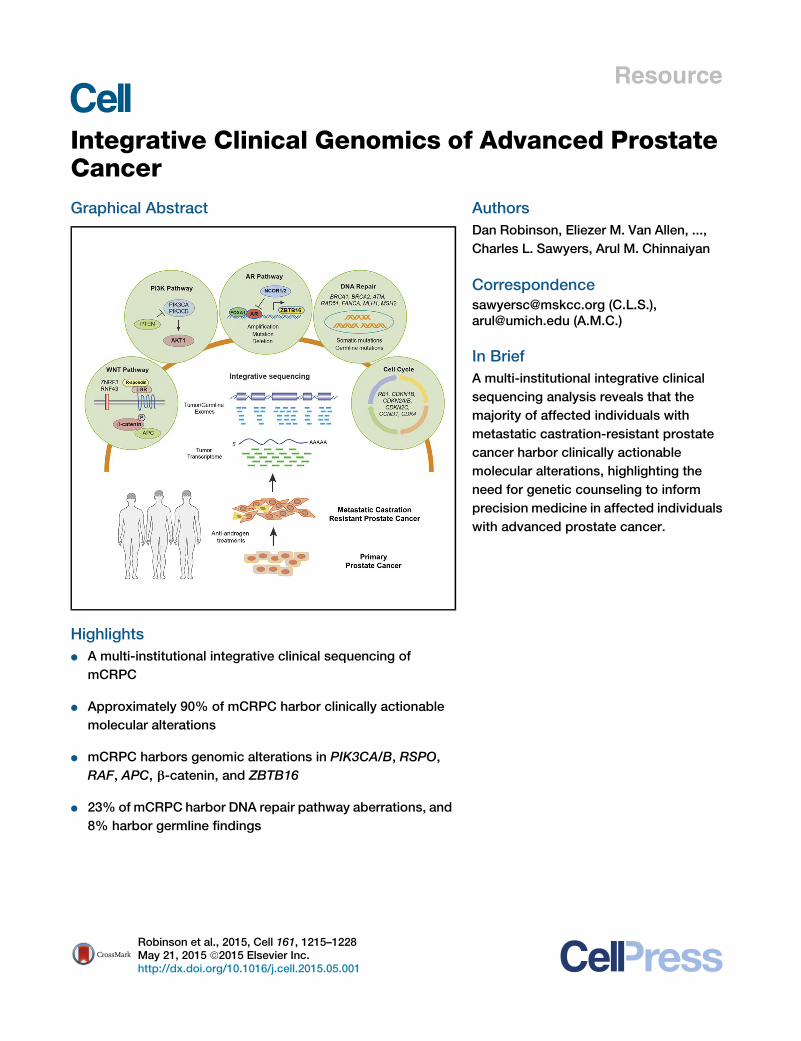
Graphical Abstract
Highlightsd A multi-institutional integrative clinical sequencing of
mCRPC
d Approximately 90% of mCRPC harbor clinically actionable
molecular alterations
d mCRPC harbors genomic alterations in PIK3CA/B, RSPO,
RAF, APC, b-catenin, and ZBTB16
d 23% of mCRPC harbor DNA repair pathway aberrations, and
8% harbor germline findings
AuthorsDan Robinson, Eliezer M. Van Allen, ...,
Charles L. Sawyers, Arul M. Chinnaiyan
[email protected] (C.L.S.),[email protected] (A.M.C.)
In BriefA multi-institutional integrative clinical
sequencing analysis reveals that the
majority of affected individuals with
metastatic castration-resistant prostate
cancer harbor clinically actionable
molecular alterations, highlighting the
need for genetic counseling to inform
precision medicine in affected individuals
with advanced prostate cancer.
Robinson et al., 2015, Cell 161, 1215–1228May 21, 2015 ª2015 Elsevier Inc.http://dx.doi.org/10.1016/j.cell.2015.05.001
Resource
Integrative Clinical Genomicsof Advanced Prostate CancerDan Robinson,1,2,43 Eliezer M. Van Allen,3,4,43 Yi-Mi Wu,1,2 Nikolaus Schultz,5,40 Robert J. Lonigro,1
Juan-Miguel Mosquera,6,7,8,38 Bruce Montgomery,9,10 Mary-Ellen Taplin,3 Colin C. Pritchard,26 Gerhardt Attard,11,12
Himisha Beltran,7,8,13,38 Wassim Abida,14,20 Robert K. Bradley,9 Jake Vinson,15 Xuhong Cao,1,42 Pankaj Vats,1
Lakshmi P. Kunju,1,2,17 Maha Hussain,16,17,18 Felix Y. Feng,1,17,19 Scott A. Tomlins,1,2,17,18 Kathleen A. Cooney,16,17,18
David C. Smith,16,17,18 Christine Brennan,1 Javed Siddiqui,1 Rohit Mehra,1,2 Yu Chen,13,14,20 Dana E. Rathkopf,13,20
Michael J. Morris,13,20 Stephen B. Solomon,21 Jeremy C. Durack,21 Victor E. Reuter,22 Anuradha Gopalan,22
Jianjiong Gao,40 Massimo Loda,3,4,23,39 Rosina T. Lis,3,23 Michaela Bowden,3,23,39 Stephen P. Balk,24 Glenn Gaviola,25
Carrie Sougnez,4 Manaswi Gupta,4 Evan Y. Yu,10 Elahe A. Mostaghel,9,10 Heather H. Cheng,9,10 Hyojeong Mulcahy,27
Lawrence D. True,28 Stephen R. Plymate,10 Heidi Dvinge,9 Roberta Ferraldeschi,11,12 Penny Flohr,11,12
Susana Miranda,11,12 Zafeiris Zafeiriou,11,12 Nina Tunariu,11,12 Joaquin Mateo,11,12 Raquel Perez-Lopez,11,12
Francesca Demichelis,7,29 Brian D. Robinson,6,7,8,38 Marc Schiffman,7,31,38 David M. Nanus,7,8,13,38
Scott T. Tagawa,7,8,13,38 Alexandros Sigaras,7,30,32 Kenneth W. Eng,7,30,32 Olivier Elemento,30 Andrea Sboner,6,7,30,38
Elisabeth I. Heath,33,34 Howard I. Scher,13,20 Kenneth J. Pienta,35 Philip Kantoff,3,44 Johann S. de Bono,11,12,44
Mark A. Rubin,6,7,8,38,44 Peter S. Nelson,10,36,37,38,44 Levi A. Garraway,3,4,44 Charles L. Sawyers,14,41,44,*and Arul M. Chinnaiyan1,2,17,18,42,44,*1Michigan Center for Translational Pathology, University of Michigan Medical School, Ann Arbor, MI 48109, USA2Department of Pathology, University of Michigan Medical School, Ann Arbor, MI 48109, USA3Department of Medical Oncology, Dana-Farber Cancer Institute, Boston, MA 02215, USA4Broad Institute of Massachusetts Institute of Technology and Harvard, Cambridge, MA 02142, USA5Department of Epidemiology and Biostatistics, Memorial Sloan Kettering Cancer Center, New York, NY 10065, USA6Department of Pathology and Laboratory Medicine, Weill Medical College of Cornell University, New York, NY 10021, USA7Institute for Precision Medicine, Weill Medical College of Cornell University, New York, NY 10021, USA8New York Presbyterian Hospital, New York, NY 10021, USA9Computational Biology Program, Public Health Sciences Division and Basic Science Division, Fred Hutchinson Cancer Center, University ofWashington, Seattle, WA 98109, USA10Department of Medicine and VAPSHCS, University of Washington, Seattle, WA 98109, USA11Cancer Biomarkers Team, Division of Clinical Studies, The Institute of Cancer Research, London SM2 5NG, UK12Prostate Cancer Targeted Therapy Group and Drug Development Unit, The Royal Marsden NHS Foundation Trust, London SM2 5NG, UK13Department of Medicine, Weill Medical College of Cornell University, New York, NY 10021, USA14Human Oncology and Pathogenesis Program, Memorial Sloan Kettering Cancer Center, New York, NY 10065, USA15Prostate Cancer Clinical Trials Consortium, Memorial Sloan Kettering Cancer Center, New York, NY 10065, USA16Department of Internal Medicine, Division of Hematology Oncology, University of Michigan Medical School, Ann Arbor, MI 48109, USA17Comprehensive Cancer Center, University of Michigan Medical School, Ann Arbor, MI 48109, USA18Department of Urology, University of Michigan Medical School, Ann Arbor, MI 48109, USA19Department of Radiation Oncology, University of Michigan Medical School, Ann Arbor, MI 48109, USA20Genitourinary Oncology Service, Department of Medicine, Sidney Kimmel Center for Prostate and Urologic Cancers, MemorialSloan Kettering Cancer Center, New York, NY 10065, USA21Interventional Radiology, Department of Radiology Service, Memorial Sloan Kettering Cancer Center, New York, NY 10065, USA22Department of Pathology, Memorial Sloan Kettering Cancer Center, New York, NY 10065, USA23Center for Molecular Oncologic Pathology, Dana-Farber Cancer Institute, Boston, MA 02215, USA24Division of Hematology-Oncology, Department of Medicine, Beth Israel Deaconess Cancer Center, Beth Israel Deaconess Medical Center,Harvard Medical School, Boston, MA 02215, USA25Department of Musculoskeletal Radiology, Brigham and Women’s Hospital, Boston, MA 02115, USA26Department of Laboratory Medicine, University of Washington, Seattle, WA 98195, USA27Department of Radiology, University of Washington, Seattle, WA 98109, USA28Department of Pathology, University of Washington Medical Center, Seattle, WA 98109, USA29Laboratory of Computational Oncology, CIBIO, Centre for Integrative Biology, University of Trento, 38123 Mattarello TN, Italy30Institute for Computational Biomedicine, Department of Physiology and Biophysics, Weill Medical College of Cornell University, New York,NY 10021, USA31Division of Interventional Radiology, Department of Radiology, NewYork-Presbyterian Hospital/Weill Cornell Medical Center, NewYork, NY10021, USA32Department of Physiology & Biophysics, Weill Medical College of Cornell University, New York, NY 10021, USA33Department of Oncology, Wayne State University School of Medicine, Detroit, MI 48201, USA34Molecular Therapeutics Program, Barbara Ann Karmanos Cancer Institute, Detroit, MI 48201, USA35The James BuchananBradyUrological Institute and Department of Urology, JohnsHopkins School of Medicine, Baltimore,MD21205, USA36Division of Human Biology, Fred Hutchinson Cancer Research Center, Seattle, WA 98109, USA37Division of Clinical Research, Fred Hutchinson Cancer Research Center, Seattle, WA 98109, USA38Meyer Cancer, Weill Medical College of Cornell University, New York, NY 10021, USA
Cell 161, 1215–1228, May 21, 2015 ª2015 Elsevier Inc. 1215
39Department of Pathology, Brigham & Women’s Hospital, Boston, MA 02115, USA40Marie-Josee and Henry R. Kravis Center for Molecular Oncology, Memorial Sloan Kettering Cancer Center, New York, NY 10065, USA41Howard Hughes Medical Institute, Memorial Sloan Kettering Cancer Center, New York, NY 10065, USA42Howard Hughes Medical Institute, University of Michigan, Ann Arbor, MI 48109, USA43Co-first author44Co-senior author*Correspondence: [email protected] (C.L.S.), [email protected] (A.M.C.)http://dx.doi.org/10.1016/j.cell.2015.05.001
SUMMARY
Toward development of a precision medicineframework for metastatic, castration-resistant pros-tate cancer (mCRPC), we established a multi-institu-tional clinical sequencing infrastructure to conductprospective whole-exome and transcriptome se-quencing of bone or soft tissue tumor biopsiesfrom a cohort of 150 mCRPC affected individuals.Aberrations of AR, ETS genes, TP53, and PTENwere frequent (40%–60% of cases), with TP53 andAR alterations enriched in mCRPC compared toprimary prostate cancer. We identified new genomicalterations in PIK3CA/B, R-spondin, BRAF/RAF1,APC, b-catenin, and ZBTB16/PLZF. Moreover, aber-rations of BRCA2, BRCA1, and ATM were observedat substantially higher frequencies (19.3% overall)compared to those in primary prostate cancers.89% of affected individuals harbored a clinicallyactionable aberration, including 62.7% with aberra-tions in AR, 65% in other cancer-related genes, and8% with actionable pathogenic germline alterations.This cohort study provides clinically actionable infor-mation that could impact treatment decisions forthese affected individuals.
INTRODUCTION
Prostate cancer is among the most common adult malig-nancies, with an estimated 220,000 American men diagnosedyearly (American Cancer Society, 2015). Some men will developmetastatic prostate cancer and receive primary androgendeprivation therapy (ADT). However, nearly all men with meta-static prostate cancer develop resistance to primary ADT, astate known as metastatic castration-resistant prostate cancer(mCRPC). Multiple ‘‘second generation’’ ADT treatments, likeabiraterone acetate (de Bono et al., 2011; Ryan et al., 2013)and enzalutamide (Beer et al., 2014; Scher et al., 2012), haveemerged for mCRPC affected individuals; however, nearly allaffected individuals will also develop resistance to these agents.In the U.S., an estimated 30,000 men die of prostate canceryearly.
Multiple studies have identified recurrent somatic mutations,copy number alterations, and oncogenic structural DNArearrangements (chromoplexy) in primary prostate cancer(Baca et al., 2013; Barbieri et al., 2012; Berger et al., 2011;
Cooper et al., 2015; Pflueger et al., 2011; Taylor et al., 2010;Tomlins et al., 2007; Wang et al., 2011). These include pointmutations in SPOP, FOXA1, and TP53; copy number alterationsinvolving MYC, RB1, PTEN, and CHD1; and E26 transforma-tion-specific (ETS) fusions, among other biologically relevantgenes. Although certain primary prostate cancer alterationsor signatures have prognostic clinical significance (Hieronymuset al., 2014; Lalonde et al., 2014), the therapeutic impactof primary prostate cancer genomic events has not yet beenrealized.Genomic studies of metastatic prostate cancers demon-
strated additional alterations in AR (Taplin et al., 1995) and inthe androgen signaling pathway (Beltran et al., 2013; Grassoet al., 2012; Gundem et al., 2015; Hong et al., 2015), althoughthese studies were performed predominantly using autopsysamples or preclinical models with limited cohort sizes. Prospec-tive genomic characterization of fresh biopsy samples from livingmCRPC affected individuals has been limited due to challengesin obtaining adequate tumor tissue, especially from bone bi-opsies (Mehra et al., 2011; Van Allen et al., 2014a), which is themost common site of metastatic disease. Thus, the landscapeof genomic alterations in mCRPC disease remains incompletelycharacterized. Moreover, the low frequency of actionablegenomic alterations in primary prostate cancer has limited the in-clusion of mCRPC among cohorts wherein precision cancermedicine approaches have been piloted to guide treatment orclinical trial enrollment.We conducted a systematic and multi-institutional study
of mCRPC tumors obtained from living affected individualsto determine the landscape of somatic genomic alterationsin this cohort, dissect genomic differences between primaryprostate cancer and mCRPC, and discover the potentialrelevance of these findings from a biological and clinicalperspective.
RESULTS
Clinical, Biopsy, and Pathology ParametersAn international consortium consisting of eight academic medi-cal center clinical sites was established to capture fresh clinicalmCRPC affected individual samples as part of standard-of-careapproaches or through a cohort of prospective clinical trials (Fig-ures 1A and 1B). Standard-of-care approaches for mCRPCincluded abiraterone acetate or enzalutamide. Clinical trialsincluded in this study focused on combination strategiesinvolving abiraterone acetate or enzalutamide, inhibitors ofpoly ADP ribose polymerase (PARP), or inhibitors of aurora ki-nase. Here, we report the results of genomic profiling from
1216 Cell 161, 1215–1228, May 21, 2015 ª2015 Elsevier Inc.
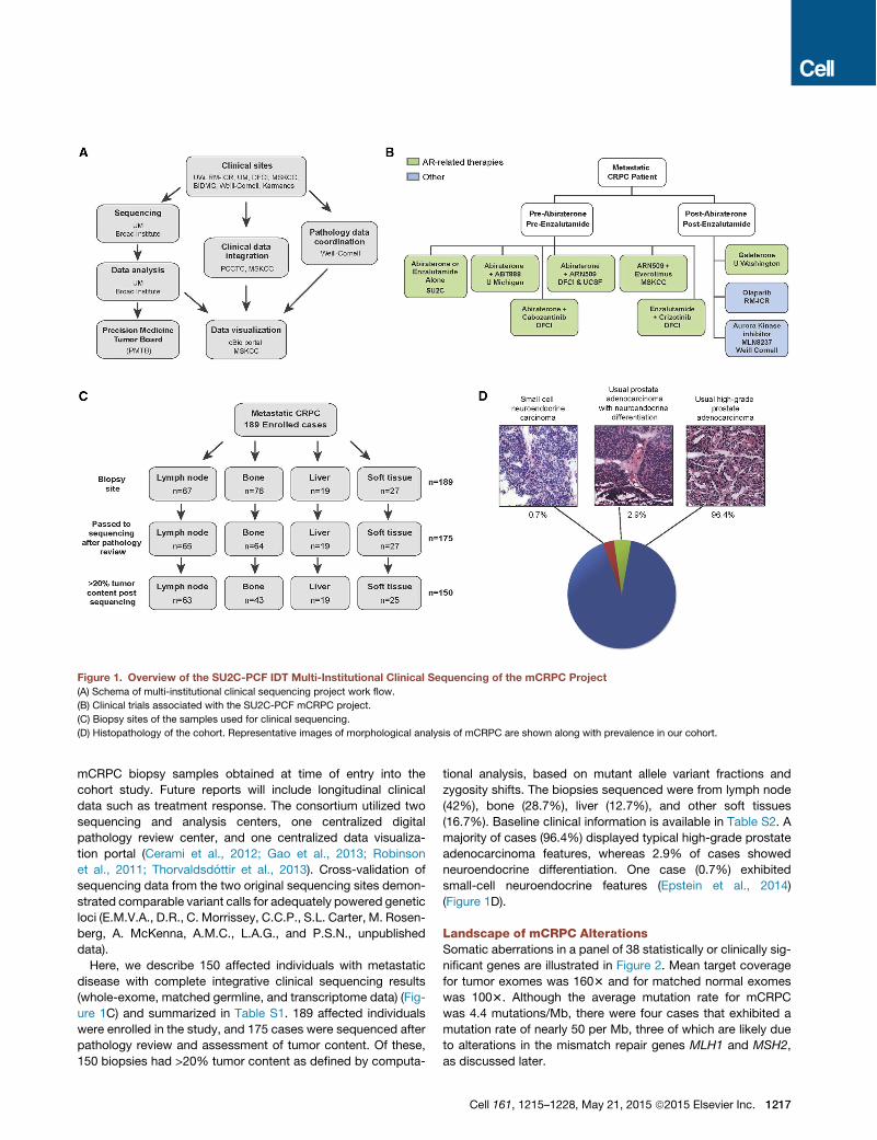
mCRPC biopsy samples obtained at time of entry into thecohort study. Future reports will include longitudinal clinicaldata such as treatment response. The consortium utilized twosequencing and analysis centers, one centralized digitalpathology review center, and one centralized data visualiza-tion portal (Cerami et al., 2012; Gao et al., 2013; Robinsonet al., 2011; Thorvaldsdottir et al., 2013). Cross-validation ofsequencing data from the two original sequencing sites demon-strated comparable variant calls for adequately powered geneticloci (E.M.V.A., D.R., C. Morrissey, C.C.P., S.L. Carter, M. Rosen-berg, A. McKenna, A.M.C., L.A.G., and P.S.N., unpublisheddata).Here, we describe 150 affected individuals with metastatic
disease with complete integrative clinical sequencing results(whole-exome, matched germline, and transcriptome data) (Fig-ure 1C) and summarized in Table S1. 189 affected individualswere enrolled in the study, and 175 cases were sequenced afterpathology review and assessment of tumor content. Of these,150 biopsies had >20% tumor content as defined by computa-
tional analysis, based on mutant allele variant fractions andzygosity shifts. The biopsies sequenced were from lymph node(42%), bone (28.7%), liver (12.7%), and other soft tissues(16.7%). Baseline clinical information is available in Table S2. Amajority of cases (96.4%) displayed typical high-grade prostateadenocarcinoma features, whereas 2.9% of cases showedneuroendocrine differentiation. One case (0.7%) exhibitedsmall-cell neuroendocrine features (Epstein et al., 2014)(Figure 1D).
Landscape of mCRPC AlterationsSomatic aberrations in a panel of 38 statistically or clinically sig-nificant genes are illustrated in Figure 2. Mean target coveragefor tumor exomes was 1603 and for matched normal exomeswas 1003. Although the average mutation rate for mCRPCwas 4.4 mutations/Mb, there were four cases that exhibited amutation rate of nearly 50 per Mb, three of which are likely dueto alterations in the mismatch repair genes MLH1 and MSH2,as discussed later.
Figure 1. Overview of the SU2C-PCF IDT Multi-Institutional Clinical Sequencing of the mCRPC Project(A) Schema of multi-institutional clinical sequencing project work flow.
(B) Clinical trials associated with the SU2C-PCF mCRPC project.
(C) Biopsy sites of the samples used for clinical sequencing.
(D) Histopathology of the cohort. Representative images of morphological analysis of mCRPC are shown along with prevalence in our cohort.
Cell 161, 1215–1228, May 21, 2015 ª2015 Elsevier Inc. 1217
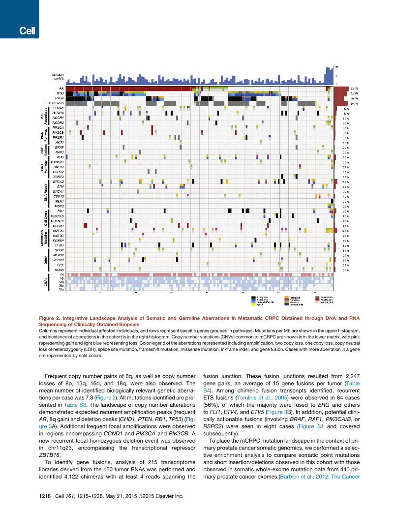
Frequent copy number gains of 8q, as well as copy numberlosses of 8p, 13q, 16q, and 18q, were also observed. Themean number of identified biologically relevant genetic aberra-tions per case was 7.8 (Figure 2). All mutations identified are pre-sented in Table S3. The landscape of copy number alterationsdemonstrated expected recurrent amplification peaks (frequentAR, 8q gain) and deletion peaks (CHD1, PTEN, RB1, TP53) (Fig-ure 3A). Additional frequent focal amplifications were observedin regions encompassing CCND1 and PIK3CA and PIK3CB. Anew recurrent focal homozygous deletion event was observedin chr11q23, encompassing the transcriptional repressorZBTB16.
To identify gene fusions, analysis of 215 transcriptomelibraries derived from the 150 tumor RNAs was performed andidentified 4,122 chimeras with at least 4 reads spanning the
fusion junction. These fusion junctions resulted from 2,247gene pairs, an average of 15 gene fusions per tumor (TableS4). Among chimeric fusion transcripts identified, recurrentETS fusions (Tomlins et al., 2005) were observed in 84 cases(56%), of which the majority were fused to ERG and othersto FLI1, ETV4, and ETV5 (Figure 3B). In addition, potential clini-cally actionable fusions (involving BRAF, RAF1, PIK3CA/B, orRSPO2) were seen in eight cases (Figure S1 and coveredsubsequently).To place the mCRPCmutation landscape in the context of pri-
mary prostate cancer somatic genomics, we performed a selec-tive enrichment analysis to compare somatic point mutationsand short insertion/deletions observed in this cohort with thoseobserved in somatic whole-exome mutation data from 440 pri-mary prostate cancer exomes (Barbieri et al., 2012; The Cancer
Figure 2. Integrative Landscape Analysis of Somatic and Germline Aberrations in Metastatic CRPC Obtained through DNA and RNASequencing of Clinically Obtained BiopsiesColumns represent individual affected individuals, and rows represent specific genes grouped in pathways. Mutations per Mb are shown in the upper histogram,
and incidence of aberrations in the cohort is in the right histogram. Copy number variations (CNVs) common tomCRPC are shown in in the lower matrix, with pink
representing gain and light blue representing loss. Color legend of the aberrations represented including amplification, two copy loss, one copy loss, copy neutral
loss of heterozygosity (LOH), splice site mutation, frameshift mutation, missense mutation, in-frame indel, and gene fusion. Cases with more aberration in a gene
are represented by split colors.
1218 Cell 161, 1215–1228, May 21, 2015 ª2015 Elsevier Inc.
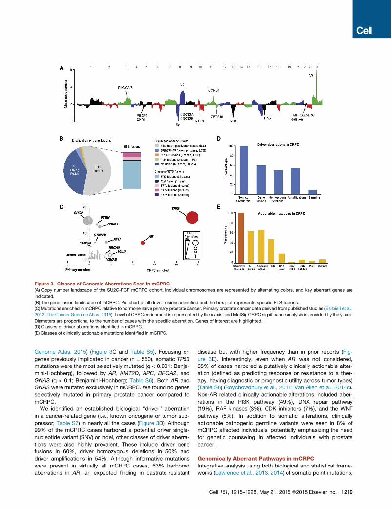
Genome Atlas, 2015) (Figure 3C and Table S5). Focusing ongenes previously implicated in cancer (n = 550), somatic TP53mutations were the most selectively mutated (q < 0.001; Benja-mini-Hochberg), followed by AR, KMT2D, APC, BRCA2, andGNAS (q < 0.1; Benjamini-Hochberg; Table S6). Both AR andGNAS were mutated exclusively in mCRPC. We found no genesselectively mutated in primary prostate cancer compared tomCRPC.We identified an established biological ‘‘driver’’ aberration
in a cancer-related gene (i.e., known oncogene or tumor sup-pressor; Table S7) in nearly all the cases (Figure 3D). Although99% of the mCPRC cases harbored a potential driver single-nucleotide variant (SNV) or indel, other classes of driver aberra-tions were also highly prevalent. These include driver genefusions in 60%, driver homozygous deletions in 50% anddriver amplifications in 54%. Although informative mutationswere present in virtually all mCRPC cases, 63% harboredaberrations in AR, an expected finding in castrate-resistant
disease but with higher frequency than in prior reports (Fig-ure 3E). Interestingly, even when AR was not considered,65% of cases harbored a putatively clinically actionable alter-ation (defined as predicting response or resistance to a ther-apy, having diagnostic or prognostic utility across tumor types)(Table S8) (Roychowdhury et al., 2011; Van Allen et al., 2014c).Non-AR related clinically actionable alterations included aber-rations in the PI3K pathway (49%), DNA repair pathway(19%), RAF kinases (3%), CDK inhibitors (7%), and the WNTpathway (5%). In addition to somatic alterations, clinicallyactionable pathogenic germline variants were seen in 8% ofmCRPC affected individuals, potentially emphasizing the needfor genetic counseling in affected individuals with prostatecancer.
Genomically Aberrant Pathways in mCRPCIntegrative analysis using both biological and statistical frame-works (Lawrence et al., 2013, 2014) of somatic point mutations,
Figure 3. Classes of Genomic Aberrations Seen in mCPRC(A) Copy number landscape of the SU2C-PCF mCRPC cohort. Individual chromosomes are represented by alternating colors, and key aberrant genes are
indicated.
(B) The gene fusion landscape of mCRPC. Pie chart of all driver fusions identified and the box plot represents specific ETS fusions.
(C) Mutations enriched in mCRPC relative to hormone naive primary prostate cancer. Primary prostate cancer data derived from published studies (Barbieri et al.,
2012; The Cancer Genome Atlas, 2015). Level of CRPC enrichment is represented by the x axis, andMutSig CRPC significance analysis is provided by the y axis.
Diameters are proportional to the number of cases with the specific aberration. Genes of interest are highlighted.
(D) Classes of driver aberrations identified in mCRPC.
(E) Classes of clinically actionable mutations identified in mCRPC.
Cell 161, 1215–1228, May 21, 2015 ª2015 Elsevier Inc. 1219
short insertion/deletions, copy number alterations, fusion tran-scripts, and focused germline variant analysis identified discretemolecular subtypes of mCRPC (Figure 2). These subtypes wereclassified based on alteration clustering and existing biologicalpathway knowledge and implicated the AR signaling pathway,phosphatidylinositol-4,5-bisphosphate 3-kinase (PI3K), WNT,DNA repair, cell cycle, and chromatin modifier gene sets, amongothers. The most frequently aberrant genes in mCRPC includedAR (62.7%), ETS family (56.7%), TP53 (53.3%), and PTEN(40.7%) (Figure 2).
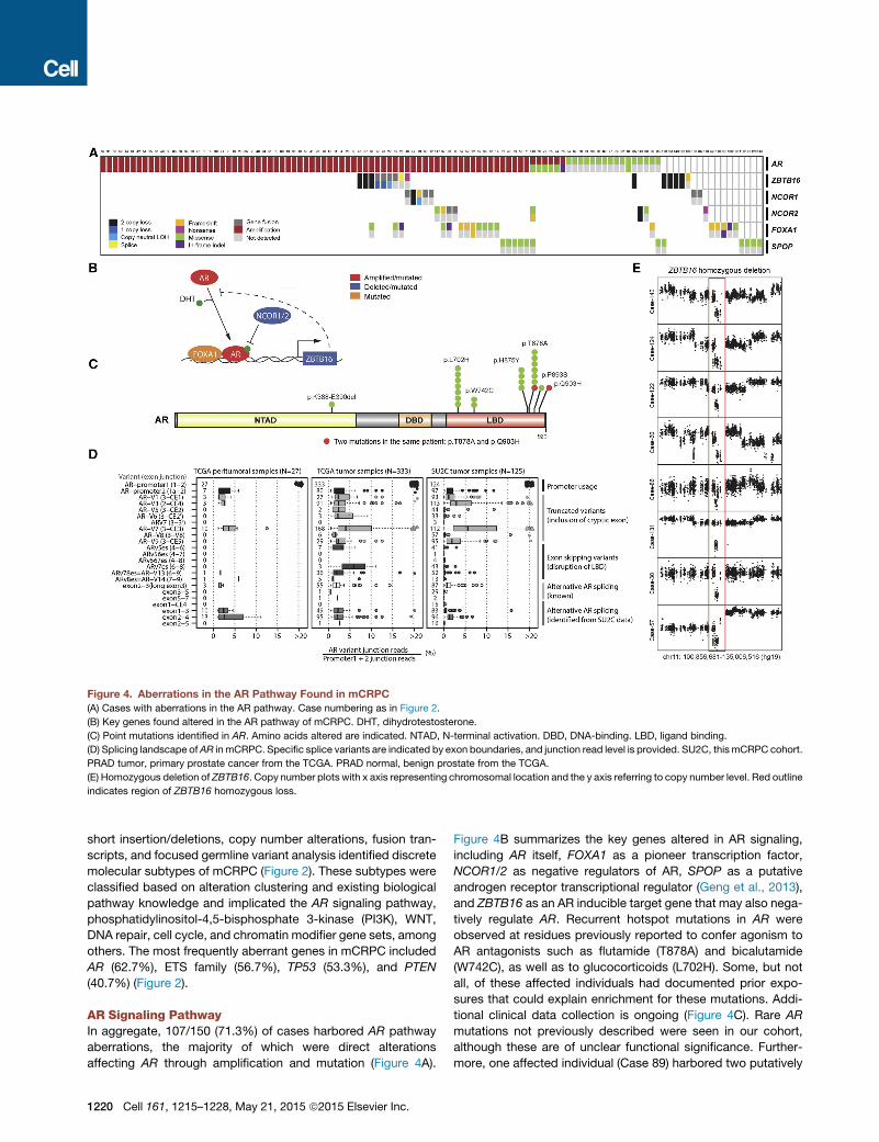
AR Signaling PathwayIn aggregate, 107/150 (71.3%) of cases harbored AR pathwayaberrations, the majority of which were direct alterationsaffecting AR through amplification and mutation (Figure 4A).
Figure 4B summarizes the key genes altered in AR signaling,including AR itself, FOXA1 as a pioneer transcription factor,NCOR1/2 as negative regulators of AR, SPOP as a putativeandrogen receptor transcriptional regulator (Geng et al., 2013),and ZBTB16 as an AR inducible target gene that may also nega-tively regulate AR. Recurrent hotspot mutations in AR wereobserved at residues previously reported to confer agonism toAR antagonists such as flutamide (T878A) and bicalutamide(W742C), as well as to glucocorticoids (L702H). Some, but notall, of these affected individuals had documented prior expo-sures that could explain enrichment for these mutations. Addi-tional clinical data collection is ongoing (Figure 4C). Rare ARmutations not previously described were seen in our cohort,although these are of unclear functional significance. Further-more, one affected individual (Case 89) harbored two putatively
Figure 4. Aberrations in the AR Pathway Found in mCRPC(A) Cases with aberrations in the AR pathway. Case numbering as in Figure 2.
(B) Key genes found altered in the AR pathway of mCRPC. DHT, dihydrotestosterone.
(C) Point mutations identified in AR. Amino acids altered are indicated. NTAD, N-terminal activation. DBD, DNA-binding. LBD, ligand binding.
(D) Splicing landscape ofAR in mCRPC. Specific splice variants are indicated by exon boundaries, and junction read level is provided. SU2C, thismCRPC cohort.
PRAD tumor, primary prostate cancer from the TCGA. PRAD normal, benign prostate from the TCGA.
(E) Homozygous deletion of ZBTB16. Copy number plots with x axis representing chromosomal location and the y axis referring to copy number level. Red outline
indicates region of ZBTB16 homozygous loss.
1220 Cell 161, 1215–1228, May 21, 2015 ª2015 Elsevier Inc.

functional AR mutations (T878A and Q903H), which may furthersuggest intra-tumor heterogeneity emerging in the CRPCsetting (Carreira et al., 2014). Analysis of AR splice variantsfrom RNA-seq data demonstrated a distribution of splice vari-ants observed throughout these mCRPC tumor cases (Fig-ure 4D). Analysis of the TCGA prostate dataset revealed thatmany of these variants were also present at varying levels in pri-mary prostate cancer and benign prostate tissue. AR-V7, whichhas been implicated in abiraterone acetate and enzalutamideresistance (Antonarakis et al., 2014), was observed in a majorityof pre-abiraterone/enzalutamide cases but at very low ratiosrelative to full length AR. Implications for treatment responseare unknown at this time.In addition to AR mutations itself, we observed alterations in
AR pathway members (Figure 4A). These included known alter-ations inNCOR1,NCOR2, and FOXA1 that have been previouslyreported in primary prostate cancers andmCRPC (Barbieri et al.,2012; Grasso et al., 2012). In this cohort, truncating andmissense mutations in FOXA1 form a cluster near the end ofthe Forkhead DNA binding domain (Figure S2).Recurrent homozygous deletions of the androgen-regulated
gene ZBTB16 (also known as PLZF) were seen in 8 (5%) cases(Figure 4E) not previously reported in clinical mCRPC biopsies.Analysis of the minimally deleted region seen in this cohort nar-rowed the candidate genes in the chr11q23 region to ZBTB16(Figure S3). ZBTB16 has been previously implicated in prostatecancer tumorigenesis and androgen resistance in preclinicalmodels (Cao et al., 2013; Kikugawa et al., 2006), with loss ofZBTB16 upregulating the MAPK signaling pathway (Hsiehet al., 2015).
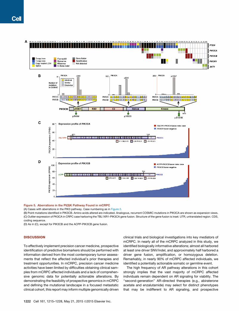
New PI3K Pathway DiscoveriesThe PI3K pathway was also commonly altered, with somatic al-terations in 73/150 (49%) of mCRPC affected individuals (Fig-ure 5A). This included biallelic loss of PTEN, as well as hotspotmutations, amplifications and activating fusions in PIK3CA,and p.E17K activating mutations in AKT1 (Figure S2). Of note,PIK3CA amplifications resulted in overexpression compared tothe remaining cohort (Figure S3).Interestingly, mutations in another member of the PI3K cata-
lytic subunit, PIK3CB, were observed in this cohort for the firsttime, at equivalent positions to canonical activating mutationsin PIK3CA (Figure 5B). PIK3CB mutations appeared in thecontext of PTEN-deficient cases, which is consistent with a pre-vious report demonstrating that some PTEN-deficient cancersare dependent on PIK3CB, rather than PIK3CA (Wee et al.,2008). Furthermore, two affected individuals harbored fusionsinvolving PIK3CA/B, with these events resulting in overexpres-sion of the gene relative to other tumors in the cohort (Figures5C and 5D).
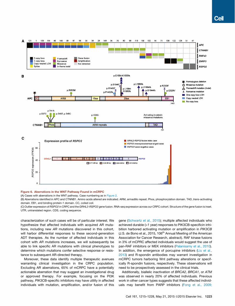
New Wnt Pathway Discoveries27/150 (18%) of our cases harbored alterations in the Wntsignaling pathway (Figure 6A). Hotspot activating mutations inCTNNB1 were seen (Figure 6B), as previously described (Voel-ler et al., 1998). Notably, recurrent alterations in APC were alsoobserved, which have not been previously described in clinicalmCRPC affected individuals. This prompted a broader exami-
nation of Wnt signaling genes (Figure 6B). Through integrativeanalysis, we identified alterations in RNF43 and ZNRF3, whichwere recently described in colorectal, endometrial, and adreno-cortical cancers (Assie et al., 2014; Giannakis et al., 2014) andwere mutually exclusive with APC alterations (Figure 6A). More-over, we also discovered R-spondin fusions involving RSPO2,as previously observed in colorectal carcinoma (Seshagiriet al., 2012) in association with RSPO2 overexpression in thesecases (Figure 6C). RSPO2 is a key factor in prostate cancer or-ganoid methodology (Gao et al., 2014). Affected individuals swith aberrations in RNF43, ZNRF3, or RSPO2 (overall 6% ofaffected individuals) are predicted to respond to porcupine in-hibitors (Liu et al., 2013).
Cell-Cycle PathwayWe observed RB1 loss in 21% of cases (Figure S4). Expandingthe scope of cell-cycle genes implicated in mCRPC, we notedfocal amplifications involving CCND1 in 9% of cases, as wellas less common (< 5%) events in CDKN2A/B, CDKN1B, andCDK4 (Figure S4). Cell-cycle derangement, such as throughCCND1 amplification or CDKN2A/B loss, may result in enhancedresponse to CDK4 inhibitors in other tumor types (Finn et al.,2015), and preclinical mCRPC models predict similar activity inprostate cancer (Comstock et al., 2013).
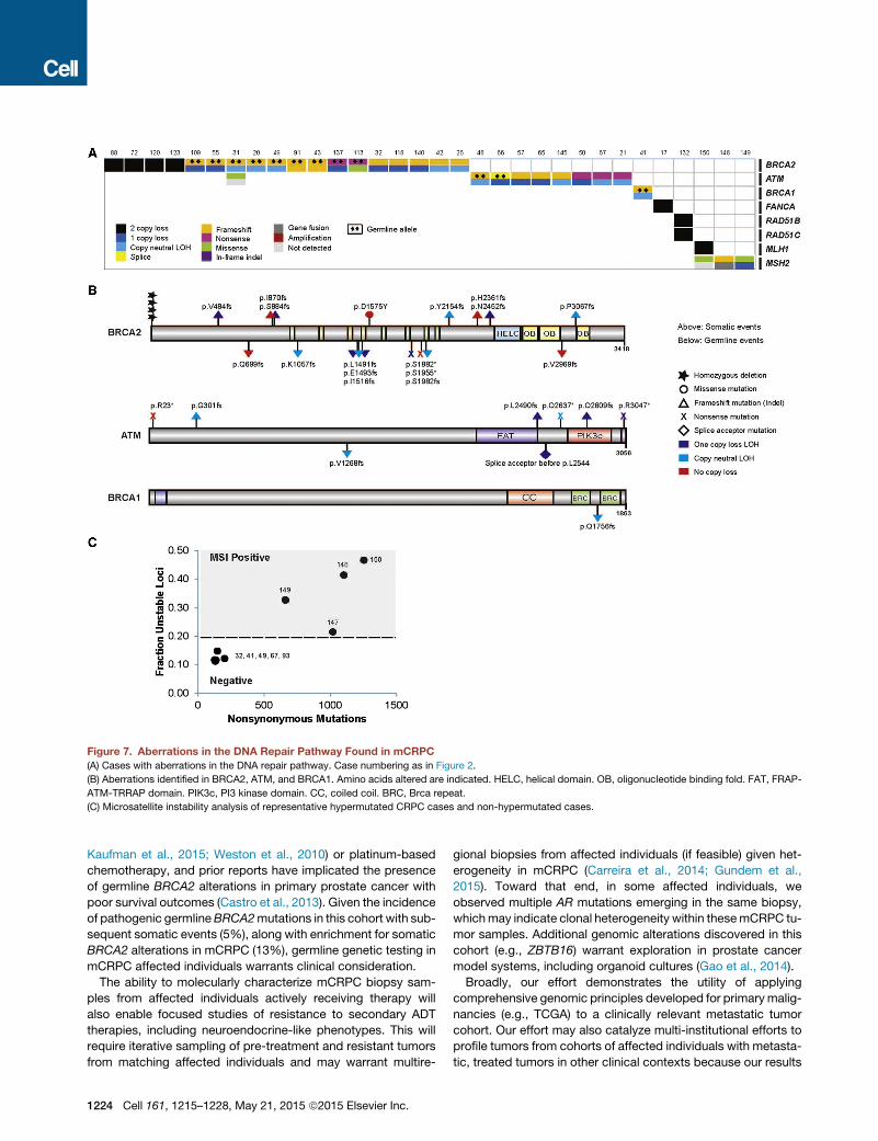
DNA Repair PathwayIntegrative analysis of both the somatic and pathogenic germlinealterations in BRCA2 identified 19/150 (12.7%) of cases withloss of BRCA2, of which !90% exhibited biallelic loss (Fig-ure 7A). This was commonly a result of somatic point mutationand loss of heterozygosity, as well as homozygous deletion.One of the clinical trials in our consortium is evaluating poly(-ADP-ribose) polymerase (PARP) inhibition in unselectedmCRPC affected individuals. Importantly, multiple affected indi-viduals in this trial who experienced clinical benefit harboredbiallelic BRCA2 loss, providing further evidence of clinical ac-tionability (Mateo et al., 2014). Eight affected individuals(5.3%) harbored pathogenic germline BRCA2 mutations (Fig-ure 7B) with a subsequent somatic event that resulted in biallelicloss, revealing a surprisingly high frequency relative to primaryprostate cancer.We therefore expanded the focus to other DNA repair/recom-
bination genes and identified alterations in at least 34/150(22.7%) of cases. These include recurrent biallelic loss ofATM (Figure 7B), including multiple cases with germline patho-genic alterations. ATM mutations were also observed inaffected individuals who achieved clinical responses to PARPinhibition (Mateo et al., 2014). In addition, we noted events inBRCA1, CDK12, FANCA, RAD51B, and RAD51C. If aberrationsof BRCA2, BRCA1, and ATM all confer enhanced sensitivity toPARP inhibitors, 29/150 (19.3%) of mCRPC affected individualswould be predicted to benefit from this therapy. Interestingly,three out of four mCRPC tumors exhibited hypermutation andharbored alterations in the mismatch repair pathway genesMLH1 or MSH2 (Figures 2 and 7C), corroborating a recentreport identifying structural alterations in MSH2 and MSH6mismatch repair genes in hypermutated prostate cancers(Pritchard et al., 2014).
Cell 161, 1215–1228, May 21, 2015 ª2015 Elsevier Inc. 1221
DISCUSSION
To effectively implement precision cancer medicine, prospectiveidentification of predictive biomarkers should be performed withinformation derived from the most contemporary tumor assess-ments that reflect the affected individual’s prior therapies andtreatment opportunities. In mCRPC, precision cancer medicineactivities have been limited by difficulties obtaining clinical sam-ples frommCRPC affected individuals and a lack of comprehen-sive genomic data for potentially actionable alterations. Bydemonstrating the feasibility of prospective genomics inmCRPCand defining the mutational landscape in a focused metastaticclinical cohort, this reportmay informmultiple genomically driven
clinical trials and biological investigations into key mediators ofmCRPC. In nearly all of the mCRPC analyzed in this study, weidentified biologically informative alterations; almost all harboredat least one driver SNV/indel, and approximately half harbored adriver gene fusion, amplification, or homozygous deletion.Remarkably, in nearly 90% of mCRPC affected individuals, weidentified a potentially actionable somatic or germline event.The high frequency of AR pathway alterations in this cohort
strongly implies that the vast majority of mCRPC affectedindividuals remain dependent on AR signaling for viability. The‘‘second-generation’’ AR-directed therapies (e.g., abirateroneacetate and enzalutamide) may select for distinct phenotypesthat may be indifferent to AR signaling, and prospective
Figure 5. Aberrations in the PI(3)K Pathway Found in mCRPC(A) Cases with aberrations in the PIK3 pathway. Case numbering as in Figure 2.
(B) Point mutations identified in PIK3CB. Amino acids altered are indicated. Analogous, recurrent COSMIC mutations in PIK3CA are shown as expansion views.
(C) Outlier expression of PK3CA in CRPC case harboring the TBL1XR1-PIK3CA gene fusion. Structure of the gene fusion is inset. UTR, untranslated region. CDS,
coding sequence.
(D) As in (C), except for PIK3CB and the ACPP-PIK3CB gene fusion.
1222 Cell 161, 1215–1228, May 21, 2015 ª2015 Elsevier Inc.
characterization of such cases will be of particular interest. Wehypothesize that affected individuals with acquired AR muta-tions, including new AR mutations discovered in this cohort,will harbor differential responses to these second-generationADT therapies. As the number of affected individuals in thiscohort with AR mutations increases, we will subsequently beable to link specific AR mutations with clinical phenotypes todetermine which mutations confer selective response or resis-tance to subsequent AR-directed therapy.Moreover, these data identify multiple therapeutic avenues
warranting clinical investigation in the CRPC population.Excluding AR aberrations, 65% of mCRPC have a potentiallyactionable aberration that may suggest an investigational drugor approved therapy. For example, focusing on the PI3Kpathway, PIK3CB-specific inhibitors may have utility in affectedindividuals with mutation, amplification, and/or fusion of this
gene (Schwartz et al., 2015); multiple affected individuals whoachieved durable (>1 year) responses to PIK3CB-specificin inhi-bition harbored activating mutation or amplification in PIK3CB(J.S. de Bono et al., 2015, 106th Annual Meeting of the AmericanAssociation for Cancer Research, abstract). RAF kinase fusionsin 3% of mCPRC affected individuals would suggest the use ofpan-RAF inhibitors or MEK inhibitors (Palanisamy et al., 2010).In addition, the emergence of porcupine inhibitors (Liu et al.,2013) and R-spondin antibodies may warrant investigation inmCRPC tumors harboring Wnt pathway alterations or specif-ically R-spondin fusions, respectively. These observations willneed to be prospectively assessed in the clinical trials.Additionally, biallelic inactivation of BRCA2, BRCA1, or ATM
was observed in nearly 20% of affected individuals. Previouswork in other cancer types suggests that these affected individ-uals may benefit from PARP inhibitors (Fong et al., 2009;
Figure 6. Aberrations in the WNT Pathway Found in mCRPC(A) Cases with aberrations in the WNT pathway. Case numbering as in Figure 2.
(B) Aberrations identified in APC and CTNNB1. Amino acids altered are indicated. ARM, armadillo repeat. Phos, phosphorylation domain. TAD, trans-activating
domain. EB1, end binding protein-1 domain. CC, coiled coil.
(C) Outlier expression ofRSPO2 in CRPC and theGRHL2-RSPO2 gene fusion. RNA-seq expression across our CRPC cohort. Structure of the gene fusion is inset.
UTR, untranslated region. CDS, coding sequence.
Cell 161, 1215–1228, May 21, 2015 ª2015 Elsevier Inc. 1223
Kaufman et al., 2015; Weston et al., 2010) or platinum-basedchemotherapy, and prior reports have implicated the presenceof germline BRCA2 alterations in primary prostate cancer withpoor survival outcomes (Castro et al., 2013). Given the incidenceof pathogenic germlineBRCA2mutations in this cohort with sub-sequent somatic events (5%), along with enrichment for somaticBRCA2 alterations in mCRPC (13%), germline genetic testing inmCRPC affected individuals warrants clinical consideration.
The ability to molecularly characterize mCRPC biopsy sam-ples from affected individuals actively receiving therapy willalso enable focused studies of resistance to secondary ADTtherapies, including neuroendocrine-like phenotypes. This willrequire iterative sampling of pre-treatment and resistant tumorsfrom matching affected individuals and may warrant multire-
gional biopsies from affected individuals (if feasible) given het-erogeneity in mCRPC (Carreira et al., 2014; Gundem et al.,2015). Toward that end, in some affected individuals, weobserved multiple AR mutations emerging in the same biopsy,whichmay indicate clonal heterogeneity within thesemCRPC tu-mor samples. Additional genomic alterations discovered in thiscohort (e.g., ZBTB16) warrant exploration in prostate cancermodel systems, including organoid cultures (Gao et al., 2014).Broadly, our effort demonstrates the utility of applying
comprehensive genomic principles developed for primarymalig-nancies (e.g., TCGA) to a clinically relevant metastatic tumorcohort. Our effort may also catalyze multi-institutional efforts toprofile tumors from cohorts of affected individuals with metasta-tic, treated tumors in other clinical contexts because our results
Figure 7. Aberrations in the DNA Repair Pathway Found in mCRPC(A) Cases with aberrations in the DNA repair pathway. Case numbering as in Figure 2.
(B) Aberrations identified in BRCA2, ATM, and BRCA1. Amino acids altered are indicated. HELC, helical domain. OB, oligonucleotide binding fold. FAT, FRAP-
ATM-TRRAP domain. PIK3c, PI3 kinase domain. CC, coiled coil. BRC, Brca repeat.
(C) Microsatellite instability analysis of representative hypermutated CRPC cases and non-hypermutated cases.
1224 Cell 161, 1215–1228, May 21, 2015 ª2015 Elsevier Inc.
demonstrate multiple discoveries within this advanced diseasestage that have not been observed in primary tumor profiling.Moreover, this study sets the stage for epigenetic and otherprofiling efforts in mCRPC not taken in this study, which mayenable biological discovery and have immediate therapeuticrelevance in mCRPC (Asangani et al., 2014). Overall, our effortsdemonstrate the feasibility of comprehensive and integrative ge-nomics on prospective biopsies from individual mCRPC affectedindividuals to enable precision cancer medicine activities in thislarge affected individual population.
EXPERIMENTAL PROCEDURES
Affected Individual EnrollmentAffected individuals with clinical evidence of mCRPC who were being consid-
ered for abiraterone acetate or enzalutamide as standard of care, or as part of
a clinical trial, were considered for enrollment. Affected individuals with meta-
static disease accessible by image-guided biopsy were eligible for inclusion.
All affected individuals provided written informed consent to obtain fresh tu-
mor biopsies and to perform comprehensive molecular profiling of tumor
and germline samples.
Biopsies and Pathology ReviewBiopsies of soft tissue or bone metastases were obtained under radiographic
guidance. Digital images of biopsy slides were centrally reviewed using
schema established to distinguish usual adenocarcinoma from neuroendo-
crine prostate cancer (Epstein et al., 2014). All images were reviewed by geni-
tourinary oncology pathologists (M.R., J.M.M., L.P.K., S.A.T., R.M., V.R., A.G.,
M.L., R.L., and M.B.).
Sequencing and AnalysisNormal DNAs from buccal swabs, buffy coats, or whole blood were isolated
using the QIAGEN DNeasy Blood & Tissue Kit. Flash-frozen needle biopsies
with highest tumor content for each case, as determined by pathology review,
were extracted for nucleic acids. Tumor genomic DNA and total RNA were pu-
rified from the same sample using the AllPrep DNA/RNA/miRNA kit (QIAGEN)
with disruption on a Tissuelyser II (QIAGEN). RNA integrity was verified on an
Agilent 2100 Bioanalyzer using RNA Nano reagents (Agilent Technologies).
Whole-exome capture libraries were constructed from 100 ng to 1 mg of DNA
from tumor and normal tissue after sample shearing, end repair, and phos-
phorylation and ligation to barcoded sequencing adaptors. Ligated DNA was
size selected for lengths between 200 and 350 bp and subjected to hybrid cap-
ture using SureSelect Exome v4 baits (Agilent). Exome sequence data pro-
cessing and analysis were performed using pipelines at the Broad Institute
and the University of Michigan. A BAM file aligned to the hg19 human genome
build was produced using Illumina sequencing reads for the tumor and normal
sample and the Picard pipeline. Somatic mutation analysis was performed as
described previously (Cibulskis et al., 2013; Van Allen et al., 2014c) and re-
viewed with Integrated Genomics Viewer (IGV) (Robinson et al., 2011).
Copy number aberrations were quantified and reported for each gene as the
segmented normalized log2-transformed exon coverage ratios between each
tumor sample and matched normal sample (Lonigro et al., 2011). To account
for observed associations between coverage ratios and GC content across
the genome, lowess normalization was used to correct per-exon coverage ra-
tios prior to segmentation analysis. Mean GC percentage was computed for
each targeted region, and a lowess curve was fit to the scatterplot of log2-
coverage ratios versus mean GC content across the targeted exome using
the lowess function in R (version 2.13.1) with smoothing parameter f = 0.05.
The resulting copy ratios were segmented using the circular binary segmenta-
tion algorithm (Olshen et al., 2004).
Statistical analysis of recurrently mutated genes was performed using Mut-
Sig (Lawrence et al., 2013). Selective enrichment analysis (Van Allen et al.,
2014b) of mutations observed in mCRPC compared to primary prostate can-
cer was performed by tabulating the frequency of affected-individual-normal-
ized mutations observed in either CRPC or primary prostate cancer and
performing a two-sided Fisher’s exact test using allelic fraction cut off of 0.1
or greater and a set of biologically relevant cancer genes (n = 550 genes) (Fu-
treal et al., 2004). Multiple hypothesis test correction was performed using
Benjamini-Hochberg method.
Transcriptome libraries were prepared using 200–1,000 ng of total RNA.
PolyA+ RNA isolation, cDNA synthesis, end-repair, A-base addition, and liga-
tion of the Illumina indexed adapters were performed according to the TruSeq
RNA protocol (Illumina). Libraries were size selected for 250–300 bp cDNA
fragments on a 3%Nusieve 3:1 (Lonza) gel, recovered using QIAEX II reagents
(QIAGEN), and PCR amplified using Phusion DNA polymerase (New England
Biolabs). Total transcriptome libraries were prepared as above, omitting the
poly A selection step and captured using Agilent SureSelect Human All Exon
V4 reagents and protocols. Library quality was measured on an Agilent 2100
Bioanalyzer for product size and concentration. Paired-end libraries were
sequenced with the Illumina HiSeq 2500, (23100 nucleotide read length)
with sequence coverage to 50 M paired reads and 100 M total reads.
Paired-end transcriptome sequencing reads were aligned to the human
reference genome (GRCh37/hg19) using a RNA-seq spliced read mapper
Tophat2 (Kim and Salzberg, 2011) (Tophat 2.0.4), with ‘‘–fusion-search’’ option
turned on to detect potential gene fusion transcripts. Potential false-positive
fusion candidates were filtered out using ‘‘Tophat-Post-Fusion’’ module.
Further, the fusion candidates were manually examined for annotation and
ligation artifacts. Gene expression, as fragments per kilobase of exon per
million fragments mapped (FPKM; normalized measure of gene expression),
was calculated using Cufflinks (Trapnell et al., 2012).
ACCESSION NUMBERS
The accession number for the data reported in this paper is dbGap:
phs000915.v1.p1.
SUPPLEMENTAL INFORMATION
Supplemental Information includes four figures and eight tables and can be
found with this article online at http://dx.doi.org/10.1016/j.cell.2015.05.001.
AUTHOR CONTRIBUTIONS
Y-M.W., N.S., R.J.L., J.-M.M., R.M., M.E.T., C.C.P., G.A., H.B., andW.A.made
equal contributions. D.R., E.M.V.A., and R.J.L. coordinated overall sequencing
and bioinformatics analysis. Y.M.W., D.R., E.M.V.A., R.J.L., W.A., and J.V. co-
ordinated figures and tables. N.S. developed the SU2C-PCF IDT cBio portal.
R.J.L. coordinated copy number analyses. J.-M.M. coordinated central pa-
thology review, and L.P.K coordinated UM pathology analysis. C.C.P. coordi-
nated hypermutation analysis and clinical germline interpretations. R.M.,
M.E.T, G.A., H.B., M.H., H.I.S., and E.I.H. coordinated clinical enrollment at
their specific sites. R.K.B., S.P., and H.D. carried out AR splice variant anal-
ysis. J.V. managed the clinical data portal. X.C., J.S., P.F., S.M., and C.S.
were involved in project management. R.M., S.A.T., V.E.R., A.G., M.L.,
S.P.B., R.T.L., M.B., B.D.R., J.-M.M, M.R., and L.D.T. were involved in pathol-
ogy review. C.B., P.V., J.G., M.G., F.D., O.E., A. Sboner, A. Sigaras, and K.W.E.
contributed to bioinformatics analysis. K.A.C., D.C.S., F.Y.F., H.I.S., D.R.,
S.B.S., M.J.M., R.F., Z.Z., N.T., G.G., J.C.D., J.M., D.M.N., S.T.T., E.Y.Y.,
Y.C., E.A.M., H.H.C., M.A.S., and K.J.P. are clinical contributors. E.M.V.A.,
D.R., C.L.S. and A.M.C. wrote the manuscript, which all authors reviewed.
P.K., J.S.d.B., M.A.R., P.S.N., and L.A.G. are SU2C-PCF Dream Team Princi-
pals, and C.L.S. and A.M.C. are Dream Team co-Leaders.
ACKNOWLEDGMENTS
We thank the affected individuals who participated in this study to better un-
derstand the feasibility and utility of precision medicine approaches for
advanced prostate cancer. Individuals at our respective institutions who
helped with this study are listed by institution. University of Michigan: Karen
Giles, Lynda Hodges, Erica Rabban, Ning Yu, Fengyun Su, Rui Wang, Brendan
Veeneman, and Moshe Talpaz. MSKCC: Brett Carver, Kristen Curtis, and Julie
Cell 161, 1215–1228, May 21, 2015 ª2015 Elsevier Inc. 1225

Filipenko. DFCI/Broad: Zhenwei Zhang, Daniele Depalo, and Joseph Kram-
kowski. University of Washington: Jina Taub, Hiep Nguyen, Colm Morrissey,
and Robert Vessella. ICR/Royal Marsden: Suzanne Carreira, Ines Figueiredo,
and Daniel Nava Rodrigues. This work was supported by a Stand Up To
Cancer-Prostate Cancer Foundation Prostate Dream Team Translational Can-
cer Research Grant. Stand Up To Cancer is a program of the Entertainment In-
dustry Foundation administered by the American Association for Cancer
Research (SU2C-AACR-DT0712). The project was also supported by the
following NIH awards: Clinical Sequencing Exploratory Research (CSER)
UM1HG006508 (A.M.C.), Early Detection Research Network grant UO1
CA111275(A.M.C.), Prostate SPORE grants P50 CA186786 (A.M.C.), P50
CA092629 (H.I.S., C.L.S., and Y.C.), P50 CA90381 (P.K.),and P50 CA097186
(P.S.N., B.M., E.A.M., and L.D.T.), P01 CA163227 (P.S.N., S.P., R.K.B.,
H.D.), R01 CA116337 (M.A.R., H.B., F.D.), R01 CA155169 (C.L.S.), R01
CA092629, P50 CA092629 (H.I.S., C.L.S., and Y.C.), and R01 CA155169
(C.L.S.). This work was supported by the following DoD awards: W81XWH-
09-1-0147 (PCCTC) and DOD PC121341 (H.B.). This work was also supported
by the Starr Cancer Consortium (N.S., M.R., C.S., Y.C., L.A.G.). A.M.C. is an A.
Alfred Taubman Scholar and an American Cancer Society Professor. H.B. is
supported by Damon Runyon Cancer Research Foundation CI-67-13. C.P.
is supported by a PCF Young Investigator Award and DoD PC131820.
E.M.V. is supported by a NIH 1K08CA188615. E.M.V., N.S., and F.Y.F. are
supported by Prostate Cancer Foundation Young Investigator Awards. The
RM and ICR team is supported by the Movember Foundation and Prostate
Cancer UK, PCF, the ECMC network from Cancer Research UK, the Depart-
ment of Health in the UK, and BRC grant funding.
Received: March 9, 2015
Revised: April 6, 2015
Accepted: April 27, 2015
Published: May 21, 2015
REFERENCES
American Cancer Society (2015). Cancer Facts and Figures 2015. http://
www.cancer.org/acs/groups/content/@editorial/documents/document/acspc-
044552.pdf.
Antonarakis, E.S., Lu, C., Wang, H., Luber, B., Nakazawa, M., Roeser, J.C.,
Chen, Y., Mohammad, T.A., Chen, Y., Fedor, H.L., et al. (2014). AR-V7 and
resistance to enzalutamide and abiraterone in prostate cancer. N. Engl. J.
Med. 371, 1028–1038.
Asangani, I.A., Dommeti, V.L., Wang, X., Malik, R., Cieslik, M., Yang, R., Es-
cara-Wilke, J., Wilder-Romans, K., Dhanireddy, S., Engelke, C., et al. (2014).
Therapeutic targeting of BET bromodomain proteins in castration-resistant
prostate cancer. Nature 510, 278–282.
Assie, G., Letouze, E., Fassnacht, M., Jouinot, A., Luscap, W., Barreau, O.,
Omeiri, H., Rodriguez, S., Perlemoine, K., Rene-Corail, F., et al. (2014). Inte-
grated genomic characterization of adrenocortical carcinoma. Nat. Genet.
46, 607–612.
Baca, S.C., Prandi, D., Lawrence, M.S., Mosquera, J.M., Romanel, A., Drier,
Y., Park, K., Kitabayashi, N., MacDonald, T.Y., Ghandi, M., et al. (2013). Punc-
tuated evolution of prostate cancer genomes. Cell 153, 666–677.
Barbieri, C.E., Baca, S.C., Lawrence, M.S., Demichelis, F., Blattner, M., Theur-
illat, J.P., White, T.A., Stojanov, P., Van Allen, E., Stransky, N., et al. (2012).
Exome sequencing identifies recurrent SPOP, FOXA1 and MED12 mutations
in prostate cancer. Nat. Genet. 44, 685–689.
Beer, T.M., Armstrong, A.J., Rathkopf, D.E., Loriot, Y., Sternberg, C.N., Hi-
gano, C.S., Iversen, P., Bhattacharya, S., Carles, J., Chowdhury, S., et al.;
PREVAIL Investigators (2014). Enzalutamide in metastatic prostate cancer
before chemotherapy. N. Engl. J. Med. 371, 424–433.
Beltran, H., Yelensky, R., Frampton, G.M., Park, K., Downing, S.R., MacDon-
ald, T.Y., Jarosz, M., Lipson, D., Tagawa, S.T., Nanus, D.M., et al. (2013).
Targeted next-generation sequencing of advanced prostate cancer identifies
potential therapeutic targets and disease heterogeneity. Eur. Urol. 63,
920–926.
Berger, M.F., Lawrence, M.S., Demichelis, F., Drier, Y., Cibulskis, K., Siva-
chenko, A.Y., Sboner, A., Esgueva, R., Pflueger, D., Sougnez, C., et al.
(2011). The genomic complexity of primary human prostate cancer. Nature
470, 214–220.
Cao, J., Zhu, S., Zhou, W., Li, J., Liu, C., Xuan, H., Yan, J., Zheng, L., Zhou, L.,
Yu, J., et al. (2013). PLZF mediates the PTEN/AKT/FOXO3a signaling in sup-
pression of prostate tumorigenesis. PLoS ONE 8, e77922.
Carreira, S., Romanel, A., Goodall, J., Grist, E., Ferraldeschi, R., Miranda, S.,
Prandi, D., Lorente, D., Frenel, J.S., Pezaro, C., et al. (2014). Tumor clone dy-
namics in lethal prostate cancer. Sci. Transl. Med. 6, 254ra125.
Castro, E., Goh, C., Olmos, D., Saunders, E., Leongamornlert, D., Tymrakie-
wicz, M., Mahmud, N., Dadaev, T., Govindasami, K., Guy, M., et al. (2013).
Germline BRCA mutations are associated with higher risk of nodal involve-
ment, distant metastasis, and poor survival outcomes in prostate cancer.
J. Clin. Oncol. 31, 1748–1757.
Cerami, E., Gao, J., Dogrusoz, U., Gross, B.E., Sumer, S.O., Aksoy, B.A., Ja-
cobsen, A., Byrne, C.J., Heuer, M.L., Larsson, E., et al. (2012). The cBio cancer
genomics portal: an open platform for exploring multidimensional cancer ge-
nomics data. Cancer Discov. 2, 401–404.
Cibulskis, K., Lawrence,M.S., Carter, S.L., Sivachenko, A., Jaffe, D., Sougnez,
C., Gabriel, S., Meyerson, M., Lander, E.S., and Getz, G. (2013). Sensitive
detection of somatic point mutations in impure and heterogeneous cancer
samples. Nat. Biotechnol. 31, 213–219.
Comstock, C.E., Augello, M.A., Goodwin, J.F., de Leeuw, R., Schiewer, M.J.,
Ostrander, W.F., Jr., Burkhart, R.A., McClendon, A.K., McCue, P.A., Trabulsi,
E.J., et al. (2013). Targeting cell cycle and hormone receptor pathways in can-
cer. Oncogene 32, 5481–5491.
Cooper, C.S., Eeles, R., Wedge, D.C., Van Loo, P., Gundem, G., Alexandrov,
L.B., Kremeyer, B., Butler, A., Lynch, A.G., Camacho, N., et al.; ICGC Prostate
Group (2015). Analysis of the genetic phylogeny of multifocal prostate cancer
identifies multiple independent clonal expansions in neoplastic and morpho-
logically normal prostate tissue. Nat. Genet. 47, 367–372.
de Bono, J.S., Logothetis, C.J., Molina, A., Fizazi, K., North, S., Chu, L., Chi,
K.N., Jones, R.J., Goodman, O.B., Jr., Saad, F., et al.; COU-AA-301 Investiga-
tors (2011). Abiraterone and increased survival in metastatic prostate cancer.
N. Engl. J. Med. 364, 1995–2005.
Epstein, J.I., Amin, M.B., Beltran, H., Lotan, T.L., Mosquera, J.M., Reuter, V.E.,
Robinson, B.D., Troncoso, P., and Rubin, M.A. (2014). Proposed morphologic
classification of prostate cancer with neuroendocrine differentiation. Am. J.
Surg. Pathol. 38, 756–767.
Finn, R.S., Crown, J.P., Lang, I., Boer, K., Bondarenko, I.M., Kulyk, S.O., Ettl,
J., Patel, R., Pinter, T., Schmidt, M., et al. (2015). The cyclin-dependent kinase
4/6 inhibitor palbociclib in combination with letrozole versus letrozole alone as
first-line treatment of oestrogen receptor-positive, HER2-negative, advanced
breast cancer (PALOMA-1/TRIO-18): a randomised phase 2 study. Lancet On-
col. 16, 25–35.
Fong, P.C., Boss, D.S., Yap, T.A., Tutt, A., Wu, P., Mergui-Roelvink, M., Mor-
timer, P., Swaisland, H., Lau, A., O’Connor, M.J., et al. (2009). Inhibition of pol-
y(ADP-ribose) polymerase in tumors from BRCA mutation carriers. N. Engl. J.
Med. 361, 123–134.
Futreal, P.A., Coin, L., Marshall, M., Down, T., Hubbard, T., Wooster, R., Rah-
man, N., and Stratton,M.R. (2004). A census of human cancer genes. Nat. Rev.
Cancer 4, 177–183.
Gao, J., Aksoy, B.A., Dogrusoz, U., Dresdner, G., Gross, B., Sumer, S.O., Sun,
Y., Jacobsen, A., Sinha, R., Larsson, E., et al. (2013). Integrative analysis of
complex cancer genomics and clinical profiles using the cBioPortal. Sci.
Signal. 6, pl1.
Gao, D., Vela, I., Sboner, A., Iaquinta, P.J., Karthaus, W.R., Gopalan, A., Dow-
ling, C., Wanjala, J.N., Undvall, E.A., Arora, V.K., et al. (2014). Organoid cul-
tures derived from patients with advanced prostate cancer. Cell 159, 176–187.
Geng, C., He, B., Xu, L., Barbieri, C.E., Eedunuri, V.K., Chew, S.A., Zimmer-
mann, M., Bond, R., Shou, J., Li, C., et al. (2013). Prostate cancer-associated
1226 Cell 161, 1215–1228, May 21, 2015 ª2015 Elsevier Inc.
mutations in speckle-type POZ protein (SPOP) regulate steroid receptor coac-
tivator 3 protein turnover. Proc. Natl. Acad. Sci. USA 110, 6997–7002.
Giannakis, M., Hodis, E., Jasmine Mu, X., Yamauchi, M., Rosenbluh, J., Cibul-
skis, K., Saksena, G., Lawrence, M.S., Qian, Z.R., Nishihara, R., et al. (2014).
RNF43 is frequently mutated in colorectal and endometrial cancers. Nat.
Genet. 46, 1264–1266.
Grasso, C.S., Wu, Y.M., Robinson, D.R., Cao, X., Dhanasekaran, S.M., Khan,
A.P., Quist, M.J., Jing, X., Lonigro, R.J., Brenner, J.C., et al. (2012). The muta-
tional landscape of lethal castration-resistant prostate cancer. Nature 487,
239–243.
Gundem, G., Van Loo, P., Kremeyer, B., Alexandrov, L.B., Tubio, J.M., Pa-
paemmanuil, E., Brewer, D.S., Kallio, H.M., Hognas, G., Annala, M., et al.;
ICGC Prostate UK Group (2015). The evolutionary history of lethal metastatic
prostate cancer. Nature 520, 353–357.
Hieronymus, H., Schultz, N., Gopalan, A., Carver, B.S., Chang, M.T., Xiao, Y.,
Heguy, A., Huberman, K., Bernstein, M., Assel, M., et al. (2014). Copy number
alteration burden predicts prostate cancer relapse. Proc. Natl. Acad. Sci. USA
111, 11139–11144.
Hong, M.K., Macintyre, G., Wedge, D.C., Van Loo, P., Patel, K., Lunke, S.,
Alexandrov, L.B., Sloggett, C., Cmero, M., Marass, F., et al. (2015). Tracking
the origins and drivers of subclonal metastatic expansion in prostate cancer.
Nat. Commun. 6, 6605.
Hsieh, C.L., Botta, G., Gao, S., Li, T., Van Allen, E.M., Treacy, D.J., Cai, C., He,
H.H., Sweeney, C.J., Brown, M., et al. (2015). PLZF, a tumor suppressor genet-
ically lost in metastatic castration-resistant prostate cancer, is a mediator of
resistance to androgen deprivation therapy. Cancer Res. Published online
March 25, 2015. http://dx.doi.org/10.1158/0008-5472.CAN-14-3602.
Kaufman, B., Shapira-Frommer, R., Schmutzler, R.K., Audeh, M.W., Fried-
lander, M., Balmana, J., Mitchell, G., Fried, G., Stemmer, S.M., Hubert, A.,
et al. (2015). Olaparib monotherapy in patients with advanced cancer and a
germline BRCA1/2 mutation. J. Clin. Oncol. 33, 244–250.
Kikugawa, T., Kinugasa, Y., Shiraishi, K., Nanba, D., Nakashiro, K., Tanji, N.,
Yokoyama, M., and Higashiyama, S. (2006). PLZF regulates Pbx1 transcription
and Pbx1-HoxC8 complex leads to androgen-independent prostate cancer
proliferation. Prostate 66, 1092–1099.
Kim, D., and Salzberg, S.L. (2011). TopHat-Fusion: an algorithm for discovery
of novel fusion transcripts. Genome Biol. 12, R72.
Lalonde, E., Ishkanian, A.S., Sykes, J., Fraser, M., Ross-Adams, H., Erho, N.,
Dunning, M.J., Halim, S., Lamb, A.D., Moon, N.C., et al. (2014). Tumour
genomic and microenvironmental heterogeneity for integrated prediction of
5-year biochemical recurrence of prostate cancer: a retrospective cohort
study. Lancet Oncol. 15, 1521–1532.
Lawrence, M.S., Stojanov, P., Polak, P., Kryukov, G.V., Cibulskis, K., Siva-
chenko, A., Carter, S.L., Stewart, C., Mermel, C.H., Roberts, S.A., et al.
(2013). Mutational heterogeneity in cancer and the search for new cancer-
associated genes. Nature 499, 214–218.
Lawrence, M.S., Stojanov, P., Mermel, C.H., Robinson, J.T., Garraway, L.A.,
Golub, T.R., Meyerson, M., Gabriel, S.B., Lander, E.S., and Getz, G. (2014).
Discovery and saturation analysis of cancer genes across 21 tumour types.
Nature 505, 495–501.
Liu, J., Pan, S., Hsieh, M.H., Ng, N., Sun, F., Wang, T., Kasibhatla, S., Schuller,
A.G., Li, A.G., Cheng, D., et al. (2013). TargetingWnt-driven cancer through the
inhibition of Porcupine by LGK974. Proc. Natl. Acad. Sci. USA 110, 20224–
20229.
Lonigro, R.J., Grasso, C.S., Robinson, D.R., Jing, X., Wu, Y.M., Cao, X., Quist,
M.J., Tomlins, S.A., Pienta, K.J., and Chinnaiyan, A.M. (2011). Detection of so-
matic copy number alterations in cancer using targeted exome capture
sequencing. Neoplasia 13, 1019–1025.
Mateo, J., Hall, E., Sandhu, S., Omlin, A., Miranda, S., Carreira, S., Goodall, J.,
Gillman, A., Mossop, H., Ralph, C., et al. (2014). Antitumour activity of the
PARP inhibitor olaparib in unselected sporadic castration-resistant prostate
cancer (CRPC) in the TOPARP trial. Annals Oncol. 25, 1–41.
Mehra, R., Kumar-Sinha, C., Shankar, S., Lonigro, R.J., Jing, X., Philips, N.E.,
Siddiqui, J., Han, B., Cao, X., Smith, D.C., et al. (2011). Characterization of
bone metastases from rapid autopsies of prostate cancer patients. Clin. Can-
cer Res. 17, 3924–3932.
Olshen, A.B., Venkatraman, E.S., Lucito, R., and Wigler, M. (2004). Circular bi-
nary segmentation for the analysis of array-based DNA copy number data.
Biostatistics 5, 557–572.
Palanisamy, N., Ateeq, B., Kalyana-Sundaram, S., Pflueger, D., Ramnar-
ayanan, K., Shankar, S., Han, B., Cao, Q., Cao, X., Suleman, K., et al. (2010).
Rearrangements of the RAF kinase pathway in prostate cancer, gastric cancer
and melanoma. Nat. Med. 16, 793–798.
Pflueger, D., Terry, S., Sboner, A., Habegger, L., Esgueva, R., Lin, P.C., Svens-
son, M.A., Kitabayashi, N., Moss, B.J., MacDonald, T.Y., et al. (2011). Discov-
ery of non-ETS gene fusions in human prostate cancer using next-generation
RNA sequencing. Genome Res. 21, 56–67.
Pritchard, C.C., Morrissey, C., Kumar, A., Zhang, X., Smith, C., Coleman, I.,
Salipante, S.J., Milbank, J., Yu, M., Grady, W.M., et al. (2014). Complex
MSH2 and MSH6 mutations in hypermutated microsatellite unstable
advanced prostate cancer. Nat. Commun. 5, 4988.
Robinson, J.T., Thorvaldsdottir, H., Winckler, W., Guttman, M., Lander, E.S.,
Getz, G., and Mesirov, J.P. (2011). Integrative genomics viewer. Nat. Bio-
technol. 29, 24–26.
Roychowdhury, S., Iyer, M.K., Robinson, D.R., Lonigro, R.J., Wu, Y.M., Cao,
X., Kalyana-Sundaram, S., Sam, L., Balbin, O.A., Quist, M.J., et al. (2011).
Personalized oncology through integrative high-throughput sequencing: a pi-
lot study. Sci. Transl. Med. 3, 111ra121.
Ryan, C.J., Smith, M.R., de Bono, J.S., Molina, A., Logothetis, C.J., de Souza,
P., Fizazi, K., Mainwaring, P., Piulats, J.M., Ng, S., et al.; COU-AA-302 Inves-
tigators (2013). Abiraterone in metastatic prostate cancer without previous
chemotherapy. N. Engl. J. Med. 368, 138–148.
Scher, H.I., Fizazi, K., Saad, F., Taplin, M.E., Sternberg, C.N., Miller, K., deWit,
R., Mulders, P., Chi, K.N., Shore, N.D., et al.; AFFIRM Investigators (2012).
Increased survival with enzalutamide in prostate cancer after chemotherapy.
N. Engl. J. Med. 367, 1187–1197.
Schwartz, S., Wongvipat, J., Trigwell, C.B., Hancox, U., Carver, B.S., Rodrik-
Outmezguine, V., Will, M., Yellen, P., de Stanchina, E., Baselga, J., et al. (2015).
Feedback suppression of PI3Ka signaling in PTEN-mutated tumors is relieved
by selective inhibition of PI3Kb. Cancer Cell 27, 109–122.
Seshagiri, S., Stawiski, E.W., Durinck, S., Modrusan, Z., Storm, E.E., Conboy,
C.B., Chaudhuri, S., Guan, Y., Janakiraman, V., Jaiswal, B.S., et al. (2012).
Recurrent R-spondin fusions in colon cancer. Nature 488, 660–664.
Taplin, M.E., Bubley, G.J., Shuster, T.D., Frantz, M.E., Spooner, A.E., Ogata,
G.K., Keer, H.N., and Balk, S.P. (1995). Mutation of the androgen-receptor
gene in metastatic androgen-independent prostate cancer. N. Engl. J. Med.
332, 1393–1398.
Taylor, B.S., Schultz, N., Hieronymus, H., Gopalan, A., Xiao, Y., Carver, B.S.,
Arora, V.K., Kaushik, P., Cerami, E., Reva, B., et al. (2010). Integrative genomic
profiling of human prostate cancer. Cancer Cell 18, 11–22.
The Cancer Genome Atlas. (2015). Themolecular taxonomy of primary prostate
cancer. http://www.cbioportal.org/study.do?cancer_study_id=prad_tcga_pub.
Thorvaldsdottir, H., Robinson, J.T., andMesirov, J.P. (2013). Integrative Geno-
mics Viewer (IGV): high-performance genomics data visualization and explora-
tion. Brief. Bioinform. 14, 178–192.
Tomlins, S.A., Rhodes, D.R., Perner, S., Dhanasekaran, S.M., Mehra, R., Sun,
X.W., Varambally, S., Cao, X., Tchinda, J., Kuefer, R., et al. (2005). Recurrent
fusion of TMPRSS2 and ETS transcription factor genes in prostate cancer. Sci-
ence 310, 644–648.
Tomlins, S.A., Laxman, B., Dhanasekaran, S.M., Helgeson, B.E., Cao, X., Mor-
ris, D.S., Menon, A., Jing, X., Cao, Q., Han, B., et al. (2007). Distinct classes of
chromosomal rearrangements create oncogenic ETS gene fusions in prostate
cancer. Nature 448, 595–599.
Trapnell, C., Roberts, A., Goff, L., Pertea, G., Kim, D., Kelley, D.R., Pimentel,
H., Salzberg, S.L., Rinn, J.L., and Pachter, L. (2012). Differential gene and
Cell 161, 1215–1228, May 21, 2015 ª2015 Elsevier Inc. 1227

transcript expression analysis of RNA-seq experiments with TopHat and Cuf-
flinks. Nat. Protoc. 7, 562–578.
Van Allen, E.M., Foye, A., Wagle, N., Kim, W., Carter, S.L., McKenna, A.,
Simko, J.P., Garraway, L.A., and Febbo, P.G. (2014a). Successful whole-
exome sequencing from a prostate cancer bone metastasis biopsy. Prostate
Cancer Prostatic Dis. 17, 23–27.
Van Allen, E.M., Mouw, K.W., Kim, P., Iyer, G., Wagle, N., Al-Ahmadie, H., Zhu,
C., Ostrovnaya, I., Kryukov, G.V., O’Connor, K.W., et al. (2014b). Somatic
ERCC2mutations correlate with cisplatin sensitivity inmuscle-invasive urothe-
lial carcinoma. Cancer Discov. 4, 1140–1153.
Van Allen, E.M., Wagle, N., Stojanov, P., Perrin, D.L., Cibulskis, K., Marlow, S.,
Jane-Valbuena, J., Friedrich, D.C., Kryukov, G., Carter, S.L., et al. (2014c).
Whole-exome sequencing and clinical interpretation of formalin-fixed,
paraffin-embedded tumor samples to guide precision cancer medicine. Nat.
Med. 20, 682–688.
Voeller, H.J., Truica, C.I., and Gelmann, E.P. (1998). Beta-catenin mutations in
human prostate cancer. Cancer Res. 58, 2520–2523.
Wang, X.S., Shankar, S., Dhanasekaran, S.M., Ateeq, B., Sasaki, A.T., Jing, X.,
Robinson, D., Cao, Q., Prensner, J.R., Yocum, A.K., et al. (2011). Characteriza-
tion of KRAS rearrangements in metastatic prostate cancer. Cancer Discov. 1,
35–43.
Wee, S., Wiederschain, D., Maira, S.M., Loo, A., Miller, C., deBeaumont, R.,
Stegmeier, F., Yao, Y.M., and Lengauer, C. (2008). PTEN-deficient cancers
depend on PIK3CB. Proc. Natl. Acad. Sci. USA 105, 13057–13062.
Weston, V.J., Oldreive, C.E., Skowronska, A., Oscier, D.G., Pratt, G., Dyer,
M.J., Smith, G., Powell, J.E., Rudzki, Z., Kearns, P., et al. (2010). The PARP in-
hibitor olaparib induces significant killing of ATM-deficient lymphoid tumor
cells in vitro and in vivo. Blood 116, 4578–4587.
1228 Cell 161, 1215–1228, May 21, 2015 ª2015 Elsevier Inc.