intensive care in chronic obstructive pulmonary...
TRANSCRIPT

Linköping University Medical Dissertations No. 1294
Intensive care in chronic obstructive pulmonary disease:
treatment with non-invasive ventilation and long-term outcome.
Johan Berkius
Division of Cardiovascular medicine Department of Medical and Health Sciences
Faculty of Health Sciences Linköping University, Sweden
Linköping 2013

Johan Berkius, 2013 Cover picture: Photo by Stefan Gelebo. (Arranged photo, with the permission of Inger Johansson). Published articles has been reprinted with the permission of the copyright holder. Printed in Sweden by LiU-Tryck, Linköping, Sweden, 2013 ISBN: 978-91-7519-959-7 ISSN: 0345-0082

For a better future! Nu finns det inte mer jag kan göra åt min sjukdom, nu får jag förlita mig på läkarvetenskapen. Bengt Berkius 2011. If it were not for the great variability among individuals, medicine might as well be a science and not an art. Sir William Osler.
3

Contents Abstract. ..................................................................................................................................... 6 List of papers .............................................................................................................................. 7 Abbreviations ............................................................................................................................. 8 Background .............................................................................................................................. 11
The history of COPD ........................................................................................................ 11 COPD healthcare programmes ......................................................................................... 11 COPD diagnosis. .............................................................................................................. 12 COPD spirometry ............................................................................................................. 13 Acute exacerbation of COPD (AECOPD). ...................................................................... 13 Differential diagnosis of COPD and AECOPD ............................................................... 14 COPD epidemiology ........................................................................................................ 14 COPD phenotypes ............................................................................................................ 14 COPD and gender ............................................................................................................. 15 COPD nutritional status and BMI .................................................................................... 15 COPD and genetics .......................................................................................................... 15 COPD and animal research .............................................................................................. 15 Co-morbidity in COPD .................................................................................................... 16 Disease severity in COPD ................................................................................................ 16 COPD and biomarkers ..................................................................................................... 17 COPD pathophysiology .................................................................................................... 17 COPD and respiratory physiology ................................................................................... 18 COPD and the respiratory musculature ............................................................................ 18 COPD and pharmacologic treatment ................................................................................ 19 The history of non-invasive ventilation ........................................................................... 20 Indications for non-invasive and invasive ventilation in AECOPD ................................ 20 Non-invasive ventilation and respiratory physiology ...................................................... 21 Non-invasive versus invasive ventilation in AECOPD .................................................... 21 NIV failure ....................................................................................................................... 22 Experience of NIV for AECOPD among personnel and patients .................................... 22 Structural changes in the diaphragm during ventilation ................................................... 23 Advance directives ........................................................................................................... 23 Predicting mortality in COPD .......................................................................................... 24 Prediction of mortality in AECOPD ................................................................................ 24 Causes of death in COPD patients ................................................................................... 25 Long-term mortality after ICU-care in COPD patients .................................................... 25 The EuroQol instrument ................................................................................................... 25 Short Form 36 Health Survey (SF-36) ............................................................................. 26 APACHE II ...................................................................................................................... 26 NEMS and NCR 11 .......................................................................................................... 27 The Swedish Intensive Care Registry (SIR) .................................................................... 27 Quality of ICU databases ................................................................................................. 27
Aims of this thesis .................................................................................................................... 29 Material and methods ............................................................................................................... 31
Study design ......................................................................................................................... 31 Study I .............................................................................................................................. 31 Study II ............................................................................................................................. 31 Study III ............................................................................................................................ 31
4
Study IV ........................................................................................................................... 31 Participants and settings ....................................................................................................... 31
Patients in Study I ............................................................................................................ 31 Patients in Study II ........................................................................................................... 31 Patients and participants in Study III ............................................................................... 31 Patients in Study IV .......................................................................................................... 32
Measurements and measuring instruments .......................................................................... 33 Study I .............................................................................................................................. 33 Study II ............................................................................................................................. 34 Study III, Part 1 ................................................................................................................ 35 Study III, Part 2 ................................................................................................................ 36 Study IV ........................................................................................................................... 38
Statistical Methods used in the studies ................................................................................. 39 Ethical aspects ...................................................................................................................... 39
Results ...................................................................................................................................... 41 Results Study I ................................................................................................................. 41 Results Study II ................................................................................................................ 41 Results Study III ............................................................................................................... 42 Results Study IV ............................................................................................................... 42
Discussion ................................................................................................................................ 45 Introduction ...................................................................................................................... 45 The COPD patient before admission to the ICU .............................................................. 45 The COPD patient in need of ventilatory support on admission to the ICU. ................... 45 Non-invasive ventilation in patients with COPD on the ICU .......................................... 46 The choice between NIV and invasive ventilation in patients with COPD on the ICU ... 46 Treatment on the ICU ....................................................................................................... 46 Mortality on the ICU ........................................................................................................ 47 Readmission after ICU treatment ..................................................................................... 47 Short-term mortality after treatment on the ICU .............................................................. 47 Long-term mortality after treatment on the ICU .............................................................. 47 Health-related quality of life in COPD patients after ICU care ....................................... 48 Strengths and limitations .................................................................................................. 48
Conclusions .............................................................................................................................. 51 Populärvetenskaplig sammanfattning på svenska .................................................................... 53 Acknowledgements .................................................................................................................. 55 References ................................................................................................................................ 57
5

Abstract Background Chronic obstructive pulmonary disease (COPD) is a major cause of chronic morbidity and mortality throughout the world. When we began this project our knowledge about the outcome of COPD patients admitted to the ICU in Sweden was scarce. Aims To investigate the characteristics, survival and health-related quality of life (HRQL) of COPD patients admitted to Swedish ICUs. To investigate how ICU personnel decide whether to use invasive or non-invasive ventilatory treatment (NIV) of the newly admitted COPD patient in need of ventilatory support. To investigate outcome according to mode of ventilation. Material and methods Detailed data, including HRQL during recovery, from COPD patients admitted to ICUs that participated in the Swedish intensive care registry were analysed. A questionnaire was distributed to personnel in 6 of the participating ICUs in order to define factors deemed important in making the choice between invasive and non-invasive ventilation immediately after admission. The answers were analysed. Results The proportion of COPD patients admitted to Swedish ICUs in need of ventilatory support is 1.3-1.6 % of all admissions. The patients are around 70 years-old and are severely ill on admission, with high respiratory rates and most have life-threatening disturbances in their acid-base balance and blood gases. There are more women than men. The short- and long-term mortality is high despite intensive care treatment. The majority of patients are treated with NIV. The length of stay on the ICU is shorter when NIV is used. The choice between NIV and invasive ventilation in these patients may be irrational. It is guided by current guidelines, but other non-patient-related factors seem to influence this decision. NIV seems to be preferable to invasive ventilation at admission, not only according to short-term benefits but also to long-term survival. Failure of NIV followed by invasive ventilation does not have a poorer prognosis than directly employing invasive ventilation. The health-related quality of life of COPD patients after treatment on Swedish ICUs is lower than in the general population. However it does not decline between 6 and 24 months after ICU discharge. After 24 months the HRQL is quite similar to that of COPD patients not treated on the ICU. Conclusions COPD patients in need of ventilatory support admitted to Swedish ICUs are severely ill on admission, and their short- and long-term mortality is high despite ICU care and ventilatory treatment. Non-invasive ventilation should be the first line treatment on admission. NIV has short- and long-term benefits compared to invasive ventilation, without increasing mortality risk in case of failure. After discharge from the ICU and recovery, the HRQL of COPD patients is lower than in the general population, but comparable to COPD patients not treated on the ICU.
6
List of papers J. Berkius, T. Nolin, C. Mårdh, G. Karlström, S.M. Walther. Characteristics and long-term outcome of acute exacerbations in chronic obstructive pulmonary disease: an analysis of cases in the Swedish Intensive Care Registry during 2002–2006. Acta Anaesthesiol Scand 2008; 52: 759–765. Johan Berkius, Josefin Sundh, Lennart Nilholm, Mats Fredrikson, Sten M Walther. Long-term survival according to ventilation mode in acute respiratory failure secondary to chronic obstructive pulmonary disease: A multicenter, inception cohort study. J Crit Care. 2010 Sep;25(3):539.e13-8. Epub 2010 Apr 8 J. Berkius, J. Sundh, L. Nilholm, M. Fredrikson, S.M. Walther. What determines immediate use of invasive ventilation in patients with COPD? Acta Anaesthesiol Scand 2013; 57: 312–319 Johan Berkius, Lars Engerström, Lotti Orwelius, Peter Nordlund, Folke Sjöberg, Mats Fredrikson, Sten M. Walther. A prospective longitudinal multicentre study of health related quality of life in ICU survivors with COPD. Crit Care. 2013 Sep 24;17(5):R211. [Epub ahead of print]
7

Abbreviations
ADO index Age, Dyspnoea, Obstruction index. AECOPD Acute Exacerbation of Chronic Obstructive Pulmonary Disease. APACHE II Acute Physiologic and Chronic Health Evaluation II. ATS American Thoracic Society. BODE index BMI, Degree of airflow Obstruction, Dyspnoea and Exercise capacity index. BMI Body Mass Index. BP Bodily Pain. BTS British Thoracic Society. COPD Chronic Obstructive Pulmonary Disease. CRP C-Reactive Protein. CPE Cardiogenic Pulmonary oedema. DOSE index Dyspnoea, Obstruction, Smoking status and Exacerbation frequency index. EFL Expiratory Flow Limitation. EMR Estimated Mortality Risk. EQ-VAS EuroQoL Visual Analogue Scale. EQ-5D EuroQoL 5 Dimensions. FEV1 Forced Expiratory Volume in one second. FiO2 Fraction of inspired oxygen. FRC Functional Residual Capacity. FVC Forced Vital Capacity. GH General Health. GOLD The Global Initiative for Chronic Obstructive Lung Disease. HRQL Health-related quality of life. HCO3¯ Bicarbonate. IC Inspiratory Capacity. ICD-10 International Classification of Diseases Tenth revision. ICU Intensive Care Unit. MCS Mental Component Summary Measure. MH Mental Health. NCR 11 The Nursing Care Recording System for assessment of workload in the ICU. NEMS The Nine Equivalents of nursing Manpower use Score. NIV Non-invasive ventilation. PaO2 Partial pressure of the arterial oxygen tension. PaCO2 Partial pressure of the arterial carbon dioxide tension. PCS Physical Component Summary Measure. PEEP Positive End Expiratory Pressure. PEEPi Intrinsic Positive End Expiratory Pressure. PF Physical Functioning. pH Power of Hydrogen. PH Physical Health. PIM 2 The Paediatric Index of Mortality 2. RE Role Emotional. RP Role Physical. SAPS 3 Simplified Acute Physiology Score 3. SAR Small Airway Remodelling.
8

SD Standard Deviation. SF Social Functioning. SF-36 Short Form 36 Health Survey. SIR The Swedish Intensive Care Registry. TLC Total Lung Capacity. UK United Kingdom. US United States of America. VC Vital Capacity. VT Vitality.
9

10
Background
The history of COPD Chronic obstructive pulmonary disease is a relatively new entity in the field of medicine. It is believed to have been introduced at the Aspen Emphysema Conference in 1965 1. Regarding the fact that one of the major causes of COPD is thought to be smoke from household stoves, it is logical to assume that the disease has been present for a much longer time. Indeed it has, but described in terms of two of the basic components; emphysema and chronic bronchitis. Emphysema was described by Bonnet in 1679, Morgagni in 1769, and not least by the inventor of the stethoscope Laënnec in 1821. Bronchitis was described by Badham in 1814. Two scientific meetings, the CIBA guest symposium 1959 and the American Thoracic Society’s Committee on Diagnostic Standards in 1962, defined the components of COPD. Several hypotheses on the pathology of COPD have been proposed since the 1950’s. A British hypothesis stated that infection and air pollution were the main causes of bronchitis, whereas a Dutch hypothesis stated that COPD was due to genetically determined hyper-reactivity of the airways 1. A Swedish hypothesis also involved the importance of genetic factors, and an American group stressed the importance of an impaired repair process 2. The use of invasive ventilation in patients with COPD began in the early 1960‘s. This was followed by the use of oxygen, which had a dramatic effect in patients with advanced COPD and chronic hypoxaemia at rest. The development of national healthcare programmes started in the 1990’s, and GOLD (the Global Initiative of Obstructive Lung Disease) was launched by WHO in 2001 1. The Swedish COPD healthcare programme was published in 2003 3.
COPD healthcare programmes There are several COPD healthcare programmes at present. Their goal is to optimise diagnosis, treatment and care of patients with COPD. GOLD (Global Initiative for Chronic Obstructive Pulmonary Disease) co-operates with healthcare professionals and public health officials to increase the awareness of Chronic Obstructive Pulmonary Disease (COPD) and to improve prevention and treatment of this lung disease throughout the world (www.goldcopd.com ) 4. There are also several national guidelines. A Swedish healthcare programme for all stages of COPD may be found at www.SLMF.se/kol/.
11
COPD diagnosis The diagnosis of COPD is dependent on current definitions. Previous COPD healthcare programmes were based on different definitions 5-6, but now global actors in this field are much more congruent in their definitions. The American Thoracic Society and the European Respiratory Society have together published COPD guidelines 7. But now most national COPD healthcare programmes follow the Global Initiative for Chronic Obstructive Pulmonary Disease guidelines as regards diagnosis 4. The GOLD definition of COPD is:”chronic obstructive pulmonary disease (COPD) is a preventable and treatable disease with some significant extra pulmonary effects that may contribute to the severity in individual patients. Its pulmonary component is characterised by airflow limitation that is not fully reversible. The airflow limitation is usually progressive and associated with an abnormal inflammatory response of the lung to noxious particles or gases”. The way in which the diagnosis is set is also defined by GOLD as follows:”for educational reasons, a simple spirometric classification of disease severity into four stages is recommended. Spirometry is essential for diagnosis and provides a useful description of the severity of pathologic changes in COPD. Specific spirometric cut points (e.g., post-bronchodilator FEV1/FVC ratio, 0.70 or FEV1, 80, 50, or 30 % predicted) are used for purposes of simplicity; these cut points have not been clinically validated. A study in a random population sample found that the post-bronchodilator FEV1/FVC exceeded 0.70 in all age groups, supporting the use of this fixed ratio. However, because the process of aging does affect lung volumes, the use of this fixed ratio may result in over-diagnosis of COPD in the elderly, especially in those with mild disease”. The main goal for GOLD is to improve diagnosis, prevention and management of COPD 4. It is naturally of great importance that the GOLD definition of COPD is easy to understand and to apply. There is however some concern regarding the current definition. The FEV/FVC ratio is not just dependent on age but also height. The limit FEV1/FVC should be defined as the lower fifth percentile of the normal value for the population in the country 8. However the limit seems to have been created by an expert panel and not based on studies. Because of this methodological deficiency there is a risk of over-diagnosis when we use the GOLD definition for COPD. Symptoms are not obligate for a COPD diagnosis according to the GOLD guidelines that describe four stages of COPD. Symptoms such as shortness of breath, chronic cough and sputum production are present in Stages 2-4 4. This has led to criticism of the GOLD COPD definition because: the classification is not evidence-based, does not advance our understanding of the pathophysiological mechanisms of COPD, bears no relationship to what is important for the patient and is based on FEV1 9. FEV1 correlates poorly to all the things that matter to the patient: symptoms, quality of life, exacerbation frequency, and exercise intolerance 9-10. This criticism is of course important to keep in mind, but the main goal of the GOLD initiative is to improve diagnosis, management and prevention of COPD using a definition that is easy to understand. Previous when our studies began, the GOLD guidelines stated that the diagnosis COPD also could be made on the basis of clinical signs such as chronic cough, sputum production and shortness of breath 11.
12

COPD spirometry Spirometry is the timed measurement of dynamic lung volumes and capacities during forced expiration and inspiration to quantify how effectively and quickly the lungs can be emptied and filled 12. Several measurements can be performed, the most common measurement used in COPD patients is the vital capacity either forced (FVC) or not forced (VC), or forced in one second (FEV1). A further spirometry measurement undergoing a renaissance is the inspiratory capacity (IC) i.e. the maximum volume of air that can be inspired from the end of quiet expiration [functional residual capacity (FRC) to total lung capacity (TLC)] 12. Spirometry is important to confirm the diagnosis in COPD patients 4. Apart from that, lung volumes, in the majority of patients, do not provide much additional information as regards clinical decision-making 13.
Acute exacerbation of COPD (AECOPD) COPD is not a stable disease. Patients can experience a sudden deterioration in their breathing, so-called “Acute Exacerbation of COPD” (AECOPD). The definition of an exacerbation of COPD according to the current GOLD COPD guidelines 4 is: ”an event in the natural course of the disease characterised by a change in the patient’s baseline dyspnoea, cough, and/or sputum that is beyond normal day-to-day variations, is acute in onset, and may warrant a change in regular medication in a patient with underlying COPD”. This definition is very broad and leads to great inclusion heterogeneity in studies on AECOPD 14-16. The definition has not been validated in clinical trials 15. Other definitions are also used, and this influences the effect of treatment reported in available studies 16. Respiratory infections, bacterial or viral, are the most common causes of AECOPD (80 % of all cases). But environmental triggers such as air pollutants or change in air temperature can also initiate an exacerbation 14-15,17. The type of infection responsible for the exacerbation is often unclear 15. Of respiratory infections, 40-50 % are believed to be caused by bacteria and about 30% by virus 14. In some studies it has been difficult to isolate the bacterial or viral agent in patients admitted to hospital (36 % positive) 15,18. It is possible, therefore, that the rate of viral infections in AECOPD is even higher than expected; 53 % according to a study from Greece 19. There is no clear definition of the grades of severity of AECOPD 15-17. Burge et al. 17 proposed a definition comprising five grades from mild to life-threatening where patients with respiratory failure, acidosis and carbon dioxide retention are classified as having a life-threatening exacerbation. PaCO2 and breathing rate vary with the severity of the exacerbation 20. There are clear guidelines for the treatment of AECOPD including oxygen, bronchodilating agents, steroids, antibiotics, mucolytic agents, non-invasive ventilation and invasive ventilation, and intensive care 4,14. There is no indication for acute spirometry in patients with AECOPD 14. Several studies conclude that an exacerbation leads to a decrease in FEV1, and that frequent exacerbations increases the rate of FEV1 decline 21-23, but this statement is also questioned 24. AECOPD does not necessarily occur in all patients, and 40 % of patients do not develop AECOPD at all 15.
13

Differential diagnosis of COPD and AECOPD The differential diagnoses of COPD according to GOLD are: asthma; congestive heart failure, bronchiectasis, tuberculosis, obliterative bronchiolitis; and diffuse panbronchiolitis 4. The main differential diagnoses of AECOPD are respiratory symptoms caused by co-morbidity, for example: cardiac events such as acute heart failure and cardiac arrhythmias; pulmonary events such as pneumothorax, lung cancer and pulmonary embolism 15,17.
COPD epidemiology The prevalence of COPD in the world is said to be between 4 and10 % 6. In Sweden it is 8.1 % according to the BTS guidelines and 14.3 % according to the GOLD guidelines in the population aged 45 years and older in the OLIN study 25 a prevalence comparable to 13.4 % in subjects aged 40 years and older in Korea 26. COPD is underdiagnosed 6,27. The prevalence is influenced by age, exposure to smoke from tobacco and from environmental sources 28, and is also dependent on the definition of COPD used in studies 6,29. It, can vary up to 200 % according to which definition is used 29. Smoking is very common in the world today. In Sweden about 20 % of adults smoke, and about 50 % have smoked in their lifetime 30. Data from the OLIN study provides reliable data about the COPD prevalence in Sweden25. The distribution of stages in the population is as follows:
• according to the BTS guidelines: moderate COPD 5,3 % , severe COPD 2,2 % and very severe 0,6 % .
• according to the GOLD guidelines: mild 8.2 %, moderate 5.3 %, severe 0.7 %, and very severe 0.1 %. 25.
COPD phenotypes COPD may be graded according to degree of airflow limitation. On the other hand, airflow limitation explains less than 10-25 % of the impact on the patient’s quality of life. COPD is a complex disease with numerous pulmonary and extra-pulmonary components. Patients present in many different ways regarding clinical signs, physiology, imaging, response to therapy, decline in lung function, and survival 31. A new way of describing patients with COPD is to relate to their COPD phenotype 31. COPD phenotypes have not yet been strictly defined, but one can divide COPD into various subgroups according to: physiological manifestations such as hypercapnia and resting hypoxia, radiologic features according to CT scanning, exacerbation rate, the magnitude of systemic inflammation assessed by biomarkers, the presence of co-morbidity and the results of various multidimensional indexes such as the BODE index 31.
14
COPD and gender It has long been stated that female smokers are more susceptible to tobacco smoke than men 32-33. It is now questioned if women are more susceptible to COPD 34. However, women with COPD show an accelerated decline in FEV1 compared to men 35. Women also suffer from more severe dyspnoea and poorer health status, and suffer more frequent exacerbations, more anxiety and depression, and more airway hyper-responsiveness compared to men with a similar level of tobacco exposure 36. Death rates and causes of death in male and female COPD patients do not show any statistically significant difference when covariates such as airflow obstruction, BMI and others are taken into account 37.
COPD nutritional status and BMI The nutritional status of patients admitted to hospital with an acute exacerbation of COPD is important for the long-term prognosis. Studies from Copenhagen have shown that low body mass index (BMI) is a marker of poor prognosis in chronic obstructive pulmonary disease 38. A Scandinavian study has also shown that a low BMI has an impact on long-term survival after hospital admission for AECOPD 39. COPD patients with a low BMI are more difficult to wean from ventilator support compared to patients with a normal weight 40. The fat-free mass of the body is believed to reflect the systemic effects of COPD. Even in patients with a normal BMI, a low fat-free mass index is associated with an unfavourable prognosis 41. The mechanisms behind the fall in fat-free mass are complicated and also have effects on the respiratory chain in the mitochondria of muscle cells 42. On the other hand, weight gain is a predictor of longer survival 43.
COPD and genetics Not all smokers develop COPD. It is believed that COPD results from a gene-environment interaction 4. It is also well-known that a severe hereditary deficiency of alfa1-antitrypsin can lead to COPD 4. Intense research in this area is under way, and at present 192 genes correlated to COPD have been identified. However, the specific genes responsible for enhanced risk or host differences in susceptibility to smoke exposure remain poorly understood 44.
COPD and animal research There are several animal models for COPD research, but they have limitations. The models represent only mild forms of the disease (Gold Stages 1 and 2), the animals never develop spontaneous exacerbations, they are often treated as though they have mild forms of the disease and very early after exposure to smoke 45.
15
Results from animal research have been encouraging as several substances have been able to prevent emphysema in mice, but unfortunately their effect in humans have been very mild or absent 45. However, animal research provides important data on the heterogeneity of the pathophysiology of COPD, including four different anatomic lesions: emphysema; small airway remodelling (SAR); vascular remodelling associated with pulmonary hypertension; and excessive large airway mucus secretion (chronic bronchitis) 46. These may be independent processes all driven by direct effects of cigarette smoke on specific compartments of the lung, but not necessarily occurring by the same mechanisms 46.
Co-morbidity in COPD Unfortunately there is no universally accepted definition of co-morbidity 47. Rodriguez-Roisin et al. proposed the following definition:‘‘The presence of one or more distinct disorders (or diseases) in addition to COPD, regardless of whether the co-morbid conditions are or are not directly related to, or caused by COPD, and regardless of whether they are or are not part of the spectrum of the natural history of COPD’’ 48. It is believed that COPD is associated with a persistent low-grade systemic inflammation independent of smoking status 49. The ‘‘spill-over’’ of inflammatory mediators into the circulation may result in important systemic manifestations of the disease such as skeletal muscle wasting and cachexia. Systemic inflammation may also initiate or worsen co-morbid diseases, such as cardiovascular disease, osteoporosis, normocytic anaemia, lung cancer, depression, diabetes, muscle wasting and asthma 49-50. There is increasing awareness that data on co-morbidity is of great importance in studies using mortality as an endpoint. The Charlson co-morbidity index was developed in 1987. It investigated the 10 year mortality in patients with breast cancer. The mortality risk was related to the presence of 19 medical conditions with a weighted score of 1-6 points according to the relative risk for death from a certain condition within 12 months. The system can generate scores from 0-37 points. In some calculations age is also included as one point per decade. The maximum value when age is included is 43 51-53. This score has been shown to relate to long-term survival after intensive care 54.
Disease severity in COPD Disease severity in COPD is not strictly defined 55. Forced expiratory volume in one second (FEV1) is used to define and stage COPD 56. FEV1 has been used as a global marker of COPD, but it does not fully reflect the burden of COPD on patients 57. Patients with COPD have systemic manifestations that are not reflected by the FEV1 57-58. The predictive value of FEV1 for mortality is weak when it is higher than 50 % of the predicted value 10. Other systems have been developed that assess the respiratory and systemic expressions of COPD in order to categorise and predict outcome in COPD patients 58. For example:
• the BODE index (BMI, Degree of airflow Obstruction, Dyspnoea and Exercise capacity).
• the ADO index (Age, Dyspnoea, Obstruction)59. • the DOSE index (Dyspnoea, Obstruction, Smoking status, and Exacerbation
frequency)60.
16
These indexes can predict mortality, health status, hospital admissions, exacerbations and respiratory failure 55.
COPD and biomarkers There is a need for biomarkers in COPD, not least to identify exacerbations. In 2006 Franciosi et al. stated that there are no reliable biomarkers 20. Nevertheless research in this area is intense and there are some reports of progress. Simultaneously elevated CRP, fibrinogen and leukocyte count can predict exacerbation in stable COPD patients 61. Elevated fibrinogen levels can predict progression of COPD, hospitalisation and mortality 62. Serum myeloperoxidase is related to rapid decline in lung function in COPD patients 63. Research on exhaled volatile organic compounds in pulmonary disease, and the balance between inflammatory and repair serum biomarker patterns is promising 64-65.
COPD pathophysiology The pathophysiology of COPD affects several structures in the lung. It involves the airways, the lung parenchyma and the pulmonary vasculature 66. Four anatomic lesions predominate; emphysema, small airway remodelling (SAR), vascular remodelling associated with pulmonary hypertension, and excessive large airway mucus secretion (chronic bronchitis) 46. The main cause of COPD is exposure to smoke, mainly from tobacco but also household fires and other sources of smoke in the environment. Inhaled smoke leads to disruption of the epithelial layer in the airways and inflammation 67. The airways affected cannot repair damage caused by this inflammation. The ciliar function in the airways is reduced leading to impaired clearance of mucus. The epithelial layer in the airways is irritated by the chronic inflammation leading to a persistent cough. The blood vessels in the lungs are also affected by the chronic inflammation leading to a thicker intimal layer. In the later stages of the disease the lung tissue mass is reduced leading to reduced gas exchange and a larger dead space. This also affects the elastic properties of the lung resulting in decreased elastic recoil and a change in chest wall mechanics to provide sufficient lung ventilation 4,66-67. Air exchange occurs at higher lung volumes leading to an increase in chest size and FVC 1. A decline in FEV1 is closely related to the progress of emphysema, and dynamic collapse of the central airways is related to this reduction in FEV1. Emphysema is characterised by both hyperinflation and increased FVC. Mucus gland hyperplasia is not correlated to changes in airflow 1. These changes all contribute to the three main symptoms of COPD; breathlessness, cough, and sputum production. COPD can also develop in non-smoking individuals, furthermore not every smoker develops COPD. It is believed that about 15-50 % of smokers develop the disease 1,68. We know that genetic factors contribute to some of these cases such as alfa-1 antitrypsin deficiency, but the precise mechanism behind this susceptibility is not known 66-67,69. The pathophysiology of COPD is also described in terms of a pathogenic triad consisting of oxidative stress, protease-antiprotease imbalance, and inflammation 70. This process is believed to be influenced by genes and environmental factors 70. Furthermore an imbalance in apoptosis and proliferation, between acetylases and deacetylases, and an accelerated aging
17

process, are present 71. These changes lead to altered levels of several inflammatory biomarkers in the lung tissue such as cytokines, acute phase proteins and circulating cells 49,67.
COPD and respiratory physiology When the inspiratory muscles and the diaphragm contract in healthy humans, the alveolar pressure falls below the barometric pressure and the alveoli are filled with air. When the inspiratory muscles relax, the thoracic cavity and lung recoils and the alveolar pressure rises above the barometric pressure and the lung is emptied. At the end of expiration the alveolar pressure returns to zero. At this point the volume of air left in the lungs is equal to the functional residual capacity (FRC) 72. In patients with COPD the recoil force of the lung elastic tissues is reduced resulting in a prolonged expiration and a new equilibrium point between inspiration and expiration 72. This leads to a closing volume that exceeds the FRC 73. COPD patients have an expiratory flow limitation (EFL), with dynamic hyperinflation in the lung, an intrinsic PEEP (PEEPi) and dynamic airway collapse 74. This leads to a concomitant increase in the work of breathing, functional impairment of inspiratory muscle function, and adverse effects on haemodynamics 73. In an acute exacerbation of COPD, the increased expiratory effort simply increases the alveolar pressure without improving expiratory airflow 73-74. With the help of new physiological tools such as the negative pressure method, optoelectric plethysmography, and ultrasound, the dynamic and heterogeneity of expiratory flow limitation and dynamic hyperinflation can be further investigated 73,75.
COPD and the respiratory musculature Muscle dysfunction is prevalent in all stages of COPD. The diaphragm is the principal inspiratory muscle. The intercostal and scalene muscles are also important in inspiration. The sternocleidomastoid muscles can also be used in inspiration but only when respiratory demands are very high. The most important muscles in expiration are the abdominal muscles 76. In COPD patients the respiratory muscles adapt to the dynamic hyperinflation. The diaphragm is about 25 % shorter due to the hyperinflation, which leads to a reduction in the capacity to contract 76. Patients with COPD thus have a small reserve for diaphragmal shorting at end-inspiration, and the ability to increase the volume of the lower ribcage is reduced because of the hyperinflation 76. The intercostal and scalene muscles are shortened and the mechanical advantage of these muscles decreases with hyperinflation and elevation of the sternum 76-78. The abdominal muscles are often more active in COPD patients, particularly during late expiration 76. Patients with COPD cannot increase their tidal volume by reducing the expiratory reserve capacity like healthy individuals. Instead they must increase the respiratory rate. Inadequate time to empty the airways can lead to an even more pronounced dynamic hyperinflation compromising the inspiratory muscles even more. Dynamic hyperinflation is accompanied by intrinsic PEEP (PEEPi). This represents an inspiratory threshold load that must be overcome to initiate inspiration, this increases the work of breathing and the oxygen expenditure of breathing exponentially 76-77. Apart from adaptation to the dynamic hyperinflation, muscle dysfunction is prevalent in all stages of COPD. The muscles most studied are the quadriceps muscle and the diaphragm, both showing changes in COPD. The patients quadriceps muscle is characterised by reduced
18

mass and loss of fatigue-resistant type 1 fibres and oxidative capacity. The diaphragm remodels to give a fatigue-resistant profile 79. COPD patients have an increased respiratory drive to the diaphragm, but this increase in frequency is not uniformly distributed to the major inspiratory muscles 76,80. Development of hypercapnia is not the result of impaired voluntary action of the diaphragm 80. Animal studies have shown that inspiratory loading causes respiratory failure and acidosis before output decreases or substrate is depleted in the diaphragm 80. In a study on failed ventilatorweaning of COPD patients, Tobin et al, showed that most patients with COPD develop acute respiratory failure because of excessive mechanical load, insufficient respiratory muscle strength, or a combination of the two 80.
COPD and pharmacologic treatment Pharmacologic therapy is used to prevent and control COPD symptoms, reduce the frequency and severity of exacerbations, improve health status, and improve exercise tolerance. None of the existing medications for COPD have been shown to modify the long-term decline in lung function that is the hallmark of this disease 4. Long term oxygen therapy for COPD patients with hypoxia at rest increases survival time 4. Previously it has been difficult defining what constitutes a response to pharmacological intervention in COPD, but recently important endpoints for COPD studies have been defined 56, though few pharmacological studies have used mortality as an endpoint 7. The standard treatment of stable COPD, according to the current GOLD guidelines, aims at decreasing symptoms and/or complications 4. The pharmacological agents used include bronchodilators (long-acting ß-2stimulators, anticholinergics and methylxanthines) and the inhalation of glucocoticosteroids in severe COPD with frequent exacerbations, though long-term treatment with systemic glucocorticosteroids should be avoided. Influenza vaccination can reduce serious illness 4. The treatment of exacerbation includes short-acting ß-2 stimulators, anticholinergics, glucocorticosteroids, oxygen, and antibiotics in severe exacerbations 4,81. This is a therapy tradition that has remained unchanged over at least the last ten years 81, despite the fact that the effect of inhaled drug therapy in very severe COPD has not yet been assessed 82.
19

The history of non-invasive ventilation
Non-invasive ventilation (NIV) first took the form of a body ventilator introduced by Daziel in 1838. The “iron lung” was invented by Drinkert in 1928, developed by Emerson and was regularly used for ventilator support for neuromuscular disorders until the polio outbreak in Copenhagen 1952. In Copenhagen many patients received invasive ventilation, and these had better survival rates than those who received non-invasive ventilation 83. The use of invasive ventilation for COPD patients on the ICU was introduced in the 1960’s 1. Non-invasive ventilation continued to be used for the ventilation of patients with neuromuscular disease. The introduction of nasal masks for nocturnal ventilator support in patients with neuromuscular disease, and excellent results in correcting gas exchange disturbances in studies from France in the late 80’s initiated the increased use of NIV in COPD patients 83. In 1989 Meduri et al.84 were successful in treating respiratory failure with a facemask. In 1990 Brochard et al. 85showed the benefit of NIV compared to conventional treatment without ventilatory support for patients with an acute exacerbation of COPD. In 1993 Bott et al. using historical control patients for comparison 86showed beneficial results for COPD patients with nasal NIV compared to standard therapy. In 1995 in a randomised study Brochard et al. compared conventional therapy, invasive and non-invasive ventilation for the treatment of COPD patients. Non-invasive ventilation showed better hospital survival, fewer intubations, less complications and a shorter hospital stay 87. These findings have been confirmed in a comparison between NIV and standard therapy by Plant 88 and others 89. There is some criticism regarding how NIV was introduced in the treatment of AECOPD and the scientific evidence from the studies leading to this introduction 90. Two studies investigated whether the effect of NIV is real or due to a placebo effect, both confirming that NIV per se is effective 91-92. NIV has now been established as first-line treatment in AECOPD, hypoxic respiratory failure, community-acquired pneumonia, and cardiogenic pulmonary oedema after whole organ transplant 93.
Indications for non-invasive and invasive ventilation in AECOPD
The indications for non-invasive ventilation in AECOPD are stated in the GOLD guidelines 4:
• moderate to severe dyspnoea with use of accessory muscles and paradoxical abdominal motion
• moderate to severe acidosis (pH < 7.35) and/or hypercapnia (PaCO2, >6.0 kPa, >45 mm Hg)
• respiratory rate > 25 breaths/min There has been considerable debate as to whether there are any factors that may be considered contraindications to NIV. The ATS conference in 2000 94 listed examples: need for secure airway; convulsive seizures; copious secretions; hemodynamic instability; agitation; and undue mask intolerance. These factors, however, were based on exclusion criteria in previous studies, and not because they were proven contraindications to NIV. Brochard and Elliot state that there are no absolute contraindications to NIV 93,95.
20

The indications for invasive ventilation according to the GOLD guidelines are: • unable to tolerate NIV or NIV failure (or exclusion criteria) • severe dyspnoea, with use of accessory muscles and paradoxical
abdominal wall motion • respiratory rate >35 breaths/min • life-threatening hypoxaemia • severe acidosis (pH <7.25) and/or hypercapnia
(PaCO2 >8.0 kPa, >60 mm Hg) • respiratory arrest • worsening of mental status despite optimal therapy • cardiovascular complications (hypotension, shock) • other complications (metabolic abnormalities, sepsis, pneumonia,
pulmonaryembolism, barotrauma, massive pleural effusion). The pH limit has been questioned, and NIV is also offered successfully to patients with a pH<7.25 96-97.
Non-invasive ventilation and respiratory physiology
In COPD patients with an acute exacerbation, non-invasive ventilation reduces respiratory muscle activity and improves the breathing pattern (respiratory rate and tidal volume). Gas exchange improves, with increasing pH, PaO2 and decreasing PaCO2.
85,98-99. Diaphragmal activity is reduced 85,99. NIV also reduces the work of breathing and promotes airflow as it overcomes intrinsic PEEP and enhances respiratory muscle performance as oxygenation improves 90. The improvement in PaO2 is believed to be due to better alveolar ventilation rather than an improvement in the recruitment of non- or poorly ventilated alveolar units 100.
Non-invasive versus invasive ventilation in AECOPD
Some studies have investigated the difference in AECOPD patient outcomes between the ventilatory modes normally used. Vitacca et al. showed an improved one-year survival rate for COPD patients with NIV treatment compared to invasively ventilated patients in a retrospective study 101. Conti et al. noticed shorter hospital stays and fewer readmissions among patients treated with NIV compared to intubated patients, but no difference in one-year survival rate in a prospective randomised trial 102. Squadrone et al. found no difference in outcome between NIV treated COPD patients and matched controls with invasive ventilation, though there were fewer complications in the NIV group 103. Scala et al. showed fewer complications in the NIV group, but similar one-year survival rates when compared to intubated COPD patients, with a lower nosocomial infection rate and shorter duration of ventilation 104. Chandra et al. studied the use of NIV in the US. In the NIV failure group where patients were intubated after an NIV trial, mortality was high and increased with time 105. A finding contrary to the findings of Squadrone and Conti. Furthermore, in an audit of data from the UK the authors could not reproduce the benefits of NIV on survival found in these studies cited above 106. The reason for this discrepancy is unclear. Apart from different case mixes, the differences in hospital organisation and the setting where NIV is performed, could explain
21
these conflicting results 93,107-108. Whatever, late failure after initial success is a bad prognostic sign associated with high mortality 81,105,109-110.
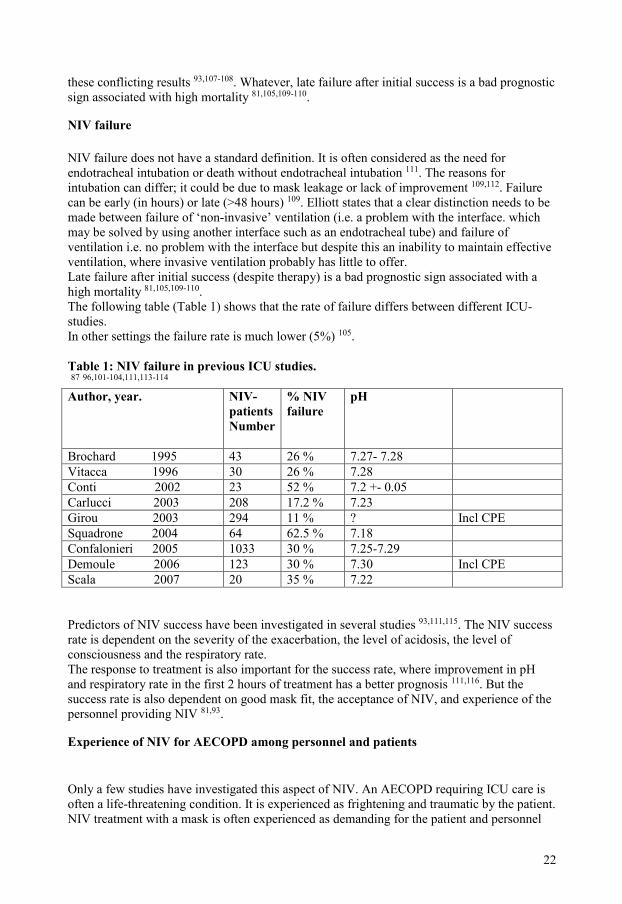
NIV failure NIV failure does not have a standard definition. It is often considered as the need for endotracheal intubation or death without endotracheal intubation 111. The reasons for intubation can differ; it could be due to mask leakage or lack of improvement 109,112. Failure can be early (in hours) or late (>48 hours) 109. Elliott states that a clear distinction needs to be made between failure of ‘non-invasive’ ventilation (i.e. a problem with the interface. which may be solved by using another interface such as an endotracheal tube) and failure of ventilation i.e. no problem with the interface but despite this an inability to maintain effective ventilation, where invasive ventilation probably has little to offer. Late failure after initial success (despite therapy) is a bad prognostic sign associated with a high mortality 81,105,109-110. The following table (Table 1) shows that the rate of failure differs between different ICU- studies. In other settings the failure rate is much lower (5%) 105. Table 1: NIV failure in previous ICU studies. 87 96,101-104,111,113-114 Author, year. NIV-
patients Number
% NIV failure
pH
Brochard 1995 43 26 % 7.27- 7.28 Vitacca 1996 30 26 % 7.28 Conti 2002 23 52 % 7.2 +- 0.05 Carlucci 2003 208 17.2 % 7.23 Girou 2003 294 11 % ? Incl CPE Squadrone 2004 64 62.5 % 7.18 Confalonieri 2005 1033 30 % 7.25-7.29 Demoule 2006 123 30 % 7.30 Incl CPE Scala 2007 20 35 % 7.22 Predictors of NIV success have been investigated in several studies 93,111,115. The NIV success rate is dependent on the severity of the exacerbation, the level of acidosis, the level of consciousness and the respiratory rate. The response to treatment is also important for the success rate, where improvement in pH and respiratory rate in the first 2 hours of treatment has a better prognosis 111,116. But the success rate is also dependent on good mask fit, the acceptance of NIV, and experience of the personnel providing NIV 81,93.
Experience of NIV for AECOPD among personnel and patients
Only a few studies have investigated this aspect of NIV. An AECOPD requiring ICU care is often a life-threatening condition. It is experienced as frightening and traumatic by the patient. NIV treatment with a mask is often experienced as demanding for the patient and personnel
22

alike. The patient feels anxiety over loss of control over the situation, they feel trapped and panic, and are completely dependent on the attending personnel. They often lack information about the treatment and how it is performed. The personnel often have problems in informing the patient because of the patient’s level of consciousness, their life-threatening situation, and dyspnoea. Personnel also feel that they must persuade or force the patient to accept NIV while the patient struggles and fights. This is a frustrating situation and creates an ethical dilemma for the attending personnel. Some experience feelings of helplessness, fear and concern about torturing the patient. Concern about the appropriateness of ICU admission and employment of a futile treatment method for the patient is also apparent amongst those interviewed 117-118.
Structural changes in the diaphragm during ventilation
The diaphragm is susceptible to the physiologic and biochemical changes that occur when a COPD patient is mechanically ventilated. Animal studies have shown that prolonged mechanical ventilation alters both structure and function of the diaphragm 119. Mechanical ventilation can promote weakness of the diaphragm due to atrophy and contractile dysfunction, it is rapid in onset and is already seen after 12 hours of ventilation in animal studies 120. Controlled mechanical ventilation changes the ultrastructure of diaphragm muscle fibres, promotes diaphragmatic contractile dysfunction, leading to depression of diaphragmatic strength. The changes are most pronounced during controlled ventilation, but also appear with ventilatory support. Mechanical ventilation also leads to disturbance of diaphragmatic protein turnover, promotes oxidative injury, and changes diaphragmatic gene expression and cellular communication in animals 120. Levine et al showed that 18 to 69 hours of controlled mechanical ventilation resulted in atrophy of the human diaphragm 121. Juan et al. showed that a carbon dioxide tension of about 54 mmHg (7.2kPa) decreased the contractility of muscle fibres in the diaphragm and endurance time in humans 122. Jaber et al. showed that a short exposure to respiratory acidosis in pigs decreased diaphragmatic contractility proportional to the degree of hypercapnia 123. The diaphragm’s contractile function, structure and biochemistry are altered in COPD patients. These changes occur early in the course of the disease and show a strong negative correlation with respiratory muscle strength. The diaphragm in COPD patients is exposed to oxidative stress and sarcomeric injury leading to contractile protein wasting and consequently, loss of the force-generating capacity of diaphragm fibres. Even so the endurance of the diaphragm appears to be increased in COPD due to remodelling 124. These changes are believed to make the diaphragm in COPD patients more susceptible to injury even in clinically stable COPD patients. Sarcomere disruption in the diaphragm is more evident in patients with COPD than in healthy subjects after inspiratory loading 125. In the study by Orozco-Levi et al. FEV1 was inversely correlated to sarcomere disruption intensity. Pulmonary hyperinflation explained up to 40% of the extent of diaphragm injury 125.
Advance directives COPD is a chronic and progressive disease where some patients have reached the terminal stage. It is possible that terminal patients will not benefit from the treatment we offer on the ICU.
23
The prognosis of advanced COPD is very difficult to predict 126-128. Advance directives are important decisions to be made in severe COPD cases, in order to maintain their autonomy 126-127. Advance planning of ICU patient care is also important as it reduces stress, anxiety and depression in relatives 129. The fact is, however, that only 25% of patients with terminal COPD have such decisions available 130. Furthermore, compliance with these decisions in clinical practice is said to be low (< 15 %) 130. It may be that aids to decision- making and more information could improve the number of advance directives available in the future 128,130. Despite the poor prognosis, about 70 % of patients with severe COPD want cardiopulmonary resuscitation and mechanical ventilation 131should such treatment be necessary. The in-hospital mortality rate in patients where intensive care treatment has been denied or withdrawn is very high, ranging from 80.3 to 95.4 % 132.
Predicting mortality in COPD
There is a great interest in exploring the possibility to predict mortality in COPD. This task is made very difficult because of the heterogeneity of COPD as a disease. Multiple factors reflecting degree of respiratory disease, the systemic influences of COPD, and co-morbidity, predict long- and short-term survival. Physiologic derangements such as airflow obstruction, hypoxaemia, lung hyperinflation, dyspnoea, exercise capacity, anaemia, cachexia, and reduction in lean body mass also relate to prognosis. Last but not least, health-related quality of life is also an important predictor of survival 133. FEV1 has previously been considered to have a strong correlation with mortality following the studies by Fletcher and Peto 134. More recent research has shown that the relationship between FEV1 and mortality is weak 10. Exercise capacity together with multidimensional risk assessments such as the BODE index are better predictors of mortality 10.
Prediction of mortality in AECOPD It is difficult to predict survival for COPD patients in general, but it is particularly difficult to predict the survival of patients admitted to the ICU with an AECOPD requiring respiratory support. This has been shown in prospective studies on ICU patients 135-136, as well as in studies where doctors judged the likelihood of survival in a predefined case. Clinicians differ markedly in their decisions to admit and in their prediction of survival of identical patients 137-138. When predicting survival in patients with AECOPD admitted to the ICU, premorbid variables, traditionally believed to have prognostic value, have not been shown to be predictive for intermediate survival. These include: age; functional capacity; quality-of-life; male sex; oral steroids; spirometry; previous hospital or ICU admissions; body mass index; smoking status; and long-term oxygen therapy 139.
24
Causes of death in COPD patients In milder COPD, cancer and cardiovascular disease are more common causes of death than respiratory failure 140. In more severe cases, the leading cause of death is respiratory disease 10or in more recent studies COPD itself 141.
Long-term mortality after ICU-care in COPD patients There are studies providing data on long-term mortality after ICU care. In a meta-analysis, Williams et al. found a 5-year mortality ranging from 40-58 %, for a mixed ICU population 142.Wright et al. followed long-term mortality in one hospital and found a 5-year mortality rate of 47.1 % in the mixed population, and a mortality rate of 43.6 % in patients with respiratory disease. Mortality was 3.4 times higher than in a matched group from the general population. Mortality in the ICU group was higher than in the general population over a four-year study period 143. The long-term mortality of COPD patients after ICU care is very high! Seneff et al. showed a one-year mortality rate of 59 % in COPD patients after ICU care 144. Breen et al. studied the mortality in invasively ventilated patients and showed a one-year mortality rate of almost 48.6 % and a 3-year mortality of 64.5 % 145. Ai Ping et al. studied the mortality in COPD patients with NIV and invasive ventilation The mortality at 1, 3, and 5 years was 42.7 %, 61.2 %, and 75.9 % 146. Chu et al. studied the long-term survival of COPD patients treated with NIV on the ICU and found a 1-year mortality of 49.1 %. A further study on COPD patients with a “Do not intubate” decision receiving NIV on the ICU showed a one-year mortality of 70.3 % 147-148. Furthermore, Christensen et al. showed a one-year mortality rate of 40.4 % in COPD patients that had been invasively ventilated on the ICU 149.
Table 2: Long-term mortality after ICU-care in COPD patients. Author 1-year mortality 3-year mortality 5-year mortality Seneff 59 % Breen 48.6 % 64.5 % Ai-Ping 42.7 % 61.2 % 75.9 % Chu 49.1 % Chu 70.3 % Christensen 40.4 %
The EuroQol instrument One of the most used instruments for the evaluation of health-related quality of life is the EuroQol (EQ-5D) 150. The EQ-5D involves a health-state classification scheme comprising five items: mobility; self-care; usual activities; pain/discomfort; and anxiety/depression, each having three alternatives 150: no problems, some problems and extreme problems, thus defining 243 distinct health states. The answers in the first part range from 0 to 1. The second part of the EQ-5D is a Visual Analogue Scale (VAS), ranging from 0 (worst possible health-state) to 100 (best possible health-state) where respondents rate how they perceive their
25

health. Health-states can also be evaluated better than, equal to, or worse than death. For health-states evaluated as worse than death, the utility is negative 151.
Short Form 36 Health Survey (SF-36) The SF-36 152 was developed to investigate generic health concepts not specific to any age, disease or treatment group. It investigates physical, social and emotional functioning it has 36 questions and generates a health profile of eight sub-scale scores:
• physical functioning (PF). • role limitations due to physical problems (RP). • bodily pain (BP). • general health (GH). • vitality (VT). • social functioning (SF). • role limitations due to emotional problems (RE). • mental health (MH).
The scores on all sub-scales are transformed to a scale from 0 (worst score) to 100 (best score) 153.
There are also two summary measures:
• Physical Component Summary Measure (PCS) consisting of values from: PF (10 items); RP (4 items); BP (2 items); and GH (5 items).
• Mental Component Summary Measure (MCS) consisting of values from : VT (4 items); SF (2 items); RE (3 items); and MH (5 items).
.
The patient also answers two questions:
• Compared to one year ago how would you rate your general health now? • In general would you say your health is excellent, very good, good, fair or poor?
154.
APACHE II APACHE II (Acute Physiologic and Chronic Health Evaluation II) is a disease severity classification system developed by Knaus et al 155 at the end of the 70’s. Their aim was to create a system able to systematically assess the severity of illness, to increase our knowledge about case mix adjustment, and to make it possible to assess critical care outcome using basic information that is more than just patient diagnosis 156. The system generates two values; a disease severity score (APACHE II Score) and an Estimated Mortality Risk (EMR) predicting the patient's probability of hospital mortality. Apart from the diagnosis, these values are based on the patient’s age and physiological derangement in 12 domains assessed during the first 24 hours on the ICU (Acute Physiology Score=APS). And further, information on the patient’s chronic health: liver, cardiovascular, respiratory or renal insufficiency and immune deficiency (Chronic Health Evaluation=CHE).
26

The Estimated Mortality Risk is calculated according to the formula:[Ln(R/1-R)= (-3,517+ (APACHE II points x 0,146) + (0,603,if the patient was taken to the op ambulant)+ (the predefined value for the main diagnosis responsible for the ICU admission)]. The APACHE II score makes it easier to describe and understand the case mix of different populations of COPD patients treated on the ICU.
NEMS and NCR 11 The Nine Equivalents of Nursing Manpower Use Score (NEMS) and the Nursing Care Recording System for assessment of workload on the ICU (NCR 11) are two systems used in the measurement of workload among the personnel on the ICUs in Sweden. NEMS registers the workload in 9 and NCR 11 in 11 different dimensions connected to the care of ICU patients creating a daily score 157-158.
The Swedish Intensive Care Registry (SIR)
The Swedish Intensive Care Registry (SIR) is a co-operative endeavour initiated by Swedish Intensive Care Units (ICU) to audit and benchmark intensive care. It started to recruit general ICUs in Sweden during 2001, and currently covers 62 of 65 general ICUs. A comprehensive dataset has been jointly specified by participating units, with data collection guidelines available on the internet and annual training sessions. Registry data are recorded prospectively in local databases by physicians and nurses, then transmitted electronically to the registry using a common data transfer protocol that includes a validation script. Data with logical defects are returned for local validation and then returned centrally and entered into the master database. The dataset includes basic patient characteristics including a unique 12-digit personal number, reason for admission and severity of illness score. From the beginning the Acute Physiology and Chronic Health Evaluation II system (APACHE II) was employed, but now scores are according to the Simplified Acute Physiology Score 3 (SAPS 3) for adults, the Paediatric Index of Mortality 2 (PIM 2) for children, and Higgins’ ICU Score for patients on thoracic intensive care units that have undergone cardiac surgery. Resource consumption is assessed by recording length of stay, cumulative NEMS scores or cumulative NCR11 scores. A primary ICD-10 diagnostic code is given at discharge and used as principal diagnosis of the admission. Vital status is followed continuously for all admissions.
Quality of ICU databases The quality of research using data from a medical registry is dependent on the quality of the data in the registry. Definitions of data quality given in the literature are frequently unclear 159. There will always be errors in databases such as insufficient data quality, incomplete data, non-adherence to data definitions and random errors 159. Furthermore there is a lack of clinical definitions for ICD-diagnoses 53. These weaknesses can be reduced and the quality of data improved with the aid of guidelines, training, quality control and education 160. Efforts to increase the quality of data in a national registry must be performed not only at the national level but also at the local level 159. Dutch studies on ICU registries have shown 4.8% inaccurate data and 3.3% incomplete data after manual data collection 159.
27

28
Aims of this thesis The general aim of this thesis was to study characteristics, treatment and outcome in patients with an acute exacerbation of chronic obstructive pulmonary disease admitted to intensive care units in Sweden. The specific aims as described in the following papers were: I To examine characteristics, resource use and long-term survival in patients with
an acute exacerbation of chronic obstructive pulmonary disease that were admitted to Swedish intensive care units.
II. To investigate 5-year survival stratified by mechanical ventilation modality in
chronic obstructive pulmonary disease patients treated on the intensive care unit.
III. To examine those patient characteristics and circumstances deemed important in the choice made between non-invasive ventilation and invasive ventilation on the intensive care unit.
IV. To examine health-related quality of life in patients with chronic obstructive
pulmonary disease after discharge from the intensive care unit.
29

30
Material and methods
Study design
Study I Study I is an observational study, with prospectively recorded data from the Swedish Intensive Care Registry.
Study II Study II is an observational multicentre inception cohort study with prospectively collected data from 9 ICUs in the south-east region of Sweden.
Study III Study III is divided in to two parts: the first part is an observational study with prospectively collected data from 9 ICUs in the south-east region of Sweden, the second part is a questionnaire answered by ICU personnel.
Study IV Study IV is a longitudinal multicentre study with prospectively collected data from 3 ICUs in the south- east region of Sweden.
Participants and settings
Patients in Study I 1009 patients with 1199 admissions on 42 ICUs in Sweden with a primary diagnosis of COPD were analysed according to characteristics, resource use and long-term survival.
Patients in Study II 93 patients on 9 ICUs in Sweden were analysed according to their 5-year survival stratified by mode of mechanical ventilation.
Patients and participants in Study III 95 admissions of AECOPD patients on 9 ICUs in Sweden were analysed, variables associated with immediate intubation were identified. Doctors, nurses and assistant nurses on 6 ICUs in Sweden answered a questionnaire about factors having a possible influence on the decision to use immediate invasive ventilation in AECOPD patients.
31
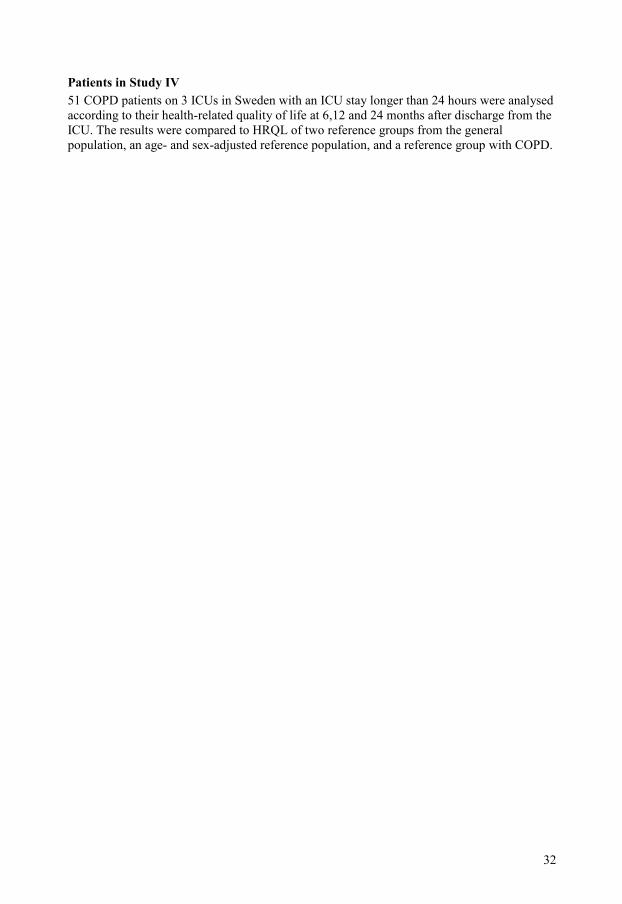
Patients in Study IV 51 COPD patients on 3 ICUs in Sweden with an ICU stay longer than 24 hours were analysed according to their health-related quality of life at 6,12 and 24 months after discharge from the ICU. The results were compared to HRQL of two reference groups from the general population, an age- and sex-adjusted reference population, and a reference group with COPD.
32

Measurements and measuring instruments
Study I The following measurements and measuring instruments were included in Study I.
Measurement Measured as: 1. Admissions on the ICU for patients with COPD.
Admissions per hour/ 24hours.
Admissions per day in the week. Admissions per month. 2. Age. Years, mean age. 3. Crude mortality risk within 90 days. Factors related to mortality within 90 days
after admission to ICU. 4. Discharge time. Discharge during office hours (07.00-16.00). Discharge out of office hours. Discharge day of week (Mo-Fri 16.00). Discharge day of week (Fri16.00-Sun 12.00). 5. Gender. Female/ Male. 6. Hospital size. Regional hospital. County hospital. Local hospital. 7. ICD-10 diagnostic code. J.44 Present/ Not present. 8. Length of stay on the ICU. Hours, days. 9. NCR 11 Score. Score, points. 10. NEMS. Score, points. 11. Severity illness score, APACHE II. Score, points. 12. APACHE II, Estimated Mortality Risk Probability, % 13. Readmissions. Readmission and COPD diagnosis present. 14. Route of admission, location before admission.
• Admission from emergency room. • Admission from regular ward. • Admission from other location.
15. Vital status. Alive/ Dead.
33
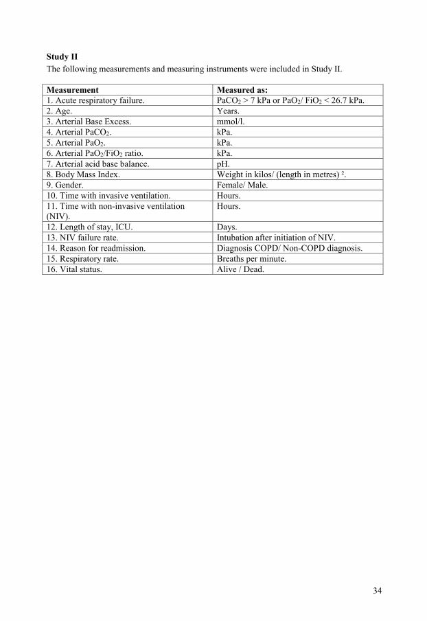
Study II The following measurements and measuring instruments were included in Study II.
Measurement Measured as: 1. Acute respiratory failure. PaCO2 > 7 kPa or PaO2/ FiO2 < 26.7 kPa. 2. Age. Years. 3. Arterial Base Excess. mmol/l. 4. Arterial PaCO2. kPa. 5. Arterial PaO2. kPa. 6. Arterial PaO2/FiO2 ratio. kPa. 7. Arterial acid base balance. pH. 8. Body Mass Index. Weight in kilos/ (length in metres) ². 9. Gender. Female/ Male. 10. Time with invasive ventilation. Hours. 11. Time with non-invasive ventilation (NIV).
Hours.
12. Length of stay, ICU. Days. 13. NIV failure rate. Intubation after initiation of NIV. 14. Reason for readmission. Diagnosis COPD/ Non-COPD diagnosis. 15. Respiratory rate. Breaths per minute. 16. Vital status. Alive / Dead.
34

Study III, Part 1 The following measurements and measuring instruments were included in Study III. The study consisted of two parts. Measurements and measuring instruments of Part 1 included: Measurement Measured as: 1. Age. Years. 2. Acidosis rate among admissions. Number.
• Moderate-to-severe acidosis (arterial pH<7.35).
• Severe acidosis (arterial pH<7.25).
3. Arterial Base Excess. mmol/l. 4. Arterial HCO3¯. mmol/l. 5. Arterial PaCO2. kPa. 6. Arterial PaO2. kPa. 7. Body Mass Index. Weight in kilos/ (length in metres)² . 8. Respiratory rate. Breaths per minute. 9. Gender. Female/ Male. 10. Intubation rate. % of patients admitted.
• Daytime 07-22. • Nighttime 22-07.
11. Mode of ventilation. Number. Non-invasive ventilation started. Invasive ventilation started.
12. Presence of Advance directives. Present/ Not present. 13. Readmission rate. Number readmitted with a COPD diagnosis. 14. Severity illness score. APACHE II. Score, points. 15. Spirometry available. % of patients admitted.
35
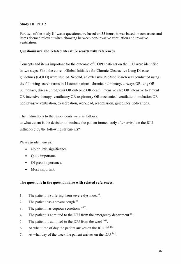
Study III, Part 2 Part two of the study III was a questionnaire based on 35 items, it was based on constructs and items deemed relevant when choosing between non-invasive ventilation and invasive ventilation. Questionnaire and related literature search with references
Concepts and items important for the outcome of COPD patients on the ICU were identified
in two steps. First, the current Global Initiative for Chronic Obstructive Lung Disease
guidelines (GOLD) were studied. Second, an extensive PubMed search was conducted using
the following search terms in 11 combinations: chronic, pulmonary, airways OR lung OR
pulmonary, disease, prognosis OR outcome OR death, intensive care OR intensive treatment
OR intensive therapy, ventilatory OR respiratory OR mechanical ventilation, intubation OR
non invasive ventilation, exacerbation, workload, readmission, guidelines, indications.
The instructions to the respondents were as follows:
to what extent is the decision to intubate the patient immediately after arrival on the ICU
influenced by the following statements?
Please grade them as:
• No or little significance.
• Quite important.
• Of great importance.
• Most important.
The questions in the questionnaire with related references.
1. The patient is suffering from severe dyspnoea 4.
2. The patient has a severe cough 56.
3. The patient has copious secretions 4,97.
4. The patient is admitted to the ICU from the emergency department 161.
5. The patient is admitted to the ICU from the ward 161.
6. At what time of day the patient arrives on the ICU 162-163.
7. At what day of the week the patient arrives on the ICU 162.
36

8. The ICU-workload as the patient arrives on the ICU 96,162-164.
9. Which doctor who is in charge of the patient on the ICU 137-138,162,165-166.
10. Which nurse who is in charge of the patient on the ICU 117.
11. Which assistant nurse who is in charge of the patient on the ICU 117.
12. The patient is a man 161.
13. The patient is a woman 161.
14. The patient’s age 161.
15. The patient is restless 4,97.
16. The patient’s state of consciousness 4,97.
17. The patient has recently been treated for a COPD exacerbation on the ICU 167-169.
18. The patient’s “advance directives” is pro ICU care 110.
19. The patient is thin 38,115,135,170.
20. The patient is normal weight 43.
21. The patient is obese 38.
22. The patient’s respiratory rate 4,111.
23. The pH value in the arterial blood gas analysis 4,111.
24. The PaCO2 value in the arterial blood gas analysis 4,149,171-172.
25. The PaO2 value in the arterial blood gas analysis 4,56,173.
26. The base excess value in the arterial blood gas analysis 174.
27. The standard bicarbonate value in the arterial blood gas analysis 175-176.
28. The patient’s activities of daily living before being admitted to the ICU 109,177.
29. The patient has received antibiotics before being admitted to the ICU 178-180.
30. The patient is able to inhale nebulised medication 4,113.
31. The patient has received corticosteroid treatment before being admitted to the ICU 4,172.
32. The patient has a fever 56,103,181.
33. The patient has an elevated CRP 182.
34. The result of the patient’s latest spirometry 56,133,161.
35. My personnel knowledge about current COPD guidelines 183-186.
37
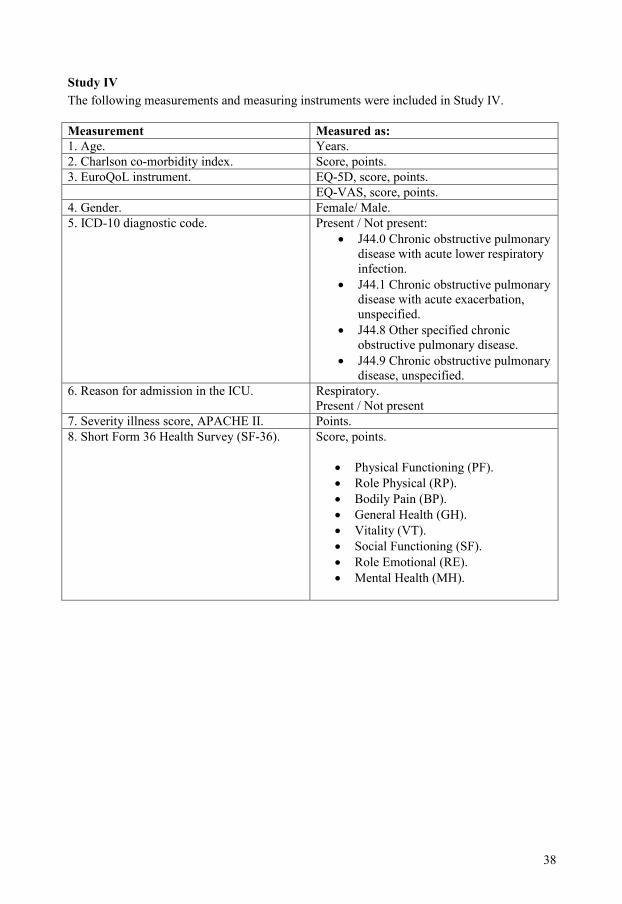
Study IV The following measurements and measuring instruments were included in Study IV.
Measurement Measured as: 1. Age. Years. 2. Charlson co-morbidity index. Score, points. 3. EuroQoL instrument. EQ-5D, score, points. EQ-VAS, score, points. 4. Gender. Female/ Male. 5. ICD-10 diagnostic code. Present / Not present:
• J44.0 Chronic obstructive pulmonary disease with acute lower respiratory infection.
• J44.1 Chronic obstructive pulmonary disease with acute exacerbation, unspecified.
• J44.8 Other specified chronic obstructive pulmonary disease.
• J44.9 Chronic obstructive pulmonary disease, unspecified.
6. Reason for admission in the ICU. Respiratory. Present / Not present
7. Severity illness score, APACHE II. Points. 8. Short Form 36 Health Survey (SF-36).
Score, points. • Physical Functioning (PF). • Role Physical (RP). • Bodily Pain (BP). • General Health (GH). • Vitality (VT). • Social Functioning (SF). • Role Emotional (RE). • Mental Health (MH).
38

Statistical Methods used in the studies Statistical method
Study I Study II Study III Study IV
Chi square test x x Kaplan-Meier method
x x
Kruskal-Wallis test
x x
Linear regression
x
Logistic regression
x
Log-rank test x x Multivariate Cox model
x
Multivariable regression model analysis
x x
Shapiro-Wilks test
x
Student´s t-test x x x Fisher test x Friedmans test x Mann-Whitney U
x
Wilcoxon rank-sum test.
x
Ethical aspects There was no need for ethic approval and informed consent for the Studies I-III, as they were observational and included no change in clinical practice. In Study IV the study protocol was approved by the Linköping University Regional Ethics Review board. The patients that answered the questionnaire about Health-related quality of life in Study IV had given an informed consent to use their data in scientific studies.
39

40

Results
Results Study I In Study I we investigated characteristics and long-term outcome of COPD patients admitted to Swedish ICUs. 1009 patients with 1199 admissions were identified (1.3 % of all admissions). 53 % of the patients were admitted to ICUs in a county hospital, 26 % to ICUs in a local hospital and 21 % to a regional hospital. The patients were admitted to the ICU from the emergency department in 49.3 % of cases and from the ward in 44.6 % of cases, leaving 6.1% with other locations prior to ICU admission. The patients had an mean age of 70.2 years (SD 9.1), the majority were female (61.5 %). The mean APACHE II score was 20.2 (SD 7.0). Admissions were evenly distributed over the year with the highest number admitted in April and the lowest in September. The resource usage for the COPD patients was higher than the median resource usage for all other patients. A high percentage (15.8 %) of these COPD patients were readmitted. Older patients had less chance of being readmitted. The mortality in this patient cohort was very high with a ICU mortality of 7.8 %, a 30-day mortality of 26 %, and a 3-year mortality of 70 %. Median survival time was 15 months. Risk factors for mortality after 90 days were male gender, age, APACHE II probability and discharge at the weekend.
Results Study II In this study, 93 COPD patients admitted to 9 ICUs in the south-east region of Sweden were stratified according to mode of ventilation. These COPD patients represented 1.6 % of all ICU admissions at this point in time. The mean age was 70.6 years (SD 9.6), and 57 % were female. Sixteen patients (17 %) were intubated and invasively ventilated and 77 patients (83%) began with non-invasive ventilation. NIV failed in 15 patients (19 %). The groups with NIV and invasive ventilation differed in body mass index, ICU length of stay and age. Other characteristics were similar including arterial blood gases, acid base status and respiratory rate. The mortality in both groups was high, with a 5-year survival of 17 % in the NIV group and 6 % in the invasive ventilation group. Non-invasive ventilation was associated with increased survival, whereas increased APACHE II score and age were associated with decreased survival. The mortality in the NIV fail group was not significantly different from survival in the invasive ventilation group.
41

Results Study III In this study 95 admissions for acute exacerbation of COPD were analysed according to characteristics associated with invasive ventilation. Thereafter a questionnaire with 35 items was sent to ICU personnel with different professions (doctors, nurses and assistant nurses) in the admitting ICUs to study the relative importance of various factors that possibly influence the decision to use invasive ventilation immediately. Seventy-nine patients (83 %) began with NIV on admission, 16 (17 %) were intubated immediately. The two groups did not differ in arterial blood gases, acid base status or respiratory rate. The NIV group was significantly younger and had a lower BMI than patients that were intubated immediately. BMI was the only variable remaining associated with increased risk for intubation in a multivariate analysis. The intubation rate day and night-time was similar. The usage of NIV was not correlated to the presence of acidosis. Eighty-three per cent of the patients with pH<7.35 were treated with NIV, and 81 % of the patients with severe acidosis pH<7.25 received NIV. Ranking of responses to the questionnaire showed that consciousness, respiratory symptoms and blood gases were powerful factors determining invasive ventilation. Non-patient-related factors (physician in charge, presence of guidelines, ICU workload) were also deemed important for the decision to intubate immediately. There were small differences between professions in ranking of the five most important factors in the questionnaire.
Results Study IV In this study we examined the health-related quality of life (HRQL) in COPD patients treated on the ICU. Their results were compared to the HRQL of two other groups, a reference group with COPD (COPD-reference), and reference group without COPD from the general population (Non-COPD reference). HRQL was measured using the EuroQol (EQ-5D) and the Short Form 36 Health Survey (SF-36) questionnaires. HRQL in the COPD-ICU group was always markedly lower compared to the Non-COPD reference group from the general population. In COPD-ICU patients where a complete follow-up was done, changes in HRQL over time were minor and did not decline further during 6 to 24 months after discharge. There were differences in EQ-VAS (p<0.05) and PF (p<0.059) between the COPD-ICU group and the COPD-reference group. The SF-36 results in the COPD-ICU group were not statistically different from the COPD-reference group. Patients that died during the study had a poorer HRQL. PF and SF scores were statistically lower in this group of patients. HRQL in COPD patients 24 months after discharge from the ICU was quite similar to that of COPD patients not treated on the ICU.
42
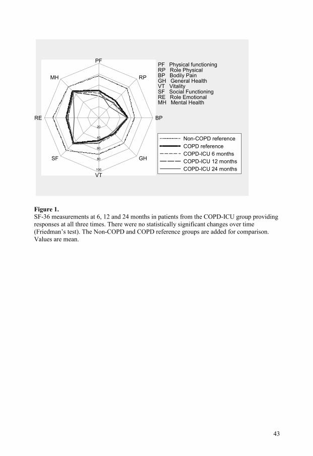
PF
RP
BP
GH
VT
SF
RE
MH
20
40
60
80
100
PF Physical functioningRP Role PhysicalBP Bodily PainGH General HealthVT VitalitySF Social FunctioningRE Role EmotionalMH Mental Health
Non-COPD referenceCOPD referenceCOPD-ICU 6 monthsCOPD-ICU 12 monthsCOPD-ICU 24 months
Figure 1. SF-36 measurements at 6, 12 and 24 months in patients from the COPD-ICU group providing responses at all three times. There were no statistically significant changes over time (Friedman’s test). The Non-COPD and COPD reference groups are added for comparison. Values are mean.
43

44

Discussion
Introduction
When we started our research project, at the beginning of this millennium, on COPD patients in need of ventilatory support admitted to Swedish ICUs, not much was known about this group of patients. All Swedish ICU physicians were familiar with this group of patients and knew of the severity of their disease and that the short-term mortality was high. Available details of admission and treatment were scarce and little was known about outcomes beyond the ICU. The creation of a national registry on ICU patients in Sweden (the Swedish Intensive Care Registry, SIR) in 2001, together with a programme intended to support research and the creation of healthcare networks in the south-east region of Sweden (The Health Research Council in the South-East of Sweden ,FORSS), however, made it possible to gather essential information about these patients. The introduction of international and national healthcare programmes for COPD patients at the beginning of the millennium, with guidelines on the indication for ICU care of these patients, was a further important development. Much of the research that we have performed during this project is based on the views of attending personnel on the ICU department where COPD patients with respiratory failure are admitted.
The COPD patient before admission to the ICU When a patient with COPD and respiratory failure arrives at the ICU, some information about the individual that could help in deciding the best possible treatment for that patient is often missing. We have seen that spirometry, that is obligate for the diagnosis and staging of COPD according to present guidelines, is surprisingly often missing. Information about activities of daily living and the patients opinion regarding advance directives, if any, is very often missing for COPD patients admitted to the ICU in Sweden, as well as information about nutritional status and health-related quality of life prior to admission. The absence of such information could lead to important decision-making becoming arbitrary and dependent on the physician responsible for the patient at admission rather than being objective. Since COPD is a chronic and progressive disease, some patients have reached the endpoint in their disease and will not survive despite ventilatory treatment on the intensive care unit. At present objective information on admission, indicating whether the patient has a treatable acute exacerbation of COPD or has reached an end stage in the disease, is often lacking.
The COPD patient in need of ventilatory support on admission to the ICU
Our studies have increased our knowledge of the characteristics of COPD patients admitted to the ICU in need of ventilatory support. We now know that these patients have a mean age of about 70 years, and that the proportion of women is higher. COPD patients have a high severity illness score on admission according to their APACHE II scores. The main route of admission is via the emergency department and the ward in about equal proportions.
45

A large proportion of the patients have acute respiratory failure on arrival with a high respiratory rate and severe life-threatening disturbances in their acid base status and blood gases.
Non-invasive ventilation in patients with COPD on the ICU Despite it’s long history NIV is a quite new treatment modality for patients with COPD in need of ventilatory support admitted to the ICU. When we started these studies there were no data on the usage and results of NIV on Swedish ICUs. The correct way of using NIV in COPD patients in the intensive care setting is stated in the current national and international guidelines. However this treatment is not always easy to apply, and is dependent of the skill and education of the attending personnel. As COPD patients are admitted to the ICU at any time, night and day, all year, NIV-treatment must be understood by all personnel on the ICU and not just a few skilled and interested individuals. Our data show that NIV is now well-established in Sweden, it’s usage is high even in patients with severe respiratory derangement, and the failure rate is low and in line with current international experience.
The choice between NIV and invasive ventilation in patients with COPD on the ICU The advantages of NIV compared to invasive ventilation are better short-term survival, fewer complications, and a shorter length of stay on the ICU. It also gives the attending personnel time to gather important information in order to proceed with optimal care of the patient. The indications for NIV and invasive ventilation are stated in the current national and international guidelines for the treatment of COPD patients. Our studies show that in Sweden some COPD patents with very severe respiratory derangements that fulfil the criteria for invasive ventilation may successfully be treated with NIV. This is probably due to the evolution of this treatment modality following the development of special facemasks, ventilators, and the extensive experience and education of ICU personnel. The decision to start treatment with invasive ventilation in a patient with acute exacerbation of COPD, where it is presumed that the patient will recover and return to a state with good health-related quality of life, is an easy decision. The decision to treat a patient with terminal disease with invasive ventilation, on the other hand, is very demanding. Unfortunately we lack objective criteria indicating when a patient has end-stage disease and where ICU treatment is futile. Our questionnaire revealed the importance of NIV in providing the personnel time to gather information considered important when deciding whether or not to intubate the patient. The questionnaire also showed that information other than that present in current healthcare programmes was important for this decision. We are not quite sure why overweight patients (BMI>25kg/m²) were more likely to be intubated immediately in Study III.
Treatment on the ICU
In these studies we did not have information about treatment of the patients before being admitted to the ICU apart from the absence of ventilatory support. We did not use a formal
46

treatment protocol on the ICU stating pharmacological treatment or the use of different pressure levels or levels of positive end-expiratory pressures during ventilation. We measured the resource use among the personnel, which was slightly higher for COPD patients than the median value for all ICU patients. In the answers to our questionnaire, the presence of guidelines and ongoing inhalational treatment were ranked 15 and 16 of the 35 items important for the decision to intubate immediately, other factors associated with pharmacological treatment of the patients on the ICU were ranked low. The length of stay on the ICU was much shorter for patients treated with NIV than patients that were intubated and invasively ventilated, and was shorter than in previous international studies.
Mortality on the ICU The ICU mortality in our studies varied between 7.8 % in Study I and 15 % in Study II. These numbers are lower than previously reported results from the UK and US (23.1 % and 9.3 %) 144,187.
Readmission after ICU treatment In Study I, 15.8 % of all COPD patients were readmitted to the ICU during this 5-year study. In Study III 11 % of the patients in the NIV group were readmitted. This is a high proportion of patients with regard to our finding that readmission is less likely in older patients. We do not know if this is preventable. The readmission rate in previous studies in mixed ICU populations is 4-14 % 167 and 2.56 % in COPD patients readmitted to the ICU 188.
Short-term mortality after treatment on the ICU The 30-day mortality in Study I was 26 %. Most studies do not present 30-day mortality, but the hospital mortality in studies from the US144is 23.8 %, and in the UK 38.3 % 187. The 90-day mortality rate in Study I was dependent on male gender, higher age, increased APACHE II probability and discharge during the weekend. We do not know if the increased mortality related to discharge at the weekend can be reduced. Theoretically this is due either to premature discharge or the presence of an advance directive indicating reduced level of care.
Long-term mortality after treatment on the ICU The long-term mortality rate in our studies was high; 49.4 % of the patients treated with NIV in Study II died within a year, and 70 % of the patients in Study I died within 3 years. In Study II five-year survival rates of 17 % in the NIV group and 6 % in the invasive ventilation group were found. In Study IV which included survivors after ICU care, the 4-year mortality rate was 60 %.There are only a few other diseases with such a pessimistic prognosis after treatment on the ICU.
47

Health-related quality of life in COPD patients after ICU care
There is a growing interest in studies of health-related quality of life after ICU care. I do believe that all personnel involved in the care of patients with severe diseases in need of intensive care are interested in getting feedback about the recovery and subsequent life of their patients. It is also important to study HRQL to avoid the notion that ICU treatment of COPD patients is futile and a burden for the patient and society. The study of HRQL is challenging as the methods used are quite new, HRQL in COPD patients is dependent on several factors such as dyspnoea, depression, anxiety and exercise tolerance. Clinically important differences in HRQL scores are not unanimously defined, there are floor and ceiling effects in the methods used and there is no clear consensus about the use of statistics and how to deal with missing data. Nevertheless it is important to describe the patient’s situation after ICU care using objective measurements not restricted to just age, disease or treatment group. The findings in Study IV were to some extent surprising to us. In view of the fact that COPD is a chronic and progressive disease, we expected to see a decline in HRQL over time in these patients. This was not the case. The HRQL in this group of patients was low compared to the general population, but did not decline further during the 24-month period after discharge. After 24 months it was similar to the HRQL of COPD patients not treated in the ICU. Patients that died during the study had a poorer HRQL. PF and SF scores were statistically lower in this group of patients.
Strengths and limitations
This thesis has some major strengths. The data source is multi-center in all four studies. Secondly, the Swedish 12-digit personal number made it possible to ascertain almost complete vital status for all the patients. The randomised controlled trial is the gold standard for evaluation of medical methods. There is however one major limitation with randomised trials, they have difficulty in detecting rare or late adverse effects of treatment. Observational studies are better in detecting the benefits and harms of medical interventions. The quality of observational studies is dependent on appropriate selection of participants, appropriate measurement of variables and appropriate control of confounding 189-191. Non-invasive ventilation has been shown to be beneficial in the treatment of AECOPD on the ICU in randomised trials. Observational studies may thus be the only way to investigate several aspects of the usage of NIV in COPD patients in need of ventilatory support. We believe that the selection of participants in our studies was correct. We have confirmed the diagnosis COPD in several ways, from the local databases at different ICUs in the south-east region of Sweden, by the APACHE II system on admission, with the ICD-10 code and with help of the Swedish Cause-of-death register. The diagnosis was made with the help of clinical signs and not by spirometry in many patients, but this is inevitable in patients in need of ventilatory support admitted to the ICU from the emergency department. The proportion of COPD patients in the ICUs in Sweden is lower than in other countries. We believe that this is dependent on the smoking habits, or rather lack of, in Sweden rather than selection bias. The measurement of most variables used is defined in the guidelines of the Swedish intensive care registry (SIR) and we believe they are appropriate and precise.
48

The questionnaire in Study III. was designed with a systematic approach 192 including a pilot testrun and reliability and validity testing. We do not have a complete control of confounding factors in our studies as the treatment of COPD patients in and outside the ICU has many components apart from those we can measure on the ICU. The group of patients for example, is not uniform and COPD has several phenotypes and genetic factors that also influence the outcome not least when mortality is used as end-point. Acute exacerbations of COPD are poorly defined and possibly heterogeneous in their pathophysiology and biomarkers for AECOPD are lacking. The treatment COPD patients receive is not standard neither outside nor on the ICU. Co-morbidity scores are not always present in the data from COPD patients in Sweden. Furthermore the cause of death was not separately analysed in all studies included.
49

50
Conclusions
• The mortality of COPD patients with respiratory failure admitted to Swedish ICUs is high in the short- and long-term perspectives despite ICU care.
• Non-invasive ventilation (NIV) is used as first-line treatment in a very high proportion of COPD patients with respiratory failure admitted to the ICU in Sweden.
• Non-invasive ventilation had a better long-term outcome compared to immediate invasive ventilation in COPD patients in need of ventilator support admitted to the ICU in our observational study.
• A failed NIV-trial does not increase the long-term mortality compared to immediate invasive ventilation in COPD patients in need of ventilatory support on the ICU.
• Factors other than those deemed most important in guidelines appear to have an inappropriate influence on the choice between NIV and immediate intubation of patients with an acute exacerbation of COPD.
• The health-related quality-of-life (HRQL) of COPD patients admitted to the ICU in Sweden is lower than in the general population, but does not decline further during the period 6 to 24 months after discharge.
• HRQL in COPD patients 24 months after discharge from the ICU was quite similar to that of COPD patients not treated on the ICU.
51

52

Populärvetenskaplig sammanfattning på svenska Kroniskt obstruktiv lungsjukdom (KOL) är ett växande hälsoproblem över hela världen. Idag är det den femte vanligaste dödsorsaken, men sjukdomen kommer att bli än mer vanlig som dödsorsak de närmaste årtiondena. WHO räknar med att dödligheten i KOL kommer att öka med 30 % de närmaste 10 åren. 5 % av alla dödsfall tros bero på KOL. KOL sjukdomen är lömsk då det är en kronisk sjukdom som progredierar, det finns i nuläget ingen bot för sjukdomen, bara lindring i form av läkemedelsbehandling och syrgasbehandling. Sjukdomen karakteriseras av en försämrad lungfunktion samt andnöd, ökad slemproduktion och hosta. Efterhand påverkas hela kroppen av den kroniska inflammation som finns i luftvägarna. Den vanligaste orsaken till KOL är rökning. Diagnosen KOL ställs med hjälp av en spirometriundersökning. Tyvärr ställs diagnosen oftast sent och ett rökstopp i det skedet av sjukdomen ger bara en viss bättring, men ingen bot. Många patienter med KOL är inte medvetna om sin sjukdom. I Sverige har ca 500 000 personer sjukdomen. Ca 2500 avlider av sjukdomen i Sverige varje år. Sjukdomen har ett ojämnt förlopp. KOL-patienter kan drabbas av plötsliga försämringar, sk exacerbationer. En exacerbation kan bero på en infektion, men orsaken är inte alltid känd. Försämringen i hosta, slemproduktion och andnöd kan kräva sjukhusvård och de svårast sjuka patienternas andning kan vara så påverkad att de kan behöva intensivvård. Intensivvårdsavdelningen kan erbjuda ventilationsstöd där man stöttar eller tar över patientens andningsarbete med hjälp av en respirator. Respiratorbehandling har sedan 1960 talet genomförs med hjälp av sövning av patienten varefter man för ner en sk endotrachaltub i patientens luftrör och stöttar andningen, detta kallas invasiv ventilatorbehandlig. Under 1990 talet och 2000 talet etablerades en alternativ behandling för ventilationsstöd på världens intensivvårdsavdelningar: Så kallad non-invasiv ventilatorbehandling (NIV). Vid NIV ger man ventilationsstöd med hjälp av en respirator som kopplas till en mask eller en huva. Patienten behöver då inte sövas. NIV har i tidigare studier visat sig vara till fördel för patienter med KOL som behöver intensivvård. Det ger ökad överlevnad, kortare vårdtid och färre komplikationer jämfört med invasiv ventilatorbehandling. Syftet med denna avhandling var att studera de KOL patienter som var så svårt sjuka att de behövde intensivvård. Vi ville veta mer om dessa patienter och studera deras överlevnad efter intensivvård. Vi ville också se hur deras vårdresultat påverkades av vilken ventilatorbehandling (non-invasiv eller invasiv) de behandlades med. Vi ville också veta mer om hur personalen valde mellan non-invasiv och invasiv ventilatorbehandling till KOL-patienter på intensivvårdsavdelningarna. Vidare ville vi se vilken hälsorelaterad livskvalitet dessa KOL patienter hade efter intensivvård och hur den förändrade sig över tid efter intensivvården, samtidigt som vi jämförde deras hälsorelaterade livskvalitet med personer ur befolkningen utan KOL och dessutom med KOL-patienter som inte hade genomgått intensivvårdsbehandling. I delarbete 1 studerades 1009 KOL-patienter som vårdats på 42 intensivvårdsavdelningar i hela Sverige. Vi fann att KOL-patienterna utgjorde 1.3 % av samtliga intensivvårdspatienter, patienterna kom till IVA från akutmottagningen och vårdavdelningar i lika stora proportioner. De var kring 70 år gamla, andelen kvinnor var högre än andelen män. De var svårt sjuka när de kom till intensivvårdsavdelningarna. Dödligheten hos KOL patienter var betydande både på kort och lång sikt trots intensivvård. Många patienter (15.8 %) blev återinlagda. Äldre patienter hade färre återinläggningar. Risken för att dö inom 90 dagar efter inläggningen
53

påverkades av patientens kön, ålder och sjuklighet vid inläggningen. Det fanns en ökad risk för död om man skrevs ut från IVA under en helg. I delarbete 2 studerades 93 KOL-patienter som kommit till 9 intensivvårdsavdelningar och behandlats med ventilatorstöd. Vi jämförde patienterna utifrån vilket ventilatorstöd (invasivt eller non-invasivt) de fick direkt när de kom till intensivvårdsavdelningen. Även bland dessa patienter var dödligheten hög. De patienter som behandlades med non-invasiv ventilation hade en bättre långtidsöverlevnad jämfört med de som fick invasiv ventilation direkt i denna grupp patienter. De patienter som började med non-invasiv ventilation för att sedan gå över till invasiv ventilation hade inte sämre överlevnad än de som behandlades med invasiv ventilation direkt. En hög andel av patienterna behandlades med non-invasiv ventilation. I delarbete 3 studerades 95 inläggningar vid 9 intensivvårdsavdelningar pga KOL och andningssvikt. Vi försökte identifiera faktorer som var kopplade till att patienterna behandlades med invasiv ventilatorbehandling direkt när de kom till intensiv-vårdsavdelningen. Vidare analyserades en enkät som skickats till personalen vid intensivvårdsavdelningarna i sydöstra Sverige. Frågorna i enkäten berörde i vilken mån olika faktorer påverkade beslutet att använda invasivt eller non-invasivt ventilatorstöd direkt när KOL patienter kommer till intensivvårdsavdelningen. I analysen av patienterna skilde de sig åt i BMI men inte i värden i blodgasanalys, syra-bas status eller i andningsfrekvens. Resultatet från enkäten visade att faktorer som finns med i riktlinjer för ventilatorbehandling i vårdprogram för KOL är viktiga för beslutet kring valet av metod, men flera andra faktorer i vårdmiljön som till exempel bemanningen, vem som är ansvarig för patienten och förekomst av vårdprogram på avdelningen, påverkar också beslutet. I delarbete 4 studerades den hälsorelaterade livskvaliteten (HRLK) efter intensivvård hos en grupp patienter som vårdats på IVA för KOL. HRLK mättes vid tre tillfällen vid 6, 12 och 24 månader efter intensivvård. Dessutom jämfördes HRLK hos de KOL patienter som fått intensivvård med två andra patientgrupper. Dels en ålders och könsmatchad grupp ur befolkningen utan diagnosen KOL och dels en grupp KOL-patienter som inte vårdats med intensivvård. KOL patienterna som hade vårdats med intensivvård hade lägre värden för HRLK än gruppen ur befolkningen. Värdena för de KOL patienter som fått intensivvård och överlevde var stabila från 6 till 24 månader efter intensivvårdsbehandlingen. De hade efter 24 månader värden som var jämförbara med KOL patienterna som inte hade fått intensivvård. En sammanfattning av resultaten i dessa fyra studier är att: KOL-patienter i Sverige som behöver intensivvård och respiratorstöd är mycket sjuka då de anländer till intensivvårdsavdelningarna. Trots att de behandlas med intensivvård och respiratorstöd är dödligheten hög både på kort och lång sikt. Non-invasiv ventilatorbehandling bör vara förstahandsval till de KOL patienter som behöver respiratorstöd. Den hälsorelaterade livskvaliteten hos de KOL patienter som överlever efter intensivvård och återhämtar sig är lägre än för befolkningen i allmänhet, men stabil och jämförbar med den hälsorelaterade livskvaliteten hos KOL patienter som inte vårdats på IVA.
54

Acknowledgements I would like to express my gratitude to everyone who has supported me during this research project. I would especially like to thank: Sten Walther: Associate professor, my supervisor: For his knowledge, stringency, his patience and great support in all possible questions related to research. Also for his ability to see into the future and to focus on the enjoyable and interesting aspects of science. Finally but not least for the fun and great hospitality he provides. Folke Sjöberg: Professor, my co-supervisor: For his great knowledge and excellent supervision of this project. All my co-authors: Thomas Nolin, Caroline Mårdh, Göran Karlström, Josefin Sundh, Lennart Nilholm, Mats Fredriksson, Lars Engerström, Lotti Orwelius and Peter Nordlund. For all their support and hard work in collecting data, taking part in the designing of the studies and for correcting any errors in the papers. Peter Cox: MD, for necessary help with the English language. Jan Idvall: Associate professor and Ewa Idvall: Professor. For their knowledge, support and encouraging help that made it possible to complete this research project in a county hospital. All those involved in the creation and management of the Swedish Intensive Care Registry (SIR): For their immense belief in better times, stringency, and interesting discussions about intensive care. Everyone involved in the south-east Sweden ICU network (The RÖSTI group): For their hard work in the collection of data, designing protocols and developing intensive care in this region of Sweden. Ulla Jonasson: RN. For her great work in the RÖSTI group, and also with the studies performed within this network. Stefan Gelebo: RN. For important support and beautiful photos. All my colleagues at Västervik hospital: For their interest in and support of my research. The Scientific Committee of Kalmar County Council: For their support and my education during these studies.
55

The Health Research Council in the south-east region of Sweden, FORSS: For making it possible to perform research in a county hospital in their region. My family: For their support and understanding that research is so time-consuming but even so gives me such pleasure in life.
56

References 1. Petty TL. The history of COPD. Int J Chron Obstruct Pulmon Dis 2006; 1: 3-14. 2. Mannino DM, Buist AS. Global burden of COPD: risk factors, prevalence, and future trends. Lancet 2007; 370: 765-73. 3. Berkius J, Walther SM. [The COPD national guidelines should be implemented in a better way. The use of non-invasive ventilation is optimal, but spirometry is performed too seldom]. Lakartidningen 2007; 104: 1897-901. 4. Rabe KF, Hurd S, Anzueto A, Barnes PJ, Buist SA, Calverley P, Fukuchi Y, Jenkins C, Rodriguez-Roisin R, van Weel C, Zielinski J. Global strategy for the diagnosis, management, and prevention of chronic obstructive pulmonary disease: GOLD executive summary. Am J Respir Crit Care Med 2007; 176: 532-55. 5. Viegi G, Pedreschi M, Pistelli F, Di Pede F, Baldacci S, Carrozzi L, Giuntini C. Prevalence of airways obstruction in a general population: European Respiratory Society vs American Thoracic Society definition. Chest 2000; 117: 339S-45S. 6. Lindberg A, Jonsson AC, Ronmark E, Lundgren R, Larsson LG, Lundback B. Prevalence of chronic obstructive pulmonary disease according to BTS, ERS, GOLD and ATS criteria in relation to doctor's diagnosis, symptoms, age, gender, and smoking habits. Respiration 2005; 72: 471-9. 7. Celli BR, MacNee W. Standards for the diagnosis and treatment of patients with COPD: a summary of the ATS/ERS position paper. Eur Respir J 2004; 23: 932-46. 8. Belfrage B. [Are healthy persons diagnosed with COPD?]. Lakartidningen 2006; 103: 850-2. 9. Kerstjens HA. The GOLD classification has not advanced understanding of COPD. Am J Respir Crit Care Med 2004; 170: 212-3; discussion 14. 10. Celli BR. Predictors of mortality in COPD. Respir Med 2010; 104: 773-9. 11. Pauwels RA, Buist AS, Ma P, Jenkins CR, Hurd SS. Global strategy for the diagnosis, management, and prevention of chronic obstructive pulmonary disease: National Heart, Lung, and Blood Institute and World Health Organization Global Initiative for Chronic Obstructive Lung Disease (GOLD): executive summary. Respir Care 2001; 46: 798-825. 12. Pierce R. Spirometry: an essential clinical measurement. Aust Fam Physician 2005; 34: 535-9. 13. Ruppel GL. What is the clinical value of lung volumes? Respir Care 2012; 57: 26-35; discussion 35-8. 14. Soto FJ, Varkey B. Evidence-based approach to acute exacerbations of COPD. Curr Opin Pulm Med 2003; 9: 117-24. 15. Caramori G, Adcock IM, Papi A. Clinical definition of COPD exacerbations and classification of their severity. South Med J 2009; 102: 277-82. 16. Effing TW, Kerstjens HA, Monninkhof EM, van der Valk PD, Wouters EF, Postma DS, Zielhuis GA, van der Palen J. Definitions of exacerbations: does it really matter in clinical trials on COPD? Chest 2009; 136: 918-23. 17. Burge S, Wedzicha JA. COPD exacerbations: definitions and classifications. Eur Respir J Suppl 2003; 41: 46s-53s. 18. Boixeda R, Rabella N, Sauca G, Delgado M, Martinez-Costa X, Mauri M, Vicente V, Palomera E, Serra-Prat M, Capdevila JA. Microbiological study of patients hospitalized for acute exacerbation of chronic obstructive pulmonary disease (AE-COPD) and the usefulness of analytical and clinical parameters in its identification (VIRAE study). Int J Chron Obstruct Pulmon Dis 2012; 7: 327-35.
57
19. Dimopoulos G, Lerikou M, Tsiodras S, Chranioti A, Perros E, Anagnostopoulou U, Armaganidis A, Karakitsos P. Viral epidemiology of acute exacerbations of chronic obstructive pulmonary disease. Pulm Pharmacol Ther 2012; 25: 12-8. 20. Franciosi LG, Page CP, Celli BR, Cazzola M, Walker MJ, Danhof M, Rabe KF, Della Pasqua OE. Markers of exacerbation severity in chronic obstructive pulmonary disease. Respir Res 2006; 7: 74. 21. Donaldson GC, Seemungal TA, Bhowmik A, Wedzicha JA. Relationship between exacerbation frequency and lung function decline in chronic obstructive pulmonary disease. Thorax 2002; 57: 847-52. 22. Anzueto A. Impact of exacerbations on COPD. Eur Respir Rev 2010; 19: 113-8. 23. Halpin DM, Decramer M, Celli B, Kesten S, Liu D, Tashkin DP. Exacerbation frequency and course of COPD. Int J Chron Obstruct Pulmon Dis 2012; 7: 653-61. 24. Silverman EK. Exacerbations in chronic obstructive pulmonary disease: do they contribute to disease progression? Proc Am Thorac Soc 2007; 4: 586-90. 25. Lindberg A, Bjerg A, Ronmark E, Larsson LG, Lundback B. Prevalence and underdiagnosis of COPD by disease severity and the attributable fraction of smoking Report from the Obstructive Lung Disease in Northern Sweden Studies. Respir Med 2006; 100: 264-72. 26. Yoo KH, Kim YS, Sheen SS, Park JH, Hwang YI, Kim SH, Yoon HI, Lim SC, Park JY, Park SJ, Seo KH, Kim KU, Oh YM, Lee NY, Kim JS, Oh KW, Kim YT, Park IW, Lee SD, Kim SK, Kim YK, Han SK. Prevalence of chronic obstructive pulmonary disease in Korea: the fourth Korean National Health and Nutrition Examination Survey, 2008. Respirology 2011; 16: 659-65. 27. Bednarek M, Maciejewski J, Wozniak M, Kuca P, Zielinski J. Prevalence, severity and underdiagnosis of COPD in the primary care setting. Thorax 2008; 63: 402-7. 28. Bhome AB. COPD in India: Iceberg or volcano? J Thorac Dis 2012; 4: 298-309. 29. Celli BR, Halbert RJ, Isonaka S, Schau B. Population impact of different definitions of airway obstruction. Eur Respir J 2003; 22: 268-73. 30. Lofdahl K, Strom K. [Increasing morbidity and mortality in women with COPD]. Lakartidningen 2007; 104: 1040-2. 31. Han MK, Agusti A, Calverley PM, Celli BR, Criner G, Curtis JL, Fabbri LM, Goldin JG, Jones PW, Macnee W, Make BJ, Rabe KF, Rennard SI, Sciurba FC, Silverman EK, Vestbo J, Washko GR, Wouters EF, Martinez FJ. Chronic obstructive pulmonary disease phenotypes: the future of COPD. Am J Respir Crit Care Med 2010; 182: 598-604. 32. Xu X, Weiss ST, Rijcken B, Schouten JP. Smoking, changes in smoking habits, and rate of decline in FEV1: new insight into gender differences. Eur Respir J 1994; 7: 1056-61. 33. Silverman EK, Weiss ST, Drazen JM, Chapman HA, Carey V, Campbell EJ, Denish P, Silverman RA, Celedon JC, Reilly JJ, Ginns LC, Speizer FE. Gender-related differences in severe, early-onset chronic obstructive pulmonary disease. Am J Respir Crit Care Med 2000; 162: 2152-8. 34. Jordan RE, Miller MR, Lam KB, Cheng KK, Marsh J, Adab P. Sex, susceptibility to smoking and chronic obstructive pulmonary disease: the effect of different diagnostic criteria. Analysis of the Health Survey for England. Thorax 2012; 67: 600-5. 35. Gan WQ, Man SF, Postma DS, Camp P, Sin DD. Female smokers beyond the perimenopausal period are at increased risk of chronic obstructive pulmonary disease: a systematic review and meta-analysis. Respir Res 2006; 7: 52. 36. Han MK, Postma D, Mannino DM, Giardino ND, Buist S, Curtis JL, Martinez FJ. Gender and chronic obstructive pulmonary disease: why it matters. Am J Respir Crit Care Med 2007; 176: 1179-84.
58
37. Celli B, Vestbo J, Jenkins CR, Jones PW, Ferguson GT, Calverley PM, Yates JC, Anderson JA, Willits LR, Wise RA. Sex differences in mortality and clinical expressions of patients with chronic obstructive pulmonary disease. The TORCH experience. Am J Respir Crit Care Med 2011; 183: 317-22. 38. Landbo C, Prescott E, Lange P, Vestbo J, Almdal TP. Prognostic value of nutritional status in chronic obstructive pulmonary disease. Am J Respir Crit Care Med 1999; 160: 1856-61. 39. Hallin R, Gudmundsson G, Suppli Ulrik C, Nieminen MM, Gislason T, Lindberg E, Brondum E, Aine T, Bakke P, Janson C. Nutritional status and long-term mortality in hospitalised patients with chronic obstructive pulmonary disease (COPD). Respir Med 2007; 101: 1954-60. 40. Li-Dong S, Chang-Sheng G, Zi-Yu Z. Explore the influence of BMI in the optimal time of weaning from sequential mechanical ventilation for severity chronic obstructive pulmonary disease. BMC Emerg Med 2013; 13 Suppl 1: S1. 41. Vestbo J, Prescott E, Almdal T, Dahl M, Nordestgaard BG, Andersen T, Sorensen TI, Lange P. Body mass, fat-free body mass, and prognosis in patients with chronic obstructive pulmonary disease from a random population sample: findings from the Copenhagen City Heart Study. Am J Respir Crit Care Med 2006; 173: 79-83. 42. Rabinovich RA, Bastos R, Ardite E, Llinas L, Orozco-Levi M, Gea J, Vilaro J, Barbera JA, Rodriguez-Roisin R, Fernandez-Checa JC, Roca J. Mitochondrial dysfunction in COPD patients with low body mass index. Eur Respir J 2007; 29: 643-50. 43. Schols AM, Slangen J, Volovics L, Wouters EF. Weight loss is a reversible factor in the prognosis of chronic obstructive pulmonary disease. Am J Respir Crit Care Med 1998; 157: 1791-7. 44. Bosse Y. Updates on the COPD gene list. Int J Chron Obstruct Pulmon Dis 2012; 7: 607-31. 45. Churg A, Sin DD, Wright JL. Everything prevents emphysema: are animal models of cigarette smoke-induced chronic obstructive pulmonary disease any use? Am J Respir Cell Mol Biol 2011; 45: 1111-5. 46. Churg A, Wright JL. Testing drugs in animal models of cigarette smoke-induced chronic obstructive pulmonary disease. Proc Am Thorac Soc 2009; 6: 550-2. 47. Sin DD, Anthonisen NR, Soriano JB, Agusti AG. Mortality in COPD: Role of comorbidities. Eur Respir J 2006; 28: 1245-57. 48. Rodriguez-Roisin R, Soriano JB. Chronic obstructive pulmonary disease with lung cancer and/or cardiovascular disease. Proc Am Thorac Soc 2008; 5: 842-7. 49. van der Molen T. Co-morbidities of COPD in primary care: frequency, relation to COPD, and treatment consequences. Prim Care Respir J 2010; 19: 326-34. 50. Barnes PJ, Celli BR. Systemic manifestations and comorbidities of COPD. Eur Respir J 2009; 33: 1165-85. 51. Charlson ME, Pompei P, Ales KL, MacKenzie CR. A new method of classifying prognostic comorbidity in longitudinal studies: development and validation. J Chronic Dis 1987; 40: 373-83. 52. Hall WH, Ramachandran R, Narayan S, Jani AB, Vijayakumar S. An electronic application for rapidly calculating Charlson comorbidity score. BMC Cancer 2004; 4: 94. 53. Needham DM, Scales DC, Laupacis A, Pronovost PJ. A systematic review of the Charlson comorbidity index using Canadian administrative databases: a perspective on risk adjustment in critical care research. J Crit Care 2005; 20: 12-9. 54. Ho KM, Knuiman M, Finn J, Webb SA. Estimating long-term survival of critically ill patients: the PREDICT model. PLoS ONE 2008; 3: e3226.
59
55. Oga T, Tsukino M, Hajiro T, Ikeda A, Nishimura K. Predictive properties of different multidimensional staging systems in patients with chronic obstructive pulmonary disease. Int J Chron Obstruct Pulmon Dis 2011; 6: 521-6. 56. Cazzola M, MacNee W, Martinez FJ, Rabe KF, Franciosi LG, Barnes PJ, Brusasco V, Burge PS, Calverley PM, Celli BR, Jones PW, Mahler DA, Make B, Miravitlles M, Page CP, Palange P, Parr D, Pistolesi M, Rennard SI, Rutten-van Molken MP, Stockley R, Sullivan SD, Wedzicha JA, Wouters EF. Outcomes for COPD pharmacological trials: from lung function to biomarkers. Eur Respir J 2008; 31: 416-69. 57. Jones PW, Agusti AG. Outcomes and markers in the assessment of chronic obstructive pulmonary disease. Eur Respir J 2006; 27: 822-32. 58. Celli BR, Cote CG, Marin JM, Casanova C, Montes de Oca M, Mendez RA, Pinto Plata V, Cabral HJ. The body-mass index, airflow obstruction, dyspnea, and exercise capacity index in chronic obstructive pulmonary disease. N Engl J Med 2004; 350: 1005-12. 59. Puhan MA, Garcia-Aymerich J, Frey M, ter Riet G, Anto JM, Agusti AG, Gomez FP, Rodriguez-Roisin R, Moons KG, Kessels AG, Held U. Expansion of the prognostic assessment of patients with chronic obstructive pulmonary disease: the updated BODE index and the ADO index. Lancet 2009; 374: 704-11. 60. Jones RC, Donaldson GC, Chavannes NH, Kida K, Dickson-Spillmann M, Harding S, Wedzicha JA, Price D, Hyland ME. Derivation and validation of a composite index of severity in chronic obstructive pulmonary disease: the DOSE Index. Am J Respir Crit Care Med 2009; 180: 1189-95. 61. Valvi D, Mannino DM, Mullerova H, Tal-Singer R. Fibrinogen, chronic obstructive pulmonary disease (COPD) and outcomes in two United States cohorts. Int J Chron Obstruct Pulmon Dis 2012; 7: 173-82. 62. Thomsen M, Ingebrigtsen TS, Marott JL, Dahl M, Lange P, Vestbo J, Nordestgaard BG. Inflammatory biomarkers and exacerbations in chronic obstructive pulmonary disease. JAMA 2013; 309: 2353-61. 63. Park HY, Man SF, Tashkin D, Wise RA, Connett JE, Anthonisen NA, Sin DD. The relation of serum myeloperoxidase to disease progression and mortality in patients with chronic obstructive pulmonary disease (COPD). PLoS One 2013; 8: e61315. 64. van de Kant KD, van der Sande LJ, Jobsis Q, van Schayck OC, Dompeling E. Clinical use of exhaled volatile organic compounds in pulmonary diseases: a systematic review. Respir Res 2012; 13: 117. 65. Pinto-Plata V, Casanova C, Mullerova H, de Torres JP, Corado H, Varo N, Cordoba E, Zeineldine S, Paz H, Baz R, Divo M, Cortopassi F, Celli BR. Inflammatory and repair serum biomarker pattern: association to clinical outcomes in COPD. Respir Res 2012; 13: 71. 66. Baraldo S, Turato G, Saetta M. Pathophysiology of the small airways in chronic obstructive pulmonary disease. Respiration 2012; 84: 89-97. 67. Hogg JC. Pathophysiology of airflow limitation in chronic obstructive pulmonary disease. Lancet 2004; 364: 709-21. 68. Lundback B, Lindberg A, Lindstrom M, Ronmark E, Jonsson AC, Jonsson E, Larsson LG, Andersson S, Sandstrom T, Larsson K. Not 15 but 50% of smokers develop COPD?--Report from the Obstructive Lung Disease in Northern Sweden Studies. Respir Med 2003; 97: 115-22. 69. Stoller JK, Aboussouan LS. Alpha1-antitrypsin deficiency. Lancet 2005; 365: 2225-36. 70. Fischer BM, Pavlisko E, Voynow JA. Pathogenic triad in COPD: oxidative stress, protease-antiprotease imbalance, and inflammation. Int J Chron Obstruct Pulmon Dis 2011; 6: 413-21.
60
71. Cherneva RV, Georgiev OB, Petrova DS, Trifonova NL, Stamenova M, Ivanova V, Vlasov VI. The role of small heat-shock protein alphaB-crystalline (HspB5) in COPD pathogenesis. Int J Chron Obstruct Pulmon Dis 2012; 7: 633-40. 72. DiCarlo SE. Teaching alveolar ventilation with simple, inexpensive models. Adv Physiol Educ 2008; 32: 185-91. 73. Calverley PM, Koulouris NG. Flow limitation and dynamic hyperinflation: key concepts in modern respiratory physiology. Eur Respir J 2005; 25: 186-99. 74. Blanch L, Bernabe F, Lucangelo U. Measurement of air trapping, intrinsic positive end-expiratory pressure, and dynamic hyperinflation in mechanically ventilated patients. Respir Care 2005; 50: 110-23; discussion 23-4. 75. Dos Santos Yamaguti WP, Paulin E, Shibao S, Chammas MC, Salge JM, Ribeiro M, Cukier A, Carvalho CR. Air trapping: The major factor limiting diaphragm mobility in chronic obstructive pulmonary disease patients. Respirology 2008; 13: 138-44. 76. McKenzie DK, Butler JE, Gandevia SC. Respiratory muscle function and activation in chronic obstructive pulmonary disease. J Appl Physiol 2009; 107: 621-9. 77. Loring SH, Garcia-Jacques M, Malhotra A. Pulmonary characteristics in COPD and mechanisms of increased work of breathing. J Appl Physiol 2009; 107: 309-14. 78. De Troyer A, Wilson TA. Effect of acute inflation on the mechanics of the inspiratory muscles. J Appl Physiol 2009; 107: 315-23. 79. Donaldson AV, Maddocks M, Martolini D, Polkey MI, Man WD. Muscle function in COPD: a complex interplay. Int J Chron Obstruct Pulmon Dis 2012; 7: 523-35. 80. Tobin MJ, Laghi F, Brochard L. Role of the respiratory muscles in acute respiratory failure of COPD: lessons from weaning failure. J Appl Physiol 2009; 107: 962-70. 81. Elliott MW, Nava S. Noninvasive ventilation for acute exacerbations of chronic obstructive pulmonary disease: "Don't think twice, it's alright!". Am J Respir Crit Care Med 2012; 185: 121-3. 82. Ambrosino N, Di Giorgio M, Di Paco A. The patients with severe chronic obstructive pulmonary disease and chronic respiratory insufficiency. Monaldi Arch Chest Dis 2007; 67: 148-53. 83. Mehta S, Hill NS. Noninvasive ventilation. Am J Respir Crit Care Med 2001; 163: 540-77. 84. Meduri GU, Conoscenti CC, Menashe P, Nair S. Noninvasive face mask ventilation in patients with acute respiratory failure. Chest 1989; 95: 865-70. 85. Brochard L, Isabey D, Piquet J, Amaro P, Mancebo J, Messadi AA, Brun-Buisson C, Rauss A, Lemaire F, Harf A. Reversal of acute exacerbations of chronic obstructive lung disease by inspiratory assistance with a face mask. N Engl J Med 1990; 323: 1523-30. 86. Bott J, Carroll MP, Conway JH, Keilty SE, Ward EM, Brown AM, Paul EA, Elliott MW, Godfrey RC, Wedzicha JA, Moxham J, et al. Randomised controlled trial of nasal ventilation in acute ventilatory failure due to chronic obstructive airways disease. Lancet 1993; 341: 1555-7. 87. Brochard L, Mancebo J, Wysocki M, Lofaso F, Conti G, Rauss A, Simonneau G, Benito S, Gasparetto A, Lemaire F, et al. Noninvasive ventilation for acute exacerbations of chronic obstructive pulmonary disease. N Engl J Med 1995; 333: 817-22. 88. Plant PK, Owen JL, Elliott MW. Early use of non-invasive ventilation for acute exacerbations of chronic obstructive pulmonary disease on general respiratory wards: a multicentre randomised controlled trial. Lancet 2000; 355: 1931-5. 89. Lightowler JV, Wedzicha JA, Elliott MW, Ram FS. Non-invasive positive pressure ventilation to treat respiratory failure resulting from exacerbations of chronic
61

obstructive pulmonary disease: Cochrane systematic review and meta-analysis. Bmj 2003; 326: 185. 90. Jasmer RM, Luce JM, Matthay MA. Noninvasive positive pressure ventilation for acute respiratory failure: underutilized or overrated? Chest 1997; 111: 1672-8. 91. Thys F, Roeseler J, Reynaert M, Liistro G, Rodenstein DO. Noninvasive ventilation for acute respiratory failure: a prospective randomised placebo-controlled trial. Eur Respir J 2002; 20: 545-55. 92. Carrera M, Marin JM, Anton A, Chiner E, Alonso ML, Masa JF, Marrades R, Sala E, Carrizo S, Giner J, Gomez-Merino E, Teran J, Disdier C, Agusti AG, Barbe F. A controlled trial of noninvasive ventilation for chronic obstructive pulmonary disease exacerbations. J Crit Care 2009; 24: 473 e7-14. 93. Elliott MW. Non-invasive ventilation for acute respiratory disease. Br Med Bull 2004; 72: 83-97. 94. Evans TW. International Consensus Conferences in Intensive Care Medicine: non-invasive positive pressure ventilation in acute respiratory failure. Organised jointly by the American Thoracic Society, the European Respiratory Society, the European Society of Intensive Care Medicine, and the Societe de Reanimation de Langue Francaise, and approved by the ATS Board of Directors, December 2000. Intensive Care Med 2001; 27: 166-78. 95. Brochard L, Mancebo J, Elliott MW. Noninvasive ventilation for acute respiratory failure. Eur Respir J 2002; 19: 712-21. 96. Carlucci A, Delmastro M, Rubini F, Fracchia C, Nava S. Changes in the practice of non-invasive ventilation in treating COPD patients over 8 years. Intensive Care Med 2003; 29: 419-25. 97. Schonhofer B, Kuhlen R, Neumann P, Westhoff M, Berndt C, Sitter H. Clinical practice guideline: non-invasive mechanical ventilation as treatment of acute respiratory failure. Dtsch Arztebl Int 2008; 105: 424-33. 98. Girault C, Chevron V, Richard JC, Daudenthun I, Pasquis P, Leroy J, Bonmarchand G. Physiological effects and optimisation of nasal assist-control ventilation for patients with chronic obstructive pulmonary disease in respiratory failure. Thorax 1997; 52: 690-6. 99. Ambrosino N, Nava S, Bertone P, Fracchia C, Rampulla C. Physiologic evaluation of pressure support ventilation by nasal mask in patients with stable COPD. Chest 1992; 101: 385-91. 100. Diaz O, Iglesia R, Ferrer M, Zavala E, Santos C, Wagner PD, Roca J, Rodriguez-Roisin R. Effects of noninvasive ventilation on pulmonary gas exchange and hemodynamics during acute hypercapnic exacerbations of chronic obstructive pulmonary disease. Am J Respir Crit Care Med 1997; 156: 1840-5. 101. Vitacca M, Clini E, Rubini F, Nava S, Foglio K, Ambrosino N. Non-invasive mechanical ventilation in severe chronic obstructive lung disease and acute respiratory failure: short- and long-term prognosis. Intensive Care Med 1996; 22: 94-100. 102. Conti G, Antonelli M, Navalesi P, Rocco M, Bufi M, Spadetta G, Meduri GU. Noninvasive vs. conventional mechanical ventilation in patients with chronic obstructive pulmonary disease after failure of medical treatment in the ward: a randomized trial. Intensive Care Med 2002; 28: 1701-7. 103. Squadrone E, Frigerio P, Fogliati C, Gregoretti C, Conti G, Antonelli M, Costa R, Baiardi P, Navalesi P. Noninvasive vs invasive ventilation in COPD patients with severe acute respiratory failure deemed to require ventilatory assistance. Intensive Care Med 2004; 30: 1303-10.
62

104. Scala R, Nava S, Conti G, Antonelli M, Naldi M, Archinucci I, Coniglio G, Hill NS. Noninvasive versus conventional ventilation to treat hypercapnic encephalopathy in chronic obstructive pulmonary disease. Intensive Care Med 2007; 33: 2101-8. 105. Chandra D, Stamm JA, Taylor B, Ramos RM, Satterwhite L, Krishnan JA, Mannino D, Sciurba FC, Holguin F. Outcomes of noninvasive ventilation for acute exacerbations of chronic obstructive pulmonary disease in the United States, 1998-2008. Am J Respir Crit Care Med 2012; 185: 152-9. 106. Kaul S, Pearson M, Coutts I, Lowe D, Roberts M. Non-invasive ventilation (NIV) in the clinical management of acute COPD in 233 UK hospitals: results from the RCP/BTS 2003 National COPD Audit. COPD 2009; 6: 171-6. 107. Hosker H, Anstey K, Lowe D, Pearson M, Roberts CM. Variability in the organisation and management of hospital care for COPD exacerbations in the UK. Respir Med 2007; 101: 754-61. 108. Manuel A, Russell RE, Jones Q. Noninvasive ventilation: has Pandora's box been opened? Int J Chron Obstruct Pulmon Dis 2010; 5: 55-6. 109. Moretti M, Cilione C, Tampieri A, Fracchia C, Marchioni A, Nava S. Incidence and causes of non-invasive mechanical ventilation failure after initial success. Thorax 2000; 55: 819-25. 110. Roberts CM, Stone RA, Buckingham RJ, Pursey NA, Lowe D. Acidosis, non-invasive ventilation and mortality in hospitalised COPD exacerbations. Thorax 2011; 66: 43-8. 111. Confalonieri M, Garuti G, Cattaruzza MS, Osborn JF, Antonelli M, Conti G, Kodric M, Resta O, Marchese S, Gregoretti C, Rossi A. A chart of failure risk for noninvasive ventilation in patients with COPD exacerbation. Eur Respir J 2005; 25: 348-55. 112. Carratu P, Bonfitto P, Dragonieri S, Schettini F, Clemente R, Di Gioia G, Loponte L, Foschino Barbaro MP, Resta O. Early and late failure of noninvasive ventilation in chronic obstructive pulmonary disease with acute exacerbation. Eur J Clin Invest 2005; 35: 404-9. 113. Girou E, Brun-Buisson C, Taille S, Lemaire F, Brochard L. Secular trends in nosocomial infections and mortality associated with noninvasive ventilation in patients with exacerbation of COPD and pulmonary edema. Jama 2003; 290: 2985-91. 114. Demoule A, Girou E, Richard JC, Taille S, Brochard L. Benefits and risks of success or failure of noninvasive ventilation. Intensive Care Med 2006; 32: 1756-65. 115. Ambrosino N, Foglio K, Rubini F, Clini E, Nava S, Vitacca M. Non-invasive mechanical ventilation in acute respiratory failure due to chronic obstructive pulmonary disease: correlates for success. Thorax 1995; 50: 755-7. 116. Plant PK, Owen JL, Elliott MW. Non-invasive ventilation in acute exacerbations of chronic obstructive pulmonary disease: long term survival and predictors of in-hospital outcome. Thorax 2001; 56: 708-12. 117. Goodridge D, Duggleby W, Gjevre J, Rennie D. Caring for critically ill patients with advanced COPD at the end of life: a qualitative study. Intensive Crit Care Nurs 2008; 24: 162-70. 118. Torheim H, Gjengedal E. How to cope with the mask? Experiences of mask treatment in patients with acute chronic obstructive pulmonary disease-exacerbations. Scand J Caring Sci 2010; 24: 499-506. 119. Scott A, Wang X, Road JD, Reid WD. Increased injury and intramuscular collagen of the diaphragm in COPD: autopsy observations. Eur Respir J 2006; 27: 51-9. 120. Powers SK, Kavazis AN, Levine S. Prolonged mechanical ventilation alters diaphragmatic structure and function. Crit Care Med 2009; 37: S347-53.
63

121. Levine S, Nguyen T, Taylor N, Friscia ME, Budak MT, Rothenberg P, Zhu J, Sachdeva R, Sonnad S, Kaiser LR, Rubinstein NA, Powers SK, Shrager JB. Rapid disuse atrophy of diaphragm fibers in mechanically ventilated humans. N Engl J Med 2008; 358: 1327-35. 122. Juan G, Calverley P, Talamo C, Schnader J, Roussos C. Effect of carbon dioxide on diaphragmatic function in human beings. N Engl J Med 1984; 310: 874-9. 123. Jaber S, Jung B, Sebbane M, Ramonatxo M, Capdevila X, Mercier J, Eledjam JJ, Matecki S. Alteration of the piglet diaphragm contractility in vivo and its recovery after acute hypercapnia. Anesthesiology 2008; 108: 651-8. 124. Ottenheijm CA, Heunks LM, Dekhuijzen RP. Diaphragm adaptations in patients with COPD. Respir Res 2008; 9: 12. 125. Orozco-Levi M, Lloreta J, Minguella J, Serrano S, Broquetas JM, Gea J. Injury of the human diaphragm associated with exertion and chronic obstructive pulmonary disease. Am J Respir Crit Care Med 2001; 164: 1734-9. 126. Ambrosino N, Goldstein R. Series on comprehensive management of end-stage COPD. Eur Respir J 2007; 30: 828-30. 127. Ambrosino N, Simonds A. The clinical management in extremely severe COPD. Respir Med 2007; 101: 1613-24. 128. Hansen-Flaschen J. Chronic obstructive pulmonary disease: the last year of life. Respir Care 2004; 49: 90-7; discussion 97-8. 129. Detering KM, Hancock AD, Reade MC, Silvester W. The impact of advance care planning on end of life care in elderly patients: randomised controlled trial. BMJ 2010; 340: c1345. 130. Hajizadeh N, Crothers K, Braithwaite RS. A theoretical decision model to help inform advance directive discussions for patients with COPD. BMC Med Inform Decis Mak 2010; 10: 75. 131. Janssen DJ, Spruit MA, Schols JM, Wouters EF. A call for high-quality advance care planning in outpatients with severe COPD or chronic heart failure. Chest 2011; 139: 1081-8. 132. Azoulay E, Metnitz B, Sprung CL, Timsit JF, Lemaire F, Bauer P, Schlemmer B, Moreno R, Metnitz P. End-of-life practices in 282 intensive care units: data from the SAPS 3 database. Intensive Care Med 2009; 35: 623-30. 133. Celli BR, Cote CG, Lareau SC, Meek PM. Predictors of Survival in COPD: more than just the FEV1. Respir Med 2008; 102 Suppl 1: S27-35. 134. Fletcher C, Peto R. The natural history of chronic airflow obstruction. Br Med J 1977; 1: 1645-8. 135. Wildman MJ, Sanderson C, Groves J, Reeves BC, Ayres J, Harrison D, Young D, Rowan K. Predicting mortality for patients with exacerbations of COPD and Asthma in the COPD and Asthma Outcome Study (CAOS). QJM 2009; 102: 389-99. 136. Kaelin RM, Assimacopoulos A, Chevrolet JC. Failure to predict six-month survival of patients with COPD requiring mechanical ventilation by analysis of simple indices. A prospective study. Chest 1987; 92: 971-8. 137. Wildman MJ, O'Dea J, Kostopoulou O, Tindall M, Walia S, Khan Z. Variation in intubation decisions for patients with chronic obstructive pulmonary disease in one critical care network. Qjm 2003; 96: 583-91. 138. Pearlman RA. Variability in physician estimates of survival for acute respiratory failure in chronic obstructive pulmonary disease. Chest 1987; 91: 515-21. 139. Messer B, Griffiths J, Baudouin SV. The prognostic variables predictive of mortality in patients with an exacerbation of COPD admitted to the ICU: an integrative review. QJM 2012; 105: 115-26.
64

140. Anthonisen NR, Skeans MA, Wise RA, Manfreda J, Kanner RE, Connett JE. The effects of a smoking cessation intervention on 14.5-year mortality: a randomized clinical trial. Ann Intern Med 2005; 142: 233-9. 141. Chung LP, Winship P, Phung S, Lake F, Waterer G. Five-year outcome in COPD patients after their first episode of acute exacerbation treated with non-invasive ventilation. Respirology 2010; 15: 1084-91. 142. Williams TA, Dobb GJ, Finn JC, Knuiman MW, Geelhoed E, Lee KY, Webb SA. Determinants of long-term survival after intensive care. Crit Care Med 2008; 36: 1523-30. 143. Wright JC, Plenderleith L, Ridley SA. Long-term survival following intensive care: subgroup analysis and comparison with the general population. Anaesthesia 2003; 58: 637-42. 144. Seneff MG, Wagner DP, Wagner RP, Zimmerman JE, Knaus WA. Hospital and 1-year survival of patients admitted to intensive care units with acute exacerbation of chronic obstructive pulmonary disease. Jama 1995; 274: 1852-7. 145. Breen D, Churches T, Hawker F, Torzillo PJ. Acute respiratory failure secondary to chronic obstructive pulmonary disease treated in the intensive care unit: a long term follow up study. Thorax 2002; 57: 29-33. 146. Ai-Ping C, Lee KH, Lim TK. In-hospital and 5-year mortality of patients treated in the ICU for acute exacerbation of COPD: a retrospective study. Chest 2005; 128: 518-24. 147. Chu CM, Chan VL, Lin AW, Wong IW, Leung WS, Lai CK. Readmission rates and life threatening events in COPD survivors treated with non-invasive ventilation for acute hypercapnic respiratory failure. Thorax 2004; 59: 1020-5. 148. Chu CM, Chan VL, Wong IW, Leung WS, Lin AW, Cheung KF. Noninvasive ventilation in patients with acute hypercapnic exacerbation of chronic obstructive pulmonary disease who refused endotracheal intubation. Crit Care Med 2004; 32: 372-7. 149. Christensen S, Rasmussen L, Horvath-Puho E, Lenler-Petersen P, Rhode M, Johnsen SP. Arterial blood gas derangement and level of comorbidity are not predictors of long-term mortality of COPD patients treated with mechanical ventilation. Eur J Anaesthesiol 2008; 25: 550-6. 150. Brooks R. EuroQol: the current state of play. Health Policy 1996; 37: 53-72. 151. Lamers LM. The transformation of utilities for health states worse than death: consequences for the estimation of EQ-5D value sets. Med Care 2007; 45: 238-44. 152. Ware JE, Jr., Sherbourne CD. The MOS 36-item short-form health survey (SF-36). I. Conceptual framework and item selection. Med Care 1992; 30: 473-83. 153. Ware JE, Jr. SF-36 health survey update. Spine (Phila Pa 1976) 2000; 25: 3130-9. 154. Fayers P M. Quality of life second edition: ISBN 978-0-470-02450-8(H/B). 155. Knaus WA, Draper EA, Wagner DP, Zimmerman JE. APACHE II: a severity of disease classification system. Crit Care Med 1985; 13: 818-29. 156. Al-Khafaji A, Angus DC, Knaus WA. The Acute Physiology and Chronic Health Evaluation II. Article of Knaus et al with expert commentary by Dr Derek Angus. J Crit Care 2007; 22: 85-8. 157. Reis Miranda D, Moreno R, Iapichino G. Nine equivalents of nursing manpower use score (NEMS). Intensive Care Med 1997; 23: 760-5. 158. Walther SM, Jonasson U, Karlsson S, Nordlund P, Johansson A, Malstam J. Multicentre study of validity and interrater reliability of the modified Nursing Care Recording System (NCR11) for assessment of workload in the ICU. Acta Anaesthesiol Scand 2004; 48: 690-6.
65
159. Arts DG, De Keizer NF, Scheffer GJ. Defining and improving data quality in medical registries: a literature review, case study, and generic framework. J Am Med Inform Assoc 2002; 9: 600-11. 160. Arts DG, Bosman RJ, de Jonge E, Joore JC, de Keizer NF. Training in data definitions improves quality of intensive care data. Crit Care 2003; 7: 179-84. 161. Wildman MJ, Sanderson C, Groves J, Reeves BC, Ayres J, Harrison D, Young D, Rowan K. Predicting mortality for patients with exacerbations of COPD and Asthma in the COPD and Asthma Outcome Study (CAOS). Qjm 2009. 162. Escher M, Perneger TV, Chevrolet JC. National questionnaire survey on what influences doctors' decisions about admission to intensive care. Bmj 2004; 329: 425. 163. Kramer N, Meyer TJ, Meharg J, Cece RD, Hill NS. Randomized, prospective trial of noninvasive positive pressure ventilation in acute respiratory failure. Am J Respir Crit Care Med 1995; 151: 1799-806. 164. Chevrolet JC, Jolliet P, Abajo B, Toussi A, Louis M. Nasal positive pressure ventilation in patients with acute respiratory failure. Difficult and time-consuming procedure for nurses. Chest 1991; 100: 775-82. 165. Yarnold PR, Nightingale SD, Curry RH, Martin GJ. Psychological androgyny and preference for intubation in a hypothetical case of end-stage lung disease. Medical Decision Making 1990; 10: 215-22. 166. Hanson LC, Danis M, Garrett JM, Mutran E. Who decides? Physicians' willingness to use life-sustaining treatment. Archives of Internal Medicine 1996; 156: 785-9. 167. Rosenberg AL, Watts C. Patients readmitted to ICUs* : a systematic review of risk factors and outcomes. Chest 2000; 118: 492-502. 168. Rosenberg AL, Hofer TP, Hayward RA, Strachan C, Watts CM. Who bounces back? Physiologic and other predictors of intensive care unit readmission. Crit Care Med 2001; 29: 511-8. 169. Campbell AJ, Cook JA, Adey G, Cuthbertson BH. Predicting death and readmission after intensive care discharge. Br J Anaesth 2008; 100: 656-62. 170. Demoule A, Girou E, Richard JC, Taille S, Brochard L. Increased use of noninvasive ventilation in French intensive care units. Intensive Care Med 2006; 32: 1747-55. 171. Soler-Cataluna JJ, Martinez-Garcia MA, Roman Sanchez P, Salcedo E, Navarro M, Ochando R. Severe acute exacerbations and mortality in patients with chronic obstructive pulmonary disease. Thorax 2005; 60: 925-31. 172. Groenewegen KH, Schols AM, Wouters EF. Mortality and mortality-related factors after hospitalization for acute exacerbation of COPD. Chest 2003; 124: 459-67. 173. Gunen H, Hacievliyagil SS, Kosar F, Mutlu LC, Gulbas G, Pehlivan E, Sahin I, Kizkin O. Factors affecting survival of hospitalised patients with COPD. Eur Respir J 2005; 26: 234-41. 174. Budweiser S, Murbeth RE, Jorres RA, Heinemann F, Pfeifer M. Predictors of long-term survival in patients with restrictive thoracic disorders and chronic respiratory failure undergoing non-invasive home ventilation. Respirology 2007; 12: 551-9. 175. Ucgun I, Metintas M, Moral H, Alatas F, Yildirim H, Erginel S. Predictors of hospital outcome and intubation in COPD patients admitted to the respiratory ICU for acute hypercapnic respiratory failure. Respir Med 2006; 100: 66-74. 176. Ucgun I, Oztuna F, Dagli CE, Yildirim H, Bal C. Relationship of metabolic alkalosis, azotemia and morbidity in patients with chronic obstructive pulmonary disease and hypercapnia. Respiration 2008; 76: 270-4. 177. Roberts CM, Lowe D, Bucknall CE, Ryland I, Kelly Y, Pearson MG. Clinical audit indicators of outcome following admission to hospital with acute exacerbation of chronic obstructive pulmonary disease. Thorax 2002; 57: 137-41.
66

178. Ram FS, Rodriguez-Roisin R, Granados-Navarrete A, Garcia-Aymerich J, Barnes NC. Antibiotics for exacerbations of chronic obstructive pulmonary disease. Cochrane Database Syst Rev 2006: CD004403. 179. Stolz D, Tamm M. Discriminate use of antibiotics for exacerbation of COPD. Current Opinion in Pulmonary Medicine 2009; 15: 126-32. 180. Nouira S, Marghli S, Belghith M, Besbes L, Elatrous S, Abroug F. Once daily oral ofloxacin in chronic obstructive pulmonary disease exacerbation requiring mechanical ventilation: a randomised placebo-controlled trial. Lancet 2001; 358: 2020-5. 181. Phua J, Kong K, Lee KH, Shen L, Lim TK. Noninvasive ventilation in hypercapnic acute respiratory failure due to chronic obstructive pulmonary disease vs. other conditions: effectiveness and predictors of failure. Intensive Care Med 2005; 31: 533-9. 182. Stolz D, Christ-Crain M, Morgenthaler NG, Leuppi J, Miedinger D, Bingisser R, Muller C, Struck J, Muller B, Tamm M. Copeptin, C-reactive protein, and procalcitonin as prognostic biomarkers in acute exacerbation of COPD. Chest 2007; 131: 1058-67. 183. Smith BJ, Dalziel K, McElroy HJ, Ruffin RE, Frith PA, McCaul KA, Cheok F. Barriers to success for an evidence-based guideline for chronic obstructive pulmonary disease. Chron Respir Dis 2005; 2: 121-31. 184. Cabana MD, Rand CS, Powe NR, Wu AW, Wilson MH, Abboud PA, Rubin HR. Why don't physicians follow clinical practice guidelines? A framework for improvement. Jama 1999; 282: 1458-65. 185. Sinuff T, Cook D, Giacomini M, Heyland D, Dodek P. Facilitating clinician adherence to guidelines in the intensive care unit: A multicenter, qualitative study. Crit Care Med 2007; 35: 2083-9. 186. Sinuff T, Eva KW, Meade M, Dodek P, Heyland D, Cook D. Clinical practice guidelines in the intensive care unit: a survey of Canadian clinicians' attitudes. Can J Anaesth 2007; 54: 728-36. 187. Wildman MJ, Harrison DA, Brady AR, Rowan K. Case mix and outcomes for admissions to UK adult, general critical care units with chronic obstructive pulmonary disease: a secondary analysis of the ICNARC Case Mix Programme Database. Critical Care 2005; 9: S38-S48. 188. Lai JI, Lin HY, Lai YC, Lin PC, Chang SC, Tang GJ. Readmission to the intensive care unit: a population-based approach. J Formos Med Assoc 2012; 111: 504-9. 189. von Elm E, Altman DG, Egger M, Pocock SJ, Gotzsche PC, Vandenbroucke JP. The Strengthening the Reporting of Observational Studies in Epidemiology (STROBE) statement: guidelines for reporting observational studies. Lancet 2007; 370: 1453-7. 190. Sanderson S, Tatt ID, Higgins JP. Tools for assessing quality and susceptibility to bias in observational studies in epidemiology: a systematic review and annotated bibliography. Int J Epidemiol 2007; 36: 666-76. 191. Black N. Why we need observational studies to evaluate the effectiveness of health care. BMJ 1996; 312: 1215-8. 192. Passmore C, Dobbie AE, Parchman M, Tysinger J. Guidelines for constructing a survey. Fam Med 2002; 34: 281-6.
67

List of Papers
The articles associated with this thesis have been removed for copyright reasons. For more details about these see: http://urn.kb.se/resolve?urn=urn:nbn:se:liu:diva-100738