interleukin-11 in endometrial adenocarcinoma is regulated ... · tumorigenesis and neoplastic...
TRANSCRIPT
Edinburgh Research Explorer
Interleukin-11 in Endometrial Adenocarcinoma Is Regulated byProstaglandin F-2 alpha-F-Prostanoid Receptor Interaction viathe Calcium-Calcineurin-Nuclear Factor of Activated T CellsPathway and Negatively Regulated by the Regulator ofCalcineurin-1Citation for published version:Sales, KJ, Grant, V, Cook, IH, Maldonado-Perez, D, Anderson, RA, Williams, ARW & Jabbour, HN 2010,'Interleukin-11 in Endometrial Adenocarcinoma Is Regulated by Prostaglandin F-2 alpha-F-ProstanoidReceptor Interaction via the Calcium-Calcineurin-Nuclear Factor of Activated T Cells Pathway andNegatively Regulated by the Regulator of Calcineurin-1' American Journal Of Pathology, vol. 176, no. 1, pp.435-445. DOI: 10.2353/ajpath.2010.090403
Digital Object Identifier (DOI):10.2353/ajpath.2010.090403
Link:Link to publication record in Edinburgh Research Explorer
Document Version:Publisher's PDF, also known as Version of record
Published In:American Journal Of Pathology
Publisher Rights Statement:Available under Open Access
General rightsCopyright for the publications made accessible via the Edinburgh Research Explorer is retained by the author(s)and / or other copyright owners and it is a condition of accessing these publications that users recognise andabide by the legal requirements associated with these rights.
Take down policyThe University of Edinburgh has made every reasonable effort to ensure that Edinburgh Research Explorercontent complies with UK legislation. If you believe that the public display of this file breaches copyright pleasecontact [email protected] providing details, and we will remove access to the work immediately andinvestigate your claim.
Download date: 19. Feb. 2019

Tumorigenesis and Neoplastic Progression
Interleukin-11 in Endometrial Adenocarcinoma IsRegulated by Prostaglandin F2�-F-ProstanoidReceptor Interaction via the Calcium-Calcineurin-Nuclear Factor of Activated T Cells Pathway andNegatively Regulated by the Regulator ofCalcineurin-1
Kurt J. Sales,* Vivien Grant,* Ian H. Cook,*David Maldonado-Perez,*† Richard A. Anderson,†
Alistair R.W. Williams,‡ and Henry N. Jabbour*From the Medical Research Council Human Reproductive
Sciences Unit,* and the Departments of Reproductive and
Developmental Sciences,† and Pathology,‡ The Queen’s Medical
Research Institute, University of Edinburgh, Edinburgh,
United Kingdom
Interleukin-11 (IL-11) up-regulates the proliferativeand invasive capacity of many cancers. Coexpressionof glycoprotein 130 (GP130) and IL-11 receptor � (IL-11R�) is necessary for high-affinity binding of IL-11 toIL-11R�. This study investigated the expression ofIL-11 and role of prostaglandin F2�-F-prostanoid re-ceptor (FP receptor) signaling in the modulation ofIL-11 expression in endometrial adenocarcinomacells. Localization of IL-11, IL-11R� , and GP130 ex-pression was performed by immunohistochemistry.IL-11 and regulator of calcineurin 1 isoform 4 (RCAN1-4)mRNA and protein expression were determined byreal-time RT-PCR and/or enzyme-linked immunosor-bent assay/Western blot analysis using Ishikawa en-dometrial adenocarcinoma cells stably expressing theFP receptor (FPS cells) and endometrial adenocarci-noma explants. IL-11 mRNA expression was signifi-cantly elevated in endometrial adenocarcinoma samplescompared with normal endometrium and increasedwith tumor grade. IL-11 protein expression localizedwith FP receptor, IL-11R� , and GP130 in the neoplas-tic glandular epithelium of endometrial adenocarci-nomas. Prostaglandin F2�-FP receptor signaling sig-nificantly elevated the expression of IL-11 mRNA andprotein in a Gq-protein kinase C-calcium-calcineurin-nuclear factor of activated T cells-dependent manner
in FPS cells. The calcineurin signaling pathway isknown to be controlled by the RCAN (RCAN1-4). In-deed, RCAN1-4 expression was significantly elevatedin well-differentiated endometrial adenocarcinomacompared with normal endometrium and was foundto decrease with tumor grade and negatively regulateIL-11 expression in vitro. This study has highlighted anew mechanism regulating IL-11 expression in endo-metrial adenocarcinoma cells by the FP receptor viathe calcium-calcineurin-nuclear factor of activated Tcells pathway. (Am J Pathol 2010, 176:435–445; DOI:
10.2353/ajpath.2010.090403)
Endometrial cancer is the most common female gyneco-logical malignancy in the Western world, ranking fourth inincidence among invasive tumors in women.1–3 Mostcases of endometrial carcinomas are sporadic estrogen-dependent disorders that occur in pre- or postmeno-pausal women as low-grade (well differentiated, type I)endometrioid adenocarcinomas.4 However, �20% of tu-mors in postmenopausal women are not estrogen depen-dent and have a poor prognosis.1–4 In these patients,predominantly high-grade (poorly differentiated, type II)tumors arise either as endometrioid adenocarcinomas,uterine papillary serous carcinomas, or clear cell carci-nomas with a high frequency of myometrial invasion andspread into the pelvic lymph nodes.5
Supported by Medical Research Council core funding to H.N.J.(U.1276.00.004.00002.01).
Accepted for publication September 17, 2009.
Address reprint requests to Henry N. Jabbour, Ph.D., Human Repro-ductive Sciences Unit, Medical Research Council, The Queen’s MedicalResearch Institute, 47 Little France Crescent, Edinburgh EH16 4TJ, UK.E-mail: [email protected].
The American Journal of Pathology, Vol. 176, No. 1, January 2010
Copyright © American Society for Investigative Pathology
DOI: 10.2353/ajpath.2010.090403
435
Although the mechanisms regulating endometrial ad-enocarcinomas are still poorly defined, there is muchevidence for a role for cyclooxygenase (COX) enzymesand prostaglandins (PGs) in uterine pathology. We andothers6,7 have demonstrated elevated expression ofCOX-2, biosynthesis of PG, and elevated expression ofnuclear6 and membrane-bound G protein-coupled re-ceptors6,8 like the F-prostanoid (FP) receptor8 in endo-metrial adenocarcinomas. Moreover, we have shown thatelevated PGF2�-FP receptor signaling, via the Gq activa-tion of inositol-1,4,5-trisphosphate, leads to up-regulationof tumorigenic and angiogenic genes including COX-2,9
fibroblast growth factor 2,10 and vascular endothelialgrowth factor,11 indicating that PGF2�-FP receptor signal-ing can promote endometrial tumor growth by regulatingvascular function. Furthermore, FP receptor can regulatethe proliferation of endometrial epithelial cells and canalter their adhesiveness to extracellular matrix and motil-ity via the reorganization of the actin cytoskeleton andactivation of focal adhesion kinase.8,12,13 These findingssuggest that PGF2�-FP receptor signaling plays a multi-factorial role in regulating endometrial adenocarcinomaby promoting an environment for angiogenesis and tissueremodeling to facilitate tumor growth.
In addition to the regulation of cell architecture andgrowth factors by the COX-PG axis, a link between PGand chemoattractive cytokines (chemokines) such asCXCL1 has been demonstrated in colorectal cancer,14
where PGE2 signaling has been shown to induce CXCL1expression in colorectal cancer cells to enhance tumorgrowth. Similarly in COX-2-overexpressing breast cancercells that had metastasized to bone, Singh et al15 haveshown recently that the pleiotropic cytokine interleukin-11(IL-11) is significantly elevated.15 IL-11 mediates its func-tion via the IL-11 receptor � (IL-11R�). On ligand bindingto IL-11R�, the glycoprotein (GP) 130 subunit, critical forsignal transduction of IL-11, is recruited to form a IL-11/IL-11R�/GP130 complex.16 Once activated, the IL-11/IL-11R�/GP130 complex can activate signal transductionpathways17 to modulate target gene expression. IL-11and IL-11R� expression has been shown to correlate withcellular growth, differentiation, invasiveness, tumor pro-gression, and poor prognosis in breast and colorectalcancer;18–20 however, the expression and regulation ofIL-11 in endometrial cancer has yet to be reported.
Here we investigated the expression profile of IL-11,IL-11R�, and GP130 in endometrial adenocarcinomascompared with normal proliferative-phase endometriumand its regulation in an in vitro model of endometrialadenocarcinoma cells by PGF2� via the FP receptor.
Materials and Methods
Reagents
YM-254890 was donated by Astellas Pharma (Tsukuba,Japan). Cyclosporin A and Inhibitor of nuclear factor ofactivated T cells (NFAT)-calcineurin association-6 (Inca-6), 4-cyano-3-methylisoquinoline, and RO-318220 werepurchased from Calbiochem (Nottingham, UK). PGF2�,
AL8810, and EGTA were purchased from Sigma Chem-ical (Dorset, UK).
Cell Line, Culture, and Treatments
Ishikawa cells engineered to stably express the full-length human FP (PTGFR, accession no. NM_000959)receptor to the levels observed in endometrial adenocar-cinomas, referred to as FPS cells, were cultured as de-scribed previously.11 FPS cells were manufactured com-mercially, clonally selected, and verified as described inour previous study.11 The concentrations of all chemicalinhibitors and antibodies were determined empirically bytitration using the manufacturer’s guidelines as describedin our previous studies.21,22 Cell viability in the presenceof chemical inhibitor was determined using the CellTitre96 AQueous One Solution assay (Promega, Southamp-ton, UK) as described previously.22 Cells were treatedwith 100 nmol/L PGF2� alone or in the presence ofAL8810 (50 �mol/L), YM254890 (1 �mol/L), 43CMQ (1�mol/L), RO-318220 (1 �mol/L), AG1478 (200 nmol/L),cyclosporine A (1 �mol/L), Inca-6 (40 �mol/L), and EGTA(1.5 mmol/L) for the time indicated. In parallel, cellstreated with vehicle and chemical inhibitor served as acontrol for each treatment. Fold increase was calculatedby dividing the values obtained from the PGF2�/PGF2�-inhibitor treatments by the vehicle/vehicle-inhibitor treat-ments. All in vitro cell culture experiments were per-formed in duplicate.
Human Tissue
Poorly differentiated (grade 3; n � 10), moderately differ-entiated (grade 2; n � 10), and well-differentiated (grade1; n � 10) endometrial adenocarcinoma tissues with clin-ical characteristics as outlined in Table 1 were collectedfrom women undergoing hysterectomy and who hadbeen prediagnosed on endometrial biopsy to have endo-metrial adenocarcinoma of the uterus of the endometrioidtype. The patients had received no treatment before sur-gery. All patients were postmenopausal women with agesthat ranged between 50 and 71 years of age and pre-sented with complaint of postmenopausal bleeding. Themedian age of all patients in our study was 60.5 years.Total abdominal hysterectomy specimens with bilateralsalpingo-oophorectomy for adenocarcinoma were col-lected from the operating theater and placed on ice. Withminimal delay, the specimens were opened by a gyne-cological pathologist. Small samples (�5 mm to 3 cm) ofpolypoidal adenocarcinoma tissue were collected fromthe endometrial lumen. Tissue samples were transferredinto neutral-buffered formalin (for paraffin wax embed-ding for immunohistochemistry and immunofluorescencestudies), snap-frozen in dry ice and stored at �70°C (forRNA extraction), and placed in RPMI 1640 culture me-dium containing 2 mmol/L L-glutamine, 100 U of penicillin,100 �g/ml streptomycin, and 8.4 �mol/L indomethacin (toinhibit endogenous COX activity) for ex vivo explant cul-tures. The diagnosis of endometrial adenocarcinoma wasconfirmed histologically in all cases as defined in Table 1
436 Sales et alAJP January 2010, Vol. 176, No. 1

and the percentage of tumor cells to stroma was esti-mated to be not less than 75%:25%. Normal endome-trium from the proliferative phase of the menstrual cycle,with clinical parameters as outlined in Table 2 (n � 10),was collected with an endometrial suction curette fromwomen undergoing surgery for gynecological proce-dures including surgical sterilization or abnormal uterinebleeding in whom histological examination of endometriumwas normal with no underlying endometrial pathology(Pipelle, Laboratoire CCD, Paris, France). This phase of the
menstrual cycle was chosen as a comparator for theendometrial cancer samples as it exhibits all of the hall-marks of endometrial tumorigenesis, namely low proges-terone levels, rapid cellular proliferation, differentiationand tissue remodeling and is the phase of the menstrualcycle with the highest expression of FP receptor.13 Allwomen had regular menstrual cycles (25 to 35 days) andthe tissue collected was processed exactly as describedabove. The ages of the control women ranged from 21 to39 years of age with a median age of 30.5 years. None ofthe control women had received a hormonal preparationin the 3 months preceding biopsy collection. Biopsieswere dated according to stated last menstrual period andconfirmed by histological assessment according to thecriteria of Noyes et al.23 Ethical approval was obtainedfrom Lothian Research Ethics Committee, and writteninformed consent was obtained from all subjects beforetissue collection.
TaqMan Quantitative RT-PCR
Total RNA was extracted from FPS cells and proliferative-phase endometrium (n � 10; samples 1 to 10 in Table 2),poorly differentiated (n � 10; samples 1 to 10 in Table 1),moderately differentiated (n � 10; samples 11 to 20 inTable 1), and well-differentiated (n � 10; samples 21 to30 in Table 1) adenocarcinoma tissues using Total RNAIsolation Reagent (Abgene, Epsom, UK), according tothe manufacturer’s instruction. Tissue lysis was per-formed using a tissue lyser (Qiagen, Crawley, UK). Quan-tified RNA samples were reverse transcribed, and quan-titative RT-PCR was performed as described before8
using the following primers and probes: IL-11, forward,5�-CCCAGTTACCCAAGCATCCA-3�, and reverse, 5�-AGACAGAGAACAGGGAATTAAATGTGT-3�, and probe,5�-FAM-CCCCAGCTCTCAGACAAATCGCCC-3�; IL-11R�, forward, 5�-CCAGCCAGATCAGCGGTTTA-3�, andreverse, 5�-TGGCTATCAGCTCCTAGGACTGT-3�, andprobe, 5�-FAM-CCACCCGCTACCTCACCTCCTACAGG-3�; and GP130, forward, 5�-CTGAATGGGCAACACA-CAAGTT-3�, and reverse, 5�-CCAGACTTCAATGTTGA-CAAAATACA-3�, and probe, 5�-FAM-CAAAGCAAAA-CGTGACACCCCCACC-3�. Primers and data were ana-lyzed and processed using Sequence Detector version1.6.3 (Applied Biosystems, Warrington, UK). Expressionof analyzed genes was normalized to RNA loading foreach sample using the 18S ribosomal RNA as an internalstandard. Results are expressed either as relative mRNAexpression or as fold increase above vehicle-treatedcells. Data are presented as mean � SEM Cell cultureexperiments were repeated three times in duplicate.
Immunohistochemistry
Immunohistochemistry for IL-11, IL-11R�, and GP130 wasperformed using poorly (n � 10; samples 1 to 10 in Table 1),moderately (n � 10; samples 11 to 20 in Table 1), andwell-differentiated (n � 10; samples 21 to 30 in Table 1)endometrial adenocarcinomas and proliferative-phaseendometrium (n � 10; Table 2) using the Vision Biosys-
Table 1. Clinical Parameters and Tumor Characteristics forPoorly (Poor, n � 10), Moderately (Mod, n � 10),and Well-Differentiated (Well, n � 10) EndometrialAdenocarcinomas
Sample no. Grade FIGO stage
1 Poor Ia2 Poor IIIa3 Poor Ib4 Poor Ib5 Poor IIIa6 Poor Ib7 Poor Ib8 Poor IIIa9 Poor IIIa
10 Poor IIb*11 Mod Ib12 Mod Ic13 Mod IIb*14 Mod IIIa15 Mod Ic16 Mod Ic17 Mod Ic18 Mod Ia19 Mod Ib20 Mod IIb21 Well Ib22 Well Ib23 Well Ia24 Well Ic25 Well IIIa26 Well Ib27 Well Ib28 Well Ib29 Well Ia30 Well Ia
All endometrial adenocarcinomas studied were of pure or predominantlyof endometrioid type. There were no pure serous or clear cell subtypes inthe series, and none of the tumors showed carcinosarcoma.� In these twosamples myometrial invasion was �50%.
Table 2. Clinical Parameters for Normal Endometrial Samplesfrom Proliferative-Phase Endometrium (n � 10)
Sampleno. Histology
Progesterone(nmol/)L
Estradiol(pmol/L)
1 Proliferative 4.26 339.382 Proliferative 2.22 641.373 Proliferative 4.62 525.004 Proliferative 4.57 495.005 Proliferative 2.82 214.006 Proliferative 1.78 400.007 Proliferative 4.80 204.008 Proliferative 3.12 731.009 Proliferative 2.32 1796.00
10 Proliferative 0.17 989.70
PGF2�-FP Receptor Regulation of IL-11 437AJP January 2010, Vol. 176, No. 1

tems Bond Immunostaining Robot (Leica Microsystems,Wetzlar, Germany) as described previously.7,8,10 Controlsections were incubated with equivalent concentration ofnormal IgG from host species.
Immunofluorescence and Confocal LaserMicroscopy
Colocalization of FP receptor, IL-11R�, and IL-11 proteinexpression was investigated using a random selection oftissues from patient samples as outlined in Tables 1 and2, comprising normal proliferative (n � 5; samples 1 to 5in Table 2) and well-differentiated endometrial cancertissues (n � 4; samples 21 to 24 in Table 1). Five-micrometer paraffin wax-embedded tissue sections werecut and mounted onto coated slides. Sections were de-waxed in xylene, rehydrated in graded ethanol, andwashed in water. Antigen retrieval was performed bypressure cooking the slides for 5 minutes in 0.01 Msodium citrate (pH 6). After antigen retrieval, endoge-nous endoperoxidase activity was blocked by incubat-ing slides in methanol containing 3% H2O2. For thetriple immunofluorescence, sections were blocked innormal goat serum (NGS) and incubated overnight witha polyclonal anti-IL-11R� antibody at 1/600 (C-20; SantaCruz Biotechnology, Santa Cruz, CA). Sections werewashed, incubated in goat anti-rabbit peroxidase (1/500in NGS for 30 minutes), followed by fluorochromes tissuespecific antigen-plus cyanide 3 (1/50 in diluent for 10minutes; PerkinElmer, Wellesley, MA; Applied Biosys-tems). Sections were washed, and antigen retrieval incitrate buffer and blocking in NGS was repeated beforebeing incubated overnight with a polyclonal anti-FP re-ceptor antibody at 1/600 (Cayman Chemical, Ann Arbor,MI). Sections were washed, incubated in goat anti-rabbitperoxidase (1/500 in NGS), followed by fluorochromestissue specific antigen-plus cyanide 5 (1/50 in diluent).Finally, after further antigen retrieval and blocking steps,sections were incubated overnight with a monoclonalanti-IL-11 antibody at 1/100 (R&D Systems, Oxford, UK).Sections were washed, incubated in goat anti-mouseperoxidase (1/500 in NGS), followed by fluorochromestissue specific antigen-plus flourescein (green) (1/50 indiluent). All sections were washed, mounted in Per-mafluor, and visualized using a laser-scanning micro-scope (meta confocal; Carl Zeiss, Jena, Germany).Matched serial control sections from each tissue wereincubated with equivalent concentration of normal IgGfrom host species.
Enzyme-Linked Immunosorbent Assay
Secreted IL-11 was quantified using an in-house enzyme-linked immunosorbent assay (ELISA). IL-11 was mea-sured by ELISA using matched pairs of capture andbiotinylated labeled detection antibodies for IL-11 (R&DSystems). Briefly, plates were coated overnight at 4°Cwith capture antibody added at 100 �l/well, followed by100 �l/well of coating solution for 1 hour at room temper-ature. The coating solution was removed and the plates
stored at �20°C. Before use, the plates were washedtwice in wash buffer (0.05% Tween 20, 10 mmol/L Tris,and 0.15 M NaCl). Recombinant IL-11 standard (R&DSystems) was diluted in ELISA buffer (using a concentra-tion range of 4000 to 31.25 pg/ml), and 100 �l of standardor conditioned media sample was added per well induplicate. Plates were incubated overnight at 4°C andthen washed four times as above, before addition ofanti-IL-11 detection antibody (R&D Systems) at 100 ng/mlin ELISA buffer. Plates were again washed four timesbefore addition of streptavidin–peroxidase at 1/2000adding 100 �l/well for 20 minutes at room temperature ona rocker. Plates were again washed four times beforeaddition of tetramethylbenzidine substrate. Color changewas monitored and stopped using 2 N sulfuric acid.Plates were read on a plate reader at 450 nm within 10minutes of quenching. Samples were serially diluted be-fore assay, and the concentration of IL-11 in each samplewas interpolated from the within plate standard curve.The minimum detection limit was 16 pg/ml. The interas-say and intra-assay coefficient of variance were 7.31 and6.04%, respectively. Data are represented as mean �SEM for four individual experiments.
Western Blot Analysis
Proteins were extracted and quantified as described pre-viously.8 Samples were loaded in duplicate. In parallel,cell lysate from Ishikawa cells infected with full-lengthregulator of calcineurin 1–4 (RCAN1-4) adenovirus asdescribed by Maldonado-Perez et al21 was used as apositive control (data not shown). After resolving andimmunoblotting, membranes were incubated overnight at4°C, with a rabbit anti RCAN1-4 antibody (1/5000), a giftfrom Dr. E. W. Bush (Myogen, Westminster, CO), togetherwith a mouse anti-�-actin antibody (1/800) (Santa CruzBiotechnology). The following day, cells were washedand incubated with goat anti-rabbit Alexafluor 680 (1/5000; Invitrogen, Carlsbad, CA) and goat anti-mouseIRDye 800 (1/5000; Rockland, Gilbersville, PA) for 60minutes at room temperature. Blots were visualized andthe protein immunoreactivity quantified using an Odysseyinfrared imaging system (LI-COR, Cambridge, UK).RCAN1-4 relative density was calculated by dividing thevalue obtained for RCAN1-4 by the value obtained for�-actin and expressed as fold above vehicle controls.Data are presented as mean � SEM from three indepen-dent experiments.
Adenovirus Infection
The RCAN adenovirus was constructed using the cDNAof RCAN1-4 (Origene, Rockville, MD) as detailed in ourprevious study.21 Ishikawa FPS cells were plated in 5-cmdishes or 6-well plates at a density of 200,000 cells/well.Thereafter, cells were infected with RCAN1-4 adenovirusat a multiplicity of infection of five viruses per cell. Inparallel, cells were infected with a negative control ade-novirus containing a cytomegalovirus promoter with noinsert (Rad60)24 at the same multiplicity of infection. For
438 Sales et alAJP January 2010, Vol. 176, No. 1

RCAN infection of endometrial adenocarcinoma tissues,four poorly differentiated tissues (samples 1 to 4; Table 1)were used. The tissues were processed in the order inwhich they were received from the gynecology clinicwithout bias toward clinical parameters such as Interna-tional Federation of Gynecology and Obstetrics (FIGO)stage or depth of invasion. Tissues were prepared forinfection by finely sectioning using a sterile scalpel bladeand incubated with 1 � 106 virus/ml of either the RAD60or RCAN1-4 adenovirus. Cells and tissues were incu-bated with virus for 24 hours and serum starved overnightbefore treatment with vehicle or 100 nmol/L PGF2�. Allinfections and treatments were conducted in duplicate.Data are presented as mean � SEM (from four indepen-dent experiments for in vitro cell culture and n � 4 foradenocarcinoma explants).
Statistical Analysis
The data in this study were analyzed by unpaired t-test oranalysis of variance (ANOVA) to compare groups usingMicrosoft Excel (Microsoft) and Prism 4.0c (GraphPad,San Diego, CA) with an additional posttest to assess theevidence of a linear trend across grades of adenocarci-nomas where appropriate. Values of P � 0.05 were con-sidered significant.
Results
IL-11, IL-11R�, and GP130 Expression inEndometrial Adenocarcinoma and NormalProliferative-Phase Endometrium
The relative mRNA expression of IL-11 (Figure 1A), itsreceptor IL-11R� (Figure 1B), and its coreceptor GP130(Figure 1C) was investigated in human endometrial ade-nocarcinoma and normal proliferative-phase endome-trium by TaqMan quantitative RT-PCR analysis. The ex-pression of IL-11 mRNA (Figure 1A) was significantlyup-regulated in all endometrial adenocarcinomas repre-
sented in Table 1 irrespective of grade or FIGO stage,compared with normal proliferative-phase endometrialtissues listed in Table 2 (t-test, prolif versus Poor P �0.001; t-test, prolif versus Mod P � 0.005; t-test, prolifversus well P � 0.02). In addition, a significant lineartrend was observed across the different grades of cancerfor IL-11 mRNA with highest levels of expression ob-served in poorly differentiated cancer (�, ANOVA posttestfor linear trend; P � 0.0001). Expression of IL-11R� (Fig-ure 1B) and GP130 (Figure 1C) was significantly de-creased in all endometrial adenocarcinomas representedin Table 1 compared with normal proliferative-phase en-dometrium listed in Table 2 (t-test, prolif versus Poor P �0.03; t-test, prolif versus Mod P � 0.04; t-test, prolifversus well P � 0.04) and did not appear to vary withtumor grade or FIGO stage.
The site of IL-11, IL-11R�, and GP130 protein expres-sion in endometrial adenocarcinomas was determined byimmunohistochemistry (Figure 2). Strong apical andbaso-lateral immunoreactive staining was observed forIL-11 (Figure 2, A, D, and G), IL-11R� (Figure 2, B, E, andH), and GP130 (Figure 2, C, F, and I) in the glandularepithelium and vascular endothelium (BV, Figure 2, M–O,as indicated by the arrow; representative sample 29;Table 1) of poorly differentiated (representative sample 2;Table 1), moderately differentiated (Mod, representativesample 15; Table 1), and well-differentiated (representa-tive sample 26; Table 1) endometrial adenocarcinomaswith stromal staining observed for all three biomarkers.The expression patterns of IL-11, IL-11R�, and GP130were similar within each respective grade irrespective ofclinical parameter outlined in Table 1. The pattern ofstaining for IL-11, IL-11R�, and GP130 was similar inendometrial adenocarcinomas to that observed for nor-mal proliferative endometrium (Figure 2, J–L). IL-11 local-ized to the apical surface (dark arrow) with punctuatelateral and basal (white arrow) surface staining (Figure2J, representative sample 2; Table 2) in proliferative-phase endometrium with low levels of staining observedfor IL-11 in the stroma. IL-11R� exhibited higher intensityof staining on the apical (dark arrow) compared with
Figure 1. IL-11, IL-11R�, and GP130 mRNA expression. A: The relative mRNA expression of IL-11 in normal proliferative-phase endometrium (n� 10) and poorly(n � 10), moderately (n � 10), and well-differentiated (n � 10) endometrial adenocarcinoma as determined by real-time quantitative RT-PCR analysis. Data arerepresented as mean � SEM proliferation is significantly different from poorly (*P � 0.001), moderately (**P � 0.005), and well-differentiated (***P � 0.02)adenocarcinomas compared with proliferative-phase endometrium; ANOVA posttest for linear trend for IL-11 expression between poorly, moderately, andwell-differentiated adenocarcinomas, ****P � 0.0001. The relative mRNA expression of IL-11R� (B) and GP130 (C) in normal proliferative-phase endometrium(n� 10) and poorly (n� 10), moderately (n� 10), and well-differentiated (n� 10) endometrial adenocarcinoma as determined by real-time quantitative RT-PCRanalysis. Data are represented as mean � SEM proliferation is significantly different from poorly (*P � 0.03), moderately, and well-differentiated (**P � 0.04)adenocarcinomas compared with proliferative-phase endometrium.
PGF2�-FP Receptor Regulation of IL-11 439AJP January 2010, Vol. 176, No. 1

basal or lateral surfaces (Figure 2K, representative sam-ple 2; Table 2) with greater intensity of staining observedin the stroma compared with IL-11. GP130 immunoreac-tivity was localized to the apical and baso-lateral surfacesof the luminal and glandular epithelium with stromal re-
activity, which was greater than that observed for IL-11 orIL-11R� (Figure 2L, representative sample 2; Table 2).Incubating sections with nonimmune IgG from the hostspecies showed no immunoreactivity (inset shown; Fig-ure 2L). Furthermore, the expression of FP receptor (Fig-
Figure 2. Localization of the site of expressionof IL-11, IL-11R�, and GP130 in endometrial ad-enocarcinomas and normal proliferative endo-metrium. The site of expression of IL-11 (A, D,G, J, and M), IL-11R� (B, E, H, K, and N), andGP130 (C, F, I, L, and O) in samples of poorly(poor, A–C, representative sample 2; Table 1),moderately (mod, D–F; representative sample15; Table 1), and well-differentiated (G–I, repre-sentative sample 26; Table 1) endometrial ade-nocarcinomas and proliferative-phase endome-trium (J–L, representative sample 2; Table 2) byserial section. Intense immunoreactivity as indi-cated by the brown 3,3�-diaminobenzidine stain-ing was observed to be localized to the glandularepithelial and vascular compartment (BV, M–Oas indicated by the arrow, representative sam-ple 29; Table 1) with diffuse stromal immunore-activity in all tissue sections with no discernabledifference in staining pattern observed withineach grade or stage of adenocarcinoma. IL-11and IL-11R� displayed distinct immunoreactivityon the apical surface in proliferative-phase en-dometrium (black arrow for IL-11 and IL-11R�,J and K, respectively) with lesser punctuatestaining on the basal (white arrow, for IL-11, J)and lateral surfaces. Control sections were neg-ative for immunoreactivity (Inset, representativeserial section of L, representative sample 2; Ta-ble 2). Scale bar � 50 �mol/L.
A Proliferative B Well differentiated Adenocarcinoma
A B
C D
C Negative Control
AAAAAAAAAAA BBBBBBBBBBBBB
CCCCCCCCCCCCCCCCCCCCC DDDDDDDDDDDDDDDDDDDDDDDDDDDDDDDDDDDDDDDDDDDDDDDDDDDDDDDDDDDDDDDDDDDDDDDDDDDDDDDDDDDDDDDDDDDDDDDDDDDDDDDDDDDDDDDDDDDDDDDDDDDDDDDDDDDDDDDDDDDDDDDDDDDDDDDDDDDDDDDDDDDDDDDDDDDDDDDDDDDDDDDDDDDDDDDDDDDDDDDDDDDDDDDDDDDDDDDDDDDDDDDDDDDDDDDDDDDDDDDDDDDDDDDDDDDDDDDDDDDDDDDDDDDDDDDDDDDDDDDDDDDDDDDDDDDDDDDDDDDDDDDDDDDDDDDDDDDDDDDDDDDDDDDDDDDDDDDDDDDDDDDDDDDDDDDDDDDDDDDDDDDDDDDDDDDDDDDDDDDDDDDDDDDDDDDDDDDDDDDDDDDDDDDDDDDFigure 3. Colocalization of expression of FP receptor, IL-11, and IL-11R� in well-differentiated endometrial adenocarcinoma and proliferative-phase endome-trium. Colocalization of the expression of FP receptor (A and B, lower left panel, blue channel), IL-11R� (A and B, upper right panel, red channel), and IL-11protein (A and B, upper left panel, green channel) colocalized (A and B, lower right panel, merged purple channel) in the glandular epithelium and vascularendothelium in proliferative-phase endometrium (A, representative sample 9; Table 2) and well-differentiated endometrial adenocarcinoma (B, representativesample 27; Table 1) by immunofluorescence. The control section C (representative of serial section of representative sample 9; Table 2) was negative forimmunoreactivity for all three markers. Scale bar � 50 �mol/L.
440 Sales et alAJP January 2010, Vol. 176, No. 1
ure 3, A and B, lower left panel, blue channel), IL-11R�(Figure 3, A and B, upper right panel, red channel), andIL-11 protein (Figure 3, A and B, upper left panel, greenchannel) colocalized (Figure 3, A and B, lower rightpanel, merged purple channel) in glandular epitheliumand vascular endothelium in proliferative-phase endome-trium (Figure 3A, representative sample 9; Table 2) andwell-differentiated endometrial adenocarcinoma (Figure3B, representative sample 27, Table 1) by immunofluo-rescence. The control section (Figure 3C, representativeof serial section sample 9; Table 2) was negative forimmunoreactivity for all three biomarkers.
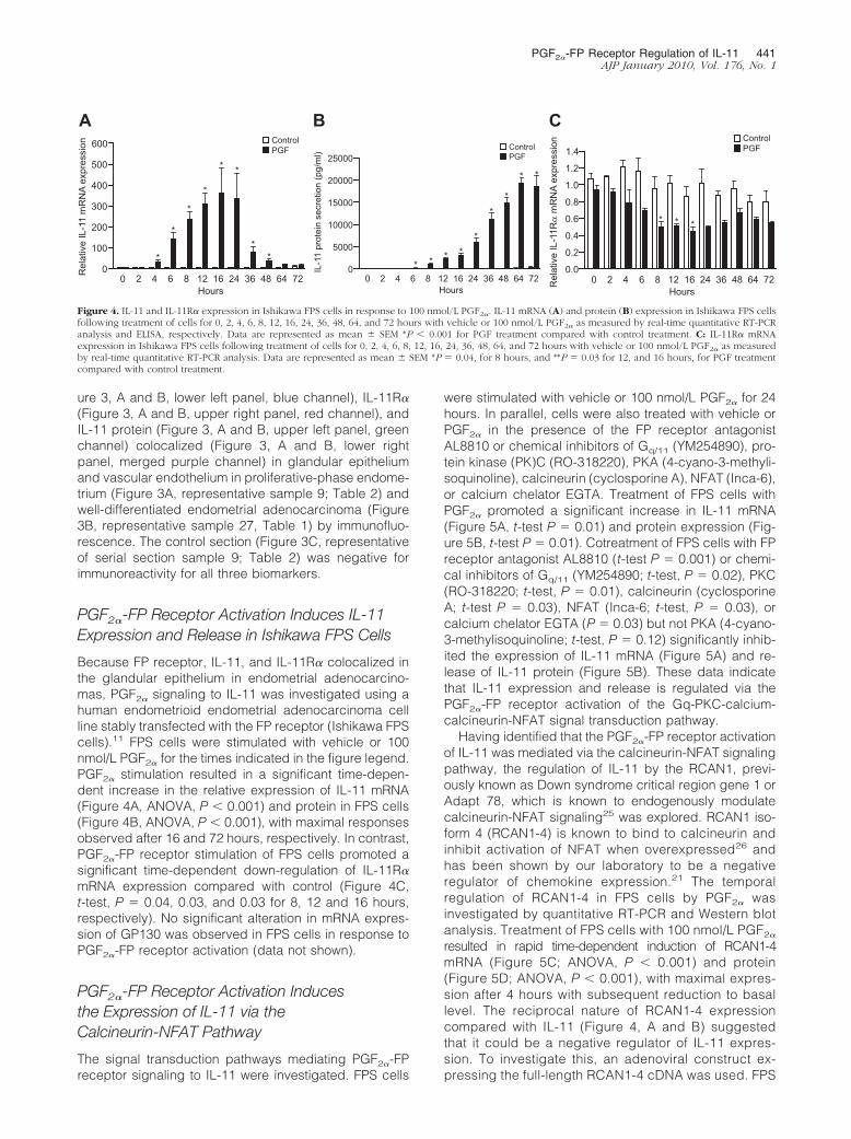
PGF2�-FP Receptor Activation Induces IL-11Expression and Release in Ishikawa FPS Cells
Because FP receptor, IL-11, and IL-11R� colocalized inthe glandular epithelium in endometrial adenocarcino-mas, PGF2� signaling to IL-11 was investigated using ahuman endometrioid endometrial adenocarcinoma cellline stably transfected with the FP receptor (Ishikawa FPScells).11 FPS cells were stimulated with vehicle or 100nmol/L PGF2� for the times indicated in the figure legend.PGF2� stimulation resulted in a significant time-depen-dent increase in the relative expression of IL-11 mRNA(Figure 4A, ANOVA, P � 0.001) and protein in FPS cells(Figure 4B, ANOVA, P � 0.001), with maximal responsesobserved after 16 and 72 hours, respectively. In contrast,PGF2�-FP receptor stimulation of FPS cells promoted asignificant time-dependent down-regulation of IL-11R�mRNA expression compared with control (Figure 4C,t-test, P � 0.04, 0.03, and 0.03 for 8, 12 and 16 hours,respectively). No significant alteration in mRNA expres-sion of GP130 was observed in FPS cells in response toPGF2�-FP receptor activation (data not shown).
PGF2�-FP Receptor Activation Inducesthe Expression of IL-11 via theCalcineurin-NFAT Pathway
The signal transduction pathways mediating PGF2�-FPreceptor signaling to IL-11 were investigated. FPS cells
were stimulated with vehicle or 100 nmol/L PGF2� for 24hours. In parallel, cells were also treated with vehicle orPGF2� in the presence of the FP receptor antagonistAL8810 or chemical inhibitors of Gq/11 (YM254890), pro-tein kinase (PK)C (RO-318220), PKA (4-cyano-3-methyli-soquinoline), calcineurin (cyclosporine A), NFAT (Inca-6),or calcium chelator EGTA. Treatment of FPS cells withPGF2� promoted a significant increase in IL-11 mRNA(Figure 5A, t-test P � 0.01) and protein expression (Fig-ure 5B, t-test P � 0.01). Cotreatment of FPS cells with FPreceptor antagonist AL8810 (t-test P � 0.001) or chemi-cal inhibitors of Gq/11 (YM254890; t-test, P � 0.02), PKC(RO-318220; t-test, P � 0.01), calcineurin (cyclosporineA; t-test P � 0.03), NFAT (Inca-6; t-test, P � 0.03), orcalcium chelator EGTA (P � 0.03) but not PKA (4-cyano-3-methylisoquinoline; t-test, P � 0.12) significantly inhib-ited the expression of IL-11 mRNA (Figure 5A) and re-lease of IL-11 protein (Figure 5B). These data indicatethat IL-11 expression and release is regulated via thePGF2�-FP receptor activation of the Gq-PKC-calcium-calcineurin-NFAT signal transduction pathway.
Having identified that the PGF2�-FP receptor activationof IL-11 was mediated via the calcineurin-NFAT signalingpathway, the regulation of IL-11 by the RCAN1, previ-ously known as Down syndrome critical region gene 1 orAdapt 78, which is known to endogenously modulatecalcineurin-NFAT signaling25 was explored. RCAN1 iso-form 4 (RCAN1-4) is known to bind to calcineurin andinhibit activation of NFAT when overexpressed26 andhas been shown by our laboratory to be a negativeregulator of chemokine expression.21 The temporalregulation of RCAN1-4 in FPS cells by PGF2� wasinvestigated by quantitative RT-PCR and Western blotanalysis. Treatment of FPS cells with 100 nmol/L PGF2�
resulted in rapid time-dependent induction of RCAN1-4mRNA (Figure 5C; ANOVA, P � 0.001) and protein(Figure 5D; ANOVA, P � 0.001), with maximal expres-sion after 4 hours with subsequent reduction to basallevel. The reciprocal nature of RCAN1-4 expressioncompared with IL-11 (Figure 4, A and B) suggestedthat it could be a negative regulator of IL-11 expres-sion. To investigate this, an adenoviral construct ex-pressing the full-length RCAN1-4 cDNA was used. FPS
600 ControlPGF
A
Rel
ativ
e IL
-11
mR
NA
expr
essi
on
0 2 4 6 8 12Hours
16 24 36 48 64 72 0 2 4 6 8 12Hours
16 24 36 48 64 72 0 2 4 6 8 12Hours
16 24 36 48 64 72
500
400
*
*
*
*
* *
**
* * * **
**
* * *
* *
300
200
100
0
ControlPGF
B
IL-1
1 pr
otei
n se
cret
ion
(pg/
ml) 25000
20000
15000
10000
5000
0
1.4ControlPGF
C
Rel
ativ
e IL
-11R
α m
RN
A ex
pres
sion
1.2
1.0
0.8
b b
0.6
0.4
0.2
0.0
Figure 4. IL-11 and IL-11R� expression in Ishikawa FPS cells in response to 100 nmol/L PGF2�. IL-11 mRNA (A) and protein (B) expression in Ishikawa FPS cellsfollowing treatment of cells for 0, 2, 4, 6, 8, 12, 16, 24, 36, 48, 64, and 72 hours with vehicle or 100 nmol/L PGF2� as measured by real-time quantitative RT-PCRanalysis and ELISA, respectively. Data are represented as mean � SEM *P � 0.001 for PGF treatment compared with control treatment. C: IL-11R� mRNAexpression in Ishikawa FPS cells following treatment of cells for 0, 2, 4, 6, 8, 12, 16, 24, 36, 48, 64, and 72 hours with vehicle or 100 nmol/L PGF2� as measuredby real-time quantitative RT-PCR analysis. Data are represented as mean � SEM *P � 0.04, for 8 hours, and **P � 0.03 for 12, and 16 hours, for PGF treatmentcompared with control treatment.
PGF2�-FP Receptor Regulation of IL-11 441AJP January 2010, Vol. 176, No. 1

cells were infected with either a control (RAD60) ade-novirus or RCAN1-4 adenovirus for 24 hours beforestimulation or left uninfected. Cells were stimulatedwith vehicle or 100 nmol/L PGF2� for 24 hours. Over-expression of RCAN1-4 in FPS cells significantly re-duced the PGF2�-FP receptor-mediated induction ofIL-11 mRNA expression (Figure 5E; t-test, P � 0.03) andprotein secretion (Figure 5F; t-test, P � 0.02) comparedwith cells infected with the control virus or control cells,which were uninfected.
Expression and Regulation of RCAN1-4 inEndometrial Tissues
Because PGF2�-FP receptor signaling to IL-11 was reg-ulated by RCAN1-4, the mRNA expression of RCAN1-4(Figure 6A) in human endometrial adenocarcinoma com-pared with normal proliferative-phase endometrium wasinvestigated by quantitative RT-PCR analysis. RCAN1-4expression was significantly higher in well-differentiatedadenocarcinoma compared with normal proliferativephase endometrium (t-test P � 0.03). Interestingly, weobserved a significant linear trend for RCAN1-4 mRNAexpression, which decreased with increasing tumorgrade, such that RCAN1-4 mRNA expression was signif-icantly lower in poorly differentiated endometrial adeno-carcinomas (Figure 6A, ANOVA, �posttest for linear trend,P � 0.002).
Figure 5. IL-11 is regulated by PGF2� via the calcium-calcineurin-NFATpathway. IL-11 mRNA expression (A) and protein secretion (B) in FPScells treated for 24 hours, respectively, with vehicle, 100 nmol/L PGF2�
(P � 0.01), 100 nmol/L PGF2� in the absence/presence of AL8810 (50�mol/L; P � 0.001), YM254890 (1 �mol/L; P � 0.02), RO-318220 (1�mol/L; P � 0.01), 4-cyano-3-methylisoquinoline (4C3MQ) (1 �mol/L;P � 0.12), EGTA (1.5 mmol/L; P � 0.03), cyclosporine A (CsA) (1 �mol/L;P � 0.03), or Inca-6 (40 �mol/L; P � 0.03) as determined by quantitativeRT-PCR analysis and ELISA, respectively. Data are represented as mean �SEM; PGF treatment is significantly different from PGF and inhibitortreatment or PGF and vehicle control treatment at the previously listedvalues. RCAN1-4 mRNA (C) and protein expression (D) in FPS cellstreated with 100 nmol/L PGF2� for 2, 4, 6, 8, 24, 48, and 72 hours asdetermined by quantitative RT-PCR and Western blot analysis, respec-tively. Data are represented as mean � SEM, *P � 0.001 for PGF treatmentcompared vehicle control treatment at each time point. E: FPS cells wereinfected with either RAD60 control adenovirus or RCAN1-4 adenovirus for24 hours or left uninfected. Cells were then treated with vehicle or 100nmol/L PGF2� for 24 hours and IL-11 mRNA, and protein expression (F)was determined by quantitative RT-PCR analysis and ELISA, respectively.Data are represented as mean � SEM for PGF treatment compared withvehicle control treatment for mRNA (E, *P � 0.03) and protein (F, *P �0.02).
2.5
A
C
Rel
ativ
e R
CA
N1-
4 m
RN
A ex
pres
sion
Prolif
Poor
Mod Well
2.0
1.5
1.0
0.5
0.0
98
6
4
2
VehiclePGF
B
IL-1
1 m
RN
A ex
pres
sion
in c
ance
r(F
old
abov
e co
ntro
l)
RAD60 RCAN1-4 Ad
7
5
3
10
Transcription
Cell Surface
CnB
Ca 2+
NFAT
IL-11
P PNFAT
CnA
Ca2+ Ca2+ Ca2+
Ca2+
Ca2+
NFAT
PGF
Gq
FP
PKC
RCAN1-4
Nucleus
Cam
** * ***
Figure 6. RCAN1-4 expression in endometrial tissues. A: Relative mRNAexpression of RCAN1-4 in normal proliferative-phase endometrium (n � 10)and poorly (n � 10), moderately (n � 10), and well-differentiated (n � 10)endometrial adenocarcinoma as determined by real-time quantitative RT-PCRanalysis. Data are represented as mean � SEM *P � 0.03 for well-differen-tiated adenocarcinoma compared with proliferative-phase endometriumANOVA posttest for linear trend between poor, moderate, and well-differ-entiated adenocarcinomas; **P � 0.002. B: Poorly differentiated endometrialadenocarcinomas explants (samples 1 to 4; Table 1) were infected with eitherRAD60 control adenovirus or RCAN1-4 adenovirus for 24 hours and thenstimulated with vehicle or 100 nmol/L PGF2� for 24 hours and subjected toquantitative RT-PCR analysis for IL-11 mRNA expression. Data are repre-sented as mean � SEM ***P� 0.003 for PGF treatment compared with vehiclecontrol treatment. C: A schematic summary. PGF2�-FP receptor activation inendometrial adenocarcinoma cells promotes the induction of RCAN1-4 andIL-11 via the Gq-phospholipase C-PKC-calcium (Ca2)-calcineurin-NFATcascade. RCAN1-4 is temporally activated in a reciprocal manner to IL-11 byPGF-FP receptor signaling and acts as a negative regulator of the calcineurinpathway to regulate IL-11 expression.
442 Sales et alAJP January 2010, Vol. 176, No. 1

RCAN1-4 Is a Negative Regulator of IL-11Expression in Endometrial AdenocarcinomaExplants
Finally, the role of RCAN1-4 as a negative regulator ofIL-11 in endometrial adenocarcinomas was investi-gated ex vivo. Four poorly differentiated endometrioidendometrial adenocarcinoma explants (samples 1 to 4;Table 1) were infected with either a control (RAD60)adenovirus or RCAN1-4 adenovirus for 24 hours beforestimulation with vehicle or 100 nmol/L PGF2� for 24hours (Figure 6B). Overexpression of RCAN1-4 in theseendometrial adenocarcinoma samples significantly re-duced the PGF2�-FP receptor-mediated induction of IL-11mRNA expression in explants compared with control virus-infected tissue (t-test, P � 0.003).
Discussion
IL-11 has been shown to regulate cell motility, invasion,and metastasis in vitro and in vivo.15, 20, 27, 28 The presentstudy demonstrated elevated expression of IL-11 in en-dometrial adenocarcinomas compared with normal en-dometrium from the proliferative phase of the menstrualcycle. As the endometrium of postmenopausal women isno longer under normal hormonal control, the tissue isatrophic and often not attainable for analysis, we chosenormal proliferative-phase endometrium as our compar-ator. This is the phase of the menstrual cycle that exhibitsrapid cellular proliferation, differentiation, and tissue re-modeling and is the phase of the menstrual cycle with thehighest expression of FP receptor.13 Interestingly, thepresent study highlights a significant variation in expres-sion of IL-11 with grade of adenocarcinomas, with high-est levels observed in high-grade (poorly differentiated)adenocarcinomas. Furthermore, IL-11 and IL-11R� pro-tein expression colocalized with FP receptor expressionin normal endometrial tissue and endometrial adenocar-cinomas. The pattern of expression of IL-11, IL-11R�, andits coreceptor GP130 was similar in endometrial adeno-carcinoma compared with normal proliferative-phase en-dometrium in the present study confirming the localiza-tion of these biomarkers as reported by others in normalendometrium29–32 and other carcinomas.33,34 Moreover,we have shown that IL-11 is regulated by PGF2� via theFP receptor in an in vitro model system of endometrialadenocarcinoma cells and poorly differentiated endome-trial adenocarcinoma explants ex vivo. To our knowledge,this is the first study to report on the expression, localiza-tion, and cellular mechanism regulating IL-11 in endome-trial adenocarcinomas.
Although little is known of the role of IL-11 in endome-trial cancer, IL-11 is essential for normal reproduction inthe human and mouse.35,36 IL-11 and IL-11R� expres-sion are dysregulated in endometrium of infertile womenwith endometriosis,35,36 and IL-11 in the normal endome-trium is known to promote the migration of trophoblastcells.27 IL-11 and IL-11R� have been shown to correlatewith tumor progression, cellular growth and differentia-tion, and poor prognosis in breast and colorectal can-
cers18,19 and is associated with breast cancer cell me-tastases to bone.15 To investigate the regulation of IL-11,IL-11R�, and GP130 in endometrial adenocarcinomas bythe FP receptor, we used Ishikawa FPS cells, stably ex-pressing the FP receptor. This in vitro approach has pre-viously been used by our laboratory and others to inter-rogate PGF2�-FP receptor signaling.10,11,37–39 Using thismodel system, we have previously demonstrated that thePGF2�-FP receptor signaling in Ishikawa FPS cells paral-lels the ex vivo effects of PGF2� on endometrial adeno-carcinoma explants, indicating that it is a suitable modelsystem to interrogate FP receptor signaling in endome-trial epithelial cells in vitro.10,11
Here we have demonstrated that PGF2� promotes thesynthesis and release of IL-11 in Ishikawa FPS cells in atime-dependent manner. We confirmed that PGF2�-FPreceptor signaling regulated IL-11 mRNA expression exvivo using endometrial adenocarcinoma explants. Inter-estingly, PGF2�-FP receptor interaction promoted a time-dependent decrease in the mRNA expression of IL-11R�
in Ishikawa FPS cells, suggesting that a negative feed-back loop was being activated to prevent autocrine in-duction of the IL-11R�/GP130 receptor complex by IL-11.
Because expression of IL-11R� mRNA is significantlyreduced in endometrial adenocarcinomas comparedwith normal proliferative-phase endometrium, it is feasi-ble that activation of the FP receptor in adenocarcinomasin vivo could down-regulate expression of the IL-11R�
mRNA in a similar manner to our observation with theIshikawa FPS cell line in vitro. We investigated whetherthe mechanism of down-regulation of IL-11R� in vitro wasmediated via IL-11; however, treatment of FPS cells withthe recombinant IL-11 had no effect on IL-11R� mRNAexpression (data not shown), indicating that either PGF2�
was having a direct inhibitory effect on IL-11R� mRNAsynthesis or another factor produced by PGF2�-FP recep-tor signaling was regulating IL-11R� mRNA in IshikawaFPS cells in vitro.
The intracellular signal transduction pathways mediat-ing the role of the FP receptor in regulating IL-11 wasinvestigated using chemical inhibitors of intracellular sig-naling. IL-11 was found to be regulated in FPS cells at themRNA and protein level by PGF2� via the PKC-calcium-calcineurin-NFAT signaling pathway. NFAT activation bycalcineurin, which mediates its dephosphorylation andtranslocation to the nucleus, is known to be regulated bythe RCAN.25 RCAN1-4 is known to bind to calcineurin,and previous studies have shown that overexpression ofthis protein results in an inhibition of calcineurin activationof NFAT.26,40,41 The temporal pattern of expression ofRCAN1-4 mRNA and protein in FPS cells was investi-gated in response to PGF2� and found to be regulated ina reciprocal manner to IL-11, reaching a peak of expres-sion that preceded IL-11 � 8 hours, suggesting thatRCAN1-4 could act as a negative regulator of IL-11 ex-pression. Indeed, overexpression of RCAN1-4 and sub-sequent inhibition of NFAT activation significantly inhib-ited the PGF2�-mediated activation and release of IL-11from FPS cells. This is in agreement with other published
PGF2�-FP Receptor Regulation of IL-11 443AJP January 2010, Vol. 176, No. 1
observations that show that expression of this isoform isinduced by NFAT41 and can negatively regulate chemo-kines, such as IL-8, in endometrial epithelial cells.21
Dysregulated RCAN (also called Down syndrome can-didate region 1) expression has been linked to patholog-ical processes. For example, RCAN overexpression inbrains of Down syndrome fetuses40 and Alzheimer’s pa-tients42 is thought to enhance neurodegenerative condi-tions. RCAN expression is known to be up-regulated byglucocorticoids43 and is induced in endothelial cells as anegative feedback regulator of endothelial cell prolifera-tion and branching.41,44 We investigated the expressionof RCAN1-4 in normal proliferative-phase endometriumand endometrial adenocarcinomas. We found signifi-cantly lower RCAN1-4 expression with advancing tumorgrade, such that levels of RCAN1-4 were lowest in high-grade (poorly differentiated) adenocarcinomas, theopposite pattern to that seen for IL-11 expression. Fur-thermore, RCAN1-4 overexpression could negatively reg-ulate IL-11 expression in cancer explants ex vivo similarlyto our observations using the Ishikawa FPS cell line invitro. Although we found no difference within each gradewith respect to FIGO stage, our data show that RCAN1-4expression reduces coincident with advancing grade ofendometrial adenocarcinoma (ie, from well-differentiatedor low-grade to poorly differentiated or high-grade can-cer). These findings suggest that the natural brakecreated by RCAN1-4 to control calcineurin signalingand cytokine production is defective in high-grade(poorly differentiated) compared with low-grade (well-differentiated) adenocarcinomas. This results in unop-posed PGF2�-FP receptor signaling via the calcium-calcineurin-NFAT pathway to elevate cytokine productionsuch as IL-11 and may confer an advantage to tumorprogression and metastasis. However, further studiesare needed to determine the prognostic and/or predic-tive value of IL-11, RCAN1-4, and the calcineurin-NFATpathway in women with early or advanced endometrialadenocarcinoma.
In conclusion, these data (as summarized in Figure6C) show that PGF2�-FP receptor activation in endome-trial adenocarcinoma cells regulates IL-11 expression viathe Gq-phospholipase C-PKC-calcium-calcineurin-NFATpathway. Moreover, RCAN1-4 was identified as a negativeregulator of PGF2�-FP receptor-mediated IL-11 expressionin endometrial adenocarcinoma cells stably expressing theFP receptor in vitro and tissue explants ex vivo. Furtherstudies to evaluate whether RCAN signaling can be ex-ploited therapeutically to negatively control calcineurin-NFAT signaling, and IL-11 production may be of potentialclinical significance for women with type 11 high-grade(poorly differentiated) endometrial adenocarcinoma.
Acknowledgments
We thank Ms. Anne Saunderson and Sharon McPhersonfor patient recruitment and sample collection.
References
1. Mant JW, Vessey MP: Ovarian and endometrial cancers. Cancer Surv1994, 20:287–307
2. Persson I: Estrogens in the causation of breast, endometrial andovarian cancers—evidence and hypotheses from epidemiologicalfindings. J Steroid Biochem Mol Biol 2000, 74:357–364
3. Parazzini F, La Vecchia C, Bocciolone L, Franceschi S: The epidemi-ology of endometrial cancer. Gynecol Oncol 1991, 41:1–16
4. Ryan AJ, Susil B, Jobling TW, Oehler MK: Endometrial cancer. CellTissue Res 2005, 322:53–61
5. Bokhman JV: Two pathogenetic types of endometrial carcinoma.Gynecol Oncol 1983, 15:10–17
6. Tong BJ, Tan J, Tajeda L, Das SK, Chapman JA, DuBois RN, Dey SK:Heightened expression of cyclooxygenase-2 and peroxisome prolif-erator-activated receptor-� in human endometrial adenocarcinoma.Neoplasia 2000, 2:483–490
7. Jabbour HN, Milne SA, Williams ARW, Anderson RA, Boddy SC:Expression of COX-2 and PGE synthase and synthesis of PGE2 inendometrial adenocarcinoma: a possible autocrine/paracrine regula-tion of neoplastic cell function via EP2/EP4 receptors. Br J Cancer2001, 85:1023–1031
8. Sales KJ, Milne SA, Williams AR, Anderson RA, Jabbour HN:Expression, localization, and signaling of prostaglandin F2� recep-tor in human endometrial adenocarcinoma: regulation of prolifera-tion by activation of the epidermal growth factor receptor andmitogen-activated protein kinase signaling pathways. J Clin Endo-crinol Metab 2004, 89:986–993
9. Jabbour HN, Sales KJ, Boddy SC, Anderson RA, Williams AR: Apositive feedback loop that regulates cyclooxygenase-2 expressionand prostaglandin F2� synthesis via the F-series-prostanoid receptorand extracellular signal-regulated kinase 1/2 signaling pathway. En-docrinology 2005, 146:4657–4664
10. Sales KJ, Boddy SC, Williams AR, Anderson RA, Jabbour HN: F-Prostanoid receptor regulation of fibroblast growth factor 2 signallingin endometrial adenocarcinoma cells. Endocrinology 2007, 148:3635–3644
11. Sales KJ, List T, Boddy SC, Williams AR, Anderson RA, Naor Z,Jabbour HN: A novel angiogenic role for prostaglandin F2�-FP recep-tor interaction in human endometrial adenocarcinomas. Cancer Res2005, 65:7707–7716
12. Sales KJ, Boddy SC, Jabbour HN: F-Prostanoid receptor alters ad-hesion, morphology and migration of endometrial adenocarcinomacells. Oncogene 2008, 27:2466–2477
13. Milne SA, Jabbour HN: Prostaglandin (PG) F2� receptor expressionand signaling in human endometrium: role of PGF2� in epithelial cellproliferation. J Clin Endocrinol Metab 2003, 88:1825–1832
14. Wang D, Wang H, Brown J, Daikoku T, Ning W, Shi Q, Richmond A,Strieter R, Dey SK, DuBois RN: CXCL1 induced by prostaglandin E2
promotes angiogenesis in colorectal cancer. J Exp Med 2006, 203:941–951
15. Singh B, Berry JA, Shoher A, Lucci A: COX-2 induces IL-11 produc-tion in human breast cancer cells. J Surg Res 2006, 131:267–275
16. Heinrich PC, Behrmann I, Muller-Newen G, Schaper F, Graeve L:Interleukin-6-type cytokine signalling through the gp130/Jak/STATpathway. Biochem J 1998, 334:297–314
17. Katoh M, Katoh M: STAT3-induced WNT5A signaling loop in embry-onic stem cells, adult normal tissues, chronic persistent inflammation,rheumatoid arthritis and cancer. Int J Mol Med 2007, 19:273–278
18. Hanavadi S, Martin TA, Watkins G, Mansel RE, Jiang WG: Expressionof interleukin 11 and its receptor and their prognostic value in humanbreast cancer. Ann Surg Oncol 2006, 13:802–808
19. Yamazumi K, Nakayama T, Kusaba T, Wen CY, Yoshizaki A, Yakata Y,Nagayasu T, Sekine I: Expression of interleukin-11 and interleukin-11receptor � in human colorectal adenocarcinoma; immunohistochem-ical analyses and correlation with clinicopathological factors. World JGastroenterol 2006, 12:317–321
20. Nakayama T, Yoshizaki A, Izumida S, Suehiro T, Miura S, Uemura T,Yakata Y, Shichijo K, Yamashita S, Sekin I: Expression of interleu-kin-11 (IL-11) and IL-11 receptor � in human gastric carcinoma andIL-11 up-regulates the invasive activity of human gastric carcinomacells. Int J Oncol 2007, 30:825–833
21. Maldonado-Perez D, Brown P, Morgan K, Millar RP, Thompson EA,Jabbour HN: Prokineticin 1 modulates IL-8 expression via the cal-
444 Sales et alAJP January 2010, Vol. 176, No. 1
cineurin/NFAT signaling pathway. Biochim Biophys Acta 2009, 1793:1315–1324
22. Sales KJ, Grant V, Jabbour HN: Prostaglandin E2 and F2� activate theFP receptor and up-regulate cyclooxygenase-2 expression via thecyclic AMP response element. Mol Cell Endocrinol 2008, 285:51–61
23. Noyes RW, Hertig AT, Rock J: Dating the endometrial biopsy. FertilSteril 1950, 1:3–25
24. Wilkinson GW, Akrigg A: Constitutive and enhanced expression fromthe CMV major IE promoter in a defective adenovirus vector. NucleicAcids Res 1992, 20:2233–2239
25. Davies KJ, Ermak G, Rothermel BA, Pritchard M, Heitman J, Ahnn J,Henrique-Silva F, Crawford D, Canaider S, Strippoli P, Carinci P, MinKT, Fox DS, Cunningham KW, Bassel-Duby R, Olson EN, Zhang Z,Williams RS, Gerber HP, Perez-Riba M, Seo H, Cao X, Klee CB,Redondo JM, Maltais LJ, Bruford EA, Povey S, Molkentin JD, McKeonFD, Duh EJ, Crabtree GR, Cyert MS, de la Luna S, Estivill X: Renamingthe DSCR1/Adapt78 gene family as RCAN: regulators of calcineurin.FASEB J 2007, 21:3023–3028
26. Chan B, Greenan G, McKeon F, Ellenberger T: Identification of apeptide fragment of DSCR1 that competitively inhibits calcineurinactivity in vitro and in vivo. Proc Natl Acad Sci USA 2005, 102:13075–13080
27. Paiva P, Salamonsen LA, Manuelpillai U, Walker C, Tapia A, WallaceEM, Dimitriadis E: Interleukin-11 promotes migration, but not prolifer-ation, of human trophoblast cells, implying a role in placentation.Endocrinology 2007, 148:5566–5572
28. Yoshizaki A, Nakayama T, Yamazumi K, Yakata Y, Taba M, Sekine I:Expression of interleukin (IL)-11 and IL-11 receptor in human colo-rectal adenocarcinoma: IL-11 up-regulation of the invasive and pro-liferative activity of human colorectal carcinoma cells. Int J Oncol2006, 29:869–876
29. Karpovich N, Chobotova K, Carver J, Heath JK, Barlow DH, MardonHJ: Expression and function of interleukin-11 and its receptor � in thehuman endometrium. Mol Hum Reprod 2003, 9:75–80
30. Dimitriadis E, Salamonsen LA, Robb L: Expression of interleukin-11during the human menstrual cycle: coincidence with stromal celldecidualization and relationship to leukaemia inhibitory factor andprolactin. Mol Hum Reprod 2000, 6:907–914
31. Linjawi S, Li TC, Tuckerman EM, Blakemore AI, Laird SM: Expressionof interleukin-11 receptor � and interleukin-11 protein in the endome-trium of normal fertile women and women with recurrent miscarriage.J Reprod Immunol 2004, 64:145–155
32. Cullinan EB, Abbondanzo SJ, Anderson PS, Pollard JW, Lessey BA,Stewart CL: Leukemia inhibitory factor (LIF) and LIF receptor expres-sion in human endometrium suggests a potential autocrine/paracrine
function in regulating embryo implantation. Proc Natl Acad Sci USA1996, 93:3115–3120
33. Campbell CL, Guardiani R, Ollari C, Nelson BE, Quesenberry PJ,Savarese TM: Interleukin-11 receptor expression in primary ovariancarcinomas. Gynecol Oncol 2001, 80:121–127
34. Campbell CL, Jiang Z, Savarese DM, Savarese TM: Increased ex-pression of the interleukin-11 receptor and evidence of STAT3 acti-vation in prostate carcinoma. Am J Pathol 2001, 158:25–32
35. Dimitriadis E, Stoikos C, Stafford-Bell M, Clark I, Paiva P, Kovacs G,Salamonsen LA: Interleukin-11. IL-11 receptor � and leukemia inhib-itory factor are dysregulated in endometrium of infertile women withendometriosis during the implantation window. J Reprod Immunol2006, 69:53–64
36. Dimitriadis E, White CA, Jones RL, Salamonsen LA: Cytokines, che-mokines and growth factors in endometrium related to implantation.Hum Reprod Update 2005, 11:613–630
37. Fujino H, Pierce KL, Srinivasan D, Protzman CE, Krauss AH, WoodwardDF, Regan JW: Delayed reversal of shape change in cells expressingFP(B) prostanoid receptors: possible role of receptor resensitization.J Biol Chem 2000, 275:29907–29914
38. Fujino H, Regan JW: Prostaglandin F2� stimulation of cyclooxygen-ase-2 promoter activity by the FP(B) prostanoid receptor. Eur J Phar-macol 2003, 465:39–41
39. Srinivasan D, Fujino H, Regan JW: Differential internalization of theprostaglandin F2� receptor isoforms: role of protein kinase C andclathrin. J Pharmacol Exp Ther 2002, 302:219–224
40. Fuentes JJ, Genesca L, Kingsbury TJ, Cunningham KW, Perez-RibaM, Estivill X, de la Luna S: DSCR1, overexpressed in Down syndrome,is an inhibitor of calcineurin-mediated signaling pathways. Hum MolGenet 2000, 9:1681–1690
41. Minami T, Horiuchi K, Miura M, Abid MR, Takabe W, Noguchi N,Kohro T, Ge X, Aburatani H, Hamakubo T, Kodama T, Aird WC:Vascular endothelial growth factor- and thrombin-induced terminationfactor: Down syndrome critical region-1, attenuates endothelial cellproliferation and angiogenesis. J Biol Chem 2004, 279:50537–50554
42. Ermak G, Morgan TE, Davies KJ: Chronic overexpression of thecalcineurin inhibitory gene DSCR1 (Adapt78) is associated with Alz-heimer’s disease. J Biol Chem 2001, 276:38787–38794
43. Faruqi SA, Noumoff MJ, Deger RB, Jalal SM, Antoniades K: Trisomy21 as the only recurrent chromosomal anomaly in a clinically aggres-sive ovarian carcinoma. Cancer Genet Cytogenet 2002, 138:165–168
44. Qin L, Zhao D, Liu X, Nagy JA, Hoang MV, Brown LF, Dvorak HF,Zeng H: Down syndrome candidate region 1 isoform 1 mediatesangiogenesis through the calcineurin-NFAT pathway. Mol CancerRes 2006, 4:811–820
PGF2�-FP Receptor Regulation of IL-11 445AJP January 2010, Vol. 176, No. 1