interpretation of gram stains and other common microbiologic slide preparations - library
DESCRIPTION
Gram stainingTRANSCRIPT
1/2/12 Interpretation of Gram Stains and Other Common Microbiologic Slide Prepa…
1/4microbelibrary.org/…/3046-interpretation-of-gram-stains-and-other-common…
RegisterLogin
Home
Submit
About
Permissions
Downloading Resources
Contact
Search
Search
Browse/Advanced Search Options
ASM Updates
Compound in Apples Inhibits E. coli O157:H7
Bacterial Filters Reduce Stink from Big Pig FactoriesCold Spots Contaminated in High Humidity Incubators
Select Language
Pow ered by Translate
Interpretation of Gram Stains and Other Common Microbiologic Slide Preparations
Excellent +1 Votes (1) | Hits (4281) | Comments (0)
Created: Tuesday, 06 February 2007
Last update: Friday, 19 August 2011
AuthorFred Tenover
Author - SecondaryJ. V. Hirschmann
View / Comment
1/2/12 Interpretation of Gram Stains and Other Common Microbiologic Slide Prepa…
2/4microbelibrary.org/…/3046-interpretation-of-gram-stains-and-other-common…
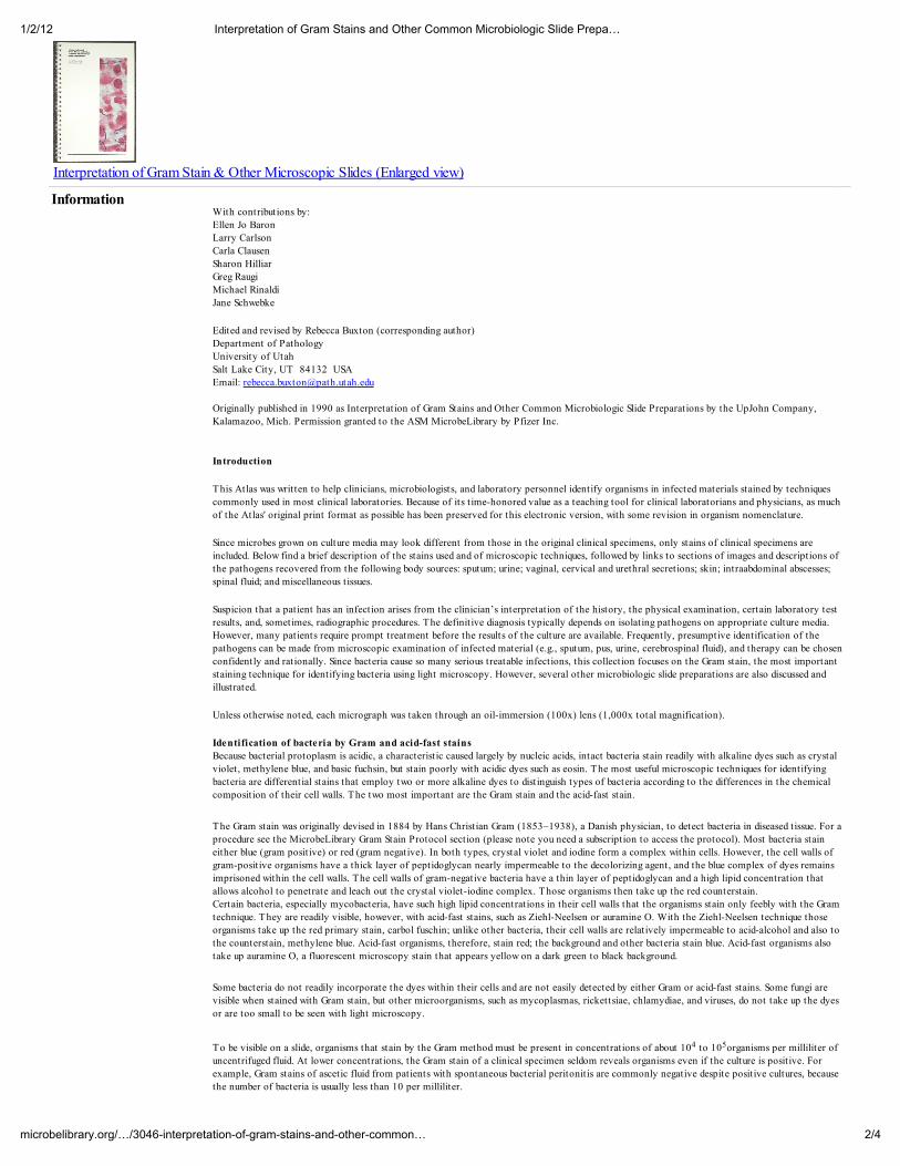
Interpretation of Gram Stain & Other Microscopic Slides (Enlarged view)
InformationWith contributions by:
Ellen Jo Baron
Larry Carlson
Carla Clausen
Sharon Hilliar
Greg Raugi
Michael Rinaldi
Jane Schwebke
Edited and revised by Rebecca Buxton (corresponding author)
Department of Pathology
University of Utah
Salt Lake City, UT 84132 USA
Email: [email protected]
Originally published in 1990 as Interpretation of Gram Stains and Other Common Microbiologic Slide Preparations by the UpJohn Company,
Kalamazoo, Mich. Permission granted to the ASM MicrobeLibrary by Pfizer Inc.
Introduction
This Atlas was written to help clinicians, microbiologists, and laboratory personnel identify organisms in infected materials stained by techniques
commonly used in most clinical laboratories. Because of its t ime-honored value as a teaching tool for clinical laboratorians and physicians, as much
of the Atlas' original print format as possible has been preserved for this electronic version, with some revision in organism nomenclature.
Since microbes grown on culture media may look different from those in the original clinical specimens, only stains of clinical specimens are
included. Below find a brief description of the stains used and of microscopic techniques, followed by links to sections of images and descriptions of
the pathogens recovered from the following body sources: sputum; urine; vaginal, cervical and urethral secretions; skin; intraabdominal abscesses;
spinal fluid; and miscellaneous tissues.
Suspicion that a patient has an infection arises from the clinician’s interpretation of the history, the physical examination, certain laboratory test
results, and, sometimes, radiographic procedures. The definitive diagnosis typically depends on isolating pathogens on appropriate culture media.
However, many patients require prompt treatment before the results of the culture are available. Frequently, presumptive identification of the
pathogens can be made from microscopic examination of infected material (e.g., sputum, pus, urine, cerebrospinal fluid), and therapy can be chosen
confidently and rationally. Since bacteria cause so many serious treatable infections, this collection focuses on the Gram stain, the most important
staining technique for identifying bacteria using light microscopy. However, several other microbiologic slide preparations are also discussed and
illustrated.
Unless otherwise noted, each micrograph was taken through an oil-immersion (100x) lens (1,000x total magnification).
Identification of bacteria by Gram and acid-fast stains
Because bacterial protoplasm is acidic, a characteristic caused largely by nucleic acids, intact bacteria stain readily with alkaline dyes such as crystal
violet, methylene blue, and basic fuchsin, but stain poorly with acidic dyes such as eosin. The most useful microscopic techniques for identifying
bacteria are differential stains that employ two or more alkaline dyes to distinguish types of bacteria according to the differences in the chemical
composition of their cell walls. The two most important are the Gram stain and the acid-fast stain.
The Gram stain was originally devised in 1884 by Hans Christian Gram (1853–1938), a Danish physician, to detect bacteria in diseased tissue. For a
procedure see the MicrobeLibrary Gram Stain Protocol section (please note you need a subscription to access the protocol). Most bacteria stain
either blue (gram positive) or red (gram negative). In both types, crystal violet and iodine form a complex within cells. However, the cell walls of
gram-positive organisms have a thick layer of peptidoglycan nearly impermeable to the decolorizing agent, and the blue complex of dyes remains
imprisoned within the cell walls. The cell walls of gram-negative bacteria have a thin layer of peptidoglycan and a high lipid concentration that
allows alcohol to penetrate and leach out the crystal violet-iodine complex. Those organisms then take up the red counterstain.
Certain bacteria, especially mycobacteria, have such high lipid concentrations in their cell walls that the organisms stain only feebly with the Gram
technique. They are readily visible, however, with acid-fast stains, such as Ziehl-Neelsen or auramine O. With the Ziehl-Neelsen technique those
organisms take up the red primary stain, carbol fuschin; unlike other bacteria, their cell walls are relatively impermeable to acid-alcohol and also to
the counterstain, methylene blue. Acid-fast organisms, therefore, stain red; the background and other bacteria stain blue. Acid-fast organisms also
take up auramine O, a fluorescent microscopy stain that appears yellow on a dark green to black background.
Some bacteria do not readily incorporate the dyes within their cells and are not easily detected by either Gram or acid-fast stains. Some fungi are
visible when stained with Gram stain, but other microorganisms, such as mycoplasmas, rickettsiae, chlamydiae, and viruses, do not take up the dyes
or are too small to be seen with light microscopy.
To be visible on a slide, organisms that stain by the Gram method must be present in concentrations of about 104 to 105organisms per milliliter of
uncentrifuged fluid. At lower concentrations, the Gram stain of a clinical specimen seldom reveals organisms even if the culture is positive. For
example, Gram stains of ascetic fluid from patients with spontaneous bacterial peritonitis are commonly negative despite positive cultures, because
the number of bacteria is usually less than 10 per milliliter.

1/2/12 Interpretation of Gram Stains and Other Common Microbiologic Slide Prepa…
3/4microbelibrary.org/…/3046-interpretation-of-gram-stains-and-other-common…
Examination of Gram stains
The examiner should first determine whether the stain is adequate (see Sputum–Unacceptable Specimens and Staining Artifacts).In an appropriately
stained specimen, the nuclei of neutrophils are red. If the nuclei are blue, the decolorization is insufficient. In thick specimens like sputum, if certain
areas are difficult to decolorize, the examiner should look for other areas on the slide where the nuclei are red. If the entire specimen is
unsatisfactory, it should be decolorized again with alcohol or acetone-alcohol and then restained with safranin. Repeating the crystal violet and
iodine staining is unnecessary. If the decolorization is still inadequate, the slide should be flooded with acid-alcohol (used in acid-fast stains) to
remove all stain, and the complete Gram stain procedure should be repeated. If the entire specimen is too thick for adequate decolorization, a new
slide with a thinner smear must be prepared.
If both red and blue organisms are visible, decolorization is satisfactory. Excessive decolorization will cause gram-positive organisms to appear to be
gram negative. If only gram-negative organisms are visible and their morphology suggests that they are really gram positive, the entire staining
procedure should be repeated. You may refer to the "Comments and T ips" section of the Gram Stain Protocol for further suggestions (please note
you need a subscription to access the protocol).
When examining a properly stained slide, the examiner should note the following characteristics:
the presence of a single type or several types of organisms
the predominant type of organism if more than one is present
the staining characteristics (gram positive or gram negative)
the shape of the organisms, rods (bacilli) or round (cocci)
the size of the organisms: small, large, thin, plump
the configuration: single organisms, pairs, chains, clumps, clusters, branching
the relation to inflammatory cells because some organisms are characteristically inside inflammatory cells (intracellular) or adherent to
them
With that information, the source of the specimen, and a knowledge of the organisms likely to cause infections at the involved site, the examiner
can presumptively identify many of the pathogens. However, the genus or species of organisms cannot be predicted reliably from their appearance
on a Gram stain. In the case of a patient with acute pneumonia, for example, a properly collected sputum specimen that demonstrates mostly
lancet-shaped, gram-positive diplococci will probably grow Streptococcus pneumoniae (pneumoccocci). Although other streptococci can look
identical on a Gram stain, pneumococcal pneumonia can be diagnosed confidently because other morphologically similar streptococci only rarely
cause acute pulmonary infections. Similarly, to differentiate enteric gram-negative bacilli by their microscopic appearance is difficult . Plump, gram-
negative rods in a sputum specimen from a patient with pneumonia strongly suggest that an enteric gram-negative bacillus is the cause. However it
cannot be safely assumed to be Klebsiella pneumoniae instead of other enteric bacilli such as Escherichia coli, Enterobacter species, or Serratia
marcescens,because these bacteria can also cause lower respiratory tract infections. Clinicians using the Gram stain to help make therapeutic
decisions must recognize the limitations of the technique and not overinterpret the findings.
Links to photographs and legends:
Examination of Gram Stains of Sputum
Examination of Gram Stains of Urine (6 images)
Examination of Gram Stains of Vaginal Secretions (6 images)
Examination of Gram Stains of Cervical and Urethral Discharges (3 images)
Examination of Gram Stains of Bacterial Skin Infections
Examination of Gram Stains of Intraabdominal Infections (5 images)
Examination of Gram Stains of Spinal Fluid–Bacterial Meningitis
Examination of Gram Stains of Miscellaneous T issue Infections (6 images)
Alphabetical Index of Organisms for Interpretation of Gram Stain and Other Common Microbiologic Slide Preparations
References.
Primary reference:
1. Tenover, F. C., and J. V. Hirschmann. 1990. Interpretation of Gram stains and other common microbiologic slide preparations. The UpJohn
Company, Kalamazoo, Mich.
References for editing and revisions in nomenclature:
2. Kwon-Chung, K. J., and J. E. Bennett. 1992. Medical mycology. Lea & Febiger, Philadelphia, Pa.
3. Murray, P. R., E. J. Baron, J. H. Jorgensen, M. A. Pfaller, and R. H. Yolken. 2003. Manual of clinical microbiology. ASM Press,
Washington, D.C.
Related ContentGram Stain ProtocolsExamination of Gram Stains of Cervical and Urethral DischargesExamination of Gram Stains of Intraabdominal Infections
Sputum–Unacceptable Specimens and Staining Artifacts
1/2/12 Interpretation of Gram Stains and Other Common Microbiologic Slide Prepa…
4/4microbelibrary.org/…/3046-interpretation-of-gram-stains-and-other-common…
Examination of Gram Stains of SputumExamination of Gram Stains of Vaginal SecretionsExamination of Gram Stains of Bacterial Skin InfectionsExamination of Gram Stains of Spinal Fluid—Bacterial MeningitisExamination of Gram Stains of Miscellaneous Tissue InfectionsExamination of Gram Stains of Urine
Share
Tags: Cell biology (248) , Microbial genetics (74) , Microbes in humans (371)
There are no comments for this item
Be the first to leave a comment
Login to leave a comments
Copyright © 2010 American Society for Microbiology.All Rights Reserved.
Joomla template created with Artisteer.