intestinal microbiota is altered in patients with colon cancer and ... · this study, lactobacillus...
TRANSCRIPT
Intestinal microbiota is altered inpatients with colon cancer and modifiedby probiotic intervention
Ashley A Hibberd,1 Anna Lyra,2 Arthur C Ouwehand,2 Peter Rolny,3
Helena Lindegren,3 Lennart Cedgård, Yvonne Wettergren,4,5
To cite: Hibberd AA, Lyra A,Ouwehand AC, et al. Intestinalmicrobiota is altered inpatients with colon cancerand modified by probioticintervention. BMJ OpenGastro 2017;4:e000145.doi:10.1136/bmjgast-2017-000145
Received 15 March 2017Revised 5 May 2017Accepted 22 May 2017
1Department of Genomicsand Microbiome Science,DuPont Nutrition & Health,Saint Louis, Missouri, USA2Department of Kantvik ActiveNutrition, DuPont GlobalHealth & Nutrition Science,Kantvik, Finland3Department of Medicine,Sahlgrenska Academy,University of Gothenburg,Gothenburg, Sweden4Probiotic Division, WasaMedicals AB, Halmstad,Sweden5Department of Surgery,Sahlgrenska Academy,University of Gothenburg,Gothenburg, Sweden
Correspondence toDr Yvonne Wettergren;[email protected]
ABSTRACTObjective: The colonic microbiota is altered inpatients with colorectal cancer (CRC). We investigatedthe microbiota composition of patients with coloncancer compared with controls devoid of neoplastic orinflammatory disease and the potential to modify thecolonic microbiota with probiotics.Design: Biopsy samples were obtained from thenormal mucosa and tumour during colonoscopy from15 patients with colon cancer. Subsequent patient-matched samples were taken at surgery from thetumour and nearby mucosa from the patients withcancer, eight of whom had received two daily tabletstotalling 1.4×10¹⁰ CFUs Bifidobacterium lactis Bl-04and 7×10⁹ CFUs Lactobacillus acidophilus NCFM.Faecal samples were obtained after colonoscopy priorto starting the intervention and at surgery. In addition,21 mucosal biopsies from non-cancer controls wereobtained during colonoscopy followed by later faecalsamples. The colonic and faecal microbiota wasassessed by 16S rRNA gene amplicon sequencing.Results: The tumour microbiota was characterised byincreased microbial diversity and enrichment of severaltaxa including Fusobacterium, Selenomonas andPeptostreptococcus compared with the control microbiota.Patients with colon cancer that received probiotics hadan increased abundance of butyrate-producing bacteria,especially Faecalibacterium and Clostridiales spp in thetumour, non-tumour mucosa and faecal microbiota.CRC-associated genera such as Fusobacterium andPeptostreptococcus tended to be reduced in the faecalmicrobiota of patients that received probiotics.Conclusions: Patients with colon cancer harbour adistinct microbiota signature in the tumour tissue andnearby mucosa, which was altered with probioticintervention. Our results show promise for potentialtherapeutic benefits in CRC by manipulation of themicrobiota.Trial registration number: NCT03072641; Results.
INTRODUCTIONColorectal cancer (CRC) currently affects∼1.4 million people each year and its inci-dence is increasing worldwide.1 Despite novel
Summary box
What is already known about this subject?▸ The risk of colorectal cancer (CRC) is strongly
associated with lifestyle factors, including adietary component which may be mediated bythe intestinal microbiota.
▸ The intestinal microbiome is altered in patientswith CRC, and there is a strong interest in identi-fying potential microbial markers for CRC.
▸ Specific probiotic bacteria have been shown tomodulate inflammation and reduce tumour pro-liferation in animal models of carcinogenesisand may offer therapeutic benefits for CRCpatients.
What are the new findings?▸ The colon cancer-associated microbiota exhibits
a distinct signature characterised by increasedmucosal microbial diversity and differentialabundance of specific bacterial taxa comparedwith non-cancer controls. Oral-associated patho-gens are over-represented in colon cancertumours and tend to co-occur.
▸ Although more difficult to obtain, intestinalmucosa samples, rather than faecal, provide amore comprehensive assessment of microbiotachanges in colon cancer. Peptostreptococcuswas over-represented in both mucosal andfaecal samples and shows promise as a CRCmarker.
▸ The colon cancer-associated microbial signaturewas modified by probiotic intervention and wascharacterised by the enrichment of butyrate-producing bacteria in the intestinal tissue.
How might it impact on clinical practice inthe foreseeable future?▸ The CRC-associated microbiota is being continu-
ally defined as new biomarkers of CRC are dis-covered. The microbial dysbiosis observed inpatients with CRC may be manipulated by pro-biotic bacteria, and the probiotic strains used inthis study show promise as a beneficial compo-nent of treatment and therapeutic developmentin CRC.
Hibberd AA, Lyra A, Ouwehand AC, et al. BMJ Open Gastro 2017;4:e000145. doi:10.1136/bmjgast-2017-000145 1
Gut microbiotacopyright.
on May 16, 2020 by guest. P
rotected byhttp://bm
jopengastro.bmj.com
/B
MJ O
pen Gastroenterol: first published as 10.1136/bm
jgast-2017-000145 on 17 July 2017. Dow
nloaded from

treatment strategies, the mortality rate is very high amongpatients with advanced stages of the disease. The majorityof CRC cases (70%) arise sporadically in a time-dependentmanner according to the adenoma-carcinoma sequence ofgenetic alterations.2 Accumulating evidence points to astrong link between lifestyle factors and the risk of develop-ing CRC. Risk factors include advanced age, tobacco andalcohol consumption, physical inactivity, increased bodyweight and diet (eg, high consumption of red and pro-cessed meat), with the latter being the most significant.3
The strong connection to lifestyle factors indicates thatCRC incidence can be influenced by lifestyle changes.Microbial imbalance (dysbiosis) in the gut can be
caused by environmental factors (eg, diet, infection,antibiotics), but little is known about how the compos-ition of the microbiota affects development of CRC. Wehypothesise that the association between CRC risk anddiet is partially mediated by the gut microbiota. Recentstudies show that the gut microbiota differs betweenpatients with and without CRC or colon adenomas, thatis, precancerous lesions that may develop into CRC, andthat the microbiota is a risk factor for cancer develop-ment.4 5 Increased bacterial diversity has been reportedin the microbiota of patients with colon adenomas andtumours compared with non-CRC controls.6–8 TheCRC-associated microbiota also has a microbial profiledistinct from healthy tissue, including bacteria thatthrive in the cancer-related microenvironment. The pro-liferation of carcinoma-associated taxa such asFusobacterium in tumours is a potential microbial bio-marker of a dysbiotic microbiota in CRC.8 9 This micro-bial dysbiosis may reduce the regulatory effect ofcommensal bacteria on cell proliferation in colonmucosa and contribute to the development of aden-omas.10 It is plausible that dietary changes or interven-tion with probiotic bacteria may reduce the risk of CRCdevelopment; however, it is not known whether specifictumour-associated alterations in the microbiota aremodifiable in patients who manifest disease.Probiotics are defined as live microorganisms that,
when administered in adequate amounts, confer ahealth benefit on the host.11 Studies have demonstratedbeneficial effects of probiotic bacteria on reducing CRCtumour development and mucosal inflammation inanimal models; however, supporting clinical data inhumans are limited.12 To investigate the potential of
altering the microbiota in patients with colon cancer, weconducted a prospective intervention study usingselected probiotic strains. The bacterial strains used inthis study, Lactobacillus acidophilus NCFM andBifidobacterium animalis subsp. lactis Bl-04, have a longhistory of safe use as commercial probiotics and docu-mented health benefits. NCFM has shown efficacy forcolonic tumour growth attenuation in rodents and wasassociated with reduced levels of procarcinogenic metabo-lites in the human gut.13 14 Bl-04, although less studiedwith relation to CRC, has anti-inflammatory propertiesand was shown to alleviate colitis in mouse models.15 Inthis pilot study, we obtained intestinal tissue and faecalsamples from patients with colon cancer that received ordid not receive probiotics, and from non-cancer controls,to characterise the colon cancer-associated microbiotaand determine whether this signature could be altered byprobiotic intervention.
PATIENTS AND METHODSStudy outlineSamples were obtained from patients with colon cancer atcolonoscopy and at surgery, and the colonic microbiota wasstudied in a prospective manner (figure 1). Patientswithout cancer or adenomas at colonoscopy were includedas controls for comparisons of baseline data. In addition, aprospective randomised intervention with probiotics inpatients with colon cancer was carried out (figure 1). Thestudy was approved by the Regional Ethical Review Boardin Gothenburg under study number 233-10 and registeredat ClinicalTrials.gov (ID: NCT03072641). Informed consentwas obtained from all study subjects.
Non-cancer controlsTwenty-one non-cancer controls were included in thestudy (table 1). Colonoscopy was performed due toabdominal symptoms such as diarrhoea, constipation,abdominal pain, or lower gastrointestinal bleeding andiron-deficient anaemia. The prerequisites for inclusioninto the control group were a normal-appearing colonicmucosa. Study subjects with significant pathology such ascolonic polyps, inflammatory bowel disease, malignancyor ischaemic colitis were excluded. Microscopic colitiswas ruled out by light microscopic examination of biopsyspecimens obtained from the mid-portion of the
Figure 1 Clinical study outline
and sample collection for
microbiota analyses.
2 Hibberd AA, Lyra A, Ouwehand AC, et al. BMJ Open Gastro 2017;4:e000145. doi:10.1136/bmjgast-2017-000145
Open Accesscopyright.
on May 16, 2020 by guest. P
rotected byhttp://bm
jopengastro.bmj.com
/B
MJ O
pen Gastroenterol: first published as 10.1136/bm
jgast-2017-000145 on 17 July 2017. Dow
nloaded from
ascending and sigmoid colon. The presence of colonicdiverticula was accepted provided there were no signs ofacute diverticulitis or diverticulosis-associated colitis.Study subjects who received recent antibiotic therapy orconsumed probiotics regularly were also excluded.
Colon cancer patientsFifteen patients who were diagnosed with stage I–IIIcolon cancer at colonoscopy were included in the study(table 1). Tumours were classified according to thetumour-node-metastasis (TNM) staging system.16 Theprerequisite for inclusion into the colon cancer groupwas the presence of at least one malignant tumour inthe colon and ≥18 years of age. Study subjects that hadadenomas, received recent antibiotic therapy or con-sumed probiotics regularly were excluded.
Probiotic interventionPatients with cancer were randomised to receive (n=8)or not to receive (n=7) probiotic supplementation. Theintervention started at the second revisit to the clinicand continued until the day of surgery (average lengthof intervention was 31±28 days; range 8–78 days). Theprobiotic supplementation consisted of two ProBionClinica (Wasa Medicals AB, Halmstad, Sweden) tablets,yielding a daily dose of 1.4×10¹⁰ CFUs Bifidobacteriumlactis Bl-04 (ATCC SD5219), 7×10⁹ CFUs Lactobacillus acid-ophilus NCFM (ATCC 700396) and 0.63 g inulin. Flowcytometry analysis showed that on average 85% of cellswere live cells with non-compromised cell membranes.The tablets were produced using a low-compression tech-nique which results in greater cell survival during produc-tion and prolonged stability compared with conventionaltechniques. In addition to a slow-release profile (150–180 min), the tablets improve survival of the probioticbacteria in the gastrointestinal tract.
Tissue and faecal sample collectionIn controls, tissue samples were obtained from the mid-portion of the ascending colon (right side samples) andthe sigmoid colon (left side samples) using regularbiopsy forceps. One of these samples was used for ana-lysis. In patients with colon cancer, biopsies were taken
during colonoscopy from the tumour as well as fromnormal-appearing mucosa distant from the tumour.There were no complications related to the colonoscopyor biopsy procedures. Faecal samples were obtained postcolonoscopy and frozen at −80°C within 24 hours. Allparticipants attended colonoscopy and provided a base-line faecal sample. Thereafter the patients with coloncancer were randomised to receive, or not receive, pro-biotics and eventually attended surgery. At surgery,tumour tissue, macroscopically normal mucosal samplesobtained 10 cm from tumours, and faecal samples wereobtained, snap-frozen in liquid nitrogen and stored at−80°C until analysis. Owing to healthcare procedures,the timing between colonoscopy and faecal sampledonation or randomisation and surgery (ie, length ofintervention) could not be controlled beforehand.Hereafter non-tumour biopsies taken from patients withcolon cancer and control participants are referred to asmucosa and control samples, respectively.
DNA isolation and 16S rRNA gene sequencingDNA was extracted from the samples with the PromegaWizard Genomic DNA Purification Kit (PromegaCorporation, Madison, Wisconsin, USA) as described previ-ously17 18 followed by PCR inhibitor removal withOneStep-96 PCR Inhibitor Removal Kit (Zymo Research,Irvine, California, USA), and DNA concentration measure-ment with Qubit 2.0 Fluorometer (Life Technologies,Darmstadt, Germany). The microbiota composition wasanalysed by targeted PCR amplification of the V4 variableregion of the 16S rRNA gene of bacteria and archaea.Microbial DNA was amplified in triplicate PCR withprimers 515F (5′-GTGCCAGCMGCCGCGGTAA) and806R (5′-GGACTACHVGGGTWTCTAAT) as described.19 20
The PCR amplification proceeded at 95°C for 3 min forinitial denaturation, followed by 30 cycles at 95°C for 45 s,55°C for 60 s and 72°C for 90 s; and final extension of10 min at 72°C. PCR products were purified and normal-ised using the SequalPrep Normalisation Plate Kit(Thermo Fisher Scientific, Waltham, Massachusetts, USA).Paired-end 250 bp reads were generated from ampliconlibraries with the MiSeq instrument (Illumina, San Diego,California, USA).
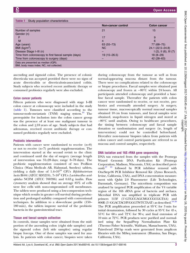
Table 1 Study population characteristics
Non-cancer control Colon cancer
Number of samples 21 15
Gender (n)
F 17 9
M 4 6
Age (years) 63 (55–73) 77 (68–75)
BMI (kg/m2) NC 24.1 (22.5–24.8)
Disease Stage I–III (n) – I (2), II (6), III (7)
Time from colonoscopy to first faecal sample (days) 19 (15–26.5) 18 (8–29)
Time from colonoscopy to surgery (days) – 42 (28–63)
Data are presented as median (IQR).BMI, body mass index; NC, not collected.
Hibberd AA, Lyra A, Ouwehand AC, et al. BMJ Open Gastro 2017;4:e000145. doi:10.1136/bmjgast-2017-000145 3
Open Accesscopyright.
on May 16, 2020 by guest. P
rotected byhttp://bm
jopengastro.bmj.com
/B
MJ O
pen Gastroenterol: first published as 10.1136/bm
jgast-2017-000145 on 17 July 2017. Dow
nloaded from
Analysis of microbiota compositionSequence analysis was conducted with the QuantitativeInsights Into Microbial Ecology (QIIME V.1.9.1) bioinfor-matics pipeline.21 Reads were paired using fastq-join22 andthose with a Phred quality score <20 were discarded. Anopen reference scheme with uclust23 was used for cluster-ing reads into operational taxonomic units (OTUs) at97% sequence similarity, such that OTUs not matching areference sequence in the Greengenes database24 25
(V.13.8) were clustered de novo. Python nearest alignmentspace termination tool (PyNAST)26 was used for sequencealignment, and a taxonomic tree was constructed withFastTree-2.27 OTUs containing <5 sequences wereremoved. After quality filtering, 11 276 994 sequenceswere retained, with an average of 99 796 sequences persample. Metrics for α-diversity (within-sample richness),including observed OTUs and phylogenetic diversity(PD),28 and β-diversity (pairwise dissimilarity) UniFrac dis-tance29 were calculated on OTU tables rarefied to a depthof 10 695 sequences.
Statistics and network analysesGroup comparisons for diversity metrics were conductedwithin QIIME and graphed using Prism V.7 (GraphPadSoftware, La Jolla, California, USA). α-Diversity compari-sons and group distances for β-diversity (weightedUniFrac metric) were generated with a non-parametrict-test using 1000 Monte Carlo permutations.Discriminate taxa (>0.1% abundance) between groupswere identified with the Wilcoxon rank sum test inQIIME. Adjusted p values controlling the false discoveryrate (FDR)30 are reported where appropriate.
A correlation network analysis was constructed fortumour samples using the CoNet software31 for genus-summarised abundance data and clinical factors ofdisease and tumour severity. Pairwise correlations(Pearson and Spearman), Bray Curtis dissimilarity andKullback-Leibler divergence were used to create aninitial association network. The edgeScores randomisa-tion routine was used where row-wise permutations werecalculated with 100 iterations and the 1000 highest andlowest scoring edges were retained. Renormalisationoption was enabled. A second network was created with100 bootstrap iterations and merged into one finalnetwork. The p values were merged using the Brownoption and adjusted with the Benjamini-Hochberg FDRcorrection at a threshold of 5%. Only significant edgessupported by a minimum of two methods were retained.The network was visualised in Cytoscape (V.3.1.1).Heat maps were generated using a two-way hierarch-
ical cluster analysis with Ward’s minimum variancemethod. Data were standardised across individual groupsby Z-scores, where group abundance was subtractedfrom the population mean and divided by the SD inJMP V.9 (SAS Institute Cary, North Carolina, USA).
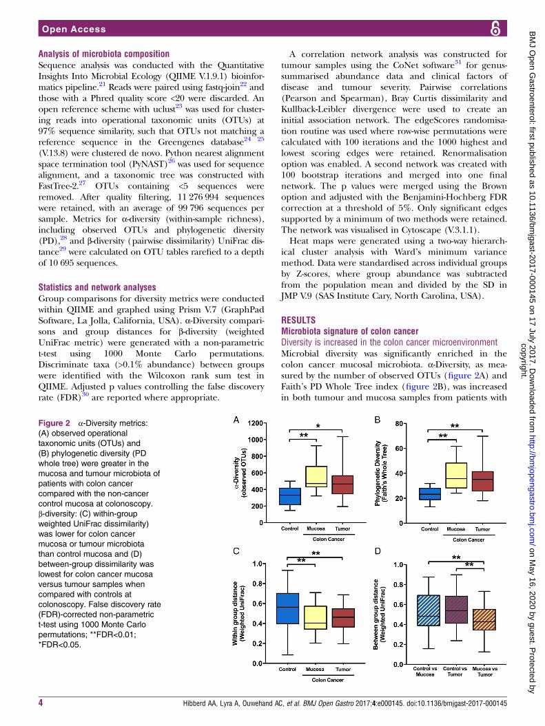
RESULTSMicrobiota signature of colon cancerDiversity is increased in the colon cancer microenvironmentMicrobial diversity was significantly enriched in thecolon cancer mucosal microbiota. α-Diversity, as mea-sured by the number of observed OTUs (figure 2A) andFaith’s PD Whole Tree index (figure 2B), was increasedin both tumour and mucosa samples from patients with
Figure 2 α-Diversity metrics:
(A) observed operational
taxonomic units (OTUs) and
(B) phylogenetic diversity (PD
whole tree) were greater in the
mucosa and tumour microbiota of
patients with colon cancer
compared with the non-cancer
control mucosa at colonoscopy.
β-diversity: (C) within-groupweighted UniFrac dissimilarity)
was lower for colon cancer
mucosa or tumour microbiota
than control mucosa and (D)
between-group dissimilarity was
lowest for colon cancer mucosa
versus tumour samples when
compared with controls at
colonoscopy. False discovery rate
(FDR)-corrected non-parametric
t-test using 1000 Monte Carlo
permutations; **FDR<0.01;
*FDR<0.05.
4 Hibberd AA, Lyra A, Ouwehand AC, et al. BMJ Open Gastro 2017;4:e000145. doi:10.1136/bmjgast-2017-000145
Open Accesscopyright.
on May 16, 2020 by guest. P
rotected byhttp://bm
jopengastro.bmj.com
/B
MJ O
pen Gastroenterol: first published as 10.1136/bm
jgast-2017-000145 on 17 July 2017. Dow
nloaded from
colon cancer compared with control samples (FDR≤0.05);however, no significant differences were found betweengroups for faecal samples (data not shown; FDR>0.1).β-Diversity based on the weighted UniFrac distancerevealed that the tissue microbiota was more similaramong patients with colon cancer within tumour ormucosa biopsies than among control samples (figure 2C).The microbiota composition from the mucosa adjacent tothe tumour was more similar to the tumour-associatedmicrobiota than to that of control samples (figure 2D).For patients with colon cancer, α-diversity or β-diversity didnot differ between the tumour-associated microbiota andthe macroscopically healthy appearing mucosa-associatedmicrobiota sampled from the same patients at a distanceof 10 cm from the tumour (FDR>0.1).
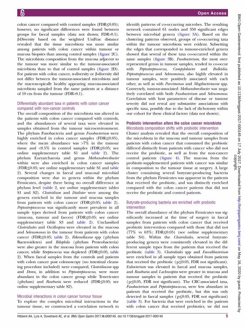
Differentially abundant taxa in patients with colon cancercompared with non-cancer controlsThe overall composition of the microbiota was altered inthe patients with colon cancer compared with controls,and the abundances of several taxa were elevated insamples obtained from the tumour microenvironment.The phylum Fusobacteria and genus Fusobacterium werehighly enriched in colon cancer samples (FDR≤0.05),where the mean abundance was >7% in the tumourtissue and <0.5% in control samples (FDR≤0.05; seeonline supplementary table S1 and table 2). Thephylum Euryarchaeota and genus Methanobrevibacterwithin were also enriched in colon cancer samples(FDR≤0.05; see online supplementary table S1 and table2). Several changes in faecal and mucosal microbialcomposition were due to genera within the phylumFirmicutes, despite there being no overall difference atphylum level (table 2, see online supplementary tablesS1 and S2). Clostridium and Dialister were among thegenera enriched in the tumour and mucosa samplesfrom patients with colon cancer (FDR≤0.05; table 2).Peptostreptococcus was significantly more prevalent in allsample types derived from patients with colon cancer(mucosa, tumour and faeces) (FDR≤0.05; see onlinesupplementary table S3 and table 2). UnclassifiedClostridiales and Oscillospira were elevated in the mucosaand Selenomonas in the tumour from patients with coloncancer (FDR≤0.05; table 2). Rikenellaceae spp (phylumBacteroidetes) and Bilophila (phylum Proteobacteria)were also greater in the mucosa from patients with coloncancer, while Streptococcus was depleted (FDR≤0.05, table2). When faecal samples from the controls and patientswith colon cancer post colonoscopy (no intestinal cleans-ing procedure included) were compared, Clostridiaceae sppand Dorea, in addition to Peptostreptococcus, were moreabundant in the colon cancer group while Tenericutes(phylum) and Roseburia were reduced (FDR≤0.05; seeonline supplementary table S2).
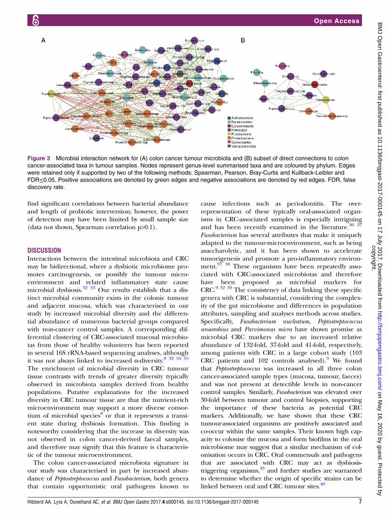
Microbial interactions in colon cancer tumour tissueTo explore the complex microbial interactions in thetumour tissue, we constructed a correlation network to
identify patterns of co-occurring microbes. The resultingnetwork contained 61 nodes and 350 significant edgesbetween microbial genera (figure 3A). Based on theclustering patterns observed, groups of co-occurring taxawithin the tumour microbiota were evident. Subsettingthe edges that corresponded to tumour-enriched generashowed that several of these taxa co-occurred within thesame samples (figure 3B). Fusobacterium, the most over-represented genus in tumour samples, tended to co-occurwith Peptostreptococcus, Campylobacter and Bulleidia.Peptostreptococcus and Selenomonas, also highly elevated intumour samples, were positively associated with eachother, as well as with Parvimonas and Mogibacteriaceae spp.Conversely, tumour-associated Methanobrevibacter was nega-tively correlated with both Fusobacterium and Selenomonas.Correlation with host parameters of disease or tumourseverity did not reveal any substantive associations withspecific taxa, possibly due to the lack of dichotomy withinour cohort for these clinical factors (data not shown).
Probiotic intervention alters the colon cancer microbiotaMicrobiota composition shifts with probiotic interventionCluster analysis revealed that the overall composition ofthe microbiota in the mucosa and tumour samples frompatients with colon cancer that consumed the probioticdiffered distinctly from patients with cancer who did notreceive the probiotic, as well as from the non-cancercontrol patients (figure 4). The mucosa from theprobiotic-supplemented patients with cancer was similarin composition to the tumour tissue at surgery. A largecluster containing several butyrate-producing bacteriafrom the phylum Firmicutes was apparent in the patientsthat received the probiotic, and was distinctly enrichedcompared with the colon cancer patients that did notreceive the probiotic and control patients.
Butyrate-producing bacteria are enriched with probioticinterventionThe overall abundance of the phylum Firmicutes was sig-nificantly increased at the time of surgery in faecalsamples from patients with colon cancer that receivedprobiotic intervention compared with those that did not(77% vs 63%; FDR≤0.05) (see online supplementarytable S4). Within the Clostridiales, several butyrate-producing genera were consistently elevated in the dif-ferent sample types from the patients that received theprobiotic (table 3). Clostridiales spp and Faecalibacteriumwere enriched in all sample types obtained from patientsthat received the probiotic (p≤0.05, FDR not significant).Eubacterium was elevated in faecal and mucosa samples,and Roseburia and Lachnospira were greater in mucosa andtumour samples in patients that received the probiotic(p≤0.05, FDR not significant). The CRC-associated taxa,Fusobacterium and Peptostreptococcus, were less abundant inpatients that received the probiotic, but this was onlydetected in faecal samples (p≤0.05, FDR not significant)(table 3). For bacteria that were enriched in the patientswith colon cancer that received probiotics, we did not
Hibberd AA, Lyra A, Ouwehand AC, et al. BMJ Open Gastro 2017;4:e000145. doi:10.1136/bmjgast-2017-000145 5
Open Accesscopyright.
on May 16, 2020 by guest. P
rotected byhttp://bm
jopengastro.bmj.com
/B
MJ O
pen Gastroenterol: first published as 10.1136/bm
jgast-2017-000145 on 17 July 2017. Dow
nloaded from
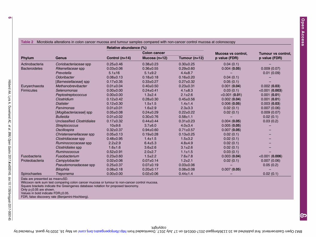
Table 2 Microbiota alterations in colon cancer mucosa and tumour samples compared with non-cancer control mucosa at colonoscopy
Phylum Genus
Relative abundance (%)
Colon cancer Mucosa vs control,
p value (FDR)
Tumour vs control,
p value (FDR)Control (n=14) Mucosa (n=12) Tumour (n=12)
Actinobacteria Coriobacteriaceae spp 0.25±0.46 0.38±0.23 0.30±0.25 0.04 (0.1) –
Bacteroidetes Rikenellaceae spp 0.03±0.06 0.36±0.55 0.29±0.60 0.004 (0.05) 0.009 (0.07)
Prevotella 5.1±16 5.1±9.2 4.4±8.7 – 0.01 (0.09)
Odoribacter 0.08±0.13 0.18±0.18 0.16±0.20 0.04 (0.1) –
[Barnesiellaceae] spp 0.17±0.35 0.33±0.27 0.27±0.32 0.05 (0.1) –
Euryarchaeota Methanobrevibacter 0.01±0.04 0.40±0.50 0.23±0.31 0.001 (0.04) 0.002 (0.03)
Firmicutes Selenomonas 0.00±0.00 0.24±0.41 4.1±8.3 0.03 (0.1) <0.001 (0.003)
Peptostreptococcus 0.00±0.00 1.3±2.4 2.1±2.6 <0.001 (0.01) 0.001 (0.01)
Clostridium 0.12±0.42 0.28±0.30 0.45±0.56 0.002 (0.04) 0.001 (0.01)
Dialister 0.12±0.30 1.5±1.5 1.4±1.4 0.006 (0.05) 0.003 (0.03)
Parvimonas 0.01±0.01 1.6±2.9 2.3±3.3 0.02 (0.1) 0.007 (0.06)
[Mogibacteriaceae] spp 0.05±0.08 0.24±0.29 0.22±0.22 0.02 (0.1) 0.009 (0.07)
Bulleidia 0.01±0.02 0.30±0.76 0.58±1.1 – 0.02 (0.1)
Unclassified Clostridiales 0.17±0.32 0.44±0.44 0.31±0.23 0.004 (0.05) 0.03 (0.2)
Streptococcus 10±9.6 3.7±6.0 4.0±3.4 0.005 (0.05) –
Oscillospira 0.32±0.37 0.94±0.60 0.71±0.57 0.007 (0.05) –
Christensenellaceae spp 0.05±0.13 0.19±0.28 0.13±0.25 0.02 (0.1) –
Clostridiaceae spp 0.48±0.95 1.4±1.5 1.5±3.2 0.02 (0.1) –
Ruminococcaceae spp 2.2±2.9 6.4±5.3 4.8±4.9 0.02 (0.1) –
Clostridiales spp 1.6±1.6 3.6±2.6 3.1±2.6 0.02 (0.1) –
Ruminococcus 0.52±0.91 2.0±2.7 1.1±1.5 0.03 (0.1) –
Fusobacteria Fusobacterium 0.23±0.60 1.5±2.2 7.6±7.8 0.003 (0.04) <0.001 (0.006)
Proteobacteria Campylobacter 0.02±0.06 0.07±0.14 1.2±2.1 0.02 (0.1) 0.007 (0.06)
Pseudomonadaceae spp 0.25±0.37 0.07±0.19 0.03±0.06 – 0.05 (0.2)
Bilophila 0.08±0.18 0.20±0.17 0.08±0.08 0.007 (0.05) –
Spirochaetes Treponema 0.00±0.00 0.02±0.06 0.44±1.4 – 0.02 (0.1)
Data are presented as mean±SD.Wilcoxon rank sum test comparing colon cancer mucosa or tumour to non-cancer control mucosa.Square brackets indicate the Greengenes database notation for proposed taxonomy.Only p≤0.05 are shown.Values in bold indicate FDR≤0.05.FDR, false discovery rate (Benjamini-Hochberg).
6Hibberd
AA,LyraA,Ouw
ehandAC,etal.BM
JOpen
Gastro2017;4:e000145.doi:10.1136/bm
jgast-2017-000145
OpenAccess
copyright. on May 16, 2020 by guest. Protected by http://bmjopengastro.bmj.com/ BMJ Open Gastroenterol: first published as 10.1136/bmjgast-2017-000145 on 17 July 2017. Downloaded from
find significant correlations between bacterial abundanceand length of probiotic intervention; however, the powerof detection may have been limited by small sample size(data not shown, Spearman correlation p>0.1).
DISCUSSIONInteractions between the intestinal microbiota and CRCmay be bidirectional, where a dysbiotic microbiome pro-motes carcinogenesis, or possibly the tumour micro-environment and related inflammatory state causemicrobial dysbiosis.32 33 Our results establish that a dis-tinct microbial community exists in the colonic tumourand adjacent mucosa, which was characterised in ourstudy by increased microbial diversity and the differen-tial abundance of numerous bacterial groups comparedwith non-cancer control samples. A corresponding dif-ferential clustering of CRC-associated mucosal microbio-tas from those of healthy volunteers has been reportedin several 16S rRNA-based sequencing analyses, althoughit was not always linked to increased α-diversity.8 32 34 35
The enrichment of microbial diversity in CRC tumourtissue contrasts with trends of greater diversity typicallyobserved in microbiota samples derived from healthypopulations. Putative explanations for the increaseddiversity in CRC tumour tissue are that the nutrient-richmicroenvironment may support a more diverse consor-tium of microbial species8 or that it represents a transi-ent state during dysbiosis formation. This finding isnoteworthy considering that the increase in diversity wasnot observed in colon cancer-derived faecal samples,and therefore may signify that this feature is characteris-tic of the tumour microenvironment.The colon cancer-associated microbiota signature in
our study was characterised in part by increased abun-dance of Peptostreptococcus and Fusobacterium, both generathat contain opportunistic oral pathogens known to
cause infections such as periodontitis. The over-representation of these typically oral-associated organ-isms in CRC-associated samples is especially intriguingand has been recently examined in the literature.36 37
Fusobacterium has several attributes that make it uniquelyadapted to the tumour-microenvironment, such as beingasaccharolytic, and it has been shown to acceleratetumorigenesis and promote a pro-inflammatory environ-ment.37 38 These organisms have been repeatedly asso-ciated with CRC-associated microbiotas and thereforehave been proposed as microbial markers forCRC.9 32 39 The consistency of data linking these specificgenera with CRC is substantial, considering the complex-ity of the gut microbiome and differences in populationattributes, sampling and analyses methods across studies.Specifically, Fusobacterium nucleatum, Peptostreptococcusanaerobius and Parvimonas micra have shown promise asmicrobial CRC markers due to an increased relativeabundance of 132-fold, 37-fold and 41-fold, respectively,among patients with CRC in a large cohort study (103CRC patients and 102 controls analysed).9 We foundthat Peptostreptococcus was increased in all three coloncancer-associated sample types (mucosa, tumour, faeces)and was not present at detectible levels in non-cancercontrol samples. Similarly, Fusobacterium was elevated over30-fold between tumour and control biopsies, supportingthe importance of these bacteria as potential CRCmarkers. Additionally, we have shown that these CRCtumour-associated organisms are positively associated andco-occur within the same samples. Their known high cap-acity to colonise the mucosa and form biofilms in the oralmicrobiome may suggest that a similar mechanism of col-onisation occurs in CRC. Oral commensals and pathogensthat are associated with CRC may act as dysbiosis-triggering organisms,33 and further studies are warrantedto determine whether the origin of specific strains can belinked between oral and CRC tumour sites.40
Figure 3 Microbial interaction network for (A) colon cancer tumour microbiota and (B) subset of direct connections to colon
cancer-associated taxa in tumour samples. Nodes represent genus-level summarised taxa and are coloured by phylum. Edges
were retained only if supported by two of the following methods: Spearman, Pearson, Bray-Curtis and Kullback-Leibler and
FDR≤0.05. Positive associations are denoted by green edges and negative associations are denoted by red edges. FDR, false
discovery rate.
Hibberd AA, Lyra A, Ouwehand AC, et al. BMJ Open Gastro 2017;4:e000145. doi:10.1136/bmjgast-2017-000145 7
Open Accesscopyright.
on May 16, 2020 by guest. P
rotected byhttp://bm
jopengastro.bmj.com
/B
MJ O
pen Gastroenterol: first published as 10.1136/bm
jgast-2017-000145 on 17 July 2017. Dow
nloaded from
Additional differentially abundant taxa over-representedin colon cancer tumour and mucosa samples includedthe phyla Tenericutes and Euryarchaeota as well as thegenus Methanobrevibacter. Tenericutes was found to be lessabundant compared with controls in faecal samples
suggesting the colon cancer association was localised tothe mucosal surface or that different organisms weredetected from this phylum between the sample types.Both Tenericutes and Methanobrevibacter have previouslybeen associated with adenoma or CRC microbiotas, and
Figure 4 Heat map with
two-way hierarchical clustering of
genus-summarised microbiota
abundance and sample grouping
for mucosal and tumour
microbiotas from patients with
colon cancer at colonoscopy and
surgery (with or without
probiotics), and for non-cancer
control mucosal microbiota at
colonoscopy.
8 Hibberd AA, Lyra A, Ouwehand AC, et al. BMJ Open Gastro 2017;4:e000145. doi:10.1136/bmjgast-2017-000145
Open Accesscopyright.
on May 16, 2020 by guest. P
rotected byhttp://bm
jopengastro.bmj.com
/B
MJ O
pen Gastroenterol: first published as 10.1136/bm
jgast-2017-000145 on 17 July 2017. Dow
nloaded from
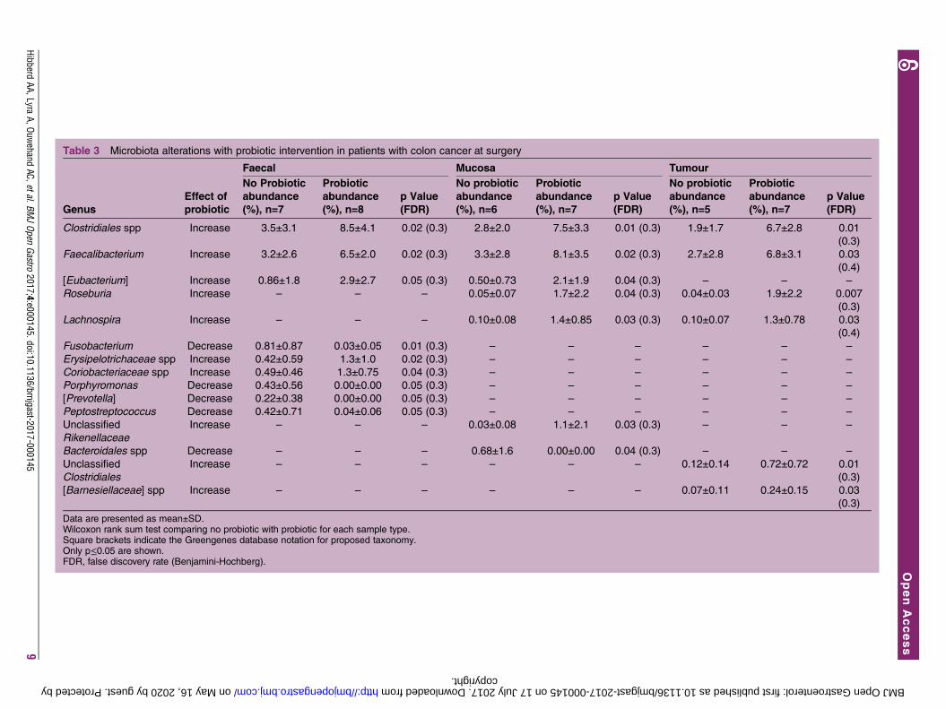
Table 3 Microbiota alterations with probiotic intervention in patients with colon cancer at surgery
Genus
Effect of
probiotic
Faecal Mucosa Tumour
No Probiotic
abundance
(%), n=7
Probiotic
abundance
(%), n=8
p Value
(FDR)
No probiotic
abundance
(%), n=6
Probiotic
abundance
(%), n=7
p Value
(FDR)
No probiotic
abundance
(%), n=5
Probiotic
abundance
(%), n=7
p Value
(FDR)
Clostridiales spp Increase 3.5±3.1 8.5±4.1 0.02 (0.3) 2.8±2.0 7.5±3.3 0.01 (0.3) 1.9±1.7 6.7±2.8 0.01
(0.3)
Faecalibacterium Increase 3.2±2.6 6.5±2.0 0.02 (0.3) 3.3±2.8 8.1±3.5 0.02 (0.3) 2.7±2.8 6.8±3.1 0.03
(0.4)
[Eubacterium] Increase 0.86±1.8 2.9±2.7 0.05 (0.3) 0.50±0.73 2.1±1.9 0.04 (0.3) – – –
Roseburia Increase – – – 0.05±0.07 1.7±2.2 0.04 (0.3) 0.04±0.03 1.9±2.2 0.007
(0.3)
Lachnospira Increase – – – 0.10±0.08 1.4±0.85 0.03 (0.3) 0.10±0.07 1.3±0.78 0.03
(0.4)
Fusobacterium Decrease 0.81±0.87 0.03±0.05 0.01 (0.3) – – – – – –
Erysipelotrichaceae spp Increase 0.42±0.59 1.3±1.0 0.02 (0.3) – – – – – –
Coriobacteriaceae spp Increase 0.49±0.46 1.3±0.75 0.04 (0.3) – – – – – –
Porphyromonas Decrease 0.43±0.56 0.00±0.00 0.05 (0.3) – – – – – –
[Prevotella] Decrease 0.22±0.38 0.00±0.00 0.05 (0.3) – – – – – –
Peptostreptococcus Decrease 0.42±0.71 0.04±0.06 0.05 (0.3) – – – – – –
Unclassified
Rikenellaceae
Increase – – – 0.03±0.08 1.1±2.1 0.03 (0.3) – – –
Bacteroidales spp Decrease – – – 0.68±1.6 0.00±0.00 0.04 (0.3) – – –
Unclassified
Clostridiales
Increase – – – – – – 0.12±0.14 0.72±0.72 0.01
(0.3)
[Barnesiellaceae] spp Increase – – – – – – 0.07±0.11 0.24±0.15 0.03
(0.3)
Data are presented as mean±SD.Wilcoxon rank sum test comparing no probiotic with probiotic for each sample type.Square brackets indicate the Greengenes database notation for proposed taxonomy.Only p≤0.05 are shown.FDR, false discovery rate (Benjamini-Hochberg).
HibberdAA,Lyra
A,Ouwehand
AC,etal.BMJOpen
Gastro2017;4:e000145.doi:10.1136/bm
jgast-2017-0001459
OpenAccess
copyright. on May 16, 2020 by guest. Protected by http://bmjopengastro.bmj.com/ BMJ Open Gastroenterol: first published as 10.1136/bmjgast-2017-000145 on 17 July 2017. Downloaded from
the phylum Tenericutes includes parasitic pathogens(Mollicutes) that have previously been suspected as causalagents in other cancers.8 34 41 Methanobrevibacter has beenlinked to a multitude of intestinal disorders, periodontitisand CRC, although the mechanism of CRC association isunknown.42 It was negatively associated with Fusobacteriumin our network analysis, suggesting a different mechanismof involvement in CRC. Other tumour-associated generawere from within Firmicutes (Selenomonas, Clostridium,Dialister and Parvimonas); however, contradictory findingshave been published on their presence in adenoma andcarcinoma-associated tissue.5 34 The non-cancer controlsdisplayed the greatest amount of within-group variabilityin microbiota composition; however, the increased abun-dance of genus Streptococcus was evident in control partici-pants’ biopsies relative to colon cancer. Streptococcus bovishas previously been associated with CRC tumours,32 35 butin our data this elevation appears to be primarily attribu-ted to sequences related to Streptococcus thermophilus. Itcould be interesting to further investigate whether S. ther-mophilus may have protective properties in healthy popula-tions or if it is solely depleted in patients with coloncancer. The control patients were not diagnosed withcolon cancer; however, colonoscopy was performed dueto various gastrointestinal complaints with manifest symp-toms, which may partially explain the high level of vari-ability among their microbiota profiles.Overall, the composition of the microbiota in samples
from patients with colon cancer that received probioticshad a unique profile characterised by an increasedabundance of butyrate-producing bacteria in tumour,mucosa and faecal samples compared with patients withcancer who did not receive probiotics. Butyrate interactsintimately with colonic epithelial cells as an energysource for colonocytes and by modulating signallingpathways. It plays a beneficial role in colon cancer byinhibiting cell proliferation, reducing IFN-γ-mediatedinflammation and promoting cell apoptosis and tumoursuppressor gene expression.43–45 Clostridiales spp andbutyrate-producing Faecalibacterium, Roseburia andEubacterium were enriched in samples obtained frompatients with colon cancer with probiotic intervention.Despite the FDR-corrected p values not reaching statis-tical significance, this finding was detected consistentlyin tumour, mucosa and faecal samples. A depletion ofbutyrate-producing bacteria in the microbiota has beenreported in patients with various stages of CRC progres-sion,46–48 and butyrate’s tumour-suppressive propertieshave been shown to be directly mediated by the gutmicrobiota, further supporting its importance in CRC.49
Additionally, the faecal microbiota of patients with coloncancer taking probiotics had reduced levels ofCRC-associated genera Fusobacterium and Peptostreptococcusaccording to the non-FDR-corrected p value. Thisfinding is in accordance with a previous probiotic inter-vention trial where supplementation with Bifidobacteriumlongum, Lactobacillus acidophilus and Enterococcus faecalisreduced Fusobacterium and Peptostreptococcus in CRC
patients to a level comparable to healthy controls.39 Asthese two genera are strongly associated with CRC micro-biota in several studies, these results emphasise the valueof evaluating probiotics for CRC prevention and care.Moreover, probiotics have also been shown to mediateinflammatory responses, as Gianotti and colleaguesobserved that the mucosal colonisation of probioticstrain Lactobacillus johnsonii La1 was correlated withreduced proliferation and modulation of specific den-dritic cells in CRC.50 Unfortunately, we were unable toachieve the level of sensitivity necessary to differentiallydetect colonisation of our specific probiotic strains fromthe native populations by qPCR in the mucosal samplesfrom this study (data not shown).The difficulty in obtaining intestinal mucosal and
tumour samples as compared with faecal sampling formicrobiota analysis presented challenges in the studydesign and sampling. Patients underwent bowel cleans-ing prior to colonoscopy but no bowel preparation wasdone prior to surgery, and the timing of the faecalsample collection post colonoscopy was not controlled,both of which may influence the microbiota profiles.Additionally, the intervention length varied amongpatients as it would have been unethical to restrict thetime to their surgery. We therefore chose to focus pri-marily on comparisons among samples that wereobtained either at the time of colonoscopy or at surgery.In a future study, it would be preferable to more strin-gently control participant groups, but we avoided this inthe pilot trial primarily for ethical reasons. Despite theselimitations, by obtaining tumoral, mucosal and faecalsamples, we assessed the colon cancer-associated micro-biota by several comparisons: (1) tumour to mucosawithin close proximity; (2) the aforementioned to non-cancer control samples; (3) colon cancer faeces to non-cancer control faeces; (4) colon cancer probiotic inter-vention to no probiotic intervention. Fewer differentiallyabundant taxa were detected in the faecal microbiotathan the corresponding tissue microbiota, suggestingthat the tissue microbiota profile is more informative foridentifying putative microbial markers of colon cancer.Bacterial adherence to the intestinal epithelium orbiofilm formation may have contributed to the differ-ences we observed between the tissue and faecalsamples, but tissue samples more likely represent organ-isms that directly interact with host and immune cellsand are thus preferable to more easily obtained faecalsamples. Peptostreptococcus, however, was significantlyenriched in both tissue and faecal samples and showspromise as a microbial CRC marker.The results of this study support the hypothesis that
the colon cancer-associated microbiota can be manipu-lated by specific probiotic strains, resulting in an alteredmicrobiota enriched with beneficial bacteria. Our studyprovides evidence that microbiota modulation by probio-tics could be considered as part of a therapeutic regimefor CRC patients. Further studies should be conductedin a larger population to confirm these initial findings,
10 Hibberd AA, Lyra A, Ouwehand AC, et al. BMJ Open Gastro 2017;4:e000145. doi:10.1136/bmjgast-2017-000145
Open Accesscopyright.
on May 16, 2020 by guest. P
rotected byhttp://bm
jopengastro.bmj.com
/B
MJ O
pen Gastroenterol: first published as 10.1136/bm
jgast-2017-000145 on 17 July 2017. Dow
nloaded from
and ideally should be complemented with metabolomicsdata to elucidate the role of butyrate. Future studiescould also expand beyond the microbiota to include thepotential influences of fungi (mycobiome), viruses(virome) and microbial bioactive molecules on CRCdevelopment. The interplay between diet, microbiotaand host in maintaining homeostasis is an importantconsideration in therapeutic strategies for CRC, and ana-lysis of the microbiome is a critical component in under-standing how these complex interactions influence thedevelopment and progression of carcinogenesis.
Acknowledgements The authors thank Ann-Louise Helminen and HilleviBjörkqvist for inclusion of study subjects and collection of samples, LenaMunro for work with the clinical database and Jaqueline Flach, MarianneÅkerström, and Elisabeth Odin for handling of samples. The authors alsothank Nicolas Yeung, Krista Salli, Paige Roos and Wes Morovic for technicalwork in preparing the samples for sequencing and Buffy Stahl for supportingproject management.
Contributors YW, PR, AL, ACO and HL designed and executed the clinicalresearch study. YW was the principal investigator. AAH analysed the data andperformed statistics. AAH, AL, ACO and YW interpreted the data. AL and YWsupported data integration and project management. LC contributed expertiseon ProBion Clinica. AAH, AL and YW wrote the manuscript. All authorsreviewed and approved the final manuscript.
Funding This work was supported by a grant from the Swedish state underthe LUA/ALF agreement (grant number ALFGBG-542821).
Competing interests At the time of the research, AAH, AL and ACO wereemployees of DuPont, who manufactures the probiotic strains used, and LC isan employee of Wasa Medicals who produced the probiotic product used inthis study.
Patient consent Obtained.
Ethics approval The study was approved by the Regional Ethical ReviewBoard in Gothenburg under study number 233-10.
Provenance and peer review Not commissioned; externally peer reviewed.
Data sharing statement No additional data are available.
Open Access This is an Open Access article distributed in accordance withthe Creative Commons Attribution Non Commercial (CC BY-NC 4.0) license,which permits others to distribute, remix, adapt, build upon this work non-commercially, and license their derivative works on different terms, providedthe original work is properly cited and the use is non-commercial. See: http://creativecommons.org/licenses/by-nc/4.0/
REFERENCES1. Torre LA, Bray F, Siegel RL, et al. Global cancer statistics, 2012. CA
Cancer J Clin 2015;65:87–108.2. Mármol I, Sánchez-de-Diego C, Pradilla Dieste A, et al. Colorectal
carcinoma: a general overview and future perspectives in colorectalcancer. Int J Mol Sci 2017;18:pii:E197.
3. Huxley RR, Ansary-Moghaddam A, Clifton P, et al. The impact ofdietary and lifestyle risk factors on risk of colorectal cancer: aquantitative overview of the epidemiological evidence. Int J Cancer2009;125:171–80.
4. Nistal E, Fernández-Fernández N, Vivas S, et al. Factorsdetermining colorectal cancer: The role of the intestinal microbiota.Front Oncol 2015;5:220.
5. Nakatsu G, Li X, Zhou H, et al. Gut mucosal microbiome acrossstages of colorectal carcinogenesis. Nat Commun 2015;6:8727.
6. Burns MB, Lynch J, Starr TK, et al. Virulence genes are a signatureof the microbiome in the colorectal tumor microenvironment.Genome Med 2015;7:55.
7. Geng J, Fan H, Tang X, et al. Diversified pattern of the humancolorectal cancer microbiome. Gut Pathog 2013;5:2.
8. Mira-Pascual L, Cabrera-Rubio R, Ocon S, et al. Microbial mucosalcolonic shifts associated with the development of colorectal cancer
reveal the presence of different bacterial and archaeal biomarkers.J Gastroenterol 2015;50:167–79.
9. Wong SH, Kwong TN, Chow TC, et al. Quantitation of faecalFusobacterium improves faecal immunochemical test in detectingadvanced colorectal neoplasia. Gut 2016;•••. http://dx.doi.org/10.1136/gutjnl-2016-312766
10. Pagnini C, Corleto VD, Mangoni ML, et al. Alteration of localmicroflora and alpha-defensins hyper-production in colonic adenomamucosa. J Clin Gastroenterol 2011;45:602–10.
11. Hill C, Guarner F, Reid G, et al. Expert consensus document. TheInternational Scientific Association for Probiotics and Prebioticsconsensus statement on the scope and appropriate use of the termprobiotic. Nat Rev Gastroenterol Hepatol 2014;11:506–14.
12. Zhu Y, Michelle Luo T, Jobin C, et al. Gut microbiota and probioticsin colon tumorigenesis. Cancer Lett 2011;309:119–27.
13. Chen CC, Lin W-C, Kong M-S, et al. Oral inoculation of probioticsLactobacillus acidophilus NCFM suppresses tumour growth both insegmental orthotopic colon cancer and extra-intestinal tissue. BrJ Nutr 2012;107:1623–34.
14. Goldin BR, Gorbach SL. The effect of oral administration ofLactobacillus and antibiotics on intestinal bacterial activity andchemical induction of large bowel tumors. Dev Indus Microbiol1984;25:139–50.
15. Foligne B, Nutten S, Grangette C, et al. Correlation between in vitroand in vivo immunomodulatory properties of lactic acid bacteria.World J Gastroenterol 2007;13:236–43.
16. Compton C, Fenoglio-Preiser CM, Pettigrew N, et al. American jointcommittee on cancer prognostic factors consensus conference:colorectal working group. Cancer 2000;88:1739–57.
17. Costabile A, Fava F, Röytiö H, et al. Impact of polydextrose on thefaecal microbiota: a double-blind, crossover, placebo-controlledfeeding study in healthy human subjects. Br J Nutr 2012;108:471–81.
18. Lyra A, Forssten S, Rolny P, et al. Comparison of bacterial quantitiesin left and right colon biopsies and faeces. World J Gastroenterol2012;18:4404–11.
19. Caporaso JG, Lauber CL, Walters WA, et al. Global patterns of 16SrRNA diversity at a depth of millions of sequences per sample. ProcNatl Acad Sci USA 2011;108(Suppl 1):4516–22.
20. Caporaso JG, Lauber CL, Walters WA, et al. Ultra-high-throughputmicrobial community analysis on the Illumina HiSeq and MiSeqplatforms. ISME J 2012;6:1621–4.
21. Caporaso JG, Kuczynski J, Stombaugh J, et al. QIIME allowsanalysis of high-throughput community sequencing data. NatMethods 2010;7:335–6.
22. Aronesty E. ea-utils: Command-line tools for processing biologicalsequencing data. http://code.google.com/p/ea-utils. 2011.
23. Edgar RC. Search and clustering orders of magnitude faster thanBLAST. Bioinformatics 2010;26:2460–1.
24. DeSantis TZ, Hugenholtz P, Larsen N, et al. Greengenes, achimera-checked 16S rRNA gene database and workbenchcompatible with ARB. Appl Environ Microbiol 2006;72:5069–72.
25. McDonald D, Price MN, Goodrich J, et al. An improved Greengenestaxonomy with explicit ranks for ecological and evolutionaryanalyses of bacteria and archaea. ISME J 2012;6:610–18.
26. Caporaso JG, Bittinger K, Bushman FD, et al. PyNAST: a flexibletool for aligning sequences to a template alignment. Bioinformatics2010;26:266–7.
27. Price MN, Dehal PS, Arkin AP. FastTree 2--approximatelymaximum-likelihood trees for large alignments. PLoS ONE 2010;5:e9490.
28. Faith DP. Conservation evaluation and phylogenetic diversity. BiolConserv 1992;61:1–10.
29. Lozupone C, Knight R. UniFrac: a new phylogenetic method forcomparing microbial communities. Appl Environ Microbiol2005;71:8228–35.
30. Benjamini Y, Hochberg Y. Controlling the false discovery rate: apractical and powerful approach to multiple testing. J R Statist Soc B1995;57:289–300.
31. Faust K, Sathirapongsasuti JF, Izard J, et al. Microbialco-occurrence relationships in the human microbiome. PLoSComput Biol 2012;8:e1002606.
32. Gagnière J, Raisch J, Veziant J, et al. Gut microbiota imbalance andcolorectal cancer. World J Gastroenterol 2016;22:501–18.
33. Hajishengallis G, Darveau RP, Curtis MA. The keystone-pathogenhypothesis. Nat Rev Microbiol 2012;10:717–25.
34. Lu Y, Chen J, Zheng J, et al. Mucosal adherent bacterial dysbiosisin patients with colorectal adenomas. Sci Rep 2016;6:26337.
35. Chen W, Liu F, Ling Z, et al. Human intestinal lumen andmucosa-associated microbiota in patients with colorectal cancer.PLoS ONE 2012;7:e39743.
Hibberd AA, Lyra A, Ouwehand AC, et al. BMJ Open Gastro 2017;4:e000145. doi:10.1136/bmjgast-2017-000145 11
Open Accesscopyright.
on May 16, 2020 by guest. P
rotected byhttp://bm
jopengastro.bmj.com
/B
MJ O
pen Gastroenterol: first published as 10.1136/bm
jgast-2017-000145 on 17 July 2017. Dow
nloaded from
36. Colucci F. An oral commensal associates with disease: chicken,egg, or red herring? Immunity 2015;42:208–10.
37. Flynn KJ, Baxter NT, Schloss PD. Metabolic and community synergyof oral bacteria in colorectal cancer. mSphere 2016;1;pii:e00102-16.
38. Kostic AD, Chun E, Robertson L, et al. Fusobacterium nucleatumpotentiates intestinal tumorigenesis and modulates thetumor-immune microenvironment. Cell Host Microbe2013;14:207–15.
39. Gao Z, Guo B, Gao R, et al. Probiotics modify human intestinalmucosa-associated microbiota in patients with colorectal cancer. MolMed Rep 2015;12:6119–27.
40. Favia G, Maiorano E, Lo Muzio L. Image of the month. Gingivalmetastasis from colonic adenocarcinoma. Clin Gastroenterol Hepatol2010;8:A28.
41. Zarei O, Rezania S, Mousavi A. Mycoplasma genitalium and cancer:a brief review. Asian Pac J Cancer Prev 2013;14:3425–8.
42. Gaci N, Borrel G, Tottey W, et al. Archaea and the human gut: newbeginning of an old story. World J Gastroenterol 2014;20:16062–78.
43. Hague A, Manning AM, Hanlon KA, et al. Sodium butyrateinduces apoptosis in human colonic tumour cell lines in ap53-independent pathway: implications for the possible role ofdietary fibre in the prevention of large-bowel cancer. Int J Cancer1993;55:498–505.
44. Zeng H, Lazarova DL, Bordonaro M. Mechanisms linking dietaryfiber, gut microbiota and colon cancer prevention. WorldJ Gastrointest Oncol 2014;6:41–51.
45. Zimmerman MA, Singh N, Martin PM, et al. Butyrate suppressescolonic inflammation through HDAC1-dependent Fas upregulationand Fas-mediated apoptosis of T cells. Am J Physiol GastrointestLiver Physiol 2012;302:G1405–15.
46. Chen HM, Yu YN, Wang JL, et al. Decreased dietary fiber intakeand structural alteration of gut microbiota in patients with advancedcolorectal adenoma. Am J Clin Nutr 2013;97:1044–52.
47. Weir TL, Manter DK, Sheflin AM, et al. Stool microbiome andmetabolome differences between colorectal cancer patients andhealthy adults. PLoS ONE 2013;8:e70803.
48. Wang T, Cai G, Qiu Y, et al. Structural segregation of gut microbiotabetween colorectal cancer patients and healthy volunteers. ISME J2012;6:320–9.
49. Donohoe DR, Holley D, Collins LB, et al. A gnotobiotic mouse modeldemonstrates that dietary fiber protects against colorectaltumorigenesis in a microbiota- and butyrate-dependent manner.Cancer Discov 2014;4:1387–97.
50. Gianotti L, Morelli L, Galbiati F, et al. A randomized double-blind trialon perioperative administration of probiotics in colorectal cancerpatients. World J Gastroenterol 2010;16:167–75.
12 Hibberd AA, Lyra A, Ouwehand AC, et al. BMJ Open Gastro 2017;4:e000145. doi:10.1136/bmjgast-2017-000145
Open Accesscopyright.
on May 16, 2020 by guest. P
rotected byhttp://bm
jopengastro.bmj.com
/B
MJ O
pen Gastroenterol: first published as 10.1136/bm
jgast-2017-000145 on 17 July 2017. Dow
nloaded from