oxalate-degrading activity in bifidobacterium …aem.asm.org/content/76/16/5609.full.pdf ·...
TRANSCRIPT

APPLIED AND ENVIRONMENTAL MICROBIOLOGY, Aug. 2010, p. 5609–5620 Vol. 76, No. 160099-2240/10/$12.00 doi:10.1128/AEM.00844-10Copyright © 2010, American Society for Microbiology. All Rights Reserved.
Oxalate-Degrading Activity in Bifidobacterium animalis subsp. lactis:Impact of Acidic Conditions on the Transcriptional Levels of the
Oxalyl Coenzyme A (CoA) Decarboxylase and Formyl-CoATransferase Genes�
Silvia Turroni, Claudia Bendazzoli, Samuele C. F. Dipalo, Marco Candela, Beatrice Vitali,Roberto Gotti, and Patrizia Brigidi*
Department of Pharmaceutical Sciences, University of Bologna, Via Belmeloro 6, 40126 Bologna, Italy
Received 7 April 2010/Accepted 22 June 2010
Oxalic acid occurs extensively in nature and plays diverse roles, especially in pathological processes. Due toits highly oxidizing effects, hyperabsorption or abnormal synthesis of oxalate can cause serious acute disordersin mammals and can be lethal in extreme cases. Intestinal oxalate-degrading bacteria could therefore bepivotal in maintaining oxalate homeostasis and reducing the risk of kidney stone development. In this study,the oxalate-degrading activities of 14 bifidobacterial strains were measured by a capillary electrophoresistechnique. The oxc gene, encoding oxalyl-coenzyme A (CoA) decarboxylase, a key enzyme in oxalate catabolism,was isolated by probing a genomic library of Bifidobacterium animalis subsp. lactis BI07, which was one of themost active strains in the preliminary screening. The genetic and transcriptional organization of oxc flankingregions was determined, unraveling the presence of two other independently transcribed open reading frames,potentially responsible for the ability of B. animalis subsp. lactis to degrade oxalate. pH-controlled batchfermentations revealed that acidic conditions were a prerequisite for a significant oxalate degradation rate,which dramatically increased in cells first adapted to subinhibitory concentrations of oxalate and then exposedto pH 4.5. Oxalate-preadapted cells also showed a strong induction of the genes potentially involved in oxalatecatabolism, as demonstrated by a transcriptional analysis using quantitative real-time reverse transcription-PCR. These findings provide new insights into the characterization of oxalate-degrading probiotic bacteria andmay support the use of B. animalis subsp. lactis as a promising adjunct for the prophylaxis and managementof oxalate-related kidney disease.
Oxalate is a normal end product of amino acid metabolismand must be excreted, predominantly via the kidney, to main-tain homeostasis (22). Oxalate is also present in a wide rangeof foods and drinks, and the normal dietary intake is variable,ranging from 70 to 920 mg per day (23). Because of its highlyoxidizing effects and the capability to combine with cations toform insoluble salts, this organic dicarboxylate is extremelytoxic for most forms of life. In humans, oxalate can cause avariety of pathological disorders, including hyperoxaluria, uro-lithiasis, cardiomyopathy, and renal failure (29, 41, 59). Hyper-oxaluria is the single strongest promoter of kidney stone for-mation, whose medical management represents a burden tothe individual patient as well as the health care system (49).The lack of new medications and the continued poor compli-ance with drug therapy have led to a growing interest in dietarymanipulation and novel therapies aimed at preventing recur-rent stone formation. Unfortunately, an oxalate-free diet isdifficult to achieve and would probably be deficient in essentialnutrients. Hence, other approaches to reducing urinary oxalatefor management of stone disease have been explored.
The discovery of oxalate-degrading bacteria within the hu-
man gastrointestinal tract has opened the way to a flurry ofresearch regarding their potential role in reducing urinary ex-cretion of oxalic acid (12, 20, 21, 25, 26, 30, 31, 35, 40, 44, 45).The first intestinal oxalate-degrading bacterium to be de-scribed was Oxalobacter formigenes, an obligate anaerobewhich relies exclusively on oxalate metabolism for energy (2).In O. formigenes, oxalate is decarboxylated to give CO2 andformate in a two-step pathway that is mediated by the coupledaction of the enzymes formyl-coenzyme A (CoA) transferase(Frc) (6) and oxalyl-CoA decarboxylase (Oxc) (7). Frc cata-lyzes the transfer of CoA from formate to oxalate, thus acti-vating the oxalyl moiety for the thiamine-dependent decarbox-ylation mediated by Oxc. Several studies have established adirect correlation between the disappearance of O. formigenesfrom the intestinal microbiota and the appearance of hyper-oxaluria symptoms, confirming its major role in maintainingoxalate homeostasis and making it the leading probiotic can-didate for the management of kidney stone disease (21, 25, 26,30, 44, 45, 49). However, the complicated growth requirementsof O. formigenes, with the need to determine the optimal di-etary oxalate level required to maintain its colonization, se-verely limited its administration (47).
Probiotic use of Lactobacillus and Bifidobacterium strainsthat metabolize oxalate might provide a valid alternative to O.formigenes. Bifidobacteria and lactobacilli are common inhab-itants of mammalian guts, including that of humans (55). Somestrains of these genera are known to have health-promoting
* Corresponding author. Mailing address: Department of Pharma-ceutical Sciences, University of Bologna, Via Belmeloro 6, 40126 Bo-logna, Italy. Phone: 39 051 2099743. Fax: 39 051 2099734. E-mail:[email protected].
� Published ahead of print on 2 July 2010.
5609
on August 22, 2018 by guest
http://aem.asm
.org/D
ownloaded from
effects and have been added for decades as functional ingre-dients in food products (48). In particular, strains of Bifidobac-terium animalis subsp. lactis are widely used in dairy productsin North America and Europe (8) due to several benefitsassociated with their administration, such as improvement ofvarious physiological conditions, modulation of the host im-mune response, and reduction of certain risks that impacthealth (37).
Increasing reports documented a significant reduction inurinary oxalate levels associated with probiotic administrationin both animals and humans (12, 31, 35, 39, 40, 58) and sug-gested the existence of oxalate-degrading activity in Bifidobac-terium and Lactobacillus species. Azcarate-Peril et al. (5) iden-tified a cluster of genes encoding the Oxc and Frc proteins inLactobacillus acidophilus NCFM and showed that mildly acidicconditions were a prerequisite for their transcription. Similarly,an oxalate-dependent induction of the oxc gene in cells firstadapted to subinhibitory concentrations of oxalate and thenexposed to pH 5.5 was also observed in Lactobacillus gasseriATCC 33323 (34). Moreover, Turroni et al. (51) screened theoxalate-degrading ability of several lactobacilli isolated fromfunctional foods and pharmaceutical preparations and demon-strated the functionality of L. acidophilus LA14 Oxc and Frc inoxalate catabolism.
To the best of our knowledge, only one study has so farinvestigated the potential role of Bifidobacterium in the hu-man intestinal degradation of oxalate (19). In that study, theoxalate-degrading capacity of 12 bifidobacterial strains wasevaluated and the first in-depth genetic and functional charac-terization of Oxc from B. animalis subsp. lactis DSM 10140 wasprovided. However, until now no information has existed aboutother bifidobacterial genes potentially involved in oxalate deg-radation and their regulation at the transcriptional level underdifferent culture conditions.
In the present work, a sensitive and selective capillary elec-trophoretic method was applied to determine the oxalate-de-grading activities in several Bifidobacterium strains. Among themost active strains, B. animalis subsp. lactis BI07 was selectedfor further analysis. Putative genes encoding Oxc and Frc wereidentified and characterized. The impact of acidic conditionson oxalate degradation and the expression levels of these pre-dicted genes were evaluated.
MATERIALS AND METHODS
Bacterial strains and media. The bacterial strains used in this study were thefollowing: Bifidobacterium adolescentis ATCC 15703, B. animalis subsp. lactisDSM 10140, BA30, Bb12, BI07, and L15, B. bifidum S16, B. breve ATCC 15700and BBSF, B. catenulatum B665, B. longum biotype longum S123, ATCC 15707,and W11, and B. longum biotype suis ATCC 27533. The B, L, S, and W strainsbelonged to the collection of our laboratory. In particular, B. bifidum S16 (15),B. catenulatum B665, B. longum biotype longum S123 (13), and B. animalis subsp.lactis BA30 (14) had been isolated from human feces, whereas B. breve BBSF andB. animalis subsp. lactis BI07 and L15 had been isolated from commercialprobiotic products (15). B. longum biotype longum W11 and B. animalis subsp.lactis Bb12 were kindly provided by Alfa Wassermann (Bologna, Italy) andChristian Hansen A/S (Hørsholm, Denmark), respectively. All strains were ver-ified for genus and species by 16S rRNA gene sequence typing.
Bifidobacteria were grown anaerobically at 37°C in the nutritionally complexmedium (NCM) described by Mlobeli et al. (38), containing (in grams per liter)tryptone (Oxoid, Hampshire, United Kingdom), 10.0; peptone water, 5.0; yeastextract (Oxoid), 5.0; Tween 80, 0.5; NaCl, 4.5; KCl, 0.5; MgCl2 � 6H2O, 0.15;KH2PO4, 0.4; NH4Cl, 1.0; and cysteine-HCl, 1.2. Peptone water was prepared asfollows (in g/liter): bacteriological peptone (Oxoid), 10.0; NaCl, 5.0; and Tween
80, 1.0 (pH 7.2). The pH of the medium was adjusted to 6.5 before autoclaving.Solid medium was prepared by the addition of 1.5% (wt/vol) agar (Oxoid) toNCM. Sucrose (20 g/liter) was used as an energy source, sterilized separately,and aseptically added to the culture medium. Anaerobic conditions wereachieved in anaerobic jars supplemented with a pad of Anaerocult A (Merck,Milan, Italy).
Capillary electrophoresis analysis of oxalate-degrading activity and sucroseconsumption in Bifidobacterium strains. Bifidobacterium strains were cultured for5 days with 5 mM sodium oxalate. B. animalis subsp. lactis DSM 10140, for whichoxalate-degrading activity had already been demonstrated (19), was used as apositive control. As a negative control, B. adolescentis ATCC 15703, whosecomplete genome sequence does not harbor oxc-related genes (NCBI accessionno. NC-008618), was chosen. Uninoculated broth medium was utilized as theexperimental control. Residual oxalate in bacterial culture supernatants andcontrol broth was measured at the end of the incubation period using thecapillary electrophoresis (CE) method with indirect UV absorbance detection asdescribed by Holmes (24), slightly modified as described below.
Electrophoretic experiments were performed on a Biofocus 2000 apparatus(Bio-Rad, Hercules, CA) in a polyethylenimine-coated capillary (50-�m internaldiameter; 31-cm effective length). The capillary was provided by CompositeMetal Service (Ilkley, United Kingdom). The coating procedure followed themethod proposed by Erim et al. (18). The applied voltage was maintained at 20kV (anodic detection) at a controlled temperature of 25°C. The samples, diluted1:10 and filtered (0.45-�m filters), were injected hydrodynamically using a pres-sure of 2 lb/in2 s�1. Ten mM sodium chromate, pH 7.7, was used as the back-ground electrolyte; 0.2 g/liter sodium sulfate was chosen as the internal standard.The detection wavelength was 254 nm.
Culture broth samples of B. animalis subsp. lactis BI07 were taken over timeand further analyzed for formate production by using the CE conditions de-scribed above for oxalate. On these cultural supernatants, the residual concen-tration of sucrose was also measured. For the carbon source determination,electrophoretic separations were carried out at a constant voltage of 7 kV(anodic detection) at a controlled temperature of 25°C on a capillary with a500�m internal diameter and an effective length of 31 cm (Composite MetalService). The electrolyte solution developed by Jager et al. (28) for carbohydratedeterminations in dairy products, comprising 15 mM potassium sorbate, 0.3 mMcetyl trimethyl ammonium bromide, and 50 mM NaOH, pH 12, was chosen asthe background electrolyte; 1.0 g/liter lactose was used as the internal standard.
Identification of the analytes was performed by comparison of the migrationtimes obtained in actual samples with those of standard solutions. Spiking ex-periments (standard addition method) confirmed the peak identity. The quan-tification of oxalate, formate, and sucrose was carried out by determining thecorrected peak area A�, defined as the ratio between the analyte peak area andthe corresponding migration time, and by comparing this ratio to that of theinternal standard (sodium sulfate for oxalate and formate and lactose for su-crose). The sensitivity data at the detection wavelength of 254 nm were estimatedas the limit of detection (LOD) (signal/noise ratio [S/N] � 3) and limit ofquantitation (LOQ) (S/N � 10), and they were found to be 0.05 and 0.15 mM foroxalate and formate and 0.03 and 0.10 mg/ml for sucrose. The repeatability of theseparation system for oxalate and formate was evaluated by replicated analysis ofa reaction mixture containing oxalate and formate at the concentration of 0.25mM. The intraday (n � 5) relative standard deviation percentages (RSD%) ofmigration time of oxalate (tm � 2.10) and formate (tm � 3.99) were 1.24% and1.04%, respectively. The interday (n � 10) RSD% of migration time were 2.14%for oxalate and 2.66% for formate. The RSD% of the corrected peak area ratio(analyte to internal standard) were 2.06% and 1.83% (intraday) for oxalate andformate, respectively, and they were found to be less than 4% over a two-consecutive-day experiment. Replicated injections of 0.15 mg/ml sucrose wereperformed to determine the reproducibility of the electrophoretic system for thecarbon source. The RSD% of migration time (tm � 8.74) were 1.15% (intraday)and 2.49% (interday). The intra- and interday (2 days) RSD% of the correctedpeak area ratio were found to be 1.49% and 3.37%, respectively.
B. animalis subsp. lactis BI07 growth was monitored by following changes inoptical density at 600 nm (OD600).
Detection of the oxc gene in Bifidobacterium strains. Genomic DNA ofBifidobacterium strains was isolated using the DNeasy blood and tissue kit (Qia-gen, Hilden, Germany) according to the manufacturer’s instructions.
PCR was carried out in a Biometra thermal cycler T gradient (Biometra,Gottingen, Germany) with AmpliTaq Gold (Applied Biosystems, Foster City,CA) as the DNA polymerase. The primer set oxc-L/oxc-R (Table 1), used toamplify the chromosomal DNAs, was designed by aligning the oxc genes from O.formigenes (accession no. M77128) and probiotic bacteria whose complete orpartial genome sequences were available at the beginning of this study: L.
5610 TURRONI ET AL. APPL. ENVIRON. MICROBIOL.
on August 22, 2018 by guest
http://aem.asm
.org/D
ownloaded from

acidophilus NCFM (3), L. gasseri ATCC 33323 (36), and B. animalis subsp. lactisDSM 10140 (8).
Construction and screening of B. animalis subsp. lactis BI07 genomic libraryfor oxc gene. The isolated chromosomal DNA from B. animalis subsp. lactis BI07was partially digested with the restriction enzyme MboI. DNA fragments rangingfrom 9 to 23 kb were isolated using a continuous sucrose gradient (10 to 40%)and then ligated to the lambda EMBL3 vector BamHI arms using the lambdaEMBL3/BamHI vector kit (Stratagene, La Jolla, CA) as per the manufacturer’sinstructions. Ligated DNA was packed in vitro using Gigapack III Gold-11packaging extract provided with the kit, and the library was plated on theXL1-Blue MRA (P2) host strain.
The screening of the B. animalis subsp. lactis BI07 genome library was per-formed by using the 1,001-bp amplicon, obtained by amplifying the B. animalissubsp. lactis BI07 genome with the oxc-L and oxc-R primers, digoxigenin-dUTPlabeled (Roche, Mannheim, Germany) following the supplier’s instructions. Theplaque hybridization was carried out according to standard procedures (43).
Sequencing and analysis of the genomic DNA fragment from oxc-positiveclones. The B. animalis subsp. lactis BI07 genome fragments from positive cloneswere amplified by using the primers EMBL3-F and EMBL3-R (Table 1), based
on the cloning site flanking regions on the lambda EMBL3 vector BamHI arms.To determine the nucleotide sequence of the oxc gene and its up- and down-stream regions, a primer walking sequencing strategy was employed (Table 1).Nucleotide sequencing was performed by the dideoxy chain termination methodusing BigDye terminators and the ABI 3730 automated DNA sequencer (Ap-plied Biosystems) for analysis. The primary DNA sequence data were assembledusing the GCG software program, version 10 (Wisconsin package; GeneticsComputer Group, Madison, WI).
DNA sequence analysis and similarity searches of nucleotide and proteindatabases were carried out using the BLAST network service (BlastN, BlastX,and BlastP) (4) at the NCBI website (http://www.ncbi.nlm.nih.gov/).
Conserved domains in the potential proteins encoded by the open readingframes (ORFs) of interest were inferred from the amino acid sequences by usingthe Protein Families Database of Alignments and HMMs (http://www.sanger.ac.uk/Software/Pfam/) and clusters of orthologous groups of proteins (http://www.ncbi.nlm.nih.gov/COG/). Membrane-spanning regions of the translated geneproducts were predicted by the TMpred software program (http://www.ch.embnet.org/software/TMPRED_form.html).
Fermentation experiments. Batch fermentations were performed in 1-literstirred-tank bioreactors (ADI 1025 Bio Console and ADI 1010 Bio Controller;Applikon Biotechnology, Schiedam, Netherlands) using 500 ml NCM supple-mented with 20 g/liter sucrose. For inoculum buildup, B. animalis subsp. lactisBI07 was incubated anaerobically at 37°C for 18 h in the same culture mediumand propagated twice using 2% (vol/vol) inoculum. All fermentations were car-ried out anaerobically by sparging the medium with filter-sterilized nitrogen at arate of 0.1 liters/min. The temperature was kept at 37°C, and constant stirring(300 rpm) was applied. The initial pH value was adjusted to 6.5 and controlledthrough automatic addition of 4 M NaOH. Six parallel fermentation processeswere performed, in which, 24 h after the inoculum, when the culture was inexponential phase with an average specific growth rate of 0.23 h�1 and a biomassconcentration of approximately 9 � 108 CFU/ml, the medium composition andthe culture pH were modified with the following: (i) lowering of pH to 5.5 withor without addition of 5 mM sodium oxalate, (ii) lowering of pH to 4.5 with orwithout addition of 5 mM sodium oxalate, (iii) addition of 5 mM sodium oxalate,and (iv) no variation. Each treatment was applied for 24 h. Finally, a furtherfermentation experiment was carried out under the following conditions: 24 h ofgrowth at pH 6.5 in the presence of 5 mM sodium oxalate and then addition of50 mM sodium oxalate and lowering and maintenance of pH 4.5 for 24 h. As acontrol, B. animalis subsp. lactis BI07 was grown at pH 6.5 in the presence of 5mM sodium oxalate without a change in the experimental conditions. All thefermentative parameters were controlled online (BioXpert Lite software; App-likon Biotechnology). Samples were withdrawn at appropriate intervals andanalyzed for bacterial growth, oxalate degradation, and transcriptional activity.Each fermentation was carried out in triplicate.
Cellular growth was followed by determination of the OD600 and plate count-ing on NCM agar.
Concentrations of oxalate and formate were measured on the cultural super-natants by CE as reported above.
Data concerning cellular growth and oxalate consumption in the differentpH-controlled batch cultures were compared using Student’s t test on pairedsamples (SigmaStat v. 3.5; Systat Software Inc., San Jose, CA).
RNA extraction and cDNA synthesis. Total RNA was extracted from 1.5 ml ofbacterial culture using the Illustra RNAspin Mini RNA isolation kit (GE Health-care, Uppsala, Sweden), starting with digestion of bacterial cell wall in 100 �l of0.1% diethyl pyrocarbonate (DEPC)-treated Tris-EDTA (TE) buffer containing15 mg/ml lysozyme for 3 h at 37°C. The purified RNA was treated with DNaseI on RNAspin Mini columns (GE Healthcare) and eluted in 60 �l of RNase-freewater. The RNA integrity was confirmed by formaldehyde agarose gel electro-phoresis according to standard procedures (43). RNA quantity (A260) and quality(A260/280) were assessed by analysis with the NanoDrop ND-1000 spectropho-tometer (NanoDrop Technologies, Wilmington, DE).
First-strand cDNA was synthesized in a total volume of 20 �l using randomhexamers and Transcriptor reverse transcriptase according to the manufacturer’sinstructions (Transcriptor First Strand cDNA synthesis kit; Roche). To check forresidual DNA, each RNA sample was also subjected to a cDNA synthesisreaction without addition of reverse transcriptase enzyme (NoRT). The reactionmixtures were incubated in a Biometra thermal cycler T gradient (Biometra)under the following cycling conditions: 65°C for 10 min, 25°C for 10 min, 55°C for30 min, and 85°C for 5 min to inactivate the enzyme.
Quantitative real-time PCR. Quantitative real-time PCR (qPCR) was carriedout with a LightCycler system (Roche). Amplifications were performed in a 20-�lreaction volume containing 1 �l of cDNA template, equivalent to 50 ng RNAstarting material, 4 �l of SYBR green PCR master mix, and a 500-nM concen-
TABLE 1. Oligonucleotides used in this study
Primer Nucleotide sequence (5�–3�) Purposea
oxc-L GCTACCACGAACTGCTTCCC PCRoxc-R CCCATAACGCCCCAAGTACC PCREMBL3-F TTATGCCCGAGAAGATGTTGA SequencingEMBL3-R TATACATGGTTCTCTCCAGAG SequencingA TCGCTTTCGCGCACCTGTATT Primer walkingB TCGCGAACTGCGCATTCCGT Primer walkingC TCATGCGCGCAATCACCGATT Primer walkingD TGGGTGGTTGTCGGTGGTCT Primer walkingE ATGAGCGTCTCGGCGAGGTA Primer walkingF GGTCACTGATTTCGCACGTAT Primer walking
and RT-qPCRG TCGGTCTTCTCGACGAATTCA Primer walking
and RT-qPCRH ACCGCGAACCCCTACCTCAA Primer walkingI AAGCGGCCGTATGCGCGT
TGATPrimer walking
and RT-qPCRJ GAGATGATGATGCCGGAGA
ATGCPrimer walking
and RT-qPCRK CGCCTGCGTAATTTGGGACA Primer walkingL TACGCAGCGACAGGCACCAT Primer walkingM ACCAGCTCGATCGGGGATAT Primer walkingN CGACTTGCACTCGTTGACGAT Primer walkingO CGACGATCATGAGCTACA
CCAAPrimer walking
P GAAGCTCGGCGAGAACAA Primer walkingQ ATCGGTCTCCCAGCCCTT Primer walking
and RT-qPCRR CGTGTACCAGTCCATGCAGAA Primer walking
and RT-qPCRS GGCCAACAAGATACGGGAAA Primer walkingT GTGTGGACGGTGACGTA
GATTPrimer walking
Bif164b GGGTGGTAATGCCGGATG RT-qPCRBif662b CCACCGTTACACCGGGAA RT-qPCRGAPDH-L CTCGAGTGCACCGGCTTCTA RT-qPCRGAPDH-R TGAAGCCGTACTCGTTGT
CGTART-qPCR
gmk-L TGGGTTTCGGTCTCCGCCAC RT-qPCRgmk-R AGCTCCACCTTCGCGGTCTC RT-qPCRrecA-L ACGGCCATCTTCATCAACCAG RT-qPCRrecA-R GGTGAACCAAGAACCGG
ACTTRT-qPCR
rpoB-Lc CCAGGTCGGCGAGCTGAT RT-qPCRrpoB-Rc TACGGGGTCTCGATGAAGC RT-qPCR
a RT-qPCR, quantitative real-time reverse transcription-PCR.b Langengijk et al. (33).c Vitali et al. (57).
VOL. 76, 2010 OXALATE CONSUMPTION IN BIFIDOBACTERIUM 5611
on August 22, 2018 by guest
http://aem.asm
.org/D
ownloaded from

tration of the appropriate gene-specific primers (Table 1). The following cycleprofile was used: one cycle at 95°C for 10 min, 40 cycles at 95°C for 15 s, 58°C or60°C (only for the Q/R primer set) for 25 s, 72°C for 30 s, and an additionalincubation step at 89°C for 5 s for fluorescence acquisition. The cycle threshold(CT) was defined as the first PCR cycle in which the generated fluorescence isrecorded as statistically significant above the background. All sample and primercombinations were assessed in triplicate. To check for background contamina-tion or residual chromosomal DNA, each run included a negative control and aNoRT cDNA reaction. When a �CT value of �4 between the sample and itsrespective NoRT was obtained, the DNA contamination level was considerednegligible. Product detection and PCR specificity were checked postamplificationby examining the temperature-dependent melting curves of the PCR products.
cDNA products were subsequently amplified by conventional PCR using prim-ers internal to the genes of interest. PCR products were analyzed by agarose gelelectrophoresis.
Analysis of the relative expression levels of the oxc, ORF-1, and ORF-4 genes.To quantify the gene expression, relative quantification by means of a standardcurve was used. Standard curves of the same RNA sample were registered ineach qPCR run for each gene by analyzing 2-fold serial dilutions of the cDNA.The amplification efficiency (E) was calculated for each primer pair using theequation E � [10(�1/slope) � 1] � 100.
A panel of five housekeeping genes, one gene encoding rRNA (the 16S rRNAgene) and four genes encoding mRNA (glyceraldehyde 3-phosphate dehydroge-nase [GAPDH] gene, gmk, recA, and rpoB), was evaluated for gene expressionstability. CT values from the LightCycler system (Roche) were converted intorelative quantities and imported into the geNorm v. 3.5 software program, aVisual Basic application (VBA) for Microsoft Excel (53). For each housekeepinggene, the gene stability M, defined as the average pairwise variation for a par-ticular reference gene with all the other tested control genes, was measured.Ideally, M should be lower than or equal to 1.5, and the highest M value reflectsthe least stable expression level. A normalization factor (NF), based on thegeometric mean of the best-performing housekeeping genes, was calculated bygeNorm and applied for data processing. To determine the optimal number ofgenes (n) to include in NF, the pairwise variation Vn/n � 1 was calculated betweentwo sequential NFs (NFn and NFn � 1). According to Vandesompele et al. (53),a cutoff value of 0.15, below which the inclusion of an additional control gene isnot required, was considered. The relative oxc, ORF-1, and ORF-4 expressionlevels were reported as relative quantities of the target gene against NF.
Differences between experimental conditions were assessed by Student’s t test,using SigmaStat v. 3.5 (Systat Software Inc.), with P values of 0.05 consideredto be significant.
Nucleotide sequence accession numbers. The sequence of the DNA fragmentcontaining the oxc gene from B. animalis subsp. lactis BI07 was deposited in theGenBank database under the following accession numbers: AB488699,AB488700, AB488701, AB488702, and AB488703.
RESULTS
Oxalate-degrading activity and oxc gene detection in Bi-fidobacterium strains. A CE technique was used for the deter-mination of the oxalate-degrading activities in 14 Bifidobacte-rium strains belonging to the human species B. adolescentis, B.bifidum, B. breve, B. catenulatum and B. longum biotype lon-gum, the B. animalis subsp. lactis, widely used in dairy andpharmaceutical products, and the swine species B. longum bio-type suis.
Only the five B. animalis subsp. lactis strains showed oxalate-degrading activity with 100% of oxalate consumption after 5days of incubation, whereas no degrading activity was exhibitedby all the other bifidobacterial strains tested. As expected, 0and 100% oxalate degradation values were obtained for thenegative (B. adolescentis ATCC 15703) and positive (B. ani-malis subsp. lactis DSM 10140) controls, respectively. The 14Bifidobacterium strains were subsequently analyzed by PCR forthe detection of the oxc gene by using the oxc-L/oxc-R primerset. Again, only the five B. animalis subsp. lactis strains gave thespecific amplicon of 1,001 bp (data not shown).
Kinetic analysis of oxalate and sucrose consumption by B.animalis subsp. lactis BI07. The time course of oxalate con-sumption by B. animalis subsp. lactis BI07 was determined. Asshown in Fig. 1A, the degradation profile was characterized bya linear, growth-phase-independent disappearance of 5 mMoxalate from the culture supernatants. Thirty-three percent ofoxalate was consumed during the first 24 h of incubation,corresponding to the active growth phase, whereas stationary-phase cells showed a linear consumption until 72 h, when aresidual oxalate concentration of 0.4 mM, equivalent to 92%degradation, was reached. At the same time, an increase in theconcentration of formic acid as a result of oxalate decarboxyl-ation was observed (Fig. 1A). Formate production started im-mediately and simultaneously with oxalate consumption.Within 72 h of growth, a concentration of 4.2 mM, correspond-ing to a conversion of 84% of the oxalate added, was produced.
B. animalis subsp. lactis BI07 oxalate-degrading activity was
FIG. 1. Oxalate-degrading activity and sucrose consumption by B. animalis subsp. lactis BI07. (A) Kinetics of 5 mM oxalate degradation(triangles) and formate production (circles) and growth curve (squares) in the presence of 20 g/liter sucrose in the culture medium. For formate,the concentration values were corrected by subtracting at each time point the corresponding values obtained when B. animalis subsp. lactis BI07was grown in the absence of oxalate. (B) Time course of 20 g/liter sucrose consumption (triangles) and growth curves (squares) in the presence(solid lines) or absence (dashed lines) of 5 mM sodium oxalate. Each point represents the mean of data from three independent experiments. Theerror bars indicate standard deviations.
5612 TURRONI ET AL. APPL. ENVIRON. MICROBIOL.
on August 22, 2018 by guest
http://aem.asm
.org/D
ownloaded from
also evaluated under the same experimental conditions exceptfor the absence of sucrose in the culture medium composition.In this case, bifidobacteria were unable to grow and to utilizeoxalate, as demonstrated by the recovery of almost all theoxalate added to the cultural broth at the fifth day of incuba-tion (data not shown). Lack of growth was also observed whenneither oxalate nor sucrose was added to NCM (data notshown).
Consumption kinetics of sucrose was also analyzed (Fig. 1B).Exponentially dividing cells rapidly consumed the carbonsource (30% degradation, 0.25-g/liter/h consumption rate) dur-ing the first 24 h. From 24 to 120 h of incubation, after theexponential growth phase, the remaining sucrose was slowlydegraded to half the initial concentration (0.07-g/liter/h con-sumption rate). When oxalate was not added to the culturemedium, a similar profile for sucrose consumption was ob-tained, whereas the growth patterns showed statistically signif-icant differences in terms of biomass yields at all time points(Fig. 1B).
Construction of a gene bank for B. animalis subsp. lactisBI07 and isolation of the oxc gene. To identify the genes po-tentially involved in oxalate consumption, an MboI-digestedgenomic library for B. animalis subsp. lactis BI07 was created inlambda EMBL3 and screened in Escherichia coli XL1-BlueMRA (P2) cells. Two positive clones were identified by plaquehybridization with the digoxigenin-dUTP-labeled oxc geneprobe and were designated F and S. Preliminary restrictionenzyme analysis revealed that F contained a 10.2-kb insert andS contained a 9.5-kb insert (data not shown).
Analysis of the 10.2-kb insert of clone F. A total of 10,208 bpwas sequenced from clone F by a primer walking strategy,unraveling the presence of five complete and two partial ORFsaround the putative oxc gene (Fig. 2A). Apart from ORF-2 and-4, all the other ORFs were oriented in the same direction asoxc. Bioinformatic analysis of the 1,782-bp oxc gene, encodinga 593-amino-acid (aa) protein (Mr of 63,710), revealed 100%homology with the corresponding gene from B. animalis subsp.lactis DSM 10140, whose complete genome has been recentlysequenced (8). The function and significance of B. animalissubsp. lactis DSM 10140 Oxc in oxalate catabolism have al-ready been demonstrated (19). BlastX analysis of the B. ani-malis subsp. lactis oxc gene revealed the highest similarities tothe putative oxalyl-CoA decarboxylases of Bifidobacterium gal-licum, B. dentium, and B. pseudocatenulatum. High similarityscores were also found to the Oxc enzymes of several Lacto-bacillus strains, as well as E. coli and O. formigenes (Fig. 2B).The protein encoded by oxc has a conserved domain that ispresent in thiamine pyrophosphate (TPP)-requiring enzymes(acetolactate synthase, pyruvate dehydrogenase [cytochrome],glyoxylate carboligase, and phosphonopyruvate decarboxylase)(COG0028). In the oxc product, the N-terminal TPP-bindingdomain (pfam02776) starts at residue 23 and spans 165 aa, thecentral domain (pfam00205) starts at residue 212 and spans130 aa, and the C-terminal TPP-binding domain (pfam02775)starts at residue 418 and spans 135 aa.
ORF-1 begins on the same strand, 104 nucleotides down-stream of the oxc gene. The deduced protein has a theoreticalMr of 39,350 and a pI of 8.99. BLAST analysis of the 367 aaencoded by ORF-1 revealed similarities to a number of per-meases, containing the conserved COG0679 and pfam03547
domains (Fig. 2B). The hydrophobicity plot of the predictedprotein encoded by ORF-1 revealed the presence of 10 trans-membrane segments, composed of several hydrophobic resi-dues. The transmembrane topology was predicted as N termi-nus outside, with the N-terminal portion of the protein likely tobe located in the extracellular space.
ORF-2 was identified 7 nucleotides downstream of ORF-1in the opposite orientation to oxc. The gene was predicted toencode a protein containing 43 aa and having a calculated Mr
of 4,740. Protein comparison showed a significant similarityonly to a non-pfam hypothetical protein of B. animalis subsp.lactis DSM 10140 (accession no. gbACS48365).
The third complete ORF, located 863 nucleotides down-stream of ORF-1 on the same strand, is 1,137 bp in length,encoding 378 aa (Mr of 40,030). At the protein level, theORF-3 product showed the highest overall identities to thesecreted beta-mannosidases of B. animalis subsp. lactis(100% identity) (8), Streptomyces coelicolor, and Streptomy-ces lividans (50% identity; accession no. embCAA20610 andgbAAA26710). A conserved cellulose or protein bindingdomain (pfam02013) is present in the C-terminal end ofthese proteins.
ORF-4, which reads divergently from the oxc gene, consistsof 1,332 bp encoding a protein of 443 aa, with a deduced Mr of48,900 and a calculated pI of 5.03. Comparison of the pre-dicted amino acid sequence to the database revealed the high-est similarities to the putative formyl-CoA transferases of B.animalis subsp. lactis, B. dentium, B. gallicum, and B. pseudo-catenulatum. Levels of identity of �63% were observed withthe Frc enzymes from lactobacilli which harbor the oxc gene.Additionally, the ORF-4 product exhibited 44 and 49% iden-tity (62 and 67% similarity) with the proteins encoded by theO. formigenes frc and E. coli yfdW genes, respectively (Fig. 2B).Analysis of the predicted protein sequence for pfam matchesrevealed the presence of the conserved pfam02515 domain,belonging to the third family of CoA transferases, which startsat residue 74 and spans 207 aa. As for Oxc, no regions involvedin membrane sorting or anchoring were detected in the ORF-4amino acid chain, suggesting its cytoplasmic localization.
The last complete ORF, identified upstream of oxc anddesignated ORF-7, contains 552 codons. The deduced aminoacid sequence, with theoretical Mr and pI values of 60,260 and8.98, displayed 100% identity with the voltage gated chloridechannel family protein from B. animalis subsp. lactis DSM10140 (8) and significant similarities to the putative chloridechannel EriC proteins from B. gallicum (Fig. 2B) and Egger-thella lenta (44% identity; accession no. ZP_03895186). In thepredicted protein sequence, a conserved domain, present inEriC proteins and involved in inorganic ion transport andmetabolism, was detected (COG0038). The hydropathy plot ofthe ORF-7 product revealed the presence of 11 membrane-spanning regions with an inside-outside orientation.
Two incomplete ORFs were also identified, both on thesame strand of the oxc gene. The first, ORF-5, contains 8codons for the N-terminal end of a protein, which showed thehighest similarity to a putative integrase of B. animalis subsp.lactis DSM 10140 (83% identity; accession no. YP_002970424).The second, ORF-6, located upstream of ORF-7, contains 345aa, which exhibited 100% identity to a predicted outer mem-
VOL. 76, 2010 OXALATE CONSUMPTION IN BIFIDOBACTERIUM 5613
on August 22, 2018 by guest
http://aem.asm
.org/D
ownloaded from

brane protein involved in collagen adhesion of B. animalissubsp. lactis DSM 10140 (accession no. YP_002970431).
The comparison with other bifidobacteria revealed that thegenetic organizations around the oxc gene are similar (Fig.2B). Indeed, oxc, ORF-1, and ORF-4 are present in the samegene order in B. gallicum, B. dentium, and B. pseudocatenula-
tum. Interestingly, in the B. gallicum genome, the gene encod-ing the putative Frc protein is located 14 ORFs downstream onthe same strand of oxc. The gene inventory in unrelated taxa(e.g., Lactobacillus, Escherichia, and Oxalobacter) was shown tobe different in terms of gene order and transcriptional orien-tation, which is in accordance with their distant phylogenetic
FIG. 2. Genetic organization around the oxc gene in B. animalis subsp. lactis BI07 (A) and comparison with the corresponding loci inbifidobacteria, lactobacilli, E. coli, and O. formigenes (B). A through T, primers A through T used for primer walking (Table 1). Each arrowindicates an ORF, and the length of the arrow is proportional to the length of the predicted ORF. Levels of amino acid identity, expressed aspercentages, are shown. The organisms used were B. gallicum DSM 20093 (accession no. NZ_ABXB00000000), B. dentium Bd1 (54), B.pseudocatenulatum DSM 20438 (accession no. NZ_ABXX00000000), L. acidophilus NCFM (3), Lactobacillus johnsonii ATCC 33200 (accession no.NZ_ACGR00000000), Lactobacillus gasseri ATCC 33323 (36), Lactobacillus ultunensis DSM 16047 (accession no. NZ_ACGU00000000), Lacto-bacillus crispatus JV-V01 (accession no. NZ_ACKR00000000), Lactobacillus reuteri DSM 20016 (accession no. NC_009513), E. coli K-12 MG1655(accession no. NC_000913), and O. formigenes OXCC13 (accession no. NZ_ACDQ00000000).
5614 TURRONI ET AL. APPL. ENVIRON. MICROBIOL.
on August 22, 2018 by guest
http://aem.asm
.org/D
ownloaded from

positions. The only exception is represented by O. formigenes,where, more than 500 kbp are inserted between oxc andORF-4 (Fig. 2B).
Characterization of the oxc transcript. To examine if the oxcgene is transcribed polycistronically, cDNA synthesized from B.animalis subsp. lactis BI07 total RNA was amplified by PCR withseveral primer pairs that probed four different regions (Fig. 2A).Primers B and G were used to detect transcripts running fromORF-7 to oxc, primers F and J for transcripts running from oxcto ORF-1, primers I and N for transcripts running from ORF-1to ORF-3, and the primer set O/R to determine if ORF-3and ORF-4 were coordinately expressed on the same RNAtranscript. A clear signal with the predicted product size wasobtained only with primers B and G, whereas the PCRsamplifying the intergenic regions between oxc and ORF-1and between ORF-1 and ORF-3 did not yield amplicons(Fig. 3). No PCR product was obtained even with the primerpair O/R, as expected on the basis of the opposite transcrip-tional directions of ORF-3 and ORF-4. These results indicatedthat the ORF-7 and oxc genes were present on the same
mRNA transcript, but the transcript did not extend down-stream of oxc. In contrast, ORF-1, ORF-3, and ORF-4 weremost likely transcribed independently.
Fermentation experiments. To evaluate the impact of acidicconditions on the oxalate-degrading activity of B. animalissubsp. lactis BI07, different pH-controlled batch cultures werecarried out as previously performed by Azcarate-Peril et al. (5)for L. acidophilus NCFM. As reported in Table 2, significantoxalate consumption (82.6%) was measured when B. animalissubsp. lactis was challenged with pH 4.5, whereas negligiblevalues of degradation were exhibited by the pH 6.5- and 5.5-controlled cultures. At pH 4.5, B. animalis subsp. lactis BI07consumed most of the oxalate added (70%) within 10 h offermentation, with an accompanying viable cell decline of 0.1log unit (Fig. 4A). During the last 14 h, both the cell viabilityand the oxalate consumption strongly decreased. However,statistically significant differences were found between the pH4.5-controlled culture and the other pH-controlled cultures interms of oxalate consumption rates throughout the whole fer-mentation course (Table 2). Formic acid was produced simul-taneously with the oxalate degradation (Fig. 4A). After 24 h offermentation, a formate concentration of 3.7 mM, correspond-ing to a conversion of 90% of the degraded oxalate, was mea-sured. Interestingly, at the end of the fermentation process, acell concentration value 1 log unit higher than that found in theabsence of oxalate was reached (Fig. 4A). When comparing pH6.5- and 5.5-controlled batch cultures with and without oxalateaddition, no statistically significant differences in terms of cellviability were found (data not shown).
Based on these results, a further fermentation experimentwas performed by preadapting Bifidobacterium to a noninhib-iting concentration of oxalate (5 mM) prior to the pH shift to4.5. The acid challenge was combined with the addition of ahigher concentration of oxalate (50 mM), as reported byAzcarate-Peril et al. (5). Similarly to what occurred at pH 4.5without preadaptation, oxalate was degraded mainly at thebeginning of fermentation but to a much greater extent (Fig.4B and Table 2). Although, after a short lag phase, the cellviability began to decline sharply, B. animalis subsp. lactis BI07preadapted cells degraded oxalic acid with a statistically higherrate, approaching 200-fold the corresponding value measuredin the absence of preadaptation during the first 10 h of fer-mentation after the pH shift (Table 2). Formic acid was pro-duced at a concentration of 18.1 mM, which was equivalent toa conversion of 76% of the oxalate degraded. In the pH 6.5
FIG. 3. RT-PCR analysis of the transcriptional organizationaround the oxc gene in B. animalis subsp. lactis BI07. G, positivegenomic DNA control; �, first-strand cDNA generated by RT reac-tion; �, negative-control reaction lacking RT enzyme. For cDNAsynthesis, total RNA extracted from B. animalis subsp. lactis BI07 cellsgrown in a batch fermentor at pH 6.5 for 24 h was used as a template.B, F, G, I, J, N, O, and R, primers used for RT-PCR (see Table 1); M,molecular weight marker (M1, lambda DNA/EcoRI � HindIII; M2,lambda DNA/HindIII; M-Medical Fermentas, Milan, Italy). The ex-pected size of amplicons is indicated on each side in bp.
TABLE 2. B. animalis subsp. lactis BI07 oxalate-degrading activities in different pH-controlled batch fermentationsa
Exptl conditionsb Oxalate degraded,mmol/liter (%)
Mean oxalate degradation rate (mmol/CFU/h � 10�13)in time periodc:
1 2
pH 6.5 � 5 mM oxa 0.28 � 0.02 (5.6) 0.71 � 0.09 0.34 � 0.08pH 5.5 � 5 mM oxa 0.59 � 0.03 (11.9) 1.10 � 0.09 0.10 � 0.07pH 4.5 � 5 mM oxa 4.13 � 0.2 (82.6) 4.64 � 0.93 1.62 � 0.15pH 4.5 � 50 mM oxa 23.81 � 1.7 (43.3) 989.87 � 62.42 2,099.06 � 222.01
a Data are the means � standard deviations of data from three independent experiments.b Variations in terms of culture pH and oxalate (oxa) concentration applied after 24 h of growth at pH 6.5. In the last batch fermentation, B. animalis subsp. lactis
BI07 was preadapted to 5 mM sodium oxalate during the first 24 h and then exposed to pH 4.5 and a 10-times-higher concentration of oxalic acid.c Mean oxalate degradation rate, expressed as mmol of oxalate degraded per CFU per h and calculated during the first 10 h (1) or in the last 14 h (2) of fermentation
after changing the experimental conditions.
VOL. 76, 2010 OXALATE CONSUMPTION IN BIFIDOBACTERIUM 5615
on August 22, 2018 by guest
http://aem.asm
.org/D
ownloaded from

control culture preadapted to 5 mM oxalate, only 5% of oxalicacid was degraded (data not shown).
Relative expression of the oxc, ORF-1, and ORF-4 genes inresponse to pH shift and oxalate addition. To investigate theeffect of pH shift and oxalate addition on the transcriptionallevels of the B. animalis subsp. lactis BI07 oxc, ORF-1, andORF-4 genes, the relative abundances of gene transcripts weredetermined following the normalization strategy outlined byVandesompele et al. (53).
In each qPCR run, a standard curve was used to calculatethe amplification efficiency (E) for all the genes analyzed (oxc,ORF-1, ORF-4, 16S rRNA, GAPDH gene, gmk, recA, andrpoB). E was �85% for the GAPDH, gmk, and recA referencegenes, whereas a slope of the standard curve of less than �4.1,indicative of an E value of 75%, was obtained for 16S rRNA,rpoB, and the target genes. The correlation coefficients were�0.99. The reproducibility and reliability of the assay wereassessed by repeating the cDNA synthesis and qPCR threetimes under identical conditions. The intra-assay coefficients ofvariation (CV) for cDNA synthesis and qPCR were about 5%and 3%, respectively. The interassay CVs ranged between 10%and 5%. The interreplicate (between fermentations) CVs werelower than 10%.
The gene expression stability measure M calculated bygeNorm was 0.739 for the GAPDH gene, 0.773 for gmk, 0.828for recA, and 0.841 for rpoB. The 16S rRNA gene was theworst-scoring housekeeping gene, with an M value of 1.717;thus, it was eliminated from the calculation of a suitable nor-malization factor (NF). Since the pairwise variation betweentwo sequential NFs containing an increasing number of geneswas above the cutoff value of 0.15 proposed by Vandesompeleet al. (53), the remaining four genes were needed as internalcontrols for geometric averaging.
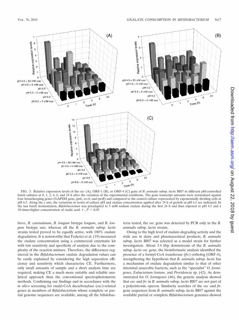
As shown in Fig. 5, when 5 mM sodium oxalate was pro-vided, maintaining a pH of 6.5, all the three genes evaluated,oxc, ORF-1, and ORF-4, were expressed without considerabledifferences, indicating the absence of a specific effect of oxalatewithout lowering the pH. In contrast, after exposure to pH 4.5,
for oxc (Fig. 5A) and ORF-4 (Fig. 5C), the relative expressionimmediately and consistently increased independently ofoxalate addition. The highest expression levels, with a statisti-cally significant average increase of 5.5-fold with respect to thebasal expression, were observed after 1 and 2 h in the absenceof oxalate, respectively, for ORF-4 and oxc. At pH 4.5, ORF-1transcripts were detected but their amounts were not signifi-cantly higher than those found in B. animalis subsp. lactis BI07control cells (Fig. 5B). Modest levels of induction of the geneencoding the putative permease were exhibited after the pHshift to 5.5 and addition of oxalate, with a mean of 2.7 at 1 hafter the end of log-phase growth (t1). In pH 5.5-controlledbatch cultures, statistically larger amounts of the oxc genetranscript were also observed (with 3.2 as the maximal foldchange). In almost all the pH-controlled batch cultures, thegene transcript levels tended to decrease over time.
When Bifidobacterium was preadapted to 5 mM oxalate dur-ing the first 24 h of growth, all the loci examined were highlyinduced. Their expression showed a maximum level (6.5 foroxc, 3.5 for ORF-1, and 12 for ORF-4) immediately after thevariation in the experimental conditions and had alreadystarted to decrease after 1 h of treatment. However, the rela-tive amounts of all transcripts remained consistently higherthan the basal expression at all time points. Statistically signif-icant differences were observed at t0 for all genes betweenpreadapted cultures and pH 4.5-controlled cultures withoutpreadaptation. In all the other samples analyzed, no significantinduction or repression of the oxc, ORF-1, and ORF-4 geneswas observed.
DISCUSSION
In this work, a preliminary screening concerning oxalate-degrading activity in 14 Bifidobacterium strains comprising 4species and 3 subspecies originating from humans, animals orprobiotic products was carried out by capillary electrophoresis(CE). In a difference from previous reports (19), no oxalateconsumption was observed for B. adolescentis, B. bifidum, B.
FIG. 4. Oxalate-degrading activity of B. animalis subsp. lactis BI07 in pH 4.5-controlled batch fermentations after changing the experimentalconditions. (A) After 24 h of growth at pH 6.5, the fermentation pH was lowered to 4.5 with the simultaneous addition of 5 mM sodium oxalate.(B) Bifidobacterium was preadapted to 5 mM sodium oxalate during the first 24 h and then exposed to pH 4.5 and a 10-times-higher concentrationof oxalic acid. Kinetics of oxalate degradation (triangles) and formate production (circles) were determined by capillary electrophoresis asdescribed in Materials and Methods. Cell viability (squares) is shown. In panel A, the data on B. animalis subsp. lactis viability at pH 4.5 withoutoxalate addition (dashed lines) were also reported. Each point represents the mean of data from three independent experiments. The error barsindicate standard deviations.
5616 TURRONI ET AL. APPL. ENVIRON. MICROBIOL.
on August 22, 2018 by guest
http://aem.asm
.org/D
ownloaded from
breve, B. catenulatum, B. longum biotype longum, and B. lon-gum biotype suis, whereas all the B. animalis subsp. lactisstrains tested proved to be equally active, with 100% oxalatedegradation. It is noteworthy that Federici et al. (19) measuredthe oxalate concentration using a commercial enzymatic kitwith low sensitivity and specificity of analysis due to the com-plexity of the reaction mixture. Therefore, the differences reg-istered in the Bifidobacterium oxalate degradation values canbe easily explained by considering the high separation effi-ciency and sensitivity which characterize CE. Furthermore,only small amounts of sample and a short analysis time arerequired, making CE a much more suitable and reliable ana-lytical approach than the conventional spectrophotometricmethods. Confirming our findings and in accordance with thein silico screening for oxalyl-CoA decarboxylase (oxc)-relatedgenes in members of Bifidobacterium whose complete or par-tial genome sequences are available, among all the bifidobac-
teria tested, the oxc gene was detected by PCR only in the B.animalis subsp. lactis strains.
Owing to the high level of oxalate-degrading activity and thewide use in dairy and pharmaceutical products, B. animalissubsp. lactis BI07 was selected as a model strain for furtherinvestigation. About 3.4 kbp downstream of the B. animalissubsp. lactis oxc gene, the bioinformatic analysis identified thepresence of a formyl-CoA transferase (frc) ortholog (ORF-4),strengthening the hypothesis that B. animalis subsp. lactis hasa mechanism of oxalate degradation similar to that of otherintestinal anaerobic bacteria, such as the “specialist” O. formi-genes, Eubacterium lentum, and Providencia sp. (42). As dem-onstrated for O. formigenes (46), the genetic analysis showedthat oxc and frc in B. animalis subsp. lactis BI07 are not part ofa polycistronic operon. Similarity searches of the oxc and frcgene sequences from B. animalis subsp. lactis BI07 against theavailable partial or complete Bifidobacterium genomes showed
FIG. 5. Relative expression levels of the oxc (A), ORF-1 (B), or ORF-4 (C) gene of B. animalis subsp. lactis BI07 in different pH-controlledbatch cultures at 0, 1, 2, 4, 6, and 24 h after the variation of the experimental conditions. The gene transcript amounts were normalized againstfour housekeeping genes (GAPDH gene, gmk, recA, and rpoB) and compared to the control culture represented by exponentially dividing cells atpH 6.5. Along the y axis, the variations in terms of culture pH and oxalate concentration applied after 24 h of growth at pH 6.5 are indicated. Inthe last batch fermentation, Bifidobacterium was preadapted to 5 mM sodium oxalate during the first 24 h and then exposed to pH 4.5 and a10-times-higher concentration of oxalic acid. * , P 0.05.
VOL. 76, 2010 OXALATE CONSUMPTION IN BIFIDOBACTERIUM 5617
on August 22, 2018 by guest
http://aem.asm
.org/D
ownloaded from

that besides B. animalis subsp. lactis, only B. gallicum, B. den-tium, and B. pseudocatenulatum harbor putative oxc and frcgenes. Interestingly, apart from B. gallicum, which appears tobe exclusively associated with animal feces (32), the otherBifidobacterium species with a potential oxalate-degrading ac-tivity seem to prefer a cosmopolitan lifestyle without a clearecological specialization (32, 50, 56). As reported for all thesebifidobacterial genome sequences, an ORF encoding a puta-tive permease (ORF-1) was identified flanking the oxc gene onthe same strand in B. animalis subsp. lactis BI07. Although thisgene product has no similarity to the O. formigenes oxalate:formate antiporter protein OxlT (1), nor to the ABC trans-porter of L. acidophilus NCFM, which is associated with oxc(5), its presence in the oxc surrounding regions suggests itspotential role as an oxalate transporter protein. However, thearrangement of oxc, frc, and the gene encoding the putativeoxalate transporter in the B. gallicum, B. dentium, and B.pseudocatenulatum genomes is somewhat different from B. ani-malis subsp. lactis with regard to gene order and orientation. Inparticular, B. animalis subsp. lactis BI07 ORF-2 and ORF-3,located between oxc and frc orthologs, did not display a signif-icant sequence similarity to any known genes of Bifidobacte-rium, indicating that they are either specific to B. animalissubsp. lactis or are yet to be determined in the other incom-plete genomes. In contrast, the complete B. animalis subsp.lactis ORF (ORF-7) identified upstream of oxc showed 49%predicted amino acid identity to the B. gallicum putative chlo-ride channel protein EriC gene. It has been proposed thatthese channel proteins may function as an electrical shunt foran outwardly directed virtual proton pump that is linked toamino acid decarboxylation, allowing enteric bacteria to sur-vive in the strongly acidic environment of the stomach (27). Inthis way, the persistent proton influx that arises from highextracellular acidity could be continually counteracted in thecytoplasm by the decarboxylation-linked proton utilization.Since oxalate is decarboxylated to formyl-CoA in a reactionthat consumes a proton after intracellular uptake and activa-tion to oxalyl-CoA, the concerted action of permease, Oxc,Frc, and EriC could allow Bifidobacterium to withstand acidchallenges.
To investigate whether B. animalis subsp. lactis BI07 couldrely on oxalate metabolism as a major energy source, itsoxalate-degrading activity was determined in the absence ofthe sugar source. Under these conditions, neither B. animalissubsp. lactis growth nor oxalate consumption was observed,providing evidence that oxalate cannot be used as the solecarbon source and that its metabolism is strictly dependent onthe cell physiological state. Conversely, according to our data,no impact of oxalate on B. animalis subsp. lactis sugar metab-olism was determined.
The role of an acidic environment in the B. animalis subsp.lactis BI07 oxalate degradation pattern was evaluated in pH-controlled batch cultures analogously to what has already beenreported for L. acidophilus (5) and L. gasseri (34). Fermenta-tion experiments indicated that only at pH 4.5 is there a sig-nificant oxalate consumption and that the degradation ratedramatically increases in oxalate-preadapted cells. Confirmingthat oxalate degradation by B. animalis subsp. lactis proceedsvia a decarboxylation step, formate production was demon-strated. Our results indicated that approximately 0.9 mol of
formate were produced per mol of oxalate degraded, as re-ported for O. formigenes (2). Interestingly, when oxalate wassupplied, the ability of B. animalis subsp. lactis BI07 to resistthe detrimental effects of acidic pH was improved.
For the first time, using quantitative real-time reverse tran-scription-PCR (RT-qPCR), the regulation of B. animalissubsp. lactis oxc, frc, and the gene encoding the putative per-mease was proven. To quantify accurately the cDNA levels andto identify real gene-specific variations, the normalizationstrategy proposed by Vandesompele et al. (53) was chosen.Since there is no consensus for prokaryotic endogenous con-trol (52), five presumed housekeeping genes belonging to dif-ferent functional classes, the glyceraldehyde 3-phosphate de-hydrogenase (GAPDH) gene, gmk (encoding guanylatekinase), recA (encoding recombinase A), rpoB (encoding sub-unit beta of DNA-directed RNA polymerase), and 16S rRNA(encoding small subunit [SSU] ribosome, four copies), weretested. Although 16S rRNA is among the most commonly usedcellular products for transcript normalization (11), in our studythe metabolic housekeeping gene transcripts were found towork best.
After normalization of the expression data, the B. animalissubsp. lactis BI07 ORF-4, encoding the putative Frc enzyme,was the most highly induced gene, exhibiting between 2.5- and12-fold-higher levels of expression in the pH 4.5-controlledbatch cultures. Acidic conditions were also a prerequisite foroxc transcription, which was most active within 2 h after the pHshift from 6.5 to 4.5. As for L. acidophilus NCFM (5) and L.gasseri ATCC 33323 (34), when bifidobacterial cells were pre-adapted to subinhibitory concentrations of oxalate and subse-quently exposed to lower pH values plus a higher concentra-tion of oxalic acid, the relative oxc and frc mRNA abundanceswere significantly increased. However, with respect to lactoba-cilli, in B. animalis subsp. lactis BI07 overall, higher transcrip-tional levels were observed, and more importantly, a pH valueof 4.5 instead of 5.5 was fundamental for gene expression aswell as oxalate-degrading activity. After oxalate preadaptation,a lower but significant magnitude of upregulation was alsodetected for ORF-1. For all the three genes, no specific effectof oxalate was observed without a lowering of the pH. Further,decreased transcript levels were detected over time, mainly foroxc and frc, suggesting a progressive reduction of the transcrip-tional and metabolic activities of B. animalis subsp. lactis BI07.
In conclusion, our results indicate that in B. animalis subsp.lactis BI07, the pattern of oxalate degradation and the tran-scriptional activities of the genes potentially involved in oxalatecatabolism are pH dependent. The discovery of genes whoseexpression responds to changes in pH is particularly important.The pH of the lumen of the proximal colon is reported to besomewhat lower (5.5 to 6.5) than that of the distal colon (6.5 to7.0), mainly as a result of the higher fermentation rate ofsaccharolytic microbial groups within the proximal region (17).The pH of both regions may be transiently and locally reducedfollowing ingestion of nondigestible but fermentable carbohy-drates (9, 10, 16). The oxalate-degrading activity whichuniquely characterizes the B. animalis subsp. lactis species overthe other bifidobacteria most commonly encountered in pro-biotic products may justify the use of this probiotic species asa promising adjunct for the prevention and treatment ofoxalate-associated disorders.
5618 TURRONI ET AL. APPL. ENVIRON. MICROBIOL.
on August 22, 2018 by guest
http://aem.asm
.org/D
ownloaded from

ACKNOWLEDGMENTS
This work was financed by the project “Mechanisms of interactionand cell-cell communication in lactic acid bacteria from food ecosys-tems: transcriptomics, proteomics and metabolomics,” PRIN 2007funds, from the Ministry of University and Scientific Research of Italy.
REFERENCES
1. Abe, K., Z. S. Ruan, and P. C. Maloney. 1996. Cloning, sequencing, andexpression in Escherichia coli of OxlT, the oxalate:formate exchange proteinof Oxalobacter formigenes. J. Biol. Chem. 271:6789–6793.
2. Allison, M. J., K. A. Dawson, W. R. Mayberry, and J. G. Foss. 1985.Oxalobacter formigenes gen. nov., sp. nov.: oxalate-degrading anaerobes thatinhabit the gastrointestinal tract. Arch. Microbiol. 141:1–7.
3. Altermann, E., W. M. Russell, M. A. Azcarate-Peril, R. Barrangou, B. L.Buck, O. McAuliffe, N. Souther, A. Dobson, T. Duong, M. Callanan, S. Lick,A. Hamrick, R. Cano, and T. R. Klaenhammer. 2005. Complete genomesequence of the probiotic lactic acid bacterium Lactobacillus acidophilusNCFM. Proc. Natl. Acad. Sci. U. S. A. 102:3906–3912.
4. Altschul, S. F., T. L. Madden, A. A. Schaffer, J. Zhang, Z. Zhang, W. Miller,and D. J. Lipman. 1997. Gapped BLAST and PSI-BLAST: a new generationof protein database search programs. Nucleic Acids Res. 25:3389–3402.
5. Azcarate-Peril, M. A., J. M. Bruno-Barcena, H. M. Hassan, and T. R.Klaenhammer. 2006. Transcriptional and functional analysis of oxalyl-coen-zyme A (CoA) decarboxylase and formyl-CoA transferase genes from Lac-tobacillus acidophilus. Appl. Environ. Microbiol. 72:1891–1899.
6. Baetz, A. L., and M. J. Allison. 1990. Purification and characterization offormyl-coenzyme A transferase from Oxalobacter formigenes. J. Bacteriol.172:3537–3540.
7. Baetz, A. L., and M. J. Allison. 1989. Purification and characterization ofoxalyl-coenzyme A decarboxylase from Oxalobacter formigenes. J. Bacteriol.171:2605–2608.
8. Barrangou, R., E. P. Briczinski, L. L. Traeger, J. R. Loquasto, M. Richards,P. Horvath, A. C. Coute-Monvoisin, G. Leyer, S. Rendulic, J. L. Steele, J. R.Broadbent, T. Oberg, E. G. Dudley, S. Schuster, D. A. Romero, and R. F.Roberts. 2009. Comparison of the complete genome sequences of Bifidobac-terium animalis subsp. lactis DSM 10140 and Bl-04. J. Bacteriol. 191:4144–4151.
9. Bird, A. R., M. S. Vuaran, R. A. King, M. Noakes, J. Keogh, M. K. Morell,and D. L. Topping. 2008. Wholegrain foods made from a novel high-amylosebarley variety (Himalaya 292) improve indices of bowel health in humansubjects. Br. J. Nutr. 99:1032–1040.
10. Bown, R. L., J. A. Gibson, G. E. Sladen, B. Hicks, and A. M. Dawson. 1974.Effects of lactulose and other laxatives on ileal and colonic pH as measuredby a radiotelemetry device. Gut 15:999–1004.
11. Bustin, S. A. 2000. Absolute quantification of mRNA using real-time reversetranscription polymerase chain reaction assays. J. Mol. Endocrinol. 25:169–193.
12. Campieri, C., M. Campieri, V. Bertuzzi, E. Swennen, D. Matteuzzi, S. Ste-foni, F. Pirovano, C. Centi, S. Ulisse, G. Famularo, and C. De Simone. 2001.Reduction of oxaluria after an oral course of lactic acid bacteria at highconcentration. Kidney Int. 60:1097–1105.
13. Candela, M., E. Biagi, M. Centanni, S. Turroni, M. Vici, F. Musiani, B.Vitali, S. Bergmann, S. Hammerschmidt, and P. Brigidi. 2009. Bifidobacte-rial enolase, a cell surface receptor for human plasminogen involved in theinteraction with the host. Microbiology 155:3294–3303.
14. Candela, M., F. Perna, P. Carnevali, B. Vitali, R. Ciati, P. Gionchetti, F.Rizzello, M. Campieri, and P. Brigidi. 2008. Interaction of probiotic Lacto-bacillus and Bifidobacterium strains with human intestinal epithelial cells:adhesion properties, competition against enteropathogens and modulationof IL-8 production. Int. J. Food Microbiol. 125:286–292.
15. Candela, M., G. Seibold, B. Vitali, S. Lachenmaier, B. J. Eikmanns, and P.Brigidi. 2005. Real-time PCR quantification of bacterial adhesion to Caco-2cells: competition between bifidobacteria and enteropathogens. Res. Micro-biol. 156:887–895.
16. Chung, Y.-C., C.-K. Hsu, C.-Y. Ko, and Y.-C. Chan. 2007. Dietary intake ofxylooligosaccharides improves the intestinal microbiota, fecal moisture, andpH value in the elderly. Nutr. Res. 27:756–761.
17. Cummings, J. H., and G. T. Macfarlane. 1991. The control and conse-quences of bacterial fermentation in the human colon. J. Appl. Bacteriol.70:443–459.
18. Erim, F. B., A. Cifuentes, H. Poppe, and J. C. Kraak. 1995. Performance ofa physically adsorbed high-molecular-mass polyethyleneimine layer as coat-ing for the separation of basic proteins and peptides by capillary electro-phoresis. J. Chromatogr. A 708:356–361.
19. Federici, F., B. Vitali, R. Gotti, M. R. Pasca, S. Gobbi, A. B. Peck, and P.Brigidi. 2004. Characterization and heterologous expression of the oxalylcoenzyme A decarboxylase gene from Bifidobacterium lactis. Appl. Environ.Microbiol. 70:5066–5073.
20. Goldfarb, D. S., F. Modersitzki, and J. R. Asplin. 2007. A randomized,controlled trial of lactic acid bacteria for idiopathic hyperoxaluria. Clin.J. Am. Soc. Nephrol. 2:745–749.
21. Hatch, M., J. Cornelius, M. Allison, H. Sidhu, A. Peck, and R. W. Freel.2006. Oxalobacter sp. reduces urinary oxalate excretion by promoting entericoxalate secretion. Kidney Int. 69:691–698.
22. Holmes, R. P. 2000. Oxalate synthesis in humans: assumptions, problems,and unresolved issues. Mol. Urol. 4:329–332.
23. Holmes, R. P., and M. Kennedy. 2000. Estimation of oxalate content of foodsand daily oxalate intake. Kidney Int. 57:1662–1667.
24. Holmes, R. P. 1995. Measurement of urinary oxalate and citrate by capillaryelectrophoresis and indirect ultraviolet absorbance. Clin. Chem. 41:1297–1301.
25. Hoppe, B., B. Beck, N. Gatter, G. von Unruh, A. Tischer, A. Hesse, N. Laube,P. Kaul, and H. Sidhu. 2006. Oxalobacter formigenes: a potential tool for thetreatment of primary hyperoxaluria type 1. Kidney Int. 70:1305–1311.
26. Hoppe, B., G. von Unruh, N. Laube, A. Hesse, and H. Sidhu. 2005. Oxalatedegrading bacteria: new treatment option for patients with primary andsecondary hyperoxaluria? Urol. Res. 33:372–375.
27. Iyer, R., T. M. Iverson, A. Accardi, and C. Miller. 2002. A biological role forprokaryotic ClC chloride channels. Nature 419:715–718.
28. Jager, A. V., F. G. Tonin, and M. F. Tavares. 2007. Comparative evaluationof extraction procedures and method validation for determination of carbo-hydrates in cereals and dairy products by capillary electrophoresis. J. Sep.Sci. 30:586–594.
29. James, L. F. 1972. Oxalate toxicosis. Clin. Toxicol. 5:231–243.30. Kaufman, D. W., J. P. Kelly, G. C. Curhan, T. E. Anderson, S. P. Dretler,
G. M. Preminger, and D. R. Cave. 2008. Oxalobacter formigenes may reducethe risk of calcium oxalate kidney stones. J. Am. Soc. Nephrol. 19:1197–1203.
31. Kwak, C., B. C. Jeong, J. H. Ku, H. H. Kim, J. J. Lee, C. S. Huh, Y. J. Baek,and S. E. Lee. 2006. Prevention of nephrolithiasis by Lactobacillus in stone-forming rats: a preliminary study. Urol. Res. 34:265–270.
32. Lamendella, R., J. W. Santo Domingo, C. Kelty, and D. B. Oerther. 2008.Bifidobacteria in feces and environmental waters. Appl. Environ. Microbiol.74:575–584.
33. Langendijk, P. S., F. Schut, G. J. Jansen, G. C. Raangs, G. R. Kamphuis,M. H. Wilkinson, and G. W. Welling. 1995. Quantitative fluorescence in situhybridization of Bifidobacterium spp. with genus-specific 16S rRNA-targetedprobes and its application in fecal samples. Appl. Environ. Microbiol. 61:3069–3075.
34. Lewanika, T. R., S. J. Reid, V. R. Abratt, G. T. Macfarlane, and S. Macfar-lane. 2007. Lactobacillus gasseri Gasser AM63(T) degrades oxalate in amultistage continuous culture simulator of the human colonic microbiota.FEMS Microbiol. Ecol. 61:110–120.
35. Lieske, J. C., D. S. Goldfarb, C. De Simone, and C. Regnier. 2005. Use of aprobiotic to decrease enteric hyperoxaluria. Kidney Int. 68:1244–1249.
36. Makarova, K., A. Slesarev, Y. Wolf, A. Sorokin, B. Mirkin, E. Koonin, A.Pavolv, N. Pavlova, V. Karamychev, N. Polouchine, V. Shakhova, I. Grigor-iev, Y. Lou, D. Rohksar, S. Lucas, K. Huang, D. M. Goldstein, T. Hawkins,V. Plengvidhya, D. Welker, J. Hughes, Y. Goh, A. Benson, K. Baldwin, J.-H.Lee, I. Diaz-Muniz, B. Dosti, V. Smeianov, W. Wechter, R. Barabote, G.Lorca, E. Altermann, R. Barrangou, B. Ganesan, Y. Xie, H. Rawsthorne, D.Tamir, C. Parker, F. Breidt, J. Broadbent, R. Hutkins, D. O’Sulllivan, J.Steele, G. Unlu, M. Saier, T. Klaenhammer, P. Richardson, S. Kozyavkin, B.Weimer, and D. Mills. 2006. Comparative genomics of the lactic acid bac-teria. Proc. Natl. Acad. Sci. U. S. A. 103:15611–15616.
37. Marco, M. L., S. Pavan, and M. Kleerebezem. 2006. Towards understandingmolecular modes of probiotic action. Curr. Opin. Biotechnol. 17:204–210.
38. Mlobeli, N. T., N. A. Gutierrez, and I. S. Maddox. 1998. Physiology andkinetics of Bifidobacterium bifidum during growth on different sugars. Appl.Microbiol. Biotechnol. 50:125–128.
39. Murphy, C., S. Murphy, F. O’Brien, M. O’Donoghue, T. Boileau, G. Sunvold,G. Reinhart, B. Kiely, F. Shanahan, and L. O’Mahony. 2009. Metabolicactivity of probiotics-oxalate degradation. Vet. Microbiol. 136:100–107.
40. Okombo, J., and M. Liebman. 2010. Probiotic-induced reduction of gastro-intestinal oxalate absorption in healthy subjects. Urol. Res. Doi:10.1007/s00240-010-0262-9.
41. Rodby, R. A., T. S. Tyazka, and J. W. Williams. 1991. Reversal of cardiacdysfunction secondary to type I primary hyperoxaluria after combined liver-kidney transplantation. Am. J. Med. 90:498–504.
42. Sahin, N. 2003. Oxalotrophic bacteria. Res. Microbiol. 154:399–407.43. Sambrook, J. E., F. Fritsch, and T. Maniatis. 1989. Molecular cloning: a
laboratory manual, 2nd ed. Cold Spring Harbor Laboratory Press, ColdSpring Harbor, NY.
44. Sidhu, H., M. J. Allison, J. M. Chow, A. Clark, and A. B. Peck. 2001. Rapidreversal of hyperoxaluria in a rat model after probiotic administration ofOxalobacter formigenes. J. Urol. 166:1487–1491.
45. Sidhu, H., M. E. Schmidt, J. G. Cornelius, S. Thamilselvan, S. R. Khan, A.Hesse, and A. B. Peck. 1999. Direct correlation between hyperoxaluria/oxalate stone disease and the absence of the gastrointestinal tract-dwellingbacterium Oxalobacter formigenes: possible prevention by gut recolonizationor enzyme replacement therapy. J. Am. Soc. Nephrol. 10:S334–S340.
46. Sidhu, H., S. D. Ogden, H. Y. Lung, B. G. Luttge, A. L. Baetz, and A. B. Peck.1997. DNA sequencing and expression of the formyl coenzyme A transferasegene, frc, from Oxalobacter formigenes. J. Bacteriol. 179:3378–3381.
VOL. 76, 2010 OXALATE CONSUMPTION IN BIFIDOBACTERIUM 5619
on August 22, 2018 by guest
http://aem.asm
.org/D
ownloaded from
47. Siva, S., E. R. Barrack, G. P. Reddy, V. Thamilselvan, S. Thamilselvan, M.Menon, and M. Bhandari. 2009. A critical analysis of the role of gutOxalobacter formigenes in oxalate stone disease. BJU Int. 103:18–21.
48. Stanton, C., R. P. Ross, G. F. Fitzgerald, and D. van Sinderen. 2005. Fer-mented functional foods based on probiotics and their biogenic metabolites.Curr. Opin. Biotechnol. 16:198–203.
49. Tracy, C. R., and M. S. Pearle. 2009. Update on the medical management ofstone disease. Curr. Opin. Urol. 19:200–204.
50. Turroni, F., E. Foroni, P. Pizzetti, V. Giubellini, A. Ribbera, P. Merusi, P.Cagnasso, B. Bizzarri, G. L. de’Angelis, F. Shanahan, D. van Sinderen, andM. Ventura. 2009. Exploring the diversity of the bifidobacterial population inthe human intestinal tract. Appl. Environ. Microbiol. 75:1534–1545.
51. Turroni, S., B. Vitali, C. Bendazzoli, M. Candela, R. Gotti, F. Federici, F.Pirovano, and P. Brigidi. 2007. Oxalate consumption by lactobacilli: evalu-ation of oxalyl-CoA decarboxylase and formyl-CoA transferase activity inLactobacillus acidophilus. J. Appl. Microbiol. 103:1600–1609.
52. Vandecasteele, S. J., W. E. Peetermans, R. Merckx, and J. Van Eldere. 2001.Quantification of expression of Staphylococcus epidermidis housekeepinggenes with Taqman quantitative PCR during in vitro growth and underdifferent conditions. J. Bacteriol. 183:7094–7101.
53. Vandesompele, J., K. De Preter, F. Pattyn, B. Poppe, N. Van Roy, A. DePaepe, and F. Speleman. 2002. Accurate normalization of real-time quanti-
tative RT-PCR data by geometric averaging of multiple internal controlgenes. Genome Biol. 3:research0034.1–0034.11.
54. Ventura, M., F. Turroni, A. Zomer, E. Foroni, V. Giubellini, F. Bottacini, C.Canchaya, M. J. Claesson, F. He, M. Mantzourani, L. Mulas, A. Ferrarini,B. Gao, M. Delledonne, B. Henrissat, P. Coutinho, M. Oggioni, R. S. Gupta,Z. Zhang, D. Beighton, G. F. Fitzgerald, P. W. O’Toole, and D. van Sinderen.2009. The Bifidobacterium dentium Bd1 genome sequence reflects its geneticadaptation to the human oral cavity. PLoS Genet. 5:e1000785.
55. Ventura, M., S. O’Flaherty, M. J. Claesson, F. Turroni, T. R. Klaenhammer,D. van Sinderen, and P. W. O’Toole. 2009. Genome-scale analyses of health-promoting bacteria: probiogenomics. Nat. Rev. Microbiol. 7:61–71.
56. Ventura, M., M. O’Connell-Motherway, S. Leahy, J. A. Moreno-Munoz,G. F. Fitzgerald, and D. van Sinderen. 2007. From bacterial genome tofunctionality; case bifidobacteria. Int. J. Food Microbiol. 120:2–12.
57. Vitali, B., S. Turroni, F. Dal Piaz, M. Candela, V. Wasinger, and P. Brigidi.2007. Genetic and proteomic characterization of rifaximin resistance inBifidobacterium infantis BI07. Res. Microbiol. 158:355–362.
58. Weese, J. S., H. E. Weese, L. Yuricek, and J. Rousseau. 2004. Oxalatedegradation by intestinal lactic acid bacteria in dogs and cats. Vet. Microbiol.101:161–166.
59. Williams, H. E., and L. H. Smith. 1968. Disorders of oxalate metabolism.Am. J. Med. 45:715–735.
5620 TURRONI ET AL. APPL. ENVIRON. MICROBIOL.
on August 22, 2018 by guest
http://aem.asm
.org/D
ownloaded from