introducing 3d printed models of the upper urinary tract
TRANSCRIPT

RESEARCH Open Access
Introducing 3D printed models of theupper urinary tract for high-fidelitysimulation of retrograde intrarenal surgeryLuca Orecchia1* , Diego Manfrin2, Stefano Germani1, Dario Del Fabbro3, Anastasios D. Asimakopoulos1,Enrico Finazzi Agrò1,4 and Roberto Miano1,4
Abstract
Purpose: Training in retrograde intrarenal surgery for the treatment of renal stone disease is a challenging task dueto the unique complexity of the procedure. This study introduces a series of 3D printed models of upper urinarytract and stones designed to improve the training process.
Methods: Six different models of upper urinary tract were algorithmically isolated, digitally optimized and 3Dprinted from real-life cases. Soft and hard stones in different sizes were produced from 3D printed moulds. Themodels were fitted onto a commercially available part-task trainer and tested for retrograde intrarenal surgery.
Results: Each step of the procedure was simulated with extraordinary resemblance to real-life cases. The uniqueanatomical intricacy of each model and type of stones allowed us to reproduce surgeries of increasing difficulty. Asthe case-load required to achieve proficiency in retrograde intrarenal surgery is high, benchtop simulation could beintegrated in training programs to reach good outcomes and low complication rates faster. Our models matchincredible anatomical resemblance with low production cost and high reusability. Validation studies and objectiveskills assessment during simulations would allow comparison with other available benchtop trainers and the designof stepwise training programs.
Conclusions: 3D printing is gaining a significant importance in surgical training. Our 3D printed models of the upperurinary tract might represent a risk-free training option to hasten the achievement of proficiency in endourology.
Keywords: Urology, Ureteroscopy, Lithotripsy, RIRS, 3D, Anatomy, Kidney, Simulation, Training
© The Author(s). 2021, corrected publication 2021. Open Access This article is licensed under a Creative Commons Attribution4.0 International License, which permits use, sharing, adaptation, distribution and reproduction in any medium or format, aslong as you give appropriate credit to the original author(s) and the source, provide a link to the Creative Commons licence,and indicate if changes were made. The images or other third party material in this article are included in the article's CreativeCommons licence, unless indicated otherwise in a credit line to the material. If material is not included in the article's CreativeCommons licence and your intended use is not permitted by statutory regulation or exceeds the permitted use, you will needto obtain permission directly from the copyright holder. To view a copy of this licence, visit http://creativecommons.org/licenses/by/4.0/. The Creative Commons Public Domain Dedication waiver (http://creativecommons.org/publicdomain/zero/1.0/) applies to the data made available in this article, unless otherwise stated in a credit line to the data.
* Correspondence: [email protected] Unit, Policlinico Tor Vergata Foundation, Viale Oxford 81, 00133Rome, ItalyFull list of author information is available at the end of the article
Orecchia et al. 3D Printing in Medicine (2021) 7:15 https://doi.org/10.1186/s41205-021-00105-9
BackgroundRenal stone disease represents a significant burden forhealthcare systems worldwide. In view of its multifactorialaetiology, its prevalence ranges from 1 to 20 % reaching >10 % in countries with a high standard of living [1]. It is thesecond most expensive urological disease, with accruingcosts associated with urgent care for symptomatic stonesand novel technologies used for treatment [2].Operative approaches to renal stones are rapidly evolving,
intending to reduce the invasiveness of the procedure whileensuring high stone free rates. The current treatment op-tions include extracorporeal shockwave lithotripsy alongsideendoscopic approaches, either percutaneous or transurethral(Retrograde Intrarenal Surgery, RIRS).RIRS is a fully endoscopic and minimally invasive type
of surgery. Performing RIRS presents unique challengesdue the narrowness of the operating field, the highanatomical variety within each collecting system, thecomplexity of the operating manoeuvres, the fragility ofthe dedicated instruments and potentially severe associ-ated complications [3, 4].Hence, reaching adequate proficiency with RIRS might
represent a demanding task for the urologist in trainingboth for the intrinsic technical challenges and thedifficulty to achieve an adequate case-load during theresidency years [5]. Concurrently, due to the necessity toguarantee patient safety during the entire procedure,having novices beginning their training directly on real-life cases might not represent the most convenient way ofapproaching RIRS. Therefore, this has led to a growinginterest in risk-free alternatives to the traditional surgicalapprenticeship model and to the development of severalendourology simulation programs using: animal/cadavericmodels, virtual trainers and benchtop models [6].Out of the wide array of technologies applied to simu-
lation, 3D printing had a significant impact in urologythanks to the incredible anatomical accuracy of theprinted models and the low costs associated. It has beensuccessfully used to improve patient education, pre-operative planning and simulation-based training [7–9].To improve the modular approach to teaching RIRS
practised in the operating theatre at our institution andpossibly decouple it from its strict dependency from thesurgical case-load, this study reports on the productionand experimentation with a series of completely 3Dprinted models of upper urinary tracts and stones,devised as a benchtop simulation-based addition totraditional training.
MethodsDevelopmentAnonymised Digital Imaging and Communication inMedicine (DICOM) files from Computerised Tomog-raphy Urogram (CTU) scans were collected by expert
endourologists from real renal stone cases. A total of sixCTUs were selected after reviewing each individual anat-omy in terms of type and intricacy of the pelvicalycealsystem, thus allowing to plan for training models ofdifferent complexity. The DICOM files were sent to abioengineering company (Medics srl, Moncalieri, Italy)for data extraction and modelling. A Region of Interest(ROI) comprising the selected kidney and proximalureter was designed using the software Mimics 23.0(Materialise NV, Leuven, Belgium). DICOM files weresubsequently segmented using automatic and semi-automatic algorithms for the isolation of the voxels cor-responding to the anatomical detail of interest by theidentification of variations in Hounsfield Units (HU) inthe different CTU phases for each voxel in the selectedROI.The result of the automatic process was then refined
and validated by a biomedical engineer subject matterexpert. The voxel volume was subsequently convertedand interpolated in a 3D triangulated mesh (Fig. 1A).The resulting model was then exported and finalized in3-matic 15.0 (Materialise NV, Leuven, Belgium). Theprocess included manual noise reduction and smoothingof parts included in the mesh due to approximation inHU readings of the CTU scan volume. The post-processed model was then imported back in Mimics andcorrespondence with the CTU scan ROI was confirmed(Fig. 1B).As the original post-processed geometry was solid and
completely closed, an external hollowing of 2.5mm wasapplied to the model in order to allow for real-lifeendoscopic navigation. Lastly, an expert 3D modelerperformed project optimization on the 3D mesh anddesigned an elliptical hatch with a dedicated press fitclosure system for stone insertion in the kidney pelvisduring the simulation (Fig. 2). The finalised mesh wasthen exported in stereolithography (.stl) format for theprinting phase.A A2v4 (3ntr, Oleggio, Italy) or a Ultimaker S5 (Ulti-
maker, Utrecht, Netherlands) 3D printer was used fordual extrusion printing. A white thermoplastic polyur-ethane 2.85 mm filament was used for the pelvicalycealsystem in order to grant some flexibility during modelsetup, while a water soluble polyvinyl alcohol filamentwas used for the inner support scaffold used during theprinting phase. The pelvis hatch closure was printed sep-arately from the main structure. After printing themodels were submerged in warm water in order to allowthe dissolution and detachment of the scaffold system.Training stones were produced after several laser
lithotripsy simulations with different compounds whichled to the selection of two suspensions of water andchalk in fixed proportions in order to produce soft andhard stones resembling real-life conditions in terms of
Orecchia et al. 3D Printing in Medicine (2021) 7:15 Page 2 of 9

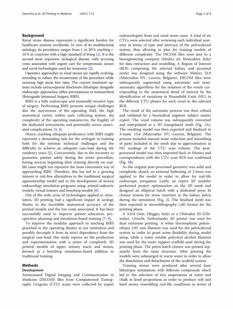
resistance to lithotripsy. Soft stones were obtained bymixing in a 1:1 chalk to water ratio by weight, whilehard stones required a 1.5:1 ratio. The differences instone hardness were devised to allow the use of differentlithotripsy techniques during the simulation (dusting vs.regular/popcorn fragmentation). Casts allowing produc-tion of spheroids of different radius were thereforesoftware modelled in negative, 3D printed and used toobtain moulds using bicomponent 1:1 pouring siliconerubber GLS Pro 20 (Prochima, Colli al Metauro, Italy)(Fig. 3). The water and chalk mixtures were then pouredthrough a dedicated channel in the mould and then driedin a food dehydrator to obtain the training stones. Produc-tion costs were monitored during the entire process.
TestingAfter completion of the development process, the 3Dprinted models were tested using a commercially availableversion of Cook Medical ureterorenoscopy (URS) part-task trainer (Cook Medical, Bloomington, IN, USA), con-sisting of a water filled box containing a bladder-dual ur-eter system connected to a silicone gel model of a penisfor realistic instrument insertion and upper urinary tractdrainage [10]. The 3D printed models were press fittedinto the proximal extremity of the ureters thus obtaining acomplete reproduction of the urinary tract. A 9.5Fr/11.5Fr35 cm Flexor ureteral access sheath (Cook Medical) wasinserted in the trainer over a 0.035 inch HiWire nitinolhydrophilic guidewire (Cook Medical). The models were
Fig. 2 Finalised 3D model in two projections, the elliptical hatch for stone insertion is visible on the side of the renal pelvis
Fig. 1 A 3D triangulated mesh after interpolation. B Post-processed model
Orecchia et al. 3D Printing in Medicine (2021) 7:15 Page 3 of 9
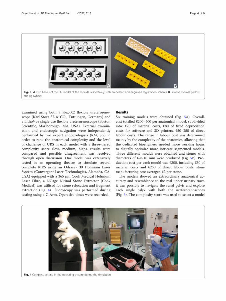
examined using both a Flex-X2 flexible ureteroreno-scope (Karl Storz SE & CO., Tuttlingen, Germany) anda LithoVue single use flexible ureterorenoscope (BostonScientific, Marlborough, MA, USA). External examin-ation and endoscopic navigation were independentlyperformed by two expert endourologists (RM, SG) inorder to rank the anatomical complexity and the levelof challenge of URS in each model with a three-tieredcomplexity score (low, medium, high), results werecompared and possible disagreement was resolvedthrough open discussion. One model was extensivelytested in an operating theatre to simulate severalcomplete RIRS using an Odyssey 30 Holmium LaserSystem (Convergent Laser Technologies, Alameda, CA,USA) equipped with a 365 μm Cook Medical HolmiumLaser Fibre, a NGage Nitinol Stone Extractor (CookMedical) was utilised for stone relocation and fragmentextraction (Fig. 4). Fluoroscopy was performed duringtesting using a C-Arm. Operative times were recorded.
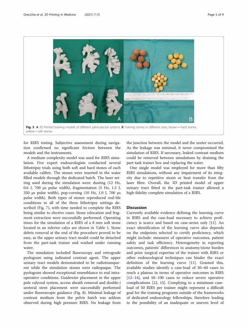
ResultsSix training models were obtained (Fig. 5A). Overall,cost totalled €200–400 per anatomical model, subdividedinto: €70 of material costs, €80 of fixed depreciationcosts for software and 3D printers, €50–250 of directlabour costs. The range in labour cost was determinedmainly by the complexity of the anatomies, allowing thatthe dedicated bioengineer needed more working hoursto digitally optimize more intricate segmented models.Three different moulds were obtained and stones withdiameters of 6-8-10 mm were produced (Fig. 5B). Pro-duction cost per each mould was €300, including €50 ofmaterial costs and €250 of direct labour costs, stonemanufacturing cost averaged €2 per stone.The models showed an extraordinary anatomical ac-
curacy and resemblance to the real upper urinary tract,it was possible to navigate the renal pelvis and exploreeach single calyx with both the ureterorenoscopes(Fig. 6). The complexity score was used to select a model
Fig. 3 A Two halves of the 3D model of the moulds, respectively with embossed and engraved registration spheres. B Silicone moulds (yellow)and jig (white)
Fig. 4 Complete setting in the operating theatre during the simulation
Orecchia et al. 3D Printing in Medicine (2021) 7:15 Page 4 of 9
for RIRS testing. Subjective assessment during naviga-tion confirmed no significant friction between themodels and the instruments.A medium complexity model was used for RIRS simu-
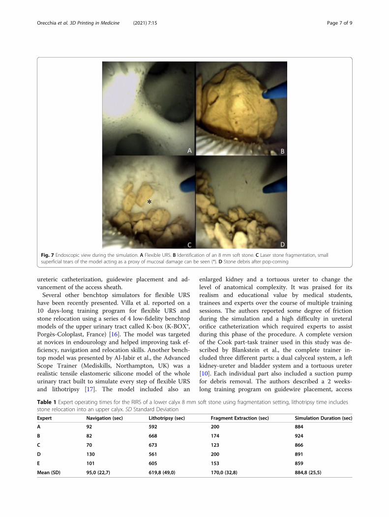
lation. Five expert endourologists conducted severallithotripsy trials using both soft and hard stones of eachavailable calibre. The stones were inserted in the waterfilled models through the dedicated hatch. The laser set-ting used during the simulation were: dusting (12 Hz,0.6 J, 700 µs pulse width), fragmentation (5 Hz, 1.5 J,350 µs pulse width), pop-corning (10 Hz, 1.0 J, 700 µspulse width). Both types of stones reproduced real-lifeconditions in all of the three lithotripsy settings de-scribed (Fig. 7), with time needed to complete the RIRSbeing similar to elective cases. Stone relocation and frag-ment extraction were successfully performed. Operatingtimes for the simulation of a RIRS of a 8 mm soft stonelocated in an inferior calyx are shown in Table 1. Stonedebris removal at the end of the procedure proved to beeasy, as the upper urinary tract model could be detachedfrom the part-task trainer and washed under runningwater.The simulation included fluoroscopy and retrograde
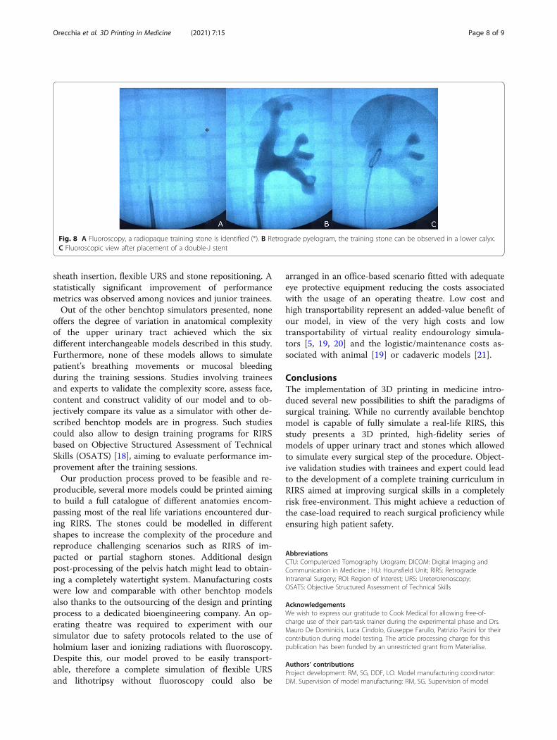
pyelogram using iodinated contrast agent. The upperurinary tract models demonstrated to be radiotranspar-ent while the simulation stones were radiopaque. Thepyelogram showed exceptional resemblance to real intra-operative conditions. Guidewire placement in the upperpole calyceal system, access sheath removal and double-jureteral stent placement were successfully performedunder fluoroscopic guidance (Fig. 8). Minimal leakage ofcontrast medium from the pelvis hatch was seldomobserved during high pressure RIRS. No leakage from
the junction between the model and the ureter occurred.As the leakage was minimal, it never compromised thesimulation of RIRS. If necessary, leaked contrast mediumcould be removed between simulations by draining thepart-task trainer box and replacing the water.One single model was employed for more than fifty
RIRS simulations, without any impairment of its integ-rity due to repetitive strain or heat transfer from thelaser fibre. Overall, the 3D printed model of upperurinary tract fitted in the part-task trainer allowed ahigh-fidelity complete simulation of a RIRS.
DiscussionCurrently available evidence defining the learning curvein RIRS and the case-load necessary to achieve profi-ciency is scarce and based on case-series only [11]. Anexact identification of the learning curve also dependson the endpoints selected to certify proficiency, whichmight include: measures of operative outcomes, patientsafety and task efficiency. Heterogeneity in reportingoutcomes, patients’ differences in anatomy/stone burdenand prior surgical expertise of the trainee with RIRS orother endourological techniques can hinder the exactdefinition of the learning curve [11]. Granted this,available studies identify a case-load of 50–60 cases toreach a plateau in terms of operative outcomes in RIRS[12–14], and 50–100 cases to reduce severe operativecomplications [12, 15]. Complying to a minimum case-load of 50 RIRS per trainee might represent a difficultgoal for the training programs outside of the frameworksof dedicated endourology fellowships, therefore leadingto the possibility of an inadequate or uneven level of
Fig. 5 A 3D Printed training models of different pelvicalyceal systems. B Training stones in different sizes, brown = hard stones,yellow = soft stones
Orecchia et al. 3D Printing in Medicine (2021) 7:15 Page 5 of 9

expertise with the surgical technique at the end of theresidency program.Our model allows to train in each surgical step of
RIRS. Its strengths are the high fidelity of the simulation,the usage of radiotransparent polymers for the upperurinary tract and radiopaque mixtures for the stones, thehigh variety in anatomical complexity, the several differ-ent options in stone size, shape or positioning and thevirtually endless reusability. These features could enablethe design of a standardized modular training program,tailored to the initial surgical expertise of each trainee,aimed at safely developing transferrable surgical skillswhich could reduce the case-load required to achieveproficiency.Experimentation with the model identified four main
differences with real life cases, which were recognised aspossible limitations. Firstly, bleeding due to accidentalmucosal damage during lithotripsy was not reproduced,it was however observed that unintended activation of
the laser fiber against the model during lithotripsy pro-duced superficial tears which were identified as a satisfy-ing proxy for mucosal damage during training. As theamount of damage was only minimal, it never compro-mised reusability. Secondly, the model reproduces RIRSin a static environment, not accounting for the potentialmovement of the stone during lithotripsy due to the dia-phragmatic excursions caused by pulmonary ventilation.This represents a drawback to the complete fidelity ofthe simulation which does not allow to fully reproduceone of the unique challenges associated with the proced-ure. Thirdly, as stones can be inserted in the model onlythrough the dedicated hatch, it is not possible to pos-ition a stone wider than the calyceal infundibulum into acalyx, not allowing to simulate RIRS of a probable real-life scenario. Lastly, the Cook URS part-task trainer doesnot allow to change position, shape and size of the ur-eteric orifices or to introduce ureteric kinks, thereforeimpeding to control the level of complexity of the
Fig. 6 Navigation, the model allowed exploration of each single calyx using a flexible ureterorenoscope
Orecchia et al. 3D Printing in Medicine (2021) 7:15 Page 6 of 9
ureteric catheterization, guidewire placement and ad-vancement of the access sheath.Several other benchtop simulators for flexible URS
have been recently presented. Villa et al. reported on a10 days-long training program for flexible URS andstone relocation using a series of 4 low-fidelity benchtopmodels of the upper urinary tract called K-box (K-BOX®,Porgès-Coloplast, France) [16]. The model was targetedat novices in endourology and helped improving task ef-ficiency, navigation and relocation skills. Another bench-top model was presented by Al-Jabir et al., the AdvancedScope Trainer (Mediskills, Northampton, UK) was arealistic tensile elastomeric silicone model of the wholeurinary tract built to simulate every step of flexible URSand lithotripsy [17]. The model included also an
enlarged kidney and a tortuous ureter to change thelevel of anatomical complexity. It was praised for itsrealism and educational value by medical students,trainees and experts over the course of multiple trainingsessions. The authors reported some degree of frictionduring the simulation and a high difficulty in ureteralorifice catheterization which required experts to assistduring this phase of the procedure. A complete versionof the Cook part-task trainer used in this study was de-scribed by Blankstein et al., the complete trainer in-cluded three different parts: a dual calyceal system, a leftkidney-ureter and bladder system and a tortuous ureter[10]. Each individual part also included a suction pumpfor debris removal. The authors described a 2 weeks-long training program on guidewire placement, access
Fig. 7 Endoscopic view during the simulation. A Flexible URS. B Identification of an 8 mm soft stone. C Laser stone fragmentation, smallsuperficial tears of the model acting as a proxy of mucosal damage can be seen (*). D Stone debris after pop-corning
Table 1 Expert operating times for the RIRS of a lower calyx 8 mm soft stone using fragmentation setting, lithotripsy time includesstone relocation into an upper calyx. SD Standard Deviation
Expert Navigation (sec) Lithotripsy (sec) Fragment Extraction (sec) Simulation Duration (sec)
A 92 592 200 884
B 82 668 174 924
C 70 673 123 866
D 130 561 200 891
E 101 605 153 859
Mean (SD) 95,0 (22,7) 619,8 (49,0) 170,0 (32,8) 884,8 (25,5)
Orecchia et al. 3D Printing in Medicine (2021) 7:15 Page 7 of 9
sheath insertion, flexible URS and stone repositioning. Astatistically significant improvement of performancemetrics was observed among novices and junior trainees.Out of the other benchtop simulators presented, none
offers the degree of variation in anatomical complexityof the upper urinary tract achieved which the sixdifferent interchangeable models described in this study.Furthermore, none of these models allows to simulatepatient’s breathing movements or mucosal bleedingduring the training sessions. Studies involving traineesand experts to validate the complexity score, assess face,content and construct validity of our model and to ob-jectively compare its value as a simulator with other de-scribed benchtop models are in progress. Such studiescould also allow to design training programs for RIRSbased on Objective Structured Assessment of TechnicalSkills (OSATS) [18], aiming to evaluate performance im-provement after the training sessions.Our production process proved to be feasible and re-
producible, several more models could be printed aimingto build a full catalogue of different anatomies encom-passing most of the real life variations encountered dur-ing RIRS. The stones could be modelled in differentshapes to increase the complexity of the procedure andreproduce challenging scenarios such as RIRS of im-pacted or partial staghorn stones. Additional designpost-processing of the pelvis hatch might lead to obtain-ing a completely watertight system. Manufacturing costswere low and comparable with other benchtop modelsalso thanks to the outsourcing of the design and printingprocess to a dedicated bioengineering company. An op-erating theatre was required to experiment with oursimulator due to safety protocols related to the use ofholmium laser and ionizing radiations with fluoroscopy.Despite this, our model proved to be easily transport-able, therefore a complete simulation of flexible URSand lithotripsy without fluoroscopy could also be
arranged in an office-based scenario fitted with adequateeye protective equipment reducing the costs associatedwith the usage of an operating theatre. Low cost andhigh transportability represent an added-value benefit ofour model, in view of the very high costs and lowtransportability of virtual reality endourology simula-tors [5, 19, 20] and the logistic/maintenance costs as-sociated with animal [19] or cadaveric models [21].
ConclusionsThe implementation of 3D printing in medicine intro-duced several new possibilities to shift the paradigms ofsurgical training. While no currently available benchtopmodel is capable of fully simulate a real-life RIRS, thisstudy presents a 3D printed, high-fidelity series ofmodels of upper urinary tract and stones which allowedto simulate every surgical step of the procedure. Object-ive validation studies with trainees and expert could leadto the development of a complete training curriculum inRIRS aimed at improving surgical skills in a completelyrisk free-environment. This might achieve a reduction ofthe case-load required to reach surgical proficiency whileensuring high patient safety.
AbbreviationsCTU: Computerized Tomography Urogram; DICOM: Digital Imaging andCommunication in Medicine ; HU: Hounsfield Unit; RIRS: RetrogradeIntrarenal Surgery; ROI: Region of Interest; URS: Ureterorenoscopy;OSATS: Objective Structured Assessment of Technical Skills
AcknowledgementsWe wish to express our gratitude to Cook Medical for allowing free-of-charge use of their part-task trainer during the experimental phase and Drs.Mauro De Dominicis, Luca Cindolo, Giuseppe Farullo, Patrizio Pacini for theircontribution during model testing. The article processing charge for thispublication has been funded by an unrestricted grant from Materialise.
Authors’ contributionsProject development: RM, SG, DDF, LO. Model manufacturing coordinator:DM. Supervision of model manufacturing: RM, SG. Supervision of model
Fig. 8 A Fluoroscopy, a radiopaque training stone is identified (*). B Retrograde pyelogram, the training stone can be observed in a lower calyx.C Fluoroscopic view after placement of a double-J stent
Orecchia et al. 3D Printing in Medicine (2021) 7:15 Page 8 of 9

testing: RM, SG, LO, ADA. Manuscript production: LO. Manuscript revision:RM, DM, EFA. The authors read and approved the final manuscript.
Authors' informationLO is a urologist in training at Policlinico Tor Vergata Foundation, Rome, ItalyDM is a biomedical engineer employed at Medics3D S.r.L., Moncalieri, ItalySG and ADA are attending urologic surgeons at Policlinico Tor VergataFoundation, Rome, ItalyDDF is an attending urologic surgeon at San Giovanni Bosco Hospital, Turin,ItalyMR and EFA are associate professors of urology at University of Rome TorVergata and attending urologic surgeons at Policlinico Tor VergataFoundation, Rome, Italy
FundingThe production costs of this project have been funded by Medics3D S.r.L.,Moncalieri, Italy.
Availability of data and materialsAll data generated or analysed during this study are included in thispublished article.
Declarations
Ethics approval and consent to participateThis study has been granted exemption from ethics approval by PoliclinicoTor Vergata Independent Ethics Committee. All enrolled patients signedwritten consents to participate.
Consent for publicationNot applicable.
Competing interestsDM receives salary from Medics3D S.r.L., Moncalieri, Italy.The other authors declare that they have no competing interests.
Author details1Urology Unit, Policlinico Tor Vergata Foundation, Viale Oxford 81, 00133Rome, Italy. 2Medics3D S.r.L, Moncalieri, Italy. 3Division of Urology, SanGiovanni Bosco Hospital, Turin, Italy. 4Division of Urology, Department ofSurgical Sciences, University of Rome Tor Vergata, Rome, Italy.
Received: 22 December 2020 Accepted: 27 May 2021
References1. Türk C, Neisius A, Petrik A, Seitz C, Skolarikos A, Thomas K. EAU
Guidelines on Urolithiasis. Edn. presented at the EAU Annual CongressAmsterdam 2020.
2. Raheem OA, Khandwala YS, Sur RL, Ghani KR, Denstedt JD. Burden ofUrolithiasis: Trends in Prevalence, Treatments, and Costs. European UrologyFocus. 2017;3:18–26.
3. Berardinelli F, De Francesco P, Marchioni M, Cera N, Proietti S, Hennessey D,et al. Infective complications after retrograde intrarenal surgery: a newstandardized classification system. Int Urol Nephrol. 2016;48:1757–62.
4. De Coninck V, Keller EX, Somani B, Giusti G, Proietti S, Rodriguez-Socarras M,et al. Complications of ureteroscopy: a complete overview. World J Urol.2020;38:2147–66.
5. Skolarikos A, Gravas S, Laguna MP, Traxer O, Preminger GM, de la Rosette J.Training in ureteroscopy: a critical appraisal of the literature: training inureteroscopy. BJU Int. 2011;108:798–805.
6. Brunckhorst O, Volpe A, van der Poel H, Mottrie A, Ahmed K. Training,Simulation, the Learning Curve, and How to Reduce Complications inUrology. Eur Urol Focus. 2016;2:10–8.
7. Cacciamani GE, Okhunov Z, Meneses AD, Rodriguez-Socarras ME, Rivas JG,Porpiglia F, et al. Impact of Three-dimensional Printing in Urology: State ofthe Art and Future Perspectives. A Systematic Review by ESUT-YAUWPGroup. Eur Urol. 2019;76:209–21.
8. Wake N, Rosenkrantz AB, Huang R, Park KU, Wysock JS, Taneja SS, et al.Patient-specific 3D printed and augmented reality kidney and prostatecancer models: impact on patient education. 3D Print Med. 2019;5:4.
9. Wake N, Rosenkrantz AB, Sodickson DK, Chandarana H, Wysock JS. MRIguided procedure planning and 3D simulation for partial gland cryoablationof the prostate: a pilot study. 3D Print Med. 2020;6:33.
10. Blankstein U, Lantz AG, D’A Honey RJ, Pace KT, Ordon M, Lee JY. Simulation-based flexible ureteroscopy training using a novel ureteroscopy part-tasktrainer. Can Urol Assoc J. 2015;9:331–5.
11. Quirke K, Aydin A, Brunckhorst O, Bultitude M, Khan MS, Dasgupta P, SaricaK, Ahmed K. Learning Curves in Urolithiasis Surgery: A Systematic Review. JEndourol. 2018;32(11):1008–20. https://doi.org/10.1089/end.2018.0425.
12. Botoca M, Bucuras V, Boiborean P, Herman I, Cumpanas A, Miclea F. 543 Thelearning curve in ureteroscopy for the treatment of ureteric stones. Howmany procedures are needed to achieve satisfactory skills? Eur Urol Suppl.2004;3:138.
13. Cho SY, Choo MS, Jung JH, Jeong CW, Oh S, Lee SB, et al Cumulative SumAnalysis for Experiences of a Single-Session Retrograde Intrarenal StoneSurgery and Analysis of Predictors for Stone-Free Status. PLoS ONE. 2014;9:e84878. Lo AWI, editor
14. da Cruz JAS, Thiago C, Barros U, de Q, de la Roca, Lima RLF, Di Migueli JPC.R. MP33-17 The learning curve for Retrograde Intra-Renal Surgery(RIRS): howmany cases are necessary? J Urol. 2016;195. Available from: http://www.jurology.com/doi/https://doi.org/10.1016/j.juro.2016.02.1374. [cited 2020Nov 29]
15. Komori M, Izaki H, Daizumoto K, Tsuda M, Kusuhara Y, Mori H, et al.Complications of Flexible Ureteroscopic Treatment for Renal and UreteralCalculi during the Learning Curve. Urol Int. 2015;95:26–32.
16. Villa L, Şener TE, Somani BK, Cloutier J, Butticè S, Marson F, Doizi S, ProiettiS, Traxer O. Initial Content Validation Results of a New Simulation Model forFlexible Ureteroscopy: The Key-Box. J Endourol. 2017;31(1):72–7. https://doi.org/10.1089/end.2016.0677.
17. Al-Jabir A, Aydin A, Abe T, Raison N, Khan MS, Dasgupta P, Ahmed K. Validationof the Advanced Scope Trainer for Flexible Ureterorenoscopy Training.Urology. 2017;110:45–50. https://doi.org/10.1016/j.urology.2017.07.047.
18. Martin JA, Regehr G, Reznick R, Macrae H, Murnaghan J, Hutchison C, et al.Objective structured assessment of technical skill (OSATS) for surgicalresidents: objectives structured assessment of technical skill. Br J Surg. 1997;84:273–8.
19. Ghazi A, Campbell T, Melnyk R, Feng C, Andrusco A, Stone J, Erturk E..Validation of a Full-Immersion Simulation Platform for PercutaneousNephrolithotomy Using Three-Dimensional Printing Technology. J Endourol.2017;31(12):1314–20. https://doi.org/10.1089/end.2017.0366.
20. Jacomides L, Ogan K, Cadeddu JA, Pearle MS. Use of a virtual realitysimulator for ureteroscopy training. J Urol. 2004;171:320–3. discussion 323.
21. Huri E, Skolarikos A, Tatar İ, Binbay M, Sofikerim M, Yuruk E, et al. Simulationof RIRS in soft cadavers: a novel training model by the Cadaveric ResearchOn Endourology Training (CRET) Study Group. World J Urol. 2016;34:741–6.
Publisher’s NoteSpringer Nature remains neutral with regard to jurisdictional claims inpublished maps and institutional affiliations.
Orecchia et al. 3D Printing in Medicine (2021) 7:15 Page 9 of 9