introduction to medical imaging - github pages · introduction to medical imaging dr kevin ho-shon...
TRANSCRIPT
Introduction to Medical Imaging
Dr Kevin Ho-Shon
Head of Medical Imaging
Macquarie Medical Imaging
Medical Imaging and Deep Learning
• Growing interest in applying deep learning to medical imaging– IBM Watson Health purchased Merge Healthcare for $1B for its imaging
software and database
• Medical imaging accounts for 90% of all data in healthcare
• In order to model a system you need to understand it
• Goals of today– An overview of medical imaging
• Plain X Ray
• CT
• US
• MRI
• PET and Nuclear Medicine
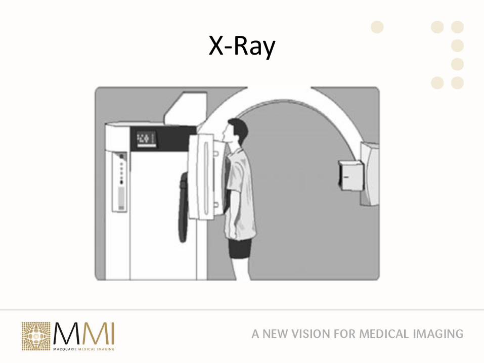
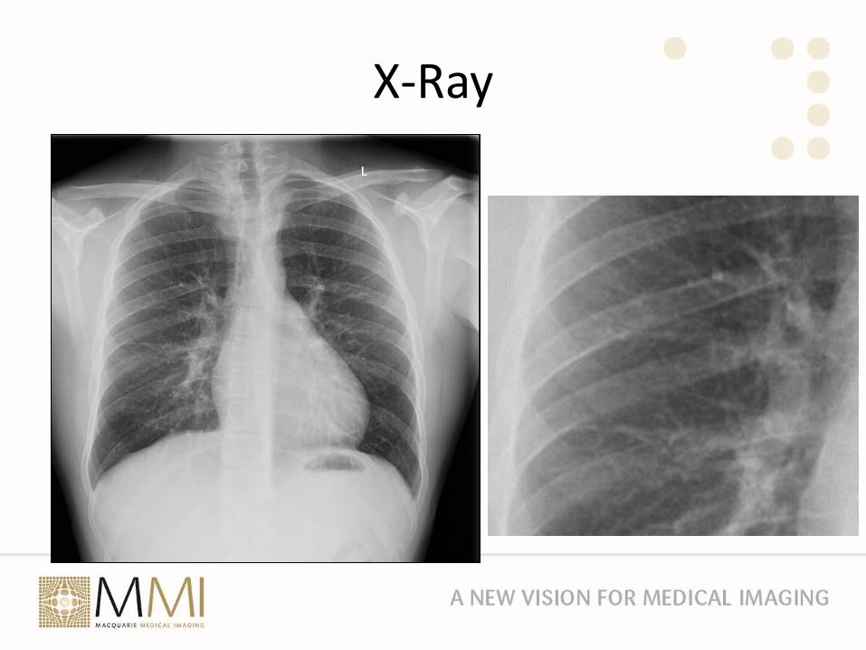
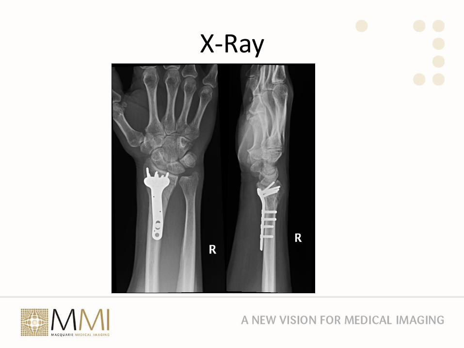
X-Ray• Simplest form of imaging using ionizing radiation
• Creates a “shadow”
• Good for bone and metal
• Divergent beam so some magnification of structures
• Overlapping soft tissue structures and bone makes interpretation difficult
• Dual energy X ray can separate bone and soft tissue
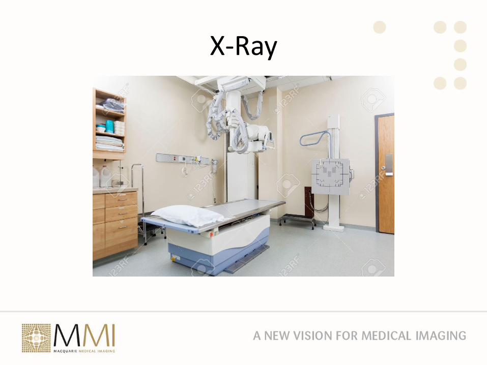
X-Ray
X-Ray
X-Ray
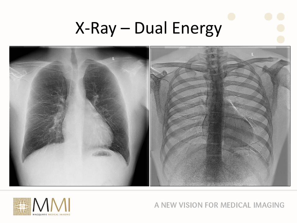
X-Ray – Dual Energy
X-Ray
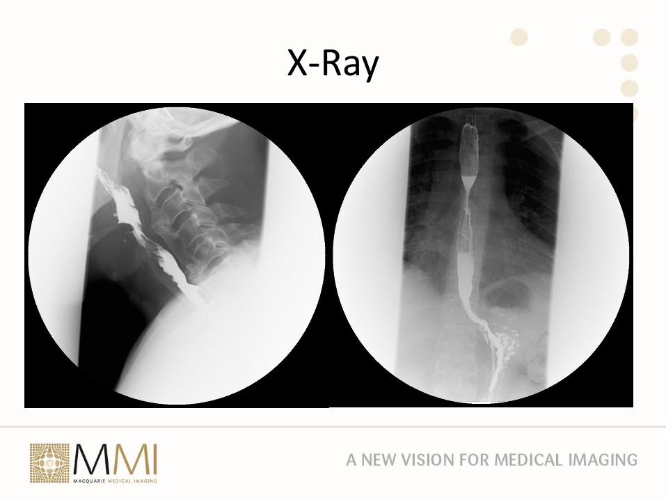
X-Ray• Contrast was initially introduced to demonstrate hollow organs like
the oesophagus, stomach and bowel
• Barium was radio opaque and not toxic (tastes like chalk)
• However still only produced 2 dimensional representations of objects
X-Ray
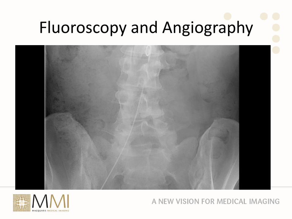
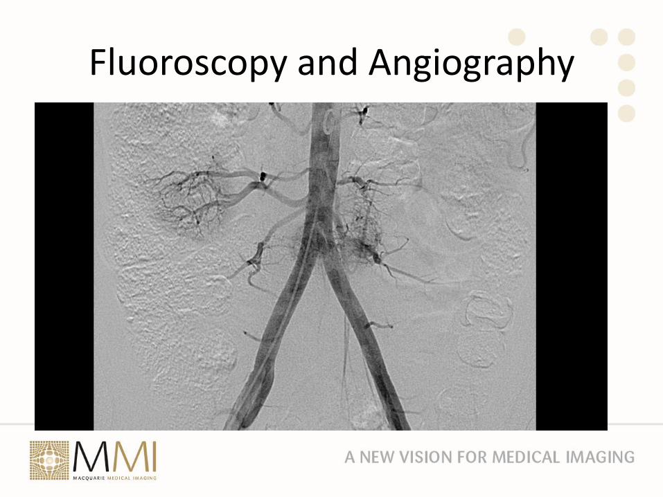
Fluoroscopy and Angiography• Rather than taking static images it was found that contrast
resolution could be enhanced by subtracting an image without contrast from one with contrast – Digital subtraction angiography
• With continuous fluoroscopy operations could be performed with image guidance and interventional radiology was born
• However that is a topic for another day
Fluoroscopy and Angiography
Fluoroscopy and Angiography
Computed Tomography• In 1924 the mathematical theory of tomographic image
reconstruction was developed by Johann Radon (Radon transform in 1917)
• In 1963 the theoretical basis of CT was described by Allan Cormack from Tufts University
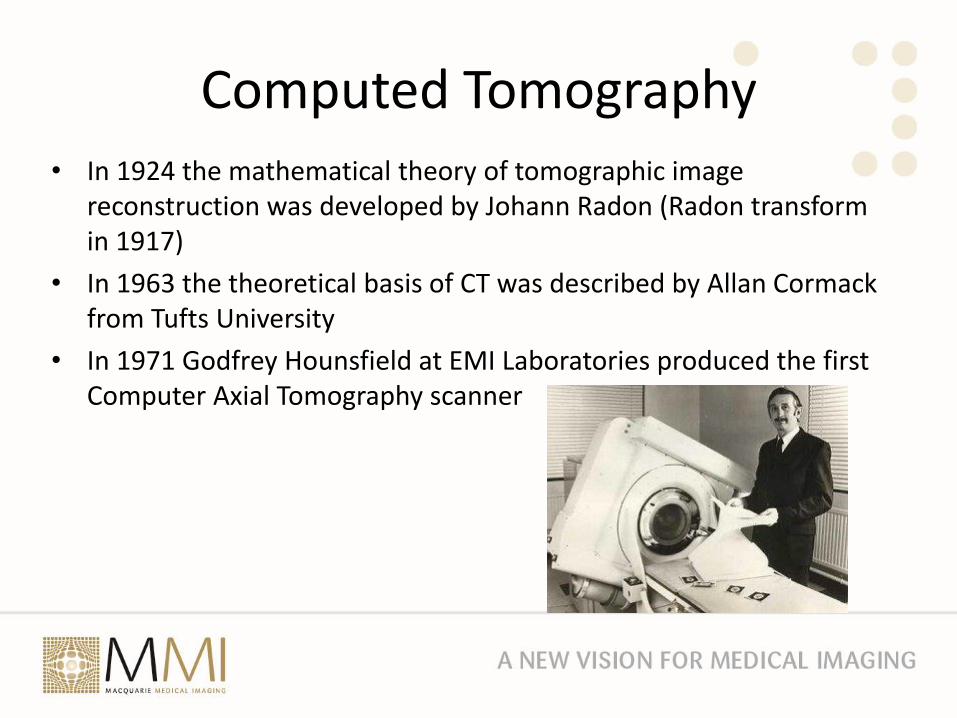
• In 1971 Godfrey Hounsfield at EMI Laboratories produced the first Computer Axial Tomography scanner
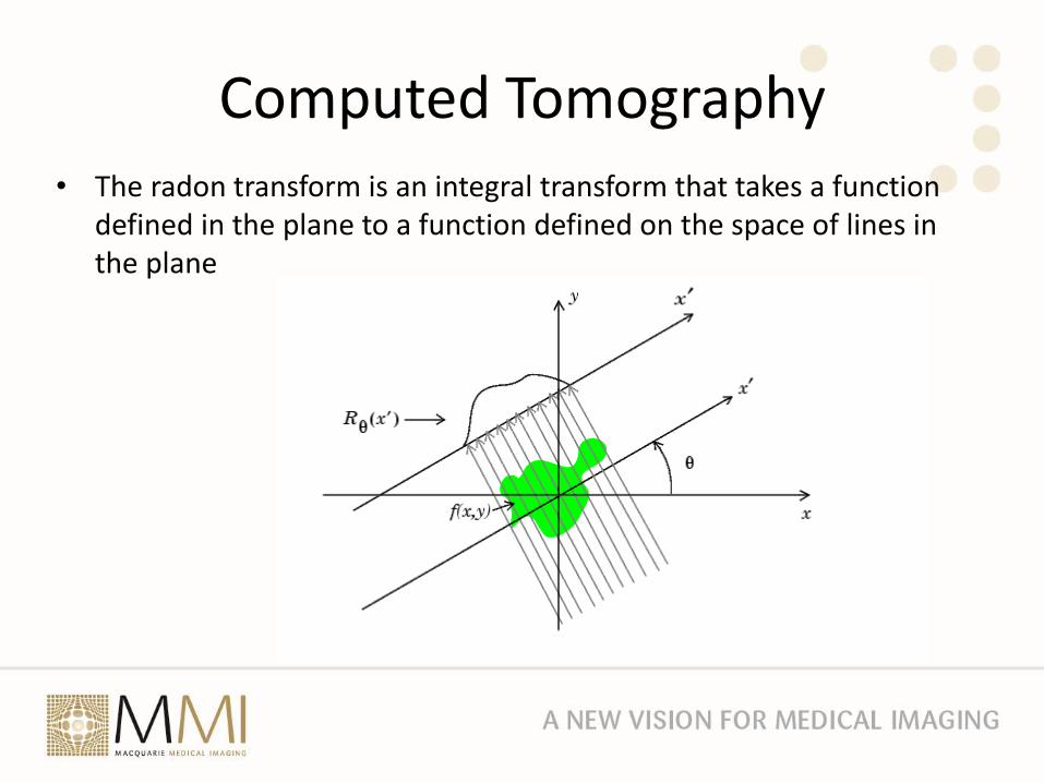
Computed Tomography• The radon transform is an integral transform that takes a function
defined in the plane to a function defined on the space of lines in the plane
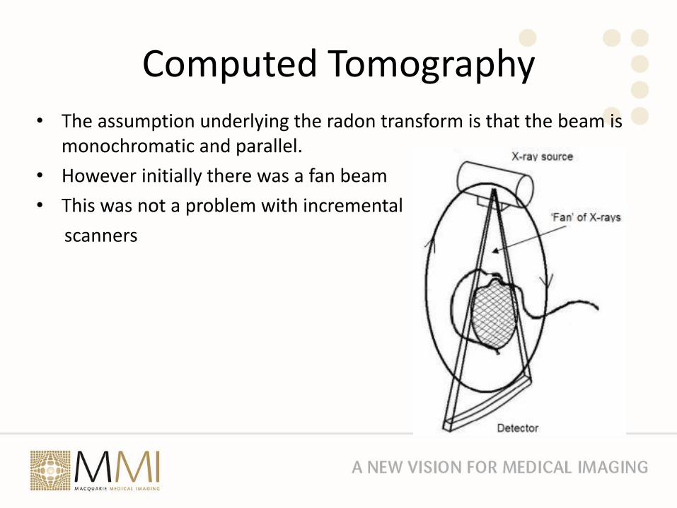
Computed Tomography• The assumption underlying the radon transform is that the beam is
monochromatic and parallel.
• However initially there was a fan beam
• This was not a problem with incremental
scanners
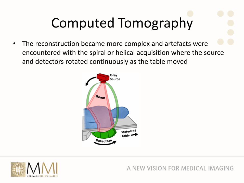
Computed Tomography• The reconstruction became more complex and artefacts were
encountered with the spiral or helical acquisition where the source and detectors rotated continuously as the table moved
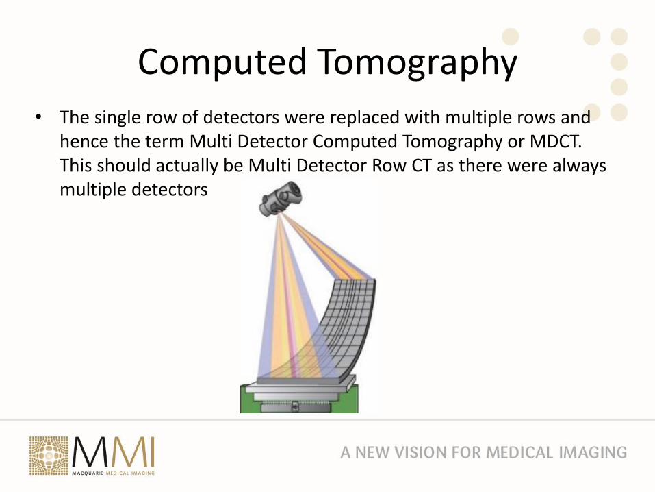
Computed Tomography• The single row of detectors were replaced with multiple rows and
hence the term Multi Detector Computed Tomography or MDCT. This should actually be Multi Detector Row CT as there were always multiple detectors
CT
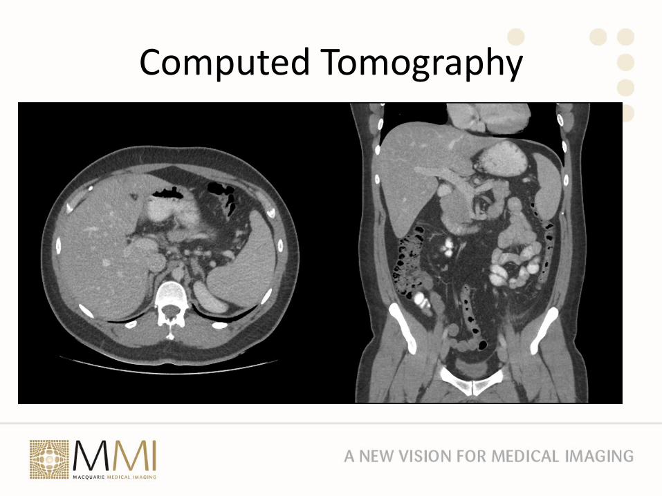
Computed Tomography
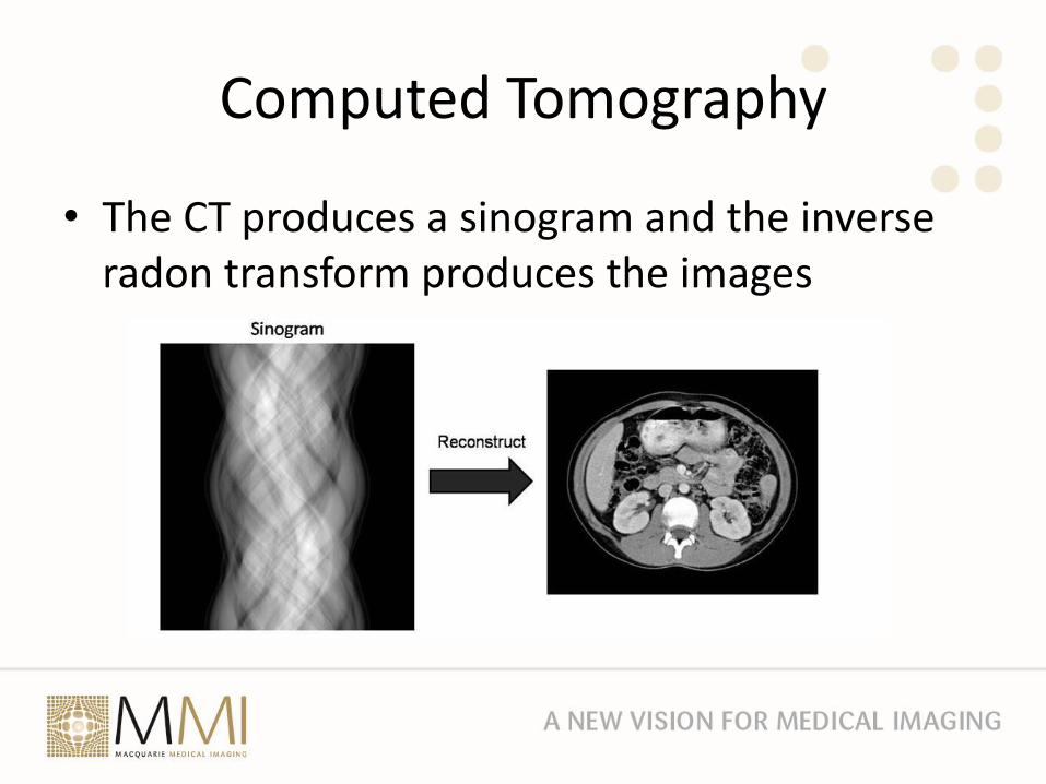
• The CT produces a sinogram and the inverse radon transform produces the images
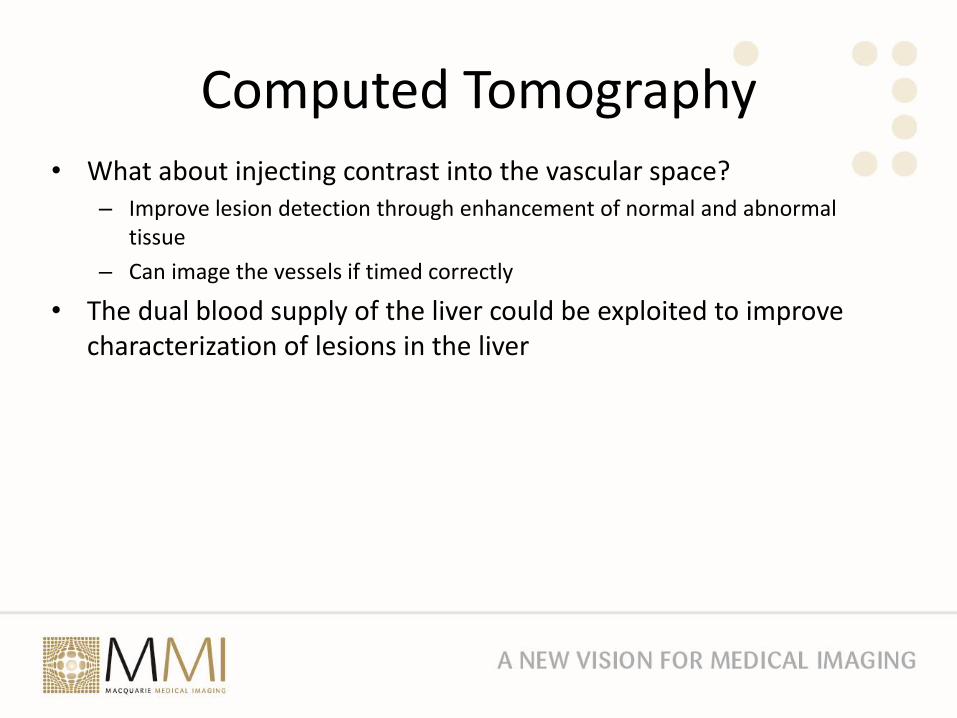
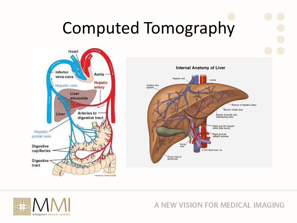
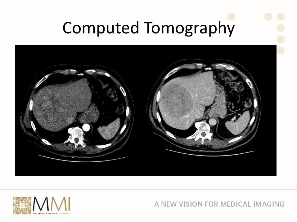
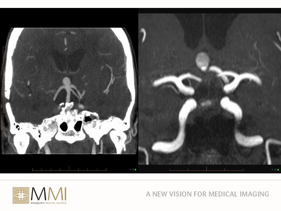
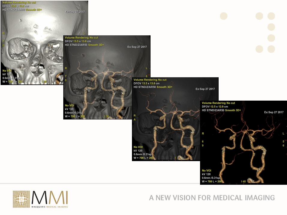
Computed Tomography• What about injecting contrast into the vascular space?
– Improve lesion detection through enhancement of normal and abnormal tissue
– Can image the vessels if timed correctly
• The dual blood supply of the liver could be exploited to improve characterization of lesions in the liver
Computed Tomography
Computed Tomography
Computed Tomography
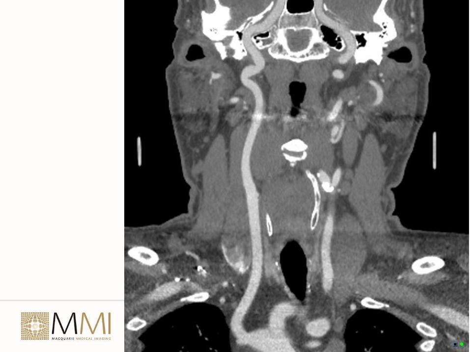
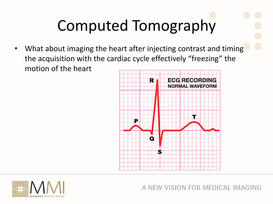
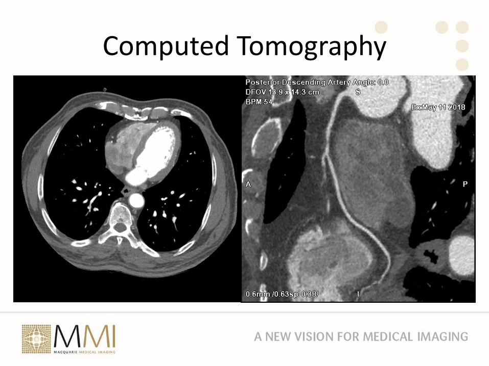
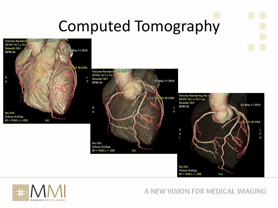
Computed Tomography• What about imaging the heart after injecting contrast and timing
the acquisition with the cardiac cycle effectively “freezing” the motion of the heart
Computed Tomography
Computed Tomography
Radiation RiskThe Glowing Elephant in the Room
• Since 1980 the has been a 600% increase in medical radiation exposure in the US
• Background Physicso Radiation Absorbed Dose measured in “rad”s or gray. 1 Gy = 100
rado 1 gray = 1 joule/kgo “dose rate” determines biological effect i.e. a reduced dose rate
but an increased exposure time for a given dose reduces biological effect
o Dose can be measuredo Effective dose is the sum of the equivalent dose to each tissue
and organ multiplied by a tissue weighting factor. Measured in Sieverts (Sv)
o Effective dose is estimated and cannot be measured
Effects of Radiation
• Deterministic
o Outcome is proportional to dose
o Examples include dermatitis and cataracts
• Stochastic
o Risk is related to dose
o Cancer and hereditary effects
o Most data from Japanese atomic bomb survivors and radiotherapy patients
o Life Span Study of 105,000 atomic bomb survivors showed a linear dose response for solid tumours
Effects of Radiation
• BUT …
• Lowest dose of X-ray or gamma radiation for which there is good evidence of increased cancer risk in humans is 10 to 50 mSv for acute exposure and 50 to 100 mSv for protracted exposure
• There is uncertainty that linear extrapolation of cancer risk from intermediate to low radiation doses is correct.
• In Australia background radiation is 2 mSv per year however no studies have shown an increased cancer risk in populations living in regions with more than 20 mSvper year
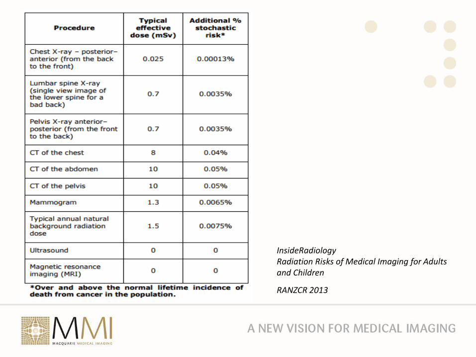
InsideRadiologyRadiation Risks of Medical Imaging for Adults and Children
RANZCR 2013
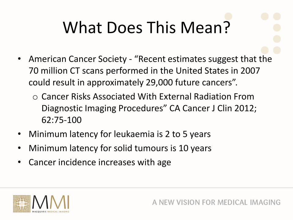
What Does This Mean?
• American Cancer Society - “Recent estimates suggest that the 70 million CT scans performed in the United States in 2007 could result in approximately 29,000 future cancers”.
o Cancer Risks Associated With External Radiation From Diagnostic Imaging Procedures” CA Cancer J Clin 2012; 62:75-100
• Minimum latency for leukaemia is 2 to 5 years
• Minimum latency for solid tumours is 10 years
• Cancer incidence increases with age
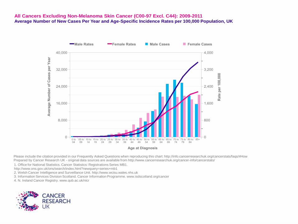
All Cancers Excluding Non-Melanoma Skin Cancer (C00-97 Excl. C44): 2009-2011 Average Number of New Cases Per Year and Age-Specific Incidence Rates per 100,000 Population, UK
Please include the citation provided in our Frequently Asked Questions when reproducing this chart: http://info.cancerresearchuk.org/cancerstats/faqs/#How Prepared by Cancer Research UK - original data sources are available from http://www.cancerresearchuk.org/cancer-info/cancerstats/
1. Office for National Statistics. Cancer Statistics: Registrations Series MB1.
http://www.ons.gov.uk/ons/search/index.html?newquery=series+mb1
2. Welsh Cancer Intelligence and Surveillance Unit. http://www.wcisu.wales.nhs.uk
3. Information Services Division Scotland. Cancer Information Programme. www.isdscotland.org/cancer
4. N. Ireland Cancer Registry. www.qub.ac.uk/nicr
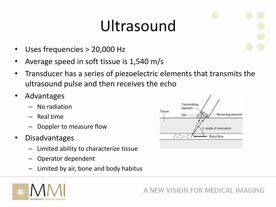
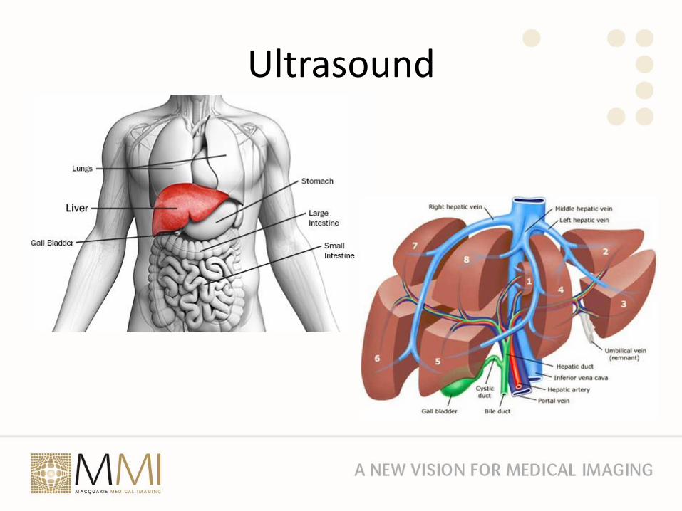
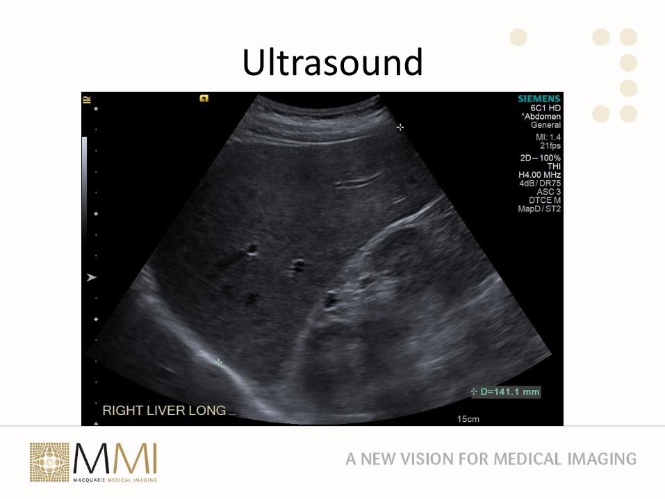
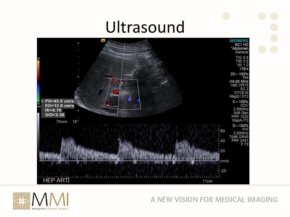
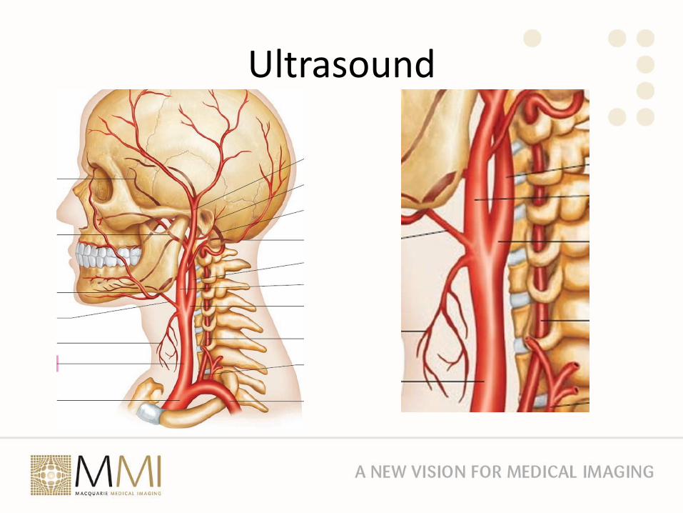
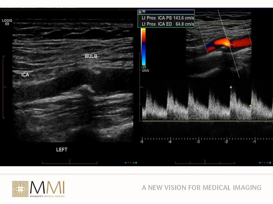
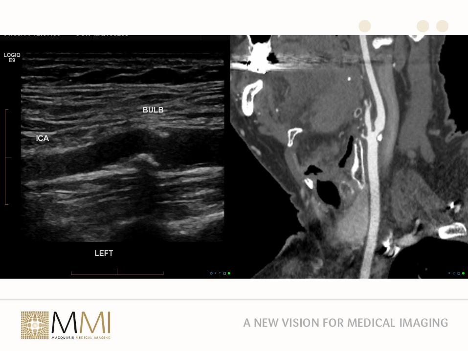
Ultrasound• Uses frequencies > 20,000 Hz
• Average speed in soft tissue is 1,540 m/s
• Transducer has a series of piezoelectric elements that transmits the ultrasound pulse and then receives the echo
• Advantages– No radiation
– Real time
– Doppler to measure flow
• Disadvantages– Limited ability to characterize tissue
– Operator dependent
– Limited by air, bone and body habitus
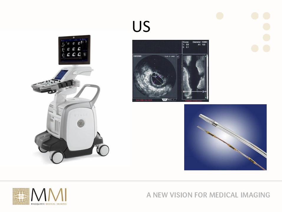
US
Ultrasound
Ultrasound
Ultrasound
Ultrasound

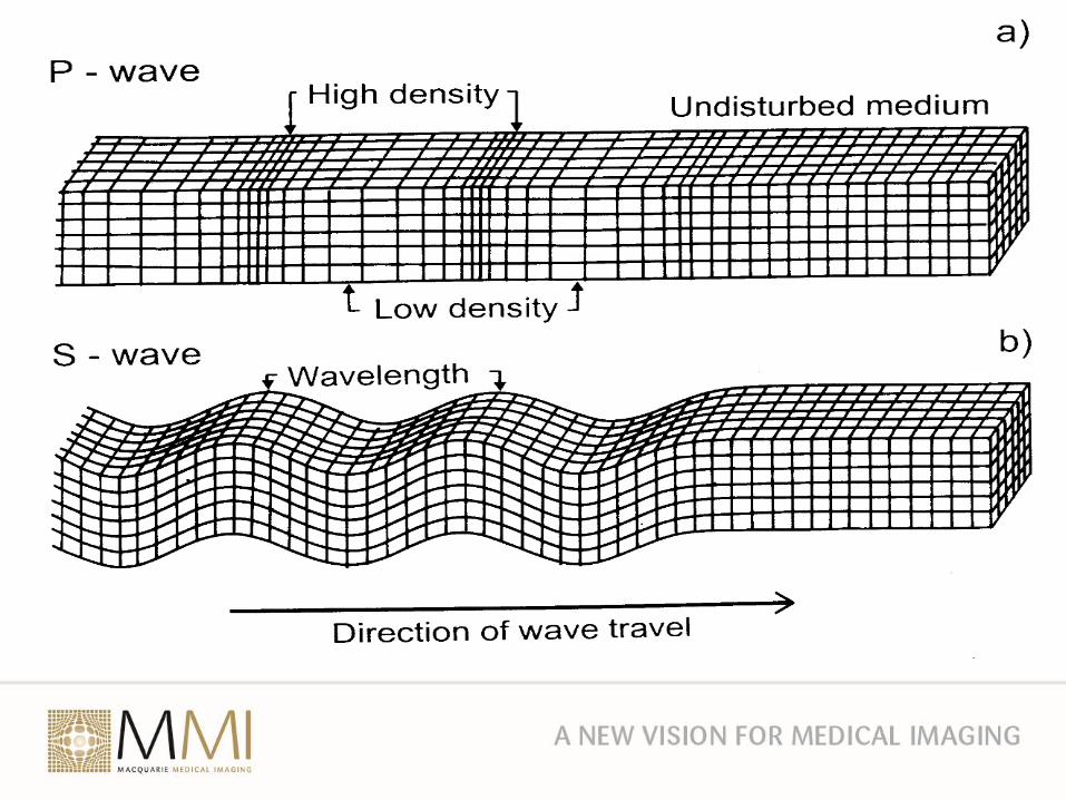
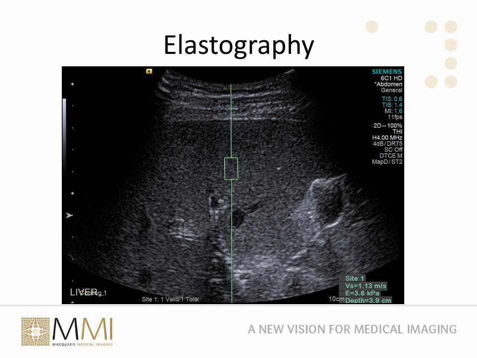
Elastography
• Ultrasound can be used to generate a shear wave in materials.
• The speed of propagation of the shear wave is related to stiffness of the material
• The faster the shear wave travels the stiffer the material
• Ultrasound is a compression wave and travels ~1000 times faster than a shear wave
Elastography
• Good for measuring
o maturity of cheese
o fibrosis of the liver
• Chronic liver disease such as in Hepatitis and NASH results in fibrosis and eventually cirrhosis
• Regular liver elastography should be part of routine surveillance in hepatitis and other chronic liver disease
Elastography
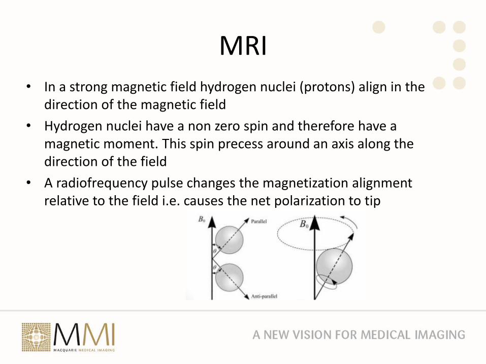
MRI• In a strong magnetic field hydrogen nuclei (protons) align in the
direction of the magnetic field
• Hydrogen nuclei have a non zero spin and therefore have a magnetic moment. This spin precess around an axis along the direction of the field
• A radiofrequency pulse changes the magnetization alignment relative to the field i.e. causes the net polarization to tip
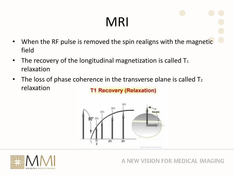
MRI• When the RF pulse is removed the spin realigns with the magnetic
field
• The recovery of the longitudinal magnetization is called T1
relaxation
• The loss of phase coherence in the transverse plane is called T2
relaxation
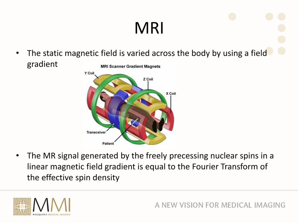
MRI• The static magnetic field is varied across the body by using a field
gradient
• The MR signal generated by the freely precessing nuclear spins in a linear magnetic field gradient is equal to the Fourier Transform of the effective spin density

MRI• The inverse Fourier Transform of the MR signal produces the image
• There is an infinite variety of RF and gradient pulses that gives the different types of images– Spin echo: T1, T2, PD
– Gradient Echo
– Inversion Recovery: FLAIR, STIR
– Diffusion Weighted: DWI, DTI
– Perfusion Weighted: ASL
– Functional MRI: BOLD
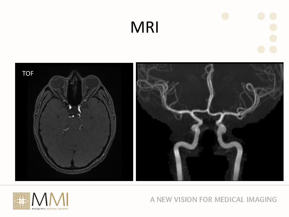
– Magnetic Resonance Angiography and venography: TOF
– Susceptibility Weighted
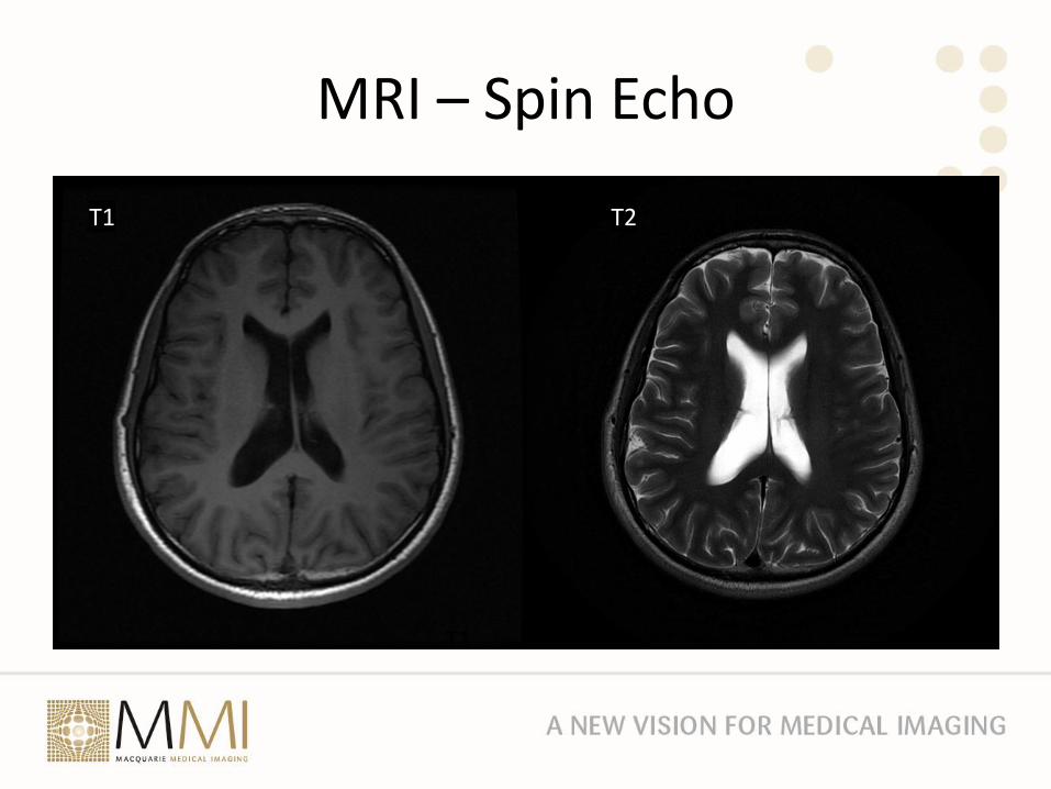
MRI – Spin Echo
T1
T1 T2
MRI
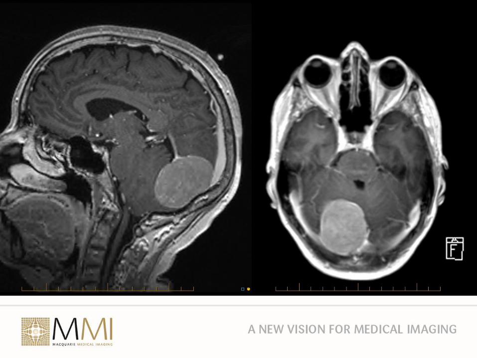
Neurological Imaging - MRI
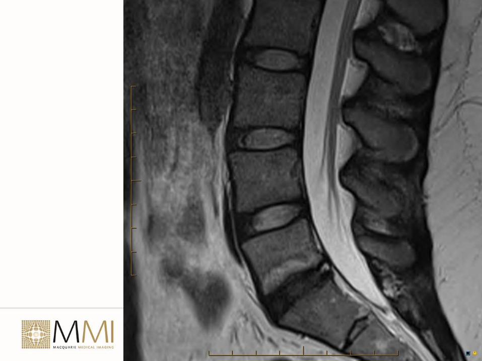
• Gold standard for the brain and spine
• Should use for chronic headache and seizures
• Soft tissue characterization
• Neck and Back pain with radiculopathy MRI is the modality of choice
• Can show
o Disc disease
o Neural compression
o CNS tumours – primary and secondary
o Bone lesions
MRI
TOF
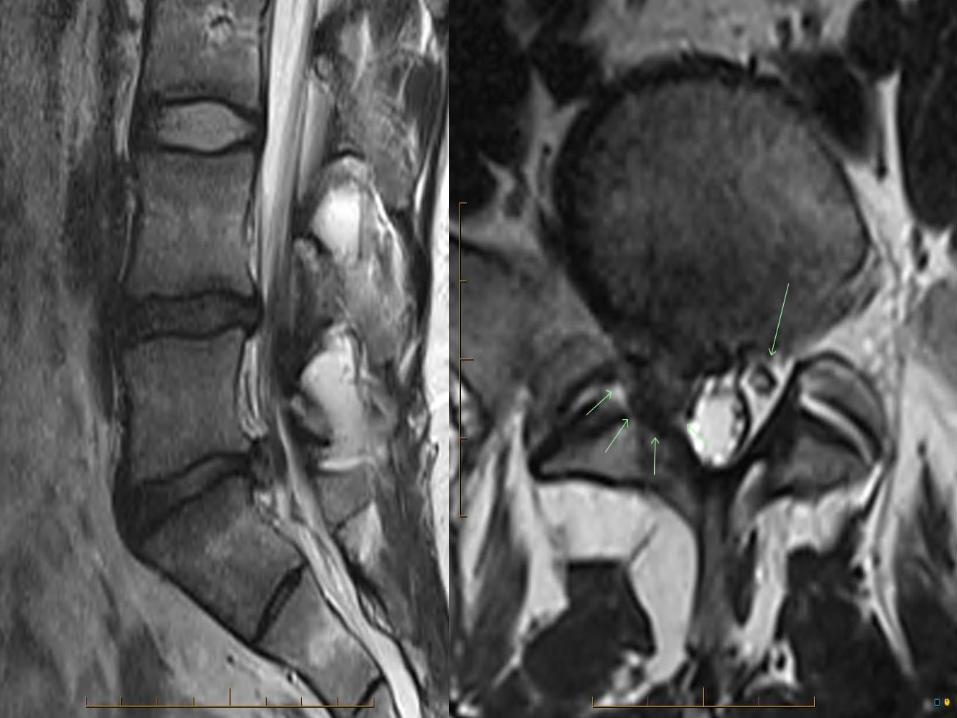
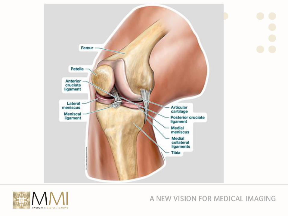
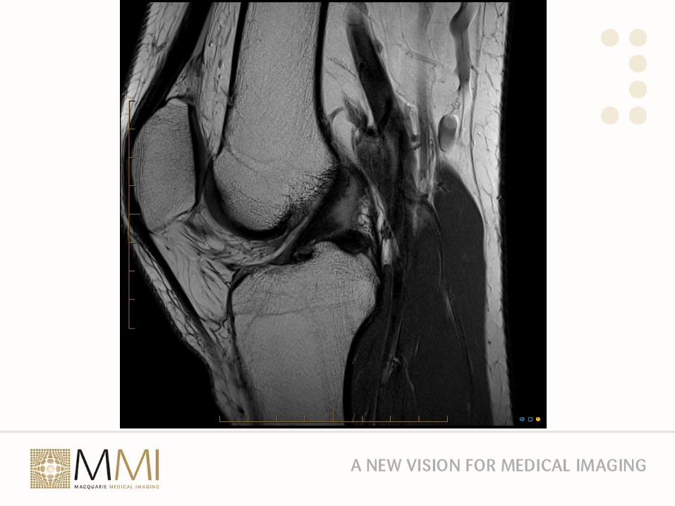
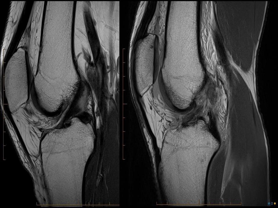
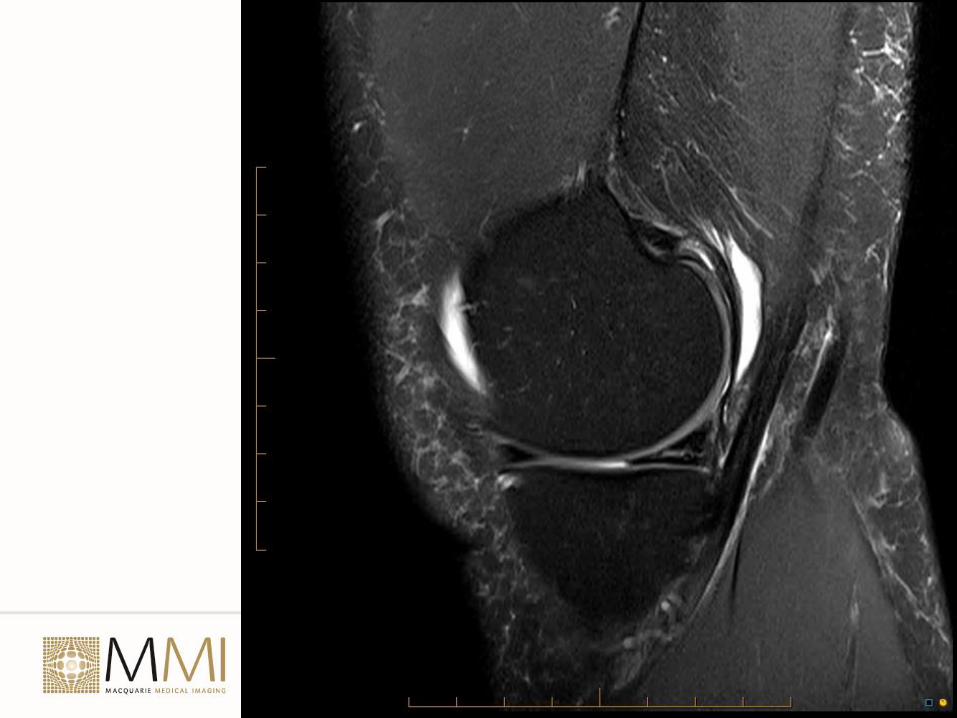
Musculoskeletal MRI
• The best test for joints particularly large joint
• Can assess bone, cartilage, menisci, ligaments, tendons, muscles
o Injury – sprains and tears. Bone contusion and fractures. Osteochondral lesions
o Inflammation – osteomyelitis, arthritis, bursitis
o Vascular – AVN, vascular malformations
o Neoplasm – Characterisation of bone and soft tissue sarcomas. Metastases
• Static images. Cannot assess function (yet)
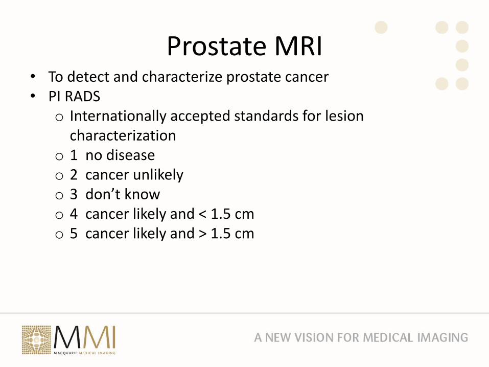
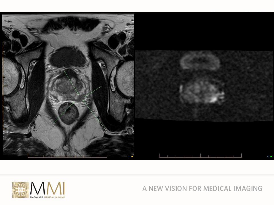
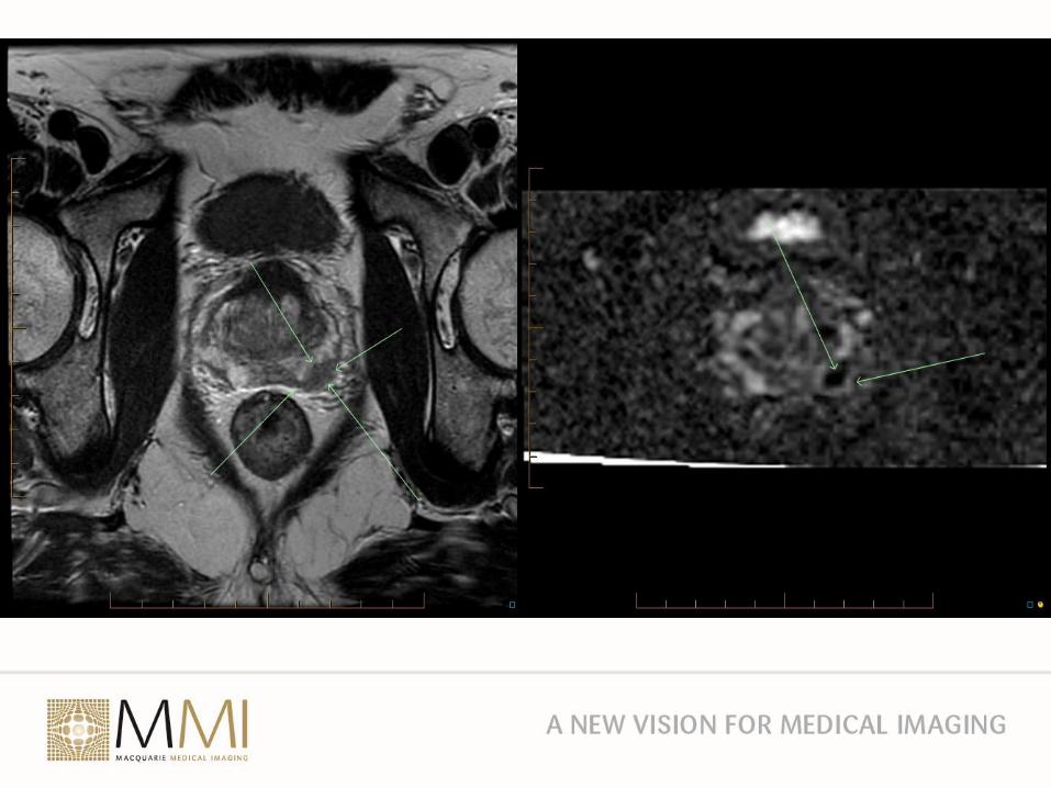
Prostate MRI• To detect and characterize prostate cancer• PI RADS
o Internationally accepted standards for lesion characterization
o 1 no diseaseo 2 cancer unlikelyo 3 don’t knowo 4 cancer likely and < 1.5 cmo 5 cancer likely and > 1.5 cm
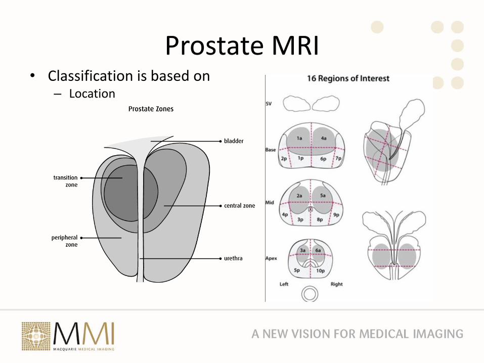
Prostate MRI• Classification is based on
– Location
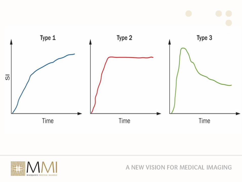
Prostate MRI– T2 hypointensity– Morphology of the lesion– Diffusion weighted imaging – Apparent Diffusion Coefficient which is a
measure of the magnitude of diffusion of water within the tissue– Contrast enhancement
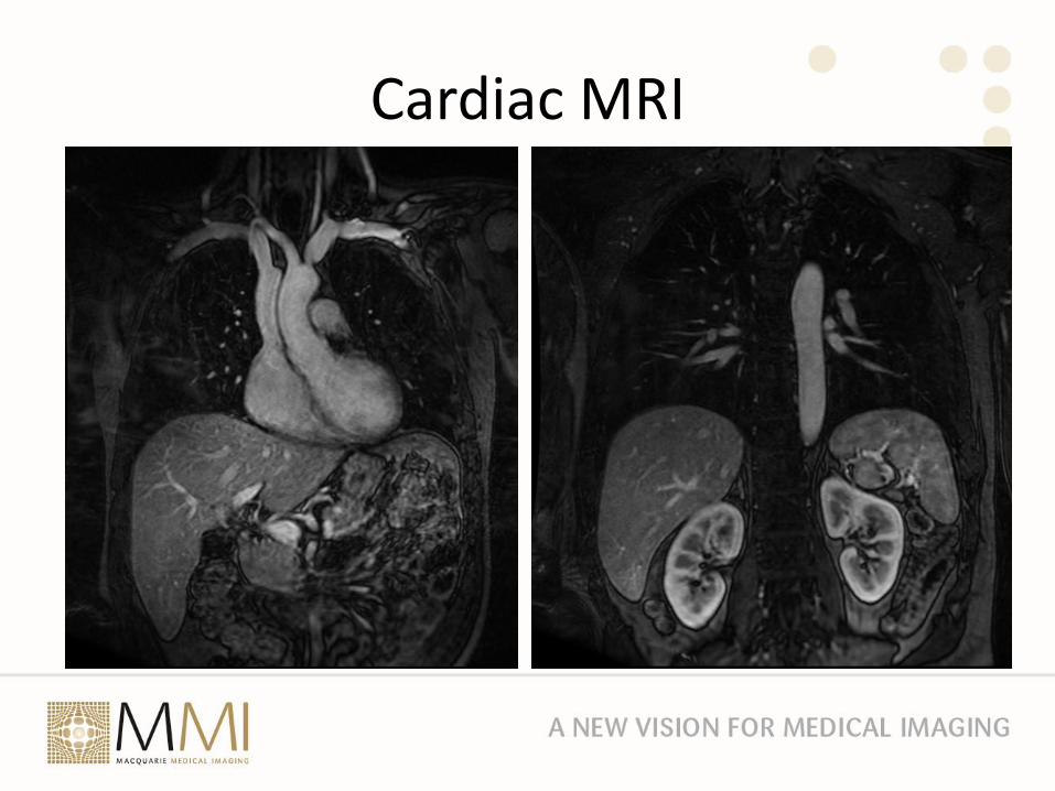
Cardiac MRI
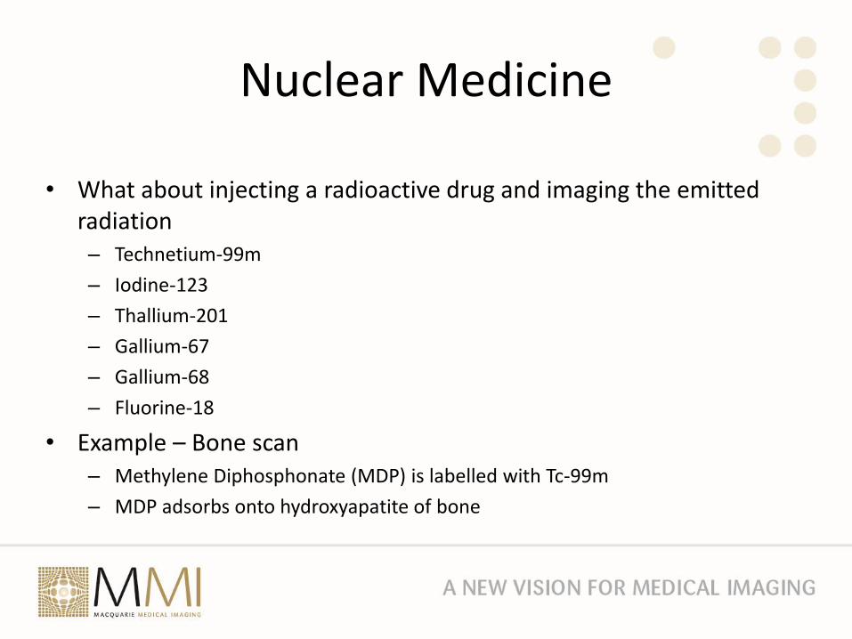
Nuclear Medicine
• What about injecting a radioactive drug and imaging the emitted radiation– Technetium-99m
– Iodine-123
– Thallium-201
– Gallium-67
– Gallium-68
– Fluorine-18
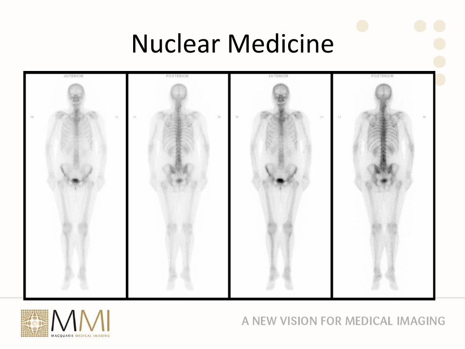
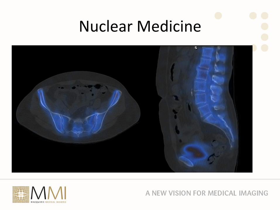
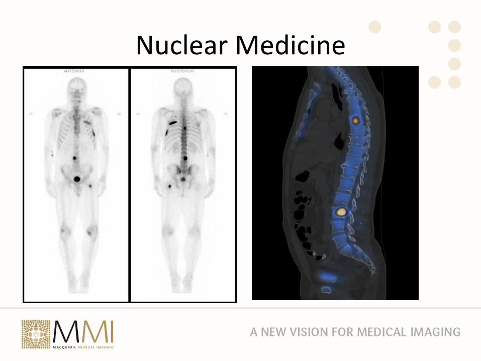
• Example – Bone scan– Methylene Diphosphonate (MDP) is labelled with Tc-99m
– MDP adsorbs onto hydroxyapatite of bone
Nuclear Medicine
Nuclear Medicine

Nuclear Medicine
Nuclear Medicine
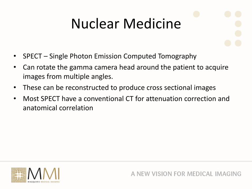
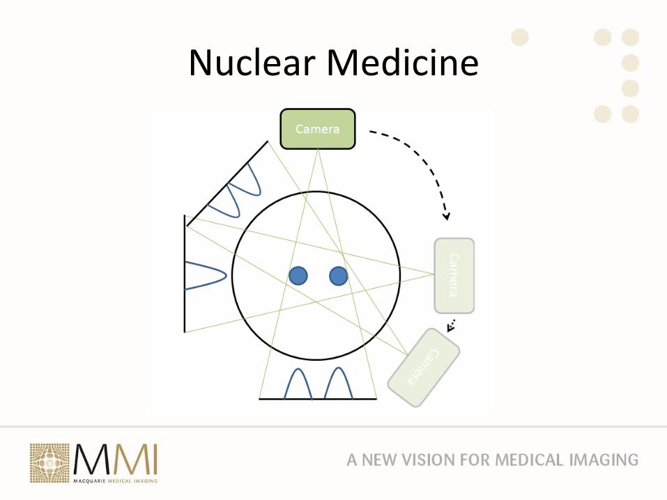
• SPECT – Single Photon Emission Computed Tomography
• Can rotate the gamma camera head around the patient to acquire images from multiple angles.
• These can be reconstructed to produce cross sectional images
• Most SPECT have a conventional CT for attenuation correction and anatomical correlation
Nuclear Medicine
Nuclear Medicine
Nuclear Medicine
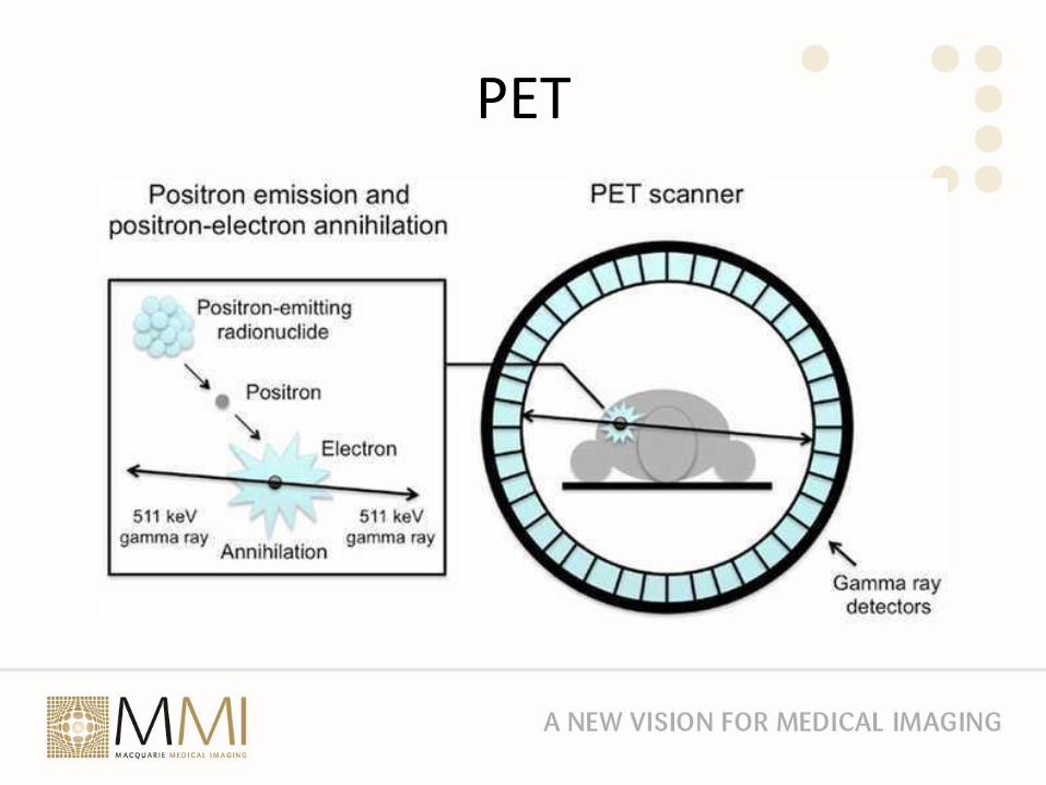
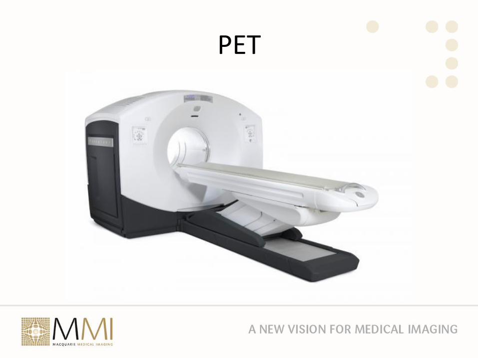
PET
• Positron Emission Tomography
• Radioactive elements like I-123 will enter the normal physiological processes hence I-123 will be taken up by the thyroid
• Technetium-99m has to be bound to a carrier eg MDP for bone scans – think passenger and taxi
• Replacing an atom in a molecule with a radioactive isotope allows you to observe the molecule entering normal physiological processes eg carbon-11 dopamine
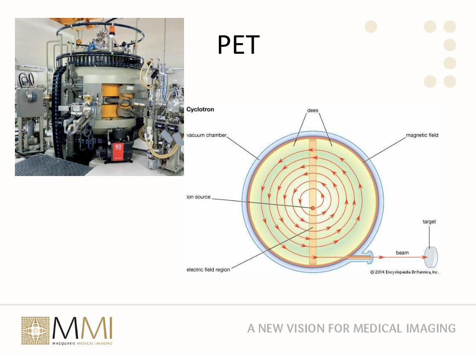
• Positron emitters such as carbon-11 and fluorine-18 can be produced in a cyclotron
PET
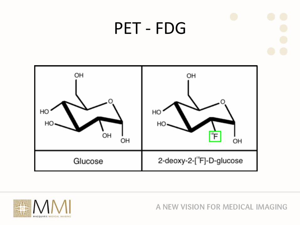
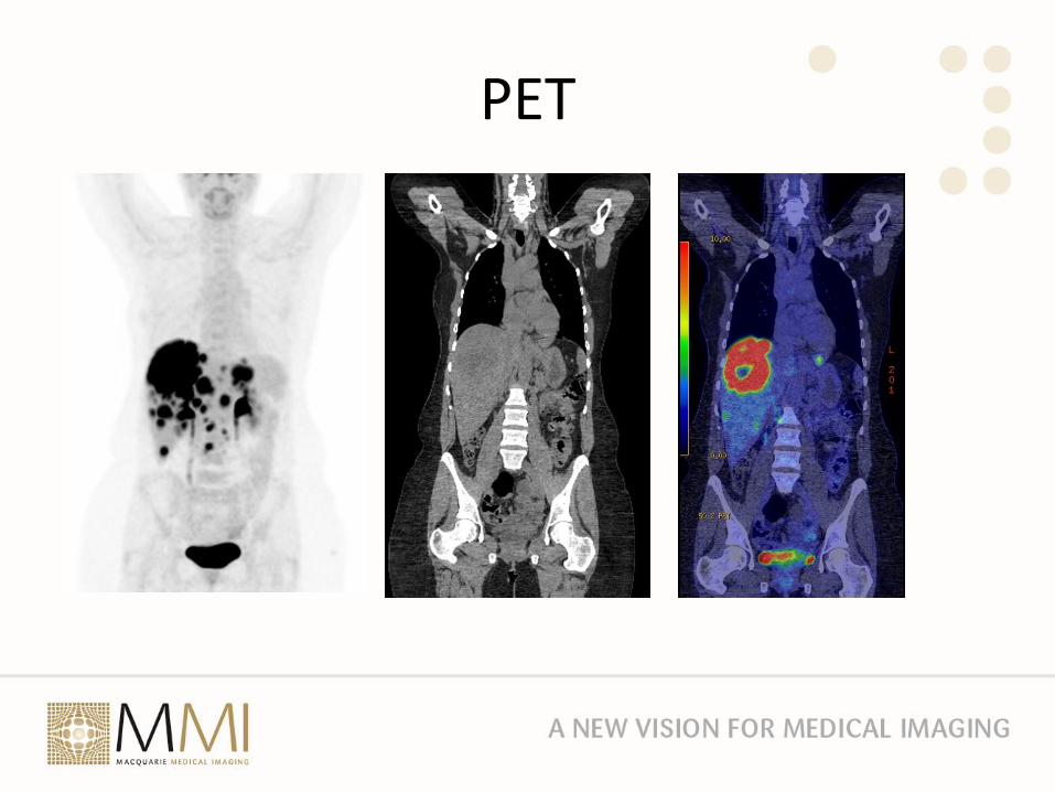
PET - FDG
PET
PET
PET
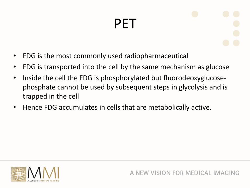
• FDG is the most commonly used radiopharmaceutical
• FDG is transported into the cell by the same mechanism as glucose
• Inside the cell the FDG is phosphorylated but fluorodeoxyglucose-phosphate cannot be used by subsequent steps in glycolysis and is trapped in the cell
• Hence FDG accumulates in cells that are metabolically active.
PET
Thank you