investigating mechanisms of telomere end-protection
TRANSCRIPT
Rockefeller UniversityDigital Commons @ RU
Student Theses and Dissertations
2015
Investigating Mechanisms of Telomere End-ProtectionShaheen Kabir
Follow this and additional works at: http://digitalcommons.rockefeller.edu/student_theses_and_dissertations
Part of the Life Sciences Commons
This Thesis is brought to you for free and open access by Digital Commons @ RU. It has been accepted for inclusion in Student Theses andDissertations by an authorized administrator of Digital Commons @ RU. For more information, please contact [email protected].
Recommended CitationKabir, Shaheen, "Investigating Mechanisms of Telomere End-Protection" (2015). Student Theses and Dissertations. Paper 269.

INVESTIGATING MECHANISMS OF TELOMERE
END-PROTECTION
A Thesis Presented to the Faculty of
The Rockefeller University
in Partial Fulfillment of the Requirements for
the degree of Doctor of Philosophy
by
Shaheen Kabir
June 2015

© Copyright by Shaheen Kabir 2015

INVESTIGATING MECHANISMS OF TELOMERE
END-PROTECTION
Shaheen Kabir, Ph.D.
The Rockefeller University 2015
Shelterin is an essential telomeric protein complex that prevents DNA
damage signaling and DNA repair in a compartmentalized manner. We assessed
contributions of the conserved shelterin component, Rap1, to telomere end-
protection. Rap1 was first discovered in budding yeast as a transcription factor
and was later shown to bind directly to telomeres. Two important functions of
Rap1 in yeast are: the negative regulation of telomere length, and the inhibition of
the double-strand break (DSB) repair pathway non homologous end-joining
(NHEJ). Mammalian Rap1 interacts with shelterin factor TRF2, to localize to
telomeres, and human Rap1 is implicated in repressing NHEJ.
Surprisingly, removal of Rap1 from telomeres revealed that mouse Rap1
was not required to inhibit NHEJ, but instead was critical for the repression of the
DSB repair pathway homology directed repair (HDR). We showed that complex
formation of Rap1 and TRF2 was most likely necessary to repress HDR,
although the mechanism of how it does so remains to be determined.
Two discrepancies exist between mouse and human Rap1 regarding
repression of NHEJ and regulation of telomere length. Human Rap1 was
proposed to inhibit NHEJ, but we observed no evidence of telomere fusions in

the mouse Rap1 knockout. Similarly, telomere elongation was observed upon
knockdown of human Rap1, but no telomere length phenotypes were observed in
Rap1-deficient mice. With the advent of new genome-editing technologies that
facilitate targeting in human cells, we constructed TALEN-mediated knockouts of
human Rap1 in numerous cell lines. Loss of human Rap1 did not lead to an
induction of NHEJ, or show consistent changes in telomere length, indicating that
similar to mouse Rap1, human Rap1 does not have an important function in
protection or length regulation of human telomeres. Instead, we found that
mammalian Rap1, like its unicellular orthologs, affects gene expression.
Therefore, perhaps the conservation of Rap1 reflects its role in transcriptional
regulation rather than a function at telomeres.
Organismal discrepancies regarding the function of shelterin components,
other than Rap1, also exist. Targeting of human shelterin component POT1
(POT1a/b in the mouse) with shRNAs shows a reduction in 3’ telomere
overhangs and a mild induction of DNA damage signaling. However, deletion of
mouse POT1a and –b results in extended 3’ overhangs and a massive induction
of the DNA damage response. To understand the role of human POT1 in
telomere protection, we used TALENs to generate human knockout cell lines
lacking POT1. We found that similar to mouse POT1, deletion of human POT1
elicited significant DNA damage signaling. Strikingly, the amount of 3’ single-
stranded DNA remained unchanged upon loss of POT1, highlighting a potential
difference in overhang regulation between mice and humans.

iii
In memory of –
My father

iv
ACKNOWLEDGEMENTS
I extend my gratitude to my advisor, Titia de Lange, who devoted an
extraordinary amount of time and energy into mentoring me, from which I have
benefitted immensely. I thank my committee members Hironori Funabiki and
Michael Young for their guidance over the years. I also thank Julie Cooper for
agreeing to be the external examiner at my thesis defense. I thank Cris, Kristin,
Marta, Stephanie, Emily and Sid at the Deans office for helping students navigate
their graduate careers without any bureaucratic hassles.
I extend my appreciation to members of the de Lange lab, past and
present, for their scientific support and for the collegial atmosphere at work. I
thank Megan van Overbeek for showing me the ropes during my rotation, Hiro
Takai for discussions on POT1, and John Maciejowski for reading parts of my
thesis. The lab would not run without the support of Devon White, Adriana
Garzon, Rosaura Mejia and Stew Barnes, and for that, I am grateful. I especially
thank Ylli Doksani for our morning coffees, and Francisca Lottersberger for our
afternoon coffees, which I will miss dearly. I’m thankful for my New York ‘family’,
Agnel Sfeir, Isabelle Schmutz and Nazario Bosco for their constant support and
our limitless adventures.
I thank my dear friends Courtney Klein, Chelsa Crowley, Mari Sheibley,
Mieun Lee, Erika Brunet, Payaal Patel, Estee Perlmutter for keeping me sane in
the best possible ways. I’m grateful to Anna Kruyer for being a fantastic

v
roommate and friend. I would also like to acknowledge Daria Zamolodchikov who
introduced me to Styles in Microsoft Word, which greatly facilitated the formatting
of this thesis.
Importantly, I thank my family, Khalid Kabir, Aparna Sarma and Zayed for
bringing joy into my life. My parents, Hasina Kabir and Kabir Hyderally deserve
the most thanks of all. I am inspired by my mother’s strength, intelligence, and
infinite capacity to love. I owe my work ethic to my father, who epitomized
humility and perseverance, and took on every task with an unmatched rigor,
asking for nothing in return. He will be remembered as a selfless man,
passionate about his work and his family, traits I can only hope to embody.
Lastly, I acknowledge Alexandre Bolze, whose admiration I strive to live up to.
His scientific curiosity engages me and his passion for pushing limits motivates
me.

vi
TABLE OF CONTENTS
Chapter 1: Introduction ...................................................................................... 1
1.1 The structure and function of telomeres .................................................... 2
1.2 Threats to chromosome termini ................................................................ 6
1.2.1 Telomerase counteracts the end-replication problem ........................... 6
1.2.2 Telomere length regulation by telomere-binding proteins ..................... 9
1.2.3 The end-protection problem ................................................................ 11
1.2.4 Shelterin solves the end-protection problem ....................................... 18
1.2.5 Phenotypes of TRF2 depletion ............................................................ 21
1.2.6 Phenotypes of POT1 depletion ........................................................... 23
1.3 Proposed functions and mechanisms of action for Rap1 ........................ 25
1.3.1 Identification of Rap1 .......................................................................... 25
1.3.2 Rap1 and telomere length regulation .................................................. 26
1.3.3 Rap1 and end-protection ..................................................................... 27
1.3.4 A role for Rap1 in meiosis and chromatin organization ....................... 30
1.3.5 Non-telomeric functions of mammalian Rap1 ..................................... 30
1.4 Objectives ............................................................................................... 32
Chapter 2: Loss of Rap1 Induces Telomere Recombination ........................ 34
2.1 Introduction ............................................................................................. 35
2.2 Results .................................................................................................... 35
2.2.1 Deletion of Rap1 does not affect cell and organismal viability ............ 35
2.2.2 No induction of DDR or NHEJ at telomeres lacking Rap1 .................. 40
2.2.3 Telomere length and chromatin unchanged in Rap1-deficient cells ... 47
2.2.4 Rap1 is a repressor of telomere recombination .................................. 47
2.3 Summary of findings ............................................................................... 53

vii
Chapter 3: Elucidating the mechanism of HDR repression by Rap1 ........... 55
3.1 Introduction ............................................................................................. 56
3.2 Results .................................................................................................... 58
3.2.1 The C-terminus of Rap1 is required to repress telomeric HDR ........... 58
3.2.2 Rap1 does not inhibit HDR at telomeres lacking TRF2 ....................... 61
3.2.3 Rap1 confers an increase in TRF2 DNA binding affinity. .................... 65
3.2.4 HDR is not aggravated by dual absence of Rap1 and POT1 .............. 66
3.2.5 Deletion of Rap1 and TRF1 does not induce T-SCEs ......................... 70
3.2.6 HDR is repressed in late-passage Ku70-deficient MEFs .................... 72
3.3 Summary of findings ............................................................................... 74
Chapter 4: TALEN gene knockouts of human Rap1 ...................................... 76
4.1 Introduction ............................................................................................. 77
4.2 Results .................................................................................................... 78
4.2.1 Efficient TALEN-mediated knockout of human Rap1 .......................... 78
4.2.2 Rap1-deficient cell proliferate and maintain fully protected telomeres 84
4.2.3 Unaltered telomere length dynamics in absence of Rap1. .................. 87
4.2.4 Telomeric positioning in Rap1 null cells .............................................. 91
4.2.5 No change in telomeric chromatin or transcription upon Rap1 loss .... 93
4.2.6 Rap1 affects transcriptional regulation ................................................ 95
4.3 Summary of findings ............................................................................... 98
Chapter 5: Investigating the role of human POT1 at telomeres ................. 100
5.1 Introduction ........................................................................................... 101
5.2 Results .................................................................................................. 102
5.2.1 The human POT1 targeting strategy ................................................. 102
5.2.2 TALEN-mediated deletion of full-length human POT1 ...................... 107
5.2.3 Telomere deprotection in POT1-deficient cells ................................. 112
5.2.4 Maintenance of telomere overhangs ................................................. 115
5.3 Summary of findings ............................................................................. 122
viii
Chapter 6: Discussion .................................................................................... 124
6.1 The role of mammalian Rap1 ................................................................ 125
6.1.1 Rap1 – a conserved telomeric protein with non-telomeric functions . 126
6.1.2 Rationale for retention of Rap1 at telomeres .................................... 129
6.1.3 No Rap1 ortholog in Drosophila ........................................................ 131
6.2 The mechanism of HDR repression at telomeres ................................. 132
6.2.1 The effect of Rap1 on the DNA-binding activity of TRF2 .................. 133
6.2.2 Rap1, TRF2 and POT1 may work together to repress HDR ............. 134
6.2.3 What is the telomeric substrate for HDR? ......................................... 135
6.2.4 Ku70/80-deficient cells adapt to recover repression of HDR ............ 136
6.3 Investigating the role(s) of human POT1 .............................................. 138
6.3.1 Functions of POT1 in telomere end-protection ................................. 138
6.3.2 Telomere length regulation by POT1-55 ........................................... 140
6.3.3 Generation of telomeric overhangs ................................................... 141
6.3.4 POT1 mutations - a compelling link between telomere dysfunction and
disease ....................................................................................................... 143
Chapter 7: Materials and Methods ................................................................ 145
7.1 General procedures .............................................................................. 146
7.1.1 Mammalian cell culture ..................................................................... 146
7.1.2 Retroviral gene delivery .................................................................... 146
7.1.3 Growth curves ................................................................................... 147
7.1.4 Immunoblotting (IB) ........................................................................... 148
7.1.5 Coimmunoprecipitation (Co-IP) from transfected 293T cells ............ 148
7.1.6 Immunofluorescence-fluorescence in situ hybridization (IF-FISH) .... 149
7.1.7 Telomeric FISH and chromosome orientation-FISH (CO-FISH) ....... 150
7.1.8 Chromatin immunoprecipitation (ChIP) ............................................. 151
7.1.9 Genomic blotting, telomere length and telomere overhang analysis 152
7.1.10 Northern analysis for TERRA .......................................................... 154
7.1.11 Microarray analysis ......................................................................... 154

ix
7.1.12 Quantitative reverse transcription-polymerase chain reaction (qRT-
PCR) .......................................................................................................... 155
7.1.13 Nickel-affinity purification of His-tagged proteins ............................ 156
7.1.14 Electrophoretic Mobility Shift Assays (EMSA) ................................. 156
7.1.15 Genotyping and sequencing ........................................................... 157
7.1.16 Gene targeting and cell cloning ....................................................... 158
7.2 Mouse and human TERF2IP targeting .................................................. 159
7.2.1 Mouse TERF2IP targeting (by Agnel Sfeir) ....................................... 159
7.2.2 TALENs and the TERF2IP targeting construct ................................. 160
7.3 POT1 gene targeting ............................................................................. 161
7.3.1 TALENs and the POT1 targeting construct ....................................... 161
7.4 List of primers ....................................................................................... 163
7.4.1 Genotyping ........................................................................................ 163
7.4.2 RT-PCR ............................................................................................. 164
7.4.3 EMSA probes .................................................................................... 166
7.5 List of shRNAs ...................................................................................... 166
7.6 List of antibodies ................................................................................... 167
7.7 List of cell lines ...................................................................................... 169
Chapter 8: References .................................................................................... 170

x
LIST OF FIGURES
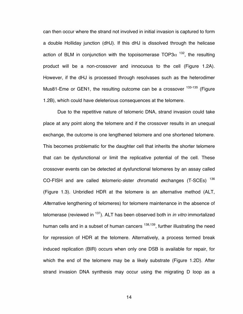
Figure 1.1 Telomere binding complexes in mammals and yeast. ......................... 5
Figure 1.2 Homology-directed repair. .................................................................. 16
Figure 1.3 CO-FISH detects T-SCEs. ................................................................. 17
Figure 1.4 Shelterin solves the end-protection problem. ..................................... 20
Figure 1.5 Mammalian Rap1 resembles yeast and trypanosome Rap1. ............ 29
Figure 2.1 Strategy to conditionally delete mouse Rap1. .................................... 37
Figure 2.2 Deletion of Rap1 does not affect localization of other shelterin
components to telomeres. ............................................................................ 38
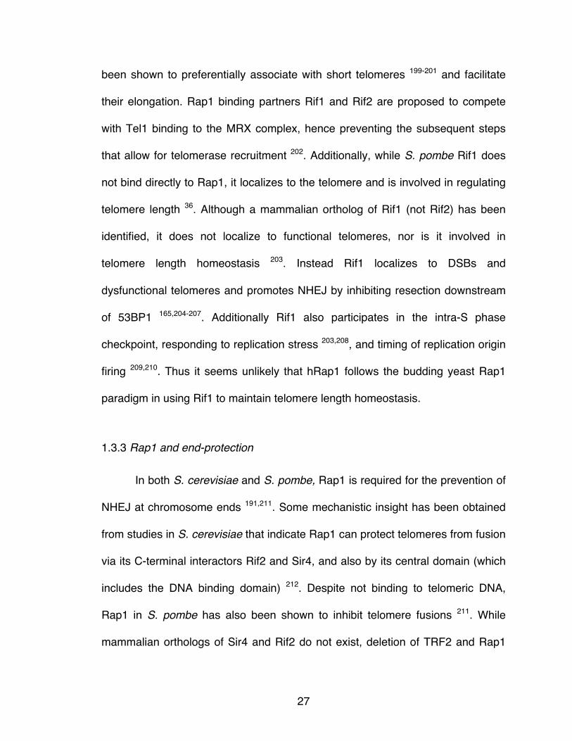
Figure 2.3 Rap1 deletion does not affect cell and organismal viability. ............... 42
Figure 2.4 A TRF2 mutant deficient for Rap1 binding. ........................................ 43
Figure 2.5 TRF2ΔRap1 expression results in diminished levels of Rap1. .............. 44
Figure 2.6 No DNA damage signaling at telomeres lacking Rap1. ..................... 45
Figure 2.7 Loss of Rap1 does not induce NHEJ. ................................................ 46
Figure 2.8 Rap1 does not affect telomere length maintenance. .......................... 49
Figure 2.9 Telomeric chromatin and transcription unchanged upon Rap1 loss. . 50
Figure 2.10 Rap1 is a repressor of telomere recombination. .............................. 51
Figure 2.11 T-SCEs observed in Rap1-deficient cells despite no DNA damage
signaling. ...................................................................................................... 52
Figure 3.1 The C-terminus of Rap1 is required to inhibit HDR. ........................... 60
Figure 3.2 Tethering Rap1 to telomeres in the absence of TRF2. ...................... 63
Figure 3.3 Rap1 cannot repress HDR in the absence of TRF2. .......................... 64
Figure 3.4 Rap1 confers an increase in the telomere binding affinity of TRF2. .. 68
Figure 3.5 No exacerbation of T-SCE levels upon removal of Rap1 and POT1. 69
xi
Figure 3.6 Ku represses T-SCEs in TRF1 null cells, as well as in MEFs lacking
both Rap1 and TRF1. ................................................................................... 71
Figure 3.7 HDR is repressed in late-passage Ku70-deficient MEFs. .................. 73
Figure 4.1 TALEN-mediated inactivation of the gene for human Rap1. .............. 81
Figure 4.2 Loss of Rap1 in targeted clones. ........................................................ 82
Figure 4.3 TALEN-induced mutations in the Rap1 locus. ................................... 83
Figure 4.4 Rap1-deficient cells proliferate and do not induce DNA damage
signaling. ...................................................................................................... 85
Figure 4.5 Telomere protection in Rap1-deficient cells. ...................................... 86
Figure 4.6 Rap1 knockouts exhibit no systematic changes in telomere length. .. 89
Figure 4.7 Loss of Rap1 does not affect telomere structure. .............................. 90
Figure 4.8 Effects of Rap1 on telomere position. ................................................ 92
Figure 4.9 No change in telomeric chromatin or transcription in Rap1-deficient
cells. ............................................................................................................. 94
Figure 4.10 Validation of Rap1 as a transcriptional regulator. ............................ 97
Figure 5.1 TALEN-mediated inactivation of the gene for human POT1. ........... 106
Figure 5.2 Loss of POT1-FL in targeted clones. ............................................... 110
Figure 5.3 Deficiency of POT1-FL is not tolerated. ........................................... 111
Figure 5.4 ATR-dependent DNA damage signaling induced upon loss of full-
length POT1. .............................................................................................. 114
Figure 5.5 No change in 3’ telomere overhangs upon loss of POT1-FL. .......... 116
Figure 5.6 Extended overhangs in POT1-deficient cells expressing the S322L
mutant. ....................................................................................................... 120
Figure 5.7 No change in 3’ telomere overhangs upon deletion of both POT1
isoforms. .................................................................................................... 121
xii
LIST OF TABLES
Table 4.1 Microarray expression analysis for ARPE-19. ..................................... 96
Table 4.2 Microarray expression analysis for HT1080. ....................................... 96
Table 4.3 Microarray expression analysis for HCT116. ...................................... 96
xiii
LIST OF ABBREVIATIONS
53BP1 Tumor suppressor p53 binding protein 1
ALT Alternative lengthening of telomeres
ATM Ataxia telangiectasia mutated
ATR ATM and Rad3 related
BrdU Bromo-deoxyuridine
Chk2 Checkpoint kinase 2
CO-FISH Chromosome orientation-fluorescence in situ hybridization
CST Cdc13/CTC1, Stn1, Ten1
DDR DNA damage response
DSB Double-stranded break
G-quartet Hoogsteen base-pairing of guanine residues resulting in a
four-stranded DNA structure
HJ Holliday Junction
dHJ Double HJ
HDR Homology-directed repair
KO Knockout
MEF Mouse embryonic fibroblast
MRN Mre11, Rad50, Nbs1
NHEJ Non homologous end-joining
C-NHEJ Classical NHEJ
Alt-NHEJ Alternative NHEJ

xiv
OB fold Oligonucleotide/oligosaccharide binding fold
PD Population doubling
POT1 Protection of telomeres 1
POT1-FL Full-length human POT1
POT1-55 Isoform of human POT1 that lacks first N-term OB fold
POT1ΔOB Dominant-negative allele of POT1, lacks first N-term OB fold
Rap1 Repressor/activator protein 1
ss Single-stranded
SV40-LT Simian virus 40-large T antigen
TALEN Transcription activator-like effector nuclease
TERC Telomerase RNA component
TERT Telomerase reverse transcriptase
T-circle Extrachromosomal circles containing telomeric DNA
T-loop Invasion of the single-stranded 3’ telomere terminus into
duplex telomeric DNA
TRF1 Telomere repeat binding factor 1
TRF2 Telomere repeat binding factor 2
T-SCE Telomere-sister chromatid exchange

1
Chapter 1: Introduction

2
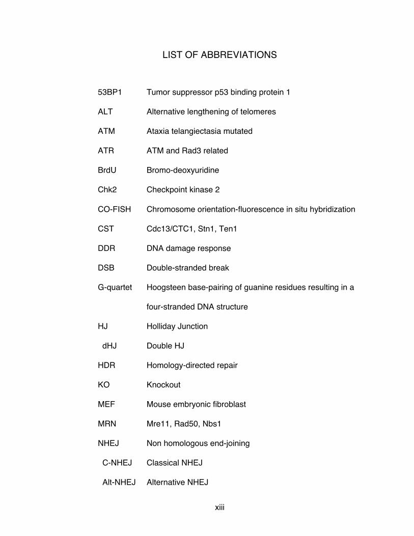
1.1 The structure and function of telomeres
Telomeres are conserved nucleoprotein structures comprised of short
tandem repetitive sequences and highly specific binding proteins that allow linear
DNA to evade recognition as double-stranded breaks (DSBs). Telomeres derive
their name from telos (end) and meros (part) after Hermann Muller irradiated
Drosophila melanogaster and noted that he never found mutants with deletions
or inversions that involved the natural ends of chromosomes 1. Independently,
Barbara McClintock observed from her expansive work in Zea mays that unlike
the natural ends of chromosomes, the ends of broken chromosomes were prone
to cycles of fusion and breakage, which were only halted in zygotes where
broken ends were able to ‘heal’ 2. These early observations illuminated special
features of chromosome termini that preserve genomic integrity.
Telomere repeats vary in sequence and length, with a unifying feature
being the G-rich nature of the strand that runs from 5’ to 3’ to the telomere
terminus. In general the telomere ends in a 3’ single-stranded (ss) overhang.
Budding and fission yeasts have short telomeres of several hundred base pairs 3,
while mammals have much longer telomeres ranging from 10-15 kb in humans 4
and from 20-50 kb in mice (Figure 1.1) 5,6.
Mammalian telomeric TTAGGG repeats 7-9 are bound by a six (seven in
mouse) subunit protein complex termed shelterin (reviewed in 10). Shelterin
consists of two double-strand (ds) DNA binding proteins, TRF1 and TRF2 11-14.
TRF1 and TRF2 have similar homodimerization (TRFH) domains and both form
3
homodimers that bind two copies of the half site 5’-YTAGGTTR-3’ using their C-
terminal Myb/SANT domains 15-17. The two proteins differ in their N-terminus
where TRF1 has an acidic domain while TRF2 has a basic domain 16. A central
region comprised minimally of 41 amino acids, present in TRF2 18,19, but not
TRF1, recruits Rap1 to the telomere 20. Rap1 forms a 1:1 complex with TRF2 21,22
using its C-terminus 23, and requires this interaction not only for its localization to
the telomere 24. TRF1 and TRF2 both bind to TIN2 21,25-27, which serves as a
bridge between the telomeric double-strand and single-strand binding proteins.
TIN2 binds to TPP1 26,28,29, which forms a heterodimer with the ssDNA binding
protein POT1 30 (POT1a and POT1b in the mouse) 31,32. POT1 binds to the
telomeric 3’ overhang via its oligonucleotide/oligosaccharide binding (OB)
domains with the minimal binding site 5’-TTAGGGTTAG-3’ 33,34.
The telomeric protein complex of the fission yeast Schizosaccharomyces
pombe (sp) resembles mammalian shelterin in that it contains Taz1 35, a TRF
ortholog that binds to duplex DNA and recruits spRap1 36. Bridging proteins Poz1
and Tpz1, proposed to be functional analogs of TIN2 and TPP1, serve to link
Taz1/Rap1 to the ssDNA binding protein POT1 30,37. The highly diverged
protozoa Trypanosoma brucei (tb) also has a TRF homolog (tbTRF) that binds
telomeres and recruits tbRap1 38,39. Budding yeast however does not have a
TRF/Taz-like protein at telomeres or a POT1 ortholog. Instead, Rap1 is the de
facto telomere dsDNA binding protein in Saccharomyces cerevisiae (sc) 40-42.
The single-stranded 3’ overhang in S. cerevisiae is bound by the CST (Cdc13,

4
Stn1, Ten1) complex 43, which modulates telomere length, recruits telomerase
and protects the telomere terminus 44-47. S. pombe contains Stn1 and Ten1 that
bind to ssDNA and are required for telomere protection 48. A similar CST
complex, originally identified as an accessory factor of DNA polymerase α-
primase 49,50, also exists in mammalian cells where its subunits are referred to as
CTC1, Stn1 and Ten1. Mammalian CST participates in overhang maintenance 51
52,53, but its function is different from budding yeast and its recruitment to
telomeres is mediated by TPP1 and POT1 52,54. Despite the divergent nature of
these complexes, they all perform the same function – the maintenance and
protection of telomeres.

5
Figure 1.1 Telomere binding complexes in mammals and yeast. Telomere sequence repeats and associated binding complexes are depicted here. Approximate telomere lengths for duplex DNA and single-stranded overhang are indicated below.

6
1.2 Threats to chromosome termini
Chromosome ends face two ominous threats referred to as the end-
replication problem and the end-protection problem. The end-replication problem
originates from the inability of DNA replication to fully synthesize the 3’ ends of
linear DNA, which would result in shorter telomeres with every cell division
thereby limiting the replicative potential of the cell 55-57. The end-protection
problem refers to the propensity of chromosome ends to be recognized as a
DSB, which would result in disastrous consequences such as cell cycle arrest or
apoptosis if DNA damage signaling and DSB repair were inappropriately
activated (reviewed in 58).
1.2.1 Telomerase counteracts the end-replication problem
Semi-conservative DNA replication involves two modes of DNA synthesis,
leading- and lagging-strand synthesis. In leading-strand synthesis, the
polymerase moves in the same direction as the replication fork allowing the
molecule to be replicated to the very end, presumably resulting in a blunt end. In
lagging-strand synthesis, DNA is synthesized in the opposite direction to the
replication fork, necessitating RNA primers to initiate DNA synthesis in short
stretches known as Okazaki fragments that are eventually ligated together once
the RNA primers have been removed (reviewed in 59). At a DNA end, lagging-
strand synthesis could be incomplete due to lack of priming for the most distal

7
Okazaki fragment or due to removal of the most distal RNA primer, thus resulting
in loss of the terminal sequence in the daughter cell 60.
In 1961 Leonard Hayflick noted that normal human fibroblasts are able to
divide in culture for a limited number of (between 40-60) population doublings
(PDs), after which they senesce 57. This proliferative barrier was termed the
Hayflick limit. Further work has illustrated that this phenotype was likely due to
telomere shortening, and in order to bypass replicative senescence, cells with
limiting telomere lengths require a mechanism by which telomere length can be
maintained 61. While fibroblasts tend to senesce with approximate telomere
lengths of 5-7 kb, it is unclear what the minimal length requirement is for proper
telomere function (reviewed in 62).
One solution to the end-replication problem is provided by telomerase, a
ribonucleoprotein enzyme. The telomerase RNA component, TERC or TR,
serves as a template for synthesis of the G-rich telomeric strand by the catalytic
subunit TERT (telomerase reverse transcriptase). The prediction that a terminal
transferase-like activity extended chromosome ends was based on the ability of
yeast to maintain linearized plasmids containing ciliate telomeres, by addition of
repetitive G-rich sequences 63. The RNA component of telomerase was cloned
from the ciliate Tetrahymena 64. Genetic screens in S. cerevisiae for senescence
mutants that displayed progressive telomere shortening 65,66, alongside
biochemical fractionation of factors that co-purified with telomerase activity in
Euplotes aediculatus 67 led to the discovery of the telomerase catalytic subunit

8
Est2 (ever shorter telomeres 2). Following this discovery, mammalian
components of telomerase were rapidly identified 68-70. Indeed genetically
engineered mice lacking TERC display shortening telomeres over several
generations and exhibit chromosomal abnormalities by the fourth generation 71.
In human cells telomerase is expressed in the germline, but not in most
somatic tissues 72. However, telomerase activity is readily detectable in
immortalized human cell lines that have either been transformed 73 or derived
from tumors (reviewed in 62). Approximately 90% of tumour biopsies tested were
shown to express telomerase 74. Lack of telomerase expression in somatic
tissues suggests that limiting the replicative potential of a cell acts as a barrier to
tumor development, and premalignant or malignant cells must reactivate
telomerase or use an alternative method of telomere length maintenance to
acquire their immortal properties.
Examination of other mammals such as elephants and whales indicated
they have similar telomere lengths to humans and no telomerase activity in
somatic cells. In contrast to these large long-lived mammals, mice and other
short-lived mammals such as shrews and opossums have longer telomeres than
humans and constitutive telomerase activity 6. Gomes et al. noted that
telomerase activity and telomere length generally correlated inversely with body
size and lifespan, giving rise to the proposal that animals with a short lifespan
accrue a lower mutational load and therefore may not require replicative aging as
a tumor suppressor mechanism. It is clear that while telomerase is necessary to
9
counteract the end-replication problem, its expression and activity must be
meticulously controlled.
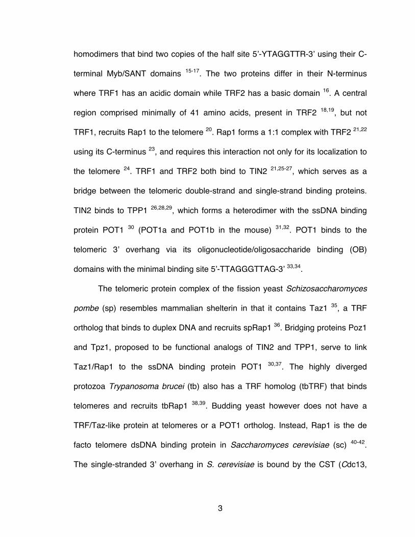
1.2.2 Telomere length regulation by telomere-binding proteins
Initial insights into the mechanism of telomere length regulation were
derived from studies in S. cerevisiae where increasing the number of scRap1
binding sites resulted in proportional shortening of the telomere 75. Thus a cis-
acting protein-counting mechanism was proposed where telomere-repeat
addition by telomerase was inhibited based on the number of Rap1 molecules
bound to the telomere. In agreement with these results, overexpression of TRF1
in a subclone of HT1080s (HTC75; a human fibrosarcoma telomerase-positivie
cell line) led to telomere shortening, while expression of a dominant negative
mutant of TRF1 (TRF166-385) that diminished TRF1 at telomeres resulted in
telomere elongation 76. Overexpression of TRF2 also led to telomere shortening
and telomerase activity was not affected by TRF1 or TRF2 76,77. Thus the two
dsDNA binding proteins negatively regulate telomere length through a similar
‘protein-counting’ mechanism as shown in yeast 75. Similarly, when double-strand
telomere binding protein Taz1 was deleted in S. pombe, negative regulation of
telomere length was abolished. Interestingly, when spRap1 was deleted,
telomeres also elongated 36, suggesting that recruitment to telomeres, as
opposed to direct binding to telomeres, might be sufficient for telomere length
control.
10
Surprisingly, overexpression of human Rap1 leads to telomere
lengthening 20, but this elongation is observed even when a Rap1 mutant
defective in localization to the telomere is overexpressed 23. Considering the
large nucleoplasmic pool of Rap1 that accumulated when exogenously
expressed, it was hypothesized that the non-telomeric Rap1 titrates away a
telomere-associated factor involved in length regulation, that otherwise along with
Rap1 would inhibit telomere elongation. Mild extension of telomeres after partial
knockdown of Rap1 in HTC75 cells provided further support for Rap1 as a
negative regulator of telomere length 78.
Telomeres also elongated when POT1 binding to ssDNA was inhibited by
expression of a POT1 mutant lacking its OB fold, POT1ΔOB 79. Furthermore, TIN2
and TPP1 levels reduced by shRNAs 80 29 and expression of TIN2 mutants that
could not bind TPP1 25 resulted in telomere elongation, illustrating that
recruitment of POT1 is required for negative regulation of telomere length.
However, TPP1 and POT1 have also been implicated in the positive regulation of
telomerase. The TPP1/POT1 heterodimer was shown to enhance telomerase
processivity in vitro 81, with telomerase acting preferentially on substrates coated
with POT1/TPP1 82. In addition, the OB fold of TPP1 can bind to and recruit
TERT to telomeres 81,83-85. Specifically, a group of surface-exposed amino acids
in the OB fold of TPP1, referred to collectively as the TEL patch, is required for
recruitment and activation of telomerase 86,87.

11
1.2.3 The end-protection problem
The integrity of the genome is constantly threatened by internal processes
such as errors introduced during DNA replication and external sources such as
genotoxic agents of radiation. Various mechanisms are in place to detect
damaged DNA, collectively referred to as the DNA damage response (DDR), and
to fix these lesions with DNA repair pathways. Mammalian DDR signaling occurs
primarily through two kinases, the ATM (ataxia telangiectasia mutated) kinase,
activated by the MRN (Mre11-Rad50-Nbs1) complex that senses DSBs, and the
ATR (ATM and Rad3 related) kinase that is activated by the binding of
Replication Protein A (RPA) to ssDNA (reviewed in 88). Chromosome ends are
vulnerable to both of these pathways with the telomere terminus being
recognized by ATM and the single-stranded overhang being a substrate for ATR
activation.
Activation of ATM and/or ATR triggers cell-cycle checkpoints by
phosphorylation of downstream effectors Chk2 89-91 and Chk1 92,93 respectively,
that phosphorylate Cdc25 phosphatases 94, which in turn act to reduce the
activity of cyclin-dependent kinases (CDKs), thereby leading to rapid cell-cycle
arrest in intra-S or G2/M 95,96. Phosphorylation of p53 97-100 and downstream
signaling also occurs to initiate a delayed response to DNA damage, leading to
cell cycle arrest at the G1/S transition and senescence or apoptosis. These
processes are thought to allow time for DNA repair to occur and prevent
transmission of damaged DNA to daughter cells. Coupled with checkpoint

12
activation, signaling by ATM and ATR also promotes a localized response at the
site of the lesion by recruitment and/or activation of DNA repair factors. Both
ATM 101 and ATR 102 phosphorylate histone H2AX (γH2AX) in chromatin proximal
to the damaged lesion. MDC1 (Mediator of DNA damage checkpoint 1) binds to
γH2AX and serves to amplify the DNA damage response 103-105. Subsequent
recruitment of E3 ubiquitin ligase RNF168 leads to ubiquitylation of lysine 15 on
histone H2A or H2AX 106 resulting in recruitment of the DNA repair effector
53BP1 (tumor suppressor p53 binding protein 1) 107. These DNA damage
response proteins are not normally detected at the telomere; however, upon
induction of telomere damage, H2AX becomes phosphorylated in the telomeric
chromatin and DNA repair factors such as 53BP1 accumulate at the telomere.
These indices of local DNA damage signaling are frequently used as readouts for
telomere dysfunction 108,109.
DSB repair occurs through two major pathways, non homologous end-
joining (NHEJ) and homology-directed repair (HDR). NHEJ is the main method of
repairing breaks in the G1 phase of the cell cycle, while HDR predominates in
S/G2 when the presence of a sister chromatid can provide a template for error-
free repair. Two forms of NHEJ have been described: classical- and alternative-
NHEJ (c-NHEJ and alt-NHEJ 110,111). Essential components of c-NHEJ 112 consist
of the Ku70/80 heterodimer which binds to DSBs 113,114, and DNA Ligase IV
(Lig4) which is responsible for ligating the ends 115. Alt-NHEJ on the other hand is
promoted by PARP1 (poly adenosine diphosphate ribose polymerase 1) 116,

13
requires resection of the DNA end, and then makes use of small
microhomologies and DNA Ligase 3 (Lig3) 117 to ligate the ends. Ku70/80 has
been shown to inhibit alt-NHEJ, potentially by competing with PARP1 for DSBs
118. Both types of NHEJ have been shown to be active at dysfunctional telomeres
24,119-121. Inappropriate repair of chromosome ends by NHEJ results in fused
dicentric chromosomes that are unstable in mitosis and can initiate breakage-
fusion-bridge (BFB) cycles thereby leading to genomic instability 2.
The central tenets of HDR are resection of a 5’ end to generate a 3’
ssDNA overhang, formation of the Rad51 (radiation sensitive 51) filament to
conduct a homology search, followed by strand invasion into a homologous
region to initiate DNA repair (Figure 1.2). Termination of the telomere in a 3’
single-stranded overhang makes it primed for HDR.
The MRN complex has been implicated in promoting DSB resection in
cooperation with CtIP 122, as well as BRCA1 (breast cancer 1) 123. In addition,
Exo1, a 5’ to 3’ exonuclease, in concert with Bloom (BLM), a member of the
RecQ helicase family, have also been shown to mediate resection at DSBs
124,125. Following resection, the current model derived from studies in yeast is that
RPA initially coats the available ssDNA to facilitate assembly of the presynaptic
filament and remove secondary structures within the ssDNA 126-128. Mediator
protein BRCA2 (breast cancer 2) subsequently assists in displacement of RPA
and loading of Rad51 to form the pre-synaptic filament 129-131. Following strand
invasion into the region of homology and DNA synthesis, ‘second end capture’
14
can then occur where the strand not involved in initial invasion is captured to form
a double Holliday junction (dHJ). If this dHJ is dissolved through the helicase
action of BLM in conjunction with the topoisomerase TOP3α 132, the resulting
product will be a non-crossover and innocuous to the cell (Figure 1.2A).
However, if the dHJ is processed through resolvases such as the heterodimer
Mus81-Eme or GEN1, the resulting outcome can be a crossover 133-135 (Figure
1.2B), which could have deleterious consequences at the telomere.
Due to the repetitive nature of telomeric DNA, strand invasion could take
place at any point along the telomere and if the crossover results in an unequal
exchange, the outcome is one lengthened telomere and one shortened telomere.
This becomes problematic for the daughter cell that inherits the shorter telomere
that can be dysfunctional or limit the replicative potential of the cell. These
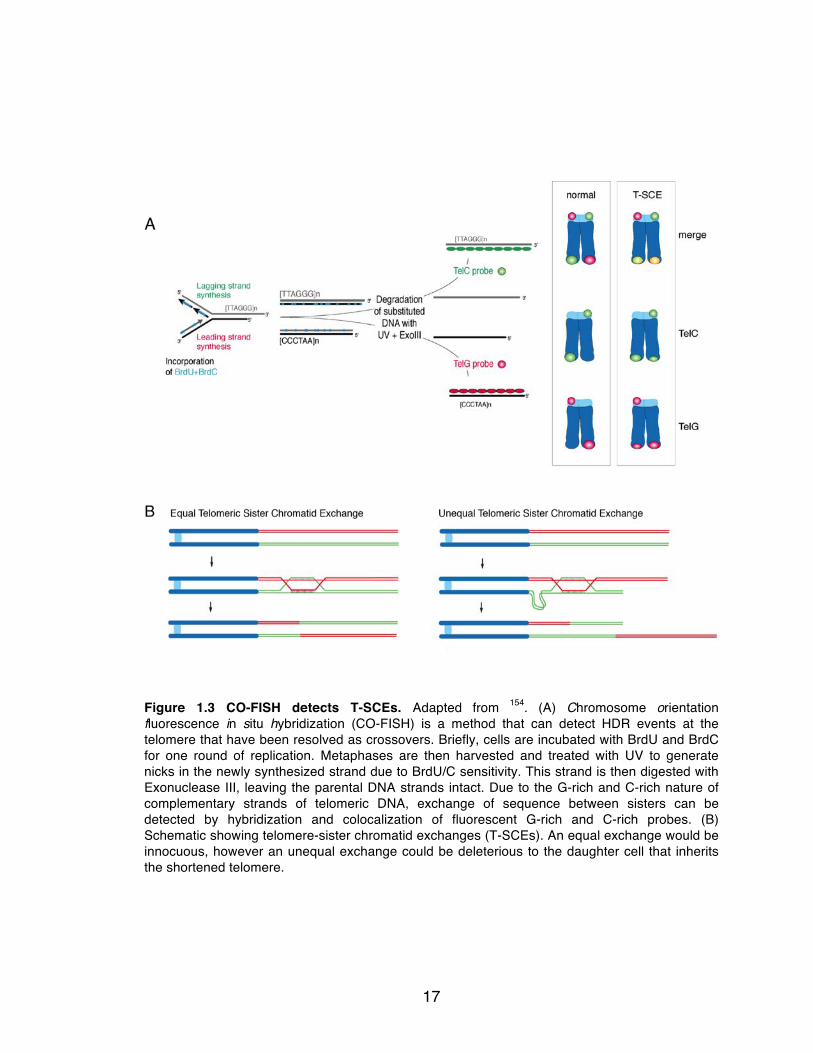
crossover events can be detected at dysfunctional telomeres by an assay called
CO-FISH and are called telomeric-sister chromatid exchanges (T-SCEs) 136
(Figure 1.3). Unbridled HDR at the telomere is an alternative method (ALT,
Alternative lengthening of telomeres) for telomere maintenance in the absence of
telomerase (reviewed in 137). ALT has been observed both in in vitro immortalized
human cells and in a subset of human cancers 138,139, further illustrating the need
for repression of HDR at the telomere. Alternatively, a process termed break
induced replication (BIR) occurs when only one DSB is available for repair, for
which the end of the telomere may be a likely substrate (Figure 1.2D). After
strand invasion DNA synthesis may occur using the migrating D loop as a
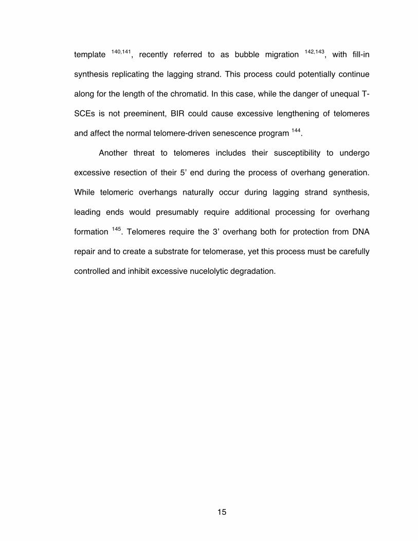
15
template 140,141, recently referred to as bubble migration 142,143, with fill-in
synthesis replicating the lagging strand. This process could potentially continue
along for the length of the chromatid. In this case, while the danger of unequal T-
SCEs is not preeminent, BIR could cause excessive lengthening of telomeres
and affect the normal telomere-driven senescence program 144.
Another threat to telomeres includes their susceptibility to undergo
excessive resection of their 5’ end during the process of overhang generation.
While telomeric overhangs naturally occur during lagging strand synthesis,
leading ends would presumably require additional processing for overhang
formation 145. Telomeres require the 3’ overhang both for protection from DNA
repair and to create a substrate for telomerase, yet this process must be carefully
controlled and inhibit excessive nucelolytic degradation.

16
Figure 1.2 Homology-directed repair. Schematic illustrating processing steps in homology directed repair and potential outcomes. (A) Non-crossover products occur when double Holliday junctions (dHJ) are dissolved. (B) Both non-crossovers and crossovers can be the result dHJ resolution by nucleases. Non-crossovers can be detected by the CO-FISH assay (Figure 1.3A). (C) Break-induced replication is an alternate form of repair that can occur when only one DSB is available for repair, for example the telomere.
17
Figure 1.3 CO-FISH detects T-SCEs. Adapted from 154. (A) Chromosome orientation fluorescence in situ hybridization (CO-FISH) is a method that can detect HDR events at the telomere that have been resolved as crossovers. Briefly, cells are incubated with BrdU and BrdC for one round of replication. Metaphases are then harvested and treated with UV to generate nicks in the newly synthesized strand due to BrdU/C sensitivity. This strand is then digested with Exonuclease III, leaving the parental DNA strands intact. Due to the G-rich and C-rich nature of complementary strands of telomeric DNA, exchange of sequence between sisters can be detected by hybridization and colocalization of fluorescent G-rich and C-rich probes. (B) Schematic showing telomere-sister chromatid exchanges (T-SCEs). An equal exchange would be innocuous, however an unequal exchange could be deleterious to the daughter cell that inherits the shortened telomere.
18
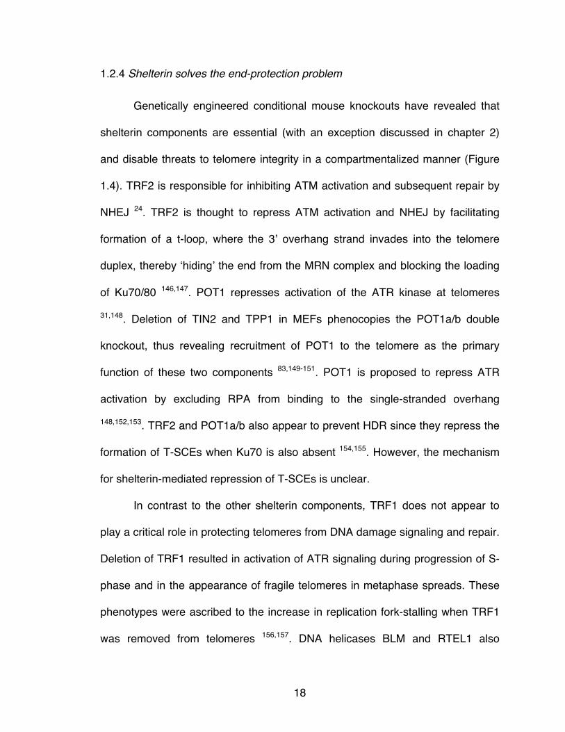
1.2.4 Shelterin solves the end-protection problem
Genetically engineered conditional mouse knockouts have revealed that
shelterin components are essential (with an exception discussed in chapter 2)
and disable threats to telomere integrity in a compartmentalized manner (Figure
1.4). TRF2 is responsible for inhibiting ATM activation and subsequent repair by
NHEJ 24. TRF2 is thought to repress ATM activation and NHEJ by facilitating
formation of a t-loop, where the 3’ overhang strand invades into the telomere
duplex, thereby ‘hiding’ the end from the MRN complex and blocking the loading
of Ku70/80 146,147. POT1 represses activation of the ATR kinase at telomeres
31,148. Deletion of TIN2 and TPP1 in MEFs phenocopies the POT1a/b double
knockout, thus revealing recruitment of POT1 to the telomere as the primary
function of these two components 83,149-151. POT1 is proposed to repress ATR
activation by excluding RPA from binding to the single-stranded overhang
148,152,153. TRF2 and POT1a/b also appear to prevent HDR since they repress the
formation of T-SCEs when Ku70 is also absent 154,155. However, the mechanism
for shelterin-mediated repression of T-SCEs is unclear.
In contrast to the other shelterin components, TRF1 does not appear to
play a critical role in protecting telomeres from DNA damage signaling and repair.
Deletion of TRF1 resulted in activation of ATR signaling during progression of S-
phase and in the appearance of fragile telomeres in metaphase spreads. These
phenotypes were ascribed to the increase in replication fork-stalling when TRF1
was removed from telomeres 156,157. DNA helicases BLM and RTEL1 also

19
facilitate replication of the telomere, presumably by removing G quartets that may
form due to the G-rich sequence of telomeric DNA 156,158,159.
Removal of the entire shelterin complex from mouse telomeres resulting in
shelterin-free telomeres revealed that chromosome ends are vulnerable to fusion
even when canonical components of the c-NHEJ pathway, Ku80 and Lig4, are
absent 120. These fusions events are mediated by PARP1 and Lig3 dependent
alt-NHEJ 120,121. Detection of HDR in shelterin-free Ku80 deficient cells was not
possible due to high levels of telomere fusions, however T-SCEs were observed
in shelterin-free Lig4- and 53BP1- deficient cells. Additionally, when shelterin was
deleted from 53BP1-deficient cells, telomeres underwent extensive nucleolytic
degradation 120. Thus, HDR, alt-NHEJ and 5’ end resection are threats to
telomeres that are thwarted redundantly by general repressors and multiple
components of shelterin.

20
Figure 1.4 Shelterin solves the end-protection problem. Shelterin components are required to inhibit DNA damage signaling and repair at chromosome ends. Ku70/80 and 53BP1 acts as general repressors of HDR and resection respectively, but how they perform their dual function in telomere protection and promoting DNA repair is not yet understood.

21
1.2.5 Phenotypes of TRF2 depletion
Deletion of TRF2 from SV40-immortalized mouse embryonic fibroblasts
(MEFs) led to the concomitant depletion of Rap1 protein from the telomere and
the cell 24. Removal of TRF2 using dominant negative alleles also results in
diminished levels of Rap1 160. Consequently, analysis of phenotypes associated
with loss of TRF2 could be due to either TRF2, or Rap1, or both. My aims
(discussed in section 1.4) were to understand the contributions to and
mechanisms of telomere protection endowed by Rap1.
Expression of dominant negative alleles of TRF2 in human cells or
deletion of TRF2 in MEFs leads to activation specifically of the ATM kinase 24,148.
This signaling can be quantified by the appearance and localization of DNA
repair factors and modifications such as 53BP1 and γH2AX, respectively, to
majority of the telomeres, referred to as TIFs (telomere dysfunction-induced foci)
108. Depletion of TRF2 also results in phosphorylation of Chk2, activation of p53
and apoptosis, consistent with ATM activation 161. Massive induction of telomere-
telomere fusions was also observed on metaphase spreads from cells lacking
TRF2. These fusions primarily occurred in G1 162 and were mediated by c-NHEJ
as revealed by their dependency on the presence of Ku70 and Lig4 24,154. The
fusion of dysfunctional telomeres is promoted by 53BP1-mediated mobility of
telomeres 163,164 and inhibition of resection 165. ATM deficiency abolishes both TIF
formation and telomere fusions upon deletion of TRF2, establishing that DNA
damage signaling is required for NHEJ-mediated DNA repair to occur 148. In

22
addition, deletion of TRF2 in the context of Mre11 or Nbs1 deficiency also
diminishes formation of TIFs and fusions indicating that MRN is required for ATM
activation at dysfunctional telomeres, as it is at DSBs 166-168. TRF2 protects
telomeres from detection by ATM and repair by NHEJ by sequestering the
overhang in a t-loop, visualized by electron microscopy and STORM imaging
147,169. T-loop formation is thought to conceal the DNA end, thereby preventing
binding of MRN or Ku70/80. In addition, ATM inhibition at linear telomeres not in
the t-loop configuration may be mediated by the so-called iDDR, a 25 amino acid
stretch (aa 407-431) in TRF2 that inhibits the E3 ubiquitin ligase RNF168,
preventing accumulation of 53BP1 at telomeres 170.
Loss of TRF2 in Ku70-deficient MEFs leads to induction of HDR,
visualized by the appearance of T-SCEs 154, indicating that TRF2 plays a role in
repressing HDR. The Ku70/80 heterodimer, which is known to repress HDR at
DSBs 171, represses HDR at telomeres independently of shelterin. The
mechanism by which TRF2 represses HDR is not known.
A third function of TRF2 is the inhibition of t-loop cleavage where the t-
loop is excised as an extrachromosomal circle and loss of telomeric DNA from
chromosome ends is detectable on metaphase spreads and genomic blots 172.
The ability of TRF2 to prevent t-loop cleavage and its associated stochastic
telomere losses is dependent on its N-terminal basic domain 172. Biochemical
experiments have shown that the basic domain of TRF2 can bind and stabilize
Holliday Junctions (HJs), which would be expected to form at the base of the t-

23
loop when branch migration takes place 173,174. Expression of a TRF2 mutant
lacking the basic domain (TRF2ΔB) was able to repress NHEJ but stochastic
events of t-loop cleavage were observed 172. T-loop cleavage was diminished in
TRF2ΔB-expressing cells when levels of HJ resolvases Mus81 and Gen1 were
reduced by shRNAs 175, suggesting TRF2 restrains the action of these HDR-
associated nucleases. Expression of TRF2ΔB in TRF2 and Ku70 null cells does
not however induce T-SCEs 154, revealing that TRF2 represses t-loop cleavage
by HJ resolvases and HDR-mediated formation of T-SCEs through different
mechanisms.
1.2.6 Phenotypes of POT1 depletion
ATR signaling at telomeres is repressed by the ssDNA binding protein
POT1 148. Depletion of mouse POT1a results in a TIF response 31,32 that is
exacerbated upon co-deletion of POT1b, however POT1b null MEFs alone
exhibit no TIFs 31. Deletion of POT1b, but not POT1a results in extended single-
stranded overhangs 31 indicating that POT1a and POT1b have evolved to
perform different functions of telomere protection. An RPA exclusion model has
been proposed to explain POT1 inhibition of ATR activation 148,152, where POT1a
prevents RPA from binding to the overhang 153. RPA is substantially more
abundant than the TPP1/POT1 heterodimer and binds single-stranded telomeric
DNA with similar affinity, but the tethering of TPP1/POT1 to the rest of shelterin
by TIN2 is proposed to give POT1 the ability to outcompete RPA 149,176.

24
The mechanism by which POT1b maintains telomere overhangs of correct
length involves the recruitment of the CST complex to presumably perform fill-in
synthesis of the C-rich strand. Leading- and lagging- strand overhangs are
generated through different mechanisms, where leading-ends require initial
processing by the Apollo/SNM1B nuclease 177,178 that is recruited by TRF2
27,179,180. POT1b is required to prevent hyper-resection of both leading and
lagging ends by Apollo. Leading and lagging ends are however both extensively
resected by Exo1 during S/G2 181 in the presence of POT1b 52. POT1b is then
responsible for recruiting the CST complex that restores overhangs to their
normal length 52.
Few chromosomal abnormalities are noted upon loss of POT1a/b, which
include a low level of telomere fusions that occur in G2, and sister telomere
‘associations’, although the molecular basis of this phenotype is unclear 31,182.
The sustained DNA damage induced by POT1a/b deletion leads to
endoreduplication and eventually growth arrest 31,183. Deletion of both POT1a/b in
Ku70 deficient cells leads to an induction of T-SCEs 155, indicating that either
POT1a or POT1b is sufficient to inhibit HDR.
Human POT1 has two isoforms, POT1-FL (full-length) and POT1-55,
which lacks the first of the two OB-folds 184. No function has been reported for
POT1-55. Depletion of human POT1 with shRNAs results in transient DDR
activation 184, a slight reduction in overhang signals and growth arrest in primary
cells 185 but not transformed cells 184. Another feature of POT1 depletion from

25
human cells is loss of specificity of the 5’ end. Greater than 80% of normal
telomere ends have been reported to end in ATC-5’ on the C-rich strand, while
there appears to be less selectivity for terminal nucleotides on the G-rich
strand186. Depletion of human POT1 abrogated sequence precision of the 5’ end
184. It remains to be seen whether the telomere maintenance mechanisms of
human POT1 resemble those of mouse POT1a and PO1b.
1.3 Proposed functions and mechanisms of action for Rap1
1.3.1 Identification of Rap1
Rap1 was first purified and cloned from S. cerevisiae as a transcriptional
regulator and thus named repressor/activator binding protein 1 187.
Transcriptional regulation by scRap1 arises from its ability to bind directly to
specific DNA sequences located in several promoter regions including ribosomal
genes, as well as at silencer elements. The activation and silencing domains of
scRap1 have been mapped to the C-terminus of Rap1 188. The silent information
regulators Sir3p and Sir4p bind to this C-terminal region of Rap1 and function in
sub-telomeric silencing of genes adjacent to telomeres 189,190. Gene disruption of
Rap1 in S. cerevisiae results in lethality, which was proposed to be due to its
function in activation of ribosomal gene loci 187. Further investigation revealed
that Rap1 binds directly to telomeric DNA 40-42 and the requirement of Rap1 for
viability was attributed to its role in telomere protection 191.

26
Initially identified in a yeast two-hybrid screen with human TRF2,
mammalian Rap1 was found to be a distant ortholog of scRap1 20. Although the
sequence conservation is extremely low (too low for a simple BLAST search), the
two Rap1 proteins have a similar domain structure featuring a single N-terminal
BRCT domain, a central region with homology to the Myb DNA binding domain,
and a Rap1-specific C-terminal (RCT) protein-interaction domain (Figure 1.5).
However, unlike budding yeast Rap1, which recognizes telomeric DNA directly
through the cooperation of its Myb domain with a second motif that forms a Myb-
like fold 41,192, mammalian Rap1 associates with telomeres solely through its
interaction with TRF2 20. The Myb domain of mammalian Rap1 is not suited for
DNA binding because its surface lacks positive charge and therefore is more
likely to bind to a protein 193. The targets of the single BRCT domain in the Rap1
proteins are not known. In other proteins, BRCT domains usually occur as
tandem pairs and can function as a phosphopeptide binding module 194.
1.3.2 Rap1 and telomere length regulation
Telomere length regulation by Rap1 in budding yeast is conducted through
its interaction with Rif1 and Rif2 (Rap1 interacting factor 1 and 2), as evidenced
by the elongation of telomeres in their absence 195,196. A proposed mechanism of
Rif1 and Rif2 action in the negative regulation of telomere length is by interfering
with recruitment of Tel1 (the ATM ortholog). The MRX (Mre11-Rad50-Xrs2)
complex binds to telomeres in S. cerevisiae and recruits Tel1197,198. Tel1 has
27
been shown to preferentially associate with short telomeres 199-201 and facilitate
their elongation. Rap1 binding partners Rif1 and Rif2 are proposed to compete
with Tel1 binding to the MRX complex, hence preventing the subsequent steps
that allow for telomerase recruitment 202. Additionally, while S. pombe Rif1 does
not bind directly to Rap1, it localizes to the telomere and is involved in regulating
telomere length 36. Although a mammalian ortholog of Rif1 (not Rif2) has been
identified, it does not localize to functional telomeres, nor is it involved in
telomere length homeostasis 203. Instead Rif1 localizes to DSBs and
dysfunctional telomeres and promotes NHEJ by inhibiting resection downstream
of 53BP1 165,204-207. Additionally Rif1 also participates in the intra-S phase
checkpoint, responding to replication stress 203,208, and timing of replication origin
firing 209,210. Thus it seems unlikely that hRap1 follows the budding yeast Rap1
paradigm in using Rif1 to maintain telomere length homeostasis.
1.3.3 Rap1 and end-protection
In both S. cerevisiae and S. pombe, Rap1 is required for the prevention of
NHEJ at chromosome ends 191,211. Some mechanistic insight has been obtained
from studies in S. cerevisiae that indicate Rap1 can protect telomeres from fusion
via its C-terminal interactors Rif2 and Sir4, and also by its central domain (which
includes the DNA binding domain) 212. Despite not binding to telomeric DNA,
Rap1 in S. pombe has also been shown to inhibit telomere fusions 211. While
mammalian orthologs of Sir4 and Rif2 do not exist, deletion of TRF2 and Rap1

28
from MEFs resulted in telomere fusions, suggesting mammalian Rap1 may have
retained its function in repressing NHEJ 24. Furthermore, human Rap1 can block
NHEJ when it binds to TRF2 loaded on a telomeric end-joining substrate in vitro
213, and a Rap1-fusion protein can reduce telomere fusions when it is tethered to
telomeres that are depleted of TRF2 160.
In several species of budding yeast, namely Kluyveromyces lactis (kl) and
Candida albicans (ca), Rap1 functions in repressing homologous recombination
at the telomere. The K. lactis strain ter1-16T expresses a re-programmed
telomerase that synthesizes telomeres lacking Rap1 binding sites, leading to
elongated telomeres with an especially long 3’ overhang. Electron microscopy of
these cells revealed the formation of T-loop structures and an abundance of t-
circles. When Rad52, a bona fide HDR component in budding yeast was absent,
the incidence of t-circles decreased indicating that the t-circles were products of
HDR, suggesting Rap1 binding to telomeres is required to inhibit recombination
214. Complete deletion of caRap1 also exhibited an abundance of t-circles in
comparison to wild type 215. Depletion of TRF2 and Rap1 from Ku70 null cells
resulted in T-SCEs, raising the possibility that mammalian Rap1 may also be
required to repress HDR at the telomere 154.

29
Figure 1.5 Mammalian Rap1 resembles yeast and trypanosome Rap1. Adapted from 227. Schematic representation of conserved protein motifs of Rap1 and its TRF2-like partners in the indicated organisms. Amino acid positions indicated in schematic of mammalian Rap1 correspond to mouse Rap1. MYB indicates regions with a MYB sequence. MYB-fold indicates a motif that lacks sequence similarity to the MYB sequence but has a similar fold. MYB-like indicates sequence similarlity to the MYB-fold of S. cerevisiae, but their structure has not been determined. S. cerevisiae interacts with several factors important for its functions via its RCT domain. “RCT” of S. pombe indicates the region of Rap1 that is required for interaction with Taz1. It is not known whether T. brucei has a RCT domain, but the C-terminus of tbRap1 is not required for interaction with its TRF interacting partner.

30
1.3.4 A role for Rap1 in meiosis and chromatin organization
Telomeres are involved in chromatin reorganization during meiosis in
yeasts, worms and mammals. During meiotic prophase I, telomeres attach to the
nuclear envelope mediated by SUN/KASH-domain nuclear transmembrane
complexes and other meiosis-specific protein complexes (reviewed in 216).
Following leptotene, telomeres cluster around the centrosome resulting in
bouquet formation, proposed to stimulate homologous chromosome pairing and
meiotic recombination. Rap1 in fission yeast is necessary for telomere clustering
at the spindle pole body during the premeiotic horsetail stage, which in mammals
is analogous to bouquet formation in meiosis 36,217,218. During normal cell division,
telomeres are tethered to the nuclear envelope during the process of nuclear
assembly, potentially mediated by an interaction between Rap1 and SUN1 219.
1.3.5 Non-telomeric functions of mammalian Rap1
Transcriptional regulation by Rap1 has been demonstrated in budding
yeast as well in trypanosomes. Similar to budding yeast tbRap1 largely localizes
to the telomere, but not exclusively. T. brucei normally express one variant
surface glycoprotein (VSG) from subtelomeric loci in a monoallelic fashion.
Knockdown of tbRap1 led to derepression of VSG expression sites causing
simultaneous expression of multiple different VSGs 39.
Unlike in S. cerevisiae, where Rap1 interacts with sirtuins (Sir3p and
Sir4p) that regulate transcription, mammalian Rap1 does not interact with the

31
sirtuins, however it has been shown to localize to over 30,000 chromosome-
internal sites and control gene expression, affecting metabolism and body weight
control 220-223. ChIP-seq for Rap1 in MEFs indicated that it predominantly
localized to chromosome internal loci with [TTAGGG]2 as a consensus motif,
suggesting TRF2-mediated recruitment to these sites. Rap1 was also detected at
telomere sequences containing a mismatch that disrupts TRF2 binding,
suggesting additional interacting partners and modes of recruitment for Rap1 220.
Indeed, regulation of metabolic genes by Rap1 was independent of its ability to
bind TRF2 221. Human Rap1 has also been reported to associate with
chromosome internal loci, but the number of sites (~100) is much lower 223.
Another unanticipated function of Rap1 is its role in the modulation of
NFκβ signaling. Rap1 was identified in a gain-of-function screen for regulators of
NFκβ 224. Despite the predominantly telomeric localization of Rap1, the authors
observed a cytoplasmic fraction of Rap1 that is constitutively associated with Iκβ
kinases (IKKs). IKKs are responsible for phosphorylation and subsequent
degradation of inhibitors of NFκβ (Iκβ proteins). They found that Rap1 requires
interaction with IKKs to activate NFκβ gene expression and suggested that Rap1
directs IKK activity specifically to p65, an inhibitory subunit of NFκβ. These
results are surprising when taking into account data that shows that very little
Rap1 is not bound to TRF2 19, however it is possible that Rap1 has other
interacting factors that recruit it to the cytoplasm, or Rap1 shuttles back and forth
from the nucleus to the cytoplasm. Numerous Rap1-interacting factors have been

32
identified by mass spectrometry and proximity-based YFP fluorescence
complementation screens, yet none of these interactions have been carefully
characterized and it is unclear what roles they may play in protection and
maintenance of telomeres 78,225,226.
1.4 Objectives
The first objective of this thesis was to understand the role of Rap1 in
telomere end-protection. This was carried out using two independent methods:
generation of a Rap1 conditional mouse knockout and analysis of a TRF2 mutant
defective in Rap1 binding. These two approaches revealed that mouse Rap1 was
required to inhibit HDR, but not NHEJ at the telomere.
The second objective, stemming from the discovery that Rap1 represses
telomeric HDR, was to investigate the mechanism of this inhibition. Examination
of a panel of Rap1 mutants indicated that the C-terminus of Rap1 was required to
inhibit HDR. Further analysis suggested that Rap1 needs to be bound to TRF2 in
order to repress HDR, although the mechanism by which Rap1/TRF2 act remains
to be elucidated.
The third objective, facilitated by the development of efficient genome-
editing technologies in human cells, was to query the discrepancy between the
claimed functions of mouse and human Rap1. In contrast to mouse Rap1, in vitro
and in vivo data had implicated human Rap1 in shielding telomeres from NHEJ.
In addition, no deregulation of telomere length was noted in mouse cells lacking

33
Rap1, whereas human Rap1 had been implicated in telomere length
homeostasis. To determine whether human Rap1 protects telomeres from NHEJ
and negatively regulates telomere length, Rap1 knockouts were generated in a
panel of human cell lines.
The fourth and final objective of this thesis was to construct human
knockouts of POT1 to investigate its functions in and mechanisms of telomere
protection, and to compare the role of human POT1 to those of mouse POT1a
and POT1b.

34
Chapter 2: Loss of Rap1 Induces Telomere Recombination
35
2.1 Introduction
Previous loss-of-function studies that removed TRF2 from the telomere
also noted concomitant loss of Rap1 24,160. Rap1 may therefore play a role in
functions ascribed to TRF2, namely repression of the ATM kinase and inhibition
of the DSB repair pathways, NHEJ and HDR 24,148,154,161. In addition,
overexpression and partial knockdown experiments revealed Rap1 as a potential
regulator of telomere length 23,78. In order to understand the contribution(s) of
mammalian Rap1 to telomere protection two strategies were devised to remove
Rap1 from the telomere and study the phenotypes of its loss. The two
approaches taken were to 1) make a conditional mouse knockout of Rap1 and 2)
to capitalize on the dependency of Rap1 on its telomeric recruitment by TRF2, by
generating a TRF2-separation-of-function mutant that could fulfill all functions of
TRF2 except interacting with Rap1. The results of these studies are reported
here.
2.2 Results
2.2.1 Deletion of Rap1 does not affect cell and organismal viability
The gene for the mouse Rap1 protein is annotated as TERF2IP (telomeric
repeat binding factor 2 interacting protein) but referred to as Rap1 henceforth.
Because the first exon of Rap1 immediately abuts the essential KARS lysyl-
tRNA-synthetase gene, a conditional knockout strategy was developed where a

36
Rap1 floxed (Rap1F) allele was created by flanking exon 2 with LoxP sites (Agnel
Sfeir, Figure 2.1A-C). Deletion of exon 2 by retroviral expression of Cre
recombinase results in a premature stop codon in exon 3. The C-terminus of
Rap1 is encoded by exon 3 and is required for its interaction with TRF2 and
recruitment to the telomere. The resulting Rap1Δex2 allele can potentially encode
a fragment containing the N-terminus of Rap1 (Agnel Sfeir, Figure 2.1A).
Exogenous expression of FLAG-tagged Rap1-ex1 (exon1) showed that this
truncated form of Rap1, if it were produced, would not bind or localize to
telomeres (Agnel Sfeir, Figure 2.1D-F). Indirect immunofluorescence (IF) on
samples where nucleoplasmic proteins had been removed by treatment with
Triton-X showed no visible signal of FLAG Rap1-ex1 (Agnel Sfeir, Figure 2.1E).
Similarly, fractionation of cells expressing FLAG Rap1-ex1 showed that the
protein was largely cytoplasmic and could not be detected in the chromatin
bound fraction (Agnel Sfeir, Figure 2.1F).
Rap1F/F MEFs were isolated at embryonic day 13.5 and immortalized with
SV40 large T antigen (SV40LT). IF and immunoblotting showed that Cre-treated
SV40LT-immortalized Rap1F/F MEFs indeed lacked any detectable full-length or
truncated Rap1 protein (Agnel Sfeir, Figure 2.2A-C) and chromatin
immunoprecipitation (ChIP) showed the loss of Rap1 from telomeres. The
expression and localization of other shelterin components were not significantly
affected (Agnel Sfeir, Figure 2.2D).

37
.
Figure 2.1 Strategy to conditionally delete mouse Rap1. (A) Schematic of Rap1, the mouse Rap1 (TERF2IP) locus, the targeting construct, the floxed allele, and the Δex2 allele. N, NdeI; B, BamHI; F1, F2, and R, PCR primers. Rap1 shRNAs shown at the bottom. At right, Rap1Δex2-encoded protein. (B) Genomic blot of NdeI-digested DNA from ES cells. Probe in (A). (C) Genotyping of tail DNAs. Primers in (A). (D) Immunoblot for Rap1 in cells expressing FLAG FL-Rap1, FLAG Rap1-ex1, or vector control. The schematic below depicts full-length Rap1 protein (FLAG FL-Rap1) and the protein fragment encoded by exon 1 (FLAG Rap1-ex1). The antigenic region recognized by Rap1 Ab 1252 is indicated. (E) IF to monitor the localization of FLAG Rap1-ex1 and FLAG FL-Rap1. Rap1Δex2/Δex2 MEFs expressing FLAG Rap1-ex1 or FLAG FL-Rap1 are stained with FLAG (green) and TRF1 (red). (F) Rap1Δex2/Δex2 MEFs expressing FLAG Rap1-ex1, FLAG FL-Rap1 or vector control were fractionated as described in the materials and methods section and equal fractions of cytoplasmic proteins (CP), nucleoplasmic proteins (NP), and chromatin-bound proteins (CB) were analyzed by immunobloting with Rap1 Ab 1252.

38
Figure 2.2 Deletion of Rap1 does not affect localization of other shelterin components to telomeres. (A) Loss of Rap1 IF signal from Cre-treated (day 5) Rap1F/F MEFs. Red, Rap1; green, telomeric FISH; blue, DNA (DAPI). (B) Western blot showing the disappearance of full- length Rap1 in cells deleted for exon 2. No new Rap1 protein was detected in cells bearing the Rap1Δex2 allele. (C) Immunoblots for Rap1 (Ab1252), TRF2 (Ab1254), and TRF1 (Ab1449) from Rap1F/F and Rap1F/+ MEFs five days after Hit&Run-Cre (first lane) or pWZL-Cre (second lane). (D) Telomeric ChIPs on Cre-treated (day 5) Rap1F/F MEFs. Numbers represent ratios of % telomeric DNA in the ChIPs (pre-immune (PI) signal subtracted) on cells + and -Cre.

39
The growth rate of the SV40LT immortalized Rap1Δex2/Δex2 MEFs was
similar to control cells, and primary MEFs lacking wild type Rap1 did not show a
growth arrest or p53 activation (Agnel Sfeir, Figure 2.3A-C). Furthermore,
Rap1Δex2/Δex2 mice were born at the expected frequencies and were fertile (Agnel
Sfeir, Figure 2.3D). The survival of Rap1Δex2/Δex2 cells and mice argues that Rap1
deletion does not result in major telomere dysfunction, which is known to be
lethal. While there was no evidence of translation of the truncated N-terminus of
Rap1, to conclusively determine that no telomere-protection was being afforded
by this fragment, Rap1Δex2/Δex2 MEFs were infected with an shRNA targeting exon
1 (Agnel Sfeir, Figure 2.3E). Treatment with the shRNA targeting exon 1 did not
induce a growth arrest or other phenotypes typical of telomere dysfunction further
validating the previous conclusions.
In the second approach to remove Rap1 from telomeres, previously
characterized TRF2F/-p53-/- MEFs were used 24 to replace the endogenous TRF2
with a mutant that does not bind to Rap1. A short predicted helix at position 290
in the previously mapped Rap1 binding region (aa 260-360; 20) was conserved in
TRF2 orthologs but not in TRF1 (Figure 2.4A-B). Two mutations in this region
(A289S and F290S) reduced the interaction between Rap1 and TRF2 in co-IP
experiments (Giulia Celli, Figure 2.4C). To generate TRF2ΔRap1, aa 284-297 were
deleted (Megan van Overbeek, Figure 2.4D). TRF2ΔRap1 failed to bind to Rap1 in
co-IP experiments whereas it retained its previously reported interaction with
Apollo (Megan van Overbeek, Figure 2.4E). TRF2ΔRap1 was expressed in TRF2F/-

40
p53-/- MEFs and the endogenous TRF2 was removed with Cre, resulting in
depleted Rap1 protein levels (Megan van Overbeek, Figure 2.4F). Although
TRF2ΔRap1 localized to telomeres efficiently, IF and ChIP indicated that the
telomeres lacked Rap1 (Figure 2.5A-B). Other shelterin components were
affected to an extent (<2-fold; Figure 2.5B) that is not expected to be functionally
significant as heterozygous MEFs and mice lacking one copy of TRF1, TPP1,
TRF2, or POT1a/b display no telomere defect. Assessment of shelterin
occupancy at telomeres by ChIP displayed some variability due to antibody
quality and inexact experimental processing (Figure 2.5B). Despite this variability,
on average less than 10% of residual Rap1 remained at telomeres in TRF2 null
MEFs complemented with TRF2ΔRap1, while levels of other shelterin components
in these cells were approximately 85% or higher, compared to cells
complemented with wild type TRF2 (Figure 2.5B). Consistent with the viability of
Rap1Δex2/Δex2 cells, cells expressing TRF2ΔRap1 proliferated at the same rate as
cells expressing wild type TRF2 (Megan van Overbeek, Figure 2.5C-D).
2.2.2 No induction of DDR or NHEJ at telomeres lacking Rap1
Rap1Δex2/Δex2 cells did not show TIFs and phosphorylation of Chk1 and
Chk2 (Chk1-P, Chk2-P) was not evident (Figure 2.6A-C). Further depletion of
Rap1 mRNA with an shRNA also failed to elicit a DNA damage signal in
Rap1Δex2/Δex2 cells (Figure 2.6B). Consistent with these results, TRF2ΔRap1 was
equivalent to wild type TRF2 in its ability to repress TIFs in cells lacking

41
endogenous TRF2 (Figure 2.6D-F). The mutant form of TRF2 also repressed the
induction of Chk2-P to the same extent as wild type TRF2 (Figure 2.6E). The low
level of Chk2-P observed in Cre-treated TRF2- and TRF2ΔRap1- expressing cells
is likely due to Cre-induced DNA damage, since the phosphorylation of Chk2 was
diminished when using a version of Cre (Hit&Run) that eventually disappears
from the cells due to self-deletion (Figure 2.6F).
Telomere fusions were not induced by deletion of Rap1 and TRF2ΔRap1
had the same ability as wild type TRF2 to repress NHEJ at telomeres (Figure
2.7A-C). However, as previously discussed, NHEJ of telomeres lacking TRF2
requires active DNA damage signaling 148 thus the lack of telomere fusions could
be due to the lack of ATM/ATR activation. In order to initiate DNA damage
signaling specifically at the telomere, we used a TPP1 shRNA to activate the
ATR kinase. This approach previously resulted in the reactivation of NHEJ at
telomeres of TRF2- and ATM-deficient cells 148. Despite ATR kinase signaling at
telomeres and induction of TIFs elicited by the TPP1 shRNA (Figure 2.6B,D),
Rap1 removal from telomeres did not induce their fusion (Figure 2.7B-C). Thus,
Rap1 does not appear to be required in either the repression of NHEJ or ATM
kinase signaling, explaining why the deletion of Rap1 does not curb cellular or
organismal viability.
42
Figure 2.3 Rap1 deletion does not affect cell and organismal viability. (A) Proliferation of SV40LT-immortalized Rap1F/F and Rap1F/+ MEFs infected as indicated. (B) FACS profiles of primary Rap1F/F cells infected with pWZL-Cre (left panel) or vector control (right panel), analyzed at day 5 after infection. The percentage of G1, S and G2 cells is noted within the FACS profile. (C) Immunoblot for Rap1 on MEFs with the indicated genotype, 96 hours after Cre treatment. (D) Offspring from Rap1∆ex2/+ and Rap1∆ex2/∆ex2 intercrosses. (E) Western blot showing the effect of Rap1 shRNAs 1 and 2 on Rap1 levels in wild type MEFs.

43
Figure 2.4 A TRF2 mutant deficient for Rap1 binding. (A) Schematic of TRF2 showing conservation of a subset of amino acids within the previously mapped Rap1 interaction domain. (B) Identification of a helical region within the conserved segment. Predicted protein structure from PredictProtein.org. (C) Co-IPs of FLAG-tagged Rap1 with wild type TRF2, TRF2 mutants or no protein. (D) The TRF2∆Rap1 mutant. H, predicted helix. (E) Co-IP of Myc-TRF2 or Myc-TRF2∆Rap1 with FLAG-Rap1 or FLAG-Apollo from co-transfected 293T cells. In, 2.5% of input. (E) Immunoblots for TRF2 and Rap1 from TRF2F/-p53-/- MEFs expressing the indicated alleles at 72 and 96 hours after Hit&Run-Cre.

44
Figure 2.5 TRF2ΔRap1 expression results in diminished levels of Rap1. (A) IF-FISH to monitor TRF2, Rap1, and TIN2 at telomeres in TRF2F/-p53-/- MEFs expressing TRF2∆Rap1 or vector control at day 4 after Cre. (B) Telomeric ChIP of TRF2F/-p53-/- MEFs expressing TRF2 or TRF2∆Rap1 at day 4 post Cre. Duplicate dot blots were probed for telomeric DNA or the dispersed BamHI repeats. ChIP ratios represent the % telomeric DNA recovered in TRF2∆Rap1 vs. TRF2 expressing cells. ChIP ratios are shown from two independent experiments and averages with SEMs are listed to the right. (C) Immunoblots showing the expression of Rap1, TRF2, and TRF2∆Rap1 in TRF2F/-p53-/- MEFs treated with pWZL-Cre for the indicated time-periods. (D) Growth curve of cells shown in (C) and the vector controls.
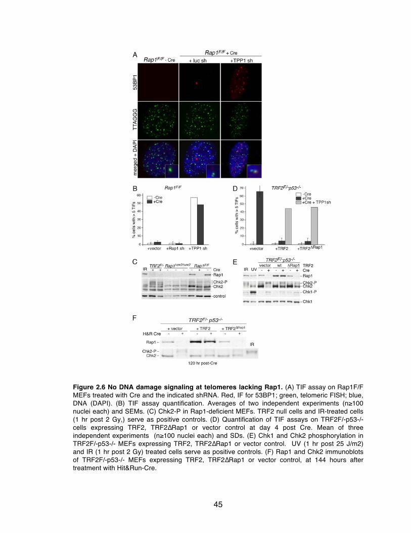
45
Figure 2.6 No DNA damage signaling at telomeres lacking Rap1. (A) TIF assay on Rap1F/F MEFs treated with Cre and the indicated shRNA. Red, IF for 53BP1; green, telomeric FISH; blue, DNA (DAPI). (B) TIF assay quantification. Averages of two independent experiments (n≥100 nuclei each) and SEMs. (C) Chk2-P in Rap1-deficient MEFs. TRF2 null cells and IR-treated cells (1 hr post 2 Gy,) serve as positive controls. (D) Quantification of TIF assays on TRF2F/-p53-/- cells expressing TRF2, TRF2∆Rap1 or vector control at day 4 post Cre. Mean of three independent experiments (n≥100 nuclei each) and SDs. (E) Chk1 and Chk2 phosphorylation in TRF2F/-p53-/- MEFs expressing TRF2, TRF2∆Rap1 or vector control. UV (1 hr post 25 J/m2) and IR (1 hr post 2 Gy) treated cells serve as positive controls. (F) Rap1 and Chk2 immunoblots of TRF2F/-p53-/- MEFs expressing TRF2, TRF2∆Rap1 or vector control, at 144 hours after treatment with Hit&Run-Cre.

46
Figure 2.7 Loss of Rap1 does not induce NHEJ. (A) Metaphase chromosomes from Rap1F/F
cells 5 days post Cre. Red, false colored DNA DAPI stain; green, telomeric FISH. (B) Quantification of telomere fusions, detected as in (A) in Rap1F/F MEFs with the indicated Cre and shRNA treatments. Average % of telomeres fused is given. (C) Quantification of telomere fusions in TRF2F/-p53-/- MEFs (+ or – Cre, day 4) complemented with TRF2 or TRF2∆Rap1 or vector control and treated with TPP1 shRNA as indicated.

47
2.2.3 Telomere length and chromatin unchanged in Rap1-deficient cells
To assess the role of Rap1 in telomere length regulation, telomere length
was examined over three generations of mouse breeding and over approximately
50 population doublings in cultured cells lacking Rap1 (Figure 2.8A-C). No overt
changes in telomere length were detected. Furthermore, the amount of the
single-stranded overhang remained unchanged in Rap1-deficient cells (Figure
2.8D).
Mammalian telomeres contain nucleosomes and epigenetic marks that are
characteristic of heterochromatin, such as heavily methylated histones (reviewed
in 228). Rap1 was not required for the maintenance of telomeric nucleosomal
organization or the methylation of telomeric H3K9 (Agnel Sfeir, Figure 2.9A-C). In
addition, the level of telomeric lncRNA called TERRA (telomeric repeat-
containing RNA; reviewed in 229) was not affected upon deletion of Rap1 (Agnel
Sfeir, Figure 2.9D).
2.2.4 Rap1 is a repressor of telomere recombination
Homology-directed repair threatens telomere integrity because unequal
telomere-sister chromatid exchanges can change telomere lengths. T-SCEs are
most frequent when either TRF2 or POT1a/b are deleted from Ku-deficient cells
154,155, although low levels of T-SCEs have been reported for POT1a deficiency
alone 32. To determine whether Rap1 was required for TRF2-mediated repression
of T-SCEs, TRF2ΔRap1 was introduced into SV40LT-immortalized TRF2F/-Ku70-/-

48
MEFs, which display frequent T-SCEs upon deletion of TRF2 with Cre. Whereas
the telomeric exchanges were repressed by wild type TRF2, TRF2ΔRap1 failed to
block the telomeric HDR (Fig. 2.10A-C). The frequency of T-SCEs was the same
whether the cells expressed TRF2ΔRap1 or no TRF2 (Figure 2.10D). Furthermore,
T-SCEs were induced by Cre-mediated deletion of Rap1 from Rap1F/FKu70-/-
cells (Figure 2.10E).
Notably, the T-SCEs occurred despite absence of TIFs in cells lacking
both Ku70 and telomeric Rap1 (Figure 2.11A-B). Basal levels of Chk2-P were
observed in cells lacking Rap1 and Ku70, but these were likely attributable to the
Cre effect and Ku70 deficiency, as they were much lower than the induction of
Chk2-P observed in the absence of TRF2 and Ku70 (Figure 2.11C-D). There
appeared to be no effect on the terminal structure of telomeres in cells lacking
Rap1 and Ku70 (Figure 2.11E).

49
Figure 2.8 Rap1 does not affect telomere length maintenance. (A) Telomeric restriction fragment analysis on cells isolated from the liver of a Rap1Δex2/+ mouse and three successive generations of Rap1Δex2/Δex2 mice. Telomeric DNA was detected by in-gel hybridization assays using a (CCCAAT)4 probe under denaturing conditions. (B) Telomere length analysis on Rap1Δex2/Δex2 MEFs and wild type cells at the indicated population doublings. (C) Telomere length analysis of TRF2F/- p53-/- cells expressing TRF2, TRF2ΔRap1, or vector, plus or minus treatment with Cre. (D) Loss of Rap1 does not alter the telomeric single-stranded DNA. MEFs with the indicated genotypes were analyzed at day 5 post Cre treatment using in-gel hybridization to MboI digested DNA. The panel on the left shows the hybridization signal using a (CCCAAT)4 probe under native conditions. The panel on the right represents total telomere signal after in situ denaturation of DNA and re-hybridization with the same probe. The numbers on the bottom represent the relative overhang signal normalized to the total telomeric repeat signal. Values of the normalized signal are compared between – Cre (set at 1) and +Cre samples.
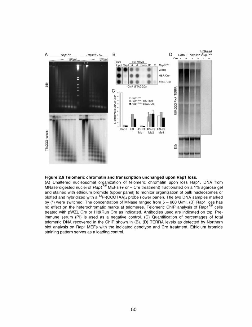
50
Figure 2.9 Telomeric chromatin and transcription unchanged upon Rap1 loss. (A) Unaltered nucleosomal organization of telomeric chromatin upon loss Rap1. DNA from MNase digested nuclei of Rap1F/F MEFs (+ or – Cre treatment) fractionated on a 1% agarose gel and stained with ethidium bromide (upper panel) to monitor organization of bulk nucleosomes or blotted and hybridized with a 32P-(CCCTAA)4 probe (lower panel). The two DNA samples marked by (*) were switched. The concentration of MNase ranged from 5 – 600 U/ml. (B) Rap1 loss has no effect on the heterochromatic marks at telomeres. Telomeric ChIP analysis of Rap1F/F cells treated with pWZL Cre or Hit&Run Cre as indicated. Antibodies used are indicated on top. Pre-immune serum (PI) is used as a negative control. (C) Quantification of percentages of total telomeric DNA recovered in the ChIP shown in (B). (D) TERRA levels as detected by Northern blot analysis on Rap1 MEFs with the indicated genotype and Cre treatment. Ethidium bromide staining pattern serves as a loading control.

51
Figure 2.10 Rap1 is a repressor of telomere recombination. (A) Rap1 and TRF2 from TRF2F/-Ku70-/- MEFs expressing TRF2, TRF2∆Rap1, or vector control analyzed 4 days after Cre. (B) CO-FISH analysis on cells in (A). Arrows: T-SCEs. (C) Enlarged T-SCE events in Cre-treated TRF2F/-Ku70-/- MEFs expressing TRF2∆Rap1. (D) Quantification of T-SCEs as assessed in (B). Bars represent averages from three independent experiments (n>1100 chromosome ends each) and SDs. P values based on Student’s two-tailed t-test. (E) Quantification of T-SCEs as assessed in (B) in cells of the indicated Rap1 and Ku70 status. Method as in (D). Errors bars: SEMs except for SDs for Rap1F/FKu70-/-.

52
Figure 2.11 T-SCEs observed in Rap1-deficient cells despite no DNA damage signaling. (A) TIF assay on TRF2F/- Ku70-/- SV40LT-immortalized MEFs expressing TRF2, TRF2∆Rap1 or vector control, before or after treatment with Cre. Red, IF for 53BP1; green, telomeric FISH; blue, DNA (DAPI). (B) Quantification of TIF assay in (A). (C) Immunoblots for indicated shelterin components, Chk1 and Chk2 of same cells as in (A). IR (1hr post 2Gy) and UV (1hr post 25 J/m2) treated cells serve as positive controls. (D) Immunoblots for Rap1 and Chk2 on SV40LT immortalized MEFs with indicated genotypes, before and after treatment with Cre. γ-tubulin serves as loading control. (E) Loss of Rap1 in Ku-deficient SV40LT immortalized MEFs does not alter the telomeric single-stranded DNA. MEFs expressing TRF2, TRF2∆Rap1, or vector control were analyzed using in-gel hybridization to MboI digested DNA before and 96 hours after treatment with Cre. Left, hybridization signal with (CCCAAT)4 probe under native conditions; right, hybridization signal with same probe after denaturation. Overhang signal was normalized to the total telomeric repeat signal. Numbers represent the relative overhang signal as compared to TRF2 plus Cre (set as 1).

53
2.3 Summary of findings
These data indicate that Rap1 functions at mouse telomeres to repress
HDR, which has the potential for generating shortened telomeres and can
promote telomerase-independent telomere maintenance. Furthermore, these
results underscore the functional compartmentalization within shelterin where
each subunit is dedicated to distinct functions. As previously reported, replication
of telomeric DNA is facilitated by TRF1 156,157, TPP1/POT1 are required for the
repression of ATR signaling 150,151, while TRF2 is the predominant repressor of
both ATM signaling and NHEJ 24. The current data show that these functions of
TRF2 do not require Rap1. Instead, Rap1 is required to repress HDR.
Interestingly, this division of roles revealed that telomeres can undergo HDR
without being detected by the ATM and ATR kinase pathways. When HDR takes
place at telomeres lacking TRF2 or POT1a/b, DNA damage signaling results in
the formation of TIFs. In the case of Rap1 removal, however, the telomeres lack
detectable TIFs, yet are susceptible to HDR. Thus, the formation of DNA damage
foci at telomeres is not a prerequisite for HDR. How exactly Rap1 represses HDR
and the action of Ku in this repression remains to be determined.
It is of interest to recall that expression of the TRF2ΔB mutant allele leads
to stochastic t-loop loss, presumably because the basic domain of TRF2
stabilizes Holliday Junctions and restrains the action of HJ resolvases 172-175.
However, if the basic domain of TRF2 cannot inhibit HJ resolution, a critical step
in the generation of T-SCEs, the question arises as to why T-SCEs are not

54
observed in TRF2ΔB-expressing TRF2/Ku70-deficient cells 154. One possibility is
that Rap1 bound to TRF2 may repress the formation of HJs so that T-SCEs will
not occur. For instance Rap1 and/or TRF2 may inhibit strand invasion into sister
chromatids, or Rad51 filament assembly, thereby preventing HJ formation and
potential exchange of sequence. In addition, it is possible that the basic domain
of TRF2 blocks branch migration of TRF2, thereby also repressing HJ formation
and preventing T-SCEs.
Rap1 was not required for the maintenance of several other features of
mouse telomeres including the amount of the single-stranded overhang, the level
of telomeric transcripts and the telomeric chromatin state and organization. In
budding yeast, Rap1 has been proposed to facilitate heterochromatin assembly
by recruitment of Sir3 and Sir4 that interact with the N-termini of histones H3 and
H4 231-233. Despite a proximity-based screen identifying potential interactions
between Rap1 and histones H2A and H4 226, Rap1 does not seem to play a role
in maintaining telomeric chromatin.
Surprisingly no gross aberrations in telomere length in Rap1 null cells
were observed either, although small differences may be difficult to discern due
to the long telomeres of mouse cells. Studies of Rap1’s involvement in telomere
length regulation may be more illuminating in human cells that have shorter
telomeres.

55
Chapter 3: Elucidating the mechanism of HDR repression by
Rap1

56
3.1 Introduction
How Rap1 represses HDR is not clear. It is possible that it functions by
recruiting another protein with its BRCT domain or MYB motif, which are
proposed to be protein-interaction domains (reviewed in 227). Factors known to
interact with Rap1 include the Rad50 and Mre11 components of the MRN
complex 78, which contributes to 5’ resection during HDR. However, the telomeric
overhang is not altered in Rap1/Ku70-deficient MEFs 19, suggesting Rap1 does
not play a major role in modulating resection at telomeres. Therefore it seems
unlikely that Rap1 would inhibit telomeric HDR by limiting or preventing resection
by MRN. The Rap1/TRF2 complex also interacts with the protein scaffold SLX4
225, which binds to a HJ resolvase Mus81/Eme1. It is possible that Rap1
modulates the activity of one of the nucleases in the SLX4 complex to interrupt
HDR.
In this chapter, Rap1 mutants were analyzed to determine which domains
of Rap1 were required for repression of T-SCEs. These experiments identified
the C-terminus of Rap1 as the important functional domain in the inhibition of
HDR. Further investigation suggested that TRF2/Rap1 complex formation was
necessary for HDR repression and preliminary experiments to assess the DNA-
binding activity of this complex were conducted.
The repression of HDR also requires the presence of either of the two
POT1 proteins 155. POT1a/b bind to single-stranded telomeric DNA and may
inhibit initiation of HDR by preventing loading of Rad51 onto ssDNA. In the

57
context of POT1a/b- and Ku70-deficiency, Rap1, which remains at telomeres, is
incapable of repressing HDR. The converse is true of POT1a/b that remain at
telomeres when Rap1 is deleted, but cannot inhibit HDR 19. These data raise the
possibility that Rap1 and POT1 may act together to repress HDR. To investigate
whether Rap1 and POT1 are epistatic in their repression of HDR, an shRNA to
TPP1 was used to remove POT1 from telomeres in Rap1/Ku70-deficient cells.
No change in the level of T-SCEs would suggest that Rap1 and POT1 are
epistatic, possibly acting together at the same step. For instance, Rap1 and
POT1 may both interfere with Rad51 filament formation. Exacerbation of the
occurrence of T-SCEs would suggest that Rap1 and POT1 act on distinct, non-
linear aspects of homology-directed repair, for example HJ resolution by either
Mus81/Eme1 or GEN1.
The lack of DNA damage signaling in Rap1/Ku70-deficient MEFs also
provides a unique setting to study the consequences of ongoing HDR on
telomere maintenance. As mouse cells constitutively express telomerase, this
setting does not recapitulate ALT. However, the analysis of the long-term
consequences of combined deficiency of Rap1 and Ku may still provide insights
into the recombination-based mechanisms of telomere maintenance.

58
3.2 Results
3.2.1 The C-terminus of Rap1 is required to repress telomeric HDR
MYC-tagged alleles of mouse Rap1 containing deletions of the relevant
motifs (BRCT, MYB, Coil) were constructed as illustrated in Figure 3.1A.
Determination of amino acid positions of domains was based on analogous
domain positions outlined in human Rap1 20,23. Based on the crystal structure of
the hRap1 C-terminus in complex with a fragment of TRF2 (TRF2RBM, Rap1
binding motif, aa 275-316) 18, the I318 residue in hRap1 was shown to be
required for its interaction with TRF2. Co-IPs of transfected proteins from 293T
cells showed that mutation of the analogous residue in mouse Rap1 (I312R)
disrupted the interaction with mouse TRF2 (Figure 3.1B).
Rap1 mutants were retrovirally expressed in Rap1F/FKu70-/- MEFs and
immunoblotting showed that most mutants were expressed at similar levels,
except for MYC-I312R, which was significantly overexpressed, and MYC-ΔBMC,
which was expressed weakly (Figure 3.1C). Rap1 protein levels are diminished
upon TRF2 depletion, yet surprisingly the Rap1 I312R mutant, which could not
bind TRF2, was overexpressed. Therefore, it seems that when Rap1 is not bound
to TRF2, the I312 residue is bound by another factor that targets it for
degradation. IF-FISH indicated that all mutants, except for MYC-I312R, localized
to telomeres (Figure 3.1D). IF for MYC-I312R showed diffuse staining throughout
the nucleus that disappeared upon extraction of nucleoplasmic proteins with

59
TritonX-100, indicating that the Rap1 I312R mutant was not associated with
chromatin. (Figure 3.1D). CO-FISH analysis after removal of endogenous Rap1
with Cre revealed that all but the MYC-I312R mutant repressed T-SCEs, thus
implicating the Rap1/TRF2 interaction in the inhibition of HDR (Figure 3.1E).
Furthermore, the data showed that the BRCT, MYB and coiled domains of Rap1
are not required for the repression of homology-directed repair, either individually
or together. Although the data argue against the N-terminus recruiting an HDR-
repressing factor to telomeres, it is not excluded that the C-terminus of Rap1
binds to TRF2 and also recruits an HDR-repressor. However, since MYC-I312R
cannot localize to telomeres, it is possible that its deficiency in repressing HDR is
due to lack of telomeric recruitment of a factor involved in HDR inhibition. To
address this latter possibility, we devised a strategy to tether Rap1 to telomeres
in the absence of TRF2.

60
Figure 3.1 The C-terminus of Rap1 is required to inhibit HDR. (A) Schematic of Rap1 domain mutants. * indicates I312 to R mutation. ΔBMC, deletion of N-terminal 264 amino acids (aa), Rap1 protein coding started at aa 265. ΔBRCT, deletion of N-terminal 106 aa, Rap1 protein coding started at 107 aa. ΔMYB, deletion of aa 128-196, such that aa 127 was fused to aa 197. ΔCoil, deletion of aa 231-264, such that aa 230 was fused to aa 265. The Rap1 antibody was raised to full-length protein and detects multiple antigenic regions in the N-terminus of Rap1. (B) 293Ts were co-transfected with constructs as indicated and immunoprecipitated with a MYC antibody. Inputs and IPs were detected by the MYC antibody (left) and the FLAG antibody (right). MYC-TRF1 serves as a negative control for immunoprecipitation of FLAG-Rap1. (C) Immunoblots of MYC-tagged constructs in (A) expressed in Rap1F/FKu70-/-MEFs, detected with Rap1 antibody (left) or MYC antibody (right). All constructs except ΔBMC contain an epitope(s) for the Rap1 antibody. n.s., non-specific band picked up by the Rap1 antibody. Running positions of constructs on gel are indicated on the right. (D) IF-FISH of constructs in (A) expressed in Rap1F/FKu70-/-
MEFs. Red, IF for MYC; green, telomeric FISH, blue, DAPI DNA stain. White square shows corresponding area enlarged below. (E) Quantification of T-SCEs. Bars represent averages from three independent experiments (n~1000 chromosome ends each) and SDs. P values based on Student’s two-tailed t-test.

61
3.2.2 Rap1 does not inhibit HDR at telomeres lacking TRF2
Full-length TRF2 and a 41 amino acid fragment of TRF2 (TRF2RBM) bind to
Rap1 in vitro with similar affinities as determined by isothermal titration
calorimetry 18. The sequence encoding these 41 amino acids was inserted into
MYC-tagged TRF1 (TRF1RBM) with the aim of allowing TRF1 to recruit Rap1 to
telomeres in the absence of TRF2 (Figure 3.2A). Expression of TRF1RBM in
TRF2F/FKu70-/- MEFs showed that endogenous levels of Rap1 could be restored
upon Cre-mediated deletion of TRF2 (Figure 3.2B). ChIP confirmed retention of
Rap1 at telomeres despite removal of TRF2, while the telomeric occupancy of
other shelterin components was not affected (Figure 3.2C-D). IF-FISH further
illustrated that both endogenous Rap1 and TRF1RBM co-localized with telomeres
(Figure 3.2E). IF for MYC displayed an unusual staining pattern of circular
patches in the nucleus due to non-specific association of the secondary antibody,
however magnification of insets show that MYC-tagged TRF1RBM localized to
telomeres (Figure 3.2E). Expression of TRF1RBM in TRF1-deficient MEFs
repressed the occurrence of fragile telomeres (Figure 3.3A-B), which are induced
upon loss of TRF1 156,157. Thus, introduction of the RBM sequence into TRF1 did
not disrupt normal functions of TRF1 and allowed for the retention of Rap1 at
telomeres lacking TRF2. However, despite the presence of Rap1 at telomeres, T-
SCEs were not repressed when TRF2 was deleted from TRF2F/FKu70-/- MEFs
(Figure 3C-D), pointing to a requirement for the interaction between Rap1 and
TRF2 in the inhibition of HDR. Rap1 binding to TRF2 may be required for a
62
conformational change of the complex that facilitates recruitment of an additional
protein involved in the repression of homology-directed repair. Alternatively,
Rap1 may change the DNA binding activity of TRF2, perhaps by changing its
affinity to DNA structures that resemble substrates for HDR, such as the telomere
terminus.
In addition to induction of telomere-sister chromatid exchanges, a low
percentage of alt-NHEJ mediated telomere fusions are also observed in
TRF2/Ku70-deficient MEFs 120. Placement of Rap1 at telomeres by TRF1RBM did
not inhibit these fusions (Figure 3.3E) indicating that Rap1 does not play a role in
the repression of alt-NHEJ. In contrast to these results, another study shows that
tethering Rap1 to HeLa (a human cervical carcinoma cell line) telomeres by a
fungal telomere-binding domain can repress c-NHEJ-mediated fusions when
TRF2 is depleted 160. It remains to be seen whether retention of Rap1 by
TRF1RBM can inhibit c-NHEJ mediated telomere fusions in TRF2-null MEFs.

63
Figure 3.2 Tethering Rap1 to telomeres in the absence of TRF2. (A) Schematic illustrating composition of the TRF1RBM allele. 41 aa encoding RBM was inserted into TRF1 at position aa 278. This did not interfere with the Bloom (BLM) helicase interaction sites shown in purple, or disrupt any other known interaction or nuclear localization signal. (B) Immunoblot showing expression of TRF1RBM in TRF2F/FKu70-/- MEFs and levels of Rap1 before and 96 hours after deletion of TRF2 with Cre. (C) ChIP for shelterin components and MYC-tagged TRF1RBM at telomeres before and 96 hours after deletion of TRF2. Dot blot was probed with TTAGGG repeats. (D) Relative telomeric ChIP signals obtained as in (C) were expressed as the ratio of signal in minus and plus Cre samples. Minus Cre set to 1. Values with error bars represent averages of two experiments. Error bars represent SEM. (E) Combined IF for MYC (orange) to detect TRF1RBM, for Rap1 (red) and FISH for telomeres (green). Blue in merged image: DAPI DNA stain. White square corresponds to area of enlarged image below.
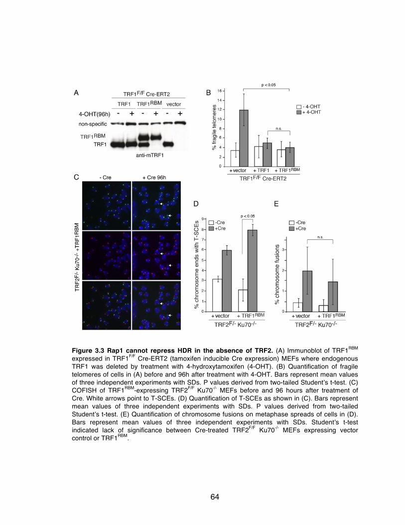
64
Figure 3.3 Rap1 cannot repress HDR in the absence of TRF2. (A) Immunoblot of TRF1RBM expressed in TRF1F/F Cre-ERT2 (tamoxifen inducible Cre expression) MEFs where endogenous TRF1 was deleted by treatment with 4-hydroxytamoxifen (4-OHT). (B) Quantification of fragile telomeres of cells in (A) before and 96h after treatment with 4-OHT. Bars represent mean values of three independent experiments with SDs. P values derived from two-tailed Student’s t-test. (C) COFISH of TRF1RBM-expressing TRF2F/F Ku70-/- MEFs before and 96 hours after treatment of Cre. White arrows point to T-SCEs. (D) Quantification of T-SCEs as shown in (C). Bars represent mean values of three independent experiments with SDs. P values derived from two-tailed Student’s t-test. (E) Quantification of chromosome fusions on metaphase spreads of cells in (D). Bars represent mean values of three independent experiments with SDs. Student’s t-test indicated lack of significance between Cre-treated TRF2F/F Ku70-/- MEFs expressing vector control or TRF1RBM.

65
3.2.3 Rap1 confers an increase in TRF2 DNA binding affinity.
To test the hypothesis that Rap1 changes the interaction of TRF2 with
certain DNA substrates, nickel-affinity purifications of baculovirus-expressed His-
Rap1, His-TRF2, and the His-Rap1/TRF2 complex were performed (Figure 3.4A).
For purification of the Rap1/TRF2 complex, His-hRap1 and hTRF2 were co-
expressed in Sf9 insect cells. Immunoblotting of the co-purified Rap1/TRF2
complex showed the expected 1:1 stoichiometry of Rap1 and TRF2 (Figure
3.4B). Electrophoretic mobility shift assays (EMSAs) with a duplex telomeric
substrate with 12 repeats showed the Rap1/TRF2 complex had a slight increase
in DNA binding affinity (less than 2-fold) over TRF2 alone (Figure 3.4C). Mixing of
separately purified Rap1 and TRF2 did not result in this improved binding affinity
and the pattern of the shifted probe looked very similar to TRF2 alone,
suggesting that Rap1/TRF2 complex formation was not efficient in vitro (Figure
3.4C). Analysis of binding to a ds-ss ‘junction’ indicated that the co-purified
complex bound to the junction substrate with a higher affinity than TRF2 alone
(Figure 3.4D). Binding of TRF2 to the junction probe was variable with an affinity
ranging from 2.5- to 6-fold less than that of the Rap1/TRF2 complex. Despite the
ability of Rap1/TRF2 to bind to the junction probe better than TRF2 alone, its
affinity for a telomeric junction was very similar to its affinity for duplex telomeric
DNA. Therefore, while Rap1 clearly increases the DNA binding affinity of TRF2, it
is unclear whether it is this activity of Rap1 that confers its inhibition of HDR. The
length of the telomere tract did not affect Rap1/TRF2 binding ability, as EMSAs

66
with a duplex DNA substrate with three telomere-repeats (like the junction probe)
did not show a change in DNA-binding affinity. Rap1 has previously been shown
not to bind duplex telomeric DNA 20 and similarly did not exhibit any DNA binding
to the junction probe (Figure 3.4D). Taken together these data suggest that Rap1
binding to TRF2 improves its ability to bind telomeric DNA. However, a significant
difference was not observed in the binding affinity of Rap1/TRF2 to a ds-ss
telomere junction relative to duplex telomeric DNA. Therefore it remains unclear
whether Rap1 inhibits HDR by altering the DNA binding activity of TRF2.
3.2.4 HDR is not aggravated by dual absence of Rap1 and POT1
To test whether Rap1 and POT1 repress HDR at the same step, I created
and analyzed cells in which both Rap1 and POT1 were removed from telomeres.
An shRNA to TPP1 was used to deplete POT1a/b from telomeres in
Rap1F/FKu70-/- MEFs. As a control, TPP1 was also depleted in Rap1F/FKu70+/-
MEFs, where the presence of one allele of Ku is sufficient to inhibit T-SCEs. The
level of TPP1 knockdown could not be assessed by immunoblotting due to lack
of a good antibody for mouse TPP1. However, the TPP1 shRNA was effective in
POT1a/b removal as indicated by the induction of TIFs (Figure 3.5A-B) as well as
the appearance of T-SCEs in Ku70-deficient cells (Figure 3.5D-E). Deletion of
Rap1 with Cre (Figure 3.5C) in TPP1-depleted cells did not exacerbate the T-
SCE phenotype (Figure 3.5D-E). In fact the levels of T-SCEs observed in a Ku70-
deficient background when Rap1 or POT1 alone were depleted, was very similar
67
to telomeres lacking both Rap1 and POT1. Telomeres remained shielded from
HDR when Rap1 and POT1 were removed in Ku70-proficient cells. These data
suggest that Rap1 and POT1 act together to repress HDR.
One caveat to consider with the interpretation of these results is whether it
is possible to observe an exacerbation of the T-SCE phenotype. Theoretically,
assuming HDR intermediates can be equally shuttled into pathways that dissolve
the Holliday junction or resolve it resulting in crossover, the maximal number of
T-SCEs we would expect to detect is 50%. Current levels of T-SCEs observed
are rarely above 15%. Various efforts to knockdown BLM or TOP3α to remove
the dissolution arm of HDR and skew resolution of HDR intermediates into
crossovers were unsuccessful, therefore it remains to be seen what the threshold
of detection for T-SCEs is.
Despite this caveat, these results raise the possibility that Rap1 and POT1
repress HDR as a subcomplex of shelterin. Indeed it has been shown that TRF2
and POT1 are capable of forming a detectable complex 21,152,185,234. However,
coimmunoprecipitation experiments failed to detect a stable interaction between
MYC-POT1a or –b and FLAG-Rap1 (Figure 3.5F).

68
Figure 3.4 Rap1 confers an increase in the telomere binding affinity of TRF2. (A) Coomassie stained SDS-PAGE gels of purified His-tagged proteins as indicated. 2-fold serial dilutions of purified protein and Bovine serum albumin (BSA) were run on the same gel to quantify protein amounts. (B) Immunoblotting of quantified protein amounts to calculate stoichiometry of Rap1 and TRF2 in the co-purified sample. (C) Top, schematic of probe (restriction fragment containing 12 TTAGGG repeats) used for EMSAs below. Bottom, EMSAs with 2-fold serial dilutions of indicated proteins; probe, no protein added. Dissociation constants (Κd) calculated below. Values for co-purified and TRF2 are mean of three experiments with SDs. (D) Top, schematic of probe with 3 TTAGGG repeats, a 3’ overhang containing the minimal binding site for POT1 (5’-GGTTAGGGTTAG-3’), and a natural telomere end (ATC-5’). Bottom, EMSAs with probe above using 2-fold serial dilutions of indicated proteins; probe, no protein added. Dissociation constant (Κd) calculated below. Value for co-purified is mean of three experiments with SDs; n.d., not determined.

69
Figure 3.5 No exacerbation of T-SCE levels upon removal of Rap1 and POT1. (A) TIF assay on Rap1F/FKu70-/- or Rap1F/FKu70+/- MEFs treated with Cre and or a TPP1 shRNA as indicated for 96 hours. White square indicates area enlarged below. (B) Quantification of TIF-positive cells as in (A). Cells with ≥ 10 TIFs were considered TIF-positive. Bars represent mean values of three independent experiments with SDs. (C) Immunoblot for Rap1 in cells as described in (A). (D) CO-FISH assay on cells as described in (A). White arrows indicate T-SCEs. (E) Quantification of CO-FISH in (D). Bars represent mean values of three independent experiments with SDs. P-values calculated based on Student’s t-test. (F) MYC-immunoprecipitation from 293T cells co-transfected with MYC-tagged constructs as indicated and FLAG-tagged Rap1. MYC-TRF2ΔRap1 was used as a negative control and MYC-TRF2 was used as a positive control for interaction with Rap1.

70
3.2.5 Deletion of Rap1 and TRF1 does not induce T-SCEs
TRF1 loss results in inefficient replication of telomeric DNA 156 and
potentially introduces replication intermediates that are susceptible to resolution
by homology-directed repair. However, T-SCEs are not observed when telomeres
lack TRF1, possibly due to repression of HDR by Ku70/80. Indeed, preliminary
analysis of MEFs lacking TRF1 and Ku80 showed an induction of T-SCEs
(Figure 3.6A), suggesting that telomeres are also vulnerable to HDR during DNA
synthesis.
Mass spectrometry and ChIP experiments using human cells have
implicated shelterin components TRF1, TRF2 and Rap1 in interacting with and
potentially recruiting Ku to telomeres 78, 265, 283, although Ku has also been shown
to bind directly to telomere repeats in vitro 284. To explore potential redundancy in
the recruitment of Ku to telomeres, we decided to delete both Rap1 and TRF1
from telomeres. If T-SCEs were observed in Rap1/TRF1 null MEFs, this would
suggest that Ku is unable to repress HDR in this setting. Alternatively, removing
both Rap1 and TRF1 could lead to an increase in telomere substrates vulnerable
to HDR and result in T-SCEs regardless of whether co-repression of HDR by Ku
was functional or not.
Analysis of metaphases lacking Rap1 and TRF1 in a Ku-proficient setting
showed an increase of fragile telomeres and telomere sister associations which
are characteristic of TRF1 loss (Figure 3.6B-C). Despite the presence of telomere
replication problems, no induction of T-SCEs was observed (Figure 3.6C),

71
suggesting that Ku was still able to repress HDR. A low level of telomere fusions
were also detected in MEFs where Rap1 and TRF1 had been deleted (Figure
3.6C), potentially due to minor destabilization of TRF2 binding to telomeres.
Figure 3.6 Ku represses T-SCEs in TRF1 null cells, as well as in MEFs lacking both Rap1 and TRF1. (A) Quantification of T-SCEs (left) and telomere sister associations (right) on metaphase spreads, before and 96 hours after treatment with Cre. Genotypes as indicated. (B) Western blot of two littermate controls with genotypes as indicated, before and 96 hours after treatment with Cre. Antibodies used for detection of proteins are indicated on left. (C) Quantification of aberrant structures on metaphase spreads in Rap1F/F TRF1F/F MEFs before and 96 hours after treatment with Cre. Data represents averages and standard deviations from analysis of three independently derived cell lines.

72
3.2.6 HDR is repressed in late-passage Ku70-deficient MEFs
The induction of homology-directed repair in absence of DNA damage
signaling and cell cycle arrest in Rap1/Ku70-deficient MEFs allowed examination
of the consequences of unbridled HDR on telomere maintenance. Rap1F/F Ku70-/-
(or Rap1F/Δ Ku70-/-) MEFs were isolated and immortalized with SV40LT. The
immortalization process is variable and takes approximately 15 to 20 PDs for
cells to escape crisis and proliferate at a steady pace. The number of PDs cells
undergo during immortalization is a reproducible estimate, but cannot be exactly
calculated due to cell death or senescence that primary MEFs undergo during the
immortalization process. After immortalization, at approximately PD 20,
Rap1F/FKu70-/- (or Rap1F/ΔKu70-/-) MEFs were further cultured to 70 PDs with and
without Cre treatment (Figure 3.7A-B). Cells were harvested at various time-
points to assess the level of T-SCEs. Surprisingly, T-SCE events began to
diminish as early as PD 30 and were reduced to basal levels similar to Rap1-
proficient cells by PD 50 (Figure 3.7C). T-SCEs were also no longer induced
when Rap1 was deleted from late passage Rap1F/F Ku70-/- showing that Ku70-
deficient MEFs exhibit downregulation of telomeric HDR when cultured
extensively.
To determine whether HDR was downregulated in general or just at the
telomere, efforts were made to use a transient reporter assay developed in the
Jasin lab 171 that assesses HDR-mediated repair events of a break induced by
expression of I-SceI (a site-specific endonuclease). However this assay did not

73
work robustly in MEFs, due to difficulty in achieving high transfection efficiencies
and minimal cell death.
Figure 3.7 HDR is repressed in late-passage Ku70-deficient MEFs. (A) Growth curve of Rap1F/FKu70-/- MEFs with and without deletion of Rap1 by Cre. (B) Western blot indicating Rap1 levels in the two cell lines shown in (A) at the indicated PDs. (C) CO-FISH analysis of Ku70 deficient MEFs with or without Rap1 at the indicated PDs. White arrows point to T-SCE events. Quantification of chromosome ends with T-SCEs for three independent experiments is listed under corresponding CO-FISH panels.

74
3.3 Summary of findings
The data presented here show that Rap1 requires its interaction with
TRF2 to repress HDR. Recruitment of Rap1 to telomeres by TRF1RBM in the
absence of TRF2 did not prevent T-SCEs. It is possible however, that
experimentally induced binding of Rap1 to TRF1RBM may disrupt Rap1’s
interaction with another factor required for repression of HDR. Additionally, the
specific localization of Rap1 at telomeres by TRF1RBM may be subtly, yet
impactfully different compared to its placement byTRF2.
The Rap1/TRF2 complex may bind to a protein involved in HDR inhibition
that Rap1 alone or TRF2 alone may be unable to bind to. Alternatively, Rap1
may enhance the DNA binding affinity of TRF2 to HDR substrates and prevent
loading of components required for repair. Preliminary gel-shift assays indicated
that Rap1 may enhance the overall binding affinity of TRF2 to telomeric DNA, but
this effect was minor and it is unclear whether it would be physiologically
relevant. However, one report showed that the Rap1/TRF2 complex binds to ds-
ss telomere junctions 10-fold better than duplex DNA 235. The discrepancy
between this study and the results reported here could be due to changes in the
activity or oligomeric state of the purified proteins, as well as the different
substrates used. In addition, another study reported Rap1 conferred no
advantage to TRF2 to bind ds-ss telomere junctions, but instead improved the
specificity of TRF2 binding to telomeric DNA 236.

75
Rap1 and POT1a/b seem to inhibit HDR via the same pathway as no
increase in the number of T-SCEs was observed when Rap1 and POT1a/b were
all removed from telomeres in a Ku70-deficient setting. Further assessment of
Rap1/TRF2 DNA-binding needs to be conducted in the context of the shelterin
complex, as POT1 binding to the overhang together with Rap1/TRF2 binding to
the ds-ss junction may contribute to the repression of telomeric HDR.
Lastly, the surprising finding that Ku70-deficient cells eventually
downregulate HDR suggests that when Ku is unable to inhibit HDR other factors
may be enlisted to regulate HDR. Efforts to determine whether this repression of
HDR was global or telomere-specific have thus far been unsuccessful and
warrant further study.

76
Chapter 4: TALEN gene knockouts of human Rap1
77
4.1 Introduction
Two discrepancies exist concerning the function of Rap1 in mammals. In
vitro, human Rap1 can block NHEJ when it binds to TRF2 loaded on an end-
joining substrate 213. In vivo, a Rap1-fusion protein can reduce telomere fusions
when it is tethered to telomeres that are depleted of TRF2 160. On the other hand,
mouse cells lacking Rap1 show no telomere fusions and Rap1-deficient mice are
alive and fertile 19,220. The only telomere deprotection phenotype in Rap1-
deficient mouse cells is a propensity for telomere-telomere recombination when
Ku70/80 are also absent 19.
In addition, it is unclear whether Rap1 has a conserved role in telomere
length homeostasis in mammals. In support of a role in telomere length control,
shRNAs to human Rap1 induce telomere lengthening 78, as does overexpression
of several Rap1 truncation mutants 23. Furthermore, overexpression of Rap1
mutants lacking its BRCT domain or MYB motif exhibit diminished telomere
length heterogeneity, suggesting Rap1 also has a role in regulating distribution of
telomere length. However, Rap1 knockout mice show no change in telomere
length, even after three generations 19.
To determine the function(s) of human Rap1, Transcription Activator Like
Effector Nucleases (TALENs) were used to knockout the Rap1 gene. Analysis of
several independent Rap1-deficient cell lines demonstrated that human Rap1
was not required for telomere protection, telomere length regulation, and other

78
aspects of telomere function. Instead, changes were observed in the transcription
of several genes upon loss of Rap1.
4.2 Results
4.2.1 Efficient TALEN-mediated knockout of human Rap1
Similar to the mouse, the human Rap1-encoding gene (TERF2IP, referred
to as Rap1) shares its promoter region with the essential KARS (lysyl-tRNA
synthetase) gene located just upstream of exon 1 (Figure 4.1A). To avoid
disrupting the KARS gene, a targeting strategy analogous to the one used for
mouse Rap1 19 was implemented and TALENs were employed to delete exon 2.
Deletion of exon 2 should result in an mRNA encoding a 226 aa ORF that ends
prematurely in a stop codon at the beginning of exon 3. As exon 3 encodes the
TRF2-binding domain, the truncated Rap1 protein is not expected to localize to
telomeres. Taking advantage of the small size of exon 2 (125 bp) and anticipated
resection of TALEN-induced DSBs 237,238, a neomycin or blasticidin donor
construct was designed containing 5’ and 3’ arms homologous to the surrounding
introns. HDR using the donor construct should result in deletion of exon 2 and
insertion of the neomycin cassette (Figure 4.1A).
Rap1 was targeted in two near-diploid cancer lines (HCT116 colorectal
carcinoma and HT1080 fibrosarcoma), a subclone of the near-triploid cervical
carcinoma HeLa cell line (HeLa1.3 22), the diploid ARPE-19 retinal pigment

79
epithelial cell line, and primary BJ fibroblasts transformed with SV40LT. With the
exception of BJ-SV40LT, all cells expressed telomerase. For HCT116, HT1080,
and ARPE-19 cells, neomycin resistant clones were obtained, analyzed by PCR,
and then evaluated by Southern blotting to verify the correct neomycin insertion
(Figure 4.1B-C). For HeLa1.3 and BJ-SV40LT, the blasticidin donor construct
was used and clones were analyzed by immunoblotting for Rap1. These
analyses identified clones that lacked the wild type Rap1 gene and expressed no
detectable Rap1 protein (Figure 4.1C-D). Polypeptides representing the
remaining ORF of the targeted Rap1 gene were not detected (Figure 4.2A-C),
perhaps due to nonsense-mediated decay.
Unexpectedly, immunoblotting revealed the complete loss of Rap1 in
heterozygous clones with one neo insertion (Figure 4.1B-C). Telomeric ChIP of
two such clones (HT1080 c21 and HeLa1.3 c28) confirmed that Rap1 was absent
from telomeres (Figure 4.2D-G). Sequencing revealed small deletions close to
the TALEN site in these and other Rap1-deficient clones with only one neo-
containing Rap1 gene (Figure 4.3A-B). Most mutations had ablated Rap1 by
deleting the exon 2 splice acceptor site or had created a frame-shift mutation,
indicating that errors generated during NHEJ had inactivated the Rap1 gene
(Figure 4.3C). As a result, the frequency of the Rap1 knockouts (KOs) is much
higher than deduced from PCR genotyping. Taking the deleterious repair events
into account, the ablation of Rap1 occurred at 20-65% efficiency in HCT116,
HT1080, and ARPE-19 cells (Figure 4.3D). The HCT116 cells showed the

80
highest KO frequency consistent with their propensity for HDR (reviewed in 239).
The actual KO frequency is probably even higher as only clones with a neo
cassette in the Rap1 gene were analyzed.
The high KO frequency allowed screening of HeLa1.3 and BJ-SV40LT
clones by Rap1 immunoblotting. Sequencing of Rap1-negative HeLa1.3 clones
revealed that all three alleles in these clones contained inactivating mutations
near the TALEN cut site (Figure 4.3B). Prolonged culturing of clones in blasticidin
resulted in cell death, suggesting that the pEF blasticidin cassette did not confer
long-term resistance. The KO frequency was lower in BJ-SV40LT fibroblasts with
only one blasticidin-resistant clone showing absence of Rap1 protein. This clone
was not analyzed because it perished in telomere crisis, as did many of the
Rap1-proficient BJ-SV40LT clones.

81
Figure 4.1 TALEN-mediated inactivation of the gene for human Rap1. (A) Schematic of human Rap1, the Rap1 locus, the targeting construct, and the resulting knockout allele. F1, R1, and R2neo: PCR primers for genotyping. Arrows in bold: TALEN binding and cut sites. (B) Southern blot of EcoRI (RI)- or BglII/EcoRV (RV)-digested genomic DNA from targeted HT1080 clones. Probe shown in (A). (C) PCR genotyping of the Rap1 gene and western blotting for Rap1 in the indicated clones. +, WT allele; Δ, targeted allele; mut, mutation resulting in loss of Rap1. (D) Immunoblotting of protein from targeted homozygous and heterozygous HCT116 clones showing absence of Rap1 in a large proportion of heterozygous clones. Genotypes indicated below.

82
Figure 4.2 Loss of Rap1 in targeted clones. (A) Immunoblots of HCT116 and HT1080 KO clones probed with anti-hRap1 showing the absence of detectable truncated Rap1 proteins. (B) Schematic of retroviral constructs expressing FLAG-tagged exon 1 (ex 1) and FLAG-tagged exon 3 (ex 3). (C) Immunoblotting for the expression of constructs in (B) in HT1080 KO and WT clones. The protein fragment encoded by exon 1 is expressed and detected by the Rap1 antibody. The protein fragment encoded by exon 3 contains epitopes that the Rap1 antibody can recognize, but lack of detectable signal indicates that it is not expressed. (D) Telomeric ChIP of WT and targeted HT1080 clones. Duplicate dot-blots were probed for telomeric or Alu repeats. (E) Average percentage of telomeric DNA recovered in ChIPs with the indicated antibodies (two independent experiments). Error bars: SEMs. (F) Telomeric ChIP of HeLa 1.3 WT and KO cells. (G) Quantification of the percent of telomeric DNA recovered in each ChIP.

83
Figure 4.3 TALEN-induced mutations in the Rap1 locus. (A) Schematic illustrating TALEN binding sites, their predicted cutting site (grey arrows), the pyrimidine tract, splice acceptor (SA), and the location of exon 2 in the Rap1 locus. A short repeat (underlined in grey) is frequently mutated in the mutant alleles. (B) Relevant sequences of the indicated clones. Predicted consequences of the mutations are indicated on the right. (C) Schematic illustrating NHEJ and HDR after TALENs cutting. (D) Table indicating number of Rap1 knockout clones acquired and the genetic alterations in Rap1.

84
4.2.2 Rap1-deficient cell proliferate and maintain fully protected telomeres
The Rap1-deficient cell lines proliferated normally (Figure 4.4A) and
lacked a significant level of TIFs (Figure 4.4B-C), indicating that removal of Rap1
from telomeres does not result in a DNA damage response. Cells lacking Rap1
also did not show a significant induction of DSB repair at telomeres. Metaphase
spreads of Rap1 KOs lacked chromosome end fusions, a read-out for telomeric
NHEJ (Figure 4.5A-B). CO-FISH to monitor HDR-mediated T-SCEs established
that recombination remained repressed at telomeres (Figure 4.5C-D). Analysis of
telomere-sister chromatid exchanges in cells lacking Rap1 was conducted in a
Ku-proficient background. Similar to the mouse, human Ku70/80 are also
required to repress telomere recombination. However, unlike the mouse, deletion
of Ku70/Ku80 is lethal in human somatic cells 240,241, thus it was not possible to
test a setting where both Rap1 and Ku70/80 were deleted. While the contribution
of human Rap1 to the repression of HDR in the context of Ku70/80-deficiency
remains to be determined, our data indicate that human telomeres remain
protected from NHEJ and HDR in absence of Rap1.

85
Figure 4.4 Rap1-deficient cells proliferate and do not induce DNA damage signaling. (A) Growth curves of WT and Rap1 KO HT1080 clones. (B) TIF assay on WT and KO HeLa1.3 clones. Green, telomeric FISH; red, IF for 53BP1; blue, DNA (DAPI). (C) Quantification of TIFs assay (see (B)). Error bars: SDs of three independent experiments (n≥100 nuclei per clone). P values from a two-tailed paired t-test combining WT and KO datasets. n.s.: not significant.

86
Figure 4.5 Telomere protection in Rap1-deficient cells. (A) Metaphase chromosomes from the indicated WT and Rap1 KO cells. Green, telomeric FISH; blue, DNA (DAPI). (B) Quantification of telomere fusions, detected as in (A), in the indicated clones. P values from a two-tailed paired t-test on combined WT and KO datasets. (C) CO-FISH analysis on the indicated WT and KO HeLa clones. (D) Table showing the percentage of telomeres showing T-SCEs as assayed in (C) in the indicated clones. P value from unpaired two-tailed t-test.

87
4.2.3 Unaltered telomere length dynamics in absence of Rap1.
To determine whether Rap1 affected telomere length homeostasis, two
HT1080 Rap1-deficient clones were cultured for 50 PDs alongside two Rap1-
proficient clones selected for their matching telomere lengths (Figure 4.6A). All
clones exhibited a mild increase in telomere length (Figure 4.6B). The two Rap1-
deficient clones lengthened their telomeres at a similar modest rate (1-2 bp/PD)
whereas the two Rap1-proficient clones differed in the rate of telomere
lengthening (28 and 4 bp/PD) (Figure 4.6B). Given the clonal variation and small
differences in telomere length changes, the removal of Rap1 did not appear to
have a strong effect on telomere length dynamics in HT1080 cells.
Similarly, Rap1 did not affect the telomere length dynamics of HCT116
clones (Figure 4.6C). Two Rap1-deficient clones showed telomere shortening at
variable rates (-3 to -32 bp/PD). Given that the telomere shortening in the
parental cells (-27 bp/PD) is similar to that of one of the Rap1-deficient clones
(c10), it can be concluded that Rap1 did not strongly affect telomere dynamics in
HCT116 cells either.
Finally, three Rap1-deficient ARPE-19 clones (c26, c25, and c30) showed
wide variations in telomere dynamics, ranging from slight shortening (-18 bp/PD)
to slight elongation (9 and 16 bp/PD) (Figure 4.6D). The single Rap1-proficient
clone (c23) showed telomere shortening at a rate of -34 bp/PD. Thus, there is
considerable variability in the telomere dynamics in ARPE-19 clones but no
consistent effect of Rap1 deletion.
88
Given the lack of consistent shortening or lengthening phenotypes in
multiple Rap1 knockouts, the simplest interpretation is that Rap1 does not play a
major role in telomere length regulation. Deletion of Rap1 also did not induce an
obvious change in the telomere length heterogeneity (Figure 4.6A and 4.7A),
which was affected by Rap1 mutants in overexpression studies 20. Removal of
Rap1 from several cell lines also did not appear to affect the 3’ telomeric
overhang. The amount of ss telomeric DNA was determined by in-gel
hybridization of a labeled C-strand telomeric repeat probe to native telomeric
restriction fragments. Quantification of normalized single-stranded telomeric DNA
signals indicated that Rap1 status had no significant impact on the 3’ overhangs
in HT1080, HCT116, and ARPE-19 cells (Figure 4.7A-B).

89
Figure 4.6 Rap1 knockouts exhibit no systematic changes in telomere length. (A) Southern blot of telomeric restriction fragments from two WT and two KO HT1080 clones at the indicated PDs.(B-D) Curves of average telomere lengths at indicated PDs in HT1080, HCT116, and ARPE-19 clones, respectively.

90
Figure 4.7 Loss of Rap1 does not affect telomere structure. (A) Telomeric DNA analysis of WT and KO clones in three different cell lines. Top, in-gel detection of native telomeric restriction fragments with a C-strand telomeric probe revealing the G-strand overhang signals; bottom, same gel rehybridized after in situ denaturation of the DNA, revealing the total telomeric DNA in each lane. Three biological replicates of each cell line were run adjacent to each other on the same gel. (B) Overhang signals were normalized to total telomeric signals in each lane and plotted as a ratio compared to the WT overhang signal of the corresponding cell line. Error bars represent SDs. Lack of statistical significance derived from two-tailed paired t-tests using the three independent experiments conducted with each cell line.

91
4.2.4 Telomeric positioning in Rap1 null cells
To determine whether Rap1 is involved in the peripheral positioning of
telomeres in newly formed G1 cells 219, mitotic shake-off was used to analyze
HeLa1.3 daughter cells in early G1. HeLa cells were used due to the ease of
detection of their long telomeres, and the same method of telomere position
analysis had been previously conducted in HeLa cells 219, thereby enabling a fair
comparison to published data.
Using Lamin A to mark the nuclear envelope (NE) and FISH to visualize
telomeres, the subnuclear position of the telomeric signals was determined and
compared between Rap1-proficient and -deficient cells (Figure 4.8A-B). The
distribution of telomeres in the nucleus and median distance from the nuclear
envelope was similar for two Rap1-proficient and -deficient clones in two
independent experiments. Moreover, the percentage of telomeres present in a
zone defined arbitrarily as the nuclear periphery (within 10% distance from the
NE) was also similar regardless of Rap1 status, illustrating that Rap1 is not
required for the more peripheral positioning of telomeres in early G1.
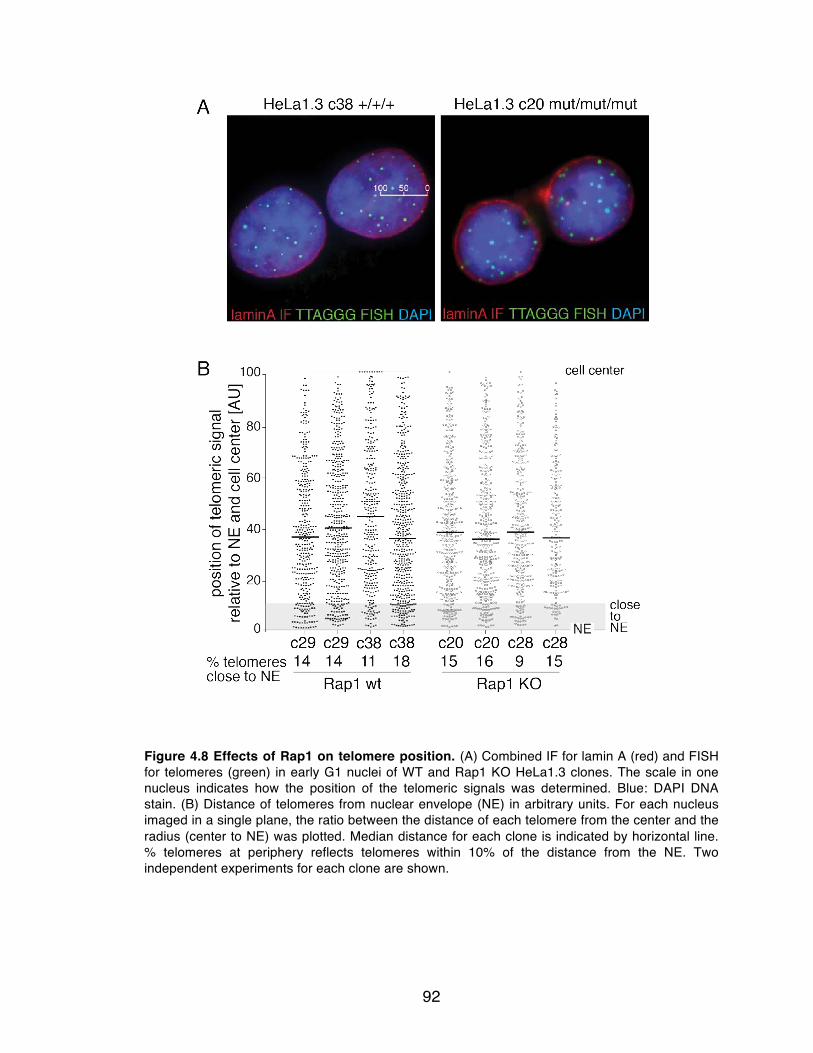
92
Figure 4.8 Effects of Rap1 on telomere position. (A) Combined IF for lamin A (red) and FISH for telomeres (green) in early G1 nuclei of WT and Rap1 KO HeLa1.3 clones. The scale in one nucleus indicates how the position of the telomeric signals was determined. Blue: DAPI DNA stain. (B) Distance of telomeres from nuclear envelope (NE) in arbitrary units. For each nucleus imaged in a single plane, the ratio between the distance of each telomere from the center and the radius (center to NE) was plotted. Median distance for each clone is indicated by horizontal line. % telomeres at periphery reflects telomeres within 10% of the distance from the NE. Two independent experiments for each clone are shown.

93
4.2.5 No change in telomeric chromatin or transcription upon Rap1 loss
Mammalian telomeres contain nucleosomes and epigenetic marks that are
characteristic of heterochromatin, such as H3K9me3, H4K20me3 and the loading
of heterochromatin protein 1 (HP1, reviewed in 228). Rap1 also had no detectable
effect on general markers for the chromatin status at telomeres, as evidenced by
ChIP for methylation of H3K9, acetylation of H4, and HP1α, HP1β and HP1γ
(Figure 4.9A-B). In addition there was no significant effect of Rap1 on the
abundance of the telomeric lncRNA TERRA (Figure 4.9C-D). This finding is
consistent with the unaltered TERRA levels after deletion of mouse Rap1 19.

94
Figure 4.9 No change in telomeric chromatin or transcription in Rap1-deficient cells. (A) ChIP for modified histones at telomeres in WT and Rap1 KO cells. Duplicate blots were probed for telomeric DNA or Alu repeats. (B) Relative telomeric ChIP signals obtained as in (A) were expressed as the ratio of signal in KO and WT clones (WT set to 1). Values represent averages of two experiments. Error bars: SEMs. (C) Northern blot hybridized with a telomeric probe showing TERRA levels of Rap1 WT and KO clones in the HT1080, HCT116 and ARPE-19 cell lines. U2OS serves as a positive control for TERRA expression. Ethidium bromide staining of ribosomal RNA serves as a loading control. (D) TERRA signals were normalized using the 18S ribosomal RNA and plotted as a ratio compared to the WT TERRA signal of the corresponding cell line. Error bars represent SDs. Two-tailed unpaired t-tests of 3 independent experiments illustrates lack of statistical significance between Rap1 WT and KO TERRA expression levels.

95
4.2.6 Rap1 affects transcriptional regulation
Mouse Rap1 localizes to over 8600 gene-associated loci, affecting
transcription of numerous genes 220-222, while human Rap1 is found at ~63 gene
loci 223. To query the effect of Rap1 on the transcriptome, microarray profiling
was performed on seven Rap1 WT and KO clones derived from three different
cell lines, ARPE-19, HT1080 and HCT116. A number of differentially regulated
genes were identified (Tables 4.1-4.3). The three Rap1-regulated genes in the
ARPE-19 cells (LHX2, LRRC17 and CDO1) were validated by quantitative RT-
PCR (qRT-PCR) and their response to Rap1 deletion was further confirmed on
an additional ARPE-19 Rap1 KO clone (c26) (Figure 4.10A-C). The Rap1-
regulated genes varied between the different cell lines, most likely due to the
different origins of the cell lines. Gene ontology analysis was uninformative,
because of the low number of genes identified by this limited analysis. However,
one Rap1-regulated gene in the HT1080 cells was among the human Rap1-
associated loci in the HT1080-derived HTC75 cell line 223. Taken together, these
data are consistent with a role for Rap1 in transcriptional control in human cells.
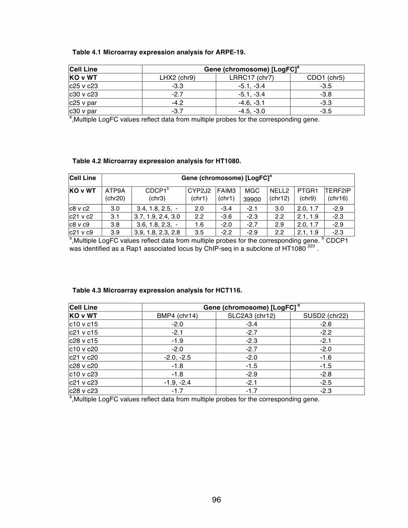
96
Table 4.1 Microarray expression analysis for ARPE-19.
Cell Line Gene (chromosome) [LogFC]a KO v WT LHX2 (chr9) LRRC17 (chr7) CDO1 (chr5) c25 v c23 -3.3 -5.1, -3.4 -3.5 c30 v c23 -2.7 -5.1, -3.4 -3.8 c25 v par -4.2 -4.6, -3.1 -3.3 c30 v par -3.7 -4.5, -3.0 -3.5 a,Multiple LogFC values reflect data from multiple probes for the corresponding gene.
Table 4.2 Microarray expression analysis for HT1080.
Cell Line Gene (chromosome) [LogFC]a
KO v WT ATP9A (chr20)
CDCP1b (chr3)
CYP2J2 (chr1)
FAIM3 (chr1)
MGC 39900
NELL2 (chr12)
PTGR1 (chr9)
TERF2IP (chr16)
c8 v c2 3.0 3.4, 1.8, 2.5, - 2.0 -3.4 -2.1 3.0 2.0, 1.7 -2.9 c21 v c2 3.1 3.7, 1.9, 2.4, 3.0 2.2 -3.6 -2.3 2.2 2.1, 1.9 -2.3 c8 v c9 3.8 3.6, 1.8, 2.3, - 1.6 -2.0 -2.7 2.9 2.0, 1.7 -2.9 c21 v c9 3.9 3.9, 1.8, 2.3, 2.8 3.5 -2.2 -2.9 2.2 2.1, 1.9 -2.3 a,Multiple LogFC values reflect data from multiple probes for the corresponding gene. b CDCP1 was identified as a Rap1 associated locus by ChIP-seq in a subclone of HT1080 223 .
Table 4.3 Microarray expression analysis for HCT116.
Cell Line Gene (chromosome) [LogFC] a KO v WT BMP4 (chr14) SLC2A3 (chr12) SUSD2 (chr22) c10 v c15 -2.0 -3.4 -2.6 c21 v c15 -2.1 -2.7 -2.2 c28 v c15 -1.9 -2.3 -2.1 c10 v c20 -2.0 -2.7 -2.0 c21 v c20 -2.0, -2.5 -2.0 -1.6 c28 v c20 -1.8 -1.5 -1.5 c10 v c23 -1.8 -2.9 -2.8 c21 v c23 -1.9, -2.4 -2.1 -2.5 c28 v c23 -1.7 -1.7 -2.3 a,Multiple LogFC values reflect data from multiple probes for the corresponding gene.
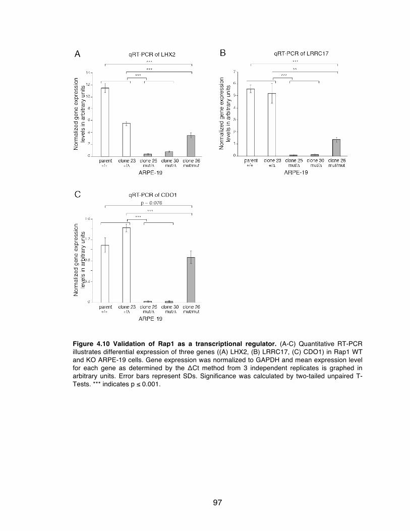
97
Figure 4.10 Validation of Rap1 as a transcriptional regulator. (A-C) Quantitative RT-PCR illustrates differential expression of three genes ((A) LHX2, (B) LRRC17, (C) CDO1) in Rap1 WT and KO ARPE-19 cells. Gene expression was normalized to GAPDH and mean expression level for each gene as determined by the ΔCt method from 3 independent replicates is graphed in arbitrary units. Error bars represent SDs. Significance was calculated by two-tailed unpaired T-Tests. *** indicates p ≤ 0.001.
98
4.3 Summary of findings
Based on the genetic data presented here, human Rap1 is not required for
the protection of telomeres from NHEJ and has no obvious effect on telomere
length regulation, contrary to what was anticipated from other studies 23,78,160,213.
The results in this chapter point to the difficulty in interpreting experiments in
which telomeric phenotypes are observed upon overexpression of shelterin
(mutant) proteins or their partial inactivation by shRNAs. The prior finding of
changes in telomere length and heterogeneity upon overexpression of Rap1
mutants 23 may have been due to nucleoplasmic titration of factors away from the
telomere that (indirectly) influence these phenotypes. Similarly, the artificial
tethering of Rap1 to telomeres may have had an effect on NHEJ that does not
reflect the normal function of the protein 160.
In addition, telomeres lacking Rap1 remained protected from DNA
damage signaling and HDR, and had a normal 3’ overhang. These findings are in
agreement with the mouse Rap1 KO, which revealed no obvious phenotype other
than that of telomeres becoming prone to undergo HDR when Ku70/80 was
absent 19. Whether human telomeres lacking Rap1 also recombine more readily
in a Ku70/80-deficient setting is difficult to assess since deletion of human
Ku70/80 leads to rapid telomere loss and cell death 240,241.
Human Rap1 was surprisingly dispensable for telomere protection,
however its role in transcriptional regulation appears to be conserved. Due to the
low number of deregulated genes identified in vastly differing cell types, it was

99
difficult to assess what transcriptional programs Rap1 participates in.
Examination of Rap1 deletion in cell lines with high metabolic activity, such as
hepatocytes, would help identify whether human Rap1 affects metabolism similar
to mouse Rap1.
These data indicate that while mammalian Rap1 has functionally diverged
away from its yeast predecessors, mouse and human Rap1 are very similar. Both
Rap1 knockout mice and human cells are viable, lack hallmarks of telomere
dysfunction, and have no overt change in telomere length settings. While the
Rap1 components of human and mouse shelterin are indistinguishable, it will be
important to query the functions of other shelterin components and associated
factors to gain a complete understanding of telomere maintenance and protection
in human cells.

100
Chapter 5: Investigating the role of human POT1 at telomeres
101
5.1 Introduction
As discussed in the general introduction, the deletion of mouse POT1a
and –b leads to embryonic and cellular lethality, elicits a telomeric ATR
dependent DNA damage response, increases 3’ telomere overhangs, and results
in infrequent post-replicative telomere fusions and telomere sister associations
31,32,182.
In human cells, two isoforms of POT1 are detected by immunoblotting with
molecular weights of 71 kDa and 55 kDa 184. The protein that migrates at 71 kDa
corresponds to full-length POT1 (POT1-FL). The isoform with the molecular
weight of 55 kDa (POT1-55) lacks the first N-terminal OB fold of POT1 and is
unable to bind telomeric DNA 34. POT1-55 is almost identical to a mutant allele of
POT1 (POT1ΔOB). POT1ΔOB cannot bind to telomeric DNA but retains its
interaction with TPP1. When overexpressed, POT1ΔOB acts as a dominant-
negative 79, presumably by binding to TPP1 and displacing endogenous POT1.
Endogenous expression levels of POT1-FL are approximately 10-fold more
abundant than POT1-55 184, and no function has been ascribed to POT1-55 as of
yet.
Partial depletion of POT1-FL with shRNAs impairs the growth of primary
but not transformed cells 184. Furthermore, there is a modest reduction (~30%) in
the overhang signals and the precise nature of the 5’ end of the telomeres is
disrupted 184,185. Knockdown of both POT1-FL and POT1-55 results in low levels

102
of TIFs, where ~40% of cells are TIF-positive, occurring during the G1 phase of
the cell cycle 184.
In addition to protecting telomeres from the DNA damage response and
modulating the nature of the telomere terminus, POT1 has been implicated in the
cis-acting negative regulation of telomerase. Depletion of POT1 with shRNAs 29
or expression of the POT1ΔOB allele leads to telomere elongation 79. The POT1
binding partner TPP1, on the other hand, interacts with telomerase and promotes
the recruitment of the enzyme to telomeres in vivo and enhances the processivity
of the enzyme in vitro 81.
Although the functions of POT1a and POT1b have been examined in
detail, it is as yet unclear to what extent the single human POT1-FL protein
incorporates these functions, and what role POT1-55 plays, if any. To understand
the role of human POT1 in telomere protection, we used TALENs with the aim to
generate human knockout cell lines lacking POT1. The preliminary analysis of
these human POT1-deficient cells is reported here.
5.2 Results
5.2.1 The human POT1 targeting strategy
Mouse POT1a and –b share greater than 70% amino acid identity with
human POT1 31. Human POT1, POT1a, and POT1b, have transcripts comprised
of 19, 18 and 17 exons respectively, 15 of which are coding exons (annotated by

103
Ensembl). Here, coding exons are numbered sequentially with exon 1 containing
the ATG. A strategy analogous to the mouse knockouts was employed to create
knockouts of human POT1. The mouse knockouts of POT1a and –b were
generated by deletion of exon 3, which results in an ORF with a premature stop
codon in exon 4 31. In that study, exons 1 and 2 were not targeted because this
could bring a putative alternate start site in the 5’ UTR into frame with exon 3
coding sequences. Similar to the mouse genes, deletion of human exon 3, which
encodes aa 41 to 85, is predicted to lead to a premature stop codon in the POT1
ORF at a position four codons into exon 4. Thus, we decided to delete exon 3 in
human cells to generate a POT1-FL knockout. Neomycin, blasticidin and zeocin
donor constructs were designed with 5’ and 3’ arms homologous to the introns
surrounding exon 3 (Figure 5.1B). The anticipated resection at the TALEN-
induced DSB and the small size of exon 3 should allow processing to extend into
the surrounding introns such that the 5’ and 3’ arms can be used for homology-
dependent repair, such that the repaired allele lacks exon 3.
In silico analysis of potential exon skipping products indicated that the
ORF could be put back into frame if splicing were to occur from the exon 2 splice
donor site to exon 7, 8, 10, 12, or 14. The potential truncated products generated
by the exon skipping events would lack most of the N-terminal OB-folds that
confer the ssDNA binding activity to POT1. Therefore, some of these versions of
POT1 (splicing of exon 2 splice donor site to exon 7, 8, or 10) might still be
recruited to telomeres via their C-terminal TPP1 binding region (encoded by

104
exons 10-15; see Figure 5.1A) and could potentially act as dominant-negative
mutants due to loss of their OB folds 79. To circumvent the formation of these
truncated proteins, the donor construct was endowed with a ‘STOP cassette’
composed of an array of four copies of the SV40 poly (A) addition site 242-244,
which should halt transcription beyond the second intron (Figure 5.1B).
Furthermore, introduction of the STOP cassette will also disrupt transcription of
the POT1-55 allele, effectively knocking out both isoforms of POT1.
Finally, given the lethality of deleting POT1a and -b in the mouse, we
considered the likely cell lethal phenotype of cells lacking both alleles human
POT1, which might be generated by TALENs (see Chapter 4). Therefore,
HT1080 fibrosarcoma cells were first infected with a retrovirus containing cDNA
encoding MYC-tagged full-length POT1 expressed from a CMV promoter (Figure
5.1B), and the retrovirus also conferred hygromycin resistance. Because the
POT1 rescuing allele is flanked with LoxP sites, exogenous POT1 can be excised
by Cre recombinase after the endogenous POT1 loci have been targeted.
Hygromycin resistant clones were isolated with the initial intent to use western
blot analysis to determine whether the exogenously expressed POT1 could be
efficiently deleted. However, the antibody used in previous studies 29 (that detects
endogenous POT1-FL and POT1-55) 184 was no longer functional. Therefore a
single clone, 20.3, was selected at random for further targeting. Eventual
identification of a commercial antibody that was able to detect POT1-FL, but not
POT1-55 (see Figure 5.1A), was used to determine that clone 20.3 expressed
105
exogenous POT1-FL at similar levels to endogenous POT1-FL, and could be
effectively deleted upon treatment with Cre (Figure 5.2A-B). In addition, as shown
previously 79, the levels of POT1 protein seem to be regulated such that
overexpression of POT1 resulted in repression of protein production from the
endogenous allele (see c20.3 in Figure 5.2B).
A TALEN pair was designed to bind a site starting in intron 2-3 and
extending into exon 3, such that the nuclease would not target the exogenous
floxed POT1 cDNA. In addition, inclusion of the intron-exon junction near the
predicted cleavage site enhances the likelihood of deleterious mutations that give
rise to either splicing errors or frame-shifts.

106
Figure 5.1 TALEN-mediated inactivation of the gene for human POT1. (A) Schematic of the human POT1 locus, and the two reported isoforms and their respective ATGs. POT1-FL, full-length POT1 protein; POT1-55, POT1 protein lacking the first OB fold. Exons not drawn to scale, coding exon numbers indicated (as annotated by Ensembl). Shading of exons corresponds to protein regions they encode. Known structures of and binding sites in POT1 are indicated. Navy line corresponds to peptide used to generate POT1 antibody used in this study (Abcam). (B) The POT1 locus, the targeting construct, and the resulting knockout allele. F1, R1, R2-PGK, R4-Zeo: PCR primers for genotyping. Arrows in bold: TALEN binding and cut sites. Schematic of MYC-tagged hPOT1 cDNA with flanking LoxP sites (triangles), cloned into the pWZL retroviral vector (confers hygro resistance).
107
5.2.2 TALEN-mediated deletion of full-length human POT1
HT1080 clone 20.3, expressing the floxed MYC-POT1 rescuing allele, was
co-transfected with the aforementioned TALEN pair and the neomycin donor
construct (Figure 5.1B). Cells were then selected with neomycin to enrich for
POT1 alleles that had been targeted by the TALENs and repaired by HDR,
resulting in deletion of exon 3 and acquisition of neomycin resistance. PCR
analysis of neomycin resistant clones showed that 4 out of 34 (~12%) clones had
integrated the donor cassette into one allele of POT1 (Figure 5.2B). Treatment of
these heterozygous clones with Cre showed that 3 (c3, c5, c17) of the 4 clones
expressed no detectable endogenous POT1-FL protein, which migrates slightly
slower than the MYC-tagged POT1, suggesting bi-allelic disruption of the gene
(Figure 5.2B). The efficiency of Cre-mediated deletion of POT1 varied between
experiments (compare Figure 5.2A to 5.2B). Therefore, POT1-FL deletion was
verified by immunoblotting for all experiments conducted, and only experiments
that had less than 10% to undetectable levels of residual POT1 protein were
analyzed.
Sequencing of uninterrupted alleles in these heterozygous clones lacking
endogenous POT1 revealed that clones c3, c5 and c17 all contained mutations
close to the TALEN cut site (Figure 5.2C). Mutations had ablated POT1-FL by
creating a frame-shift or a defect in splicing, indicating that errors generated
during NHEJ-mediated repair of the TALEN-induced break had inactivated the
second allele of POT1. However, these NHEJ-mediated mutated alleles lacked

108
integration of the STOP cassette and could still be transcriptionally active.
Therefore, while these clones served as knockouts for full length POT1, the
POT1-55 protein and potential exon skipping products generated by these
mutated alleles might still be translated. Furthermore, POT1-55 and potential
exon skipping products would not be detected by the POT1 antibody used in this
study, as it recognizes an epitope in exon 4 that is absent from the predicted
peptides (indicated in Figure 5.1A). RT-PCR with primers to exon 2 and exons 4,
9, 13 and 15 was therefore performed to determine whether POT1-55 was being
expressed. No products generated by exon skipping were detected, but products
derived from the POT1-55 isoform were visible (Figure 5.2D). Therefore POT1-
FL cannot be generated from the endogenous POT1 locus in clones c3, c5 and
c17, but POT1-55 is still transcribed and presumably translated in these clones.
No product was amplified in two lanes of the RT-PCR (asterisks in Figure 5.2D),
likely due to experimental error, as amplification from the cDNA was detected in
other lanes.
Telomeric ChIP on clone 17 (neo/mut) confirmed the loss of the
exogenously expressed MYC-POT1 after the retroviral introduction of Cre
recombinase, while levels of other shelterin components at telomeres were not
affected (Figure 5.3A-B). Both control and targeted HT1080 clones exhibited
slower growth when treated with Cre, presumably a consequence of Cre-induced
DNA damage 245 (Figure 5.3C). The POT1-targeted clones had a mild
proliferative defect, but did not cease proliferating. Immunoblotting of cells lacking

109
endogenous POT1-FL collected at four time-points during the growth curve,
showed the reappearance of MYC-POT1 at approximately 6 days after Cre
treatment (Figure 5.3C). The reappearance of MYC-POT1 was likely due to the
outgrowth of some cells that escaped Cre infection. Retention of the exogenous
allele of MYC-POT1 presumably provided these cells a proliferative advantage
over POT1 null cells. Furthermore, MYC-POT1 was not detected at any time-
point after Cre infection in cells that had the wild type endogenous POT1 gene
(Figure 5.3D). Taken together, these data suggested that POT1-deficiency
challenged the cells. However, a puzzling aspect of the growth curve was that no
plateau or trough was observed, which would be expected if POT1-deficient cells
struggled to proliferate while POT1-proficient cells underwent enough population
doublings (at least 6 PDs, if we assume 1% cells escaped Cre) to constitute
majority of the culture. Moreover, at day 12 after ~10 PDs, MYC-POT1
expressing cells had still not completely overtaken the culture, suggesting POT1-
deficient cells were still proliferating. At day 20 after Cre, complete repopulation
of the culture by POT1-proficient cells was observed. Therefore, POT1-deficiency
appears to be detrimental for cells; however from the data presented here, we
cannot conclude that it is lethal.

110
Figure 5.2 Loss of POT1-FL in targeted clones. (A) Immunoblotting for POT1 (Abcam antibody) indicated that MYC-POT1 migrates more slowly than endogenous POT1, allowing us to distinguish between exogenous and endogenous forms of POT1. Bands corresponding to MYC-POT1 and endogenous POT1 are indicated. Non-specific band serves as loading control; par, parental HT1080 population. (B) Top, PCR of neo resistant clones with F1, R1 and R2-PGK primers. Bottom, immunoblot of HT1080 clones with genotypes as indicated, before and 72 hours after treatment with Cre. Blot probed with anti-hPOT1 (Abcam). (C) Top, schematic illustrating TALEN binding sites, their predicted cutting site (grey arrows), the pyrimidine tract, splice acceptor (SA), and the location of coding exon 3 in the POT1 locus. Bottom, relevant sequences of the indicated clones. (D) RT-PCR on RNA from clones c5, c17 and the parental HT1080 cell line (par). Exons amplified as indicated. Bottom, expected sizes for the two known isoforms of POT1, and predicted sizes of potential products generated by exon skipping. Asterisks indicate lanes where no amplification was observed.

111
Figure 5.3 Deficiency of POT1-FL is not tolerated. (A) Telomeric ChIP of targeted HT1080 c17 with and without Cre treatment. Duplicate dot-blots were probed for telomeric or Alu repeats. (B) Average percentage of telomeric DNA recovered in ChIPs with the indicated antibodies (two independent experiments). Error bars: SEMs. (C) Growth curves of targeted and WT HT1080 clones, plotted as cumulative population doublings over time (days) after Cre treatment as indicated. Genotypes of cell lines as indicated above. (D) hPOT1 (Abcam) immunoblot of cells in (C) at different time-points after Cre treatment as indicated.

112
5.2.3 Telomere deprotection in POT1-deficient cells
Removal of full-length POT1 resulted in deprotected telomeres as
revealed by the induction of a strong TIF response (~65%)(Figure 5.4A-B),
similar to when mouse POT1a and –b are deleted (~75%) 31. Depletion of POT1-
FL and POT1-55 by shRNA results in only 40% of TIFs that occur in the G1
phase of the cell cycle 184. It is not clear whether the lower level of TIFs is due to
incomplete knockdown of POT1, or whether POT1-55 may contribute to TIF
formation in the POT1-FL knockout cells (see Chapter 6). The cell cycle
dependency of TIF formation in cells deficient for POT1-FL has not yet been
tested, however, considering 65% of cells were TIF positive, it is likely that TIFs
persist through most of the cell cycle. DNA damage signaling remained
repressed in clones 20.3 (homozygous for WT POT1) and c21 (heterozygous for
WT POT1), indicating that one allele of POT1 is sufficient to inhibit DNA damage
signaling at telomeres (Figure 5.4B), consistent with previous observations
showing that POT1a and POT1b are haplosufficient 31. A partial, but significant
reduction of TIFs is observed when POT1 is depleted by shRNA in ATR-deficient
HCT116 cells or MEFs 148. Treatment with an ATR inhibitor almost entirely
repressed the occurrence of TIFs in the POT1-deficient c17 clone, confirming
that human POT1-FL is required to repress activation of the ATR kinase at
telomeres (Figure 5.4B).
Metaphase spreads of cells lacking POT1-FL showed no gross
chromosomal aberrations. An increase in telomere sister associations was

113
observed (Figure 5.4C-E), which also occurs upon deletion of mouse POT1a and
-b 31. Metaphase spreads also showed several chromosome ends lacking one
(sister loss) or two (signal free ends) telomeric signals. However, this was likely
due to difficulty in detection of these telomeres by FISH, as WT cells showed the
same phenotype (Figure 5.4D). There was no significant induction of NHEJ at the
telomeres lacking POT1, as evidenced by the lack of telomere fusions (Figure
5.4E). Fragile telomeres, a readout for telomere replication problems 156, are not
observed at telomeres lacking POT1a/b, and have not yet been assessed in this
study31. The propensity of POT1-deficient cells to undergo HDR was complicated
to test for two reasons. First, due to the short telomeres of HT1080 cells, the
telomeric fluorescent signals are weak, making the analysis of CO-FISH difficult.
Second, telomere recombination in MEFs lacking POT1a and –b is observed only
when Ku is also absent. However, since Ku-deficiency is lethal in human cells,
we were unable to co-delete Ku and POT1 240,241.

114
Figure 5.4 ATR-dependent DNA damage signaling induced upon loss of full-length POT1. (A) TIF assay on HT1080 c17 before and after deletion of POT1 with Cre. Green, telomeric FISH; red, IF for 53BP1; blue, DNA (DAPI). (B) Quantification of TIFs assay as in (A). TIF analysis was performed on three cell lines 36 and 72 hours after treatment with Cre. ATR inhibitor ETP-46464 254 was added to indicated cells 48 hours after Cre treatment at a final concentration of 1μM, for 24 hours. Values represent mean from three independent experiments with SDs. Calculations of significance based on paired two-tailed Student’s T-Test (C) Metaphase spreads of c17 cell line before and after treatment with Cre as indicated. White arrows point to examples of telomeric ‘sister associations’. (D) Aberrant features of metaphase spreads as in (C) were scored. Values represent mean of two independent experiments, where > 400 chromosomes were scored. Error bars: SEM. (E) Table showing occurrence of telomere fusions as detected in (C).
115
5.2.4 Maintenance of telomere overhangs
Removal of both mouse POT1 proteins (or POT1b alone) results in
extended 3’ telomere overhangs 31,52,150, whereas knockdown of human POT1-
FL, but not POT1-55, leads to a reduction in overhang signal 184,185. Expressing
mutant alleles of POT1a or POT1b that lack their first OB fold, similar to POT1-
55, results in increased overhang signals, presumably due to displacement of
endogenous POT1a/b by competing for binding to TPP1 246. Interestingly, the
amount of single-stranded DNA at chromosome ends was unchanged upon loss
of POT1-FL (Figure 5.5A-B) based on detection of the single-stranded telomeric
DNA by in-gel hybridization of a labeled C-strand telomeric repeat probe to native
telomeric restriction fragments. The E. coli 3’ exonuclease, ExoI, was used to
confirm that the detected signal was derived from terminal 3’ overhangs (Figure
5.5A). Despite observing increased overhangs in the presence of DNA damage
signaling in the POT1a/b double knockout MEFs 31, we considered the possibility
that the DDR response at human telomeres interferes with overhang processing.
However, treatment with an ATR inhibitor to repress DNA damage signaling at
deprotected telomeres in POT1-deficient cells showed no change in the telomere
overhang signals (Figure 5.5C-D).

116
Figure 5.5 No change in 3’ telomere overhangs upon loss of POT1-FL. (A) Overhang analysis of clones lacking functional endogenous POT1 loci, before and after removal of exogenous POT1 with Cre as indicated. Left, in-gel detection of native telomeric restriction fragments with a C-strand telomeric probe revealing the G-strand overhang signals; right, same gel rehybridized after in situ denaturation of the DNA, revealing the total telomeric DNA in each lane. Samples were treated with ExoI (Exonuclease I) as indicated. (B) Overhang signals from (A) were normalized to total telomeric signals in each lane and plotted as a ratio compared to the WT overhang signal of the corresponding cell line, labeled as ref. Values plotted for clones c5 and c3 are from one experiment. Value plotted for c17 is the mean of 3 independent experiments with SD. Lack of statistical significance derived from two-tailed paired t-test. (C) Overhang analysis of clones with indicated genotypes, Cre treatment and ATR treatment as described in Figure 5.4B. Detection of native (left) and denatured (right) telomeric restriction fragments as described in (A). (D) Quantification of overhang signals in (C) using the same methodology as in (B). Values represent means of three independent experiments with SDs. Lack of statistical significance derived from two-tailed paired t-test.

117
Mouse POT1b recruits CST to telomeres, presumably to perform fill-in
synthesis 52. Human POT1 interacts with CST 247,248 and may serve a similar
function of recruitment. As a positive control for alterations in overhang
maintenance, we examined the effect of a mutant of POT1 (S322L), proposed to
disrupt the POT1-CST interaction and result in extended overhangs (Hiro Takai,
unpubl.). FLAG-tagged constructs of wild type POT1 or the mutant POT1-S322L
were retrovirally expressed in clones c5 and c17 (Figure 5.6A). Overhang
analysis showed that expression of POT1-S322L in cells lacking POT1-FL indeed
resulted in an increase in overhang signal (Figure 5.6B-C), perhaps due to the
lack of CST-mediated fill-in. Although this positive control showed that the effects
of POT1 on overhang maintenance could be detected in these clones, it remains
puzzling that there is no overhang phenotype upon the deletion of POT1. POT1
might therefore be required to both stimulate resection and regulate CST
mediated fill-in to obtain the correct overhang length.
Partial knockdown of POT1-55 yields no overhang phenotype, and shRNA
targeting both POT1-FL and POT1-55 shows no additive change in overhang
signal as compared to depletion of POT1-FL alone 184. To confirm that POT1-55
does not play a role in overhang maintenance, clone c21 (heterozygous for WT
POT1) was subjected to a second round of targeting with TALENs and the
blasticidin or zeocin donor constructs. Blasticidin and zeocin resistant clones
were screened by PCR to identify clones where both alleles of POT1 had been
targeted (Figure 5.7A). RT-PCR of homozygous targeted clones confirmed the

118
lack of the POT1-55 isoform mRNA, while it was still present in clone c17, which
had a single-integration of the STOP cassette (Figure 5.7B). Similar to the earlier
observation that endogenous POT1-FL was repressed upon overexpression of
MYC-POT1 (Figure 5.3D), detection of the POT1-55 isoform by RT-PCR was
enhanced after removal of MYC-POT1 with Cre treatment (Figure 5.7B).
Therefore, regulation of POT1 protein levels might be mediated by up- or down-
regulation of transcription, as opposed to proteasome-mediated degradation, but
the mechanism of how this occurs is unknown. However, multiple RT-PCR
replicates need to be performed to confirm this finding of potential crosstalk
between protein levels of POT1 and transcription of its locus.
POT1-FL was also still detected by RT-PCR after Cre treatment, albeit at
much lower levels (Figure 5.7B), probably due to a low percentage of cells that
had not completely deleted the exogenous MYC-POT1 rescue allele. Quantitative
RT-PCR showed that expression of POT1-FL was significantly attenuated after
treatment with Cre (Figure 5.7C), and was confirmed by immunoblotting (Figure
5.7D). Overhang analysis of clones lacking both isoforms of POT1 showed no
change in overhang signal, indicating that POT1-55 did not contribute to
overhang maintenance.
A recurrent phenotype that was observed on these overhang gels was
mild telomere elongation after deletion of POT1-FL (Figure 5.5A, Figure 5.6B,
Figure 5.7E). Given the role of POT1 in the negative regulation of telomerase
29,79,249, this lengthening could be due to the action of telomerase. Interestingly,

119
telomere elongation was not observed after deletion of both POT1-FL and POT1-
55 (Figure 5.7E), which would suggest that telomere extension is mediated by
POT1-55 when POT1-FL is deleted. Additional replicates need to be performed
to corroborate this observation.

120
Figure 5.6 Extended overhangs in POT1-deficient cells expressing the S322L mutant. (A) Immunoblot for FLAG and hPOT1 in clones expressing FLAG-POT1-FL (WT), FLAG-POT1-S322L (S322L) or vector control. Treatment with Cre (as indicated) removes expression of MYC-POT1-FL, which all cell lines also express. n.s., non-specific band detected by the antibody serves as a loading control. (B) Gel for overhang analysis as previously described in Figure 5.5A. Cre treatment and ExoI treatment as indicated. Left, native gel; Right, denatured gel. wt, FLAG-POT-FL; m, FLAG-POT1-S322L; v, vector control. (C) Quantification of overhang signals in (B). Native signals were normalized against denatured signals in the corresponding lanes and plotted as a ratio compared to the signal in the WT sample for each cell line, labeled as ref. Values represent means of two independent experiments, and error bars show SEMs.
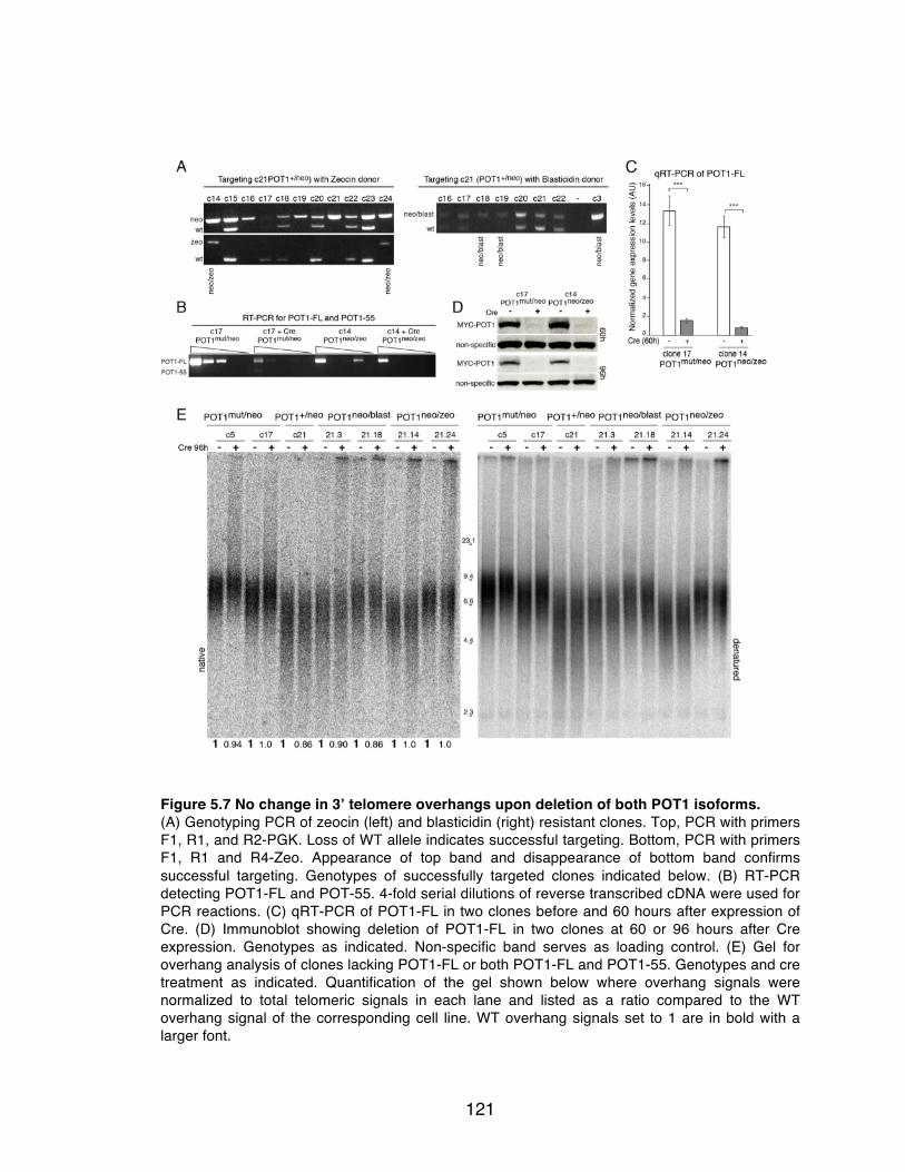
121
Figure 5.7 No change in 3’ telomere overhangs upon deletion of both POT1 isoforms. (A) Genotyping PCR of zeocin (left) and blasticidin (right) resistant clones. Top, PCR with primers F1, R1, and R2-PGK. Loss of WT allele indicates successful targeting. Bottom, PCR with primers F1, R1 and R4-Zeo. Appearance of top band and disappearance of bottom band confirms successful targeting. Genotypes of successfully targeted clones indicated below. (B) RT-PCR detecting POT1-FL and POT-55. 4-fold serial dilutions of reverse transcribed cDNA were used for PCR reactions. (C) qRT-PCR of POT1-FL in two clones before and 60 hours after expression of Cre. (D) Immunoblot showing deletion of POT1-FL in two clones at 60 or 96 hours after Cre expression. Genotypes as indicated. Non-specific band serves as loading control. (E) Gel for overhang analysis of clones lacking POT1-FL or both POT1-FL and POT1-55. Genotypes and cre treatment as indicated. Quantification of the gel shown below where overhang signals were normalized to total telomeric signals in each lane and listed as a ratio compared to the WT overhang signal of the corresponding cell line. WT overhang signals set to 1 are in bold with a larger font.

122
5.3 Summary of findings
Human POT1-FL, consistent with prior data 148, is required to inhibit ATR-
dependent DNA damage signaling at telomeres. Whether POT1-55 functions as
a dominant-negative allele exacerbating the occurrence of TIFs remains to be
determined, as does the cell cycle dependence of TIF formation in POT1-FL
knockout cells. Deprotected telomeres in POT1-FL-deficient cells were not
vulnerable to NHEJ, as deduced by the lack of telomere fusions observed in
these cells. However, an induction of telomere sister associations was noted in
cells lacking POT1, but the molecular basis of these associations is not known.
Surprisingly, unlike mouse POT1a/b-deficiency 31, human cells lacking
POT1 did not show a change in overhang status. The underlying cause of this
discrepancy is not clear, and many options remain to be tested (discussed further
in Chapter 6). Briefly, it is possible that POT1 is required to promote both
resection, and CST-mediated fill-in synthesis, thereby resulting in no overall
change in overhang signal when POT1 is deleted. Alternatively, removal of POT1
could allow telomerase to extend the overhang, and CST could be recruited to
perform fill-in synthesis independently of POT1, leaving no net change in
overhang lengths. Finally, although it seems unlikely, it is possible that steps
upstream of ATR signaling, such as RPA loading on ssDNA play a role in
overhang regulation.
The conditional knockout system we have generated in human cells will be
of great use, not only to understand the functions of human POT1, but also as a

123
means to mechanistically evaluate numerous mutations recently identified in
POT1 in various cancers 250-253 (see Chapter 6). One caveat of our system is the
incomplete Cre-mediated deletion. One possible solution is to use a retroviral Cre
that confers puromycin resistance and select for Cre-expressing cells. An option
for future targeting strategies is to flank both the exogenously expressed cDNA
and the hygromycin cassette with LoxP sites, such that cells are rendered
sensitive to hyrgomycin upon treatment with Cre, and hygromycin selection can
be retained to select for other constructs of interest.
Lastly, the POT1-55 knockout cell lines described in this chapter may be
used to gain insight into the purpose of POT1-55. While the delineation of
functions that mouse POT1a and –b perform is evident, it remains unclear why
the POT1-55 isoform of human POT1 exists. It is difficult to hypothesize about
potential roles of POT1-55 in the maintenance of telomere integrity, as it
resembles a dominant negative mutant, is weakly expressed and removal of
POT1-55 displays no apparent phenotype.

124
Chapter 6: Discussion
125
6.1 The role of mammalian Rap1
Analysis of mouse knockouts and TALEN-mediated human knockouts of
Rap1 reveal that, unlike its yeast counterparts, mammalian Rap1 is not required
for cellular and organismal viability, does not play a major role in telomere length
regulation, and is largely dispensable for telomere protection. Loss of Rap1 does
not affect the other components of shelterin, the modification of telomeric
histones, the synthesis of TERRA, or the maintenance of the 3’ telomeric
overhang. Telomeres lacking Rap1 do not show consistent changes in their
length, do not activate the DNA damage response, or become processed by
NHEJ. However, we did find that mouse Rap1 is required to repress HDR at
telomeres redundantly with Ku70/80, a function we were unable to test in human
cells due to lethality ensuing deletion of Ku70 or -80. Remarkably, we observed
that human Rap1, like its mouse and unicellular orthologs, affects gene
expression. Therefore, we propose that the conservation of Rap1 reflects its role
in transcriptional regulation, rather than a function at telomeres.
The dispensability of Rap1 in telomere end-protection was especially
surprising with regard to previous reports implicating mammalian Rap1 as a
repressor of NHEJ 213 160. An in vitro end-joining reaction with linear telomeric
substrates could be repressed by addition of the Rap1/TRF2 complex, but not
Rap1 or TRF2 alone 213. Perhaps the better binding affinity of TRF2/Rap1 to
duplex telomere substrates, than that of TRF2 (Chapter 3 and 235), allowed for
more efficient inhibition of NHEJ. An in vivo study shows Rap1 can be tethered to

126
HeLa telomeres by being fused to the DNA binding domain of a S. pombe protein
that preferentially binds TTAGGG repeats (TebDB) 160. Removal of endogenous
Rap1/TRF2 by a dominant negative mutant results in fusions, which can be
repressed by Rap1ΔCT-TebDB. Repression of NHEJ is not conferred by TebDB
itself, however, it is possible that Rap1ΔCT-TebDB acts as a neomorph. The
DNA-binding affinity of Rap1ΔCT-TebDB or its placement at telomeres may
physically occlude access of DNA repair factors. Alternatively, it is possible that
Rap1 plays a redundant role in the repression of telomere fusions in the absence
of TRF2, however our preliminary data does not support this hypothesis. In
addition, this setting of Rap1 at telomeres lacking TRF2 is highly unlikely to
naturally occur, due to the requisite binding of Rap1 to TRF2 for maintenance of
Rap1 protein levels and telomeric localization.
6.1.1 Rap1 – a conserved telomeric protein with non-telomeric functions
Several lines of evidence indicate that Rap1 is a highly conserved
component of mammalian shelterin. The human gene encoding Rap1 has
diverged little from its chimpanzee counterpart (1 base change/100 codons; 0.25
amino acid (aa) changes/100 aa), whereas the genes for other shelterin
components show much greater divergence (e.g., 3.6, 2.4, and 1.7 base
changes/100 codons and 2.2, 1.1, and 0.65 aa changes/100 aa for TRF2, TRF1,
and TIN2, respectively). Human and chimpanzee sequence was compared as

127
these sequences align most closely and allow for more accurate comparison of
changes in nucleotide and amino acid sequence between shelterin proteins.
Data from the Exome Variant Server (NHLBI GO Exome Sequencing
Project, Seattle, WA (URL: http://evs.gs.washington.edu/EVS/) [as of 02/2015])
identifies only 14 potentially deleterious (splice, nonsense, frame-shift or
missense) mutations in Rap1 in ~13,000 alleles sequenced. Compared to Rap1,
a larger number of potentially deleterious mutations have been identified in all
other shelterin components (19, 22, 50, 49 and 24 for TRF2, TRF1, TIN2, TPP1
and POT1, respectively).
Assessment of a gene damage index (GDI) derived from the 1000
Genomes Project 255, which reports on the cumulative mutational damage in a
given human coding gene, places Rap1 in the top 20% of human genes with
regard to tolerance to mutation intolerance (Y. Itan, pers. comm.). In addition,
Rap1 ranks among the top 10% of coding human genes in terms of ‘functional
indispensability’, a characteristic that incorporates gene centrality (based on
interaction data pooled from various biological systems), structural information,
and evolutionary constraints 256. In this regard, only TIN2 scores higher than
Rap1, as expected based on its multiple interaction interfaces in shelterin. The
aforementioned estimates of gene conservation do not necessarily highlight
essential genes, instead they aim to identify genes in which mutations may lead
to disease phenotypes. Furthermore, it is possible that Rap1 may serve an
important function in a specific tissue or cell type that we did not test, which may
explain the selective pressure placed upon Rap1.

128
The level of conservation of Rap1 raises the question: what functional
aspect of Rap1 is under selective pressure? The repression of HDR is unlikely to
explain the conservation of Rap1, since the HDR phenotype only manifests itself
when Ku70/80 are absent. It is also unlikely that Rap1 has a tissue specific role
at telomeres, given that mice lacking Rap1 are alive and largely normal, and our
demonstration that Rap1 deletion from human cell lines originating from different
tissues show no overt telomere phenotype. The conservation of Rap1 is also not
explained by a role in meiosis since the Rap1 knockout mice are fertile and,
unlike yeast lacking Rap1, form a normal meiotic bouquet 19,36,217,218,257,258. It is
not excluded that Rap1 has (as yet unknown) multiple interactions within
shelterin or with shelterin-associated factors that explain its conservation, or
alternatively, that its role at telomeres is redundant. We favor the idea that the
conservation of Rap1 is due to its role in transcriptional regulation 220-223,259 where
it may have multiple distinct interaction partners that constrain its evolution.
Given that mammalian Rap1 is unlikely to bind directly to DNA 20,193, and
that the majority of the chromosome-internal Rap1-associated loci are devoid of
telomeric sequence 220,223, it is expected that interacting factors recruit Rap1 to
these sites. In fact, complementation of Rap1-deficient MEFs with the Rap1
I312R allele (deficient in TRF2-binding), restores transcriptional regulation of
~85% of genes 221, consistent with the idea that Rap1 has binding partners that
target Rap1 to these loci in a TRF2-independent manner. Identification of these
factors could potentially illuminate (as yet undefined) telomere functions of Rap1.

129
Rap1 potentially evades being targeted for degradation by binding to
TRF2 (Chapter 3), which is puzzling when trying to understand how Rap1
regulates transcription independently of TRF2. One possibility is that other
binding partners are able to protect Rap1 from degradation. However, the nearly
undetectable levels of Rap1 in TRF2-depleted cells 19,22,24, suggest that very low
amounts of Rap1 would be bound to and protected by other factors. It would
therefore be of interest to determine whether this residual non-telomeric Rap1 is
sufficient for transcriptional regulation.
6.1.2 Rationale for retention of Rap1 at telomeres
If Rap is conserved due to its role in transcriptional regulation, this begs
the question why its interaction with TRF2 and localization to telomeres is
retained. One possibility is that TRF2 is required for Rap1 to localize to some of
the chromosomal loci that it transcriptionally regulates. The aforementioned
study221 shows that ~15% of transcriptional regulation by Rap1 is dependent on
its interaction with TRF2. In addition, ~15% of loci associated with human Rap1
also contain the consensus sequence for TRF2-binding 223.
Another possibility is that telomere length influences the transcriptional
control performed by Rap1. Telomere shortening may result in displaced
Rap1/TRF2 complexes, which can then bind to additional chromosome-internal
sites. Several studies in human cells show that telomere shortening, prior to the
induction of DNA damage signaling (as detected by TIF assays), can affect

130
expression of genes located both proximal 260,261 and distal 262 to telomeres. Hi-C
(chromosome capture followed by high throughput sequencing) reveals that
chromosome looping can place telomeres adjacent to genes located up to 10 Mb
away 262, which could potentially be another mode of Rap1 recruitment to loci that
it regulates. Moreover, both up- and down-regulation of transcripts are observed
following telomere shortening 262, suggesting that gene expression does not
seem to be inhibited by proximity to heterochromatic telomeric chromatin.
Instead, it is more likely to be modulated by a transcription factor, similar to our
observations of Rap1 function in both up- and down-regulation of gene
expression.
It is unclear why telomere length should play a role in the control of gene
expression. One possibility is that telomere shortening may initiate a
transcriptional program that promotes entry into senescence. In S. cerevisiae,
when critical telomere shortening is induced by deletion of telomerase (tlc1Δ),
Rap1 relocalizes to hundreds of new target genes 263. Redistribution of Rap1 in
yeast during the onset of senescence is dependent on Mec1 (the ATR homolog),
suggesting DNA damage also plays a role in Rap1 relocalization. DNA damage
signaling is not reported to accompany the gene expression changes observed
upon telomere shortening in human cells, which may suggest that other signals
promote senescence in mammalian cells. Alternatively, the TIF assay is not the
most sensitive method to evaluate DNA damage signaling and may miss low
frequency events that are responsible for triggering this change in cell state. It will

131
be interesting to see if mammalian Rap1 is required to initiate a transcriptional
program based on changes in telomere length.
6.1.3 No Rap1 ortholog in Drosophila
Drosophila telomeres, while serving the universal function of maintaining
chromosomal integrity, lack many hallmarks of eukaryotic telomere structure and
maintenance. For instance, Drosophila species lack telomerase, maintaining their
telomeres by specialized retrotransposons instead, and they also have variable
telomere repeats and sequence at their termini (reviewed in 285). Furthermore,
standard BLAST (Basic Local Alignment Search Tool) searches have revealed
no homologs of the shelterin components in Drosophila 286, while most non-
telomeric human proteins have Drosophila homologs. In consideration of the non-
telomeric role of Rap1, the selective pressure Rap1 appears to be under, and the
advances in annotated sequence databases, we reexamined whether Rap1
could be identified in flies. Due to the low sequence conservation of Rap1, we
used PSI-BLAST (Position-Specific Iterative), which derives a position-specific
scoring matrix that allows for detection of distant relationships between proteins.
Consistent with previous reports, PSI-BLAST of the conserved BRCT and MYB
domains of Rap1 revealed no related homologs in the Diptera order, suggesting
that other proteins may have evolved to become functional analogs of Rap1 in
flies.

132
6.2 The mechanism of HDR repression at telomeres
How mouse Rap1 represses HDR at telomeres when Ku70 is absent
remains to be determined. The data in Chapter 3, showing that Rap1 when
tethered to TRF1 fails to repress HDR, while negative, would suggest a
requirement for Rap1/TRF2 complex formation in the inhibition of HDR. One
possibility is that the complex might interact with a factor that represses HDR that
Rap1 alone or TRF2 alone do not interact with. Two-hybrid and mass
spectrometry have identified numerous protein complexes bound to Rap1 and/or
TRF2 21,78,225,264-266, but have not pinpointed a factor that could inhibit HDR.
However, these experiments were not carried out in a setting where HDR is
unleashed. Pull-down of TRF2/Rap1-associated proteins in Rap1F/FKu70-/- MEFs
(where HDR is repressed), followed by elimination of factors that remain
associated to TRF2 in a pull-down after Cre treatment (where HDR is induced),
might nominate candidates for such HDR repressing factors.
Alternatively, experiments described in Chapter 3 and aforementioned
studies 235,236 show that Rap1 can alter the DNA-binding activity of TRF2, but it is
unclear whether this is relevant to HDR inhibition (see below, section 6.2.1). In
addition, it is possible that Rap1, TRF2, and POT1a/b act together to repress
HDR (see below, section 6.2.2).

133
6.2.1 The effect of Rap1 on the DNA-binding activity of TRF2
Our initial hypothesis was that Rap1 might promote binding of TRF2 to
specific telomere structures that are reminiscent of substrates for HDR, such as
the ds-ss transition at the telomere terminus. Doing so would presumably prevent
loading of HDR factors, such as BRCA2 and Rad51, which are required for
repair. Arat and Griffith 235 observed that Rap1/TRF2 binds better to a ds-ss
telomere junction than a duplex telomere template. However, we, and Januskova
et al. did not observe this difference. The conflicting results may be due to
methods of protein purification and/or use of different substrates for the gel-shift
assays. Preliminary gel-filtration chromatography revealed various
oligomerization states within our purified proteins, which could affect their
efficiency of DNA-binding. Therefore, careful characterization of DNA binding
activities of TRF2 and Rap1/TRF2 to various telomere substrates needs to be
conducted with homogenous protein populations. Additionally, these DNA-
binding studies also need to be performed in the context of the entire shelterin
complex, as POT1 binding to ssDNA may affect the binding affinity of Rap1/TRF2
to the ds-ss junction.
Insight into the mechanism of HDR repression by Rap1/TRF2 could also
be acquired by performing a displacement loop (D-loop) assay. Here, in the
presence of ATP (and Rad54 to stabilize filament formation and D-loops 267),
Rad51 polymerizes on a radiolabeled ssDNA probe, which contains
complementary sequence to one strand of unlabeled supercoiled duplex DNA.

134
Strand invasion events where the ssDNA probe displaces one strand of duplex
DNA can be detected by a shift in the migration of the ssDNA probe on an
agarose gel 268. To determine whether Rap1/TRF2 inhibits HDR by preventing
strand invasion, the d-loop assay could be modified to use telomere-repeat
sequences and compare the ability of TRF2 and Rap1/TRF2 to inhibit strand
invasion.
6.2.2 Rap1, TRF2 and POT1 may work together to repress HDR
Data shown in Chapter 3 suggests that mouse Rap1/TRF2 and POT1a or
–b may act together to inhibit HDR at telomeres. POT1a and -b were removed
from telomeres in Rap1F/FKu70-/- MEFs by treatment with an shRNA targeting
TPP1 (shTPP1). 10% of T-SCEs were observed on average when both POT1a/b
were removed by shTPP1, or when Rap1 was deleted with Cre. No additive
incidence of T-SCEs was detected when all three shelterin components (Rap1,
POT1a and POT1b) were removed, suggesting they were epistatic in their
repression of HDR. In contrast to these results, deletion of mouse TPP1, which
phenocopies POT1a/b-deficiency, did not lead to induction of T-SCEs in a Ku70-
deficient background (T. Kibe, unpubl.). Furthermore, deletion of TIN2, which
also largely phenocopies POT1a/b-deletion, did not lead to induction of T-SCEs
in the context of Ku70-deficiency (K. Takai, unpubl.).
One explanation to reconcile the results described above could be the
time it takes for these MEFs to become immortalized. As discussed in Chapter 3,

135
Ku70-/- MEFs rapidly downregulate HDR at telomeres through an undefined
mechanism. MEFs isolated from Ku-deficient embryos tend to have difficulty
proliferating and immortalization times can vary significantly. Therefore, it is
possible HDR was assessed too late in the TPP1/Ku70- and TIN2/Ku70-deficient
MEFs. Isolation and immortalization of Rap1F/FKu70-/- and TPP1F/FKu70-/- MEFs
in parallel would allow for a more accurate analysis of T-SCE frequencies.
Alternatively, the TPP1sh could have off-target effects leading to the appearance
of T-SCEs, which could easily be tested by treating TPP1F/FKu70-/- MEFs with the
TPP1sh and assessing induction of T-SCEs.
The lack of T-SCEs in TPP1/Ku70- and TIN2/Ku70-deficient MEFs does
not necessarily argue against the idea that POT1a/b may still act together with
Rap1/TRF2 to repress T-SCEs. As discussed previously, POT1 can interact with
TRF2 21,152,185,234 and repression of HDR by POT1 may therefore be independent
of its TIN2/TPP1-mediated recruitment to telomeres.
6.2.3 What is the telomeric substrate for HDR?
Detection of T-SCEs serve as our readout for HDR taking place at
telomeres, however this assay does not report on the source of HDR initiation.
Most of our studies have focused on the ds-ss telomere terminus as a structure
vulnerable to HDR. Another source of telomeric HDR substrates could arise from
replication errors. For instance, HDR is involved in restarting collapsed replication
forks or bypassing a lesion encountered during replication (reviewed in 269). Our

136
preliminary analysis of MEFs lacking TRF1 (required to facilitate telomere
replication) and Ku80 showed an induction of T-SCEs, suggesting that telomeres
are also vulnerable to HDR during DNA synthesis. Therefore numerous telomere
configurations are likely to be vulnerable to HDR and further investigation into the
mechanism of HDR, especially biochemical approaches, will need to take this
into account.
6.2.4 Ku70/80-deficient cells adapt to recover repression of HDR
The downregulation of HDR at telomeres in late-passage Ku70-/- MEFs
was an intriguing finding, supporting the notion that HDR is stringently regulated
(reviewed in 270), and warrants further investigation. First, it will be of interest to
determine whether this down-regulation of HDR is global or telomere-specific.
Assessment of global HDR using the DR-GFP assay designed in the Jasin lab
171,271 proved to be difficult due to the low rate of targeted genomic integrations in
MEFs, as well as variable transfection efficiencies and cell survival post
transfection. The DR-GFP reporter construct contains one copy of GFP that has
been rendered dysfunctional (due to the insertion of an I-SceI site), and a donor
partial GFP sequence. Induction of a DSB by transfection of the I-SceI
endonuclease, and repair by HDR using the donor GFP template restores GFP
expression. Optimal use of this assay involves integration of a single copy of the
reporter into the HPRT locus to remove confounding effects from location of and
multiple copy integration. Recent developments in genome editing using
137
CRISPR-Cas9 based technologies might now allow for efficient targeting of the
DR-GFP construct into MEFs and determine global recombination levels in late-
passage Ku70 deficient MEFs.
Secondly, identification of factors that perform this inhibition of HDR will be
interesting as they may illuminate aspects of HDR regulation that are as yet
unknown. A preliminary microarray to assess gene expression changes in early-
and late-passage Ku70-deficient MEFs yielded a list of 64 significantly
deregulated genes. Additional microarrays using multiple Ku70-deficient cell lines
need to be performed to see if the same hits are generated. In addition, analysis
of gene expression changes in early- and late-passage Ku-proficient cells will
assist in eliminating candidate genes that are artifacts due to long-term tissue
culture.
Lastly, the finding that Ku70-deficient MEFs down-regulate HDR in culture,
raises the question of whether other processes adapting to Ku70-deficiency
occur during embryogenesis. A notable difference between mouse and human
Ku70/80 is that acute deletion of Ku70 or Ku80 in human somatic HCT116 cells
results in lethality and massive telomere recombination 240,241,272, while mice
lacking Ku70 or Ku80 are alive and fertile, albeit smaller and showing defects in
DNA repair 113,273. Development of conditional mouse knockouts of Ku
components should be informative to investigate whether acute deletion of Ku
from mouse somatic cells has similar effects as those reported in human cells.

138
6.3 Investigating the role(s) of human POT1
We describe the generation of conditional TALEN-mediated human
knockouts of POT1-FL and double knockouts of POT1-FL/POT1-55. Due to a
small percentage of cells that consistently escape Cre-mediated deletion of
POT1-FL, the potential cellular lethality of human POT1 knockouts cannot be
conclusively determined. Our current system provides a timeframe of 3-6 days
within which to assess telomere phenotypes of POT1-deficiency, comparable to
the approach undertaken for investigation of mouse POT1 proteins.
6.3.1 Functions of POT1 in telomere end-protection
Deletion of full-length human POT1 induces an ATR-dependent TIF
response (~65%), similar to mouse cells lacking POT1a/b (~75%) 31,148.
Incomplete knockdown of human POT1 with shRNAs may explain the
intermediate TIF response (~40%) observed in a previous study 184. Alternatively,
it is possible that the presence of POT1-55 in cells lacking POT1-FL contributes
to the additional induction of TIFs. The rationale for this explanation is derived
from phenotypes of the dominant-negative alleles of POT1a and –b that lack their
first OB fold, akin to human POT1-55. Overexpression of POT1aΔOB or
POT1bΔOB results in a moderate induction of TIFs (~25%), presumably by
displacing endogenous POT1a/b 246. No dominant-negative phenotype is
observed prior to deletion of POT1-FL, likely due to the 10-fold lower expression
of POT1-55 184, placing it at a disadvantage to compete with POT1-FL for binding

139
to TPP1 and telomeric recruitment. Analysis of TIFs in the double knockout
POT1-FL/POT1-55 will reveal whether POT1-55 acts as a dominant-negative in
the absence of POT1-FL. It is not clear why telomeres are most vulnerable to
DNA damage signaling during G1, when POT1 is depleted by shRNAs 184. Given
that 65% of cells lacking POT1-FL are TIF-positive, it is likely that TIFs persist
through most of the cell cycle. However, cell-cycle analysis needs to be
performed to confirm normal phase distribution.
Despite the telomere deprotection induced upon loss of POT1-FL,
telomeres do not appear to succumb to NHEJ. NHEJ is also largely repressed at
telomeres depleted for POT1 with siRNA or shRNA, which show a very low
incidence of telomere fusions (~0.1%) 184,185. However, 2% of chromosome
fusions are detected in POT1a and –b knockout MEFs 31. Taking into account the
low frequency of chromosome fusions and the few experimental replicates
analyzed, additional assessment is required to determine whether this difference
is significant.
Deletion of human POT1-FL and mouse POT1 proteins leads to induction
of telomere sister associations, where 8% and 1.4% 31 of telomeres engage in
sister associations, respectively. The frequency of these events seem to vary
between cell lines 159, as basal levels of telomere sister associations in the
human and mouse cell lines prior to Cre infection are 4% and 0% 31, respectively.
The telomere sister associations are not a result of NHEJ 156. Instead, low
frequencies of telomere sister associations occur in settings where ATR is

140
activated 159, and robust induction of this phenotype is observed when telomeres
encounter replication problems 156. Based on the low induction (2-fold) of
telomere sister associations in cells lacking POT1-FL, we do not expect to
observe a fragile telomere phenotype, but this remains to be confirmed.
Telomere sister associations are likely due to non-covalent interactions, such as
strand invasion, but the exact molecular basis of these events and their
consequences for telomere integrity remain unknown.
6.3.2 Telomere length regulation by POT1-55
An interesting observation shown in Chapter 5 is the slight increase of
telomere length in cells lacking POT1-FL, which is not detected upon loss of
POT1-FL/POT1-55. A mutant allele of human POT1 that lacks its first OB fold
(POT1ΔOB) 79 induces extreme telomere lengthening when overexpressed in
HTC75 cells, presumably through a similar mechanism as described before,
involving displacement of endogenous POT1 and loss of telomerase inhibition.
Similar to POT1ΔOB, POT1-55 may be responsible for the lengthening of
telomeres observed upon deletion of POT1-FL. However, depletion of POT1 with
siRNA also resulted in telomere elongation 29, therefore it is unclear why a setting
lacking both isoforms of human POT1 would not show telomere lengthening.
Since the experiment has only been conducted once, it is possible that
subsequent repetitions will show a telomere elongation phenotype. Additionally, if
treatment of cells lacking POT1-FL or POT1-FL/POT1-55 with a telomerase

141
inhibitor abolishes the observed increase in telomere length, this would confirm
that loss of POT1 allows telomerase access to telomeres.
The raison d’être of POT1-55 remains unclear. Low levels of POT1-55
protein suggest POT1-FL will most likely outcompete it for binding to TPP1 and
residency at telomeres, although this has not been formally shown with
endogenous proteins. Furthermore, the potential dominant negative action of
POT1-55 in exposing telomeres to DNA damage signaling must be repressed.
Conversely, POT1-55 may function to facilitate telomere elongation, which could
be important in cell types that proliferate extensively, but it must also be carefully
regulated such that it does not confer immortal properties to an oncogenic
transformed cell.
6.3.3 Generation of telomeric overhangs
In stark contrast to the extended overhangs observed in POT1a/b double
knockout MEFs, and the reduction in overhangs after partial knockdown of
human POT1, no change in overhang length was observed upon deletion of
POT1-FL or POT1-FL/POT1-55. In light of the role of mouse POT1a and –b in
overhang protection 152,153 and maintenance 31,32,52,177, and the existence of an
overhang phenotype in human POT1 knockdown studies 184,185, it is highly
improbable that POT1-FL is not involved in telomere 3’ overhang regulation.
Instead, we favor the hypothesis that POT1 has a more complex, dual role in
overhang generation, mediating both resection and fill-in synthesis.

142
The CST complex is required to generate telomeric 3’ overhangs 51-53, is
proposed to inhibit telomerase activity, and interacts with human POT1 247,248. It
is tempting to speculate that human POT1, similar to mouse POT1b, recruits
CST to telomeres to generate overhangs of appropriate length by fill-in synthesis.
If POT1 is required for CST recruitment to telomeres, depleting CST components
with shRNAs should have no effect on overhang length in cells lacking POT1-FL.
However, if CST associates with telomeres in the absence of POT1, depletion of
CST may result in increased overhangs, allowing us to distinguish between the
two possibilities.
Telomere 3’ overhang signal increases transiently in S/G2 177,181 due to
extensive resection by Exo1 52. While there is no evidence that POT1 and Exo1
interact, it would be of interest to determine the changes in overhang signal after
shRNA depletion of Exo1 in POT-FL knockout cells. No change in overhang
signal would suggest Exo1 recruitment or activity is promoted by POT1. If POT1
does not play a role in facilitating Exo1 action, we would expect to see a
reduction in overhang signal. An alternate experiment to determine whether
POT1 regulates Exo1 is to assess the transient increase in overhang length in
S/G2, with the expectation of not detecting an increase if POT1 mediates Exo1
recruitment.
Based on studies in yeast 274,275, mammalian Exo1 is presumed to be
recruited to DSBs after initial processing by the MRN complex, whereafter Exo1,
in parallel with the BLM helicase, can extensively resect DSBs 125 124. Therefore,

143
it is possible that POT1 regulates a nuclease other than Exo1, or binds to as of
yet undefined factors. However, the aforementioned experiments are likely to
yield insights into the mechanism of overhang regulation by human POT1. Our
observation of unchanged telomere overhangs upon loss of POT1 does not
replicate the 30% decrease in overhang signal detected following treatment with
an shRNA for POT1. A potential explanation of this discrepancy is that low levels
of residual POT1 may be sufficient to recruit CST, but higher levels of POT1 are
required to stimulate resection.
Other hypotheses, albeit less likely, include overhang extension by TERT,
and RPA-mediated regulation. Loss of POT1 may allow telomerase to extend 3’
overhangs, and fill-in synthesis could be mediated by CST, which can localize to
telomeres independently of POT1 51. Steps upstream of ATR signaling may be
involved in DNA overhang regulation. For instance, similar to its role in DNA
repair, when POT1 is removed, perhaps RPA directs the 5’ to 3’ resection 276 at
the telomere terminus and recruits Polα Primase 277 to perform the fill-in reaction.
6.3.4 POT1 mutations - a compelling link between telomere dysfunction and
disease
Genomic sequencing studies in the past two years have identified 29
heterozygous POT1 mutations (and counting) in cases of Chronic Lymphocytic
Leukemia and Familial Melanoma 250-253. Majority of the mutations occur in the N-
terminal regions of POT1, within the DNA-binding OB folds, and are predicted to
144
act as dominant-negative alleles. There are also several C-terminal mutations
that may prove interesting to investigate, as they may illuminate as yet unknown
functions or binding partners of POT1. Additionally, the C-terminus of POT1
contains the proposed interacting domains for CST and TPP1. Analysis of C-
terminal patient mutations may reveal separation-of-function mutants that are
defective in single aspects of POT1 function. Several N-terminal mutations in
POT1 have been characterized. Aside from observations of chromosomal
abnormalities, fragile telomeres, and telomere lengthening, exact mechanisms of
pathogenesis remain to be elucidated. The TALEN-mediated conditional
knockouts of human POT1 will provide a useful resource to identify the
dysfunctional phenotypes encoded by these mutations.

145
Chapter 7: Materials and Methods
146
7.1 General procedures
7.1.1 Mammalian cell culture
MEFs were isolated from E13.5 embryos and maintained in DMEM
supplemented with 1 mM sodium-pyruvate, 100 U/mL penicillin, 0.1 µg of
streptomycin per ml, 0.2 mM L-glutamine, 0.1 mM nonessential amino acids, and
15% fetal bovine serum (FBS). Primary MEFs were immortalized by retroviral
infection with pBabeSV40-LT (gift from Greg Hannon).
HCT116, HT1080, ARPE-19, and HeLa1.3 cells were grown in Dulbecco
modified Eagle medium (DMEM) supplemented with L-glutamine, penicillin-
streptomycin, nonessential amino acids and 15% bovine calf serum (BCS,
HyClone). SV40-large T transformed (neo resistant) BJ fibroblasts were cultured
in complete DMEM containing 199 medium (4:1) and 10% BCS.
7.1.2 Retroviral gene delivery
For infection of mouse cells, 24 h prior to transfection, 5x10⁶ Phoenix
ecotropic packaging cells were plated in 10 cm dishes. Phoenix cells were
transfected with 20 μg of the appropriate plasmid by calcium phosphate
precipitation. Media was changed 6-8 h after transfection. 36 h after transfection,
media containing virus was collected, and filtered through 0.45 μM filter.
Polybrene was added to a final concentration of 4 μg/mL. MEFs that had been
plated 12-24 h earlier in 10 cm dishes and were ~40% confluent were infected. 3

147
infections were performed at 12 h intervals. Time zero was set at 12h after first
infection. Infected cells were then selected for with the appropriate antibiotic
(puromycin 2μg/mL, hygromycin 90μg/mL). Selection was deemed complete
when uninfected control cells all died in the presence of antibiotic.
For human cells, the above protocol was used with the following
modifications. Phoenix amphotropic packaging cells were used to produce virus.
Human cells were infected 3 times at 4 h intervals. Time zero was set at 12 h
after first infection. Puromycin was used at 750 ng/mL, hygormycin was used at
135 μg/mL.
For infection with Cre recombinase, a Sloan Kettering facility produced
virus from retroviruses pMMP Hit&Run Cre and pWZL Cre. The viruses were
used to infect cells as described above. Selection with hygromycin followed
infection with pWZL Cre. No selection was performed after treatment with
Hit&Run Cre.
For the tamoxifen-inducible Cre-ERT2 system, 500nM of 4-OHT was
added to cells. Media was changed 6 h later and that was deemed time zero.
7.1.3 Growth curves
Growth curves were presented as population doublings over time and
were calculated by the following formula: original PD + (ln(# cells counted/# cells
seeded)/ln(2)).
148
7.1.4 Immunoblotting (IB)
Immunoblotting was performed as previously described 22. Briefly, cells
were harvested by trypsinization, resuspended in Laemmli buffer (100mM Tris-
HCl pH 6.8, 200 mM DTT, 3% SDS, 20% glycerol, 0.05% bromophenol blue) at
10,000 cells per µl, denatured for 5 min at 95°C, sheared with an insulin needle,
and resolved on SDS/PAGE gels using 100,000 cells per lane. Blots were
blocked in 5% non-fat dry milk/PBS + 0.1% Tween20.
7.1.5 Coimmunoprecipitation (Co-IP) from transfected 293T cells
2 x 10⁶ 293T cells were plated 24 h before co-transfection with calcium
phosphate co-precipitation using 10 µg each of FLAG-tagged and MYC-tagged
constructs. Cells were harvested at 36 h after transfection, washed once with
PBS, and lysed on ice for 10 min in a high salt buffer (50 mM Tris-HCl pH 7.2,
400 mM NaCl, 1% Triton X-100, 0.1% SDS, 1 mM EDTA, 1 mM DTT, 1 mM
PMSF and cocktail of protease inhibitors). Salt concentration was brought down
to 200 mM by addition of ice-cold water (drop-wise while mixing) and lysates
were spun at 4°C at 14,000 rpm for 10 min. Supernatants were used in
immunoprecipitations. 5% of the supernatant was saved as ‘input’ for western
blot analysis and the remaining supernatant was pre-cleared with preblocked
(10% BSA in PBS overnight) protein G-Sepharose beads for 1 h at 4°C. Lysates
were incubated overnight at 4°C with a 40 µl slurry of Sepharose beads
conjugated to 9E10-Myc or M2-FLAG mouse monoclonal antibody. Beads were
149
washed 5 times with ice-cold PBS. Proteins were eluted by boiling for 5 min in
Laemmli buffer and analyzed by SDS-PAGE and immunoblotting.
7.1.6 Immunofluorescence-fluorescence in situ hybridization (IF-FISH)
IF-FISH was conducted as previously described 278. Briefly, cells grown on
coverslips were fixed for 10 min in 2% paraformaldehyde/3% sucrose at room
temperature, followed by three 5 min PBS washes. Coverslips were incubated in
blocking solution (1 mg/ml BSA, 3% goat serum, 0.1% TritonX-100, 1 mM EDTA
in PBS) for 30 min, followed by incubation with primary antibodies diluted in
blocking solution for 1 h at rt. Cells were washed three times for 5 min with PBS
and then incubated with secondary antibodies diluted in blocking solution for 30
min at rt. Antibodies used were as listed at the end of this chapter. Coverslips
were dehydrated with 70%, 95% and 100% ethanol and allowed to dry.
Hybridizing solution (70% formamide, 0.5% blocking reagent from Roche, 10 mM
Tris-HCl pH 7.2 and FITC-OO-(CCCTAA)3 or TAMRA-OO-[TTAGGG]3 PNA
probe from Applied Biosystems) was added to each coverslip and denatured at
80ºC for 5 min, followed by a 2 h incubation at rt. Two 15-min washes in 70%
formamide/10 mM Tris-HCl pH 7.2 and three 5-min washes with PBS were
performed. DNA was stained with DAPI in the PBS washes and coverslips were
mounted using antifade reagent ProLong Gold from Life Technologies. Images
were captured using a Zeiss AxioPlan II microscope with a Hamamatsu C4742-

150
95 camera using Volocity software from Perkin Elmer. For distance of telomeres
from nuclear membrane, calculations were made using Image J software.
7.1.7 Telomeric FISH and chromosome orientation-FISH (CO-FISH)
Telomeric FISH and CO-FISH were conducted as previously described
119,154. Briefly, colcemid was added to cells 2 hours prior to harvest. Cells were
collected by trypsinization, swollen in 0.075 M KCl and fixed overnight at 4ºC in
methanol:acetic acid (3:1). Metaphase spreads were dropped on glass slides and
aged overnight. Slides were hybridized with FITC-OO-[CCCTAA]3 or TAMRA-
OO-[TTAGGG]3 PNA probe in hybridizing solution, denatured at 80ºC for 5 min
and incubated for 2 h at rt. Two 15-min washes in 70% formamide/10 mM Tris-
HCl pH 7.2 and three 5 min washes with 0.1 M Tris-HCl pH 7.2/0.15 M
NaCl/0.08% Tween20 were performed. DAPI was added to last wash for DNA
stain. Slides were dehydrated in 70%, 95%, and 100% ethanol and mounted
using ProLong Gold antifade from Life Technologies. Images were captured
using a Zeiss AxioPlan II microscope with a Hamamatsu C4742-95 camera using
Volocity software from Perkin Elmer. For CO-FISH, BrdU:BrdC (3:1) was added
14 h prior to harvest. Harvesting and metaphase conditions were as described for
FISH. Slides were treated with 0.5 mg/ml RNase A diluted in PBS, stained with
0.5 µg/ml Hoechst 33258, exposed to 5400J/m2 of UV light and subsequently
digested with 800 U of Exonuclease III from Promega for 10 min at room
temperature. Slides were rinsed with PBS, dehydrated with 70%, 95% and 100%

151
ethanol and sequentially hybridized with TAMRA-OO-[TTAGGG]3 and FITC-OO-
[CCCTAA]3 for 2 h each at rt, without denaturation. Washing, mounting and
capture conditions were as described for FISH.
7.1.8 Chromatin immunoprecipitation (ChIP)
ChIP was performed as previously described 79. Briefly, cells were fixed
1% paraformaldehyde/culture medium for 45 min at room temperature. Glycine
was added to 0.2 M to stop the cross-linking. Cells were pelleted by
centrifugation and washed with ice-cold PBS, followed by a final wash in PBS/1
mM PMSF. The cells were resuspended in cell lysis buffer (5 mM PIPES (pH
8.0), 85 mM KCl, 0.5% Nonidet P-40, 1 mM PMSF, protease inhibitor cocktail
from Roche), incubated on ice for 15 min and sonicated for 10 min (30 sec on/30
sec off) in a water-bath sonicator. Supernatants of lysates were incubated with
the appropriate antibody at 4 °C overnight and then for 45 min with ChIP-grade
protein G magnetic beads (Dynal, Invitrogen). Immunoprecipitated DNA was
washed, eluted from the beads, and precipitated with ethanol after reversal of the
cross-links and Proteinase K treatment. The DNA samples were dissolved in
water, boiled and loaded on dot blots, and hybridized with an 800-bp probe
labeled with Klenow and a primer for the C-rich telomeric repeat strand. Signal
intensity measured by ImageQuant software was normalized to the signals of the
input DNA on the same blot.

152
7.1.9 Genomic blotting, telomere length and telomere overhang analysis
Cells were harvested by trypsinization, washed with PBS, and either
pelleted and frozen at -80ºC (for telomere length analysis) or processed
immediately (for genotyping and telomere overhang analysis) for genomic DNA
collection. Genomic DNA was extracted as previously described 4. DNA for
genotyping was digested with appropriate enzymes, quantitated by fluorometry
using Hoechst 33258 and 10 µg was loaded on a 0.7% agarose gel run in 0.5X
TBE. DNA for telomere overhang and length analysis was digested with MboI
and AluI, quantified using Hoechst, and 1 µg was run on 0.7% agarose gels in
0.5X TBE.
For genomic blots used for genotyping and telomere length analysis, the
gels were depurinated with 0.5N HCl, denatured and neutralized using standard
Southern blotting procedures and transferred as previously described 4. For
genotyping, probes (indicated in figures) were Klenow-labeled using random
primers and α-32P-dCTP. For telomere length analysis, a Sty11 probe with ~800
bp of telomeric repeats 279 was Klenow-labeled with the TelC [CCCTAA]4 oligo
and α-32P-dCTP. Membranes were exposed to PhosphorImager screens and
quantified with ImageQuant software.
For telomere overhang analysis, gels were dried and probed with a
[CCCTAA]4 end-labeled with Polynucleotide kinase and γ-32P-ATP as previously
described 280. The gel was washed at 55°C three times for 30 min each in
4xSSC, once for 30 min in 4x SSC/0.1% SDS, and exposed to a PhosphoImager

153
screen overnight. After the image was captured, the gel was denatured in 0.5 M
NaOH, 1.5 M NaCl for 30 min, neutralized with two 15 min washes in 0.5 M Tris-
HCl pH 7.5, 3 M NaCl, and hybridized overnight with the same probe at 55°C.
The gel was washed and exposed as above. The ss G-rich overhang signal in
the native gel was quantified with ImageQuant software and normalized to the
total telomeric DNA quantified after the gel had been denatured and rehybridized
with the telomeric probe.
For telomere overhang analysis of mouse genomic DNA, 1x10⁶ cells were
resuspended in PBS and mixed 1:1 (v/v) with 2% agarose (SeaKem) in PBS and
set in plug molds (BioRad). Plugs were digested overnight with Proteinase K
buffer (10mM Tris-HCl pH 8.0, 250mM EDTA, 0.2% sodium deoxycholate, 1%
sodium lauryl sarcosine), washed four times for 1 hr each with TE, with 1 mM
PMSF in the last wash. Plugs were washed once more with H2O and digestion
buffer. Plugs were incubated overnight at 37°C with 60 U MboI. The following
day, the plugs were washed once in TE, and once in 0.5xTBE, and loaded onto a
1% agarose/0.5xTBE gel. Samples were run for 24 hours on a CHEF-DRII PFGE
apparatus (BioRad) in 0.5xTBE. The settings were as follows: initial pulse, 5 s;
final pulse, 5 s; 6 V/cm; 14°C. Gels were dried and the same protocol outlined
above was followed for hybridization, detection of signal, and quantification.

154
7.1.10 Northern analysis for TERRA
Total RNA was prepared using RNeasy Mini Spin columns (QIAGEN)
according to the manufacturer’s instructions and northern blot analysis was
performed as previously described 281. Briefly, 20µg of RNA was loaded onto
1.2% formaldehyde agarose gels and separated by gel electrophoresis. RNA was
transferred to a Hybond-N+ membrane and crosslinked in a UV Stratalinker. The
blot was prehybridized in Church mix at 55°C for 1 hour, followed by overnight
hybridization with a Sty11 probe 279. The blot was washed 3 times for 15 minutes
at 55°C with Church wash and then exposed to a Phosphorimager screen for 5
days. Screens were scanned using ImageQuant software and quantified in Image
J using the ethidium bromide stained 18S RNA as a loading control for
normalization.
7.1.11 Microarray analysis
Total RNA was isolated from cell lines using RNeasy Mini spin columns
(QIAGEN) with DNase digestion, according to the manufacturer’s instructions.
Microarray hybridization and scanning were performed at the Genomics core
facility at Rockefeller University, using Whole Human Genome DNA microarrays
(Illumina HumanHT-12 v4). The data was analyzed using GeneSpring v12.6.
Normalization was performed using quantiles and data was filtered to remove
absent genes using flag calls.

155
Experiments for HT1080 and HCT116 cell lines were performed in
replicate, using two independent isolations of RNA. Differentially expressed
genes were identified after performing moderated T-Tests and applying the
Benjamini-Hochberg False Discovery Rate method. A further fold change of 3 or
2.75 was applied to the HT1080 and HCT116 clones respectively to identify
genes that were highly transcriptionally deregulated due to the absence of Rap1.
Microarrays for ARPE-19 were not performed in replicate and therefore an
extremely stringent fold change threshold was applied to remove false negatives
and identify differentially expressed genes, which were subsequently validated by
qRT-PCR.
7.1.12 Quantitative reverse transcription-polymerase chain reaction (qRT-PCR)
For qRT-PCR, cDNA was prepared from 1µg of total RNA by using
Thermoscript Reverse Transcriptase (Invitrogen). Quantitative PCR reactions
were performed using Life Technologies SYBR Green Master Mix on an Applied
Biosystems 7900HT Sequence Detection System. Differences between samples
calculated using QuantStudio software (Applied Biosystems) using the ΔCT
method and were normalized to GAPDH.
For human Rap1, two independent isolations of RNA and reverse
transcriptase reactions were conducted and the experiment was repeated six
times for clones 23, 25, 30 and the parental cell line. The experiment for clone 26
was conducted in triplicate. For human POT1, qRT-PCR was conducted in

156
triplicate. Data was pooled to derive the mean averages and standard deviations.
Significance was calculated using a two-tailed unpaired T-Test. Primers used are
listed at the end of this chapter.
7.1.13 Nickel-affinity purification of His-tagged proteins
Human shelterin proteins were produced in baculovirus prior to this work20.
Sf9 insect cells were infected with baculovirus expressing shelterin components
for two days and purified as previously described 22. Briefly, infected cells were
harvested and lysed in 5 ml of cold lysis buffer (50 mM Tris, pH 7.4, 150 mM
NaCl, 1% Triton X-100, 5 mM β-mercaptoethanol supplemented with Roche
Applied Science protease inhibitor mixture) per 100 ml of cell culture. All
subsequent steps were done at 4 °C. After incubation on ice for 10 min, the
mixture was sonicated and cleared by centrifugation at 13,200 rpm for 20 min.
His tagged proteins were isolated using chelating Sepharose fast flow (GE
Healthcare) nickel resin. Proteins were bound to the resin, washed and eluted
with 1 M Imidazole. Protein yield and purity was quantified on SDS-PAGE gels
against a BSA standard.
7.1.14 Electrophoretic Mobility Shift Assays (EMSA)
Binding reactions were conducted as previously described 20. Briefly, of
EMSAs were conducted in in 4% Glycerol, 0.5 μgE. Coli DNA, 20 mM Glycine-
KOH [pH 9.0], 10 mM DTT, and 50 ng β-casein per 20 μl. Reaction with probe

157
and protein were incubated at 4°C for 30 min and run on a 0.6% agarose gel in
0.1× TBE at 4°C. Gels were dried onto DE81 paper and analyzed by
autoradiography or by exposure on a Phosphorimager. DNA probes were
prepared as described previously 11. In brief, the HindIII/Asp718 fragment
containing (TTAGGG)12 repeats was purified from pTH12 11 and filled in with [α-
32P]dCTP and Klenow enzyme. Other probes used in this study were generated
by annealing oligos (sequence is section 7.2), digesting with Asp718 and filling in
with [α-32P]dCTP and Klenow. Dissociation constants were calculated using
SigmaPlot by graphing % of bound probe against protein concentration.
7.1.15 Genotyping and sequencing
PCR was performed in 25 µl containing 50 ng of DNA, 0.2 µM of each
primer, 0.1 µM dNTPs, 1.5 mM MgCl2, 50 mM KCl, 10 mM Tris-HCl pH 8.0, and
0.5 U of TaKaRa Taq polymerase. Conditions were as follows: 95°C for 4 min, 25
cycles of 95°C for 30 sec, 60°C for 30 sec, 72°C for 45 sec and final extension at
72°C for 5 min.
Sequencing of heterozygous clones to identify mutations was conducted
by PCR-amplifying the remaining ‘WT’ allele. PCR products were eluted from
agarose gels and sequenced. For sequencing of homozygous ‘WT’ clones,
eluted PCR products were cloned by TA cloning (Life Technologies). A minimum
of 8 resulting TA clones per cell line were sequenced to identify mutations in both
alleles.

158
7.1.16 Gene targeting and cell cloning
All cell lines were transfected in 10 cm dishes at a density of 3x10⁶ cells
using Lipofectamine 2000 (Life Technologies) with 4 µg of each TALEN construct
and 20 µg of the appropriate donor construct. 48 hours after transfection, all cell
lines, except for HeLa1.3, were plated in selection medium in 10 cm plates at
varying densities ranging from 3,900 to 500,000 cells (using two-fold dilutions).
HeLa1.3 cells were plated in 10 cm dishes using two-fold dilutions starting from
8000 cells down to 75 cells. G418 was used at 1 mg/ml to select neomycin-
resistant HCT116 cells, at 900 µg/ml for HT1080 cells, and at 800 µg/ml for
ARPE-19 cells. Blasticidin was used at 5 µg/ml for HT1080 and HeLa1.3
selection, and at 2.5 µg/ml for SV40LT BJ selection. Zeocin was used at
100µg/ml for HT1080 cells.
For Rap1 targeting, clones emerged at a frequency of approximately 1
clone per 500 plated HCT116 cells, 1 clone per 2,600 plated HT1080 cells, 1
clone per 7,800 plated ARPE-19 cells, 1 clone per 125 plated HeLa1.3 cells, and
1 clone per 62,500 plated SV40LT BJ cells. The media was not changed after
initial plating.
For POT1 targeting, clones emerged at a frequency of approximately 1
clone per 3000 plated HT1080 cells in neomycin selection, 1 clone per 4000
plated HT1080 cells in zeocin selection, and 1 clone per 15 plated HT1080 cells
in blasticidin selection. 75% of HT1080 clones picked after blasticidin selection
died after continuous culturing in blasticidin.
159
Clones were picked 12 days later for all cell lines except the SV40LT BJ
clones, which were picked 3 weeks after plating. Approximately 60-70 clones
were picked for each cell line using cloning cylinders from plates that contained
well-spaced clones and the cells were transferred into 24 well plates. After
reaching confluence, half of the cells in each well were harvested to extract
genomic DNA, while the remaining cells were expanded into 6 well plates.
Approximately 30-40 clones were screened per cell line.
7.2 Mouse and human TERF2IP targeting
7.2.1 Mouse TERF2IP targeting (by Agnel Sfeir)
The Rap1 targeting vector was generated by cloning restriction fragments
from a BAC clone into the pSL301 vector (Invitrogen). A neomycin cassette
flanked by 2 FRT sites and containing a LoxP site was inserted into a CspCI site
in the first intron. A second LoxP site, together with an NdeI site was introduced
by inserting an oligonucleotide into a BsmBI site within the second intron. The
vector was linearized with NotI and gene targeting of C57BL/6J ES cells was
performed using standard techniques. ES cell clones with the correct integration
were identified by southern blots of NdeI digested DNA using a 350-bp probe
downstream of exon 3 outside the targeting vector. A correctly targeted ES clone
was injected into C57BL/6J blastocyst to generate chimeric male founders.
Crossing the chimeras to albino C57BL/6J females delivered offspring with the

160
Rap1F/+ genotype. Rap1 targeted mice were maintained in a C57BL/6J
background. The neomycin cassette was deleted by crossing the mice to the
FLPe-deleter mouse strain (Jackson Labs). The Rap1Δex2 allele was generated by
crossing the Rap1F/+ to the E2a-Cre deleter strain (Jackson Labs). Rap1F/F mice
were crossed with Ku70-/+ mice (obtained from F. Alt, Harvard Medical School,
Boston MA).
7.2.2 TALENs and the TERF2IP targeting construct
The heterodimeric TALEN pair for TERF2IP targeting was constructed
using the following RVD sequences. LEFT2: 5’-HD-NG-NN-NG-NN-HD-NG-NN-
NG-NG-HD-NG-NG-HD-NG-HD-NG, RIGHT1: 5’-HD-NG-NN-NN-NI-NN-NG-NG-
HD-NG-HD-NG-NG-NI-NG-NG-3’. TALENs were constructed by Dirk
Hockemeyer. The PGK Neomycin cassette from the PL451 vector (NCI) was
liberated using restriction enzymes NheI (5’) and BstBI (3’) and ligated into NheI-
and BstBI- digested pSL301 (cloning vector from Invitrogen). The pEF Blasticidin
cassette from plasmid pEF/Bsd (Life Technologies) was released using NheI (5’)
and EcoRI (3’) and ligated into NheI- and EcoRI- digested pSL301. The 5’ and 3’
homology arms were PCR-amplified with restriction site overhangs from genomic
SV40LT BJ fibroblast DNA. Primers for PCR of the 5’ arm were as follows: 5’-
ATGCGGTACCTTGCCCAAACTCCTGTCTTCTTAGGGC-3’ and 5’-
GCATGCTAGCAGAGAAGAACAGCACAGATTAGCAATAGCC-3’. Primers for
PCR of the 3’ arm were 5’-

161
ATGCTTCGAACTAGATTTACTCATTATTTTTTTCCCTACC-3’ and 5’-
GCATTTCGAACCTGTAATCCCAGCACTTTGGGAG-3’. The resulting 600 bp 5’
homology arm ends 7 bp from the intron 1/exon 2 junction and has KpnI and
NheI sites on the 5’ and 3’ ends, respectively. The resulting 578 bp 3’ homology
arm starts 32 bp from the exon 2/intron 2 junction and has BstBI restriction sites
at both ends. The homology arms were cloned into the relevant restriction sites in
pSL301 containing either the PGK Neomycin or pEF Blasticidin. The 3’ homology
arm insertion was screened for orientation and the donor constructs were
sequenced using the following primers: T7, T3, 5’-
GCTCGCGTCGTGCAGGACGT-3’ (PGK internal primer), and 5’-
GCTGTGCTCGACGTTGTCAC-3’ (Neomycin internal primer).
7.3 POT1 gene targeting
7.3.1 TALENs and the POT1 targeting construct
The heterodimeric TALEN pair for POT1 targeting was constructed using
the following RVD sequences. LEFT: 5’-NI-NI-NI-NI-NG-NI-NG-NG-NN-NI-NI-NI-
NN-NG-HD-NI-NN-3’, RIGHT: 5’-NN-NI-NN-HD-NI-NI-NG-NI-NI-NG-HD-NG-NN-
NN-NI-3’. TALENs were constructed by Siobhan Gregg, Supawat Thongtip and
Shaheen Kabir using the FLASH assembly system (REYON REF). The PGK
Neomycin cassette from the PL451 vector (NCI) was liberated using restriction
enzymes NheI (5’) and BamHI (3’) and ligated into NheI- and BamHI- digested
162
pSL301 (cloning vector from Invitrogen). The SV40 Zeocin cassette from
pSV40/Zeo2 (Invitrogen) was liberated using restriction enzymes NheI (5’) and
BamHI (3’) and ligated into NheI- and BamHI- digested pSL301. The pEF
Blasticidin cassette from plasmid pEF/Bsd (Life Technologies) was released
using NheI (5’) and BamHI (3’) was liberated using restriction enzymes NheI (5’)
and BamHI (3’) and ligated into NheI- and BamHI- digested pSL301. The pEF
promoter of the blasticidin cassette was replaced with a PGK promoter by cloning
it out of PL451 by NheI/XbaI digest and cloning it into NheI-digested pEF
blasticidin. The STOP cassette was digested out of Lox-Stop-Lox TOPO
(available from Addgene, Tyler Jacks lab) with BamHI and EcoRI and cloned into
the relevant sites of the neomycin, blasticidin and zeocin donor constructs. The 5’
and 3’ homology arms were PCR-amplified with restriction site overhangs from
genomic HT1080 DNA. Primers for PCR of the 5’ arm were as follows: 5’-
CTACCACCAGTGTTTGAAGTTATCG-3’ and 5’-
AATATTTTACCTGACTTTCAATATTTTAAAGC-3’. Primers for PCR of the 3’ arm
were 5’- CTCTGTATTGTTCACTGAAACTAGTTAGCAC-3’ and 5’-
CCAGAAGTGCATTGAACAAAAAAGCTC-3’. The resulting 647 bp 5’ homology
arm ends 14 bp from the intron 2/exon 3 junction and has XhoI and NheI sites on
the 5’ and 3’ ends, respectively. The resulting 649 bp 3’ homology arm starts 43
bp from the exon 3/intron 3 junction and has EcoRI restriction 5’ and a blunt 3’
end. The homology arms were cloned into the relevant restriction sites in pSL301
containing either the PGK Neomycin or pGK Blasticidin or pSV40 Zeocin.

163
7.4 List of primers
7.4.1 Genotyping
Mouse Rap1 primers:
F1: 5’-CATGCACTTGTACACATACAA-3’;
F2: 5’-GCTTCTTCCACCAAAACTGC-3’;
R: 5’-TTTGACAGTTGATAGGAAATGAAC-3’
Human Rap1 primers:
F1: 5’- GTGGATTGTGGTACGT GGCCCAGATCTGCC-3’;
R1: 5’-TAACATACCACAACCTCCTCAAACTCCCGG-3’;
POT1 primers:
F1: 5’-GTGTACTTCAGAACCATGTATAGCACACC-3’
R1: 5’-GGGCTTCATAGTTTCCACTAAAGAGCAGGC-3’
PGK internal primer:
R2: 5’-CATCTGCACGAGACTAGTGAGACGTGCTAC-3’

164
Zeocin internal primer:
R4: 5’-GAGCACCGGAACGGCACTGGTCAACT-3’
7.4.2 RT-PCR
Human GAPDH (published in 282)
5’-TGCACCACCAACTGCTTAGC-3’
5’-GGCATGGACTGTGGTCATGAG-3’
Human CDO1
5’-CGCCAAGTTCGACCAGTACAGG-3’
5’-GGTATGATCATGAATACTGCTGCC-3’
Human LHX2
5’-GGACGGTAGCATCTACTGCAAGG-3’
5’-CCAAGTCCCGAGCGCGCATCACC-3’
Human LRRC17
5’-CAAGCCTGAGGTGGACTCAACT-3’
5’-CTGGAGGGATGTTGTTTGGCAC-3’

165
POT1 coding exon 2 (anneals to both isoforms)
5’-CACCCCTGAATCAACTTAAGGGTGG-3’
POT1 coding exon 3 (anneals only to POT1-FL)
5’-CTGGTCCACAATAGTTACAACTGAGC-3’
POT1 coding exon 2-4 junction (anneals only to POT1-55)
5’-TATACTTGAATCAGTTCCTTTGC-3’
POT1 coding exon 4 (anneals to both isoforms)
5’-GCTCCGTCCACTTCTGCTTTGCCCA-3’
POT1 coding exon 9 (anneals to both isoforms)
5’-CAGACTGAAATAGTCTTCTGGGC-3’
POT1 coding exon 13 (anneals to both isoforms)
5’-CCATGAGATAGGCTTCTAGTACTCC-3’
POT1 coding exon 15 (anneals to both isoforms)
5’-CATCTTCTGCAACTGTGGTGTC-3’
166
7.4.3 EMSA probes
Junction probe
5’-ATGCGGTACCGGATGTCACTCAGCAGACGGTTAGGGTTAGGGTTAGGG CACTAGTTAGGGTTAG-3’
5’-CTAGTGCCCTAACCCTAACCCTAACCGTCTGCTGAGTGACATCCGGTACC GCAT-3’
[TTAGGG]3-repeat probe
5’-ATGCGGTACCGGATGTCACTCAGCAGACGGTTAGGGTTAGGGTTAG-3’
5’-CTAACCCTAACCCTAACCGTCTGCTGAGTGACATCCGGTACCGCAT-3’
7.5 List of shRNAs
Mouse TPP1:
Target sequence 5’-GGACACATGGGCTGACGGA-3’
Mouse Rap1:
sh1 target sequence 5’- ACAGGCAATGCCTTGTGGAAA -3’
sh2 target sequence 5’-CTTCATCTCCA CGCAGTACAT-3’
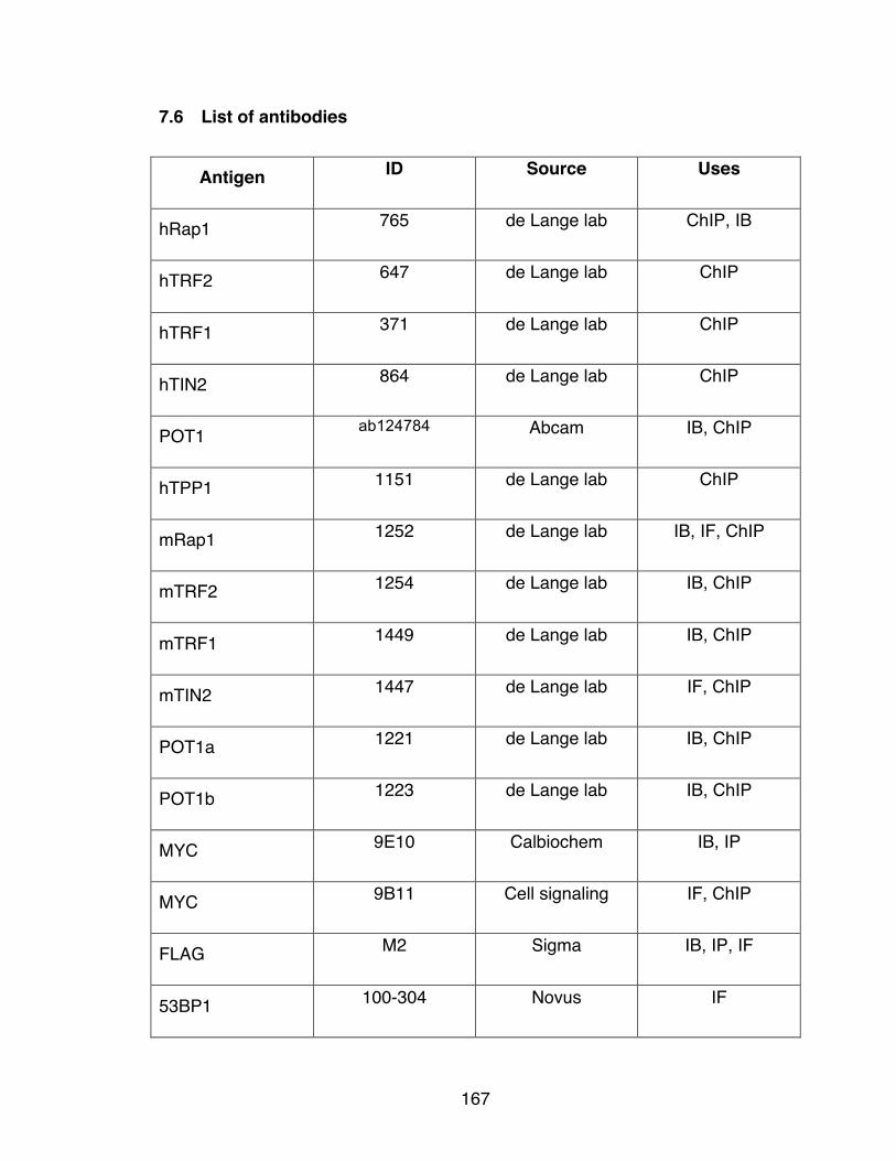
167
7.6 List of antibodies
Antigen ID Source Uses
hRap1 765 de Lange lab ChIP, IB
hTRF2 647 de Lange lab ChIP
hTRF1 371 de Lange lab ChIP
hTIN2 864 de Lange lab ChIP
POT1 ab124784 Abcam IB, ChIP
hTPP1 1151 de Lange lab ChIP
mRap1 1252 de Lange lab IB, IF, ChIP
mTRF2 1254 de Lange lab IB, ChIP
mTRF1 1449 de Lange lab IB, ChIP
mTIN2 1447 de Lange lab IF, ChIP
POT1a 1221 de Lange lab IB, ChIP
POT1b 1223 de Lange lab IB, ChIP
MYC 9E10 Calbiochem IB, IP
MYC 9B11 Cell signaling IF, ChIP
FLAG M2 Sigma IB, IP, IF
53BP1 100-304 Novus IF

168
Lamin A L1293 Sigma IF
Alpha-tubulin T9026 Sigma IB
Chk2 611570 BD Transduction IB
Chk1-P Ser345 Cell Signaling IB
Chk1 Sc-8408 Santa Cruz IB
H3K9me1 ab9045 Abcam ChIP
H3K9me2 ab1220 Abcam ChIP
H3K9me3 ab8898 Abcam ChIP
HP1alpha ab77256 Abcam ChIP
HP1beta ab10478 Abcam ChIP
HP1gamma ab10480 Abcam ChIP
Acetyl Histone H4 06-598 Millipore ChIP

169
7.7 List of cell lines
Cell Line Organism, Organ Notes
TRF2F/- p53-/- Mouse, E13.5 MEFs 24
TRF2F/F Ku70-/- Mouse, E13.5 MEFs 154
Rap1F/F Mouse, E13.5 MEFs This work
Rap1Δex2/ Δex2 Mouse, E13.5 MEFs This work, Agnel Sfeir
Rap1F/F Ku70-/- Mouse, E13.5 MEFs This work
Rap1F/F Ku70+/- Mouse, E13.5 MEFs This work
Rap1F/Δex2 Ku70-/- Mouse, E13.5 MEFs This work
TRF1F/F Cre-ERT2 Mouse, E13.5 MEFs Agnel Sfeir
HT1080 Human, Fibrosarcoma ATCC
HCT116 Human, Colorectal
carcinoma
ATCC
ARPE-19 Human, Retinal pigment
epithelial
ATCC
BJ Human, foreskin ATCC
HeLa1.3 Human, Cervical
adeoncarcinoma
22
Phoenix ecotropic Human, Epithelial
Phoenix amphotropic Human, Epithelial
293T Human, Kidney

170
Chapter 8: References
171
1. Muller, H. J. The remaking of chromosomes. The Collecting Net, Woods
Hole 8, 182-195 (1938).
2. McClintock, B. The stability of broken ends of chromosomes in Zea mays.
Genetics 26, 234-282 (1941).
3. Shampay, J., Szostak, J. W. & Blackburn, E. H. DNA sequences of
telomeres maintained in yeast. Nature 310, 154-157 (1984).
4. de Lange, T. et al. Structure and variability of human chromosome ends.
Mol Cell Biol 10, 518-527 (1990).
5. Lejnine, S., Makarov, V. L. & Langmore, J. P. Conserved nucleoprotein
structure at the ends of vertebrate and invertebrate chromosomes. Proc Natl
Acad Sci USA 92, 2393-2397 (1995).
6. Gomes, N. M. et al. Comparative biology of mammalian telomeres:
hypotheses on ancestral states and the roles of telomeres in longevity
determination. Aging Cell 10, 761-768 (2011).
7. Moyzis, R. K. et al. A highly conserved repetitive DNA sequence,
(TTAGGG)n, present at the telomeres of human chromosomes. Proc Natl
Acad Sci USA 85, 6622-6626 (1988).
8. Brown, W. R. Molecular cloning of human telomeres in yeast. Nature 338,
774-776 (1989).
9. Cross, S. H., Allshire, R. C., McKay, S. J., McGill, N. I. & Cooke, H. J.
Cloning of human telomeres by complementation in yeast. Nature 338, 771-
774 (1989).
10. Palm, W. & de Lange, T. How shelterin protects mammalian telomeres.
Annu Rev Genet 42, 301-334 (2008).
11. Zhong, Z., Shiue, L., Kaplan, S. & de Lange, T. A mammalian factor that
binds telomeric TTAGGG repeats in vitro. Mol Cell Biol 12, 4834-4843
(1992).
12. Chong, L. et al. A human telomeric protein. Science 270, 1663-1667 (1995).
13. Bilaud, T. et al. Telomeric localization of TRF2, a novel human telobox
protein. Nat Genet 17, 236-239 (1997).

172
14. Broccoli, D. et al. Comparison of the human and mouse genes encoding the
telomeric protein, TRF1: chromosomal localization, expression and
conserved protein domains. Hum Mol Genet 6, 69-76 (1997).
15. Bianchi, A., Smith, S., Chong, L., Elias, P. & de Lange, T. TRF1 is a dimer
and bends telomeric DNA. Embo J 16, 1785-1794 (1997).
16. Broccoli, D., Smogorzewska, A., Chong, L. & de Lange, T. Human
telomeres contain two distinct Myb-related proteins, TRF1 and TRF2. Nat
Genet 17, 231-235 (1997).
17. Bianchi, A. et al. TRF1 binds a bipartite telomeric site with extreme spatial
flexibility. Embo J 18, 5735-5744 (1999).
18. Chen, Y. et al. A conserved motif within RAP1 has diversified roles in
telomere protection and regulation in different organisms. Nat Struct Mol
Biol 18, 213-221 (2011).
19. Sfeir, A., Kabir, S., van Overbeek, M., Celli, G. B. & de Lange, T. Loss of
Rap1 induces telomere recombination in the absence of NHEJ or a DNA
damage signal. Science 327, 1657-1661 (2010).
20. Li, B., Oestreich, S. & de Lange, T. Identification of human Rap1:
implications for telomere evolution. Cell 101, 471-483 (2000).
21. Ye, J. Z. et al. TIN2 binds TRF1 and TRF2 simultaneously and stabilizes
the TRF2 complex on telomeres. J Biol Chem 279, 47264-47271 (2004).
22. Takai, K. K., Hooper, S., Blackwood, S., Gandhi, R. & de Lange, T. In vivo
stoichiometry of shelterin components. J Biol Chem 285, 1457-1467 (2010).
23. Li, B. & de Lange, T. Rap1 affects the length and heterogeneity of human
telomeres. Mol Biol Cell 14, 5060-5068 (2003).
24. Celli, G. B. & de Lange, T. DNA processing is not required for ATM-
mediated telomere damage response after TRF2 deletion. Nat Cell Biol 7,
712-718 (2005).
25. Kim, S. H., Kaminker, P. & Campisi, J. TIN2, a new regulator of telomere
length in human cells. Nat Genet 23, 405-412 (1999).

173
26. Houghtaling, B. R., Cuttonaro, L., Chang, W. & Smith, S. A dynamic
molecular link between the telomere length regulator TRF1 and the
chromosome end protector TRF2. Curr Biol 14, 1621-1631 (2004).
27. Chen, Y. et al. A shared docking motif in TRF1 and TRF2 used for
differential recruitment of telomeric proteins. Science 319, 1092-1096
(2008).
28. Liu, D. et al. PTOP interacts with POT1 and regulates its localization to
telomeres. Nat Cell Biol 6, 673-680 (2004).
29. Ye, J. Z. et al. POT1-interacting protein PIP1: a telomere length regulator
that recruits POT1 to the TIN2/TRF1 complex. Genes Dev 18, 1649-1654
(2004).
30. Baumann, P. & Cech, T. R. Pot1, the putative telomere end-binding protein
in fission yeast and humans. Science 292, 1171-115. (2001).
31. Hockemeyer, D., Daniels, J. P., Takai, H. & de Lange, T. Recent expansion
of the telomeric complex in rodents: Two distinct POT1 proteins protect
mouse telomeres. Cell 126, 63-77 (2006).
32. Wu, L. et al. Pot1 deficiency initiates DNA damage checkpoint activation
and aberrant homologous recombination at telomeres. Cell 126, 49-62
(2006).
33. Lei, M., Podell, E. R. & Cech, T. R. Structure of human POT1 bound to
telomeric single-stranded DNA provides a model for chromosome end-
protection. Nat Struct Mol Biol 11, 1223-1229 (2004).
34. Loayza, D., Parsons, H., Donigian, J., Hoke, K. & de Lange, T. DNA binding
features of human POT1: A nonamer 5’-TAGGGTTAG-3’ minimal binding
site, sequence specificity, and internal binding to multimeric sites. J Biol
Chem 279, 13241-13248 (2004).
35. Cooper, J. P., Nimmo, E. R., Allshire, R. C. & Cech, T. R. Regulation of
telomere length and function by a Myb-domain protein in fission yeast.
Nature 385, 744-747 (1997).

174
36. Kanoh, J. & Ishikawa, F. spRap1 and spRif1, recruited to telomeres by
Taz1, are essential for telomere function in fission yeast. Curr Biol 11,
1624-1630 (2001).
37. Miyoshi, T., Kanoh, J., Saito, M. & Ishikawa, F. Fission yeast Pot1-Tpp1
protects telomeres and regulates telomere length. Science 320, 1341-1344
(2008).
38. Li, B., Espinal, A. & Cross, G. A. Trypanosome telomeres are protected by
a homologue of mammalian TRF2. Mol Cell Biol 25, 5011-5021 (2005).
39. Yang, X., Figueiredo, L. M., Espinal, A., Okubo, E. & Li, B. RAP1 is
essential for silencing telomeric variant surface glycoprotein genes in
Trypanosoma brucei. Cell 137, 99-109 (2009).
40. Buchman, A. R., Kimmerly, W. J., Rine, J. & Kornberg, R. D. Two DNA-
binding factors recognize specific sequences at silencers, upstream
activating sequences, autonomously replicating sequences, and telomeres
in Saccharomyces cerevisiae. Mol Cell Biol 8, 210-225 (1988).
41. Longtine, M. S., Wilson, N. M., Petracek, M. E. & Berman, J. A yeast
telomere binding activity binds to two related telomere sequence motifs and
is indistinguishable from RAP1. Curr Genet 16, 225-239 (1989).
42. Conrad, M. N., Wright, J. H., Wolf, A. J. & Zakian, V. A. RAP1 protein
interacts with yeast telomeres in vivo: overproduction alters telomere
structure and decreases chromosome stability. Cell 63, 739-750 (1990).
43. Lin, J. J. & Zakian, V. A. The Saccharomyces CDC13 protein is a single-
strand TG1-3 telomeric DNA- binding protein in vitro that affects telomere
behavior in vivo. Proc Natl Acad Sci USA 93, 13760-13765 (1996).
44. Nugent, C. I., Hughes, T. R., Lue, N. F. & Lundblad, V. Cdc13p: a single-
strand telomeric DNA-binding protein with a dual role in yeast telomere
maintenance. Science 274, 249-252 (1996).
45. Grandin, N., Reed, S. I. & Charbonneau, M. Stn1, a new Saccharomyces
cerevisiae protein, is implicated in telomere size regulation in association
with Cdc13. Genes Dev 11, 512-27. (1997).

175
46. Grandin, N., Damon, C. & Charbonneau, M. Cdc13 prevents telomere
uncapping and Rad50-dependent homologous recombination. Embo J 20,
6127-6139 (2001).
47. Chandra, A., Hughes, T. R., Nugent, C. I. & Lundblad, V. Cdc13 both
positively and negatively regulates telomere replication. Genes Dev 15,
404-14. (2001).
48. Martin, V., Du, L. L., Rozenzhak, S. & Russell, P. Protection of telomeres by
a conserved Stn1-Ten1 complex. Proc Natl Acad Sci U S A 104, 14038-
14043 (2007).
49. Goulian, M., Heard, C. J. & Grimm, S. L. Purification and properties of an
accessory protein for DNA polymerase alpha/primase. J Biol Chem 265,
13221-13230 (1990).
50. Casteel, D. E. et al. A DNA polymerase-{alpha}{middle dot}primase cofactor
with homology to replication protein A-32 regulates DNA replication in
mammalian cells. J Biol Chem 284, 5807-5818 (2009).
51. Miyake, Y. et al. RPA-like mammalian Ctc1-Stn1-Ten1 complex binds to
single-stranded DNA and protects telomeres independently of the Pot1
pathway. Mol Cell 36, 193-206 (2009).
52. Wu, P., Takai, H. & de Lange, T. Telomeric 3’ Overhangs Derive from
Resection by Exo1 and Apollo and Fill-In by POT1b-Associated CST. Cell
150, 39-52 (2012).
53. Surovtseva, Y. V. et al. Conserved telomere maintenance component 1
interacts with STN1 and maintains chromosome ends in higher eukaryotes.
Mol Cell 36, 207-218 (2009).
54. Wan, M., Qin, J., Songyang, Z. & Liu, D. OB fold-containing protein 1
(OBFC1), a human homolog of yeast Stn1, associates with TPP1 and is
implicated in telomere length regulation. J Biol Chem 284, 26725-26731
(2009).
55. Watson, J. D. Origin of concatemeric T7 DNA. Nat New Biol 239, 197-201
(1972).

176
56. Olovnikov, A. M. A theory of marginotomy. The incomplete copying of
template margin in enzymic synthesis of polynucleotides and biological
significance of the phenomenon. J Theor Biol 41, 181-190 (1973).
57. Hayflick, L. The limited in vitro lifetime of human diploid cell strains. Exp Cell
Res 37, 614-636 (1965).
58. de Lange, T. How telomeres solve the end-protection problem. Science
326, 948-952 (2009).
59. Waga, S. & Stillman, B. The DNA replication fork in eukaryotic cells. Annu
Rev Biochem 67, 721-751 (1998).
60. Ohki, R., Tsurimoto, T. & Ishikawa, F. In vitro reconstitution of the end
replication problem. Mol Cell Biol 21, 5753-5766 (2001).
61. Bodnar, A. G. et al. Extension of life-span by introduction of telomerase into
normal human cells. Science 279, 349-352 (1998).
62. Harley, C. B. et al. Telomerase, cell immortality, and cancer. Cold Spring
Harb Symp Quant Biol 59, 307-315 (1994).
63. Szostak, J. W. & Blackburn, E. H. Cloning yeast telomeres on linear
plasmid vectors. Cell 29, 245-255 (1982).
64. Greider, C. W. & Blackburn, E. H. The telomere terminal transferase of
Tetrahymena is a ribonucleoprotein enzyme with two kinds of primer
specificity. Cell 51, 887-898 (1987).
65. Lundblad, V. & Szostak, J. W. A mutant with a defect in telomere elongation
leads to senescence in yeast. Cell 57, 633-643 (1989).
66. Lendvay, T. S., Morris, D. K., Sah, J., Balasubramanian, B. & Lundblad, V.
Senescence mutants of Saccharomyces cerevisiae with a defect in
telomere replication identify three additional EST genes. Genetics 144,
1399-1412 (1996).
67. Lingner, J. et al. Reverse transcriptase motifs in the catalytic subunit of
telomerase. Science 276, 561-567 (1997).
68. Nakamura, T. M. et al. Telomerase catalytic subunit homologs from fission
yeast and human. Science 277, 955-959 (1997).

177
69. Meyerson, M. et al. hEST2, the putative human telomerase catalytic subunit
gene, is up- regulated in tumor cells and during immortalization. Cell 90,
785-95. (1997).
70. Feng, J. et al. The RNA component of human telomerase. Science 269,
1236-1241 (1995).
71. Blasco, M. A. et al. Telomere shortening and tumor formation by mouse
cells lacking telomerase RNA. Cell 91, 25-34. (1997).
72. Shay, J. W. & Bacchetti, S. A survey of telomerase activity in human
cancer. Eur J Cancer 33, 787-791 (1997).
73. Counter, C. M. et al. Telomere shortening associated with chromosome
instability is arrested in immortal cells which express telomerase activity.
Embo J 11, 1921-1929 (1992).
74. Kim, N. W. et al. Specific association of human telomerase activity with
immortal cells and cancer. Science 266, 2011-205. (1994).
75. Marcand, S., Gilson, E. & Shore, D. A protein-counting mechanism for
telomere length regulation in yeast. Science 275, 986-990 (1997).
76. van Steensel, B. & de Lange, T. Control of telomere length by the human
telomeric protein TRF1. Nature 385, 740-743 (1997).
77. Smogorzewska, A. et al. Control of human telomere length by TRF1 and
TRF2. Mol Cell Biol 20, 1659-1668 (2000).
78. O’Connor, M. S., Safari, A., Liu, D., Qin, J. & Songyang, Z. The human
Rap1 protein complex and modulation of telomere length. J Biol Chem 279,
28585-28591 (2004).
79. Loayza, D. & de Lange, T. POT1 as a terminal transducer of TRF1 telomere
length control. Nature 424, 1013-1018 (2003).
80. Ye, J. Z. & de Lange, T. TIN2 is a tankyrase 1 PARP modulator in the TRF1
telomere length control complex. Nat Genet 36, 618-623 (2004).
81. Xin, H. et al. TPP1 is a homologue of ciliate TEBP-beta and interacts with
POT1 to recruit telomerase. Nature 445, 559-562 (2007).

178
82. Latrick, C. M. & Cech, T. R. POT1-TPP1 enhances telomerase processivity
by slowing primer dissociation and aiding translocation. EMBO J 29, 924-
933 (2010).
83. Tejera, A. M. et al. TPP1 is required for TERT recruitment, telomere
elongation during nuclear reprogramming, and normal skin development in
mice. Dev Cell 18, 775-789 (2010).
84. Abreu, E. et al. TIN2-tethered TPP1 recruits human telomerase to
telomeres in vivo. Mol Cell Biol 30, 2971-2982 (2010).
85. Zhong, F. L. et al. TPP1 OB-fold domain controls telomere maintenance by
recruiting telomerase to chromosome ends. Cell 150, 481-494 (2012).
86. Nandakumar, J. et al. The TEL patch of telomere protein TPP1 mediates
telomerase recruitment and processivity. Nature 492, 285-289 (2012).
87. Sexton, A. N. et al. Genetic and molecular identification of three human
TPP1 functions in telomerase action: recruitment, activation, and
homeostasis set point regulation. Genes Dev 28, 1885-1899 (2014).
88. Sancar, A., Lindsey-Boltz, L. A., Unsal-Kacmaz, K. & Linn, S. Molecular
mechanisms of mammalian DNA repair and the DNA damage checkpoints.
Annu Rev Biochem 73, 39-85 (2004).
89. Matsuoka, S., Huang, M. & Elledge, S. J. Linkage of ATM to cell cycle
regulation by the Chk2 protein kinase. Science 282, 1893-1897 (1998).
90. Chaturvedi, P. et al. Mammalian Chk2 is a downstream effector of the ATM-
dependent DNA damage checkpoint pathway. Oncogene 18, 4047-4054
(1999).
91. Matsuoka, S. et al. Ataxia telangiectasia-mutated phosphorylates Chk2 in
vivo and in vitro. Proc Natl Acad Sci U S A 97, 10389-10394 (2000).
92. Sanchez, Y. et al. Conservation of the Chk1 checkpoint pathway in
mammals: linkage of DNA damage to Cdk regulation through Cdc25.
Science 277, 1497-1501 (1997).

179
93. Liu, Q. et al. Chk1 is an essential kinase that is regulated by Atr and
required for the G(2)/M DNA damage checkpoint. Genes Dev 14, 1448-
1459 (2000).
94. Mailand, N. et al. Rapid destruction of human Cdc25A in response to DNA
damage. Science 288, 1425-149. (2000).
95. Costanzo, V. et al. Reconstitution of an ATM-dependent checkpoint that
inhibits chromosomal DNA replication following DNA damage. Mol Cell 6,
649-659 (2000).
96. Xiao, Z. et al. Chk1 mediates S and G2 arrests through Cdc25A
degradation in response to DNA-damaging agents. J Biol Chem 278,
21767-21773 (2003).
97. Tibbetts, R. S. et al. A role for ATR in the DNA damage-induced
phosphorylation of p53. Genes Dev 13, 152-17. (1999).
98. Lakin, N. D., Hann, B. C. & Jackson, S. P. The ataxia-telangiectasia related
protein ATR mediates DNA-dependent phosphorylation of p53. Oncogene
18, 3989-395. (1999).
99. Shieh, S. Y., Ahn, J., Tamai, K., Taya, Y. & Prives, C. The human homologs
of checkpoint kinases Chk1 and Cds1 (Chk2) phosphorylate p53 at multiple
DNA damage-inducible sites. Genes Dev 14, 289-300 (2000).
100. Hirao, A. et al. DNA damage-induced activation of p53 by the checkpoint
kinase Chk2. Science 287, 1824-1827 (2000).
101. Burma, S., Chen, B. P., Murphy, M., Kurimasa, A. & Chen, D. J. ATM
phosphorylates histone H2AX in response to DNA double-strand breaks. J
Biol Chem 276, 42462-42467 (2001).
102. Ward, I. M. & Chen, J. Histone H2AX is phosphorylated in an ATR-
dependent manner in response to replicational stress. J Biol Chem 276,
47759-47762 (2001).
103. Lou, Z., Minter-Dykhouse, K., Wu, X. & Chen, J. MDC1 is coupled to
activated CHK2 in mammalian DNA damage response pathways. Nature
421, 957-961 (2003).

180
104. Stewart, G. S., Wang, B., Bignell, C. R., Taylor, A. M. & Elledge, S. J.
MDC1 is a mediator of the mammalian DNA damage checkpoint. Nature
421, 961-966 (2003).
105. Goldberg, M. et al. MDC1 is required for the intra-S-phase DNA damage
checkpoint. Nature 421, 952-956 (2003).
106. Mattiroli, F. et al. RNF168 ubiquitinates K13-15 on H2A/H2AX to drive DNA
damage signaling. Cell 150, 1182-1195 (2012).
107. Fradet-Turcotte, A. et al. 53BP1 is a reader of the DNA-damage-induced
H2A Lys 15 ubiquitin mark. Nature 499, 50-54 (2013).
108. Takai, H., Smogorzewska, A. & de Lange, T. DNA damage foci at
dysfunctional telomeres. Curr Biol 13, 1549-1556 (2003).
109. d’Adda di Fagagna, F. et al. A DNA damage checkpoint response in
telomere-initiated senescence. Nature 426, 194-198 (2003).
110. Yan, C. T. et al. IgH class switching and translocations use a robust non-
classical end-joining pathway. Nature 449, 478-482 (2007).
111. Corneo, B. et al. Rag mutations reveal robust alternative end joining. Nature
449, 483-486 (2007).
112. Baumann, P. & West, S. C. DNA end-joining catalyzed by human cell-free
extracts. Proc Natl Acad Sci U S A 95, 14066-14070 (1998).
113. Gu, Y. et al. Growth retardation and leaky SCID phenotype of Ku70-
deficient mice. Immunity 7, 653-665 (1997).
114. Zhu, C., Bogue, M. A., Lim, D. S., Hasty, P. & Roth, D. B. Ku86-deficient
mice exhibit severe combined immunodeficiency and defective processing
of V(D)J recombination intermediates. Cell 86, 379-389 (1996).
115. Frank, K. M. et al. Late embryonic lethality and impaired V(D)J
recombination in mice lacking DNA ligase IV. Nature 396, 173-17. (1998).
116. Audebert, M., Salles, B., Weinfeld, M. & Calsou, P. Involvement of
polynucleotide kinase in a poly(ADP-ribose) polymerase-1-dependent DNA
double-strand breaks rejoining pathway. J Mol Biol 356, 257-265 (2006).
181
117. Simsek, D. et al. DNA ligase III promotes alternative nonhomologous end-
joining during chromosomal translocation formation. PLoS Genet 7,
e1002080 (2011).
118. Wang, M. et al. PARP-1 and Ku compete for repair of DNA double strand
breaks by distinct NHEJ pathways. Nucleic Acids Res 34, 6170-6182
(2006).
119. van Steensel, B., Smogorzewska, A. & de Lange, T. TRF2 protects human
telomeres from end-to-end fusions. Cell 92, 401-413 (1998).
120. Sfeir, A. & de Lange, T. Removal of shelterin reveals the telomere end-
protection problem. Science 336, 593-597 (2012).
121. Mateos-Gomez, P. A. et al. Mammalian polymerase theta promotes
alternative NHEJ and suppresses recombination. Nature (2015).
122. Sartori, A. A. et al. Human CtIP promotes DNA end resection. Nature 450,
509-514 (2007).
123. Moynahan, M. E., Chiu, J. W., Koller, B. H. & Jasin, M. Brca1 controls
homology-directed DNA repair. Mol Cell 4, 511-518 (1999).
124. Nimonkar, A. V., Ozsoy, A. Z., Genschel, J., Modrich, P. & Kowalczykowski,
S. C. Human exonuclease 1 and BLM helicase interact to resect DNA and
initiate DNA repair. Proc Natl Acad Sci U S A 105, 16906-16911 (2008).
125. Gravel, S., Chapman, J. R., Magill, C. & Jackson, S. P. DNA helicases Sgs1
and BLM promote DNA double-strand break resection. Genes Dev 22,
2767-2772 (2008).
126. Sugiyama, T., Zaitseva, E. M. & Kowalczykowski, S. C. A single-stranded
DNA-binding protein is needed for efficient presynaptic complex formation
by the Saccharomyces cerevisiae Rad51 protein. J Biol Chem 272, 7940-
7945 (1997).
127. Wang, X. & Haber, J. E. Role of Saccharomyces single-stranded DNA-
binding protein RPA in the strand invasion step of double-strand break
repair. PLoS Biol 2, E21 (2004).
182
128. Raderschall, E., Golub, E. I. & Haaf, T. Nuclear foci of mammalian
recombination proteins are located at single-stranded DNA regions formed
after DNA damage. Proc Natl Acad Sci U S A 96, 1921-1926 (1999).
129. Xia, F. et al. Deficiency of human BRCA2 leads to impaired homologous
recombination but maintains normal nonhomologous end joining. Proc Natl
Acad Sci U S A 98, 8644-8649 (2001).
130. Baumann, P., Benson, F. E. & West, S. C. Human Rad51 protein promotes
ATP-dependent homologous pairing and strand transfer reactions in vitro.
Cell 87, 757-766 (1996).
131. Davies, A. A. et al. Role of BRCA2 in control of the RAD51 recombination
and DNA repair protein. Mol Cell 7, 273-282 (2001).
132. Wu, L. & Hickson, I. D. The Bloom’s syndrome helicase suppresses
crossing over during homologous recombination. Nature 426, 870-874
(2003).
133. Chen, X. B. et al. Human Mus81-associated endonuclease cleaves Holliday
junctions in vitro. Mol Cell 8, 1117-1127 (2001).
134. Ciccia, A., Constantinou, A. & West, S. C. Identification and characterization
of the human mus81-eme1 endonuclease. J Biol Chem 278, 25172-25178
(2003).
135. Ip, S. C. et al. Identification of Holliday junction resolvases from humans
and yeast. Nature 456, 357-361 (2008).
136. Bailey, S. M., Goodwin, E. H. & Cornforth, M. N. Strand-specific
fluorescence in situ hybridization: the CO-FISH family. Cytogenet Genome
Res 107, 14-17 (2004).
137. Cesare, A. J. & Reddel, R. R. Alternative lengthening of telomeres: models,
mechanisms and implications. Nat Rev Genet 11, 319-330 (2010).
138. Londono-Vallejo, J. A., Der-Sarkissian, H., Cazes, L., Bacchetti, S. &
Reddel, R. R. Alternative lengthening of telomeres is characterized by high
rates of telomeric exchange. Cancer Res 64, 2324-2327 (2004).
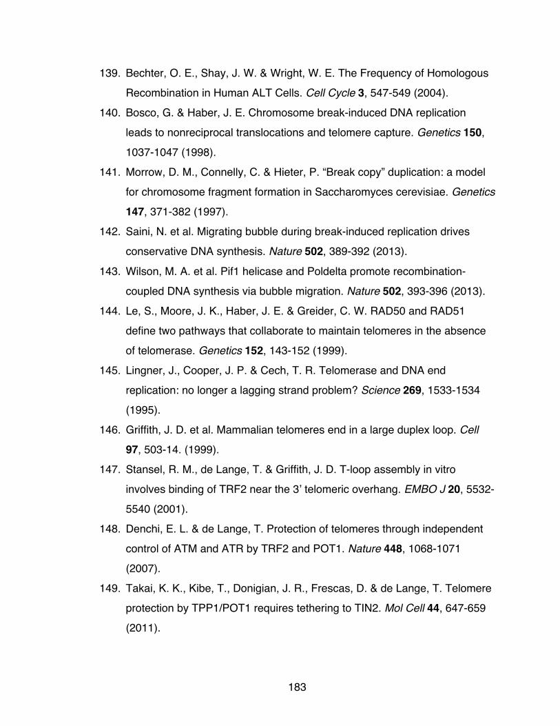
183
139. Bechter, O. E., Shay, J. W. & Wright, W. E. The Frequency of Homologous
Recombination in Human ALT Cells. Cell Cycle 3, 547-549 (2004).
140. Bosco, G. & Haber, J. E. Chromosome break-induced DNA replication
leads to nonreciprocal translocations and telomere capture. Genetics 150,
1037-1047 (1998).
141. Morrow, D. M., Connelly, C. & Hieter, P. “Break copy” duplication: a model
for chromosome fragment formation in Saccharomyces cerevisiae. Genetics
147, 371-382 (1997).
142. Saini, N. et al. Migrating bubble during break-induced replication drives
conservative DNA synthesis. Nature 502, 389-392 (2013).
143. Wilson, M. A. et al. Pif1 helicase and Poldelta promote recombination-
coupled DNA synthesis via bubble migration. Nature 502, 393-396 (2013).
144. Le, S., Moore, J. K., Haber, J. E. & Greider, C. W. RAD50 and RAD51
define two pathways that collaborate to maintain telomeres in the absence
of telomerase. Genetics 152, 143-152 (1999).
145. Lingner, J., Cooper, J. P. & Cech, T. R. Telomerase and DNA end
replication: no longer a lagging strand problem? Science 269, 1533-1534
(1995).
146. Griffith, J. D. et al. Mammalian telomeres end in a large duplex loop. Cell
97, 503-14. (1999).
147. Stansel, R. M., de Lange, T. & Griffith, J. D. T-loop assembly in vitro
involves binding of TRF2 near the 3’ telomeric overhang. EMBO J 20, 5532-
5540 (2001).
148. Denchi, E. L. & de Lange, T. Protection of telomeres through independent
control of ATM and ATR by TRF2 and POT1. Nature 448, 1068-1071
(2007).
149. Takai, K. K., Kibe, T., Donigian, J. R., Frescas, D. & de Lange, T. Telomere
protection by TPP1/POT1 requires tethering to TIN2. Mol Cell 44, 647-659
(2011).

184
150. Kibe, T., Osawa, G. A., Keegan, C. E. & de Lange, T. Telomere Protection
by TPP1 Is Mediated by POT1a and POT1b. Mol Cell Biol 30, 1059-1066
(2010).
151. Guo, X. et al. Dysfunctional telomeres activate an ATM-ATR-dependent
DNA damage response to suppress tumorigenesis. EMBO J 26, 4709-4719
(2007).
152. Barrientos, K. S. et al. Distinct functions of POT1 at telomeres. Mol Cell Biol
28, 5251-5264 (2008).
153. Gong, Y. & de Lange, T. A Shld1-controlled POT1a provides support for
repression of ATR signaling at telomeres through RPA exclusion. Mol Cell
40, 377-387 (2010).
154. Celli, G. B., Lazzerini Denchi, E. & de Lange, T. Ku70 stimulates fusion of
dysfunctional telomeres yet protects chromosome ends from homologous
recombination. Nat Cell Biol 8, 885-890 (2006).
155. Palm, W., Hockemeyer, D., Kibe, T. & de Lange, T. Functional dissection of
human and mouse POT1 proteins. Mol Cell Biol 29, 471-482 (2009).
156. Sfeir, A. et al. Mammalian telomeres resemble fragile sites and require
TRF1 for efficient replication. Cell 138, 90-103 (2009).
157. Martinez, P. et al. Increased telomere fragility and fusions resulting from
TRF1 deficiency lead to degenerative pathologies and increased cancer in
mice. Genes Dev 23, 2060-2075 (2009).
158. Vannier, J. B., Pavicic-Kaltenbrunner, V., Petalcorin, M. I., Ding, H. &
Boulton, S. J. RTEL1 Dismantles T Loops and Counteracts Telomeric G4-
DNA to Maintain Telomere Integrity. Cell 149, 795-806 (2012).
159. Zimmermann, M., Kibe, T., Kabir, S. & de Lange, T. TRF1 negotiates
TTAGGG repeat-associated replication problems by recruiting the BLM
helicase and the TPP1/POT1 repressor of ATR signaling. Genes Dev 28,
2477-2491 (2014).
160. Sarthy, J., Bae, N. S., Scrafford, J. & Baumann, P. Human RAP1 inhibits
non-homologous end joining at telomeres. EMBO J 28, 3390-3399 (2009).

185
161. Karlseder, J., Broccoli, D., Dai, Y., Hardy, S. & de Lange, T. p53- and ATM-
dependent apoptosis induced by telomeres lacking TRF2. Science 283,
1321-1325 (1999).
162. Konishi, A. & de Lange, T. Cell cycle control of telomere protection and
NHEJ revealed by a ts mutation in the DNA-binding domain of TRF2.
Genes Dev 22, 1221-1230 (2008).
163. Dimitrova, N., Chen, Y. C., Spector, D. L. & de Lange, T. 53BP1 promotes
non-homologous end joining of telomeres by increasing chromatin mobility.
Nature 456, 524-528 (2008).
164. Lottersberger, F., Bothmer, A., Robbiani, D. F., Nussenzweig, M. C. & de
Lange, T. Role of 53BP1 oligomerization in regulating double-strand break
repair. Proc Natl Acad Sci U S A 110, 2146-2151 (2013).
165. Zimmermann, M., Lottersberger, F., Buonomo, S. B., Sfeir, A. & de Lange,
T. 53BP1 regulates DSB repair using Rif1 to control 5’ end resection. Science
339, 700-704 (2013).
166. Dimitrova, N. & de Lange, T. Cell cycle dependent role of MRN at
dysfunctional telomeres: ATM signaling-dependent induction of NHEJ in G1
and resection-mediated inhibition of NHEJ in G2. Mol Cell Biol 29, 5552-
5563 (2009).
167. Attwooll, C. L., Akpinar, M. & Petrini, J. H. The mre11 complex and the
response to dysfunctional telomeres. Mol Cell Biol 29, 5540-5551 (2009).
168. Deng, Y., Guo, X., Ferguson, D. O. & Chang, S. Multiple roles for MRE11 at
uncapped telomeres. Nature 460, 914-918 (2009).
169. Doksani, Y., Wu, J. Y., de Lange, T. & Zhuang, X. Super-resolution
fluorescence imaging of telomeres reveals TRF2-dependent T-loop
formation. Cell 155, 345-356 (2013).
170. Okamoto, K. et al. A two-step mechanism for TRF2-mediated chromosome-
end protection. Nature 494, 502-505 (2013).

186
171. Pierce, A. J., Johnson, R. D., Thompson, L. H. & Jasin, M. XRCC3
promotes homology-directed repair of DNA damage in mammalian cells.
Genes Dev 13, 2633-2638 (1999).
172. Wang, R. C., Smogorzewska, A. & de Lange, T. Homologous recombination
generates T-loop-sized deletions at human telomeres. Cell 119, 355-368
(2004).
173. Fouche, N. et al. The basic domain of TRF2 directs binding to DNA
junctions irrespective of the presence of TTAGGG repeats. J Biol Chem
281, 37486-37495 (2006).
174. Poulet, A. et al. TRF2 promotes, remodels and protects telomeric Holliday
junctions. EMBO J 28, 641-651 (2009).
175. Saint-Leger, A. et al. The basic N-terminal domain of TRF2 limits
recombination endonuclease action at human telomeres. Cell Cycle 13,
(2014).
176. Choi, K. H., Farrell, A. S., Lakamp, A. S. & Ouellette, M. M. Characterization
of the DNA binding specificity of Shelterin complexes. Nucleic Acids Res
39, 9206-9223 (2011).
177. Wu, P., van Overbeek, M., Rooney, S. & de Lange, T. Apollo Contributes to
G Overhang Maintenance and Protects Leading-End Telomeres. Mol Cell
39, 1-12 (2010).
178. Lam, Y. C. et al. SNMIB/Apollo protects leading-strand telomeres against
NHEJ-mediated repair. EMBO J 29, 2230-2241 (2010).
179. van Overbeek, M. & de Lange, T. Apollo, an Artemis-related nuclease,
interacts with TRF2 and protects human telomeres in S phase. Curr Biol 16,
1295-1302 (2006).
180. Lenain, C. et al. The Apollo 5’ exonuclease functions together with TRF2 to
protect telomeres from DNA repair. Curr Biol 16, 1303-1310 (2006).
181. Dai, X. et al. Molecular steps of G-overhang generation at human telomeres
and its function in chromosome end protection. EMBO J 29, 2788-2801
(2010).

187
182. He, H. et al. POT1b protects telomeres from end-to-end chromosomal fusions
and aberrant homologous recombination. Embo J 25, 5180-5190 (2006).
183. Davoli, T., Denchi, E. L. & de Lange, T. Persistent telomere damage
induces bypass of mitosis and tetraploidy. Cell 141, 81-93 (2010).
184. Hockemeyer, D., Sfeir, A. J., Shay, J. W., Wright, W. E. & de Lange, T.
POT1 protects telomeres from a transient DNA damage response and
determines how human chromosomes end. EMBO J. 24, 2667-2678
(2005).
185. Yang, Q., Zheng, Y. L. & Harris, C. C. POT1 and TRF2 cooperate to
maintain telomeric integrity. Mol Cell Biol 25, 1070-1080 (2005).
186. Sfeir, A. J., Chai, W., Shay, J. W. & Wright, W. E. Telomere-end processing
the terminal nucleotides of human chromosomes. Mol Cell 18, 131-138
(2005).
187. Shore, D. & Nasmyth, K. Purification and cloning of a DNA binding protein
from yeast that binds to both silencer and activator elements. Cell 51, 721-
732 (1987).
188. Hardy, C. F., Balderes, D. & Shore, D. Dissection of a carboxy-terminal
region of the yeast regulatory protein RAP1 with effects on both
transcriptional activation and silencing. Mol Cell Biol 12, 1209-1217 (1992).
189. Cockell, M. et al. The carboxy termini of Sir4 and Rap1 affect Sir3
localization: evidence for a multicomponent complex required for yeast
telomeric silencing. J Cell Biol 129, 909-924 (1995).
190. Liu, C. & Lustig, A. J. Genetic analysis of Rap1p/Sir3p interactions in
telomeric and HML silencing in Saccharomyces cerevisiae. Genetics 143,
81-93 (1996).
191. Pardo, B. & Marcand, S. Rap1 prevents telomere fusions by
nonhomologous end joining. EMBO J 24, 3117-3127 (2005).
192. Konig, P., Giraldo, R., Chapman, L. & Rhodes, D. The crystal structure of
the DNA-binding domain of yeast RAP1 in complex with telomeric DNA.
Cell 85, 125-136 (1996).

188
193. Hanaoka, S. et al. NMR structure of the hRap1 Myb motif reveals a
canonical three-helix bundle lacking the positive surface charge typical of
Myb DNA-binding domains. J Mol Biol 312, 167-175 (2001).
194. Yu, X., Chini, C. C., He, M., Mer, G. & Chen, J. The BRCT domain is a
phospho-protein binding domain. Science 302, 639-642 (2003).
195. Hardy, C. F., Sussel, L. & Shore, D. A RAP1-interacting protein involved in
transcriptional silencing and telomere length regulation. Genes Dev 6, 801-
814 (1992).
196. Wotton, D. & Shore, D. A novel Rap1p-interacting factor, Rif2p, cooperates
with Rif1p to regulate telomere length in Saccharomyces cerevisiae. Genes
Dev 11, 748-760 (1997).
197. Hirano, Y. & Sugimoto, K. Cdc13 telomere capping decreases Mec1
association but does not affect Tel1 association with DNA ends. Mol Biol
Cell 18, 2026-2036 (2007).
198. Viscardi, V., Bonetti, D., Cartagena-Lirola, H., Lucchini, G. & Longhese, M.
P. MRX-dependent DNA damage response to short telomeres. Mol Biol Cell
18, 3047-3058 (2007).
199. Bianchi, A. & Shore, D. Increased association of telomerase with short
telomeres in yeast. Genes Dev 21, 1726-1730 (2007).
200. Sabourin, M., Tuzon, C. T. & Zakian, V. A. Telomerase and Tel1p
preferentially associate with short telomeres in S. cerevisiae. Mol Cell 27,
550-561 (2007).
201. Hector, R. E. et al. Tel1p preferentially associates with short telomeres to
stimulate their elongation. Mol Cell 27, 851-858 (2007).
202. Hirano, Y., Fukunaga, K. & Sugimoto, K. Rif1 and rif2 inhibit localization of
tel1 to DNA ends. Mol Cell 33, 312-322 (2009).
203. Silverman, J., Takai, H., Buonomo, S. B., Eisenhaber, F. & de Lange, T.
Human Rif1, ortholog of a yeast telomeric protein, is regulated by ATM and
53BP1 and functions in the S-phase checkpoint. Genes Dev 18, 2108-2119
(2004).

189
204. Di Virgilio, M. et al. Rif1 prevents resection of DNA breaks and promotes
immunoglobulin class switching. Science 339, 711-715 (2013).
205. Escribano-Diaz, C. et al. A cell cycle-dependent regulatory circuit composed
of 53BP1-RIF1 and BRCA1-CtIP controls DNA repair pathway choice. Mol
Cell 49, 872-883 (2013).
206. Feng, L., Fong, K. W., Wang, J., Wang, W. & Chen, J. RIF1 Counteracts
BRCA1-mediated End Resection during DNA Repair. J Biol Chem 288,
11135-11143 (2013).
207. Chapman, J. R. et al. RIF1 is essential for 53BP1-dependent
nonhomologous end joining and suppression of DNA double-strand break
resection. Mol Cell 49, 858-871 (2013).
208. Buonomo, S., Wu, Y., Ferguson, D. & de Lange, T. Mammalian Rif1
contributes to replication stress survival and homology-directed repair. J Cell
Biol 187, 385-398 (2009).
209. Cornacchia, D. et al. Mouse Rif1 is a key regulator of the replication-timing
programme in mammalian cells. EMBO J 31, 3678-3690 (2012).
210. Yamazaki, S. et al. Rif1 regulates the replication timing domains on the
human genome. EMBO J 31, 3667-3677 (2012).
211. Miller, K. M., Ferreira, M. G. & Cooper, J. P. Taz1, Rap1 and Rif1 act both
interdependently and independently to maintain telomeres. EMBO J 24,
3128-3135 (2005).
212. Marcand, S., Pardo, B., Gratias, A., Cahun, S. & Callebaut, I. Multiple
pathways inhibit NHEJ at telomeres. Genes Dev 22, 1153-1158 (2008).
213. Bae, N. S. & Baumann, P. A RAP1/TRF2 complex inhibits nonhomologous
end-joining at human telomeric DNA ends. Mol Cell 26, 323-334 (2007).
214. Cesare, A. J., Groff-Vindman, C., Compton, S. A., McEachern, M. J. &
Griffith, J. D. Telomere loops and homologous recombination-dependent
telomeric circles in a Kluyveromyces lactis telomere mutant strain. Mol Cell
Biol 28, 20-29 (2008).
190
215. Yu, E. Y., Yen, W. F., Steinberg-Neifach, O. & Lue, N. F. Rap1 in Candida
albicans: an unusual structural organization and a critical function in
suppressing telomere recombination. Mol Cell Biol 30, 1254-1268 (2010).
216. Scherthan, H. in Telomeres (eds de Lange, T., Lundblad, V. & Blackburn,
E.) 225-260 (Cold Spring Harbor Laboratory Press, Cold Spring Harbor,
New York, 2005).
217. Chikashige, Y. & Hiraoka, Y. Telomere binding of the Rap1 protein is
required for meiosis in fission yeast. Curr Biol 11, 1618-1623 (2001).
218. Chikashige, Y. et al. Meiotic proteins bqt1 and bqt2 tether telomeres to form
the bouquet arrangement of chromosomes. Cell 125, 59-69 (2006).
219. Crabbe, L., Cesare, A. J., Kasuboski, J. M., Fitzpatrick, J. A. & Karlseder, J.
Human telomeres are tethered to the nuclear envelope during postmitotic
nuclear assembly. Cell Rep 2, 1521-1529 (2012).
220. Martinez, P. et al. Mammalian Rap1 controls telomere function and gene
expression through binding to telomeric and extratelomeric sites. Nat Cell
Biol 12, 768-780 (2010).
221. Yeung, F. et al. Nontelomeric role for Rap1 in regulating metabolism and
protecting against obesity. Cell Rep 3, 1847-1856 (2013).
222. Martinez, P. et al. RAP1 protects from obesity through its extratelomeric
role regulating gene expression. Cell Rep 3, 2059-2074 (2013).
223. Yang, D. et al. Human telomeric proteins occupy selective interstitial sites.
Cell Res 21, 1013-1027 (2011).
224. Teo, H. et al. Telomere-independent Rap1 is an IKK adaptor and regulates
NF-kappaB-dependent gene expression. Nat Cell Biol 12, 758-767 (2010).
225. Svendsen, J. M. et al. Mammalian BTBD12/SLX4 assembles a Holliday
junction resolvase and is required for DNA repair. Cell 138, 63-77 (2009).
226. Lee, O. H. et al. Genome-wide YFP fluorescence complementation screen
identifies new regulators for telomere signaling in human cells. Mol Cell
Proteomics 10, M110.001628 (2011).

191
227. Kabir, S., Sfeir, A. & de Lange, T. Taking apart Rap1: An adaptor protein
with telomeric and non-telomeric functions. Cell Cycle 9, 4061-4067 (2010).
228. Blasco, M. A. The epigenetic regulation of mammalian telomeres. Nat Rev
Genet 8, 299-309 (2007).
229. Feuerhahn, S., Iglesias, N., Panza, A., Porro, A. & Lingner, J. TERRA
biogenesis, turnover and implications for function. FEBS Lett 584, 3812-
3818 (2010).
230. Tsai, Y. L., Tseng, S. F., Chang, S. H., Lin, C. C. & Teng, S. C. Involvement
of replicative polymerases, tel1p, mec1p, cdc13p, and the ku complex in
telomere-telomere recombination. Mol Cell Biol 22, 5679-5687 (2002).
231. Hecht, A., Laroche, T., Strahl-Bolsinger, S., Gasser, S. M. & Grunstein, M.
Histone H3 and H4 N-termini interact with SIR3 and SIR4 proteins: a
molecular model for the formation of heterochromatin in yeast. Cell 80, 583-
592 (1995).
232. Hecht, A., Strahl-Bolsinger, S. & Grunstein, M. Spreading of transcriptional
repressor SIR3 from telomeric heterochromatin. Nature 383, 92-96 (1996).
233. Moretti, P. & Shore, D. Multiple interactions in sir protein recruitment by
rap1p at silencers and telomeres in yeast. Mol Cell Biol 21, 8082-8094
(2001).
234. Liu, D., O’Connor, M. S., Qin, J. & Songyang, Z. Telosome, a mammalian
telomere-associated complex formed by multiple telomeric proteins. J Biol
Chem 279, 51338-51342 (2004).
235. Arat, N. O. & Griffith, J. D. Human Rap1 interacts directly with telomeric DNA
and regulates TRF2 localization at the telomere. J Biol Chem 287, 41583-41594
(2012).
236. Janouskova, E. et al. Human Rap1 modulates TRF2 attraction to telomeric
DNA. Nucleic Acids Res (2015).
237. Chen, F. et al. High-frequency genome editing using ssDNA
oligonucleotides with zinc-finger nucleases. Nat Methods 8, 753-755
(2011).

192
238. Urnov, F. D., Rebar, E. J., Holmes, M. C., Zhang, H. S. & Gregory, P. D.
Genome editing with engineered zinc finger nucleases. Nat Rev Genet 11,
636-646 (2010).
239. Sedivy, J., Vogelstein, B., Liber, H., Hendrickson, E. A. & Rosmarin, A.
Gene Trageting in Human Cells Without Isogenic DNA. Science 283, 9- 9a
(1999).
240. Li, G., Nelsen, C. & Hendrickson, E. A. Ku86 is essential in human somatic
cells. Proc Natl Acad Sci USA 99, 832-837 (2002).
241. Wang, Y., Ghosh, G. & Hendrickson, E. A. Ku86 represses lethal telomere
deletion events in human somatic cells. Proc Natl Acad Sci U S A 106,
12430-12435 (2009).
242. Jackson, E. L. et al. Analysis of lung tumor initiation and progression using
conditional expression of oncogenic K-ras. Genes Dev 15, 3243-3248
(2001).
243. Lakso, M. et al. Targeted oncogene activation by site-specific recombination
in transgenic mice. Proc Natl Acad Sci U S A 89, 6232-6236 (1992).
244. Mao, X., Fujiwara, Y. & Orkin, S. H. Improved reporter strain for monitoring
Cre recombinase-mediated DNA excisions in mice. Proc Natl Acad Sci U S
A 96, 5037-5042 (1999).
245. Janbandhu, V. C., Moik, D. & Fassler, R. Cre recombinase induces DNA
damage and tetraploidy in the absence of loxP sites. Cell Cycle 13, 462-470
(2014).
246. Hockemeyer, D. et al. Telomere protection by mammalian POT1 requires
interaction with TPP1. Nat Struct Mol Biol 14, 754-761 (2007).
247. Chen, L. Y., Redon, S. & Lingner, J. The human CST complex is a
terminator of telomerase activity. Nature 488, 540-544 (2012).
248. Diotti, R., Kalan, S., Matveyenko, A. & Loayza, D. DNA-directed
Polymerase Subunits Play a Vital Role in Human Telomeric Overhang
Processing. Mol Cancer Res (2014).

193
249. Kelleher, C., Kurth, I. & Lingner, J. Human protection of telomeres 1 (POT1)
is a negative regulator of telomerase activity in vitro. Mol Cell Biol 25, 808-
818 (2005).
250. Ramsay, A. J. et al. POT1 mutations cause telomere dysfunction in chronic
lymphocytic leukemia. Nat Genet 45, 526-530 (2013).
251. Robles-Espinoza, C. D. et al. POT1 loss-of-function variants predispose to
familial melanoma. Nat Genet 46, 478-481 (2014).
252. Shi, J. et al. Rare missense variants in POT1 predispose to familial
cutaneous malignant melanoma. Nat Genet 46, 482-486 (2014).
253. Fabbri, G. et al. Genetic lesions associated with chronic lymphocytic
leukemia transformation to Richter syndrome. J Exp Med 210, 2273-2288
(2013).
254. Toledo, L. I. et al. A cell-based screen identifies ATR inhibitors with
synthetic lethal properties for cancer-associated mutations. Nat Struct Mol
Biol 18, 721-727 (2011).
255. Abecasis, G. R. et al. An integrated map of genetic variation from 1,092
human genomes. Nature 491, 56-65 (2012).
256. Khurana, E., Fu, Y., Chen, J. & Gerstein, M. Interpretation of genomic
variants using a unified biological network approach. PLoS Comput Biol 9,
e1002886 (2013).
257. Scherthan, H., Sfeir, A. & de Lange, T. Rap1-independent telomere
attachment and bouquet formation in mammalian meiosis. Chromosoma
120, 151-157 (2011).
258. Shibuya, H., Ishiguro, K. & Watanabe, Y. The TRF1-binding protein TERB1
promotes chromosome movement and telomere rigidity in meiosis. Nat Cell
Biol 16, 145-156 (2014).
259. Kabir, S., Hockemeyer, D. & de Lange, T. TALEN Gene Knockouts Reveal
No Requirement for the Conserved Human Shelterin Protein Rap1 in
Telomere Protection and Length Regulation. Cell Rep 9, 1273-1280 (2014).

194
260. Lou, Z. et al. Telomere length regulates ISG15 expression in human cells.
Aging (Albany NY) 1, 608-621 (2009).
261. Stadler, G. et al. Telomere position effect regulates DUX4 in human
facioscapulohumeral muscular dystrophy. Nat Struct Mol Biol 20, 671-678
(2013).
262. Robin, J. D. et al. Telomere position effect: regulation of gene expression
with progressive telomere shortening over long distances. Genes Dev 28,
2464-2476 (2014).
263. Platt, J. M. et al. Rap1 relocalization contributes to the chromatin-mediated
gene expression profile and pace of cell senescence. Genes Dev 27, 1406-
1420 (2013).
264. Zhu, X. D., Kuster, B., Mann, M., Petrini, J. H. & de Lange, T. Cell-cycle-
regulated association of RAD50/MRE11/NBS1 with TRF2 and human
telomeres. Nat Genet 25, 347-352 (2000).
265. Song, K., Jung, D., Jung, Y., Lee, S. G. & Lee, I. Interaction of human Ku70
with TRF2. FEBS Lett 481, 81-5. (2000).
266. Zhu, X. D. et al. ERCC1/XPF Removes the 3’ Overhang from Uncapped
Telomeres and Represses Formation of Telomeric DNA-Containing Double
Minute Chromosomes. Mol Cell 12, 1489-1498 (2003).
267. Mazin, A. V., Alexeev, A. A. & Kowalczykowski, S. C. A novel function of
Rad54 protein. Stabilization of the Rad51 nucleoprotein filament. J Biol
Chem 278, 14029-14036 (2003).
268. Raynard, S. & Sung, P. Assay for human Rad51-mediated DNA
displacement loop formation. Cold Spring Harb Protoc 2009, pdb.prot5120
(2009).
269. Li, X. & Heyer, W. D. Homologous recombination in DNA repair and DNA
damage tolerance. Cell Res 18, 99-113 (2008).
270. Heyer, W. D., Ehmsen, K. T. & Liu, J. Regulation of homologous
recombination in eukaryotes. Annu Rev Genet 44, 113-139 (2010).
195
271. Pierce, A. J., Hu, P., Han, M., Ellis, N. & Jasin, M. Ku DNA end-binding
protein modulates homologous repair of double-strand breaks in
mammalian cells. Genes Dev 15, 3237-3242 (2001).
272. Fattah, F. J., Lichter, N. F., Fattah, K. R., Oh, S. & Hendrickson, E. A. Ku70,
an essential gene, modulates the frequency of rAAV-mediated gene
targeting in human somatic cells. Proc Natl Acad Sci U S A 105, 8703-8708
(2008).
273. Nussenzweig, A. et al. Requirement for Ku80 in growth and immunoglobulin
V(D)J recombination. Nature 382, 551-555 (1996).
274. Mimitou, E. P. & Symington, L. S. Sae2, Exo1 and Sgs1 collaborate in DNA
double-strand break processing. Nature 455, 770-774 (2008).
275. Zhu, Z., Chung, W. H., Shim, E. Y., Lee, S. E. & Ira, G. Sgs1 helicase and
two nucleases Dna2 and Exo1 resect DNA double-strand break ends. Cell
134, 981-994 (2008).
276. Nimonkar, A. V. et al. BLM-DNA2-RPA-MRN and EXO1-BLM-RPA-MRN
constitute two DNA end resection machineries for human DNA break repair.
Genes Dev 25, 350-362 (2011).
277. Dornreiter, I. et al. Interaction of DNA polymerase alpha-primase with
cellular replication protein A and SV40 T antigen. EMBO J 11, 769-776
(1992).
278. Dimitrova, N. & de Lange, T. MDC1 accelerates nonhomologous end-
joining of dysfunctional telomeres. Genes Dev 20, 3238-3243 (2006).
279. de Lange, T. Human telomeres are attached to the nuclear matrix. Embo J
11, 717-724 (1992).
280. Karlseder, J., Smogorzewska, A. & de Lange, T. Senescence induced by
altered telomere state, not telomere loss. Science 295, 2446-2449 (2002).
281. Azzalin, C. M., Reichenbach, P., Khoriauli, L., Giulotto, E. & Lingner, J.
Telomeric repeat containing RNA and RNA surveillance factors at
mammalian chromosome ends. Science 318, 798-801 (2007).

196
282. Vandesompele, J., De Preter, K., Pattyn, F., Poppe, B., Van Roy, N., De
Paepe, A., Speleman, F. Accurate normalization of real-time quantitative
RT-PCR data by geometric averaging of multiple internal control genes.
Genome Biol 3(7), (2002).
283. Hsu, H., Gilley, D., Galande, SA., Hande, MP., Allen, B., Kim, S., Li, GC.,
Campisi, J., Kohwi-Shigematsu, T., Chen, DJ. Ku acts in a unique way at
the mammalian telomere to prevent end joining. Genes Dev 14(22), (2000).
284. Bianchi, A., de Lange, T. Ku binds telomeric DNA in vitro. J Biol Chem
274(30), (1999).
285. Rong, YS. Telomere capping in Drosophila: dealing with chromosome ends
that most resemble DNA breaks. Chromosoma 117, (2008).
286. Cenci, G., Ciapponi, L., Gatti, M. The mechanism of telomere protection: a
comparison between Drosophila and humans. Chromosoma 114, (2005).