investigating the role of different food sources on the ... the role of different food sources on...
TRANSCRIPT
1
Investigating the role of different food sources on the microbiome of
Drosophila melanogaster
The American Association of Immunologists Teacher Summer Research Project and Curriculum Development
June 2015 – May 2016 By: Aaron Mathieu
Acton Boxborough Regional High School 36 Charter Rd,
Acton, MA 01720 [email protected]
Funded by The American Association of Immunologists
Mentored by Neal Silverman, Ph.D. Division of Infectious Disease, Department of Medicine
University of Massachusetts Medical School Worcester, MA 01605
Table of Contents: Page Number Teacher Section
I. Overview 2 II. Student Outcomes 3 III. Learning Objectives 3 IV. Recommended Placement & Time Requirements 9 V. Advance Preparation 10 VI. Materials and Equipment 12 VII. Student Prior Knowledge and Skills 15 VIII. Daily Unit Plan 16
Student Section
I. Rationale 24 II. Science Background 25 III. Student Handouts 27
2
Research Abstract Investigating the role of different food sources on the microbiome of Drosophila melanogaster. Aaron Mathieu1 , Neal Silverman2 , 1Acton-Boxborough Regional High School, Acton, MA. 2
University of Massachusetts Medical School.
The microbiome, the community of commensalistic and mutualistic organisms living in and on our bodies has a profound impact on our body’s systems. This includes, but is not limited to aiding our metabolism, the generation and metabolism of neurotransmitters, the training of our immune system during immune system maturation and as commensalistic microbes that crowd out potential pathogens. In this unit, students will begin by assessing their opinions about microbes. Next, students will research specific areas of the human body and complete a presentation highlighting the communities of microbes, and their role in human health. The unit will conclude with a project where we use Drosophila melanogaster as a model organism to survey the culturable species of the microbiome. With the cultured bacteria, students will extract DNA and amplify the 16S ribosomal DNA, perform gel electrophoresis to confirm amplification, and then extract the DNA from the gel to be sent to a company for sequencing. When sequences are returned, students will use BLAST to identify the species of the microbes that have been isolated. Students will also choose different food sources to test if inoculating flies with different food sources can shift their microbiome. I. Overview: This curriculum was designed to introduce high school students to the microbiome. The curriculum contains an introductory activity, small group research, a project presentation, and a series of laboratory activities that integrate microbiology and molecular techniques using Drosophila as a model. Explanation of content knowledge that may be unfamiliar to teachers and students:
- The definitions and roles of microbes in ecological systems - Principle of the microbiome - Use of model organisms - Microbiome of Drosophila - Differential and selective bacterial media - Mechanism of PCR, Gel electrophoresis, and DNA sequencing - How to complete BLAST
3
Descriptions of laboratory procedures and apparatus that may be unfamiliar to teachers and students:
- Growth and life cycle of Drosophila melanogaster - Inoculation of Drosophila with food solutions - Use and interpretation of selective and differential growth medium - Surface sterilization and maceration of Drosophila - Conducting PCR, Gel electrophoresis, and DNA extraction for gels - Using BLAST to identify species from sequencing
II. Student Outcomes:
- Learning about bacterial biodiversity and the microbiome - Applying of the ecological concepts of symbiosis, parasitism, and
competition to the human microbiome - Development of lab techniques for the care of fruit flies - Applying experimental design concepts following an initial baseline lab - Learn about selective and differential growth medium for culturing bacteria - Develop molecular techniques of PCR and gel electrophoresis - Learn about Bioinformatics and analyze DNA sequences using BLAST
Science concepts covered in the unit:
- Use of a model organism (Drosophila melanogaster) - Structural, metabolic, and ecological diversity of bacteria - Classification of microorganisms - Metabolic pathways of microorganisms - Biodiversity and Symbiosis - Molecular genetics techniques of PCR, Gel Electrophoresis, and Gene
Sequencing - Bioinformatics
III. Learning Objectives: Students will develop the following technical skills in this unit:
- Conduct background research and form a hypothesis - Design an investigation based on a baseline experiment - Lab safety protocols for working with microorganisms - Sex fruit flies - Make solutions - Analyze data, quantify survivability
This unit will allow exploration of the following science concepts and will connect to students’ lives in the following areas:
4
- Exploring the complexity of ecological relationships in the context of the microbiome
- Exposure to the importance of the microbiome to human health - Discussion of the impact of the overuse of antibiotics on the microbiome
Students will be able to demonstrate the following knowledge and skills by the conclusion of the unit:
- Students will make formal oral presentations of their research on microbes of the microbiome
- Students will complete a series of laboratory protocols and will demonstrate their skill by successfully isolating microbial DNA
- Students will write up their results in a formal lab report AP Biology Standards Addressed: Big Idea 2: Biological systems utilize free energy and molecular building blocks to grow, to reproduce and to maintain dynamic homeostasis.
Enduring understanding 2.B: Growth, reproduction and dynamic homeostasis require that cells create and maintain internal environments that are different from their external environments.
- Essential knowledge 2.B.3: Eukaryotic cells maintain internal membranes that partition the cell into specialized regions.
Enduring understanding 2.C: Organisms use feedback mechanisms to regulate growth and reproduction, and to maintain dynamic homeostasis.
- Essential knowledge 2.C.2: Organisms respond to changes in their external environments.
a. Organisms respond to changes in their environment through behavioral and physiological mechanisms.
- Essential knowledge 2.D.3: Biological systems are affected by disruptions to their dynamic homeostasis.
a. Disruptions at the molecular and cellular levels affect the health of the organism. b. Disruptions to ecosystems impact the dynamic homeostasis or balance of the ecosystem.
Enduring understanding 2.D: Growth and dynamic homeostasis of a biological system are influenced by changes in the system’s environment.
Big Idea 4: Biological systems interact, and these systems and their interactions possess complex properties.
Enduring understanding 4.B: Competition and cooperation are important aspects of biological systems.
5
- Essential knowledge 4.B.2: Cooperative interactions within organisms promote efficiency in the use of energy and matter.
a. Organisms have areas or compartments that perform a subset of functions related to energy and matter, and these parts contribute to the whole.
3. Interactions among cells of a population of unicellular organisms can be similar to those of multicellular organisms, and these interactions lead to increased efficiency and utilization of energy and matter.
AP Learning Objectives: LO 2.14 The student is able to use representations and models to describe differences in prokaryotic and eukaryotic cells. LO 2.21 The student is able to justify the selection of the kind of data needed to answer scientific questions about the relevant mechanism that organisms use to respond to changes in their external environment. LO 2.39 The student is able to justify scientific claims, using evidence, to describe how timing and coordination of behavioral events in organisms are regulated by several mechanisms. LO 2.40 The student is able to connect concepts in and across domain(s) to predict how environmental factors affect responses to information and change behavior. LO 3.33 The student is able to use representation(s) and appropriate models to describe features of a cell signaling pathway. LO 4.18 The student is able to use representations and models to analyze how cooperative interactions within organisms promote efficiency in the use of energy and matter. AP Science Practices Science Practice 1: The student can use representations and models to communicate scientific phenomena and solve scientific problems.
1.1 The student can create representations and models of natural or man-made phenomena and systems in the domain. 1.2 The student can describe representations and models of natural or man-made phenomena and systems in the domain. 1.4 The student can use representations and models to analyze situations or solve problems qualitatively and quantitatively. 1.5 The student can re-express key elements of natural phenomena across multiple representations in the domain.
6
Science Practice 4: The student can plan and implement data collection strategies appropriate to a particular scientific question.
4.1 The student can justify the selection of the kind of data needed to answer a particular scientific question.
Science Practice 5: The student can perform data analysis and evaluation of evidence.
5.3 The student can evaluate the evidence provided by data sets in relation to a particular scientific question.
Science Practice 6: The student can work with scientific explanations and theories.
6.1 The student can justify claims with evidence. 6.2 The student can construct explanations of phenomena based on evidence produced through scientific practices. 6.4 The student can make claims and predictions about natural phenomena based on scientific theories and models.
Science Practice 7: The student is able to connect and relate knowledge across various scales, concepts and representations in and across domains.
7.2 The student can connect concepts in and across domain(s) to generalize or extrapolate in and/or across enduring understandings and/or big ideas.
NGSS Standards Addressed: HS-LS1-1: Construct an explanation based on evidence for how the structure of DNA determines the structure of proteins which carry out the essential functions of life through systems of specialized cells.
LS1.A: Structure and Function - All cells contain genetic information in the form of DNA molecules. Genes are regions in the DNA that contain the instructions that code for the formation of proteins, which carry out most of the work of cells.
HS-LS2-6: Evaluate the claims, evidence, and reasoning that the complex interactions in ecosystems maintain relatively consistent numbers and types of organisms in stable conditions, but changing conditions may result in a new ecosystem.
LS2.A: Interdependent Relationships in Ecosystems Ecosystems have carrying capacities, which are limits to the numbers of organisms and populations they can support. These limits result from such factors as the availability of living and nonliving resources and from such challenges such as predation, competition, and disease. Organisms would have the capacity to produce populations of great size were it not for the fact that environments and resources are finite. This fundamental tension
7
affects the abundance (number of individuals) of species in any given ecosystem. (HS-LS2-1),(HS-LS2-2) LS2.C: Ecosystem Dynamics, Functioning, and Resilience A complex set of interactions within an ecosystem can keep its numbers and types of organisms relatively constant over long periods of time under stable conditions. If a modest biological or physical disturbance to an ecosystem occurs, it may return to its more or less original status (i.e., the ecosystem is resilient), as opposed to becoming a very different ecosystem. Extreme fluctuations in conditions or the size of any population, however, can challenge the functioning of ecosystems in terms of resources and habitat availability. (HS-LS2-2),(HS-LS2-6) Moreover, anthropogenic changes (induced by human activity) in the environment—including habitat destruction, pollution, introduction of invasive species, overexploitation, and climate change—can disrupt an ecosystem and threaten the survival of some species. (HS-LS2-7) LS2.D: Social Interactions and Group Behavior Group behavior has evolved because membership can increase the chances of survival for individuals and their genetic relatives. (HS-LS2-8) LS4.C: Adaptation Changes in the physical environment, whether naturally occurring or human induced, have thus contributed to the expansion of some species, the emergence of new distinct species as populations diverge under different conditions, and the decline–and sometimes the extinction–of some species. (HS-LS4-6) LS4.D: Biodiversity and Humans Biodiversity is increased by the formation of new species (speciation) and decreased by the loss of species (extinction). (secondary to HS-LS2-7) Humans depend on the living world for the resources and other benefits provided by biodiversity. But human activity is also having adverse impacts on biodiversity through overpopulation, overexploitation, habitat destruction, pollution, introduction of invasive species, and climate change. Thus sustaining biodiversity so that ecosystem functioning and productivity are maintained is essential to supporting and enhancing life on Earth. Sustaining biodiversity also aids humanity by preserving landscapes of recreational or inspirational value. (secondary to HS-LS2-7), (HS-LS4-6)
HS-LS3-1. Ask questions to clarify relationships about the role of DNA and chromosomes in coding the instructions for characteristic traits passed from parents to offspring.
8
LS1.A: Structure and Function - All cells contain genetic information in the form of DNA molecules. Genes are regions in the DNA that contain the instructions that code for the formation of proteins. LS1.B: Growth and Development of Organisms In multicellular organisms individual cells grow and then divide via a process called mitosis, thereby allowing the organism to grow. The organism begins as a single cell (fertilized egg) that divides successively to produce many cells, with each parent cell passing identical genetic material (two variants of each chromosome pair) to both daughter cells. Cellular division and differentiation produce and maintain a complex organism, composed of systems of tissues and organs that work together to meet the needs of the whole organism. (HS-LS1-4) LS3.A: Inheritance of Traits Each chromosome consists of a single very long DNA molecule, and each gene on the chromosome is a particular segment of that DNA. The instructions for forming species’ characteristics are carried in DNA. All cells in an organism have the same genetic content, but the genes used (expressed) by the cell may be regulated in different ways. Not all DNA codes for a protein; some segments of DNA are involved in regulatory or structural functions, and some have no known function. (HS-LS3-1)
HS-LS4-1: Communicate scientific information that common ancestry and biological evolution are supported by multiple lines of empirical evidence.
LS4.A: Evidence of Common Ancestry and Diversity Genetic information, like the fossil record, provides evidence of evolution. DNA sequences vary among species, but there are many overlaps; in fact, the ongoing branching that produces multiple lines of descent can be inferred by comparing the DNA sequences of different organisms. Such information is also derivable from the similarities and differences in amino acid sequences and from anatomical and embryological evidence. (HS-LS4-1)
HS-LS4-5: Evaluate the evidence supporting claims that changes in environmental conditions may result in: (1) increases in the number of individuals of some species, (2) the emergence of new species over time, and (3) the extinction of other species. Science and Engineering Practices Addressed: Developing and Using Models: Modeling in 9–12 builds on K–8 experiences and progresses to using, synthesizing, and developing models to predict and show relationships among variables between systems and their components in the natural and designed worlds.
9
Develop and use a model based on evidence to illustrate the relationships between systems or between components of a system. (HS-LS1-2) Planning and Carrying Out Investigations:
Plan and conduct an investigation individually and collaboratively to produce data to serve as the basis for evidence, and in the design: decide on types, how much, and accuracy of data needed to produce reliable measurements and consider limitations on the precision of the data (e.g., number of trials, cost, risk, time), and refine the design accordingly. (HS-LS1-3)
Planning and carrying out in 9-12 builds on K-8 experiences and progresses to include investigations that provide evidence for and test conceptual, mathematical, physical, and empirical models.
Constructing Explanations and Designing Solutions
Construct an explanation based on valid and reliable evidence obtained from a variety of sources (including students’ own investigations, models, theories, simulations, peer review) and the assumption that theories and laws that describe the natural world operate today as they did in the past and will continue to do so in the future. (HS-LS1-1)
: Constructing explanations and designing solutions in 9–12 builds on K–8 experiences and progresses to explanations and designs that are supported by multiple and independent student-generated sources of evidence consistent with scientific ideas, principles, and theories.
Connections to Nature of Science Scientific Investigations Use a Variety of Methods:
Scientific inquiry is characterized by a common set of values that include: logical thinking, precision, open-mindedness, objectivity, skepticism, replicability of results, and honest and ethical reporting of findings. (HS-LS1-3)
IV. Recommended Placement & Time Requirements This is lab recommended for an Advanced Placement Biology Course or a microbiology or molecular genetics elective at the High School level. Suggested time blocks for single class periods ~50 minutes per class. Activities could be done in less time if teacher sets up the fly strains on food stocks in advance (roughly 15 days, including weekends, before surface sterilization day). The teacher could also pour plates and gels to shift several workdays to homework assignments. The only thing that requires significant planning is preparing the fly stocks. Additionally, flies need to be at 25°C in order to mature fast enough to collect specimens on day 13. If the temperature is lower, it will take longer to get mature lab for maceration.

10
Day 1: Opening Activity: Opinion Stance Day 2: Introduce the Human Microbiome Project, HW: research food that might impact microbiome Day 3: Observe flies, set-up feeding strains Day 4: Assign Microbiome Topics/Groups, Begin Researching Microbiome Day 5: Research Day Day 6: Review How to break down a research paper, remove and sacrifice adult flies Day 7: Research to find potential research papers to break down Day 8: Workday: Breaking down a research paper Day 9: Workday: Putting together trifold posters Day 10: Presentations Day 11: Introducing molecular biology sequences Day 12: Pouring differential media plates Day 13: Remove 3 adult flies, surface sterilize and plate bacteria, incubate at 30°CDay 14: Observe plates, record initial observations, pour gels
Day 15: Observe plates, record initial observations, remove single colonies, run PCR on those colonies Day 16: Run gels to confirm amplification, based on plate analysis; predict possible species that will be identified through sequencing Day 17: Purify gels and send samples off for sequencing. Day 18: Introduce BLAST with the Mystery Monster on Acapulco Beach Day 19 (if sequences returned): Groups use blast to identify microbial species isolated Day 20: Groups work Question-Claim-Justification-Evidence-Reasoning to write up results of their lab V. Advance Preparation 6-8 weeks before lab, order wildtype fruit flies (this could wait until closer to the date of the lab if teacher orders one vial of flies per group, but this will significantly add to cost). If not having students pour plates, these can be made up to a week in advance, wrapped in parafilm and stored in a refrigerator. If making up media with a class, you will need to autoclave the materials so that the autoclave has finished its process approximately 30 minutes before pouring plates. If this is not possible due to scheduling constraints, I recommend pouring plates in advance. Primers should also be ordered at least 2 weeks before PCR date.
11
List of equipment and materials Drosophila melanogaster FlyNap Anesthetic Kit Fruit fly food Fruit juice or other food variables (such as orange juice, pomegranate juice, cranberry juice) Fruit fly vials Petri dishes Dehydrated bacterial media (LB, MRS, MSA, EMB were used, but others may be acceptable) Microcentrifuge tubes Micropestles Sterile PBS 10% Bleach 95% isopropyl alcohol P200 micropipette Micropipette tips Sterile inoculating loops F8 and R1492 SSU primers GoTaq Green Mastermix Thermocycler Agarose TAE gel buffer Gel boxes and power supply Either Amresco EZ-VISION, DNA Loading Dye and UV Transilluminator or Flash Blue Gel Stain and a light box QIAquick Gel Extraction Kit Directions for preparing solutions and other reagents To prepare fruit fly food, mix equal parts liquid with dehydrated food. Agars should have preparation instructions on the package. If not, here is the preparation we followed: To prepare LB Agar, mix 35 grams of dehydrated media in 1 Liter of distilled water until evenly distributed. Heat with repeated stirring and boil for one minute to dissolve the medium completely. Autoclave at 121°C for 15 minutes. To prepare MRS Agar, mix 70 grams of dehydrated media in 1 Liter of distilled water until evenly distributed. Heat with repeated stirring to dissolve the medium completely. Autoclave at 121°C for 15 minutes.
12
To prepare EMB (Eosin Methylene Blue) Agar, mix 36 grams of dehydrated media in 1 Liter of distilled water until evenly distributed. Heat with repeated stirring to dissolve the medium completely. Autoclave at 121°C for 15 minutes. After pouring, protect plates from light by storing in dark or wrapping in foil. Media is light sensitive. To prepare MSA (Mannitol Salt Agar), mix 111 grams of dehydrated media in 1 Liter of distilled water until evenly distributed. Heat with repeated stirring and boil for one minute to dissolve the medium completely. Autoclave at 121°C for 15 minutes. After pouring, protect plates from light by storing in dark or wrapping in foil. Media is light sensitive. To prepare a 1.5% agarose gel, dissolve 1.5g of ultra spec agarose in 100 ml of TAE buffer. Bring to a boil while constantly stirring on a hot plate. One the agar has cooled to between 50-60°C, the agar is safe to pour into a casting tray. Approximate preparation time If students are pouring media, allow a full class period. All plates could be poured by instructor in 30 minutes, but media preparation and autoclaving time will vary. Preparing media stocks should take ~20 minutes. Autoclaving will vary and requires at least an hour with a pressure cooker autoclave. VI. Materials and Equipment For number of students Designed for a class of 24, 6 groups of 4 students. Complete list (by procedure) For fly feeding:
● Drosophila melanogaster at least one vial of flies to start, you will want 3 adult females for each vial of flies (36 for this protocol) by the time you start the feeding protocol
● Fruit fly food (one bag of dry food will be more than enough) ● Water ● Fruit juice or other food variables (such as orange juice, pomegranate
juice, cranberry juice), ideally each group will select their own variable ● Fruit fly vials, you will want 2 vials per group (12 total for this protocol) for
when you start the feeding protocol. More may be needed to grow up fly stocks.
13
For observing fly microbiome: ● Petri dishes (at least 4 per group using this protocol or 24 total, but having
extras is always useful) ● Dehydrated bacterial media (such as LB, MRS, MSA, EMB) ● Microcentrifuge tubes ● Micropestles ● Sterile PBS ● 10% Bleach ● 95% isopropyl alcohol ● P200 micropipette ● Micropipette tips
For amplifying and visualizing microbe ribosomal DNA:
● Sterile inoculating loops ● F8 and R1492 SSU primers ● GoTaq Green Mastermix ● Thermocycler ● Agarose ● TAE gel buffer ● Gel boxes and power supply ● Either Amresco EZ-VISION, DNA Loading Dye and UV Transilluminator or
Flash Blue Gel Stain and a light box For preparing DNA for sequencing:
● QIAquick Gel Extraction Kit Estimated cost for materials
● Drosophila melanogaster $15.90 for 2 vials of wildtype flies from Carolina Biological Supply
● FlyNap Anesthetic Kit $13.75 from Carolina Biological Supply ● Fruit fly food - $8.95 for 1L from Carolina Biological Supply ● Fruit juice or other food variables (such as orange juice, pomegranate
juice, cranberry juice) ~$15 from any grocery store ● Fruit fly (Drosophila) Culture Vial Set- $51.50 for 36 vials from Carolina
Biological Supply ● Petri dishes - $38.10 for 3 x 30 dishes from Carolina Biological Supply ● Dehydrated bacterial media (such as LB, MRS, MSA, EMB) $200 total
~$50 for 100g of each ● Microcentrifuge tubes - $42.50 for a pack of 1,000 from Carolina Biological
Supply ● Micropestles - $47.50 for 6 packages of 5 from Carolina Biological Supply
14
● Sterile PBS - $31.00 for 1 L from Thermo Fisher Scientific ● 10% Bleach - $4 ● 95% isopropyl alcohol - $6 for 500ml 95% Ethanol from Carolina Biological
Supply ● P200 micropipette $900 for 6 Laboratory Pipettor (20 to 200 µL) $150
each from Carolina Biological Supply ● Micropipette Tips (Yellow, Sterile, 1-200 µL) - $95 for Case of 960 from
Carolina Biological Supply ● Disposable Inoculating Loops (Capacity 10 µL) $75 for Pack of 250 from
Carolina Biological Supply ● F8 and R1492 SSU primers - $30 from Thermo Fisher Scientific ● GoTaq Green Master Mix - $71.00 from Promega ● Thermocycler - $649 from http://openpcr.org ● Agarose - $61 for 25 g from Carolina Biological Supply ● TAE gel buffer - $22 for 100 ml of 50x concentration from Carolina
Biological Supply ● Gel boxes and power supply - $1350 for 6 gel boxes and 3 power supplies
from Edvotek ● Either Amresco EZ-VISION, DNA Loading Dye ($140) and UV
Transilluminator ($550) or Flash Blue Gel Stain ($10) and a light box ($120)
● QIAquick Gel Extraction Kit - $113 for 50 reactions Possible sources for access to expensive equipment I recommend using grants to acquire biotechnology equipment. Grant sources used by our school include MassBioEd BioTeach Grant (Massachusetts only), ASBMB HOPES grant, and DonorsChoose.org. We also encourage connecting with local college or university facilities to borrow or collaborate on using materials. Precautions and safety The primary safety issue deals with safe handling of reagents and the disposal of microorganisms. During all lab activities gloves and safety goggles must be worn. All petri dishes should be sealed with tape or parafilm once the bacteria are spread on the plate. The dishes should remain closed except when DNA is being collected. At the end of the lab, petri dishes either need to be autoclaved for 15 minutes or soaked in 10% bleach solution for 2 hours to ensure sterilization.
15
VII. Student Prior Knowledge and Skills Expected prior content knowledge Prior background knowledge for this lab involves students having an understanding of cell types (prokaryotes vs. eukaryotes), the structure and function of DNA, and the ecological principle of symbiosis. All three of these concepts could be taught within the context of the unit, but would require several days of additional instruction scaffolded into the unit. Expected prior technical skills Students should be generally familiar with using a micropipetter and plating bacteria, but both can be taught within the module. Experience with gel electrophoresis is also helpful, but not essential. Possible preconceptions The most significant preconception is that “all bacteria are bad.” The introductory opinion stance activities will help gage the group’s preconceptions about bacteria for the given group.
16
VIII. Daily Unit Plans Comprehensive plans for each day of the unit Day 1: Opening Activity: Opinion Stance An opinion stance is a series of questions intended to survey student’s opinions about a topic. Hang a continuum of opinions on one wall of the classroom from “Strongly Agree,” “Agree,” Disagree’” and “Strongly Disagree.” The teacher will then read the statements and ask students to move to the area that represents their opinion. After students take a stance, ask 1-2 students to explain their feelings. It is best to let students express their opinions without judging any opinions as right or wrong. Statements:
● Microbes are mostly bad. ● Microbes represent many diverse types of living things. ● You need very expensive equipment to study microbes. ● Some diseases are caused by non-living particles. ● The media plays an important role in how the public views microbes. ● The only good microbe is a dead microbe. ● Antibiotics cause microbes to mutate and become resistant to antibiotics. ● All members of a species of bacteria, such as E. coli bacteria, are all the
same. ● Antibiotics are needed to treat a cold or flu. ● We are no longer in jeopardy of suffering from bacterial infections of
epidemic proportions since the development of antibiotics. ● Doctor’s inappropriately prescribe antibiotics on a regular basis. ● The idea of the “superbug” (antibiotic-resistant bacteria) is an unjustified
public fear. ● I am not at all concerned about bacterial resistance.
At the conclusion of the Unit, you can revisit the activity and see how student opinions have shifted as a result of the activity. You may wish to share some scientific background for each statement. Statements with Scientific Reasoning (only to be shared with students at the conclusion of the unit):
● Microbes are mostly bad. ○ False. Microbes are essential to cycling nutrients in ecosystems,
within animals they aid in digestion, make essential vitamins,

17
promote development of the immune system, and detoxify harmful chemicals. [4]
● Microbes represent many diverse types of living things.
○ True. Worldwide, the number of bacterial species is estimated to be more than a thousand million. [1]
● You need very expensive equipment to study microbes.
○ Mostly False (depends on your definition of “expensive equipment”). Samples are easily taken from a person’s mouth or skin and grown in a petri dish; DNA can be extracted with some detergent, then amplified with PCR, purified, and sequenced (often in a countertop device that looks about as impressive as an automated teller machine married to a bar refrigerator). These are all simple devices. Sequencing can also be outsourced fairly cheaply. [3]
● Some diseases are caused by non-living particles.
○ Mostly True, based on the current classification system in high school biology textbooks. Viruses and prions are both considered non-living infectious particles. This is not as clear as most textbooks have presented the classification, and there is still some debate. [7]
● The media plays an important role in how the public views microbes.
○ True. “Unfortunately, students often think of microorganisms in negative terms. Negative attitudes are supported by the public health and media focus on microbe-caused diseases as well as advertising for microbe-destroying soaps and cleaners.”[8]
● The only good microbe is a dead microbe.
○ False. Not only are many microbes needed for healthy digestive and immune function, also destroying some microbes leads to the release of toxins that can harm a host. [4]
● Antibiotics cause microbes to mutate and become resistant to antibiotics.
○ True. Overuse of antibiotics has led to selective pressure on bacteria leading to the formation and rise of MRSA and C. difficile. [2]
18
● All members of a species of bacteria, such as E. coli bacteria, are all the same.
○ False. Like in other species, diversity exists within bacterial populations. Certain varieties of E. coli, like strain “O157:H7,” are dangerous, but other strains of E. coli are essential to normal human digestion. [4]
● Antibiotics are needed to treat a cold or flu.
○ False. Antibiotics do not treat viruses like influenza. [4]
● We are no longer in jeopardy of suffering from bacterial infections of epidemic proportions since the development of antibiotics.
○ May or May not be True, but the rise of antibacterial resistance and the fall of bacterial diversity within our microbiome may lead to new bacterial epidemics. [4]
● Doctor’s inappropriately prescribe antibiotics on a regular basis.
○ True. Great Britain’s National Health Service has identified over-prescription of antibiotics as a major trend in healthcare and has recently issued guidelines to reduce this practice.
● The idea of the “superbug” (antibiotic-resistant bacteria) is an unjustified
public fear. ○ False. “Widespread antibiotic resistance could have a far-reaching
healthcare impact. For example, emerging antibiotic resistance increases the chance that surgical sites could be infected by bacteria resistant to antibiotics and cause infection in people who may already be vulnerable as a result of their underlying illness or from having surgery.” [6]
● I am not at all concerned about bacterial resistance.
○ This is an opinion question. Work Consulted:
1. Bach HJ, Tomanova J, Schloter M, Munch JC. 2002. “Enumeration of total bacteria and bacteria with genes for proteolytic activity in pure cultures and in environmental samples by quantitative PCR mediated amplification.” J Microbiol Methods; 49:235-245.
19
2. Blaser, Martin J. “AWOL Microbes May Explain Our Modern Plagues [Excerpt].” scientificamerican.com. N.p., 11 Apr. 2014. Web. 25 Oct. 2015. http://www.scientificamerican.com/article/awol-microbes-may-explain-our-modern-plagues-excerpt/ 3. Canny G. O., McCormick B. A. (2008). Bacteria in the intestine, helpful residents or enemies from within? Infect. Immun. 76: 3360–3373. 4. Coil, David “Microbial Myths: Common misconceptions about microbes (w/ some extra focus on those in the built environment).” MicroBEnet. N.p., n.d. Web. 24 Oct. 2015. http://microbe.net/simple-guides/microbial-myths- common-misconceptions- about-microbes- in-the-built-environment/. 5. Conniff, Richard. “Microbes: The Trillions of Creatures Governing Your Health?” www.smithsonianmag.com. N.p., May 2013. Web. 24 Oct. 2015. http://www.smithsonianmag.com/science-nature/microbes-the-trillions-of-creatures-governing-your-health-37413457/ 6. “Guidelines Set to Tackle Over-prescribing of Antibiotics”; National Center for Biotechnology Information. U.S. National Library of Medicine, 18 Aug. 2015. Web. 25 Oct. 2015. 7. Luketa, Stefan. “New Views on the Megaclassification of Life.” Protistology 7.4 (2012): 218-37. Print. 8. Stark, Louisa A. “Beneficial Microorganisms: Countering Microbephobia.” CBE Life Sciences Education 9.4 (2010): 387–389. PMC. Web. 24 Oct. 2015.
Day 2: Introduce the Human Microbiome Project, HW: research food that might impact microbiome Hand out “Microbiome Project” assignment sheet (see student handouts), and organize students into groups. Assign each group an area of the body (mouth, respiratory tract, stomach, intestines, urogenital tract, or skin) and have students explore the Harvard outreach animation (http://www.outreach.mcb.harvard.edu/animations/microbiomeAnimation.swf),
the Learn Genetics microbiome overview
(http://learn.genetics.utah.edu/content/microbiome/friends/) and the Scientific
20
American Interactive
(http://www.scientificamerican.com/article.cfm?id=microbiome-graphic-explore-
human-microbiome). Day 3: Observe flies, set-up feeding strains Each group should set up one vial mixing water with dehydrated fly food and the other vial should mix their chosen variable (chosen fruit juice) with dehydrated fly food. To prepare fruit fly food, mix equal parts liquid with dehydrated food. While food sets up, use FlyNap Anesthetic Kit to anesthetize the vials of fruit flies. Give each group a small number of flies in a petri dish. Allow students to observe and sex the flies. Make sure each group has at least 3 females to put in each vial of food. Males can be added as well, though the number is less important. Day 4: Assign Microbiome Topics/Groups, Begin Researching Microbiome Assign each group one of the 6 areas of the body (mouth, respiratory tract, stomach, intestines, urogenital tract, or skin). Arrange students a means of internet access and allow groups to investigate their assigned area of the body. Students should use this day and the next class period to review the general resources for the human body (from Scientific American, Harvard Outreach, and Learn Genetics). Day 5: Research Day Allow students to continue to finish compiling notes on their assigned area of the human body. Meet with groups to check their progress. If students complete this task early, you can encourage them to begin searching for potential articles to deconstruct. Day 6: Review How to break down a research paper, remove and sacrifice adult flies Demonstrate how to search for articles on the microbiome in PLOS and allow student to use the period to continue to familiarize themselves with common microorganisms found in their assigned area of the body. Highlight resources on the Microbiome Project assignment sheet that explain how to deconstruct how a scientific paper. Encourage students to look for articles that highlight commensalistic and mutualistic microbes and not just pathogens.
21
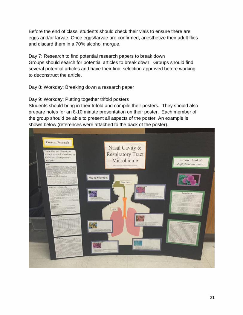
Before the end of class, students should check their vials to ensure there are eggs and/or larvae. Once eggs/larvae are confirmed, anesthetize their adult flies and discard them in a 70% alcohol morgue. Day 7: Research to find potential research papers to break down Groups should search for potential articles to break down. Groups should find several potential articles and have their final selection approved before working to deconstruct the article. Day 8: Workday: Breaking down a research paper Day 9: Workday: Putting together trifold posters Students should bring in their trifold and compile their posters. They should also prepare notes for an 8-10 minute presentation on their poster. Each member of the group should be able to present all aspects of the poster. An example is shown below (references were attached to the back of the poster).
22
Day 10: Presentations This can be accomplished a variety of ways, but I recommend a rotating poster session. Have each group set up their poster and have one member of the group stay and present while the other group members peer grade a different poster. After 10 minutes, have groups change presenters and the poster they are grading. By completing 4 rounds, each student will have an opportunity to present and a large number of peer grades with be generated. Day 11: Introducing molecular biology sequences See “Microbiome Molecular Genetics Labs” sheet in the Student Handouts section. Review these labs and explain the sequence. This class is also a good opportunity to meet with each group and discuss feedback from the project presentations. Day 12: Pouring differential media plates If having students pour agar plates, you will need to have solutions in the autoclave 45-60 minutes before class begins. Students should work in groups to pour at least 12 of each type of plate, each requiring 30-40 ml of agar. You should plan to autoclave at least 500 ml of each agar type. Additionally, time can be spent discussing the science behind autoclaving and the importance of sterile technique. Day 13: Remove 3 adult flies, Surface sterilize and plate bacteria, incubate at 30°CStudents should follow the lab protocol outlined in the “Microbiome Molecular Genetics Labs” Lab Series part 1. To help facilitate completing the lab in a single period, you may wish to pre-aliquot the 150 µl of PBS as well as set-up labeled petri dishes with 10% bleach and alcohol. Students should be sure to wear gloves and goggles, practice sterile technique, dispose of all materials that contact bacteria (pipet tips, inoculating loops) in a container with 10% bleach. Plates should be tapes or para-filmed closed after homogenate is spread on plates.
Day 14: Observe Plates, Record initial observations, pour gels Students should continue to follow the lab protocol outlined in the “Microbiome Molecular Genetics Labs.” Students should photograph plates as well as count colonies under a dissecting microscope or with a hand lens. For darker plates such as EMB agar, it may help to put plates on a light box to observe/count colonies. Students can also make initial classification of bacteria being observed on the different plates.
23
Depending on bacterial variety, groups may be able to pour one gel for every 2 groups or each group can pour their own gel. Day 15: Observe Plates, Record initial observations, Remove single colonies, run PCR on those colonies Again, students should continue to follow the lab protocol outlined in the “Microbiome Molecular Genetics Labs” like on day 14 and record observations again. Colony counts may be difficult if bacterial counts were high on day 1 of observations. To facilitate setting up PCR in a single period, you may wish to pre-aliquot the Master Mix into PCR tubes and store on ice. While transferring colonies, students should be sure to wear gloves and goggles, practice sterile technique, dispose of all materials that contact bacteria in a container with 10% bleach Day 16: Run gels to confirm amplification, based on plate analysis; predict possible species that will be identified through sequencing Following the lab protocol outlined in the “Microbiome Molecular Genetics Labs,” students can pipette visualization dye into PCR samples, and then transfer samples into wells on the gel. Gels will need to run for approximately 25 minutes before visualizing on a UV-light box. Store gels in a refrigerator to slow the diffusion of the DNA out of the gel overnight or have students cut bands out and transfer them into microcentrifuge tubes before the end of class. If not using UV visualization, there is no need to add visualization dye to samples before adding them to the gel (the Master Mix has loading dye mixed in), but you will want to stain the gel at the end of class and then de-stain overnight to visualize DNA. Gels will not be analyzed until the next day. Day 17: Purify gels and send samples off for sequencing. Before class, set up a hot water bath to 50°C to facilitate dissolving gels. Following the procedure outlined in the Qiagen kit (described in the “Microbiome Molecular Genetics Labs”), students can extract their DNA samples from the gel. If students cut their samples out the day before, Buffer QG can be aliquoted for students and dissolving can be initiated before class starts. This may allow students to complete the procedure within a single class period. It is likely that the instructor will need to add primers and label samples for submission. Day 18: Introduce BLAST with The Mystery Monster on Acapulco Beach See Student Handout for “The Mystery Monster on Acapulco Beach.” Students will read the story and enter the nucleotide sequence sequence and identify the mystery monster. If possible, either email or digitally post the sequence of the creature so students do not need to enter the nucleotides by hand.

24
The BLAST results will reveal that the creature is in the genus species Physeter macrocephalus. A quick Google search by students should identify the creature as a Humpback whale. Day 19 (if sequences returned): Groups use blast to identify microbial species isolated Digitally share the sequences and have students repeat the procedure they followed for “The Mystery Monster on Acapulco Beach” to identify the microbes isolated. Day 20: Groups work Question-Claim-Justification-Evidence-Reasoning to write up results of their lab STUDENT SECTION The purpose of the curriculum unit is to promote student learning and to make connections between concepts discussed in the classroom and their laboratory investigations. Students should learn through their laboratory experiences, not merely verify what they already know. I. RATIONALE Significance and Introduction The microbiome is the collection of microorganisms (composed of bacteria, bacteriophage, fungi, protozoa and viruses) that live in and on the human body. There are more microbial cells in and on your body than there are human cells (Sender, Fuchs and Milo). The microbiome is a rapidly growing area of biological research (Jones, 277), which has increased our awareness of the connection between our microbiota and immunity, obesity, and a wide array of health issues ("The Human Microbiome...”). Model organisms allow us to study biological phenomenon in controlled experiments. Particularly for implications relating to human health, model organisms allow scientists to run many trials on a shorter timescale with larger numbers before asking questions with humans. One such model organism is the fruit fly or Drosophila melanogaster. The fruit fly is useful because of its short life cycle (12 days), ease of growing up populations (the female fruit fly will lay between 750 and 1,500 eggs in her lifetime), its ease of culture and maintenance, and its small, well-studied genome ("Science/AAAS And Modencode | Drosophila As A Model Organism").
25
In this unit, you will collect background about the microbiome and its implications on human health and then use a Drosophila as a model organism to study the microbiome. This unit will introduce a wide variety of laboratory techniques including plating bacteria on differential media, PCR of 16S ribosomal DNA, gel electrophoresis, DNA extraction, DNA sequencing, and bioinformatics. It will also allow for asking questions about the influence of environmental factors or feeding options on the microbiome of Drosophila. II. SCIENCE BACKGROUND Microbiome There are organisms that live in or on other organisms in all kingdoms. This includes animals that are colonized by microorganisms that play an important, but poorly understood role, in the health and fitness of the animal host. The fruit fly Drosophila melanogaster is a useful system to study how animals interact with their microbiota ("The Drosophila Microbiome"). Several studies have documented the diversity of the microbiome of Drosophila and how it is typically dominated by Lactobacillus and Acetobacter species (Blum et al.; Staubach et al.). Differential and Selective Media To assess the microbiome of Drosophila we will plate out the bacteria on differential and selective media. Differential medium help distinguish microorganisms from one another based on growth characteristics when grown on specific medium types. Organisms with differing growth characteristics appear visibly different when placed on differential media. This may be due to color differences when grown to metabolic differences in microbial species. Selective medium support the growth of specific groups of organisms, inhibiting the growth others. Some media may possess both selective and differential properties ("Use Of Selective And Differential Media"). In this investigation, the type of bacteria grown can be interpreted using the following information: L.B. (Luria Broth) Agar is a nutritious medium designed for rapid bacterial growth. It is not selective or differential. MRS (de Man, Rogosa and Sharpe) Agar is a medium for the cultivation and enumeration of Lactobacillus spp. EMB (Eosin Methylene Blue) Agar is selective for gram-negative bacteria. It further differentiates Escherichia spp. and Aerobacter spp. A distinctive metallic green sheen is produced by E.coli due to acid production resulting in an amide bonding between the eosin and methylene blue; other coliforms do not produce enough acid and remain colorless. Eosin inhibits most gram-positive organisms. MSA (Mannitol Salt Agar) is a selective media for some types of gram-positive bacteria. Staphylococcus spp. grow on this media because the high sodium chloride level inhibits most other species with the exception of halophilic Vibrios. The majority of S. aureus
26
ferment mannitol producing yellow colonies and change the media color to yellow. Other Staphylococci produce small pink or red colonies with no color change to the medium. Bioinformatics Bioinformatics is the field of science that integrates molecular data with computer databases. It can be used to identify the species of organism being studied as well as to explore the evolutionary relationships of different species ("Bioinformatics: Converting Data To Knowledge"). While there are advanced techniques used to sequence organisms, in this unit we amplify and sequence the 16S rRNA sequence of the bacteria that are isolated. Because the 16S rRNA sequence is a variable sequence found in the small subunit of all bacterial ribosomes, it is an established means of identifying bacterial species through PCR and sequencing (Janda and Abbott, 2761-2764). Once the sequences are returned, the species isolated can be identified by using the nucleotide BLAST tools at the NCBI website or they can be used to estimate the evolutionary relationships of the bacteria using ClustralOmega or another phylogeny building tool. References: "Bioinformatics: Converting Data To Knowledge". (2000): n. pag. Web. 30 Apr. 2016. Blum, J. E. et al. "Frequent Replenishment Sustains The Beneficial Microbiome Of Drosophila Melanogaster". mBio 4.6 (2013): e00860-13-e00860-13. Web. 30 Apr. 2016. "The Drosophila Microbiome". Angeladouglaslab.com. N.p., 2016. Web. 30 Apr. 2016. "The Human Microbiome: Implications Of The Microcosm Within Us". AAAS - The World's Largest General Scientific Society. N.p., 2015. Web. 30 April 2016. Janda, J. M. and S. L. Abbott. "16S Rrna Gene Sequencing For Bacterial Identification In The Diagnostic Laboratory: Pluses, Perils, And Pitfalls". Journal of Clinical Microbiology 45.9 (2007): 2761-2764. Web. 30 Apr. 2016. Jones, Susan. "Trends In Microbiome Research". Nat Biotechnol 31.4 (2013): 277-277. Web. 30 April 2016.
27
"Science/AAAS And Modencode | Drosophila As A Model Organism". Modencode.sciencemag.org. N.p., 2016. Web. 30 April 2016. Sender, Ron, Shai Fuchs, and Ron Milo. "Revised Estimates For The Number Of Human And Bacteria Cells In The Body". (2016): n. pag. Web. 30 April 2016. Staubach, Fabian et al. "Host Species And Environmental Effects On Bacterial Communities Associated With Drosophila In The Laboratory And In The Natural Environment". PLoS ONE 8.8 (2013): e70749. Web. 30 Apr. 2016. "Use Of Selective And Differential Media". Highlands.edu. N.p., 2016. Web. 30 Apr. 2016. III. Student Hand-Outs:
28
Microbiome Project
Introduction
The microbiome is the collection of microorganisms (composed of bacteria,
bacteriophage, fungi, protozoa and viruses) that live in and on the human body.
There are more microbial cells in and on your body than there are as human cells!
This collection of organisms is now being researched to determine the impacts,
both positive and negative, that these organisms have on the human body. In this project your group will research microorganisms found in a particular region
of the body (mouth, respiratory tract, stomach, intestines, urogenital tract, or skin),
examine the health impacts of some of these organisms, and then deconstruct a
current research paper about this region of the microbiome.
Step 1: Research the microbiome and focus on your assigned region of the body:
Start Here by Exploring this Animation:
http://www.outreach.mcb.harvard.edu/animations/microbiomeAnimation.swf Learn Genetics also has a good overview of the microbiome and some specific
functions microbes perform for the body:
http://learn.genetics.utah.edu/content/microbiome/friends/
For another good overview, see this Scientific American Interactive:
http://www.scientificamerican.com/article.cfm?id=microbiome-graphic-explore-
human-microbiome
Additional resources include:
Microbiome of the Mouth:
● http://www.homd.org/
29
● http://jb.asm.org/content/192/19/5002.full
● http://www.the-
scientist.com/?articles.view/articleNo/40281/title/Mining-the-
Mouth-s-Many-Microbes/
Microbiome of the Nasal Cavity/Respiratory Tract
● http://www.plosone.org/article/info%3Adoi/10.1371/journal.pone.
0010598
● http://www.npr.org/2012/09/14/161156793/microbes-benefit-
more-than-just-the-gut
● http://www.the-
scientist.com/?articles.view/articleNo/40162/title/Breathing-Life-
into-Lung-Microbiome-Research/
Microbiome of the Stomach:
● http://news.ucsc.edu/2013/03/stomach-microbes.html
● http://www.plosone.org/article/info%3Adoi%2F10.1371%2Fjourn
al.pone.0076375
● http://www.hindawi.com/journals/tswj/2014/610421/
Microbiome of the Intestines:
● http://mic.sgmjournals.org/content/156/11/3203.full.pdf+html
● http://www.youtube.com/watch?v=pGpBj5T0-DY
● http://www.uchospitals.edu/news/2014/20140825-nagler.html
Microbiome of the Urogenital Tract:
● http://www.plosone.org/article/info:doi/10.1371/journal.pone.001
4116
● http://www.the-
scientist.com/?articles.view/articleNo/40092/title/Parsing-the-
Penis-Microbiome/
30
Microbiome of the Skin:
● http://www.nih.gov/news/health/may2013/nhgri-22.htm
● http://www.ncbi.nlm.nih.gov/pubmed/21407241
● https://www.aobiome.com/human-skin-microbiome
Other Microbiome Resources:
● http://www.nytimes.com/2012/06/19/science/studies-of-human-
microbiome-yield-new-insights.html?pagewanted=all&_r=0
● http://www.wired.com/magazine/2011/09/mf_microbiome/
● http://www.economist.com/node/21560523
● http://www.slideshare.net/phylogenomics/the-human-
microbiome-talk-by-jonathan-eisen-phylogenomics-for-scifoo-
2007
● http://www.ted.com/talks/jonathan_eisen_meet_your_microbes.h
tml
● http://www.youtube.com/watch?v=FWT_BLVOASI
● http://learn.genetics.utah.edu/content/microbiome/
● http://www.scientificamerican.com/article.cfm?id=microbiome-
graphic-explore-human-microbiome You will summarize this research and include a bibliography on your trifold poster.
Step 2: Find and Deconstruct a Research Paper about a Microbe found in Your assigned region of the microbiome
Your group will find a scientific journal article and then deconstruct or translate the
findings into common everyday language. You will present your deconstruction on
your trifold.
Search for journal articles on one of the following:
● PLoS: http://www.plos.org/publications/journals/
31
● mbio: http://mbio.asm.org/
● msphere: http://msphere.asm.org/index.php
Guides that can help you deconstruct your journal article:
● https://www.elsevier.com/connect/infographic-how-to-read-a-scientific-paper
● http://journals.plos.org/plosbiology/article?id=10.1371/journal.pbio.1000264
● http://www.academia.edu/8108654/GUIDELINES_FOR_DECONSTRUCTIN
G_PUBLISHED_JOURNAL_ARTICLE
Summarize research about microbes found in your assigned body region,
deconstruct your journal article, and provide work cited on a trifold poster. Each
member of the group should be able to explain all parts of the poster and present
the information on the display. Each group member will need to be able to answer
some specific questions during the presentation.
Step 3: Display Research on a Trifold Poster and prepare an 8-10 minute presentation on your topic
32
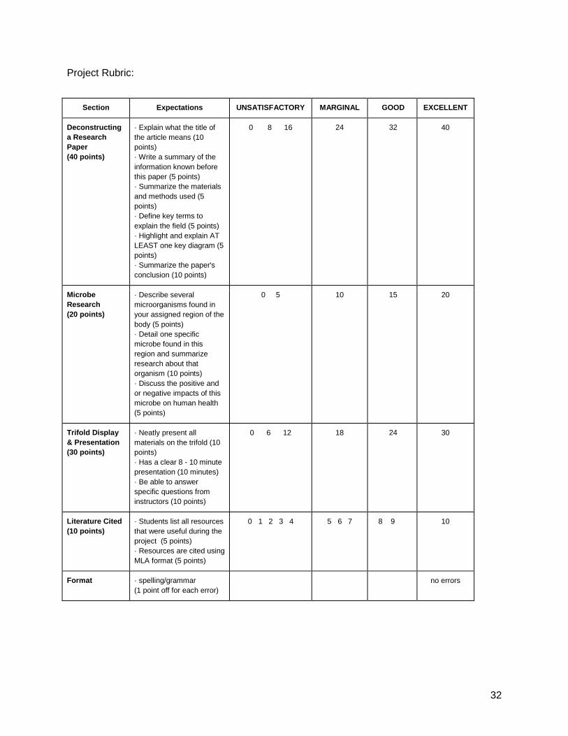
Project Rubric:
Section Expectations UNSATISFACTORY MARGINAL GOOD EXCELLENT
Deconstructing a Research Paper (40 points)
· Explain what the title of the article means (10 points) · Write a summary of the information known before this paper (5 points) · Summarize the materials and methods used (5 points) · Define key terms to explain the field (5 points) · Highlight and explain AT LEAST one key diagram (5 points) · Summarize the paper's conclusion (10 points)
0 8 16 24 32 40
Microbe Research (20 points)
· Describe several microorganisms found in your assigned region of the body (5 points) · Detail one specific microbe found in this region and summarize research about that organism (10 points) · Discuss the positive and or negative impacts of this microbe on human health (5 points)
0 5 10 15 20
Trifold Display & Presentation (30 points)
· Neatly present all materials on the trifold (10 points) · Has a clear 8 - 10 minute presentation (10 minutes) · Be able to answer specific questions from instructors (10 points)
0 6 12 18 24 30
Literature Cited (10 points)
· Students list all resources that were useful during the project (5 points) · Resources are cited using MLA format (5 points)
0 1 2 3 4 5 6 7 8 9 10
Format · spelling/grammar (1 point off for each error)
no errors
33
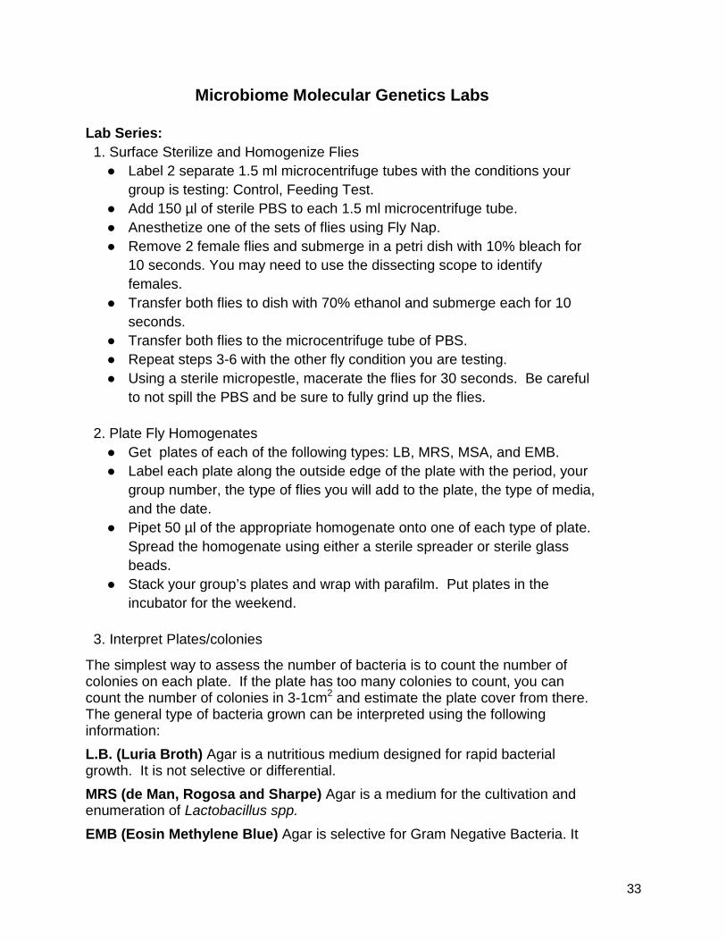
Microbiome Molecular Genetics Labs Lab Series: 1. Surface Sterilize and Homogenize Flies
● Label 2 separate 1.5 ml microcentrifuge tubes with the conditions your group is testing: Control, Feeding Test.
● Add 150 µl of sterile PBS to each 1.5 ml microcentrifuge tube. ● Anesthetize one of the sets of flies using Fly Nap. ● Remove 2 female flies and submerge in a petri dish with 10% bleach for
10 seconds. You may need to use the dissecting scope to identify females.
● Transfer both flies to dish with 70% ethanol and submerge each for 10 seconds.
● Transfer both flies to the microcentrifuge tube of PBS. ● Repeat steps 3-6 with the other fly condition you are testing. ● Using a sterile micropestle, macerate the flies for 30 seconds. Be careful
to not spill the PBS and be sure to fully grind up the flies. 2. Plate Fly Homogenates
● Get plates of each of the following types: LB, MRS, MSA, and EMB. ● Label each plate along the outside edge of the plate with the period, your
group number, the type of flies you will add to the plate, the type of media, and the date.
● Pipet 50 µl of the appropriate homogenate onto one of each type of plate. Spread the homogenate using either a sterile spreader or sterile glass beads.
● Stack your group’s plates and wrap with parafilm. Put plates in the incubator for the weekend.
3. Interpret Plates/colonies
The simplest way to assess the number of bacteria is to count the number of colonies on each plate. If the plate has too many colonies to count, you can count the number of colonies in 3-1cm2
L.B. (Luria Broth) Agar is a nutritious medium designed for rapid bacterial growth. It is not selective or differential.
and estimate the plate cover from there. The general type of bacteria grown can be interpreted using the following information:
MRS (de Man, Rogosa and Sharpe) Agar is a medium for the cultivation and enumeration of Lactobacillus spp.
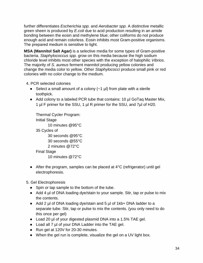
EMB (Eosin Methylene Blue) Agar is selective for Gram Negative Bacteria. It
34
further differentiates Escherichia spp. and Aerobacter spp. A distinctive metallic green sheen is produced by E.coli due to acid production resulting in an amide bonding between the eosin and methylene blue; other coliforms do not produce enough acid and remain colorless. Eosin inhibits most Gram-positive organisms. The prepared medium is sensitive to light. MSA (Mannitol Salt Agar) is a selective media for some types of Gram-positive bacteria. Staphylococcus spp. grow on this media because the high sodium chloride level inhibits most other species with the exception of halophilic Vibrios. The majority of S. aureus ferment mannitol producing yellow colonies and change the media color to yellow. Other Staphylococci produce small pink or red colonies with no color change to the medium. 4. PCR selected colonies
● Select a small amount of a colony (~1 µl) from plate with a sterile toothpick.
● Add colony to a labeled PCR tube that contains: 10 µl GoTaq Master Mix, 1 µl F primer for the SSU, 1 µl R primer for the SSU, and 7µl of H20.
Thermal Cycler Program: Initial Stage 10 minutes @95°C 35 Cycles of
30 seconds @95°C 30 seconds @55°C 2 minutes @72°C Final Stage 10 minutes @72°C
● After the program, samples can be placed at 4°C (refrigerator) until gel
electrophoresis. 5. Gel Electrophoresis
● Spin or tap sample to the bottom of the tube. ● Add 4 µl of DNA loading dye/stain to your sample. Stir, tap or pulse to mix
the contents. ● Add 2 µl of DNA loading dye/stain and 5 µl of 1kb+ DNA ladder to a
separate tube. Stir, tap or pulse to mix the contents. (you only need to do this once per gel)
● Load 20 µl of your digested plasmid DNA into a 1.5% TAE gel. ● Load all 7 µl of your DNA Ladder into the TAE gel. ● Run gel at 120V for 20-30 minutes. ● When the gel run is complete, visualize the gel on a UV light box.
35
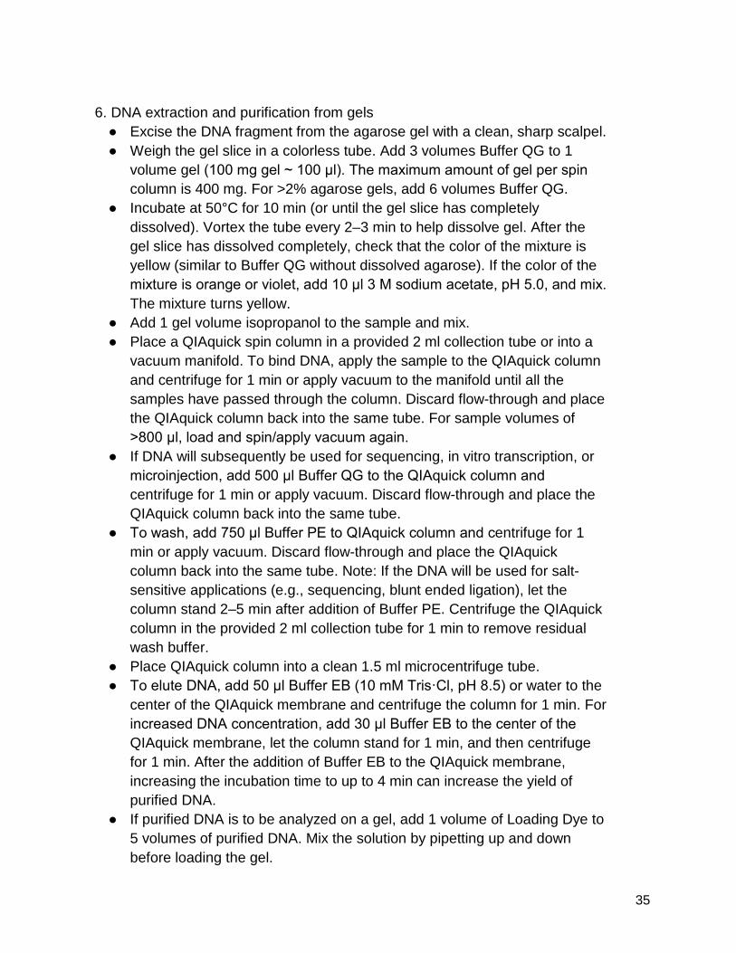
6. DNA extraction and purification from gels
● Excise the DNA fragment from the agarose gel with a clean, sharp scalpel. ● Weigh the gel slice in a colorless tube. Add 3 volumes Buffer QG to 1
volume gel (100 mg gel ~ 100 μl). The maximum amount of gel per spin column is 400 mg. For >2% agarose gels, add 6 volumes Buffer QG.
● Incubate at 50°C for 10 min (or until the gel slice has completely dissolved). Vortex the tube every 2–3 min to help dissolve gel. After the gel slice has dissolved completely, check that the color of the mixture is yellow (similar to Buffer QG without dissolved agarose). If the color of the mixture is orange or violet, add 10 μl 3 M sodium acetate, pH 5.0, and mix. The mixture turns yellow.
● Add 1 gel volume isopropanol to the sample and mix. ● Place a QIAquick spin column in a provided 2 ml collection tube or into a
vacuum manifold. To bind DNA, apply the sample to the QIAquick column and centrifuge for 1 min or apply vacuum to the manifold until all the samples have passed through the column. Discard flow-through and place the QIAquick column back into the same tube. For sample volumes of >800 μl, load and spin/apply vacuum again.
● If DNA will subsequently be used for sequencing, in vitro transcription, or microinjection, add 500 μl Buffer QG to the QIAquick column and centrifuge for 1 min or apply vacuum. Discard flow-through and place the QIAquick column back into the same tube.
● To wash, add 750 μl Buffer PE to QIAquick column and centrifuge for 1 min or apply vacuum. Discard flow-through and place the QIAquick column back into the same tube. Note: If the DNA will be used for salt-sensitive applications (e.g., sequencing, blunt ended ligation), let the column stand 2–5 min after addition of Buffer PE. Centrifuge the QIAquick column in the provided 2 ml collection tube for 1 min to remove residual wash buffer.
● Place QIAquick column into a clean 1.5 ml microcentrifuge tube. ● To elute DNA, add 50 μl Buffer EB (10 mM Tris·Cl, pH 8.5) or water to the
center of the QIAquick membrane and centrifuge the column for 1 min. For increased DNA concentration, add 30 μl Buffer EB to the center of the QIAquick membrane, let the column stand for 1 min, and then centrifuge for 1 min. After the addition of Buffer EB to the QIAquick membrane, increasing the incubation time to up to 4 min can increase the yield of purified DNA.
● If purified DNA is to be analyzed on a gel, add 1 volume of Loading Dye to 5 volumes of purified DNA. Mix the solution by pipetting up and down before loading the gel.
36
7. Send out sequences
Follow guidelines posted by GeneWiz (http://www.genewiz.com/public/Sample-Submission-Guideline.aspx) and send out the DNA to have sequences determined.
8. Bioinformatics
● Use NCBI nucleotide BLAST(http://blast.ncbi.nlm.nih.gov/Blast.cgi) to determine the species of the sequences returned from GeneWiz.
● Use Clustral Omega (http://www.ebi.ac.uk/Tools/msa/clustalo/) to create a cladogram of the species identified from the class. Clustral Omega produces biologically meaningful multiple sequence alignments of divergent sequences. Evolutionary relationships can be seen via viewing Cladograms or Phylograms.
37
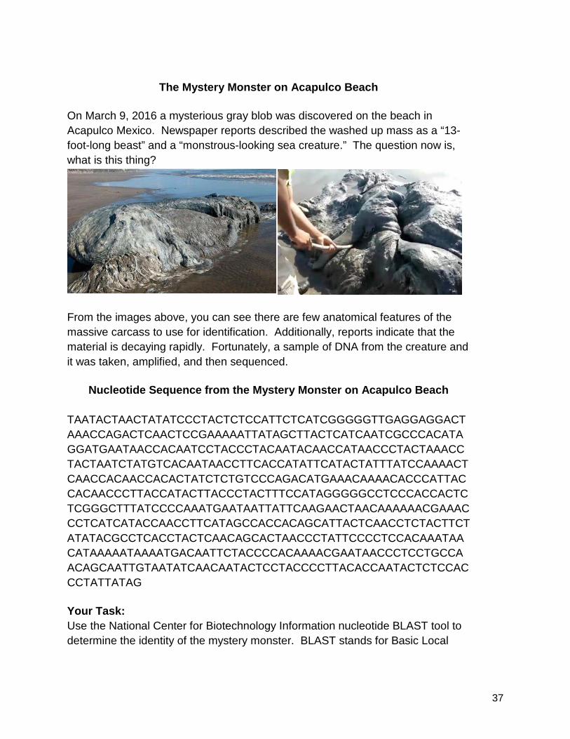
The Mystery Monster on Acapulco Beach
On March 9, 2016 a mysterious gray blob was discovered on the beach in Acapulco Mexico. Newspaper reports described the washed up mass as a “13-foot-long beast” and a “monstrous-looking sea creature.” The question now is, what is this thing?
From the images above, you can see there are few anatomical features of the massive carcass to use for identification. Additionally, reports indicate that the material is decaying rapidly. Fortunately, a sample of DNA from the creature and it was taken, amplified, and then sequenced.
Nucleotide Sequence from the Mystery Monster on Acapulco Beach TAATACTAACTATATCCCTACTCTCCATTCTCATCGGGGGTTGAGGAGGACTAAACCAGACTCAACTCCGAAAAATTATAGCTTACTCATCAATCGCCCACATAGGATGAATAACCACAATCCTACCCTACAATACAACCATAACCCTACTAAACCTACTAATCTATGTCACAATAACCTTCACCATATTCATACTATTTATCCAAAACTCAACCACAACCACACTATCTCTGTCCCAGACATGAAACAAAACACCCATTACCACAACCCTTACCATACTTACCCTACTTTCCATAGGGGGCCTCCCACCACTCTCGGGCTTTATCCCCAAATGAATAATTATTCAAGAACTAACAAAAAACGAAACCCTCATCATACCAACCTTCATAGCCACCACAGCATTACTCAACCTCTACTTCTATATACGCCTCACCTACTCAACAGCACTAACCCTATTCCCCTCCACAAATAACATAAAAATAAAATGACAATTCTACCCCACAAAACGAATAACCCTCCTGCCAACAGCAATTGTAATATCAACAATACTCCTACCCCTTACACCAATACTCTCCACCCTATTATAG Your Task: Use the National Center for Biotechnology Information nucleotide BLAST tool to determine the identity of the mystery monster. BLAST stands for Basic Local
38
Alignment Search Tool and it is an used to compare sequence information, typically of nucleotides or amino acids, with a database of known sequences. Procedure:
1. Go to the NCBI website (http://www.ncbi.nlm.nih.gov/). 2. On the right hand menu, choose “BLAST”. 3. Under the heading “Basic BLAST,” select “Nucleotide BLAST.” 4. In the “Enter Query Sequence” box, either type or paste in the Mystery
Monster’s DNA sequence listed above. 5. Be sure that under the “Choose Search Set” you select “Other” and for the
“Program Selection” you select ”Highly similar sequences (megablast).” 6. Click on the “BLAST” button at the bottom of the page and then wait for
your results (depending on how busy the servers are, this can take anywhere from a few seconds to several minutes).
7. When BLAST is done, scroll down to the “Graphic Summary” section to see the colorized diagram indicating the degree of similarity between your sequence and the sequences within the database. Red and pink/purple indicate a good match, while green, blue and black indicate a poor match. If the colored line spans the entire length of the window, then the “hit” sequence matches the query sequence along its entire length.
8. Below the colored lines in the “Sequences producing significant alignments:” section, look at the first match listed. This will be the sequence that hatches the sample from our Mystery Monster.
9. Identify the Genus Species name associated the match and complete a Google search to identify the common name of the Mystery Monster on Acapulco Beach. The Mystery Monster of Acapulco Beach is: ____________________________.
Works Consulted: Modified with permission from “What on Earth is The Chilean Blob?” by David Form at Nashoba Regional High School. "Creature from the Deep Blue Sea! Huge Monstrous-looking Beast Washes up on Popular Tourist Beach in Mexico Leaving Experts Baffled." Mail Online. Associated Newspapers, 11 Mar. 2016. Web. 12 Apr. 2016.
39
Write-Up for Drosophila Microbiome Lab Question-Claim-Justification-Evidence-Reasoning
Having completed the Drosophila Microbiome Lab series, it is time to report your results. To write-up this lab, we will use a Question-Claim-Justification-Evidence-Reasoning format. This will allow to summarize what you expected to happen when you modified the feeding stock of the fruit flies, including the research that led to that preconception. You will then present the data from your lab, including images of plates and bioinformatics data from bacteria sequences, that relates to your claims. The write-up will conclude with a reasoning section that explains if the evidence supports or refutes the claim. You will also outline questions raised by the evidence and possible future avenues of investigation to further evaluate the claim. Here are guidelines for each section of the write-up: Question: The question will be the specific question you addressed in your lab. It will meet the following criteria:
- It will be one complete sentence - The question is answerable by the investigation you conducted. - The independent variable(s) and dependent variable will be specifically
stated. Claim: A claim is a statement of your understanding about the thing you are investigating. You can make claims about the evidence you gather in a lab, or that you learn about in other aspects of this course. Your claim should include:
- It is at least one complete sentence (usually only 1-2 sentence). - The claim addresses and answers the specific question of the lab. - The claim specifically states relationship between two variables raised in
the question. Justification: The justification is where you will use outside resources to support your claim. Your justification should include:
- Two-Three references that directly connect the topic of the claim. - Statements in the justification are parenthetically referenced. - References are properly cited in the reference section.
40
Evidence: Evidence is data. Data can come from a lot of places, but in this assignment you will use the data collected in the lab. Evidence should include:
- The evidence will be specific to the question being asked. - It will have the specific independent and dependent variables raised in the
question and the claim. - The data will be interpreted, not just listed. An explanation of what the
data shows, and how that supports the claim will be provided. Reasoning: Reasoning is the most challenging aspect of the write up. Making a logically consistent reasoning when constructing a claim is what separates good scientists from hacks. Your reasoning will have the following properties:
- The reasoning will start with a restatement of the claim. - The reasoning will be based on scientific principles/accepted
understandings in science. - Reasoning will connect the particular principle to the evidence and claim
of the lab. - Your language should be precise and accurate. - You are writing so that someone who was not present for a particular
investigation could read your explanation and understand what you did and how you did it.
Adapted from What a scientific explanation looks like
by David Knuffke.
41
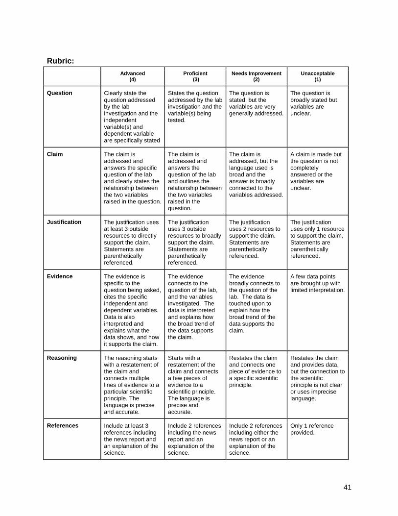
Rubric: Advanced
(4) Proficient
(3) Needs Improvement
(2) Unacceptable
(1)
Question Clearly state the question addressed by the lab investigation and the independent variable(s) and dependent variable are specifically stated
States the question addressed by the lab investigation and the variable(s) being tested.
The question is stated, but the variables are very generally addressed.
The question is broadly stated but variables are unclear.
Claim The claim is addressed and answers the specific question of the lab and clearly states the relationship between the two variables raised in the question.
The claim is addressed and answers the question of the lab and outlines the relationship between the two variables raised in the question.
The claim is addressed, but the language used is broad and the answer is broadly connected to the variables addressed.
A claim is made but the question is not completely answered or the variables are unclear.
Justification The justification uses at least 3 outside resources to directly support the claim. Statements are parenthetically referenced.
The justification uses 3 outside resources to broadly support the claim. Statements are parenthetically referenced.
The justification uses 2 resources to support the claim. Statements are parenthetically referenced.
The justification uses only 1 resource to support the claim. Statements are parenthetically referenced.
Evidence The evidence is specific to the question being asked, cites the specific independent and dependent variables. Data is also interpreted and explains what the data shows, and how it supports the claim.
The evidence connects to the question of the lab, and the variables investigated. The data is interpreted and explains how the broad trend of the data supports the claim.
The evidence broadly connects to the question of the lab. The data is touched upon to explain how the broad trend of the data supports the claim.
A few data points are brought up with limited interpretation.
Reasoning The reasoning starts with a restatement of the claim and connects multiple lines of evidence to a particular scientific principle. The language is precise and accurate.
Starts with a restatement of the claim and connects a few pieces of evidence to a scientific principle. The language is precise and accurate.
Restates the claim and connects one piece of evidence to a specific scientific principle.
Restates the claim and provides data, but the connection to the scientific principle is not clear or uses imprecise language.
References Include at least 3 references including the news report and an explanation of the science.
Include 2 references including the news report and an explanation of the science.
Include 2 references including either the news report or an explanation of the science.
Only 1 reference provided.