investigation of bacterial pathogens associated with
TRANSCRIPT
1
Investigation of Bacterial Pathogens Associated with Concentrated Animal Feeding Operations
(CAFOs) and their Potential Impacts on a National Wildlife Refuge in Oklahoma: Final Report
27 July 2004
Project 2N44, 200120004
David S. Blehert, Brenda M. Berlowski, Heather M. Gutzman, and Mark J. Wolcott
USGS - National Wildlife Health Center
6006 Schroeder Road
Madison, WI 53711-6223

2
SUMMARY
According to the United States Environmental Protection Agency (US EPA), run off from
agricultural lands is one of the greatest contributors of pollutants to surface waters in this country.
Major agricultural pollutants include bacterial pathogens and nutrients that originate from animal
wastes. Several concentrated animal feeding operations (CAFOs) are located in close proximity to
the Salt Plains National Wildlife Refuge in Alfalfa County, Oklahoma; it is our understanding that
these facilities are not US EPA registered. The goal of this study was to survey refuge surface water
and sediment for known bacterial pathogens of wildlife, especially waterfowl, which may have
originated from CAFO wastes. Sampling was conducted during the spring, summer, and fall of 2001
and 2002 at ten sites located on watersheds downstream of agricultural sites. Bacterial indicators of
fecal contamination (fecal coliforms and fecal streptococci) were identified and counted in surface
water samples. Fecal coliform levels exceeded US EPA human recreational bathing water limits in
22 of 50 samples, and fecal streptococcus numbers exceeded human recreational bathing limits in 47
of 50 samples. Antibiotic resistance profiles were determined for a subset of fecal colifom and fecal
streptococcus isolates. The tested isolates were frequently resistant to ceftiofur, enrofloxacin, and
tetracycline, antibiotics commonly administered to livestock. In addition, surface water and
sediment samples were analyzed for the presence of bacteria (Clostridium botulinum type C,
toxigenic Escherichia coli, Erysipelothrix spp., Pasteurella multocida, Salmonella spp., and Yersinia
spp) with pathogenic potential to wild and domestic animals. Of the organisms surveyed,
salmonellae and escherichiae were isolated. The data presented herein will provide a benchmark for
future studies investigating the potential long term environmental affects of agricultural practices on
the Salt Plains National Wildlife Refuge.
3
BACKGROUND AND JUSTIFICATION
Human population expansion and concurrent reductions in wildlife habitat have forced
wildlife to crowd into smaller areas and to potentially interact more frequently with domestic
animals (11). These increased interactions can result in a greater incidence of disease transmission
between domestic and wild animals, causing wildlife population declines and economic losses to the
livestock industry. Many of these diseases are also transmissible to humans (zoonotic) (11).
Further, domestic animal diseases can become endemic in wildlife populations, which can then
become reservoirs for reintroduction of disease to domestic animals and humans (9, 11).
The livestock industry has also changed significantly over the past 20 years, as exemplified
by the trend towards fewer but larger operations that emphasize intense production and
specialization (15). If a livestock facility harbors animals for at least 45 days within any 12-month
period and there is no grass or other vegetation in the confinement area during the normal growing
season, it is defined by the United States Environmental Protection Agency (US EPA) as an animal
feeding operation (AFO) (14). Based upon the number of animals harbored at the facility and upon
the potential of the facility to contaminate surface waters, the EPA can further classify an AFO as a
concentrated animal feeding operation (CAFO) (14). In the United States, an estimated 15,500
CAFOs annually produce approximately three times more raw excreted waste than is generated by
humans (38). Unlike human wastes, which are processed through sanitary treatment facilities, raw
animal wastes are typically remediated in open, wildlife-accessible containment lagoons or disposed
of through land application. These disposal methods represent a direct threat to wildlife as the
causative agents of many diseases are readily excreted through the feces of domestic animals
including those that appear clinically healthy (32, 37). In 2000, the agricultural sector was
4
designated as the leading contributor of pollutants to streams, rivers, lakes, ponds, and reservoirs in
the US (16). Thus, CAFOs are designated as potential pollution point sources under section 502 of
the EPA Clean Water Act and must develop and implement comprehensive waste management plans
in compliance with the National Pollutant Discharge Elimination System program (38).
The Salt Plains National Wildlife Refuge in Alfalfa County, OK, comprises 32,000 acres of
salt flats, open reservoirs, woodlands, and agricultural fields. The refuge, designated as one of 18 US
sites critical to the survival of shorebirds by the Western Hemisphere Shorebird Reserve Network,
provides habitat for approximately 300 migratory bird species, including endangered whooping
cranes and interior least terns, and threatened snowy plovers (http://www.greatsaltplains.com).
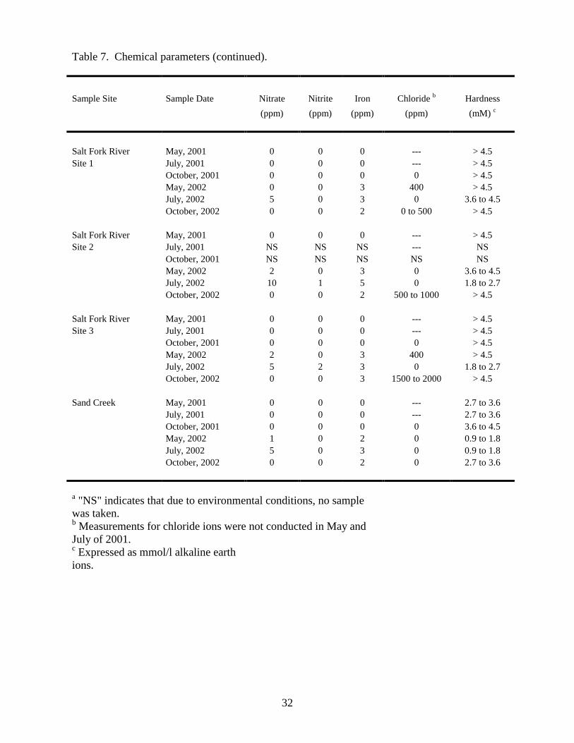
The presence of several CAFOs (it is our understanding that these facilities are not US EPA
registered) as well as the proposed development of additional CAFOs near the refuge (Fig. 1) led to
the concern that neighboring agricultural practices may continue to degrade and ultimately destroy
critical migratory bird habitat provided by the refuge. For example, in 1998, effluent from a swine
CAFO South of the refuge caused a significant fish kill in Spring Creek (D.B. Martin, Personal
Communication). In response, whooping cranes and large numbers of migrating waterfowl had to be
hazed from contaminated areas. Damage to aquatic invertebrate and amphibian populations was
never evaluated.
Salt Plains National Wildlife Refuge personnel asked us to design and complete this study to
characterize bacterial and chemical contaminants within the refuge watershed that may have
originated from agricultural sources. Bacterial indicators of fecal contamination in refuge surface
waters were assessed and water and sediment samples were surveyed for bacteria with pathogenic
potential to wild and domestic animals, including Clostridium botulinum type C, toxigenic
Escherichia coli, Erysipelothrix spp., Pasteurella multocida, Salmonella spp., and Yersinia spp. The
5
results of this study provide a summary of the environmental health of surface waters within the Salt
Plains National Wildlife Refuge on the days sampling was conducted (six times from May, 2001 to
October, 2002) and will serve as a benchmark for refuge personnel to further investigate future
impacts of agricultural practices on the refuge watershed.
OBJECTIVES
1) To survey environmental samples for bacterial indicators of fecal contamination and for a subset
of known bacterial pathogens of wildlife, especially migratory waterfowl.
2) To analyze antibiotic resistance profiles for representative E. coli and fecal streptococcus
isolates.
3) To measure chemical parameters in water samples.
METHODS AND PROCEDURES
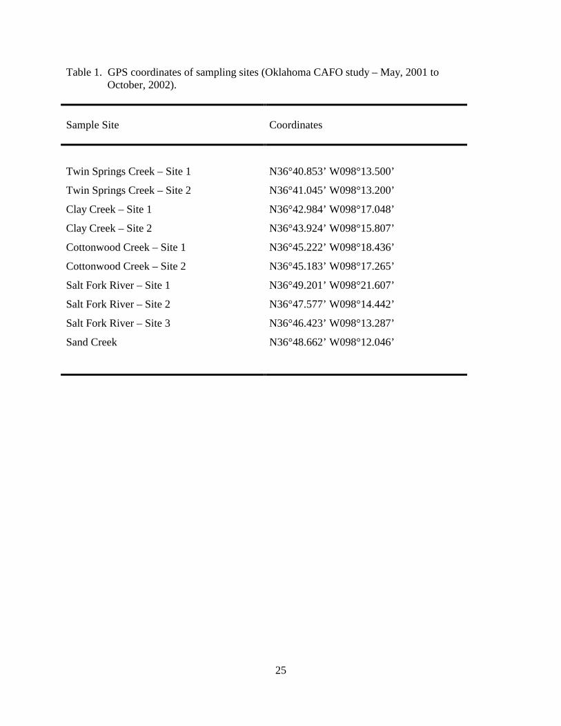
Sample collection. Water and sediment samples were collected from ten locations within
the Oklahoma Salt Plains National Wildlife Refuge (Table 1; Fig. 1) during the months of May, July,
and October of 2001 and 2002. With the exception of Sand Creek (no CAFOs immediately
upstream), sample sites targeting watersheds downstream of CAFOs or proposed CAFOs were
identified with the assistance of refuge personnel. Approximately one liter of surface water was
collected in a sterilized plastic bottle at each sample site, placed on ice, and processed within six
hours of collection. Additionally, 25 to 50 ml of sediment was collected in a 125 ml sterile plastic
container from the top 10 cm of bottom sediment at each sample site. Following collection,
sediment samples were mixed using a sterile swab, the sediment-containing swab was immersed in a
vial containing 5 ml 10% dimethyl sulfoxide (DMSO) as a cryoprotectant, and the swab was agitated
6
to remove sediment particles from the swab. DMSO-sediment suspensions and original sediment
samples were frozen on dry ice within 6 to 8 h of collection and stored at -70°C until analyzed.
Later, DMSO-treated frozen sediment samples were thawed, 2 ml aliquots were each inoculated into
6 ml BHI medium (Becton Dickinson, Cockeysville, MD), and cultures were incubated for 2.5 h at
37°C with shaking. BHI is a non-selective medium used to encourage general bacterial growth
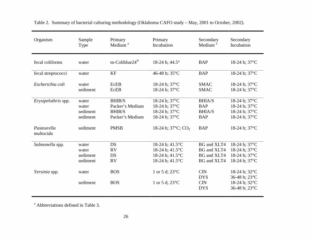
within sediment samples prior to selectively culturing for specific organisms. An overview of the
bacterial culturing conditions utilized for this study is provided below and is summarized in Table 2.
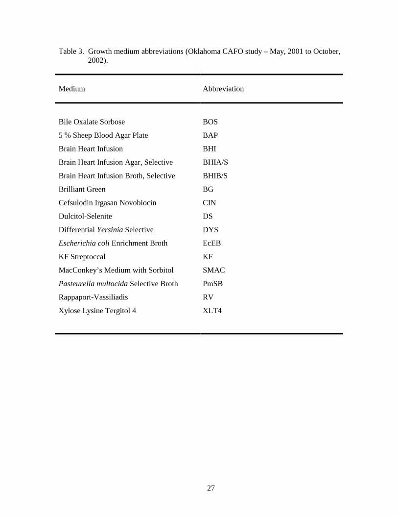
Abbreviations for the bacterial growth media used are defined in Table 3.
Fecal coliforms. The number of fecal coliform bacteria present in water samples was
estimated using a membrane filtration procedure (10). Water samples were collected, and 1:10 and
1:100 dilutions were prepared using sterile phosphate buffer. One hundred ml samples were filtered
through 47 mm diameter, 0.45 µm mean pore size mixed cellulose acetate/nitrate membranes
(Millipore Corporation, Bedford, MA). If for technical reasons less than 100 ml water was filtered,
the actual volume filtered was noted and taken into account when calculating colony forming units
(CFU)/100 ml. Filters were then placed in membrane petri dishes containing pads saturated with
m-Coliblue24® medium (Hach Company, Loveland, CO). Plates were incubated at 44.5°C for 18-24
h in a portable incubator before enumerating colonies. Blue colonies conforming to the criteria for
fecal coliforms were counted for each dilution, and colony counts obtained from plates yielding
between 10 and 150 colonies were averaged to determine the reported result.
Fecal streptococci. The number of fecal streptococci present in water samples was
determined by membrane filtration (10) as described above with the following modifications. Filters
were placed in membrane petri dishes containing pads saturated with KF Streptococcal medium
(Gelman Sciences, Inc., Ann Arbor, MI). Plates were incubated at 35°C for 46 to 48 h in a portable
7
incubator. Red colonies conforming to the criteria for fecal streptococci were counted for each
dilution, and colony counts obtained from plates yielding between 10 and 150 colonies were
averaged.
Clostridium botulinum type C. Efforts were made to identify genetic material from
Clostridium botulinum type C spores in sediment samples by PCR. Genetic material was extracted
from 0.25 to 0.5 g of each sediment sample using the Mo Bio UltraClean Soil DNA Isolation Kit
(Solana Beach, CA) according to the manufacturer’s instructions followed by a 1%
cetyltrimethylammonium bromide (CTAB) extraction. Five µl of each extracted nucleic acid sample
were then PCR-amplified using primers targeting the light-chain region of the type C neurotoxin
gene as previously described (41).
Toxigenic Escherichia coli. The inability of toxigenic E. coli strain O157:H7 to ferment
sorbitol can be used to screen for this organism in environmental samples. 150 ml water samples
were added to 50 ml 4X EcEB (USGS-National Wildlife Health Center, unpublished), and 2 ml
aliquots of BHI sediment-enrichment cultures were transferred to 5 ml EcEB. Cultures were
incubated 18 to 24 h at 37°C and streaked onto SMAC medium (Becton Dickinson, Cockeysville,
MD). SMAC plates were incubated for 18 to 24 h at 37°C and inspected for colorless colonies
indicative of non-sorbitol fermenting E. coli. Suspected isolates were subcultured onto BAPs
(Becton Dickinson) for biochemical characterization using either API-20E or Vitek systems
(bioMerieux, St. Louis, MO) and for screening by the 0157:H7 latex agglutination test (Oxoid
Limited, Hampshire, England) according to the manufacturer’s instructions. Non-sorbitol
fermenting E. coli strains identified while characterizing the antibiotic resistance profiles (see below)
of fecal coliform isolates were included in this analysis as well. Confirmed non-sorbitol fermenting
isolates were sent to The Pennsylvania State University Gastroenteric Disease Center for O and H
8
antigen serotyping and for toxin-gene testing by PCR. Isolates were assayed for the presence of
genes encoding heat labile toxin, heat stable toxins a and b, shiga-like toxin types I and II, cytotoxic
necrotizing factors 1 and 2, and intimin.
Erysipelothrix. One-hundred fifty ml water samples were added to 50 ml 4X BHIB/S
(USGS-National Wildlife Health Center, unpublished) and to 50 ml 4X Packer’s medium (3). 2 ml
aliquots of BHI sediment-enrichment cultures were each transferred to 5 ml BHIB/S and to 5 ml
Packer’s medium. Cultures were incubated 18 to 24 h at 37°C. BHIB/S cultures were then streaked
onto BHIA/S (USGS-National Wildlife Health Center, unpublished), and Packer’s medium cultures
were streaked onto BAPs. Plates were incubated 18 to 24 h at 37°C. Following incubation, plates
were examined to identify colonies characteristic of Erysipelothrix spp. Suspected isolates were
subcultured onto BAPs and tested for hydrogen sulfide production using a triple sugar iron agar
slant. Hydrogen sulfide producing isolates were further characterized using either bioMerieux API-
Coryne or Vitek systems.
Pasteurella multocida. Pasteurella multocida isolations were attempted only from BHI-
enriched sediment samples. Two ml of each enrichment culture were transferred to 5 ml PmSB (29)
and incubated at 37°C for 18 to 24 h in the presence of 5 % CO2. Each broth culture was streaked
onto a BAP and incubated at 37°C for 18 to 24 h. All bacterial colonies were screened based on
colony morphology to identify Pasteurella spp. Suspected Pasteurella isolates were biochemically
characterized using the bioMerieux API-20E system.
Salmonella. One-hundred fifty ml water samples were added to 50 ml 4X RV broth (12, 40)
(Remel, Lenexa, KA) and to 50 ml 4X DS broth (USGS-National Wildlife Health Center,
unpublished). Cultures were incubated at 41.5°C for 18 to 24 h. Two ml aliquots of BHI sediment-
enrichment cultures were transferred to 10 ml each of RV and DS media and incubated at 41.5°C for
9
18 to 24 h. Following incubation, DS and RV cultures were each streaked onto BG agar (23)
(Becton Dickinson, Cockeysville, MD) and XLT4 agar (27) (Difco Laboratories, Detroit, MI).
Plates were incubated at 37°C for 18 to 24 h. Bacterial colonies were screened based upon
morphological and biochemical characteristics to identify potential Salmonella spp., and suspected
colonies were subcultured on BAPs for biochemical characterization by either the bioMerieux API-
20E or Vitek systems. Salmonella isolates identified based upon biochemical characteristics were
further screened using a polyvalent antisera for Salmonella (Becton Dickinson, Cockeysville, MD)
before being serotyped at the US Department of Agriculture, National Veterinary Services
Laboratory (Ames, IA).
Yersinia. One-hundred fifty ml water samples were added to 50 ml 4X BOS broth (34) and
incubated at 23°C for one to five days. Two ml aliquots of BHI sediment-enrichment cultures were
transferred to 10 ml BOS medium and incubated at 23°C for one to five days. BOS cultures were
streaked onto both CIN (Becton Dickinson, Cockeysville, MD) and DYS (1) plates after one and
after five days of incubation. CIN plates were incubated 18 to 24 h at 32°C, and DYS plates were
incubated 36 to 48 h at 23°C. Following incubation, plates were examined for colonies
characteristic of Yersinia spp. Suspected isolates were subcultured onto BAPs and biochemically
characterized using either bioMerieux API-20E or Vitek systems.
Antibiotic resistance profiles. Eighteen to 24 h old cultures of bacterial isolates were tested
for antibiotic resistance using GNS-207 (gram negative) and GPS-108 (gram positive) test cartridges
on the bioMerieux Vitek system according to manufacturer’s instructions. Resistance profiles were
evaluated for a subset of isolates obtained for the evaluation of fecal coliform and fecal
streptococcus levels throughout the refuge. When assaying antimicrobial resistance profiles,
species-level identifications of presumptive E. coli and fecal streptococcus isolates were determined
10
using the bioMerieux Vitek system. For both gram negative and gram positive organisms, resistance
profiles were measured for at least five isolates, if available, from each sample site. Gram negative
isolates were screened for resistance to the antibiotics amikacin, amoxicillin, ampicillin,
carbenicillin, ceftazidime, ceftiofur, cephalothin, chloramphenicol, ciprofloxacin, enrofloxacin,
gentamicin, nitrofurantoin, piperacillin, tetracycline, tobramycin, and trimethoprim/sulfamethoxazol.
Gram positive isolates were screened for resistance to the antibiotics amoxicillin, ampicillin,
ceftiofur, cephalothin, chloramphenicol, clindamycin, enrofloxacin, erythromycin, gentamicin,
gentamicin-500, oxacillin, penicillin-G, tetracycline, trimethoprim/sulfamethoxazol, and
vancomycin.
Chemical analyses. Nitrate, nitrite, iron, and chloride concentrations, as well as total
hardness, were measured in water samples using EM Quant Test Strips (EMD Chemical, Inc.,
Gibbstown, NJ) according to the manufacturer’s instructions.
RESULTS AND DISCUSSION
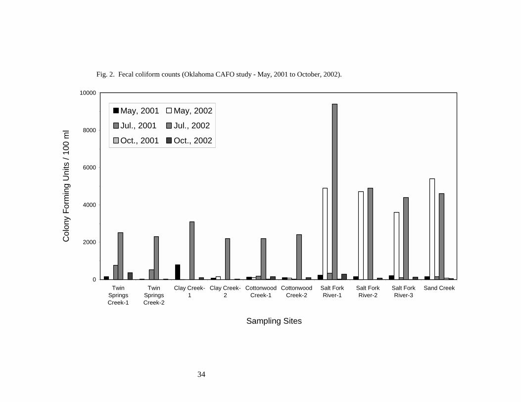
Fecal coliforms. Fecal coliforms are a designated subgroup of bacteria closely associated
with the gut and intestinal tracts of warm-blooded animals, including humans. The presence of fecal
coliforms in water serves as a general indicator of fecal contamination, and detection and
quantification of these organisms is based on standardized methodology (28). Fecal coliform counts
were conducted as part of this study to provide an assessment of bacterial contamination from fecal
sources on the days samples were collected. The US EPA recommends that fecal coliform density
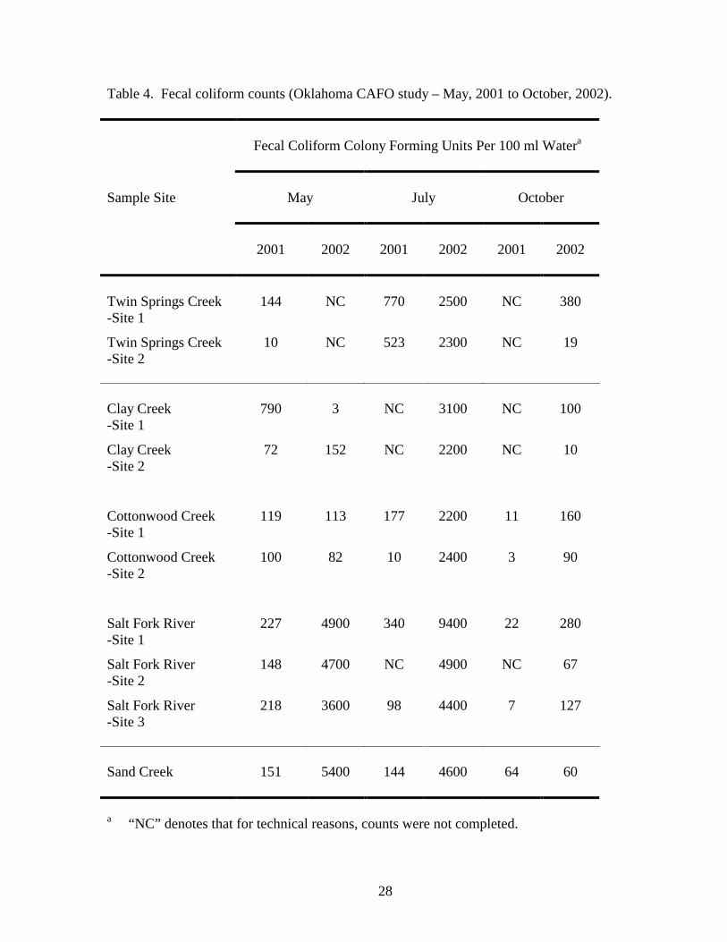
not exceed 200 CFU/100 ml of water for human recreational bathing purposes (10). As summarized
in Table 4 and Fig. 2, of 50 measurements taken over two years, 28 measurements met human
recreational bathing water criteria. Elevated coliform counts were observed in all samples collected
11
in July, 2002 and may have resulted from a wide-spread rain storm and associated run off that
proceeded sampling. Additionally, warm temperatures during the month of July would have been
favorable for coliform growth. Elevated coliform counts were also observed in May, 2002 from
samples collected at Salt Fork River and Sand Creek. May sampling took place following a smaller
rainfall event, and run off in the northern portion of the refuge was evident as elevated amounts of
sediment were present in Salt Fork River and Sand Creek. It is possible that a cattle CAFO located
upstream of the Salt Fork River sampling sites may have been a contributing factor to the elevated
coliform levels observed at these sites.
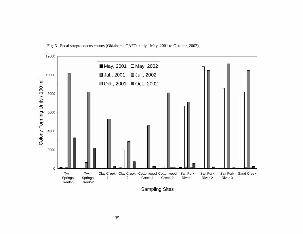
Fecal streptococci. Fecal streptococci consist of a number of species from the genera
Streptococcus and Enterococcus. The normal habitat of fecal streptococci is similar to that of fecal
coliforms, the gastrointestinal tract of warm-blooded animals, including humans (28). The US EPA
recommends that for human recreational bathing purposes, fecal streptococci not exceed a density of
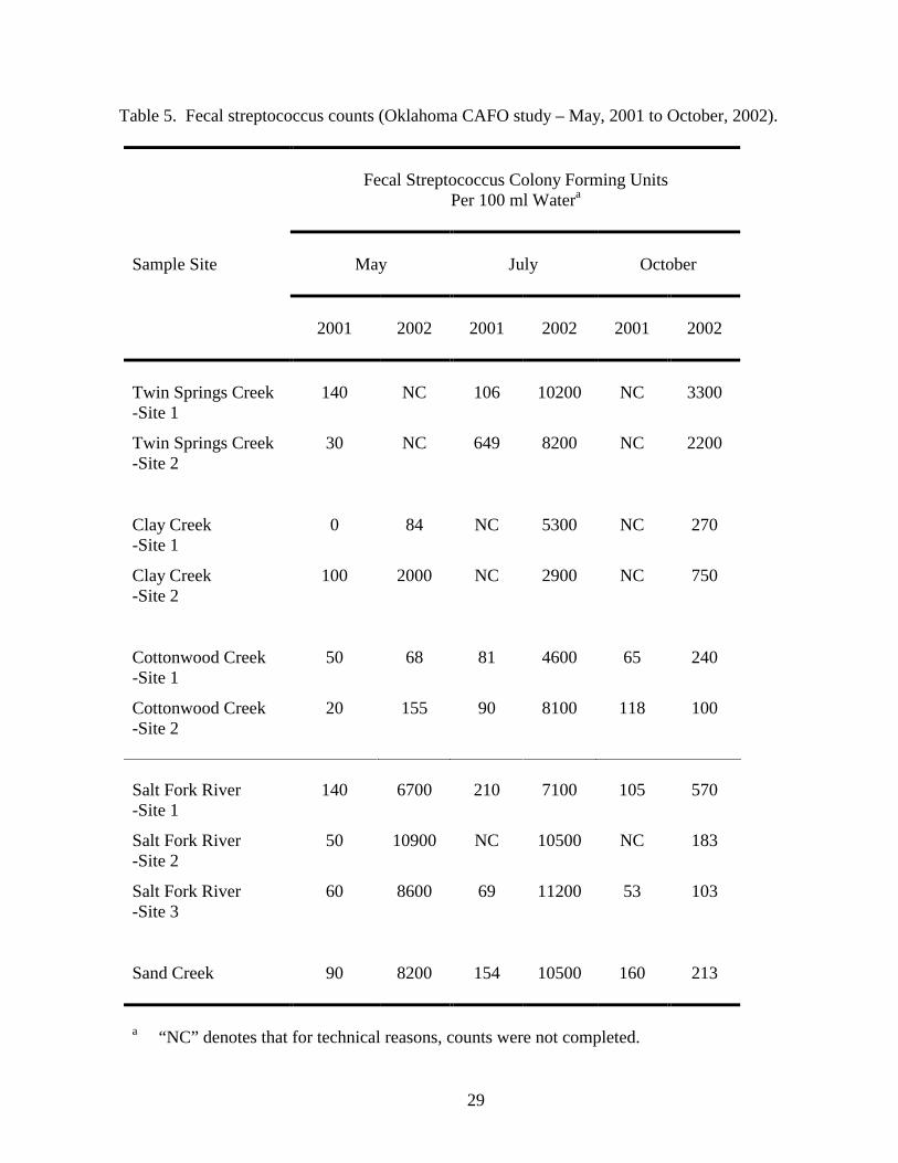
33 CFU/100 ml of fresh water (10). Only 3 of the 50 measurements reported in Table 5 and Fig. 3
met these criteria. Similar to what was observed with the fecal coliforms, elevated fecal
streptococcus counts were noted at all sampling sites in July, 2002 and at the Salt Fork River and
Sand Creek sampling sites in May, 2002. These elevated counts may have been caused by
previously-noted rainfall and resultant run off that occurred prior to sampling. Elevated fecal
streptococcus counts were also observed at Clay Creek site two in May 2002 and at the Twin
Springs Creek sampling sites in October, 2002. There is a hog CAFO upstream of the Twin Springs
Creek sampling sites. It is possible that this CAFO may have contributed to elevated fecal
streptococcus counts at these locations.
Clostridium botulinum type C. C. botulinum type C, the causative agent of avian botulism,
has significant annual impacts on waterfowl populations. Botulism occurs in wildlife following
12
ingestion of botulinum toxin, and the ability of C. botulimum to produce resistant spores allows it to
persist in the environment for extended periods of time (36). C. botulinum type C genetic material
was not detected in sediment samples by PCR. Control experiments indicated that the procedures
utilized may not have been fully effective in identifying C. botulinum genetic material. Future
efforts will be directed towards optimizing both the procedure for extracting genetic material from
sediment and the PCR protocol utilized.
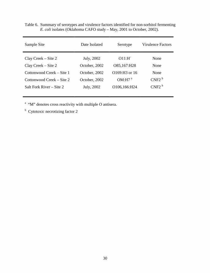
Toxigenic Escherichia coli. Toxigenic (non-sorbitol fermenting) E. coli O157:H7 has been
implicated as the causative agent of hemorrhagic colitis in humans (26). The principal reservoir for
E. coli O157:H7 is cattle (35), and this organism has also been isolated from wild deer (2). E. coli
O157:H7 can survive up to 21 months outside of an animal host (24). Non-sorbitol fermenting E.
coli were not recovered in 2001, but 5 isolates were recovered in 2002 (Table 6). None of the non-
sorbitol fermenting isolates belonged to the O157:H7 serotype, however 2 of the 5 isolates possessed
the gene encoding cytotoxic necrotizing factor 2 (CNF2). CNF2 has been implicated as a virulence
factor in E. coli strains that cause enteric and extraintestinal infections (30), and necrotoxigenic E.
coli strains that produce CNF2 have been recovered from the intestinal tracts of domestic animals
including cattle (4, 5, 13).
Erysipelothrix. Erysipelothrix spp. have been isolated from healthy and diseased
mammalian, avian, and amphibian species (32, 33). Major mortality events in eared grebes at the
Great Salt Lake in Utah (21) and in brown pelicans off the California coast (USGS-National Wildlife
Health Center, unpublished) have been attributed to Erysipelothrix. Erysipelothrix isolates were not
recovered during the course of this study.
Pasteurella multocida. P. multocida is the causative agent of avian cholera in wild and
domestic fowl (32). In animals, P. multocida infections are often acute, resulting in death within 6
13
to 12 hours (18). P. multocida can also be carried latently by birds, causing disease under conditions
of stress (32). P. multocida has world wide distribution, and in some habitats, avian cholera
outbreaks occur among waterfowl on an annual basis.
To evaluate the environmental prevalence of P. multocida within the Salt Plains National
Wildlife Refuge, attempts were made to cultivate this organism from sediment samples taken at ten
sites within and surrounding the refuge. Consistent with the observation that avian cholera has not
previously been a problem at the Salt Plains National Wildlife Refuge, P. multodica was not
cultivated from samples collected during this study.
Salmonella. Salmonella spp. are divided into six subgroups encompassing over 2000
different serotypes (31). The natural reservoir for salmonellae is the intestinal tract of both warm-
and cold-blooded animals, and the majority of infected animals are subclinically ill excretors.
Salmonella spp. can survive for up to 7 months outside of an animal host under certain
environmental conditions (6). Among wildlife, salmonellosis can cause large-scale mortality events,
especially in the young of colony nesting bird species (19).
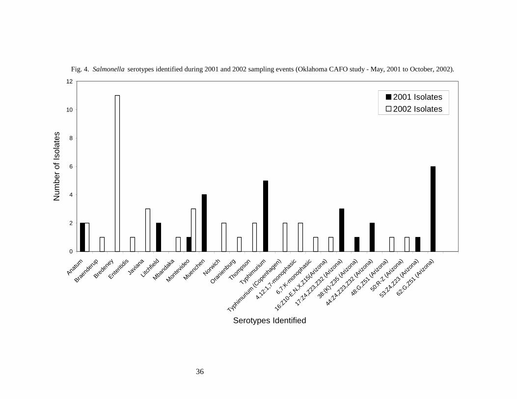
Twenty-seven Salmonella isolates were recovered during the three 2001 sampling events,
and 35 isolates were recovered over three sampling events in 2002 (Fig. 4). The greatest number of
Salmonella serotypes identified in 2001 (59%) were isolated in May from the Twin Springs Creek,
Cottonwood Creek, Salt Fork River, and Sand Creek sample sites. This does not correspond to the
relatively low fecal coliform and fecal streptococcus counts obtained during this same sampling
event (Figs. 2 and 3). The majority of Salmonella serotypes identified in 2002 (56%) were isolated
in July, during which high fecal coliform and fecal streptococcus counts were also observed
throughout the refuge. Thirty nine percent of the Salmonella serotypes identified in 2002 were
isolated in May. These isolates were obtained from the Cottonwood Creek, Salt Fork River, and
14
Sand Creek sample sites concurrent with the observation of elevated fecal coliform and fecal
streptococcus counts. In two out of three instances, increased frequency of Salmonella isolation
corresponded with elevated levels of fecal indicator bacteria at refuge sample sites that may have
resulted from run off following rain events. Thus, the associations between surface water run off
and increased levels of fecal coliforms, fecal streptococci, and salmonellae should be investigated
further to evaluate potential adverse impacts to the refuge environment.
The Salmonella serotypes most frequently identified in 2001 were 62:G,Z51(Arizona),
represented by 6 isolates, Typhimurium, represented by 5 isolates, and Muenchen, represented by 4
isolates. The serotype most frequently identified in 2002 was Salmonella Bredeney, isolated 11
times (Fig. 4). Serotype 62:G,Z51(Arizona) tends to be associated with cold-blooded animals
(USDA-National Veterinary Services Laboratories, unpublished) and is thus not likely to have
originated from CAFO sites. Salmonella Typhimurium is commonly isolated from domestic
livestock, including cattle, swine, horses, sheep, chickens, and turkeys, exhibiting the highest
prevalence in cattle and horses (17). Serotype Typhimurium is also frequently isolated from wild
bird tissues analyzed by the USGS-National Wildlife Health Center diagnostic microbiology
laboratory (unpublished data). Serotype Muenchen has previously been isolated from various crane
species by the USGS-National Wildlife Health Center diagnostic microbiology laboratory
(unpublished data), and serotype Bredeney is commonly found in domestic turkey flocks (17). Thus,
the Salmonella serotypes isolated within the Salt Plains National wildlife refuge are representative of
those that colonize both wild and domestic animals.
Yersinia. The genus Yersinia contains 11 species that are endemic to a variety of animals,
including wild rodents, wild birds, and domestic pigs. Y. enterocolitica is found in surface waters
and can cause epizootic outbreaks of diarrhea, pneumonia, and spontaneous abortion (22). Yersinia
15
isolates were not recovered from environmental samples collected during this study.
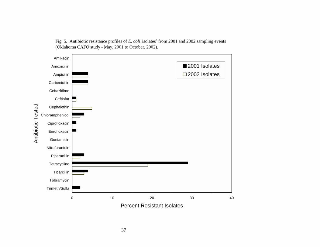
Antibiotic resistance profiles. Antibiotic resistance profiles were determined for a subset of
E. coli isolates obtained from the fecal coliform bacteria enumerated within the refuge. One-
hundred twenty-seven and 131 E. coli isolates obtained in 2001 and 2002, respectively, were
screened for resistance against a panel of 17 antibiotics (Fig. 5). Of the 2001 isolates, 39 (31%)
were resistant to at least one antibiotic, and seven (18%) of the resistant isolates were resistant to two
or more antibiotics. Analysis of the 2002 isolates revealed that 29 (22%) were resistant to at least
one antibiotic. Ten (34%) of the resistant isolates were resistant to two or more antibiotics. The
antibiotic to which the greatest number of E. coli isolates exhibited resistance was tetracycline.
Twenty nine percent of 2001 isolates and 19% of 2002 isolates were resistant to tetracycline,
whereas 5% or fewer of the isolates were resistant to each of the other antibiotics tested.
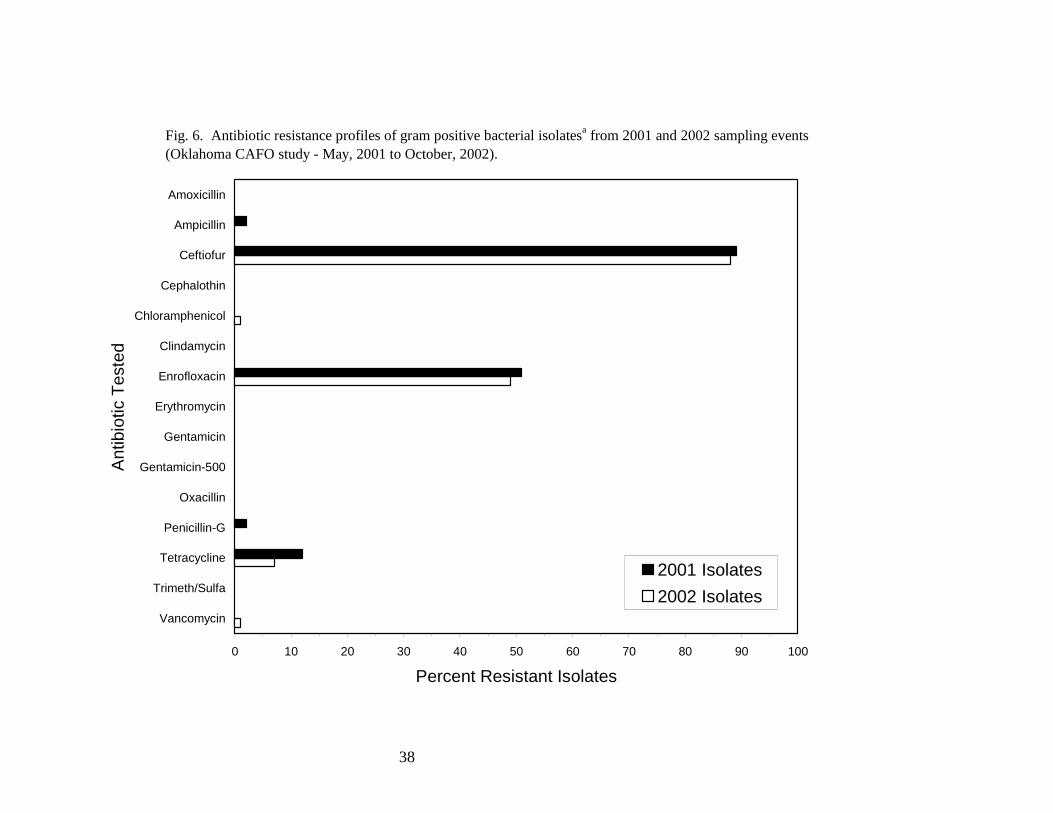
Resistance profiles against a panel of 15 antibiotics were determined for a subset of the fecal
streptococcus isolates enumerated within the refuge (Fig. 6). The isolates tested were identified to
the species level and included E. avium, E. casseliflavus, E. durans, E. faecalis, E. faecium, and E.
gallinarum. Of the 2001 isolates (110 screened), 107 (97%) were resistant to at least one antibiotic,
and 57 (53%) of the resistant isolates were resistant to two or more antibiotics. In 2002, 119 (95%)
of the 125 isolates analyzed were resistant to at least one antibiotic, and 63 (53%) of the resistant
isolates were resistant to two or more antibiotics. Antibiotics to which the gram positive isolates
were most frequently resistant included ceftiofur (≈90% resistant) and enrofloxacin (≈50% resistant).
Twelve percent and 7% of the 2001 and 2002 isolates, respectively, were resistant to tetracycline;
2% or fewer of the isolates were resistant to each of the other antibiotics tested.
Antibiotics are used in animal agriculture to promote animal growth and to prevent and treat
16
disease (8). The use of antibiotics in agriculture for prophylactic and growth-promoting purposes
has been implicated in the evolution and widespread dissemination of antibiotic resistant bacteria
throughout the environment (7). The development of antibiotic resistance by commensal enteric
bacteria, including E. coli and various enterococci that colonize the intestinal tracts of both animals
and humans provides an avenue for the transmission of antibiotic resistant bacteria from domestic
animals into humans (8). As bacterial infections of wild animals are not routinely treated with
antibiotics, antibiotic resistant bacteria do not likely pose a direct threat to wildlife health. However,
due to the free-ranging nature of wild animals, especially migratory waterfowl that inhabit the Salt
Plains National Wildlife Refuge, wildlife may facilitate the transfer of antibiotic resistant bacteria
throughout the environment.
In this study, antibiotics to which the bacteria tested most frequently exhibited resistance
included tetracycline, ceftiofur, and enrofloxacin. Each of these antibiotics belongs to a different
class of pharmaceutical agents and exerts its clinical effects through a unique mechanism of action
(8, 20, 39). Thus, the physiological mechanisms that confer bacterial resistance to each antibiotic
are also distinct and likely arose independently in response the selective pressure exhibited by each
antibiotic.
Tetracycline was the most common antibiotic to which E. coli strains exhibited resistance
and the third most common antibiotic to which gram positive isolates were resistant. Tetracycline is
commonly used in the swine industry, and in the United States, farm animals were treated with
3,488,000 kg of tetracycline per year during the 1990s (7, 8). Among gram positive bacteria
analyzed, almost 90% of strains tested were resistant to ceftiofur. Ceftiofur is used worldwide in
veterinary medicine to treat respiratory diseases in swine, ruminants, and horses and to treat foot rot
17
and metritis infections in cattle (20). Also among the gram positive isolates analyzed, approximately
50% were resistant to enrofloxacin, another commonly used veterinary antibiotic (39, 42). Thus, the
identification of bacteria resistant to antibiotics commonly used in veterinary medicine in refuge
water samples suggests that agricultural practices in areas surrounding the Salt Plains National
Wildlife Refuge may have contributed to the dissemination of these organisms within the refuge.
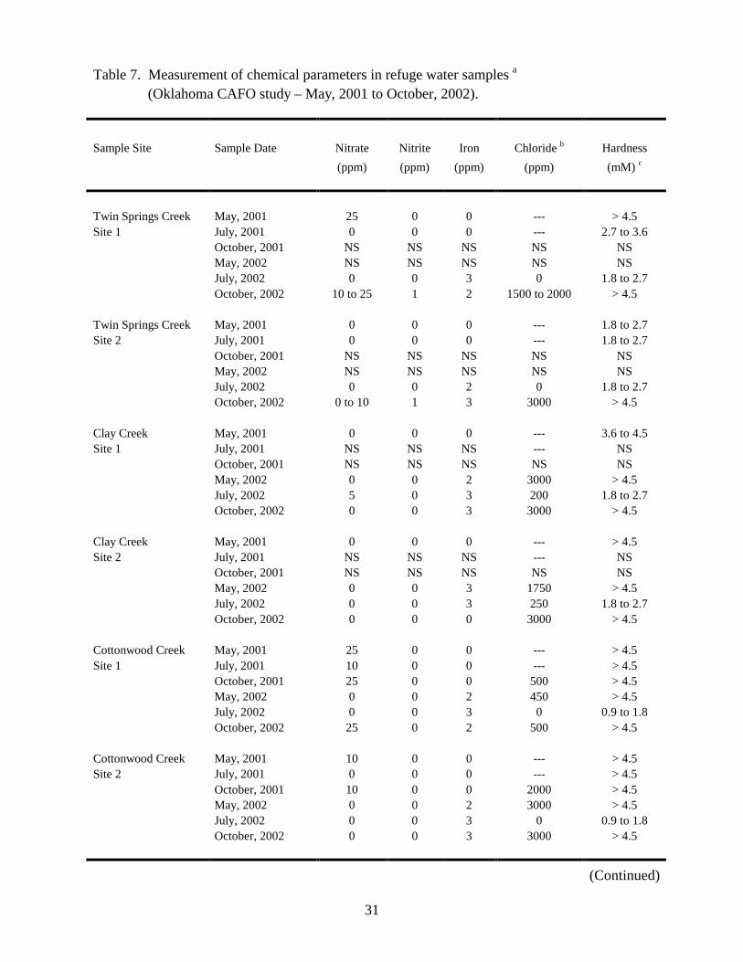
Chemical analyses. Nitrate, nitrite, iron, and chloride concentrations, as well as total water
hardness levels, were measured in water samples collected throughout the refuge (Table 7). Of the
chemical parameters analyzed, nitrogen levels (measured as nitrate and nitrite) have the greatest
potential to impact refuge water quality. Sources that contribute to environmental nitrogen levels
include the natural decay of organisms, animal feces, and agricultural fertilizer. EPA limits for
nitrogen levels in drinking water are set at 10 ppm for nitrate and 1 ppm for nitrite (10). Nitrate
concentrations of 10 ppm or higher were detected in ten water samples obtained from Twin Springs
Creek, Cottonwood Creek, and the Salt Fork River (Table 7). Nitrite concentrations of 1 ppm or
greater were detected in four samples collected from Twin Springs Creek and the Salt Fork River
(Table 7). There are CAFOs located upstream of the Twin Springs Creek and Salt Fork River
sampling sites, and it is possible that they may have contributed to elevated nitrogen levels observed
at these sites.
CONCLUSIONS
The main goals of this study were to assess levels of bacterial indicators of fecal
contamination within Salt Plains National Wildlife Refuge surface water and to identify known
bacterial pathogens of wildlife, especially waterfowl, within refuge water and sediment samples.
Fecal indicator bacteria frequently exceeded US EPA guidelines for human recreational bathing
18
waters within the refuge, and elevated bacterial counts observed in May and July, 2002 correlated
with rainfall that occurred prior to sampling (Figs. 3 and 4; Tables 4 and 5). High levels of fecal
indicator bacteria were observed in Salt Fork River and Twin Springs Creek sampling sites. These
sites are downstream of noted cattle and hog CAFOs, respectively (Fig. 1). Counts in excess of US
EPA human recreational bathing limits, however, were measured at least once at every sample site.
Laboratory efforts to experimentally trace the bacteria identified in this study to agricultural sources
were not conducted.
Antibiotic resistance profiles were characterized for representative isolates of fecal indicator
bacteria (Figs. 5 and 6). These analyses revealed that high percentages of bacteria of fecal origin
isolated from refuge surface water were resistant to antibiotics commonly used in veterinary
medicine, including tetracycline, ceftiofur, and enrofloxacin. The threat that antibiotic-resistant
bacteria may pose to wildlife has not been characterized, however it is well established that the
emergence of resistance in human and veterinary pathogens has exerted significant consequences for
the therapeutic use of antibiotics (8).
Erysipelothrix spp., P. multocida, and Yersinia spp., known pathogens of wildlife, were not
identified during the course of this study. This is consistent with the observation that these
organisms have not previously been implicated in disease outbreaks among wildlife within the Salt
Plains National Wildlife Refuge (USGS–National Wildlife Health Center, unpublished). Continued
nutrient loading of the refuge watershed may, however, increase the probability of these pathogens
causing future problems to wildlife within the refuge.
Salmonella spp. were isolated from all sample sites within the refuge. Elevated isolation
frequencies of salmonellae correlated with rainfall-induced run off, and nutrient inputs from CAFOs
or other sources may enhance the growth of these organisms within the refuge watershed. The
19
presence of salmonellae within refuge surface water poses a potential pathogenic threat both to
wildlife utilizing these habitats and to humans visiting the refuge. Necrotoxigenic E. coli, similar to
strains previously noted to be associated with cattle (4, 5), were also isolated from two sample sites
within the refuge (Table 6). The pathogenicity of necrotoxigenic E. coli to wildlife has not been
characterized, but these organisms can cause infections in humans (30).
RECOMMENDATIONS
While this study demonstrated that antibiotic resistant bacteria of fecal origin, potentially
pathogenic Salmonella spp., and necrotoxigenic E. coli were present in refuge surface waters, the
direct threat that these organisms pose to wildlife within the refuge is not known. Sentinel mallard
flocks could be utilized to assess the potential of waterfowl to become infected by these organisms
and to shed them throughout the environment. Additionally, the role that small mammals inhabiting
the refuge may play in disseminating bacteria throughout the environment is not known. Surveys to
address the diversity and dispersal patterns of wild mammals within the refuge would provide a
starting point for addressing this question. It would also be worthwhile to expand chemical and
toxicological monitoring efforts in the refuge to measure nutrient levels in refuge surface waters and
to assess possible bioaccumulation of toxic compounds within fish, amphibian, and invertebrate
populations. As highlighted by the 1998 fish kill in Spring Creek, potential environmental quality
problems within the refuge watershed may first manifest within these groups of sensitive organisms.
As many of the bacteria isolated during the course of this study possess attributes that
suggest they originated from agricultural sites, CAFOs adjacent to the refuge may be sources of
these pollutants. The work described herein does not, however, experimentally link bacteria isolated
within the refuge to agricultural sources. Short of employing bacterial source tracking techniques
20
(25) to experimentally trace these organisms to specific point sources, less involved follow-up
studies could be done. For example, levels of fecal indicator bacteria could be reassessed both
upstream and downstream of the Twin Springs Creek and Salt Fork River CAFOs, sites from which
high levels of fecal indicator bacteria were observed. If fecal indicator bacteria originate from these
sites, decreasing gradients of indicator organisms should be observed moving downstream from the
CAFOs, and lower levels of fecal indicator bacteria should be present upstream of the sites than
downstream.
By educating the surrounding community about the health implications of fecal
contamination of refuge surface waters and by emphasizing the consequences of widespread
environmental dissemination of antibiotic resistant bacteria, perhaps the community will appeal to
CAFO operators to voluntarily improve their waste disposal practices. Changes such as ensuring
that waste lagoons do not overflow following rainstorms, refraining from applying manure to
saturated ground, keeping animals out of streams, and maintaining buffer zones around streams
during land application of manure may lead to significant improvements in refuge water quality.
QUALITY ASSURANCE
Planning, data collection, and analysis of information under this contract were completed
with the technical care necessary to meet the needs for valid and high quality data. All work done
during the contract followed the tenants of Good Laboratory Procedures (GLP) as described in 40
CFR 160 or as institutional policy provided. References and internal quality controls were included
when possible, and data were validated with respect to their feasibility and according to the
investigators’ experience.
21
ACKNOWLEDGEMENTS
This study was funded under intragovernmental agency agreement #1448-20181-01-N752
with the US Fish and Wildlife Service Division of Ecological Services, Tulsa, Oklahoma. We would
like to thank M. Schneider (USGS-Water Resources Division) for his assistance conducting field
work. We acknowledge M. Moore and S. Taylor (USGS-National Wildlife Health Center) for
providing technical assistance, including preparation of media. We also thank the Salt Plains
National Wildlife Refuge for providing field facilities and equipment to facilitate sample collection
and preliminary analyses.
DISCLAIMER
Mention of trade names or commercial products does not constitute endorsement or
recommendation for use.
REFERENCES
1. Agbonlahor, D. E., T. Odugbemi, and O. Dosunmu-Ogunbi. 1982. Differential and Selective Medium for Isolation of Yersinia enterocolitica from Stools. Journal of Clinical Microbiology. 15:599-602.
2. Asakura, H., S. Makino, T. Shirahata, T. Tsukamoto, H. Kurazono, T. Ikeda, and K. Takeshi. 1998. Detection and Genetical Characterization of Shiga Toxin-Producing Escherichia coli from Wild Deer. Microbiol Immunol. 42:815-822.
3. Atlas, R. M. 1993. Handbook of Microbiological Media. CRC Press, Inc., Boca Raton, FL.
4. Blanco, M., J. E. Blanco, A. Mora, and J. Blanco. 1998. Distribution and Characterization of Faecal Necrotoxigenic Escherichia coli CNF1+ and CNF2+ Isolated from Healthy Cows and Calves. Vet. Microbiol. 59:183-192.
5. Blanco, M., J. E. Blanco, A. Mora, and J. Blanco. 1998. Prevalence and Characteristics of Necrotoxigenic Escherichia coli CNF1+ and CNF2+ in Healthy Cattle. Res. Microbiol. 149:47-53.
6. Blood, D. C., O. M. Radostits, and J. A. Henderson. 1983. Diseases Caused by Salmonella
22
spp., p. 576-588. In Veterinary Medicine: A Textbook of the Diseases of Cattle, Sheep, Pigs, Goats, and Horses. Bailliere-Tindale, London.
7. Chee-Sanford, J. C., R. I. Aminov, I. J. Krapac, N. Garrigues-Jeanjean, and R. I. Mackie. 2001. Occurrence and Diversity of Tetracycline Resistance Genes in Lagoons and Groundwater Underlying Two Swine Production Facilities. Applied and Environmental Microbiology. 67:1494-502.
8. Chopra, I. and M. Roberts. 2001. Tetracycline Antibiotics: Mode of Action, Applications, Molecular Biology, and Epidemiology of Bacterial Resistance. Microbiology and Molecular Biology Reviews. 65:232-260.
9. Cleaveland, S., M. K. Laurenson, and L. H. Taylor. 2001. Diseases of Humans and their Domestic Mammals: Pathogen Characteristics, Host Range, and the Risk of Emergence. Phil. Trans. R. Soc. Lond. B. 356:991-999.
10. Clesceri, L. S., A. E. Greenberg, and A. D. Eaton. 1998. Standard Methods for the Examination of Water and Wastewater. American Public Health Association, Washington, D.C.
11. Daszak, P., A. A. Cunningham, and A. D. Hyatt. 2000. Emerging Infectious Diseases of Wildlife: Threats to Biodiversity and Human Health. Science. 287:443-449.
12. De Smedt, J. and R. Bolderdijk. 1987. Dynamics of Salmonella Isolation with Modified Semi-solid Rappaport-Vassiliadis Medium. Journal of Food Protection. 50:658-661.
13. DebRoy, C. and C. W. Maddox. 2001. Identification of Virulence Attributes of Gastrointestinal Escherichia coli Isolates of Vetrinary Significance. Animal Health Research Reviews. 2:129-140.
14. EPA-821-03-001. EPA Administered Permit Programs: The National Pollutant Discharge Elimination System. Code of Federal Regulations, Part 122. U.S. Environmental Protection Agency, Washington, D.C.
15. EPA-821-F-03-003. 2003. NPDES Permit Regulation and Effluent Limitations Guidelines for Concentrated Animal Feeding Operations. U.S. Environmental Protection Agency, Washington, D.C.
16. EPA-841-R-02-001. National Water Quality Inventory: 2000 Report. U.S. Environmental Protection Agency, Washington, D.C.
17. Ferris, K. 1997. DxMonitor Animal Health Report (Winter 1996-Spring 1997). National Veterinary Services Laboratories' Annual and Quarterly Salmonella Reports. Centers for Epidemiology and Animal Health, Fort Collins, CO.
18. Friend, M. 1987. Field Guide to Wildlife Disease. U.S. Department of the Interior, Washington, D.C.
23
19. Friend, M. and J. C. Franson. 1999. Field guide to wildlife disease. General Field Procedures and Diseases of Birds. U.S. Department of the Interior, Washington, D.C.
20. Hornish, R. E. and S. F. Kotarski. 2002. Cephalosporins in Veterinary Medicine - Ceftiofur Use in Food Animals. Current Topics in Medicinal Chemistry. 2:717-731.
21. Jensen, W. I. and S. E. Cotter. 1976. An Outbreak of Erysipelas in Eared Grebes (Podiceps nigricollis). Journal of Wildlife Diseases. 12:583-586.
22. Koneman, E. W., S. D. Allen, W. M. Janda, P. C. Schreckenberg, and W. C. Winn, Jr. 1997. Color Atlas and Textbook of Diagnostic Microbiology. Lippincott, Philadelphia.
23. Kristensen, M., V. Lester, and A. Jurgens. 1925. On the Use of Trypsinized Casein, Bromthymol Blue, Bromcresol Purple, Phenol Red, and Brilliant Green for Bacteriological Nutrient Media. British Journal of Experimental Pathology. 6:291-299.
24. Kudva, I. T., K. Blanch, and C. J. Hovde. 1998. Analysis of Escherichia coli O157:H7 Survival in Ovine or Bovine Manure and Manure Slurry. Applied and Environmental Microbiology. 64:3166-3174.
25. Malakoff, D. 2002. Is E. coli Distinct Enough to Join the Hunt. Science. 295:2353.
26. March, S. B. and S. Ratnam. 1986. Sorbitol-MacConkey Medium for the Detection of Escherichia coli O157:H7 Associated with Hemorrhagic Colits. Journal of Clinical Microbiology. 23:869-872.
27. Miller, R. G., C. R. Tate, E. T. Mallinson, and J. A. Scherrer. 1991. Xylose-Lysine-Tergitol 4: An Improved Selective Agar Medium for the Isolation of Salmonella. Poultry Science. 70:2429-2432.
28. Moe, C. L. 1997. Waterborne Transmission of Infectious Agents, p. 136-152. In C. J. Hurst (ed.), Manual of Environmental Microbiology. ASM Press, Washington, D.C.
29. Moore, M. K., L. Cicnjak-Chubbs, and R. J. Gates. 1994. A New Selective Enrichment Prodedure for Isolating Pasteurella multocida from Avian and Environmental Samples. Avian Diseases. 38:317-324.
30. Oswald, E., M. Sugai, A. Labigne, H. C. Wu, C. Fiorentini, P. Boquet, and A. D. O'Brien. 1994. Cytotoxic Necrotizing Factor Type 2 Produced by Virulent Escherichia coli Modifies the Small GTP-Binding Proteins Rho Involved in Assembly of Actin Stress Fibers. Proceedings of the National Academy of Sciences, USA. 91:3814-3818.
31. Popoff, M. Y. and L. Le Minor. 1997. Antigenic Formulas of the Salmonella Serovars. WHO Collaborating Centre for Reference and Research on Salmonella, Paris, France.
32. Quinn, P. J., M. E. Carter, B. Markey, and G. R. Carter. Clinical veterinary microbiology. Wolfe Publishing, Spain.
24
33. Reboli, A. C. and W. E. Farrar. 1989. Erysipelothrix rhusiopathiae: An Occupational Pathogen. Clin. Microbiol. Rev. 2:354-359.
34. Schiemann, D. A. 1982. Development of a Two-Step Enrichment Procedure for Recovery of Yersinia enterocolitica from Food. Applied and Environmental Microbiology. 43:14-27.
35. Shere, J. A., K. J. Bartlett, and C. W. Kaspar. 1998. Longitudinal Study of Escherichia coli O157:H7 Dissemination on Four Dairy Farms in Wisconsin. Applied and Environmental Microbiology. 64:1390-1399.
36. Songer, J. G. 1997. Clostridial Diseases of Animals, p. 153-182. In J. I. Rood, B. A. McClane, J. G. Songer, and R. W. Titball (eds.), The Clostridia: Molecular Biology and Pathogenesis. Academic Press, San Diego, CA.
37. Strauch, D. 1991. Survival of Pathogenic Microorganisms and Parasites in Excreta, Manure and Sewage Sludge. Revue Scientifique et Technique Office International des Epizooties. 10:813-846.
38. U.S. Environmental Protection Agency. 2003. National Pollutant Discharge Elimination System Permit Regulation and Effluent Limitation Guidelines and Standards for Concentrated Animal Feeding Operations (CAFOs); Final Rule. Federal Register. 68:7176-7184.
39. Vancutsem, P. M., J. G. Babish, and W. S. Schwark. 1990. The Fluoroquinolone Antimicrobials: Structure, Antimicrobial Activity, Pharmacokinetics, Clinical Use in Domestic Animals, and Toxicity. Cornell Vet. 80:173-186.
40. Vassiliadis, P. 1983. The Rappaport-Vassiliadis (RV) Enrichment Medium for the Isolation of Salmonellas: An Overview. Journal of Applied Bacteriology. 54:69-76.
41. Williamson, J. L., T. E. Rocke, and J. M. Aiken. 1999. In Situ Detection of the Clostridium botulinum Type C1 Toxin Gene in Wetland Sediments with a Nested PCR Assay. Applied and Environmental Microbiology. 65:3240-3243.
42. Wiuff, C., J. Lykkesfeldt, O. Svendsen, and F. M. Aarestrup. 2003. The Effects of Oral and Intramuscular Administration and Dose Escalation of Enrofloxacin on the Selection of Quinolone Resistance Among Salmonella and Coliforms in Pigs. Research in Veterinary Science. 75:185-193.
25
Table 1. GPS coordinates of sampling sites (Oklahoma CAFO study – May, 2001 to October, 2002). Sample Site
Coordinates
Twin Springs Creek – Site 1
N36°40.853’ W098°13.500’
Twin Springs Creek – Site 2 N36°41.045’ W098°13.200’
Clay Creek – Site 1 N36°42.984’ W098°17.048’
Clay Creek – Site 2 N36°43.924’ W098°15.807’
Cottonwood Creek – Site 1 N36°45.222’ W098°18.436’
Cottonwood Creek – Site 2 N36°45.183’ W098°17.265’
Salt Fork River – Site 1 N36°49.201’ W098°21.607’
Salt Fork River – Site 2 N36°47.577’ W098°14.442’
Salt Fork River – Site 3 N36°46.423’ W098°13.287’
Sand Creek N36°48.662’ W098°12.046’
26
Table 2. Summary of bacterial culturing methodology (Oklahoma CAFO study – May, 2001 to October, 2002). Organism
Sample Type
Primary Medium a
Primary Incubation
Secondary Medium a
Secondary Incubation
fecal coliforms
water
m-Coliblue24®
18-24 h; 44.5°
BAP
18-24 h; 37°C
fecal streptococci water KF 46-48 h; 35°C BAP 18-24 h; 37°C
Escherichia coli water EcEB 18-24 h; 37°C SMAC 18-24 h; 37°C sediment EcEB 18-24 h; 37°C SMAC 18-24 h; 37°C
Erysipelothrix spp. water BHIB/S 18-24 h; 37°C BHIA/S 18-24 h; 37°C water Packer’s Medium 18-24 h; 37°C BAP 18-24 h; 37°C sediment BHIB/S 18-24 h; 37°C BHIA/S 18-24 h; 37°C sediment Packer’s Medium 18-24 h; 37°C BAP 18-24 h; 37°C
Pasteurella multocida
sediment PMSB 18-24 h; 37°C; CO2 BAP 18-24 h; 37°C
Salmonella spp. water DS 18-24 h; 41.5°C BG and XLT4 18-24 h; 37°C water RV 18-24 h; 41.5°C BG and XLT4 18-24 h; 37°C sediment DS 18-24 h; 41.5°C BG and XLT4 18-24 h; 37°C sediment RV 18-24 h; 41.5°C BG and XLT4 18-24 h; 37°C
Yersinia spp. water BOS 1 or 5 d; 23°C CIN 18-24 h; 32°C DYS 36-48 h; 23°C sediment BOS 1 or 5 d; 23°C CIN 18-24 h; 32°C DYS 36-48 h; 23°C
a Abbreviations defined in Table 3.
27
Table 3. Growth medium abbreviations (Oklahoma CAFO study – May, 2001 to October, 2002).
Medium
Abbreviation
Bile Oxalate Sorbose
BOS
5 % Sheep Blood Agar Plate BAP
Brain Heart Infusion BHI
Brain Heart Infusion Agar, Selective BHIA/S
Brain Heart Infusion Broth, Selective BHIB/S
Brilliant Green BG
Cefsulodin Irgasan Novobiocin CIN
Dulcitol-Selenite DS
Differential Yersinia Selective DYS
Escherichia coli Enrichment Broth EcEB
KF Streptoccal KF
MacConkey’s Medium with Sorbitol SMAC
Pasteurella multocida Selective Broth PmSB
Rappaport-Vassiliadis RV
Xylose Lysine Tergitol 4 XLT4
28
Table 4. Fecal coliform counts (Oklahoma CAFO study – May, 2001 to October, 2002).
Fecal Coliform Colony Forming Units Per 100 ml Watera
Sample Site
May
July
October
2001
2002
2001
2002
2001
2002
Twin Springs Creek -Site 1
144
NC
770
2500
NC
380
Twin Springs Creek -Site 2
10
NC
523
2300
NC
19
Clay Creek -Site 1
790
3
NC
3100
NC
100
Clay Creek -Site 2
72
152
NC
2200
NC
10
Cottonwood Creek -Site 1
119
113
177
2200
11
160
Cottonwood Creek -Site 2
100
82
10
2400
3
90
Salt Fork River -Site 1
227
4900
340
9400
22
280
Salt Fork River -Site 2
148
4700
NC
4900
NC
67
Salt Fork River -Site 3
218
3600
98
4400
7
127
Sand Creek
151
5400
144
4600
64
60
a “NC” denotes that for technical reasons, counts were not completed.
29
Table 5. Fecal streptococcus counts (Oklahoma CAFO study – May, 2001 to October, 2002).
Fecal Streptococcus Colony Forming Units Per 100 ml Watera
Sample Site
May
July
October
2001
2002
2001
2002
2001
2002
Twin Springs Creek -Site 1
140
NC
106
10200
NC
3300
Twin Springs Creek -Site 2
30
NC
649
8200
NC
2200
Clay Creek -Site 1
0
84
NC
5300
NC
270
Clay Creek -Site 2
100
2000
NC
2900
NC
750
Cottonwood Creek -Site 1
50
68
81
4600
65
240
Cottonwood Creek -Site 2
20
155
90
8100
118
100
Salt Fork River -Site 1
140
6700
210
7100
105
570
Salt Fork River -Site 2
50
10900
NC
10500
NC
183
Salt Fork River -Site 3
60
8600
69
11200
53
103
Sand Creek
90
8200
154
10500
160
213
a “NC” denotes that for technical reasons, counts were not completed.
30
Table 6. Summary of serotypes and virulence factors identified for non-sorbitol fermenting E. coli isolates (Oklahoma CAFO study – May, 2001 to October, 2002). Sample Site
Date Isolated
Serotype
Virulence Factors
Clay Creek – Site 2
July, 2002
O11:H-
None
Clay Creek – Site 2 October, 2002 O85,167:H28 None
Cottonwood Creek – Site 1 October, 2002 O169:H3 or 16 None
Cottonwood Creek – Site 2 October, 2002 OM:H7 a CNF2 b
Salt Fork River – Site 2 July, 2002 O106,166:H24 CNF2 b
a “M” denotes cross reactivity with multiple O antisera. b Cytotoxic necrotizing factor 2
31
Table 7. Measurement of chemical parameters in refuge water samples a (Oklahoma CAFO study – May, 2001 to October, 2002). Sample Site Sample Date Nitrate Nitrite Iron Chloride b Hardness (ppm) (ppm) (ppm) (ppm) (mM) c Twin Springs Creek May, 2001 25 0 0 --- > 4.5 Site 1 July, 2001 0 0 0 --- 2.7 to 3.6 October, 2001 NS NS NS NS NS May, 2002 NS NS NS NS NS July, 2002 0 0 3 0 1.8 to 2.7 October, 2002 10 to 25 1 2 1500 to 2000 > 4.5 Twin Springs Creek May, 2001 0 0 0 --- 1.8 to 2.7 Site 2 July, 2001 0 0 0 --- 1.8 to 2.7 October, 2001 NS NS NS NS NS May, 2002 NS NS NS NS NS July, 2002 0 0 2 0 1.8 to 2.7 October, 2002 0 to 10 1 3 3000 > 4.5 Clay Creek May, 2001 0 0 0 --- 3.6 to 4.5 Site 1 July, 2001 NS NS NS --- NS October, 2001 NS NS NS NS NS May, 2002 0 0 2 3000 > 4.5 July, 2002 5 0 3 200 1.8 to 2.7 October, 2002 0 0 3 3000 > 4.5 Clay Creek May, 2001 0 0 0 --- > 4.5 Site 2 July, 2001 NS NS NS --- NS October, 2001 NS NS NS NS NS May, 2002 0 0 3 1750 > 4.5 July, 2002 0 0 3 250 1.8 to 2.7 October, 2002 0 0 0 3000 > 4.5 Cottonwood Creek May, 2001 25 0 0 --- > 4.5 Site 1 July, 2001 10 0 0 --- > 4.5 October, 2001 25 0 0 500 > 4.5 May, 2002 0 0 2 450 > 4.5 July, 2002 0 0 3 0 0.9 to 1.8 October, 2002 25 0 2 500 > 4.5 Cottonwood Creek May, 2001 10 0 0 --- > 4.5 Site 2 July, 2001 0 0 0 --- > 4.5 October, 2001 10 0 0 2000 > 4.5 May, 2002 0 0 2 3000 > 4.5 July, 2002 0 0 3 0 0.9 to 1.8 October, 2002 0 0 3 3000 > 4.5
(Continued)
32
Table 7. Chemical parameters (continued). Sample Site Sample Date Nitrate Nitrite Iron Chloride b Hardness (ppm) (ppm) (ppm) (ppm) (mM) c Salt Fork River May, 2001 0 0 0 --- > 4.5 Site 1 July, 2001 0 0 0 --- > 4.5 October, 2001 0 0 0 0 > 4.5 May, 2002 0 0 3 400 > 4.5 July, 2002 5 0 3 0 3.6 to 4.5 October, 2002 0 0 2 0 to 500 > 4.5 Salt Fork River May, 2001 0 0 0 --- > 4.5 Site 2 July, 2001 NS NS NS --- NS October, 2001 NS NS NS NS NS May, 2002 2 0 3 0 3.6 to 4.5 July, 2002 10 1 5 0 1.8 to 2.7 October, 2002 0 0 2 500 to 1000 > 4.5 Salt Fork River May, 2001 0 0 0 --- > 4.5 Site 3 July, 2001 0 0 0 --- > 4.5 October, 2001 0 0 0 0 > 4.5 May, 2002 2 0 3 400 > 4.5 July, 2002 5 2 3 0 1.8 to 2.7 October, 2002 0 0 3 1500 to 2000 > 4.5 Sand Creek May, 2001 0 0 0 --- 2.7 to 3.6 July, 2001 0 0 0 --- 2.7 to 3.6 October, 2001 0 0 0 0 3.6 to 4.5 May, 2002 1 0 2 0 0.9 to 1.8 July, 2002 5 0 3 0 0.9 to 1.8 October, 2002 0 0 2 0 2.7 to 3.6 a "NS" indicates that due to environmental conditions, no sample was taken. b Measurements for chloride ions were not conducted in May and July of 2001. c Expressed as mmol/l alkaline earth ions.
33
•
Fig. 1. Salt Plains National Wildlife Refuge sampling locations (Oklahoma CAPO studyMay, 2001 to October, 2002)
e Denotes locations of sample sites.
• Denotes locations of cattle CAPOs.
* Denotes locations of proposed cattle CAPOs .
.A. Denotes location ofhog CAPO.
Refuge boundary denoted by wide grey line.
34
Fig. 2. Fecal coliform counts (Oklahoma CAFO study - May, 2001 to October, 2002).
0
2000
4000
6000
8000
10000
TwinSpringsCreek-1
TwinSpringsCreek-2
Clay Creek-1
Clay Creek-2
CottonwoodCreek-1
CottonwoodCreek-2
Salt ForkRiver-1
Salt ForkRiver-2
Salt ForkRiver-3
Sand Creek
Sampling Sites
Col
ony
Form
ing
Uni
ts /
100
ml
May, 2001 May, 2002
Jul., 2001 Jul., 2002
Oct., 2001 Oct., 2002
35
Fig. 3. Fecal streptococcus counts (Oklahoma CAFO study - May, 2001 to October, 2002).
0
2000
4000
6000
8000
10000
12000
TwinSpringsCreek-1
TwinSpringsCreek-2
Clay Creek-1
Clay Creek-2
CottonwoodCreek-1
CottonwoodCreek-2
Salt ForkRiver-1
Salt ForkRiver-2
Salt ForkRiver-3
Sand Creek
Sampling Sites
Col
ony
Form
ing
Uni
ts /
100
ml
May, 2001 May, 2002
Jul., 2001 Jul., 2002
Oct., 2001 Oct., 2002
36
0
2
4
6
8
10
12
Anatum
Braend
erup
Breden
eyEnte
ritidis
Javia
naLit
chfie
ldMba
ndak
aMon
tevide
oMue
nche
nNorw
ichOran
ienbu
rgTho
mpson
Typhim
urium
Typhim
urium
(Cop
enha
gen)
4,12:1
,7-mon
opha
sic
6,7:K-m
onop
hasic
16:Z10
-E,N
,X,Z15(A
rizon
a)
17:Z4,Z
23,Z3
2 (Ariz
ona)
38:(K
)-Z35
(Ariz
ona)
44:Z4,Z
23,Z3
2 (Ariz
ona)
48:G
,Z51 (A
rizon
a)
50:R
-Z (Ariz
ona)
53:Z4,Z
23 (A
rizon
a)
62:G
,Z51 (A
rizon
a)
Serotypes Identified
Num
ber o
f Iso
late
s
2001 Isolates2002 Isolates
Fig. 4. Salmonella serotypes identified during 2001 and 2002 sampling events (Oklahoma CAFO study - May, 2001 to October, 2002).
37
Fig. 5. Antibiotic resistance profiles of E. coli isolatesa from 2001 and 2002 sampling events (Oklahoma CAFO study - May, 2001 to October, 2002).
0 10 20 30 40
Trimeth/Sulfa
Tobramycin
Ticarcillin
Tetracycline
Piperacillin
Nitrofurantoin
Gentamicin
Enrofloxacin
Ciprofloxacin
Chloramphenicol
Cephalothin
Ceftiofur
Ceftazidime
Carbenicillin
Ampicillin
Amoxicillin
Amikacin
Antib
iotic
Tes
ted
Percent Resistant Isolates
2001 Isolates2002 Isolates
38
Fig. 6. Antibiotic resistance profiles of gram positive bacterial isolatesa from 2001 and 2002 sampling events (Oklahoma CAFO study - May, 2001 to October, 2002).
0 10 20 30 40 50 60 70 80 90 100
Vancomycin
Trimeth/Sulfa
Tetracycline
Penicillin-G
Oxacillin
Gentamicin-500
Gentamicin
Erythromycin
Enrofloxacin
Clindamycin
Chloramphenicol
Cephalothin
Ceftiofur
Ampicillin
Amoxicillin
Antib
iotic
Tes
ted
Percent Resistant Isolates
2001 Isolates2002 Isolates