involvementofinflammationandadversevascular...
TRANSCRIPT

Hindawi Publishing CorporationInternational Journal of Vascular MedicineVolume 2012, Article ID 404025, 10 pagesdoi:10.1155/2012/404025
Research Article
Involvement of Inflammation and Adverse VascularRemodelling in the Blood Pressure Raising Effect ofRepeatedly Heated Palm Oil in Rats
Chun-Yi Ng,1 Yusof Kamisah,1 Othman Faizah,2 Zakiah Jubri,3
Hj Mohd Saad Qodriyah,1 and Kamsiah Jaarin1
1 Department of Pharmacology, Faculty of Medicine, Universiti Kebangsaan Malaysia, 50300 Kuala Lumpur, Malaysia2 Department of Anatomy, Faculty of Medicine, Universiti Kebangsaan Malaysia, 50300 Kuala Lumpur, Malaysia3 Department of Biochemistry, Faculty of Medicine, Universiti Kebangsaan Malaysia, 50300 Kuala Lumpur, Malaysia
Correspondence should be addressed to Kamsiah Jaarin, [email protected]
Received 3 March 2012; Accepted 12 April 2012
Academic Editor: Masaki Mogi
Copyright © 2012 Chun-Yi Ng et al. This is an open access article distributed under the Creative Commons Attribution License,which permits unrestricted use, distribution, and reproduction in any medium, provided the original work is properly cited.
Oil thermoxidation during deep frying generates harmful oxidative free radicals that induce inflammation and increase the riskof hypertension. This study aimed to investigate the effect of repeatedly heated palm oil on blood pressure, aortic morphometry,and vascular cell adhesion molecule-1 (VCAM-1) expression in rats. Male Sprague-Dawley rats were divided into five groups:control, fresh palm oil (FPO), one-time-heated palm oil (1HPO), five-time-heated palm oil (5HPO), or ten-time-heated palm oil(10HPO). Feeding duration was six months. Blood pressure was measured at baseline and monthly using tail-cuff method. Aftersix months, the rats were sacrificed and the aortic arches were dissected for morphometric and immunohistochemical analyses.FPO group showed significantly lower blood pressure than all other groups. Blood pressure was increased significantly in 5HPOand 10HPO groups. The aortae of 5HPO and 10HPO groups showed significantly increased thickness and area of intima-media,circumferential wall tension, and VCAM-1 than other groups. Elastic lamellae were disorganised and fragmented in 5HPO- and10HPO-treated rats. VCAM-1 expression showed a significant positive correlation with blood pressure. In conclusion, prolongedconsumption of repeatedly heated palm oil causes blood pressure elevation, adverse remodelling, and increased VCAM-1, whichsuggests a possible involvement of inflammation.
1. Introduction
The practice of reusing vegetable oils several times fordeep frying before disposing them is quite common amongMalaysians. It is thought to be a way to cut the expense.Such practice might be detrimental. However, general publicawareness about this is only at moderate level [1]. Deepfried foods have been becoming more popular in dailydiet, especially in this modern fast-paced lifestyle. Heatingthe vegetable oils to a high level of temperature, that is,approximately 160–180◦C, also exposes them to the air andmoisture at the same time, in which the oils will undergo acomplex series of physical and chemical deterioration knownas oil thermoxidation. This oxidative deterioration affects thechemical compositions of the vegetable oils by saturating
its fatty acids and generating reactive oxygen species (ROS)which are potential in causing deleterious effects on thenormal function of endothelial cells [2] and increasing riskof hypertension [3, 4].
Due to their unpaired shell electron, ROS are highlydamaging to cells and therefore recognised to be a majorcause of endothelial dysfunction and vascular inflammation[5–7]. Pathogenesis of hypertension might be attributed toinflammation [8]. Several reports documented that inflam-mation may play a pivotal role in the initiation as well asprogression of hypertension [9, 10]. Endothelial cells whichline the intimal surface of blood vessel and maintain theintegrity of the vascular system are the primary target ofimmunological attack in inflammatory diseases. Endothelialdysfunction is manifested by altered anti-inflammatory
2 International Journal of Vascular Medicine
properties of the endothelium, impaired modulation ofvascular growth, leukocyte adhesion, dysregulation of vaso-motion, and smooth muscle cell proliferation [11–13], whichmay play a major role in the development of high bloodpressure. Vascular cell adhesion molecule-1 (VCAM-1) isone of the endothelial cell adhesion molecules that mediateleukocytes binding. The increased expression of VCAM-1on endothelial cells is a common process in response toinflammation [14], and it is recognised as an importantcardiovascular risk marker [15, 16]. Nevertheless, ROS alsostimulate expression of adhesion and chemotactic molecules,which promote uptake of inflammatory cells into the vesselwall [5]. Previous works found elevated level of solubleVCAM-1 in hypertensive subjects [17–19].
Palm oil, which contains both saturated fatty acids (SFA)and monounsaturated fatty acids (MUFA) at almost similarlevels [20], is popular in the food industry as well as infamily kitchen due to its oxidative stability. It is a commonlyused vegetable oil in Malaysia. It has been previouslydemonstrated that consumption of repeatedly heated palmoil causes a significant elevation in blood pressure [4]. Webelieve that the ROS and other harmful oxidation productspresent in the repeatedly heated vegetable oils may induceinflammation in vascular system. The present study aimedto investigate the possible role of inflammation in bloodpressure elevation after the prolonged intake of repeatedlyheated palm oil in blood vessel as well as the vascularmorphometric alterations.
2. Materials and Methods
2.1. Experimental Design. Thirty adult male Sprague-Dawleyrats (n = 30) aged three months, weighing 200–280 g wereobtained from the Laboratory Animal Resource Unit, Uni-versiti Kebangsaan Malaysia. The handling and experimentalprotocols were approved by the Universiti KebangsaanMalaysia Animal Ethics Committee. The animals werehoused in stainless-steel cages and kept at room temperatureof 27◦C± 2◦C with a 12-hour light cycle at the PharmacologyDepartment Animal House. All rats had free access to foodand tap water throughout the experiment. The animals wereacclimatised for one week, prior to administration of testdiets. The rats were divided into five groups comprising sixanimals each and given the following course of diet: (i) basaldiet without any addition of oil (as control) or basal dietfortified with 15% weight/weight (w/w), (ii) fresh palm oil(FPO) as described earlier by Owu et al. [21], (iii) one-time-heated palm oil (1HPO), (iv) five-time-heated palm oil(5HPO), or (v) ten-time-heated palm oil (10HPO) for sixmonths. Body weight and blood pressure were determinedbefore the treatment and at monthly intervals. At the endof the study, rats were sacrificed and aortic arches wereexcised and processed according to the routine histologicalprocedures for histological and immunohistochemical exam-ination.
2.2. Preparation of Palm Oil Diets. Commercially purchasedpalm oil (Cap Buruh, Lam Soon Edible Oil, Kuala Lumpur,
Malaysia) was used in fresh state or heated once, five times,and ten times, according to the modified method of Owuet al. [21]. The heating process involved using 2.5 L of theoil to fry 1 kg of sweet potatoes in a stainless-steel wok atabout 180◦C for 10 min. The heated oil was cooled for fivehours, and then the entire frying process was repeated with afresh batch of sweet potatoes. The process was repeated four,and nine times to obtain the five- and ten-times-heated-oilrespectively. No replenishment of fresh oil was done betweenbatches to make up for the loss due to uptake of the oil bythe frying material. Standard rat chow (Gold Coin, Kepong,Malaysia) was ground and formulated by mixing 15% (w/w)of respective oils prepared. The pellets were reformed anddried in an oven at 80◦C overnight.
2.3. Measurement of Blood Pressure. Systolic blood pressureof rats was measured by the tail-cuff method using PowerLabdata acquisition systems (ADI Instruments, NSW, Australia)after warming the rats for 10 minutes. Five readings wereobtained from each rat and then averaged.
2.4. Aortic Morphometry. Aortic arches were embedded inParaplast Plus (Sigma-Aldrich, St. Louis, MO, USA), and5 μm cuts were accomplished (LEICA RM2235, Walldorf,Germany). Aortic sections were stained with Verhoeff-VanGieson to identify elastic fibres and smooth muscle cells.Digital images of aortic sections were acquired (JPEG format,24-bit colour, 2560 × 1920 pixels) with a MicroPublisher5.0 RTV camera (Q Imaging, Surrey, BC, Canada) and aNikon Eclipse 80i microscope (Nikon Corporation, Tokyo,Japan) and analysed with the software Image-Pro Plusversion 7.0 (Media Cybernetics, Silver Spring, MD, USA).Morphometric measurements, which included intima-mediathickness (IMT), intima-media area (IMA), lumen diameter,lamellar units, circumferential wall tension (CWT), andtensile stress (TS), were done according to the methoddescribed by Fernandes-Santos et al. [22].
Briefly, four measurements of IMT per image wereobtained at 0◦, 90◦, 180◦, and 270◦ by drawing a lineacross the tunica intima and media. The measurements wereaveraged to get the value corresponding to the single image.Lumen area (a) was estimated by drawing a line over thecircle delimited by the inner face of the intima layer. Then byusing the values of a, the lumen diameter (d) was calculatedas d = (2
√a)/π, where a is expressed in mm2 and π is
3.14. The mean cross-sectional area of the tunica intimaand tunica media (intima-media area, IMA) was calculatedas IMA = [π(d/2 + IMT)2] − [π(d/2)2]. The number ofelastic fibres lamellae (lamellar units) in the tunica mediawas counted. CWT was calculated as CWT = MSBP× (d/2),where CWT was expressed in dyne/cm, MSBP (mean systolicblood pressure) as dynes/cm2, and d (lumen diameter) in cm.TS was calculated as TS = CWT/IMT. It was expressed indyne/cm2 and IMT in cm.
2.5. Immunohistochemical Study of VCAM-1. Aortic sections(5 μm) cuts were accomplished and adhered to polylysineglass slides (Polysine, Thermo Scientific, Braunschweig,

International Journal of Vascular Medicine 3
Table 1: Body weight and food intake for all the experimental groups.
Groups
Control FPO 1HPO 5HPO 10HPO
Food intake (g/week) 163.67± 4.60 151.42± 4.94 159.18± 5.01 153.61± 4.031 152.42± 4.11
Initial body weight (g) 252.50± 7.09 230.67± 4.52 230.17± 11.65 244.33± 10.48 245.33± 7.14
Final body weight (g) 485.83± 34.25∗ 477.50± 20.35∗ 440.67± 16.96∗ 503.67± 28.23∗ 504.00± 30.66∗
Weight gain (g) 233.33± 36.38 246.83± 20.74 210.50± 27.46 259.33± 36.75 258.67± 36.56
Data are expressed as mean ± SEM. FPO: fresh palm oil; 1HPO: one-time-heated palm oil; 5HPO: five-time-heated palm oil; 10HPO: ten-time-heated palmoil.∗P < 0.05 versus initial body weight for the same group.
Germany). After deparaffinised and hydrated gradually, thesections were rinsed and subjected to microwave antigenretrieval in sodium citrate buffer (10 mM sodium citrate,0.05% Tween 20, pH 6.0). After blocking endogenousperoxidase and nonspecific background staining, the aorticsections were then incubated with anti-VCAM-1 antibody(1 : 100, sc-8304, Santa Cruz Biotechnology, CA, USA) atroom temperature for an hour. After washing, the reactionwas amplified with a micropolymeric labelling technology(UltraVision Quanto Detection System HRP DAB, ThermoFisher Scientific, Fremont, CA, USA). Antibody bindingwas visualised with diaminobenzidine. Sections were thencounterstained with haematoxylin.
VCAM-1 immunostaining was quantified as described byMoraes-Teixeira et al. [23]. Briefly, tunica intima boundarywas delimited by drawing a line over it using an irregular“area of interest” tool. Inside the delimited tunica intima,VCAM-1 immunostaining was selected and segmentedinto a new binary image, where white colour representedimmunostaining and black colour represented unstainedarea. The percentage of area that was occupied by whitecolour was quantified using the image histogram tool [24].VCAM-1 immunostaining was expressed as the percentageof tunica intima area (%). Measurements were obtained fromfive nonconsecutive aortic sections from each animal.
2.6. Statistical Analysis. All results were expressed as mean ±SEM. Normality of data was determined using Kolmogorov-Smirnov test. Paired Student’s t-test was used to comparepre- and posttreatment data. The data among groups wereanalysed using one-way analysis of variances (ANOVA)followed by Tukey’s Honestly Significant Differences (HSD)post-hoc test. Correlation between blood pressure andVCAM-1 density was analysed using Pearson’s correlationtest for all the animals irrespective of treatment groups. Avalue of P < 0.05 was considered as statistically significant.All statistical analyses were performed using the SPSS version14.0 software (SPSS Inc., Chicago, IL, USA).
3. Results
3.1. Body Weight and Food Intake. There was a significantincrease (P < 0.05) in body weight at the end of this studyin all groups. However, the body weight gain and final bodyweight did not significantly differ among the groups. There
80
100
120
140
160
0 1 2 3 4 5 6
(month)
Blo
od p
ress
ure
(m
mH
g)
ControlFPO1HPO
5HPO10HPO
∗ ∗∗
∗∗ ∗
∗ ∗
∗ ∗∗#†‡
#†‡
††
#‡
Figure 1: Blood pressure in all groups during the study period. Dataare expressed as mean ± SEM. FPO, fresh palm oil: 1HPO, one-times-heated palm oil: 5HPO five-time-heated palm oil; 10HPOten-time-heated palm oil. ∗P < 0.05 between pre- and posttreat-ment values for the same group; #P < 0.05 versus control; †P < 0.05versus FPO; ‡P < 0.05 versus 1HPO.
was no significant difference in the weekly food intake in allstudy groups as well (Table 1).
3.2. Blood Pressure. By the end of the study, there was asignificant increase (P < 0.05) in blood pressure in ratsfed 5HPO or 10HPO along and at the end of the study,which was observed as early as after the first month ofdiet administration. Rats fed 5HPO or 10HPO showed asignificant increase (P < 0.05) in blood pressure comparedto the control, FPO, and 1HPO groups. On the other hand,the blood pressure of the rats fed basal diet (control),FPO, or 1HPO did not change significantly throughoutthe experiment. However, we found that the rats fed FPOshowed significantly lower blood pressure at the final monthcompared to all experimental groups (Figure 1).
3.3. Aortic Morphometry. Aortic sections from rats fed 5HPOor 10HPO showed significant increase (P < 0.05) in IMTcompared to control, FPO, and 1HPO groups. Aortic IMAfrom 5HPO and 10HPO groups were also significantlygreater (P < 0.05) than the control, FPO, and 1HPO groups.However, lumen diameter and elastic lamellar units did not

4 International Journal of Vascular Medicine
L
TM
TA
TI
(a)
L
TM
TA
TI
(b)
L
TM
TA
TI
(c)
L
TM
TA
TI
(d)
L
TM
TA
TI
(e)
Figure 2: Photomicrographs of aortic sections stained with Verhoeff-Van Gieson. Groups are as follows: (a) control rats; rats fed, (b) fresh(FPO), (c) one-time-heated (1HPO), (d) five-time-heated (5HPO), or (e) ten-time-heated palm oil (10HPO). Thickened tunica media isobserved in 5HPO and 10HPO groups [(d) and (e)], with an increased interlamellar space when compared to the control and FPO groups[(a) and (b)]. Disorganisation and fragmentation of the elastic lamellae were also observed in 5HPO and 10HPO (arrow, (d) and (e)). L:lumen; TI: tunica intima; TM: tunica media; TA: tunica adventitia. Same magnification is applied to all pictures (×200). Calibration bar =50 μm.
differ significantly among the groups. With increased IMTand IMA but unaltered lumen diameter, a hypertrophicoutward remodelling was indicated in the 5HPO and 10HPOgroups. CWT was increased significantly (P < 0.05) in ratsfed 5HPO or 10HPO compared to the control, FPO, and1HPO groups. We did not observe significant difference inCWT between the control, FPO, and 1HPO groups. Therewere no significant differences in TS among the groups(Table 2).
Aortic architecture in rats fed 5HPO or 10HPO wereobserved and characterised by an increase in interlamellarspace in the tunica media when compared to the control
and FPO groups. In addition, the elastic lamellae in 5HPOand 10HPO groups were observed to be disorganised andfragmented (arrow, Figures 2(d) and 2(e)). On the otherhand, the aortic structure in FPO, and 1HPO groups did notshow much remarkable difference than the control (Figures2(a)–2(c)).
3.4. Expression of VCAM-1. Positive immunostaining forVCAM-1 was observed in the endothelial cells. The aorticVCAM-1 expression was found to be significantly higher(P < 0.05) in rats fed 5HPO or 10HPO than the control, FPO,

International Journal of Vascular Medicine 5
Table 2: Aortic morphometric measurements.
Group
Control FPO 1HPO 5HPO 10HPO
Intima-media thickness (μm) 105.26± 2.18 107.87± 1.38 112.00± 5.51 134.54± 1.71∗ 143.09± 3.83∗
Lumen diameter (mm) 1.33± 0.03 1.27± 0.04 1.33± 0.04 1.39± 0.04 1.35± 0.04
Intima-media area (mm2) 0.48± 0.02 0.47± 0.01 0.51± 0.04 0.65± 0.02∗ 0.67± 0.02∗
Lamellar units 10.33± 0.67 9.67± 0.51 10.07± 0.28 9.89± 0.71 10.58± 0.56
CWT (104 dyne/cm) 1.05± 0.06 0.97± 0.03 1.06± 0.04 1.25± 0.03∗ 1.27± 0.05∗
Tensile stress (104 dyne/cm2) 99.88± 5.65 89.93± 3.04 95.73± 3.80 92.88± 3.37 89.08± 5.05
Data are expressed as mean ± SEM. FPO: fresh palm oil; 1HPO: one-time-heated palm oil; 5HPO: five-time-heated palm oil; 10HPO: ten-time-heated palmoil; CWT, circumferential wall tension.∗P < 0.05 versus control, FPO, and 1HPO.
40
30
20
10
0Control FPO 5HPO1HPO 10HPO
Group
VC
AM
-1 d
ensi
ty (
% o
f tu
nic
a in
tim
a ar
ea)
∗∗
Figure 3: Endothelial VCAM-1 expression in rats. Data areexpressed as mean ± SEM. FPO, fresh palm oil; 1HPO, one-time-heated palm oil; 5HPO, five-time-heated palm oil; 10HPO, ten-time-heated palm oil. ∗P < 0.05 versus control, FPO, and 1HPO.
and 1HPO groups (Figure 3). As shown in Figures 4(a)–4(c),little VCAM-1 immunostaining was observed in the aorticsections of rats fed FPO or 1HPO when compared to thecontrol. On the other hand, the aortic VCAM-1 expressionon tunica intima was distinctly denser in the rats that fed5HPO or 10HPO when compared to the control (Figures4(d) and 4(e)).
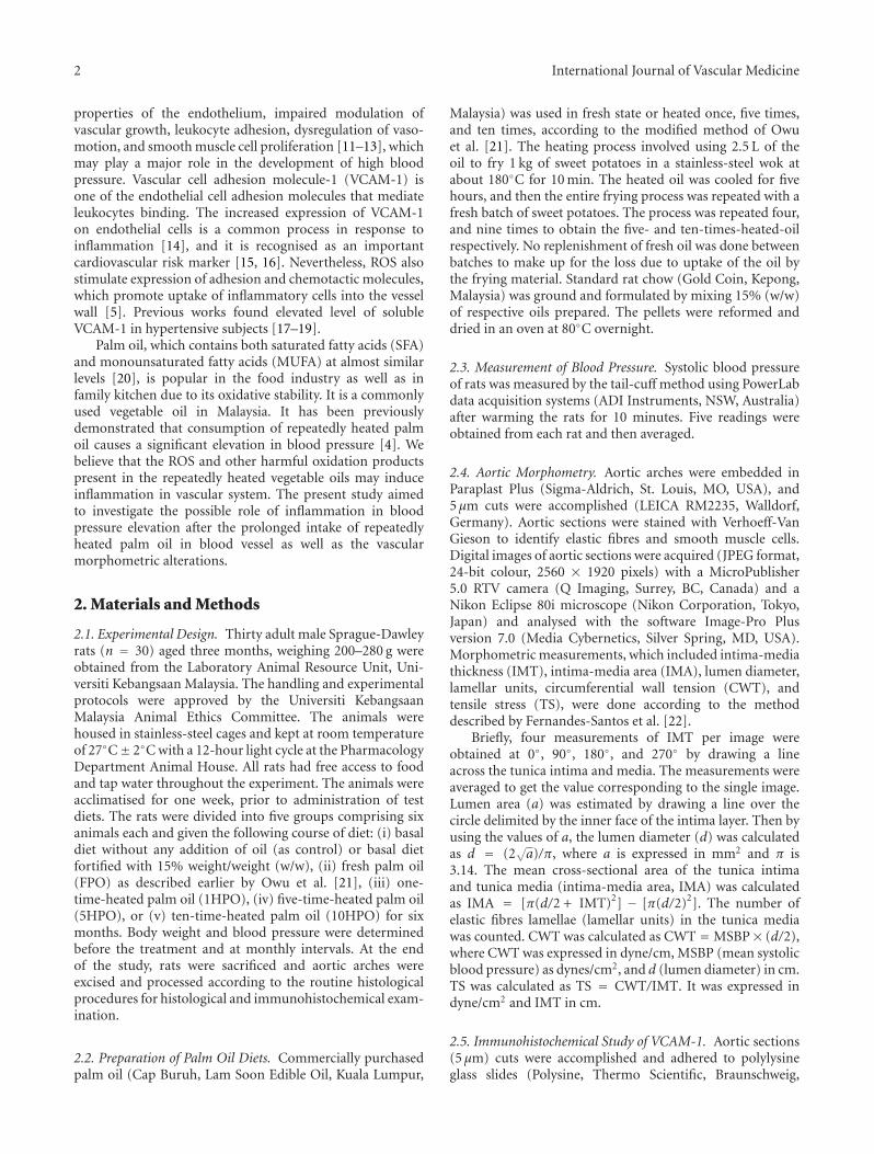
3.5. Correlation between Blood Pressure and VCAM-1 Expres-sion. There was a significant positive relationship (r = 0.757,P < 0.001) between systolic blood pressure and aorticVCAM-1 expression (Figure 5).
4. Discussion
This study was carried out to ascertain the involvement ofinflammation in blood pressure elevation after consumptionof heated palm oil We postulated that heating the palm oilrepeatedly would generate harmful ROS and hence induceinflammation and endothelial dysfunction.
In the present study, we observed a significant increasein blood pressure in the rats fed 5HPO or 10HPO compared
to the control and rats fed FPO or 1HPO. This significantincrease in blood pressure was in agreement with a previousstudy [4] showing prolonged intake of repeatedly heatedpalm oil increased blood pressure. Osim et al. [25] alsoreported that the oxidised oil-fed group had a greater risein blood pressure than the fresh oil-fed group. A studycarried out by Soriguer et al. [3] showed that the risk ofhypertension was positively correlated with the consumptionof polar compounds that were present in the cooking oil.Repeatedly heating makes the oil more susceptible to lipidperoxidation [26], which also reduces the vitamin Econstituents such as α-tocopherol, α-tocotrienol, andγ-tocotrienol in palm and soy oils [27]. Consumption ofrepeatedly heated palm oil in postmenopausal state maycontribute to the development of atherosclerosis because ofincreased lipid peroxidation [28]. Therefore, the deleteriouseffect of prolonged intake of 5HPO and 10HPO on bloodpressure that we observed might be contributed by theoverproduction of ROS that causes vascular inflammationand impairs the endothelial function. Oxidative stress, dueto over-production of ROS, exerts endothelial dysfunctionwhich plays a key role in pathogenesis of hypertension.A study done by Chan et al. [29] demonstrated that theincreased levels of ROS may contribute to oxidative stress andhypertension in rats.
Blood pressure in the rats fed FPO or 1HPO did notshow any remarkable change throughout the experiment. Infact, interestingly, we found that rats fed FPO even showed asignificantly lower blood pressure after six months comparedto the control. This shows that palm oil did not only preventthe increase of blood pressure but also had a tendencyto lower it. Palm oil is rich in natural antioxidants liketocotrienol which have beneficial effect on oxidative stressassociated with hypertension [30]. Medeiros et al. [31] alsodemonstrated that the long-term intake of palm oil hadbeneficial effect in reducing blood pressure in spontaneouslyhypertensive rats. Free radical scavenging antioxidants inpalm oil serve to protect endothelial cells against oxida-tive injury and thus improve endothelial functions [32].Furthermore, the blood pressure lowering mechanism ofpalm oil may also involve the beneficial alteration inendothelium-derived factors [33]. The present finding pro-vides further support to the cardiovascular protective effectof palm oil.

6 International Journal of Vascular Medicine
TM
TI
(a)
TMTI
(b)
TM
TI
(c)
TMTI
(d)
TM
TI
(e)
Figure 4: Photomicrographs of aortic sections showing immunostaining for VCAM-1. Groups are as follows: (a) control rats; rats fed (b)fresh (FPO), (c) one-time-heated (1HPO), (d) five-time-heated (5HPO), or (e) ten-time-heated palm oil (10HPO). Aortic sections fromrats fed 5HPO and 10HPO showed an intense staining of VCAM-1 on the tunica intima [(d) and (e)]. In contrast, aortic sections from ratsfed FPO and 1HPO showed relatively little staining of VCAM-1 [(b) and (c)] compared to control (a). TI: tunica intima; TM: tunica media.Same magnification is applied to all pictures (×400). Calibration bar = 50 μm.
From morphometric aspects, we observed that prolongedconsumption of 5HPO or 10HPO resulted in significantlyincreased thickness (IMT) and area (IMA) of intima-mediaof the aortic wall in rats compared to those fed basaldiet, FPO, or 1HPO. This finding suggests that prolongedconsumption of heated oils causes vascular hypertrophicremodelling. IMT thickening and vascular architecturalchanges are commonly associated with high blood pressure[34]. Therefore, IMT is a marker of coronary disease [35].Albeit increased IMT, the number of elastic lamellae didnot differ among the groups, which might suggest thatthe IMT thickening is due to vascular smooth muscle cells
hypertrophy, as indicated by increased interlamellar space.We believe that the ROS present in the heated oils may play arole in this remodelling. Previously, we observed a reductionin nitric oxide (NO) level following administration ofrepeatedly heated oil in rats [36]. Increased ROS generationmay contribute to endothelial dysfunction by decreasingthe bioavailability of NO which functions to reduce cellularproliferation. Hypertension is accompanied by an alteredbiochemical environment between the factors that act ascell growth promoters and the factors that reduce cellularproliferation [37]. This may lead to an imbalance betweenthe rate of growth and death, causing wall hypertrophy.
International Journal of Vascular Medicine 7
Nakaki and Kato [38] also reported that vascular remodellingcould be due to decreased NO and high blood pressure.Further, TS (which is the tension per unit of thickness andacts perpendicularly to the wall) did not differ among thegroups, maybe due to the IMT thickening. On the otherhand, there was no significant difference of lumen diameteramong the groups. This may suggest that the increased CWTthat we observed in rats fed 5HPO or 10HPO could be dueto the elevated blood pressure in them. CWT is the force thatacts in longitudinal and circumferential directions to opposethe distending effects of blood pressure. High blood pressureincreases CWT, in which further predisposing the vascularwall to damage and impairing its normal functions [39].
Prolonged consumption of 5HPO or 10HPO signifi-cantly increased the aortic endothelial expression of VCAM-1 in rats when compared to the control, FPO, and 1HPOgroups. FPO that did not induce VCAM-1 expression mightbe due to its rich antioxidant contents that maintain theendothelial oxidative status [30]. VCAM-1 is induced onendothelial inflammatory sites [40] and this finding suggeststhat 5HPO and 10HPO might induce vascular inflammation.The repeated deep frying process is deleterious on theoxidative stability and biochemical characteristics of theoil, in which it generates ROS and other lipid oxidationproducts. Our earlier experiment [28] reported that heatedpalm oil increased lipid peroxidation as indicated by asignificant increase in serum thiobarbituric acid reactivesubstance (TBARS). Moreover, the effect of deep fryingprocess on oil thermoxidation has been documented byprevious studies [41–44]. ROS in the diet are absorbedinto the blood circulation and their increased presence canoverwhelm the intercellular antioxidant defence, leading tooxidative stress which can induce endothelial injury [2, 45].Vascular endothelial cells, which provide a physical barrierfor the underlying smooth muscle cells and play a pivotal rolein maintaining cardiovascular homeostasis, are sensitive todisturbances in the redox steady state [46]. Previous reportshave demonstrated the association between the disturbancein the redox steady state with modulated endothelial functionand inflammation [47–50]. Therefore, prolonged intakeof 5HPO and 10HPO may induce inflammation, henceendothelial cells respond by expressing VCAM-1.
VCAM-1 is a member of the immunoglobulin gensuperfamily that participates in the adhesion and migrationof leukocytes into tissues during immune response. Wemeasured the aortic expression of VCAM-1 in rats becauseVCAM-1 is expressed on endothelial cells in inflammatorysites. Although VCAM-1 has been well documented to beassociated with the development of atherosclerosis [51–53],several studies also demonstrated that the expression ofVCAM-1 was increased in patients with hypertension [17–19, 54]. A study performed by Parissis et al. [17] showedthat hypertensive patients exhibited higher plasma levels ofVCAM-1. We found that there was a significant positivecorrelation between blood pressure and VCAM-1 densityon endothelial cells. This suggests that augmented VCAM-1 expression may reflect inflammation and endothelialdysfunction and hence impairing the regulation of bloodpressure. This is because a significant reduction in the cell
15014013012011010090
40
30
20
10
Blood pressure (mmHg)
R Sq linear = 0.572VC
AM
-1 d
ensi
ty (
% o
f tu
nic
a in
tim
a ar
ea)
Figure 5: Correlation between blood pressure and endothelialVCAM-1 expression in rats.
adhesion molecules expression may indicate a better preser-vation of endothelial function [55]. Besides, we do believethat the increased VCAM-1 (that is inflammation) may playa role, at least in part, in causing adverse remodelling. Aprevious study documented that increased inflammatorymediator aggravated vascular remodelling [56]. Remodellingimpairs the compliance of the blood vessel and leads topathogenesis of raising blood pressure [57].
In addition, our results suggest that intake of 5HPOand 10HPO is detrimental to vascular morphometry as wellas the regulation of blood pressure. Intake of 5HPO and10HPO increased blood pressure, morphometric alterations,and expression of VCAM-1 significantly than 1HPO. In fact,we found that rats fed 1HPO did not exhibit significantchange when we compared to the control and FPO groups.Palm oil contains saturated fatty acids and unsaturated fattyacids at almost equal ratio [58], in which saturated fattyacids are more resistant to thermoxidation. Though beingheated once, the palm oil might still retain abundant antiox-idants and generate less ROS than 5HPO and 10HPO andhence protects the vascular endothelium from inflammation.Therefore, it suggests that intake of palm oil heated oncemight not cause remarkable harmful effect in rats. However,when the oil is reused repeatedly, deleterious effects ensue.
This study provides the hints about the pivotal roleof inflammation in the blood pressure raising effect ofrepeatedly heated palm oil. Repeatedly heated palm oilinduces inflammation and vascular remodelling in therats, which subsequently leads to an increase in bloodpressure. Further research need to focus on more inflam-matory biomarkers such as intercellular adhesion molecule-1(ICAM-1), endothelin-1, and others as well as their pathwaysto understand thepagebreak possible molecular mechanismsinvolved in the inducing of hypertension by heated palm oil.
5. Summary
In conclusion, prolonged intake of repeatedly heated palmoil appears to increase blood pressure in rats, which might

8 International Journal of Vascular Medicine
be mediated by inflammation and endothelial dysfunction,as reflected by the adverse vascular remodelling and theinduction of VCAM-1 expression on endothelial cells.
Acknowledgments
This study was funded by UKM Medical Faculty ResearchGrant FF-161-2010. The authors wish to thank Madame.Azizah Osman and Madame. Sinar Suriya Muhamad of theDepartment of Pharmacology, as well as Professor Dr. SitiAishah Md Ali of the Histopathology Unit, Department ofPathology, UKM Medical Centre, Cheras, for their kind helpand technical assistance.
References
[1] A. Abdullah, M. S. Suondoh, C. S. Xuan et al., “Level ofawareness amongst the general public regarding usage ofrepeatedly heated cooking oil in Kuala Lumpur, Malaysia,”International Medical Journal, vol. 17, no. 4, pp. 310–311, 2010.
[2] M. J. A. Williams, W. H. F. Sutherland, M. P. McCormick, S. A.De Jong, R. J. Walker, and G. T. Wilkins, “Impaired endothelialfunction following a meal rich in used cooking fat,” Journal ofthe American College of Cardiology, vol. 33, no. 4, pp. 1050–1055, 1999.
[3] F. Soriguer, G. Rojo-Martınez, M. C. Dobarganes et al.,“Hypertension is related to the degradation of dietary fryingoils,” The American Journal of Clinical Nutrition, vol. 78, no. 6,pp. 1092–1097, 2003.
[4] X. F. Leong, A. Aishah, U. Nor Aini, S. Das, and K. Jaarin,“Heated palm oil causes rise in blood pressure and cardiacchanges in heart muscle in experimental rats,” Archives ofMedical Research, vol. 39, no. 6, pp. 567–572, 2008.
[5] D. G. Harrison, M. C. Gongora, T. J. Guzik, and J. Widder,“Oxidative stress and hypertension,” Journal of the AmericanSociety of Hypertension, vol. 1, no. 1, pp. 30–44, 2007.
[6] S. Pennathur and J. W. Heinecke, “Oxidative stress andendothelial dysfunction in vascular disease,” Current DiabetesReports, vol. 7, no. 4, pp. 257–264, 2007.
[7] G. Zalba, A. Fortuno, G. San Jose, M. U. Moreno, O. Beloqui,and J. Dıez, “Oxidative stress, endothelial dysfunction andcerebrovascular disease,” Cerebrovascular Diseases, vol. 24, no.1, pp. 24–29, 2007.
[8] F. A. Ghanem and A. Movahed, “Inflammation in high bloodpressure: a clinician perspective, ” Journal of the AmericanSociety of Hypertension, vol. 1, no. 2, pp. 113–119, 2007.
[9] C. U. Chae, R. T. Lee, N. Rifai, and P. M. Ridker, “Bloodpressure and inflammation in apparently healthy men,”Hypertension, vol. 38, no. 3, pp. 399–403, 2001.
[10] A. A. Elmarakby, J. Faulkner, S. P. Posey, and J. C. Sullivan,“Induction of hemeoxygenase-1 attenuates the hypertensionand renal inflammation in spontaneously hypertensive rats,”Pharmacological Research, vol. 62, no. 5, pp. 400–407, 2010.
[11] L. E. Bautista, “Inflammation, endothelial dysfunction, andthe risk of high blood pressure: epidemiologic and biologicalevidence,” Journal of Human Hypertension, vol. 17, no. 4, pp.223–230, 2003.
[12] P. Balakumar, T. Kaur, and M. Singh, “Potential targetsites to modulate vascular endothelial dysfunction: current
perspectives and future directions,” Toxicology, vol. 245, no.1-2, pp. 49–64, 2008.
[13] F. Grover-Paez and A. B. Zavalza-Gomez, “Endothelialdysfunction and cardiovascular risk factors,” DiabetesResearch and Clinical Practice, vol. 84, no. 1, pp. 1–10, 2009.
[14] T. Chiba and O. Ezaki, “Dietary restriction suppressesinflammation and delays the onset of stroke in stroke-pronespontaneously hypertensive rats,” Biochemical and BiophysicalResearch Communications, vol. 399, no. 1, pp. 98–103, 2010.
[15] A. S. Postadzhiyan, A. V. Tzontcheva, I. Kehayov, and B.Finkov, “Circulating soluble adhesion molecules ICAM-1and VCAM-1 and their association with clinical outcome,troponin T and C-reactive protein in patients with acutecoronary syndromes,” Clinical Biochemistry, vol. 41, no. 3, pp.126–133, 2008.
[16] J. Constans and C. Conri, “Circulating markers of endothelialfunction in cardiovascular disease,” Clinica Chimica Acta, vol.368, no. 1-2, pp. 33–47, 2006.
[17] J. T. Parissis, K. F. Venetsanou, D. G. Mentzikof et al., “Plasmalevels of soluble cellular adhesion molecules in patients witharterial hypertension. Correlations with plasma endothelin-1,” European Journal of Internal Medicine, vol. 12, no. 4, pp.350–356, 2001.
[18] S. Cottone, G. Mule, E. Nardi et al., “Relation of C-reactiveprotein to oxidative stress and to endothelial activation inessential hypertension,” American Journal of Hypertension,vol. 19, no. 3, pp. 313–318, 2006.
[19] J. J. Lozano-Nuevo, T. Estrada-Garcia, H. Vargas-Robles, B.A. Escalante-Acosta, and A. F. Rubio-Guerra, “Correlationbetween circulating adhesion molecules and resistin levelsin hypertensive type-2 diabetic patients,” Inflammation &Allergy—Drug Targets, vol. 10, no. 1, pp. 27–31, 2011.
[20] A. Azlan, K. N. Prasad, H. E. Khoo et al., “Comparison of fattyacids, vitamin E and physicochemical properties of Canariumodontophyllum Miq. (dabai), olive and palm oils,” Journal ofFood Composition and Analysis, vol. 23, no. 8, pp. 772–776,2010.
[21] D. U. Owu, E. E. Osim, and P. E. Ebong, “Serum liver enzymesprofile of Wistar rats following chronic consumption of freshor oxidized palm oil diets,” Acta Tropica, vol. 69, no. 1, pp.65–73, 1998.
[22] C. Fernandes-Santos, L. D. S. Mendonca, and C. A.Mandarim-de-Lacerda, “Favorable cardiac and aorticremodeling in olmesartan-treated spontaneously hypertensiverats,” Heart and Vessels, vol. 24, no. 3, pp. 219–227, 2009.
[23] J. D. A. Moraes-Teixeira, A. Felix, C. Fernandes-Santos, A. S.Moura, C. A. Mandarim-de-Lacerda, and J. J. de Carvalho,“Exercise training enhances elastin, fibrillin and nitric oxide inthe aorta wall of spontaneously hypertensive rats,” Experimen-tal and Molecular Pathology, vol. 89, no. 3, pp. 351–357, 2010.
[24] C. A. Mandarim-de-Lacerda, C. Fernandes-Santos, and M.B. Aguila, “Image analysis and quantitative morphology,”Methods in Molecular Biology, vol. 611, pp. 211–225, 2010.
[25] E. E. Osim, D. U. Owu, and K. M. Etta, “Arterial pressure andlipid profile in rats following chronic ingestion of palm oildiets, ” African Journal of Medicine and Medical Sciences, vol.25, no. 4, pp. 335–340, 1996.
[26] E. N. Frankel, “Lipid oxidation,” Progress in Lipid Research,vol. 19, no. 1-2, pp. 1–22, 1980.
[27] S. K. Adam, N. A. Sulaiman, A. G. Mat Top, and K. Jaarin,“Heating reduces vitamin E content in palm and soy oils,”

International Journal of Vascular Medicine 9
Malaysian Journal of Biochemistry and Molecular Biology, vol.15, no. 2, pp. 76–79, 2007.
[28] S. K. Adam, I. N. Soelaiman, N. A. Umar, N. Mokhtar,N. Mohamed, and K. Jaarin, “Effects of repeatedly heatedpalm oil on serum lipid profile, lipid peroxidation andhomocysteine levels in a post-menopausal rat model,” McGillJournal of Medicine, vol. 11, no. 2, pp. 145–151, 2008.
[29] S. H. H. Chan, M. H. Tai, C. Y. Li, and J. Y. H. Chan,“Reduction in molecular synthesis or enzyme activity ofsuperoxide dismutases and catalase contributes to oxidativestress and neurogenic hypertension in spontaneouslyhypertensive rats,” Free Radical Biology and Medicine, vol. 40,no. 11, pp. 2028–2039, 2006.
[30] D. Narang, S. Sood, M. K. Thomas, A. K. Dinda, and S. K.Maulik, “Effect of dietary palm olein oil on oxidative stressassociated with ischemic-reperfusion injury in isolated ratheart,” BMC Pharmacology, vol. 4, article 29, 2004.
[31] F. J. Medeiros, C. G. Mothe, M. B. Aguila, and C. A.Mandarim-De-Lacerda, “Long-term intake of edibleoils benefits blood pressure and myocardial structure inspontaneously hypertensive rat (SHR) and streptozotocindiabetic SHR,” Prostaglandins & Other Lipid Mediators, vol.78, no. 1–4, pp. 231–248, 2005.
[32] S. P. Muharis, A. G. M. Top, D. Murugan, and M. R. Mustafa,“Palm oil tocotrienol fractions restore endothelium dependentrelaxation in aortic rings of streptozotocin-induced diabeticand spontaneously hypertensive rats,” Nutrition Research, vol.30, no. 3, pp. 209–216, 2010.
[33] A. A. Ganafa, R. R. Socci, D. Eatman, N. Silvestrov, I. K.Abukhalaf, and M. A. Bayorh, “Effect of palm oil on oxidativestress-induced hypertension in sprague-dawley rats,” Ameri-can Journal of Hypertension, vol. 15, no. 8, pp. 725–731, 2002.
[34] H. D. Intengan and E. L. Schiffrin, “Vascular remodeling inhypertension: roles of apoptosis, inflammation, and fibrosis.,”Hypertension, vol. 38, no. 3, part 2, pp. 581–587, 2001.
[35] B. Erzen, M. Sabovic, M. Sebestjen, and P. Poredos, “Endothe-lial dysfunction, intima-media thickness, ankle-brachialpressure index, and pulse pressure in young post-myocardialinfarction patients with various expressions of classical riskfactors,” Heart and Vessels, vol. 22, no. 4, pp. 215–222, 2007.
[36] X. F. Leong, M. N. M. Najib, S. Das, M. R. Mustafa, and K.Jaarin, “Intake of repeatedly heated palm oil causes elevationin blood pressure with impaired vasorelaxation in rats,” TheTohoku Journal of Experimental Medicine, vol. 219, no. 1, pp.71–78, 2009.
[37] S. M. Arribas, A. Hinek, and M. C. Gonzalez, “Elastic fibresand vascular structure in hypertension,” Pharmacology &Therapeutics, vol. 111, no. 3, pp. 771–791, 2006.
[38] T. Nakaki and R. Kato, “Nitric oxide in vascular remodeling,”Japanese Heart Journal, vol. 37, no. 4, pp. 431–445, 1996.
[39] H. D. Intengan, G. Thibault, J. S. Li, and E. L. Schiffrin,“Resistance artery mechanics, structure, and extracellularcomponents in spontaneously hypertensive rats: effects ofangiotensin receptor antagonism and converting enzymeinhibition,” Circulation, vol. 100, no. 22, pp. 2267–2275, 1999.
[40] J. M. Cook-Mills, “VCAM-1 signals during lymphocytemigration: role of reactive oxygen species,” MolecularImmunology, vol. 39, no. 9, pp. 499–508, 2002.
[41] C. Garrido-Polonio, M. C. Garcıa-Linares, M. T. Garcıa-Ariaset al., “Thermally oxidised sunflower-seed oil increasesliver and serum peroxidation and modifies lipoprotein
composition in rats,” British Journal of Nutrition, vol. 92, no.2, pp. 257–265, 2004.
[42] C. F. Rueda-Clausen, F. A. Silva, M. A. Lindarte et al., “Olive,soybean and palm oils intake have a similar acute detrimentaleffect over the endothelial function in healthy young subjects,”Nutrition, Metabolism and Cardiovascular Diseases, vol. 17,no. 1, pp. 50–57, 2007.
[43] A. K. Das, R. Babylatha, A. S. Pavithra, and S. Khatoon,“Thermal degradation of groundnut oil duringcontinuousand intermittent frying,” Journal of Food Science andTechnology. In press.
[44] M. D. Juarez, C. C. Osawa, M. E. Acuna, N. Samman, and L.A. G. Goncalves, “Degradation in soybean oil, sunflower oiland partially hydrogenated fats after food frying, monitoredby conventional and unconventional methods,” Food Control,vol. 22, no. 12, pp. 1920–1927, 2011.
[45] I. Staprans, J. H. Rapp, X. M. Pan, and K. R. Feingold, “Theeffect of oxidized lipids in the diet on serum lipoproteinperoxides in control and diabetic rats,” The Journal of ClinicalInvestigation, vol. 92, no. 2, pp. 638–643, 1993.
[46] B. Hennig and C. K. Chow, “Lipid peroxidation andendothelial cell injury: implications in atherosclerosis,” FreeRadical Biology & Medicine, vol. 4, no. 2, pp. 99–106, 1988.
[47] K. Irani, “Oxidant signaling in vascular cell growth, death,and survival: a review of the roles of reactive oxygen speciesin smooth muscle and endothelial cell mitogenic andapoptotic signaling,” Circulation Research, vol. 87, no. 3,pp. 179–183, 2000.
[48] H. Li, M. Han, L. Guo, G. Li, and N. Sang, “Oxidative stress,endothelial dysfunction and inflammatory response in ratheart to NO2 inhalation exposure,” Chemosphere, vol. 82, no.11, pp. 1589–1596, 2011.
[49] X. Chen, H. Y. Zhong, J. H. Zeng, and J. Ge, “Thepharmacological effect of polysaccharides from Lentinusedodes on the oxidative status and expression of VCAM-1mRNA of thoracic aorta endothelial cell in high-fat-dietrats,” Carbohydrate Polymers, vol. 74, no. 3, pp. 445–450, 2008.
[50] Y. W. Lee, H. Kuhn, B. Hennig, A. S. Neish, and M. Toborek,“IL-4-induced oxidative stress upregulates VCAM-1 geneexpression in human endothelial cells,” Journal of Molecularand Cellular Cardiology, vol. 33, no. 1, pp. 83–94, 2001.
[51] C. Tikellis, K. A. Jandeleit-Dahm, K. Sheehy et al., “Reducedplaque formation induced by rosiglitazone in an STZ-diabetes mouse model of atherosclerosis is associated withdownregulation of adhesion molecules,” Atherosclerosis, vol.199, no. 1, pp. 55–64, 2008.
[52] Y. Wu, R. Zhang, C. Zhou et al., “Enhanced expression ofvascular cell adhesion molecule-1 by corticotrophin-releasinghormone contributes to progression of atherosclerosis in LDLreceptor-deficient mice,” Atherosclerosis, vol. 203, no. 2, pp.360–370, 2009.
[53] S. E. Choi, H. J. Jang, Y. Kang et al., “Atherosclerosis inducedby a high-fat diet is alleviated by lithium chloride viareduction of VCAM expression in ApoE-deficient mice,”Vascular Pharmacology, vol. 53, no. 5-6, pp. 264–272, 2010.
[54] M. S. Boulbou, G. N. Koukoulis, E. D. Makri, E. A. Petinaki, K.I. Gourgoulianis, and A. E. Germenis, “Circulating adhesionmolecules levels in type 2 diabetes mellitus and hypertension,”International Journal of Cardiology, vol. 98, no. 1, pp. 39–44,2005.

10 International Journal of Vascular Medicine
[55] E. K. Iliodromitis, I. Andreadou, S. Markantonis-Kyroudiset al., “The effects of tirofiban on peripheral markers ofoxidative stress and endothelial dysfunction in patients withacute coronary syndromes,” Thrombosis Research, vol. 119, no.2, pp. 167–174, 2007.
[56] H. W. Tan, X. Liu, X. P. Bi et al., “IL-18 overexpressionpromotes vascular inflammation and remodeling in a ratmodel of metabolic syndrome,” Atherosclerosis, vol. 208, no. 2,pp. 350–357, 2010.
[57] M. J. Mulvany, “Small artery remodeling and significancein the development of hypertension,” News in PhysiologicalSciences, vol. 17, no. 3, pp. 105–109, 2002.
[58] D. O. Edem, “Palm oil: biochemical, physiological, nutritional,hematological, and toxicological aspects: a review,” Plant Foodsfor Human Nutrition, vol. 57, no. 3-4, pp. 319–341, 2002.

Submit your manuscripts athttp://www.hindawi.com
Stem CellsInternational
Hindawi Publishing Corporationhttp://www.hindawi.com Volume 2014
Hindawi Publishing Corporationhttp://www.hindawi.com Volume 2014
MEDIATORSINFLAMMATION
of
Hindawi Publishing Corporationhttp://www.hindawi.com Volume 2014
Behavioural Neurology
EndocrinologyInternational Journal of
Hindawi Publishing Corporationhttp://www.hindawi.com Volume 2014
Hindawi Publishing Corporationhttp://www.hindawi.com Volume 2014
Disease Markers
Hindawi Publishing Corporationhttp://www.hindawi.com Volume 2014
BioMed Research International
OncologyJournal of
Hindawi Publishing Corporationhttp://www.hindawi.com Volume 2014
Hindawi Publishing Corporationhttp://www.hindawi.com Volume 2014
Oxidative Medicine and Cellular Longevity
Hindawi Publishing Corporationhttp://www.hindawi.com Volume 2014
PPAR Research
The Scientific World JournalHindawi Publishing Corporation http://www.hindawi.com Volume 2014
Immunology ResearchHindawi Publishing Corporationhttp://www.hindawi.com Volume 2014
Journal of
ObesityJournal of
Hindawi Publishing Corporationhttp://www.hindawi.com Volume 2014
Hindawi Publishing Corporationhttp://www.hindawi.com Volume 2014
Computational and Mathematical Methods in Medicine
OphthalmologyJournal of
Hindawi Publishing Corporationhttp://www.hindawi.com Volume 2014
Diabetes ResearchJournal of
Hindawi Publishing Corporationhttp://www.hindawi.com Volume 2014
Hindawi Publishing Corporationhttp://www.hindawi.com Volume 2014
Research and TreatmentAIDS
Hindawi Publishing Corporationhttp://www.hindawi.com Volume 2014
Gastroenterology Research and Practice
Hindawi Publishing Corporationhttp://www.hindawi.com Volume 2014
Parkinson’s Disease
Evidence-Based Complementary and Alternative Medicine
Volume 2014Hindawi Publishing Corporationhttp://www.hindawi.com