isolation and characterization of erwinia chrysanthemi mutants
TRANSCRIPT
Vol. 161, No. 2JOURNAL OF BACTERIOLOGY, Feb. 1985, p. 702-7080021-9193/85/020702-07$02.00/0Copyright © 1985, American Society for Microbiology
Isolation and Characterization of Erwinia chrysanthemi MutantsDefective in Degradation of Hexuronates
FREDERIQUE VAN GIJSEGEM,l* NICOLE HUGOUVIEUX-COTTE-PATTAT,2 AND JEANINEROBERT-BAUDOUY2
Laboratoire de Gene'tique, Universite Libre de Bruxelles, Rhode-Saint-Genese, Belgium,' and Laboratoire deMicrobiologie, Institut National des Sciences Applique'es, Villeurbanne, France2
Received 9 July 1984/Accepted 12 November 1984
Spontaneous and Tn9-induced mutants of Erwinia chrysanthemi were isolated which affect the degradativepathway of galacturonate and ketodeoxygluconate. The mutations were characterized both biochemically andfunctionally by complementation analysis and localized in the E. chrysanthemi chromosome. The kdgK genemapped very close to ile, the kdgA gene was between trp and his, and the exuT-uxaC-uxaB-uxaA cluster waslinked to thy. The different types of mutants obtained were consistent with an organization of the exu-uxacluster into two transcription units, one containing the exuT gene, and the other containing the three uxa genes,with the transcription going from uxaC to uxaA.
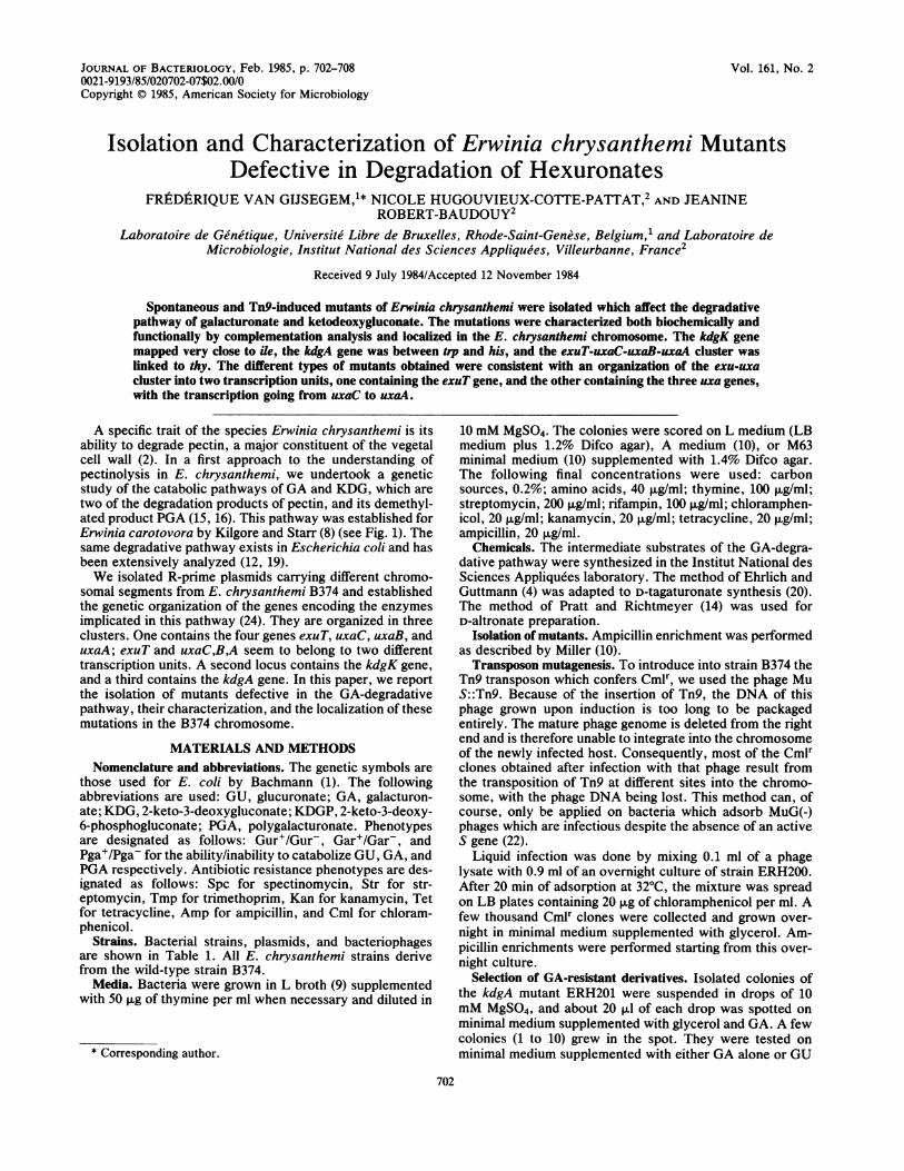
A specific trait of the species Erwinia chrysanthemi is itsability to degrade pectin, a major constituent of the vegetalcell wall (2). In a first approach to the understanding ofpectinolysis in E. chrysanthemi, we undertook a geneticstudy of the catabolic pathways of GA and KDG, which aretwo of the degradation products of pectin, and its demethyl-ated product PGA (15, 16). This pathway was established forErwinia carotovora by Kilgore and Starr (8) (see Fig. 1). Thesame degradative pathway exists in Escherichia coli and hasbeen extensively analyzed (12, 19).We isolated R-prime plasmids carrying different chromo-
somal segments from E. chrysanthemi B374 and establishedthe genetic organization of the genes encoding the enzymesimplicated in this pathway (24). They are organized in threeclusters. One contains the four genes exuT, uxaC, uxaB, anduxaA; exuT and uxaC,B,A seem to belong to two differenttranscription units. A second locus contains the kdgK gene,and a third contains the kdgA gene. In this paper, we reportthe isolation of mutants defective in the GA-degradativepathway, their characterization, and the localization of thesemutations in the B374 chromosome.
MATERIALS AND METHODSNomenclature and abbreviations. The genetic symbols are
those used for E. coli by Bachmann (1). The followingabbreviations are used: GU, glucuronate; GA, galacturon-ate; KDG, 2-keto-3-deoxygluconate; KDGP, 2-keto-3-deoxy-6-phosphogluconate; PGA, polygalacturonate. Phenotypesare designated as follows: Gur+/Gur-, Gar'/Gar-, andPga+/Pga- for the ability/inability to catabolize GU, GA, andPGA respectively. Antibiotic resistance phenotypes are des-ignated as follows: Spc for spectinomycin, Str for str-eptomycin, Tmp for trimethoprim, Kan for kanamycin, Tetfor tetracycline, Amp for ampicillin, and Cml for chloram-phenicol.
Strains. Bacterial strains, plasmids, and bacteriophagesare shown in Table 1. All E. chrysanthemi strains derivefrom the wild-type strain B374.
Media. Bacteria were grown in L broth (9) supplementedwith 50 ,ug of thymine per ml when necessary and diluted in
* Corresponding author.
10 mM MgSO4. The colonies were scored on L medium (LBmedium plus 1.2% Difco agar), A medium (10), or M63minimal medium (10) supplemented with 1.4% Difco agar.The following final concentrations were used: carbonsources, 0.2%; amino acids, 40 ,ug/ml; thymine, 100 jxg/ml;streptomycin, 200 ,ug/ml; rifampin, 100 ,ug/ml; chloramphen-icol, 20 ,ug/ml; kanamycin, 20 ,ug/ml; tetracycline, 20 p,g/ml;ampicillin, 20 ,ug/ml.
Chemicals. The intermediate substrates of the GA-degra-dative pathway were synthesized in the Institut National desSciences Appliquees laboratory. The method of Ehrlich andGuttmann (4) was adapted to D-tagaturonate synthesis (20).The method of Pratt and Richtmeyer (14) was used forD-altronate preparation.
Isolation of mutants. Ampicillin enrichment was performedas described by Miller (10).Transposon mutagenesis. To introduce into strain B374 the
Tn9 transposon which confers Cmlr, we used the phage MuS::Tn9. Because of the insertion of Tn9, the DNA of thisphage grown upon induction is too long to be packagedentirely. The mature phage genome is deleted from the rightend and is therefore unable to integrate into the chromosomeof the newly infected host. Consequently, most of the Cmlrclones obtained after infection with that phage result fromthe transposition of Tn9 at different sites into the chromo-some, with the phage DNA being lost. This method can, ofcourse, only be applied on bacteria which adsorb MuG(-)phages which are infectious despite the absence of an activeS gene (22).
Liquid infection was done by mixing 0.1 ml of a phagelysate with 0.9 ml of an overnight culture of strain ERH200.After 20 min of adsorption at 32°C, the mixture was spreadon LB plates containing 20 ,ug of chloramphenicol per ml. Afew thousand Cmlr clones were collected and grown over-night in minimal medium supplemented with glycerol. Am-picillin enrichments were performed starting from this over-night culture.
Selection of GA-resistant derivatives. Isolated colonies ofthe kdgA mutant ERH201 were suspended in drops of 10mM MgSO4, and about 20 ,ul of each drop was spotted onminimal medium supplemented with glycerol and GA. A fewcolonies (1 to 10) grew in the spot. They were tested onminimal medium supplemented with either GA alone or GU
702
E. CHRYSANTHEMI HEXURONATE MUTANTS 703
TABLE 1. B
Strain, plasmid,or bacteriophage
StrainB374
ERH200
ERH201ERH202ERH203ERH204ERH205ERH206ERH207ERH208ERH209
ERH210
ERH213
ERH214
ERH215
ERH216
ERH217
ERH218ERH219
ERH220
ERH221
ERH223
ERH224RH6010
ERH80
PlasmidspULB113
pULB114
pULB115
pULB116
pULB117
pULB118
pULB119
pULB120
acterial strains, plasmids, and bacteriophages
Description Source or reference
E. chrysanthemia
B374 his-5 GurA+Rifr
ERH200 Tn9 kdgAlERH200 kdgA2ERH200 kdgKIERH200 kdgK2::Tn9ERH200 kdgK3::Tn9ERH200 kdgK4::Tn9ERH200 uxaAl::Tn9ERH200 uxaBl::Tn9ERH201 uxaA2kdgA+
ERH201 uxaA3kdgA+
ERH201 uxaCBAlkdgA+
ERH201 uxaCBJkdgA+
ERH201 exuTIkdgA+
ERH201 exuT2kdgA+
ERH201 exuT3kdgA+
ERH200 exuT4::Tn9ERH200 A(exu-
uxa)l: :Tn9ERH201 A(exu-uxa)2kdgA+
ERH200 exuT uxaATn9
ERH200 A(exu-uxa)3: :Tn9
ERH200 uxaA4::Tn9thr-l leu-J pro-i his-5
trp-I thyA ile-J StrrRifr
tra+ bla+ (TEM-2)TnI tet+ aphA+(Mu3A)
pULB113 carryingexuT
pULB113 carryingexuT-uxaC
pULB113 carryinguxaC-uxaB
pULB113 carryinguxaC-uxaB-uxaA
pULB113 carryingexuT-uxaC-uxaB-uxaA
pULB113 carryingkdgK
pULB113 carryingkdgA
BacteriophagesMu cts62 S::Tn9,EC2
6; Lemattre, person-al communication
This work
21
His' transductant ofRH6010
23
24
17Resibois et al., in
press
and glycerol or PGA and glycerol. The Gar- clones wereassumed to be kdgA mutants which acquired a secondarymutation. Among them, clones growing on GA plus glyceroland PGA plus glycerol were assumed to be kdgK kdgAmutants and were discarded. Among the clones still poi-soned by PGA, two types of clones were found: those whichwere still poisoned by GU and those which were notpoisoned by either GA or GU.To transduce the kdgA+ allele in these clones, we mixed
0.1 ml of a lysate of the generalized transducing phagephiEC-2 (A. Resibois, M. Colet, M. Faelen, E. Schoonejans,and A. Toussaint, Virology, in press) grown on strain B374with 0.1 ml of an overnight culture of the bacterium andspread this mixture on minimal medium supplemented withPGA (multiplicity of infection, 1 to 3).Complementation tests. A first screening of the mutants
was performed by suspending a colony of the mutants in 1drop of 10 mM MgSO4 and spotting it on a selective mediumseeded with 0.1 ml of an overnight culture of an E. coli thyAstrain carrying the R-prime plasmid to be tested. The mu-tants described here were also tested by titration aftermating. Single drops of overnight cultures of donors carryingthe R-prime plasmid and the Gar- mutants to be tested weremixed on an A plate, incubated for 4 to 6 h at 33°C, andsuspended in 1 ml of 10 mM MgSO4. Mating mixtures weretitrated on minimal medium supplemented with glucose andkanamycin and lacking thymine (to counterselect the donor)to measure the frequency of transfer of the plasmid and onminimal medium supplemented with GA. The mutant wasassumed to be complemented by the R-prime plasmid testedwhen about the same titer was obtained on the two types ofplates (i.e., 5 to 100% of the frequency of transfer of the RP4resistance marker).Mapping. The Gar- mutations were localized by measur-
ing the frequency of cotransfer of the mutation with differentmarkers available on the chromosome of strain B374. PlasmidRP4: :mini-Mu pULB113 was transferred in the differentmutants, and these strains were mated with the polyauxo-trophic strain ERH80 or RH6010 by mixing on A medium 0.1ml of overnight cultures of the mutants carrying pULB113and the recipient. The plates were incubated for 4 to 6 h at33°C. Bacteria were collected from the growth area, sus-pended in 1 ml of 10 mM MgSO4, and spread on minimalmedium supplemented with glucose, streptomycin (to coun-terselect the donor), and all but one of the amino acidsrequired for growth of ERH80 or RH6010. The transconjug-ants which acquired one prototrophic marker from the donorwere further tested for their Gar phenotype.
Preparation of cell extracts and enzyme assays. Enzymeactivities were measured in cells grown exponentially inglycerol minimal medium alone or supplemented with GA.Cell extracts were prepared by breaking the cells suspendedin 10 mM phosphate buffer (pH 7.0) with a French press.Extracts were then centrifuged at 15,000 x g for 20 min toremove whole cells and cellular debris. Hydrogenase activ-ity was measured by monitoring the decrease in absorbanceat 340 nm in a mixture assay consisting of 50 mM phosphatebuffer (pH 6.3), 0.4 mM NADH, and 10 mM tagaturonate(13).Hydrolyase activity was measured by monitoring the
formation of KDG in a mixture containing 200 mM glycyl-glycine buffer (pH 8.3), 50 mM 2-mercaptoethanol, 0.8 mMFeSO4, and 3 mM D-altronate (18). The reaction was stoppedby adding 3 volumes of 10% trichloroacetic acid plus 20 mMHgCl2. The amount of KDG was then measured by themethod of Weissbach and Hurwitz (25).
a Strain B374, which was first reported to be E. carotovora (6), was recentlyretested and identified as E. chrysanthemi (Lemattre, personal communica-tion; Perombelon, personal communication).
VOL. 161, 1985
704 VAN GIJSEGEM ET AL.
TABLE 2. Genetic characterization of Gar- mutantsGrowth with Complementation with:C source:
Mutant pULB115 pL18Most likely OrgnGA GU PGA pULB114 exuT pULB116 pULB117 pUTLB18 pULB119 pULB120 phenotype(s) OnginexuT uxaC uxaC,B uxaC,B,A uxaC,B,A kdgK kdgA
ERH200 + + +ERH201 - - - - - - - - + KdgA bERH202 - - - - - - - - - + KdgA aERH203 - - - - - - - - + - KdgK aERH204 - - - - - - - - + - KdgK bERH205 - - - - - - - - + - KdgK bERH206 - - - - - - - - + - KdgK bERH207 - + + - - - + + - - UxaA or UxaB bERH208 - + + - - - + + - - UxaA or UxaB bERH209 - + + - - - + + - - UxaA or UxaB cERH210 - + + - - - + + - - UxaA or UxaB cERH213 - - + - - - + + - - UxaCBA cERH214 - - + - - + + + - - UxaCB cERH215 - - + + + - - + - - ExuT cERH216 - - + + + - - + - - ExuT cERH217 - - + + + - - + - - ExuT cERH218 - - + + + - - + - - ExuT bERH219 - - + - - - - + - - ExuT-UxaCBA bERH220 - - + - - - - + - - ExuT-UxaCBA cERH221 - - + - - - - + - - ExuT-UxaCBA cERH223 - - + - - - - + - - ExuT-UxaCBA bERH224 - + + - - - + + - - UxaA or UxaB b
aa, Spontaneous mutants recovered after ampicillin enrichment; b, Tn9-induced mutants; c, mutants isolated from a kdgA strain as GA resistant.
Isomerase activity was detected by measuring the taga-turonate formed with an excess extract of altronate oxido-reductase. The assay mixture contained 50 mM phosphatebuffer (pH 7.6), 0.4 mM NADH, an excess of purifiedaltronate oxidoreductase, and 10 mM GA. Activity wasmonitored by the decrease in absorbance at 340 nm (11).Enzyme activity was always expressed in nanomoles of
product per min per mg (dry weight) of bacteria. Thesubstrate was omitted from the blank mixture in each assay.All the reactions were started by adding the substrate.The hexuronate transport system was measured by mon-
itoring the uptake of [14C]GU in whole cells (7). Cellsharvested at the late-exponential phase of growth werewashed in M63 minimal medium and suspended at a concen-tration of approximately 109 cells per ml in the same me-dium. [14C]GU was added to 1 ml of cells, and at timedintervals, 0.1-ml samples were removed, diluted, and imme-diately filtered. The filters were washed twice, dried, and
EXTRACELLULAR EXTRACELLULARGLUCURONATE FRUCTURONATE
I exuT l|, uxaC uxuB
GLUCURONATE *± FRUCTURONATE *MANNONATE
GAL ACTURONATE * TAGATURONATE ± ALTRONATEI X ! exuT II m
uxaC uxaB
E XTRACELLULARGAL ACT URONATE
counted. The results were expressed as nanomoles of GUaccumulated per min per mg (dry weight) of cells.
RESULTS
Isolation of Gar- mutants. Mutants unable to use GA ascarbon source (Gar- mutants) were isolated from ERH200which is a His- Rif Gur+ derivative of strain B374. Wild-type B374 is Gur- (unable to use GU), but spontaneousGur+ derivatives are found at a relatively high frequency(10-6) (7, 24).Gar- mutants were isolated in different ways. A first set
was isolated without mutagenesis after ampicillin enrich-ment by screening the survivors after three or four cycles.These mutants are shown in Table 2.A second set of Gar- mutants (Table 2) was isolated after
transposon mutagenesis with Mu S::Tn9 (see above) andsubsequent ampicillin enrichment.
EXIRACELLULARKDG
uxuA /dgT
kdgK kdgA2 KETo y 2 KETO n PYRUVATE3EX 3 DEOXY PRV
GLUCONATE 6 PHOSPHO1 -
/ GLUCONATE TRIOSEPHOSPHATE
uxaA
E XTRACELLUL ARTAGATURONATE
FIG. 1. GA and GU degradation pathways in E. coli K-12.
J. BACTERIOL.

E. CHRYSANTHEAfI HEXURONATE MUTANTS 705
TABLE 3. Enzymatic assays of Gar- mutants
Sp act (nmol of product/minper mg [dry wtl)a
Strain Gene(s) affectedTHU ISO AOR HLAexuT uxaC uxaB uxaA
B374b 1 14 17 1B374C 7 175 175 14ERH200 14 190 384 13 ConstitutiveERH201 13 165 214 12 kdgAERH202 13 205 409 13 kdgAERH203 12 193 278 14 kdgKERH204 16 136 114 9 kdgKERH205 16 124 227 14 kdgKERH206 15 179 222 12 kdgKERH207 14 186 252 0.1 uxaAERH208 15 182 18 0.2 uxaB uxaAERH209 13 156 116 0.2 uxaAERH210 13 168 136 0.1 uxaAERH213 12 5 5 0.3 uxaC uxaB uxaAERH214 9 1 2 7 uxaC uxaBERH215 2 98 129 10 exuTERH216 1 180 334 10 exuTERH217 1 190 294 12 exuTERH218 3 186 156 17 exuTERH219 1 5 2 0.6 exuT uxaC uxaB
uxaAERH220 1 1 1 0.4 exuTuxaC uxaB
uxaAERH221 1 204 122 0.3 exuT uxaAERH223 1 4 9 0.3 exuT uxaC uxaB
uxaAERH224 15 101 290 0.1 uxaA
a THU, Hexuronate transport system; ISO, uronate isomerase; AOR,altronate oxidoreductase; HLA, altronate hydrolase.
b Grown in minimal glycerol medium.'Grown in minimal GA medium.
Finally, we took advantage of the fact that a mutantinactivated in the kdgA gene is poisoned by GA because ofthe accumulation of the toxic compound KDGP (Fig. 1).From a kdgA mutant of ERH200 we isolated clones able togrow on minimal medium supplemented with glycerol andGA. Most of these clones carry a secondary mutation in theGA degradative pathway which allows them to escape thepoisoning by KDGP. In these mutants, the kdgA mutationwas removed by phiEC2 transduction of the kdgA+ allele.Mutants which belong to this third set are indicated in Table2.
Characterization of Gar- mutants. The fact that the paren-tal strain used in this work is Gur+ allowed us to easilyscreen the mutants isolated. The exuT and uxaC genes whichcode for the transport of GA and the first step of GAcatabolism are involved in degradation of both GA and GU,while the uxaA and uxaB genes are specific for GA degra-dation (Fig. 1). Since E. chrysanthemi degrades PGA by atranseliminative process which also produces KDG, mutantsunable to grow on GA, GU, and PGA must be affected ineither the kdgK or the kdgA gene. Thus, Gar- Gur+ Pga+mutants must be impaired in expression of either uxaA oruxaB or both. Gar- Gur- Pga+ mutants must be impaired inthe expression of at least the exuT or uxaC gene, and Gar-Gur- Pga- mutants must be impaired in the expression of atleast the kdgK or kdgA gene. Moreover, kdgA mutants areeasily characterized by their sensitivity to GA, GU, andPGA. The Gar, Gur, and Pga phenotypes of the differentmutants are summarized in Table 2.The Gar- mutants were further analyzed by complemen-
tation tests. We previously isolated R-prime plasmids carry-
ing one or several genes of strain B374 involved in GAdegradation. One plasmid of each type obtained (R' exuT, R'exuT-uxaC, R' uxaC-uxaB, R' uxaC-uxaB-uxaA, R' exuT-uxaC-uxaB-uxaA, R' kdgK, and R' kdgA) was transferedinto each of the Gar- mutants to test whether the presenceof the R-prime plasmids restored the ability to grow on GA.The results of these complementation tests (Table 2) alloweda final characterization of kdgK, kdgA, and exuT mutants.The situation was more complex for the three uxa genes.Indeed, as inferred from the fact that no R-prime plasmidcarrying either the uxaA or the uxaB gene alone was found,the uxaC, uxaB, and uxaA genes are most probably arrangedin one transcription unit proceeding from uxaC to uxaA (24).To determine which of these three genes is affected in theGar- mutants complemented by the R' exuT-uxaC-uxaB-uxaA plasmid, we measured the enzymatic activities en-coded by exuT (hexuronate transport system), uxaC (uron-ate isomerase), uxaB (altronate oxidoreductase), and uxaA(altronate hydrolyase).
Assays of hexuronate enzymes and the hexuronate transportsystem. Strain ERH200, the parent of the Gar- mutants, is aGur+ derivative of B374. Table 3 shows measurements ofthe hexuronate transport system and of the different enzy-matic activities of the GA-degradative pathway on extractsprepared from B374, ERH200, and the Gar- mutants grownin minimal glycerol medium and B374 grown in the samemedium supplemented with GA. GA induced the four pro-teins assayed in B374, whereas in ERH200 they wereexpressed constitutively. As expected, these proteins werealso constitutively expressed in mutants ERH201 to ERH206,which by complementation were determined to be eitherkdgA or kdgK. Four mutants (ERH207, ERH209, ERH210,and ERH224) were only defective for the product of theuxaA gene (pUxaA, altronate hydrolase), and four others(ERH215 to ERH218) were only defective in the transportsystem (pExuT). On the contrary, in the other mutants, theexpression of more than one gene was affected. ERH208 wasdefective in pUxaA and pUxaB (altronate oxydoreductase),ERH213 was defective in the products of genes uxaC, B, andA (uronate isomerase), ERH214 was defective for pUxaBand pUxaC, ERH221 for pExuT and pUxaA. Mutantsdefective in the four genes exuT, uxaA, uxaB, and uxaC(ERH219, ERH220, and ERH223) have also been found bothspontaneously and after Tn9 mutagenesis.
Localization of the genes involved in GA degradation.Mapping of the different Gar- mutations was achieved byusing RP4::mini-Mu pULB113. This plasmid can promotethe transfer of the chromosome of its host from randompoints of origin (23). This property was used to construct thefirst map of B374 (21). pULB113 was transfered in eachGar- mutant, which was then mated with the polyauxo-trophic strain RH6010 (thr leu pro trp his thy ile) or its His'derivative ERH80. Transconjugants which acquired the pro-totrophic allele for one of these markers were selected andtested for simultaneous acquisition of the Gar- phenotype.Table 4 shows the percentage of cotransfer of the differentGar- mutations and the prototrophic marker to which theyare linked. The exuT-uxa region lies near the thy locus (1 to14% cotransfer). As the thy marker is far from all the otherknown markers localized, we could not determine the re-spective order of these two markers.kdgK was located very close to ile (about 40% cotransfer).
To determine the relative order of the kdgK, ile, and thr loci,we reasoned that if a marker is located between two others,transconjugants selected for the simultaneous inheritance ofthe two external markers should in most cases also have
VOL. 161, 1985

706 VAN GIJSEGEM ET AL.
acquired the central marker. We tested this assumption withthe three markers thr, leu, and pro, which have been orderedpreviously (21). leu was cotransferred with thr at 38%,whereas pro was cotransfered with thr at about 2%. Weselected Thr+ Pro' transconjugants and tested their Leuphenotype; 95% of them were Leu+. kdgK cotransferredwith ile at 40%, and thr contransferred with ile at about 1%.When we selected lle+ Thr+ transconjugants in the matingERH203(pULB113) x ERH80, only 34% were Gar. Thissuggests that the order of the markers in this region is kdgKile thr (Fig. 2).The kdgA gene cotransferred at 60% with the trp marker.
trp was previously shown to cotransfer at about 30% with his(21). The kdgA mutant ERH201 is Trp+ His-. As expected,we found that 30% of the Trp+ transconjugants recoveredfrom the mating between ERH8O and ERH201(pULB113)were His-. Moreover, all these Trp+ His- transconjugantswere kdgA, strongly suggesting that kdgA is located betweentrp and his.
In the mutants which were isolated after Tn9 mutagenesis,the matings also allowed us to check the 100% linkagebetween the Gar mutation and the transposon which isexpected if the mutation is indeed inducesd by Tn9 insertion.In mutant ERH201 we found no linkage between the Gar-phenotype and Cmlr. This mutant must therefore carry Tn9outside the kdgA gene, and the kdg4l mutation was notinduced by Tn9. For the other mutants isolated after Tn9mutagenesis, all the Cmlr transconjugants tested in thematings with ERH80 as a recipient acquired the Gar muta-tion; these mutations are thus really caused by the insertionof Tn9.
After mating RH6010 or ERH80 with the Tn9-inducedmutants as donors, we observed that a variable proportion ofthe Gar- transconjugants were Cml'. This led us to test forthe stability of the Cml phenotype in the mutants. We foundthat indeed after a few weeks, a majority (sometimes morethan 95%) of the isolated colonies that were recovered fromstabs containing the mutants were Cmls. In strains ERH207,
TABLE 4. Localization of Gar- mutationsa% of cotransfer
Mutant Marker linked (ratio)
ERH201 trp 60 (59/97)ERH202 trp 59 (52/88)ERH203 ile 39 (59/150)ERH204 ile 44 (44/100)ERH205 ile 42 (30/71)ERH206 ile 35 (35/100)ERH207 thy 4 ( 4/99)ERH208 thy 2 ( 2/100)ERH209 thy 10 ( 5/48)ERH210 thy 8 ( 8/100)ERH213 thy 1 ( 1/100)ERH214 thy 5 ( 5/99)ERH215 thy 5 (10/195)ERH216 thy 4 ( 4/100)ERH217 thy 4 ( 4/100)ERH218 thy 5 ( 5/100)ERH219 thy 14 (11/80)ERH220 thy 4 ( 3/79)ERH221 thy 2 ( 2/92)ERH223 thy 2 ( 2/94)ERH224 thy 8 ( 4/49)
a The procedure is given in the text. The frequencies of cotransfer of theGar- mutation with the selected marker are given by the ratio of the numberof Gar- mutants among the transconjugants which received the selectedmarker. These numbers are shown within parentheses.
thy (exuT uxaC-B-A)
*- 1 -14%-BP
kdgK ile thr leu pro
-1%%- f -2
trp kdgA his
F 60%'-30%--
FIG. 2. Genetic organization in strain B374 of the genes involvedin the degradation of GA and KDG. See the text for details. Thenumbers on the arrows show the percentage of cotransfer byRP4::mini-Mu between the different markers.
ERH224 (uxaA), and ERH208 (uxaB-uxaA), the majority ofthe Cmls segregants had in addition become Gur-, suggest-ing that a deletion had occurred around Tn9, eliminatingboth the transposon gene cat and some adjacent genes, suchas uxaC or exuT.
This instability of the Cml marker was also observed inother Tn9-induced mutants of B374, such as auxotrophs. Itcan, of course, be avoided by growing the mutants in thepresence of chloramphenicol.
DISCUSSIONThe genetic organization of strain B374 genes involved in
galacturonate degradation was previously partially deter-mined by in vivo cloning of these genes on the RP4: :mini-Muplasmid pULB113 (24). The kdgK and kdgA genes werefound to be unlinked and separated from a cluster containinggenes exuT, uxaC, uxaB, and uxaA, which itself was sug-gested to contain two transcriptional units, one includingexuT, the other including the three uxa genes and proceedingfrom uxaC to uxaA.To confirm these results and to map the different genes on
the B374 chromosome, we isolated and characterized spon-taneous and Tn9-induced Gar- mutants. Tn9 was usedbecause it is known to generate polar mutations. Consistentwith previous results, none of the kdgA and kdgK mutantsisolated (including those induced by Tn9) showed any defectin expression of other genes involved in the GA pathway.We found uxaA::Tn9 and exuT::Tn9 mutants which were
not affected for the expression of any other gene of theexu-uxa cluster, suggesting that these genes are either eachthe last gene of a polycistronic operon or form distincttranscription units. This again is in agreement with theorganization of the exu-uxa cluster proposed previously.This gene order was also confirmed by the behavior ofmutants ERH208 and ERH214, which are respectively af-fected in uxaA and uxaB and in uxaC and uxaB genes. Theformer was induced by Tn9 and might be either a Tn9insertion in uxaB polar on uxaA or a Tn9-induced deletioncovering uxaB and uxaA. ERH214 is a spontaneous mutantwhich might be a small deletion covering the uxaC and uxaBgenes but leaving expression of uxaA unaffected.
J. BACTERIOL.

E. CHRYSANTHEMI HEXURONATE MUTANTS 707
If the gene order actually is exuT-uxaC-uxaB-uxaA, themutant ERH221, which is exuT uxaA, should be the result ofa multiple event. ERH221 was induced by Tn9 which, asmentioned earlier, seems to generate deletions in its vicinityat very high frequencies (see above). The exuT uxaA muta-tion might be the result of a more complex rearrangementinduced by Tn9; for instance, the transposon could haveinserted in one of these two genes and provoked an inversionof the chromosomal fragment located between the twogenes, breaking the continuity of the second mutated gene.ISl-induced inversions of adjacent DNA have been reportedpreviously (3). This hypothesis is currently being tested.
Several of the Gar- Gur- mutants we isolated, some ofwhich were spontaneous and some of which were inducedby Tn9, were affected in the four genes of the exu-uxacluster. These mutations do not seem to be due to theinactivation of a gene, the product of which would benecessary for the expression of the genes of the cluster;indeed, neither R' exuT-uxaC-uxaB plasmids nor R' uxaC-uxaB-uxaA plasmids complemented these mutations, al-though a very small R' exuT-uxaC-uxaB-uxaA plasmid whichcarried only 7 kilobases (kb) of chromosomal DNA did(Table 2). Since the former plasmids carry overlapping partsof this 7 Kb DNA fragment, the putative "activator" genepresent in this little fragment would also be carried by one ofthe two other types of R' plasmids. The most likely expla-nation is that these mutations also result from deletions inthe exu-uxa region. Deletions would therefore seem to occurvery frequently in that region. Such an high proportion ofdeletions is not unusual. In a very accurate study of muta-tions occurring in the lacI gene of E. coli, Farabaugh et al.showed that 14% of spontaneous mutations are deletions (5).kdgK and kdgA genes, as well as the exuT-uxaC-uxaB-
uxaA cluster, have been localized on the B374 chromosome.kdgA mapped between the trp and his markers, kdgK wasvery close to the ile marker, and the exu-uxa cluster mappednear the thy marker (see Fig. 2). So far, not enough geneticmarkers are available in B374 to allow mapping by cotrans-duction. It is therefore difficult to establish a correlationbetween the frequencies of cotransfer by RP4::mini-Mu andthe frequencies of transduction. We have two sets of datawhich allow a very preliminary correlation between thefrequency of cotransfer of two markers and their physicaldistance. In E. coli, thr and leu are 1.5 min away from eachother (i.e., -67 kb) and they are cotransferred by pULB113at -45%. In B374, we have isolated an R-prime plasmidwhich carries the trp-kdgA region of the chromosome andcarries 80 kb of chromosomal DNA. Therefore trp and kdgAare at most 80 kb apart, and they are cotransfered at 60% bypULB113. We attempted to isolate R-prime plasmids carry-ing the ile-kdgK region of the B374 chromosome but did notsucceed. Recent results suggest that it is most probably dueto the presence in that region of an unidentified factor whichis lethal for most E. coli strains (van Gijsegem et al.,submitted for publication). We definitely need additionalresults to be able to establish the function which correlatesthe frequency of cotransfer of two markers by pULB113with their physical distance.
In E. coli, kdgK lies at min 78, (i.e., 7 min away from ile),exuT uxaC uxaA lies at min 67 (i.e., at 6 min away from thy),and uxaB, which is not linked to the previous cluster, lies atmin 52. kdgA is localized between his and trp as in Erwiniaspp. although the distance between his and trp in E. coliK-12 is much longer (16 min of the chromosome) than inB374. It seems that for the genes involved in GA degrada-tion, the maps of E. coli and E. chrysanthemi are not really
correlated. Finally, the fact that kdgK mutants do not growwith PGA as sole carbon source indicates that all themetabolites produced by PGA degradation are finally trans-formed into KDG.
ACKNOWLEDGMENTS
We thank A. Toussaint for discussion and help in writing thispaper.
This work was realized with the support of ATP Interpationalegrant 8105 from the Centre National de la Recherche Scientifique(C.N.R.S.) and of an ATP Microbiologie grant from the C.N.R.S.
LITERATURE CITED1. Bachmann, B. J. 1983. Linkage map of Escherichia coli K-12.
Microbiol. Rev. 47:180-230.2. Chatterjee, A. K., ard M. P. Starr. 1980. Genetics of Erwinia
species. Annu. Rev. Microbiol. 34:645-676.3. Cornelis, G., and H. Saedler. 1980. Deletions and inversions
induced by a resident IS] of the lactose transposon Tn951. Mol.Gen. Genet. 178:367-374.
4. Ehrlich, F., and R. Guttmann. 1934. Zur Kenntnisder 5-keto-1-galaktonsaure (d-tagaturonsaure). Ber. Dtsch. Chem. Ges.67:1345-1347.
5. Farabaugh, P., U. Schmeissner, M. Hofer, and J. H. Miller.1978. Genetic studies of the lac repressor. VII. On the molec-ular nature of the spontaneous hotspots in the Iac4 gene ofEscherichia coli. J. Mol. Biol. 126:847-863.
6. Hamon, Y., and Y. Peron. 1961. Les proprietes antagonistesreciproques parmi les Erwinia. Discussion de la positiontaxonomique de ce genre. C.R. Acad. Sci. 253:913-915.
7. Hugouvieux-Cotte-Pattat, N., Y. Quesneau, and J. Robert-Baudouy. 1983. Aldohexuronate transport system in Erwiniacarotovora. J. Bacteriol. 154:663-668.
8. Kilgore, W. W., and M. P. Starr. 1959. Catabolism of galac-turonic and glucuronic acids by Erwinia carotovora. J. Biol.Chem. 234:2227-2235.
9. Lennox, E. S. 1953. Transduction of linked genetic characters ofthe host by bacteriophage P1. Virology 1:190-206.
10. Miller, J. H. 1972. Experiments in molecular genetics. ColdSpring Harbor Laboratory, Cold Spring Harbor, N.Y.
11. Portalier, R., J. Robert-Baudouy, and G. Nemoz. 1974. Etude demutations affectant les genes de structure de l'isomerase uron-ique et et de l'oxidoreductase altronique chez E. coli K-12. Mol.Gen. Genet. 128:301-319.
12. Portalier, R., J. Robert-Baudouy, and F. Stoeber. 1980. Regula-tion of the hexuronate system genes in E. coli K-12: the exuregulon. J. Bacteriol. 143:1095-1107.
13. Portalier, R., and F. Stoeber. 1974. La D-altronate NAD+:oxidoreductase d'E. coli K-12: purification, proprietes et indi-vidualite. Eur. J. Biochem. 26:50-61.
14. Pratt, J., and N. Richtmeyer. 1955. D-Glycero-D-allo-heptose,L-allo-heptulose, D-talo-heptulose and related substances de-rived from the addition of cyanide to D-allose. J. Am. Chem.Soc. 77:1906-1908.
15. Preiss, J., and G. Ashwell. 1963. Polygalacturonic acid metabo-lism in bacteria. I. Enzymatic formation of 4-deoxy-L-threo-5-hexoseulose uronic acid. J. Biol. Chem. 238:1571-1576.
16. Preiss, J., and G. Ashwell. 1963. Polygalacturonic acid metabo-lism in bacteria. II. Formation and metabolism of 3-deoxy-D-glycero-2,5-hexodiulosonic acid. J. Biol. Chem. 238:1577-1583.
17. Resibois, A., A. Toussaint, F. van Gisegem, and M. Faelen.1981. Physical characterization of mini-Mu and mini-D108 de-rivatives. Gene 14:103-113.
18. Robert-Baudouy, J., J. Jimeno-Abendano, and F. Stoeber. 1975.Individualite des hydrolyases mannonique et altronique chez E.coli K-12. Biochimie 57:1-8.
19. Robert-Baudouy, J., R. Portalier, and F. Stoeber. 1981. Regula-tion of E. coli K-12 hexuronate system genes: multiple regula-tion of the uxu operon by the exuR and uxuR gene products. J.Bacteriol. 145:211-220.
20. Robert-Baudouy, J., and F. Stoeber. 1973. Purification et propri-
VOL. 161, 1985

708 VAN GIJSEGEM ET AL.
etes de la D-mannonate hydrolyase d'E. coli K-12. Biochim.Biophys. Acta 309:474-485.
21. Schoonejans, E., and A. Toussaint. 1983. Utilization of plasmidpULB113 (RP4::mini-Mu) to construct a linkage map of Erwiniacarotovora subsp. chrysantemi. J. Bacteriol. 154:1489-1492.
22. van de Putte, P., S. Cramer, and M. Giphart-Gassler. 1980.Invertible DNA determines host specificity of bacteriophageMu. Nature (London) 286.218-222.
23. van Gisegem, F., and A. Toussaint. 1982. Chromosome transfer
and R-prime formation by an RP4::mini-Mu derivative in E.coli, Salmonella typhimurium, Klebsiella pneumoniae and Pro-teus mirabilis. Plasmid 7:30-44.
24. van Gijsegem, F., and A. Toussaint. 1983. In vivo cloning ofErwinia genes involved in the catabolism of hexuronates. J.Bacteriol. 154:1227-1235.
25. Weissbach, A., and J. Hurwitz. 1959. The formation of 2-keto-3-deoxyheptonic acid in extracts of E. coli B. J. Biol. Chem.234:705-709.
J. BACTERIOL.