isolation, purification, characterization and …...isolation, purification, characterization and...
TRANSCRIPT
Isolation, purification, characterization and application of proteinaceous protease inhibitor
from marine bacterium Pseudomonas mendocina BTMW 301
Ph.D. Thesis
By Sapna KImmunotechnology LaboratoryDepartment of BiotechnologyCochin University of Science and Technology Dept. of Biotechnology, Cochin University of Science and Technology, Cochin 682 022
March 2013
Ph.D. Th
esisSapna K
March 2013
ISOLATION, PURIFICATION, CHARACTERIZATION AND APPLICATION OF PROTEINACEOUS PROTEASE INHIBITOR FROM
MARINE BACTERIUM PSEUDOMONAS MENDOCINA BTMW 301
Thesis submitted to the Cochin University of Science and Technology
Under the Faculty of Science in partial fulfillment of the requirements
for the degree of
DOCTOR OF PHILOSOPHY
IN
BIOTECHNOLOGY
By
SAPNA K
Immuno Technology Laboratory Department of Biotechnology
Cochin University of Science and Technology Cochin - 682 022, Kerala, India.
MARCH 2013
DECLARATION
I hereby declare that the thesis entitled “Isolation, purification,
characterization and application of proteinaceous protease inhibitor from
marine bacterium Pseudomonas mendocina BTMW 301” is the authentic
record of research work carried out by me at the Department of Biotechnology,
Cochin University of Science and Technology for my doctoral degree, under the
supervision and guidance of Dr. Elyas K K, Professor, Department of
Biotechnology, University of Calicut and that no part thereof has previously
formed the basis for the award of any degree or diploma, associateship or other
similar titles or recognition.
Cochin-22 Sapna K
19.03.2013

Dedicated to my Achan and Amma
ACKNOWLEDGEMENT I express deep sense of gratitude to my supervising guide Dr. Elyas K K, Professor, Department of Biotechnology, University of Calicut. His logical insight and in-depth knowledge in science are highly appreciable. I am also grateful to him for the freedom of experimentation that he had provided through out my research days. It is difficult to overstate my gratitude to Prof (Dr) M Chandrasekaran, a motivated and amazing teacher with unsurpassed knowledge, for his devotion to his students’ success in life. He has been a constant source of support that has proven to be truly invaluable in my life. Although he is often very busy with official and scientific duty, his door was always open to students for discussion and advices. I fall short of words to express my gratitude for his inspiration, support and encouragement without which it would not have been possible to shape this thesis. Thank you sir for pointing me in the right direction and for continuing to encourage me throughout. I have had the great pleasure of working on the project with Dr. Sarita G Bhat, co-guide. I gratefully acknowledge my sincere gratitude to her who has been very gracious and generous with her time and ideas. Without her encouragement and constant guidance I could not have finished this thesis. I am grateful to Dr. C. S Paulose, inspiring teacher for the support and encouragement. His unrelenting enthusiasm and positive approach will for ever remain as source of inspiration. I am thankful to Dr. Padma Nambisan, for valuable suggestions and support which have been very helpful for this study through out the research period. I owe heart felt gratitude to all my graduate teachers of Nirmalagiri college, Kuthuparamba, especially Dr. Joselet Mathew. With his enthusiasm and inspiration he helped me to develop a focused vision in science. I am indebted to all my teachers of Mattanur High School and Mettadi L P School, for enthusing me to rise above myself. I am also thankful to many student colleagues for providing a stimulating environment. My friends are my asset and I wish to owe my thanks to Shamema, Neetha, Anita, Mehru, Sreepriya and Deepa. Support and co-operation from them is greatly acknowledged.

I thankfully remember the affection, help and support from Radhika, Sheeja, Latha, Biju and Nadiya. Support from Ajitha, Jasmin Shibu, Tess and Reeni is also acknowledged. I express deep sense of gratitude to Smitha Sajith and family and Anuradha for their sisterly concern and support, which also helped me to create a homely atmosphere in the hostel. It had been awesome to have Deepa, my friend with me who passed away after the start of my PhD study. She was a wonderful human being who is much beloved and deeply missed by all that knew her, especially me. It is extremely hard to convey my gratitude to her in words. The great companionship, experiences and thoughts we shared are more valuable. I am deeply indebted to her. Dee..my tears can’t do any magic but I can see your cheerful face through that. Most people are lucky to have one friend as constant and close to their heart as Preeja is to mine. I have had the great fortune of having such a friend. Words fail to express my sincere gratitude to her for the motivation, companionship and support by all means. I gratefully acknowledge the love and support from Prasheena. Special thanks to Dr. Madhu K M, and Manoj Narayanan who were always there to help me with valuable suggestions and support. The ITL sorority will always be remembered. Words fail to express my heart felt gratitude to Rekha, for being with me all the time in lab and for the support by all means and Abraham for great companionship and support. I am indebted to both of them for the camaraderie, entertainment, and sisterly affection they provided. The encouragement from Ramesh sir is greatly acknowledged. I am indebted to my MGL colleagues Jeena, Raghul, Helvin, Smitha, Linda, Satheesh, Harisree, Mridula ,Lakshmi, Noble and FIPs, Vijaya miss and Siju for providing me a comfortable and co-operative working environment. The support, timely help and encouragement from each and every one of them is greatly acknowledged. (Special mention for the guidance for using EndNote by Siju). I will always cherish the memory of lunch time fun and comedy stories which really helped me to relax. I warmly thank my friends at MTL, for their help and support. Special regards to Mrs. Roselin, Cikesh, Manjula miss, Bindiya, Karthikeyan, Mr. Sajan and Mr. Doles.

I take this opportunity to extend my gratitude to all my friends of Neurobiology lab especially Anju, Anita, Chindu, Smijin, Korah, Jayanarayanan and Naijil. I express my gratitude to all my friends of Plant Biotechnology lab Soumya, Jasmine, Anita, Reji chechi, Habeeba, Jikku and Sudha. I am thankful to the office staff Mrs. Shahnas Beegum, Mr. Sudhi, Mrs. Fathima, Mr. Roshan, Mr. Ravi, Mr. Rasheed and Mrs. Sumathi. My deep sense of gratitude to former office staff Shanthakumari Chechi, Usha Chechi, Ajitha Chechi, Ratna Bai Chechi, Ismailkka and Libi Chechi is acknowledged. I also thank CUSAT office staff; Mr. Nicolas, Mr. Ansar, Mr. Pramod, Mrs. Suja for their timely help and support. I am grateful to my neighbours Mr. Sarasan and Mrs. Mani for their support. Special regards to their daughter Achu, for the timely help and caring. Support from Suku chettan and family (special mention to Swathi for the support and timely help), Sebastian chettan and family, Ramesh uncle and family of Aluva, Raju chettan and family of Kombara, Manojettan and family, their parents Uncle and Aunty of Kalamassery are greatly acknowledged. The help and support by Shobha chechi is appreciated. I am deeply indebted to Babu ammavan for his timely help and father-like care to my family. His helping paw was there through all our ups and downs. I am truly appreciative of all that he has done for me over the years. Special mention to Pixel Studio for the thesis cover design. Regards to Ammu and Shobhammayi. Regards to my uncles and cousins. Special memory and regards to my uncle, K Sankar. I wish to express deepest thanks to all people in my entire family and who have supported me since I was born. I express my gratitude to appan Ramakrishnan Namboodiri, Mettadi and family for the support. Special mention to Sree, Sasi and Saji. I am indebted to Nawshadkka and Rasheeda, Imthias and Rehana, Mehbu and Shanooba, Elappa and Zuhra for their love and encouragement. Regards to Hairuthatha & family, and Salmathatha & family for their support. I could not have reached my goals without the help and support of my husband Mr. Manzur at all times. He has been my pillar of strength through all my ups and downs. He always takes my problems as his own, help me to overcome them, and encourage me. I feel blessed to be a part of his life. Thank you so much for the faithful love and endless help.
Affection to my daughter Athena (Pippy), for inspiring me with her love and caring even as she bore the brunt of my moods and for concerned, late night vigils. Regards to my younger daughter Nunnu who was there in my womb during the final stages of my PhD. My brother Kuttan and sister Liji have given me their unequivocal support throughout, as always, for which my mere expression of thanks likewise does not suffice. I cannot thank enough for the constant encouragement they provide and for always being there for me. I am so fortunate to have them, who are supportive and motivating in equal measures. I would have been lost without them. Support from brother in law Mr. Jyothiraj is greatly acknowledged. Regards to Adwaith. I owe deepest thanks to my father in law and mother in law for the constant affection and caring that has proven to be truly invaluable in my life. I am deeply obliged to my grand parents, especially my Achamma, who looked after us with extreme care and affection. It is hard to express in word the pain and effort taken by my Achan, Mr. K.K Namboodiri and Amma Mrs. K I Radha for making me the person I have become. They instilled added value to my thinking which helped me to develop clear perspective of life and the will to strive. I cannot thank enough for the opportunities they have given me. Their endless love and support are greatly admired without which it would not have been possible to embark upon this journey of life. Financial assistance from the Department of Biotechnology (DBT), Govt. of India in the form of Junior and Senior research fellowship is greatly acknowledged. The research fellowship awarded by Cochin University of Science and Technology is also duly acknowledged. SAPNA K
LIST OF ABBREVIATIONS
% - Percentage
°C - Degree Celsius
μg - microgram
μL - microlitre
μM - micromole
nM - nanomole
A280 - Absorbance at 280 nm
BLAST - Basic Local Alignment Search Tool
bp - base pair
AIDS - Acquired immunodeficiency syndrome
Arg - Arginine
Asn - Asparagine
Asp - Aspartate
ATP - Adenosine tri phosphate
BAPNA - α-N-benzoyl-DL-arginine-p-nitroanilide
BSA - Bovine serum albumin
C18 - Octadecyl bonded Silica
cfu - Colony forming unit
cm - Centimetre
Cys - Cysteine
Da - Dalton
DEAE - Diethyl amino ethyl
DMSO - Dimethyl sulphoxide
DNA - Deoxyribonucleic acid
dNTP - Deoxyribonucleotide triphosphate
DEPC - Diethyl pyrocarbonate
DW - Distilled water
EDTA - Ethylene diamine tetra acetic acid
EtBr - Ethidium bromide
Fig - Figure
FPLC - Fast protein liquid chromatography
g - Gram
Glu - Glutamic acid
Gly - Glycine
HPLC - High performance liquid chromatograpghy
HCl - Hydrochloric acid
His - Histidine
h - Hours
IC50 - Molar concentration of the inhibitor that
gives 50% of the target enzyme activity
ICP-AES - Inductively coupled plasma atomic
emission spectroscopy
kDa - Kilo Dalton
Km - Michaelis-Menten constant
Ki - Dissociation constant
Lys - Lysine
M - Molar
MALDI - Matrix Assisted Laser Desorption Ionization
(Mr) - Relative molecular weight
mg - milligram
min - minutes
MIC - Minimum inhibitory concentration
mL - millilitre
mm - millimetre
NCBI - National Center for Biotechnology
Information
OD - Optical density
PAGE - Polyacrylamide gel electrophoresis
PCR - Polymerase chain reaction
PEG - Polyethylene glycol
pI - Isoelectric point
PI - Protease inhibitor
PMSF - Phenyl methyl sulphonyl fluoride
RNA - Ribonucleic acid
rpm - Revolutions per minute
RP - Reverse phase
RT - Room temperature
SDS - Sodium dodecyl sulphate
Ser - Serine
Sl. No. - Serial number
Smf - Submerged fermentation
sp. - Species
TCA - Trichloro acetic acid
TE - Tris-EDTA
TEMED - N-N-N’-N’-Tetramethyl ethylene diamine
Thr - Threonine
TPCK - N- tosyl-L-phenylalanyl chloromethyl ketone
u - Protease inhibitor activity expressed for
caseinolytic assay
U - Protease inhibitor activity expressed for
BAPNA assay
UV-VIS - Ultraviolet-Visible
Vmax - Maximal velocity
v/v - Volume/volume
w/v - Weight/volume
w/w - Weight/weight

CONTENTS 1. INTRODUCTION 1
1.1 Objectives of the present study 6
2. REVIEW OF LITERATURE 7
2.1 Proteases 7 2.2 Protease inhibitors 9 2.3 Sources of protease inhibitors 11 2.3.1 Microorganisms as the source of protease inhibitors 11 2.3.2 Plants as the source of protease inhibitors 14 2.3.3 Protease inhibitors from animals 17 2.4 Classification of protease inhibitors 21 2.4.1 Small molecule inhibitors 21 2.4.2 Proteinaceous inhibitors 24 2.4.3 Classification according to the physiological outcome or relevance
of the inhibition 30
2.5 Mechanism of action of protease inhibitors 31 2.5.1 Competitive inhibition 32 2.5.2 Competitive inhibition with exosite binding 35 2.5.3 Allosteric inhibition 37 2.5.4 Irreversible inhibition 37 2.6 Protease inhibitor purification methods 38 2.7 Characterization of protease inhibitors 42 2.7.1 pH and temperature stability 43 2.7.2 Effect of metal ions on inhibitor 44 2.7.3 Effect of oxidizing agents on inhibitor 45 2.7.4 Effect of reducing agents on inhibitor 46 2.7.5 Effect of detergents on inhibitor 46 2.7.6 Effect of chemical modifiers on inhibitor 48 2.7.7 Fluorescence binding studies 50 2.7.8 Kinetics of inhibition 52 2.8 Application studies of protease inhibitors 54 2.8.1 Protease inhibitors as defense tools for protection 55 2.8.2 Protease inhibitors in food processing and preservation 58 2.8.3 Protease inhibitors as therapeutic agents 59 2.9 Pseudomonas mendocina 61 3. MATERIALS AND METHODS 63 3.1 Screening of microorganisms for protease inhibitor production 63 3.1.1 Microorganisms 63 3.1.2 Media 63 3.1.2.1 Medium for bacteria 63 3.1.2.2 Medium for fungi 63
3.1.2.3 Actinomycetes isolation medium 63 3.1.2.4 Skimmed milk agar plates 64 3.1.3 Screening 64 3.1.3.1 Crude inhibitor preparation 65 3.1.3.1.1 Bacteria 65 3.1.3.1.2 Fungi 65 3.1.3.1.3 Actinomycetes 65 3.1.3.2 Primary screening-plate assay 66 3.1.3.3 Secondary screening-caseinolytic broth assay 66 3.2 Analytical methods 66 3.2.1 Protease inhibitor assay 66 3.2.1.1 Caseinolytic assay 66 3.2.1.2 Assay using BAPNA 67 3.2.2 Protein estimation 68 3.2.3 Specific activity 68 3.3 Final screening 69 3.3.1 Ammonium sulphate precipitation 69 3.3.2 Dialysis 70 3.3.2.1 Pretreatment of dialysis tube 70 3.3.2.2 Dialysis procedure 70 3.3.3 Selection of potential strain 71 3.4 Identification of the selected bacterium BTMW 301 71 3.4.1 Template preparation for PCR 71 3.4.2 Primer sequence 72 3.4.3 Polymerase Chain Reaction (PCR) 72 3.4.4 PCR Mix composition 72 3.4.5 PCR conditions 73 3.4.6 Agarose gel electrophoresis 73 3.4.7 DNA sequencing 73 3.4.8 Multiple sequence alignment and phylogenetic tree construction 73 3. 5 Biochemical and morphological characterization of P. mendocina
BTMW 301 74
3.6 Time course study and growth profile 74 3.7 Protease inhibitor production by P. mendocina BTMW 301:
Optimization of bioprocess variables by “one factor at-a-time” method
75
3.7.1 Minimal salt medium 75 3.7.2 Inoculum preparation 76 3.7.3 Inoculation, incubation and recovery of the protease inhibitor 76 3.7.4 Carbon source optimization 77 3.7.5 Optimization of additional NaCl concentration 77 3.7.6 Optimization of incubation temperature 77 3.7.7 Optimization of inoculum concentration 78 3.7.8 Nitrogen source optimization 78 3.8 Protease inhibitor purification 79
3.8.1 Ammonium sulphate precipitation 79 3.8.2 Dialysis 79 3.8.3 Ion exchange chromatography 79 3.8.3.1 Purification using DEAE sepharose column 79 3.8.4 Affinity chromatography 80 3.8.4.1 Preparation of CNBr-activated sepharose 4B 80 3.8.4.2 Coupling of trypsin 81 3.8.4.3 Purification using trypsin-affinity chromatography 81 3.8.5 Calculation of yield of protein, yield of protease inhibitor activity
and fold of purification 81
3.8.6 Reverse-phase HPLC 82 3.9 Characterization of protease inhibitor 82 3.9.1 Electrophoretic methods 83 3.9.1.1 Native polyacrylamide gel electrophoresis 87 3.9.1.1.1 Gel preparation 87
3.9.1.2 Sodium dodecyl sulphate polyacrylamide gel electrophoresis (SDS-PAGE)
89
3.9.2 Mass by MALDI-TOF 90 3.9.3 Isoelectric focusing 90 3.9.3.1 Rehydration of sample with IPG strip 90 3.9.3.2 Isoelectric focusing of the inhibitor 91 3.9.3.3 Staining and 2-D electrophoresis 91 3.9.4 Analysis of protease inhibitor by Dot-blot method 92 3.9.5 Effect of temperature on inhibitor stability 93 3.9.6 Stability of protease inhibitor at different pH 93 3.9.7 Effect of various metal ions on protease inhibitor activity 93 3.9.8 Metal chelation of protease inhibitor using EDTA 94 3.9.9 Metal ion concentration of protease inhibitor 94 3.9.10 Effect of various detergents on protease inhibitor activity 94 3.9.11 Effect of oxidizing agents on protease inhibitor activity 95 3.9.12 Effect of reducing agents 95 3.9.13 Chemical modification of amino acids in protease inhibitor 95 3.9.14 Effect of acid treatment on protease inhibitor 96 3.9.15 Intrinsic fluorescence spectroscopy 96 3.9.16 Peptide mass fingerprinting 97 3.9.17 Specificity of protease inhibitor 97 3.9.17.1 Assay of elastase, proteinase K and subtilisin inhibitory activity 97 3.9.17.2 Assay of chymotrypsin inhibitory activity 97 3.9.18 Stoichiometry of protease-protease inhibitor interaction 98 3.9.19 Kinetic studies of inhibition 98 3.10 Application studies 99 3.10.1 Protease inhibitor as seafood preservative 99
3.10.2 Protease inhibitor as antibacterial agent 100 4. RESULTS 103 4.1 Isolation of microorganisms for protease inhibitor activity 103 4.2 Primary screening –Caseinolytic plate assay 103 4.3 Secondary screening- caseinolytic broth assay 104 4.4 Selection of potential source and isolation of protease inhibitor 104 4.5 Identification of the selected bacterial strain BTMW 301 105 4.5.1 Phylogenetic tree construction 107 4.5.2 Biochemical and morphological characterization
Of P. mendocina BTMW 301 108
4.6 Time profile for the production of inhibitor 109 4.7 Submerged fermentation (SMF): production optimization of
protease inhibitor by P. mendocina BTMW 301 110
4.7.1 Carbon source 110 4.7.2 Additional NaCl concentration 111 4.7.3 Incubation temperature 112 4.7.4 pH 113 4.7.5 Inoculum concentration 114 4.7.6 Effect of nitrogen sources 115 4.8 Purification of protease inhibitor 116 4.8.1 Ion exchange chromatography 118 4.8.2 Affinity chromatography 119 4.8.3 Native Polyacrylamide Gel Electrophoresis 120 4.8.4 HPLC profile of the inhibitor 120 4.9 Characterization of the inhibitor 121 4.9.1 Sodium dodecyl sulphate polyacrylamide gel electrophoresis
(SDSPAGE) 121
4.9.2 Molecular mass determination by MALDI-TOF 123 4.9.3 Isoelectric focusing and 2D electrophoresis 123 4.9.4 Analysis of protease inhibitor by Dot-blot method 124 4.9.5 Effect of temperature 125 4.9.6 Determination of stability of protease inhibitor at different pH 126 4.9.7 Effect of metal ions 127 4.9.8 Metal chelation of protease inhibitor using EDTA 128 4.9.9 Effect of detergents 129 4.9.10 Effect of oxidizing agents 130 4.9.11 Effect of reducing agents 131 4.9.12 Chemical modifications of amino acids in protease inhibitor 133 4.9.13 Effect of acid treatment on protease inhibitor 135 4.9.14 Binding studies of inhibitor using flourimetry 136 4.9.15 Peptide mass fingerprinting 137 4.9.16 Specificity of the inhibitor 137 4.9.17 Stoichiometry of protease-protease inhibitor interaction 137 4.9.18 Kinetic studies of inhibition 138 4.10 Application studies 139

4.10.1 Protease inhibitor as sea food preservative 139 4.10.2 Protease inhibitor as antibacterial agent 143 5. DISCUSSION 145 6. SUMMARY AND CONCLUSION 163 7. REFERENCES 169 LIST OF PUBLICATIONS 213 APPENDIX
1
Chapter 1 INTRODUCTION
Enzyme inhibitors are agents that combine with an enzyme in such a
manner as to slow down or prevent the catalytic action of the enzyme. Enzyme
inhibitors are important as therapeutic agents, as regulators in controlling the
enzymic processes in living organisms, and as useful agents in the study
of enzyme structures and reaction mechanisms (Bode and Huber, 1992; Cyran,
2002; Imada, 2005; Robert, 2005). Protease enzyme inhibitors exercise control of
unwanted proteolysis and play an essential role in physiological and pathological
regulation. Applications of protease inhibitors are intimately connected to the
proteases they inhibit. To understand the inhibitors, understanding of proteases
and the modes of regulation of their proteolytic activity is important.
Proteolytic enzymes or proteases are the single class of enzymes, which
occupy a pivotal position with respect to their applications in both physiological
and commercial fields (Poldermans, 1990). They are protein degrading enzymes
that catalyze the cleavage of peptide bonds in proteins and perform essential
metabolic and regulatory functions in many biological processes. These functions
extend from the cellular level to the organ and organism level to produce a
cascade of systems such as homeostasis and inflammation, and complex processes
at all levels of physiology and pathophysiology. They are involved in various
processes including fertilization, digestion, tissue morphogenesis and remodelling,
angiogenesis, neurogenesis, ovulation, wound repair, stem cell mobilization,
homeostasis, blood coagulation, inflammation, immunity, autophagy, senescence,
immune and endocrine functions and also in many pathological processes like
cardio pulmonary disease emphysema, pancreatitis, rheumatic disease, cancer,
AIDS, as well as other bacterial, viral and parasitic diseases (Darby and Smith,
1990; Demuth, 1990; Johnson and Pellecchia, 2006; Klemm et al., 1991;
Koivunen et al., 1991; Lasson, 1984; Lopez-Otin and Bond, 2008; Nilsson, 1987;
Chapter 1
2
Sabotic and Kos, 2012; Tetley, 1993; Turk, 2006; Utermann, 1989; Willoughby et
al., 1991).
Proteases are ubiquitous, present in a wide diversity of sources such as
plants, animals and microorganisms. Among the six major groups of proteases,
serine proteases have been studied in great detail in numerous physiological
systems (Kraut, 1977; Neurath, 1989). Proteases are one of the uppermost value
commercial enzymes. These enzymes find applications in detergents, leather
processes, food processing, silk gumming, pharmaceuticals, bioremediation,
biosynthesis and biotransformation (Bhaskar et al., 2007; Gupta et al., 2002) and
are important tools in studying the structure of proteins and polypeptides (Bhosale
et al., 1995).
The major cause of food spoilage is microbial proteases (Chandrasekaran,
1985). Thorough understanding of the growth and activity of spoilage microflora
in seafood as well as any other foods is crucial for the development of effective
preservation techniques and subsequent reduction of losses due to spoilage. The
use of an adequate amount of natural protease inhibitors could be an effective way
to extend the shelf life of many proteinaceous seafoods such as salted fish
products. Microbial proteases have been recognized as virulence factors in a
variety of diseases caused by microorganisms. These enzymes have also been
responsible to degrade proteins that function in host defense in vivo (Sakata et al.,
1993). In the light of rapidly spreading antibiotic resistance, bacterial proteases are
promising targets for the design of novel antibiotics. Serine proteases are
important pathogenesis factors in bacteria like Treponema denticola involved in
dental diseases (Sabotic and Kos, 2012).
Proteases are potentially hazardous to their proteinaceous environment
and their activities need to be kept strictly under control. Any system that
encompasses normal and abnormal bodily functions in such a way must have
effective regulatory, counterparts, important amongst which are the interactions of
the enzymes with inhibitor proteins. Specific inhibition of these proteases can be
Introduction
3
used as a strategy for drug designing. In medicine, protease inhibitors can be used
as diagnostic or therapeutic agents for viral, bacterial, fungal and parasitic diseases
as well as for treating cancer and immunological, neurodegenerative and
cardiovascular diseases. Some diseases may be subjected to treatment with the
inhibitors administered as drugs, with synthetic inhibitors that take over their
function, or with the natural inhibitors made available by gene therapy. Gene
therapy to introduce inhibitors is under consideration (Grant and Mackie, 1977;
Hamilton et al., 2001). There are a number of inherited diseases that are
attributable to abnormalities in protease inhibitors. These include forms of
emphysema, epilepsy, hereditary angioneurotic oedema and Netherton syndrome
(Bitoun et al., 2002; Lehesjoki, 2003; Lomas et al., 2002; Ritchie, 2003).
Moreover, protease inhibitors are indispensable in protein purification
procedures to thwart undesired proteolysis during heterologous expression or
protein extraction. Protease inhibitors are also important tools for simple and
effective purification of proteases, using affinity chromatography. They can be
involved in crop protection against plant pathogens and herbivorous pests (Sabotic
and Kos, 2012). Search for novel protease inhibitors with potential protective
function is very important in crop protection for the development of
environmentally friendly pest and pathogen management strategies. In agriculture,
genetically modified crop plants expressing inhibitors of the digestive enzymes of
their insect pests are already under study (Samac and Smigocki, 2003 ; Telang et
al., 2003)
Although there are numerous protease inhibitors isolated and studied from
plants (Bijina et al., 2011a; Joshi et al., 1998; Lorito et al., 1994; Ryan, 1990)
there are only a small number of reports on proteinaceous inhibitors from
microbial sources (Anderson et al., 2009; Stoeva and Efferth, 2008; Zeng et al.,
1988). Microorganisms represent an efficient and low-cost source of protease
inhibitors due to their rapid growth, limited space required for cultivation and
ready accessibility for genetic manipulation (Pandhare et al., 2002). Nearly 50,000
Chapter 1
4
natural products have been discovered from microorganisms. Over 10,000 of these
are reported to have biological activity and over 100 microbial products are in use
today as antibiotics, antitumour agents, and agrochemicals (Filippis et al., 2002).
Protease inhibitors of microbial origin have been studied and already proven
useful in many different applications (Rawlings, 2010; Rawlings and Barrett,
2011). The search for novel types of inhibitors from natural sources such as fungi
and microbes, is important for identifying new lead compounds (Sabotic and Kos,
2012).
The marine environment is characterized by high salinity and low
concentrations of organic matter. The existence of marine microorganisms was
first reported in the late 19th century, and they were found to be metabolically and
physiologically different from terrestrial microorganisms. Their microbial growth
and metabolic products differ significantly from those of terrestrial
microorganisms (Bitoun et al., 2002). The marine environment contains over 80%
of world’s plant and animal species (McCarthy and Pomponi, 2004). In recent
years, many bioactive compounds have been extracted from various marine
organisms (Donia and Hamann, 2003; Haefner, 2003). Thermo-stable proteases,
lipases, esterases, starch and xylan degrading enzymes have been actively sought
and in many cases are found in bacterial and archaeal hyperthermophilic marine
microorganisms (Bitoun et al., 2002). The search for new metabolites from marine
organisms has resulted in the isolation of more or less 10,000 metabolites
(Fuesetani and Fuesetani, 2000), many of which are endowed with
pharmacodynamic properties. Apart from the fact that the biodiversity in the
marine environment far exceeds that of the terrestrial environment, research to
exploit marine natural products is still in its infancy. In addition, there are
numerous reports that disclose the physiological and functional similarity of
marine organisms to that of terrestrial ones (Halvey et al., 1990; Salisbury, 1971;
Wolf et al., 1978). So the products from them will be more compatible to our body
systems to use as biopreservatives and as pharmaceutical agents.
Introduction
5
Several naturally occurring inhibitors, such as the anticoagulant hirudin,
are being used as the basis of engineered proteins for injection in their own right
(Filippis et al., 2002). In agriculture, genetically modified crop plants expressing
inhibitors of the digestive enzymes of their insect pests are already under study
(Filippis et al., 2002; Hunneke et al., 2000). In spite of the advances in computer –
assisted drug design, in molecular biology and gene therapy there is a pressing
need for new drugs to counteract medical problems such as drug-resistant
pathogens and multi-drug resistant cancers or to combat Alzheimers disease and
the Human Immunodeficiency Virus (HIV) (Borowitzka, 1995; Schaeffer and
Krylov, 2000; Witvrow and Clercq, 1997).
Certain marine microbial extracts have been shown to inhibit binding at
brain muscarinic receptors or to activate protein kinase C, both sites which may be
linked to the etiology of AD. Moreover, the continual emergence of new natural
products with desired biological activities promise well for the utility of natural
products. The great apprehension today in all industries is to develop the ability to
find new products for use and to accelerate the speed with which newer ones are
discovered and developed. This will only be successfully achieved if the
procedures for target revelation and lead compound identification and
optimization are accomplished.
Unrelenting research into natural sources will continue to deliver newer
and more promising products with novel mechanisms of action that suits for
specific applications and even with higher degrees of efficiency. While plants
have been the commonly searching sources for new natural products; many other
sources are now starting to be explored, as well as the marine environment.
Detailed study of new marine microbial proteinaceous inhibitors will provide the
basis for future research. So an attempt has made to screen effective inhibitor for
trypsin from marine environment.
Chapter 1
6
OBJECTIVES OF THE PRESENT STUDY
Protein protease inhibitors constitute a very important mechanism for
regulating protease activity. The marine environment representing approximately
half of the global biodiversity estimated between 3 and 500 million different
species offer an enormous resource for novel compounds. To date, many bioactive
compounds with pharmaco dynamic properties have been isolated from marine
organisms. Among them, protease inhibitors have drawn the attention recently due
to their key role in pharmaceutical, agricultural and industrial fields. The serine
proteases are being recognized as important factors in the control of multiple
pathways associated with many physiological as well as pathological processes.
Hence they are often targets for therapeutic interventions.
Regardless of the reports available on the scope for utilizing plants and
other terrestrial organisms as useful source for deriving protease inhibitors, marine
microorganisms have not been explored as potential source. With the anticipation
that the abundant microbial floras inhabiting the 70% of the Earth’s surface
covered by the ocean waters which remain relatively unexplored could produce
industrially and pharmacologically important protease inhibitor, an attempt was
made to screen marine microorganisms for protease inhibitors and select a
potential candidate for possible application.
Thus, the primary objectives of the present study included
1. Screening of marine bacteria, actinomycetes and fungi for serine
protease inhibitor
2. Optimization of bioprocess towards indigenous production of the
protease inhibitor
3. Purification of protease inhibitor
4. Characterization and property studies of protease inhibitor
5. Evaluation of protease inhibitor for various applications
7
Chapter 2
REVIEW OF LITERATURE
2.1 Proteases
Proteases are considered mainly as "enzymes of digestion". They are one
of the prevalent and most diverse families of enzymes known and are present in all
living organisms, including viruses, bacteria, archaea, protists, fungi, plants and
animals. Proteases constitute one of the largest groups of functional proteins, with
more than 560 members actually described (Barrett et al., 1998). They are linked
to every aspect of organismal function and play a critical role in many
physiological and pathological processes such as protein catabolism, cell growth
and migration, tissue arrangement, morphogenesis in development, inflammation,
tumor growth and metastasis, blood coagulation, activation of zymogens, release
of hormones and pharmacologically active peptides from precursor proteins, and
transport of secretory proteins across membranes (Chambers and Laurent, 2001).
Proteolytic enzymes are essential for the survival of all kinds of
organisms, and are encoded by approximately 2% of all genes (Barrett et al.,
2001). Proteases encompass a broad range of hydrolytic enzymes that are found
across nature which catalyse the cleavage of targeted protein substrates. All
promote the hydrolysis of peptide bonds by nucleophilic attack, but there are
variations in their catalytic mode of action which forms the basis of their
classification. Proteases are grossly subdivided into two major groups, i.e.,
exopeptidases and endopeptidases, depending on their site of action.
Exopeptidases cleave the peptide bond proximal to the amino or carboxy termini
of the substrate, whereas endopeptidases cleave peptide bonds distant from the
termini of the substrate. Proteases are also classified according to their catalytic
type into aspartic, cysteine, glutamic, serine and threonine peptidases, based on
Chapter 2
8
the functional amino acid residue present at the active site. But for
metallopeptidases, the catalytic activity depends on the presence of a divalent
metal ion bound within the active site (Sabotic and Kos, 2012).
Peptidases are classified further into families, according to their sequence
similarity, and into clans, according to their structural similarity in the MEROPS
database (http://merops.sanger.ac.uk/). There are 226 peptidase families assigned
in the MEROPS database (Release 9.5, July 2011) and based on the structural
data, divided in to 57 clans (Barrett et al., 2001; Rawlings, 2010). Originally
thought to be simply involved in the non-discriminate degradation of unwanted
proteins, the fact that the human genome has revealed at least 500 protease genes
is indicative of the complexity of their biological roles (Rawlings et al., 2006).
Proteases catalyze the addition of water across amide (and ester) bonds to
influence cleavage using a reaction involving nucleophilic attack on the carbonyl
carbon of the scissile bond. The exact mechanisms of cleavage and the active site
substituents vary broadly among different protease subtypes. Cleavage of peptide
bonds can be general, which leads to the degradation of the entire protein
substrate into their constituent amino acids, or it can be specific, leading to
selective protein cleavage for post-translational modification and processing.
Aspartic, glutamic and metalloproteases exploit a coordinated water molecule to
destabilise the peptide bond of substrates, whereas cysteine, serine and threonine
protease classes use these respective amino acids in their active sites as
nucleophiles (Lopez-Otin and Bond, 2008).
Serine peptidases form the most abundant class comprising about 1/3 of
the total proteases, being recognized as important factors in the control of multiple
pathways associated with coagulation, fibrinolysis, connective tissue turnover,
homeostasis, fertilization, complement activation and inflammatory reactions (Ana
et al., 2010), followed by metallo-, cysteine, aspartic and threonine peptidases. In
eukaryotic organisms there has been an explosive growth of the number of
peptidase families observed, there being 100 peptidases in bacterial genomes and
Review of literature
9
half as many in archaeal genomes and from 400 to 700 peptidase genes in plant
and mammal genomes. In addition, there is a remarkable difference between the
compositions of eubacterial and eukaryotic degradomes (the complete set of
proteases present in an organism). There are 16 peptidase families that comprise
the core of the nearly ubiquitous peptidase families present in all living forms.
Additional 34 peptidase families are widely distributed in eukaryotic organisms,
while another 10 are unique to higher metazoan organisms, performing mainly
limited proteolysis in extracellular environments (Page and Cera, 2008; Rawlings
et al., 2010).
In addition to the MEROPS database, information on proteases can be
found in several other online databases, including the Degradome database (http://
degradome.uniovi.es/) (Quesada et al., 2009) and the Proteolysis Map (PMAP)
(http://www.proteolysis.org/) that comprises five different databases (CutDB,
PathwayDB, ProteaseDB, SubstrateDB and ProfileDB) (Igarashi et al., 2009).
There are a few miscellaneous proteases, which do not precisely fit into the
standard classification, e.g., ATP-dependent proteases, which require ATP for
activity (Menon and Goldberg, 1987).
2.2 Protease inhibitors
The presence of proteases in all living organisms signifies their role in
essential metabolic and regulatory functions in various biological processes.
Uncontrolled proteolytic pathways have been clearly linked to diseases. Some
proteases are the key virulence factors in many pathogenic bacteria, parasites and
viruses (Lopez-Otin and Bond, 2008; Turk, 2006). Specific and selective
inhibition of proteases can be a powerful strategy for preventing pathogenesis.
Proteolytic enzymes have a long history of application in various biotechnological
industries (Kumar and Takagi, 1999; Rao et al., 1998; Sabotic and Kos, 2012). But
uncontrolled proteases can be hazardous to the system and must be regulated both
in time and place. Proteases in biological systems are regulated by diverse
Chapter 2
10
mechanisms. Inactivation of proteases can be achieved by degradation or by
binding with inhibitor molecules. Interaction with protease inhibitors constitutes a
very important mechanism of protease regulation (Lopez-Otin and Bond, 2008;
Rawlings et al., 2010) and among them protein protease inhibitors constitute a
very important control mechanism over proteases. The inhibitor can bind at the
active site by mimicking the structure of the tetrahedral intermediates in the
enzyme-catalyzed reaction (Bode and Huber, 2000). Serine protease inhibitors are
the largest and most important superfamily of protease inhibitors which act as
suicide substrates by covalently binding to their target leading to inactivation.
They act as modulators, performing key roles in regulating the activities of
numerous serine and cysteine proteases (Gettins, 2002). The study of enzyme
inhibitors give important information on the method and pathway of enzyme
catalysis, the nature of the active site functional group, the specificity of the
enzyme to the substrate and the contribution of certain functional group in
maintaining the active site conformation of the enzyme molecule.
The specific inhibition of proteases by their inhibitors can be used as a
strategy for drug design for the prevention of propagation of the causative agents
of many dreadful diseases like malaria, cancer and AIDS (Johnson and Pellecchia,
2006). Excessive proteolysis plays a significant role in cancer and in
cardiovascular, inflammatory, neurodegenerative, bacterial, viral and parasitic
diseases. Due to the evident relevance of protease inhibitors, they have been
studied extensively with the intent to develop therapeutic drugs (Drag et al., 2010;
Haq et al., 2010; Turk, 2006). Thus, the studies on protease inhibitors, the
valuable regulators of proteases are very important. Better understanding of the
enzymes’ specificity for the substrate and inhibitor binding enables a more rational
design of potent inhibitors, suitable for a particular enzyme.
Review of literature
11
2.3 Sources of protease inhibitors
Protease inhibitors, especially serine protease inhibitors are one of the
most abundant classes of proteins in eukaryotes widely distributed in plants,
animals and microorganisms as well as archea (Silverman et al., 2010; Umezawa,
1982). Protein inhibitors of mammalian serine proteases have been purified from a
number of plant and animal sources (Laskowski and Kato, 1980; Lorand, 1976).
2.3.1 Microorganisms as the source of protease inhibitors
Protease inhibitors of microbial origin have already found many different
applications (Rawlings and Barrett, 2011; Sabotic and Kos, 2012). The number
and diversity of proteases found in microorganisms (Rao et al., 1998) and higher
fungi (Sabotič et al., 2007b) make them an important source of novel protease
inhibitors with unique features. A review has identified bacterial proteinases as
targets for development of “second-generation” antibiotics (Travis and Potempa,
2000). Even though a plethora of low molecular weight non-protein inhibitors of
various proteases from microorganisms have been reported, (Imada et al., 1985a)
there are only a few reports of proteinaceous protease inhibitors. The
microorganisms of prokaryotic domains archaea and bacteria and of the kingdom
of fungi constitute important sources of protease inhibitors. Extracellular
proteolytic enzymes hydrolyze organic nitrogen compounds in the medium and
are thought to be harmful to cells. The production of inhibitors of the proteolytic
enzymes by microorganisms has probably evolved as a mechanism to provide cell
protection. Specific inhibitors of microbial origin have been used as useful tools in
biochemical analysis of biological functions and diseases. Microbial protease
inhibitors are versatile in their structures and mechanisms of inhibition in ways
that differ from those of other sources. They have therefore found countless
applications in the fields of medicine, agriculture and biotechnology (Sabotic and
Kos, 2012).
Chapter 2
12
Majority of the extracellular protein protease inhibitors produced by
microorganims are from the genus Streptomyces. The widely distributed and well-
characterized proteinaceous inhibitors from Streptomyces are the inhibitors of
bacterial serine alkaline protease, subtilisin (Kourteva and Boteva, 1989 ; Sato and
Murao, 1973). Besides the subtilisin inhibitors there are reports of other related
inhibitors of trypsin and other serine proteases from Streptomyces. A potent
plasmin inhibitor, plasminostreptin has been studied from S. antiplasminolyticus
(Sugino et al., 1978). Two naturally occurring abundantly produced trypsin
inhibitors have been purified from S. lividans and S. longisporus (Strickler et al.,
1992). A novel double-headed proteinaceous inhibitor of serine and
metalloproteases has been reported from a Streptomyces sp. (Hiraga et al., 2000).
Kexstatin, a proteinaceous Kex 2 proteinase and subtilisin inhibitor was purified
from the culture supernatent of Streptomyces platensis (Oda et al., 1996). A
Streptomyces sp., which produces an alkaline protease inhibitor (API) exhibiting
antifungal activity has been isolated from soil (Pandhare et al., 2002; Vernekar et
al., 1999). Streptomyces lactacystinaeus has been reported to obstruct replication
of several viruses, including influenza virus, herpes simplex virus type 1,
paramyxovirus and rhabdoviruses, as well as cytomegalovirus (Kaspari and
Bogner, 2009).
Ecotin, a serine protease inhibitor found in the periplasm of E. coli, is a
competitive inhibitor that strongly inhibits trypsin, chymotrypsin and elastase
(Chung et al., 1983; Eggers et al., 2004; Yang et al., 1998). A number of
pathogenic Gram-negative bacteria such as Escherichia coli, Klebsiella
pneumoniae, Serratia marcescens or Erwinia chrysanthemi seem to be able to
protect themselves against their own proteases by producing periplasmic protease
inhibitors such as the protease inhibitor ecotin, which has orthologous sequences
widely distributed in the bacterial kingdom (Eggers et al., 2004). Erwinia
chrysanthemi, a phytopathogenic bacterium, produces a protease inhibitor which is
a low-molecular-weight, heat-stable protein. In addition to its action on the three
Review of literature
13
E. chrysanthemi extracellular proteases A, B and C, it also strongly inhibits the 50
kD extracellular protease of Serratia marcescens (Letoffe et al., 1989). A broad
spectrum protease inhibitor was isolated from the entemopathogenic bacterium
Photorhabdus luminescens (Wee et al., 2000). A novel serine protease inhibitor
gene designated as Spi1C was cloned via the sequenced-based screening of a
metagenomic library from uncultured marine microorganisms (Cheng-Jian Jiang
et al., 2011). Two serine protease inhibitor genes were identified as encoding
proteins from Clostridium thermocellum which act as protectors or regulators of
external proteases (Schwarz et al., 2006). Different role for serine protease
inhibitors in bacteria was proposed for Bacillus brevis and Prevotella intermedia,
where they are thought to protect endogenous proteins against proteolysis
(Grenier, 1994; Shiga et al., 1992; Shiga et al., 1995). A potent peptidic inhibitor
of HIV-1 protease of bacterial origin (ATBI) has been found in an extremophilic
Bacillus sp. (Dash and Rao, 2001; Vathipadiekal et al., 2010). A few inhibitors of
the cytomegalovirus protease have been described from bacterial (Streptomyces)
and fungal (Cytonaema) origins (Anderson et al., 2009; Stoeva and Efferth, 2008).
Kinetic analysis, expression pattern and production of a recombinant fungal
protease inhibitor in tasar silkworm Antheraea mylitta were carried out (Roy et al.,
2012).
Microorganisms represent an efficient and inexpensive source of protease
inhibitors due to their rapid growth, limited space required for cultivation and
ready accessibility for genetic manipulation (Pandhare et al., 2002). It was
reported that entomopathogenic nematode symbiotic bacterium is a valuable
resource of protease inhibitors which can be engineered into plants for insect pest
management (Zeng et al., 2012). The advantage of using microbial and fungal
protease inhibitors is that many of them display unique inhibitory profiles and
resistance to proteolytic cleavage, as well as high thermal and broad pH range
stability, with the latter being very convenient since harsh conditions may be used
Chapter 2
14
for immobilization (Rawlings, 2010; Rawlings and Barrett, 2011; Sabotic and
Kos, 2012).
Marine microorganisms, with their unique nature differ very much in
many aspects from their terrestrial counterparts and are known to produce diverse
spectra of novel useful substances including protease inhibitors (Imada, 2004;
Kanaori et al., 2005; Rawlings and Barrett, 2011). The potential for marine
microbes to become valuable sources of serine protease inhibitors and other
industrial enzymes is also proven (Mayer and Lehmann, 2000; Yooseph et al.,
2010). It has been reported that among sea organisms, sponge was the most
potential producer of bioactive agents including enzyme inhibitor components
(Lee et al., 2001). Sponge-associated bacteria are also produce bioactive
components (Webster et al., 2001). Bacterial and cyanobacterial symbions of
sponge, especially Aplysina aerophobia, could reach up to 40% of total sponge
biomass (Ahn et al., 2003). It was reported that a bacterium designed as isolate
6A3 (identified as Chromohalobacter sp.) isolated from sponge X. testudinaria
produced protease inhibitor against protease produced by P. aeruginosa (Wahyudi
et al., 2010). It was also showed that Pseudomonas sagamiensis, the marine
bacterium produced protease inhibitor (Kobayashi et al., 2003).
2.3.2 Plants as the source of protease inhibitors
Plant protease inhibitors are generally small proteins or peptides that
occur in storage tissues, such as tubers and seeds and also in the aerial parts of
plants (Macedo et al., 2003; Valueva and Mosolov, 2004). There are numerous
protease inhibitors isolated and studied from plants (Bijina et al., 2011a; Green
and Ryan, 1972; Joshi et al., 1998; Lorito et al., 1994; Ryan, 1990). Of these, the
serine PIs are the most studied and have been isolated from various Leguminosae
seeds (Macedo et al., 2002; Macedo and Xavier-Filho, 1992; Mello et al., 2002;
Oliva et al., 2000; Souza et al., 1995). Legume seeds contain various PIs classified
as Kunitz-type, Bowman–Birk-type, potato I, potato II, squash, cereal superfamily
Review of literature
15
and thaumatin-like and Ragi A1 inhibitors (Richardson, 1991). Plant protease
ihibitors prevent proteolysis in the insect gut which leads to poor nutrient uptake,
retarded development and, eventually, death by starvation (Gatehouse et al.,
1999).
Plant protease inhibitors have received special attention because of their
potential applications in agriculture as bioinsecticide, nematicidal, acaricidal,
antifungal and antibacterial agents. In biomedical ground they are remarkable
candidates in the production of therapeutic agents. Plant protease inhibitors are
usually regulators of endogenous proteinases and also function as plant defense
agents blocking the insect and microbial proteinases (Kim et al., 2009). The
defensive capabilities of plant protease inhibitors rely on inhibition of proteases
present in insect guts or secreted by microorganisms, causing a reduction in the
availability of amino acids necessary for their growth and development (Kim et
al., 2005).
Plant protease inhibitors occur naturally in a wide range of plants as a part
of their natural defence system against herbivores or phytophagous insects where
the inhibitors impair protein digestion (Broadway and Duffey, 1986; Ryan, 1990).
In some cases moulting and non-digestive enzyme regulation could also be
affected (Faktor and Raviv, 1997). The pea and soybean trypsin-chymotrypsin
inhibitors (PsTI-2,SbBBI) belonging to the Bowman–Birk family (Rahbe´ et al.,
2003b) and the mustard-type trypsin-chymotrypsin variant Chy8 (Ceci et al.,
2003) induced significant mortality and growth inhibition on the pea aphid
Acyrthosiphon pisum. The phytocystatin oryzacystatin I (OCI) isolated from rice
seeds (Abe et al., 1987) significantly reduced adult weight and fecundity of the
aphid M. persicae (Rahbe´ et al., 2003a).
A novel protease inhibitor, designated mungoin, with both antifungal and
antibacterial activities, was isolated from mung bean (Phaseolus mungo) seeds
(Wanga et al., 2006). Effects of plant protease inhibitors, oryzacystatin I and
soybean Bowman–Birk inhibitor, on the aphid Macrosiphum euphorbiae
Chapter 2
16
(Homoptera, Aphididae) and its parasitoid Aphelinus abdominalis (Hymenoptera,
Aphelinidae) was studied (Azzouz et al., 2005).
The defence against pathogens in plants involves the activation or
repression of different signalling pathways leading to the over expression of target
genes with defence properties. Protease inhibitors are one of the main groups of
proteins induced after plant pathogen exposition. Various plant protease inhibitors
with significant defensive role have been isolated from several plant species.
Differential in vitro and in vivo effect of cysteine and serine protease inhibitors
from barley on phytopathogenic microorganisms were analysed (Carrillo et al.,
2011a). Abundant accumulation of serpins in seeds and their presence in phloem
sap suggest additional functions in plant defense by irreversible inhibition of
digestive proteases from pests or pathogens (Robert et al., 2012). The use of
recombinant protease inhibitors to protect plants has emerged as an interesting
strategy for insect pest control using genetic engineering (Lawrence and Koundal,
2002; Reeck et al., 1997; Whetstone and Hammock, 2007). Expression of a
nematode symbiotic bacterium-derived protease inhibitor protein in tobacco,
enhanced tolerance against Myzus persica (Zhang et al., 2012). It was found that
the Arabidopsis extracellular Unusual serine Protease Inhibitor (UPI) functions in
resistance to necrotrophic fungi (Botrytis cinerea and Alternaria brassicicola) and
herbivorous insect, Trichoplusia ni (Laluk and Mengiste, 2011). In silico
characterization and expression analysis of the multigene family encoding the
Bowman–Birk protease inhibitor (BBI) in soybean, identified 11 potential BBI
genes in the soybean genome (Barros et al., 2012). Studies were conducted on
physical organization of mixed protease inhibitor gene clusters, coordinated
expression and association with resistance to late blight at the StKI locus on potato
chromosome III and found that protease inhibitors (PIs) play a role in plant
defence against pests and pathogens (Odeny et al., 2010).
Protease inhibitors from potato (Bryant et al., 1976; Melville and Ryan,
1972; Pearce et al., 1982; Richardson, 1977; Ryan, 1973), sweet potato (Sugiura et
Review of literature
17
al., 1973), Alocasia macrorhiza and Colocasia antiquorum (Sumathi and
Pattabiraman, 1979), arrow root tuber (Rao et al., 1983), chick pea (Smirnoff et
al., 1976), flax (Linum usitatissimum L.) (Kubis et al., 2001) and Fagopyrum
tataricum Seeds (Tartary buckwheat) (Ruan et al., 2011) have been studied.
Effects of the medicinal plant Mucuna pruriens protease inhibitors on Echis
carinatus venom were studied (Onyekwere et al., 2012). Potato protease inhibitors
were found to inhibit food intake and increase circulating cholecystokinin levels
by a trypsin-dependent mechanism (Komarnytsky et al., 2010). Coexpression of
potato type I and II proteinase inhibitors in cotton plants showed that the inhibitors
are produced by solanaceous plants as a defense mechanism against insects, give
protection against insect damage in the field (Dunse et al., 2010). Interaction of
recombinant CanPIs, protease inhibitor of Capsicum annuum (common name: hot
pepper; Solanaceae) with Helicoverpa armigera gut proteases reveals their
processing patterns, stability and efficiency which signify isoform complexity in
plant protease inhibitors and insect proteases (Mishra et al., 2010). The effect of
protease inhibitors derived from potato was formulated in a minidrink, and its
effect on appetite, food intake and plasma cholecystokinin levels in humans etc.
were studied (Peters et al., 2011).
2.3.3 Protease inhibitors from animals
Mammalian defence protease inhibitors belong to two classes: the active-
site inhibitors, represented by superfamilies of serpins and cystatins; and the α2-
macroglobulins. The members of the former group inactivate enzymes by binding
to the active site, the latter act as molecular traps for the proteases (Władyka and
Pustelny, 2008). A novel protease inhibitor in Bombyx mori is involved in defense
against Beauveria bassiana (Li et al., 2012). The primary structure of a new
Kunitz-type protease inhibitor InhVJ from the sea anemone Heteractis crispa was
determined. InhVJ amino acid sequence was shown to share high sequence
identity (up to 98%) with the other known Kunitz-type sea anemones sequences
Chapter 2
18
(Gladkikh et al., 2012). Cysteine protease inhibitors namely sialostatins L and L2
have been demonstrated in the saliva of tick Ixodes scapularis, in which they play
essential roles in transmitting the pathogenic spirochete Borellia burgdoferi
(Kotsyfakis et al., 2010).
A 30.5 kDa cysteine protease inhibitor (Eel-CPI-1) with lectin activity
was isolated from the epidermis of the eel which was shown to bind strongly to
both lactose and carboxymethylated papain-affinity gels (Saitoh et al., 2005).
Protease inhibitor (FPI-F) of 6000 Da in the haemolymph of the
silkworm, Bombyx mori was purified which inhibit fungal proteases and subtilisin
(Eguchi et al., 1993). A Kunitz-type protease inhibitor (Gm KTPI) was
characterized from the hemolymph of Galleria mellonella larvae immunized
with Escherichia coli which was capable of inhibiting only the trypsin-like
activity of the larval midgut extracts. Gm KTPI induced the activation of
extracellular signal-regulated kinase (ERK) in the fat bodies and integument cells,
and this kinase is known to perform a central role in cell proliferation signaling. It
was suggested that Gm KTPI might be responsible for the protection of other
tissues against proteolytic attack by trypsin-like protease(s) from larval midgut
during metamorphosis, and might play a role in the proliferation of cells in the fat
body and integument (Lee et al., 2010). A 10.4 kDa inhibitor of Aspergillus
oryzae fungal protease was purified to homogeneity (AmFPI-1) from the
hemolymph of fifth instar larvae of Indian tasar silkworm, Antheraea mylitta.
After cDNA cloning and sequence comparison, it was clear that the sequence
exhibits similarity to several Bombyx mori ESTs and in particular to N-terminal
amino acid sequence of an inducible serine protease inhibitor (ISPI-1)
from Galleria mellonella, indicating its relatedness to ISPI-1 of G. mellonella. The
presence of this protease inhibitor in the hemolymph may play an important role
as a natural defense system against invading microorganisms (Shrivastava and
Ghosh, 2003).
Review of literature
19
Spermiogenesis is a series of poorly understood morphological,
physiological and biochemical processes that occur during the transition of
immotile spermatids into motile, fertilization-competent spermatozoa. A serpin
(serine protease inhibitor) family protein (As_SRP-1) secreted from spermatids
during nematode Ascaris suum spermiogenesis (also called sperm activation)
facilitated sperm motility acquisition and also inhibited in trans, the activation of
surrounding spermatids by inhibiting vas deferens-derived As_TRY-5, a trypsin-
like serine protease necessary for sperm activation (Zhao et al., 2012).
A Kunitz-type serine protease inhibitor was identified from adult
Ancylostoma ceylanicum RNA by using a PCR-based approach. The inhibitor
plays a role in parasite survival and the pathogenesis of hookworm anemia
(Milstone et al., 2000). The recombinant protein (AceKI) inhibits the pancreatic
enzymes chymotrypsin, pancreatic elastase, and trypsin in vitro. The native AceKI
protein was also purified from adult hookworm excretory-secretory (ES) products,
which strongly suggests that it has a role in the biology of the adult hookworm
(Chu et al., 2004). A number of bioactive molecules from adult Ancylostoma
caninum hookworms were isolated, including a family of anticoagulant serine
protease inhibitors (Cappello et al., 1995; Stassens et al., 1996). The translated
amino acid sequence of the Ancylostoma ceylanicum Kunitz type inhibitor1
(AceKI-1) cDNA demonstrates homology to members of the Kunitz type family
of serine protease inhibitors (Jespers et al., 1995) and a chymotrypsin inhibitor
from the silkworm Bombyx mori (Sasaki and Kobayashi, 1984). A protease
inhibitor of the Kunitz Family from skin secretions of the tomato frog, Dyscophus
guineti (Microhylidae) was identified and it was demonstrated that selective
evolutionary pressure acted to conserve those domains in the molecule
(corresponding to positions 12–18 and 34–39) that interact with trypsin. The broad
spectrum antimicrobial activity of the inhibitor was described which hypothesize
that the synthesis of a proteinase inhibitor in the skin of the tomato frog may be a
Chapter 2
20
component of an alternative strategy of this animal to defend it self against
microorganisms (Conlon and Kim, 2000).
In plants, the apoplast (intercellular fluid) forms a protease-rich
environment that is colonized by many pathogens, including P. infestans and the
fungus Cladosporium fulvum.The oomycete Phytophthora infestans causes late
blight, a ravaging disease of potato and tomato. An extracellular protease
inhibitor, EPI1, from P. infestans was characterized which contains two domains
with significant similarity to the Kazal family of serine protease inhibitors.
Database searches suggested that Kazal-like proteins are mainly restricted to
animals and apicomplexan parasites but appear to be widespread and diverse in
the oomycetes. Inhibition of tomato proteases by EPI1 could form a novel type of
defense-counterdefense mechanism between plants and microbial pathogens. In
addition, this study pointed to a common virulence strategy between the oomycete
plant pathogen P. infestans and several mammalian parasites, such as the
apicomplexan Toxoplasma gondii (Tian et al., 2004). Parasitic eukaryotes often
face inhospitable environments in their hosts. For example, parasites that colonize
or transit through the mammalian digestive tract must adapt to the diverse and
abundant array of proteases secreted in the gastric juices (Dubey, 1998; Milstone
et al., 2000; Morris et al., 2002). The apicomplexan obligate parasite Toxoplasma
gondii secretes TgPI-1 and TgPI-2, four-domain serine protease inhibitors of the
Kazal family (Lindh et al., 2001; Morris et al., 2002; Pszenny et al., 2000).
Lymnaea trypsin inhibitor (LTI) was purified and characterized from
Lymnaea albumen gland extracts. Comparison of the LTI sequence with other
known serine protease inhibitors indicates that LTI is a member of the bovine
pancreatic trypsin inhibitor family. Abundant amounts of intact LTI are packaged
in egg masses. The presence of a trypsin inhibitor in the perivitelline fluid
compartment of the egg mass may minimize digestion of peptides and proteins in
the perivitelline fluid that are important for the development of the embryo (Nagle
et al., 2001).
Review of literature
21
2.4 Classification of protease inhibitors
Protease inhibitors can be classified according to the source organism
(microbial, fungal, plant, animal), according to their structure (primary and three-
dimensional), or according to their inhibitory profile (broadrange, specific) and
reaction mechanism (competitive, non-competitive, uncompetitive as well as
reversible or irreversible). They are commonly classified according to the class of
protease they inhibit (aspartic, cysteine or serine protease inhibitors).
Protease inhibitors are grouped broadly in to two;
i) Small molecule inhibitors, and ii) Proteinaceous inhibitors.
While protein inhibitors can gain potency through the burial of a large surface
area and specificity through contacts with specific exosites, small-molecule
inhibitors primarily gain potency through interactions with the catalytic machinery
of the enzyme, and specificity through interactions with the substrate binding
sites. While there are several examples of successful small-molecule protease
inhibitors in the clinic, selectivity and potency can be significant challenges when
targeting particular protease family members.
2.4.1 Small molecule inhibitors
Small molecule inhibitors (SMIs) include naturally occurring compounds
such as pepstatin, bestatin and amastatin, as well as synthetic inhibitors generated
in a laboratory. So it is difficult to provide any of natural classification, unlike the
peptidases and protein inhibitors and a new series of identifiers has been created.
Accordingly each SMI is assigned an identifier consisting of an initial J followed
by a five digit number. For example, pepstatin is J00095 and ethylene diamine
tetraacetic acid is J00149 (Rawlings and Barrett, 2011).
SMIs are inhibitors that are not proteins, including peptides and synthetic
inhibitors that are generally of microbial origin and are low molecular weight
Chapter 2
22
peptides of unusual structures (Umezawa, 1982). Many of them have been
synthesized in the laboratory; however, those that occur naturally have been
isolated from bacteria and fungi (Rawlings, 2010). Small molecule inhibitors
which have proved useful include reversible transition state mimics such as
peptide aldehydes and boronates, and irreversible reagents such as peptidyl
chloromethanes and sulfonyl fluoride derivatives (Powers and Harper, 1986).
These include many substances that are laboratory reagents used in the
characterization of peptidases, and others that are compounds that are inhibitors of
peptidases known to be important in diseases, such as retropepsin of the HIV virus
(Kempf et al., 1998) thrombin (Gustafsson et al., 1998) which can cause
thrombosis; dipeptidyl-peptidase IV (Feng et al., 2007; Hughes et al., 1999; Kim
et al., 2005), which is implicated in type 2 diabetes; γ-secretase (Imbimbo, 2008)
which is implicated in Alzheimer’s disease; renin (Wood et al., 2003) and
angiotensin-converting enzyme (Sybertz et al., 1987), which control blood
pressure; and peptidases from the malarial parasite Plasmodium (Andrews et al.,
2006).
Among the small-molecule inhibitors of bacterial and fungal origin,
peptidyl aldehydes such as leupeptin and antipain, hexapeptide pepstatin and
epoxysuccinyl peptide E-64 and their analogues have been studied as anticancer
agents. The thiol-protease specific inhibitor, E-64, originally isolated from
Aspergillus japonicus (Hanada et al., 1978), has been studied extensively as a
potential antitumour agent in cell culture and animal models. Derivatives of E-64,
displaying selectivity between different cysteine proteases (Frlan and Gobec,
2006), represent the next step towards their application in treating cancer and other
diseases. They were designed on the basis of the X-ray crystal structures of
individual cathepsins, and the most studied were cathepsin B specific inhibitors
CA-074 and CA-030, cathepsin L specific inhibitors CLIK-148 and CLIK-195,
and cathepsin X specific inhibitor AMS-36. Cathepsin S specific inhibitor CLIK-
060 was designed on the basis of the structure of leupeptin and antipain
Review of literature
23
(Katunuma, 2011). Antitumour activity was exhibited particularly by CA-074, a
specific inhibitor of the cysteine protease cathepsin B (Johansson et al., 2000),
which appears to be crucial for tumour cell invasion (Lah et al., 2006). Better cell
permeability was demonstrated for ethyl ester E-64 and the methyl ester of CA-
074, which are also highly soluble and effective for prolonged periods (Frlan and
Gobec, 2006).
A few examples of utilization of microbial small-molecule inhibitors as
antinutritional agents are available, e.g. aminopeptidase inhibitors of
actinomycetes amastatin and bestatin against the red flour beetle (Tribolium
castaneum) (Oppert et al., 2011), aspartic protease inhibitor pepstatin A from
actinomycetes against the cowpea bruchid (Callosobruchus maculatus)
(Amirhusin et al., 2007), the serine and cysteine protease inhibitor leupeptin from
actinomycetes against western corn rootworm (Diabrotica virgifera) (Kim and
Mullin, 2003) and cysteine protease inhibitor E-64 from Aspergillus japonicus
against Colorado potato beetle (Leptinotarsa decemlineata) (Bolter and Green,
1997).
For secreted recombinant proteins, small-molecule inhibitors can be added
to the culture medium to inhibit the predominant secreted proteolytic activity of
the host organism that is often of the serine and aspartic catalytic type (Sabotic et
al., 2012). Allosteric small-molecule inhibitors could be useful against many
proteases by, for example, binding to protease exosites and preventing protein
substrate binding or recognition. A recent breakthrough in allosteric protease
inhibitor design has been achieved with the development of the first allosteric
caspase inhibitors: 5-fluoro-1H-indole-2-carboxylic aci (2-mercapto-ethyl)-amide
(FICA) and 2-(2,4-dichlorophenoxy)- N-(2-mercapto-ethyl)-acetamide (DICA).
These were shown to bind to a cysteine residue in the vicinity of the active site
cleft of caspases 3 and 7, respectively, locking the specificity loops of the protease
into a zymogen-like conformation, thereby abolishing enzymatic activity (Hardy
et al., 2004).
Chapter 2
24
Some bacteria synthesize peptides and derivatives of peptides that are
efficient peptidase inhibitors, often ones that affect peptidases from different
families and different catalytic types. The best known of these is leupeptin (N-
acetyl-L-leucyl-L-leucyl-D,L-argininaldehyde), which was originally isolated
from Streptomyces exfoliates and inhibits a wide range of serine, cysteine and
threonine-type peptidases, including trypsin, PACE4 calpain, clostripain and the
trypsin-like activity of the proteasome (Aoyagi et al., 1969; Kembhavi et al., 1991;
Kurinov and Harrison, 1996; Mains et al., 1997; Moldoveanu et al., 2004; Savory
et al., 1993). Other small molecule inhibitors produced by actinomycetes include
bestatin and amastatin (inhibitors of aminopeptidases) and tyrostatin, which
inhibits sedolisin of family S53 (Aoyagi et al., 1978; Oda et al., 1989; Umezawa et
al., 1976).
2.4.2 Proteinaceous inhibitors
Protein inhibitors of proteases are ubiquitous and have been isolated from
numerous plants, animals and microorganisms (Birk, 1987; Leo et al., 2002).
Naturally occurring proteinaceous inhibitors are of immense interest as templates
for the modification of natural control mechanisms and as a source of basic design
principles.
Formerly, protease inhibitors were grouped according to the kind of
protease inhibited. Then, they were classified as cysteine, serine, aspartic, and
metalloprotease inhibitors (Laskowski and Kato, 1980). With the exception of the
plasma macroglobulins, which inhibit proteases of all classes (Barrett, 1981),
individual protein inhibitors inhibit only proteases belonging to a single
mechanistic class. Of these inhibitors, the most extensively studied are the
inhibitors of serine proteases. Protein inhibitors of aspartic proteases are relatively
uncommon and are found in only a few specialized locations (Bennet et al., 2000).
Few of the examples include a 17 kDa inhibitor of pepsin and cathepsin E from
the parasite Ascaris lumbicoides (Kageyama, 1998), proteins from potato (Kreft et
Review of literature
25
al., 1997), and a pluoripotent inhibitor from sea anemone of cysteine protease as
well as cathepsin D (Lenarcic and Turk, 1999). There is a report of an 8 kDa
polypeptide inhibitor of the vacuolar aspartic protease (protease A or
saccharopepsin) from yeast (Saheki et al., 1972).
It was evident that peptidase inhibitors could best be classified in their
homologous families (Laskowski and Kato, 1980), but the sequence information
then available allowed only about a dozen families to be recognized and they are
now classified in function of their sequence similarities and three dimensional
structures (Rawlings, 2010). Hundreds of protein inhibitors of peptidases are now
known and they are the subjects of thousands of research communications. A
detailed classification of protein protease inhibitors based on their evolutionary
relationship is available in the MEROPS database
(http://merops.sanger.ac.uk/inhibitors/) which follows a hierarchy similar to that
for proteases. PIs have been grouped into families and subfamilies and into
different clans on the basis of sequence relationship and the relationship of protein
folds of the inhibitory domains or units. An ‘inhibitor unit’ was defined as the
segment of the amino acid sequence containing a single reactive site (or bait
region, for a trapping inhibitor) after removal of any parts that are known not to be
directly involved in the inhibitory activity. A protein that contained only a single
inhibitor unit was termed a simple inhibitor, and one that contained multiple
inhibitor units was termed a compound inhibitor (Rawlings et al., 2004).
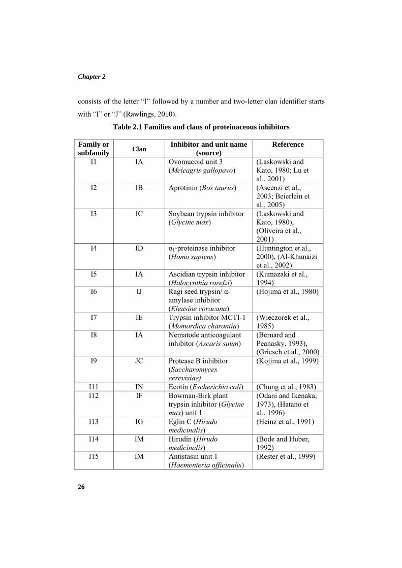
The classification of protein peptidase inhibitors is continually being
revised by MEROPS database and currently inhibitors are grouped into 71
families based on comparisons of protein sequences which include 17451 inhibitor
sequences. Their molecular weight and mechanism of inhibition varies from
inhibitor to inhibitor. These families can be further grouped into 39 clans based on
comparisons of tertiary structure. The family and clan of some protein peptidase
inhibitors are depicted in Table 2.1. Each clan, family and biochemically
characterized peptidase inhibitor is given a unique identifier. A family identifier
Chapter 2
26
consists of the letter “I” followed by a number and two-letter clan identifier starts
with “I” or “J” (Rawlings, 2010).
Table 2.1 Families and clans of proteinaceous inhibitors
Family or subfamily
Clan Inhibitor and unit name
(source) Reference
I1 IA Ovomucoid unit 3 (Meleagris gallopavo)
(Laskowski and Kato, 1980; Lu et al., 2001)
I2 IB Aprotinin (Bos taurus) (Ascenzi et al., 2003; Beierlein et al., 2005)
I3 IC Soybean trypsin inhibitor (Glycine max)
(Laskowski and Kato, 1980), (Oliveira et al., 2001)
I4 ID α1-proteinase inhibitor (Homo sapiens)
(Huntington et al., 2000), (Al-Khunaizi et al., 2002)
I5 IA Ascidian trypsin inhibitor (Halocynthia rorefzi)
(Kumazaki et al., 1994)
I6 IJ Ragi seed trypsin/ α-amylase inhibitor (Eleusine coracana)
(Hojima et al., 1980)
I7 IE Trypsin inhibitor MCTI-1 (Momordica charantia)
(Wieczorek et al., 1985)
I8 IA Nematode anticoagulant inhibitor (Ascaris suum)
(Bernard and Peanasky, 1993), (Griesch et al., 2000)
I9 JC Protease B inhibitor (Saccharomyces cerevisiae)
(Kojima et al., 1999)
I11 IN Ecotin (Escherichia coli) (Chung et al., 1983) I12 IF Bowman-Birk plant
trypsin inhibitor (Glycine max) unit 1
(Odani and Ikenaka, 1973), (Hatano et al., 1996)
I13 IG Eglin C (Hirudo medicinalis)
(Heinz et al., 1991)
I14 IM Hirudin (Hirudo medicinalis)
(Bode and Huber, 1992)
I15 IM Antistasin unit 1 (Haementeria officinalis)
(Rester et al., 1999)
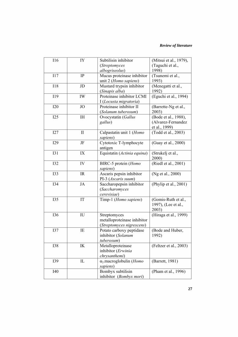
Review of literature
27
I16 IY Subtilisin inhibitor (Streptomyces albogriseolus)
(Mitsui et al., 1979), (Taguchi et al., 1998)
I17 IP Mucus proteinase inhibitor unit 2 (Homo sapiens)
(Tsunemi et al., 1993)
I18 JD Mustard trypsin inhibitor (Sinapis alba)
(Menegatti et al., 1992)
I19 IW Proteinase inhibitor LCMI I (Locusta migratoria)
(Eguchi et al., 1994)
I20 JO Proteinase inhibitor II (Solanum tuberosum)
(Barrette-Ng et al., 2003)
I25 IH Ovocystatin (Gallus gallus)
(Bode et al., 1988), (Alvarez-Fernandez et al., 1999)
I27 II Calpastatin unit 1 (Homo sapiens)
(Todd et al., 2003)
I29 JF Cytotoxic T-lymphocyte antigen
(Guay et al., 2000)
I31 IX Equistatin (Actinia equina) (Strukelj et al., 2000)
I32 IV BIRC-5 protein (Homo sapiens)
(Riedl et al., 2001)
I33 IR Ascaris pepsin inhibitor PI-3 (Ascaris suum)
(Ng et al., 2000)
I34 JA Saccharopepsin inhibitor (Saccharomyces cerevisiae)
(Phylip et al., 2001)
I35 IT Timp-1 (Homo sapiens) (Gomis-Ruth et al., 1997), (Lee et al., 2003)
I36 IU Streptomyces metalloproteinase inhibitor (Streptomyces nigrescens)
(Hiraga et al., 1999)
I37 IE Potato carboxy peptidase inhibitor (Solanum tuberosum)
(Bode and Huber, 1992)
I38 IK Metalloproteinase inhibitor (Erwinia chrysanthemi)
(Feltzer et al., 2003)
I39 IL α2-macroglobulin (Homo sapiens)
(Barrett, 1981)
I40 Bombyx subtilisin inhibitor (Bombyx mori)
(Pham et al., 1996)
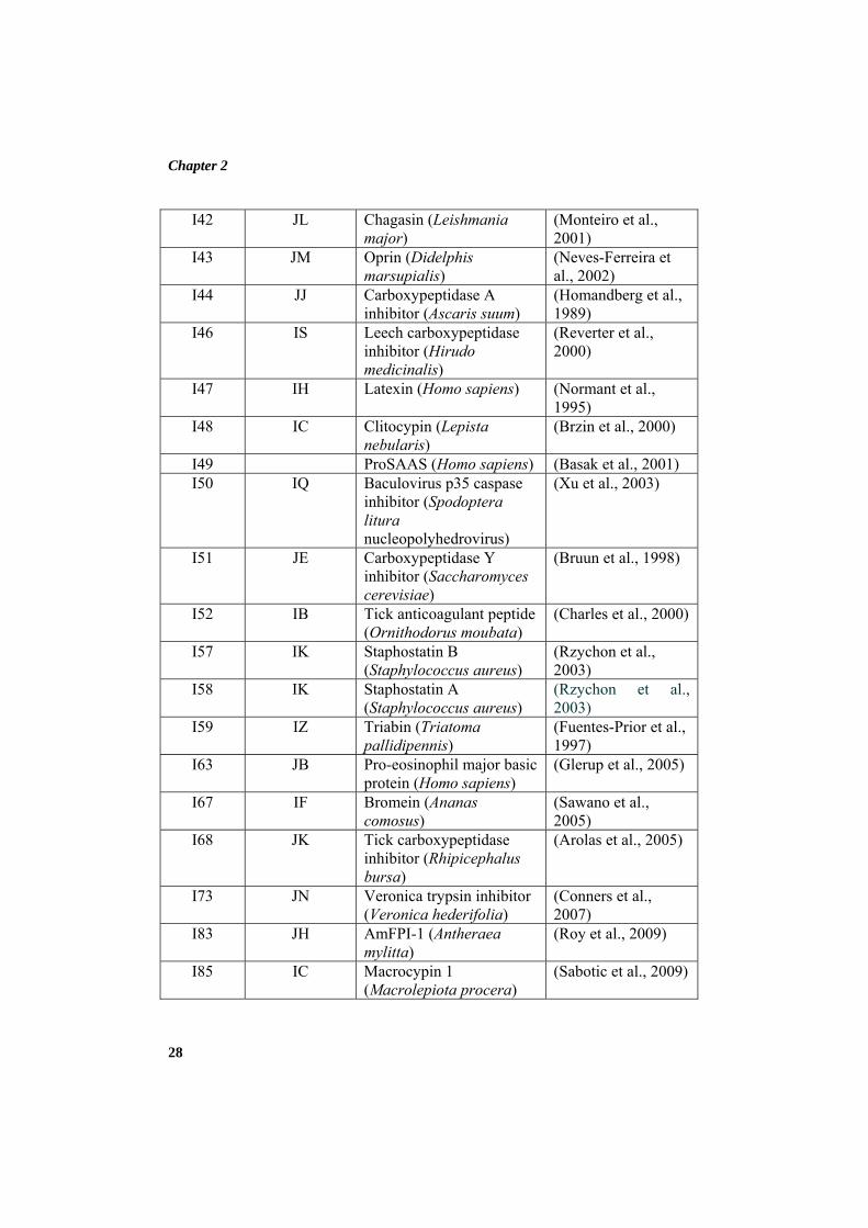
Chapter 2
28
I42 JL Chagasin (Leishmania major)
(Monteiro et al., 2001)
I43 JM Oprin (Didelphis marsupialis)
(Neves-Ferreira et al., 2002)
I44 JJ Carboxypeptidase A inhibitor (Ascaris suum)
(Homandberg et al., 1989)
I46 IS Leech carboxypeptidase inhibitor (Hirudo medicinalis)
(Reverter et al., 2000)
I47 IH Latexin (Homo sapiens) (Normant et al., 1995)
I48 IC Clitocypin (Lepista nebularis)
(Brzin et al., 2000)
I49 ProSAAS (Homo sapiens) (Basak et al., 2001) I50 IQ Baculovirus p35 caspase
inhibitor (Spodoptera litura nucleopolyhedrovirus)
(Xu et al., 2003)
I51 JE Carboxypeptidase Y inhibitor (Saccharomyces cerevisiae)
(Bruun et al., 1998)
I52 IB Tick anticoagulant peptide (Ornithodorus moubata)
(Charles et al., 2000)
I57 IK Staphostatin B (Staphylococcus aureus)
(Rzychon et al., 2003)
I58 IK Staphostatin A (Staphylococcus aureus)
(Rzychon et al., 2003)
I59 IZ Triabin (Triatoma pallidipennis)
(Fuentes-Prior et al., 1997)
I63
JB Pro-eosinophil major basic protein (Homo sapiens)
(Glerup et al., 2005)
I67 IF Bromein (Ananas comosus)
(Sawano et al., 2005)
I68 JK Tick carboxypeptidase inhibitor (Rhipicephalus bursa)
(Arolas et al., 2005)
I73
JN Veronica trypsin inhibitor (Veronica hederifolia)
(Conners et al., 2007)
I83 JH AmFPI-1 (Antheraea mylitta)
(Roy et al., 2009)
I85 IC Macrocypin 1 (Macrolepiota procera)
(Sabotic et al., 2009)
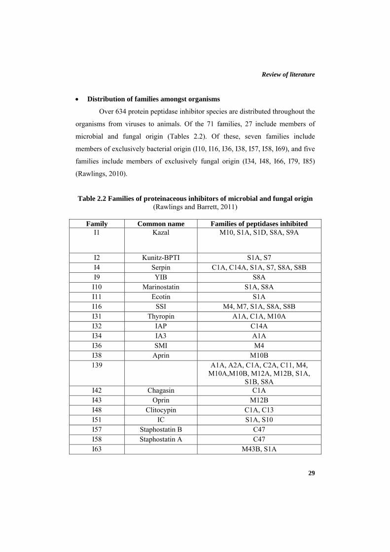
Review of literature
29
• Distribution of families amongst organisms
Over 634 protein peptidase inhibitor species are distributed throughout the
organisms from viruses to animals. Of the 71 families, 27 include members of
microbial and fungal origin (Tables 2.2). Of these, seven families include
members of exclusively bacterial origin (I10, I16, I36, I38, I57, I58, I69), and five
families include members of exclusively fungal origin (I34, I48, I66, I79, I85)
(Rawlings, 2010).
Table 2.2 Families of proteinaceous inhibitors of microbial and fungal origin (Rawlings and Barrett, 2011)
Family Common name Families of peptidases inhibited
I1
Kazal M10, S1A, S1D, S8A, S9A
I2 Kunitz-BPTI S1A, S7 I4 Serpin C1A, C14A, S1A, S7, S8A, S8B I9 YIB S8A
I10 Marinostatin S1A, S8A I11 Ecotin S1A I16 SSI M4, M7, S1A, S8A, S8B I31 Thyropin A1A, C1A, M10A I32 IAP C14A I34 IA3 A1A I36 SMI M4 I38 Aprin M10B 139 A1A, A2A, C1A, C2A, C11, M4,
M10A,M10B, M12A, M12B, S1A, S1B, S8A
I42 Chagasin C1A I43 Oprin M12B I48 Clitocypin C1A, C13 I51 IC S1A, S10 I57 Staphostatin B C47 I58 Staphostatin A C47 I63 M43B, S1A
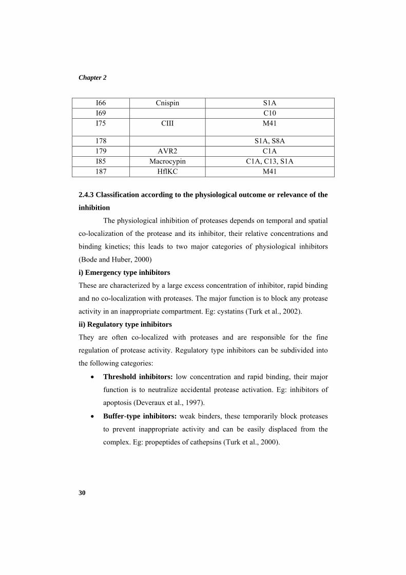
Chapter 2
30
I66 Cnispin S1A I69 C10 I75
CIII M41
178 S1A, S8A 179 AVR2 C1A I85 Macrocypin C1A, C13, S1A 187 HflKC M41
2.4.3 Classification according to the physiological outcome or relevance of the
inhibition
The physiological inhibition of proteases depends on temporal and spatial
co-localization of the protease and its inhibitor, their relative concentrations and
binding kinetics; this leads to two major categories of physiological inhibitors
(Bode and Huber, 2000)
i) Emergency type inhibitors
These are characterized by a large excess concentration of inhibitor, rapid binding
and no co-localization with proteases. The major function is to block any protease
activity in an inappropriate compartment. Eg: cystatins (Turk et al., 2002).
ii) Regulatory type inhibitors
They are often co-localized with proteases and are responsible for the fine
regulation of protease activity. Regulatory type inhibitors can be subdivided into
the following categories:
• Threshold inhibitors: low concentration and rapid binding, their major
function is to neutralize accidental protease activation. Eg: inhibitors of
apoptosis (Deveraux et al., 1997).
• Buffer-type inhibitors: weak binders, these temporarily block proteases
to prevent inappropriate activity and can be easily displaced from the
complex. Eg: propeptides of cathepsins (Turk et al., 2000).
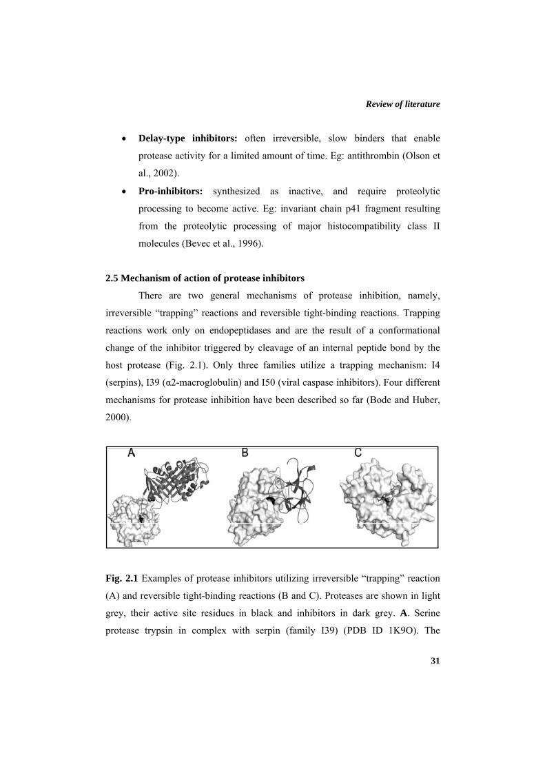
Review of literature
31
• Delay-type inhibitors: often irreversible, slow binders that enable
protease activity for a limited amount of time. Eg: antithrombin (Olson et
al., 2002).
• Pro-inhibitors: synthesized as inactive, and require proteolytic
processing to become active. Eg: invariant chain p41 fragment resulting
from the proteolytic processing of major histocompatibility class II
molecules (Bevec et al., 1996).
2.5 Mechanism of action of protease inhibitors
There are two general mechanisms of protease inhibition, namely,
irreversible “trapping” reactions and reversible tight-binding reactions. Trapping
reactions work only on endopeptidases and are the result of a conformational
change of the inhibitor triggered by cleavage of an internal peptide bond by the
host protease (Fig. 2.1). Only three families utilize a trapping mechanism: I4
(serpins), I39 (α2-macroglobulin) and I50 (viral caspase inhibitors). Four different
mechanisms for protease inhibition have been described so far (Bode and Huber,
2000).
Fig. 2.1 Examples of protease inhibitors utilizing irreversible “trapping” reaction
(A) and reversible tight-binding reactions (B and C). Proteases are shown in light
grey, their active site residues in black and inhibitors in dark grey. A. Serine
protease trypsin in complex with serpin (family I39) (PDB ID 1K9O). The
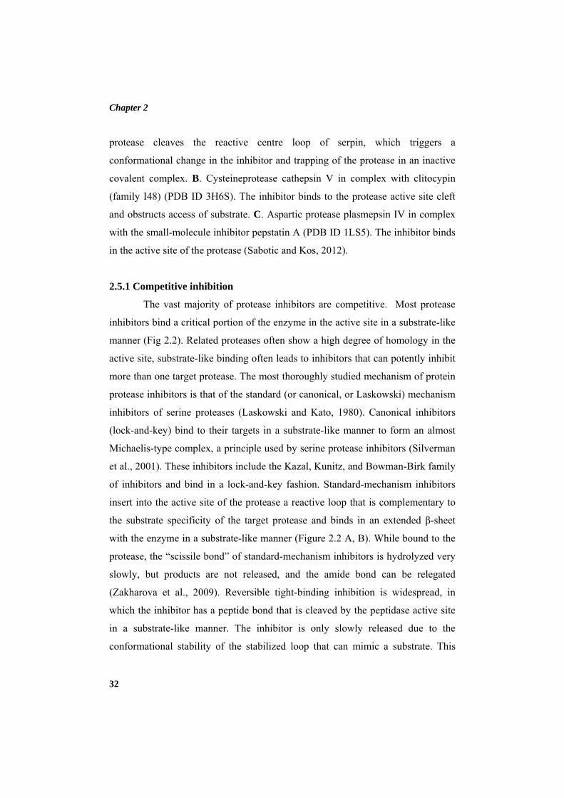
Chapter 2
32
protease cleaves the reactive centre loop of serpin, which triggers a
conformational change in the inhibitor and trapping of the protease in an inactive
covalent complex. B. Cysteineprotease cathepsin V in complex with clitocypin
(family I48) (PDB ID 3H6S). The inhibitor binds to the protease active site cleft
and obstructs access of substrate. C. Aspartic protease plasmepsin IV in complex
with the small-molecule inhibitor pepstatin A (PDB ID 1LS5). The inhibitor binds
in the active site of the protease (Sabotic and Kos, 2012).
2.5.1 Competitive inhibition
The vast majority of protease inhibitors are competitive. Most protease
inhibitors bind a critical portion of the enzyme in the active site in a substrate-like
manner (Fig 2.2). Related proteases often show a high degree of homology in the
active site, substrate-like binding often leads to inhibitors that can potently inhibit
more than one target protease. The most thoroughly studied mechanism of protein
protease inhibitors is that of the standard (or canonical, or Laskowski) mechanism
inhibitors of serine proteases (Laskowski and Kato, 1980). Canonical inhibitors
(lock-and-key) bind to their targets in a substrate-like manner to form an almost
Michaelis-type complex, a principle used by serine protease inhibitors (Silverman
et al., 2001). These inhibitors include the Kazal, Kunitz, and Bowman-Birk family
of inhibitors and bind in a lock-and-key fashion. Standard-mechanism inhibitors
insert into the active site of the protease a reactive loop that is complementary to
the substrate specificity of the target protease and binds in an extended β-sheet
with the enzyme in a substrate-like manner (Figure 2.2 A, B). While bound to the
protease, the “scissile bond” of standard-mechanism inhibitors is hydrolyzed very
slowly, but products are not released, and the amide bond can be relegated
(Zakharova et al., 2009). Reversible tight-binding inhibition is widespread, in
which the inhibitor has a peptide bond that is cleaved by the peptidase active site
in a substrate-like manner. The inhibitor is only slowly released due to the
conformational stability of the stabilized loop that can mimic a substrate. This
Review of literature
33
mechanism has been conclusively demonstrated only for inhibitors of serine
proteases. Other reversible tight-binding protease inhibitors physically block the
protease active site by high-affinity binding to sites on either side of the active
site.
Fig 2.2 Competitive, active-site inhibitors of proteases. A. Inhibitors bind
in the active site, but not in a substrate-like manner. Peptide extensions bind in
specificity subsites, and can interact with the catalytic residues (rectangle). B.
Crystal structures of a serine protease (matriptase/MT-SP1, PDB ID: 1EAW) in
complex with the standard-mechanism inhibitor aprotinin, and C. the cystatin
stefin A in complex with a cysteine protease (cathepsin H, PDB ID: 1NB5). Both
inhibitors bind in the active-site groove of their targets (Farady and Craik, 2010).
The standard mechanism is used by many structurally disparate protein
scaffolds to create potent inhibitors. However, the majority of standard-
mechanism protease inhibitors tend to have relatively broad specificity within
subclasses of serine proteases. For example, bovine pancreatic trypsin inhibitor
(BPTI) efficiently inhibits almost all trypsin-fold serine proteases with P1-Arg
specificity with sub-nanomolar potency, and can also potently inhibit
Chapter 2
34
chymotrypsin (Phe P1 specificity) with a Ki of 10 nm (Castro and Anderson,
1996). Binding of an inhibitor to the active site can also be irreversible, when an
electrophilic reactive group of the inhibitor forms a covalent bond with an amino
acid residue in the enzyme active site.
The majority of protease inhibitors bind in and blocks access to the active
site of their target protease, but do not bind in a strictly substrate-like manner.
Instead they interact with the protease subsites and catalytic residues in a
noncatalytically competent manner. This differentiates them from standard
mechanism inhibitors; however, like standard-mechanism inhibitors, they get
most of their potency from interactions within the protease active site, and tend to
potently inhibit many related proteases. The cystatins, a superfamily of proteins
that inhibit papainlike cysteine proteases, are a classic example of these inhibitors
(Fig 2.2 A, C). The cystatins insert into the V-shaped active site of a cysteine
protease a wedge-like face of the inhibitor that consists of the protein N terminus
and two hairpin loops. The N-terminal residues bind in the S3–S1 pockets in a
substratelike manner, but the peptide then turns away from the catalytic residues
and out of the active site. The two hairpin loops bind to the prime side of the
active site, which provides the majority of the binding energy for the interaction.
Thus, both the prime and nonprime sides of the active site are occupied, but no
interactions are actually made with the catalytic machinery of the enzyme (Bode
and Huber, 2000). The four human tissue inhibitors of metalloproteases (TIMPs)
are responsible for the inhibition of dozens of extracellular metalloproteases (Fig
2.2 A). They bind to their target enzymes in a two-step mechanism similar to that
of the cystatins. While the N-terminal residues of cystatins bind to the nonprime
side of cysteine proteases, TIMPs N termini bind in the P1-P3’ pockets of the
protease, coordinate the catalytic Zn2+ ion, and exclude a catalytic water molecule
from the active site. Meanwhile a second loop of the TIMP binds both in the P3
and P2 pockets, and binds to the N terminus of the matrix metalloprotease (MMP).
Despite the similarities in mechanistic architecture between TIMPs and cystatins
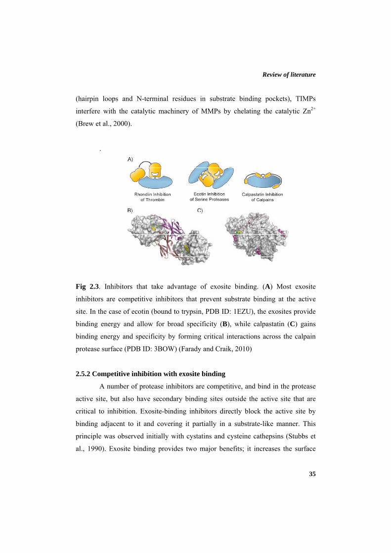
Review of literature
35
(hairpin loops and N-terminal residues in substrate binding pockets), TIMPs
interfere with the catalytic machinery of MMPs by chelating the catalytic Zn2+
(Brew et al., 2000).
.
Fig 2.3. Inhibitors that take advantage of exosite binding. (A) Most exosite
inhibitors are competitive inhibitors that prevent substrate binding at the active
site. In the case of ecotin (bound to trypsin, PDB ID: 1EZU), the exosites provide
binding energy and allow for broad specificity (B), while calpastatin (C) gains
binding energy and specificity by forming critical interactions across the calpain
protease surface (PDB ID: 3BOW) (Farady and Craik, 2010)
2.5.2 Competitive inhibition with exosite binding
A number of protease inhibitors are competitive, and bind in the protease
active site, but also have secondary binding sites outside the active site that are
critical to inhibition. Exosite-binding inhibitors directly block the active site by
binding adjacent to it and covering it partially in a substrate-like manner. This
principle was observed initially with cystatins and cysteine cathepsins (Stubbs et
al., 1990). Exosite binding provides two major benefits; it increases the surface
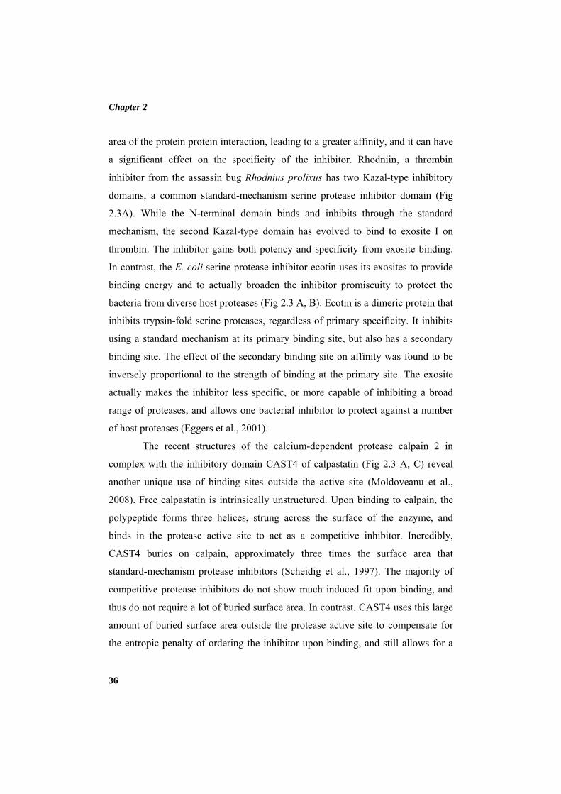
Chapter 2
36
area of the protein protein interaction, leading to a greater affinity, and it can have
a significant effect on the specificity of the inhibitor. Rhodniin, a thrombin
inhibitor from the assassin bug Rhodnius prolixus has two Kazal-type inhibitory
domains, a common standard-mechanism serine protease inhibitor domain (Fig
2.3A). While the N-terminal domain binds and inhibits through the standard
mechanism, the second Kazal-type domain has evolved to bind to exosite I on
thrombin. The inhibitor gains both potency and specificity from exosite binding.
In contrast, the E. coli serine protease inhibitor ecotin uses its exosites to provide
binding energy and to actually broaden the inhibitor promiscuity to protect the
bacteria from diverse host proteases (Fig 2.3 A, B). Ecotin is a dimeric protein that
inhibits trypsin-fold serine proteases, regardless of primary specificity. It inhibits
using a standard mechanism at its primary binding site, but also has a secondary
binding site. The effect of the secondary binding site on affinity was found to be
inversely proportional to the strength of binding at the primary site. The exosite
actually makes the inhibitor less specific, or more capable of inhibiting a broad
range of proteases, and allows one bacterial inhibitor to protect against a number
of host proteases (Eggers et al., 2001).
The recent structures of the calcium-dependent protease calpain 2 in
complex with the inhibitory domain CAST4 of calpastatin (Fig 2.3 A, C) reveal
another unique use of binding sites outside the active site (Moldoveanu et al.,
2008). Free calpastatin is intrinsically unstructured. Upon binding to calpain, the
polypeptide forms three helices, strung across the surface of the enzyme, and
binds in the protease active site to act as a competitive inhibitor. Incredibly,
CAST4 buries on calpain, approximately three times the surface area that
standard-mechanism protease inhibitors (Scheidig et al., 1997). The majority of
competitive protease inhibitors do not show much induced fit upon binding, and
thus do not require a lot of buried surface area. In contrast, CAST4 uses this large
amount of buried surface area outside the protease active site to compensate for
the entropic penalty of ordering the inhibitor upon binding, and still allows for a
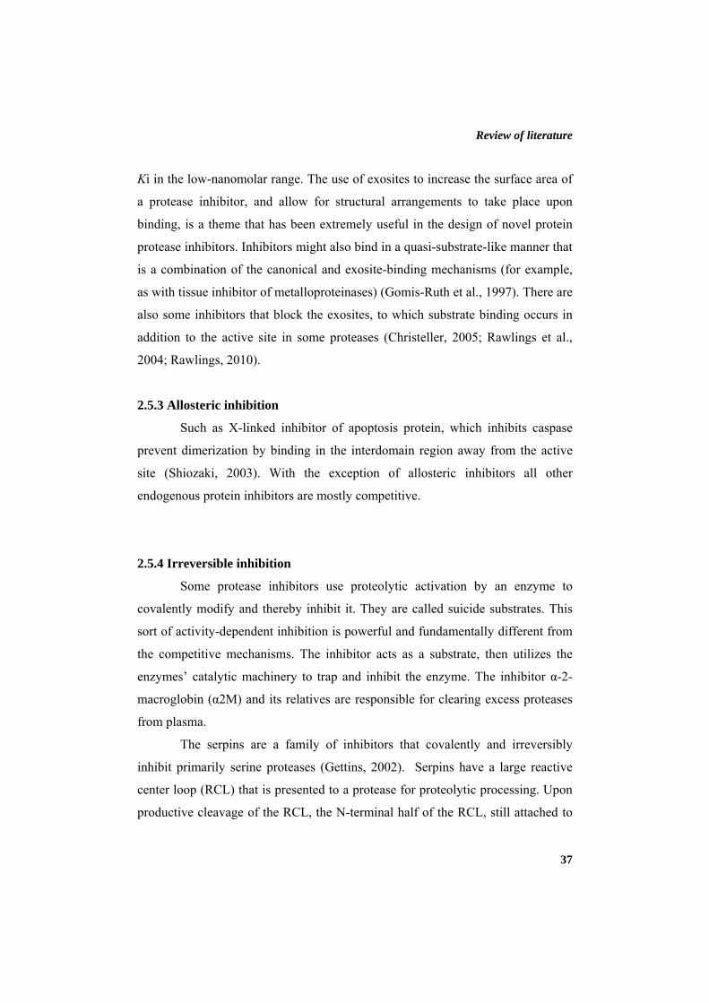
Review of literature
37
Ki in the low-nanomolar range. The use of exosites to increase the surface area of
a protease inhibitor, and allow for structural arrangements to take place upon
binding, is a theme that has been extremely useful in the design of novel protein
protease inhibitors. Inhibitors might also bind in a quasi-substrate-like manner that
is a combination of the canonical and exosite-binding mechanisms (for example,
as with tissue inhibitor of metalloproteinases) (Gomis-Ruth et al., 1997). There are
also some inhibitors that block the exosites, to which substrate binding occurs in
addition to the active site in some proteases (Christeller, 2005; Rawlings et al.,
2004; Rawlings, 2010).
2.5.3 Allosteric inhibition
Such as X-linked inhibitor of apoptosis protein, which inhibits caspase
prevent dimerization by binding in the interdomain region away from the active
site (Shiozaki, 2003). With the exception of allosteric inhibitors all other
endogenous protein inhibitors are mostly competitive.
2.5.4 Irreversible inhibition
Some protease inhibitors use proteolytic activation by an enzyme to
covalently modify and thereby inhibit it. They are called suicide substrates. This
sort of activity-dependent inhibition is powerful and fundamentally different from
the competitive mechanisms. The inhibitor acts as a substrate, then utilizes the
enzymes’ catalytic machinery to trap and inhibit the enzyme. The inhibitor α-2-
macroglobin (α2M) and its relatives are responsible for clearing excess proteases
from plasma.
The serpins are a family of inhibitors that covalently and irreversibly
inhibit primarily serine proteases (Gettins, 2002). Serpins have a large reactive
center loop (RCL) that is presented to a protease for proteolytic processing. Upon
productive cleavage of the RCL, the N-terminal half of the RCL, still attached to
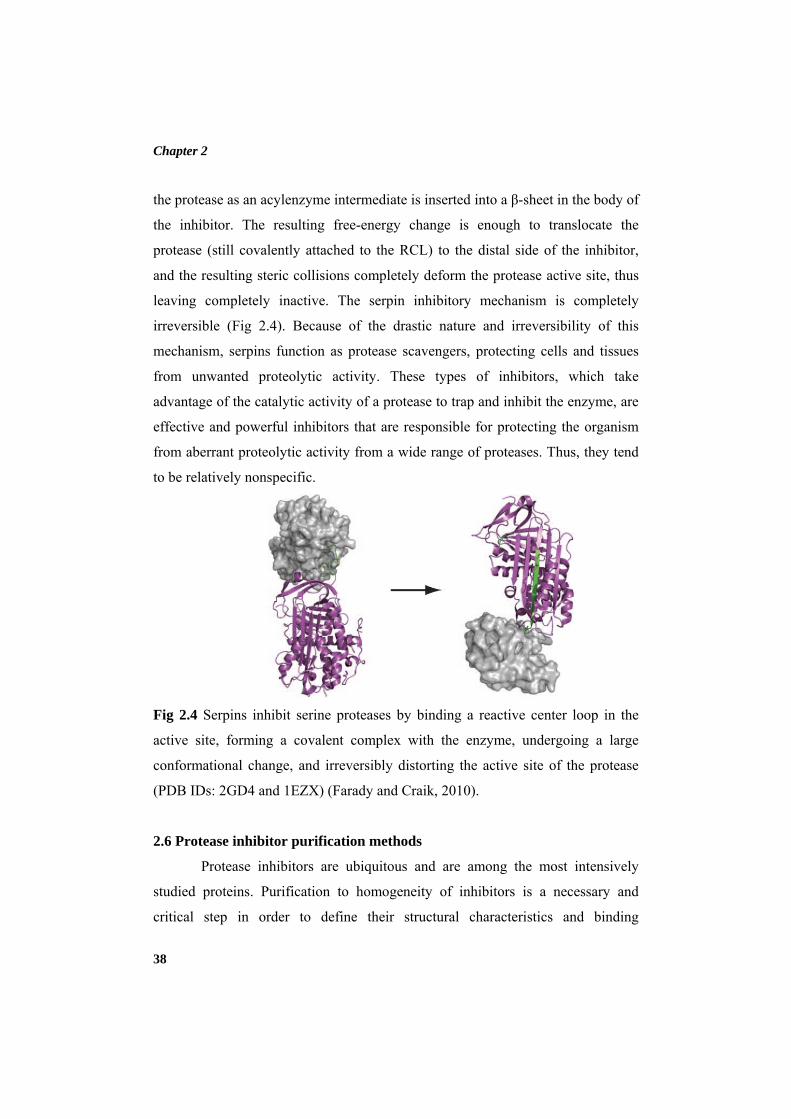
Chapter 2
38
the protease as an acylenzyme intermediate is inserted into a β-sheet in the body of
the inhibitor. The resulting free-energy change is enough to translocate the
protease (still covalently attached to the RCL) to the distal side of the inhibitor,
and the resulting steric collisions completely deform the protease active site, thus
leaving completely inactive. The serpin inhibitory mechanism is completely
irreversible (Fig 2.4). Because of the drastic nature and irreversibility of this
mechanism, serpins function as protease scavengers, protecting cells and tissues
from unwanted proteolytic activity. These types of inhibitors, which take
advantage of the catalytic activity of a protease to trap and inhibit the enzyme, are
effective and powerful inhibitors that are responsible for protecting the organism
from aberrant proteolytic activity from a wide range of proteases. Thus, they tend
to be relatively nonspecific.
Fig 2.4 Serpins inhibit serine proteases by binding a reactive center loop in the
active site, forming a covalent complex with the enzyme, undergoing a large
conformational change, and irreversibly distorting the active site of the protease
(PDB IDs: 2GD4 and 1EZX) (Farady and Craik, 2010).
2.6 Protease inhibitor purification methods
Protease inhibitors are ubiquitous and are among the most intensively
studied proteins. Purification to homogeneity of inhibitors is a necessary and
critical step in order to define their structural characteristics and binding
Review of literature
39
specificity to the proteases. Isolate these from all other proteins that are present in
the same biological source is a difficult task, since the protease inhibitors have a
large molecular diversity. So the process of purification includes a combination of
separation strategies, such as: extraction, fractionation by precipitation and
chromatographic techniques addressing different properties. The type and amount
of technique will depend on the nature and characteristics of the molecule of
interest, as well as the degree of purity desired in the final product.
Once inhibitory activity is detected in a crude sample, it is important to
perform an initial simple fractionation of the inhibitor activity: eg. ammonium
sulphate, organic solvent and polyethylene glycol fractionation. Protease inhibitors
are precipitated using acetic acid, ethanol, acetone, or combination of ethanol and
acetone (Bhattacharyya et al., 2006; Haq et al., 2005; Kubiak et al., 2009; Macedo
et al., 2000; Tian and Zhang, 2005). This is to determine whether the specific
activity of the inhibitor increases after such fractionation. Methods like SDS-
PAGE, Isoelectric focusing, MALDI-TOF etc. are used for confirming
homogeneity. Protease inhibitor with antipathogenic and anti-proliferative
activities from mung bean has been purified by ammonium sulphate precipitation,
Cation-exchange chromatography on CM-Sephadex C-50 column, perfusion
chromatography on a BioCAD 700E work station and C18 capillary reverse-phase
high-performance liquid chromatography (Wanga et al., 2006). To establish
effective purification of the inhibitor sample it is important to determine the
specific activity of the inhibitor sample in each step of purifications (Hideaki and
Guy, 2001). The homogeneity is confirmed by methods like SDS-PAGE,
Isoelectric focusing and MALDI-TOF. Depending on the type of protein-resin
interactions, there are four types of chromatographic techniques. These techniques
are ion exchange, affinity, size exclusion and hydrophobic chromatography.
Though all four chromatographic techniques are used in the purification process,
affinity and ion exchange chromatography are by far the widely used techniques in
the biopharmaceutical industry and in academia.
Chapter 2
40
Purification procedures like ion exchange chromatography, gel filtration
chromatography, high performance liquid chromatography, reversed-phase high-
performance liquid chromatography and affinity chromatography are used for
purifying protease inhibitor from the crude extract. Ion exchange chromatography
is based on the principle of charge-charge interaction, wherein similar charges
repel and opposite charges attract. The protein migration in an ion exchange
column is dependent on the interaction between the protein net charge and the
charge of resin. The net charge of the protein is primarily dependent on the
protein’s isoelectric point (pI). In ion exchange chromatography, the whole protein
surface with its charge moieties modulates the protein binding process. The
matrices for ion exchange chromatography contain ionizable functional groups
such as diethyl amino ethyl (DEAE) and carboxy methyl (CM), which gets
associated with the charged protein molecule, thereby adsorbing the protein to the
matrices. The adsorbed protein molecule is eluted by a gradient change in the pH
or ionic strength of the eluting buffer or solution (Arindam et al., 2006;
Shrivastava and Ghosh, 2003).
Another very rapid and selective method for peptide and protein
purification is reversed-phase high performance liquid chromatography on porous,
microparticulate, chemically bonded alkylsilicas (Hearn, 1982). The most
commonly used reverse phase HPLC column includes, octadecylsilane column
(Shoji et al., 1999), Vyda C18 HPLC column (Cesar et al., 2004), Vydac 218 TP
1022 C-18 (Sivakumar et al., 2005) and μ-Bondapak C18 column (Ligia et al.,
2003). Among the chromatographic techniques, the affinity chromatography, by
which specific biological properties can be exploited, stands out for its high
purification capacity. For example, affinity chromatography on columns
containing immobilized enzymes provides an efficient and rapid process of
isolation and purification of protease inhibitors from different sources. This
procedure provides advantages as high enrichment of inhibitor fraction, reduction
of purification steps due to high binding specificity of the protein immobilized on
Review of literature
41
a chromatographic matrix and purification of different protease inhibitors in the
same fraction by differential elution of material retained on chromatographic
matrix.
Affinity chromatography is a very efficient technique capable of purifying
proteins based on reversible interactions between a protein and a specific ligand
coupled to a chromatographic matrix. The affinity chromatography is usually used
as one of the last steps in the purification process of protease inhibitors. The
binding property of the protease inhibitors with enzymes has been exploited for
the affinity purification of inhibitors from various sources. Nevertheless, due to
their specific and reversible binding capacity to the enzymes (without undergoing
chemical change), protease inhibitor purification can be greatly enhanced by the
use of affinity chromatography techniques, where the binding agent are particular
proteases (Araújo et al., 2005; Gomes et al., 2005). The common affinity ligands
used for the protease inhibitor purification includes, S-carboxymethyl-papain-
Sepharose (Gounaris et al., 1984), trypsin-Sepharose (Alinda et al., 2003),
chymotrypsin-Sepharose (Gennis and Cantor, 1976) and trypsin-agarose
(Sivakumar et al., 2005). Trypsin inhibitors from wild-type soybean (Glycine
soya) (WBTI) and domesticated soybean (Glycine max) (SBTI) are purified using
prepared chitosan resin-trypsin as filler on the affinity chromatography column
(Zhang et al., 2009). Serine protease inhibitor from the tissue-penetrating
Nematode Anisakis simplex was purified using C3 reverse-phase HPLC
chromatographic procedure and streptavidin-agarose affinity chromatography
(Morris and Sakanari, 1994). Serine protease inhibitor (AmPI) purified from larval
hemolymph of tasar silkworm Antheraea mylitta using trypsin-sepharose CL-6B
affinity column (Rai et al., 2010). A heat-stable serine protease inhibitor (Potide-
G) from the tubers of new potato variety ‘‘Golden Valley’’ through extraction of
the water-soluble fraction, dialysis, ultrafiltration and DEAE-cellulose and C18
reversed-phase high performance liquid chromatography (Mi-Hyun et al., 2006).
Chapter 2
42
Affinity chromatography on Affi-gel blue gel is used for the purification
of a 20-kDa Kunitz-type trypsin inhibitor from Gymnocladus chinensis (Yunnan
bean) seeds (Zhu et al., 2011). Trypsin inhibitors (TI) from wild-type soybean
(Glycine soya) (WBTI) and domesticated soybean (Glycine max) (SBTI) were
purified using prepared chitosan resin-trypsin as filler on the affinity
chromatography column (Zhang et al., 2009). A Kunitz-type trypsin inhibitor with
high stability from Spinacia oleracea L. seeds is purified by Sepharose 4B-
trypsin affinity chromatography (Kang et al., 2009). Purification of the inhibitors
from skin extract of Atlantic salmon (Salmo salar L.) was carried out in four
chromatographic steps: papain-affinity chromatography on papain-Sepharose
column, gel filtration on a Superdex 200 HR10/30 (Pharmacia Biotech) column,
anion exchange chromatography on a Mono Q HR 5/5 (Pharmacia Biotech)
column and reverse phase Chromatography on a 0.46 X 4 cm TSK TMS 250
column (C1, TosoHaas Corporation, Japan) (YloÈnen et al., 1999).
2.7 Characterization of protease inhibitors
Protease inhibitors play various important roles in living systems. They
can perform important functions due to their unique structures (Pandhare et al.,
2002; Sabotic and Kos, 2012). The characters of proteinaceous protease inhibitors
isolated from diverse sources were thoroughly studied based on their physiological
roles and accordingly the possible use of inhibitors can be determined. Therefore,
characterizing protease inhibitors by understanding the relationship between
protein structure and its function is crucial for determining the scope and
application. The determination of the physicochemical parameters characterizing
the structural stability of the inhibitors is essential to select effective and stable
inhibitors under a large variety of environmental conditions. Moreover, the
knowledge of their structural features is fundamental to understand the inhibitor-
enzyme interactions and allow novel approaches in the use of synthetic inhibitors
aiming for drug design.
Review of literature
43
2.7.1 pH and temperature stability
Protease inhibitors constitute one of the most important classs of proteins
that has got applications in various fields. Higher thermal stability is one of the
crucial properties for their biotechnological applications. A novel thermostable
protein inhibitor of trypsin and subtilisin, called BN, was isolated from the seeds
of Brassica nigra (Genov et al., 1997). The pH and temperature stability of the
alkaline protease inhibitors and the influence of various additives on their
thermostability were investigated as a prelude to their exploitation as biocontrol
agents (Pandhare et al., 2002). Serratia marcescens produced very small amounts
of a thermostable inhibitor protein (SmaPI) that shows an inhibitory activity
against extracellular 50 kDa metalloprotease (Kim et al., 1995). The pH and
temperature stability of the inhibitor can be attributed to the presence of disulfide
linkages (Vernekar et al., 1999). It was reported that the intra molecular disulphide
bridges are presumably responsible for the functional stability of Kunitz type
protease inhibitors in the presence of physical and chemical denaturants such as
temperature, pH and reducing agents (Kridric et al., 2002). There is a correlation
between the conformational stability and thermostability of serine protease
inhibitors (Kwon et al., 1994). Probably the extreme conditions of pH and
temperature might have totally distorted the structure of the inhibitor such that
they no longer bind with the enzymes or with their substrates. Temperature, ionic
strength, pH and other physicochemical factors can influence protein–protein
interactions (Veselovsky et al., 2002).
Thermostable variants of serpins may be of practical use because heat-
induced deactivation of serpins was shown to be a concentration-dependent
aggregation process (Kwon et al., 1994; Lomas et al., 1993). Temperature and pH
are important physical variables in enzyme-catalyzed reactions. The majority of
the protease inhibitors exhibiting anti-feedent properties reported so far, are active
against the neutral serine proteases such as trypsin and chymotrypsin (Ryan,
Chapter 2
44
1990). The alkaline protease inhibitor from Streptomyces sp. was found to be
stable over a wide pH and temperature ranges. Therefore, one can envision the
direct application of it as a biocontrol agent for the protection of plants against
phytopathogenic fungi by encapsulation for surface application or can be sprayed
directly (Vernekar et al., 1999). Three actinomycetes strains producing alkaline
protease inhibitors API-I, API-II and API-III respectively, exhibited different
properties in their molecular nature and in their pH and temperature stabilities.
The thermostability of protease inhibitor signifies its role as a stablizer for many
commercially important proteases, which has a major role in detergent industries
to withstand higher temperature (Pandhare et al., 2002).
High pH-stability and thermostability was shown by preparations of new
low molecular weight protein inhibitors of serine proteinases from buckwheat
Fagopyrum esculentum seeds (Tsybina et al., 2004). The pH stability dictates the
affinity of the inhibitor with different digestive proteases of the pest and insects,
mainly neutral proteases like trypsin and chymotrypsin implies the role of
inhibitors as biopesticides (Joshi et al., 1999). Protease inhibitor (PISC-2002)
isolated from culture supernatants of Streptomyces chromofuscus was found to be
pH tolerant and thermostable due to the presence of proline and a high content of
hydrophobic amino acids (Angelova et al., 2006).
2.7.2 Effect of metal ions on inhibitor
The presence of metal ions is necessary for the activity of some protease
inhibitors as they have a major role in maintaining the structural integrity of
protease inhibitor. The structural stability of a protein is enhanced by divalent
metal ions. This is because the metal ions serve to attain the critical conformation
that is needed for biological activity of the protein. Cysteine protease inhibitor
(CPI) from pearl millet needs the Zn2+ for the protease inhibitory and antifungal
activity of the protein, indicating a key role for this metal ion in these activities.
Review of literature
45
Analysis of metal chelated CPI using CD spectroscopy indicated a significant
change in the secondary structure (Joshi et al., 1999).
The side chain carboxylates of glutamate and aspartate residues can
participate in binding of divalent cations to metalloproteins. The main amino acid
residues that serve as ligands to divalent ions are Asp, His, Thr, and Glu (Kim and
Wyckoff, 1991; Sowadski et al., 1985). Studies on the impact of metal ions on the
activity of M. oleifera protease inhibitor illustrated that addition of divalent ions
such as Zn2+ at a concentration of 1 mM enhanced the protease inhibitor activity
up to 31% and Hg2+ at a concentration of 10 mM enhanced up to 64% of protease
inhibitor activity. Ca2+ and Mg2+ at higher concentration (10 mM) enhanced the
protease inhibitor activity only to a marginal level (Bijina et al., 2011a). The
inactivation of human coagulation factor Xa by the plasma proteinase inhibitors
α1-antitrypsin, antithrombin III and α2-macroglobulin in purified systems was
found to be accelerated by the divalent cations Ca2+, Mn2+ and Mg2+, whilst the
decrease in rate constant at higher concentrations of Ca2+ and Mn2+ may be due to
factor Xa dimerization (Vincent et al., 1983).
2.7.3 Effect of oxidizing agents on inhibitor
When proteins are exposed to oxidising agents such as hydrogen peroxide
(H2O2), periodate, dimethyl sulfoxide, chloramine-T, N-chlorosuccinamide and to
oxidants released by neutrophils (e.g. superoxide, hydroxyl radical), they are
subjected for oxidation (Brot and Weissbach, 1983; Vogt, 1995). Usually a decline
in the biological activity of the protein is observed by the oxidation of methionine
residues. The effect of methionine oxidation of α1-protease inhibitor (α1-PI) was
thoroughly studied. There are eight methionine residues and upon oxidation of one
of these residues (Met358), a complete loss of inhibitory activity of α1-PI was
observed toward its primary biological target, elastase (Johnson and Travis, 1979).
The specificity pattern of chymotrypsin has changed significantly upon oxidation
of methionine to its sulfoxide (Weiner et al., 1966). Studis on α2-macroglobulin
Chapter 2
46
revealed that the loss of activity of the protein on oxidation is due to the oxidation
of a single tryptophan residue (Reddy et al., 1994). Inhibitory activity of protease
inhibitor isolated from M. oleifera is declined by the increase in the concentration
(from 1% to 5%) of oxidising agents DMSO and H2O2 (Bijina et al., 2011a).
2.7.4 Effect of reducing agents on inhibitor
In many proteins the covalent link of cysteine residues by disulfide bonds
is important in maintaining a stable conformational stability for their biological
activity. So conformational destabilization of the protein is occured by the
removal of the disulfide bonds link of cysteine residues by reduction or
substitution for another amino acid residue (Kawamura et al., 2008; Zavodszky et
al., 2001). This covalent linkage is vital for the proper folding, stability and
function of many proteins of prokaryotes and eukaryotes (Creighton et al., 1995;
Frand et al., 2000; Hogg, 2003). There was no effect on protease inhibitors
isolated from Peltophorum dubium and Erythrina caffra, a Kunitz type trypsin
inhibitor when lower concentration of dithiothreitol (DTT) was used (Lehle et al.,
1996; Macedo et al., 2003). Unfolding of mutant leech carboxypeptidase inhibitor
(LCI) was resulted when reduced with dithiothreitol (DTT) (Arolas et al., 2009).
2.7.5 Effect of detergents on inhibitor
Three distinct modes of interaction of detergents with proteins has been
proposed (a) association with specific binding sites of native proteins; (b)
cooperative association between protein and a large number of detergent
molecules without major conformational change; (c) cooperative association with
conformational changes in the protein in which most of the hydrophobic residues
are presumably exposed for association with the detergent (Reynolds and Tanford,
1974). Proteases are added as key ingredients in detergents, which accounts for
approximately 25% of the total worldwide sales of enzymes and all detergent
proteases currently used in the market are serine proteases (Rao et al., 1998). So
Review of literature
47
studying the effect of detergents on inhibitor is significant when it is used along
with their corresponding proteases.
Detergents (cationic, anionic, zwitterionic and non-ionic) are used for
solubilizing proteins from lipid membranes. Moreover, proteinase inhibitors and
detergents are routinely used together in cell lysis buffers to inhibit unwanted
proteolysis and facilitate membrane protein solubilization in protein purification
procedures. Conformation of C. cajan inhibitor changed in the presence of SDS
and deoxycholate (DOC), in the presence of anionic bile salt, deoxycholate
resulting in the loss of inhibitory activity (Cardamone et al., 1994). Nonionic
detergents normally are considered as mild detergents and that they do not interact
extensively with the protein surface, whereas ionic detergents, in particular SDS,
generally bind unspecifically to the protein surface, which usually lead to protein
unfolding (Mogensen et al., 2005). Triton X-100, Tween 20 and Tween 80 are
nonionic polyoxyethylene detergents and the major interaction with proteins are
hydrophobic (Salameh and Wiegel, 2010). The effects of SDS and Tween 80 on
thermal stability to the alkaline protease inhibitors (API-I and API-II) from
actinomycetes were studied. SDS provided 60% protection only to API-I and
Tween-80 conferred 45% and 30% protection to API-I and API-II, respectively
(Pandhare et al., 2002).
The inhibitory activity of protease inhibitor isolated from Moringa
oleifera showed an increase in the inhibitory activity in the presence SDS (Bijina
et al., 2011a). Small amounts of nonionic or zwitterionic detergents are used in
crystallizing integral membrane proteins (Garavito et al., 1983; Michel, 1982;
Michel and Oesterhelt, 1980). These detergents help to inhibit micelle formation
and permit crystallization of proteins (Gilliland and Davies, 1984). The three
dimensional conformation of soybean Kunitz inhibitor has been changed due to
the action of SDS (Jirgensons, 1973). The property of detergents to mimic the
native, hydrophobic environment of the phospholipid bilayer is explored for the
Chapter 2
48
application in protein chemistry and in biotechnological processes (Israelachvili,
1991; Jones and Chapman, 1995; Tanford, 1980).
2.7.6 Effect of chemical modifiers on inhibitor
Chemical modification offer a very useful approach for giving proteins
some new and desired characteristics related to stability and catalytic activity. In
chemical modification, a chemical reagent binds covalently to specific amino acid
side chains of a protein to produce changes in the biological property of a protein
(Jonossen and Svendson, 1982; Yang et al., 1998). Chemical modification is an
important tool for studying structure–function relationship of biologically active
proteins. This can be done without knowing the protein structure (Gote et al.,
2007). Chemical modification is an alternative approach to genetic modification
for studying the structure-function-stability relationship of proteins and for
modifying their activity-stability properties reynold (Kaspari and Bogner, 2009).
It is widely used as a tool for studying role of individual amino acids, their
participation in the maintenance of the native conformation and for their
stabilization (He et al., 2000; Torchilin et al., 1979). The involvement and role
of amino acids in the dimerization of Bowman-Birk type of protease inhibitors
from the seeds of Horsegram (Dolichos biflorus) was investigated by chemical
modification (Kumar et al., 2004).
Chemical modification is an effective approach to obtain inhibitors highly
active against proteinases of the pests. This approach is exemplified by the new
form of oryzacystatin I, a protease inhibitor which differs from the original
inhibitor by the absence of one amino acid residue, Asp86 and is highly active
against proteinases of the pest, Caenorhabditis elegans and toxic to the parasitic
nematode Globodera pallida (Urwin et al., 1995). Chemical modification of
soybean cystatin scN and tomato (Lycopersicum esculentum L.) multicystatin
reveals that substitution of individual amino acid residues in the N-terminal
portion of one of multicystatin domains may considerably affect its ability to
Review of literature
49
inhibit diverse proteinases (Kiggundu et al., 2006; Koiwa et al., 2001). In the
majority of specific serine protease inhibitors, the inhibitory action is localized to
a specific reactive site situated within a loop closed by a disulphide bridge
(Mosolov and Valueva, 2005). It was reported that chemical modification of
Pearl millet cysteine protease inhibitor provided evidence for the presence of two
distinct sites responsible for antifungal and antifeedant activities (Joshi et al.,
1998).
Amino acids are usually modified with following chemicals: serine with
Phenyl methyl sulphonyl fluoride (PMSF), aspartic acid with N-Ethyl-5-
phenylisoxazolium-39-sulfonic acid (Woodward’s reagent K, WRK), arginine
with phenylglyoxal, histidine with Diethylpyrocarbonate (DEPC), tryptophan with
N-bromosuccinanmide (NBS) and cysteine with N-ethylmaleimide. A trypsin
inhibitor (PDTI) from Peltophorum dubium seeds was inactivated by lysine and
arginine modifying reagents, trinitrobenzene-sulfonic acid (TNBS) and 1,2-
cyclohexanedione (CHD), respectively (Ligia et al., 2003). Modification of
cysteine, lysine, tyrosine and carboxylic acids had no effect on the Pearl millet
cysteine protease inhibitor (CPI) activity, whereas modification of arginine and
histidine resulted in the activation of CPI activity (Joshi et al., 1999). The
inhibitory properties of a serine proteinase inhibitor from the fruiting body of the
basidiomycete, Lentinus edodes was studied by chemical modification. On
modification of arginine residues with cyclohexane-1,2-dione, the inhibitor
activity was completely abolished while modification of three lysine residues out
of the eight with sodium 2,4,6-trinitrobenzene-1-sulfonate did not significantly
affect the activity (Odani et al., 1999).
Peltophorum dubium protease inhibitor, a Kunitz type serine protease
inhibitor, was inactivated by lysine and arginine modification with tri-
nitrobenzene-sulfonic acid and 1, 2- 1 cyclohexanedione respectively (Macedo et
al., 2003). Chemical modification of the carboxylic and amine groups of a
bifunctional inhibitor (ATBI) from an extremophilic Bacillus sp. using N-Ethyl-5-
Chapter 2
50
phenylisoxazolium-39-sulfonic acid (Woodward’s reagent K, WRK) a carboxyl
group modifier, and 2,4,6-trinitrobenzenesulfonic acid (TNBS) an amine group
modifier of lysine resulted in the loss of inhibitory activity against xylanase and
aspartic protease and also in the loss of antifungal property, suggesting the
involvement of amino acids containing ionizable side chains (Lys, Asp, and Glu)
in the mechanistic pathway (Dash et al., 2001b). Dimerization of protease
inhibitors from the seeds of Horsegram (Dolichos biflorus) was investigated by
chemical modification of arginine (using 1,2-cyclohexanedione) lysine (using
citraconic anhydride) and histidine (using diethylpyrocarbonate, DEPC) residues
(Kumar et al., 2004).
2.7.7 Fluorescence binding studies
The fluorescence property of a folded protein is due to the presence of
aromatic amino acid residues. Tryptophan, tyrosine and phenylalanine are the
three aromatic aminoacids which may contribute to the intrinsic fluorescence of a
protein. The measure of the intrinsic flourescence of a folded protein is the total
contribution of individual aromatic aminoacids present in it. For the understanding
of the folding and binding events in proteins, fluorescence spectroscopy is used in
which the fluorescence property is made use of. This is an extremely sensitive
technique. Protein fluorescence is generally excited at 280 nm or at longer
wavelengths, usually at 295 nm. Fluorescence spectroscopy can give information
about the solvent accessibility of the chromophores (tryptophan, tyrosine, and
phenylalanine). Therefore, the fluorescence spectrum is sensitive to local changes
in the tertiary structure of a protein (Pace et al., 1989).
The conformational stability of potato cysteine protease inhibitor (PCPI)
was investigated by fluorescence spectroscopy. The PCPI isoforms investigated
have a highly similar structure at both the secondary and the tertiary level.
Changes in the tertiary structure of PCPI during heating were also monitored by
measuring the changes in fluorescence intensity as a function of temperature
Review of literature
51
(Pouvreau et al., 2005). In a study the monomeric form of an originally dimeric
serine proteinase inhibitor, ecotin, was compared with that of the iosoforms
fluorescent spectra (Gfibor and Grfif, 1996). The structural and functional studies
on ecotin and its interactions with different serine proteases have been investigated
by fluorescence titration (Seymour et al., 1994) which revealed the remarkable
features of this protein in inhibiting a strikingly large subset of the chymotrypsin
family of serine proteases (Mcgrath et al., 1999).
The role of the dimer interface on ecotin's unique broad specificities was
investigated by monitoring the monomer dimer dissociation constant (Kd)
(Seymour et al., 1994) by measuring the fluorescence (Yang et al., 1998).
Detergent-induced structural changes of proteins were analyzed efficaciously by
fluorescence emission as the fluorescence signal undergoes changes, reflective of
changes in protein conformation (Haq and Khan, 2005). The value of static
fluorescence spectra of the three isoforms from Acacia plumosa Lowe seeds
measured after excitation at 280 and 295 nm, were the same and the λmax emission
value at 341 nm (Lopes et al., 2009) corresponds to the position of spectra of class
II chromophores, in which tryptophan residues are exposed to the surface of the
compact native protein molecule (Burstein et al., 1973).
The intrinsic fluorescence spectrum of pearl millet cysteine protease
inhibitor (CPI) was found to be typical of a protein devoid of tryptophan residues.
The interaction of CPI modified for arginine or histidine with papain resulted in an
enhancement of CPI activity accompanied by a slight decrease in fluorescence
intensity. In contrast, modification of serine resulted in inhibition of CPI activity
with a concomitant increase of 20% in the fluorescence intensity when complexed
by the enzyme. This implies the involvement of enzyme-based tryptophan in the
formation of a biologically active enzyme-nhibitor complex (Joshi et al., 1998).
The tryptophan free variant α1-antitrypsin, an inhibitor of thrombin upon
formation of the serpin proteinase complex the emission maximum of 340 nm of
thrombin which blue shifts to 346 nm, concomitant with a 40% increase in
Chapter 2
52
intensity, indicative of substantial conformational change within the proteinase
(Tew and Bottomley, 2001). The binding studies on inhibition of the thermostable
xylanase (Xyl I) from a Thermomonospora sp. by a bifunctional inhibitor ATBI,
isolated from an extremophilic Bacillus sp. were carried out. The binding of ATBI
resulted in a concentration-dependent quenching of the fluorescence. The steady-
state kinetics revealed a two-step inhibition mechanism, and the conformational
modes observed during the binding of inhibitor to the enzyme were conveniently
monitored by fluorescence analysis. Fluorescence spectroscopy plays a very
important role in the determination of kinetic constants of enzyme inhibition
(Dash et al., 2001b).
2.7.8 Kinetics of inhibition
Enzyme inhibition studies are conducted to evaluate the occurrence and
extent of protein-drug interactions. To characterize an inhibitor, one needs to
understand the nature of inhibition process (i.e., competitive, noncompetitive, or
uncompetitive) and the inhibition constant (Ki). The inhibitor constant, Ki,
indicates the efficacy of an inhibitor to its target enzyme. A given inhibitor may
inhibit a number of proteases, and potency of an inhibitor is often described as the
molar concentration of the inhibitor that gives 50% inhibition of the target enzyme
activity (IC50) or just the percentage inhibition of the enzymatic activity of a fixed
concentration of the inhibitor (Hideaki and Guy, 2001). Determination of the
kinetic parameters of the inhibition of proteases will provide insight into the
mechanism of the interaction between the enzyme and the inhibitor.
A broad spectrum kunitz type serine protease inhibitor (rAceKI-1)
secreted by the hookworm Ancylostoma ceylanicum shows a dose-response
behavior suggesting the competitive nature with the protease substrates. Kinetic
assays showed that rAceKI-1 is a “tight binding” (Williams and Morrison, 1979)
inhibitor of chymotrypsin, pancreatic elastase, neutrophil elastase, and trypsin
(Aaron et al., 2000). A full-length cDNA, encoding a Bowman-Birk protease
Review of literature
53
inhibitor (BBI) from lentil immature seeds found to inhibit trypsin and
chymotrypsin, with Ki values at the nanomolar level (Caccialupi et al., 2010). This
is comparable to those previously reported for trypsin/chymotrypsin BBI extracted
from lentil seeds (Caccialupi et al., 2010; Ragg et al., 2006).
Ecotin, a serine protease inhibitor found in the periplasm of Escherichia
coli, has been characterized as a potent reversible tight-binding inhibitor of the
human contact activation proteases factor XIIa (FXIIa) and plasma kallikrein,
having Ki values of 89 pM and 163 pM, respectively. Ecotin also inhibited human
leukocyte elastase (HLE) with high affinity (Ki = 55 pM) (Ulmer et al., 1995). The
serine protease inhibitor protein obtained from a gene Spi1C from a metagenomic
library of uncultured marine microorganisms exhibited inhibitory activity against
α-chymotrypsin and trypsin with Ki values of around 1.79 × 10⁻⁸ and 1.52 × 10⁻⁸
M, respectively (Cheng-Jian Jiang et al., 2011).
The Ki of a serine protease inhibitor named bicolin from the venom of
Vespa bicolor Fabricius toward trypsin was 5.5 × 10⁻⁷ M and no inhibition was
detected to elastase and α-chymotrypsin, respectivley (Yang et al., 2009). A serine
protease inhibitor (Bungaruskuni) from Bungarus fasciatus venom had a Ki of
around 6.1 × 10⁻⁶ M to chymotrypsin (Lu et al., 2008). Protease inhibitor (PISC-
2002) from culture supernatants of Streptomyces chromofuscus inhibited
subtilisin, proteinase K, trypsin and proteinase of S. albovinaceus strongly, with
Ki of micromolar range and weakly inhibited pepsin (Angelova et al., 2006).
A cDNA encoding a precursor of equistatin, a potent cysteine and aspartic
proteinase inhibitor, was isolated from the sea anemone Actinia equine and the
mature protein region as well as those coding forn each of the domains were
expressed in the periplasmic space of Escherichia coli. It was found that the whole
recombinant equistatin and its first domain, but not the second and third domains,
inhibited the cysteine proteinase papain (Ki 0.60 nM) (Trukelj et al., 2000). A
bifunctional inhibitor (ATBI) from an extremophilic Bacillus sp. exhibiting an
activity against phytopathogenic fungi, inhibited xylanase and aspartic protease
Chapter 2
54
competitively, with Ki values 1.75 and 3.25 mM, respectively. The double
reciprocal plots of reaction velocity versus substrate concentration obtained for the
enzymes demonstrated steady-state kinetic behavior and a competitive mode of
inhibition (Dash et al., 2001a). The recombinant protease inhibitor from Clitocybe
nebularis found to inhibit trypsin with a Ki value of 3.1 nM (Avanzo et al., 2009).
An extracellular protease inhibitor, EPI1, from P. infestans was
characterized and the inhibitory constant (Ki) for subtilisin was determined at 2.77
+/- 1.07 nM (Tian et al., 2004). The kinetic characteristics, of interaction between
hK6 (human Kallikrein) and soybean (BBI), protease inhibitor and antitumor
agent , were investigated. The hK6 were found to bind soybean in two reversible
steps. The Ki of the first step binding was 13 nM and Ki of the second binding step
was 1.6 nM. The results suggested that the interaction mechanism between hK6
and soybean was like that of trypsin with this inhibitor but with rather lower
inhibitory constants values (Mellati and Diamendis, 2004). The molar ratio of
proteases to protease inhibitors determined by stoichiometry. Recombinant
inhibitory protein cnispin (rCnp) from basidiomycete, inhibitor from Leucaena
leucocephala LlTI and Dimorphandra mollis, DmTI showed 1:1 stoichiometry
(Macedoz et al., 2000; Oliva et al., 2000). Serine proteinase inhibitor from the
seeds of leguminous plant Archidendron ellipticum (AeTI), inhibited trypsin with
a stoichiometric ratio of 1:1, but lacked similar stoichiometry against
chymotrypsin (Bhattacharyya et al., 2006).
2.8 Application studies of protease inhibitors
The highly specific nature of the inhibitor-protease interaction makes it a
valuable tool in medicine, agriculture and food technology. The digestive systems
of crop pests, human immune deficiency virus proteinase, and fish muscle
proteases are some examples of targets for study.
Review of literature
55
2.8.1 Protease inhibitors as defense tools for protection
Present methods of crop protection depend primarily on the use of
chemical pesticides. The exclusive use of chemical pesticides not only results in
rapid build-up of resistance to such compounds, but their non-selectivity affects
the balance between pests and natural predators, and is generally in favour of pests
(Metcalf, 1986). To limit the harmful effects of these synthetic molecules on
environment and human health, plant genetic engineering was proposed as an
alternative to create insect-resistant plants. Among the proteins exhibiting
insecticidal effects originating from plants, protease inhibitors emerged as an
interesting strategy for insect pest control (Lawrence and Koundal, 2002; Reeck et
al., 1997). Protease inhibitors have been involved in plant defence due to the
implication of extracellular proteases from microorganisms in pathogenesis and
the digestive role of proteases in herbivorous pests. In plants protease inhibitors
can be counted among the defensive mechanisms displayed against phytophagous
insects and microorganisms (Fan and Wu, 2005). The defensive capacities of PIs
rely on inhibition of proteases present in insect guts or secreted by
microorganisms, causing a reduction in the availability of amino acids necessary
for their growth and development (Leo et al., 2002). Protease inhibitors (PIs) are
one of the prime candidates with highly proven inhibitory activity against insect
pests and also known to improve the nutritional quality of food (Lawrence and
Koundal, 2002). The inhibitory activities of several Kunitz Type protease
inhibitors against larval midgut proteases from bertha armyworm (BAW;
Mamestra configurata) and forest tent caterpillar (FTC; Malacosoma disstria),
pests of populus and crucifers, respectively were demonstrated, in which the direct
evidence of the role of the poplar Kunitz Type protease inhibitors in the inducible
defense of poplar against insect herbivores was revealed (Major and Constabel,
2008).
Protease inhibitors as defence proteins is supported by their specificity
against enzymes from insect pests (Alfonso et al., 1997; Franco et al., 2002;
Chapter 2
56
Franco et al., 2000). Insecticidal effects of serine and cysteine protease inhibitors
have been studied by diet incorporation assays or by in vitro inhibition studies.
They induce delayed growth and development, reduced fecundity and sometimes
increased mortality (Annadana et al., 2002; Edmonds et al., 1996; Gatehouse et
al., 1999 ; Oppert et al., 2003; Ortego et al., 1998). The implication of trypsin and
chymotrypsin inhibitors on fungal and bacterial growth inhibition has been
previously reported (Giudici et al., 2000; Kim et al., 2005; Lorito et al., 1994). In
addition to enzymes of the digestive tract of insects, the plant inhibitors of
proteinases may affect proteinases of phytopathogenic microorganisms. It was
shown in 1976 that trypsin and chymotrypsin inhibitors from diverse plants are
capable of suppressing the activity of proteinases excreted by the fungus Fusarium
solani (Mosolov et al., 1976). Similar results were obtained subsequently in
studies of the effects of other proteinase inhibitors of plant origin on the activity of
enzymes and the growth and development of phytopathogenic fungi (Dunaevskii
et al., 2005; Gvozdeva et al., 2006; Ievleva et al., 2006; Valueva and Mosolov,
2004). The effects of protease inhibitors towards aphid parasitoids, bioassays
using soybean Bowman–Birk inhibitor (SbBBI) or oryzacystatin I (OCI) on
artificial diet were performed on Macrosiphum euphorbiae-Aphelinus abdominalis
system. OCI significantly reduced nymphal survival of the potato aphid M.
euphorbiae and prevented aphids from reproducing (Azzouz et al., 2005).
Transgenic plants expressing a proteinase inhibitor have enhanced levels
of insect resistance. The protease inhibitor genes have been used for the
construction of transgenic crop plants to be incorporated in integrated pest
management programs (Haq et al., 2004; Lawrence and Koundal, 2002). Several
serine and cysteine protease inhibitors have been expressed in transgenic plants to
enhance their resistance against Lepidoptera (Falco and Silva-Filho, 2003; Hilder
et al., 1987; Leo et al., 2001) and Coleoptera (Alfonso-Rubi et al., 2003;
Lecardonnel et al., 1999). Numerous transgenic plants expressing entomotoxic
proteins of various origins have thus been engineered (Carlini and Grossi-de-Sa,
Review of literature
57
2002; Gatehouse and Gatehouse, 1998; Jouanin et al., 1998; Ranjekar et al., 2003).
The in vitro antifungal capability of cystatins, protease inhibitors from barley
(HvCPI-1 to HvCPI-13) against three important phytopathogenic fungi,
Magnaporthe grisea, Plectosphaerella cucumerina and F. oxysporum, and two
plant pathogen bacteria, Dickeya dadantii and Pseudomonas syringae was studied
(Carrillo et al., 2011a). Arabidopsis and maize plants have been transformed with
the HvCPI-6 cystatin and their partial resistance against acari and aphids were
characterized (Carrillo et al., 2011b; Carrillo et al., 2011c). Protease inhibitor
from maize seed was able to inhibit spore germination and mycelial growth of
nine different plant pathogen fungi (Chen et al., 1999). In barley trypsin inhibitor
gene, Itr1,was specifically expressed and the purified protein BTI-CMe has been
shown to be active in vitro against insect trypsin proteases (Alfonso et al., 1997)
and transgenic rice, wheat and tobacco plants expressing this protein, BTI-CMe
were tested against the performance of several herbivorous pests showing a
negative impact on their performance (Alfonso-Rubi et al., 2003; Altpteter et al.,
1999; Lara et al., 2000).
Sugarcane productivity is challenged by a wide array of biotic and abiotic
stresses. The sugarcane borer Diatraea saccharalis (Lepidoptera: Crambidae) is
an insect pest causing stalk damage to sugarcane (Saccharum offıcinarum L.)
plants which results in production loss for both the sugar and alcohol industries.
The introduction of cDNAs of proteinase inhibitor encoding genes of soybean
(Glycine max L.) Kunitz trypsin inhibitor (SKTI) and soybean Bowman–Birk
inhibitor (SBBI) into sugarcane genome conferred partial resistance to the
sugarcane borer D. saccharalis as the growth of neonate larvae of Diatraea
saccharalis feeding the leaf tissues was significantly retarded as compared to
larvae feeding on leaf tissues from untransformed plants (Falco and Silva-Filho,
2003). Insect hemolymph protease inhibitors have been suggested to serve many
biological functions such as defense against invading pathogens. A low molecular
mass protease inhibitor called AmPI has been successfully purified from the
Chapter 2
58
hemolymph of tasar silkworm, Antheraea mylitta. Studies on the distribution
pattern of AmPI indicate the involvement of AmPI in defense against pathogens
and pests by either preventing the digestion of cuticle proteins by inhibiting the
proteases of entomopathogenic fungi or by regulating the protease mediated
phenoloxidase activation (Rai et al., 2010). A Streptomyces alkaline protease
inhibitor (API), exhibiting antifungal activity against several phytopathogenic
fungi, has been reported (Vernekar et al., 1999; Vernekar et al., 2001). An
antifungal peptide ATBI from an extremophilic Bacillus sp. was isolated and its
inhibitory activity against xylanase and aspartic protease and the correlation of
these enzymatic activities to growth inhibition of plant pathogenic fungi was
evaluated. ATBI was found to be active against a relatively broad spectrum of
filamentous fungi, and its IC50s indicated an exceptionally high potency (Dash et
al., 2001a).
2.8.2 Protease inhibitors in food processing and preservation
In food technology, proteases are pervasive, perplexing, persistent and
pernicious for protein and in these cases; inhibitors of proteases can be desirable.
In surimi production, proteinase inhibitors are deliberately added as a means of
circumventing the problem of reduced gel-forming ability that occurs as a result of
the action of endogenous myosin-degrading proteinases. Microbial food spoilage
is an area of global concern as it has been estimated that as much as 25% of all
food produced is lost post-harvest owing to microbial activity (Baird-Parker,
2003). Postmortem muscle softening in fish, molluscs and crustaceans, and
blackspot development in crustaceans are serious problems in seafood storage and
processing. This softening or mushiness is mainly caused by muscle proteinases,
mostly cathepsins (Garcia-Carreiio, 1996). The use of an adequate amount of
natural protease inhibitors is an effective way to extend the shelf life of many
types of seafood such as salted fish products. This is because the inhibitors can
retard the aging and other deteriorative processes like protein degradation caused
Review of literature
59
by the action of endogenous and exogenous proteases, during the food processing
and preservation (Reppond and Babbitt, 1993).
2.8.3 Protease inhibitors as therapeutic agents
Protease inhibitors are of great importance in medicine as proteolysis have
significant role in almost every biological processes. Unregulated proteolysis
affects physiological processes like cell-cycle progression, cell proliferation and
cell death, DNA replication, tissue remodeling, haemostasis (coagulation), wound
healing and immune response. Loss of proteolytic control plays significant role in
cancer and in cardiovascular, inflammatory, neurodegenerative, bacterial, viral
and parasitic diseases (Christopher and Clifford, 2010). As a result, proteases are
often targets for therapeutic interventions (Murphy, 2008; Polanowski et al., 2003;
Turk, 2006).
Due to the evident relevance of protease inhibitors, they have been studied
extensively with the intent to develop therapeutic drugs (Drag et al., 2010; Haq et
al., 2010; Turk, 2006). Among compounds able to selectively abrogate serine
protease actions, the serine protease inhibitors are able to form less active or fully
inactive complexes with their cognate enzymes and therefore they can be used
either as drugs by themselves or may serve as templates for the design and
development of highly specific drugs (Leung et al., 2000). Aprotinin, a potent
inhibitor of plasmin, trypsin and kallikrein and has been used for antifibrinolytic
therapy (Waxler and Rabito., 2003). Therapeutic intervention using protease
inhibitors include angiotensin converting enzyme (ACE) inhibitors for the
treatment of hypertension (Abbenante and Fairlie, 2005), HIV aspartyl protease
inhibitors to prevent development of AIDS (Collier et al., 1996; Gulick et al.,
1997) and application of proteasome inhibitors in the treatment of multiple
myeloma (Orlowski, 2004).
Several proteasome inhibitors (e.g. lactacystin from Streptomyces
lactacystinaeus) have been reported to obstruct replication of several viruses,
Chapter 2
60
including influenza virus, herpes simplex virus type1, paramyxovirus and
rhabdoviruses, as well as cytomegalovirus (Kaspari and Bogner, 2009). A potent
peptidic inhibitor of HIV-1 protease of bacterial origin (ATBI) has been found in
an extremophilic Bacillus sp. (Dash and Rao, 2001; Vathipadiekal et al., 2010). A
few inhibitors of the cytomegalovirus protease have been described from bacterial
(Streptomyces) and fungal (Cytonaema) origins (Anderson et al., 2009; Stoeva
and Efferth, 2008). Protease inhibitor isolated from Streptomyces chromofuscus
was with efficient antiviral activity against influenza virus A/Rostock/34 (H7N7).
This is because serine endoproteases are essential for the proteolytic cleavage of
influenza virus haemagglutinin that unravels viral infectivity (Angelova et al.,
2006). The reproduction of human immuno- deficiency virus type1 (Clercq,
2001), cytomegalovirus (Shu et al., 1997) and influenza virus (Lozitsky et al.,
2002) was efficiently inhibited by protease inhibitor produced by Streptomyces
species. The serine proteases NS3 and NS2 of flaviviruses are targets for antiviral
drug development against hepatitis C virus and dengue virus and several protease
inhibitors are in different phases of clinical evaluation to combat these diseases
(Anderson et al., 2009).
Proteases are important virulence factors of many pathogenic bacteria. In
the light of rapidly spreading antibiotic resistance, bacterial proteases are
promising targets for the design of novel antibiotics. Omptins, proteases found in
several Gram-negative bacteria, inhibited partially by serine protease inhibitors
has been reported (Hritonenko and Stathopoulos, 2007; Seron et al., 2010; Yun et
al., 2009). Inhibitors of thermolysin family proteases are of bacterial origin,
including those isolated from Streptomyces, the small-molecule inhibitor
phosphoramidon and protein inhibitor SMPI (Streptomyces metalloproteinase
inhibitor) of family I36. Parasitic proteases constitute one of the very important
druggable targets since they are key virulence factors due to their essential roles in
cell metabolism and interaction with the host (McKerrow et al., 2008; Renslo and
McKerrow, 2006; Zucca and Savoia, 2011). The ability of tumour cells to invade
Review of literature
61
extracellular barriers and to metastasize to distant sites is associated with the
activity of proteases (Kos and Lah, 1998). The thiol-protease specific inhibitor, E-
64, originally isolated from Aspergillus japonicus (Hanada et al., 1978), has been
studied extensively as a potential antitumour agent in cell culture and animal
models.
2.9 Pseudomonas mendocina
The genus Pseudomonas is the most heterogenous and ecologically
significant group of known bacteria. They include Gram-negative motile aerobic
rods that are wide-spread throughout nature and characterized by elevated
metabolic versatility, with a complex enzymatic system. They have primary role in
the use as biocontrol agents in seawater and are therefore of great interest. The
nutritional requirements of Pseudomonas sp. are very simple, and the genus is
found in natural habitats like soil, fresh water, marine environments etc., but it has
also been isolated from clinical instruments, aseptic solutions, cosmetics and
medical products (Franzetti and Scarpellini, 2007).
Several species of the genus Pseudomonas are encountered in most
natural environments, from freshwater to hypersaline habitats (Holt et al., 1994).
They are known to tolerate a salt concentration up to1.2 M and the mechanism of
osmoadaptation was due to the presence of glucosylglycerol. In response to
osmotic stress, this species accumulated a number of compatible solutes, the
intracellular levels of which depended on both the osmolarity and the ionic
composition of the growth medium (Pocard Jean-Alain et al., 1994; Tripathy et al.,
2006). They are intensively studied because of their primary importance in
medicine, phytopathology, food spoilage, biological control, and environmental
research. Traits of fluorescent Pseudomonas sp. involved in suppression of plant
root pathogens.
Species P. mendocina have a particularly high potential for
bioremediation of contaminated soils and waters because they can detoxify
Chapter 2
62
recalcitrant pollutants such as organomercurials, pesticides, and halogenated
aromatic compounds found in crude oil and petroleum (Meer et al., 1992). P.
mendocina shares phenotypic characteristics with Pseudomonas stutzen and
members of the fluorescent pseudomonad group (particularly Pseudomonas
aeruginosa). Lipase of P. mendocina 3121-1 was shown to hydrolyze a broad
spectrum of substrates (Vida˙ et al., 2004). But, so far there are no reports
available on protease inhibitors from P. mendocina.
63
Chapter 3
MATERIALS AND METHODS
3.1 SCREENING OF MICROORGANISMS FOR PROTEASE INHIBITOR
PRODUCTION
3.1.1 Microorganisms
Marine microorganisms were isolated from the sediment and sea water of
coastal areas of Cochin, Kerala. Microorganisms; actinomycetes, bacteria and
fungi were isolated on suitable media by pour plating of serially diluted samples.
After incubation, the isolated colonies were picked and purified by single
colony/mycelium plating technique. The isolated cultures were maintained in
suitable media slants at 4oC and were sub cultured periodically for further use.
3.1.2 Media
3.1.2.1 Medium for bacteria
Zobell Marine Broth 2216 (HiMedia) was used in 4% level (w/v)
concentration for the isolation of bacteria for the present study. The isolates were
maintained on the slants at 4oC.
3.1.2.2 Medium for fungi
Mycological Broth (HiMedia) containing 2% NaCl was used for the
isolation of fungi. The isolates were maintained on the slants at 4oC.
3.1.2.3 Actinomycetes isolation medium
Composition
Starch 2.5 g
Glucose 1 g
Yeast extract 0.2 g
Chapter 3
64
CaCO3 0.3 g
NaCl 2 g
Trace salt solution* 1 mL
Water 100 mL
Contents were dissolved and autoclaved (pH 7.5)
*Trace salt solution
MgSO4.7H2O
FeSO4.7H2O
CuSO4 .5H2O ZnSO4 .7H2O MnCl2.4H2O
Water
0.5 g
0.5 g
0.5 g 0.5 g 0.5 g
100 mL
3.1.2.4 Skimmed milk agar plates
Composition
Agar 0.8% (w/v)
Skimmed milk powder 1% (w/v)
Skimmed milk powder was dissolved in distilled water, agar was added
and sterilized. The media was poured in to sterile Petri plates and allowed for
solidification. The plates were kept at 4oC for further use.
3.1.3 Screening
Screening of microbial isolates for the selection of potential trypsin
protease inhibitor producing strain was carried out in two steps. Primary
screening, employing plate assay as detailed under section 3.1.3.2. Positive
cultures were then subjected to secondary screening by caseinolytic broth assay
method as described under section 3.2.1.1.
Materials and methods
65
3.1.3.1 Crude inhibitor preparation
Crude inhibitor samples were prepared from bacteria, fungi and
actinomycetes as described under section 3.1.3.1.1, 3.1.3.1.2 and 3.1.3.1.3.
3.1.3.1.1 Bacteria
A loopful of cells from the freshly grown stock culture slants were
transferred to a 10 mL medium (3.1.2.1) and incubated at room temperature (28 ±
2oC) for 18 h with constant shaking at 150 rpm on a rotary shaker (Orbitek,
Scigenics India). After incubation, this pre culture was transferred to a 40 mL
medium in 250 mL Erlenmeyer flask. The flask was incubated at room
temperature (28 ± 2oC) with constant shaking at 150 rpm on a rotary shaker. After
48 h incubation the culture broth was subjected to centrifugation (Sigma,
Germany) at 6000 rpm for 5 min. The supernatant was carefully transferred to a
fresh tube. This cell free culture supernatant was used as crude inhibitor sample.
3.1.3.1.2 Fungi
The inoculum was prepared from a freshly raised 8 days old culture slants.
The slants were surface washed using 0.1% Tween 80 prepared in sterile
physiological saline for the preparation of spore suspension. One millilitre spore
suspension (22 x 108 cfu/ml) was used as inoculum. Fungi were grown at room
temperature (28 ± 2oC) under shaking (150 rpm) for 120 h in 250 mL Erlenmeyer
flasks containing 50 mL of medium (3.1.2.2). After incubation the culture filtrate
was separated by centrifugation at 6000 rpm for 5 min. This cell free culture
supernatants was used as crude inhibitor for screening experiments.
3.1.3.1.3 Actinomycetes
The actinomycetes isolates were grown in the medium (3.1.2.3) for 96 h at
28°C on a rotary shaker at 200 rpm. The cells were separated by centrifugation.
The cell free supernatants were collected and used as crude inhibitor preparation
for screening.
Chapter 3
66
3.1.3.2 Primary screening-Plate assay
The plate assay for the detection of protease inhibitor was performed in
skimmed milk agar plate (3.1.2.4) with wells made in the center and on the
periphery at a distance of 1 cm from the central well. Twenty microlitre of 0.5
mg/mL trypsin (SRL, India) was added in the central well. A suitable dilution of
prepared crude culture supernatant (3.1.3.1.1) was added in one of the peripheral
wells and sterile distilled water in the other to serve as a negative control. The
plate was incubated at 37°C. The inhibition of hydrolysis of casein by trypsin was
indicated by the absence of clearance zone around the well containing the
inhibitor.
3.1.3.3 Secondary screening-caseinolytic broth assay
The crude inhibitor preparations (3.1.3.1.1, 3.1.3.1.2 and 3.1.3.1.3) were used to
assay protease inhibitor activity, protein content and specific activity as described
under section 3.2.1.1, 3.2.2 and 3.2.3 respectively.
3.2 ANALYTICAL METHODS
3.2.1 Protease inhibitor assay
Presence of protease inhibitor in the extract of P. mendocina was
determined by assaying residual activity of trypsin (EC 3.4.21.4) that was N-tosyl-
L-phenylalanyl chloromethyl ketone (TPCK) treated from Sigma-Aldrich using
Hammerstein casein (SRL, India) and α-N-benzoyl-DL-arginine-p-nitroanilide
(BAPNA, Sigma) as substrates.
3.2.1.1 Caseinolytic assay
Protease inhibitor activity was assayed against trypsin according to the
method described by Kunitz with slight modifications (Kunitz, 1947). Hundred
Materials and methods
67
microlitre of trypsin (0.1 mg/mL) was preincubated with suitable dilution of
protease inhibitor at 37°C for 15 min and made up to 500 μL with 0.01 M
phosphate buffer pH 7.5. To this mixture 100 μL of 1% Hammerstein casein
prepared in 0.1 M phosphate buffer was added and incubated at 37°C for 30 min.
The reaction was terminated by the addition of equal volume of 0.44 M
trichloroacetic acid (TCA) solution. The reaction mixture was centrifuged at
10,000 rpm for 15 min. The absorbance of the clear supernatant was measured at
280 nm in UV-Visible spectrophotometer (Shimadzu, Japan) against appropriate
blanks. The TCA soluble peptide fractions of casein formed by the action of
trypsin in the presence and absence of inhibitor were quantified by comparing
with tyrosine as standard. Appropriate blanks for the enzyme, inhibitor and the
substrate were also included in the assay along with the test.
One unit of trypsin activity was defined as the amount of enzyme that
liberated 1µmol of tyrosine per mL of the reaction mixture per min under the
assay conditions. One unit of protease inhibitor activity (u) was defined as the
decrease by one unit of trypsin activity.
3.2.1.2 Assay using BAPNA
Protease inhibitor activity was also measured using the synthetic substrate
BAPNA (Kakade et al., 1974). Three seventy-five microlitre of the inhibitory
protein diluted with phosphate buffer (pH 7.5) was incubated with 25 μL of 0.1
mg/mL trypsin in phosphate buffer pH 7.5 for 10 min at 37oC. Then 50 μL, 2 mM
freshly prepared BAPNA was added and incubated at 37oC for 30 min. The
reaction was stopped by the addition of 500 μL of 30% acetic acid. The optical
absorbance of p-nitroaniline released by the reaction was read at 410 nm. The
difference in OD was calculated by assaying trypsin activity in the absence and
presence of inhibitor.
Chapter 3
68
One unit of inhibitory activity (U) was defined as the amount of inhibitor
required to inhibit the release of 1 μmol of p-nitroaniline per mL per min at pH 7.5
and at 37oC. The amount of protein present in each step was estimated.
3.2.2 Protein estimation
Protein content was estimated using bovine serum albumin (BSA) as the
standard (Bradford, 1976) and the concentration was expressed in mg/mL.
Bradford reagent
Hundred milligram Coomassie Brilliant Blue G-250 (Sigma-Aldrich ) was
dissolved in 50 mL 95% ethanol, added 100 mL of 85% (w/v) phosphoric acid and
diluted to 1 L when the dye has completely dissolved, followed by filtration
through Whatman #1 paper.
Estimation
a. Prepared standards containing a range of 5 to 25 µg of bovine serum
albumin (BSA) in 100 µL volume.
b. Diluted unknowns if necessary to a final volume of 100 µL. Added 1 mL
Bradford reagent and incubated for 5 min at 28 ± 2oC.
c. Measured the absorbance at 595 nm in a UV-Visible spectrophotometer
(Shimadzu, Japan).
3.2.3 Specific activity
Specific activity of the sample was calculated by dividing the inhibitory
activity units with the protein content and expressed as Units /mg protein.
Specific activity = Inhibitory activity (u/mL)
Protein (mg/mL)
or
Materials and methods
69
Specific activity = Inhibitory activity (U/mL)
Protein (mg/mL)
3.3 FINAL SCREENING
Bacteria that are positive for secondary screening were selected and
subjected to final screening by ammonium sulphate precipitation. For this crude
inhibitor samples were precipitated with ammonium sulphate as described under
section 3.3.1.
3.3.1 Ammonium sulphate precipitation
Ammonium sulphate precipitation of the prepared sample was done
(Englard and Seifter, 1990). The fractionation using ammonium sulphate
precipitation has the advantage of intermediate removal of unwanted proteins with
the simultaneous concentration of the protein of interest. Ammonium sulphate
(SRL, India) required to precipitate the protease inhibitor was optimized by adding
varying concentrations (30%, 60% and 90% saturations) to the crude extract as
detailed below.
a. To precipitate the protein, ammonium sulphate was slowly added initially
at 30% saturation to the crude extract while keeping in ice with gentle
stirring.
b. After complete dissolution of ammonium sulphate, the solution was kept
for precipitation at 4°C for 4 h.
c. Protein precipitated was collected by centrifugation at 10,000 rpm for 15
min at 4°C.
d. To the supernatant, required ammonium sulphate for next level of
saturation was added and the procedure mentioned above was repeated.
The precipitation was continued up to 90% of ammonium sulphate
saturation.
Chapter 3
70
3.3.2 Dialysis
The precipitate obtained after ammonium sulphate precipitation was
further dialyzed against 0.01 M phosphate buffer (pH 7.5), in order to remove the
ammonium sulphate from the precipitate, as detailed below.
3.3.2.1 Pretreatment of dialysis tube
Dialysis tube (Sigma-Aldrich) was treated to remove the humectants and
protectants like glycerin and sulfur compounds present in it, and to clear the pores
of the tube. The treated tube retain most of the proteins of molecular weight 12
kDa or greater. The method followed for the treatment of the dialysis tube was as
follows.
a. Washed the tube in running water for 3-4 h.
b. Rinsed in 0.3% (w/v) solution of sodium sulfide, at 80°C for 1 min.
c. Washed with hot water (60°C) for 2 min.
d. Acidified with 0.2% (v /v) sulphuric acid.
e. Rinsed with hot water (60°C).
3.3.2.2 Dialysis procedure
a. The precipitated protein was resuspended in minimum quantity of 0.1 M
phosphate buffer (pH 7.5).
b. The solution was taken in the pretreated dialysis tube (3.3.2.1) (Sigma-
Aldrich, cut off value 12 kDa) against 0.01 M solution of phosphate buffer
pH 7.5 for 24 h at 4°C with frequent changes of buffer and assayed for
protease inhibitory activity, protein content and specific activity as
described under section 3.2.1.1, 3.2.2 and 3.2.3 respectively.
Materials and methods
71
3.3.3 Selection of potential strain
A single protease inhibitor producing strain (bacterial isolate, BTMW
301) was selected based on the results obtained from the final screening and was
used for further studies.
3.4 IDENTIFICATION OF THE SELECTED BACTERIUM BTMW 301
Molecular identification of the selected bacterium BTMW 301 was
performed by PCR amplification of 16S rDNA sequences. This was carried out
using two degenerate primers. PCR amplification product was subjected to
sequencing, followed by homology analysis.
3.4.1 Template preparation for PCR
DNA extraction was performed employing phenol-chloroform method
(Ausubel et al., 1995).
a. Transferred 2 mL bacterial overnight culture in LB medium (HiMedia,
Mumbai, India) into a micro centrifuge tube.
b. Centrifuged at 8000 rpm for 10 min. and decanted the supernatant
completely.
c. Resuspended the pellet in 875 mL of Tris-EDTA (TE) buffer and added 5
µL Proteinase K and 100 µL 10% sodium dodecyl sulphate (SDS) to it.
d. The mixture was incubated in a water bath at 37oC for 1 h.
e. Added equal volume of phenol-chloroform-isoamyl alcohol (25:24:1) and
mixed properly by inverting the tube until a white precipitate was seen.
f. Centrifuged at 8000 rpm for 10 min.
g. With the help of a wide mouth-cut-tip carefully collected the upper
aqueous layer into a fresh tube, taking care not to carry over any phenol
while pipetting.
h. Repeated the process twice and added equal volume of chloroform.
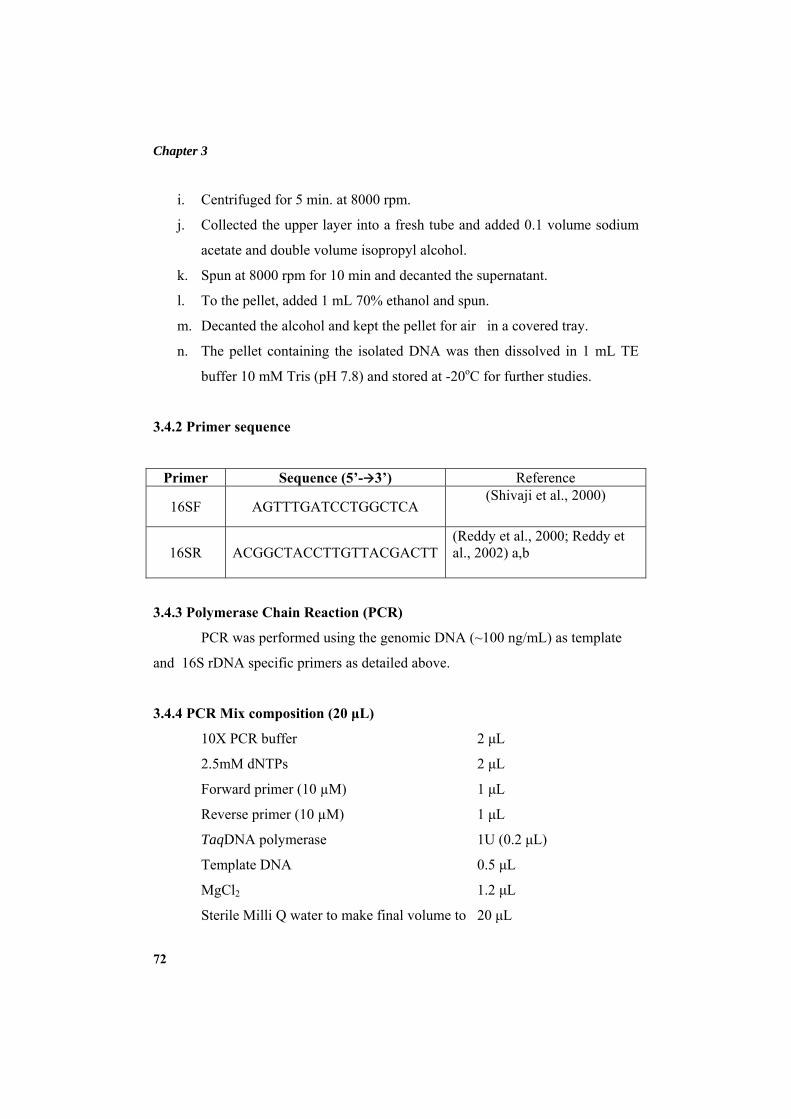
Chapter 3
72
i. Centrifuged for 5 min. at 8000 rpm.
j. Collected the upper layer into a fresh tube and added 0.1 volume sodium
acetate and double volume isopropyl alcohol.
k. Spun at 8000 rpm for 10 min and decanted the supernatant.
l. To the pellet, added 1 mL 70% ethanol and spun.
m. Decanted the alcohol and kept the pellet for air in a covered tray.
n. The pellet containing the isolated DNA was then dissolved in 1 mL TE
buffer 10 mM Tris (pH 7.8) and stored at -20oC for further studies.
3.4.2 Primer sequence
Primer Sequence (5’- 3’) Reference
16SF AGTTTGATCCTGGCTCA (Shivaji et al., 2000)
16SR ACGGCTACCTTGTTACGACTT(Reddy et al., 2000; Reddy et al., 2002) a,b
3.4.3 Polymerase Chain Reaction (PCR)
PCR was performed using the genomic DNA (~100 ng/mL) as template
and 16S rDNA specific primers as detailed above.
3.4.4 PCR Mix composition (20 μL)
10X PCR buffer 2 μL
2.5mM dNTPs 2 μL
Forward primer (10 µM) 1 μL
Reverse primer (10 µM) 1 μL
TaqDNA polymerase 1U (0.2 μL)
Template DNA 0.5 μL
MgCl2 1.2 μL
Sterile Milli Q water to make final volume to 20 μL
Materials and methods
73
3.4.5 PCR conditions
Annealing - 56oC -30 sec.
Extension - 72oC -2 min.
PCR was performed in a thermal cycler (BioRad, USA).
3.4.6 Agarose gel electrophoresis (Sambrook and Russell, 2001)
a. Agarose gel with a concentration of 0.8% was prepared for electrophoresis
of the PCR products.
b. 5 μL of the PCR products was loaded on to the gel and electrophoresed at
80 V until the migrating dye (Bromophenol blue) had traversed two-thirds
distance of the gel. 1 kb DNA ladder (Fermentas, India) was used as the
marker.
c. The gel was stained in freshly prepared 10 μg/mL ethidium bromide
solution for 10 min.
d. The gel was viewed on a UV- Transilluminator, and image captured with
the help of Digi Doc system (Bio-Rad).
3.4.7 DNA sequencing
Nucleotide sequences determined by the ABI Prism 310 genetic analyzer
by using the big dye Terminator kit (Applied Biosystems) at BioServe India Ltd.,
Hyderabad. The sequenced PCR products were analyzed online using BLAST
software (http://www.ncbi.nlm.nih.gov/blast) and the identity of the sequences
were determined (Altschul et al., 1990).
3.4.8 Multiple sequence alignment and phylogenetic tree construction
All the nucleotide sequences were converted into FASTA format.
Multiple sequence alignment for the assembled nucleotide sequences was done by
using the Clustal X program (Thompson et al., 1997) in BIOEDIT software (Hall,
1999). Aligned sequences were imported into an MEGA5: Molecular
Chapter 3
74
Evolutionary Genetics Analysis (MEGA) software version 5.0 (Tamura et al.,
2007) software for further analysis. The ends of the alignment were trimmed to
obtain equal lengths for all sequences and the aligned sequences were converted
into MEGA format for carrying out phylogenetic analysis. The phylogenetic tree
was constructed using the Neighbor-Joining method (Saitou and Nei, 1987) using
nucleotide based TN84 evolutionary model for estimating genetic distances based
on synonymous and non synonymous nucleotide substitutions. Statistical support
for branching was estimated using 1000 bootstrap steps.
3.5 BIOCHEMICAL AND MORPHOLOGICAL CHARACTERIZATION
OF P. MENDOCINA BTMW 301
Various biochemical tests were carried out according to Bergey’s Manual
of Systematic Bacteriology (Palleroni et al., 1984) in order to analyse the
characteristics of the selected bacterium. The colony morphology was determined
based on the external appearance of the colony.
3.6 TIME COURSE STUDY AND GROWTH PROFILE
Time course of protease inhibitor production was studied in Zobell marine
broth medium (3.1.2.1) using shake flask cultures. Optimum incubation time for
maximum protease inhibitor production was determined by incubating the
inoculated media for a total of 120 h and the samples were drawn and analysed at
different time intervals for protease inhibitor production and assayed for protease
inhibitory activity, protein content and specific activity as described under section
3.2.1.1, 3.2.2 and 3.2.3 respectively.
The growth of selected bacterium was studied for 120 h. Aliquots were
sampled at different intervals to analyze the cell growth by checking OD at 600
nm in a spectrophotometer. All the experiments were carried out in triplicates.
From the results obtained, the growth profile of the isolate was prepared by
plotting a graph of OD at 600 nm against incubation time.
Materials and methods
75
3.7 PROTEASE INHIBITOR PRODUCTION BY P. MENDOCINA BTMW
301: OPTIMIZATION OF BIOPROCESS VARIABLES BY “ONE-
FACTOR-AT-A-TIME” METHOD
Various physico-chemical and bioprocess parameters influencing protease
inhibitor production by P. mendocina under submerged fermentation were
optimized towards maximal protease inhibitor production in the production
medium. The strategy adopted for the optimization was to evaluate individually
the effect of different parameters (“one-variable-at-a-time” method) on protease
inhibitor production under submerged fermentation using minimal salt media as
basal medium.
The parameters optimized included carbon sources, additional sodium
chloride concentration, incubation temperature, initial pH of medium, inoculum
concentration and additional nitrogen sources. All the experiments were carried
out in triplicates. The results are an average of triplicate experiments and standard
deviation was determined using Excel 2007 (Microsoft Corporation, Redmond,
USA).
3.7.1 Minimal salt medium
Minimal salt medium was used as the basal medium for optimization studies. The
contents were mixed thoroughly autoclaved at 121oC for 30 min and cooled to
room temperature before inoculation.
Minimal media with the composition mentioned below.
Na2HPO4 0.6 g
KH2PO4 0.3 g
NaCl 0.1 g
NH4Cl 0.1 g
MgSO4.7H2O (1 M) 0.2 mL
Chapter 3
76
CaCl2. 2H2O (0.1 M) 0.1 mL
Distilled water 100 mL
pH 7
The various components of the media were added to distilled water taken in a 500
mL Erlenmeyer flask, and autoclaved.
3.7.2 Inoculum preparation
For the preparation of inoculum, a loopful of cells from the freshly grown
culture (Zobell marine agar slant) of P. mendocina was transferred to a 5 mL
sterile Zobell Marine Broth and incubated at room temperature (28 ± 2oC) at 150
rpm on a rotary shaker. After 18 h of incubation, this pre culture was transferred to
a 40 mL Zobell marine broth in a 250 mL Erlenmeyer flask and incubated at room
temperature (28 ± 2oC) at 150 rpm on a rotary shaker for 18 h. Cells were
harvested by centrifugation at 6000 rpm for 5 min at 4oC. The cell pellet was
resuspended in 10 mL of 0.8% physiological saline. This culture suspension
(OD600 = 1) was used as primary inoculum for production medium.
3.7.3 Inoculation, incubation and recovery of the protease inhibitor
The minimal salt medium (3.7.1) was inoculated with 1% (v/v) of
bacterial inoculum as prepared as mentioned under 3.8.2 and incubated on a rotary
shaker at 28 ± 2oC at 150 rpm. After incubation (48 h arbitrarily taken) the
inhibitor was recovered from the production broth by centrifugation at 10,000 rpm
at 4oc for 15 min. The clear supernatant obtained after centrifugation was used as
the crude inhibitor. This was assayed for inhibitory activity and protein content as
mentioned under section 3.2.1.1, 3.2.2 and 3.2.3 respectively.
Materials and methods
77
3.7.4 Carbon source optimization
Different carbon sources viz. lactose, maltose, sucrose, glycerol, mannitol,
galactose, glucose, fructose, sorbitol and xylose at 25 mM concentration and
starch (1%) were tested. The carbon sources sterilized separately and added to the
production medium. Medium preparation, inoculation, incubation and inhibitor
recovery were performed as detailed under section 3.7.1, 3.7.2 and 3.7.3.Culture
supernatants were analyzed for protease inhibitor production and assayed for
protease inhibitory activity, protein content and specific activity as described
under section 3.2.1.1, 3.2.2 and 3.2.3 respectively.
3.7.5 Optimization of additional NaCl concentration
Optimal sodium chloride for maximum protease inhibitor was evaluated
by adding different sodium chloride concentrations (1%, 2%, 3%, 4% and 5%) in
addition to the NaCl in the minimal medium. Twenty five millimolar glucose was
used as carbon source. Medium preparation, inoculation, incubation and inhibitor
recovery were performed as detailed under section 3.7.1, 3.7.2 and 3.7.3.Culture
supernatants were analyzed for protease inhibitor production and assayed for
protease inhibitory activity, protein content and specific activity as described
under section 3.2.1.1, 3.2.2 and 3.2.3 respectively.
3.7.6 Optimization of incubation temperature
For the optimization of different incubation temperatures the protease
inhibitor production was evaluated by incubating the inoculated minimal media at
25oC, 30oC, 37oC, 45oC and 50oC. Twenty five millimolar glucose was used as
carbon source with 3% additional NaCl. Medium preparation, inoculation,
incubation and inhibitor recovery were performed as detailed under section 3.7.1,
3.7.2 and 3.7.3.Culture supernatants were analyzed for protease inhibitor
production and assayed for protease inhibitory activity, protein content and
specific activity as described under section 3.2.1.1, 3.2.2 and 3.2.3 respectively.
Chapter 3
78
3.7.7 Optimization of inoculum concentration
Optimal inoculum concentration that supports maximum protease
inhibitor production in P. mendocina was evaluated using different concentrations
of initial inoculum (2%, 4%, 6%, 8% and 10%) prepared as mentioned in section
3.7.2. Twenty five millimolar glucose was used as carbon source with 3%
additional NaCl and incubated at 30oC. The medium preparation, inoculation and
inhibitor recovery were performed as detailed under section 3.7.1 and 3.7.3.
Culture supernatants were analyzed for protease inhibitor production and assayed
for protease inhibitory activity, protein content and specific activity as described
under section 3.2.1.1, 3.2.2 and 3.2.3 respectively.
3.7.8 Nitrogen source optimization
Various nitrogen sources (1%, w/v) were incorporated in minimal medium
with 25 mM glucose, 3% NaCl at pH 7. 4% inoculum was used for the study.
Incubated on a rotary shaker at 300 C. Effect of complex organic nitrogen source
on protease inhibitor production was studied using malt extract, beef extract, yeast
extract, peptone, casein, tryptone and tryptone soya broth (TSB) were added
individually.
Samples were analyzed after 48 h incubation and the studies were carried
out at a media pH 7, 150 rpm and at 30oC incubation temperature with 25 mM
glucose as carbon source and 3 % additional NaCl in minimal medium. The
medium preparation, inoculation and inhibitor recovery were performed as
detailed under section 3.7.1, 3.7.2 and 3.7.3. Culture supernatants were analyzed
for protease inhibitor production and assayed for protease inhibitory activity,
protein content and specific activity as described under section 3.2.1.1, 3.2.2 and
3.2.3 respectively.

Materials and methods
79
3.8 PROTEASE INHIBITOR PURIFICATION
Proteinaceous protease inhibitor isolated from P. mendocina was purified
employing standard protein purification methods which included ammonium
sulphate precipitation, followed by dialysis, DEAE sepharose ion exchange
chromatography and Trypsin sepharose affinity chromatography. The purity was
further confirmed by reverse phase HPLC. All purification steps were carried out
at 4°C unless otherwise mentioned.
3.8.1 Ammonium sulphate precipitation
Ammonium sulphate precipitation was done (Englard and Seifter, 1990)
as described earlier under section 3.3.1.
3.8.2 Dialysis
The precipitate obtained after ammonium sulphate precipitation was
further dialyzed against 0.01 M phosphate buffer (pH 7.5) as described previously
under section 3.3.2.
3.8.3 Ion exchange chromatography
The active protease inhibitor fraction obtained after the dialysis of
ammonium sulphate precipitation was further purified by ion exchange
chromatography using DEAE sepharose (Sigma-Aldrich) as the anion exchanger.
Proteins bind to ion-exchangers due to surface charge. These reversibly adsorbed
proteins were eluted out using a salt gradient.
3.8.3.1 Purification using DEAE sepharose column
DEAE sepharose was carefully packed in Bio-Rad econo column (42.5 X
2.5 cm) without any air bubble and the column was equilibrated with five column
volumes of 0.01 M phosphate buffer pH 7.5. Twenty five millilitre of dialyzed
sample, prepared as described in section 3.5.2, with a protein content of 120 mg
Chapter 3
80
was applied to the pre equilibrated DEAE sepharose column. After the complete
entry of sample into the column, the column was connected to the reservoir
containing 0.01 M phosphate buffer, pH 7.5 with a flow rate of 1.5 mL/min which
was set up in an FPLC system (Bio-Rad), reading absorbance at 280nm. The
bound fraction was eluted by a step gradient of 0.5 M NaCl in 0.01 M phosphate
buffer. Peak fractions from the column were pooled and dialyzed against the
phosphate buffer pH 7.5 (0.01 M) as mentioned under section 3.5.2. The dialyzed
fractions were concentrated using amicon UF-30 kDa membrane (Millipore, USA)
and assayed for protease inhibitory activity, protein content and specific activity as
described under section 3.2.1.1, 3.2.2 and 3.2.3 respectively.
3.8.4 Affinity chromatography
CNBr-activated Sepharose 4B was coupled to trypsin was used for the
affinity purification of protease inhibitor from P. mendocina.
3.8.4.1 Preparation of CNBr-activated sepharose 4B
The lyophilized CNBr-activated Sepharose 4B (Sigma-Aldrich) was
supplied in the presence of additives. These additives were washed away at low
pH (pH 3) before coupling the trypsin. The use of low pH (pH 3) preserves the
activity of the reactive groups, which otherwise hydrolyze at high pH. Three gram
of lyophilized powder was (1 g lyophilized powder gives about 3.5 mL final
volume of medium) suspended in 1 mM HCl. The swelled medium was washed
for 15 min with 1 mM HCl on a sintered glass filter (porosity G3). Two hundred
millilitre, 1 mM HCl was used per gram freeze-dried powder and added in several
aliquots.
Materials and methods
81
3.8.4.2 Coupling of trypsin
a. Thirty milligram of trypsin was dissolved in coupling buffer, 0.1 M
NaHCO3 pH 8.3 containing 0.5 M NaCl in a stopper vessel. Five millilitre
coupling solution/g lyophilized powder was used.
b. Rotated the mixture end-over-end for 1 h at room temperature.
c. Washed away excess trypsin with 5 medium (gel) volumes of coupling
buffer.
d. The free active groups were blocked by transferring the medium to 0.1 M
Tris-HCl buffer, pH 8 and allowed to stand for 2 h. Washing was
performed with 5 medium volumes of each buffer with three cycles of
alternating pH (Each cycle consisted of a wash with 0.1 M acetic
acid/sodium acetate, pH 4 containing 0.5 M NaCl followed by a wash
with 0.1 M Tris-HCl, pH 8 containing 0.5 M NaCl).
3.8.4.3 Purification using trypsin-affinity chromatography
The concentrated ion exchange fraction (2 mg) was applied to the trypsin
affinity column (15 X 1 cm) which was equilibrated with 0.01 M phosphate buffer
pH 7.5. The experiment was performed on an FPLC system with a flow rate of 0.5
mL/min. The bound protein was eluted using 0.5 M NaCl in 0.01 M HCl. The
bound peak was immediately dialyzed against 0.01 M phosphate buffer pH 7.5
and concentrated using amicon UF-30 kDa membrane. Protease inhibitory
activity, protein content and specific activity found as described under section
3.2.1.1, 3.2.2 and 3.2.3 respectively. The yield and fold of purifications were
calculated.
3.8.5 Calculation of yield of protein, yield of protease inhibitor activity and
fold of purification
Yield of protein and yield of protease inhibitory activity after each step of
purification were calculated. The percentage activity obtained by dividing the total
Chapter 3
82
protein content or activity of that fraction with the total protein content or activity
of the crude extract as the case may be. Fold of purification in each step was
calculated by dividing the specific activity of the respective fraction with that of
the crude extract.
Yield of protein = Total protein content of the purified fraction X 100
Total protein content of the crude extract
Yield of activity = Total activity of the purified fraction X 100
Total activity of the crude extract
Fold of purification = Specific activity of the purified fraction
Specific activity of the crude extract
3.8.6 Reverse-phase HPLC
The purified active fraction after gel filtration (20 µL, 0.1 mg/mL) was
subjected to reversed phase HPLC (Schimadzu LC 2010) using Phenomenex C18
HPLC column (22.5 mm ID X 250 mm length) at a flow rate of 1 mL/min with
100% solvent A (0.1% trifluoroacetic acid (TFA) in water) for 10 min and a linear
gradient (0–100%) of solvent B (0.09% TFA in 60% acetonitrile) over 60 min.
Proteins were detected by monitoring the absorbance at 220 nm.
3.9 CHARACTERIZATION OF PROTEASE INHIBITOR
Purified inhibitor was further subjected to characterization for their
biophysical as well as biochemical properties like molecular weight, isoelectric
point, stability at different temperature and pH, binding studies and inhibition
kinetics to determine the type of inhibition as described in the following sections.
Materials and methods
83
3.9.1 Electrophoretic methods
The active fractions of protease inhibitor from P. mendocina, after ion
exchange chromatography, affinity chromatography and gel filtration
chromatography were subjected to electrophoretic analysis by non denaturing
Native-PAGE and denaturing SDS-PAGE in a vertical slab electrophoresis (Mini-
PROTEAN Tetra cell, Bio-Rad). Electrophoresis was carried out in a 16%
polyacrylamide gel according to the method described by (Laemmli, 1970).
Reagents for polyacrylamide gel electrophoresis
1. Stock acrylamide solution ( 30% T and 2.6% C)
Acrylamide - 29.22 g
Bis-acrylamide - 0.78 g
Distilled water (DW) - 100 mL
Stored at 4°C in amber coloured bottle
2. Stacking gel buffer stock
Tris buffer (0.5 M) - 6.05 g in 40 mL DW
Titrated to pH 6.8 with 1 M HCl and made up to 100 mL with DW.
Filtered through Whatman No: 1 filter paper and stored at 4°C.
3. Resolving gel buffer stock
Tris buffer (1.5 M) - 18.15 g
Titrated to pH 8.8 with 1M HCl and made up to 100 mL with DW.
Filtered through Whatman No: 1 filter paper and stored at 4°C.
4. Running buffer for Native-PAGE (pH 8.3)
Tris buffer - 3 g
Glycine - 14.4 g
Dissolved and made up to 1L with DW.
Prepared in 10X concentration and stored at 4°C.
Chapter 3
84
5. Running buffer for SDS-PAGE (pH 8.3)
Tris buffer - 3 g
Glycine - 14.4 g
SDS - 1 g
Dissolved and made up to 1L with DW. Prepared in 10X concentration
and stored at 4°C.
6. Sample buffer for Native-PAGE
Tris-HCl (pH 6.8) - 0.0625 M
Glycerol - 10% (v/v)
Bromophenol blue - 0.01%
Prepared in 2X concentrations and stored at 4°C..
7. Sample buffer for Non-Reductive SDS-PAGE
Tris-HCl (pH 6.8) - 1.25 mL
Glycerol - 2.5 mL
SDS (10%, W/v) - 2 mL
Deionised water - 3.55 mL
Bromophenol blue (0.5%, w/v) - 0.2 mL
Samples were diluted with sample buffer in 1:2 ratios and heated at 95oC
for 4 min.
8. Sample buffer for Reductive SDS-PAGE
Tris-HCl (pH 6.8) - 1.25 mL
Glycerol - 2.5 mL
SDS (10%, W/v) - 2 mL
Deionised water - 3.55 mL
Bromophenol blue (0.5%, w/v) - 0.2 mL
Prior to use added 50 µL β-mercaptoethanol to 950 µL sample buffer.
Samples were diluted with sample buffer in 1:2 ratios and heated at 95oC for 4
min.
Materials and methods
85
9. SDS (10%) - 1 g in 10 mL DW
10. Ammonium persulfate (10%, w/v) - 0.1 g of ammonium persulfate
was dissolved
in 1 mL DW (prepared
freshly).
11. Protein staining solution
Coomassie brilliant - 100 mg
blue (0.1%)
Methanol (40%) - 40 mL
Glacial acetic acid (10%) - 10 mL
DW - 50 mL
12. Destaining solution
Methanol (40%) - 40 mL
Glacial acetic acid (10%) - 10 mL
DW - 50 mL
Silver staining
Silver staining of the gel after electrophoresis was performed (Blum,
1987) with slight modifications.
Reagents
1. Fixer
Methanol (50%, v/v) - 50 mL
Acetic acid (5%, v/v) - 5 mL
Milli Q water - 45 mL
2. Wash
Methanol (50%, v/v) - 50 mL
Milli Q water - 50 mL
Chapter 3
86
3. Sensitizing solution
Sodium thiosulfate (0.02%, w/v) - 20 mg
Milli Q water - 100 mL
4. Silver nitrate solution
Silver nitrate (0.2%, w/v) - 200 mg
Milli Q water - 100 mL
5. Developer
Sodium carbonate (6%, w/v) - 3 g
Formaldehyde - 12.5 µL
Milli Q water - 100 mL
6. Stop solution
Sodium-EDTA - 1.4 g
Milli Q water - 100 mL
Procedure
a. The SDS-PAGE gel was incubated in fixer for 30 min.
b. Washed the gel in wash solution for 15 min followed by five washes in
five min interval with Milli-Q water.
c. Incubated the gel in sensitizer for exactly 60 sec and washed twice in 60
sec interval with Milli-Q water.
d. The gel was immersed in chilled silver nitrate solution for 25 min and
washed two times for 60 sec with Milli-Q water.
e. The gel was transferred to developer solution and kept until protein bands
were developed.
f. Reaction was arrested by adding stop solution.
13. Protein Markers for SDS-PAGE
Prestained protein molecular weight marker (Fermentas, India) was used
for non-reductive SDS-PAGE analysis.
Materials and methods
87
Components MW (Mr) in Da
β-galactosidase - 118,000
Bovine serum albumin - 90,000
Ovalbumin - 50,000
Carbonic anhydrase - 36,000
β-lactoglobulin - 27,000
Lysozyme - 20,000
a. Low range molecular weight marker mix of Bio-Rad was used for the
reductive SDS-PAGE. Lyophilized marker mix was reconstituted in
1X sample buffer for reductive SDS-PAGE, heated for 5 min at 95oC,
and 5 µL of marker was loaded on to the gel. The composition of the
marker mix is as given below.
Components MW (Mr) in Da
Phosphorylase b - 97,000
Bovine Serum Albumin - 66,000
Ovalbumin - 45,000
Carbonic anhydrase - 31,000
Trypsin inhibitor - 21,500
α-Lactalbumin - 14,400
3.9.1.1 Native polyacrylamide gel electrophoresis
3.9.1.1.1 Gel preparation
Resolving gel (10%)
Stock acrylamide: bis-acrylamide - 3.3 mL
Resolving gel buffer stock - 2.5 mL
Water - 4.2 mL
Ammonium persulphate (APS) - 75 µL
TEMED - 5 µL
Chapter 3
88
Stacking Gel (4%)
Stock acrylamide: bis-acrylamide - 1.34 mL
Stacking gel buffer stock - 2.5 mL
Water - 6 mL
Ammonium persulphate (APS) - 50 µL
TEMED - 10 µL
Sample preparation
Samples were prepared in 1X sample buffer up to a concentration of 25
µg and loaded 25 µL sample to the gel.
Procedure
a. The gel plates were cleaned and assembled.
b. Resolving gel – Prepared resolving gel solution combining all reagents
except APS and TEMED in a beaker. Degassed the solution, added APS
and TEMED. Immediately poured the mixture into the cast and poured a
layer of water over the gel and allowed to polymerize at least for 45 min.
c. Stacking gel - Added the components of stacking gel except APS and
TEMED into a beaker, mixed gently and finally added APS and TEMED.
Poured the contents into the cast above the resolving gel and immediately
inserted the comb between the glass plates. Allowed to polymerize for at
least for 45 min.
d. Gel was placed in the electrophoresis apparatus, and reservoir was filled
with running buffer for Native-PAGE.
e. Protein samples were loaded to the gel.
f. The gel was run at 80 V till the sample entered the resolving gel.
g. When the dye front entered the resolving gel, increased the current to 120
V.
h. The run was stopped when the dye front reached 1 cm above the lower
end of the glass plate.
Materials and methods
89
i. Removed the gel from cast and stained for at least 1 h in the staining
solution.
j. Destained the gel till the bands became clear and observed the protein
bands under a transilluminator.
3.9.1.2 Sodium dodecyl sulphate polyacrylamide gel electrophoresis (SDS-
PAGE)
The purified inhibitor protein was subjected to SDS-PAGE for evaluating
the nature of polypeptide. Low range molecular weight marker of Bio-Rad or
broad range marker from sigma was used as standard molecular weight markers.
Reductive SDS–PAGE gel preparation
Resolving gel (16%)
Stock acrylamide: bis-acrylamide - 5.333 mL
Resolving gel buffer stock - 2.5 mL
10% SDS - 0.1 mL
Water - 1.987 mL
Ammonium persulphate (APS) - 75 µL
TEMED - 15 µL
Stacking Gel (5%)
Stock acrylamide: bis-acrylamide - 1.665 mL
Stacking gel buffer stock - 2.5 mL
10% SDS - 0.1 mL
Water - 5.6675 mL
Ammonium persulphate (APS) - 50 µL
TEMED - 15 µL
Chapter 3
90
Sample preparation
Prepared samples in 1X SDS-PAGE sample buffer to a concentration of
25 µg, mixed well, boiled for 5 min in a water bath, cooled to room temperature,
and 25 µL sample and 5 µL low molecular weight marker mix was loaded to the
gel.
Procedure
Procedure followed for SDS-PAGE was essentially the same as that of
Native-PAGE which was described under section 3.5.1.1 with the exception that
SDS was incorporated in gel preparation and running buffer used was that of SDS-
PAGE. The gels were either coomassie or silver stained.
3.9.2 Mass by MALDI-TOF
The purified protease inhibitor from gel filtration chromatography was
subjected to intact mass analysis. The protease inhibitor sample was desalted with
ZipTip-C18 (Millipore, Billerica; MA). The intact molecular mass of the purified
inhibitor was determined by MALDI TOF/TOF (ABI 4800).
3.9.3 Isoelectric focusing
Isoelectric point (pI) of the purified inhibitor protein was determined by
isoelectric focusing which was performed in a Bio-Rad isoelectric focusing unit.
Immobilized pH gradient (IPG) strip of pH 3-10 (Bio-Rad) was used for the
purpose. The detailed procedure is as given below.
3.9.3.1 Rehydration of sample with IPG strip
a. The lyophilized protease inhibitor sample (0.1 mg/mL) was prepared in 2
mL rehydration buffer.
b. Loaded 125 µL of this prepared sample to the equilibration tray.
c. Immobilized pH gradient (IPG) strip of pH 3-10 (Bio-Rad) was gently
placed gel side down in the equilibration tray. Removed air bubbles if any.
Materials and methods
91
(Two IPG strips were used, one for isoelectric focusing and the other for
2-D electrophoresis).
d. The strips were overlaid with 2-3 mL of mineral oil to prevent evaporation
during rehydration process.
e. Covered the equilibration tray and left the tray for overnight to rehydrate
IPG strips.
3.9.3.2 Isoelectric focusing of the inhibitor
a. Paper wicks were placed at both ends of the clean, dry IEF focusing tray
covering the wire electrodes.
b. IPG strips were taken out from the rehydration tray and drained the
mineral oil by holding the strip vertically for 8 sec.
c. Paper wicks were made wet with Milli-Q water and placed the IPG strips
in the focusing tray. Placed in the PROTEAN IEF cell and closed the
cover.
d. Programmed the IEF cell as given below and run the electrophoresis.
Voltage Time Volt-Hours Ramp
Step 1 250 20 min --- Linear
Step 2 4000 2 h --- Linear
Step 3 4000 --- 10,000 V-h Rapid
(Maintained the cell temperature at 20oC with maximum current of 50
µA/strip and no dehydration in all steps).
3.9.3.3 Staining and 2-D electrophoresis
a. IPG strips were removed from focusing tray after electrophoresis. Drained
mineral oil and pressed the strip against a wet blotting paper.
b. One of the strips were transferred to coomassie stain tray and kept for 1 h.
Chapter 3
92
c. Destained the gel for 10 min.
d. The other IPG strip was rinsed in a 1X Tris-glycine buffer and placed on
the precast 16% SDS-PAGE gel (A portion of the gel was left at the top,
according to the dimension of IPG strip during the casting of SDS-PAGE
gel).
e. Overlaid molten agarose and allowed to set the agarose.
f. The electrophoresis was carried out at 200 V. The gel was coomassie
stained after electrophoresis.
3.9.4 Analysis of protease inhibitor by Dot-Blot method
The purified fraction collected from gel filtration chromatography, was
analyzed for its protease inhibitory activity according to the method of (Veerappa
et al., 2002) as described below.
a. Three microlitre of protease inhibitor (0.1 mg/mL) was mixed with 3μL
trypsin (0.1 mg/mL) and spotted on to a strip of X-ray film.
b. Three microlitre of Soya bean trypsin inhibitor (0.1 mg/mL) was mixed
with 3 μL trypsin (0.1 mg/mL) as positive control and spotted on to a strip
of X-ray film.
c. Three microlitre of trypsin was mixed with 3 μL phosphate buffer 0.01 M
(pH 7.5) as negative control and spotted on to the X-ray film.
d. Incubated the X-ray film at 37°C for 10 min.
e. Washed the film under tap water till the zone of gelatin hydrolysis by
trypsin was visualized.
f. Inference was made by observing the zone of hydrolysis indicating
degradation of gelatin by trypsin. In the absence of the inhibitor a clear
zone is formed at the site of trypsin application on the X-ray film due to
gelatinase activity. Whereas, trypsin cannot degrade gelatin on the X-ray
film in the presence of the inhibitor.
Materials and methods
93
3.9.5 Effect of temperature on inhibitor stability
Thermal stability of inhibitor was studied by incubating purified protease
inhibitor (0.1 mg/mL) at different temperatures ranging from 4°C-100oC for 60
min. The sample was drawn and further incubated at 4°C for 15 min. The protease
inhibitory activity of each sample was assessed by conducting the assay as
described under section 3.2.2.
3.9.6 Stability of protease inhibitor at different pH
The stability of protease inhibitor over a range of pH was determined by
performing the inhibitor activity assay at pH 7.5, after incubating the purified
protease inhibitor in different buffers of pH ranging from 2-12 for 4 h at 4°C. Ten
microlitre (0.1 mg/mL) of purified inhibitor was incubated with 40 μL of different
buffer systems, which included KCl-HCl buffer (pH 2), citrate buffer (pH 4-6),
phosphate buffer (pH 7), Tris-HCl buffer (pH 8-9), borax/NaOH (pH 10),
disodium hydrogen phosphate/ NaOH (pH 11) and potassium chloride/NaOH (pH
12). After incubation sample was assayed for protease inhibitory activity as
described under section 3.2.2.
3.9.7 Effect of various metal ions on protease inhibitor activity
Effect of various metal ions on activity of protease inhibitor was evaluated
by incubating the protease inhibitor along with 1 mM concentrations of various
metals ions in the inhibitor solution for 30 min followed by measuring the
protease inhibitory activity as described under section 3.2.2. The metals studied
included sodium chloride, calcium chloride, magnesium sulphate, ferric chloride,
manganese chloride, nickel chloride, mercury chloride, barium chloride, cadmium
sulphate, sodium molybdate and aluminum sulphate which contributes the metal
ions, Na+, Ca2+, Mg2+, Fe3+, Mn2+, Ni2+, Hg2+, Ba2+, Cd2+, Mo6+ and Al3+
respectively.
Chapter 3
94
3.9.8 Metal chelation of protease inhibitor using EDTA
The metal ion concentration of purified protease inhibitor and its effect on
inhibitory properties in its native state was determined by metal chelation using 30
mM EDTA (SRL, India) according to the method described by Jack et al (2004).
Purified protease inhibitor (0.1 mg/mL) was dialyzed extensively against 30 mM
EDTA over night at 4°C for chelation of metal ions. The EDTA was removed
further by dialyzing against deionised water over night with frequent changes of
deionised water.
3.9.9 Metal ion concentration of protease inhibitor
Minerals concentrations of protease inhibitor were determined as follows:
I. An aliquot of protease inhibitor was dialyzed extensively against
deionised water and was used for mineral analysis by Inductively
Coupled Plasma Atomic Emission Spectroscopy (ICP-AES).
II. Another aliquot was dialyzed extensively against 30 mM EDTA,
overnight at 4°C for chelation of metal ions and then the EDTA was
removed by dialyzing against deionised water overnight with
frequent changes. The mineral concentration of dialyzed sample
was also determined by ICP-AES.
3.9.10 Effect of various detergents on protease inhibitor activity
Effect of various non-ionic and ionic detergents such as Triton X-100,
Tween-80, Tween-20, SDS and CTAB (1% each w/v) on protease inhibitory
activity was determined by incubating the protease inhibitor in each detergent for
30 min, dialyzed against 0.01 M phosphate buffer pH 7.5 and estimated the
residual inhibitory activity as described under section 3.2.2.
Materials and methods
95
3.9.11 Effect of oxidizing agents on protease inhibitor activity
Impact of oxidizing agents on the activity of protease inhibitor was
studied by incubating the protease inhibitor with hydrogen peroxide (1-2%, v/v)
and dimethyl sulfoxide (1-5%, v/v) for 30 min and measuring the residual
inhibitory activity as described under section 3.2.2.
3.9.12 Effect of reducing agents
The effect of reducing agents on the activity of protease inhibitor was
studied by incubating the protease inhibitor with 50, 100, 150, 200, 250, 300, 350
µM of β-mercaptoethanol and 20, 40, 60, 80, 100, 120 µM dithiothreitol for 30
min and measuring the residual inhibitory activity as described under section
3.2.2.
3.9.13 Chemical modification of amino acids in protease inhibitor
To determine the impact of chemical structure of the amino acids at the
reactive sites of inhibitor molecule on its inhibitory activity was evaluated by
effecting chemical modification in the selected amino acids of the inhibitor
molecules. Thus four different amino acids were individually modified using
specific chemical modifiers and the effect of modifiers on the anti proteolytic
activity of the inhibitor molecule was determined. Chemical modifications of
amino acids of purified inhibitor were carried out using different chemical
modifiers under their respective reaction conditions. Hundred microlitre of
purified inhibitor (0.1 mg/mL) was used for this study. After the incubation with
different concentrations ranging from 5, 10, 15, 20 and 25 mM of each modifier,
the sample was dialyzed against phosphate buffer and the residual protease
inhibitory activity was estimated as described under section 3.4.2.
Chapter 3
96
Chemical modifier Amino acid
modified
Reaction conditions
N-Ethylmaleimide Cysteine 30°C 0.1 M Tris/HCl
buffer (pH-7.0) for
60 min(Colman and
chu, 1970).
Diethyl pyrocarbonate Histidine 30°C 0.1 M Tris/HCl
buffer (pH-7.0) for
30 min (Ovaldi et
al., 1967).
PMSF Serine 25°C 0.05 M Tris/HCl
buffer (pH-7.8) for
120 min (Gold and
Farney, 1964).
3.9.14 Effect of acid treatment on protease inhibitor
Sensitivity of protease inhibitor in an acidic environment was evaluated
by incubating purified protease inhibitor with different concentrations of HCl
ranging from 0.02, 0.04, 0.06, 0.08 & 0.1 M for 30 min. After incubation, the pH
was neutralized with 0.1 M Tris-HCl buffer pH 9. The residual protease inhibitory
activity was estimated as described under section 3.2.2.
3.9.15 Intrinsic fluorescence spectroscopy
Fluorimetry was performed on a Cary Eclipse spectrofluorimeter using a
slit width of 5 mm and an excitation wavelength of 278 nm. The emission
wavelength was observed from 290 nm. Phosphate buffer, pH 7.5, served as
control. Protease inhibitor having an A280 value, 0.1 was used for the emission
Materials and methods
97
spectra. The emission spectra for inhibitor and trypsin alone were also performed.
Trypsin was complexed with inhibitor in a ratio of 1: 1 concentration.
3.9.16 Peptide mass fingerprinting
Protease inhibitor was reduced, alkylated with iodoacetamide and trypsin
digested. Peptides were extracted according to standard techniques and were
analyzed by MALDI-TOF-TOF mass spectrometer using a 4800 Proteomics
Analyzer (Applied Biosystems). Spectra were analyzed to identify protein of
interest using Mascot sequence matching software (Matrix Science) with Swiss-
Prot and NCBInr database.
3.9.17 Specificity of protease inhibitor
The specificity of the purified inhibitor was studied against commercially
important proteases like elastase (Sigma-Aldrich), proteinase K (Sigma-Aldrich),
subtilisin (Sigma-Aldrich) and chymotrypsin (Sigma-Aldrich).
3.9.17.1 Assay of elastase, proteinase K and subtilisin inhibitory activity
The elastase, thermolysin and proteinase K inhibitory activity was tested
taking 0.1 mg/mL of respective proteases with 0.1 mg/mL purified protease
inhibitor according to Kunitz caseinolytic method as described under section
3.1.3.1.1.
3.9.17.2 Assay of chymotrypsin inhibitory activity
Chymotrypsin inhibitory activity was assayed according to the modified
method of Fritz et al (1966). Chymotrypsin from Bovine pancreas (Sigma-
Aldrich) was prepared by dissolving freeze dried material in 0.001 M HCl at a
concentration of 1 mg/mL. Standard assay mixture contained 0.05 M Tris-HCl
buffer (pH 7.5), 20 mM peptide substrate, N-Suc-Ala-Ala-Pro-Phe-p-nitroanilide,
0.1 mg/mL inhibitor solution and chymotrypsin (10 μg/mL).
Chapter 3
98
One unit of chymotrypsin is defined as the amount of enzyme that induces
the conversion of 1 µmol substrate/min. One chymotrypsin inhibitory unit (U) was
defined as the amount of inhibitor needed to inhibit the release of 1 μmol of p-
nitroaniline per mL per min at pH 7.5 and at 37oC.
3.9.18 Stoichiometry of protease-protease inhibitor interaction
The molar concentration of the purified protease inhibitor for the complete
inactivation of the trypsin was determined by preincubating 1 nM trypsin (based
on Mr 23,800) in 100 μL of 0.01 M phosphate buffer pH 7.5 with different
amounts of purified protease inhibitor (0.05-1 nM) at 37°C for 30 min (based on
Mr 11,567). The long incubation time was necessary to ensure that the reaction had
gone to completion. The remaining activity of the trypsin was determined
according to the method described under section 3.1.3.1.2.
3.9.19 Kinetic studies of inhibition
Kinetics of trypsin inhibition by protease inhibitor was conducted with
different concentrations of inhibitor [I] against various concentrations of substrate
[S]. Lineweaver-Burk 1/v versus 1/ [s] was plotted, the apparent Km (Km’) and
maximum velocity (Vmax) were calculated for each concentration of inhibitor and
secondary plot was plotted by taking 1/Vmax versus [I] to determine dissociation
constant of the inhibitor (Ki).
a. Twenty-five microlitre of trypsin (0.1 mg/mL) was incubated with
different concentrations of substrate (0.1 mM-2 mM) α-N-benzoyl-DL-
arginine-p-nitroanilide (BAPNA) suitably diluted to 500 µL with 0.01 M
phosphate buffer, pH 7.5 and kept at 37°C for 10 min. The reaction was
arrested by adding 500 µL of 30% (v/v) acetic acid.
Materials and methods
99
b. Lineweaver-Burk 1/v versus 1/ [s] was plotted, the Km and maximum
velocity (Vmax) were calculated (Dixon, 1953).
c. The apparent Km’ and maximum velocity (Vmax) in the presence of
different purified protease inhibitor concentrations (0.014, 0.07 and 0.27
nM) was calculated similarly.
d. A secondary plot was plotted by taking 1/Vmax versus [I] to determine
dissociation constant of the inhibitor (Ki).
3.10 APPLICATION STUDIES
3.10.1 Protease inhibitor as seafood preservative
The inhibitory activity of P. mendocina protease inhibitor towards the
spoilage microbial flora isolated from the shrimp Peneaus monodon was
evaluated. To assess the effect of inhibitor on protein degradation of shrimp during
preservation under different storage conditions like room temperature, 4°C and –
20°C for different duration was analysed. For this, head was removed from the
peeled shrimp. Five gram shrimp biomass was weighed out and taken in a sterile
container, sealed and kept at different storage condition as control. For the test
experiment, the same weight of samples were taken in the same conditions as that
of control and incubated with in a sterile container, having 5 mL of (0.1 mg/mL)
purified protease inhibitor prepared as mentioned under section 3.9.4 at each
temperature for 8 h, 24 h and 168 h respectively.
After incubation, the samples were taken and extracted by homogenizing
in sterile distilled water using a mortar and pestle under sterile conditions and kept
in a rotary shaker for 30 min at 150 rpm. One milliliter of extract was taken under
sterile condition, and serially diluted the sample in physiological saline. The total
microbial population of each sample was analyzed by pour plating on Skim milk
agar (HiMedia) plates. In the same way, the experiments were repeated with the
Chapter 3
100
control also. The colony forming units are enumerated in both cases and a
comparative strategy was made to analyse the efficacy of the inhibitor to prevent
microbial growth.
The spoilage bacteria from P. monodon were isolated, purified and checked
for the production of protease enzyme on Skim milk agar (HiMedia) plates. The
positive cultures were characteristic of clearing zone around the colonies due to
casein hydrolysis. They were picked and grown in Nutrient Broth (HiMedia) with
1% casein. To isolate spoilage protease enzyme from each bacterium, the culture
broth was transferred in to centrifuge tube and subjected to centrifugation at 6000
rpm, at 4oC in a rotary shaker. The resulted cell free culture supernatants were
taken as crude proteolytic enzymes and stored at -20oC which were used for
further inhibition studies. The purified inhibitor was evaluated for its ability to
inhibit the isolated spoilage proteases as described under section 3.2.1.1.
The complete protein of the samples were extracted using 5% NaCl in
0.02 M sodium bicarbonate according to (Chandrasekaran, 1985) and the cell
pellet were removed by centrifugation at 10,000 rpm for 15 min at 4°C. The clear
supernatant was assayed for total protein content as described in section 3.2.2.
3.10.2 Protease inhibitor as antibacterial agent
Inhibition of growth of bacteria by P. mendocina protease inhibitor was
studied using agar well diffusion assay (Tagg and McGiven, 1971). The bacterial
strains Escherichia coli (NCIM 5051) Pseudomonas aeruginosa (NCIM 2863),
Pseudomonas solanicerum (NCIM 5103), Pseudomonas fluorescens (NCIM
2099), Pseudomonas syringae (NCIM 5102), Pseudomonas putida (NCIM
2650), Salmonella typhimurium (NCIM 2501), Salmonella abony (NCIM 2257),
Klebsiella pneumoniae (NCIM 2957), Proteus vulgaris (NCIM 2027),
Clostridium perfringens (NCIM 2677), Staphylococcus aureus (NCIM 2127),
Bacillus cereus (NCIM 2155), Bacillus circulans (NCIM 2107), Bacillus
coagulans (NCIM 2030), Bacillus macerans (NCIM 2131), and Bacillus
Materials and methods
101
pumilus (NCIM 2189) were purchased from National Collection of Industrial
Microorganisms (NCIM), NCL Pune, India and used as test bacteria.
Test bacteria freshly grown in nutrient broth (HiMedia) for 12 h were used
for the study. Antibacterial analysis was performed in Petri plates containing
Mueller-Hinton agar (HiMedia) in which each of the test bacterium was swab-
inoculated. Well was cut aseptically in the test bacterial plate and 20 µL (5
mg/mL) of purified protease inhibitor was added in it. Plates were incubated for
24 h at 37°C and observed for the clearance zones of inhibited bacterial growth.
Minimum inhibitory concentration (MIC) was determined by adding varying
concentrations (70 µg, 75 µg, 80 µg, 85 µg, 90 µg, 95 µg and 100 µg) of inhibitor
preparations in the wells made in the test bacterial plate, and observed for the
clearance of inhibited bacterial growth.
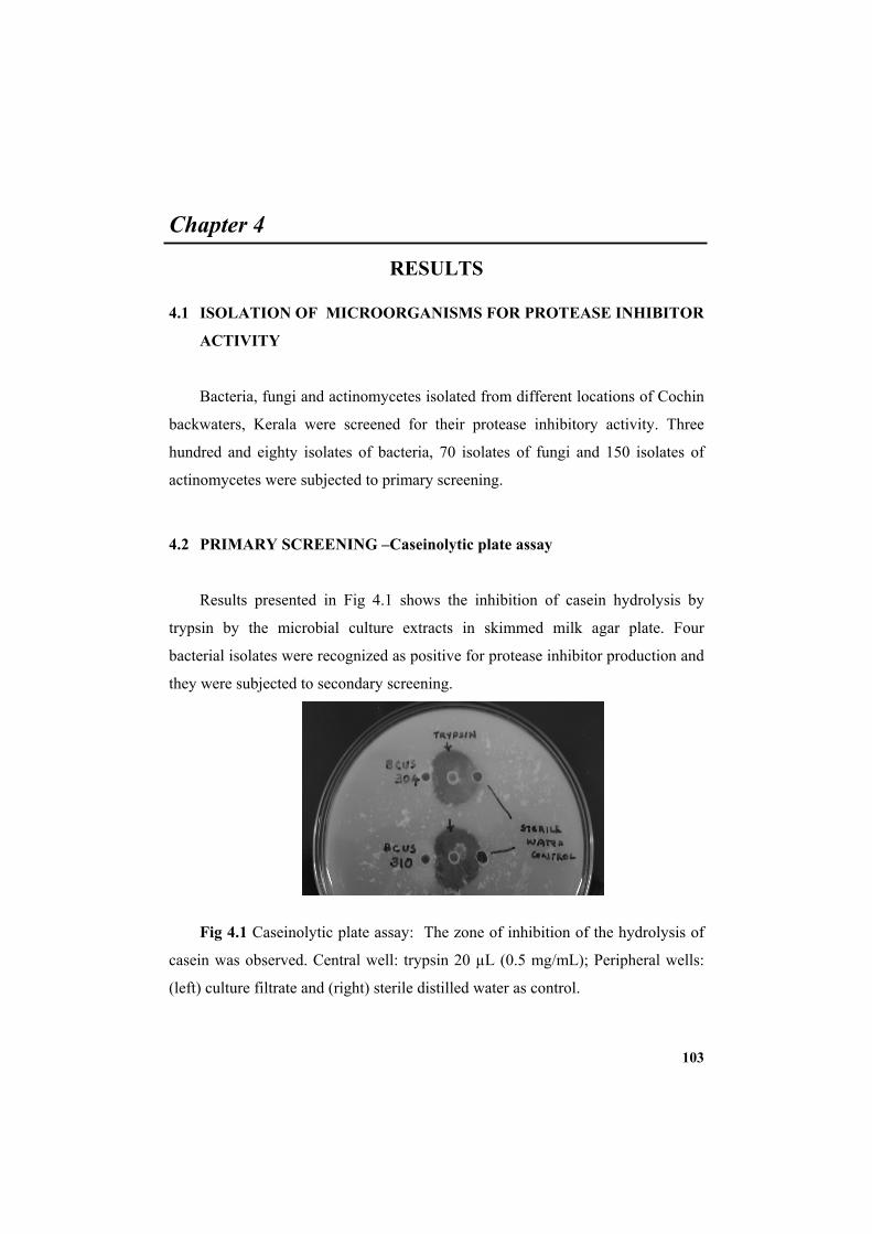
103
Chapter 4
RESULTS
4.1 ISOLATION OF MICROORGANISMS FOR PROTEASE INHIBITOR
ACTIVITY
Bacteria, fungi and actinomycetes isolated from different locations of Cochin
backwaters, Kerala were screened for their protease inhibitory activity. Three
hundred and eighty isolates of bacteria, 70 isolates of fungi and 150 isolates of
actinomycetes were subjected to primary screening.
4.2 PRIMARY SCREENING –Caseinolytic plate assay
Results presented in Fig 4.1 shows the inhibition of casein hydrolysis by
trypsin by the microbial culture extracts in skimmed milk agar plate. Four
bacterial isolates were recognized as positive for protease inhibitor production and
they were subjected to secondary screening.
Fig 4.1 Caseinolytic plate assay: The zone of inhibition of the hydrolysis of
casein was observed. Central well: trypsin 20 µL (0.5 mg/mL); Peripheral wells:
(left) culture filtrate and (right) sterile distilled water as control.
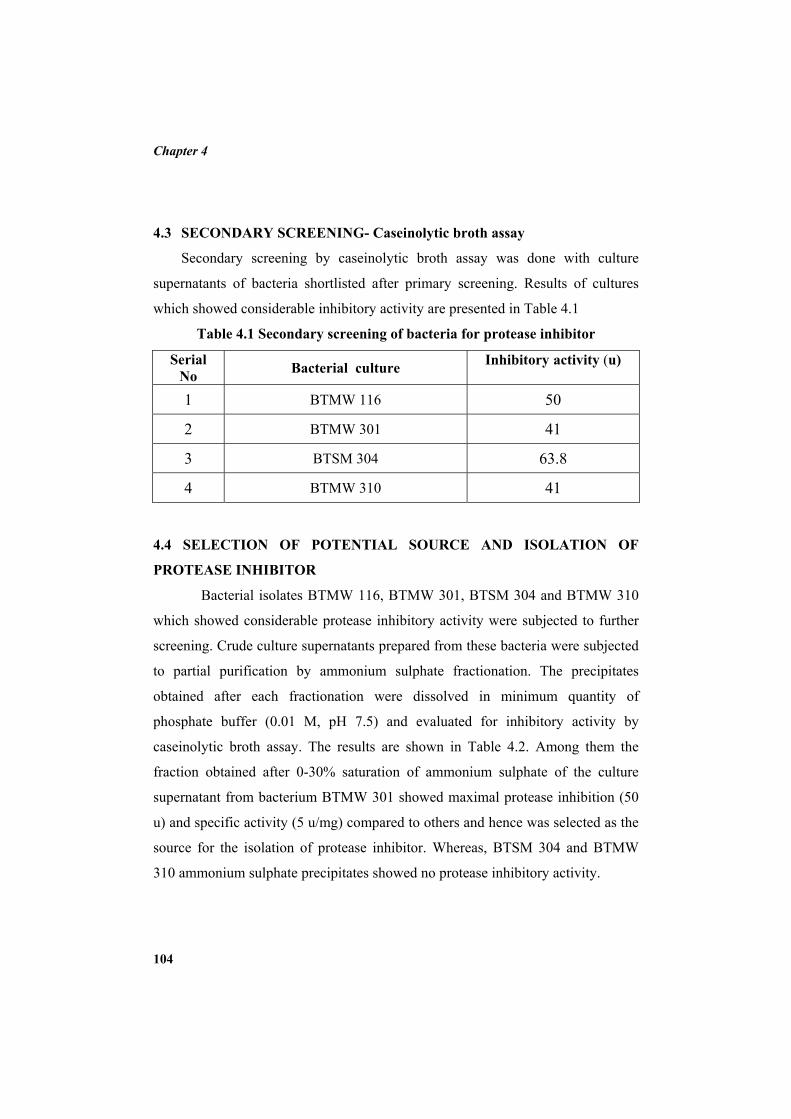
Chapter 4
104
4.3 SECONDARY SCREENING- Caseinolytic broth assay
Secondary screening by caseinolytic broth assay was done with culture
supernatants of bacteria shortlisted after primary screening. Results of cultures
which showed considerable inhibitory activity are presented in Table 4.1
Table 4.1 Secondary screening of bacteria for protease inhibitor
Serial No Bacterial culture Inhibitory activity (u)
1 BTMW 116 50
2 BTMW 301 41
3 BTSM 304 63.8
4 BTMW 310 41
4.4 SELECTION OF POTENTIAL SOURCE AND ISOLATION OF
PROTEASE INHIBITOR
Bacterial isolates BTMW 116, BTMW 301, BTSM 304 and BTMW 310
which showed considerable protease inhibitory activity were subjected to further
screening. Crude culture supernatants prepared from these bacteria were subjected
to partial purification by ammonium sulphate fractionation. The precipitates
obtained after each fractionation were dissolved in minimum quantity of
phosphate buffer (0.01 M, pH 7.5) and evaluated for inhibitory activity by
caseinolytic broth assay. The results are shown in Table 4.2. Among them the
fraction obtained after 0-30% saturation of ammonium sulphate of the culture
supernatant from bacterium BTMW 301 showed maximal protease inhibition (50
u) and specific activity (5 u/mg) compared to others and hence was selected as the
source for the isolation of protease inhibitor. Whereas, BTSM 304 and BTMW
310 ammonium sulphate precipitates showed no protease inhibitory activity.
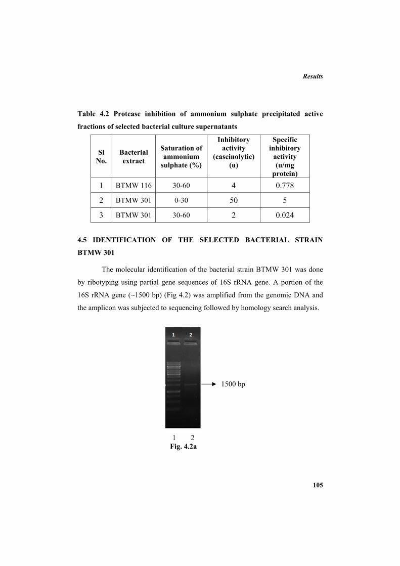
Results
105
Table 4.2 Protease inhibition of ammonium sulphate precipitated active
fractions of selected bacterial culture supernatants
Sl No.
Bacterial extract
Saturation of ammonium
sulphate (%)
Inhibitory activity
(caseinolytic) (u)
Specific inhibitory
activity (u/mg
protein) 1 BTMW 116 30-60 4 0.778
2 BTMW 301 0-30 50 5
3 BTMW 301 30-60 2 0.024
4.5 IDENTIFICATION OF THE SELECTED BACTERIAL STRAIN
BTMW 301
The molecular identification of the bacterial strain BTMW 301 was done
by ribotyping using partial gene sequences of 16S rRNA gene. A portion of the
16S rRNA gene (~1500 bp) (Fig 4.2) was amplified from the genomic DNA and
the amplicon was subjected to sequencing followed by homology search analysis.
1 2 Fig. 4.2a
1500 bp
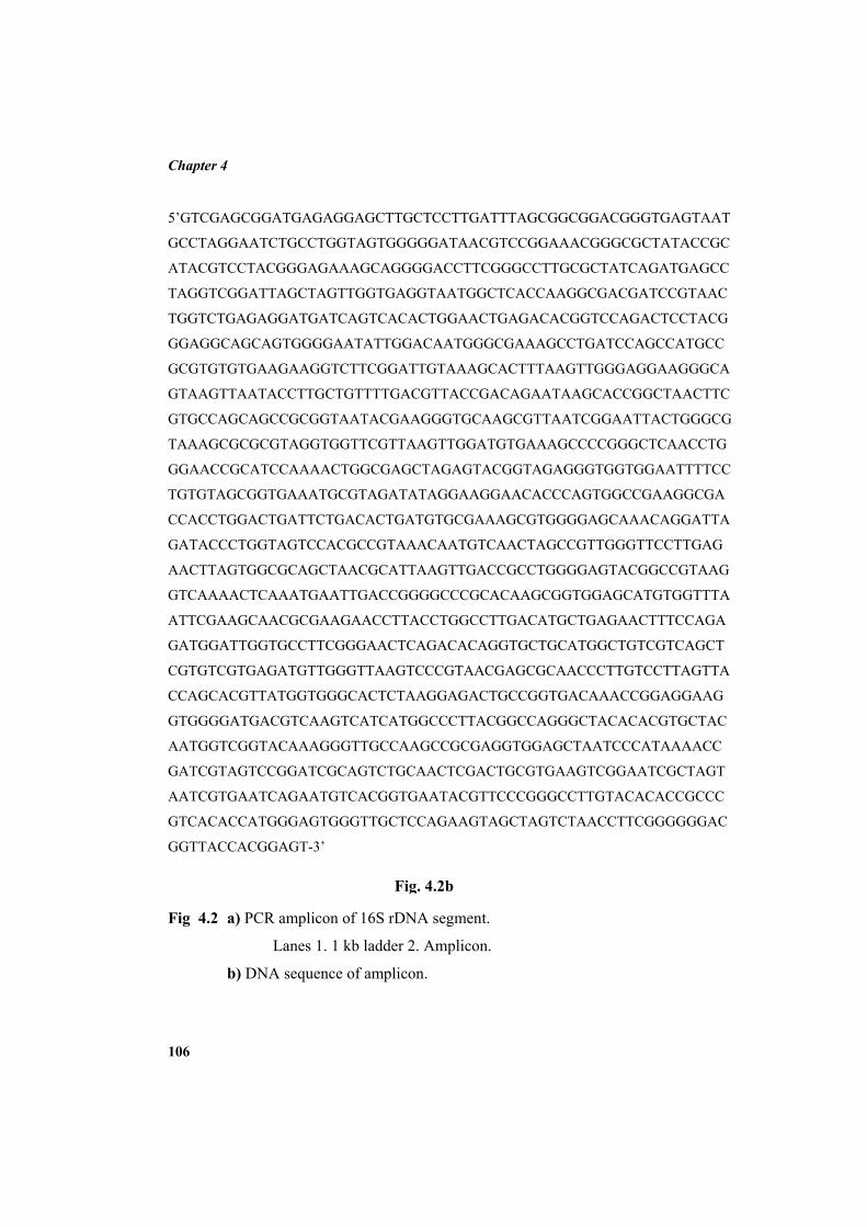
Chapter 4
106
5’GTCGAGCGGATGAGAGGAGCTTGCTCCTTGATTTAGCGGCGGACGGGTGAGTAAT
GCCTAGGAATCTGCCTGGTAGTGGGGGATAACGTCCGGAAACGGGCGCTATACCGC
ATACGTCCTACGGGAGAAAGCAGGGGACCTTCGGGCCTTGCGCTATCAGATGAGCC
TAGGTCGGATTAGCTAGTTGGTGAGGTAATGGCTCACCAAGGCGACGATCCGTAAC
TGGTCTGAGAGGATGATCAGTCACACTGGAACTGAGACACGGTCCAGACTCCTACG
GGAGGCAGCAGTGGGGAATATTGGACAATGGGCGAAAGCCTGATCCAGCCATGCC
GCGTGTGTGAAGAAGGTCTTCGGATTGTAAAGCACTTTAAGTTGGGAGGAAGGGCA
GTAAGTTAATACCTTGCTGTTTTGACGTTACCGACAGAATAAGCACCGGCTAACTTC
GTGCCAGCAGCCGCGGTAATACGAAGGGTGCAAGCGTTAATCGGAATTACTGGGCG
TAAAGCGCGCGTAGGTGGTTCGTTAAGTTGGATGTGAAAGCCCCGGGCTCAACCTG
GGAACCGCATCCAAAACTGGCGAGCTAGAGTACGGTAGAGGGTGGTGGAATTTTCC
TGTGTAGCGGTGAAATGCGTAGATATAGGAAGGAACACCCAGTGGCCGAAGGCGA
CCACCTGGACTGATTCTGACACTGATGTGCGAAAGCGTGGGGAGCAAACAGGATTA
GATACCCTGGTAGTCCACGCCGTAAACAATGTCAACTAGCCGTTGGGTTCCTTGAG
AACTTAGTGGCGCAGCTAACGCATTAAGTTGACCGCCTGGGGAGTACGGCCGTAAG
GTCAAAACTCAAATGAATTGACCGGGGCCCGCACAAGCGGTGGAGCATGTGGTTTA
ATTCGAAGCAACGCGAAGAACCTTACCTGGCCTTGACATGCTGAGAACTTTCCAGA
GATGGATTGGTGCCTTCGGGAACTCAGACACAGGTGCTGCATGGCTGTCGTCAGCT
CGTGTCGTGAGATGTTGGGTTAAGTCCCGTAACGAGCGCAACCCTTGTCCTTAGTTA
CCAGCACGTTATGGTGGGCACTCTAAGGAGACTGCCGGTGACAAACCGGAGGAAG
GTGGGGATGACGTCAAGTCATCATGGCCCTTACGGCCAGGGCTACACACGTGCTAC
AATGGTCGGTACAAAGGGTTGCCAAGCCGCGAGGTGGAGCTAATCCCATAAAACC
GATCGTAGTCCGGATCGCAGTCTGCAACTCGACTGCGTGAAGTCGGAATCGCTAGT
AATCGTGAATCAGAATGTCACGGTGAATACGTTCCCGGGCCTTGTACACACCGCCC
GTCACACCATGGGAGTGGGTTGCTCCAGAAGTAGCTAGTCTAACCTTCGGGGGGAC
GGTTACCACGGAGT-3’
Fig 4.2 a) PCR amplicon of 16S rDNA segment.
Lanes 1. 1 kb ladder 2. Amplicon.
b) DNA sequence of amplicon.
Fig. 4.2b
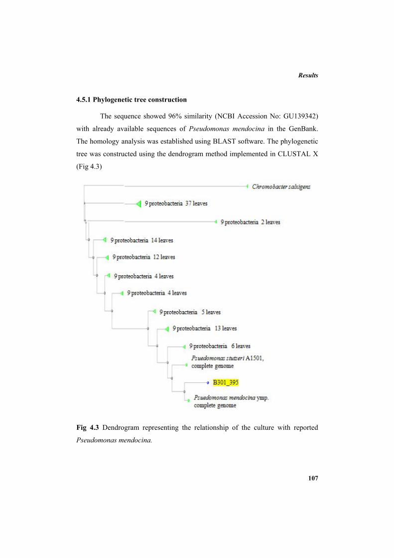
Results
107
4.5.1 Phylogenetic tree construction
The sequence showed 96% similarity (NCBI Accession No: GU139342)
with already available sequences of Pseudomonas mendocina in the GenBank.
The homology analysis was established using BLAST software. The phylogenetic
tree was constructed using the dendrogram method implemented in CLUSTAL X
(Fig 4.3)
Fig 4.3 Dendrogram representing the relationship of the culture with reported
Pseudomonas mendocina.
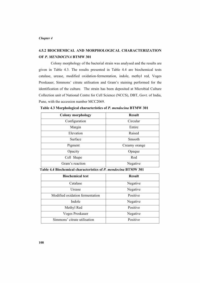
Chapter 4
108
4.5.2 BIOCHEMICAL AND MORPHOLOGICAL CHARACTERIZATION
OF P. MENDOCINA BTMW 301
Colony morphology of the bacterial strain was analysed and the results are
given in Table 4.3. The results presented in Table 4.4 are biochemical tests
catalase, urease, modified oxidation-fermentation, indole, methyl red, Voges
Proskauer, Simmons’ citrate utilisation and Gram’s staining performed for the
identification of the culture. The strain has been deposited at Microbial Culture
Collection unit of National Centre for Cell Science (NCCS), DBT, Govt. of India,
Pune, with the accession number MCC2069.
Table 4.3 Morphological characteristics of P. mendocina BTMW 301
Colony morphology Result Configuration Circular
Margin Entire Elevation Raised Surface Smooth
Pigment Creamy orange Opacity Opaque
Cell Shape Rod Gram’s reaction Negative
Table 4.4 Biochemical characteristics of P. mendocina BTMW 301
Biochemical test Result
Catalase Negative Urease Negative
Modified oxidation fermentation Positive Indole Negative
Methyl Red Positive Voges Proskauer Negative
Simmons’ citrate utilisation Positive
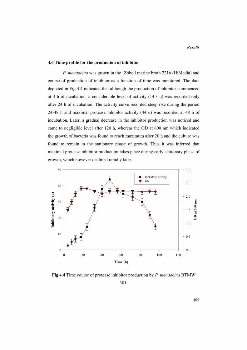
Results
109
4.6 Time profile for the production of inhibitor
P. mendocina was grown in the Zobell marine broth 2216 (HiMedia) and
course of production of inhibitor as a function of time was monitored. The data
depicted in Fig 4.4 indicated that although the production of inhibitor commenced
at 4 h of incubation, a considerable level of activity (14.3 u) was recorded only
after 24 h of incubation. The activity curve recorded steep rise during the period
24-48 h and maximal protease inhibitor activity (44 u) was recorded at 48 h of
incubation. Later, a gradual decrease in the inhibitor production was noticed and
came to negligible level after 120 h, whereas the OD at 600 nm which indicated
the growth of bacteria was found to reach maximum after 20 h and the culture was
found to remain in the stationary phase of growth. Thus it was inferred that
maximal protease inhibitor production takes place during early stationary phase of
growth, which however declined rapidly later.
Fig 4.4 Time course of protease inhibitor production by P. mendocina BTMW
301.
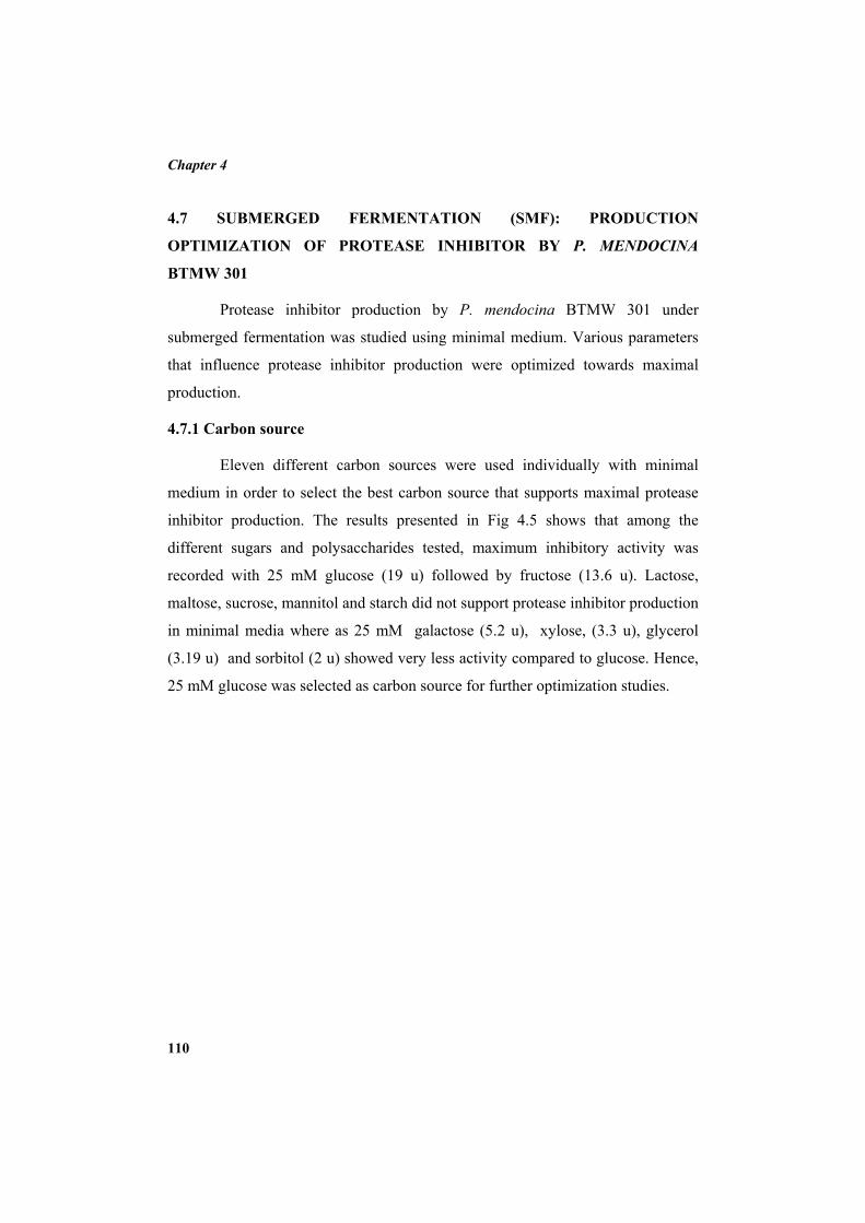
Chapter 4
110
4.7 SUBMERGED FERMENTATION (SMF): PRODUCTION
OPTIMIZATION OF PROTEASE INHIBITOR BY P. MENDOCINA
BTMW 301
Protease inhibitor production by P. mendocina BTMW 301 under
submerged fermentation was studied using minimal medium. Various parameters
that influence protease inhibitor production were optimized towards maximal
production.
4.7.1 Carbon source
Eleven different carbon sources were used individually with minimal
medium in order to select the best carbon source that supports maximal protease
inhibitor production. The results presented in Fig 4.5 shows that among the
different sugars and polysaccharides tested, maximum inhibitory activity was
recorded with 25 mM glucose (19 u) followed by fructose (13.6 u). Lactose,
maltose, sucrose, mannitol and starch did not support protease inhibitor production
in minimal media where as 25 mM galactose (5.2 u), xylose, (3.3 u), glycerol
(3.19 u) and sorbitol (2 u) showed very less activity compared to glucose. Hence,
25 mM glucose was selected as carbon source for further optimization studies.
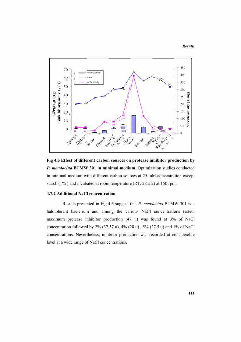
Fig 4.5 Ef
P. mendoc
in minimal
starch (1%
4.7.2 Addi
R
halotoleran
maximum
concentrat
concentrat
level at a w
1
2
3
4
5
6
7
Inhi
bito
ry a
ctiv
ity (u
)Pr
otei
n (µ
g)
ffect of diffe
cina BTMW
l medium wi
% ) and incub
itional NaCl
Results presen
nt bacterium
protease in
ion followed
ions. Nevert
wide range of
0
0
20
0
40
50
60
70
rent carbon
W 301 in min
ith different
ated at room
l concentrat
nted in Fig 4
m and amo
nhibitor pro
d by 2% (37.
theless, inhib
f NaCl conce
n sources on
nimal mediu
carbon sour
m temperature
tion
4.6 suggest t
ong the var
oduction (47
.57 u), 4% (2
bitor produc
entrations.
n protease in
um. Optimiza
rces at 25 mM
e (RT, 28 ± 2
that P. mend
rious NaCl
7 u) was fo
28 u) , 5% (2
ction was re
nhibitor pro
ation studies
M concentra
2) at 150 rpm
docina BTM
concentratio
found at 3%
27.5 u) and 1
ecorded at co
Results
111
duction by
s conducted
ation except
m.
W 301 is a
ons tested,
% of NaCl
1% of NaCl
onsiderable
050100150200250300350400450
Spec
ific
activ
ity (
U/m
g)
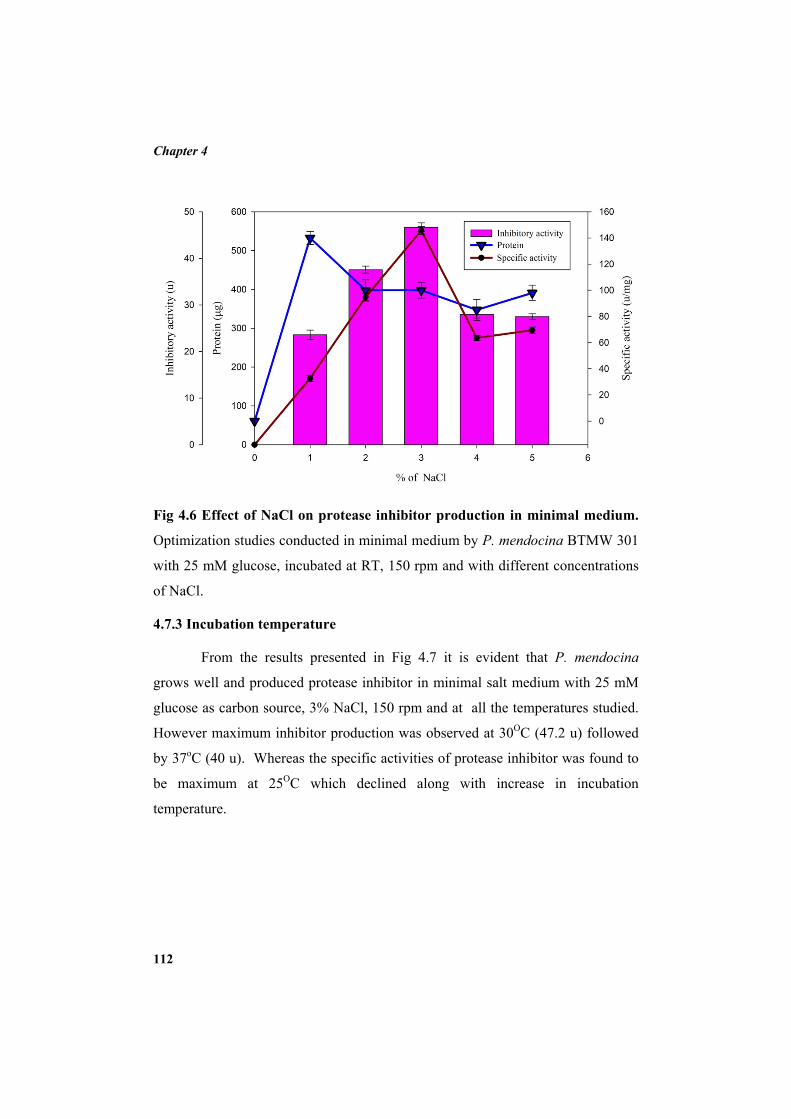
Chapter 4
112
Fig 4.6 Effect of NaCl on protease inhibitor production in minimal medium.
Optimization studies conducted in minimal medium by P. mendocina BTMW 301
with 25 mM glucose, incubated at RT, 150 rpm and with different concentrations
of NaCl.
4.7.3 Incubation temperature
From the results presented in Fig 4.7 it is evident that P. mendocina
grows well and produced protease inhibitor in minimal salt medium with 25 mM
glucose as carbon source, 3% NaCl, 150 rpm and at all the temperatures studied.
However maximum inhibitor production was observed at 30OC (47.2 u) followed
by 37oC (40 u). Whereas the specific activities of protease inhibitor was found to
be maximum at 25OC which declined along with increase in incubation
temperature.
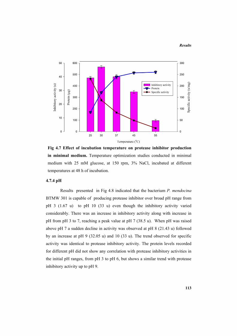
Results
113
Fig 4.7 Effect of incubation temperature on protease inhibitor production
in minimal medium. Temperature optimization studies conducted in minimal
medium with 25 mM glucose, at 150 rpm, 3% NaCl, incubated at different
temperatures at 48 h of incubation.
4.7.4 pH
Results presented in Fig 4.8 indicated that the bacterium P. mendocina
BTMW 301 is capable of producing protease inhibitor over broad pH range from
pH 3 (1.67 u) to pH 10 (33 u) even though the inhibitory activity varied
considerably. There was an increase in inhibitory activity along with increase in
pH from pH 3 to 7, reaching a peak value at pH 7 (38.5 u). When pH was raised
above pH 7 a sudden decline in activity was observed at pH 8 (21.43 u) followed
by an increase at pH 9 (32.05 u) and 10 (33 u). The trend observed for specific
activity was identical to protease inhibitory activity. The protein levels recorded
for different pH did not show any correlation with protease inhibitory activities in
the initial pH ranges, from pH 3 to pH 6, but shows a similar trend with protease
inhibitory activity up to pH 9.
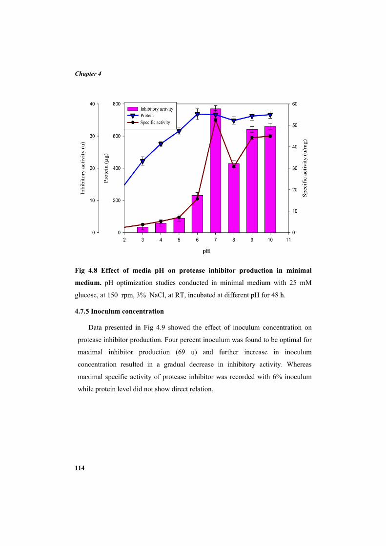
Chapter 4
114
Fig 4.8 Effect of media pH on protease inhibitor production in minimal
medium. pH optimization studies conducted in minimal medium with 25 mM
glucose, at 150 rpm, 3% NaCl, at RT, incubated at different pH for 48 h.
4.7.5 Inoculum concentration
Data presented in Fig 4.9 showed the effect of inoculum concentration on
protease inhibitor production. Four percent inoculum was found to be optimal for
maximal inhibitor production (69 u) and further increase in inoculum
concentration resulted in a gradual decrease in inhibitory activity. Whereas
maximal specific activity of protease inhibitor was recorded with 6% inoculum
while protein level did not show direct relation.
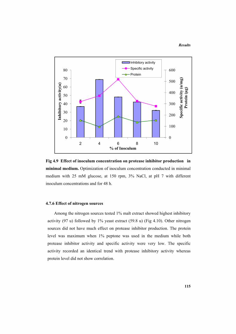
Results
115
Fig 4.9 Effect of inoculum concentration on protease inhibitor production in
minimal medium. Optimization of inoculum concentration conducted in minimal
medium with 25 mM glucose, at 150 rpm, 3% NaCl, at pH 7 with different
inoculum concentrations and for 48 h.
4.7.6 Effect of nitrogen sources
Among the nitrogen sources tested 1% malt extract showed highest inhibitory
activity (97 u) followed by 1% yeast extract (59.8 u) (Fig 4.10). Other nitrogen
sources did not have much effect on protease inhibitor production. The protein
level was maximum when 1% peptone was used in the medium while both
protease inhibitor activity and specific activity were very low. The specific
activity recorded an identical trend with protease inhibitory activity whereas
protein level did not show correlation.
0
100
200
300
400
500
600
0
10
20
30
40
50
60
70
80
2 4 6 8 10
Spec
ific
activ
ity (u
/mg)
Prot
ein
(µg)
Inhi
bito
ry a
ctiv
ity(u
)
% of Inoculum
Inhibitory activity
Specific activity
Protein
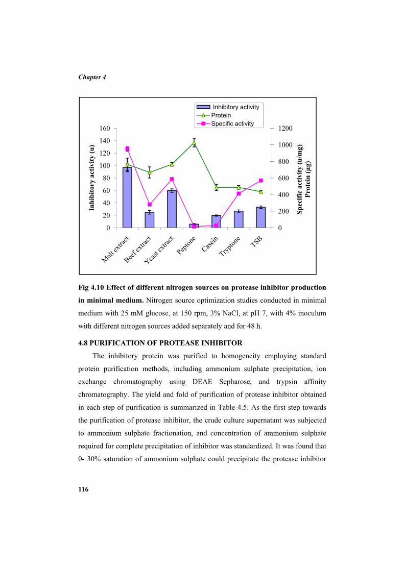
Chapter 4
116
Fig 4.10 Effect of different nitrogen sources on protease inhibitor production
in minimal medium. Nitrogen source optimization studies conducted in minimal
medium with 25 mM glucose, at 150 rpm, 3% NaCl, at pH 7, with 4% inoculum
with different nitrogen sources added separately and for 48 h.
4.8 PURIFICATION OF PROTEASE INHIBITOR
The inhibitory protein was purified to homogeneity employing standard
protein purification methods, including ammonium sulphate precipitation, ion
exchange chromatography using DEAE Sepharose, and trypsin affinity
chromatography. The yield and fold of purification of protease inhibitor obtained
in each step of purification is summarized in Table 4.5. As the first step towards
the purification of protease inhibitor, the crude culture supernatant was subjected
to ammonium sulphate fractionation, and concentration of ammonium sulphate
required for complete precipitation of inhibitor was standardized. It was found that
0- 30% saturation of ammonium sulphate could precipitate the protease inhibitor
0
200
400
600
800
1000
1200
0
20
40
60
80
100
120
140
160
Spec
ific
activ
ity (u
/mg)
Prot
ein
(µg)
Inhi
bito
ry a
ctiv
ity (u
)
Inhibitory activity Protein Specific activity
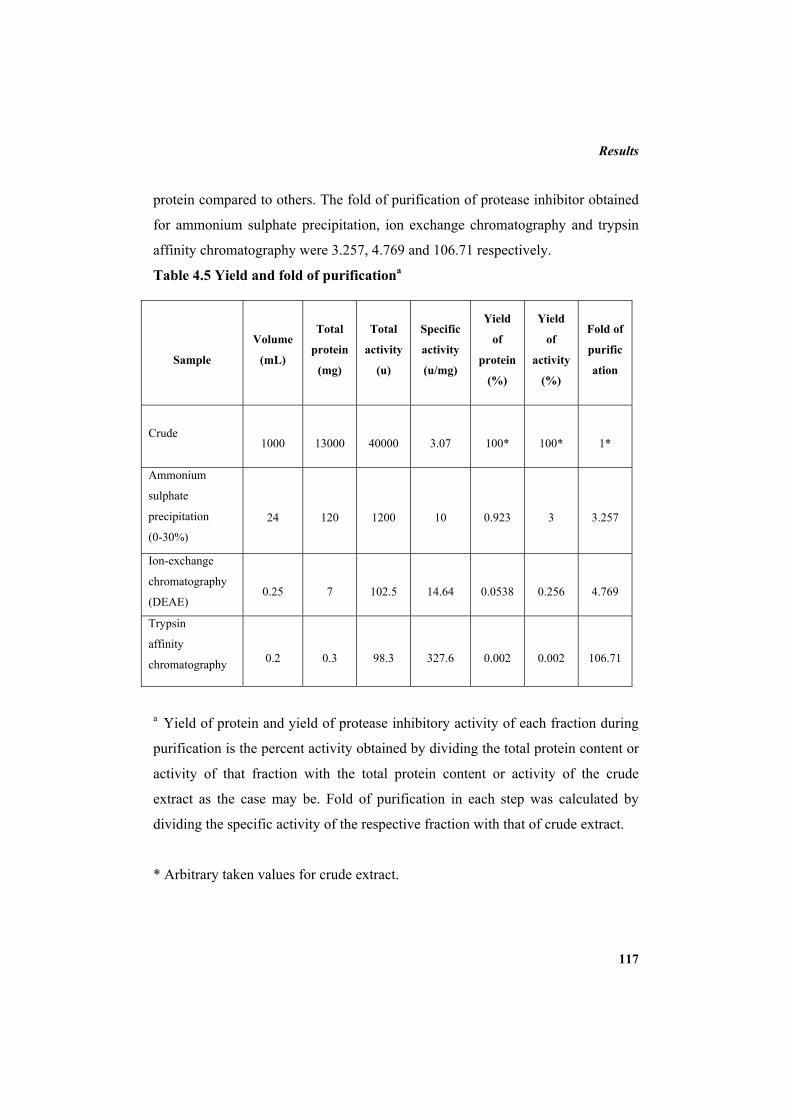
Results
117
protein compared to others. The fold of purification of protease inhibitor obtained
for ammonium sulphate precipitation, ion exchange chromatography and trypsin
affinity chromatography were 3.257, 4.769 and 106.71 respectively.
Table 4.5 Yield and fold of purificationa
Sample
Volume
(mL)
Total
protein
(mg)
Total
activity
(u)
Specific
activity
(u/mg)
Yield
of
protein
(%)
Yield
of
activity
(%)
Fold of
purific
ation
Crude
1000
13000
40000
3.07
100*
100*
1*
Ammonium
sulphate
precipitation
(0-30%)
24
120
1200
10
0.923
3
3.257
Ion-exchange
chromatography
(DEAE)
0.25
7
102.5
14.64
0.0538
0.256
4.769
Trypsin
affinity
chromatography
0.2
0.3
98.3
327.6
0.002
0.002
106.71
a Yield of protein and yield of protease inhibitory activity of each fraction during
purification is the percent activity obtained by dividing the total protein content or
activity of that fraction with the total protein content or activity of the crude
extract as the case may be. Fold of purification in each step was calculated by
dividing the specific activity of the respective fraction with that of crude extract.
* Arbitrary taken values for crude extract.
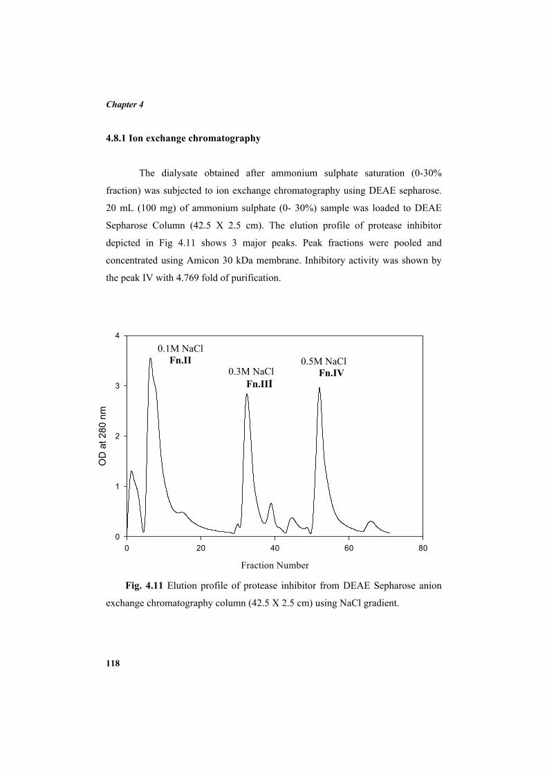
Chapter 4
118
4.8.1 Ion exchange chromatography
The dialysate obtained after ammonium sulphate saturation (0-30%
fraction) was subjected to ion exchange chromatography using DEAE sepharose.
20 mL (100 mg) of ammonium sulphate (0- 30%) sample was loaded to DEAE
Sepharose Column (42.5 X 2.5 cm). The elution profile of protease inhibitor
depicted in Fig 4.11 shows 3 major peaks. Peak fractions were pooled and
concentrated using Amicon 30 kDa membrane. Inhibitory activity was shown by
the peak IV with 4.769 fold of purification.
Fig. 4.11 Elution profile of protease inhibitor from DEAE Sepharose anion
exchange chromatography column (42.5 X 2.5 cm) using NaCl gradient.
Fraction Number
0 20 40 60 80
OD
at 2
80 n
m
0
1
2
3
4
0.1M NaCl Fn.II
0.3M NaCl Fn.III
0.5M NaCl Fn.IV
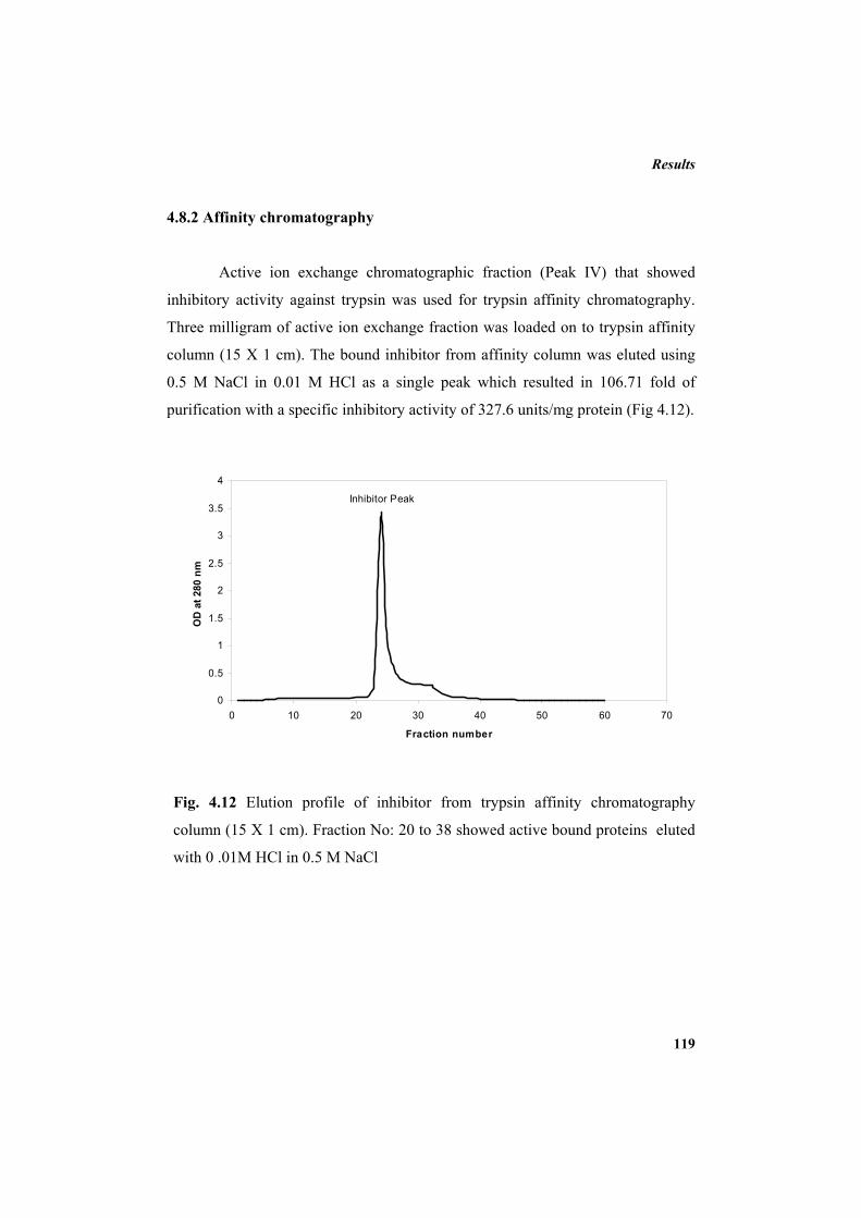
Results
119
Inhibitor Peak
0
0.5
1
1.5
2
2.5
3
3.5
4
0 10 20 30 40 50 60 70
Fraction number
OD
at 2
80 n
m
4.8.2 Affinity chromatography
Active ion exchange chromatographic fraction (Peak IV) that showed
inhibitory activity against trypsin was used for trypsin affinity chromatography.
Three milligram of active ion exchange fraction was loaded on to trypsin affinity
column (15 X 1 cm). The bound inhibitor from affinity column was eluted using
0.5 M NaCl in 0.01 M HCl as a single peak which resulted in 106.71 fold of
purification with a specific inhibitory activity of 327.6 units/mg protein (Fig 4.12).
Fig. 4.12 Elution profile of inhibitor from trypsin affinity chromatography
column (15 X 1 cm). Fraction No: 20 to 38 showed active bound proteins eluted
with 0 .01M HCl in 0.5 M NaCl
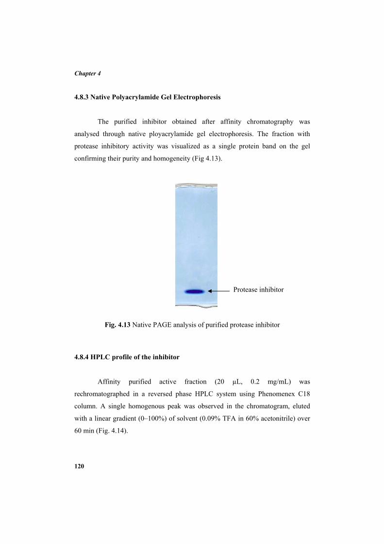
Chapter 4
120
4.8.3 Native Polyacrylamide Gel Electrophoresis
The purified inhibitor obtained after affinity chromatography was
analysed through native ployacrylamide gel electrophoresis. The fraction with
protease inhibitory activity was visualized as a single protein band on the gel
confirming their purity and homogeneity (Fig 4.13).
Fig. 4.13 Native PAGE analysis of purified protease inhibitor
4.8.4 HPLC profile of the inhibitor
Affinity purified active fraction (20 µL, 0.2 mg/mL) was
rechromatographed in a reversed phase HPLC system using Phenomenex C18
column. A single homogenous peak was observed in the chromatogram, eluted
with a linear gradient (0–100%) of solvent (0.09% TFA in 60% acetonitrile) over
60 min (Fig. 4.14).
Protease inhibitor
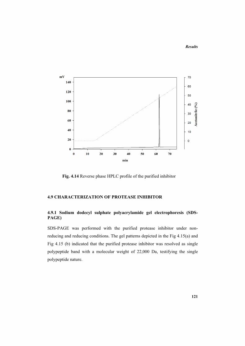
Results
121
Fig. 4.14 Reverse phase HPLC profile of the purified inhibitor
4.9 CHARACTERIZATION OF PROTEASE INHIBITOR
4.9.1 Sodium dodecyl sulphate polyacrylamide gel electrophoresis (SDS-PAGE) SDS-PAGE was performed with the purified protease inhibitor under non-
reducing and reducing conditions. The gel patterns depicted in the Fig 4.15(a) and
Fig 4.15 (b) indicated that the purified protease inhibitor was resolved as single
polypeptide band with a molecular weight of 22,000 Da, testifying the single
polypeptide nature.
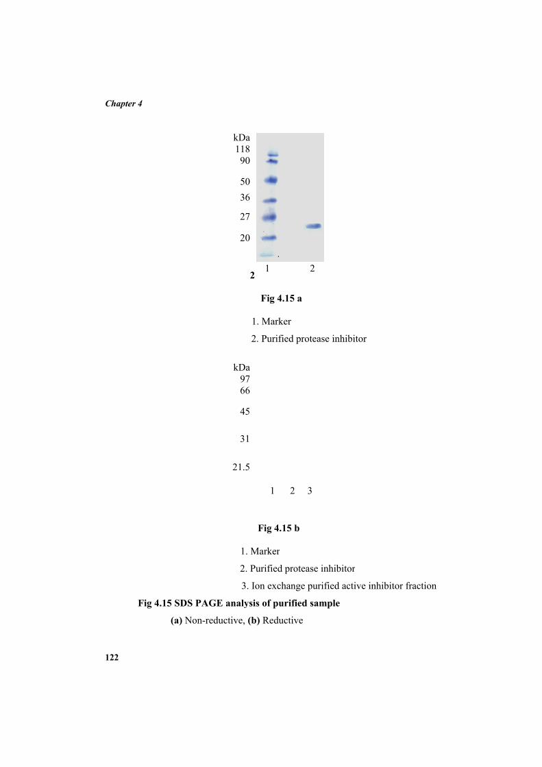
Chapter 4
122
Fi
g 4.15 SDS P
(a) No
kD 9
6
4
3
21.
1
2
3
PAGE analy
on-reductive,
kD 11 9
5 3
2
2
2
Fig 4.15
1. Marker
2. Purified
Da
1 2
7 6
5
1
5
Fig 4.15
. Marker
. Purified pro
3. Ion exchan
ysis of purif
(b) Reductiv
Da
1
8 0
06
7
0
5 a
protease inh
2 3
b
otease inhibi
nge purified a
fied sample
ve
2
hibitor
itor
active inhibittor fraction
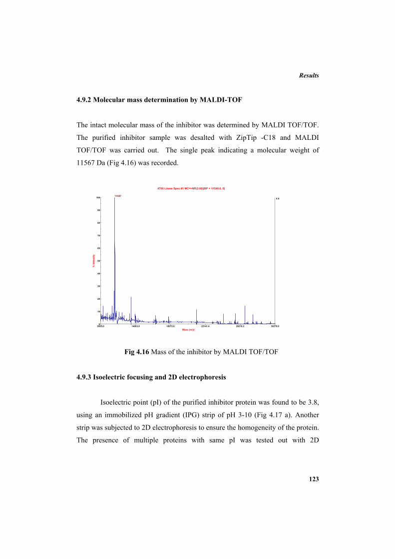
Results
123
4.9.2 Molecular mass determination by MALDI-TOF
The intact molecular mass of the inhibitor was determined by MALDI TOF/TOF.
The purified inhibitor sample was desalted with ZipTip -C18 and MALDI
TOF/TOF was carried out. The single peak indicating a molecular weight of
11567 Da (Fig 4.16) was recorded.
Fig 4.16 Mass of the inhibitor by MALDI TOF/TOF
4.9.3 Isoelectric focusing and 2D electrophoresis
Isoelectric point (pI) of the purified inhibitor protein was found to be 3.8,
using an immobilized pH gradient (IPG) strip of pH 3-10 (Fig 4.17 a). Another
strip was subjected to 2D electrophoresis to ensure the homogeneity of the protein.
The presence of multiple proteins with same pI was tested out with 2D
9935.0 14003.8 18072.6 22141.4 26210.2 30279.0Mass (m/z)
5.5
0
10
20
30
40
50
60
70
80
90
100
% In
tens
ity
4700 Linear Spec #1 MC=>NR(2.00)[BP = 11549.6, 6]
11557
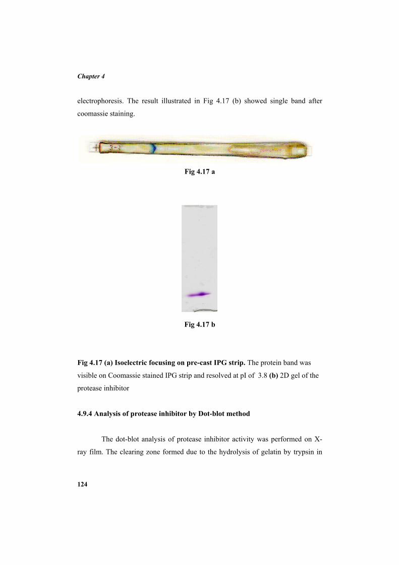
Chapter 4
124
electropho
coomassie
Fig 4.17 (a
visible on
protease in
4.9.4 Anal
Th
ray film. T
oresis. The r
staining.
a) Isoelectric
Coomassie s
nhibitor
lysis of prote
he dot-blot a
The clearing
result illustra
c focusing o
stained IPG s
ease inhibito
analysis of p
zone forme
ated in Fig
Fig 4.17 a
Fig 4.17 b
on pre-cast I
strip and reso
or by Dot-b
rotease inhib
d due to the
4.17 (b) sho
IPG strip. Th
olved at pI of
lot method
bitor activity
e hydrolysis
owed single
he protein ba
f 3.8 (b) 2D
y was perfor
of gelatin by
band after
and was
D gel of the
rmed on X-
y trypsin in
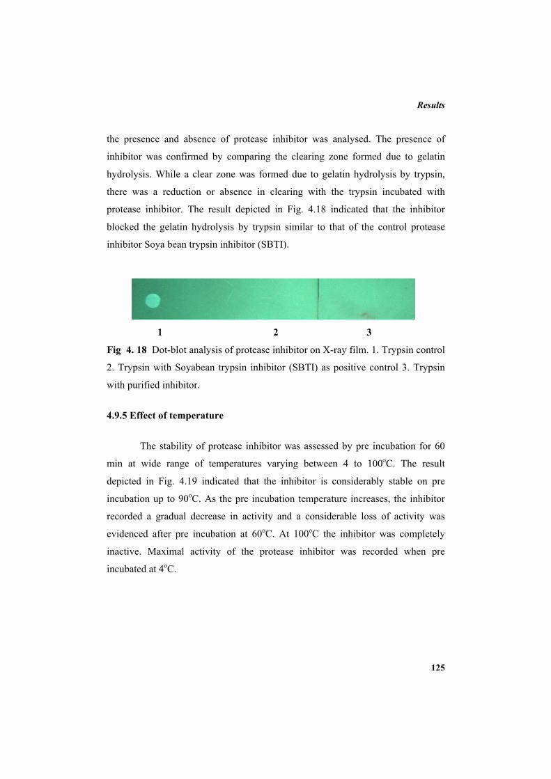
Results
125
the presence and absence of protease inhibitor was analysed. The presence of
inhibitor was confirmed by comparing the clearing zone formed due to gelatin
hydrolysis. While a clear zone was formed due to gelatin hydrolysis by trypsin,
there was a reduction or absence in clearing with the trypsin incubated with
protease inhibitor. The result depicted in Fig. 4.18 indicated that the inhibitor
blocked the gelatin hydrolysis by trypsin similar to that of the control protease
inhibitor Soya bean trypsin inhibitor (SBTI).
1 2 3
Fig 4. 18 Dot-blot analysis of protease inhibitor on X-ray film. 1. Trypsin control
2. Trypsin with Soyabean trypsin inhibitor (SBTI) as positive control 3. Trypsin
with purified inhibitor.
4.9.5 Effect of temperature
The stability of protease inhibitor was assessed by pre incubation for 60
min at wide range of temperatures varying between 4 to 100oC. The result
depicted in Fig. 4.19 indicated that the inhibitor is considerably stable on pre
incubation up to 90oC. As the pre incubation temperature increases, the inhibitor
recorded a gradual decrease in activity and a considerable loss of activity was
evidenced after pre incubation at 60oC. At 100oC the inhibitor was completely
inactive. Maximal activity of the protease inhibitor was recorded when pre
incubated at 4oC.
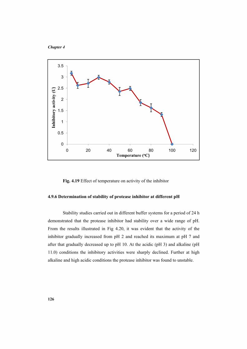
Chapter 4
126
0
0.5
1
1.5
2
2.5
3
3.5
0 20 40 60 80 100 120
Inhi
bito
ry a
ctiv
ity (U
)
Temperature (oC)
Fig. 4.19 Effect of temperature on activity of the inhibitor 4.9.6 Determination of stability of protease inhibitor at different pH
Stability studies carried out in different buffer systems for a period of 24 h
demonstrated that the protease inhibitor had stability over a wide range of pH.
From the results illustrated in Fig 4.20, it was evident that the activity of the
inhibitor gradually increased from pH 2 and reached its maximum at pH 7 and
after that gradually decreased up to pH 10. At the acidic (pH 3) and alkaline (pH
11.0) conditions the inhibitory activities were sharply declined. Further at high
alkaline and high acidic conditions the protease inhibitor was found to unstable.
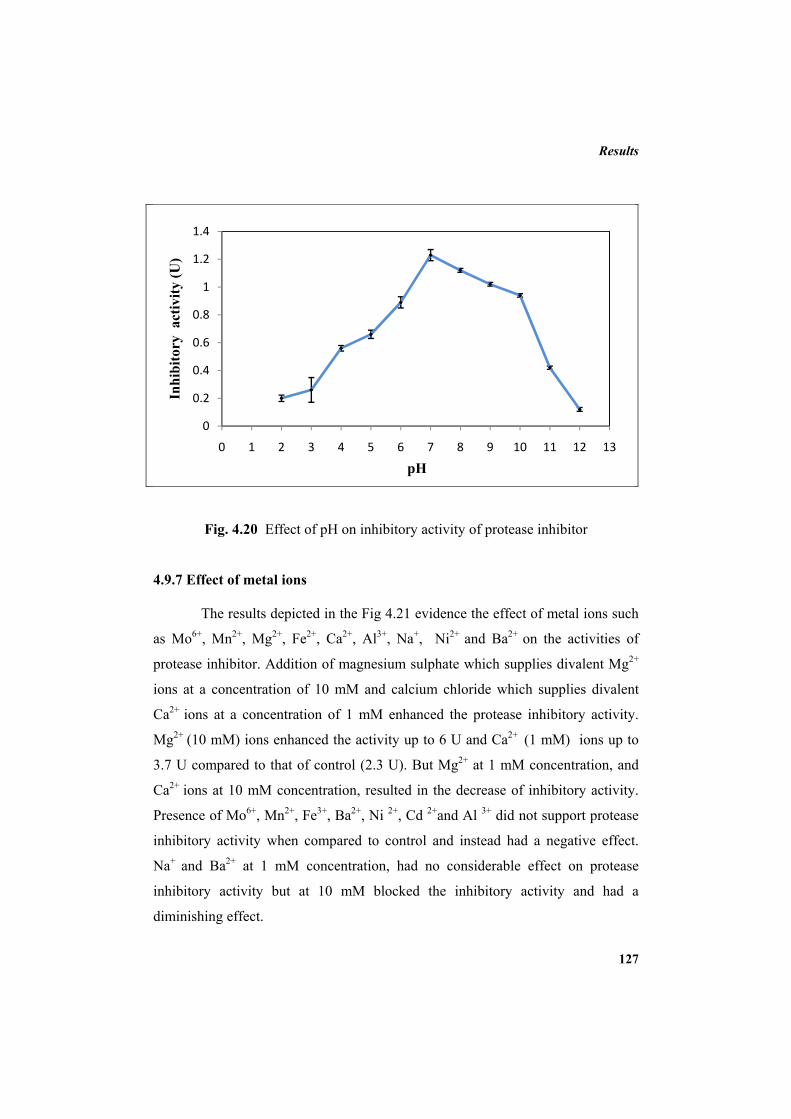
Results
127
Fig. 4.20 Effect of pH on inhibitory activity of protease inhibitor
4.9.7 Effect of metal ions
The results depicted in the Fig 4.21 evidence the effect of metal ions such
as Mo6+, Mn2+, Mg2+, Fe2+, Ca2+, Al3+, Na+, Ni2+ and Ba2+ on the activities of
protease inhibitor. Addition of magnesium sulphate which supplies divalent Mg2+
ions at a concentration of 10 mM and calcium chloride which supplies divalent
Ca2+ ions at a concentration of 1 mM enhanced the protease inhibitory activity.
Mg2+ (10 mM) ions enhanced the activity up to 6 U and Ca2+ (1 mM) ions up to
3.7 U compared to that of control (2.3 U). But Mg2+ at 1 mM concentration, and
Ca2+ ions at 10 mM concentration, resulted in the decrease of inhibitory activity.
Presence of Mo6+, Mn2+, Fe3+, Ba2+, Ni 2+, Cd 2+and Al 3+ did not support protease
inhibitory activity when compared to control and instead had a negative effect.
Na+ and Ba2+ at 1 mM concentration, had no considerable effect on protease
inhibitory activity but at 10 mM blocked the inhibitory activity and had a
diminishing effect.
0
0.2
0.4
0.6
0.8
1
1.2
1.4
0 1 2 3 4 5 6 7 8 9 10 11 12 13
Inhi
bito
ry a
ctiv
ity (U
)
pH
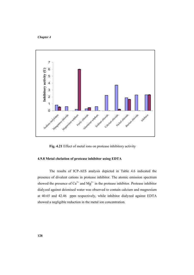
Chapter 4
128
0
1
2
3
4
5
6
7
Inhi
bito
ry a
ctiv
ity (U
)
Fig. 4.21 Effect of metal ions on protease inhibitory activity
4.9.8 Metal chelation of protease inhibitor using EDTA
The results of ICP-AES analysis depicted in Table 4.6 indicated the
presence of divalent cations in protease inhibitor. The atomic emission spectrum
showed the presence of Ca2+ and Mg2+ in the protease inhibitor. Protease inhibitor
dialyzed against deionised water was observed to contain calcium and magnesium
at 40.65 and 42.46 ppm respectively, while inhibitor dialyzed against EDTA
showed a negligible reduction in the metal ion concentration.
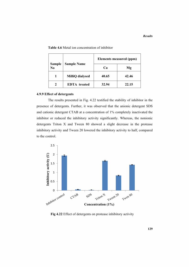
Results
129
Table 4.6 Metal ion concentration of inhibitor
Sample No
Sample Name
Elements measured (ppm)
Ca Mg
1 MilliQ dialysed 40.65 42.46
2 EDTA treated 32.94 22.15 4.9.9 Effect of detergents
The results presented in Fig. 4.22 testified the stability of inhibitor in the
presence of detergents. Further, it was observed that the anionic detergent SDS
and cationic detergent CTAB at a concentration of 1% completely inactivated the
inhibitor or reduced the inhibitory activity significantly. Whereas, the nonionic
detergents Triton X and Tween 80 showed a slight decrease in the protease
inhibitory activity and Tween 20 lowered the inhibitory activity to half, compared
to the control.
Fig 4.22 Effect of detergents on protease inhibitory activity
0
0.5
1
1.5
2
2.5
Inhi
bito
ry a
ctiv
ity (U
)
Concentration (1%)
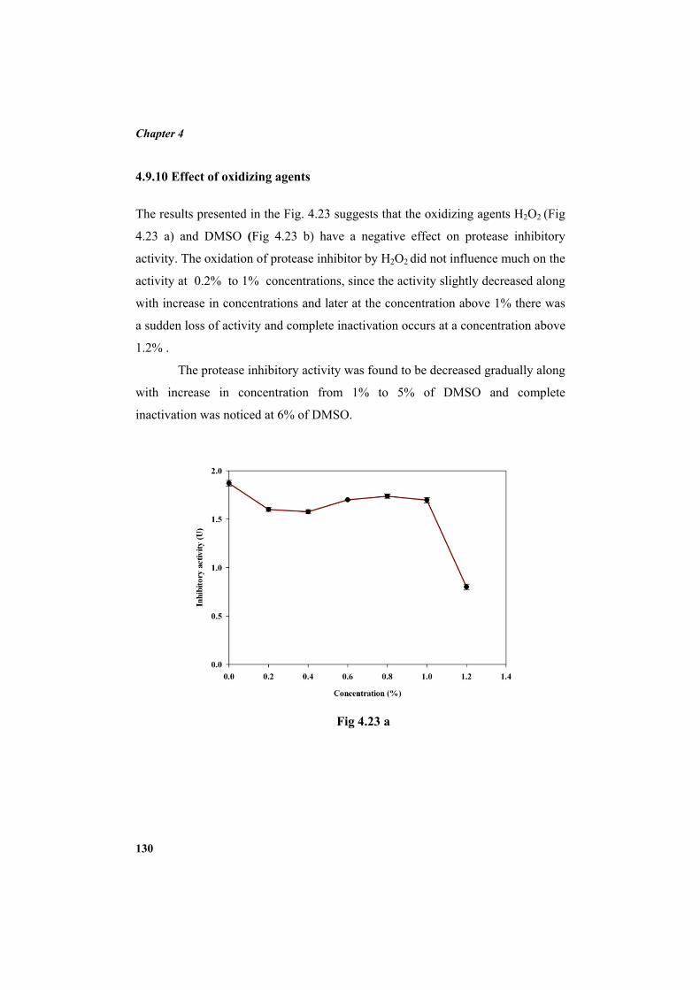
Chapter 4
130
4.9.10 Effect of oxidizing agents
The results presented in the Fig. 4.23 suggests that the oxidizing agents H2O2 (Fig
4.23 a) and DMSO (Fig 4.23 b) have a negative effect on protease inhibitory
activity. The oxidation of protease inhibitor by H2O2 did not influence much on the
activity at 0.2% to 1% concentrations, since the activity slightly decreased along
with increase in concentrations and later at the concentration above 1% there was
a sudden loss of activity and complete inactivation occurs at a concentration above
1.2% .
The protease inhibitory activity was found to be decreased gradually along
with increase in concentration from 1% to 5% of DMSO and complete
inactivation was noticed at 6% of DMSO.
Fig 4.23 a
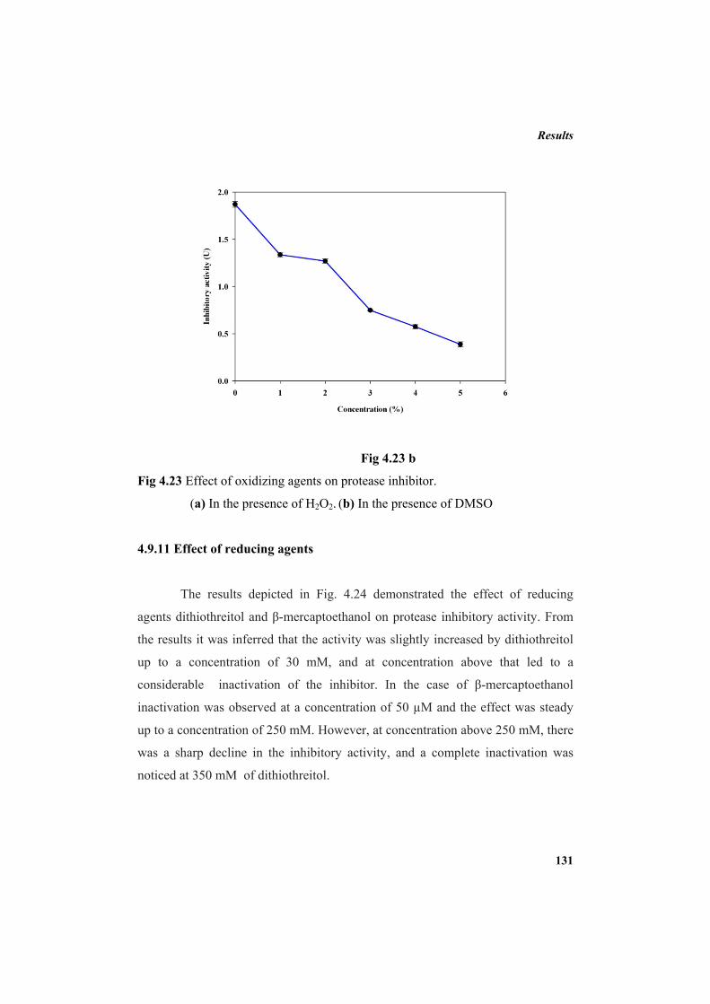
Results
131
Fig 4.23 b
Fig 4.23 Effect of oxidizing agents on protease inhibitor.
(a) In the presence of H2O2. (b) In the presence of DMSO
4.9.11 Effect of reducing agents
The results depicted in Fig. 4.24 demonstrated the effect of reducing
agents dithiothreitol and β-mercaptoethanol on protease inhibitory activity. From
the results it was inferred that the activity was slightly increased by dithiothreitol
up to a concentration of 30 mM, and at concentration above that led to a
considerable inactivation of the inhibitor. In the case of β-mercaptoethanol
inactivation was observed at a concentration of 50 µM and the effect was steady
up to a concentration of 250 mM. However, at concentration above 250 mM, there
was a sharp decline in the inhibitory activity, and a complete inactivation was
noticed at 350 mM of dithiothreitol.
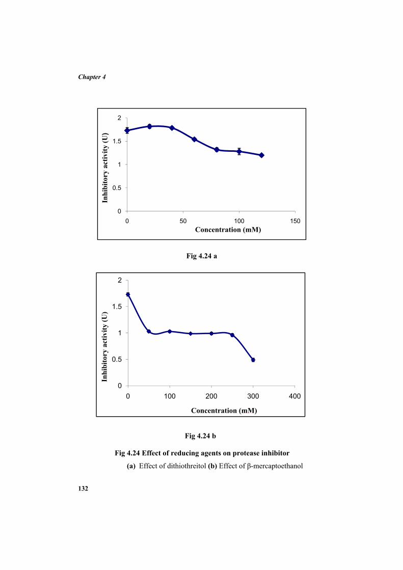
Chapter 4
132
0
0.5
1
1.5
2
0 100 200 300 400
Inhi
bito
ry a
ctiv
ity (U
)
Concentration (mM)
0
0.5
1
1.5
2
0 50 100 150
Inhi
bito
ry a
ctiv
ity (U
)
Concentration (mM)
Fig 4.24 a
Fig 4.24 b
Fig 4.24 Effect of reducing agents on protease inhibitor
(a) Effect of dithiothreitol (b) Effect of β-mercaptoethanol
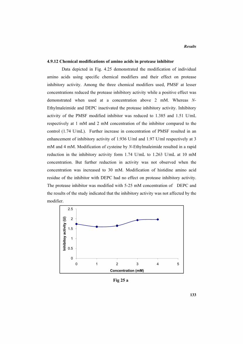
Results
133
0
0.5
1
1.5
2
2.5
0 1 2 3 4 5
Inhi
bito
y ac
tivity
(U)
Concentration (mM)
4.9.12 Chemical modifications of amino acids in protease inhibitor
Data depicted in Fig. 4.25 demonstrated the modification of individual
amino acids using specific chemical modifiers and their effect on protease
inhibitory activity. Among the three chemical modifiers used, PMSF at lesser
concentrations reduced the protease inhibitory activity while a positive effect was
demonstrated when used at a concentration above 2 mM. Whereas N-
Ethylmaleimide and DEPC inactivated the protease inhibitory activity. Inhibitory
activity of the PMSF modified inhibitor was reduced to 1.385 and 1.51 U/mL
respectively at 1 mM and 2 mM concentration of the inhibitor compared to the
control (1.74 U/mL). Further increase in concentration of PMSF resulted in an
enhancement of inhibitory activity of 1.936 U/ml and 1.97 U/ml respectively at 3
mM and 4 mM. Modification of cysteine by N-Ethylmaleimide resulted in a rapid
reduction in the inhibitory activity form 1.74 U/mL to 1.263 U/mL at 10 mM
concentration. But further reduction in activity was not observed when the
concentration was increased to 30 mM. Modification of histidine amino acid
residue of the inhibitor with DEPC had no effect on protease inhibitory activity.
The protease inhibitor was modified with 5-25 mM concentration of DEPC and
the results of the study indicated that the inhibitory activity was not affected by the
modifier.
Fig 25 a
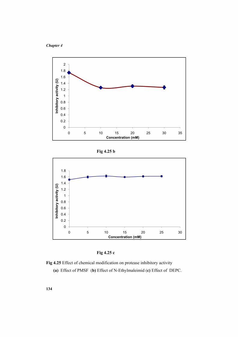
Chapter 4
134
0
0.2
0.4
0.6
0.8
1
1.2
1.4
1.6
1.8
0 5 10 15 20 25 30
Inhi
bito
ry a
ctiv
ity (U
)
Concentration (mM)
0
0.2
0.4
0.6
0.8
1
1.2
1.4
1.6
1.8
2
0 5 10 15 20 25 30 35
Inhi
bito
ry a
ctiv
ity (U
)
Concentration (mM)
Fig 4.25 b
Fig 4.25 c Fig 4.25 Effect of chemical modification on protease inhibitory activity
(a) Effect of PMSF (b) Effect of N-Ethylmaleimid (c) Effect of DEPC.
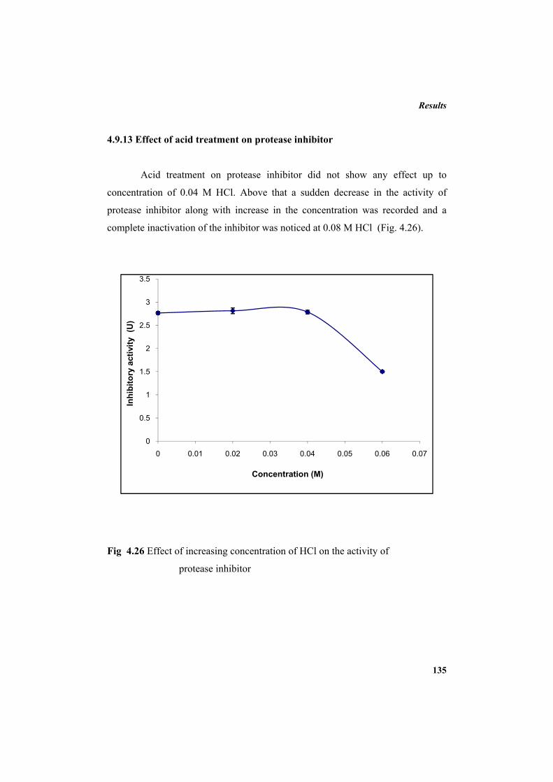
Results
135
0
0.5
1
1.5
2
2.5
3
3.5
0 0.01 0.02 0.03 0.04 0.05 0.06 0.07
Inhi
bito
ry a
ctiv
ity (
U)
Concentration (M)
4.9.13 Effect of acid treatment on protease inhibitor
Acid treatment on protease inhibitor did not show any effect up to
concentration of 0.04 M HCl. Above that a sudden decrease in the activity of
protease inhibitor along with increase in the concentration was recorded and a
complete inactivation of the inhibitor was noticed at 0.08 M HCl (Fig. 4.26).
Fig 4.26 Effect of increasing concentration of HCl on the activity of
protease inhibitor
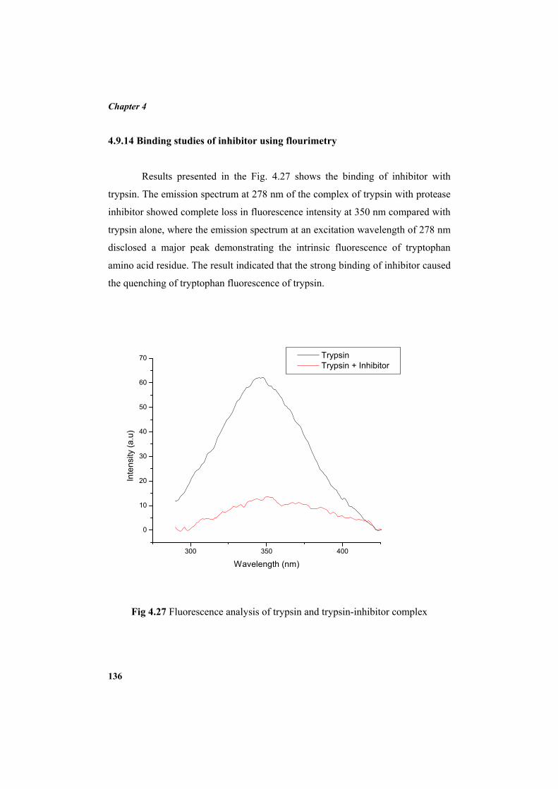
Chapter 4
136
4.9.14 Binding studies of inhibitor using flourimetry
Results presented in the Fig. 4.27 shows the binding of inhibitor with
trypsin. The emission spectrum at 278 nm of the complex of trypsin with protease
inhibitor showed complete loss in fluorescence intensity at 350 nm compared with
trypsin alone, where the emission spectrum at an excitation wavelength of 278 nm
disclosed a major peak demonstrating the intrinsic fluorescence of tryptophan
amino acid residue. The result indicated that the strong binding of inhibitor caused
the quenching of tryptophan fluorescence of trypsin.
300 350 400
0
10
20
30
40
50
60
70
Inte
nsity
(a.u
)
Wavelength (nm)
Trypsin Trypsin + Inhibitor
Fig 4.27 Fluorescence analysis of trypsin and trypsin-inhibitor complex
Results
137
4.9.15 Peptide mass fingerprinting
Peptide mass fingerprint produced by MALDI-TOF-TOF of protease
inhibitor was evaluated with the Mascot sequence matching software in Swiss-
Prot database, did not match any of the inhibitors, but identified peptide sequence
homology (31% sequence coverage) to Glycine cleavage system H protein of P.
mendocina with matched sequence in bold underlined is as follows.
1 MSNIPADLRY AASHEWARLE ADGSVTVGIS DHAQEALGDV
VFIELPEVGK 51 QLDAGQEAGV VESVKAASDI YAPVGGEVIA INEALVDSPE
SVNSDPYGSW 101 FFKLKPSDAS ELDKLLDASA YQAAADADA 4.9.16 Specificity of the inhibitor
The evaluation of inhibitory activity of P. mendocina protease inhibitor
with different serine proteases indicated high specificity towards trypsin. The
inhibitor did not show inhibitory activity towards elastase, proteinase K, subtilisin
and chymotrysin.
4.9.17 Stoichiometry of protease-protease inhibitor interaction
The data obtained for the studies carried out on protease-protease inhibitor
interaction is illustrated in Fig. 4.28. Extrapolation to zero protease activity (100%
inhibition) corresponds to 1 nM of inhibitor. It is predicted that the stoichiometry
of trypsin–protease inhibitor interaction is 1:1 and 11.67 g of protease inhibitor is
necessary to completely inactivate 23.4 g of trypsin. It was also found that the
amount of inhibitor needed for 50% inhibition (IC50) of trypsin calculated from the
graph was 0.48 nM.
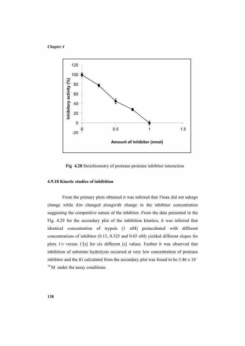
Chapter 4
138
Fig 4.28 Stoichiometry of protease-protease inhibitor interaction
4.9.18 Kinetic studies of inhibition
From the primary plots obtained it was inferred that Vmax did not udergo
change while Km changed alongwith change in the inhibitor concentration
suggesting the competitive nature of the inhibitor. From the data presented in the
Fig. 4.29 for the secondary plot of the inhibition kinetics, it was inferred that
identical concentration of trypsin (1 nM) preincubated with different
concentrations of inhibitor (0.13, 0.325 and 0.65 nM) yielded different slopes for
plots 1/v versus 1/[s] for six different [s] values. Further it was observed that
inhibition of substrate hydrolysis occurred at very low concentration of protease
inhibitor and the Ki calculated from the secondary plot was found to be 3.46 x 10 -
10 M under the assay conditions.
-20
0
20
40
60
80
100
120
0 0.5 1 1.5
Inhi
bito
ry a
ctiv
ity (%
)
Amount of inhibitor (nmol)
4.10 APPL
4.10.1 Pro
Th
preservatio
naturally p
protease en
hundred an
quantities
inhibitor w
caseinolyti
against 61
F
LICATION
otease inhibi
he potential
on was asses
present on sh
nzyme, the
nd ten bacte
of protease
was evaluat
ic assay. The
of spoilage b
Fig 4.29 Seco
STUDIES
itor as seafo
l of protea
sed using sh
hrimp was is
major enzym
erial isolates
e enzyme in
ted for its
e data obtain
bacterial pro
ondary plot o
od preserva
ase inhibitor
hrimp, Penea
solated and a
me responsib
98 bacteria
n skimmed
activity aga
ed showed th
oteases (Tabl
of the inhibit
ative
r as preser
us monodon
assessed for
ble for food
a were found
milk agar p
ainst these
hat the prote
e 4.6).
tor
rvative for
. Viable mic
their ability
spoilage. A
d to produce
plates (Fig
spoilage pr
ase inhibitor
Results
139
sea food
crobial flora
to produce
Among one
e enormous
4.30). The
roteases by
r was active
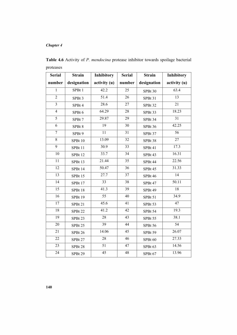
Chapter 4
140
Table 4.6 Activity of P. mendocina protease inhibitor towards spoilage bacterial
proteases
Serial
number
Strain
designation
Inhibitory
activity (u)
Serial
number
Strain
designation
Inhibitory
activity (u)
1 SPBt 1 42.2 25 SPBt 30 63.4
2 SPBt 3 51.4 26 SPBt 31 13
3 SPBt 4 28.6 27 SPBt 32 21
4 SPBt 6 64.29 28 SPBt 33 18.23
5 SPBt 7 29.87 29 SPBt 34 31
6 SPBt 8 19 30 SPBt 36 42.25
7 SPBt 9 11 31 SPBt 37 56
8 SPBt 10 13.09 32 SPBt 38 27
9 SPBt 11 30.9 33 SPBt 41 17.3
10 SPBt 12 33.7 34 SPBt 43 16.31
11 SPBt 13 21.44 35 SPBt 44 22.56
12 SPBt 14 50.47 36 SPBt 45 31.33
13 SPBt 15 27.7 37 SPBt 46 14
14 SPBt 17 33 38 SPBt 47 50.11
15 SPBt 18 41.3 39 SPBt 49 18
16 SPBt 19 55 40 SPBt 51 34.9
17 SPBt 21 45.6 41 SPBt 53 47
18 SPBt 22 41.2 42 SPBt 54 19.3
19 SPBt 23 28 43 SPBt 55 38.1
20 SPBt 25 39 44 SPBt 56 54
21 SPBt 26 14.06 45 SPBt 59 26.07
22 SPBt 27 28 46 SPBt 60 27.33
23 SPBt 28 51 47 SPBt 63 14.56
24 SPBt 29 45 48 SPBt 67 13.96
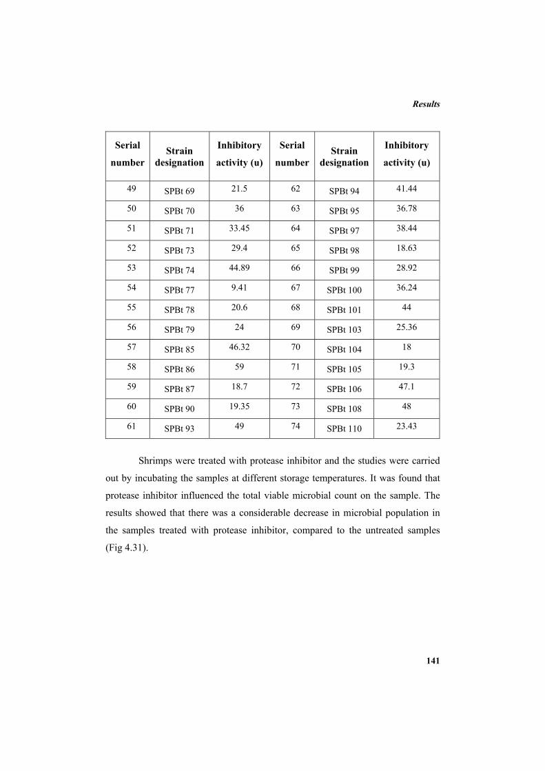
Results
141
Serial
number Strain
designation
Inhibitory
activity (u)
Serial
number Strain
designation
Inhibitory
activity (u)
49 SPBt 69 21.5 62 SPBt 94 41.44
50 SPBt 70 36 63 SPBt 95 36.78
51 SPBt 71 33.45 64 SPBt 97 38.44
52 SPBt 73 29.4 65 SPBt 98 18.63
53 SPBt 74 44.89 66 SPBt 99 28.92
54 SPBt 77 9.41 67 SPBt 100 36.24
55 SPBt 78 20.6 68 SPBt 101 44
56 SPBt 79 24 69 SPBt 103 25.36
57 SPBt 85 46.32 70 SPBt 104 18
58 SPBt 86 59 71 SPBt 105 19.3
59 SPBt 87 18.7 72 SPBt 106 47.1
60 SPBt 90 19.35 73 SPBt 108 48
61 SPBt 93 49 74 SPBt 110 23.43
Shrimps were treated with protease inhibitor and the studies were carried
out by incubating the samples at different storage temperatures. It was found that
protease inhibitor influenced the total viable microbial count on the sample. The
results showed that there was a considerable decrease in microbial population in
the samples treated with protease inhibitor, compared to the untreated samples
(Fig 4.31).
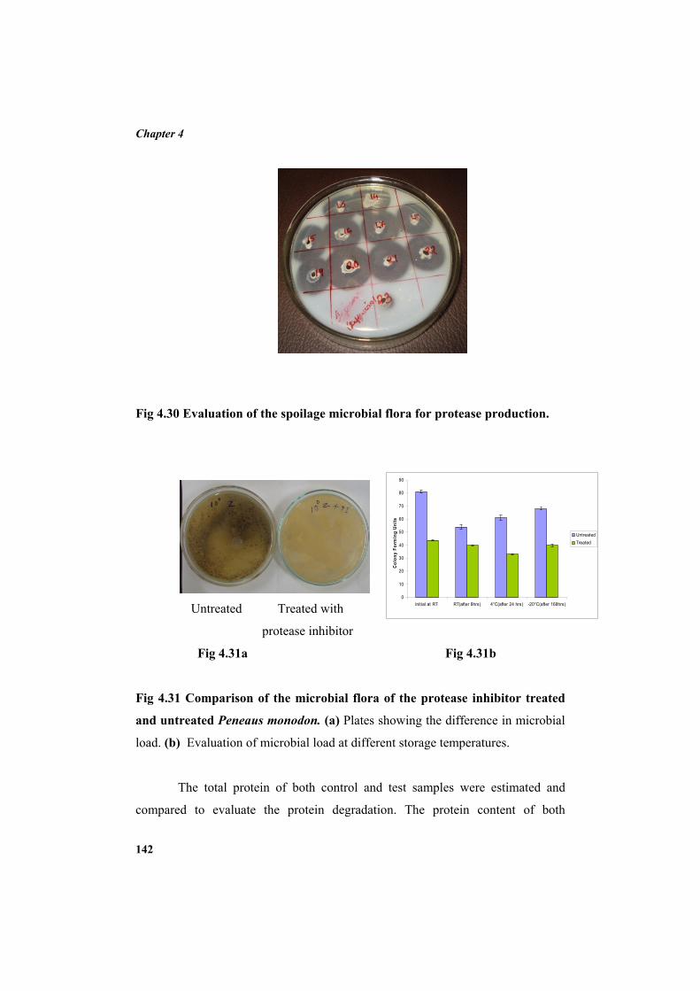
Chapter 4
142
Fig 4.30 Evaluation of the spoilage microbial flora for protease production.
Untreated Treated with
protease inhibitor
Fig 4.31a Fig 4.31b
Fig 4.31 Comparison of the microbial flora of the protease inhibitor treated
and untreated Peneaus monodon. (a) Plates showing the difference in microbial
load. (b) Evaluation of microbial load at different storage temperatures.
The total protein of both control and test samples were estimated and
compared to evaluate the protein degradation. The protein content of both
0
10
20
30
40
50
60
70
80
90
Initial at RT RT(after 8hrs) 4°C(after 24 hrs) -20°C(after 168hrs)
Colo
ny F
orm
ing
Units
UntreatedTreated
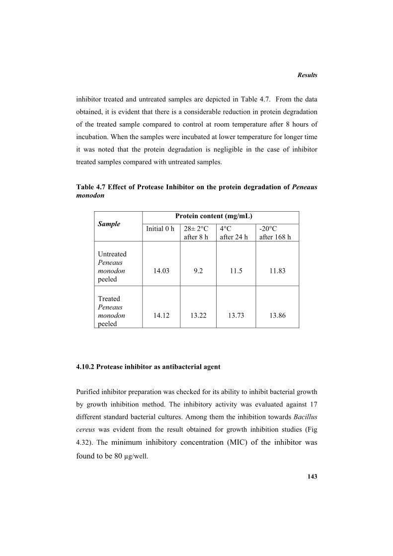
Results
143
inhibitor treated and untreated samples are depicted in Table 4.7. From the data
obtained, it is evident that there is a considerable reduction in protein degradation
of the treated sample compared to control at room temperature after 8 hours of
incubation. When the samples were incubated at lower temperature for longer time
it was noted that the protein degradation is negligible in the case of inhibitor
treated samples compared with untreated samples.
Table 4.7 Effect of Protease Inhibitor on the protein degradation of Peneaus monodon
4.10.2 Protease inhibitor as antibacterial agent
Purified inhibitor preparation was checked for its ability to inhibit bacterial growth
by growth inhibition method. The inhibitory activity was evaluated against 17
different standard bacterial cultures. Among them the inhibition towards Bacillus
cereus was evident from the result obtained for growth inhibition studies (Fig
4.32). The minimum inhibitory concentration (MIC) of the inhibitor was
found to be 80 µg/well.
Sample Protein content (mg/mL)
Initial 0 h 28± 2°C after 8 h
4°C after 24 h
-20°C after 168 h
Untreated Peneaus monodon peeled
14.03
9.2
11.5
11.83
Treated Peneaus monodon peeled
14.12
13.22
13.73
13.86

Chapter 4
144
Fig 4.32 Growth inhibition of Bacillus cereus using protease inhibitor from P. mendocina BTMW 301
145
Chapter 5
DISCUSSION 5.1 SCREENING OF MICROORGANISMS FOR PROTEASE INHIBITOR
PRODUCTION AND PROCESS OPTIMIZATION
The microorganisms are recognized as important sources of protease
inhibitors which are valuable in the fields of medicine, agriculture and
biotechnology. The protease inhibitors of microbial origin are found to be versatile
in their structure and mode of inhibition that vary from those of other sources.
Although surplus of low molecular weight non-protein protease inhibitors from
microorganisms have been reported, there is a dearth of reports on proteinaceous
protease inhibitors. The search for new metabolites from marine organisms has
resulted in the isolation of more or less 10,000 metabolites (Fuesetani and
Fuesetani, 2000) many of which are gifted with pharmacodynamic properties. The
existence of marine microorganisms was reported earlier, and they were found to
be metabolically and physiologically dissimilar from terrestrial microorganisms.
Marine microorganisms have potential as important new sources of enzyme
inhibitors and consequently a detailed study of new marine microbial inhibitors
will provide the basis for future research (Imada, 2004).
In the present study, bacteria, actinomycetes and fungi isolated from
different locations of Cochin backwaters, Kerala were screened for protease
inhibitors towards identification of new potential strain as source for protease
inhibitor production. The primary screening of protease inhibitor on skimmed
milk agar plates was carried out and the shortlisted cultures were subjected to
caseinolytic liquid assay. The isolates with considerable inhibitory activity were
shortlisted and further screened after partial purification using ammonium sulphate
precipitation. The precipitate was dissolved and dialysed against phosphate buffer
of pH 7.5. Protease inhibitor from Moringa oleifera (Bijina et al., 2011b) and
Chapter 5
146
Streptomyces sp. (Pandhare et al., 2002) were resuspended and dialysed in
phosphate buffer. The optimum activity of trypsin protease is at pH 7.5. So the pH
of the phosphate buffer was selected as 7.5.
Among the four bacterial isolates shortlisted, bacterium BTMW 301 was
selected as the potential source since it recorded the highest activity compared to
others. This strain was identified as Pseudomonas mendocina by 16S ribotyping
since it showed very close similarity with already existing 16S sequences of P.
mendocina in the NCBI database. Species of the genus Pseudomonas are
metabolically very versatile and are encountered in most natural environments,
from freshwater to hypersaline habitats (Holt et al., 1994). P. mendocina is a salt
tolerant bacterium (Palleroni et al., 1984) with high potential for bioremediation of
contaminated soils and waters (Meer et al., 1992). Although there are reports
available on protease inhibitor produced by marine Pseudoalteromonas
sagamiensis (Takeshi et al., 2003), so far no protease inhibitor has been reported
to be produced by P. mendocina. In this context the present investigation
indicates the potential of P. mendocina as a dependable source of protease
inhibitor.
Evaluation of time course of production of protease inhibitor in Zobell
marine broth indicated that maximal production was at 48 h of incubation, i.e.
during the commencement of stationary phase. Process optimization studies
performed towards maximal production of protease inhibitor using minimal
medium evidenced that protease inhibitor production was influenced by incubation
temperature, media pH, NaCl concentration, carbon sources, nitrogen sources and
inoculum concentration. A previous study on the production of protease inhibitor
by an actinomycetes sp. showed that the production of inhibitor depends upon
nitrogen and carbon sources (Pandhare et al., 2002). Among the different carbon
sources studied, 25 mM glucose induced maximum production of protease
inhibitor. Study conducted on the effect of additional NaCl concentrations on
protease inhibitor production with the selected carbon source showed that the
Discussion
147
production was maximum at 3% NaCl. In fact P. mendocina was reported as salt
tolerant bacterium (Pocard et al., 1994). The tolerance to higher NaCl
concentration is an added advantage when the protease inhibitor finds application
as food preservative, as the protease inhibitor produced by this hyper salt tolerant
strain is likely to serve a preservative function in salt preserved foods as well as
for sea foods.
Temperature is a significant factor that has to be regulated. Hence
temperature optimization studies for maximal production of protease inhibitor was
done and it was observed that maximal production occurred at 30oC, which is
ambient temperature and hence an advantage considering the production economy.
P. mendocina is capable of producing protease inhibitor over a broad range of pH
from pH 3 to 10 although the production was very less in acidic pH conditions
compared to neutral and alkaline pH ranges. Varying concentrations of inoculum
were added to the production media to analyse the effect of inoculum
concentration on the production of protease inhibitor by P. mendocina. The
protease inhibitor production was found to be maximum when 4% inoculum was
used. Above that concentration decline in inhibitory activity along with increase in
inoculum concentration was noticed. For 10% of inoculum, the production of
inhibitor was lesser than that produced for 2% inoculum. This decrease may be
attributed to the over populated culture and nutrient limitation in the media.
Among the nitrogen sources tested, the maximal protease inhibitor production was
recorded when malt extract was used in the production media.
5.2 PURIFICATION AND CHARACTERIZATION OF PROTEASE
INHIBITOR
Protease inhibitor obtained from P. mendocina was purified by
ammonium sulphate precipitation, followed by DEAE sepharose ion exchange
chromatography and trypsin affinity chromatography. P. mendocina protease
inhibitor was precipitated efficiently with 0-30% saturation of ammonium
Chapter 5
148
sulphate. The inhibitor eluted out from trypsin affinity column yielded a single
protein fraction with maximum protease inhibitory activity. The homogeneity of
this fraction was further proved by reverse phase high pressure liquid
chromatography and single band was obtained after polyacrylamide gel
electrophoresis, reconfirming its homogeneity. From the results obtained it is
evident that the fold and yield of protein can be enhanced by repetitive
purifications using combinations of several advanced purification techniques.
Purification of protease inhibitors from skin extract of Atlantic salmon (Salmo
salar L.) was carried out in four chromatographic steps: papain-affinity
chromatography, gel filtration, anion-exchange chromatography and reversed
phase chromatography (Anne et al., 1999) and protease inhibitor from potato
tubers (Solanum tuberosum) was purified through extraction of the water-soluble
fraction, dialysis, ultra filtration, DEAE-cellulose and C18 reversed-phase high
performance liquid chromatography (Mi-Hyun et al., 2006).
Protease inhibitor purification can be significantly improved by the use of
affinity chromatography techniques, where the binding agents are particular
proteases (Araújo et al., 2005). Trypsin affinity chromatography was found to
augment the fold of purification of trypsin inhibitors (TI) from wild-type soybean
(Glycine soya) (WBTI) and domesticated soybean (Glycine max) (SBTI) using
chitosan resin-trypsin on the affinity chromatography column (Zhang et al., 2009).
A trypsin inhibitor was purified from Sapindus saponaria seeds (SSTI) using a
trypsin-Sepharose column, where the inhibitor was eluted with 0.01 M HCl
(Macedo et al., 2011). Trypsin agarose affinity chromatography was used for the
isolation of a protein from Helianthus annuus flowers (Giudici et al., 2000). The
purity of inhibitor was further confirmed by reverse phase HPLC. Homogeneity of
the proteinase inhibitor from the marine annelid Hermodice carunculata was
confirmed by reversed phase high performance liquid chromatography (RP-
HPLC) on C-18 column (Isel et al., 2004).
Discussion
149
The purity and homogeneity of the affinity chromatography purified
inhibitor was proved by single band obtained in both Native-PAGE and SDS-
PAGE. The SDS-PAGE analysis of the purified proteinaceous protease inhibitor
molecule evidenced the single polypeptide nature of the inhibitor. An apparent
molecular weight of 22 kDa was obtained in SDS-PAGE, both under non-reducing
and reducing conditions. Whereas the intact molecular mass by MALDI-TOF
analysis indicated a value of 11.567 kDa. It was considered that the MALDI-TOF
analysis is a more precise method for molecular weight determination with a mass
accuracy of 0.05-0.1% (Jany et al., 1986). The occurrence of a high molecular
weight protein form along with protease inhibitor (PISC-2002) of 11.264 kDa
isolated from Streptomyces chromofuscus is explained as a thin band of molecular
mass 25 kDa. This band was observed which corresponded to a dimer of the
inhibitor. This band did not disappear when electrophoresis was performed in the
presence of β-mercaptoethanol (Angelova et al., 2006). The molecular mass of the
present inhibitor is different from protease inhibitor (ecotin) isolated from Gram-
negative bacteria E. coli, which is a homodimer of 16 kDa subunits (Maurizi,
1992). A 12 kDa protease inhibitor was isolated and studied from the
entemopathogenic bacterium Photorhabdus luminescens (Wee et al., 2000).
Marinostatin is a low molecular weight peptide inhibitor and monastatin is a novel
glycoprotein with a molecular weight of 20 kDa. Both were isolated from marine
bacterium Alteromonas sp. (Imada et al., 1986a ; Imada et al., 1985a). A serine
protease inhibitor was obtained from a metagenomic library from uncultured
marine microorganisms with a predicted molecular mass of about 28.7 kDa
(Cheng-Jian Jiang et al., 2011). The serine protease inhibitors isolated from silk
worms (pacifastin family) have molecular weight of 4 kDa and Kunitz inhibitors
are in the range of 18-26 kDa (Clynen et al., 2005). Serpins (serine proteinase
inhibitors) are the largest super family of protease inhibitors with 350–400 amino
acids and the molecular weight of 40–50 kDa (Khan et al., 2011).
Chapter 5
150
Most preferred criteria for distinguishing proteins are the isoelectric point
(pI). Protease inhibitors exhibit a range of pI from acidic to alkaline with respect
to the source and type of inhibitor. In the present study isoelectric point of the
inhibitory protein resolved on the IPG strip was 3.8, whereas isoelectric point of
the protease inhibitor isolated from Manduca sexta is 5.4 (Wang and Jiang, 2004)
and Streptomyces chromofuscus is 7.4 (Angelova et al., 2006). Kunitz type
inhibitors generally show a pI of acidic nature. Three isoforms of protease
inhibitors, ApTIA, ApTIB and ApTIC from Acacia plumose resolved on
isoelectric focusing gel had isoelectric points of an acidic nature: 5.05, 5.25 and
5.55, respectively (Laber et al., 1989; Lopes et al., 2009). Serine protease
inhibitors from the basidiomycete Clitocybe nebularis CnSPIs, have isoelectric
points 4.8 and 5.2 (Avanzo et al., 2009). The isoelectric point of protease inhibitor
isolated from the seeds of pearl millet (Pennisetum glaucum) was 9.8 indicating its
basic nature (Joshi et al., 1998). Serine protease inhibitors (ISPI-1, 2, 3) purified
from larval hemolymph of greater wax moth larvae, Galleria mellonella had
isoelectric points ranging between 7.2 and 8.3 (Andreas et al., 2000).
Protease inhibitory activity was confirmed on X-ray film by Dot-blot
analysis. The results indicated that the protease inhibitor blocked the hydrolysis of
gelatin on X-ray film by inhibiting trypsin, in the same way as that of the control
protease inhibitor soya bean trypsin inhibitor (SBTI). Thermal stability increases
the efficiency of proteins and is one of the essential features for their commercial
exploitation (Pandhare et al., 2002). In this respect thermal inactivation studies of
protease inhibitor isolated from P. mendocina was carried out by evaluating its
activity and stability at wide range of temperatures varying between 4oC to 100oC.
Results demonstrated considerable stability of the inhibitor up to 90oC although
maximal stability of the protease inhibitor was observed at 4oC. However a
progressive decline in activity was noted after pre-incubation at temperatures
above 40°C. Considerable decrease in activity was observed at 60°C to 90°C for 1
h and a complete loss of activity at 100°C. From these observations it is evident
Discussion
151
that the protease inhibitor has high intrinsic stability in its native state, which
imparts a high degree of thermal stability. The protease inhibitor of bacterium
Photorhabdus luminescens was inactivated at temperatures above 500 C (Wee et
al., 2000). Kinetic studies reveal the competitive nature of the inhibitor and shows
high specificity to trypsin. Protease inhibitors API-I, API-II and API-III were
isolated from three different actinomycetes strains showed different properties in
their molecular nature as well as pH and temperature stabilities (Pandhare et al.,
2002). Streptomyces chromofuscus protease inhibitor (PISC-2002) was stable over
pH 2–10 and at temperatures 80 °C/30 min, mainly due to the presence of proline
and of a high content of hydrophobic amino acids (Angelova et al., 2006).
Proteases of the larger grain borer Prostephanus truncatus (Coleoptera:
Bostrichidae) was inhibited by an 8.7 kDa protease inhibitor isolated from chia
seeds (Hyptis suaveolens L.). This inhibitor was found to be stable over wide
range of temperatures, from 4oC-95oC (Aguirre et al., 2004). Buckwheat
Fagopyrum esculentum was found to be a rich source of low molecular weight
protein inhibitors of serine proteinases which possessed high pH-stability in the
pH range 2-12 and thermo stability (Tsybina et al., 2004).
pH stability studies were conducted using different buffer systems. The
result showed that the protease inhibitor was stable over a broad range of pH
showing considerable protease inhibitor activity over a range of 4-11. The
inhibitor showed maximum stability at pH 7, similar to that of the inhibitor
produced from the clone obtained from marine metagenome (Cheng-Jian Jiang et
al., 2011), the enteropathogenic bacterium Photorhabdus luminescens (Wee et al.,
2000) and API-III from soil actinomycete (Pandhare et al., 2002). At high alkaline
(pH 11-12) and high acidic conditions (pH 2-3) the protease inhibitor was not
stable. Although the P. mendocina inhibitor was found to be stable in the pH range
of 4-11, the activity was decreased when pre incubated at pH levels above pH 7.
The protease inhibitor showed sharp decline in stability at pH conditions above pH
10 as the activity reduced steeply at this range.
Chapter 5
152
Intramolecular disulfide bridges are assumed to be responsible for the
functional stability of the inhibitor at different temperature, pH, and in the
presence of reducing agents (Oliveira et al., 2007). It is presumed that the
denaturation of protein inhibitors which happen under strong acidic or alkaline
conditions causes the loss of activity partially or completely. In order to control
insect pests and pathogens, protease inhibitors of appropriate pH stability and
activity are used as antifeedents to target their gut proteases. Lepedopteran and
dipteran larvae are voracious plant feeders and have alkaline guts. They depend
upon serine proteases like trypsin and chymotrypsin for digestion. But cysteine
proteases are predominant in Hemiptera, Coleoptera and Thysanoptera (Sabotič
and Kos, 2012). Most inhibitors in the Kunitz family are acidic and some are very
sensitive to acidic pH and stable in the alkaline pH (Mello et al., 2002). The
stability of P. mendocina protease inhibitor in a wide range of pH suggests its use
as biopesticides, which can with stand highly alkaline conditions of insects gut
flora. The high thermal and pH stabilities of P. mendocina inhibitor demonstrated
its possible applications in various industries. Enhancement of thermal stability is
desirable trait for most of the biotechnological applications of proteins and for
their commercial exploitation (Pandhare et al., 2002), as it increases the efficiency
of proteins and is therefore one of the essential requirements.
Metal ions such as Mo6+, Mn2+, Mg2+, Fe2+, Ca2+, Al3+, Na+, Ni 2+and Ba2+
were studied to know their influence on the activity of protease inhibitor from P.
mendocina. Addition of 1 mM Mg2+ and 10 mM Ca2+ enhanced the protease
inhibitory activity. Inductively coupled atomic emission spectroscopy (ICP-AES)
analysis of the metal ion concentration of both native and demetallized protein
demonstrated the presence of Mg2+ and Ca2+. The role of heavy metal ions Cd2+,
Zn2+, Fe2+, Cu2+, Hg2+ and Pb2+ in the inhibitory activity of cysteine proteases,
papain and clostripain was studied (Schirmeister and Peric, 2000).
Discussion
153
Detergents act as surfactants as they lower the surface tension of water
and mimic the native, hydrophobic environment of the phospholipid bilayer in
vivo. The ability of detergents to solubilise the protein from lipid membranes and
other protein bound membranes is made use in protein extraction processes. To
prevent proteolysis, protease inhibitors along with detergents are normally
employed in cell lysis buffers. This procedure facilitates membrane protein
solubilization in protein purification processes. Nonionic detergents do not interact
extensively with the protein surface and they are generally considered as mild
detergents. The ionic detergents like SDS, generally bind non-specifically to the
protein surface, leading protein unfolding (Mogensen et al., 2005). In the present
study, P. mendocina protease inhibitor was completely inactivated or reduced the
inhibitory activity significantly in the presence of anionic detergent SDS and
cationic detergent CTAB at a concentration of 1%. While, the nonionic detergents
Triton X and Tween 80 showed a slight decrease in the protease inhibitory activity
and Tween 20 lowered the inhibitory activity to half, compared to the control. This
indicated that the anionic detergent SDS and cationic detergent CTAB, at 1%,
reduced the inhibitory activities significantly probably due to unfavorable
electrostatic interactions that might have caused unfolding and/or disrupt trypsin
binding. The interaction of mild detergents with hydrophobic amino acids of the
inhibitor probably might have changed its conformation in such a way that the
binding with trypsin was not disturbed. So only a slight reduction n activity was
noticed.
Detergent sensitivity of Soybean Kunitz inhibitor was due to the absence
of hydrogen bonded α-helical or β-structure. It was concluded that the inhibitor is
stabilized mainly by hydrophobic interactions with the loop and bend structure
which forms major conformation in this protein (Jirgensons, 1973). Whereas
either sodium dodecyl sulfate (SDS) or sodium deoxycholate (DOC) had no effect
on Cajanus cajan inhibitor. No significant conformational changes were observed
but the inhibitory activity of the PI was decreased significantly in the presence of
Chapter 5
154
DOC. Loss of inhibitory activity without a concomitant loss or change in structure
indicated that certain reactive site amino acids are required for inhibiting enzyme
activity (Haq and Khan, 2005).
Studies on the effect of oxidizing agents, H2O2 and DMSO indicated that
they have a negative effect on protease inhibitory activity. A decrease in inhibitory
activity with the increase in concentration of oxidizing agents was observed.
Lower concentration of H2O2 and DMSO did not influence the inhibitory activity
much, but above 1% there was a fall in activity in both cases. An abrupt loss of
protease inhibitory activity was noticed at a concentration above 1% in the case of
H2O2 where as a gradual decrease was observed with increase in concentration
from 1% to 5% of DMSO. The complete inactivation was noticed when 1.4%
H2O2 was used, but 6% DMSO was needed to cause total loss of inhibitory
activity. It is clear that the oxidation of protease inhibitor by H2O2 was stronger
than that caused by DMSO. Oxidation inactivation of the inhibitor evidenced the
presence of methionine residue at the reactive site of the inhibitor isolated from P.
mendocina.
Significant reduction in activity was observed in the case of protease
inhibitor isolated from M. oleifera. The activity was declined along with an
increase in the concentration (from 1% to 5%) of oxidizing agents DMSO and
H2O2 (Bijina et al., 2011a). Oxidation of one of the eight methionine residues of
α1-protease inhibitor (α1-PI) (Met358) resulted in complete loss of inhibitory
activity of α1-PI toward its primary biological target, elastase (Johnson and
Travis, 1979).
Influence of reducing agents on the activity of protease inhibitor depicts
that the concentration of above 40 µM dithiothreitol resulted in significant
inactivation of the inhibitor. In the case of β-mercaptoethanol reduction in
inhibitory activity was observed at a concentration of 50 µM and at concentration
higher than 250 µM, there was a sharp decline in the inhibitory activity. Complete
inactivation was noticed at 350 µM of dithiothreitol. The intra molecular
Discussion
155
disulphide bridges are presumably responsible for the functional stability of
Kunitz type protease inhibitor in the presence of reducing agents. Dithiothretol at a
concentration of 1 mM had no effect on the activity or stability of Peltophorum
dubium protease inhibitor (Ligia et al., 2003). A Kunitz type trypsin inhibitor from
Erythrina caffra retained its inhibitory activity after reduction with dithiothretol
(Lehle et al., 1996). Leech carboxypeptidase inhibitor (LCI) was found to have a
compact domain with a five-stranded β-sheet and a short α-helix stabilized with
four disulfide bonds. The importance of disulfide bonds in LCI for both correct
folding and achievement of a functional structure was reported (Arolas et al.,
2009). Whereas, lower concentration of dithiothreitol (DTT) had no effect on
protease inhibitors isolated from Peltophorum dubium and Erythrina caffra,
(Lehle et al., 1996; Macedo et al., 2003).
Data obtained for the effect of chemical modifiers on the activity of P.
mendocina protease inhibitor demonstrated that modification of amino acid serine
influenced the inhibitory activity since the PMSF modified inhibitor was less
active at 1 mM and 2 mM concentrations. Further increase in concentration of
PMSF resulted in an enhancement of inhibitory activity. The modification of
cysteine with N-Ethylmaleimide led to a drastic reduction in the activity even at
smaller concentrations. The decline in the inhibitory activity was evident at 10
mM concentration of N-Ethylmaleimide. In contrast, the modification of histidine
with DEPC had no effect on the activity of the inhibitor. The results indicated the
occurrence of serine and cysteine in the reactive site of the inhibitor.
Protease inhibitors are modified by reactive site modification using
specific chemicals proved to be a significant tool in specific protein–protein
interactions. A covalent binding of chemical reagents with amino acid side chains
causes changes in the properties/activity without knowing the protein structure.
Lentinus proteinase inhibitor, purified from the fruiting bodies of the edible
mushroom, Lentinus edodes, was observed to suggested involve one or more
arginine residues in the inhibition of trypsin (Odani et al., 1999). Participation of
Chapter 5
156
an arginine residue in amylase inhibitor activity in the case of barley has been
described (Abe et al., 1993). Primary sites interacting with the target proteases
called as Kunitz-domains, (and determining their protease-specificity) are short
segment having conserved cysteine, and also a secondary site contacting the target
proteases includes residues adjacent to the fourth conserved cysteine (Scheidig et
al., 1997). Anti-fungal activity of pearl millet cysteine protease inhibitor (CPI)
was reported to be lost after modification of cysteine, arginine or aspartic/glutamic
acid residues, where CPI activity was selectively enhanced by modification of
histidine or arginine residues (Joshi et al., 1999).
Acid treatment on protease inhibitor did not affect up to 0.04 M
concentration of HCl. But further increase in concentration of HCl resulted in a
gradual decrease in inhibitory activity and a complete loss of activity was recorded
at 0.08 M HCl.
Fluorescence binding studies of the inhibitor isolated from P. mendocina
with trypsin showed a major peak at 350 nm in the emission spectrum of trypsin
demonstrating the intrinsic fluorescence of tryptophan amino acid residue. On
comparison with the emission spectrum of the complex of trypsin it was noted that
binding of inhibitor resulted in the quenching of tryptophan fluorescence in
trypsin. The enhancement in the tryptophan fluorescence of papain observed at
343 nm indicated that on demetallization, the inhibitor was not able to bind with
the active site of the enzyme (Joshi et al., 1999). Fluorescence intensity studies of
pearl millet cysteine protease inhibitor revealed that removal of a Zn2+ atom from
the inhibitor resulted in the loss of anti-fungal and protease inhibitor activity with
concomitant decline in the fluorescence intensity. This is an indication of
quenching of tyrosine fluorescence upon the binding of the metal ion.
Data obtained after peptide mass fingerprinting using Matrix-Assisted
Laser Desorption Ionization Time-Of-Flight (MALDI-TOF) of protease inhibitor
isolated from P. mendocina was analysed with the MASCOT search tool in Swiss-
Prot database. But none of the characteristic features of serine protease inhibitors
Discussion
157
was found in the peptide similarity search, even though it is a potent inhibitor of
trypsin. It is possible that the inhibitory edge of serine protease inhibitors could be
reproduced by others. But relatively high similarity (31%) restricted to two
regions of the P. mendocina inhibitor for glycine cleavage system H Protein of P.
mendocina. The majority of the peptide sequence data showed similarity to
uncharacterized protein from higher to lower level of organisms structural
elements as a result of convergent evolution (Brzin et al., 2000). As P. mendocina
protease inhibitor shows 31% similarity to glycine cleavage system H protein it is
concluded that the inhibitor is a novel inhibitor of trypsin showing similarity to
glycine cleavage system H protein because of the convergent evolution or it may
be associated with the particular glycine cleavage H protein to stabilize or regulate
the enzyme complex system (Wolfgang et al., 2006). Further detailed studies are
needed to establish the precise physiological function of this new inhibitor in
bacteria. Similarly the low molecular weight peptide inhibitor marinostatin
isolated from marine bacterium Alteromonas sp. has no similarity to the known
amino acid sequences of terrestrial inhibitors (Imada et al., 1986a; Imada et al.,
1985a) and the protease inhibitor (ecotin) isolated from Gram-negative bacteria E.
coli does not contain any consensus reactive site sequences of known serine
protease inhibitor families, suggesting that ecotin is a novel inhibitor (Maurizi,
1992).
The sequence similarity search of inhibitors from Bacillus brevis and
Bacillus subtilis revealed that these are unrelated to any other peptidase inhibitors
(Shiga et al., 1992; Shiga et al., 1995). It is possible that the inhibitory edge of
serine protease inhibitors could be reproduced by other structural elements as a
result of convergent evolution (Brzin et al., 2000). The other possibility is that the
inhibitor may be associated to stabilize the enzyme system from the attack of
intracellular proteases. Two serine protease inhibitor genes identified in the
cellulosome of Clostridium thermocellum indicate that protease inhibitor could
play unrecognized roles in protein stability and regulation in bacteria (Schwarz et
Chapter 5
158
al., 2006). The mass spectra obtained after tryptic digestion (peptidemass
fingerprint) of inhibitor isolated from Solanum tuberosum cv. Desirée and
analysed with the “MASCOT search tool” also did not match to any of the
inhibitors of other plants (Obregón et al., 2012).
Data obtained for the specificity studies conducted for the isolated
inhibitor from P. mendocina showed remarkable specificity towards trypsin.
Specificity studies showed no detectable inhibition against elastase, proteinase K,
subtilisin and chymotrypsin. A broad spectrum inhibitor isolated from the
bacterium Photorhabdus luminescens exhibited inhibitory activity towards
proteases like trypsin, elastase, proteinase, cathepsin G, in addition to proteases
from Ph. luminescens and X. nematophila (Wee et al., 2000). Ecotin is a serine
protease inhibitor from the periplasmic space of E. coli (Chung et al., 1983).
Ecotin is unique among proteinaceous protease inhibitors in that it binds in a
substrate-like manner but has a very broad specificity for almost all serine
proteases in the chymotrypsin-trypsin-elastase super family (Gillmor et al., 2000).
Trypsin-specific inhibitors obtained from the basidiomycete Clitocybe nebularis,
Cnispin inhibited trypsin with high specificity. Cnispin inhibited chymotrypsin
with Ki in the micromolar range, and showed weaker inhibition of subtilisin and
kallikrein, and no inhibition of the other serine proteases tested (Avanzo et al.,
2009). Human WFIKKN-KU2 protein domain which is a Kunitz-type protease
inhibitor protein displayed remarkable specificity for trypsin, but no detectable
inhibition was observed in the case of plasmin, lung tryptase, plasma kallikrein,
thrombin, urokinase, tissue plasminogen activator, pancreatic kallikrein,
chymotrypsin or elastase (Nagy et al., 2003).
The inhibitory constant Ki of the P. mendocina inhibitor was found to be
3.46 x 10 -10 M from secondary plot. The extracellular protease inhibitor, EPI1,
from P. infestans having inhibitory constant (Ki) for subtilisin A inhibition was
determined at 2.77 +/- 1.07 nM. The Ki value of serine protease inhibitor from the
basidiomycete Clitocybe nebularis, CnSPIs, for the inhibition of trypsin was 3.1
Discussion
159
nM (Avanzo et al., 2009). Some were specific for both trypsin and chymotrypsin
with high affinity. P. dubium protease inhibitor inhibited bovine and porcine
trypsin stoichiometrically (Ki of 4×10⁻¹⁰ M and 1.6×10⁻¹⁰ M respectively) but
affected bovine chymotrypsin only weakly (Ki of 2.6×10⁻⁷ M) (Macedo et al.,
2003). The serine protease inhibitor protein obtained from a gene Spi1C from a
metagenomic library of uncultured marine microorganisms exhibited inhibitory
activity against α-chymotrypsin and trypsin with Ki values of around 1.79 × 10⁻⁸
and 1.52 × 10⁻⁸ M, respectively (Cheng-Jian Jiang et al., 2011). Where as the
protease inhibitor (PISC-2002) from culture supernatants of Streptomyces
chromofuscus inhibited subtilisin, proteinase K, trypsin and proteinase of
S.albovinaceus strong, with Ki of micromolar range and weakly inhibited pepsin
(Angelova et al., 2006 ).
The kinetic studies of P. mendocina inhibitor with trypsin were carried
out. The mode of inhibition (competitive, uncompetitive or non-competitive) was
studied by plotting a Lineweaver–Burk curve, 1/v versus 1/[s]. For that identical
concentration of trypsin (1 nM) was preincubated with enzyme buffer alone and
with different concentrations of inhibitor (0.13, 0.325 and 0.65 nM). This yielded
different slopes for plots 1/v versus 1/[s] for various concentrations of substrate
(BAPNA) ranging from 0.05 to 0.4 mM. Inhibition of substrate hydrolysis
occurred at very low concentration of protease inhibitor and Ki was calculated
from the secondary plot as 3.46 x 10 -10 M under the assay conditions. The low Ki
values indicated a relatively high affinity of the inhibitor for the enzyme. The
kinetic studies suggest the competitive nature of the inhibitor since Vmax is not
changing but Km changes with the inhibitor concentration. In fact protease
inhibitors from plants and microorganisms were characterized by either a
reversible or irreversible mechanism (Polgar, 1989).
The trypsin-protease inhibitor interaction studies showed that 1 nM
trypsin was completely inhibited by 1 nM of the inhibitor indicating that
extrapolation to zero protease activity (100% inhibition) corresponds to 1 nM of
Chapter 5
160
inhibitor. The amount of inhibitor required for 50% inhibition (IC50) of trypsin
calculated from the graph, was 0.48 nM. The purified protease inhibitor from the
bacterium Photorhabdus luminescens inactivated the homologous protease with an
almost 1:1 stoichiometry (Wee et al., 2000). The stoichiometry of trypsin-inhibitor
interaction of the basidiomycete Clitocybe nebularis, inhibitor purified from the
fruiting body of edible mushroom Lentinus edodes and with serine proteinase
inhibitor from the leguminous plant seeds of Archidendron ellipticum (AeTI),
were in the stoichiometric ratio of 1:1 (Avanzo et al., 2009; Bhattacharyya et al.,
2006 ; Odani et al., 1999).
5.3 APPLICATION STUDIES
Food spoilage is mainly due to changes in sensory characteristics as a
result of protein hydrolysis and is generally an undesirable process. Changes in
myofibrillar proteins which affect the quality of muscle have been related to
proteolytic activity (Jasra et al., 2001). The protease-producing organisms are
responsible for the fish and shrimp muscle degradation during preservation
(Chandrasekaran, 1985). They thrive where food and water are present and the
temperature is suitable (Kumar et al., 2011) In order to prevent the development of
characteristic spoilage off-flavours and off-odours, so many methods are
employed. Although the use of chemical preservatives are the most widely used
method, an increasingly health conscious public may seek to avoid foods that have
undergone extensive processing or which contain chemical preservatives.
Therefore, the use of protease inhibitor as preservative will be advantageous and
could be exploited by the food industry as a tool to control undesirable bacteria in
a food-grade and natural manner, which is likely to be more acceptable to
consumers.
Protease inhibitors isolated from Legumes are found to have inhibitory
effects on the extracts of fish enzymes (Soottawat et al., 1999). In the present
investigation, P. mendocina protease inhibitor was analyzed for its efficacy as
Discussion
161
shrimp preservative. The experiments were carried out on the basis of the ability
of the inhibitor to inhibit microbial proteases responsible for spoilage and resulted
in a reduction in microbial population. Total viable microbial flora naturally
present along with shrimp was found as producers of protease enzyme. Among the
110 proteases isolated, the inhibitor was found to be active against 55% of
spoilage bacterial proteases. A reduction in the number of microbial flora by the
action of inhibitor studied at different storage duration and temperatures indicated
that P. mendocina protease inhibitor has the potential to be used as natural sea
food preservative.
Storage experiments on protein degradation were carried out on fresh
shrimp tissue in the absence as well as in the presence of inhibitor at various
storage conditions. The results demonstrated that the total protein present in the
protease inhibitor treated Peneaus monodon was higher compared to control. At
room temperature the P. mendocina protease inhibitor could prevent the
proteolysis to a greater extent since a 29% increase in protein content in the
inhibitor treated sample was noted compared to the untreated sample after 8 h of
incubation. A 16% increase was noted in the protein content of the inhibitor
treated shrimp tissue at 4°C after 24 h of incubation compared to the untreated
sample and a 14.3% increase at -20oC for 168 h. When the samples were
incubated at lower temperature for longer time it was found that the protein
degradation is negligible in the case of inhibitor treated samples compared with
untreated samples.
Antimicrobial protease inhibitors have captured the attention on account
of their therapeutic implications associated with the protection of our body against
microbial attack. Microbes are the richest source of anti microbial proteins. The
potent anti fungal protein isolated from broad bean is a trypsin-chymotrypsin
inhibitor (Banks et al., 2002; Marcela et al., 2000). A bifunctional inhibitor
isolated from an extremophilic Bacillus sp. was able to inhibit fungal growth
(Dash and Rao, 2001). In the present study P. mendocina protease inhibitor was
Chapter 5
162
analysed for antibacterial property. Results indicated that the inhibitor is capable
of inhibiting Bacillus cereus with a minimum inhibitory concentration of 80
µg/well. It has been reported that that the bacterial isolate 6A3 (identified as
Chromohalobacter sp.) isolated from sponge X. testudinaria produced protease
inhibitor activity (93.5% activity) against the protease produced by P.aeruginosa
(Wahyudi et al., 2010). Protease inhibitor monastatin isolated from the marine
bacterium Alteromonas sp. was found to have inhibitory activity against crude
proteases from pathogenic fish bacteria such as Aeromonas hydrophila and Vibrio
anguillarum (Imada, 2004; Imada et al., 1985c).
P. mendocina protease inhibitor together with its broad pH and
temperature stability make it an ideal candidate for its exploration in various
biotechnological applications especially in food industry and as biocontrol agent
in pharmaceutical industry. Being of microbial origin it can be conveniently
subjected to various recombinant techniques with minimum genetic
manipulations.
163
Chapter 6
SUMMARY AND CONCLUSION
Bacteria, fungi and actinomycetes of Cochin backwaters, Kerala were
screened for their protease inhibitory activity. Among the microorganisms studied,
the bacterial strain BTMW 301 was selected as the potential source of protease
inhibitor based on its highest activity. The selected bacterium was identified as
Pseudomonas mendocina by 16S ribotyping. The sequence showed 96% similarity
with already available sequences of P. mendocina and was deposited in the NCBI
GenBank (Accession No: GU139342). The strain was deposited at Microbial
Culture Collection unit of National Centre for Cell Science (NCCS), Pune, with
the accession number MCC2069.
Time course study revealed that the protease inhibitor production by the
bacterium P. mendocina BTMW 301 was maximal at 48 h of incubation. Different
process parameters affecting protease inhibitor production were optimized
individually towards maximal production. Incubation temperature of 30oC, pH 7.0,
3% additional NaCl, 25 mM glucose, 1% malt extract, 4% inoculum and 48 h of
incubation were observed to be the optimal conditions that supported maximum
protease inhibitor production by P. mendocina BTMW 301.
The protease inhibitor isolated from P. mendocina BTMW 301 was
purified up to homogeneity employing ammonium sulphate precipitation,
followed by ion exchange chromatography and affinity chromatography. It was
observed that 0-30% saturation of ammonium sulphate was required for the
complete precipitation of the protease inhibitor. This fraction was further purified
by DEAE Sepharose ion exchange chromatography and trypsin affinity
chromatography to get a homogenous protein. The fold of purification of protease
inhibitor obtained for ammonium sulphate precipitation, DEAE Sepharose
chromatography and trypsin affinity chromatography were 3.257, 4.769 and
106.71 respectively. The purity of the protease inhibitor was further confirmed by
Chapter 6
164
a single band in native PAGE as well as by reverse phase high pressure liquid
chromatography, which showed a single peak.
SDS-PAGE under non reducing and reducing conditions yielded a single
band which indicated the single polypeptide nature of the inhibitor.
Electrophoretic mobility analysis showed that the P. mendocina protease inhibitor
has an apparent molecular mass of 22 kDa. Whereas, the intact molecular mass of
the inhibitor protein was confirmed as 11.567 kDa by MALDI-TOF analysis.
MALDI-TOF analysis is a more precise method for molecular weight
determination with a mass accuracy of 0.05-0.1% and hence the molecular mass of
P. mendocina protease inhibitor was considered as 11.567 kDa.
Isoelectric focusing confirmed that pI of the inhibitory protein is 3.8. A
single band obtained in 2-D electrophoresis further testified the purity of the
protease inhibitor protein. Dot-blot analysis performed on X-ray film testified that
the protease inhibitor blocked the gelatin hydrolysis by trypsin similar to that of
the soya bean trypsin inhibitor (SBTI) which was used as control.
Maximal stability of the protease inhibitor was observed at 4oC but a
progressive decline in activity was evidenced after pre-incubation at temperatures
above 40°C. Considerable decrease in activity was observed at 60°C - 90°C after
incubation for 1 h and a complete loss of activity at 100°C after 60 min. The
protease inhibitor was found to be stable over a broad range of pH showing
considerable protease inhibitor activity over a range of 4-11. The inhibitor showed
maximum stability at pH 7. However, the activity was observed to get diminished
when pre incubated at pH levels above pH 7. Moreover, the protease inhibitor
showed sharp decline in stability at pH conditions above pH 10 as the activity
reduced steeply at this range.
Additional supplementation of 1 mM Mg2+ and 10 mM Ca2+ enhanced the
protease inhibitory activity. Inductively coupled atomic emission spectroscopy
(ICP-AES) analysis of the metal ion concentration of both native and demetallized
Summary and conclusion
165
protein demonstrated the presence of Mg2+ and Ca2+ as the integral part of the
protein.
The protease inhibitor was completely inactivated in the presence of
anionic detergent SDS and cationic detergent CTAB at a concentration of 1%.
While, the nonionic detergents Triton X and Tween 80 showed a slight decrease in
the protease inhibitory activity and Tween 20 lowered the inhibitory activity to
half, compared to the control.
Studies on the effect of oxidizing agents, H2O2 and DMSO on protease
inhibitory activity indicated that they have a negative effect on protease
inhibitory activity. The complete inactivation was noticed at 1.4% H2O2 while 6%
DMSO caused total loss of inhibitory activity. It was inferred that the oxidation of
protease inhibitor by H2O2 was stronger than that caused by DMSO.
Studies on the influence of reducing agents on the activity of protease
inhibitor revealed that the inhibitory activity was slightly increased by
dithiothreitol up to a concentration of 30 mM, and at higher concentrations a
considerable inactivation of the inhibitor was noticed. β-mercaptoethanol caused
complete inactivation at 350 mM of β-mercaptoethanol.
Chemical modification of amino acid serine in the inhibitor protein
molecule led to a reduction in the activity at a lesser concentration and
enhancement of activity was noticed at higher concentrations. The modification of
cysteine with N-Ethylmaleimide led to a drastic reduction in the activity even at
smaller concentrations while modification of tryptophan with N-
Bromosuccinamide did not show any effect on the inhibitory activity. Similarly
modification of histidine with DEPC had no effect on the activity of the inhibitor.
These results indicated the presence of serine and cysteine residues in the reactive
site of the inhibitor.
The inhibitor was found to be stable in 0.04 M HCl and pretreatment of
the inhibitor with trypsin decreased the activity of inhibitor in response to
Chapter 6
166
increasing concentration of trypsin. Whereas the complete loss of activity was
recorded in 0.08 M HCl.
Fluorescence binding studies of the inhibitor with trypsin endorsed strong
binding of protease inhibitor with trypsin as the emission spectra of trypsin was
quenched on binding with the inhibitor.
But none of the characteristic features of serine protease inhibitors was
found in the peptide similarity search, even though it is a potent inhibitor of
trypsin. But relatively high similarity (31%) restricted to two regions of the P.
mendocina inhibitor for glycine cleavage system H Protein of P. mendocina.
Hence was concluded that the inhibitor is a novel inhibitor of trypsin and may
show similarity to glycine cleavage system H protein because of the convergent
evolution or it may be associated with the particular glycine cleavage system H
protein to stabilize or regulate the enzyme complex system.
Protease inhibitor was found to show remarkable specificity towards
trypsin. Specificity studies carried out with elastase, proteinase K, subtilisin and
chymotrypsin showed no detectable inhibition against these proteases.
The inhibitory constant Ki of the inhibitor was found to be 3.46 x 10 -10M
and the predicted stoichiometry of trypsin–protease inhibitor interaction were
observed as 1:1. Hence 11.567 g of protease inhibitor was necessary to completely
inactivate 23.4 g of trypsin. It was also found that the amount of inhibitor needed
for 50% inhibition (IC50) of trypsin was 0.48 nM.
The prospect of P. mendocina protease inhibitor as a preservative against
proteolysis towards preventing spoilage of shrimp was assessed. Storage
experiments on protein degradation were carried out on fresh shrimp tissue in the
absence as well as in the presence of inhibitor at various storage conditions. It was
observed that the inhibitor could block the protease activity of the spoilage
bacteria efficiently evidenced by negligible protein degradation in the case of
inhibitor treated samples compared with untreated samples. Further there was a
considerable reduction in the microbial load in inhibitor treated samples.
Summary and conclusion
167
Moreover antibacterial property of the inhibitor analyzed against various standard
bacterial cultures indicated that the inhibitor is capable of inhibiting Bacillus
cereus with a minimum inhibitory concentration of 80 µg/well.
Conclusion
A potential protease inhibitor producing bacterial strain, identified as
Pseudomonas mendocina, was isolated from the marine environment of Cochin,
Kerala. The purified protease inhibitor was recognized to be a new inhibitory
protein with novel characteristics. The protease inhibitor was stable over a wide
range of pH and temperatures and it is active against spoilage microbes in shrimp.
The studies on the antagonistic properties suggested the possible use of this
protease inhibitory protein as a biocontrol agent in pharmaceutical industry.
Further there is scope for its application in the development as biopesticide, which
can withstand alkaline conditions of insect’s gut flora. There is ample scope for
further research on structure elucidation and protein engineering employing
bioinformatics tools to facilitate their use in wide range of applications.
169
Chapter 7
REFERENCES
Aaron, M. M., Lisa, M. H., Richard, D. B., Petr, K., and Michael, C. (2000): A broad spectrum Kunitz type serine protease inhibitor secreted by the hookworm Ancylostoma ceylanicum. J. Biol. Chem. 275, 29391–29399.
Abbenante, G., and Fairlie, D. P. (2005): Protease inhibitors in the clinic. Med. Chem 1, 71–104.
Abe, J. C., Sidenius, U., and Svensson, B. (1993): Arginine is essential for the α-amylase/substilisin inhibitor (BASI) from barley seeds. Biochem. J. 293, 151-155.
Abe, K., Emori, Y., Kondo, H., Suziki, K., and Arai, S. (1987): Molecular cloning of a cysteine proteinase inhibitor of rice (oryzacystatin). Homology with animal cystatins and transient expression in the ripening process of rice seeds. J Biol Chem 262, 16793-16797.
Aguirre, C., Valdes-Rodrıguez, S., a ´ ´ aGuillermo Mendoza-Herna´ndezb, Rojo-Dom´ınguezc, A., and Blanco-Labra, A. (2004): A novel 8.7 kDa protease inhibitor from chan seeds (Hyptis suaveolens L.) inhibits proteases from the larger grain borer Prostephanus truncatus (Coleoptera: Bostrichidae). Comparative Biochemistry and Physiology Part B 138, 81–89.
Ahn, Y.-B., Rhee, S.-K., Fennell, D. E., Kerkhof, L. J., Hentschel, U., and Haggblom, M. M. (2003): Reductive dehalogenation of brominated phenolic compounds by microorganisms associated with the marine sponge Aplysina aerophoba. Appl Environ Microbiol 69, 4159-4166.
Al-Khunaizi, M., Luke, C. J., Askew, Y. S., Pak, S. C., Askew, D. J., Cataltepe, S., Miller, D., Mills, D. R., Tsu, C., and Bromme, D. (2002): The serpin SQN-5 is a dual mechanistic-class inhibitor of serine and cysteine proteinases. Biochemistry 41, 3189–3199.
Alfonso-Rubi, J., Ortego, F., Castanera, P., Carbonero, P., and Diaz., I. (2003): Transgenic expression of trypsin inhibitor CMe from barley in indica and japonica rice, confers resistance to the rice weevil Sitophilus oryzae. Transgenic Research 12, 23–31.
Alfonso, F., Ortego, R., Sanchez-Monge, G., Garcia-Casado, M., Pujol, P., Castañera, G., and Salcedo, J. (1997): Wheat and barley inhibitors active towards α-amylase and trypsin-like activities from Spodoptera frugiperda. J. Chem. Ecol 23, 1729-1741.
Chapter 7
170
Alinda, N., ria, T. M., and sz, P. L. (2003): Expression, purification and characterization of the second Kunitz-type protease inhibitor domain of the human WFIKKN protein. Eur J Biochem 270, 2101 –2107.
Altpteter, F., Diaz, I., McAuslane, H., Gaddour, K., Carbonaro, P., and Vasil, I. K. (1999): Increased insect resistance in transgenic wheat stably expressing trypsin inhibitor CMe. Mol Breeding 5, 53-63.
Altschul, S., W Gish, W Miller, EW Myers, and Lipman (1990): Basic local alignment search tool. J. Mol. Biol 215.
Alvarez-Fernandez, M., Barrett, A. J., Gerhartz, B., Dando, P. M., Ni, J. A., and Abrahamson, M. (1999): Inhibition of mammalian legumain by some cystatins is due to a novel second reactive site. J Biol Chem 274, 19195–19203.
Amirhusin, B., Shade, R. E., Koiw, H., Paul M. Hasegawa, Bressan, R. A., Murdock, L. L., and Zhu-Salzman, K. (2007): Protease inhibitors from several classes work synergistically against Callosobruchus maculatus. Journal of Insect Physiology 53, 734–740.
Ana, R., Mihaela-Carmen, E., Maria, S., GuideaSilvana, Rina, L., and Stefana, J. (2010): In search of plant sources for serine protease inhibitors: I. Detection of serine protease inhibitors in callus cultures induced from somatic explants of flax (Linum usitatissimum L.). Romanian Biotechnological Letters 15
Anderson, J., Schiffer, C., KLee, S., and Swanstrom, R. (2009): Viral protease inhibitors. Handb Exp Pharmacol 189, 85–110.
Andreas, C. F., Michael, R. K., Peter, G., and Andreas, V. (2000): Isolation and characterization of novel inducible serine protease inhibitors from larval hemolymph of the greater wax moth Galleria mellonella. Eur. J. Biochem 267, 2046-2053.
Andrews, K. T., Fairlie, D. P., Madala, P. K., Ray, J., Wyatt, D. M., Hilton, P. M., Melville, L. A., Beattie, L., Gardiner, D. L., Reid, R. C., Stoermer, M. J., Adams, T. S.-., Berry, C., and McCarthy, J. S. (2006): Potencies of human immunodeficiency virus protease inhibitors in vitro against Plasmodium falciparum and in vivo against murine malaria. Antimicrob. Agents Chemother 50, 639-648.
Angelova, L., Dalgalarrondo, M., Minkov, I., Danova, S., Kirilov, N., Serkedjieva, J., Chobert, J.-M., Haertl, T., and Ivanova, I. (2006): Purification and characterization of a protease inhibitor from Streptomyces chromofuscus 34-1 with an antiviral activity. Biochimica et Biphysica acta 1760, 1210-1216
References
171
Annadana, S., Peters, J., Gruden, K., Schipper, A., Outchkourov, N. S., Beekwilder, M. J., Udayakumar, M., and Jongsma, M. A. (2002): Effects of cysteine protease inhibitors on oviposition rate of the western flower thrips Frankliniella occidentalis. Journal of Insect Physiology 48, 701-706.
Anne, Y., RinneAri, Herttuainen, J., Jarl, B., Mikko, J., and Kalkkinen, N. (1999): Atlantic salmon (Salmo salar L.) skin contains a novel kininogen and another cysteine proteinase inhibitor Eur. J. Biochem. 266, 1066-1072.
Aoyagi, T., Takeuchi, T., Matsuzaki, A., Kawamura, K., and Kondo, S. (1969): Leupeptins, new protease inhibitors from Actinomycetes. J. Antibiot. 22, 283-286.
Aoyagi, T., Tobe, H., Kojima, F., Hamada, M., Takeuchi, T., and Umezawa, H. (1978): Amastatin, an inhibitor of aminopeptidase A, produced by actinomycetes. J. Antibiot. (Tokyo) 31, 636=638.
Araújo, C. L., Bezerra, I. W. L., Oliveira, A. S., Moura, F. T., Macedo, L. L. P., Gomes, C. E. M., Barbosa, A. E. A. D., Macedo, F. P., Souza, T. M. S., Franco, O. L., Bloch-Jr, C., and Sales, M. P. (2005): In vivo bioinsecticidal activity toward Ceratitis capitata (fruitfly) and Callosobruchus maculatus (cowpea weevil) and in vitro bioinsecticidal activity toward different orders of insect pests of a typsin inhibitor purified from tamarind tree (Tamarindus indica) seeds Journal Agricultural and Food Chemistry 53, 313-319.
Arindam, B., Suman, M., Sudeshna, M. L., and Cherukuri, R. B. (2006): A Kunitz proteinase inhibitor from Archidendron ellipticum seeds: Purification, characterization,and kinetic properties. Phytochem 67, 232 –241.
Arolas, J. L., Castillo, V., Bronsoms, S., Aviles, F. X., and Ventura, S. (2009): Designing out disulfide bonds of leech carboxypeptidase inhibitor: Implications for Its folding, stability and function.. J. Mol. Biol. 392, 529–546.
Arolas, J. L., Lorenzo, J., Rovira, A., Castella, J., Aviles, F. X., and Sommerhoff, C. P. (2005): A carboxypeptidase inhibitor from the tick Rhipicephalus bursa: isolation, cDNA cloning, recombinant expression, and characterization. J Biol Chem 280, 3441-3448.
Ascenzi, P., Bocedi, A., Bolognesi, M., Spallarossa, A., Coletta, M., Cristofaro, R. D., and Menegatti, E. (2003): The bovine basic pancreatic trypsin inhibitor (Kunitz inhibitor): a milestone protein. Curr Protein Pept Sci 4, 231-251.
Chapter 7
172
Ausubel, F., Brent, R., Kingston, R., Moore, D., Seidman, J., Smith, J., and Struhl, K. (1995): Short Protocols in Molecular Biology. John Wiley & Sons New York, Ch. 2.4.
Avanzo, P., Saboticˇ, J., Anzˇlovar, S., Popovicˇ, T., Leonardi, A., Pain, R. H., Kos, J., and Brzin, J. (2009): Trypsin-specific inhibitors from the basidiomycete Clitocybe nebularis with regulatory and defensive functions. J. Microbiology 155, 3971–3981.
Azzouz, H., Cherqui, A., Campan, E. D. M., Rahbe´, Y., Duport, G., Jouanin, L., Kaiser, L., and Giordanengo, P. (2005): Effects of plant protease inhibitors, oryzacystatin I and soybean Bowman–Birk inhibitor,on the aphid Macrosiphum euphorbiae (Homoptera,Aphididae) and its parasitoid Aphelinus abdominalis (Hymenoptera,Aphelinidae). Journal of Insect Physiology 51, 75–86.
Baird-Parker, T. C. (2003): The production of microbiologically safe and stable foods, pp. 3-18. In B. M. Lund, T. C. Baird-Parker, G. W. Gould, and Gaithersburg (Eds): In the Microbiological Safety and Quality of Food, Aspen Publishers, Inc.
Banks, W., Niehoff, M., Brown, R., Chen, Z., and Cleveland, T. (2002): Transport of an antifungal trypsin inhibitor isolated from corn across the blood-brain barrier. Anti microb Agents Chemother 46, 2633-2635.
Barrett, A. J. (1981): alpha 2-Macroglobulin. Methods Enzymol 80, 737–754. Barrett, A. J., Rawlings, N. D., and O’Brien, E. A. (2001): The MEROPS database
as a protease information system. J Struct Biol 134, 95–102. Barrett, A. J., Rawlings, N. D., and Woessner, J. J. F. (1998): Hand book of
proteolytic enzymes. Academic Press. London. Barrette-Ng, I. H., Ng, K. K., Cherney, M. M., Pearce, G., Ryan, C. A., and
James, M. N. (2003): Structural basis of inhibition revealed by a 1:2 complex of the two-headed tomato inhibitor-II and subtilisin Carlsberg. J Biol Chem 278, 24062-24071.
Barros, A., Silva, B., Moreira, W. G., Barros, M. A., and Gonçalves, E. (2012): In silico characterization and expression analysis of the multigene family encoding the Bowman–Birk protease inhibitor in soybean. Molecular Biology Reports 39, 327 - 334.
Basak, A., Koch, P., Dupelle, M., Fricker, L. D., Devi, L. A., Chretien, M., and Seidah, N. G. (2001): Inhibitory specificity and potency of proSAAS-derived peptides toward proprotein convertase. J Biol Chem 276, 32720–32728.
References
173
Beierlein, W., Scheule, A. M., Dietrich, W., and Ziemer, G. (2005): Forty years of clinical aprotinin use: a review of 124 hypersensitivity reactions. Ann Thorac Surg 79, 741-748.
Bennet, B. D., Babu-Khan, S., Leoloff, R., Louis, J. C., Curran, E., Citron, M., and Vassar, R. (2000): Expression analysis of BACE2 in brain and peripheral tissues. J. Biol. Chem 275, 20647-20651.
Bernard, V. D., and Peanasky, R. J. (1993): The serine protease inhibitor family from Ascaris suum: chemical determination of the five disulphide bridges. Arch Biochem Biophys 303, 367–376.
Bevec, T., Stoka, V., Pungercic, G., Dolenc, I., and Turk, V. (1996): Major histocompatibility complex class II-associated p41 invariant chain fragment is a strong inhibitor of lysosomal cathepsin L. J. Exp. Med. 183, 1331–1338.
Bhaskar, N., Sudeepa, E. S., Rashmi, H. N., Selvi, A. T., and (2007): Partial purification and characterization of protease of Bacillus proteolyticus-CFR3001 isolated from fish processing waste and its antibacterial activities. Bioresour. Technol. 98, 2758–2764.
Bhattacharyya, A., Mazumdar, S., Leighton, S. M., and Babu, C. R. (2006): A Kunitz proteinase inhibitor from Archidendron ellipticum seeds: purification, characterization, and kinetic properties. Phytochemistry 67, 232-241..
Bhosale, S. H., Rao, M., Deshpande, V., and Srinivasan, M. (1995): Thermostability of high activity alkaline protease from Conidiobolus coronatus (NCL 86.8.20). Enzyme Microbiol Technol 17, 136-139.
Bijina, B., Chellappan, S., Basheer, S. M., Elyas, K. K., Bahkali, A. H., and Chandrasekaran, M. (2011a): Protease inhibitor from Moringa oleifera leaves: Isolation, purification, and characterization. Process Biochemistry 46, 2291-2300.
Bijina, B., Chellappan, S., Krishna, J. G., Basheer, S. M., Elyas, K. K., Bahkali, A. H., and Chandrasekaran, M. (2011b): Protease inhibitor from Moringa oleifera with potential for use as therapeutic drug and as seafood preservative. Saudi Journal Biological Sciences 18, 273-281.
Birk, Y. (1987): Hydrolytic enzymes, pp. 257-305. In A. Neuberger, and K. Brocklehurst (Eds), Elsevier Sci Publishers.
Bitoun, E., Chavanas, S., Irvine, A. D., Lonie, L., Bodemer, C., Paradisi, M., Hamel-Teillac, D., Ansai, S., Mitsuhashi, Y., Taieb, A., and 118 (2002): Netherton syndrome: disease expression and spectrum of SPINK5 mutations in 21 families. J. Invest. Dermatol. .
Chapter 7
174
Bode, W., Engh, R., Musil, D., Thiele, U., Huber, R., Karshikov, A., Brzin, J., Kos, J., and Turk, V. (1988): The 2.0 Å X-ray crystal structure of chicken egg white cystatin and its possible mode of interaction with cysteine proteinases. EMBO J 7, 2593–9.
Bode, W., and Huber, R. (1992): Natural protein proteinase inhibitors and their interaction with proteinases. Eur J Biochem 204, 433-451.
Bode, W., and Huber, R. (2000): Structural basis of the endoproteinase- protein inhibitor interaction. Biochim Biophys Acta 1477, 241–52.
Bolter, C. J., and Green, M. L. (1997): Effect of chronic ingestion of the cysteine proteinase inhibitor, E-64, on Colorado potato beetle gut proteinases. Entomol Exp Appl 83, 295–303.
Borowitzka, M. A. (1995): Microalgae as sources of pharmaceuticals and other biologically active compounds. J. Appl. Phycol. 7, 3-15.
Bradford, M. (1976): A rapid and sensitive method for the quantitation of microgram quantities proteins utilizing the principle of protein dye binding. Anal. Biochem 72, 248-254.
Brew, K., Dinakarapandian, D., and Nagase, H. (2000): Tissue inhibitors of metalloproteinase: evolution, structure and function. Biochim Biophys Acta 1-2, 267-283.
Broadway, R. M., and Duffey, S. S. (1986): Plant proteinase inhibitors: mechanism of action and effect on the growth and digestive physiology of larval Heliothis zea and Spodoptera exiqua. Journal of Insect Physiology 32, 827–833.
Brot, N., and Weissbach, H. (1983): Biochemistry and physiological role of methionine sulfoxide residues in proteins. Arch. Biochem. Biophys 223, 271–281.
Bruun, A. W., Svendsen, I., Sorensen, S. O., Kielland-Brandt, M. C., and Winther, J. R. (1998): A high-affinity inhibitor of yeast carboxypeptidase Y is encoded by TFS1and shows homology to a family of lipid-binding proteins. Biochemistry 37, 3351–3357.
Bryant, J., Green, T. R., Gurusaddaihah, T., and Ryan, C. A. (1976): Proteinase inhibitor II from potatoes: isolation and characterization of its promoter components. Biochemistry 15, 3418-3424.
Brzin, J., Rogelj, B., Popovi¡, C. T., Strukelj, B., and Ritonja, A. (2000): Clitocypin, a new type of cysteine proteinase inhibitor from fruit bodies of mushroom Clitocybe nebularis. J Biol Chem 275, 20104–20109.
References
175
Burstein, E. A., Vedenkina, N. S., and Ivkova, M. N. (1973): Fluorescence and the location of tryptophan residues in protein molecules. Photochem. Photobiol. 18, 263–279.
Caccialupi, P., Ceci, L. R., Siciliano, R. A., Pignone, D., Clemente, A., and Sonnante, G. (2010): Bowman-Birk inhibitors in lentil: Heterologous expression, functional characterisation and anti-proliferative properties in human colon cancer cells. Food Chemistry 120, 1058–1066.
Caccialupi, P., Ceci, L. R., Siciliano, R. A., Pignone, D., Clemente, A., and Sonnante, G. (2010): Bowman-Birk inhibitors in lentil: Heterologous expression, functional characterisation and anti-proliferative properties in human colon cancer cells. Food Chemistry 120, 1058–1066.
Cappello, M., Vlasuk, G. P., Bergum, P., Huang, S., and Hotez, P. J. (1995): Ancylostoma caninum anticoagulant peptide: a hookworm-derived inhibitor of human coagulation factor Xa. Proc. Natl. Acad. Sci, 6152–6156.
Cardamone, M., Puri, N. K., Sawyer, W. H., Capon, R. J., and Brandon, M. R. (1994): A spectroscopic and equilibrium binding analysis of cationic detergent-protein interactions using soluble and insoluble recombinant porcine growth hormone. Biochim. Biophys. Acta 1206, 71-82.
Carlini, C. R., and Grossi-de-Sa, M. F. (2002): Plant toxic proteins with insecticidal properties.A review on their potentialities as bioinsecticides. Toxicon 40, 1515-1539.
Carrillo, L., Herrero, I., Cambra, I., Sánchez-Monge, R., Diaz, I., and Martinez, M. (2011a): Differential in vitro and in vivo effect of barley cysteine and serine protease inhibitors on phytopathogenic microorganisms. Plant Physiology and Biochemistry 49, 1191-1200.
Carrillo, L., Martinez, M., Alvarez-Alfageme, F., Castañera, P., Smagghe, G., Diaz, I., and Ortego, F. (2011b): A barley cysteine-proteinase inhibitor reduces the performance of two aphid species in artificial diets and transgenic Arabidopsis plants. Transgenic Res 20, 305-319.
Carrillo, L., Martinez, M., Ramessar, K., Cambra, I., Castañera, P., Ortego, F., and Diaz, I. (2011c): Expression of a barley cystatin gene in maize enhances resistance against phytophagous mites by altering their cysteine-proteases. Plant Cell Rep 30, 101-112.
Castro, M. J., and Anderson, S. (1996): Alanine point-mutations in the reactive region of bovine pancreatic trypsin inhibitor: effects on the kinetics and thermodynamics of binding to β-trypsin and α-chymotrypsin. Biochemistry 35, 11435-46.
Chapter 7
176
Ceci, L. R., Volpicella, M., Y. R., Gallerani, R., Beekwilder, J., and Jongsma, M. A. (2003): Selection by phage display of a variant mustard trypsin inhibitor toxic against aphids. The Plant Journal 33, 557–566.
Cesar, A., Silvia, V., Guillermo, M., Arturo, R., and Alejandro, B. (2004): A novel 8.7 kDa protease inhibitor from chan seeds (Hyptis suaveolens L.) inhibits proteases from the larger grain borer Prostephanus truncatus (Coleoptera: Bostrichidae). Comp Biochem Physiol Part B 138, 81 –89.
Chambers, R. C., and Laurent, G. J. (2001): Coagulation cascade proteass and tissue fibrosis. Biochem Soc Trans 30, 194-200.
Chandrasekaran, M. (1985): Studies on Microbial spoilage of Penaeus indicus: School of Marine Sciences, Cochin University of Science and Technology, Cochin.
Charles, R. S., Padmanabhan, K., Arni, R. V., Padmanabhan, K. P., and Tulinsky, A. (2000): Structure of tick anticoagulant peptide at 1.6 A ° resolution complexed with bovine pancreatic trypsin inhibitor. Protein Sci 9, 265–272.
Chen, Z. Y., Brown, R. L., Lax, A. R., Cleveland, T. E., and Russin, J. S. (1999): Inhibition of plant pathogenic fungi by a Corn trypsin inhibitor. Appl Environ Microbiol 65, 1320-1324.
Cheng-Jian Jiang, Hao, Z. Y., Zeng, R., Shen, P. H., and Li, J. F. (2011): Characterization of a novel serine protease inhibitor gene from a marine metagenome. Mar. Drugs 9, 1487-1501.
Christeller, J. T. (2005): Evolutionary mechanisms acting on proteinase inhibitor variability. FEBS Journal 272, 5710–5722.
Christopher, S. J., and Clifford, T. C. (2010): Biologic protease inhibitors as novel therapeutic agents. Biochimie 92, 1681-1688.
Chu, D., Bungiro, R. D., Ibanez, M., Harrison, L. M., Campodonico, E., Jones, B. F., Mieszczanek, J., Kuzmic, P., and Cappello, M. (2004): Molecular Characterization of Ancylostoma ceylanicum Kunitz-Type Serine Protease Inhibitor: Evidence for a Role in Hookworm-Associated Growth Delay. Infection and immunity 72, 2214–2221.
Chung, C. H., Ives, H. E., Almeda, S., and Goldberg, A. L. (1983): Purification from Escherichia coli of a periplasmic protein that is a potent inhibitor of pancreatic proteases. J Biol Chem 258, 11032–11038.
Clercq, E. D. (2001): New developments in anti-HIV chemotherapy. Curr. Med.Chem. 8, 1543–1572.
References
177
Clynen, E., Schoofs, L., and Salzet, M. ( 2005): A Review of the Most Important Classes of Serine Protease Inhibitors in Insects and Leeches. Medicinal Chemistry Reviews 2, 1-10.
Collier, A. C., Coombs, R. W., Schoenfeld, D. A., Bassett, R. L., Timpone, J., A. Baruch, Jones, M., Facey, K., Whitacre, C., McAuliffe, V. J., Friedman, H. M., T.C. Merigan, Reichman, R. C., Hooper, C., and Corey, L. (1996): Treatment of human immunodeficiency virus infection with saquinavir, zidovudine, and zalcitabine. AIDS Clinical Trials Group. N. Engl. J. Med 334, 1011-1017.
Colman, R. F., and chu, R. (1970): The role of sulfahydryl groups in catalytic function of isocitrate dehydrogenase. J Biol Chem 245, 601-607.
Conlon, J. M., and Kim, J. B. (2000): A protease inhibitor of the kunitz family from skin secretions of the tomato frog, Dyscophus guineti (Microhylidae). Biochem Biophys Res Commun 279, 961-964.
Conners, R., Konarev, A. V., Forsyth, J., Lovegrove, A., Marsh, J., Joseph-Horne, T., Shewry, P., and Brady, R. L. (2007): An unusual helix-turn-helix protease inhibitory motif in a novel trypsin inhibitor from seeds of veronica (Veronica hederifolia L.). J Biol Chem 282, 27760-27768.
Creighton, T. E., Zapun, A., and Darby, N. J. (1995): Mechanisms and catalysts of disulfide bond formation in proteins. Trends Biotechnol 13, 18–23.
Cyran, R. (2002): C-202R New developments in therapeutic enzyme inhibitors and blockers. BCC Research. Norwalk.
Darby, N., and Smith, D. (1990): Endopeptidases and prohormone processing. Biosci. Rep 10, 1 -13.
Dash, C., Ahmad, A., Nath, D., and Rao, M. (2001a): Novel bifunctional inhibitor of xylanase and aspartic protease: implications for inhibition of fungal growth. Antimicrob Agents Chemother 45, 2008-17.
Dash, C., Phadtare, S., Deshpande, V., and Rao, M. (2001b): Structural and mechanistic insight into the inhibition of aspartic proteases by a slow-tight binding inhibitor from an extremophilic Bacillus sp.: correlation of the kinetic parameters with the inhibitor induced conformational changes. Biochemistry 40, 11525-32.
Dash, C., and Rao, M. (2001): Interactions of a novel inhibitor from an extremophilic Bacillus sp. with HIV-1 protease: implications for the mechanism of inactivation. J Biol Chem 276, 2487–2493.
Demuth, H. U. (1990): Recent developments in inhibiting cysteine and serine proteases. Enz Inhib 3, 249-278.
Chapter 7
178
Deveraux, Q., Takahashi, R., Salvesen, G. S., and Reed, J. C. (1997): X-linked IAP is a direct inhibitor of cell death proteases. Nature 388, 300–304.
Dixon, M. (1953): The determination of enzyme inhibitor constants. Biochem J 55, 170 -171.
Donia, M., and Hamann, M. T. (2003): Marine natural products and their potential applications as antiinfective agents. The Lancet 3, 338-348.
Drag, M., and Salvesen, G. S. (2010): Emerging principles in protease-based drug discovery. Nature Reviews Drug Discovery 9, 690–701.
Dubey, J. P. (1998): Ultrastructure of early stages of infections in mice fed Toxoplasma gondii oocytes. Parasitology 116, 43–50.
Dunaevskii, Y. E., Elpidina, E. N., Vinokurov, K. S., and Belozerskii, M. A. (2005): Protease inhibitors: use to increase plant tolerance to insects and pathogens. Mol. Biol. 39, 1-7.
Dunse, K. M., Stevens, J. A., Lay, F. T., Gaspar, Y. M., Heath, R. L., and M. A. Anderson (2010): Coexpression of potato type I and II proteinase inhibitors gives cotton plants protection against insect damage in the field. PNAS 107, 15011–15015.
Edmonds, H. S., Gatehouse, L. N., Hilder, V., and Gatehouse, J. A. (1996): The inhibitory effects of the cysteine protease inhibitor, oryzacystatin, on digestive proteases and on larval survival and development of the southern corn rootworm (Diabrotica undecimpunctata howardi). Entomol Exp Appl 78, 83-94.
Eggers, C. T., Murray, I. A., Delmar, V. A., Day, A. G., and Craik, C. S. (2004): The periplasmic serine protease inhibitor ecotin protects bacteria against neutrophil elastase. Biochem J 379, 107–118.
Eggers, C. T., Wang, S. X., Fletterick, R. J., and Craik, C. S. (2001): The role of ecotin dimerization in protease inhibition. Mol Biol 308, 975–991.
Eguchi, M., Itoh, M., Chou, L.-Y., and Nishino, K. (1993): Purification and characterization of a fungal protease specific protein inhibitor (FPI-F) in the silkworm haemolymph. Comparative Biochemistry and Physiology Part B: Comparative Biochemistry 104, 537–543.
Eguchi, M., Itoh, M., Nishino, K., Shibata, H., Tanaka, T., Kamei-Hayashi, K., and Hara, S. (1994): Amino acid sequence of an inhibitor from the silkworm (Bombyx mori) hemolymph against fungal protease. J Biochem 115, 881–884.
Englard, S., and Seifter, S. (1990): Precipitation Techniques, pp. 285-300: Methods Enzymol, Academic Press,Inc.
References
179
Faktor, O., and Raviv, M. (1997): Inhibition of molt in Spodoptera littoralis larvae treated with soybean Bowman–Birk protease inhibitor. Entomologia Experimentalis et Applicata 82, 109–113.
Falco, M. C., and Silva-Filho, M. C. (2003): Expression of soybean proteinase inhibitors in transgenic sugarcane plants: effects on natural defense against Diatraea saccharalis. Plant Physiology and Biochemistry 41, 761–766.
Fan, S.G., and Wu, G.J. (2005): Characteristics of plant proteinase inhibitors and their applications in combating phytophagous insects. Bot. Bull. Acad. Sin. 273, 273-292.
Farady, C. J., and Craik, C. S. (2010): Mechanisms of macromolecular protease inhibitors. Chem Bio Chem 11, 2341 – 2346.
Feltzer, R. E., Trent, J. O., and Gray, R. D. (2003): Alkaline proteinase inhibitor of Pseudomonas aeruginosa: a mutational and molecular dynamics study of the role of N-terminal residues in the inhibition of Pseudomonasalkaline proteinase. J Biol Chem 278, 25952–25957.
Feng, J., Zhang, Z., Wallace, M. B., Stafford, J. A., Kaldor, S. W., Kassel, D. B., Navre, M., Shi, L., Skene, R. J., Asakawa, T., Takeuchi, K., Xu, R., Webb, D. R., and Gwaltney, S. L. (2007): Discovery of alogliptin: a potent, selective, bioavailable, and efficacious inhibitor of dipeptidyl peptidase IV. J. Med. Chem 50, 2297-2300.
Filippis, V. D., Russo, I. C. G., Spadari, B., and Fontana, A. (2002): Probing the hirudin-thrombin interaction by incorporation of noncoded amino acids and molecular dynamics simulation. Biochemistry 41, 13556-13569.
Franco, O. L., Rigden, D. J., Melo, F. R., and Grossi-de-Sa, M. F. (2002): Plant α-amylase inhibitors and their interaction with α-amylases-structure,function and potential for crop protection. Eur J Biochem 269, 397-412.
Franco, O. L., Rigden, D. J., Melo, F. R., Jr, C. B., Silva, C. P., and Sá, M. F. G. d. (2000): Activity of wheat alpha-amylase inhibitors towards bruchid alpha-amylases and structural explanation of observed specificities. Eur. J. Biochem 267, 2166-2173.
Frand, A. R., Cuozzo, J. W., and Kaiser, C. A. (2000): Pathways for protein disulphide bond formation. Trends Cell Biol 10, 203–210.
Franzetti, L., and Scarpellini, M. (2007): Characterisation of Pseudomonas sp. isolated from foods. Annals of Microbiology. 57, 39-47.
Fritz, H., Hartwich, G., Hoppe, E. W., and Seylers, Z. (1966): On protease inhibitors. I. Isolation and characterization of trypsin inhibitors from dog pancreas tissue and pancreas secretion. Physiol Chem 345, 150-165.
Chapter 7
180
Frlan, R., and Gobec, S. (2006): Inhibitors of cathepsin B. Curr Med Chem 13, 2309–2327.
Fuentes-Prior, P., Noeske-Jungblut, C., Donner, P., Schleuning, W. D., Huber, R., and Bode, W. (1997): Structure of the thrombin complex with triabin, a lipocalin-like exosite-binding inhibitor derived from a triatomine bug. Proc Natl Acad Sci U S A 94, 11845–11850.
Fuesetani, N., and Fuesetani, M. (2000): In Drugs from the Sea. .Ed.; Basel: Karger Chapter 1, p1-5.
Garavito, R. M., Jenkins, J., Jansonius, J. N., Karlsson, R., and Rosenbusch, J. P. (1983): X-ray diffraction analysis of matrix porin, an integral membrane protein from Escherichia coli outer membranes. J. Mol. Biol. 164, 313-327.
Garcia-Carreiio, I. F. h. (1996): Proteinase inhibitors. Trends in Food Science & Technology, Elsevier Science Ltd 71.
Gatehouse, A. M. R., and Gatehouse, J. A. (1998): Identifying proteins with insecticidal activity: Use of encoding genes to produce insect resisitant transgenic crops. Pest Scie 52, 165-175.
Gatehouse, A. M. R., Norton, E., Davison, G. M., Babbe´, S. M., Newell, C. A., and Gatehouse, J. A. (1999 ): Digestive proteolytic activity in larvae of tomato moth, Lacanobia oleraceae; effects of plant protease inhibitors in vitro and in vivo. J. Insect Physiol 45, 545– 558.
Gennis, L., and Cantor, C. (1976): Double-headed protease inhibitors from black-eyed peas. I. Purification of two new protease inhibitors and the endogenous protease by affinity chromatography. J Biol chem 251, 734-40.
Genov, N., Goshev, I., Nikolova, D., Georgieva, D., Filippi, B., and Svendsen, I. (1997): A novel thermostable inhibitor of trypsin and subtilisin from the seeds of Brassica nigra: amino acid sequence, inhibitory and spectroscopic properties and thermostability. Biochim Biophys Acta 1341, 157-164.
Gettins, P. (2002): Serpin structure, mechanism, and function. Chem. Rev. 102, 4751–4803.
Gfibor, P., and Grfif (1996): Stable monomeric form of an originally dimeric serine proteinase inhibitor, ecotin, was constructed via site directed mutagenesis. FEBS Letters 385, 165-70.
Gilliland, G. L., and Davies, D. R. (1984): Protein Crystallization: The Growth of Large-Scale Single Crystals. Methods in Enzymology 104.
References
181
Gillmor, S. A., Takeuchi, T., Yang, S. Q., Craik, C. S., and Fletterick, R. J. (2000): Compromise and accommodation in Ecotin, a dimeric macromolecular Inhibitor of serine proteases. J. Mol. Biol 299, 993-1003.
Giudici, A. M., Regente, M. C., and Canal, L. d. l. (2000): A potent antifungal protein from Helianthus annuus flowers is a trypsin inhibitor. Plant Physiol. Biochem. 38, 881−888.
Gladkikh, I., Monastyrnaya, M., Leychenko, E., Zelepuga, E., Chausova, V., Isaeva, M., Anastyuk, S., Andreev, Y., Peigneur, S., Tytgat, J., and Kozlovkaya (2012): Atypical reactive center kunitz type inhibitor from the Sea anemone Heteractis crispa. Mar Drugs 10, 1545-1565.
Glerup, S., Boldt, H. B., Overgaard, M. T., Sottrup-Jensen, L., Giudice, L. C., and Oxvig, C. (2005): Proteinase inhibition by proform of eosinophil major basic protein (pro-MBP) is a multistep process of intra- and intermolecular disulfide rearrangements. J Biol Chem 280, 9823-9832.
Gold, A. M., and Farney, D. (1964): Sulfonyl fluorides as inhibitors of esterase II.Formation and reaction of phenyl methane sulfonyl alpha chymotrypsin. Biochem 3, 783-791.
Gomes, C. E., Barbosa, A. E., Macedo, L. L., Pitang, J. C., Moura, F. T., Oliviera, A. S., Moura, R. M., Queiroz, A. F., Macedo, F. P., Andrade, L. B., Vidal, M. S., and Sales, M. P. (2005): Effect of trypsin inhibitor from Crotalaria pallida seeds on Callosobruchus maculatus (cowpea weevil) and Ceratitis capitata (fruit fly). Plant physiol Biochem 43, 1095-1102.
Gomis-Ruth, F. X., Maskos, K., Betz, M., Bergner, A., Huber, R., Suzuki, K., Yoshida, N., Nagase, H., Brew, K., Bourenkov, G. P., Bartunik, H., and Bode, W. (1997): Mechanism of inhibition of the human matrix metalloproteinase stromelysin-1 by TIMP-1. Nature 389, 77–81.
Gote, M. M., Khan, M. I., and Khire, J. M. (2007): Active site directed chemical modification of α-galactosidase from Bacillus stearothermophilus (NCIM 5146): Involvement of lysine, tryptophan and carboxylate residues in catalytic site. Enzyme and Microbial Technology 40, 1312–1320.
Gounaris, A., Brown, M., and Barrett, A. (1984): Human plasma alpha-cysteine proteinase inhibitor. Purification by affinity chromatography, characterization and isolation of an active fragment. Biochem J 221, 445-52.
Grant, P., and Mackie, A. (1977): Drugs from the sea-facts and fantasy. Nature 267, 786-788.
Chapter 7
182
Green, T. R., and Ryan, C. A. (1972): Wound-induced proteinase inhibitor in plant leaves: a possible defense mechanism against insects. Science 175, 776–777.
Grenier, D. (1994): Characteristics of a protease inhibitor produced by Prevoella intermedia. FEMS Microbiol Lett 119, 13-18.
Griesch, J., Wedde, M., and Vilcinskas, A. (2000): Recognition and regulation of metalloproteinase activity in the haemolymph of Galleria mellonella: a new pathway mediating induction of humoral immune responses. Insect Biochem Mol Biol 30, 461–472.
Guay, J., Falgueyret, J. P., Ducret, A., Percival, M. D., and Mancini, J. A. (2000): Potency and selectivity of inhibition of cathepsin K, L and S by their respective propeptides. Eur J Biochem 267, 6311–6318.
Gulick, M., Mellors, J. W., Havlir, D., Eron, J. J., Gonzalez, C., McMahon, D., Richman, D. D., Valentine, F. T., Jonas, L., Meibohm, A., Emini, E. A., and Chodakewitz, J. A. (1997): Treatment with indinavir, zidovudine, and lamivudine in adults with human immunodeficiency virus infection and prior antiretroviral therapy. N. Engl. J. Med 337, 734-739.
Gupta, R., Beg, Q. K., and Lorenz, P. (2002): Bacterial alkaline proteases: molecular approaches and industrial applications Appl. Microbiol. Biotechnol. 59, 15-32.
Gustafsson, D., Antonsson, T., Bylund, R., Eriksson, U., Gyzander, E., I. Nilsson, Elg, M., Mattsson, C., Deinum, J., Pehrsson, S., Karlsson, O., A. Nilsson, and Sorensen, H. (1998): Effects of melagatran, a new low-molecular-weight thrombin inhibitor, on thrombin and fibrinolytic enzymes. . Thromb. Haemost 79, 110-118.
Gvozdeva, E. L., Volotskaia, A. V., Sofin, A. V., Kudriavtseva, N. N., Revina, T. A., and Valuvela, T. A. (2006): Interaction between proteinases secreted by the fungal plant pathogen Rhizoctonia solani and natural proteinase inhibitors produced by plants. Prikl Biokhim Mikrobiol 42, 572-579.
Haefner, B. (2003): Drugs from the Deep. Drug Discov.Today 8, 536-544. Hall, T. A. (1999): Bioedit: a user-friendly biological sequence alignment editor
and analysis program for Windows 95/98/NT. Nucleic Acids Symp Ser 41, 95-8.
Halvey, S., Nga, B. H., and Lu, Y. K. (1990): In Microbiology: Applications in Food Biotechnology. . Elsevier Applied Science Press: New York, 123-134.
Hamilton, S. C., Farchaus, J. W., and Davis, M. C. (2001): DNA polymerases as engines for biotechnology. Biotechniques 31, 370-376,378-380, 382-393.
References
183
Hanada, K., Tamai, M., Yamagishi, M., Ohmura, S., Sawada, J., and Tanaka, I. (1978): Isolation and characterization of E-64, a new thiol protease inhibitor. Agr Biol Chem Tokyo 42, 523–528.
Haq, S., Atif, S., and Khan, R. (2004): Protein proteinase inhibitor genes in combat against insects, pests, and pathogens: Natural and engineered phytoprotection. Arch Biochem Biophys 431, 145-159.
Haq, S., Rabbani, G., Ahmad, E., Atif, S., and Khan, R. (2010): Protease inhibitors: a panacea? J Biochem Mol Toxicol 24, 270–277.
Haq, S. K., Atif, S. M., and Khan, R. H. (2005): Biochemical characterization, stability studies and N-terminal sequence of a bi-functional inhibitor from Phaseolus aureus Roxb. (Mung bean). Biochimie 87, 1127-1136.
Haq, S. K., and Khan, R. H. (2005): Effect of detergents and hexafluoroisopropanol on the conformation of a non-helical and a helical plant protease inhibitor. International Journal of Biological Macromolecules 36, 47–53.
Haq, S. K., and Khan, R. H. (2005): Spectroscopic analysis of thermal denaturation of Cajanus cajan proteinase inhibitor at neutral and acidic pH by circular dichroism. Int J Biol Macromol 35, 111-116.
Hardy, J. A., Lam, J., Nguyen, J. T., O’Brien, T., and Wells, J. A. (2004): Discovery of an allosteric site in the caspases. Proc. Natl Acad. Sci. USA 101, 12461–12466.
Hatano, K., Kojima, M., Tanokura, M., and Takahashi, K. (1996): Solution structure of bromelain inhibitor VI from pineapple stem: structural similarity with Bowman–Birk trypsin/chymotrypsin inhibitor from soybean. Biochemistry 35, 5379–5384.
He, Z., Zhang, Z., and He, M. (2000): Kinetic study of thermal inactivation for native and methoxypolyethylene glycol modified trypsin. . Process Biochem. 35, 1235–1240.
Hearn, M. T. W. (1982): High performance liquid chromatography and its application to protein chemistry. Adv. Chromatogr 20, 7-17.
Heinz, D. W., Priestle, J. P., Rahuel, J., Wilson, K. S., and Grutter, M. G. (1991): Refined crystal structures of subtilisin novo in complex with wild-type and two mutant eglins. Comparison with other serine proteinase inhibitor complexes. J Mol Biol 217, 353–371.
Hideaki, N., and Guy, S. S. (2001): Finding,purification and characterization of natural protease inhibitors. In B. Robert, and S. B. Judith (Eds): Proteolytic Enzymes Second Edition A Practical Approach, Oxford University Press, New york.
Chapter 7
184
Hilder, V., Gatehouse, A. M., Sheerman, S. E., FBarker, R., and D Boulter, D. (1987): A novel mechanism of insect resistance engineered into tabacco. . Nature 300, 160-163.
Hiraga, K., Seeram, S. S., Tate, S., Tanaka, N., Kainosho, M., and Oda, K. (1999): Mutational analysis of the reactive-site loop of Streptomyces metalloproteinase inhibitor-SMPI. J Biochem 125, 202–209.
Hiraga, K., Suzuli, T., and K, K. O. (2000): A novel double headed proteinaceous inhibitor for metalloproteinase and serine proteinase. J Biol Chem 275, 25173–25179.
Hogg, P. J. (2003): Disulfide bonds as switches for protein function. Trends Biochem. Sci 28, 210–214.
Hojima, Y., Pierce, J. V., and Pisano, J. J. (1980): Hageman factor fragment inhibitor in corn seeds: purification and characterization. Thromb Res 20, 149–162.
Holt, J. G., Krieg, N. R., Sneath, P. H. A., Staley, J. T., ., S. W., and Hensyl, W. R. (1994): Bergey's manual of determinative bacteriology. The Williams & Wilkins Co.,Baltimore. 9th ed.
Homandberg, G. A., Litwiller, R. D., and Peanasky, R. J. (1989): Carboxypeptidase inhibitors from Ascaris suum: the primary structure. Arch Biochem Biophys 270, 153–161.
Hritonenko, V., and Stathopoulos, C. (2007): Omptin proteins: an expanding family of outer membrane proteases in Gram-negative Enterobacteriaceae. Mol Membr Biol 24, 395-406.
Hughes, T. E., Mone, M. D., Russell, M. E., Weldon, S. C., and Villhauer, E. B. (1999): NVPDPP728 (1-[[[2-[(5-cyanopyridin-2-yl) amino]ethyl] amino] acetyl]-2-cyano-(S)-pyrrolidine), a slow-binding inhibitor of dipeptidyl peptidase IV. Biochemistry 38, 11597-11603.
Hunneke, G., Raffin, J. P., Ferrari, E., OJonsson, Z., Deitrich, J., and Hubscher, U. (2000): The PCNA from Thermococcus fumicolans functionally interacts with DNA polymerase delta. Biochem. Biophys. Res. Commun. 276, 600-606.
Huntington, J. A., Read, R. J., and Carrell, R. W. (2000): Structure of a serpin-protease complex shows inhibition through deformation. Nature 407, 923-926.
Ievleva, E. V., Revina, T. A., Kudryavtseva, N. N., Sof’in, A. V., and Valueva, T. A. (2006): Extracellular proteinases from phytopathogenic fungus Fusarium culmorum. Prikl. Biokhim. Mikrobiol 42, 338–344.
References
185
Igarashi, Y., Heureux, E., Doctor, K. S., Talwar, P., Gramatikova, S., Gramatikoff, K., Y, Z., M, B., Ibragimova, S. S., Boyd, S., Ratnikov, B., Cieplak, P., Godzik, A., Smith, J. W., Osterman, A. L., and Eroshkin, A. M. (2009): PMAP: databases for analyzing proteolytic events and pathways. Nucleic Acids Res 37, D611–D618.
Imada, C. (2004): Enzyme inhibitors of marine microbial origin with pharmaceutical importance. Mar Biotechnol 6, 193–198.
Imada, C. (2005): Enzyme inhibitors and other bioactive compounds from marine actinomycetes. Antonie van Leeuwenhoek 87, 59 –63.
Imada, C., Hara, S., Maeda, M., and Simidu, U. (1986a): Amino acid sequences of marinostatins C-1 and C-2 from marine Alteromonas sp. Bull Jpn Soc Sci Fish 52, 1455–1459.
Imada, C., Maeda, M., and Taga, N. (1985c): Purification and characterization of the protease inhibitor ‘‘monastatin’’ from a marine Alteromonas sp. with reference to inhibition of the protease produced by a bacterium pathogenic to fish. Can J Microbiol 31, 1089–1094.
Imada, C., Simidu, U., and Taga, N. (1985a): Isolation and characterization of marine bacteria producing alkaline protease inhibitor. Bull Jpn Soc Sci Fish 51, 799–803.
Imbimbo, B. P. (2008): Alzheimer’s disease: gamma-secretase inhibitors. rug Discov. Today Ther. Strat. , 169-175.
Isel, P., Shirley, G., Cisneros, M., Lage, J., Díaz, J., Bravo, J. L. C. P. J., and A, C. M. (2004): Isolation and purification of HcPI, a natural inhibitor of Pyroglutamil aminopeptidase II (TRH-degrading ectoenzyme) from extracts of Hermodice carunculata (Annelide:Polychaeta). Revista Biologia 18, 38-49.
Israelachvili, J. N. (1991): Intermolecular and Surface Forces, second ed. Academic Press, London.
Jack, N. L., Cate, N. M., Rishipal, R. B., and Hiba, A. B. (2004): Inhibition of matrix metalloproteinase-1 activity by the soybean Bowman-Birk inhibitor. Biotech Lett 26, 901-905.
Jany, K. D., Lederer, G., and Mayer, B. (1986): Amino acid sequence of proteinase K from the mold Tritirachium album Limber. FEBS Lett 199, 139-144.
Jasra, S. K., Jasra, P. K., and LTalesara, C. (2001): Myofibrillar proteindegradation of carp (Labeo rohita (Hamilton)) muscle afterpostmortem unfrozen and frozen storage. Journal of the Science of Food and Agriculture 81, 519–524.
Chapter 7
186
Jespers, L. S., Messens, J. H., Keyser, A. D., Eeckhout, D., Stanssens, P. E., and Vlasuk, G. P. (1995): Surface expression and ligand-based selection of cDNA fused to filamentous phage gene VI. Bio/Technology 13, 378–382.
Jirgensons, B. (1973): The sensitivity of some nonhelical proteins to structural modification by sodium dodecyl sulfate and its homologues. Circular dichroism studies on Bence-Jones protein, concanavalin A, soybean trypsin inhibitor and trypsin. Biochim Biophys Acta 328, 314.
Johansson, N., Ahonen, M., and Kahari, V. M. (2000): Matrix metalloproteinases in tumor invasion. Cell Mol Life Sci 5–15.
Johnson, D., and Travis, J. (1979): The oxidative inactivation of human alpha-1-proteinase inhibitor. Further evidence for methionine at the reactive center. J. Biol. Chem. 254, 4022-4026.
Johnson, S., and Pellecchia, M. (2006): Structure- and fragment based approaches to protease inhibition. Curr Top Med Chem 6, 317-329.
Jones, M. N., and Chapman, D. (1995): Micelles, Monolayers and Biomembranes. Wiley–Liss, New York.
Jonossen, I. B., and Svendson, I. B. (1982): Identification of the reactive sites in two homologous serine prteinase inhibitors isolated from barley. Corlsberg Res Commun 47, 199-203.
Joshi, B., Sainani, M., Bastawad, E. K., Gupta, V. S., and Ranjekar, P. K. (1998): Cysteine protease inhibitor from Pearl millet: A new class of antifungal protein. Biochem Biophys Res Commun 246, 382-387.
Joshi, B. N., Sainani, M. N., Bastawade, K. B., Deshpande, V. V., Gupta, V. S., and Ranjekar, P. K. (1999): Pearl millet cysteine protease inhibitor.Evidence for the presence of two distinct sites responsible for anti-fungal and anti-feedent activities. Eur. J. Biochem 265, 556-563.
Jouanin, L., Bonade-Bottino, M., Girard, C., Morrot, G., and Giband, M. (1998): Transgenic plants for insect resistance. Plant Science 131, 1-11.
Kageyama, T. (1998): Molecular cloning, expression and characterization of an Ascaris inhibitor for pepsin and cathepsin E. Eur. J. Biochem 253, 804-809.
Kakade, M. L., Rackis, J. J., McGhee, J. E., and Puski, G. (1974): Determination of trypsin inhibitory activity of soy products: a collaborative analysis of an improved procedure. Cereal Chem 51, 376-382.
Kanaori, K., Kamei, K., Taniguchi, M., Koyama, T., Yasui, T., Takano, R., Imada, C., Tajima, K., and Hara, S. (2005): Solution structure of marinostatin, a natural ester-linked protein protease inhibitor. Biochemistry 44, 2462–2468.
References
187
Kang, Z., Jiang, J. H., Wang, D., Liu, K., and Du, L. F. (2009): Kunitz-type trypsin inhibitor with high stability from Spinacia oleracea L. seeds. Biochemistry (Mosc) 74, 102-109.
Kaspari, M. S., and Bogner, E. (2009): Antiviral activity of proteasome inhibitors/cytomegalovirus. In: Lendeckel U, Hooper NM (eds) Viral proteases and antiviral protease inhibitor therapy. Springer Media B.V, Houten, pp 71–81.
Katunuma, N. (2011): Structure-based development of specific inhibitors for individual cathepsins and their medical applications. Proc Jpn Acad Ser B Phys Biol Sci 87, 29–39.
Kawamura, S., Ohkuma, M., Chijiiwa, Y., Kohno, D., Nakagawa, H., Hirakawa, H., Kuhara, S., and Torikata, T. (2008): Role of disulfide bonds in goose-type lysozyme. FEBS J 275, 2818–2830.
Kembhavi, A. A., Buttle, D. J., Rauber, P., and Barrett, A. J. (1991): Clostripain: characterization of the active site. . FEBS Lett. 283, 277-280.
Kempf, D. J., Sham, H. L., Marsh, K. C., Flentge, C. A., Betebenner, D., Green, B. E., Mcdonald, E., Vasavanonda, S., Saldivar, A., Wideburg, N. E., Kati, W. M., Ruiz, L., Zhao, C., Fino, L., Patterson, J., Molla, A., Plattner, J. J., and Norbeck, D. W. (1998): Discovery of ritonavir, a potent inhibitor of HIV protease with high oral bioavailability and clinical efficacy. J. Med. Chem 41.
Khan, M. S., Singh, P., Azhar, A., Naseem, A., Rashid, Q., Kabir, M. A., and Jairajpuri, M. A. (2011): Serpin inhibition mechanism: A delicate balance between native metastable state and polymerization. Journal of Amino Acids 2011 10 pages.
Kiggundu, A., Goulet, M. C., Goulet, C., Dubuc, J. F., Rivard, D., Benchabane, M., Pepin, G., Vyver, C. V. d., Kunert, k., and Michaud, D. (2006): Modulating the proteinase inhibitory profile of a plant cystatin by single mutations at positively selected amino acid sites. Plant J 48, 403–413.
Kim, E. E., and Wyckoff, H. W. (1991): Reaction mechanism of alkaline phosphatase based on crystal structures. J Mol Biol 218, 449–464.
Kim, J. H., and Mullin, C. A. (2003): Antifeedant effects of proteinase inhibitors on feeding behaviors of adult western corn rootworm (Diabrotica virgifera virgifera). J Chem Ecol 29, 795–810.
Kim, J. Y., Park, S. C., Kim, M. H., Lim, H. T., Park, Y., and Hahm, K. S. (2005): Antimicrobial activity studies on a trypsin-chymotrypsin protease inhibitor obtained from potato. Biochem. Biophys. Res. Commun 330, 921-927.
Chapter 7
188
Kim, K. S., Kim, T. U., Kim, I. J., Byun, S. M., and Shin, Y. C. (1995): Characterization of a metalloprotease inhibitor protein (SmaPI) of Serratia marcescens. Appl Environ Microbiol 61, 3035-3041.
Kim, Y. H., Ryoo, I. J., Choo, S. J., Xu, G. H., Lee, S., Seok, S. J., Bae, K., and Yoo, I. D. (2009): Clitocybin D, a novel human neutrophil elastase inhibitor from the culture broth of Clitocybe aurantiaca. J Microbiol Biotechnol 19, 1139-1141.
Klemm, U., Muller-Esterl, W., and Engel, W. ( 1991): Acrosin, the peculiar sperm- specific serine protease. Hum genet 87, 635-641.
Kobayashi, T., Imada, C., Hiraishi, A., Tsujibo, H., and Miyamo, K. (2003): Pseudoalteromonas sagamiensis sp. nov., a marine bacterium that produces protease inhibitors. Int J Syst Evol Microbiol 53, 1807-1811.
Koivunen, E., Ristimaki, A., Itkonen, O., Vuento, M., and Stenman, U. (1991): Tumor associated trypsin participates in cancer cell mediated degradation of extra cellular matrix. Cancer Res 51, 2107-2112.
Koiwa, H., D’Urzo, M. P., Assfalg-Machleidt, I., Salzman, K. Z., Shade, R. E., Murdock, L. L., Machleidt, W., Bressan, R. A., and Hasegawa, P. M. (2001): Phage display selection of hairpin loop soyacystatin variants that mediate high affinity inhibition of a cysteine proteinase. Plant J 17, 383–391.
Kojima, S., Deguchi, M., and Miura, K. (1999): Involvement of the C-terminal region of yeast proteinase B inhibitor 2 in its inhibitory action. J Mol Biol 286, 775-785.
Komarnytsky, S., Cook, A., Komarnytsky, I. R. S., Cook, A., and Raskin, I. (2010): Potato protease inhibitors inhibit food intake and increase circulating cholecystokinin levels by a trypsin-dependent mechanism. International Journal of Obesity advance online publication 35.
Kos, J., and Lah, T. (1998): Cysteine proteinases and their endogenous inhibitors: target proteins for prognosis, diagnosis and therapy in cancer. Oncol Reports 5, 1349-1361.
Kotsyfakis, M., Horka, H., Salat, J., and Andersen, J. F. (2010): The crystal structures of two salivary cystatins from the tick Ixodes scapularis and the effect of these inhibitors on the establishment of Borrelia burgdorferi infection in a murine model. Molecular Microbiology 77, 456–470.
Kourteva, Y., and Boteva, R. (1989): A novel extracellular subtilisin inhibitor produced by a Streptomyces sp. FEBS Lett. 247, 468-70.
Kraut, J. (1977): Serine proteases. Annu. Rev. Biochem 46, 331-358.
References
189
Kreft, S., Ravnikar, M., Mesko, P., Pungercar, J., Umek, A., Kregar, I., and Strukelj, B. (1997): Jasmonic acid inducible aspartic proteinase inhibitors from potato. Phytochemistry 44, 1001-1006.
Kridric, M., Fabian, H., Brzin, J., Popovic, T., and Pain, R. (2002): Folding, stability and secondary structure of a new cysteine dimeric proteinase inhibitor. Biochem Biophys Res Commun 297, 962–967.
Kubiak, A., Jakimowicz, P., and Polanowski, A. (2009): A Kazal-type serine proteinase inhibitor from chicken liver (clTI-1): purification, primary structure, and inhibitory properties. Int J Biol Macromol. 45, 194-199.
Kubis, I. L., Kowalska, J., Pochron, B., Zuzlo, A., and Wilsuz, T. (2001): Isolation and aminoacid sequence of a serine proteinase inhibitor from common flax (Linum usitatissimum) seeds. ChemBioChem 2, 45 -51.
Kumar, A., Bhushan, V., Verma, S., Srivastav, G., and Kumar, S. (2011): Isolation and characterization of microorganisms responsible for different types of food spoilages. International Journal of Research in Pure and Applied Microbiology 1, 22-31.
Kumar, C., and Takagi, H. (1999): Microbial alkaline proteases: from a bioindustrial viewpoint. Biotechnol Adv 17, 561–594.
Kumar, P., Rao, A. G. A., Hariharaputran, S., Chandra, N., and Gowda, L. R. (2004): Molecular mechanism of dimerization of Bowman-Birk Inhibitors. Pivotal role of Asp76 in the dimerization. J Biochem, 30425–30432.
Kumazaki, T., Ishii, S., and Yokosawa, H. (1994): Identification of the reactive site of ascidian trypsin inhibitor. J Biochem 116, 787–793.
Kunitz, M. (1947): Crystalline soyabean trypsin inhibitor II. General properties. J Gen Physiol 30, 291-310.
Kurinov, I. V., and Harrison, R. W. (1996): Two crystal structures of the leupeptin-trypsin complex. Protein Sci. 5, 752-758.
Kwon, K. S., Kim, J., Shin, H. S., and Yu, M. H. (1994): Single amino acid substitutions of alpha 1-antitrypsin that confer enhancement in thermal stability. J. Biol. Chem 269, 9627–9731.
Laber, B., Krieglstein, K., Henschen, A., Kos, J., and Turk, V. (1989): The cysteine proteinase inhibitor chicken cystatin is a phosphoprotein. FEBS. Lett. 248, 162-168.
Laemmli, U. K. (1970): Cleavage of structural proteins during the assembly of the head of bacteriophage T4. Nature 227, 680-685.
Lah, T. T., Alonso, M. B. D., and Noorden, C. J. V. (2006): Antiprotease therapy in cancer: hot or not? Expert Opin Biol Ther 6, 257– 279.
Chapter 7
190
Laluk, K., and Mengiste, T. (2011): The Arabidopsis extracellular unusual serine protease inhibitor functions in resistance to necrotrophic fungi and insect herbivory. Plant J. 68, 480-494.
Lara, P., Ortego, F., Gonzalez-Hidalgo, E., Castañera, P., Carbonero, P., and Diaz, I. (2000): Adaptation of Spodoptera exigua (Lepidoptera: Noctuidae) to barley trypsin inhibitor BTI-CMe expressed in transgenic tobacco. Transgenic Res 9, 169-178.
Laskowski, M., and Kato, I. (1980): Protein inhibitors of proteinases. Annu.Rev. Biochem. 49, 593-626.
Lasson, A. (1984): Acute pancreatitis in man. Scand. J gastroenterol Suppl 19, 1-57.
Lawrence, P. K., and Koundal, K. R. (2002): Plant protease inhibitors in control of phytophagous insects. Electron. J. Biotechnol 5, 93-109.
Lecardonnel, A., Chauvin, L., Jouanin, L., Beaujean, A., Prevost, G., and Sangwan-Norreel, B. S. (1999): Effects of rice cystatin I expression in transgenic potato on Colorado potato beetle larvae. Plant Science 140, 71-79.
Lee, F. Y., Lien, J. C., Huang, L. J., Huang, T. M., Tsai, S. C., Teng, C. M., Wu, C. C., Cheng, F. C., and Kuo, S. C. (2001): Synthesis of 1-benzyl-3-(5'-hydroxymethyl-2'-furyl) indazole analogues as novel antiplatelet agents. J Med Chem 44, 3746-9.
Lee, J. H., Park, S., and Chae, K.-S. (2010): Galleria mellonella 6-Tox Gene, Putative Immune Related Molecule in Lepidoptera. In Hee Lee .Int. J. Indust. Entomol. 21, 127-132.
Lee, M. H., Dodds, P., Verma, V., Maskos, K., Knauper, V., and Murphy, G. (2003): Tailoring tissue inhibitor of metalloproteinases-3 to overcome the weakening effects of the cysteine-rich domains of tumour necrosis factor-IIÁ converting enzyme. Biochem J 371, 369-376.
Lehesjoki, A. E. (2003): Molecular background of progressive myoclonus epilepsy. EMBO J. 22, 3473–3478.
Lehle, K., Konert, U., Stern, A., Propp, A., and Jaenicke, R. (1996): Effect of disulphide bonds on the structure, function and stability of trypsin/tPA inhibitor from Erythrina caffra site directed mutagenesis, expression and physiochemical characterization. Nat Biotechnol 14, 476–80.
Lehle, K., Konert, U., Stern, A., Propp, A., and Jaenicke, R. (1996): Effect of disulphide bonds on the structure, function and stability of trypsin/tPA Inhibitor from Erythrina caffra:Site directed mutagenesis,expression and physiochemical characterization. Nat Biotechnol 14, 476-480.
References
191
Lenarcic, B., and Turk, V. (1999): Thyroglobulin type-1 domains in equistatin inhibit both papain-like cysteine proteinases and cathepsin D. J. Biol. Chem 274, 563-566.
Leo, F. D., Bonade-Bttino, M., Ceci, L. R., Gallerani, R., and Jouanin, R. (2001): Effects of a mustard trypsin expressed in different plants on three lepidopteran pests. Insect Biochemistry and Molecular Biology 31, 593-602.
Leo, F. D., Volpicella, M., Licciulli, F., Liuni, S., Gallerani, R., and Ceci, L. (2002): PLANT-PIs: a database for plant protease inhibitors and their genes. Nucl. Acids Res 30, 347-348.
Letoffe, S., Delepelaire, P., and Wandersman, C. (1989): Characterization of a protein inhibitor of extracellular proteases produced by Erwinia chrysanthemi Mol. Microbiol 3, 79–86.
Leung, D., Abbenante, D., and FAIRLIE, G. (2000): Protease inhibitors: current status and future prospects. J. Med. Chem 43, 305-341.
Li, Y., Zhao, P., Liu, S., Dong, Z., Chen, J., Xiang, Z., and Xi (2012): A novel protease inhibitor in Bombyx mori is involved in defense against Beauveria bassiana. Insect Biochem Mol Biol.
Ligia, R. M. M., Gracas, M. F. M. D., Cristina, C. E., Marcos, H. T., Novello, J. C., and Sergio, M. (2003): A Trypsin inhibitor from Pletophorum dubium seeds active against pest protease and its affect on the survival of Anagasta kuehniella. Biochem Biophys Acta 1621, 170-182.
Lindh, J. G., Botero-Kleiven, S., Arboleda, J. I., and Wahlgren, M. (2001): A protease inhibitor associated with the surface of Toxoplasma gondii. Mol.Biochem. Parasitol. 116, 137–145.
Lomas, D. A., Evans, D. L., Stone, S. R., Chang, W. S., and Carrell, R. W. (1993): Effect of the Z mutation on the physical and inhibitory properties of alpha 1-antitrypsin. . Biochemistry 32, 500–508.
Lomas, D. A., Lourbakos, A., Cumming, S. A., and Belorgey, D. (2002): Hypersensitive mousetraps, alpha1-antitrypsin deficiency and dementia. Biochem. Soc. Trans. 30, 89–92.
Lopes, J. L. S., Valadares, N. F., Moraes, D. I., Rosa, J. C., Araújo, H. S. S., and Beltramini, L. M. (2009): Physico-chemical and antifungal properties of protease inhibitors from Acacia plumose. Phytochemistry 70, 871–879.
Lopez-Otin, C., and Bond, J. S. (2008): Proteases: multifunctional enzymes in life and disease. J Biol Chem 283, 30433–30437.
Lorand, L. J. (1976): Methods Enzymol 45 (Part B.), 1-939.
Chapter 7
192
Lorito, M., Broadway, R. M., Hayes, C. K., Woo, S. L., Naviello, C., Williams, D. L., and Herman, G. E. (1994): Proteinase inhibitors in plants as a novel class of fungicides. Mol Plant Microbe Interact 7, 525-527.
Lozitsky, V. I., Kashkin, A. P., Shneider, M. A., Fedchuk, A. S., E. Puzis, and Alexander., N. G. (2002): Antiviral action of proteolysis inhibitor from Streptomyces sp. . Abstracts 15th International Conference on Antiviral Research.Czech Republic, p. A75.
Lu, J., Yang, H., Yu, H., Gao, W., Lai, R., Liu, J., and Liang, X. (2008): A novel serine protease inhibitor from Bungarus fasciatus venom. Peptides 29, 369-374.
Lu, S. M., Lu, W., Qasim, M. A., Anderson, S., Apostol, I., Ardelt, W., Bigler, T., Chiang, Y. W., Cook, J., James, M. N., Kato, I., Kelly, C., Kohr, W., Komiyama, T., Lin, T. Y., Ogawa, M., Otlewski, J., Park, S. J., Qasim, S., Ranjbar, M., Tashiro, M., Warne, N., Whatley, H., Wieczorek, A., Wieczorek, M., Wilusz, T., Wynn, R., Zhang, W., and Jr, M. L. (2001): Predicting the reactivity of proteins from their sequence alone: Kazal family of protein inhibitors of serine proteinases. Proc Natl Acad Sci U S A 98, 1410-1415.
Macedo, M. L., Matos, D. G. d., Machado, O. L., Marangoni, S., and Novello, J. C. (2000): Trypsin inhibitor from Dimorphandra mollis seeds: purification and properties. Phytochemistry 54, 553-558.
Macedo, M. L. R., Filho, E. B. S. D., Freire, M. G. M., Oliva, M. L. V., Sumikawa, J. T., Toyama, M. H., and Marangoni, S. (2011): A trypsin inhibitor from Sapindus saponaria L. seeds: purification, characterization, and activity towards pest insect digestive enzyme. The protein journal 30, 9–19.
Macedo, M. L. R., Freire, M. G. M., Cabrini, E. C., Toyama, M. H., Novello, J. C., and Marangoni, S. A. (2003): Trypsin inhibitor from Peltophorum dubium seeds active against pest protease and its affect on the survival of Anagasta kuehniella. Biochim Biophys Acta 1621, 170–82.
Macedo, M. L. R., Mello, G. C., Freire, M. G. M., Novello, J. C., Marangoni, S., and Matos, D. G. G. (2002): Effect of a trypsin inhibitor from Dimorphandra mollis seeds on the development of Callosobruchus maculatus. Plant Physiol. Biochem. 40, 891–898.
Macedo, M. L. R., and Xavier-Filho, J. (1992): Purification and characterization of trypsin inhibitors from seeds of Clitoria ternatea. J. Sci. Food Agric. 58, 55-58.
References
193
Macedoz, M. L., Matos, D. G. d., Machado, O. L., Marangoni, S., and Novello, J. C. (2000): Trypsin inhibitor from Dimorphandra mollis seeds: purification and properties. Phytochemistry 54, 553–558.
Mains, R. E., Berard, C. A., Denault, J. B., Zhou, A., Johnson, R. C., and Leduc, R. (1997): PACE4:a subtilisin-like endoprotease with unique properties. Biochem. J. 321, 587-593.
Major, I. T., and Constabel, C. P. (2008): Functional analysis of the kunitz trypsin inhibitor family in poplar reveals biochemical diversity and multiplicity in defense against herbivores. Plant Physiology 146, 888-903.
Marcela, G. A., Clelia, R. M., and la, C. L. d. (2000): A potent antifungal protein from Helianthus annuus flowers is a trypsin inhibitor. Plant Physiol Biochem 38, 881-888.
Maurizi, M. R. (1992): Proteases and protein degradation in Escherichia coli. Experientia. 48, 178–201.
Mayer, A. M. A., and Lehmann, V. K. B. (2000): Marine pharmacology. Pharmacology 42, 62-69.
McCarthy, P., and Pomponi, S. A. (2004): A search for new Pharmaceutical Drugs from marine organisms. Marine Biomed. Res. 1-2 (www.at_sea.org/missions/fathoming/biomedical.html).
McGrath, M. E., Gillmor, S. A., and Fletterick, R. J. (1999): Ecotin: Lessons On Survival In A Protease-Filled World. Protein Science 4, 141-148.
McKerrow, J. H., Rosenthal, P. J., Swenerton, R., and Doyle, P. (2008): Development of protease inhibitors for protozoan infections. . Curr Opin Infect Dis 21, 668–672.
Meer, J. R., Vos, W. M., Harayama, S., and Zehnder, A. J. B. (1992): Molecular mechanisms of genetic adaptation to xenobiotic compounds. Microbiol. Rev. 56, 677-694.
Mellati, A. A., and Diamendis, E. P. (2004): Kinetic Characterization Of Hk6 Inhibition By Protease Inhibitor, Soybean DARU, 165-169.
Mello, G. C., Oliva, M. L. V., Sumikava, J. T., Machado, O. L. T., Marangoni, S., and Matos, D. G. G. (2002): Purification and characterization of new trypsin inhibitor from Dimorphandra mollis. J Protein Chem 20, 625-632.
Melville, J. C., and Ryan, C. A. (1972): Chymotrypsin inhibitor 1 from potatos:large scale preparation and characterisation of its subunits. J Biol Chem 247, 3443-3453.
Menegatti, E., Tedeschi, G., Ronchi, S., Bortolotti, F., Ascenzi, P., Thomas, R. M., Bolognesi, M., and Palmieri, S. (1992): Purification, inhibitory
Chapter 7
194
properties and amino acid sequence of a new serine proteinase inhibitor from white mustard ( Sinapis alba L.) seed. FEBS Lett 301, 10–14.
Menon, A. S., and Goldberg, A. L. (1987): Protein substrates activate the ATP-dependent protease La by promoting nucleotide binding and release of bound ADP. J Biol Chem 262, 14929–14934.
Metcalf, R. L. (1986): The ecology of insecticides and the chemical control of insects. In: KOGAN, M. ed. Ecological theory and integrated pest management. New York, John Wiley and Sons, p. 251-297.
Mi-Hyun, K., Seong-Cheol, P., Jin-Young, K., Sun, L. Y., Hak-Tae, L., Cheong, L. H., Kyung-Soo, H., and Yoonkyunga, P. (2006): Purification and characterization of a heat-stable serine protease inhibitor from the tubers of new potato variety ‘‘Golden Valley’’ Biochemical and Biophysical Research Communications 346, 681–686.
Michel, H. (1982): The photosynthetic reaction centre from Rhodopseudomonas viridis. J. Mol. Biol 158, 567-572.
Michel, H., and Oesterhelt, D. (1980): Three-dimensional crystals of membrane proteins: Bacteriorhodopsin. Proc. Natl. Acad. Sci. U S A 77 1283-1285.
Milstone, A. M., Harrison, L. M., Bungiro, R. D., Kuzmic, P., and Cappello, M. (2000): A broad spectrum Kunitz type serine protease inhibitor secreted by the hookworm Ancylostoma ceylanicum. J. Biol. Chem. 275, 29391–29399.
Mishra, M., Tamhane, V. A., Khandelwal, N., Kulkarni, M. J., Gupta, V. S., and Giri, A. P. (2010): Interaction of recombinant CanPIs with Helicoverpa armigera gut proteases reveals their processing patterns, stability and efficiency. Proteomics, 2845-2857.
Mitsui, Y., Satow, Y., watanable, Y., Hirono, S., and Litaka, Y. (1979): Crystal structures of Streptomyses subtilisin inhibitor and its complex with subtilisin BPN. Nature 277, 447-452.
Mogensen, J. E., Sehgal, P., and Otzen, D. E. (2005): Activation, Inhibition, and destabilization of Thermomyces lanuginosus lipase by detergents. Biochemistry 44, 1719-1730.
Moldoveanu, T., Campbell, R. L., Cuerrier, D., and Davies, P. L. (2004): Crystal structures of calpain-E64 and -leupeptin inhibitor complexes reveal mobile loops gating the active site. . J. Mol. Biol. 343, 1313-1326.
Moldoveanu, T., Gehring, K., and Green, D. R. (2008): Concerted multi-pronged attack by calpastatin to occlude the catalytic cleft of heterodimeric calpains. Nature 456, 404– 408.
References
195
Monteiro, A. C. S., Abrahamson, M., Lima, A. P. C. A., Vannier-Santos, M. A., and Scharfstein, J. (2001): Identification, characterization and localization of chagasin, a tight-binding cysteine protease inhibitor in Trypanosoma cruzi. J Cell Sci 114, 3933–3942.
Morris, M. T., Coppin, A., Tomavo, S., and Carruthers, V. B. (2002): Functional analysis of Toxoplasma gondii protease inhibitor 1. J. Biol.Chem 45259–45266.
Morris, S. R., and Sakanari, J. A. (1994): Characterization of the serine protease and serine protease inhibitor from the tissue-penetrating nematode Anisakis simpzex. Journal of Biological Chemistry 269, 27650-27656.
Mosolov, V., Loginova, M., Fedurkina, N., and Benken, I. (1976): The biological significance of proteinase inhibitors in plants. Plant Sci. Lett 7, 77-80.
Mosolov, V. V., and Valueva, T. A. (2005): Proteinase inhibitors and their function in plants: a review. Prikl Biokhim Mikrobiol 41, 261-82.
Murphy, G. (2008): The ADAMs: signalling scissors in the tumour microenvironment. Nat. Rev. Cancer 8, 929-941.
Nagle, G. T., Jong-Brink, M. D., Painter, S. D., and Li, K. W. (2001): Structure, localization and potential role of a novel molluscan trypsin inhibitor in Lymnaea. Eur. J. Biochem 268, 1213-1221.
Nagy, A., Trexler, M. r., and Patthy, L. s. (2003): Expression, purification and characterization of the second Kunitz-type protease inhibitor domain of the human WFIKKN protein. Eur. J. Biochem. 270, 2101–2107.
Neurath, H. (1989): Proteolytic processing and physiological regulations. Trends Biochem Sci 14, 268-271.
Neves-Ferreira, A. G. C., Perales, J., Fox, J. W., Shannon, J. D., Makino, D. L., Garratt, R. C., and Domont, G. B. (2002): Structural and functional analyses of DM43, a snake venom metalloproteinase inhibitor from Didelphis marsupialis serum. J Biol Chem 277, 13129–13137.
Ng, K. K. S., Petersen, J. F. W., Cherney, M. M., Garen, C., Zalatoris, J. J., Rao-Naik, C., Dunn, M. N., Martzen, M. R., Peanasky, R. J., and James, M. N. G. (2000): Structural basis for the inhibition of porcine pepsin by Ascaris pepsin inhibitor-3. Nat Struct Biol 7, 653–657.
Nilsson, I. (1987): Coagulation and fibrinolysis. . Scand J Gastroenterol suppl 137, 11-18.
Normant, E., Martres, M. P., Schwartz, J. C., and Gros, C. (1995): Purification, cDNA cloning, functional expression and characterization of a 26-kDa endogenous mammalian carboxypeptidase inhibitor. Proc Natl Acad Sci U S A 92, 12225–12229.
Chapter 7
196
Obregón, W. D., Ghiano, N., Tellechea, M., Cisneros, J. S., Lazza, C. M., López, L. M. I., and Avilés, F. X. (2012): Detection and characterisation of a new metallocarboxypeptidase inhibitor from Solanum tuberosum cv. Desirèe using proteomic techniques. Food Chemistry 133, 1163–1168.
Oda, K., Fukuda, Y., Murao, S., Uchida, K., and Kainosho, M. (1989): A novel proteinase inhibitor, tyrostatin, inhibiting some pepstatin-insensitive carboxyl proteinases. Agric. Biol. Chem 53, 405-415.
Oda, K., Takahashi, S., Kikuchi, N., and Shibano, Y. (1996): A novel proteinaceous Kex 2 proteinase inhibitor, Kexstatin, from Streptomyces platensis Q268. Biosci. Biotech. Biochem 60, 1388-1389.
Odani, S., and Ikenaka, T. (1973): Scission of soybean Bowman-Birk proteinase inhibitor into two small fragments having either trypsin or chymotrypsin inhibitor activity. J Biochem 74, 857–860.
Odani, S., Tominaga, K., Kondou, S., Hori, H., Koide, T., s, S. H., Isemura, M., and Tsunasawa, S. (1999): The inhibitory properties and primary structure of a novel serine proteinase inhibitor from the fruiting body of the basidiomycete, Lentinus edodes. Eur. J. Biochem 262, 915–923.
Odeny, Achieng, D., Stich, Benjamin, Gebhardt, and Christiane (2010): physical organization of mixed protease inhibitor gene clusters, coordinated expression and association with resistance to late blight at the StKI locus on potato chromosome III . Plant, Cell & Environment 33, 2149–2161.
Oliva, M. L., Souza-Pinto, J. C., Batista, I. F., Araujo, M. S., Silveira, V. F., Auerswald, E. A., Mentele, R., Eckerskorn, C., Sampaio, M. U., and Sampaio, C. A. (2000): Leucaena leucocephala serine proteinase inhibitor:primary structure and action on blood coagulation, kinin release and rat paw edema. Biochim. Biophys. Acta 1477 (1-2), 64–74.
Oliveira, A. S., Migliolo, L., Aquino, R. O., Ribeiro, J. K. C., Macedo, L. L. P., Andrade, L. B. S., Bemquerer, M. P., Santos, E. A., Kiyota, S., and Sales, M. P. (2007): Identification of a Kunitz-type proteinase inhibitor from Pithecellobium dumosum seeds with insecticidal properties and double activity. J. Agric. Food Chem 55, 7342-7349.
Oliveira, C. D., Santana, L. A., Carmona, A. K., HCezari, M., Sampaio, M. U., Sampaio, C. A. M., and Oliva, M. L. (2001): Structure of cruzipain/cruzain inhibitors isolated from Bauhinia bauhinioides seeds. Biol Chem 382, 847–852.
Olson, S. T., Björk, I., and Bock, S. C. (2002): Identification of critical molecular interactions mediating heparin activation of antithrombin. Trends Cardiovasc. Med 12, 198–205
References
197
Onyekwere, H., Stanley, N., Ogueli, Ifeanyi, G., Cortelazzo, Alessio, Cerutti, Helena Cito, Aguiyi, A., Guerranti, J. C., and Roberto (2012): Effects of Mucuna pruriens protease inhibitors on Echis carinatus Venom. Phytotherapy Research.
Oppert, B., Morgan, T. D., Hartzer, K., Lenarcic, B., Galesa, K., Brzin, J., Turk, V., Yoza, K., Ohtsubo, K., and Kramer, K. J. (2003): Effects of proteinase inhibitors on digestive proteinases and growth of the red flour beetle, Tribolium castaneum (Herbst) (Coleoptera:Tenebrionidae). Comparative Biochemistry and Physiology 134, 481–490.
Oppert, B., Morgan, T. D., and Kramer, K. J. (2011): Efficacy of Bacillus thuringiensis Cry3Aa protoxin and protease inhibitors against coleopteran storage pests. Pest Manag Sci 67, 568–573.
Orlowski, R. Z. (2004): Bortezomib and its role in the management of patients with multiple myeloma. Expert Rev. Anticancer Ther 4, 171-179.
Ortego, F., Farinos, G. P., Ruiz, M., Marco, V., and Castanera, P. (1998): Characterization of digestive proteases in the weevil Aubeonymus mariaefranciscae and effects of proteinase inhibitors on larval development and survival. . Entomologia Experimentalis et Applicata 88, 265–274.
Ovaldi, J., Libor, S., and Elodi, P. (1967): Spectrophotometric detremination of Histidine in protein with diethyl pyrocarbonate. Acta Biochem Biophys (Budapest) 2, 455-458.
Pace, C., N., Shirley, B. A., and Thomson, J. A. (1989): Measuring the conformational stability of a protein. In protein structure; A practical approach; . Creighton, T. E., Eds.; IRL Press: Oxford,Ed.; Basel: Karger.
Page, M., and Cera, E. D. (2008): Evolution of peptidase diversity, pp. 30010–30014: J Biol Chem
Palleroni N. J. (1984): Family I. Pseudomonaceae, p. 140-218. In N. R. Krieg and J. G. Holt (ed.), Bergey's manual of systematic bacteriology. Vol. 1. Williams & Wilkins Co., Baltimore.
Pandhare, J., Zog, K., and Deshpande, V. V. (2002): Differential stabilities of alkaline protease inhibitors from actinomycetes: Effect of various additives on thermostability. Biores Technol 84, 165-169.
Pearce, G., Sy, L., Russell, C., Ryan, C. A., and Hass, G. M. (1982): Isolation and characterization from potato tubers of two polypeptide inhibitors of serine proteinases. Arch Biochem Biophys 213, 456–462.
Peters, H. P. F., Foltz, M., Kovacs, E. M. R., Mela, D. J., Schuring, E. A. H., and Wiseman, S. A. (2011): The effect of protease inhibitors derived from
Chapter 7
198
potato formulated in a minidrink on appetite, food intake and plasma cholecystokinin levels in humans. International Journal of Obesity 35, 244–250.
Pham, T. N., Hayashi, K., Takano, R., Itoh, M., Eguchi, M., Shibata, H., Tanaka, T., and Hara, S. (1996): A new family of serine protease inhibitors ( Bombyx family) as established from the unique topological relation between the positions of disulphide bridges and reactive site. J Biochem 119, 428–434.
Phylip, L. H., Lees, W. E., Brownsey, B. G., Bur, D., Dunn, B. M., Winther, J. R., Gustchina, A., Li, M., Copeland, T., Wlodawer, A., and J, K. (2001): The potency and specificity of the interaction between the IA3 inhibitor and its target aspartic proteinase from Saccharomyces cerevisiae. J Biol Chem 276, 2023–2030.
Pocard, J.-A., LindaTombras, S., M, S. G., and Daniel, L. (1994): A prominent role for glucosylglycerol in the adaptation of Pseudomonas mendocina SKB70 to osmotic stress. Journal of Bacteriology 176, 6877-6884.
Polanowski, A., A, W.-P., Kowalska, J., Grybel, J., MZelazko, and T, W. (2003): Non-conventional affinity chromatography of serine proteinases and their inhibitors. Acta Biochim Pol 50, 765-773.
Poldermans, B. (1990): Proteolytic enzymes. In W. Gerhartz (ed.), VCH Publishers, Weinheim, Germany, 108-123.
Polgar, L. (1989): Mechanism of protease action. Boca Raton, FL: CRC Press. Pouvreau, L., Kroef, T., Gruppen, H., Koningsveld, G. V., M, L. A., Broek, V. D.,
and Voragen, A. G. J. (2005): Structure and stability of the potato cysteine protease inhibitor group (Cv. Elkana). J. Agric. Food Chem. 53, 5739-5746.
Powers, J. C., and Harper, J. W. (1986): Proteinase inhibitors. In Barrett, A.J. and Salveson,G. (eds), New York.
Pszenny, V., Angel, S. O., Duschak, V. G., Paulino, M., Ledesma, B., M I Yabo, Guarnera, E., Ruiz, A. M., and Bontempi, E. J. (2000): Molecular cloning, sequencing and expression of a serine proteinase inhibitor gene from Toxoplasma gondii. Mol. Biochem.Parasitol. 107, 241–249.
Quesada, V., Ordonez, G., Sanchez, L., Puente, X., and Lopez-Otin, C. (2009): The Degradome database: mammalian proteases and diseases of proteolysis. Nucleic Acids Res. 37, 239–243.
Ragg, E. M., Galbusera, V., Scarafoni, A., Negri, A., Tedeschi, G., and Consonni, A. (2006): Inhibitory properties and solution structure of a potent
References
199
Bowman-Birk protease inhibitor from lentil (Lens culinaris L.) seeds. FEBS Journal 273, 4024–4039.
Rahbe´, Y., Deraison, C., Bonade-Bottino, M., Girard, C., Nardon, C., and Jouanin, L. (2003a): Effects of the cysteine protease inhibitor oryzacystatin (OC-I) on different aphids and reduced performance of Myzus persicae on OC-I expressing transgenic oilseed rape. Plant Science 164, 441–450.
Rahbe´, Y., Ferrasson, E., Rabesona, H., and Quillien, L. (2003b): Toxicity to the pea aphid Acyrthosiphon pisum of anti-chymotrypsin isoforms and fragments of Bowman–Birk protease inhibitors from pea seeds. Insect Biochemistry and Molecular Biology 33, 299–306.
Rai, S., Aggarwal, K. K., Mitra, B., Das, T. K., and Babu, C. R. (2010): Purification, characterization and immunolocalization of a novel protease inhibitor from hemolymph of tasar silkworm, Antheraea mylitta. Peptides 31, 474–481.
Ranjekar, P. K., Patankar, A., Gupta, V., Bhatnagar, R., Bentur, J., and Kumar, P. A. (2003): Genetic engineering of crop plants for insect resisitance. Curr Scie 84, 321-329.
Rao, M., Tanksale, A., Ghatge, M., and Deshpande, V. (1998): Molecular and biotechnological aspects of microbial proteases. Microbiol Mol Biol Rev 62, 597–635.
Rao, N. M., Rao, H. N., and Pattabiraman, T. N. (1983): Enzyme inhibitors from plants. Isolation and characterization of a protease inhibitor from arrow root (Maranta arundinaceae) tuber. Biosci 5, 21-23.
Rawlings, D. N., Tolle, P. D., and Barrett, A. J. (2004): Evolutionary families of peptidase inhibitors. Biochem J 378, 705–716.
Rawlings, N. D., Barrett, A. J., and Bateman, A. (2010): MEROPS: the peptidase database. Nucleic Acids Res 38, D227–D233.
Rawlings, N. D. (2010): Peptidase inhibitors in the MEROPS database. Biochimie 92, 1463-1483.
Rawlings, N. D., and Barrett, A. J. (2011): MEROPS: the peptidase database, Wellcome Trust.
Rawlings, N. D., Morton, F. R., Barrett, A. J., and (2006): MEROPS: the peptidase database. Nucleic Acids Res. 34, D270eD272.
Reddy, G., Aggarwal, R., Matsumoto, G., and Shivaji, S. (2000a): Arthrobacter flavus sp. nov., a psychrophilic bacterium isolated from a pond in McMurdo Dry Valley, Antarctica. . Int. J. Syst. Evol. Microbiol 50, 1553-1561.
Chapter 7
200
Reddy, G., Prakash, J., Vairamani, M., Prabhakar, S., Matsumoto, G., and Shivaji, S. (2002b): Planococcus antarcticus and Planococcus psychrophilus sp. nov. isolated from cyanobacterial mat samples collected from ponds in Antarctica. Extremophiles 6, 253-261.
Reddy GSN, Prakash JSS, Matsumoto GI, Stackebrandt E, Shivaji S (2002): Arthrobacter roseus sp. nov., a psychrotolerant bacterium isolated from an Antarctic cyanobacterial mat sample. Int. J. Syst. Evol. Microbiol. 52, 1017-1021.
Reddy, V. Y., Desorchers, P. E., Pizzo, S. V., Gonias, S. L., Sahakian, J. A., Levine, R. L., and Weiss, S. J. (1994): Oxidative dissociation of human alpha-2-microglobulin tetramers into dysfunctional dimers. J. Biol. Chem. 269, 4683–4691.
Reeck, G. R., Kramer, K. J., Baker, J. E., Kanost, M. R., Fabrick, J. A., and Behnike, C. A. (1997): Proteinase inhibitors and resistance of transgenic plants to insects, pp. 157-183. In N. CAROZZI, and M. KOZIEL (Eds): Advances in insect control: the role of transgenic plants, Taylor and Francis, London. pp. 157–183.
Renslo, A. R., and McKerrow, J. H. (2006): Drug discovery and development for neglected parasitic diseases. Nat Chem Biol 2, 701–710.
Reppond, K. D., and Babbitt, J. K. (1993): Protease inhibitors affect physical properties of arrowtooth flounder and walleye pollack surimi. J Food Sci 58, 96-98.
Rester, U., Bode, W., Moser, M., Parry, M. A., Huber, R., and Auerswald, E. (1999): Structure of the complex of the antistasin-type inhibitor bdellastasin with trypsin and modelling of the bdellastasin-microplasmin system. J Mol Biol 293, 93–106.
Reverter, D., Fernandez-Catalan, C., Baumgartner, R., Pfander, R., Huber, R., Bode, W., Vendrell, J., Holak, T. A., and Aviles, F. X. (2000): Structure of a novel leech carboxypeptidase inhibitor determined free in solution and in complex with human carboxypeptidase A2. Nat Struct Biol 7, 322–328.
Reynolds, J. A., and Tanford, C. (1974): The interaction of polypeptide components of human high density lipoprotein with sodium dodecyl sulfate. J. Biol. Chem 249, 4452.
Richardson, M. (1977): The proteinase inhibitors of plants and micro-organisms Phytochem 16, 159-169.
Richardson, M. (1991): Seed storage proteins: the enzyme inhibitors. Methods Plant Biochem 5, 295-305.
References
201
Riedl, S. J., Renatus, M., Schwarzenbacher, R., Zhou, Q., Sun, C. H., Fesik, S. W., Liddington, R. C., and Salvesen, G. S. (2001): Structural basis for the inhibition of caspase-3 by XIAP. Cell 104, 791–800.
Ritchie, B. C. (2003): Protease inhibitors in the treatment of hereditary angioedema. . Transfus. Apheresis. Sci. 29, 259–267.
Robert, A. C. (2005): Evaluation of Enzyme Inhibitors in Drug Discovery: A Guide for Medicinal Chemists and Pharmacologists, Wiley, Germany.
Robert, F., Nardy, L., and Roberts Thomas H Source: , V., Number 1, 1 May 2012 , pp. (8) (2012): Serpin protease inhibitors in plant biology. Physiologia Plantarum 145, 95-102.
Roy, Ravipati, S., Rao, V., Ghorai, Suvankar, Chakrabarti, Mrinmay, Das, Kumar, A., Ghosh, and Kumar, A. (2012): Kinetic analysis, expression pattern, and production of a recombinant fungal protease inhibitor of tasar silkworm Antheraea mylitta. Applied Biochemistry and Biotechnology, 1076-1085.
Roy, S., Aravind, P., Madhurantakam, C., Ghosh, A. K., Sankaranarayanan, R., and Das, A. K. (2009): Crystal structure of a fungal protease inhibitor from Antheraea mylitta. . J Struct Biol 166, 79-87.
Ruan, J.J., Zhou, M.L., Chen, H., and Shao, J.R. (2011): Identification and Characterization of a Trypsin Inhibitor from Fagopyrum tataricum Seeds. Applied Biochemistry and Biotechnology
Ryan, C. A. (1973): Proteolytic enzymes and their inhibitors in palnts. Ann Rev Plant Physiol 24, 173-196.
Ryan, C. A. (1990): Protease inhibitors in plants: genes for improving defenses against insects and pathogens. Ann Rev Phytopatho 28, 425–49.
Rzychon, M., Sabat, A., Kosowska, K., Potempa, J., and Dubin, A. (2003): Staphostatins: an expanding new group of proteinase inhibitors with a unique specificity for the regulation of staphopains, Staphylococcus spp. cysteine proteinase. Mol Microbiol 49, 1051–1066.
Sabotic, J., Bleuler-Martinez, S., Renko, M., Caglic, P. A., Kallert, S., Strukelj, B., Turk, D., Aebi, M., Kos, J., and Kunzler, M. (2012): Structural basis of trypsin inhibition and entomotoxicity of cospin serine protease inhibitor involved in defense of Coprinopsis cinerea fruiting bodies. J Biol Chem 287, 3898-3907.
Sabotič, J., and Kos, J. (2012): Microbial and fungal protease inhibitors—current and potential applications. Appl Microbiol Biotechnol 93, 1351–1375.
Chapter 7
202
Sabotic, J., Popovic, T., Puizdar, V., and Brzin, J. (2009): Macrocypins, a family of cysteine protease inhibitors from the basidiomycete Macrolepiota procera. FEBS J 276, 4334-4345.
Sabotič, J., T Trček, T Popovič , Brzin, J., and (2007b): Basidiomycetes harbour a hidden treasure of proteolytic diversity. J Biotechnol 128, 297–307.
Saheki, T., Matsuda, Y., and Holzer, H. (1972): Purification and characterization of macromolecular inhibitors of proteinase A from yeast. . Eur. J. Biochem 47, 325-327.
Saitoh, E., Isemura, S., Chiba, A., Oka, S., and Odani, S. (2005): A novel cysteine protease inhibitor with lectin activity from the epidermis of the Japanese eel Anguilla japonica. Comp Biochem Physiol B Biochem Mol Biol 141, 103-109.
Saitou, N., and Nei, M. (1987): The Neighbor-Joining method: A new method for reconstructing phylogenetic trees. Mol. Biol. Evol 4, 406-425.
Sakata, K., Yajima, H., Tanaka, K., Sakamoto, Y., Yamamoto, K., Yoshida, A., Dohs, Y., and (1993): Erythromycin inhibits the production of elastase by Pseudomonas aeruginosa without affecting its proliferation in vitro. Am Rev Respir Dis 148 1061-1065.
Salameh, M. d. A., and Wiegel, J. (2010): Effects of detergents on activity, thermostability and aggregation of two alkalithermophilic lipases from Thermosyntropha lipolytica. The Open Biochemistry Journal 4, 22-28.
Salisbury, F. (1971): Doubts about the modern synthetic theory of evolution. Am Biol Teach 33, 335-336.
Samac, D., and Smigocki, A. (2003 ): Expression of oryzacystatin I and II in alfalfa increases resistance to the root-lesion nematode. Phytopathology 93, 799–804.
Sambrook, J., and Russell, D. W. (2001): Molecular Cloning: A laboratory manual,. 3rd ed. Cold Spring Harbor Laboratory Press, New York.
Sasaki, T., and Kobayashi, K. (1984): Isolation of two novel proteinase inhibitors from hemolymph of silkworm larva, Bombyx mori. Comparison with human serum proteinase inhibitors. J Biochem 95, 1009-1017.
Sato, S., and Murao, S. (1973): Isolation and Crystallization of Microbial Alkaline Protease Inhibitor, S-SI. Agr. Biol. Chem. 37 1067-1074,. Savory, P. J., Rivett, A. J., and J., B. (1993): Leupeptin-binding site(s) in the
mammalian multicatalytic proteinase complex. 289, 45-48. Sawano, Y., Hatano, K., and Tanokura, M. (2005): Susceptibility of the interchain
peptide of a bromelain inhibitor precursor to the target proteases bromelain, chymotrypsin, and trypsin. Biol Chem 386, 491-498.
References
203
Schaeffer, D., and Krylov, V. (2000): Anti-HIV activity of extracts and compounds from algae and cyanobacteria. Ecotoxicol. Environ. Saf. Environ. Res. Sec. B 45, 208-227.
Scheidig, A. J., Hynes, T. R., Pelletier, L. A., Wells, J. A., and Kossiakoff, A. A. (1997): Crystal structures of bovine chymotrypsin and trypsin complexed to the inhibitor domain of Alzheimer’s amyloid beta-protein precursor (APPI) and basic pancreatic trypsin inhibitor (BPTI): engineering of inhibitors with altered specificities. Protein Sci 6, 1806–1824.
Schirmeister, T., and Peric, M. (2000): Aziridinyl peptides as inhibitors of cysteine proteases: effect of a free carboxylic acid function on inhibition. Bioorg Med Chem 8, 1281–1291.
Schwarz, W. H., Zverlov, V. V., and (2006): Protease inhibitors in bacteria: an emerging concept for the regulation of bacterial protein complexes? Mol Microbiol. 60, 1323-1326.
Seron, M. V., Haiko, J., DEG, P., Korhonen, T. K., and Meijers, J. C. (2010): Thrombin-activatable fibrinolysis inhibitor is degraded by Salmonella enterica and Yersinia pestis. J Thromb Haemost 8, 2232–2240.
Seymour, J. L., Lindquist, R. N., Dennis, M. S., Moffat, B., Yansura, D., Reilly, D., Wessinger, M. E., and Lazarus, R. A. (1994): Ecotin is a potent anticoagulant and reversible tight-binding inhibitor of factor Xa. Biochemistry (Mosc) 33, 3949-3958.
Shiga, Y., Hasegawa, K., Tsuboi, A., Yamagata, H., and Udaka, S. (1992): Characterization of an extracellular protease inhibitor of Bacillus brevis HPD31 and nucleotide sequence of the corresponding gene. Appl. Environ. Microbiol 58, 525-531.
Shiga, Y., Yamagata, H., Tsukagoshi, N., and Udaka, S. (1995): BbrPI, an extracellular proteinase inhibitor of Bacillus brevis , protects cells from the attack of exogenous proteinase. Biosci Biotechnol Biochem 59, 2348-2350.
Shiozaki, E. N. e. a. (2003): Mechanism of XIAP-mediated inhibition of caspase-9. Mol. Cell 11, 519–527.
Shivaji, S., Bhanu, N., and Aggarwal, R. (2000): Identification of Yersinia pestis as the causative organism of plague in India as determined by 16S rDNA sequencing and RAPD-based genomic fingerprinting. FEMS Microbiol. Lett. 189, 247-252.
Shoji, O., Kei, T., Satomi, K., Hiroshi, H., Takehiko, K., Saburo, H., Mamoru, I., and Susumu, T. (1999): The inhibitory properties and primary structure of
Chapter 7
204
a novel serine proteinase inhibitor from the fruiting body of the basidiomycete, Lentinus edodes. Eur. J. Biochem 262, 915-923.
Shrivastava, B., and Ghosh, A. K. (2003): Protein purification, cDNA cloning and characterization of a protease inhibitor from the Indian tasar silkworm, Antheraea mylitta. Insect Biochemistry and Molecular Biology 33, 1025–1033.
Shu, Y. Z., Ye, Q., Kolb, J. M., Huang, S., Veitch, J. A., Lowe, S. E., and Manly, S. P. (1997): Bripiodionen, a new inhibitor of human cytomegalovirus protease from Streptomyces sp. WC76599. J. Nat. Prod 60, 529–532.
Silverman, G. A., Bird, P. I., Carrell, R. W., Church, F. C., Coughlin, P. B., Gettins, P. G., Irving, J. A., Lomas, D. A., Luke, C. J., Moyer, R. W., Pemberton, P., Remold-O’Donnell, E., Salvesen, G. S., Travis, J., and Whisstock, J. C. (2001): The serpins are an expanding superfamily of structurally similar but functionally diverse proteins. Evolution, mechanism of inhibition, novel functions and a revised nomenclature. J Biol Chem 276, 33293–33296.
Silverman, G. A., C.Whisstock, J., Bottomley, S. P., Huntington, J. A., Kaiserman, D., Luke, C. J., Pak, S. C., Reichhart, J. M., and Bird, P. I. (2010): Serpins flex their muscle: Putting the clamps on proteolysis in diverse biological systems. J. Biol. Chem 285, 24299–24305.
Sivakumar, S., Franco, O. L., Tagliari, P. D., Jr, C. B., Mohan, M., and Thayumanavan, B. (2005): Screening and purification of a novel trypsin inhibitor from prosopis juliflora seeds with activity toward pest digestive enzymes. Protein Peptide Lett 12, 561-565.
Smirnoff P, Khalef S, Birk Y, Applebaum SW. (1976): A trypsin and chymotrypsin inhibitor from chick peas (Cicer arietinum). Biochem J.157, 745–751.
Soottawat, B., Somkid, K., and Angkana, S. (1999): Inhibitory effects of legume seed extracts on fish proteinases. J Sci Food Agric 79, 1875-1881.
Souza, E. M. T., Sampaio, M., Mizuta, K., and Sampaio, C. A. M. (1995): Purification and partial characterization of a Schizolobium parahyba chymotrypsin inhibitor. Phytochem 39, 521–525.
Sowadski, J. M., Handschumacher, M. D., Murthy, H. M. K., Foster, B. A., and Wickoff, H. W. (1985): Refined structure of alkaline phosphatase from Escherichia coli at 2.8A° resolution. J Mol Biol 12, 417–433.
Stassens, P., Bergum, P. W., gansemans, Y., jespers, L., Laroache, L., Huang, S., maki, S., Messens, S., lauwereys, J., capello, M., Hotez, P. J., lasters, I.,
References
205
and Vlasuk, G. P. (1996): Anticoagulant repertoire of the hookworm Ancylostoma caninum. PNAS 93, 2149-2154.
Stoeva, S., and Efferth, T. (2008): Human cytomegalovirus: drug resistance and new treatment options using natural products. Mol Med Rep 1, 781–785.
Strickler, J. E., Berka, T. R., Gorniak, J., Fornwald, J., Keys, R., Rowland, J. J., Rosenberg, M., and Taylor, D. P. (1992): Two novel Streptomyces protein protease inhibitors. Purification, activity, cloning and expression. J. Biol. Chem 267, 3236-3241.
Strukelj, B., Lenarcic, B., Gruden, K., Pungercar, J., Rogelj, B., Turk, V., Bosch, D., and Jongsma, M. A. (2000): Equistatin, a protease inhibitor from the sea anemone Actinia equina, is composed of three structural and functional domains. Biochem Biophys Res Commun 269, 732–736.
Stubbs, M. T., Laber, B., Bode, W., Huber, R., Jerala, R., and Lenarcic, B. (1990): The refined 2.4 Å X-ray crystal structure of recombinant human stefin B in complex with the cysteine proteinase papain: a novel type of proteinase inhibitor interaction. EMBO 9, 1939–1947.
Sugino, H., Kakinuma, A., and Iwanaga, S. (1978): Plasminostreptin, a protein protease inhibitor produced by Streptomyces antifibrinolyticus. Elucidation of the primary structure. J. Biol. Chem. 253, 1546-1555.
Sugiura, M., Ogiso, T., Takeuti, K., Tamura, S., and Ito, A. (1973): Study on trypsin inhibitor in sweet potato.I. purification and some properties. Biochim. Biophys. Acta 328, 407-417.
Sumathi, S., and Pattabiraman, T. N. (1979): Natural plant enzyme inhibitors. VI. Studies on trypsin inhibitors of Colocasia antiquorum tubers. Biochim. Biophys. Acta 586, 115.
Sybertz, E. J., Watkins, R. W., Ahn, H. S., Baum, T., Rocca, P. L., Patrick, J., and Leitz, F. (1987): Pharmacologic, metabolic, and toxicologic profile of spirapril (SCH 33844),a new angiotensin converting inhibitor. J. Cardiovasc. Pharmacol. 10, 105-108.
Tagg, J. R., and McGiven, A. R. (1971): Assay System for bacteriocins. Appl. Microbiol 21, 943-945.
Taguchi, S., Yamada, S., Kojima, S., and Momose, H. (1998): An endogenous target protease, SAM-P26, of Streptomyces protease inhibitor (SSI): primary structure, enzymatic characterization and its interaction with SSI. J Biochem 124, 804–810.
Takeshi, K., Chiaki, I., Akira, H., Hiroshi, T., Katsushiro, M., Yoshihiko, I., Naoko, H., and Etsuo, W. (2003): Pseudoalteromonas sagamiensis sp. a
Chapter 7
206
novel marine bacterium that produces protease inhibitors. Inte J Syst Evol Microbiol 53, 1807–1811.
Tamura, K., Dudley, S., Nei, M., and Kumar, S. (2007): MEGA4: Molecular Evolutionary Genetics Analysis (MEGA) software version 4.0. Molecular Biology and Evolution 24, 1596-1599.
Tanford, C. (1980): The Hydrophobic Effect: Formation of Micelles and Biological Membranes, second ed. Wiley, New York.
Telang, M., Patankar, A., Harsulkar, A., VJoshi, Damle, A., Deshpande, V., Sainani, M., Ranjekar, P., and G Gupta (2003): Bitter gourd proteinase inhibitors: potential growth inhibitors of Helicoverpa armigera and Spodoptera litura. Phytochemistry 63, 643–652.
Tetley, T. (1993): Proteinase imbalance: its role in lung disease. Thorax 48, 560-565.
Tew, D. J., and Bottomley, S. P. (2001): Intrinsic flourescence changes and rapid kinetics of proteinase deformation during serpin inhibition. FEBS Letters 494, 30-33.
Thompson, J. D., TJ Gibson, Plewniak, F., F Jeanmougin, and D G Higgins (1997): The CLUSTAL X windows interface: flexible strategies for multiple sequence alignment aided by quality analysis tools. Nucleic Acids Res. 3, 4876–4882.
Tian, M., Huitema, E., Cunha, L. d., Torto-Alalibo, T., and Kamoun, S. (2004): A Kazal-like extracellular serine protease inhibitor from Phytophthora infestans targets the tomato pathogenesis-related protease The Journal Of Biological Chemistry 279, 26370–26377.
Tian, Y., and Zhang, K. (2005): Purification and characteristic of proteinase inhibitor GLPIA2 from Ganoderma lucidum by submerged fermentation. Se Pu 23.
Todd, B., Moore, D., Deivanayagam, C. C. S., Lin, G., Chattopadhyay, D., and Maki, M. (2003): A structural model for the inhibition of calpain by calpastatin: crystal structures. of the native domain VI of calpain and its complexes with calpastatin peptide and a small molecule inhibitor. J Mol Biol 328, 131–146.
Torchilin, V. P., Maksimenko, A. V., Smirnov, V. N., Berezin, I. V., Klibanov, A. M., and Martinek, K. (1979): The principles of enzyme stabilization IV. Modification of ‘key’ functional groups in the tertiary structure of proteins. . Biochim. Biophys. Acta 567, 1–11.
References
207
Travis, J., and Potempa, J. (2000): Bacterial proteinases as targets for the development of second-generation antibiotics. Biochim Biophys Acta 1477, 35–50.
Tripathy, S., Kumar, N., Mohanty, S., Samanta, M., Mandal, R., and Maiti, N. (2006): Characterisation of Pseudomonas aeruginosa isolated from freshwater culture systems. Microbiol. Res. 162, 391-396.
Trukelj, B. S., Lenarcˇ, B., Gruden, K., Pungercˇ, J. e., Rogelj, B., Turk, V., Bosch, D., and Jongs, M. A. (2000): Equistatin, a protease inhibitor from the sea anemone Actinia equina, is composed of three structural and functional domains. Biochemical and Biophysical Research Communications 269, 732–736.
Tsunemi, M., Matsuura, Y., Sakakibara, S., and Katsube, Y. (1993): Crystallization of a complex between an elastase-specific inhibitor elafin and porcine pancreatic elastase. J Mol Biol 232, 310–311.
Tsybina, T., Dunaevsky, Y., Musolyamov, A., Egorov, T., Larionova, N., Popykina, N., and Belozersky, M. (2004): New protease inhibitors from buckwheat seeds: properties, partial amino acid sequences and possible biological role. Biol Chem 385, 429-434.
Turk, B. (2006): Targeting proteases: successes, failures and future prospects. Nat Rev Drug Discov 5, 785–799.
Turk, B., Turk, D., and Salvesen, G. S. (2002): Regulating cysteine protease activity: essential role of protease inhibitors as guardians and regulators. Curr Pharm Des 8, 1623–1637.
Turk, B., Turk, D., and Turk, V. (2000): Lysosomal cysteine proteases: more than scavengers. Biochim. Biophys. Acta 1477, 98–111.
Ulmer, J. S., Lindquist, R. N., Dennis, M. S., and Lazarus, R. A. (1995): Ecotin is a potent inhibitor of the contact system proteases factor XIIa and plasma kallikrein. FEBS Letters 365, 159-163.
Umezawa, H. (1982): Low molecular weight enzyme inhibitors of microbial origin. Ann Rev Microbiol 36, 75-99.
Umezawa, H., Aoyagi, T., Suda, H., Hamada, M., and Takeuchi, T. (1976): Bestatin, an inhibitor of aminopeptidase B, produced by actinomycetes. J. Antibiot. 29, 97-99.
Urwin, P. E., Atkinson, H. J., Waller, D. A., and Mcpherson, M. J. (1995): Engineered oryzacystatinI expressed in hairy roots confers resistance to Globodera pallida. Plant Journal 8, 121-131.
Utermann, G. (1989): The mysteries of lipoprotein(a). Science, 246904-910
Chapter 7
208
Valueva, T. A., and Mosolov, V. V. (2004): Role of inhibitors of proteolytic enzymes in plant defense against phytopathogenic microorganisms. Biochemistry 69, 1305-1309.
Vathipadiekal, V., Umasankar, P. K., Patole, M. S., and M, M. R. (2010): Molecular cloning, over expression, and activity studies of a peptidic HIV-1 protease inhibitor: designed synthetic gene to functional recombinant peptide. Peptides 31, 16–21.
Veerappa, H. M., Kulkarni, S., and Ashok, P. G. (2002): Detection of legume protease inhibitors by the Gel-X-ray film contact print technique. Biochem Mol Biol Edu 30, 40-44.
Vernekar, J. V., Ghatge, M. S., and Deshpande, V. V. (1999): Alkaline protease inhibitor: a novel class of antifungal proteins against phytopathogenic fungi. Biochem Biophys Res Commun 262, 702−727.
Vernekar, J. V., Tanksale, A. M., Ghatge, M. S., and Deshpande, V. V. (2001): Novel bifunctional alkaline protease inhibitor: protease inhibitory activity as the biochemical basis of antifungal activity. Biochem Biophys Res Commun 285, 1018-24.
Veselovsky, A. V., Ivanov, Y. D., Ivanov\, A. S., Archakov, A. I., Lewi, P., and Janssen, P. (2002): Protein–protein interactions: mechanisms and modification by drugs. J. Mol. Recognit 15, 405–422.
Vida, B., Birut, S., JuodkaBenediktas, and SafarikovaMirka (2004): Insights into catalytic action mechanism of Pseudomonas mendocina 3121-1 lipase. Enzyme and Microbial Technology 34, 572–577.
Vincent Ellis, Scully, M., and Kakkar, V. (1983): The effect of divalentmetal cations on the inhibition of human coagulation factor Xa by plasma proteinase inhibitors. Biochimica et Biophysica Acta (BBA) - Protein Structure and Molecular Enzymology 747, 123–129.
Vogt, W. (1995): Oxidation of methionyl residues in proteins: tools, targets, and reversal. Free Radical Biol. Med 18, 93-105.
Wahyudi, A. T., Qatrunnada, and Mubarik, N. R. (2010): Screening and characterization of protease inhibitors from marine bacteria associated with Sponge Jaspis sp. HAYATI Journal of Biosciences 17, 173-178.
Wang, Y., and Jiang, H. (2004): Purification and characterization of Manduca sexta serpin-6: a serine proteinase inhibitor that selectively inhibits prophenoloxidase-activating proteinase-3. Insect Biochem Mol Biol 34, 387-95.
References
209
Wanga, S., Lin, J., Ye, M., Ng, T. B., Rao, P., and Ye, X. (2006): Isolation and characterization of a novel mung bean protease inhibitor with antipathogenic and anti-proliferative activities. Peptides 2 7, 3129 –3136.
Waxler, B., and Rabito., S. F. (2003): Aprotinin: a serine protease inhibitor with therapeutic actions: its interaction with ACE inhibitors. Curr. Pharm. Des. 9, 777-787.
Webster, N. S., Wilson, K. J., Blackall, L. L., and Hill, R. T. (2001): Phylogenetic diversity of bacteria associated with the marine sponge Rhopaloeides odorabile. Appl Environ Microbiol 67, 434-444.
Wee, K. E., Yonan, C. R., and Chang, F. N. (2000): A new broad-spectrum protease inhibitor from the entemopathogenic bacterium Photorhabdus luminescens. Microbiol 146, 3141-3147.
Weiner, H., Batt, C. W., KoshlandJr, D. E., and (1966): A change in specificity of chymotrypsin caused by chemical modification of methionine residues. J. Biol. Chem., 2687-2693.
Whetstone, P. A., and Hammock, B. D. (2007): Delivery methods for peptide and protein toxins in insect control. Toxicon 49, 576–596.
Wieczorek, M., Otlewski, J., Cook, J., Parks, K., Leluk, J., Wilimowska-Pelc, A., Polanowski, A., Wilusz, T., and Laskowski, J. M. (1985): The squash family of serine proteinase inhibitors. Amino acid sequences and association equilibrium constants of inhibitors from squash, summer squash, zucchini and cucumber seeds. Biochem Biophys Res Commun 126, 646–652.
Williams, J. W., and Morrison, J. F. (1979): The kinetics of reversible tight-binding inhibition. Methods Enzymol. 63, 437–467.
Willoughby, D., M Seed , A Moore , Colville-Nash, P., and Chander, C. (1991): New areas for therapeutic intervention in the treatment of rheumatic disease. Thérapie 46, 461-467.
Witvrow, M., and Clercq, E. D. (1997): Sulfated polysaccharides extracted from sea algae as potential antiviral drugs. Gen. Pharmac. 29, 497-511.
Władyka, B., and Pustelny, K. (2008): Regulation of bacterial protease activity. Cellular & Molecular Biology Letters 13, pp 212-229.
Wolf, S. G., Kaul, P. N., and Siderman, C. S. (1978): In Drugs from the Sea. The University of Oklahoma Press Norman, 7-15.
Wolfgang, H., Schwarz, V, V., and Zverlov (2006): Protease inhibitors in bacteria: an emerging concept for the regulation of bacterial protein complexes? Mol micro Biol 60, 1323–1326.
Chapter 7
210
Wood, J. M., Maibaum, J., Rahuel, J., Grutter, M. G., Cohen, N. C., Rasetti, V., Ruger, H., Goschke, R., Stutz, S., Fuhrer, W., Schilling, W., Rigollier, P., Yamaguchi, Y., Cumin, F., Baum, H. P., Schnell, C. R., P. Herold, R., Mah, Jensen, C., O’Brien, E., Stanton, A., and Bedigian, M. P. (2003): Structure-based design of aliskiren, a novel orally effective renin inhibitor. Biochem. Biophys. Res. Commun 308, 698-705.
Xu, G., Rich, R. L., Steegborn, C., Min, T., Huang, Y., Myszka, D. G., Wu, H., and . . (2003): Mutational analyses of the p35-caspase interaction – a bowstring kinetic model of caspase inhibition by p35. J Biol Chem 278, 5455–5461.
Yang, S. Q., Wang, C. I., Gillmor, S. A., Fletterick, R. J., and Craik, C. S. (1998): Ecotin: a serine protease inhibitor with two distinct and interacting binding sites. J Mol Biol 279, 945-957.
Yang, X., Wang, Y., Lu, Z., Zhai, L., Jiang, J., Liu, J., and Yu, H. (2009): A novel serine protease inhibitor from the venom of Vespa bicolor Fabricius. Comp. Biochem.Physiol. B Biochem. Mol. Biol. 153, 116-120.
YloÈnen, A., Rinne, A., Herttuainen, J., Bùgwald, J., JaÈrvinen, M., and Kalkkinen, N. (1999): Atlantic salmon (Salmo salar L.) skin contains a novel kininogen and another cysteine proteinase inhibitor. Eur. J. Biochem 266, 1066+1072
Yooseph, S., Nealson, K. H., Rusch, D. B., McCrow, J. P., Dupont, C. L., Kim, M., Johnson, J., Montgomery, R., Ferriera, S., Beeson, K., Williamson, S. J., Tovchigrechko, A., Allen, A. E., Zeigler, L. A., Sutton, G., Eisenstadt, E., Rogers, Y. H., Friedman, R., Frazier, M., and Venter, J. C. (2010): Genomic and functional adaptation in surface ocean planktonic prokaryotes. Nature 468, 60-66.
Yun, T. H., Cott, J. E., Tapping, R. I., Slauch, J. M., and Morrissey, J. H. (2009): Proteolytic inactivation of tissue factor pathway inhibitor by bacterial omptins. Blood 113, 1139–1148.
Zakharova, M. P., Horvath, D. P., and Goldenberg (2009): Structure of a serine protease poised to resynthesize a peptide bond. Proc. Natl. Acad. Sci. USA 106, 11034 –11039.
Zavodszky, M., Chen, C. W., Huang, J. K., Zolkiewski, M., Wen, L., and Krishnamoorthi, R. (2001): Disulfide bond effects on protein stability: designed variants of Cucurbita maxima trypsin inhibitor-V. Protein Sci 10, 149–160.
References
211
Zeng, F., Xue, R., Zhang, H., and Jiang, T. (2012): A new gene from Xenorhabdus bovienii and its encoded protease inhibitor protein against Acyrthosiphon pisum. Pest Management Science 68, 1345-1351.
Zeng, F. Y., Qian, R. R., and Wang, Y. (1988): The amino acid sequence of trypsin inhibitor from the seeds of Momordica charantia Linn,cucurbitaceae. FEBS Lett 234, 35-38.
Zhang, B., Wang, D.F., Fan, Y., Zhang, L., and Luo, Y. (2009): Affinity Purification of trypsin inhibitor with anti- Aspergillus Flavus activity from cultivated and wild soybean. Mycopathologia 167, 163–171.
Zhang, H. H., Mao, J. J., Liu, F. F., and Zeng, F. F. (2012): Expression of a nematode symbiotic bacterium-derived protease inhibitor protein in tobacco enhanced tolerance against Myzus persicae. Plant Cell Rep 31, 1981-1989.
Zhao, Y., Sun, W., Zhang, P., Chi, H., Zhang, M. J., Song, C. Q., Ma, X., Shang, Y., Wang, B., Hu, Y., et, and al. (2012): Nematode sperm maturation triggered by protease involves sperm-secreted serine protease inhibitor (Serpin). Proc Natl Acad Sci U S A 109, 1542–1547.
Zhu, M. J., Zhang, G. Q., Wang, H. X., and Ng, T. B. (2011): Isolation and characterization of a Kunitz-type trypsin inhibitor with antiproliferative activity from Gymnocladus chinensis (Yunnan bean) seeds. Protein J 30, 240-246.
Zucca, M., and Savoia, D. (2011): Current developments in the therapy of protozoan infections. J Open Med Chem 5, 4-10.
213
LIST OF PUBLICATIONS a) Peer Reviewed
1. Sapna. K, Manzur Ali P. P, Abraham Mathew, Rekha mol K. R, Sarita
G. Bhat, Chandrasekaran. M and Elyas K. K. “Marine Pseudomonas mendocina BTMW 301 as a potential source for Extracellular Proteinaceous Protease Inhibitor”(2012) Advanced Biotechnology 06/2012; 11(12):16-19
2. K. Sapna, P.P Manzur Ali, Rekha Mol K.R., Chandrasekaran M., Sarita. G Bhat and K. K. Elyas “Isolation, purification and characterization of a pH tolerant and temperature stable proteinaceous protease inhibitor from marine Pseudomonas mendocina BTMW 301” Biotech Letters (Under Review).
3. Manzur Ali P. P, Sapna. K, Abraham Mathew, Rekha Mol K. R and Elyas K K. (2012). Screening and activity characterization of protease inhibitor isolated from mushroom Pleurotus floridanus. Advanced Biotech. 12 (04):27-30.
4. Elyas K.K.; Abraham Mathew; Rajeev K Sukumaran; Manzur Ali P.P.;
Sapna K.; Ramesh Kumar S.; Rekha Mol K.R.(2010) “Production optimization and properties of β-glucosidases from a marine fungus Aspergillus -SA 58”. New Biotechnology 02/2010; 27(4):347-51.
5. Sajimol Augustine, P P Manzur Ali, K Sapna, K K Elyas, S Jayalekshmi (2013). Size-dependent optical properties of bio-compatible ZnS:Mn nanocrystals and their application in the immobilization of trypsin. Spectrochimica Acta Part A: Molecular and Biomolecular Spectroscopy. 108, 223–228.
6. Manzur Ali P. P, Sapna. K, Rekha Mol K.R, Chandrasekaran M and
Elyas K. K “Trypsin specific Inhibitor from edible mushroom Pleurotus floridanus active against proteases of microbial origin” Process Biochemistry (Under Review).
List of publications
214
b) Full paper in proceedings of National/International Symposium/Conferences/Seminars
1. K. Sapna, P. P Manzur Ali and K. K. Elyas (2008) “Screening, isolation
and application studies of protease enzyme inhibitors from marine microorganisms” Proceeding of the International Conference on Biodiversity Conservation and Management (Biocam 2008) conducted by Rajeev Gandhi Chair, CUSAT, 3rd to 6th February, 2008. Natarajan et al (eds) ISBN: 978-81-907269-7-9.
2. Rekha Mol K. R, Manzur Ali P.P , Abraham Mathew, Smitha S, Sapna K, Sarita G Bhat and Elyas K.K (2011) “Screening of various biological sources for antibacterial peptides.” Proceedings of National symposium on “Emerging trends in Biotechnology” conducted by Department of Biotechnology, CUSAT, 1st and 2nd September, 2011. Sarita G bhat (edr) ISBN number : 978-93-80095-30-1
c) Oral/ Poster presentations in National/International
Symposium/Conferences/Seminars
1. K. Sapna, P.P Manzur Ali, Abraham Mathew and K. K “Serine protease enzyme inhibitor from marine bacteria: Implications for inhibition on microbial growth.” Book of abstracts of MECOS ‘09, Cochin, Kerala. International symposium on Marine Ecosystems challenges and Opportunities, Cochin from 9-12 February, 2009
2. Rekha Mol K.R, Manzur Ali P.P , Abraham Mathew, Smitha S, Sapna K, Sarita G Bhat and Elyas K.K “Isolation and purification of extracellular antibacterial protein from Aspergillus sp. MF 9”. At International Conference on “Advances in Biological Sciences” (ICABS), Dept. of Biotechnology, Microbiology and Inter University Centre for Biosciences, Kannur University, from 15-17 March, 2012.
3. Sajimol Augustine, P P Manzur Ali, K Sapna, K K Elyas, S Jayalekshmi “Immobilization of trypsin with chitosan capped ZnS:Mn nanocrystals for therapeutic and diagnostic applications (2012) International conference on Nanotechnology at the Bio-Medical interface, NanoBio 2012 at Amrita
List of publications
215
centre for nanoscience and molecular medicine, 21-23 February, 2012 (2nd Best poster award).
GENBANK SUBMISSIONS
1. 16S rRNA gene sequence of Pseudomonas mendocina partial. Sapna,K.,
Elyas, K.K., Chandrasekaran, M., Sarita,B.G. and Manzur, A. GenBank Acc No. GU139342.
2. Pleurotus floridanus strain PF101 18S ribosomal RNA gene, partial sequence; internal transcribed spacer 1, 5.8S ribosomal RNA gene,and internal transcribed spacer 2, complete sequence; and 28S ribosomal RNA gene, partial sequence. Manzur,P.A., Elyas,K., Linda,L., Sapna,K., Rekha,K.M. and Chandrasekaran,M. GenBank Acc No. GU7210580
3. Calvatia candida strain PB101 internal transcribed spacer 1, partial
sequence; 5.8S ribosomal RNA gene and internal transcribed spacer 2, complete sequence; and 28S ribosomal RNA gene, partial sequence. Manzur,P.P., Elyas,K.K., Sapna,K., Rekha,K.R.M. and Chandrasekaran,M. GenBank Acc No. GU939632
4. Rekha Mol,K.R., Elyas, K.K.,Manzur Ali,P.P., Sapna,K., Abraham,M., and Ramesh Kumar,S. Aspergillus fumigatus strain MF 9 18S ribosomal RNA gene, partial sequence; internal transcribed spacer 1, 5.8S ribosomal RNA gene, and internal transcribed spacer 2, complete sequence; and 28Sribosomal RNA gene, partial sequence. GenBank Acc No. HQ285882.
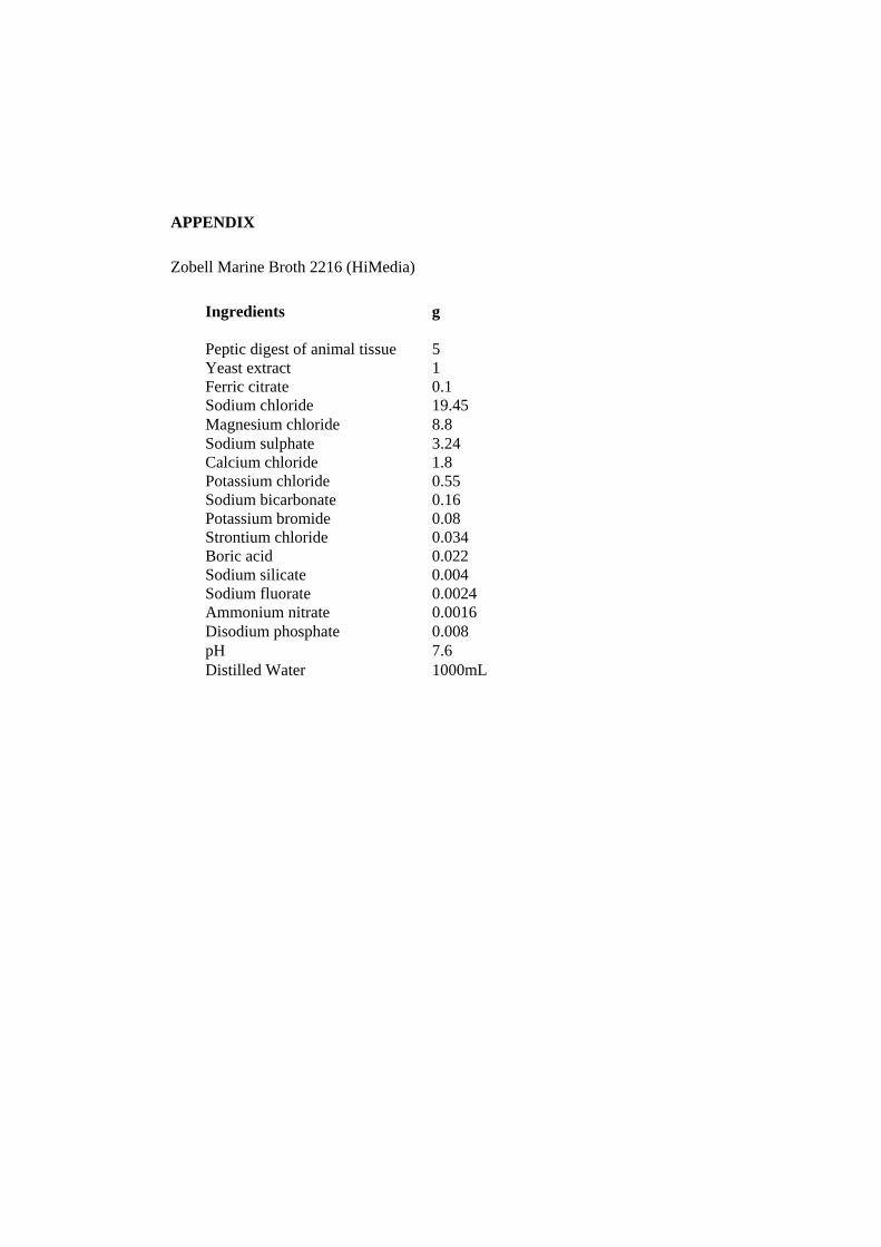
APPENDIX Zobell Marine Broth 2216 (HiMedia)
Ingredients
g
Peptic digest of animal tissue 5 Yeast extract 1 Ferric citrate 0.1 Sodium chloride 19.45 Magnesium chloride 8.8 Sodium sulphate 3.24 Calcium chloride 1.8 Potassium chloride 0.55 Sodium bicarbonate 0.16 Potassium bromide 0.08 Strontium chloride 0.034 Boric acid 0.022 Sodium silicate 0.004 Sodium fluorate 0.0024 Ammonium nitrate 0.0016 Disodium phosphate 0.008 pH 7.6 Distilled Water 1000mL