issn 1881-7831 online issn 1881-784x dd tddtjournal.com/files/ddt_2018vol12no4_pp185_258_e1.pdf ·...
TRANSCRIPT
ISSN 1881-7831 Online ISSN 1881-784X
DD&TDrug Discoveries & Therapeutics
www.ddtjournal.com
Volume 12, Number 4August 2018

www.ddtjournal.com
Drug Discoveries & Therapeutics is one of a series of peer-reviewed journals of the International Research and Cooperation Association for Bio & Socio-Sciences Advancement (IRCA-BSSA) Group and is published bimonthly by the International Advancement Center for Medicine & Health Research Co., Ltd. (IACMHR Co., Ltd.) and supported by the IRCA-BSSA and Shandong University China-Japan Cooperation Center for Drug Discovery & Screening (SDU-DDSC).
Drug Discoveries & Therapeutics publishes contributions in all fields of pharmaceutical and therapeutic research such as medicinal chemistry, pharmacology, pharmaceutical analysis, pharmaceutics, pharmaceutical administration, and experimental and clinical studies of effects, mechanisms, or uses of various treatments. Studies in drug-related fields such as biology, biochemistry, physiology, microbiology, and immunology are also within the scope of this journal.
Drug Discoveries & Therapeutics publishes Original Articles, Brief Reports, Reviews, Policy Forum articles, Case Reports, News, and Letters on all aspects of the field of pharmaceutical research. All contributions should seek to promote international collaboration in pharmaceutical science.
ISSN: 1881-7831 Online ISSN: 1881-784X
CODEN: DDTRBXIssues/Year: 6
Language: EnglishPublisher: IACMHR Co., Ltd.
Editor-in-Chief:Kazuhisa SEKIMIZU Teikyo University, Tokyo, Japan
Co-Editors-in-Chief:Xishan HAO Tianjin Medical University, Tianjin, ChinaMunehiro NAKATATokai University, Hiratsuka, Japan
Chief Director & Executive Editor:Wei TANG National Center for Global Health and Medicine, Tokyo, Japan
Senior Editors:Guanhua DU Chinese Academy of Medical Science and Peking Union Medical College, Beijing, ChinaXiao-Kang LI National Research Institute for Child Health and Development, Tokyo, JapanMasahiro MURAKAMI Osaka Ohtani University, Osaka, JapanYutaka ORIHARA The University of Tokyo, Tokyo, JapanTomofumi SANTA The University of Tokyo, Tokyo, JapanHongbin SUNChina Pharmaceutical University, Nanjing, China
Fengshan WANGShandong University, Ji'nan, China
Managing Editor:Hiroshi HAMAMOTOTeikyo University, Tokyo, Japan
Web Editor:Yu CHEN The University of Tokyo, Tokyo, Japan
Proofreaders:Curtis BENTLEY Roswell, GA, USAThomas R. LEBON Los Angeles, CA, USA
Editorial and Head Office:Pearl City Koishikawa 603, 2-4-5 Kasuga, Bunkyo-ku, Tokyo 112-0003, JapanTel.: +81-3-5840-9697Fax: +81-3-5840-9698E-mail: [email protected]
Editorial Board
i

www.ddtjournal.com
Editorial Board Members
Drug Discoveries & TherapeuticsEditorial and Head OfficePearl City Koishikawa 603, 2-4-5 Kasuga, Bunkyo-ku, Tokyo 112-0003, Japan
Tel: +81-3-5840-9697, Fax: +81-3-5840-9698E-mail: [email protected]: www.ddtjournal.com
Alex ALMASAN(Cleveland, OH)John K. BUOLAMWINI(Memphis, TN)Jianping CAO(Shanghai)Shousong CAO(Buffalo, NY)Jang-Yang CHANG(Tainan)Fen-Er CHEN(Shanghai)Zhe-Sheng CHEN(Queens, NY)Zilin CHEN(Wuhan, Hubei)Xiaolan CUI(Beijing)Shaofeng DUAN(Lawrence, KS)Mohamed F. EL-MILIGI(6th of October City)Hao FANG(Ji'nan, Shandong)Marcus L. FORREST(Lawrence, KS)Takeshi FUKUSHIMA(Funabashi, Chiba)Harald HAMACHER(Tübingen, Baden-Württemberg)Kenji HAMASE(Fukuoka, Fukuoka)Junqing HAN(Ji'nan, Shandong)Xiaojiang HAO(Kunming, Yunnan)Kiyoshi HASEGAWA(Tokyo)Waseem HASSAN(Rio de Janeiro)Langchong HE(Xi'an, Shaanxi)
Rodney J. Y. HO(Seattle, WA)Hsing-Pang HSIEH(Zhunan, Miaoli)Yongzhou HU(Hangzhou, Zhejiang)Yu HUANG(Hong Kong)Amrit B. KARMARKAR(Karad, Maharashra)Toshiaki KATADA(Tokyo)Gagan KAUSHAL(Philadelphia, PA)Ibrahim S. KHATTAB(Kuwait)Shiroh KISHIOKA(Wakayama, Wakayama)Robert Kam-Ming KO(Hong Kong)Nobuyuki KOBAYASHI(Nagasaki, Nagasaki)Norihiro KOKUDO(Tokyo, Japan)Toshiro KONISHI(Tokyo)Chun-Guang LI(Melbourne)Minyong LI(Ji'nan, Shandong)Xun LI(Ji'nan, Shandong)Jikai LIU(Wuhan, Hubei)Xinyong LIU(Ji'nan, Shandong)Yuxiu LIU(Nanjing, Jiangsu)Hongxiang LOU(Jinan, Shandong)Xingyuan MA(Shanghai)
Ken-ichi MAFUNE(Tokyo)Sridhar MANI(Bronx, NY)Tohru MIZUSHIMA(Tokyo)Abdulla M. MOLOKHIA(Alexandria)Yoshinobu NAKANISHI(Kanazawa, Ishikawa)Siriporn OKONOGI(Chiang Mai)Weisan PAN(Shenyang, Liaoning)Chan Hum PARK(Eumseong)Rakesh P. PATEL(Mehsana, Gujarat)Shivanand P. PUTHLI(Mumbai, Maharashtra)Shafiqur RAHMAN(Brookings, SD)Adel SAKR(Cairo)Gary K. SCHWARTZ(New York, NY)Yuemao SHEN(Ji'nan, Shandong)Brahma N. SINGH(New York, NY)Tianqiang SONG(Tianjin)Sanjay K. SRIVASTAVA(Abilene, TX)Chandan M. THOMAS(Bradenton, FL)Li TONG(Xining, Qinghai)Murat TURKOGLU(Istanbul)Hui WANG(Shanghai)
Quanxing WANG(Shanghai)Stephen G. WARD(Bath)Yuhong XU(Shanghai)Bing YAN(Ji'nan, Shandong)Chunyan YAN(Guangzhou Guangdong) Xiao-Long YANG(Chongqing)Yun YEN(Duarte, CA)Yasuko YOKOTA(Tokyo)Takako YOKOZAWA(Toyama, Toyama)Rongmin YU(Guangzhou, Guangdong)Guangxi ZHAI(Ji'nan, Shandong)Liangren ZHANG(Beijing)Lining ZHANG(Ji'nan, Shandong)Na ZHANG(Ji'nan, Shandong)Ruiwen ZHANG(Houston, TX)Xiu-Mei ZHANG(Ji'nan, Shandong)Yongxiang ZHANG(Beijing)Jian-hua ZHU(Guangzhou, Guangdong)
(As of February 2018)
ii

www.ddtjournal.com
Bacterial polysaccharides inhibit sucrose-induced hyperglycemia in silkworms.Masaki Ishii, Yasuhiko Matsumoto, Kazuhisa Sekimizu
Green synthesis and inhibitory effects against oral pathogens of silver nanoparticles mediated by rice extracts.Temsiri Suwan, Sakornrat Khongkhunthian, Siriporn Okonogi
Caesalpinia sappan: A promising natural source of antimicrobial agent for inhibition of cariogenic bacteria.Rinrampai Puttipan, Sunee Chansakaow, Sakornrat Khongkhunthian, Siriporn Okonogi
Effect of rice variety and modification on antioxidant and anti-inflammatory activities.Siriporn Okonogi, Adchareeya Kaewpinta, Taepin Junmahasathien, Songwut Yotsawimonwat
Fungal-derived xenobiotic exhibits antibacterial and antibiofilm activity against Staphylococcus aureus.Siddhartha Kumar, Arvind Kumar, Manisha Kaushal, Prince Kumar, Kasturi Mukhopadhyay, Antresh Kumar
Differences in how bronchial asthma patients transmit experience about adverse reactions and usability of inhaled steroids to others: A qualitative focus-group study.Fuki Kurimoto, Satoko Hori, Yasufumi Sawada
Constipation in the elderly in a Japanese long-term medical facility: An ultrasonographic investigation.Koichi Yabunaka, Gojiro Nakagami, Keiko Tabata, Junko Sugama, Masaru Matsumoto, Yoshifumi Kido, Terumi Iuchi, Hiromi Sanada
Whole body vibration exercise in the management of cancer therapy-related morbidities: A systematic review.Patrícia Lopes-Souza, Carla Fontoura Dionello, Danúbia da Cunha Sá-Caputo,Eloá Moreira-Marconi, Eric Heleno Freire Ferreira Frederico, Renata Marques Marchon, Anke Bergmann, Trentham Furness, Mario Bernardo-Filho
Criteria for the selection of switch OTC drugs based on patient benefits, efficacy, and safety [II]: Comparing the physicochemical and pharmaceutical properties of brand-name and switch OTC terbinafine hydrochloride cream.
Motoari Takata, Yuko Wada, Yurika Iwasawa, Miyuki Kumazawa, Ken-ichi Shimokawa, Fumiyoshi Ishii
Original Article
185 - 188
189 - 196
197 - 205
206 - 213
214 - 223
224 - 232
233 - 238
239 - 247
248 - 253
CONTENTS Volume 12, Number 4, 2018
iii
www.ddtjournal.com
CONTENTS (Continued )
Atypical cases of filariasis from a non-endemic area.
Parul Kodan, Nitin Gupta, Ankita Baidya, Ayan Basu, Abdul Razik, Ankit Mittal, Nishant Verma, Bijay Ranjan Mirdha, Sandeep Mathur, Madhu Rajeshwari, Neeraj Nischal, Manish Soneja, Naveet Wig
Erratum for "Anesthetic activity of plant essential oils on Cyprinus carpio (koi carp)" by Khumpirapang N et al. (Drug Discoveries & Therapeutics. 2018; 12(1):21-30.).
Case Report
254 - 258
Erratum
E1
Guide for Authors
Copyright
iv

www.ddtjournal.com
Drug Discoveries & Therapeutics. 2018; 12(4):185-188. 185
Bacterial polysaccharides inhibit sucrose-induced hyperglycemia in silkworms
Masaki Ishii1, Yasuhiko Matsumoto2, Kazuhisa Sekimizu1,2,*
1 Genome Pharmaceuticals Institute Co., Ltd., Tokyo, Japan;2 Teikyo University Institute of Medical Mycology, Tokyo, Japan.
1. Introduction
Transient and rapid postprandial increases in blood glucose levels, referred to as a blood glucose spike, are a potential risk factor for diabetes. Suppression of the blood glucose spike is expected to be useful for preventing the onset of diabetes. Generally, blood glucose levels and their suppression by drugs are evaluated in mammalian animal models. We previously reported that an increase in blood glucose levels after glucose intake could also be evaluated in silkworms, an alternative model animal (1-3). Not only glucose, but also sucrose intake increases silkworm blood glucose levels (4). Glucose level increases in the silkworm hemolymph after sucrose intake are suppressed by acarbose and voglibose, α-glucosidase inhibitors that are used clinically for human diabetes patients (4). We propose that the silkworm evaluation system is useful
for screening substances that suppress sucrose-induced hyperglycemia. Bacterial polysaccharides have various structures and biologic activities (5-7). We recently reported a bacterial polysaccharide with high innate immunity-stimulating activity in a silkworm evaluation system (8). Bacterial polysaccharides can be obtained in large quantities at low cost. We therefore propose the use of bacterial polysaccharide libraries for screening seeds of medicines and supplements for human health. In this paper, we describe the collection of polysaccharide-producing bacteria and preliminary screening of polysaccharides that suppress sucrose-induced hyperglycemia in a silkworm model system.
2. Materials and Methods
2.1. Collection of polysaccharide-producing bacteria
Bacteria isolated from soil and plants that formed viscous colonies on agar plates were collected. The bacteria grown on the plates (10 cm) were recovered with a spreader and 15 ml of saline, and the cells were removed by centrifugation (8,000 rpm, 5 min). Ethanol (final concentration: 67%) was added to the centrifuge
Summary Diabetes and obesity result from sucrose-induced hyperglycemia. Prevention of hyperglycemia contributes to inhibit the onset of these life-related diseases. Here we show that polysaccharides obtained from soil bacteria inhibit sucrose-induced hyperglycemia in an in vivo silkworm evaluation system. Ethanol precipitates of extracellular polysaccharides were prepared from viscous bacterial colonies. Among 24 samples obtained from different bacterial species, oral administration of 6 samples from Rhizobium altiplani, Cupriavidus sp., Paenibacillus polymyxa, Pantoea eucalypti, Variovorax boronicumulans, and Xanthomonas cynarae suppressed sucrose-induced hyperglycemia in silkworm insect larvae. The R. altiplani fraction treated further with DNase I, RNase A, and proteinase K, followed by phenol extraction also exhibited suppressive activity. Our results suggest that silkworms provide an efficient screening system of bacterial polysaccharides that inhibit sucrose-induced hyperglycemia.
Keywords: Bacteria, polysaccharide, silkworm, sucrose-induced hyperglycemia
DOI: 10.5582/ddt.2018.01040Original Article
Released online in J-STAGE as advance publication August 24, 2018.
*Address correspondence to:Dr. Kazuhisa Sekimizu, Teikyo University Institute of Medical Mycology. 359 Otsuka, Hachioji, Tokyo, 192-0395, Japan.E-mail: [email protected]
www.ddtjournal.com
Drug Discoveries & Therapeutics. 2018; 12(4):185-188.
supernatant, and fibrous precipitates were collected by centrifugation. Bacterial 16S rRNA was sequenced and homology searches were performed to determine the bacterial species using the EZBiocloud database. The crude polysaccharide fractions were treated enzymatically as follows. DNase I (1,000 U/mL; Promega) and RNase A (10 μg/mL; NIPPON GENE CO., LTD.) were added to the ethanol precipitate, dissolved in water, and incubated overnight at 37°C, and then further incubated overnight at 37°C with protease K (100 µg/mL). Phenol: chloroform: isoamyl alcohol (50:49:1) was added to the fraction, and the samples were vigorously shaken, followed by the addition of two volumes of ethanol to the upper layer fraction. The precipitates were then collected by centrifugation.
2.2. Sucrose tolerance test of silkworms
Silkworms (Hu Yo x Tsukuba Ne, Ehime Sericulture Incorporated Company, Ehime, Japan) were reared as described previously (9,10). The silkworm sucrose tolerance test was conducted according to the previously reported method (4). Briefly, sucrose (10%) and test samples were mixed with silkworm artificial diet. Sucrose diet with or without polysaccharide samples was fed to 5th-instar larva of silkworms for 1 h, the silkworm hemolymph was collected, and glucose concentrations were measured with a glucometer (Accu-Chek, Roche).
3. Results
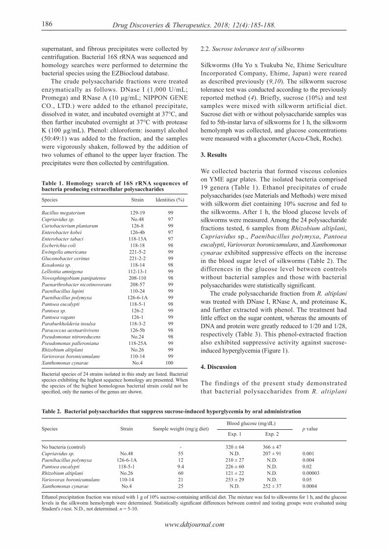
We collected bacteria that formed viscous colonies on YME agar plates. The isolated bacteria comprised 19 genera (Table 1). Ethanol precipitates of crude polysaccharides (see Materials and Methods) were mixed with silkworm diet containing 10% sucrose and fed to the silkworms. After 1 h, the blood glucose levels of silkworms were measured. Among the 24 polysaccharide fractions tested, 6 samples from Rhizobium altiplani, Cupriavidus sp., Paenibacillus polymyxa, Pantoea eucalypti, Variovorax boronicumulans, and Xanthomonas cynarae exhibited suppressive effects on the increase in the blood sugar level of silkworms (Table 2). The differences in the glucose level between controls without bacterial samples and those with bacterial polysaccharides were statistically significant. The crude polysaccharide fraction from R. altiplani was treated with DNase I, RNase A, and proteinase K, and further extracted with phenol. The treatment had little effect on the sugar content, whereas the amounts of DNA and protein were greatly reduced to 1/20 and 1/28, respectively (Table 3). This phenol-extracted fraction also exhibited suppressive activity against sucrose-induced hyperglycemia (Figure 1).
4. Discussion
The findings of the present study demonstrated that bacterial polysaccharides from R. altiplani
186
Table 1. Homology search of 16S rRNA sequences of bacteria producing extracellular polysaccharides
Species
Bacillus megateriumCupriavidus sp.Curtobacterium plantarumEnterobacter kobeiEnterobacter tabaciEscherichia coliEwingella americanaGluconobacter cerinusKosakonia sp.Lelliottia amnigenaNovosphingobium panipatense Paenarthrobacter nicotinovoransPaenibacillus lupiniPaenibacillus polymyxaPantoea eucalypti Pantoea sp.Pantoea vagansParaburkholderia insulsaParacoccus aestuariivivensPseudomonas nitroreducensPseudomonas palleronianaRhizobium altiplaniVariovorax boronicumulansXanthomonas cynarae
Identities (%)
99 97 99 97 97 98 99 99 98 99 98 99 99 99 98 99 99 99 98 98 99 99 99100
Strain
129-19No.48126-8126-4b
118-13A118-18221-5-2221-2-2118-14
112-13-1208-110208-57110-24
126-6-1A118-5-1126-2126-1
118-3-2126-5bNo.24
118-25ANo.26110-14No.4
Bacterial species of 24 strains isolated in this study are listed. Bacterial species exhibiting the highest sequence homology are presented. When the species of the highest homologous bacterial strain could not be specified, only the names of the genus are shown.
Table 2. Bacterial polysaccharides that suppress sucrose-induced hyperglycemia by oral administration
Species
No bacteria (control)Cupriavidus sp.Paenibacillus polymyxaPantoea eucalypti Rhizobium altiplaniVariovorax boronicumulansXanthomonas cynarae
Strain
No.48126-6-1A118-5-1No.26110-14No.4
Sample weight (mg/g diet)
-55129.4602125
Ethanol precipitation fraction was mixed with 1 g of 10% sucrose-containing artificial diet. The mixture was fed to silkworms for 1 h, and the glucose levels in the silkworm hemolymph were determined. Statistically significant differences between control and testing groups were evaluated using Student's t-test. N.D., not determined. n = 5-10.
Exp. 1
320 ± 64N.D.
210 ± 27226 ± 60121 ± 22253 ± 29
N.D.
Exp. 2
366 ± 47207 ± 91
N.D. N.D. N.D. N.D.
252 ± 37
p value
0.001 0.004 0.02 0.00003 0.05 0.0004
Blood glucose (mg/dL)

www.ddtjournal.com
Drug Discoveries & Therapeutics. 2018; 12(4):185-188. 187
polysaccharides that inhibit increases in blood glucose levels in humans. Bacteria secreting polysaccharides can be easily obtained as viscous colonies on agar plates. Polysaccharides secreted from bacteria have various structures depending on the bacterial species (6,7). Furthermore, industrial mass production of bacterial polysaccharides is possible. Based on these properties, it is expected that the library of bacterial polysaccharides will be useful for screening compounds with physiologic activities, such as agents with blood sugar lowering effects.
Acknowledgements
We thank Mari Maeda and Miki Takahashi (Genome Pharmaceuticals Institute Co., Ltd, Tokyo, Japan) for their technical assistance in rearing the silkworms. The project was supported by JSPS KAKENHI grant number JP15H05783 (Scientific Research (S) to KS), and JSPS KAKENHI grant number JP17K08288 (Scientific Research (C) to YM). The project was also supported by Genome Pharmaceuticals Institute Co., Ltd (Tokyo, Japan).
References
1. Matsumoto Y, Sumiya E, Sugita T, Sekimizu K. An invertebrate hyperglycemic model for the identification of anti-diabetic drugs. PloS one. 2011; 6:e18292.
2. Matsumoto Y, Sekimizu K. Evaluation of anti-diabetic drugs by using silkworm, Bombyx mori. Drug Discov Ther. 2016; 10:19-23.
3. Matsumoto Y, Ishii M, Hayashi Y, Miyazaki S, Sugita T, Sumiya E, Sekimizu K. Diabetic silkworms for evaluation of therapeutically effective drugs against type II diabetes. Sci Rep. 2015; 5:10722.
4. Matsumoto Y, Ishi i M, Sekimizu K. An in vivo inver tebrate evaluat ion system for ident i fying substances that suppress sucrose-induced postprandial hyperglycemia. Sci Rep. 2016; 6:26354.
5. Rehm BH. Bac te r i a l po lymers : B iosyn thes i s , modifications and applications. Nat Rev Microbiol. 2010; 8:578-592.
6. N w o d o U U , G r e e n E , O k o h A I . B a c t e r i a l exopolysaccharides: Functionality and prospects. Int J Mol Sci. 2012; 13:14002-14015.
7. Freitas F, Alves VD, Reis MA. Advances in bacterial exopolysaccharides: From production to biotechnological applications. Trends Biotechnol. 2011; 29:388-398.
8. Urai M, Aizawa T, Imamura K, Hamamoto H, Sekimizu K. Characterization of the chemical structure and innate immune-stimulating activity of an extracellular polysaccharide from Rhizobium sp. strain M2 screened using a silkworm muscle contraction assay. Drug Discov Ther. 2017; 11:238-245.
9. Kaito C, Akimitsu N, Watanabe H, Sekimizu K. Silkworm larvae as an animal model of bacterial infection pathogenic to humans. Microb Pathog. 2002; 32:183-190.
10. Kurokawa K, Kaito C, Sekimizu K. Two-component signaling in the virulence of Staphylococcus aureus: A
markedly suppressed sucrose-induced hyperglycemia in silkworms. We propose that the polysaccharides screened using the silkworm model are promising candidates for healthy foods and medicines to prevent the onset and exacerbation of diabetes. The silkworm system is superior to mammalian systems in terms of cost and ethical issues (11-13). Therefore, the silkworm system is expected to be useful for screening bacterial
Figure 1. Effect of enzyme-treated polysaccharide fraction on sucrose-induced hyperglycemia. Crude fraction (50 mg/g diet) prepared from R. altiplani No. 26, enzyme-treated polysaccharide fraction (46 mg/g diet), and acarbose (40 mg/g diet) were mixed with 10% sucrose containing diet, respectively. Diets with the polysaccharide fraction were orally administered to silkworms for 1 h. The glucose levels in silkworm hemolymph were determined. Acarbose was used as a control according to a previous report (4). Statistically significant differences between control and testing groups were evaluated using Student's t-test.
Table 3. Comparison of the amounts of DNA and protein in a crude polysaccharide fraction and an enzyme-treated fraction prepared from Rhizobium altiplani
Fraction
Crude polysaccharideEnzyme-treated sample
DNA (mg/g)
100.45
Sugar (mg/g)
490520
Crude polysaccharide was incubated with DNase Ⅰ(1,000 U/mL) and RNaseA (10 μg/mL) 24 h at 37˚C. Then, proteinase K (100 µg/mL) was added to the samples and incubated 24 h at 37˚C. Phenol/chloroform/isoamyl alcohol was added and vigorously mixed, followed by centrifugation. The upper layer fraction was collected and mixed with ethanol (final concentration: 67%). Fibrous precipitates were collected by centrifugation. The amounts of sugars, DNA, and proteins in the crude polysaccharide fraction and the enzyme-treated fraction were measured by the phenol-sulfuric acid method, the fluorescent-based Qubit assay, and Bradford assay, respectively.
Protein (mg/g)
562.1
www.ddtjournal.com
Drug Discoveries & Therapeutics. 2018; 12(4):185-188.188
silkworm larvae-pathogenic agent infection model of virulence. Methods Enzymol. 2007; 422:233-244.
11. Nwibo DD, Hamamoto H, Matsumoto Y, Kaito C, Sekimizu K. Current use of silkworm larvae (Bombyx mori) as an animal model in pharmaco-medical research. Drug Discov Ther. 2015; 9:133-135.
12. Panthee S, Paudel A, Hamamoto H, Sekimizu K. Advantages of the Silkworm As an Animal Model
for Developing Novel Antimicrobial Agents. Front Microbiol. 2017; 8:373.
13. Paudel A, Panthee S, Urai M, Hamamoto H, Ohwada T, Sekimizu K. Pharmacokinetic parameters explain the therapeutic activity of antimicrobial agents in a silkworm infection model. Sci Rep. 2018; 8:1578.
(Received July 12, 2018; Accepted August 2, 2018)
www.ddtjournal.com
Drug Discoveries & Therapeutics. 2018; 12(4):189-196. 189
Green synthesis and inhibitory effects against oral pathogens of silver nanoparticles mediated by rice extracts
Temsiri Suwan1,2, Sakornrat Khongkhunthian2,3, Siriporn Okonogi2,4,*
1 Interdisciplinary Program in Nanoscience and Nanotechnology, Chiang Mai University, Chiang Mai, Thailand;2 Research Center of Pharmaceutical Nanotechnology, Chiang Mai University, Chiang Mai, Thailand;3 Department of Restorative Dentistry and Periodontology, Faculty of Dentistry, Chiang Mai University, Chiang Mai, Thailand;4 Department of Pharmaceutical Sciences, Faculty of Pharmacy, Chiang Mai University, Chiang Mai, Thailand.
1. Introduction
Nanoparticles of certain metals, such as titanium, zinc, magnesium, gold, copper, and silver have been developed for various fields. Among them, silver nanoparticles (AgNPs) have gained interest for commercialization applications since they have considerably versatile properties, such as a variable surface area to volume ratio, which is very useful for many biomedical and technological applications. They have been used extensively in electronic industry and as excellent catalyst. In medical applications, many reports demonstrate their biological activities, such
as anticancer (1), antioxidant (2), and antimicrobial activities (3). AgNPs have been used as antibacterial agent for many kinds of applications including home appliances and water treatment (4). The biological activity of metal nanoparticles is closely related to their size. The smaller size usually gives the higher activity.Thus, control the size and size distribution of these nanoparticles is an important issue. Generally, specific control of shape, size, and size distribution is often achieved by varying the synthesis methods, reducing agents, and stabilizers (5). Recently, the ability of AgNPs on inhibition of certain viruses, such as human immunodeficiency virus 1 (HIV-1) has been reported. AgNPs showed a half maximal inhibitory concentration against the virus of 11.2 ± 0.6 µg/mL (p < 0.0001) with no significant toxicity against normal cells (6). Silver has long been historical used since ancient times and it has been demonstrated that, in low concentrations, silver is nontoxic to human cells (7).
Summary Rice is staple food for people in many countries for centuries. It is therefore considered as safe and environmental friendly material for pharmaceutical formulations. In the present study, aqueous extracts of three different parts of rice grain; rice bran (RB), rice husk (RH), and rice germ (RG) were compared for their use as reducing agents in synthesis of silver nanoparticles (AgNPs). AgNPs from those three different parts of rice, RB-AgNPs, RH-AgNPs, and RG-AgNPs, respectively showed different reducing activity, which the highest capacity was RB. RG-AgNPs and RB-AgNPs showed the maximum absorption of AgNPs at 440 nm whereas that of RH-AgNPs was at 480 nm. FTIR spectra of all AgNPs indicated the presence of different functional groups from rice attached to the nanoparticles and these groups prevented the particle agglomeration. Size analysis using dynamic light scattering revealed that RB-AgNPs was the smallest particles (346.4 ± 36.8 nm) and possessed the highest negative zeta potential. Antimicrobial test showed that the AgNPs obtained from green synthesis mediated by rice extracts have great antimicrobial activity against Streptococcus mutans, the severe oral pathogenic bacteria causing dental caries. These results suggest that aqueous extracts of RB, RH, and RG have potential to be used as reducing agents in synthesis of silver nanoparticles.
Keywords: AgNPs, rice grain, reducing agent, antimicrobial, Streptococcus mutans
DOI: 10.5582/ddt.2018.01034Original Article
*Address correspondence to:Dr. Siriporn Okonogi, Department of Pharmaceutical Sciences, Faculty of Pharmacy, Chiang Mai University, Chiang Mai 50200, Thailand.E-mail: [email protected]

www.ddtjournal.com
Drug Discoveries & Therapeutics. 2018; 12(4):189-196.
In former time, AgNPs were generally synthesized by chemical reaction based on the reduction of silver nitrate (AgNO3) by chemical reducing agent (8). In global efforts to reduce generated hazardous chemical waste, the use of chemicals is decreased in the synthesis protocols and green or biological synthesis of AgNPs has been increased. The biological methods are using eco-friendly resources, such as plant (9), algae (10), bacteria (11), and fungi (12). The extracts from many plants have been reported to act as reducing agent in the green synthesis of AgNPs (9,13,14). Synthesis of AgNPs using microorganisms is readily scalable and of course eco-friendly, however, production of microorganisms is more expensive than the production of plant extracts (15). Rice (Oryza sativa L.) is a cereal plant in family Poaceae. It is the predominant dietary energy source for many countries in the world. Rice is low in fat and high in starchy carbohydrate, packed full of vitamins and minerals. Dietary minerals and trace elements play a significant role in maintenance of optimal health (16). Rice grain has a hard cover called rice husk (RH) to protect the kernel inside. After RH is removed, the remaining product contains the inside endosperm and the outside rice bran (RB) and rice germ (RG). Many parts of rice grain contain high amount of compositions having antioxidant activity and high reducing property (17,18). RB and RG are commercial available for health care consumers. The commercial rice-milling process separates RH from the kernel inside because this part of rice grain is inedible and used in non-food applications. The aim of present study is to synthesize AgNPs by eco-friendly method using RB, RH, and RG extracts as reducing agents. The reducing property of the extracts was confirmed using ferric reducing antioxidant power (FRAP) assay. For AgNPs synthesizing, the extracts were reacted with AgNO3 as a precursor in a certain condition. The obtained AgNPs were characterized and investigated for antimicrobial activity against oral pathogens.
2. Materials and Methods
2.1. Materials
RB, RH, and RG of Jasmine rice was purchased from the local producer in Chiang Mai, Thailand. 2,4,6-Tris(2-pyridyl)-s-triazine (TPTZ) was purchased from Sigma-Aldrich, Inc (St. Louis, MO, USA). AgNO3 was supplied by RCI Lab-scan Co., Ltd. (Bangkok, Thailand). Potassium bromide (KBr) for infrared spectroscopy purchased from Fisher Scientific (Lough-borough, UK). Tryptic soy agar (TSA) and broth (TSB) were supplied by DifcoTM (Balti-more, Maryland, USA). Brain heart infusion agar (BHI-A) and broth (BHI-B) were purchased from Becton, Dickinson and Company (Franklin Lakes, New Jersey, USA).
Sabouraud dextrose agar (SDA) and broth (SDB) were purchased from BBLTM (Baltimore, Maryland, USA). All other chemicals and solvents were of analytical reagent grade or the highest grade available.
2.2. Microbial strains
Two aerobic bacterial strains, Staphylococcus aureus ATCC 25923 and Escherichia coli ATCC 25922 which represent for Gram positive and Gram negative bacteria, respectively, and one facultative Gram-positive bacteria, Streptococcus mutans DMST 18777 were used as the oral pathogenic bacteria. Candida albicans ATCC 10231 was used as the oral pathogenic fungi.
2.3. Preparation of rice extracts
The obtained RB, RH, and RG powders were sieved through a 14-mesh sieve in order to remove the large particles. The sieved samples were pulverized and 2 g of RB, RH, and RG powder samples were dispersed in deionized water to obtain 2% aqueous dispersions. The dispersions were macerated with continuous stirring at room temperature for 24 h. Subsequently, they were filtered through Whatman No.1 filter paper and the filtrates were stored in the refrigerator at 4℃ for further use.
2.4. FRAP assay
Reducing property of the rice extracts was determined using FRAP assay previously described (19) with some modification. The FRAP reagent was freshly prepared by mixing 2.5 mL of 10 mM TPTZ solution in 40 mM HCl with 2.5 mL of 20 mM FeCl3 and 25 mL of 0.3 M acetate buffer, pH 3.6. An amount of 20 μL rice extracts were mixed with 180 μL of FRAP reagent in 96 well plate. Then, they were incubated for 10 min at room temperature and the absorbance of was determined at 595 nm using a microplate reader (Bio-Rad, Model 680, Hercules, California, USA). All data were run in triplicate. The reducing power of the samples was evaluated by calculating the amount of Fe+2 produced by the rice extract samples using the calibration curved of FeSO4.
2.5. Synthesis of AgNPs
The synthesis of AgNPs was done by the following procedure. The rice extract solution was filled into the 125 mL Erlenmeyer flasks and heated until 75℃. An exact amount of AgNO3 solution (0.1 M) was added drop wise until the volume ratio of the rice solution to AgNO3 solution was 100:1. The mixture was kept at 75℃ under continuous stirring for 60 min. The obtained mixture was added thrice with deionized water and subjected to centrifugation (Heraeus™
190
www.ddtjournal.com
Drug Discoveries & Therapeutics. 2018; 12(4):189-196. 191
independent AgNPs batches.
2.7. Antimicrobial activity of AgNPs
The antimicrobial activity of the obtained RB-AgNPs, RH-AgNPs, and RG-AgNPs against oral pathogenic microorganisms; S. aureus, E. coli, S. mutans, and C. albicans was tested based on Kirby-Bauer method (20). The aerobic and facultative bacteria were grown in TSA and BHI-A, respectively at 37℃ for 24 h. The bacterial strains were diluted in TSB and BHI-B, respectively to a final density of 1.5 × 10⁶ colony forming unit (CFU)/mL. C. albicans were cultured in SDA at 37℃ for 36-48 h. The fungal suspension was prepared to a final concentration of 1-2 × 10⁵ CFU/mL in SDB. The density of the microbial suspension was adjusted with 0.5 McFarland constant by observing the OD at 600 nm under a UV-vis spectrophotometer. The bacterial and fungal suspensions were swabbed on the surface of their corresponding agars. Freshly prepared lyophilized RB-AgNPs, RH-AgNPs, and RG-AgNPs suspensions (40 μL) were added onto the 6 mm-diameter filter paper discs which would be put onto the surface of the seeded agar plates. The discs filled with deionized water and 0.1 M AgNO3 solution at the same amount (40 μL) were used as negative controls. The plates were incubated at 37℃ for 24 h. The antimicrobial activity of the samples was evaluated by determining the diameter of the clear zone of inhibition around the paper discs. All samples were done in triplicate.
2.8. Statistical analysis
Descriptive statistics for continuous variables were calculated and reported as a mean ± standard deviation. Data were analyzed using a One-way analysis of variance and Duncan's multiple range test using Statistic a software version 17 (SPSS Inc., Chicago, Illinois, USA). The values were presented as means ± standard deviation which a p-value less than 0.05 was considered as significant difference.
3. Results
3.1. Reducing property of rice extracts
In the present study, the reducing property of three different parts of rice grain, RB, RH, and RG were compared. The values of FRAP assay showed wide variation among the samples (Figure 1). The significantly highest (p < 0.05) ferric reduction ability was obtained from RB extract (524.0 ± 7.68 µmol Fe2+/g sample), followed by RH and RG with the reducing values of 247.1 ± 8.49 and 152.1 ± 4.24 µmol Fe2+/g sample, respectively. Our results show that RG also possessed the reducing property, even less than RB and RH, respectively.
Megafuge™ 40 Centrifuge Series, ThermoFisher Scientific, Waltham, Massachusettes, USA) at 8,000 rpm for 15 min to remove any traces of un-utilized phyto-constituents. After removing the liquid phase, the AgNPs obtained were kept in the refrigerator for further study. The AgNPs obtained from RB, RH, and RG were named as RB-AgNPs, RH-AgNPs, and RG-AgNPs, respectively.
2.6. Characterization of AgNPs
The obtained AgNPs were characterized using UV-vis and Fourier transform infrared (FTIR) spectroscopy measurement. Their particle size and zeta potential were measured using dynamic light scattering (DLS).
2.6.1. Visualization and UV-vis spectroscopy
The outer color appearance of the obtained AgNPs was early observed by visualization. The obtained particles were confirmed by UV-vis spectra using UV 2450 double-beam sspectrophotometer (Shimadzu-2450, Kyoto, Japan). The rice-AgNPs samples were diluted to 100 folds with deionized water before subjecting to this investigation. The optical property of the AgNPs solution was observed in the wavelength range of 300-700 nm. The UV-vis absorbance spectra were recorded at room temperature.
2.6.2. FTIR
In this experiment, the lyophilized RB-AgNPs, RH-AgNPs, and RG-AgNPs were used. The obtained AgNPs in the powder form were characterized using FTIR in order to investigate the possible role of the phyto-constituents presented in the rice extracts on the surface modification of the synthesized AgNPs. The KBr disc of the lyophilized AgNPs were prepared. The IR spectra in the range of 4,000-400 cm–1 of the samples were recorded using a Nicolet Nexus 470 FT-IR (Minneapolis, Minnesota, USA) in the diffuse reflectance mode at room temperature at a resolution of 4 cm–1. The spectra were collected against a KBr disc background at room temperature. The instrument was maintained with the automatic dehumidifier to di-minish water vapor interference.
2.6.3. DLS
The obtained AgNPs were investigated for their particle size and size distribution as well as zeta potential using DLS (Malvern Zeta sizer Nano-ZS, Malvern Instruments, Worcestershire, UK) at 25℃. Each sample was diluted to 100 folds with deionized water before investigation. The disposable plastic cuvettes and folded capillary zeta cell were used in the measurement. The measurement was done in triplicate of three

www.ddtjournal.com
Drug Discoveries & Therapeutics. 2018; 12(4):189-196.192
3.2. Synthesis and characterization of AgNPs
Reduction of silver ion to produce AgNPs during exposure to the rice extracts could be easily detected by color change. The color of RB, RH and RG extract solutions was pale yellow and the color of the precursor AgNO3 was colorless. However, when AgNPs were formed, the color of the solution began to change. After complete reduction, the color of the system were yellow-brown as presented in Figure 2. The intensity of the resultant color was observed during 0-60 min. It was found that the color change of the reaction mixtures of all rice extracts and AgNO3 was gradually appeared as pale yellow-brown around 10-30 min of reaction, depending on the type of the
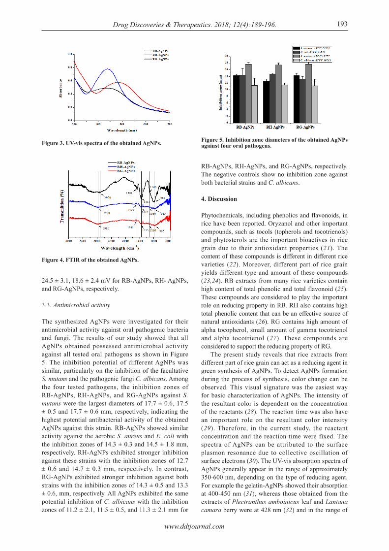
rice extracts. It was noted that the color change in the formation of RB-AgNPs was observed within 10 min, significantly faster than that in the systems of RH-AgNPs (25 min) and RG-AgNPs (30 min). After 60 min, all systems turned to intense yellow-brown color, indicating the complete formation was occurred. The UV-vis spectra of RB-AgNPs and RG-AgNPs showed the maximum absorption at 440 nm, whereas that of RH-AgNPs appeared at 480 nm, as shown in Figure 3. The absorption of the precursor AgNO3 was at 216 nm, however the UV-vis spectra of all AgNPs did not exhibit the absorption at this wavelength, indicating that there was no trace AgNO3 left in the obtained AgNPs systems. After the synthesis of AgNPs, the dispersions containing nanoparticles were centrifuged to separate AgNPs from other rice compositions in the solutions. The FTIR of RB-AgNPs, RH-AgNPs, and RG-AgNPs are shown in Figure 4. The results showed the peaks at 965, 1,160, 1,445-1,453, 1,591-1,599, and 2,980 cm–1. Analysis using DLS reveals that the size of the AgNPs obtained from different part of rice grain is different. RB-AgNPs showed average diameter of 346.4 ± 36.8 nm whereas RH-AgNPs and RG-AgNPs showed similar particle size of 587.3 ± 49.6 and 510.9 ± 84.4 nm, respectively. Particle size distribution expressed as polydispersity index (PdI) of RB-AgNPs, RH-AgNPs, and RG-AgNPs were similar with the PdI values of 0.271, 0.260, and 0.266, respectively. The zeta potential of all AgNPs obtained was negative values of 32 ± 2.6, Figure 1. Reducing activities of RB, RH, and RG.
Figure 2. Outer appearance of three parts of rice grains (A), rice extracts (B), and the obtained AgNPs (C).
www.ddtjournal.com
Drug Discoveries & Therapeutics. 2018; 12(4):189-196. 193
24.5 ± 3.1, 18.6 ± 2.4 mV for RB-AgNPs, RH- AgNPs, and RG-AgNPs, respectively.
3.3. Antimicrobial activity
The synthesized AgNPs were investigated for their antimicrobial activity against oral pathogenic bacteria and fungi. The results of our study showed that all AgNPs obtained possessed antimicrobial activity against all tested oral pathogens as shown in Figure 5. The inhibition potential of different AgNPs was similar, particularly on the inhibition of the facultative S. mutans and the pathogenic fungi C. albicans. Among the four tested pathogens, the inhibition zones of RB-AgNPs, RH-AgNPs, and RG-AgNPs against S. mutans were the largest diameters of 17.7 ± 0.6, 17.5 ± 0.5 and 17.7 ± 0.6 mm, respectively, indicating the highest potential antibacterial activity of the obtained AgNPs against this strain. RB-AgNPs showed similar activity against the aerobic S. aureus and E. coli with the inhibition zones of 14.3 ± 0.3 and 14.5 ± 1.8 mm, respectively. RH-AgNPs exhibited stronger inhibition against these strains with the inhibition zones of 12.7 ± 0.6 and 14.7 ± 0.3 mm, respectively. In contrast, RG-AgNPs exhibited stronger inhibition against both strains with the inhibition zones of 14.3 ± 0.5 and 13.3 ± 0.6, mm, respectively. All AgNPs exhibited the same potential inhibition of C. albicans with the inhibition zones of 11.2 ± 2.1, 11.5 ± 0.5, and 11.3 ± 2.1 mm for
RB-AgNPs, RH-AgNPs, and RG-AgNPs, respectively. The negative controls show no inhibition zone against both bacterial strains and C. albicans.
4. Discussion
Phytochemicals, including phenolics and flavonoids, in rice have been reported. Oryzanol and other important compounds, such as tocols (topherols and tocotrienols) and phytosterols are the important bioactives in rice grain due to their antioxidant properties (21). The content of these compounds is different in different rice varieties (22). Moreover, different part of rice grain yields different type and amount of these compounds (23,24). RB extracts from many rice varieties contain high content of total phenolic and total flavonoid (25). These compounds are considered to play the important role on reducing property in RB. RH also contains high total phenolic content that can be an effective source of natural antioxidants (26). RG contains high amount of alpha tocopherol, small amount of gamma tocotrienol and alpha tocotrienol (27). These compounds are considered to support the reducing property of RG. The present study reveals that rice extracts from different part of rice grain can act as a reducing agent in green synthesis of AgNPs. To detect AgNPs formation during the process of synthesis, color change can be observed. This visual signature was the easiest way for basic characterization of AgNPs. The intensity of the resultant color is dependent on the concentration of the reactants (28). The reaction time was also have an important role on the resultant color intensity (29). Therefore, in the current study, the reactant concentration and the reaction time were fixed. The spectra of AgNPs can be attributed to the surface plasmon resonance due to collective oscillation of surface electrons (30). The UV-vis absorption spectra of AgNPs generally appear in the range of approximately 350-600 nm, depending on the type of reducing agent. For example the gelatin-AgNPs showed their absorption at 400-450 nm (31), whereas those obtained from the extracts of Plectranthus amboinicus leaf and Lantana camara berry were at 428 nm (32) and in the range of
Figure 4. FTIR of the obtained AgNPs.
Figure 3. UV-vis spectra of the obtained AgNPs. Figure 5. Inhibition zone diameters of the obtained AgNPs against four oral pathogens.

www.ddtjournal.com
Drug Discoveries & Therapeutics. 2018; 12(4):189-196.194
390-520 nm (33), respectively. The obtained AgNPs in the current study were confirmed by UV-vis spectra. Our results showed good agreement with the previous studies on the UV absorption range. FTIR study was carried out in order to determine the possibility of the residual bio-reducing functional groups of the rice extracts involved in reduction process and their possible unique interactions with the surface of AgNPs. The organic functional groups like OH or C=O interacted to the surface of AgNPs can be detected by FTIR (15). The IR peaks of the obtained AgNPs in the current study indicate that many functional groups are involved. The peaks at 965 and 1160 cm–1 are considered to be the stretching vibrations of C–OCH3, C–H stretching of alkenes and C–O stretching aromatic side chain of proteins (34). The peaks in 1,445-1,453 cm–1 are relevant to the N–O stretching of nitro groups (35). The peaks located at 1,591-1,599 cm–1 are assigned to C=O stretching vibrations of amides characteristic of rice proteins. The broad peaks at 2,980 cm–1 are assigned as –OH stretching in alcohols and phenolic compounds with strong hydrogen bonds (36). The presence of these peaks confirmed that the obtained AgNPs were covered by certain rice phytochemicals, including flavonoids and phenols, with functional groups, such as ketone, aldehyde, and carboxylic acid. The presence of these groups on the surface of AgNPs is considered to support the stability of the nanoparticles. They can prevent the pairing and agglomerating of the nanoparticles. If the amount of these compounds is high in the rice extracts, they can cover wide surface area of the obtained AgNPs. This can cause the size of the synthesized AgNPs to be extremely small since high agglomeration cannot occur. The size of the nanoparticles obtained from DLS is hydrodynamic size which is slightly bigger than that measured by a transmission electron microscope due to the hydrodynamic radius (37). However, the size of the obtained nanoparticles can be compared by using the same equipment. In this study, the size of all AgNPs obtained was measured using DLS which their zeta potential could also be detected. The results reveal that among the three kinds of AgNPs, the smallest size and the highest negative value of zeta potential were obtained from RB-AgNPs. This was considered to be influenced by the high phytochemical content existed in the RB extract. The oral cavity is the hub of an extremely diverse microflora consisting of about 500 species of microorganisms (38). The oral pathogenic bacteria (S. aureus, E. coli, and S. mutans) and fungi (C. albicans) used in the current study are the most common microorganisms found in oral cavity. The overgrowth of these microorganisms, particularly S. mutans and C. albicans can cause severe diseases in oral cavity. S. mutans is the major cause of dental caries (39). While C. albicans is the major cause of oral candidiasis (40),
which the symptoms include pain, oral discomfort, and loss of taste (41). Several mechanisms of action on antibacterial activity of AgNPs have been proposed, such as the ability of AgNPs to attach bacterial cell wall and cause structure change in cell membrane, the ability to damage and make porous in the cell membrane resulted from free radicals of AgNPs, and the ability of silver ion that can be released to the inner cell and destroy several function in the cell (42,43). The mechanism of action on antifungal activity of AgNPs against C. albicans was previously proposed that AgNPs have high potential to disrupt cell membrane and arrest the cell cycle at the G2/M phase of C. albicans (44). The current study demonstrates that rice can be used as natural reducing agent to synthesize AgNPs. We explore the different reducing potential and advantages of many parts of rice grains in green synthesis of AgNPs without the use of any chemical stabilize and reducer. We also demonstrate the potential of the synthesized AgNPs on many important oral pathogens including aerobic bacteria, facultative bacteria, and fungi. The AgNPs obtained can inhibit all tested microorganisms especially S. mutans which is the most important oral pathogenic bacteria causing dental carries and oral infections. Among the three parts of rice grains, RB is the most effective and suitable part for the synthesis of AgNPs.
Acknowledgements
The authors acknowledge the financial support received from the Thailand Research Fund through the Research and Researcher for Industry, grant number PHD57I0024. We also thank the Research Center of Pharmaceutical Nanotechnology, Chiang Mai University, Faculty of Pharmacy and Faculty of Dentistry, Chiang Mai University for facility supports.
References
1. Potara M, Bawaskar M, Simon T, Gaikwad S, Licarete E, Ingle A, Banciu M, Vulpoi A, Astilean S, Rai M. Biosynthesized silver nanoparticles performing as biogenic SERS-nanotags for investigation of C26 colon carcinoma cells. Colloids Surf B Biointerfaces. 2015; 133:296-303.
2. Priya RS, Geetha D, Ramesh PS. Antioxidant activity of chemically synthesized AgNPs and biosynthesized Pongamia pinnata leaf extract mediated AgNPs – A comparative study. Ecotoxicol Environ Saf. 2016; 134:308-318.
3. Kora AJ, Arunachalam J. Assessment of antibacterial activity of silver nanoparticles on Pseudomonas aeruginosa and its mechanism of action. World J Microbiol Biotechnol. 2011; 27:1209-1216.
4. Jain P, Pradeep T. Potential of silver nanoparticle-coated polyurethane foam as an antibacterial water filter. Biotechnol Bioeng. 2005; 90:59-63.
5. Raza M, Kanwal Z, Rauf A, Sabri A, Riaz S, Naseem S. Size- and shape-dependent antibacterial studies of

www.ddtjournal.com
Drug Discoveries & Therapeutics. 2018; 12(4):189-196. 195
silver nanoparticles synthesized by wet chemical routes. Nanomaterials. 2016; 6:1-15.
6. Trefry JC, Wooley DP. Rapid assessment of antiviral activity and cytotoxicity of silver nanoparticles using a novel application of the tetrazolium-based colorimetric assay. J Virol Methods. 2012; 183:19-24.
7. Martínez-Castañón GA, Niño-Martínez N, Martínez-Gutierrez F, Martínez-Mendoza JR, Ruiz F. Synthesis and antibacterial activity of silver nanoparticles with different sizes. J Nanopart Res. 2008; 10:1343-1348.
8. Bastús NG, Merkoçi F, Piella J, Puntes V. Synthesis of highly monodisperse citrate-stabilized silver nanoparticles of up to 200 nm: Kinetic control and catalytic properties. Chem Mater. 2014; 26:2836-2846.
9. Kuppusamy P, Yusoff MM, Maniam GP, Govindan N. Biosynthesis of metallic nanoparticles using plant derivatives and their new avenues in pharmacological applications − An updated report. Saudi Pharm J. 2016; 24:473-484.
10. Salari Z, Danafar F, Dabaghi S, Ataei SA. Sustainable synthesis of silver nanoparticles using macroalgae Spirogyra varians and analysis of their antibacterial activity. J Saudi Chem Soc. 2016; 20:459-464.
11. Nanda A, Saravanan M. Biosynthesis of s i lver nanoparticles from Staphylococcus aureus and its antimicrobial activity against MRSA and MRSE. Nanomedicine. 2009; 5:452-456.
12. Sas t ry M, Ahmad A, Is lam Khan M, Kumar R. Biosynthesis of metal nanoparticles using fungi and actinomycete. Curr Sci. 2003; 85:162-170.
13. Sharma G, Jasuja ND, Rajgovind, Singhal P, Joshi SC. Synthesis, characterization and antimicrobial activity of Abelia grandiflora assisted AgNPs. J Microb Biochem Technol. 2014; 6:274-278.
14. Singhal G, Bhavesh R, Kasariya K, Sharma AR, Singh RP. Biosynthesis of silver nanoparticles using Ocimum sanctum (Tulsi) leaf extract and screening its antimicrobial activity. J Nanopart Res. 2011; 13:2981-2988.
15. Mittal AK, Chisti Y, Banerjee UC. Synthesis of metallic nanoparticles using plant extracts. Biotechnol Adv. 2013; 31:346-356.
16. Renuka N, Mathure SV, Zanan RL, Thengane RJ, Nadaf AB. Determination of some minerals and β-carotene contents in aromatic indica rice (Oryza sativa L.) germplasm. Food Chem. 2016; 191:2-6.
17. Moongngarm A, Daomukda N, Khumpika S. Chemical compositions, phytochemicals, and antioxidant capacity of rice bran, rice bran layer, and rice germ. APCBEE Procedia. 2012; 2:73-79.
18. Arab F, Alemzadeh I, Maghsoudi V. Determination of antioxidant component and activity of rice bran extract. Sci Iran. 2011; 18:1402-1406.
19. Tachakittirungrod S, Okonogi S, Chowwanapoonpohn S. Study on antioxidant activity of certain plants in Thailand: Mechanism of antioxidant action of guava leaf extract. Food Chem. 2007; 103:381-388.
20. Bauer AW, Kirby WM, Sherris JC, Turck M. Antibiotic susceptibility testing by a standardized single disk method. Am J Clin Pathol. 1966; 45:493-496.
21. Shen Y, Jin L, Xiao P, Lu Y, Bao J. Total phenolics, flavonoids, antioxidant capacity in rice grain and their relations to grain color, size and weight. J Cereal Sci. 2009; 49:106-111.
22. Zaupa M, Calani L, Del Rio D, Brighenti F, Pellegrini N. Characterization of total antioxidant capacity and
(poly)phenolic compounds of differently pigmented rice varieties and their changes during domestic cooking. Food Chem. 2015; 187:338-347.
23. Bhatnagar AS, Prabhakar DS, Prasanth Kumar PK, Raja Rajan RG, Gopala Krishna AG. Processing of commercial rice bran for the production of fat and nutraceutical rich rice brokens, rice germ and pure bran. LWT - Food Sci Technol. 2014; 58:306-311.
24. Yu S, Nehus ZT, Badger TM, Fang N. Quantification of vitamin E and γ-oryzanol components in rice germ and bran. J Agric Food Chem. 2007; 55:7308-7313.
25. Jun HI, Song GS, Yang EI, Youn Y, Kim YS. Antioxidant activities and phenolic compounds of pigmented rice bran extracts. J Food Sci. 2012; 77:C759-764.
26. Butsat S, Siriamornpun S. Antioxidant capacities and phenolic compounds of the husk, bran and endosperm of Thai rice. Food Chem. 2010; 119:606-613.
27. Goufo P, Trindade H. Rice antioxidants: Phenolic acids, flavonoids, anthocyanins, proanthocyanidins, tocopherols, tocotrienols, γ-oryzanol, and phytic acid. Food Sci Nutr. 2014; 2:75-104.
28. Zhang Y, Cheng X, Zhang Y, Xue X, Fu Y. Biosynthesis of silver nanoparticles at room temperature using aqueous aloe leaf extract and antibacterial properties. Colloids Surf A Physicochem Eng Asp. 2013; 423:63-68.
29. Bagherzade G, Tavakoli MM, Namaei MH. Green synthesis of silver nanoparticles using aqueous extract of saffron (Crocus sativus L.) wastages and its antibacterial activity against six bacteria. Asian Pac J Trop Biomed. 2017; 7:227-233.
30. Mulvaney P. Surface plasmon spectroscopy of nanosized metal particles. Langmuir. 1996; 12:788-800.
31. Kanmani P, Rhim JW. Physicochemical properties of gelatin/silver nanoparticle antimicrobial composite films. Food Chem. 2014; 148:162-169.
32. Ajitha B, Ashok Kumar Reddy Y, Sreedhara Reddy P. Biosynthesis of silver nanoparticles using Plectranthus amboinicus leaf extract and its antimicrobial activity. Spectrochim Acta A Mol Biomol Spectrosc. 2014; 128:257-262.
33. Kumar B, Smita K, Cumbal L, Debut A. Lantana camara berry for the synthesis of silver nanoparticles. Asian Pac J Trop Biomed. 2015; 5:192-195.
34. Barth A. The infrared absorption of amino acid side chains. Prog Biophys Mol Biol. 2000; 74:141-173.
35. Onchoke KK, Hadad CM, Dutta PK. Structure and vibrational spectra of mononitrated benzo[a]pyrenes. J Phys Chem A. 2006; 110:76-84.
36. Gunasekaran S, Ponnusamy S. Vibrational spectra and normal coordinate analysis on an organic non-linear optical crystal-3-methoxy-4-hydroxy benzaldehyde. IJPAP. 2005; 43:838-843.
37. Huang NM, Lim HN, Radiman S, Khiew PS, Chiu WS, Hashim R, Chia CH. Sucrose ester micellar-mediated synthesis of Ag nanoparticles and the antibacterial properties. Colloids Surf A Physicochem Eng Asp. 2010; 353:69-76.
38. Zhang Q, Liu JL, Qi XM, Qi CT, Yu Q. Inhibitory activities of Lignum Sappan extractives on growth and growth-related signaling of tumor cells. Chin J Nat Med. 2014; 12:607-612.
39. Mounika S, Jagannathan N, Murali. Association of Streptococcus mutants and Streptococcus sanguis in act of dental caries. J Pharm Sci Res. 2015; 7:764-766.
40. Parihar S. Oral candidiasis − A review. Webmed Central

www.ddtjournal.com
Drug Discoveries & Therapeutics. 2018; 12(4):189-196.196
Dentistry. 2011; 2:1-18.41. Sondi I, Salopek-Sondi B. Silver nanoparticles as
antimicrobial agent: A case study on E. coli as a model for Gram-negative bacteria. J Colloid Interface Sci. 2004; 275:177-182.
42. Prabhu S, Poulose EK. Silver nanoparticles: Mechanism of antimicrobial action, synthesis, medical applications, and toxicity effects. Int Nano Lett. 2012; 2:1-10.
43. Williams DW, Kuriyama T, Silva S, Malic S, Lewis
MAO. Candida biofilms and oral candidosis: Treatment and prevention. Periodontology. 2000. 2011; 55:250-265.
44. Kim KJ, Sung WS, Suh BK, Moon SK, Choi JS, Kim JG, Lee DG. Antifungal activity and mode of action of silver nano-particles on Candida albicans. Biometals. 2009; 22:235-242.
(Received June 12, 2018; Revised August 28, 2018; Accepted August 28, 2018)
www.ddtjournal.com
Drug Discoveries & Therapeutics. 2018; 12(4):197-205. 197
Caesalpinia sappan: A promising natural source of antimicrobial agent for inhibition of cariogenic bacteria
Rinrampai Puttipan1, Sunee Chansakaow1, Sakornrat Khongkhunthian2,3, Siriporn Okonogi1,3,*
1 Department of Pharmaceutical Sciences, Faculty of Pharmacy, Chiang Mai University, Chiang Mai, Thailand;2 Department of Restorative Dentistry and Periodontology, Faculty of Dentistry, Chiang Mai University, Chiang Mai, Thailand;3 Research Center of Pharmaceutical Nanotechnology, Chiang Mai University, Chiang Mai, Thailand.
1. Introduction
Many Streptococcus spp. are normal flora microorganisms in oral cavity but some insidious species are found to be oral pathogenic stains. Streptococcus mutans is considered to be one of the severe cariogenic bacteria leading to dental caries (1,2). They can early colonized on hard tooth surfaces and the epithelial tissues to form a biofilm or known as a dental plaque, which later contains multiple bacterial species (3). These biofilms can produce acid that destroys the tooth's enamel layer which leads to periodontitis and dental carries (4). Although, the oral pathogens may be controlled by meticulous mechanical oral hygiene but they cannot be completely exterminated from oral cavity. Controlling oral microorganisms and keeping dental plaque at levels
compatible with oral health are important. Therefore, many oral health care products are formulated to contain antiplaque or antiseptic agents to achieve good oral health. Using plant extracts instead of chemical synthetic agents in treatment of certain bacterial infections are of increasing interest for green environment. Various potential plant extracts have been reported on antimicrobial activity against oral pathogens (5). Nowadays, only a few of them are considered to be used as ingredients in dental products whereas many of them are not, according to their limited properties. For example curcumin, the high effective secondary metabolite compound from turmeric has been reported to have strong ability against biofilm formation of S. mutans (6). However, curcumin is not used as ingredient in toothpaste or mouthwash because of its low aqueous solubility and rapid degradation, as well as its yellowish color that affects the physical characteristics of the products (7). Therefore, searching for other potential plants is still needed. Caesalpinia sappan or sappan wood is the plant
Summary From the previous findings, the ethanolic fractionated extract of Caesalpinia sappan (F-EtOH) has high activity against Streptococcus mutans, the most severe cariogenic bacteria. The present study was aimed to isolate and identify the active compound of F-EtOH and compare its inhibitory activity against the biofilm of S. mutans as well as the cytotoxicity to oral fibroblast cells with F-EtOH. Compound isolation was done by column chromatography. The active compound was identified using liquid chromatography-mass spectrometry with electrospray ionization and nuclear magnetic resonance spectroscopy. It was found that the major compound of F-EtOH is brazilin. F-EtOH and brazilin were compared for inhibitory potential on the biofilms of three strains of S. mutans. The results exhibited that both F-EtOH and brazilin had potential on inhibiting biofilm formation and eradicating the preformed biofilms and their activity was dose dependent. F-EtOH showed significantly less toxic to normal periodontal ligament fibroblast than brazilin. At low concentration of 1- and 2-MBC, F-EtOH showed higher effective than brazilin. The results of our study suggest that the antibacterial activity of F-EtOH is according to the synergistic effects of the existing compounds including brazilin in F-EtOH.
Keywords: Sappan wood, brazilin, oral pathogens, antibiofilm, cytotoxicity
DOI: 10.5582/ddt.2018.01035Original Article
*Address correspondence to:Dr. Siriporn Okonogi, Faculty of Pharmacy, Chiang Mai University, Chiang Mai 50200, Thailand.E-mail: [email protected]

www.ddtjournal.com
Drug Discoveries & Therapeutics. 2018; 12(4):197-205.
commonly found in many Asian countries. In Thailand, it has been used as traditional medicine since the ancient time to promote blood circulation. The heartwood part of C. sappan can produce natural red dye that can be used as coloring agent in food, beverage, and cosmetics. For biological properties, it has been reported to have antioxidant (8,9), anti-inflammation (10), anti-rheumatoid arthritis (11) and antimicrobial activity (12-14). Many phenolic compounds, such as xanthone, coumarin, chalcones, flavones, and homoisoflavonoids have been found from C. sappan (15). Brazilin [(6a S-cis)-7, 11b-dihydrobenz[b]indeno[1,2-d]pyran-3,6a,9,10(6H)-tetrol] is reported as one of the major constituents present in the heartwood part of C. sappan (16). The color of brazilin can be changed from amber to red in pH ≥ 7 (17,18). The intensity of red color is depends on the amount of brazilin. The extract of C. sappan heartwood has been reported to have activity against several kinds of bacteria including S. mutans (19,20). While brazilin has also been reported to have inhibition activity against many oral pathogens (14). From the literature review, there is still lack of deep detail of C. sappan extracts against oral pathogens, particularly on inhibition of the biofilms of such severe pathogens. Our previous study presented that the ethanolic fractionated extract of C. sappan (F-EtOH) showed strong antibacterial activity on these bacteria (20). The current study was aimed to identify the active compound of F-EtOH and compare the antibiofilm activity and cytotoxicity between F-EtOH and its isolated active compound. The minimum bactericidal concentration (MBC) of both F-EtOH and the isolated active compound was determined before antibiofilm activity investigation.
2. Materials and Methods
2.1. Chemicals and reagents
Hexane, ethyl acetate, ethanol, methanol, chloroform, and dimethyl sulfoxide (DMSO) of analytical grade and HPLC grade were from RCI Labscan (Bangkok, Thailand). DifcoTM Brain heart infusion (BHI) broth and agar were from Bacton, Dickinson and Company (Sparks, Maryland, USA). Human blood for blood agar preparation was supported by Maharaj Nakorn Chiang Mai Hospital (Chiang Mai, Thailand). Dulbecco's modified eagle medium (DMEM), trypsin-EDTA, fetal bovine serum (FBS), and antibiotic-antimycotic solution (AA) were from GibcoTM, Life Technologies (Grand Island, New York, USA). Thiazolyl blue tetrazolium bromide was from Sigma-Aldrich (St. Louis, Missouri, USA). The other chemicals and solvents were of the highest grade available.
2.2. Plant materials and fractionated extracts preparation
The heartwood of C. sappan was collected from the
local area in Chiang Mai Province of Thailand and identified by the botanist in the botanical herbarium of Faculty of Pharmacy, Chiang Mai University to obtain the reference voucher numbers (002276). F-EtOH was prepared according to the previous report (20). Briefly, the dried powder of C. sappan heartwood was subjected to fractionated extraction by maceration method using 3 different solvents; hexane, ethyl acetate, and ethanol, respectively. The filtrate from ethanol extraction was subjected to a rotary evaporator, EYELA rotary evaporation N-1000 (Tokyo, Japan) for removing the solvent and to obtain F-EtOH.
2.3. Identification of the major compound
Liquid chromatography–mass spectrometry (LC-MS) with electrospray ionizationc (ESI), Flexar SQ300 MS (Single Quad) (PerkinElmer, Waltham, Massachusetts, USA) was used to determine the molecular mass of the compound. The maximum absorption was determined using UV-2450 UV-VIS spectrophotometer (Shimadzu, Kyoto, Japan) with spectrum mode in wavelength range of 200-700 nm. Nuclear magnetic resonance (NMR) spectra of the isolated compound were obtained from Bruker 400 MHz NMR spectrometer, UltraShieldTM (Billerica, Massachusetts, USA). 1H NMR was operated at 400 MHz and 13C NMR was operated at 100 MHz. CD3OD was used as a solvent. As brazilin was previously reported to be the major compound in C. sappan (20), for quantitative determination of the major compound of F-EtOH, the pure brazilin was dissolved in methanol at the final concentration ranges of 0.5-500 µg/mL and used for preparing calibration curve. HPLC analysis was carried out using HPLC Shimadzu CLASS-VP™ model (Kyoto, Japan) and the reversed phase Eurospher 100, i.d. 4 mm, C18 column, Knauer (Berlin, Germany). F-EtOH and brazilin were dissolved in methanol to proper concentrations before injection. Aqueous solution of 1% v/v acetic acid in DI water (A) was mixed with methanol (B) at a volume ratio of 3:1. This solution was used as the mobile phase with the flow rate of 1 mL/min, injection volume of 10 µL, running time of 30 min, and detected at 280 nm. Running system was performed at room temperature. A standard curve of brazilin was constructed. The amount of brazilin in F-EtOH was calculated from the linear equation of y = 8710.9x + 435.18 (r2 = 0.9998).
2.4. Bacterial strains culture conditions
Three strains of S. mutans including S. mutans DMST9567, S. mutans DMST18777, and S. mutans DMST41283 were cultured and incubated under anaerobic condition at 37°C in 5% CO2 using anaerobic chamber BACTRONⅡ-2, SHEL LAB® (Cornelius, Oregon, USA). Blood agar plates were prepared from
198

www.ddtjournal.com
Drug Discoveries & Therapeutics. 2018; 12(4):197-205. 199
were rinsed 3 times with 200 µL of PBS. Then, 100 µL of 30% v/v acetic acid was added to dissolve crystal violet stains and measured at 595 nm using a microtiter plate reader. The biofilm eradication activity of the samples was evaluated from the percentage biofilm left which was calculated by the following equation; % preformed biofilm = As × 100/Ac. Whereas As is the absorbance of the culture treated with the samples and Ac is the absorbance of the control culture (untreated cells). The lower percentage found indicates the higher eradication activity of the test samples.
2.6. Cytotoxicity
The cytotoxicity of F-EtOH and its major component against normal cells was evaluated. Periodontal ligament (PDL) fibroblast cells were collected from the healthy human subjects. This experiment was under ethical clearance No. 02/2015, approved by the Human Experimentation Committee, Faculty of Dentistry, Chiang Mai University, Thailand. Cell viability was determined by MTT assay. The PDL cells were cultured in completed DMEM (supplemented with 10% v/v FBS and 1% v/v AA) and incubated in humidified atmosphere, 5% CO2 at 37°C. For the test, the cell suspension at a density of 1 × 10⁴ cells/well was cultured in 96-well plates and then incubated under the same condition for 24 h. After that, the medium was removed and replaced with 100 µL of completed DMEM and 100 µL of the samples (final concentration ranged from 3.9-2,000 µg/mL in 0.4% v/v DMSO). The plates were further incubated for 24 h. Then, 100 µL of the medium was removed from each well and 100 µL of MTT solution (0.5 mg/mL in PBS) was added and further incubated for 4 h. Next, the medium was removed, and the formed formazan crystals were dissolved by mixing with 100 µL of DMSO for 10 min. The absorbance was measured at 540 nm and 690 nm as a reference wavelength using a microtiter plate reader. The cell viability was compared with the untreated culture or vehicle control culture. The percentage of cell viability was calculated using the following equation; % cell viability = OD × 100/OD0. Whereas OD is the optical density of the well containing cells treated with the samples and OD0 is the optical density of the well containing cells treated with 0.4% v/v DMSO (a negative control). The higher percentage of cell viability indicates the lower cytotoxicity of the samples.
2.7. Statistical analysis
The results of all experiments were conducted in triplicate and expressed as mean ± SD and statistically analyzed via SPSS statistic 17.0 software. ANOVA and Turkey's Multiple test have been determined the significant at p < 0.05.
5% human blood in BHI agar.
2.5. MBC determination and antibiofilm susceptibility
The MBC of the test samples was determined according to the method previously described (20). Briefly, the diluted samples were added into the suspension of 1 × 10⁶ CFU/mL of the tested bacterial strains in the 96-well plates and incubated at 37°C in 5% CO2 anaerobic chamber for 24 h. Subsequently, the cultures were streaked on blood agar plates and incubated at 37°C in 5% CO2 anaerobic chamber for 24 h. The lowest concentration in the plates that bacterial growth could not be visible was considered as the MBC. In antibiofilm susceptibility test, the antibiofilm formation and eradication of the preformed biofilms were investigated. For antibiofilm formation, the samples were prepared to have the final concentrations of 1-, 2-, and 4-fold MBC. The sample solutions of 100 µL were transferred to 96-well plates followed by adding 50 µL of BHI broth and 50 µL of the culture suspensions (1 × 10⁶ CFU/mL). Chlorhexidine at 0.12% (CHX) was used as a positive control. The plates were incubated in anaerobic chamber for 24 h. After incubation, nonadherent planktonic cells were removed and the wells were gently rinsed with 200 µL of phosphate-buffered saline (PBS). The adherent biomass was stained with 200 µL of 0.1% (w/v) crystal violet at room temperature for 30 min. The solutions were removed, and the wells were rinsed 3 times with 200 µL of PBS. Then, 100 µL of 30% (v/v) acetic acid was added to dissolve the crystal violet stains and measured at 595 nm using a microtiter plate reader Model 680, BIO RAD (Tokyo, Japan). The percentage of biofilm formation was calculated by the following equation; % biofilm formation = As × 100/Ac. Whereas Ac is the absorbance of the control culture (untreated cell) and As is the absorbance of the culture treated with the sample. The lower percentage of biofilm formation indicates the higher inhibitory activity of the test samples. For antibiofilm activity that the preformed biofilms were eradicated, 50 µL of the culture suspensions (1 × 10⁶ CFU/mL) and 150 µL of BHI broth were transferred to 96-well plates and incubated in an anaerobic chamber for 24 h. After incubation, nonadherent planktonic cells were removed and the wells were gently rinsed with 200 µL of PBS. Next, 100 µL of BHI broth was added in each well and the adherent biomass was treated with 100 µL of the test sample at the concentrations described above. The plates were further incubated in an anaerobic chamber for 24 h. After incubation, the nonadherent planktonic cells were removed by gently rinsing the wells with 200 µL of PBS. The viability biomass was stained with 200 µL of 0.1% w/v crystal violet at room temperature for 30 min. The solutions were removed, and the wells

www.ddtjournal.com
Drug Discoveries & Therapeutics. 2018; 12(4):197-205.200
3. Results
3.1. Sappan wood extract and the major compound
F-EtOH obtained had the same outer appearance as the previous report (20). Isolation of F-EtOH using column chromatography yielded many fractions but only fraction-6 (F6) was identify since our previous work found that F6 has the highest activity. It was found that F6 was a relatively pure compound. The HPLC chromatograms of F-EtOH and F6 were compared as shown in Figure 1. It is found that F6 demonstrated a major single peak at a retention time of 6.83 min (Figure 1A). Meanwhile, F-EtOH demonstrated 2 obvious peaks and 2 tiny peaks exhibited at obviously distinct retention times (Figure 1B). The peak of F-EtOH at the same retention time as F6 is the largest one. The results of identification of the single compound of F6 using 1H NMR spectra and 13C-NMR spectra are as follows and shown in Figure 2. 1H NMR data (400 MHz, CD3OD): δ 2.77 (1H, d, J = 15.6 Hz, H-7), 3.02 (1H, d, J = 15.6 Hz, H-7), 3.69 (1H, d, J = 11.2 Hz, H-6), 3.92 (1H, d, J = 11.2 Hz, H-6), 3.96 (1H, s, H-12), 6.29 (1H, d, J = 2.4 Hz, H-4), 6.46 (1H, dd, J = 10.5, 2.4 Hz, H-2), 6.60 (1H, s, H-11), 6.70 (1H, s, H-8), 7.19 (1H, d, J = 8.1 Hz, H-1). 13C NMR data (100 MHz, CD3OD): δ 43.0 (C-7), 51.2 (C-12), 71.0 (C-6), 78.2 (C-6a), 104.4 (C-4), 110.1 (C-2), 112.6 (C-11), 113.0 (C-8), 115.7 (C-1a), 131.5 (C-7a), 132.4 (C-1), 137.6 (C-11a), 145.5 (C-10), 145.8 (C-9), 155.9 (C-3), 158.0 (C-4a). The UV-visible spectrum of F6 showed maximum absorption at 224.5 and 289 nm. The mass spectrum of the compound showed a molecular weight at m/z 286. Using the NMR spectra as well as UV absorption and mass spectrum to compare with the data reported previously (21,22), we considered that the isolated compound of F6 was identical to brazilin (C16H14O5), which the chemical structure is shown in Figure 3.
The physical appearance of F6 is orange red powder. An amber color solution was observed when it was solubilized in ethanol, methanol, DMSO or in the solutions of pH less than 7. The color of the solution changed to red or pink when the pH was increased to 7 or higher. These physical characteristics as well as the identified NMR spectra confirmed that the pure compound of F6 was brazilin (17,21,22). The physical appearance of F-EtOH was red brown powder. HPLC analysis indicated that the content of brazilin in F-EtOH was 325.14 ± 25.91 µg/mg of F-EtOH. It was also
Figure 1. HPLC chromatograms of F6 (A) and F-EtOH (B) detected at 280 nm.
Figure 2. Nuclear magnetic resonance (NMR) spectra of brazilin from C. sappan, 1H NMR (A) and 13C NMR (B).
Figure 3. Fragmentation pattern from LC-MS by ESI technique (negative ion mode) of brazilin (m/z 286) isolated from F-EtOH.

www.ddtjournal.com
Drug Discoveries & Therapeutics. 2018; 12(4):197-205. 201
found that brazilin played a role on the color of F-EtOH solution.
3.2. MBC and antibiofilm susceptibility
The MBC of F-EtOH and brazilin against the three strains of S. mutans was found to be the same value of 125 µg/mL. To evaluate the potential of F-EtOH as an antibiofilm agent, we examined the inhibition of biofilm formation and the ability to eradicate the preformed biofilms. We also compared these activities of F-EtOH with its major compound brazilin. The results are shown in Figure 4. It was found that all S. mutans strains were able to form biofilms completely (100%) after treated with the negative control but significant reduction after treated with F-EtOH and brazilin. Both F-EtOH and brazilin demonstrated dose dependent curves. Biofilm
formation of all tested strains of S. mutans could be inhibited by F-EtOH and brazilin. After contacting with F-EtOH at 1-fold MBC, only 6.97 ± 0.79, 8.97 ± 1.60, and 9.17 ± 0.97% of biofilm of S. mutans DMST9567, S. mutans DMST18777, and S. mutans DMST41283 could be detected, respectively. After contacting with brazilin at the same concentration, 9.55 ± 1.08, 10.79 ± 0.33, and 14.29 ± 2.32% of biofilm of the three strains, respectively, could be detected. At the higher concentrations as 2-fold MBC, only 2.56 ± 0.21, 4.20 ± 0.13, and 5.25 ± 0.97% of the biofilm of the three strains, respectively, could be detected after contacting with F-EtOH. While, after contacting with brazilin, 9.55± 1.08, 5.59 ± 1.68, and 5.08 ± 1.17%, respectively, could be found. At 4-fold MBC, the biofilm formation of the three strains was almost completely inhibited and not significantly different from CHX. At this concentration, both F-EtOH and brazilin could inhibit more than 85% biofilm formation of all strains. At lower concentrations as 1- and 2-fold MBC, F-EtOH showed significantly higher effective on inhibition of biofilm formation of the three strains than brazilin. The results indicated that F-EtOH and brazilin even at low concentration as 1-fold MBC was sufficient to inhibit biofilm formation of all three strains of S. mutans. For the ability to eradicate the preformed biofilm, the results are shown in Figure 5. A dose dependent reduction in cell viability of S. mutans was observed. Treating with 1-fold MBC of F-EtOH and brazilin was not much effective on eradication of the preformed biofilm of S. mutans DMST41283. More than 50% (52.07 ± 2.65 and 59.59 ± 1.84) of viable cells could be found after treating with F-EtOH and brazilin, respectively. However, at this concentration, 38.54 ± 1.50 and 46.52 ± 0.77% of viable cells of DMST9567 and S. mutans DMST18777, respectively were detected after contacting with F-EtOH. Whereas, after contacting with brazilin at this concentration, higher amount of S. mutans DMST9567 (49.42 ± 0.91) and S. mutans DMST18777 (54.79 ± 3.11%) could be detected. Increasing the extract concentration, a higher reduction in cell viability of the three strains was observed. Treatment with 4-fold MBC of F-EtOH, only 2.34 ± 1.63, 2.34 ± 0.47, and 5.49 ± 1.13% of viable cells of S. mutans DMST9567, S. mutans DMST18777, and S. mutans DMST41283 could be detected, respectively. At the same concentration of brazilin, 2.46 ± 0.82, and 3.79 ± 1.04, and 9.86 ± 2.62% of viable cell could be detected, respectively. This viability reduction indicated that more than 90% of the pathogens could be killed. From these results, it is shown that F-EtOH presented the significantly higher effective than brazilin. F-EtOH and brazilin at concentration of 4-fold MBC could killed S. mutans DMST9567 and S. mutans DMST18777 as much as CHX. Meanwhile, the reduction of viable S. mutans DMST41283 from F-EtOH and brazilin was lower than CHX. It was
Figure 4. Biofilm formations of S. mutans DMST9567 (A), S. mutans DMST18777 (B), and S. mutans DMST41283 (C) after treating with F-EtOH, brazilin, and CHX in comparison with the untreated control.

www.ddtjournal.com
Drug Discoveries & Therapeutics. 2018; 12(4):197-205.202
considered that the biofilm communication of this strain might be tolerant to F-EtOH and brazilin.
3.4. Cytotoxicity
The cytotoxicity of F-EtOH and brazilin on normal PDL cells of healthy volunteers was evaluated by MTT assay. DMSO at the final concentration of 0.4% v/v and its series of 2-fold dilution were also tested as it was used as a solvent for the test samples. The results showed that 85.01 ± 4.34% of cell viability could be detected when DMSO at 0.4% v/v was used indicating that DMSO at this concentration was not toxic to the cells. Comparison between F-EtOH and brazilin, the results are shown in Figure 6. Both F-EtOH and brazilin demonstrated the dose-response curves, however it was
obviously different in levels of toxicity. Cell viability higher than 80% demonstrates safety to normal cell. Monitoring at log concentration of 2.1 which referred to the concentration of 125 μg/mL, cell viability after treating with F-EtOH was 87.36 ± 4.83% whereas that treating with brazilin at the same concentration was obviously reduced to 54.73 ± 4.23%. Decreasing the concentration to 31.25 μg/mL which eqivalent to log 1.49, cell viability after treating with brazilin was 80.59 ± 3.77%. Therefore, brazilin at this concentration is safety to PDL.
4. Discussion
The oral cavity is the hub of an extremely diverse microflora consisting of about 500 species of microorganisms. The surface of mucosal membrane, teeth and tongue as well as gingival pocket can be the suitable habitats for microbial growth. These areas therefore can promote the formation of distinct oral microflora communities. Oral biofilm found on a tooth surface and define as a dental plaque is the important microbial community and identified by the initial adhesion of the early colonizers. The dental plaques commonly found in oral cavity are supragingival, subgingival, buccal mucosa, and tongue coating plaques. The metabolic activities of these normal flora communities can change the properties of the oral environment and to a suitable condition for promoting the growth of oral pathogens. The over growth of these oral pathogens can lead to several oral diseases such as dental caries, gingivitis, and periodontitis (23,24). Oral streptococci especially S. mutans is significantly role lead to dental caries and gingivitis (2). CHX is widely used in oral health care products for the prevention and treatment of oral diseases due to these pathogenic bacteria. However, CHX is cytotoxic to human periodontal cells, inhibits protein synthesis, affects mitochondrial activity, and thus, has adverse effects on vital tissues (25). Therefore, it is a need to find new agents that could be used as biological alternatives in management of these oral diseases. Many bioactive compounds from various plants
Figure 5. Percentage of preformed biofilms of S. mutans DMST9567 (A), S. mutans DMST18777 (B), and S. mutans DMST41283 (C) after treating with F-EtOH, brazilin, and CHX in comparison with the untreated control.
Figure 6. Viability of PDL cells after treating with F-EtOH (■) and Brazilin (▼).
www.ddtjournal.com
Drug Discoveries & Therapeutics. 2018; 12(4):197-205. 203
have been reported their antimicrobial efficacy on a wide range of microorganisms including oral pathogens. Among phenolic compounds, tannins and the members of flavonoid group, especially flavan-3-ols and flavonols possessed broad spectrum, high antimicrobial activity and show synergism effect with antibiotics. Moreover, they are able to suppress the factors influence to microbial virulence, such as inhibition of biofilm formation, reduction of host ligands adhesion, and neutralization of bacterial toxins (26). Although, the specific mode of action of phenolic compounds is not completely understood (27). According to the numerous of report, phenolic compound and aromatics are disrupted at the cytoplasmic membrane of microbial cells by changing their structure and function influence to cell contents leak out and the microorganisms die (28). In Thailand, it is well familiar that the most natural health care products contain the extracts of green tea. Its bioactive constituents are catechins and derivatives; epigallocatechin-3-gallate (EGCG), epigallocatechin (EGC), epicatechin-3-gallate (ECG), and epicatechin (EC) have the strong oral protection by against oral pathogens such as streptococci and lactobacilli (29,30). C. sappan has been reported its antimicrobial activity against oral pathogens including gram positive and negative strains lead to oral diseases as dental caries and periodontitis. However, research related to this activity is still needed according to the lack of important detail such as antibiofilm activity which is directly related with severe dental diseases, particularly dental caries, gingivitis, and periodontitis. The current study provides the intensive data from our previous works (20) which reported the antibacterial activity of F-EtOH which is the ethanolic fractionated extracts of C. sappan possesses significantly higher antimicrobial activity against S. mutans and S. intermedius than the other extracts. In the present study, we therefore worked deeply on F-EtOH to identify the major active compound of this fractionated extract and compare their antibiofilm activities. The inhibitory activity test was performed against three strains of S. mutans because this pathogen is the most severe cariogenic bacteria that can cause dental carries and other important oral disease like periodontitis. Isolation of the major compound from F-EtOH was done by using silica gel column chromatography. The obtained isolated compound was confirmed to be brazilin by comparing the obtained 1H NMR and 13C NMR spectra as well as the supporting data of MS and UV with the data previously reported in the literatures (11,15,21,22). Considering the HPLC chromatogram of F-EtOH, it confirmed that F-EtOH contained not only brazilin but also one obvious minor peak and the other two tiny peaks. The calculation based on the HPLC chromatograms of F-EtOH and brazilin resulted that F-EtOH contained brazilin of approximately 300 µg/mg extract. According to the eradication of the preformed biofilm, the result clearly
showed that the percentage of preformed biofilms of all strains after treating with F-EtOH at the concentration of 125 µg/mL were significantly less than after treating with brazilin at the same concentration indicating that F-EtOH had stronger inhibitory activity than brazilin. Using the above calculation, it is indicated that F-EtOH at the concentration of 125 µg/mL contains brazilin only 37.5 µg/mL. This result emphasizes that F-EtOH which consists brazilin at approximately 3 folds less than the pure brazilin has stronger activity than the isolated brazilin. The antibacterial potential of F-EtOH, therefore, is considered to be due to the synergistic effect of brazilin and the other minor compounds existed in the extract. Biofilm formation of S. mutans is a severe etiological factor for dental caries. In the current study, we used two methods to investigate the antibiofilm activity of the test samples. One is the test on inhibition of biofilm formation and the other is the inhibitory activity test on eradication of the preformed biofilms of the test pathogenic bacteria. Both methods are different in the time that the samples are exposed to the test bacterial strains. For antibiofilm formation, the bacterial strains were mixed together with the samples at the first time before forming biofilms. This method indicates the ability of the sample to prevent the adsorption or adhesion of microorganisms to surfaces that is the initial stage of biofilm formation by killing the bacterial strains (31). Regarding to the inhibitory activity on eradication of the preformed biofilms, the bacterial strains were incubated for 24 h, that their biofilms were completely formed, before mixing with the test samples. F-EtOH showed the higher potential than brazilin on inhibition of biofilms of all strains by both methods. Our study demonstrated that the antibiofilm activity of F-EtOH and brazilin was dose dependent. At the low concentrations, such as at 1- and 2-fold MBC corresponding to 125 and 250 µg/mL, respectively, different potential of inhibition between F-EtOH and brazilin was obviously detected. F-EtOH showed significantly higher effective than brazilin. The concentration of 125 µg/mL was sufficient to inhibit the biofilms from S. mutans. Meanwhile, at the high concentration (4-fold MBC) corresponding to 500 µg/mL, brazilin and F-EtOH showed similar inhibitory power to almost complete inhibition (about 98%), and same as the positive control CHX. The cytotoxicity test on normal cell is necessary for the candidates to be used in human. PDL is the connective tissue consists of fibroblasts cells in the periodontal area (32,33). These PDL cells were used in the present study to test for the toxicity of F-EtOH and brazilin to human normal cells. Our results show that PDL could tolerate to F-EtOH higher than brazilin. F-EtOH did not induce nonspecific toxicity to PDL cells when the concentration of 125 µg/mL was used, whereas brazilin at this concentration showed severe

www.ddtjournal.com
Drug Discoveries & Therapeutics. 2018; 12(4):197-205.204
toxic to the cells. This result confirmed the significant difference on higher safety of F-EtOH than brazilin. In conclusion, this study reveals that the extract of C. sappan heartwood prepared from ethanolic fractionated extract F-EtOH and its isolated major compound brazilin possess the effective inhibition against three strains of cariogenic bacteria (S. mutans). Their inhibitory activity on pathogenic biofilms is along with the inhibition of biofilm formation and the eradication of the preformed biofilms of these pathogens. The antibacterial potential of F-EtOH at its MBC and 2-fold MBC is stronger than its major compound brazilin and it has significantly less toxicity to normal cells than brazilin. F-EtOH at 125 μg/mL is nontoxic to human normal cells. The antibacterial activity of F-EtOH is the results of synergism of the existed compounds including brazilin. From these results, F-EtOH of C. sappan is a promising natural antibacterial candidate suitable for further study on development of oral health care product for treatment and prevention of oral infection and related disorders like dental carries.
Acknowledgements
The authors would like to thank the Thailand Research Fund (TRF) for financial support through the Research and Researcher for Industry (RRi), Grant No. PHD5710050. We also thank P.O Care (Thailand) CO. LTD for the partial support.
References
1. Aas JA, Paster BJ, Stokes LN, Olsen I, Dewhirst FE. Defining the normal bacterial flora of the oral cavity. J Clin Microbiol. 2005; 43:5721-5732.
2. Kreth J, Merritt J, Qi F. Bacterial and host interactions of oral streptococci. DNA Cell Biol. 2009; 28:397-403.
3. Kolenbrander PE, Palmer RJ, Periasamy S, Jakubovics NS. Oral multispecies biofilm development and the key role of cell-cell distance. Nat Rev Microbiol. 2010; 8:471-480.
4. M a r s h P D . C o n t r o l l i n g t h e o r a l b i o fi l m w i t h antimicrobials. J Dent. 2010; 38:387–391.
5. Teanpaisan R, Kawsud P, Pahumunto N. Screening for antibacterial and antibio film activity in Thai medicinal plant extracts against oral microorganisms. J Tradit Complement Med. 2016; 7:6-11.
6. Hu P, Huang P, Chen MW. Curcumin r educes Streptococcus mutans biofilm formation by inhibiting sortase A activity. Arch Oral Biol. 2013; 58:1343-1348.
7. Naksuriya O, Okonogi S, Schiffelers RM, Hennink WE. Curcumin nanoformulations: A review of pharmaceutical properties and preclinical studies and clinical data related to cancer treatment. Biomaterials. 2014; 35:3365-3383.
8. Jin SK, Ha SR, Choi JS. Effect of Caesalpinia sappan L. extract on physico-chemical properties of emulsion-type pork sausage during cold storage. Meat Sci. 2015; 110:245-252.
9. Sasaki Y, Hosokawa T, Nagai M, Nagumo S. In vitro study for inhibition of NO production about constituents of sappan lignum. Biol Pharm Bull. 2007; 30:193-196.
10. Min BS, Cuong TD, Hung TM, Min BK, Shin BS, Woo MH. Compounds from the heartwood of Caesalpinia sappan and their anti-inflammatory activity. Bioorg Med Chem Lett. 2012; 22:7436-7439.
11. Jung EG, Han K Il, Hwang SG, Kwon HJ, Patnaik BB, Kim YH, Han MD. Brazilin isolated from Caesalpinia sappan L. inhibits rheumatoid arthritis activity in a type-II collagen induced arthritis mouse model. BMC Complement Altern Med. 2015; 15:1-11.
12. Bukke AN, Hadi FN, Produtur CS. Comparative study of in vitro antibacterial activity of leaves, bark, heart wood and seed extracts of Caesalpinia sappan L. Asian Pacific J Trop Dis. 2015; 5:903-907.
13. Srinivasan R, Karthik S, Mathivanan K, Baskaran R, Karthikeyan M, Gopi M. In vitro antimicrobial activity of Caesalpinia sappan L. Asian Pac J Trop Biomed. 2012; 2:S136-S139.
14. Xu HX, Lee SF. The antibacterial principle of Caesalpina sappan. Phyther Res. 2004; 18:647-651.
15. Nirmal NP, Rajput MS, Prasad RGSV, Ahmad M. Brazilin from Caesalpinia sappan heartwood and its pharmacological activities: A review. Asian Pac J Trop Med. 2015; 8:421-430.
16. Mueller M, Weinmann D, Toegel S, Holzer W, Unger FM, Viernstein H. Compounds from Caesalpinia sappan with anti-inflammatory properties in macrophages and chondrocytes. Food Funct. 2016; 7:1671-1679.
17. Rina O, Ibrahim S, Dharma A, Afrizal, W CU, Widodo YR. Stabilities natural colorant of sappan wood (Caesalpinia sappan L.) for food and beverages in various pH, temperature, and matrices of food. Int J ChemTech Res. 2017; 10:98-103.
18. Shanmugam V, Manoharan S, Sharafali A, Anandan S, Murugan R. Performance of Caesalpinia sappan heartwood extract as photo sensitizer for dye sensitized solar cells. Spectrochim Acta A Mol Biomol Spectrosc. 2015; 35:947-952.
19. Yim NH, Jung YP, Cho WK, Kim T, Kim A, Im M, Ma JY. Screening of aqueous extracts of medicinal herbs for antimicrobial activity against oral bacteria. Integr Med Res. 2013; 2:18-24.
20. Puttipan R, Wanachantararak P, Khongkhunthian S, Okonogi S. Effects of Caesalpinia sappan on pathogenic bacteria causing dental caries and gingivitis. Drug Discov Ther. 2017; 11:316-322.
21. Batubara I, Mitsunaga T, Ohashi H. Brazilin from Caesalpinia sappan wood as an antiacne agent. J Wood Sci. 2009; 56:77-81.
22. Zhang Q, Liu JL, Qi XM, Qi CT, Yu Q. Inhibitory activities of Lignum Sappan extractives on growth and growth-related signaling of tumor cells. Chin J Nat Med. 2014; 12:607-612.
23. Marsh PD. Role of the oral microflora in health. Microb Ecol Health Dis. 2000; 12:130-137.
24. Takahashi N. Microbial ecosystem in the oral cavity: Metabolic diversity in an ecological niche and its relationship with oral diseases. Int Congr Ser. 2005; 1284:103-112.
25. Chang YC, Huang FM, Tai KW, Chou MY. The effect of sodium hypochlorite and chlorhexidine on cultured human periodontal ligament cells. Oral Surg Oral Med Oral Pathol Oral Radiol Endod. 2001; 92:446-450.
26. Daglia M. Polyphenols as antimicrobial agents. Curr Opin Biotechnol. 2012; 23:174-181.
27. Simões M, Bennett RN, Rosa EAS. Understanding

www.ddtjournal.com
Drug Discoveries & Therapeutics. 2018; 12(4):197-205.
antimicrobial activities of phytochemicals against multidrug resistant bacteria and biofilms. Nat Prod Rep. 2009; 26:746-757.
28. Rempe CS, Burris KP, Lenaghan SC, Stewart CN. The potential of systems biology to discover antibacterial mechanisms of plant phenolics. Front Microbiol. 2017; 8:422.
29. Narotzki B, Reznick AZ, Aizenbud D, Levy Y. Green tea: A promising natural product in oral health. Arch Oral Biol. 2012; 57:429-435.
30. X u X , Z h o u X D , Wu C D . T h e t e a c a t e c h i n epigallocatechin gallate suppresses cariogenic virulence factors of Streptococcus mutans. Antimicrob Agents Chemother. 2011; 55:1229-1236.
31. Palombo EA. Traditional medicinal plant extracts and natural products with activity against oral bacteria: Potential application in the prevention and treatment of oral diseases. Evidence-Based Complement Altern Med. 2011; 2011:680354.
32. Nanci A, Bosshardt DD. Structure of periodontal tissues in health and disease. Periodontol 2000. 2006; 40:7-10.
33. Seo B, Miura M, Gronthos S, Bartold PM, Batouli S, Brahim J, Young M, Robey PG, Wang C, Shi S. Investigation of multipotent postnatal stem cells from human periodontal ligament. 2004; 364:149-155.
(Received June 12, 2018; Revised August 28, 2018; Accepted August 28, 2018)
205

www.ddtjournal.com
Drug Discoveries & Therapeutics. 2018; 12(4):206-213.206
Effect of rice variety and modification on antioxidant and anti-inflammatory activities
Siriporn Okonogi1,2,*, Adchareeya Kaewpinta2,3, Taepin Junmahasathien1,2, Songwut Yotsawimonwat1
1 Department of Pharmaceutical Sciences, Faculty of Pharmacy, Chiang Mai University, Chiang Mai, Thailand;2 Research Center of Pharmaceutical Nanotechnology, Chiang Mai University, Chiang Mai, Thailand;3 Interdisciplinary Program in Nanoscience and Nanotechnology, Chiang Mai University, Chiang Mai, Thailand.
1. Introduction
Oxidative stress is the resulting from the accumulation of reactive oxygen species and it can induce inflammatory cells to produce inflammatory mediators, such as cytokines and chemokines (1). Pro-inflammatory cytokines, such as tumor necrosis factor (TNF) -α, interferon-γ, interleukin (IL) -1β, IL-6, and IL-18, play an important role in signal transduction cascades during
the progression of inflammation (2). Oxidative stress and inflammation increase the risk of various chronic diseases such as heart disease, cancer, diabetes (3), and Alzheimer's disease (4). There is a need for alternative treatment such as natural remedies due to conventional treatments show several side effects. Moreover, several studies have demonstrated the importance of diet in the control of chronic diseases. It has been reported that fruits, legumes, and vegetables, as well as grains consumption (5), have been associated with reduced the risk of chronic disease development (6). This could be attributed to the presence of natural bioactive compounds in these foods (7). Rice (Oryza sativa L.) is one of the most important cereal grains and the economic agriculture in Asia. It
Summary The effects of variety and modification of rice on its antioxidant and anti-inflammatory activities were investigated. White rice varieties; Jasmine (JM) and Saohai (SH), and pigmented rice varieties; Doisket (DS) and Homnil (HN) were used. The modified rice samples were obtained from chemical modification using etherification reaction. The activities of the modified rice samples were compared with the ethanol extracts of the raw rice at the same rice concentration. Antioxidant activity was measured by the free radical scavenging activity tests and ferric reducing power assay. Results indicated that the ethanol extracts of raw rice had higher antioxidant activity than the modified rice. Among the raw rice tested, the pigmented rice showed higher antioxidant activity than white rice. Trolox equivalent antioxidant capacity values from free radical scavenging activity test were revealed that 50% ethanol extracts of HN and DS possessed the highest antioxidant activity. Ferric reducing power assay showed that 50% ethanol extracts of DS had the highest antioxidant activity. The anti-inflammatory activity was evaluated in vitro using a lipopolysaccharide-stimulated RAW264.7 macrophage cell model with enzyme-linked immunosorbent assay. Absolute ethanol extracts of HN reduced interleukin-6 secretion whereas that of DS suppressed interleukin-6 and tumor necrosis factor -α secretion. These results indicate that variety of rice, chemical modification, and extracting solvent were the factors that play an important role on antioxidant and anti-inflammatory activity. This study supports the potential use of the pigmented rice, especially DS, as a promising choice of a natural source because of its antioxidant and anti-inflammatory activities.
Keywords: White rice, pigmented rice, modified rice, antioxidant, anti-inflammatory
DOI: 10.5582/ddt.2018.01041Original Article
*Address correspondence to:Dr. Siriporn Okonogi, Department of Pharmaceutical Sciences, Faculty of Pharmacy, Chiang Mai University, Chiang Mai 50200, Thailand.E-mail: [email protected]
www.ddtjournal.com
Drug Discoveries & Therapeutics. 2018; 12(4):206-213.
is the principal staple food for more than half of the world's population. Based on the color of rice grains, rice can be categorized into two groups, non-pigmented rice and pigmented rice. Non-pigmented or white rice grains are white or pale yellow whereas pigmented rice grains show specific color, such as red, purple, and black. Rice grain is a rich source of many bioactive compounds including phenolics, flavonoids, and sterol derivatives (8). Phenolic compounds such as ferulic acid, p-coumaric and diferulate are presented in rice grains (9). Those compounds directly scavenge some reactive species as chain breaking antioxidants and suppress lipid peroxidation recycling other antioxidants, such as tocopherol (10). Some phenolic compounds may bind pro-oxidant metals, such as iron and copper, preventing the formation of free radicals from these pro-oxidants while simultaneously maintaining their capacity to scavenge free radicals. Previous studies indicated that phenolics might also suppress gene expression for pro-inflammatory factors (11). Recent studies have reported that rice exhibit the potential to reduce risk of disease due to their antioxidant and anti-inflammatory activities. Especially in the pigmented rice, it has been reported that the pigmented rice contains natural anthocyanin compounds, such as cyanidin 3-glucoside and peonidin 3-glucoside (12), which possess antioxidant and anti-inflammatory activities (13). Those compounds have the potential to reduce the risk of chronic disease (14) and help to prevent cellular damage from oxidative stress. Modification of rice starch has been shown to access various valuable features to the rice. Modified rice is widely used in pharmaceutical field because of their less toxicity and biodegradable properties. It can be used as gelling agents (15,16), flocculants, thickeners, stabilizers, fillers, binders, and disintegrants (17). Our previous studies reported that the modified rice obtained from chemical modification by etherification method could improve some property of rice (15) and it can be feasible to be useful in pharmaceutical preparation (16,18). Although the antioxidant and anti-inflammatory activities of rice have been previously investigated (19,20), however, there is limited information available about local rice varieties that were chosen for these studies. Moreover, less investigation on the effect of chemical modification method to antioxidant and anti-inflammatory activity of modified rice was found. The aim of the present study is to investigate the effects of rice variety and rice modification on their antioxidant and anti-inflammatory properties.
2. Materials and Methods
2.1. Materials
Thai white rice grain varieties; Jasmine (JM) and Saohai (SH), and Thai pigmented rice grain varieties; Doisket
(DS) and Homnil (HN), were obtained from a different rice cultivation area of Chiang Mai province, Thailand. Monochloroacetic acid, silver nitrate, Trolox, potassium persulfate, 2,'-azinobis-(3-ethylbenzothiazoline-6-sulfonicacid) diammonium salt (ABTS), 2-diphenyl-1-picrylhydrazyl (DPPH), 2,4,6-tri(2-pyridyl)-S-triazine (TPTZ), thiazolyl blue tetrazolium bromide (MTT), sodium dodecyl sulfate (SDS), hydrochloric acid (HCl), and lipopolysaccharide (LPS) were purchased from Sigma-Aldrich (St. Louis, MO, USA). Dulbecco's minimum essential medium (DMEM), fetal bovine serum (FBS), penicillin, streptomycin, and L-glutamine were obtained from Life Technologies (Carlsbad, CA, USA). Macrophage RAW264.7 cells were purchased from an American type culture collection. Enzyme linked immunosorbent assay (ELISA) kits were purchased from eBioscience (San Diego, CA, USA). Methanol, ethanol and glacial acetic acid were from RCI Lab-scan Co., Ltd. (Bangkok, Thailand). All other chemicals and solvents were of analytical grade or the highest grade available.
2.2. Sample preparation
2.2.1. Preparation of raw rice and rice extracts
Raw rice powder was prepared by wet milling method (21). Each rice sample was accurately weighed and extracted with absolute ethanol (Abs-EtOH) and 50% EtOH (50-EtOH), at a ratio of rice powder to a solvent of 1:10 w/v. The mixtures were stirred at 750 rpm for 24 h at room temperature. The mixtures were filtered through Whatman No. 1 filter paper. The filtrate was subjected to a rotary evaporator (EYELA N-1000, NY, USA) to remove the solvent. The obtained extracts of Abs-EtOH from JM (JM-Abs-EtOH), SH (SH-Abs-EtOH), DS (DS-Abs-EtOH), and HN (HN-Abs-EtOH) and those of 50-EtOH of JM (JM-50-EtOH), SH (SH-50-EtOH), DS (DS-50-EtOH), and HN (HN-50-EtOH) were kept at 4°C for further studies.
2.2.2. Preparation of modified rice.
Raw rice powder was modified using etherification method previously described by Okonogi et al. (15) with some modification. Briefly, 50% (w/w) sodium hydroxide aqueous solution was mixed with methanol at a ratio of 1:4 (w/w) in a 100-mL three necked round-bottom flask. The raw rice powder was added and stirred at room temperature until homogenous slurry was obtained. After that, proper amount of monochloroacetic acid was added. The temperature of the mixture was controlled at 50 ± 1℃ for 3 h with continuous stirring. At the end of the reaction, the mixture was neutralized to pH 7.0. The solid phase was collected and washed by 80% (w/w) methanol until the filtrate testing for chloride by using silver nitrate test
207

www.ddtjournal.com
Drug Discoveries & Therapeutics. 2018; 12(4):206-213.208
DPPH free radicals was prepared by dissolving the free radicals in Abs-EtOH to a concentration of 100 μM. The exact amount of 20 μL solution of each rice dilution was mixed with 180 μL DPPH free radical solution. The mixture was protected from light and left to stand for 30 min at room temperature. The amount of DPPH remaining in each period of stand was determined at 520 nm using the microplate reader. Trolox was used to construct a standard curve. The antioxidant activity is expressed as TEAC in millimolar concentration of Trolox which antioxidant capacity is equivalent to 1 mg of the test sample.
2.4.3. Ferric reducing antioxidant power (FRAP) assay
FRAP assay was determined according to a procedure described previously by Tachakittirungrod et al. (24). Briefly, the FRAP reagent was prepared by mixing TPTZ solution with FeCl3 solution. The exact amount of 20 μL solution of each rice dilution was mixed with 180 μL FRAP reagent. After 5 min of mixing, the absorbance was taken at 595 nm using the microplate reader. The standard curve was constructed using FeSO4 solution. The antioxidant compound will reduce the ferric ion into ferrous ion; the later reacts with TPTZ to form a blue complex which increases the absorption. The reducing power was expressed as equivalent concentration (EC). This parameter was defined as the concentration of antioxidant having a ferric reducing ability equivalent to that of 1 mM FeSO4.
2.5. Determination of anti-inflammatory activity
RAW 264.7 macrophages were cultured in DMEM media containing 10% FBS, 4 mM L-glutamine, and 100 units-penicillin-streptomycin. The cells were seeded with a density of 2 × 10⁶ cells per well into a 24 well plate and incubated at 37°C for 24h. Subsequently, the exact amount of 1 µL solution of each rice dilution was added to each well and the plates were incubated at 37°C for 3h. Then, the macrophages were activated by adding 1 µg/mL of LPS and the plate was further incubated at 37°C for 24 h. The positive control was the plates treated with LPS without the solution of rice dilution. The negative control was the plates treated without neither LPS nor the solution of rice dilution. The 500 µL supernatant of each well was removed and kept in −20°C for the ELISA assay. MTT assay was used to determine cell viability. The 100 µL solution of 5 mg/mL MTT was added into the plates and incubated at 37°C for 2 h. After that, the supernatant was removed and the cells were lysed with 500 µL of lysis buffer (10% SDS in 0.01N HCl).The absorbance was recorded at 570 nm with reference at 690 nm using microplate reader. Pro-inflammatory cytokines; IL-6 and TNF-α, in the cell supernatants were quantified using ELISA
was negative. The obtained slurry was dried at 50℃ for 48 h. The obtained dried solid was pulverized and passed through the 80-mesh sieve. After passing the sieve, the obtained modified rice samples of JM (JM-M), SH (SH-M), DS (DS-M) and HN (HN-M) were kept in a desiccator for further use.
2.3. Sample preparation for antioxidant and anti-inflammatory activity tests
The samples used in antioxidant and anti-inflammatory activity tests were prepared as following. Exact amount of 4 g of raw rice powder was extracted using Abs-EtOH and 50-EtOH as extracting solvent. The solvents of the extract solution were evaporated. The yield of the extracts was recorded. The obtained semisolid mass extracts from Abs-EtOH and 50-EtOH were diluted with Abs-EtOH and 50-EtOH, respectively, to obtain the clear sample solutions of 40 mL. Meanwhile, exact amount of 4 g of modified rice was used without extraction. The modified rice powder samples were dissolved in water to obtain the clear sample solutions and the volume was adjusted with water to 40 mL. The sample solutions of rice extracts and modified rice solutions were further diluted with their respectively vehicles, e.g. Abs-EtOH and 50-EtOH for the rice extracts and water for the modified rice to obtain the 1,000-fold dilutions. This dilution of each sample was used for determination of antioxidant activity and anti-inflammatory activity test.
2.4. Determination of antioxidant activity
2.4.1. Free-radical scavenging activity on ABTS
ABTS assay described previously by Saeio et al. (22) was used in this experiment. Briefly, free radical ABTS was generated by reacting ABTS solution with potassium persulfate. The mixture was prevented from light and left to stand at room temperature for 12 h. Then, the mixture was diluted with Abs-EtOH to obtain the absorbance of approximately 0.7 units at 750 nm. The exact amount of 20 μL solution of each rice dilution was mixed with 180 μL ABTS free radical solution. The mixture was left to stand for 5 min at room temperature then the absorbance at 750 nm was recorded using microplate reader (Biorad 680, Hercules, CA, USA). Trolox was used to construct a standard curve. The antioxidant activity is expressed as Trolox equivalent antioxidant capacity (TEAC) in millimolar concentration of Trolox which antioxidant capacity is equivalent to 1 mg of the test sample.
2.4.2. Free-radical scavenging activity on DPPH
DPPH assay described previously by Okonogi et al. (23) was used in this experiment. Briefly, the solution of

www.ddtjournal.com
Drug Discoveries & Therapeutics. 2018; 12(4):206-213. 209
kit according to the manufacturer's instructions. The absorbance was recorded at 450 nm with a reference at 570 nm using the microplate reader. The variation from cell density was reduced by using MTT values for normalization. The amount of cytokines of the positive control was defined as 100%. The results of the samples were calculated as a percent of this value. The inflammation assay was repeated in triplicate on independent days. The influence of the test compounds on cytokine secretion was defined as significant if the level of the positive control was changed by at least 25%.
2.6. Statistical analysis
Descriptive statistics for continuous variables were calculated and reported as a mean ± standard deviation (SD). Data were analyzed using a one-way analysis of variance (ANOVA) and Duncan's multiple range test (p < 0.05) using SPSS statistic software version 22.
3. Results
3.1. Rice samples
The raw rice powders of white rice grains (JM and SH) and pigmented rice grains (DS and HN) appeared different color. JM powder was white whereas SH powder was yellowish white. The color of both pigmented rice powder was purple. After extraction with Abs-EtOH and 50-EtOH, all rice extracts were clear viscous liquid. The color of white rice extracts was light brown-yellow for SH and light yellow for JM. The color of the pigmented rice extracts obtained from Abs-EtOH was dark purple whereas that obtained from 50-EtOH was different. The color of HN-50-EtOH was dark purple-green whereas that of DS-50-EtOH was dark red-purple. The yield of all rice extracts ranged from 0.96% to 6.18 % (w/w), as shown in Table 1. Among the rice extracts obtained from Abs-EtOH, DS gave the highest yield of 6.18% whereas JM gave the lowest yield of 0.96%. Among the rice extracts obtained from 50-EtOH, DS also gave the highest yield of 3.91% whereas JM gave the lowest yield of 0.72%.
After subjecting to the chemical modification, the color of rice powder was changed to off-white. The obtained modified rice powders showed high ability to dissolve in water. Ethanol extraction was also carried out with the obtained modified rice samples using the same manner as the extraction of the raw rice. It was found that no yield of extract was obtained from all modified rice samples.
3.2. Antioxidant activities of rice samples
3.2.1. ABTS radical scavenging activity
The results from ABTS assay are presented in Figure 1. Raw rice extracts showed higher antioxidant activity than their modified rice (p < 0.05). Among the raw rice extracts, the samples extracted by 50-EtOH gave higher activity than those extracted by Abs-EtOH. HN-50-EtOH and DS-50-EtOH possessed the highest free radical scavenging property with TEAC values of 1.18 ± 0.17 mM/mg and 1.07 ± 0.07 mM/mg followed by SH-50-EtOH and JM-50-EtOH, respectively. The rice samples extracted by Abs-EtOH showed lower TEAC values than those extracted by 50-EtOH. Among the rice samples extracted by Abs-EtOH samples, HN-Abs-EtOH exhibited the highest free radical scavenging property with TEAC values of 0.62 ± 0.12 mM/mg followed by DS-Abs-EtOH and JM-Abs-EtOH, respectively. The lowest TEAC was found in SH-Abs-EtOH.
3.2.2. DPPH radical scavenging activity
The results of DPPH radical scavenging assay are shown in Figure 2. The raw rice extracts showed higher antioxidant activity than their modified rice (p < 0.05). Among the raw rice extracts, DS-50-EtOH and HN-50-EtOH showed the highest scavenging activity with TEAC values of 1.06 ± 0.07 mM/mg and 1.05 ± 0.02
Figure 1. TEAC values of rice samples from ABTS assay. Different letters indicate significant differences (p < 0.05).
Table 1. Yield of rice extractsSamples
SH-Abs-EtOHSH-50-EtOHJM-Abs-EtOHJM-50-EtOHHN-Abs-EtOHHN-50-EtOHDS-Abs-EtOHDS-50-EtOH
Yield (%)*
1.46 ± 0.040.99 ± 0.010.96 ± 0.010.72 ± 0.012.26 ± 0.033.43 ± 0.026.18 ± 0.023.93 ± 0.03
*yield (%) = dry weight of extracts/raw weight of sample × 100%.
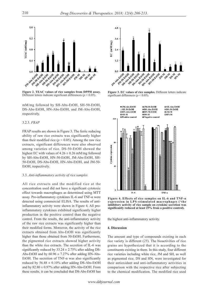
www.ddtjournal.com
Drug Discoveries & Therapeutics. 2018; 12(4):206-213.210
mM/mg followed by SH-Abs-EtOH, SH-50-EtOH, DS-Abs-EtOH, HN-Abs-EtOH, and JM-Abs-EtOH, respectively.
3.2.3. FRAP
FRAP results are shown in Figure 3. The ferric reducing ability of raw rice extracts was significantly higher than their modified rice (p < 0.05). Among the raw rice extracts, significant differences were also observed among varieties of rice. DS-50-EtOH showed the highest EC with values of 4.26 ± 0.26 mM/mg followed by SH-Abs-EtOH, HN-50-EtOH, JM-Abs-EtOH, SH-50-EtOH, DS-Abs-EtOH, HN-Abs-EtOH, and JM-50-EtOH, respectively.
3.3. Anti-inflammatory activity of rice samples
All r ice extracts and the modified rice at the concentration used did not have a significant cytotoxic effect towards macrophages as determined using MTT assay. Pro-inflammatory cytokines IL-6 and TNF-α were detected using commercial ELISA. The results of anti-inflammatory activity were shown in Figure 4. All pro-inflammatory cytokines exhibited significantly higher production in the positive control than the negative control. From the results, the anti-inflammatory activity of the raw rice extracts was significantly higher than their modified forms. Moreover, the activity of the rice extracts obtained from Abs-EtOH was significantly higher than those obtained from 50-EtOH. Furthermore, the pigmented rice extracts showed higher activity than the white rice extracts. The secretion of IL-6 was significantly reduced by 33.24 ± 2.75% after adding DS-Abs-EtOH and by 60.96 ± 7.27% after adding HN-Abs-EtOH. The secretion of TNF-α was also significantly reduced by 56.48 ± 0.10% after adding DS-Abs-EtOH and by 82.80 ± 0.97% after adding HN-Abs-EtOH. From these results, it can be concluded that DS-Abs-EtOH has
the highest anti-inflammatory activity.
4. Discussion
The amount and type of compounds existing in each rice variety is different (25). The bioactivities of rice grains are hypothesized that it is according to the constituents existing in them. In this study, four different rice varieties including white rice, JM and SH, as well as pigmented rice, DS and HN, were investigated for their antioxidant and anti-inflammatory activities in comparison with the respective rice after subjecting to the chemical modification. The modified rice used
Figure 2. TEAC values of rice samples from DPPH assay. Different letters indicate significant differences (p < 0.05).
Figure 3. EC values of rice samples. Different letters indicate significant differences (p < 0.05).
Figure 4. Effects of rice samples on IL-6 and TNF-α expression in LPS-stimulated macrophages (*the inhibitory activity of rice sample on cytokine secretion was significantly reduced at least 25% from a positive control).

www.ddtjournal.com
Drug Discoveries & Therapeutics. 2018; 12(4):206-213. 211
in the present study was obtained from etherification. Under this chemical reaction, raw rice starch can be modified into carboxymethyl starch (15) which can easily dissolve in water. Raw rice powder was found to be water insoluble, therefore extraction with organic solvent was used to collect essential compounds from raw rice samples. Abs-EtOH, containing 99.9% ethanol, was select as a suitable solvent because of its less polarity than water and less toxicity than other organic solvents (26). Water-cosolvent systems have been used for extraction of various plant compounds as an alternative to the extraction processes (27). Therefore 50-EtOH, containing 50% ethanol, was also selected to use in this study. The obtained rice extracts possessed different color due to rice variety and type of extracting solvent system. Different yield of extracts was found even from the same rice variety, depending on the solubility in the used extracting solvents and amount of the beneficial compounds in each rice grain variety. Extraction of the modified rice using the same manner and same extracting solvent showed that no any extract could be obtained. This might be due to the loss of certain ingredients at the final step of rice modification that the residual reagents and the unwanted products were completely washed out using ethanol. All solutes in the modified rice samples that could be dissolved in ethanol were removed in this step. Therefore, we decided to use the whole part of modified rice in the activity comparison test. To be comparable, the amount of the modified rice sample used was the same as that of the raw rice that gave the extract concentration used in the test. Many reactions and mechanisms are involved in antioxidant processes. The reactions of antioxidant to decrease free radical are direct and indirect processes. The direct processes are via free radical scavenging and metal ion chelating activities. The indirect processes are via inhibiting the activity of free radical generating enzymes and enhancing the activity of intracellular antioxidant enzymes (28). To demonstrate antioxidant activity by a free radical scavenging mechanism, ABTS and DPPH assay were applied. The principle of ABTS assay is to monitor the decay of the radical-cation of ABTS resulting from the oxidation of ABTS. Radical-cation of ABTS is soluble in both aqueous and organic solvents. This assay is not affected by ionic strength and can be done at different pH levels, therefore it was commonly used (29). Another method for detection of antioxidant activity based on free radical scavenging mechanism is DPPH assay. This assay is according to the color changes of the DPPH free radicals. The purple radical DPPH solution was converted to the yellow non-radical DPPH by the antioxidant having electron donating activity. These two methods can determine the free radical scavenging activity of the test samples directly to indicate the antioxidant activity of the samples. These ABTS and DPPH assays
were selected to use for antioxidant activity testing in the present study. Moreover, due to the different mechanism of antioxidant action, FRAP assay was used for determination of reducing activity. This method was applied to measure antioxidant activity by determination of the total reducing capacity of the compound based on the ability to reduce Fe3+ into Fe2+. Two mechanisms complement one another and give useful information of antioxidant activity (30). From the results of ABTS assay, the highest potential of ABTS free radical scavenging was DS-50-EtOH and HN-50-OH. This result is confirmed by the result from DPPH assay that DS-50-EtOH and HN-50-EtOH also showed the highest potential of DPPH free radical scavenging. The result from FRAP assay reveals that the highest reducing power was obtained from DS-50-EtOH. These results demonstrated that 50-EtOH is the better extracting solvent than Abs-EtOH. This was due to the ability of solvent to dissolve antioxidant compounds in rice grains. Moreover, the results indicated that the pigmented rice grains possess antioxidant activity with respect to the mechanisms of free radical scavenging and reducing activity. Previous studies demonstrated that pigmented rice exhibited higher antioxidant activity than white rice (31). This is according to the existing anthocyanins and proanthocyanins or condensed tannins which are the most prevalent phenolic compounds found in the pigmented rice (19). Those compounds have ability to donate hydrogen and act as reducing agents (32). Rice bioactive compounds such as anthocyanin are water soluble and the existing phenolic compounds are in the soluble and insoluble forms (30,33) Therefore, extracting solvent systems also play an important role on the antioxidant activity of rice due to the amount and type of the compounds that can be extracted. Besides the results of bioactive analysis and antioxidant activity, the in vitro anti-inflammatory effect of rice was evaluated with macrophages cells. The effects of white rice and pigmented rice on IL-6 and TNF-α expression were examined for their potential anti-inflammatory activities. From our results, the pigmented rice has a potent anti-inflammatory action. The extracts of DS and HN obtained from Abs-EtOH achieved the inhibition against IL-6. The anti-inflammatory activity of the test samples is accounted when the pro-inflammatory cytokines, such as IL-6 and TNF-α were significantly reduced by at least 25%. In the present study, DS-Abs-EtOH significantly suppressed IL-6 and TNF-α secretion, indicating that DS-Abs-EtOH extract has high potential as an anti-inflammatory agent. To the best of our knowledge, this is the first study which reports the significantly high anti-inflammatory effects of DS, an important pigmented rice variety of Thailand. In previous report from other groups, the Abs-EtOH extracts of two varieties of HN grown in Phayao province showed low anti-inflammatory activity against IL-6, TNF-α, and

www.ddtjournal.com
Drug Discoveries & Therapeutics. 2018; 12(4):206-213.212
nuclear factor-kappa B. Those results contrast to our results that HN-Abs-EtOH in the present study exhibits high inhibition against IL-6. This difference might be due to the variation of different cultivation area that the yield of active compounds obtained is different amount (13). Different polarity of extracting solvents can affect the bioactive composition of the obtained extracts. Previous report from other groups shows that the water extracts of Chaenomeles sinensis have higher antioxidant activity whereas the ethanolic extracts have higher anti-inflammatory activity (34). Another report demonstrates that the polar fractions of Suaeda asparagoides have higher antioxidant activity than the non-polar fractions whereas the non-polar fractions have higher anti-inflammatory activity than the polar fractions (35). In the present study, the higher polar rice extracts from 50-EtOH showed higher antioxidant activity than the lower polar extracts from Abs-EtOH, while Abs-EtOH extracts showed higher anti-inflammatory activity than 50-EtOH extracts. These results are in line with the previous reports and that was according to the different bioactive compounds in the extracts. The present results from antioxidant and anti-inflammatory studies clearly indicate that modification of rice structure has an effect to reduce antioxidant and anti-inflammatory property of rice. It is hypothesized that some bioactive compounds were degraded during the chemical modification process. In our process of rice modification, rice powder was treated with acid, base, and high temperatures. Previous studies reported that phenolic compounds and anthocyanins are labile to heat treatment and unstable under the alkaline pH. These factors induced the degradation and resulting in color change of bioactive compounds (34). Moreover, as mentioned above, the moodified rice was washed several times with ethanol at the final step of rice modification. This step also caused the loss of many active ingredients that could be dissolved in the ethanol. This was confirmed by visual observation, that the color of the modified rice was different from the raw rice indicating that some active compounds were lost. In addition, high viscosity of the systems containing modified rice might retard the reaction of the test. According to these factors, DS-M and HN-M showed less activities of antioxidant and anti-inflammatory than their respective raw rice. Our results confirm that after rice modification, the antioxidant and anti-inflammatory activities can be obtained from only pigmented rice variety. From the results of MTT assay, cytotoxicity to the macrophage cells was not found in all rice samples indicating the safety of the samples. In conclusion, difference in rice varieties leads to the difference antioxidant and anti-inflammatory potential. Chemical modification of rice causes significant reduction on the antioxidant and anti-inflammatory activity of rice. The pigmented rice possesses better
antioxidant and anti-inflammatory activities than the white rice. In addition, the results suggest that DS is a promising source of antioxidant and anti-inflammatory compounds.
Acknowledgements
The authors would like to thank the Thailand Research Fund for the financial support through the Research and Researcher for Industry (Grant No. PHD58I0012).
References
1. Breitenbach M, Eckl P. Introduction to oxidative stress in biomedical and biological research. Biomolecules. 2015; 5:1169-1177.
2. Wang W, Tan M, Yu J, Tan L. Role of pro-inflammatory cytokines released from microglia in Alzheimer's disease. Ann Transl Med. 2015; 3:1-15.
3. Kashiwagi A. Complications of diabetes mellitus and oxidative stress. Japan Med Assoc J. 2001; 44:521-528.
4. Candore G, Bulati M, Castiglia L, Colonna-romano G, Bona DD, Duro G, Lio D, Matranga D, Pellicanò M, Rizzo C, Scapagnini G, Vasto S. inflammation, cytokines, immune response, apolipoprotein e, cholesterol, and oxidative stress in Alzheimer disease: Therapeutic Implications. Rejuvenation Res. 2010; 13:301-313.
5. Chun OK, Kim DO, Smith N, Schroeder D, Han JT, Chang YL. Daily consumption of phenolics and total antioxidant capacity from fruit and vegetables in the American diet. J Sci Food Agric. 2005; 85:1715-1724.
6. Rohman A, Helmiyati S, Hapsari M, Setyaningrum DL. Rice in health and nutrition. Int Food Res J. 2014; 21:13-24.
7. Choi Y, Jeong H, Lee J. Antioxidant activity of methanolic extracts from some grains consumed in Korea. Food Chem. 2007; 103:130-138.
8. Walter M, Marchesan E. Phenolic compounds and antioxidant activity of rice. Braz Arch Biol Technol. 2011; 54:371-377.
9. Goufo P, Trindade H. Rice antioxidants: Phenolic acids, flavonoids, anthocyanins, proanthocyanidins, tocopherols, tocotrienols, γ -oryzanol, and phytic acid. Food Sci Nutr. 2014; 2:75-104.
10. Lobo V, Patil A, Phatak A, Chandra N. Free radicals, antioxidants and functional foods: Impact on human health. Pharmacogn Rev. 2010; 4:118-126.
11. Whent M, Huang H, Xie Z, Lutterodt H, Yu L, Fuerst EP, Morris CF, Yu LL, Luthria D. Phytochemical composition, anti-inflammatory, and antiproliferative activity of whole wheat flour. J Agric Food Chem. 2012; 60:2129-2135.
12. Choi SP, Kim SP, Kang MY, Nam SH, Friedman M. Protective effects of black rice bran against chemically-induced inflammation of mouse skin. J Agric Food Chem. 2010; 58:10007-10015.
13. Kitisin T, Saewan N, Luplertlop N. Potential anti-inflammatory and anti-oxidative properties of Thai colored-rice extracts. Plant Omics. 2015; 8:69-77.
14. Wang Q, Han P, Zhang M, Xia M, Zhu H, Ma J, Hou M, Tang Z, Ling W. Supplementation of black rice pigment fraction improves antioxidant and anti-inflammatory status in patients with coronary heart disease. Asia Pac J
www.ddtjournal.com
Drug Discoveries & Therapeutics. 2018; 12(4):206-213. 213
Clin Nutr. 2007; 16:295-301.15. Okonogi S , Khongkhunth ian S , Ja turas i tha S .
Development of mucoadhesive buccal films from rice for pharmaceutical delivery systems. Drug Discov Ther. 2014; 8:262-267.
16. Okonogi S , Kaewpin ta A, Khongkhunth ian S , Yotsawimonwat S. Effect of r ice variety on the physicochemical properties of the modified rice powders and their derived mucoadhesive gels. Drug Discov Ther. 2015; 9:221-228.
17. Builders PF, Arhewoh MI. Pharmaceutical applications of native starch in conventional drug delivery. Starch/Stärke. 2016; 68:1-10.
18. Junmahasathien T, Khongkhunthien S, Okonogi S. Preparation and characterization of modified colored rice as a gelling carrier for buccal drug delivery system. Int J Pharm Pharm Sci. 2016; 8:124-130.
19. Gunaratne A, Wu K, Li D, Bentota A, Corke H, Cai YZ. Antioxidant activity and nutritional quality of traditional red-grained rice varieties containing proanthocyanidins. Food Chem. 2013; 138:1153-1161.
20. Niu Y, Gao B, Slavin M, Zhang X, Yang F, Bao J, Shi H, Xie Z, Yu L. Phytochemical compositions, and antioxidant and anti-inflammatory properties of twenty-two red rice samples grown in Zhejiang. LWT - Food Sci Technol. 2013; 54:521-527.
21. Yoenyongbuddhagal S, Noomhorm A. Effect of raw material preparation on rice vermicelli quality. Starch/Stärke. 2002; 54:534-539.
22. Saeio K, Chaiyana W, Okonogi S. Antityrosinase and antioxidant activities of essential oils of edible Thai plants. Drug Discov Ther. 2011; 5:144-149.
23. O k o n o g i S , D u a n g r a t C , A n u c h p r e e d a S , Tachakittirungrod S, Chowwanapoonpohn S. Comparison of antioxidant capacities and cytotoxicities of certain fruit peels. Food Chem. 2007; 103:839-846.
24. Tachakittirungrod S, Okonogi S, Chowwanapoonpohn S. Study on antioxidant activity of certain plants in Thailand: Mechanism of antioxidant action of guava leaf extract. Food Chem. 2007; 103:381-388.
25. Ravichanthiran K, Ma Z, Zhang H, Cao Y, Wang C, Muhammad S, Aglago EK, Zhang Y, Jin Y, Pan B. Phytochemical profile of brown rice and its nutrigenomic implications. Antioxidants. 2018; 7:71.
26. Escribano-bail MT, Santos-buelga C, Rivas-gonzalo C. Anthocyanins in cereals. J Chromatogr A. 2004; 1054:129-141.
27. Puttipan R, Okonogi S. Antioxidant activity of Rafflesia kerrii flower extract. Drug Discov Ther. 2014; 8:18-24.
28. Lü J-M, Lin PH, Yao Q, Chen C. Chemical and molecular mechanisms of antioxidants: Experimental approaches and model systems. J Cell Mol Med. 2010; 14:840-860.
29. Shalaby EA, Shanab SMM. Comparison of DPPH and ABTS assays for determining antioxidant potential of water and methanol extracts of Spirulina platensis. Indian J Geomarine Sci. 2013; 42:556-564.
30. Biskup I, Golonka I, Gamian A, Sroka Z. Antioxidant activity of selected phenols estimated by ABTS and FRAP methods. Postepy Hig Med Dosw. 2013; 67:958-963.
31. Muntana N, Prasong S. Study on total phenolic contents and their antioxidant activities of Thai white, red and black rice bran extracts. Pak J Biol Sci. 2010; 13:170-174.
32. Saikia S, Dutta H, Saikia D, Mahanta CL. Quality characterization and estimation of phytochemicals content and antioxidant capacity of aromatic pigmented and non-pigmented rice varieties. Food Res Int. 2012; 46:334-340.
33. Park YS, Kim SJ, Chang HI. Isolation of anthocyanin from black rice (Heugjinjubyeo) and screening of its antioxidant activities. Korean J Microbiol Biotechnol. 2008; 36:55-60.
34. Han YK, Kim YS, Natarajan SB, Kim WS, Hwang JW, Jeon NJ, Jeong JH, Moon SH, Jeon BT, Park PJ. Antioxidant and anti-Inflammatory effects of Chaenomeles sinensis leaf extracts on LPS-stimulated RAW 264.7 Cells. Molecules. 2016; 21:422.
35. Park JM, Kim SD, Lee WM, Cho JY, Park HJ, Kim TW, Choe NH, Kim SK, Rhee MH. In vitro anti-oxidative and anti-inflammatory effects of solvent-extracted fractions from Suaeda asparagoides. Pharmazie. 2007; 62:453-458.
36. Oancea S, Drăghici O. pH and thermal stability of anthocyanin-based optimised extracts of Romanian red onion cultivars. Czech J Food Sci. 2013; 31:283-291.
(Received July 14, 2018; Revised August 30, 2018; Accepted August 30, 2018)

www.ddtjournal.com
Drug Discoveries & Therapeutics. 2018; 12(4):214-223.214
Fungal-derived xenobiotic exhibits antibacterial and antibiofilm activity against Staphylococcus aureus
Siddhartha Kumar1, Arvind Kumar1, Manisha Kaushal2, Prince Kumar3, Kasturi Mukhopadhyay3, Antresh Kumar1,*
1 Department of Biotechnology, Central University of South Bihar, Gaya, India;2 Department of Botany, Gurunakak Degree College Umra kala Siohara, Bijnor, U.P. India; 3 School of Environmental Sciences, Jawaharlal Nehru University, New Delhi, India.
1. Introduction
Staphylococcus aureus is a dangerous human pathogen which causes a diverse range of superficial and invasive diseases such as acute skin and tissue lesions to severe necrotizing pneumonia, osteomyelitis endocarditis, septicemia, and catheter-associated bacteremia (1-4). This notorious bacterium continues to parade higher morbidity, mortality, and a significant financial burden to the public. Individuals at high risk for S. aureus infection include patients with surgical, organ transplantation, indwelling catheters, ventilator-assisted respiration, diabetics, tracheal intubation, late-stage
renal disease, immunosuppressive or cancer therapy and individuals with low-birth weight neonates, and hospital residents (5-8). In spite of high rate of infections, continuous and rapidly emerging drug resistance against available different antibiotics has made it difficult to treat (9). Secretion of virulence factors and formation of biofilm in S. aureus are the two hallmark determinants that majorly contribute to therapeutic failure. A multitude of virulence factors including enterotoxins, pore-forming toxins (α-hemolysin, Panton-Valentine leukocidin) is considered to be the key factors essential for the establishment of infection (10). In contrast, formation of multicellular biofilm over on cell boundary acts as a major obstacle for drugs to enter inside and that makes them 100 folds more resistant to antibiotics, antimicrobials, and immune defense (11,12). Many Staphylococcus species showed an increased resistance to available drugs (13,14). Consequently, the prognosis of such infections is problematic and has now
Summary Staphylococcus aureus is an opportunistic pathogen, responsible for superficial and invasive infections both in nosocomial and community-acquired settings. The incidences of infection have become more problematic attributable to emerging drug resistance and biofilm formation. These challenges suggest the need for new antimicrobial agents against S. aureus. In present work, we purified a fungal xenobiotic (FI3) which elicits a potent antimicrobial activity against a list of tested microbes including methicillin sensitive (MSSA) and methicillin resistance (MRSA) S. aureus. The cell growth of MSSA and MRSA were completely ceased with the 1× minimum inhibitory concentration (MIC); 32 µg/mL and 128 µg/mL, respectively. The cell viability severely decreased within 90 min, due to disturbance of membrane homeostasis. This bactericidal effect was enhanced at lower pH (pH 4) with a speculation to retain positive charge. The FI3 potently disrupts biofilm adherence at 64 µg/mL and found to be a safe with no toxic effect on mammalian tissue. FI3 also leads to increase the potency of tested antibiotics. Taken together, we established that FI3 has a potent antimicrobial activity against tested microbes and safer to human tissue. It may be proven a leading molecule for the treatment of bacterial infections.
Keywords: Staphylococcal infections, antibacterial, antibiofilm, fungal xenobiotic, Staphylococcus aureus
DOI: 10.5582/ddt.2018.01042Original Article
*Address correspondence to:Dr. Antresh Kumar, Centre for Biological Science (Biotechnology), Central University of South Bihar, Patna 800014, Bihar, India.E-mail: [email protected]
www.ddtjournal.com
Drug Discoveries & Therapeutics. 2018; 12(4):214-223.
become an alarming issue both in developed as well as developing countries. The drug resistance condition is developed by frequent and indiscriminate uses of antibiotics. More than 11,000 people died from drug resistance S. aureus related infections in the U.S. (CDC report 2013) and now the situations have reached to an epidemic proportion with a serious health concern (15). In India, the incidence of drug resistant S. aureus infections has endemic nature and it increased steadily up to 54% both in hospital-acquired (HA) and community-acquired (CA) infections (16-18). There is a major deficit of effective and sustainable treatment against S. aureus. Hence, there is an urgent need for finding new antibacterial and novel approaches against both planktonic and sessile bacteria. Since a long time, the discovery of new active compounds from both natural and synthetic sources has been explored and still gaining much attention (19,20). Fungi have been used as a natural source of traditional medicine and variety of drugs has been derived and still, the research on fungi is of a great interest. Antibiotics penici l l in , s t reptomycin, immunosuppressant cyclosporine, anti-hypercholesterolemic compactin, and lovastatin are such examples. In fact, fungal secondary metabolites have a vital ecological role; they act as a weapon of defense and competitor to protect themselves against parasites and predators. The endophytic fungi, habitat to colonize on plants, represent a storehouse of the diverse array of bioactive secondary metabolites which can prove to be a miracle and combat plethora of pathogens including resistant microorganisms. With this background, the objective of this study is to evaluate the antimicrobial and anti-biofilm activity of the endophytic fungal xenobiotic (FI3) against methicillin-sensitive S. aureus (MSSA) and methicillin-resistant S. aureus (MRSA). We showed that FI3 leads to eradicate S. aureus infection load speedily by targeting cell membrane and biofilm adherence of Staphylococcal strain without showing any detrimental effect to E.coli and human tissue.
2. Materials and Methods
2.1. Bacterial and fungal strains and culture media
The methici l l in-sensi t ive S. aureus (MSSA); ATCC29213 and methicillin-resistant S. aureus (MRSA); ATCC33591, Pseudomonas aeruginosa MTCC1034, Bacil lus subti l is MTCC441 and, Escherichia coli strains were used in this study. MSSA was received as a gift by Prof. Kasturi Mukhopadhyay, JNU, New Delhi, India and P. aeruginosa MTCC1034, B. subtilis MTCC441, and E. coli were purchased from MTCC, Chandigarh, India. These strains were used for analysis of drug susceptibility assay. Drug susceptibility test was also performed using MRSA. S. aureus strains
were cultured in Muller-Hinton Broth/Agar (MHB/MHA). Tryptic Soya Agar/Broth (TSA/TSB) was used for biofilm study. P. aeruginosa MTCC1034, B. subtilis MTCC441 and E. coli were cultured in LB (Luria-Bertani) broth. The fungal isolates (FI) were isolated from the bark and dried tissue of the plant from the Sanjay Gandhi Zoological Park (SGZP), Patna, Bihar and also received from the National Culture Collection of Pathogenic Fungi (NCCPF), PGIMER, Chandigarh, India. All media were purchased from the Hi-media laboratory.
2.2. Purification of fungal xenobiotic (FI3)
The fungal isolates maintained on YEPD agar plate (seed culture) was inoculated in YEPD medium and allowed to incubate on shaking condition for six days at 30ºC. Cells were pelleted by centrifugation at 12,000× g for 15 min and the culture supernatant was filtered through Whatman filter No 1. The cell-free supernatant was extracted as described by Samuel et al. (21). The supernatant was gently agitated for 30 min after addition of equal volume of chloroform. This step was repeated thrice by adding fresh Chloroform. Fractions recovered from chloroform extraction were evaporated in the rotatory evaporator, dried under vacuum, weighed and dissolved in phosphate buffer saline (PBS) for purification. The sample was loaded on C-18 column using a gradient flow rate with 1 mL/min of acetonitrile (A) and water (B) mobile solvents. The chromatogram was run as A5% + B95% (0 min); A15% + B85% (0-5 min); A95% + B5% (5-25 min); A15% + B85% (25-40 min). The prominent peak with high intensities were collected in a sterile glass vial for further processing and antimicrobial analysis. Peak No. 6 showed postive results was collected and termed this xenobiotic as FI3.
2.3. Susceptibility assay
The antimicrobial susceptibility of FI3 was determined by two different methods mentioned here under.
2.3.1. Spot assay
FI3 was initially screened for antimicrobial activity against S. aureus by spot assay described elsewhere (22,23) with minor modifications. For this, both MSSA and MRSA cells were cultured in Muller-Hinton broth (MHB) till the cell density (Abs 600 nm) reaches 0.5. It was achieved at the mid-exponential phase of the cell growth (data not shown). One OD600nm of S. aureus cell density was calculated which equals to 2 × 10⁸ cells/mL (24). The cell pellet was resuspended in PBS to maintain 0.1 OD for spotting. 2 × 10⁶ (0.01 OD600nm) MSSA or MRSA cells were uniformly spread over the surface of the Muller-Hinton agar (MHA) plate and FI3 was put on plate for antimicrobial screening for
215
www.ddtjournal.com
Drug Discoveries & Therapeutics. 2018; 12(4):214-223.216
of biofilm formation and biofilm attachment assay, respectively. After incubation, both slides and wells were washed with 250 μL of PBS to remove planktonic bacteria and air dried. The cells were then fixed with 200 μL of methanol for 15 min and plates were allowed to dry. The plate wells or glass slides were stained with 200 μL of 0.1% (v/v) crystal violet for 5 min. Excess stain was gently rinsed off and plates were air-dried. After staining, the attachment of biofilm was observed under the inverted microscope. The stained wells were re-solubilized in 200 μL of 95% (v/v) ethanol and cell concentration was measured at OD595nm for biofilm inhibition analysis respectively (30). The FI3 untreated S. aureus cells were considered as a positive control in both experiments.
2.6. Haemolysis assay
To test the hemolytic activity FI3, chicken blood and human red blood cells (RBCs) were taken as reported elsewhere (31,32). Briefly, fresh human blood was collected and layered on histopaque-1077. The RBCs cells were recovered from the pellet by centrifugation at 400× g for 30 min. The pellet was washed with 0.9% (w/v) NaCl for three times and finally resuspended in 0.9% NaCl. Then, various concentrations (16-256 µg/mL) of FI3 were added in 95 μL of the RBCs cells suspension and it was then incubated at 37°C with gentle mixing for 3 h. After incubation, the supernatant was recovered by centrifugation as above. The cell lysis was quantified at 415 nm. As a positive control, 1% triton X-100 was added in RBCs suspension for measuring the complete lysis and in the negative control, RBCs suspension was incubated only in the presence of PBS. Haemolysis was calculated as: Percentage haemolysis = [(A − A0)/ (A100 − A0)] × 100. Where: A, represents absorbance of the FI3 treated sample at 415 nm and A0 and A100 represent zero percent and 100% lysis determined in PBS and 1% Triton X-100, respectively.
2.7. Cytoplasmic membrane permeability assay
To investigate the membrane permeabilizing property of the FI3 against S. aureus cell membrane, calcein release assay was performed using calcein acetoxymethyl ester (calcein-AM) as described elsewhere with minor modifications (33). Calcein-AM is a non-fluorescent, lipid-soluble, cell membrane permeable dye. Upon hydrolysis by cytoplasmic esterase, it breaks into non-permeable, fluorescent calcein with an excitation (Ext) and emission (Em) 494 nm and 527 nm, respectively. Briefly, S. aureus cells were harvested to mid-exponential phase and diluted to maintain 2 × 10⁴ cells/mL in PBS, supplemented with 5 mM glucose. Cells were then incubated with 2 μM calcein-AM for 1h for maximal uptake of dye followed by addition of 1× MIC concentration (32 µg/mL) of FI3 and then
calculation of zone of inhibition. 5 μL of ten folds serial dilutions were spotted on MHA plate in the absence and presence of FI3 and other tested drugs. The difference in S. aureus cell growth was observed after incubation for 24 h at 37°C.
2.3.2. Minimum inhibitory concentration (MIC)
MIC was determined by broth microdilution method according to the guidelines of Clinical and Laboratory Standards Institute (CLSI) (25). The assay was done in three independent experiments. Briefly, different dilutions of FI3 and other tested drugs viz. Tet (tetracycline), Genta (gentamycin), and Van (vancomycin) were prepared in 96 wells plate by addition of final volume 100 μL of MHB medium in each well of the plate. Subsequently, it was serially diluted in 1:2 ratios. After successful dilutions of tested compounds and drugs, 2 × 10⁶ cells of mid-exponential phase S. aureus (MSSA and MRSA), P. aeruginosa, B. subtilis and E. coli were added in each well. MIC value was calculated by observing the optical density (OD600nm) after 24 h of plate incubation at 37°C. 1× MIC was defined as the concentration of tested compound (FI3) that enables to restrict cell growth 100% as compared to the cell growth control. The FI3 showing 100% cell growth inhibition was considered as the 1× MIC.
2.4. Cell viability assay
To determine the antimicrobial activity of FI3, 2 × 10⁶ MSSA cells of mid exponential phase were taken as described elsewhere (26). The cells were incubated with varying concentration of FI3 i.e. 16 μg/mL, 32 μg/mL, 64 μg/mL and 128 μg/mL, those correspond to 0.5× MIC, 1× MIC, 2× MIC and 4× MIC, respectively. The mixture of tube content was incubated at 37°C at 120 rpm. After each 30 min of interval up to 90 min, 10 µL of tube content was taken and serially diluted up to 100 times in PBS to reduce FI3 and then spread out on MHA plate at 37°C. Viable colonies were calculated after overnight incubation.
2.5. Biofilm attachment assay and inhibition of biofilm formation
The effect of FI3 on biofilm inhibition of MSSA was tested as described elsewhere (27-29). Briefly, the overnight culture of S. aureus was diluted up to 2 × 10⁵ cells/mL in TSB. This diluted cell suspension with 5 mM glucose was seeded in to 24-well polystyrene plate in addition to introducing different concentrations of FI3. The assay was done in three independent experiments. In biofilm attachment assay, sterile glass slide (10 × 10 mm) was put in the wells. The plates were incubated at 37°C for 48 h and 3 h for inhibition
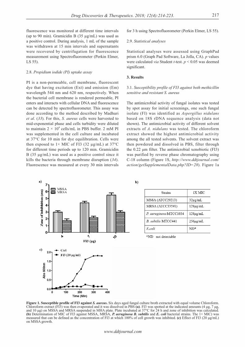
www.ddtjournal.com
Drug Discoveries & Therapeutics. 2018; 12(4):214-223. 217
fluorescence was monitored at different time intervals (up to 90 min). Gramicidin B (35 µg/mL) was used as a positive control. During analysis, 1 mL of the sample was withdrawn at 15 min intervals and supernatants were recovered by centrifugation for fluorescence measurement using Spectrofluorometer (Perkin Elmer, LS 55).
2.8. Propidium iodide (PI) uptake assay
PI is a non-permeable, cell membrane, fluorescent dye that having excitation (Ext) and emission (Em) wavelength 544 nm and 620 nm, respectively. When the bacterial cell membrane is rendered permeable, PI enters and interacts with cellular DNA and fluorescence can be detected by spectrofluorometer. This assay was done according to the method described by Madhuri et al. (33). For this, S. aureus cells were harvested to mid-exponential phase and cells turbidity were diluted to maintain 2 × 10⁴ cells/mL in PBS buffer. 2 mM PI was supplemented in the cell culture and incubated at 37°C for 10 min for dye equilibration. Cells were then exposed to 1× MIC of FI3 (32 µg/mL) at 37°C for different time periods up to 120 min. Gramicidin B (35 µg/mL) was used as a positive control since it kills the bacteria through membrane disruption (34). Fluorescence was measured at every 30 min intervals
for 3 h using Spectrofluorometer (Perkin Elmer, LS 55).
2.9. Statistical analyses
Statistical analyses were assessed using GraphPad prism 6.0 (Graph Pad Software, La Jolla, CA). p values were calculated via Student t-test. p < 0.05 was deemed significant.
3. Results
3.1. Susceptibility profile of FI3 against both methicillin sensitive and resistant S. aureus
The antimicrobial activity of fungal isolates was tested by spot assay for initial screenings, one such fungal isolate (FI) was identified as Aspergillus nidulans based on 18S rDNA sequence analysis (data not shown). The antimicrobial activity of different solvent extracts of A. nidulans was tested. The chloroform extract showed the highest antimicrobial activity among the all tested solvents. The solvent extract was then powdered and dissolved in PBS, filter through the 0.22 μm filter. The antimicrobial xenobiotic (FI3) was purified by reverse phase chromatography using C-18 column (Figure 1S, http://www.ddtjournal.com/action/getSupplementalData.php?ID=28). Figure 1a
Figure 1. Susceptible profile of FI3 against S. aureus. Six days aged fungal culture broth extracted with equal volume Chloroform. Chloroform extract (FI3) was then evaporated and it was dissolved in PBS (a). FI3 was spotted at the indicated amounts (4 μg, 7 μg, and 10 μg) on MSSA and MRSA suspended in MHA plate. Plate incubated at 37°C for 24 h and zone of inhibition was calculated. (b) Determination of MIC of FI3 against MSSA, MRSA, P. aeruginosa B. subtilis and E. coli bacterial strains. The 1× MIC) was measured that can be defined as the concentration of FI3 at which 100% of cell growth was inhibited. (c) Effect of FI3 (20 μg/mL) on MSSA growth.

www.ddtjournal.com
Drug Discoveries & Therapeutics. 2018; 12(4):214-223.218
illustrates that cell growth of both methicillin-sensitive S. aureus (MSSA) and methicillin-resistance S. aureus (MRSA) was inhibited in the presence of varying concentration of FI3. These results clearly demonstrate that FI3 is effective against both MSSA and MRSA. The antimicrobial activity of FI3 was further assessed against a panel of Gram+ve and Gram−ve strains using microdilution assay. Figure 1b demonstrates that FI3 completely inhibits the cell growth of the tested bacteria strains including resistant MRSA. The growth of MSSA and MRSA were completely arrested (1× MIC) at 32 μg/mL and 128 μg/mL, FI3 respectively. FI3 also retains its MIC values for P. aeruginosa MTCC1034 and B. subtilis MTCC441, 128 μg/mL and 256 μg/mL, respectively. On the contrary, FI3 has no appreciable inhibition of E. coli growth and was found to be insensitive even at higher concentration. Strikingly, MSSA growth was not recovered and remain suppressed when growth curve was performed in the presence of more than 0.5× MIC (16 μg/mL) concentration of FI3 (Figure 1c). These findings revealed that the FI3 effectively inhibits the growth of S. aureus (MSSA and MRSA), P. aeruginosa and B. subtilis without affecting the growth of E. coli.
3.2. Combined effect of FI3 with topical antimicrobials
Combination of two or more antimicrobials together has been explored as an alternative strategy for the treatment of systemic S. aureus infections. In order to test the combinatorial effects of FI3 together with tested topical antibiotics on S. aureus susceptibilities,
broth microdilution and spot assays were performed. Figure 2a illustrates the drug susceptibility profile of three different antibiotics named as Tet, Genta, and Van in combination with FI3. The MIC of these individual antibiotics was measured as 1 μg/mL, 1 μg/mL, and 2 μg/mL, respectively (Figure 2a). The drug susceptibility of Tet, Genta, and Van was further enhanced when cells were grown with 8 μg/mL FI3 along with tested antibiotics. The MIC values of tested antibiotics; Tet, Genta, and Van in combination with FI3 were now calculated as 62.5 ng/mL, 125 ng/mL, and 250 ng/mL, respectively. Spot assay further confirmed that the supplementation of FI3 together with tested antibiotics were noticeably more susceptible to Tet, Genta, and Van than the individual effects of the tested antibiotics (Figure 2b).
3.3. Bactericidal activity of FI3
To assess the bactericidal activity of FI3, mid-exponential phase cells were treated with different concentration of FI3 i.e. 0.5× MIC (16 μg/mL), 1× MIC (32 μg/mL), 2× MIC (64 μg/mL) and 4× MIC (128 μg/mL) for 30 min, 60 min, and 90 min of action time. Figure 3a illustrates that the FI3 treatment shows time and concentration-dependent of cell killing. For example, at 0.5× MIC concentration, 19%, 24%, and 53% cells were killed within 30 min, 60 min, 90 min, respectively whereas at 1× MIC, 38%, 67% and 96% cells were killed in 30 min, 60 min and 90 min, respectively. The cells were found to be completely eliminated with no significant viable cells (± 2.5%)
Figure 2. Combinatorial effect of FI3 with tropical antimicrobials. (a) Broth dilution assay to determine MIC value of tested antimicrobial drugs Tet (tetracycline), Genta (gentamycin), and Van (vancomycin) in absence and presence of FI3. Data was quantitatively showed with color (see the color bar in lower panel), where each shade of color represents relative optical densities of the cell and as bar graphs (see right panel). (b) Spot assays of S. aureus in presence of Tet, Genta, and Van (62.5 ng/mL, 62.5 ng/mL, and 125 ng/mL) alone and with FI3 (8 µg/mL).

www.ddtjournal.com
Drug Discoveries & Therapeutics. 2018; 12(4):214-223. 219
within 90 min exposure of 1× MIC or at higher concentrations. These results ensure that the FI3 has a potent antimicrobial activity. To access the effect of pH on cell susceptibility of S. aureus in response to FI3 treatment (0.5× MIC), cell viability assay was performed at varying pH ranging from 4.5 to 8.5. Figure 3b illustrates the impact of pH on the antimicrobial activity of FI3. Cells viability at individual pH was measured in presence or absence of FI3. Without FI3 treatment at different pH was considered as control (Ctrl). The relative cell viability (FI3/Ctrl) was analyzed at different tested pH for monitoring pH dependent effect of FI3. The data suggested that antimicrobial activity of FI3 is pH dependent and more susceptible at lower pH. The increased susceptibility at lower pH is expected to be increased a net positive charge on the molecule. This gain in the positive charge on FI3 molecule tends to interact to the negative charged cell membrane more firmly.
3.4. Membrane permeability effect of FI3
The staphylococcal cell permeabilization effect of FI3 was tested by quantification of preloaded calcein leakage via spectrofluorimetric analysis. Gramicidin B; a known pore-forming peptide was used as a positive control. For this, we performed cell permeability analysis by measuring calcein258 AM hydrolysis. Calcein AM is a cell-permeable, non-fluorescent dye which converts into a green fluorescent calcein after acetoxymethyl (AM) ester hydrolysis by intracellular esterase. When cells membrane becomes more permeable, more amount of calcein AM dye enters the cell and get hydrolyzed into membrane-impermeable green fluorescent calcein dye. Dead cells with compromised cell membrane do not retain calcein fluorescence inside the cell and release outside the cell membrane that can be measured at 515nm. Similar to the positive control, cells treated with 0.5× MIC FI3 (16 μg/mL) increased calcein fluorescence
that indicates enhanced cell permeability. The cell permeability was further corroborated when cells were spotted with cell-disrupting chaotropic agent SDS and by PI uptake assay. Figure 4B illustrates that cells treated with SDS became hyper-sensitive in presence of FI3 and PI was 45% up taken upon treatment of 0.5× MIC FI3 as compared to positive control (Figure 4c).
3.5. FI3 potentially clears up matured biofilm
To consider the potential staphylocidal activity of FI3, we also considered the possibility that FI3 would also be effective against matured biofilm. S. aureus cells were grown in biofilm forming medium with the subsequent increasing order of FI3 concentration and biofilm mass was measured after 2 h of treatment. More than 90% biofilm formation was inhibited at 64 µg/mL (2× MIC) concentration (Figure 5a). FI3 showed a significant inhibitory activity against S. aureus biofilm formation at 48 h (data not shown). The biofilm inhibitory effect of FI3 was further confirmed by microscopic analysis. Cells treated with 1× MIC (32 µg/mL) and 2× MIC (64 µg/mL) concentration of FI3 reduce the biofilm cell aggregation (cleared up) approximately 40% and 89%, respectively as compared to untreated (control) (Figure 5b).
3.6. Haemolytic effect of FI3
The antimicrobial and antibiofilm activity of FI3 against both planktonic as well as surface-attached cells prompted us to further find out any toxic effect of FI3 on human tissue. This was achieved by using human as well as chicken blood tissues. For this, RBC cells were treated with subsequently increasing concentration of FI3. 0.9% NaCl and 1% Triton X-100 were taken as a negative and positive control respectively. The FI3 did not show any haemolytic effect on both human as well as chicken blood tissues as compared to tested control. This study suggests that FI3 is safer with low hemolytic
Figure 3. Antimicrobial activity of FI3. (a) Cell killing assay to determine the antimicrobial activity of FI3. Mid-exponential phase S. aureus cells were treated with 0.5×, 1×, 2× and 4× MIC i.e. 16 μg/mL, 32 μg/mL, 64 μg/mL, and 128 μg/mL, respectively. Cells were incubated up to 90 min at 30 min intervals. (b) S. aureus cells were grown till mid-log phase and cells were exposed to 16 μg/mL (0.5× MIC), a sub-inhibitory concentration of FI3 for 2 h at different pH.

www.ddtjournal.com
Drug Discoveries & Therapeutics. 2018; 12(4):214-223.220
Figure 5. Anti-biofilm activity of FI3. The 2 × 10⁵ cells/mL S. aureus cells suspended in the TSB-glucose medium, were seeded in pre- kept glass slide, in 24 well polystyrene plate and added different concentrations of FI3 (in triplicate) as indicated. The plate was incubated at 37°C for (a) 4 h for biofilm attachment analysis and (b) 48h for inhibition of biofilm formation.
Figure 4. Cell membrane permeabilization activity of FI3 against S. aureus. (a) To assess the FI3 effect on membrane permeabilization, mid-log phase cells treated with 32 μg/mL (1× MIC) FI3 up to 90 min at 15 min intervals. 35 μg/mL gramicidin was used as a positive control. (b) Spot assay of S. aureus in the absence (negative control) and presence of 16 μg/mL (0.5× MIC), a sub-inhibitory concentration of FI3 and cell perturbation agent SDS at 0.03%. (c) PI uptake assay performs to determine cell deprivation in presence of 16 μg/mL, FI3 and 35 μg/mL, gramicidin B (positive control).
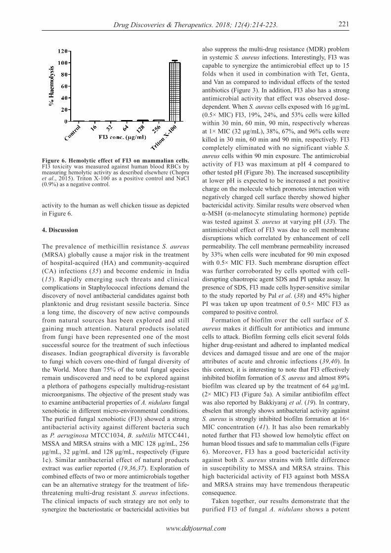
www.ddtjournal.com
Drug Discoveries & Therapeutics. 2018; 12(4):214-223. 221
activity to the human as well chicken tissue as depicted in Figure 6.
4. Discussion
The prevalence of methicillin resistance S. aureus (MRSA) globally cause a major risk in the treatment of hospital-acquired (HA) and community-acquired (CA) infections (35) and become endemic in India (15). Rapidly emerging such threats and clinical complications in Staphylococcal infections demand the discovery of novel antibacterial candidates against both planktonic and drug resistant sessile bacteria. Since a long time, the discovery of new active compounds from natural sources has been explored and still gaining much attention. Natural products isolated from fungi have been represented one of the most successful source for the treatment of such infectious diseases. Indian geographical diversity is favorable to fungi which covers one-third of fungal diversity of the World. More than 75% of the total fungal species remain undiscovered and need to be explored against a plethora of pathogens especially multidrug-resistant microorganisms. The objective of the present study was to examine antibacterial properties of A. nidulans fungal xenobiotic in different micro-environmental conditions. The purified fungal xenobiotic (FI3) showed a strong antibacterial activity against different bacteria such as P. aeruginosa MTCC1034, B. subtilis MTCC441, MSSA and MRSA strains with a MIC 128 μg/mL, 256 μg/mL, 32 μg/mL and 128 μg/mL, respectively (Figure 1c). Similar antibacterial effect of natural products extract was earlier reported (19,36,37). Exploration of combined effects of two or more antimicrobials together can be an alternative strategy for the treatment of life-threatening multi-drug resistant S. aureus infections. The clinical impacts of such strategy are not only to synergize the bacteriostatic or bactericidal activities but
also suppress the multi-drug resistance (MDR) problem in systemic S. aureus infections. Interestingly, FI3 was capable to synergize the antimicrobial effect up to 15 folds when it used in combination with Tet, Genta, and Van as compared to individual effects of the tested antibiotics (Figure 3). In addition, FI3 also has a strong antimicrobial activity that effect was observed dose-dependent. When S. aureus cells exposed with 16 μg/mL (0.5× MIC) FI3, 19%, 24%, and 53% cells were killed within 30 min, 60 min, 90 min, respectively whereas at 1× MIC (32 μg/mL), 38%, 67%, and 96% cells were killed in 30 min, 60 min and 90 min, respectively. FI3 completely eliminated with no significant viable S. aureus cells within 90 min exposure. The antimicrobial activity of FI3 was maximum at pH 4 compared to other tested pH (Figure 3b). The increased susceptibility at lower pH is expected to be increased a net positive charge on the molecule which promotes interaction with negatively charged cell surface thereby showed higher bactericidal activity. Similar results were observed when α-MSH (α-melanocyte stimulating hormone) peptide was tested against S. aureus at varying pH (33). The antimicrobial effect of FI3 was due to cell membrane disruptions which correlated by enhancement of cell permeability. The cell membrane permeability increased by 33% when cells were incubated for 90 min exposed with 0.5× MIC FI3. Such membrane disruption effect was further corroborated by cells spotted with cell-disrupting chaotropic agent SDS and PI uptake assay. In presence of SDS, FI3 made cells hyper-sensitive similar to the study reported by Pal et al. (38) and 45% higher PI was taken up upon treatment of 0.5× MIC FI3 as compared to positive control. Formation of biofilm over the cell surface of S. aureus makes it difficult for antibiotics and immune cells to attack. Biofilm forming cells elicit several folds higher drug-resistant and adhered to implanted medical devices and damaged tissue and are one of the major attributes of acute and chronic infections (39,40). In this context, it is interesting to note that FI3 effectively inhibited biofilm formation of S. aureus and almost 89% biofilm was cleared up by the treatment of 64 µg/mL (2× MIC) FI3 (Figure 5a). A similar antibiofilm effect was also reported by Bakkiyaraj et al. (19). In contrary, ebselen that strongly shows antibacterial activity against S. aureus is strongly inhibited biofilm formation at 16× MIC concentration (41). It has also been remarkably noted further that FI3 showed low hemolytic effect on human blood tissues and safe to mammalian cells (Figure 6). Moreover, FI3 has a good bactericidal activity against both S. aureus strains with little difference in susceptibility to MSSA and MRSA strains. This high bactericidal activity of FI3 against both MSSA and MRSA strains may have tremendous therapeutic consequence. Taken together, our results demonstrate that the purified FI3 of fungal A. nidulans shows a potent
Figure 6. Hemolytic effect of FI3 on mammalian cells. FI3 toxicity was measured against human blood RBCs by measuring hemolytic activity as described elsewhere (Chopra et al., 2015). Triton X-100 as a positive control and NaCl (0.9%) as a negative control.
www.ddtjournal.com
Drug Discoveries & Therapeutics. 2018; 12(4):214-223.
bactericidal and anti-biofilm activity against tested microbes including S. aureus. FI3 rapidly ceases the growth of both the sensitive (MSSA) and drug-resistant (MRSA) S. aureus, without affecting E. coli growth. The growth of MSSA was rapidly ceased at 32 µg/mL (1× MIC) within 90 min. In spite of antimicrobial activity, biofilm formation of S. aureus was also remarkably inhibited at 64 µg/mL, without showing any hemolytic effect on human blood cells. Both bactericidal, antibiofilm activity of FI3 can be used for the treatment of bacterial infections and may be proven a leading molecule.
Acknowledgements
We thank to Prof. Rajendra Prasad, Amity University, Gurgaon, and Dr. Ajay Kumar Singh, Associate Professor, Department of Bioinformatics, for motivating us to initiate this study. The work presented in this paper has been supported in part by grants to A.K. from Science & Engineering Research Board (SERB), Department of Science & Technology (DST), India, [SB/EMEQ-278/2013, 29-10-2013].
References
1. Lowy FD. Staphylococcus aureus infections. N Engl J Med. 1998; 339:520-532.
2. Odell CA. Community-associated methicillin-resistant Staphylococcus aureus (CA-MRSA) skin infections. Curr Opin Pediatr. 2010; 22:273-277.
3. Skiest DJ, Cooper TW. High recurrence rate of CA-MRSA skin and soft tissue infections. Arch Intern Med. 2007; 167:2527-2527.
4. Mohammad H, Thangamani S, Seleem MN. Antimicrobial peptides and peptidomimetics-potent therapeutic allies for staphylococcal infections. Curr Pharm Design. 2015; 21:2073-2088.
5. Peters PJ, Brooks JT, McAllister SK, Limbago B. Methicillin resistant Staphylococcus aureus colonization of the groin and risk for clinical infection among HIV-infected adults. Emerg Infect Dis. 2013; 19:623-629.
6. Chambers HF, DeLeo FR. Waves of resis tance: Staphylococcus aureus in the antibiotic era. Nat Rev Microbiol. 2009; 7:629-641.
7. Missiakas D, Schneewind O. Staphylococcus aureus vaccines: Deviating from the carol. J Exp Med. 2016; 213:1645-1653.
8. Spellberg B, Daum R. Development of a vaccine against Staphylococcus aureus. Semin Immunopathol. 2012; 34:335-348.
9. Koziel J , Maciag-Gudowska A, Mikolajczyk T, Bzowska M. Phagocytosis of Staphylococcus aureus by macrophages exerts cytoprotective effects manifested by the upregulation of antiapoptotic factors. PLoS One. 2009; 4:e5210.
10. Ohlsen K, Koller KP, Hacker J. Analysis of expression of the alpha-toxin gene (hla) of Staphylococcus aureus by using a chromosomally encoded hla::lacZ gene fusion. Infect Immun. 1997; 65:3606-3614.
11. Gilbert P, Allison DG, McBain AJ. Biofilms in vitro and
in vivo: do singular mechanisms imply cross-resistance? J Appl Microbiol. 2002; 92:98S-110S.
12. Kiedrowski MR, Horswill AR. New approaches for treating staphylococcal biofilm infections. Ann N Y Acad Sci. 2011; 1241:104-121.
13. Ceyssens PJ, Bambeke FV, Mattheus W, Bertrand S. Molecular analysis of rising fluoroquinolone resistance in Belgian non-invasive Streptococcus pneumoniae isolates (1995-2014). PLoS One. 2016; 26:e0154816.
14. McManus BA, Coleman DC, Deasy EC, Brennan GI. Comparative genotypes, Staphylococcal cassette chromosome mec (SCCmec) genes and antimicrobial resistance amongst Staphylococcus epidermidis and Staphylococcus haemolyticus isolates from infections in humans and companion animals. PLoS One. 2015; 10:e0138079.
15. Klevens RM, Edwards JR, Richards CL, Horan TC. Estimating health care-associated infections and deaths in U.S. hospitals, 2002. Public Health Rep. 2007; 122:160-166.
16. Sunagar R, Hegde NR, Archana GJ, Sinha AY. Prevalence and genotype distribution of methicillin-resistant Staphylococcus aureus (MRSA) in India. J Glob Antimicrob Resist. 2016; 7:46-52.
17. Bouchiat C, El-Zeenni N, Chakrakodi B, Nagaraj S. Epidemiology of Staphylococcus aureus in Bangalore, India: emergence of the ST217 clone and high rate of resistance to erythromycin and ciprofloxacin in the community. New Microbes New Infect. 2015; 7:15-20.
18. Nagaraju U, Bhat G, Kuruvila M, Pai GS. Methicillin-resistant Staphylococcus aureus in community-acquired pyoderma. Int J Dermatol. 2004; 43:412-414.
19. Bakkiyaraj D, Pandiana STK. In vitro and in vivo antibiofilm activity of a coral associated actinomycete against drug resistant Staphylococcus aureus biofilms. Biofoul. 2010; 26:711-717.
20. Hertiani T, Edrada-Ebel R, Ortlepp S, Van Soest RW. From anti-fouling to biofilm inhibition: new cytotoxic secondary metabolites from two Indonesian Agelas sponges. Bioorg Med Chem. 2010; 18:1297-1311.
21. Samuel S, Mathew BS, Veeraraghavan B, Fleming DH. In vitro study of elution kinetics and bio-activity of meropenem-loaded acrylic bone cement. J Orthop Traumatol. 2011; 13:131-136.
22. Kumar A, Shukla S, Mandal A, Shukla S. Divergent signature motifs of nucleotide binding domains of ABC multidrug transporter, CaCdr1p of pathogenic Candida albicans, are functionally asymmetric and non-interchangeable. Biochim Biophys Acta. 2010; 1798:1757-1766.
23. Mandal A, Kumar A, Singh A, Lynn AM. A key structural domain of the Candida albicans Mdr1 protein. Biochem J. 2012; 445:313-322.
24. Singh M, Mukhopadhyay K. C-terminal amino acids of alpha-melanocyte-stimulating hormone are requisite for its antibacterial activity against Staphylococcus aureus. Antimicrob Agents Chemother. 2011; 55:1920-1929.
25. Clinical and Laboratory Standards Institute Performance standards for antimicrobial susceptibility testing. Nineteenth information supplements. CLSI documents M100-S19. 2009; CLSI, Wayne, Pa, USA.
26. Singh M, Gadepalli R, Dhawan B, Mukhopadhyay M. Combination of alpha-melanocyte stimulating hormone with conventional antibiotics against methicillin resistant Staphylococcus aureus. PLoS One. 2013; 8:e73815.
222
www.ddtjournal.com
Drug Discoveries & Therapeutics. 2018; 12(4):214-223.
27. Chopra L, Singh G, Jena KK, Sahoo DK. Sonorensin: a new bacteriocin with potential of an anti-biofilm agent and a food biopreservative. Sci Rep. 2015; 5:13412.
28. Overhage J, Campisano A, Bains M, Torfs EC. Human host defense peptide LL-37 prevents bacterial biofilm formation. Infect Immun. 2008; 76:4176-4182.
29. Mataraci E, Dosler S. In vitro activities of antibiotics and antimicrobial cationic peptides alone and in combination against methicillin resistance Staphylococcus aureus biofilms. Antimicrob Agents Chemother. 2012; 56:6366-6371.
30. Durham-Colleran MW, Verhoeven AB, Van-Hoek ML. Francisella novicida forms in vitro biofilms mediated by an orphan response regulator. Microb Ecol. 2010; 59:457-465.
31. Joshi S, Bisht GS, Rawat DS, Kumar A. Interaction studies of novel cell selective antimicrobial peptides with model membranes and E. coli ATCC 11775. Biochim Biophys Acta. 2010; 1798:1864-1875.
32. Mangoni ML, Carotenuto A, Auriemma L, Saviello MR. Structure-activity relationship, conformational and biological studies of temporin L analogues. J Med Chem. 2011; 54:1298-1307.
33. Madhuri, Shireen T, Venugopal SK, Ghosh D. In vitro antimicrobial activity of alpha-melanocyte stimulating hormone against major human pathogen Staphylococcus aureus. Peptides. 2009; 30:1627-1635.
34. Koo SP, Baye r AS , Yeaman MR. Dive r s i ty in antistaphylococcal mechanisms among membrane-
targeting antimicrobial peptides. Infect Immun. 2001; 69:4916-4922.
35. Michael ZD, Robert SD. Community-associated methicillin-resistant Staphylococcus aureus: Epidemiology and clinical consequences of an emerging epidemic. Clin Microbiol Rev. 2010; 23:616-687.
36. Pannapa P, Bajaree J, Acharawan T, Pattra S. Evaluation of endophytic fungi extract for their antimicrobial activity from Sesbania grandiflora (L.) Pers. Int J Pharm Biomed Res. 2012; 3:132-136.
37. Liang H, Xing Y, Chen J, Zhang D, Guo S, Wang C. Antimicrobial activities of endophytic fungi isolated from Ophiopogon japonicus (Liliaceae). BMC Complement Altern Med. 2012; 12:238.
38. Pal R, Hameed S, Fatima Z. Iron deprivation affects drug susceptibilities of mycobacteria targeting membrane integrity. J Pathol. 2015; 2015:938523.
39. Tote K, Berghe DV, Deschacht M, de Wit K. Inhibitory efficacy of various antibiotics on matrix and viable mass of Staphylococcus aureus and Pseudomonas aeruginosa biofilms. Int J Antimicrob Agents. 2009; 33:525-531.
40. Stewart PS, Costerton JW. Antibiotic resistance of bacteria in biofilms. Lancet. 2001; 358:135-138.
41. Thangamani S, Younis W, Seleem MN. Repurposing ebselen for treatment of multidrug- resistant staphylococcal infections. Sci Rep. 2015; 5:11596.
(Received May 30, 2018; Revised July 28, 2018; Accepted August 20, 2018)
223

www.ddtjournal.com
Drug Discoveries & Therapeutics. 2018; 12(4):224-232.224
Differences in how bronchial asthma patients transmit experience about adverse reactions and usability of inhaled steroids to others: A qualitative focus-group study
Fuki Kurimoto1, Satoko Hori2,3, Yasufumi Sawada3,*
1 Faculty of Pharmaceutical Sciences, The University of Tokyo, Tokyo, Japan;2 Faculty of Pharmacy, Keio University, Tokyo, Japan;3 Graduate School of Pharmaceutical Sciences, The University of Tokyo, Tokyo, Japan.
1. Introduction
Post-marketing information about efficacy and safety, as well as feedback about the usability of drugs are important not only to improve compliance, but also to facilitate proper use of drugs and post-marketing development (Ikuyaku, in Japanese; the phrase meaning fostering drugs). In recent years, systems to collect
information regarding adverse drug reactions directly from patients have been investigated in many countries (1,2). However, it seems that such information from patients is not collected enough, and patients are less conscious that information transmitted by them will lead to proper use of drugs or effective post-marketing development (3). In addition, it is not clear whether patients actively transmit information about the usability of drug preparations and their preferences related to drugs. Therefore, in order to encourage the proper use of drugs and to enable effective post-marketing development, it is important to ascertain whether and how patients are transmitting such information, and to understand the background of patients' perceptions about such actions. With respect to treatment of bronchial asthma,
Summary Patients' experience of adverse reactions (ADR) and usability of drugs is important for proper use and post-marketing development of drugs, but it remains unclear whether and how patients are transmitting such information to others. The aim of this study was to explore differences in the ways in which bronchial asthma (BA) patients transmit experience of ADR to inhaled corticosteroids and usability of inhalers to others, including the reasons for these differences. A qualitative study involving focus-group interviews was conducted. Participants were fifteen Japanese BA patients treated with inhaled steroids who belonged to an association for BA patients. Data were analyzed using the constant comparative method. Almost all participants reported behavioral differences concerning ADR and usability. Participants actively expressed their doubts and anxieties about ADR to members of a patients' association and their attending physician. In contrast, information about patients' needs, including opinions and questions about the usability of steroids inhalers and anxieties regarding potential ADR to prolonged use of inhaled steroids, was shared only with members within the association and not disseminated outside, with some participants even choosing to keep it personal. Underlying this behavior was a mindset of perceiving efficacy and ADR to be more important than usability, and thinking "it is useless to inform anyone." In conclusion, behavioral differences of how BA patients transmit experience about ADR and usability was obvious, because benefit to inform usability was not perceived. It is necessary to make patients aware that transmitting their experience and comments about drugs is beneficial.
Keywords: Inhaled steroids, dissemination of information, usability of inhalers, adverse reactions
DOI: 10.5582/ddt.2018.01031Original Article
Released online in J-STAGE as advance publication August 1, 2018.
*Address correspondence to:Dr. Yasufumi Sawada, Graduate School of Pharmaceutical Sciences, the University of Tokyo, 7-3-1 Hongo Bunkyo-ku Tokyo 113-0033, Japan.E-mail: [email protected]

www.ddtjournal.com
Drug Discoveries & Therapeutics. 2018; 12(4):224-232.
treatment based on inhaled steroids is generally the first-line choice (4). In order to control asthma, continued treatment is essential, and the patient's medication compliance affects the efficacy of the treatment (5). Since inhaled steroids are administered using special devices, not only efficacy and safety, but also patients' preferences regarding the inhaler's operability, portability, and preferred usability are important factors influencing medication compliance (6,7). Patients' associations, which are formalized non-profit patient-interest organizations, promote self-help (8), through group activities such as exchanges between members, regular events, and medical lectures, providing a sense of mental security, as well as information about diseases and drugs. In addition, the associations enable patients to face the disease through exchange, sharing, and accumulation of "experience-based knowledge" that only patients can engage in (8,9). Although the members of patients' associations may have a greater tendency to actively seek information regarding drugs as compared to patients who do not belong to any association, their role in information dissemination about drugs is not clear. The purpose of this study was to explore differences in the ways in which bronchial asthma patients transmit experience of adverse reactions to inhaled corticosteroids and usability of inhalers to others, including the reasons for these differences. Since there have been few previous studies in this area, we adopted focus-group interview (FGI) methodology, which is an exploratory qualitative technique, for the present investigation.
2. Methods
2.1. Design
In this study, the FGI was adopted as a qualitative
technique, because interactive discussion among multiple participants has the potential to develop greater insights and to clarify perspectives that cannot necessarily be derived from one-on-one interviews (10). To validate the results, FGIs were conducted with three groups, a follow-up questionnaire was conducted after the FGIs to confirm the participants' remarks, and the results were discussed and confirmed with other researchers who had participated in the interviews.
2.2.Subjects
Members of two associations for bronchial asthma patients in the Kanto district (the megalopolis of Tokyo and its suburbs), who were undergoing treatment with inhaled steroids, were invited to participate in the interviews by post and e-mail. Fifteen patients responded (Table 1). A financial incentive was provided to the participants.
2.3. Data collection
Focus groups were conducted in September 2011. The total duration of one interview was approximately 1.5 hrs, and the interviews were conducted in community centers or community meeting rooms. The groups were moderated by an academic staff member and an undergraduate student who were research team members (Hori and Kurimoto) and were both women. The moderators were not acquainted with the participants and were familiar with this research theme. An independent note taker was present. The basic FGI guide used in this study was prepared based on common guidelines for conducting focus group research (10,11). This guide consisted of an introduction, stimulus questions, probes, and a conclusion. The interview was conducted
225
Table 1. Characteristics of the participants
Group
AAAABB
BB
BBBCCCC
Sex
FFFFMM
FF
MMMMFFF
Age group (years)
60–69 60–6950–59 70–79 40–49 70–79
60–69 50–59
60–69 60–69 30–39 80–89 60–69 60–69 30–39
F, female; M, male.
Asthma history
40 years11 years and 6 months20 years and 9 months35 years30 years25 years 2 months
about 20 years31 years 9 months
6 years 8 months35 years 5 months35 years37 years23 years 4 months28 years7 years
Asthma condition
Less than once a weekLess than once a weekLess than once a weekLess than once a weekLess than once a weekNot every day but more than once a weekLess than once a weekLess than once a week
Less than once a weekLess than once a weekLess than once a weekLess than once a weekLess than once a weekLess than once a weekNot every day but more than once a week
History of inhaledsteroid
20 years11 years 6 months18 yearsabout 20 years13 years10 years
About 20 yearsMore than 10 years
6 years 8 months20 years6 yearsunknown18 years 2 months18 years7 years
Current inhaledsteroid
Symbicort®
Symbicort®
Symbicort®
Alvesco®
Pulmicort®
Symbicort®60
Advair®
Symbicort®
Alvesco®
Symbicort®
Symbicort®
Advair®100Advair®250QVAR®100Alvesco®200 Advair®500
History of current Inhaled steroid usage
8 months 1 year 3 months 1 year 3 years 13 years 9 months
3 months 9 months
8 months 1 year 10 months 6 years 2 years 3 years 10 years 5 years 6 months

www.ddtjournal.com
Drug Discoveries & Therapeutics. 2018; 12(4):224-232.226
combines inductive category coding with simultaneous comparison of all units of meaning obtained. First, open-coding was used to develop categories based on commonly recurring themes, and each new unit of meaning was compared with all other units and subsequently grouped with similar units of meaning. Categories were continuously refined until saturation of themes and subthemes was reached after three focus groups. Saturation of themes was determined when the range of ideas was identified and no new information was being obtained. The focus groups and analysis were conducted in Japanese. The statements in italics were then translated into English with the aim of capturing the meaning of the statements, rather than literal translation. Regarding remarks related to each code, a four-stage weighting was adopted based on the method proposed by the previous literature (12). In the weighting scheme, A was assigned when "similar remarks were heard from three groups", B was assigned when "similar remarks were heard from several members from two groups", C was assigned when "similar remarks were heard from one member each, from two groups, or several members belonging to one group", and D was assigned when "similar remarks were heard from only one member from one group". Letters in parenthesis in the Results section and in Tables 3-5 show the results of weighting. In addition, numbers given in the Results section correspond with the sub-themes shown in Tables 3-5.
using an interview guide consisting of five open-ended questions, as shown in Table 2. The survey items for the interview were behavior when the patients had doubts or anxieties after experiencing adverse reactions due to inhaled steroids or felt that the usability of the inhalers was poor, and their reasons for these feelings. Information regarding differences in behavior and the corresponding reasons was also collected. If the topics shown in italics in Table 2 did not emerge during FGIs, these areas were probed by the moderators. A video camera and digital voice recorder were installed in the room used for the interview to record the proceedings, after participants' consent had been obtained. Each interview was conducted by one interviewer and one sub-interviewer, with an observer watching and recording the proceedings. The subjects were divided into groups of four to seven members. In order to make it easier for the participants to recall their own experiences about inhaled steroid drugs, placebo samples of steroid inhalers were placed on the table, and were available for practice.
2.4. Data analysis
The interviews were recorded with permission of the participants, and then transcribed verbatim and used along with observation data. Transcripts were independently analyzed by the first and second authors using the constant comparative method. This method
Table 2. Topic guide used for the focus groups
1) Remarks on doubts and anxieties regarding adverse reactions to inhaled drugs Do you consult someone when you have doubts or anxieties about adverse reactions to inhaled drugs? or Do you not consult anyone? – Do you use the internet? – Do you make inquiries to companies? – Do you ask the physician or the pharmacist about drugs based on your doubts and anxieties? – Is there any advantage in being a member of a patient association when you have doubts and anxieties? – Or, do you do nothing and leave it as is?
2) Thoughts driving behavior related to doubts and anxieties regarding adverse reactions to inhaled drugs Why do you consult the person? - Why do you not consult persons around you? - When you consult someone, why did you decide to talk to that person? Why did you not select someone else?
3) Remarks about worries about usability Do you consult someone when you have trouble understanding the method of operation or perceive that usability is poor? or Do you not consult anyone? – Do you use the internet? – Do you make inquiries to companies? – Do you ask the physician or the pharmacist about drugs based on your worries? – Is there any advantage of being a member of a patients’ association when you have trouble understanding the method of operation or perceive that usability is poor? – Or, do you do nothing and leave it as is?
4) Thoughts driving the behavior related to usability Why do you consult the person? - Why do you not consult persons around you? - When you consulted someone, why did you decide to talk to that person? Why did you not select someone else?
5) The reasons behind the differences in behavior 5-1. Comparing the persons you consult when you have worries about adverse reactions and usability, is there any difference? 5-2. Why do you consult different persons?

www.ddtjournal.com
Drug Discoveries & Therapeutics. 2018; 12(4):224-232. 227
2.5. Conduct and analysis of the questionnaire survey
We conducted three questionnaire surveys for participants of the FGIs at the timings shown below.
1) Before conducting the FGIs, a self-report mail form pre-survey was conducted to gather information about the participants' sex, age group, asthma history, current asthma status (frequency of attacks), inhaled
Table 3. Behaviors of participants when they had doubts or anxieties after experiencing adverse reactions of inhaled steroids, or when they felt that the usability of the inhaler was unsatisfactory
Main Themes
Behaviors taken #1. when they had doubts or anxieties after experiencing adverse reactions to inhaled steroids
#2. when they felt that the usability of the inhaler was unsatisfactory
Numbers and letters in parenthesis represent the sub-theme numbers and the results of weighting, respectively.
Sub-themes
Consulting Members of a patients’ association (#1-1, A) (#2-1, B) An advising physician of a patients’ association (#1-2, C) An attending physician (#1-3, A) (#2-2, B) A pharmacist (#1-4, D) (#2-3, C) A nurse (#1-5, D) (#2-4, D)Getting information from the bulletin of a patients’ association (#1-6, A) Attempting to deal with their doubts and anxieties themselves without consultation (#1-7, B) (#2-5, C)Gathering information from the internet (#1-8, B) Devising measures for lessening adverse reactions on their own (#1-9, B) (#2-6, B)
Table 4. Perceptions and reasons to consult someone or not to do
Main Themes
#3. Perceptions and reasons to consult members or a advising physician of a patients’ association
#4. Perceptions and reasons to consult medical staff
#5. Perceptions and reasons not to consult medical staff
Numbers and letters in parenthesis represent the sub-theme numbers and the results of weighting, respectively.
Subthemes
Tremendous reliance on an advising physician (#3-1, C)Past experience of success (#3-2, B)Achieving peace of mind (#3-3, C)Understanding of suffering (#3-4, B)Atmosphere encouraging free speech (#3-5, C)Not being able to communicate with asthma patients in their hospital (#3-6, C)No understanding of asthma from general public (#3-7, C)Possessing specialized knowledge (#4-1, A)Providing appropriate information on asthma (#4-2, C)Encouragement from medical staff (#4-3, A) Specified to an attending physician; Positive response from the attending physician (#4-4, C) Good relationship with the attending physician (#4-5, C)Specified to a pharmacist; A similar footing between physician and pharmacist (#4-6, D)Patient’s pride in possessing correct information (#5-1, C) Related to attending physician; Distrust of attending physician (#5-2, A) Hesitation regarding communicating with the attending physician (#5-3, A) Related to pharmacist; Perception of lack of specialized knowledge (#5-4, A) Poor response from a pharmacist (#5-5, D) Insufficient information-sharing by a pharmacist (#5-6, C)
Table 5. Perceptions and reasons not to disseminate information about adverse reactions or poor usability
Main Themes
#7. Perceptions and reasons not to disseminate information about adverse reactions #8. Perceptions and reasons not to disseminate information about poor usability
Numbers and letters in parenthesis represent the sub-theme numbers and the results of weighting, respectively.
Subthemes
Is this really an adverse reaction? (#7-1, C)Already having been informed about adverse reactions (#7-2, C)Occurrence of adverse reactions cannot be avoided (#7-3, C)Should be solved by oneself (#8-1, B)Useless to talk about it (#8-2, C)Preference for efficacy over usefulness (#8-3, B)Adverse reactions more important than usability (#8-4, C)Not sure whether the information will be forwarded by medical staff to pharmaceutical companies (#8-5, B)Questioning whether an attending physician would take any action (#8-6, C)Usability is acceptable (#8-7, C)Usability has been improved (#8-8, B)

www.ddtjournal.com
Drug Discoveries & Therapeutics. 2018; 12(4):224-232.228
steroid usage history, and current inhaled steroid usage. The survey form was collected on the day the FGI was held. 2) On the same day, after completing the FGI, a questionnaire survey on the operability of steroid inhalers was conducted. 3) After analyzing the interview results, in order to evaluate the accuracy and validity of the results, a self-reporting survey form was developed and a mail survey was conducted. The collected survey forms were aggregated and analyzed.
2.6. Ethics
Ethical approval for the study was obtained from the Ethics Committee of Graduate School of Pharmaceutical Sciences, the University of Tokyo. The authors confirm all patient/personal identifiers have been removed so that the patient/person(s) described are not identifiable and cannot be identified through the details of the story.
3. Results
Three FGIs were conducted with 15 participants (6 males, 9 females), who were members of asthma patients' associations. The age of the participants was between 30 and 89 years. Participants' characteristics were summarized in Table 1.
3.1. Behavior of participants
The behavior of the participants when they had doubts or anxieties after experiencing adverse reactions to inhaled steroids, or when they felt that the usability of the inhaler was unsatisfactory could be categorized into "consulting members of a patients' association or an advising physician," "consulting medical staff," "not consulting anyone," and "doing nothing because there was no doubt or anxiety" (Table 3).
3.1.1. Behavior of participants when they had doubts or anxieties after experiencing adverse reactions of inhaled steroids
With regard to the behavior of the participants when they had doubts or anxieties after experiencing adverse reactions of inhaled steroids, most participants mentioned that they firstly consulted members of a patients' association (A) (#1-1). They also put into practice various measures for lessening adverse reactions (B) that they had learned from other members, via mutual exchange of information. Some participants also collected information about adverse reactions from an advising physician of the patients' association (C) (#1-2). Even though most of the participants consulted their attending physicians (A) (#1-3), some of them also commented that since one needs to muster enough courage to consult the attending physician after encountering a possible adverse reaction, they would first wish to confirm that the symptom is in
fact an adverse reaction (C) by getting information from members or the bulletin of the patients' association (A) (#1-1, #1-6), and would then report it to the attending physician (C). A few participants said that they consulted a pharmacist (D) (#1-4) or a nurse (D) (#1-5) regarding their doubts and anxieties about adverse reactions. In contrast, some participants mentioned that they did not consult a pharmacist. Some participants said that they attempted to resolve their doubts and anxieties themselves without any consultation (B) (#1-7). In order to ascertain whether the symptom was an adverse reaction, they gathered information from the internet (B) (#1-8), and to lessen adverse reactions (B), they devised measures on their own (#1-9). Some of the participants mentioned that they did nothing because they had no doubt or anxiety (C).
3.1.2. Behavior of participants when they felt that the usability of the inhaler was unsatisfactory
With regard to the behavior of the participants when they felt that the usability of the inhaler was unsatisfactory, some participants mentioned that they first consulted members of a patients' association (B) (#2-1), and put into practice various measures for improving inhaler usability (B). Many of them also mentioned in this context that they did not consult an attending physician (B) or a pharmacist (D). In contrast, there were some participants who informed an attending physician (B) (#2-2), a pharmacist (C) (#2-3) or a nurse (D) (#2-4). Some participants said that they attempted to overcome their doubts and anxieties themselves without consultation (C) (#2-5), and devised measures on their own to lessen poor usability (B) (#2-6). Some of the participants mentioned that they did nothing because they have no doubt or anxiety (B).
3.2. Perceptions and reasons underlying behaviors
The following sections describe the perceptions and reasoning underlying the above behaviors (Tables 4-5).
3.2.1. Perceptions and reasons to consult members of a patients' association or an advising physician
One of the patients' associations selected in this study was a branch of a nationwide association with several advising physicians. The participants had tremendous reliance on one physician who was involved in editing the association's bulletin (C) (#3-1). The factors behind this were obtaining the latest information from the bulletin or in exchange meetings (C) and obtaining a specific response to consultation by e-mail or phone (D), which made the advising physician an invaluable partner for consultation and information collection (A). With respect to the reasons for consulting a member of the patients' association, the responses were
www.ddtjournal.com
Drug Discoveries & Therapeutics. 2018; 12(4):224-232. 229
psychological factors, such as achieving peace of mind (B) (#3-2), compassion for the suffering of patients afflicted with the same disease (C) (#3-3), or simply an atmosphere encouraging open dialog (B) (#3-4). Some of the participants also mentioned that they had experience having their doubts or anxieties resolved by advice from the members of the patients' association (C) (#3-5). Some of the participants also mentioned apparently paradoxical reasons, such as not being able to communicate with asthma patients in a hospital where they were visiting (C) (#3-6), and the general public has no understanding of asthma (C) (#3-7). There were discussions about their concerns regarding possible adverse reactions in the future (C). Table 6 summarizes the information dissemination based on the remarks regarding exchanges between patients' association members only.
3.2.2. Perceptions and reasons to consult medical staff, or not
The participants expected that the persons they consulted would have expertise about asthma, and thus they thought that medical staff having such knowledge would be suitable for consultation (A) (#4-1). At the same time, they also expected that medical staff would provide appropriate information on asthma to patients (C) (#4-2). Some participants said that the reason behind not consulting medical staff was pride that the patient possessed the correct information (C) (#5-1). Other reasons to consult the attending physician were encouragement from the attending physician (A) (#4-3), positive response from the attending physician (C) (#4-4), or a good relationship with the attending physician (C) (#4-5). In contrast, some participants said that the main reason behind not consulting an attending physician was distrust (A) (#5-2) or hesitation (A) (#5-3). Some of the reasons for distrust were responding with blunt answers when consulted regarding adverse reactions or requests of drugs (B), not being satisfied with diagnosis or instructions (B), insufficient sharing of information or lack of expertise (B), and lack of consensus among medical staff (B). Moreover, not having any
understanding of the pain and suffering of the patient (B), and difficulty in communicating (C) were also mentioned as factors causing distrust. With respect to hesitation, the reasons were limitations of consultation time (B), experience of being offended by the attending physician in the past (C), and the perception of avoiding a situation where consulting may result in being considered a troublesome patient (C). The participants mentioned that the reasons to consult pharmacists were on a similar footing between physicians and pharmacists (D) (#4-6), and encouragement from the pharmacists (C) (#4-3). With respect to not consulting a pharmacist, the reasons given were the negative perception of pharmacists not having expertise about asthma (A) (#5-4), providing insufficient information (D) (#5-5), or lack of response to questions (C) (#5-6). Because of these factors, some participants said that they had rejected advice from pharmacists (C). One participant mentioned that the reason to consult nurses was encouragement from a nurse (D) (#4-3).
3.2.3. Perceptions and reasons not to disseminate information about adverse reactions
As a reason not to disseminate information about adverse reactions, the participants mentioned that they did not know whether it was really an adverse reaction or not (C) (#7-1). There were also some participants who had no doubt or anxiety when an adverse reaction occurred, resulted from situations such as already having been informed by the patients' association or the attending physician about adverse reactions (C) (#7-2), or the perception that the occurrence of adverse reactions cannot be avoided (C) (#7-3).
3.2.4. Perceptions and reasons not to disseminate information about poor usability of inhalers
In all the focus groups, participants expressed opinions about usability (A), such as difficulty in inhaling and knowing how much drug remained in an inhaler. The reasons for not disseminating information about usability even under such conditions were that one
Table 6. Information dissemination by participants only within the patients' association
■Anxiety about the adverse reactions/risks regarding inhaled steroids in the future"I am worried about what will happen a few years from now" (Participant 5)"Not sure how the adverse reactions will really emerge after prolonged use. Currently, I do have physical strength to cope with them, but, with time, these will accumulate, and as I grow older and lose physical strength, I am always concerned about what might happen then." (Participant 7)
■Measures taken by the patients to alleviate adverse reactions "I try to create a thin layer of milk within my mouth before I inhale, and it has always worked for me" (Participant 1)
■Specific opinions about poor usability and measures taken by the patientsRegarding ambiguity in gauging the remaining amount of the drug "I always write the date, from the day I actually start inhaling" (Participant 1) "I write here <on the actual inhaler>, then I write on the calendar, and I also write here" (Participant 14)Regarding difficulty in operation of the inhaler"That is why I write with a color pen myself. First write it once on the right, then go towards the left and using red ink pen when I write here <on the rotation clip of the inhaler>" (Participant 2)
www.ddtjournal.com
Drug Discoveries & Therapeutics. 2018; 12(4):224-232.230
should solve usability issues oneself (B) (#8-1), and that it was useless to communicate with others (C) (#8-2). Moreover, based on the experience of improvements in symptoms as a result of inhaling steroids (C), and experience and anxiety concerning adverse reactions (A), the perception was that efficacy (B) (#8-3) and adverse reactions (C) (#8-4) were more important than usability. On the other hand, even if an opinion was expressed, participants questioned whether medical staff would notify the drug manufacturers (B) (#8-5), and even if poor usability was reported, they questioned whether the attending physician would take any action (C) (#8-6). Some of the participants mentioned that they do nothing because they have no doubt or anxiety (C), because the usability is acceptable (C) (#8-7), or because the usability has been improved (B) (#8-8). Some of the participants with a long history of asthma even favorably evaluated usability as much better than before (C) as a result of successful research and development by pharmaceutical companies. Regarding voluntary reports to drug manufacturers, neither the patient as an individual, nor the patients' association as an organization took any initiative (B). Some of the participants felt that it would be nice to have measures whereby the patients themselves could report poor usability (C). During the FGI, some of the participants mentioned that they had come to realize that it was desirable to exchange opinions about inadequacies in inhaler usability either as an individual or through the patients' association as an organization (C).
3.3. Results of the follow-up questionnaire
Responses were obtained from all 15 participants. The validity of the analysis was supported by the results of the follow-up. Among the participants, five reported that their thoughts and behavior had changed after participating in the FGI. Regarding specific changes, participants mentioned the following points. "It was important for the patients to engage more actively in information exchanges regarding opinions and questions." "I noted that it was acceptable to disseminate information to pharmacists and pharmaceutical companies, and things such as inferior usability could likely be improved by reporting them as a summary of patients' association members' opinions. Previously, we used to rely only on the attending physician, bulletin, and members of the patients' association." "When I told the pharmacist that I marked the direction of rotation of the inhaler with a color marker on the clip, she gave me some stickers to place on the clips. I also mentioned about the ambiguity in display of the remaining number of doses. This was significant for me because, based on the experience of the interview, I
was able to take a different action."
4. Discussion
The participants in the study placed tremendous reliance on the advising physician of the patients' association, and in some situations, even preferred the advising physician to their own attending physician for consultation. Sharing the latest information and dealing with the patient with a caring attitude were mentioned as factors promoting dissemination of information to the advising physician, in accordance with patients' perceptions of desirable traits in a physician (13). For participants who belonged to a patients' association, the members of the patients' association were regarded as peers with the same disease, whom they could safely consult about doubts and anxieties, and exchange information based on their own experience of the disease or knowledge gained in patients' association meetings. These are well known characteristics common to patients' associations and SHGs (14). While many of the participants chose to consult the attending physician when they had doubts or anxiety after experiencing adverse reactions, distrust and hesitation about the attending physician were barriers to information dissemination for some of the patients. Since dissatisfaction with the information and treatment provided by the physician and the attitude of the physician may lead to distrust (15), this is likely to be a barrier to communication between the patient and the physician. Moreover, the observations that patients tend to refrain from expressing their opinion when the physician is very busy or because they do not want to be seen as troublesome patients are in agreement with the findings of previous studies (16,17). With respect to pharmacists, the general perceptions of an insufficient volume of information sharing and lack of expertise about asthma acted as barriers to communicating their opinion. Most of participants in the FGIs actively disseminated information when they had doubts and anxieties after experiencing adverse reactions. However, they did not actively discuss with medical staff any anxieties regarding adverse reactions in the future resulting from prolonged use of inhaled steroids. With respect to poor usability, while information was shared among the members of the association, there was neither any active dissemination of information to medical staff nor any engagement with pharmaceutical companies. In the first place, the participants did not even perceive that it was acceptable to disseminate such information to medical staff or pharmaceutical companies. The factors influencing such behavior were the perception of efficacy being more important, and the mindset that "it was useless to inform someone." However, as a result of participating in the FGI, some of the participants realized that "it was acceptable to actively disseminate
www.ddtjournal.com
Drug Discoveries & Therapeutics. 2018; 12(4):224-232. 231
information about usability either as an individual or via the patients' association as an organization," and some of them actually practiced it after the FGI. Although medical staff should ideally capture patients' needs in the field and transmit them to the pharmaceutical companies, in practice that may be difficult to implement considering circumstances such as the busyness of the medical staff and the fact that communication between patients and medical staff is not always smooth. In view of this, a more useful approach may be to implement measures through which the patient's needs can be directly obtained from the patients. Some of the participants evaluated the opportunity to express their opinion in an FGI as highly useful. Limitations of the study are as follows: This study was conducted with participants who were members of asthma patients' associations. The information dissemination behavior or mindset of asthma patients who belong to different types of patients' associations, or who do not belong to any patients' association, may be different. With respect to doubts and anxiet ies af ter experiencing adverse reactions, the main barriers to dissemination of information by participants were distrust and hesitation about communicating with medical staff. Distrust was reported to stem from insufficient information availability from medical staff and lack of good communication capability. In order to resolve these issues, further education of medical staff is required. Previous studies have indicated that even for questions about the validity of the treatment proposed by the physician, which are difficult for a patient to ask, appropriate words from the physician can encourage patients to ask questions (18,19), and appropriate education of physicians led to better communication between patients and physicians, as well as an improvement of patients' condition (20). According to a structured review, communication between patients and medical staff regarding drugs, guiding patients, and encouraging them to ask questions to pharmacists can be effective measures to encourage patients to express their opinion (15). Moreover, according to the previous survey (21), medication compliance improved for patients who read educational materials on communication with the physician during checkups, as compared to that of patients who did not read such materials. This suggests that, in addition to measures for medical staff, simultaneously educating patients is likely to be a useful strategy. With respect to the medical staff being very busy, which is one reason for the hesitation of patients to communicate with physicians or nurses, there may be no easy solution, but one possibility would be to provide patients with opportunities to disseminate information to the attending physician and the primary care pharmacist by utilizing settings such as patients' associations, which are different from usual checkups or guidance about medication in hospitals. In fact, some hospitals have set
up patients' associations within the hospitals; these hold monthly meetings in which the physicians also take part to facilitate communication with patients. In conclusion, behavioral differences in how bronchial asthma patients transmit experience about the adverse reactions and usability of inhaled corticosteroids were obvious. Information about the patients' needs, including opinions and questions about the usability of steroids inhalers, was shared only with members within the association and not disseminated outside, with some participants even choosing to keep it personal. Additionally, many patients believed that it was not acceptable to disseminate such information to the outside. Underlying this behavior was a mindset of perceiving efficacy and adverse reactions to be more important than usability, and thinking that "it is useless to inform someone." The present qualitative study suggests that measures should be implemented to make patients aware that transmitting their needs, experience and comments about drugs is beneficial. In addition, new platforms are needed within which the patients themselves can freely transmit their opinions about improving drugs and delivery devices. Above all, such implementations would also improve awareness that patients are subjectively involved in their pharmacotherapy.
Acknowledgements
We want to thank everyone who cooperated with our survey. We also thank Dr. Hirofumi Tamaki and Ms. Sayo Matsuoka for acting as observers. This study was supported in part by a Grant-in-Aid for Scientific Research, Scientific Research (B) (No. 24390125).
References
1. Herxheimer A, Crombag R, Alves TL. Direct Patient Reporting of Adverse Drug Reactions: A Fifteen-Country Survey and Literature Review. Amsterdam, the Netherlands: Health Action International (HAI) Europe; 2010. Available from: http://www.haiweb.org/10052010/10_May_2010_Report_Direct_Patient_Reporting_of_ADRs.pdf. (Accessed October 12, 2016)
2. Yamamoto M, Kubota K, Okazaki M, Dobashi A, Hashiguchi M, Doi H, Suka M, Mochizuki M. Patients' views and experiences in online reporting adverse drug reactions: Findings of a national pilot study in Japan. Patient Prefer Adherence. 2015; 9:173-184.
3. Kurimoto F, Hori S, Satoh H, Miki A, Sawada Y. Development of Internet-based system to collect and provide drug information for patients/consumers.Yakugaku Zasshi. 2013; 133:1023-1034. (In Japanese)
4. Japan Society of Allergology, Asthma Guidelines Subcommittee. Asthma prevention and management guideline 2009. Kyowakikaku Ltd., Tokyo, 2009. (In Japanese)
5. Cochrane MG, Bala MV, Downs KE, Mauskopf J, Ben-Joseph RH. Inhaled corticosteroids for asthma therapy: Patient compliance, devices, and inhalation technique.

www.ddtjournal.com
Drug Discoveries & Therapeutics. 2018; 12(4):224-232.
Chest. 2000; 117:542-550.6. Sheth K, Jonathan A, William R, Kunal K, Lisa D,
Courtney C, Paul M. Patient perceptions of an inhaled asthma medication administered as an inhalation powder via the Diskus or as an inhalation aerosol via a metered-dose inhaler. Ann Allergy Asthma Immunol. 2003; 91:55-60.
7. Crompton GK. How to achieve good compliance with inhaled asthma therapy. Respir Med. 2004; 98:S35-40.
8. Kubo H, Ishikawa TE. Theory and application of self help groups, based on Japanese practice. Chuohoki Publishing, Tokyo, 1998. (In Japanese)
9. Borkman T. Experiential Knowledge: A new concept for the analysis of self-help groups. Soc Ser Rev. 1976; 50:445-56.
10. Vaughn S, Sinagub JM, Schumm JS. Focus group in terv iews in educat ion and psychology. Sage Publications, Thousand Oaks, CA, 1996.
11. Krueger RA, Casey MA. Focus groups: A practical guide for applied research. 4th ed., Sage Publications, Thousand Oaks, CA, 2009.
12. Sebata K, Sugisawa Y, Fetters MD, Hiraga A, Ohtaki J, Maezawa M. Focus group investigation of expectations of elderly people from health care facilities and physicians. Nihon Koshu Eisei Zasshi. 2002; 49:114-25. (In Japanese)
13. Bender BG, Bender SE. Patient-identified barriers to asthma treatment adherence: Responses to interviews focus groups, and questionnaires. Immunol Allergy Clin North Am. 2005; 25:107-130.
14. Katz AH. Self-Help in America: A Social Movement Perspective. Twayne, New York, 1992.
15. Stevenson FA, Cox K, Britten N, Dundar Y. A systematic review of the research on communication between patients and health care professionals about medicines: The consequences for concordance. Health Expect. 2004; 7:235-245.
16. Towle A, Godolphin W, Manklow J, Wiesinger H. Patient perceptions that limit a community-based intervention to promote participation. Patient Educ Couns. 2003; 50:231-233.
17. Entwistle VA, McCaughan D, Watt IS, Birks Y, Hall J, Peat M, Williams B, Wright J; Patient Involvement in Patient Safety Group. Speaking up about safety concerns: Multi-setting qualitative study of patients' views and experiences. Qual Saf Health Care. 2010; 19:e33.
18. Davis RE, Koutantji M, Vincent CA. How willing are patients to question health care staff on issues related to the quality and safety of their healthcare? An exploratory study. Qual Saf Health Care. 2007; 17:90-96.
19. Davis RE, Sevdalis N, Vincent CA. Patient involvement in patient safety: How willing are patients to participate? BMJ Qual Saf. 2010; 20:108-114.
20. Clark MN, Gong M, Schork MA, Kaciroti N, Evans D, Roloff D, Hurwitz M, Maiman LA, Mellins RB. Long-term effects of asthma education for physician on patient satisfaction and use of health services. Eur Respir J. 2000; 16:15-21.
21. Cegala DJ, Marinelli T, Post D. The effects of patient communication skills training on compliance. Arch Fam Med. 2000; 9:57-64.
(Received June 5, 2018; Revised June 19, 2018; Accepted June 24, 2018)
232
www.ddtjournal.com
Drug Discoveries & Therapeutics. 2018; 12(4):233-238. 233
Constipation in the elderly in a Japanese long-term medical facility: An ultrasonographic investigation
Koichi Yabunaka1,2,*, Gojiro Nakagami2,3, Keiko Tabata4, Junko Sugama5, Masaru Matsumoto1,2, Yoshifumi Kido6, Terumi Iuchi7, Hiromi Sanada2,3
1 Department of Imaging Nursing Science, Graduate School of Medicine, The University of Tokyo, Tokyo, Japan;2 Global Nursing Research Center, Graduate School of Medicine, The University of Tokyo, Tokyo, Japan;3 Department of Gerontological Nursing/Wound Care Management, Graduate School of Medicine, The University of Tokyo, Tokyo,
Japan;4 Department of Nursing, Sengi Hospital, Ishikawa, Japan;5 Institute for Frontier Science Initiative, Kanazawa University, Ishikawa, Japan;6 Mie Prefectural College of Nursing, Mie, Japan;7 Wellness Promotion Science Center, Institute of Medical, Pharmaceutical and Health Sciences, Kanazawa University, Ishikawa,
Japan.
1. Introduction
Chronic constipation is a frequent problem of elderly people, with its symptoms reported in up to 50% of patients in long-term care (LTC) facilities (1-3). However, no consensus exists on the definition of
constipation regarding what the elderly perceive as constipation and what physicians traditionally consider constipation (4). As Japan has become a super-aging society with an increasing number of older adults with cerebrovascular diseases and dementia, it could be considered that the number of older adults who could not complain of subjective symptoms or who have difficulty with communication is increasing. Chronic constipation is attributed to various factors and can result in complications, such as impaction, even perforation and death, when left untreated or not adequately treated (5-8). Hence, the prevention of
Summary This study aims to assess the fecal retention in elderly patients using colonic ultrasonography (US) in Japanese long-term care facility and determine the correlation between nutrition management methods and the fecal retention by US. This cross-sectional, single-center study was conducted in a long-term care facility in Japan. Patients with chronic constipation fulfilled the Rome III criteria for the diagnosis of functional constipation. US was performed on constipation patients with 4-day fecal retention before starting the standard management of constipation. After patients had defecated, nurses checked the outside of feces using King's Stool Chart and Bristol Stool Chart. All of 32 patients underwent the management of suppository laxative, the daily life independence level in grade C. In all cases, the King's Stool Chart did not detect > 200 g of fecal matter; the Bristol Stool Chart revealed type 5-7 in 56.2% of patients. The total parenteral nutrition and tube feeding did not completely detect type 1-2 in 0%. While the fecal retention groups comprised 15.6%, the non-fecal retention groups comprised 84.4%. The total parenteral nutrition did not completely detect the fecal retention in 0%. In the non-fecal retention groups, the King's Stool Chart indicated < 100 g in 81.8%, and the Bristol Stool Chart indicated type 5-7 in 100%. In conclusion, fecal properties of elderly constipation patients with the long-term parenteral nutrition should be assessed in follow-up examination by US, which is possible for personalized medical care by US, to avoid the administered regular management of constipation.
Keywords: Elderly patient, management of constipation, nutrition, ultrasonography
DOI: 10.5582/ddt.2018.01033Original Article
*Address correspondence to:Dr. Koichi Yabunaka, Department of Imaging Nursing Science, Graduate School of Medicine, The University of Tokyo, 7-3-1 Hongo, Bunkyo-ku, Tokyo, 113-0033, Japan.E-mail: [email protected]
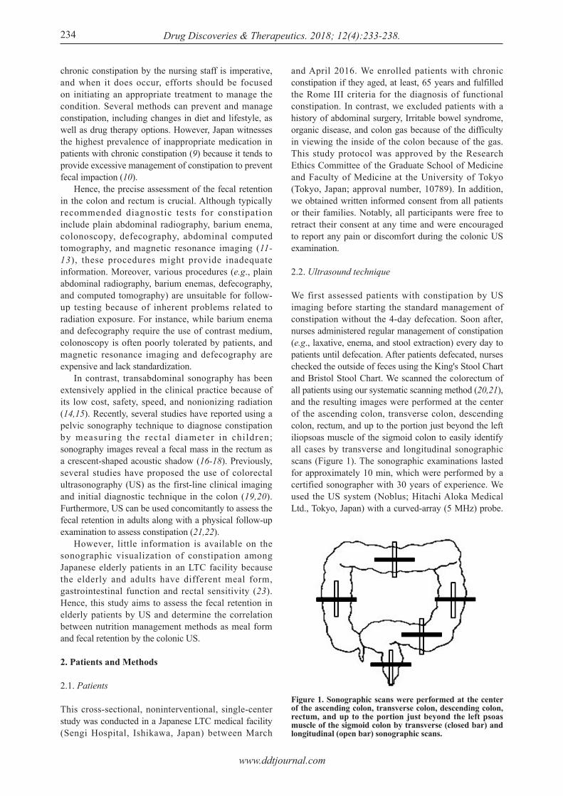
www.ddtjournal.com
Drug Discoveries & Therapeutics. 2018; 12(4):233-238.
chronic constipation by the nursing staff is imperative, and when it does occur, efforts should be focused on initiating an appropriate treatment to manage the condition. Several methods can prevent and manage constipation, including changes in diet and lifestyle, as well as drug therapy options. However, Japan witnesses the highest prevalence of inappropriate medication in patients with chronic constipation (9) because it tends to provide excessive management of constipation to prevent fecal impaction (10). Hence, the precise assessment of the fecal retention in the colon and rectum is crucial. Although typically recommended diagnostic tests for constipation include plain abdominal radiography, barium enema, colonoscopy, defecography, abdominal computed tomography, and magnetic resonance imaging (11-13), these procedures might provide inadequate information. Moreover, various procedures (e.g., plain abdominal radiography, barium enemas, defecography, and computed tomography) are unsuitable for follow-up testing because of inherent problems related to radiation exposure. For instance, while barium enema and defecography require the use of contrast medium, colonoscopy is often poorly tolerated by patients, and magnetic resonance imaging and defecography are expensive and lack standardization. In contrast, transabdominal sonography has been extensively applied in the clinical practice because of its low cost, safety, speed, and nonionizing radiation (14,15). Recently, several studies have reported using a pelvic sonography technique to diagnose constipation by measuring the rectal diameter in children; sonography images reveal a fecal mass in the rectum as a crescent-shaped acoustic shadow (16-18). Previously, several studies have proposed the use of colorectal ultrasonography (US) as the first-line clinical imaging and initial diagnostic technique in the colon (19,20). Furthermore, US can be used concomitantly to assess the fecal retention in adults along with a physical follow-up examination to assess constipation (21,22). However, little information is available on the sonographic visualization of constipation among Japanese elderly patients in an LTC facility because the elderly and adults have different meal form, gastrointestinal function and rectal sensitivity (23). Hence, this study aims to assess the fecal retention in elderly patients by US and determine the correlation between nutrition management methods as meal form and fecal retention by the colonic US.
2. Patients and Methods
2.1. Patients
This cross-sectional, noninterventional, single-center study was conducted in a Japanese LTC medical facility (Sengi Hospital, Ishikawa, Japan) between March
and April 2016. We enrolled patients with chronic constipation if they aged, at least, 65 years and fulfilled the Rome III criteria for the diagnosis of functional constipation. In contrast, we excluded patients with a history of abdominal surgery, Irritable bowel syndrome, organic disease, and colon gas because of the difficulty in viewing the inside of the colon because of the gas. This study protocol was approved by the Research Ethics Committee of the Graduate School of Medicine and Faculty of Medicine at the University of Tokyo (Tokyo, Japan; approval number, 10789). In addition, we obtained written informed consent from all patients or their families. Notably, all participants were free to retract their consent at any time and were encouraged to report any pain or discomfort during the colonic US examination.
2.2. Ultrasound technique
We first assessed patients with constipation by US imaging before starting the standard management of constipation without the 4-day defecation. Soon after, nurses administered regular management of constipation (e.g., laxative, enema, and stool extraction) every day to patients until defecation. After patients defecated, nurses checked the outside of feces using the King's Stool Chart and Bristol Stool Chart. We scanned the colorectum of all patients using our systematic scanning method (20,21), and the resulting images were performed at the center of the ascending colon, transverse colon, descending colon, rectum, and up to the portion just beyond the left iliopsoas muscle of the sigmoid colon to easily identify all cases by transverse and longitudinal sonographic scans (Figure 1). The sonographic examinations lasted for approximately 10 min, which were performed by a certified sonographer with 30 years of experience. We used the US system (Noblus; Hitachi Aloka Medical Ltd., Tokyo, Japan) with a curved-array (5 MHz) probe.
234
Figure 1. Sonographic scans were performed at the center of the ascending colon, transverse colon, descending colon, rectum, and up to the portion just beyond the left psoas muscle of the sigmoid colon by transverse (closed bar) and longitudinal (open bar) sonographic scans.

www.ddtjournal.com
Drug Discoveries & Therapeutics. 2018; 12(4):233-238. 235
did not defecate while nurses were providing defecation care, and 1 patient was excluded because of insufficient image quality. Thus, the final analysis comprised 32 patients (8 males and 24 females; mean age, 87.2 years; range, 74-106 years). All patients were receiving the management of constipation with laxative suppositories (bisacodyl), the daily life independence level in grade C. Table 2 presents the correlation between nutrition management methods and bowel movements. In all patients, the King's Stool Chart did not detect > 200 g of fecal matter, and the Bristol Stool Chart revealed type 5-7 (diarrhea) in 56.2% (18 of 32 patients). In addition, TPN and TF did not completely detect type 1-2 (constipation) in 0% (0 of 17 patients); TPN tended to delay the defecation. We observed a significant difference between the nutrition management method and the Bristol Stool Chart (p = 0.01). Table 3 summarizes US findings of the fecal retention (fecal retention/non-fecal retention). While the fecal retention groups comprised 15.6% (5 of 32) of patients, the non-fecal retention groups comprised 84.4% (27 of 32) of patients. In addition, nutrition management methods of the fecal retention groups, TPN, did not completely detect the fecal retention in 0% (0 of 15) of patients. For the non-fecal retention groups, the following three
For US imaging, we used the focal range of 4 cm and the image depth of 6-8 cm. Furthermore, we used echo gain and dynamic range to determine the appropriate range to display.
2.3. Data analysis
We defined the US images before management of constipation as the fecal retention (fecal retention/non-fecal retention). The fecal retention groups suggested high echogenicity by the brightness of the colon wall with posterior echoes (acoustic shadows) by a transverse scan and visualized haustrations by a longitudinal scan (22); these findings were detected in any of the five sites (i.e., ascending, transverse, descending, sigmoid colon, and rectum; Figure 2). All US images were classified visually as the fecal retention or non-fecal retention, and two independent certified sonographers reviewed the US images to ensure the inter-rater reliability. Furthermore, an expert sonographer assessed the data derived from the US images. Of note, all images were evaluated under blinded conditions. We assessed the correlation between the visual evaluation (fecal retention/non-fecal retention) using Cohen's kappa statistic to reach a consensus between the two certified sonographers. In this study, the correlations between the fecal retention and other variables, nutrition management method [total parenteral nutrition (TPN), gastric fistula tube, tube feeding (TF), oral foods ‒ also includes the use of TPN or gastric fistula tube in combination], amount of the King's Stool Chart and quality of the Bristol Stool Chart and bowel movement frequency were analyzed using the Fisher's exact test. We set the statistical significance level at < 0.05. Statistical analyses were conducted using IBM SPSS Statistics 21.0 for Microsoft Windows (IBM Corp., Armonk, NY).
3. Results
Table 1 summarizes patients' characteristics. Of 41 eligible patients, 8 patients were excluded because they
Table 1. Participants' characteristics (N = 32)
Items
Age (years)WomenThe level of independence Grade C (bed ridden)Main disease Cerebrovascular disease Fracture femoris Lung disease Diabetes Cancer Parkinson’s disease OthersDefecation care Suppository Laxative (Bisacodyl)
Mean ± SD, Min–Max or n (%)
87.7 ± 8.1, 74 – 10624 (75)
32 (100)
11 (34.4) 5 (15.6) 5 (15.6) 4 (12.5)2 (6.3)1 (3.1)
4 (12.5)
32 (100)
Figure 2. The presence of the fecal retention. US images of an 88-year-old female who intakes oral food. (a) A transverse scan showing a high echogenicity by the brightness of the colon wall (arrowheads) and acoustic shadow behind the descending colon (asterisks). (b) A longitudinal scan showing a clear crescent-shaped high echogenicity by the brightness of the colon wall with haustrations (arrows).

www.ddtjournal.com
Drug Discoveries & Therapeutics. 2018; 12(4):233-238.236
results were obtained: the King's Stool Chart indicated under < 100 g in 81.8% (18 of 22) patients; the Bristol Stool Chart indicated type 5-7 (diarrhea) in 100% patients; and bowel movement frequency was 5-8 days in 100% of patients. We observed a significant difference between the nutrition management method and the Bristol Stool Chart (p = 0.01). A significant difference was also observed in nutrition management methods between the fecal retention and non-fecal retention groups (p = 0.041). In addition, we observed a significant difference in the Bristol Stool Chart between the fecal retention and non-fecal retention groups (p = 0.002). Based on the Kendall W test, the results obtained by two independent sonographers (A and B) at 0.83 and 0.84, respectively, exhibited a significant correlation with each other.
4. Discussion
This s tudy invest igated elderly pat ients with constipation by colonic US imaging in LTC facilities. Consequently, US images categorized constipation as fecal retention and non-fecal retention. In the fecal retention groups, the fecal retention was < 15.6% (5 of 32 patients) before management of constipation. Several factors are accountable for the fecal retention in the colon in elderly patients with constipation. Perhaps, the low detection rate in this study could be attributed to the nutrition management methods. Reportedly, parenteral nutrition of this condition is widely used in hospitalized patients, especially elderly, who are unable to eat to aid in patients' ability to recover from illness (24). The most common adverse effect of such treatment is diarrhea, which is reported in 68% of intensive care unit patients (25) and 96% of patients with dysphagia (26). In this study, we compared nutrition management methods with US finding in elderly patients with constipation. All TPN were not completely detected in the fecal retention groups of US finding; even the oral foods groups did not detect fecal retention in 66.4% of patients by US. Perhaps, the dietary intake of elderly patients was not sufficient for the fecal retention for 4 days. Another probable reason for the low detection rate in this study could be that nurses only checked the outside of feces using the King's Stool Chart and Bristol Stool Chart. While the King's Stool Chart tends to detect < 200 g of defecated feces, the Bristol Stool Chart tends to report diarrhea in the non-fecal retention groups. This study reports a markedly high prevalence and use of medications to manage elderly constipation in LTC settings (27). The most frequently used solution to prevent or treat constipation was laxative medications. Despite considering laxatives as the solution, it was impossible to manage the balance between constipation and diarrhea because diarrhea is the common side
Tabl
e 2.
The
cor
rela
tion
betw
een
nutr
ition
man
agem
ent m
etho
ds a
nd b
owel
mov
emen
ts (N
= 3
2)
Item
s
Tota
l par
ente
ral n
utrit
ion
(n =
15)
Gas
tric
fistu
la tu
be (n
= 6
)Tu
be F
eedi
ng (
n =
2)O
ral f
oods
(n =
9)
< 10
0 g
n =
22
13 (8
6.7)
4 (6
6.7)
05
(55.
6)
100
– <
200
gn
= 10
2 (1
3.3)
2 (3
3.3)
2 (1
00.0
)4
(44.
4)
Dat
a w
ere
show
n as
n (%
). 1)Fi
sher
's ex
act t
est,
* P <
0.0
5.
> 20
0 g
n =
0
0 0 0 0
P1)
0.11
1
Type
1–2
(C
onst
ipat
ion)
n =
5
01
(16.
7)0
4 (4
4.5)
Type
3–4
(Eas
ier t
o pa
ss)
n =
9
3 (2
0.0)
3 (5
0.0)
1 (5
0.0)
2 (2
2.2)
Type
5–7
(Dia
rrhe
a)n
= 18
12 (8
0.0)
2 (3
3.3)
1 (5
0.0)
3 (3
3.3)
P1)
0.01
*
4
4 (2
6.7)
5 (8
3.3)
2 (1
00.0
)7
(77.
8)
5 0 0 02
(22.
2)
6
1 (6
.7)
1 (1
6.7)
0 0
P1)
0.06
9
7
7 (4
6.7)
0 0 0
8
3 (2
0.0)
0 0 0
K
ing's
Sto
ol C
hart
Bris
tol S
tool
Cha
rt
Bow
el m
ovem
ent f
requ
ency
(day
)
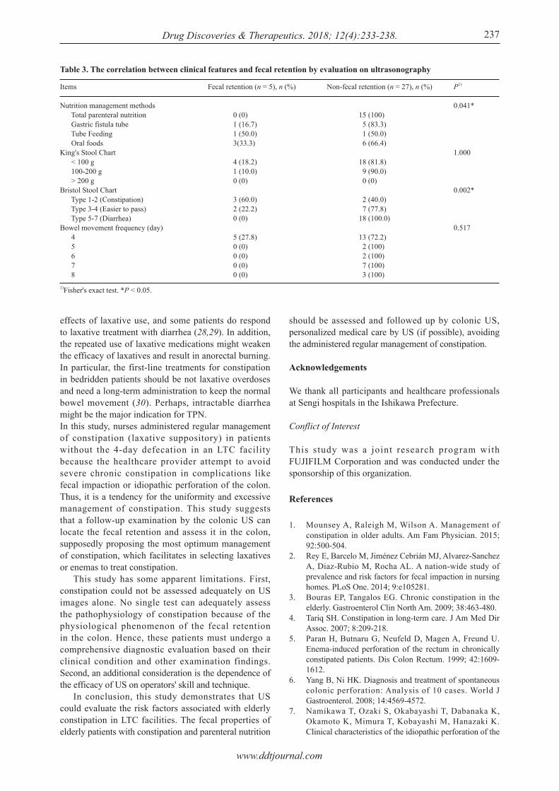
www.ddtjournal.com
Drug Discoveries & Therapeutics. 2018; 12(4):233-238. 237
effects of laxative use, and some patients do respond to laxative treatment with diarrhea (28,29). In addition, the repeated use of laxative medications might weaken the efficacy of laxatives and result in anorectal burning. In particular, the first-line treatments for constipation in bedridden patients should be not laxative overdoses and need a long-term administration to keep the normal bowel movement (30). Perhaps, intractable diarrhea might be the major indication for TPN.In this study, nurses administered regular management of constipation (laxative suppository) in patients without the 4-day defecation in an LTC facility because the healthcare provider attempt to avoid severe chronic constipation in complications like fecal impaction or idiopathic perforation of the colon. Thus, it is a tendency for the uniformity and excessive management of constipation. This study suggests that a follow-up examination by the colonic US can locate the fecal retention and assess it in the colon, supposedly proposing the most optimum management of constipation, which facilitates in selecting laxatives or enemas to treat constipation. This study has some apparent limitations. First, constipation could not be assessed adequately on US images alone. No single test can adequately assess the pathophysiology of constipation because of the physiological phenomenon of the fecal retention in the colon. Hence, these patients must undergo a comprehensive diagnostic evaluation based on their clinical condition and other examination findings. Second, an additional consideration is the dependence of the efficacy of US on operators' skill and technique. In conclusion, this study demonstrates that US could evaluate the risk factors associated with elderly constipation in LTC facilities. The fecal properties of elderly patients with constipation and parenteral nutrition
should be assessed and followed up by colonic US, personalized medical care by US (if possible), avoiding the administered regular management of constipation.
Acknowledgements
We thank all participants and healthcare professionals at Sengi hospitals in the Ishikawa Prefecture.
Conflict of Interest
This study was a joint research program with FUJIFILM Corporation and was conducted under the sponsorship of this organization.
References
1. Mounsey A, Raleigh M, Wilson A. Management of constipation in older adults. Am Fam Physician. 2015; 92:500-504.
2. Rey E, Barcelo M, Jiménez Cebrián MJ, Alvarez-Sanchez A, Diaz-Rubio M, Rocha AL. A nation-wide study of prevalence and risk factors for fecal impaction in nursing homes. PLoS One. 2014; 9:e105281.
3. Bouras EP, Tangalos EG. Chronic constipation in the elderly. Gastroenterol Clin North Am. 2009; 38:463-480.
4. Tariq SH. Constipation in long-term care. J Am Med Dir Assoc. 2007; 8:209-218.
5. Paran H, Butnaru G, Neufeld D, Magen A, Freund U. Enema-induced perforation of the rectum in chronically constipated patients. Dis Colon Rectum. 1999; 42:1609-1612.
6. Yang B, Ni HK. Diagnosis and treatment of spontaneous colonic perforation: Analysis of 10 cases. World J Gastroenterol. 2008; 14:4569-4572.
7. Namikawa T, Ozaki S, Okabayashi T, Dabanaka K, Okamoto K, Mimura T, Kobayashi M, Hanazaki K. Clinical characteristics of the idiopathic perforation of the
Table 3. The correlation between clinical features and fecal retention by evaluation on ultrasonography
Items
Nutrition management methods Total parenteral nutrition Gastric fistula tube Tube Feeding Oral foods King's Stool Chart < 100 g 100-200 g > 200 gBristol Stool Chart Type 1-2 (Constipation) Type 3-4 (Easier to pass) Type 5-7 (Diarrhea) Bowel movement frequency (day) 4 5 6 7 8
Fecal retention (n = 5), n (%)
0 (0)1 (16.7)1 (50.0)3(33.3)
4 (18.2)1 (10.0)0 (0)
3 (60.0)2 (22.2)0 (0)
5 (27.8)0 (0)0 (0)0 (0)0 (0)
Non-fecal retention (n = 27), n (%)
15 (100) 5 (83.3) 1 (50.0) 6 (66.4)
18 (81.8) 9 (90.0) 0 (0)
2 (40.0) 7 (77.8)18 (100.0)
13 (72.2) 2 (100) 2 (100) 7 (100) 3 (100)
1)Fisher's exact test. *P < 0.05.
P1)
0.041*
1.000
0.002*
0.517
www.ddtjournal.com
Drug Discoveries & Therapeutics. 2018; 12(4):233-238.238
colon. J Clin Gastroenterol. 2011; 45:82-86. 8. Seltzer R. Evaluation and diagnosis of constipation.
Gastroenterol Nurs. 2012; 35:343-348.9. Niwata S, Yamada Y, Ikegami N. Prevalence of
inappropriate medication using Beers criteria in Japanese long-term care facilities. BMC Geriatr. 2006; 6:1-7.
10. De Hert M, Dockx L, Bernagie C, Peuskens B, Sweers K, Leucht S, Tack J, Van de Straete S, Wampers M, Peuskens J. Prevalence and severity of antipsychotic related constipation in patients with schizophrenia: A retrospective descriptive study. BMC Gastroenterol. 2011; 11:11-17.
11. Seltzer R. Evaluation and diagnosis of constipation. Gastroenterol Nurs. 2012; 35:343-348.
12. Rao SS, Ozturk R, Laine L. Clinical utility of diagnostic tests for constipation in adults: A systematic review. Am J Gastroenterol. 2005; 100:1605-1615.
13. Remes-Troche JM, Rao SS. Diagnostic testing in patients with chronic constipation. Curr Gastroenterol. 2006; 8:416-424.
14. Berger MY, Tabbers MM, Kurver MJ, Boluyt N, Benninga MA. Value of abdominal radiography, colonic transit time, and rectal ultrasound scanning in the diagnosis of idiopathic constipation in children: A systematic review. J Pediatr. 2012; 161:44-50.
15. Perniola G, Shek C, Chong CC, Chew S, Cartmill J, Dietz HP. Defecation proctography and translabial ultrasound in the investigation of defecatory disorders. Ultrasound. Obstet Gynecol. 2008; 31:567-571.
16. Joensson IM, Siggaard C, Rittig S, Hagstroem S, Djurhuus JC. Transabdominal ultrasound of rectum as a diagnostic tool in childhood constipation. J Urol. 2008; 179:1997-2002.
17. Di Pace MR, Catalano P, Caruso AM, Bommarito D, Casuccio A, Cimador M, De Grazia E. Is rectal disimpact always necessary in children with chronic constipation? Evaluation with pelvic ultrasound. Pediatr Surg Int. 2010; 26:601-606.
18. Karaman A, Ramadan SU, Karaman I, Gökharman D, Erdoğan D, Kacar M, Cavuşoğlu YH, Koşar U. Diagnosis and follow-up in constipated children: Should we use ultrasound? J Pediatr Surg. 2010; 45:1849-1855.
19. Yabunaka K , Fuku i H , Tama te S , Fu j i oka M. Ultrasonographic diagnosis of advanced colorectal cancer. J Med Ultrasonics. 2003; 30:163-169.
20. Yabunaka K, Sanada S, Fukui H, Tamate S, Fujioka M.
Transabdominal sonographic appearance of adult colonic polyps. J Med Ultrasonics. 2006; 33:231-237.
21. Yabunaka K, Matsuo J, Takii M, Hara A, Nakagami G, Gotanda T, Nishimura G, Sanada H. Sonographic visualization of fecal loading in adults: Comparison with computed tomography. J Diagn Med Sonog. 2015; 31:86-92.
22. Yabunaka K, Nakagami G, Komagata K, Sanada H. Ultrasonographic follow-up of functional chronic constipation in adults: A report of two cases. SAGE Open Medical Case Reports. 2017; 5:1-4.
23. Lagier E, Delvaux M, Vellas B, Fioramonti J, Bueno L, Albarede JL, Frexinos J. Influence of age on rectal tone and sensitivity to distension in healthy subjects. Neurogastroenterol Motil. 1999; 11:101-107.
24. Shimoni Z, Averbuch Y, Shir E, Gottshalk T, Kfir D, Niven M, Moshkowitz M, Froom P. The addition of fiber and the use of continuous infusion decrease the incidence of diarrhea in elderly tube-fed patients in medical wards of a general regional hospital: A controlled clinical trial. J Clin Gastroenterol. 2007; 41:901-905.
25. Kelly TW, Patrick MR, Hillman KM. Study of diarrhea in critically ill patients. Crit Care Med. 1983; 11:7-9.
26. Ciocon JO, Galindo-Ciocon DJ, Tiessen C, Galindo D. Continuous compared with intermittent tube feeding in the elderly. JPEN J Parenter Enteral Nutr. 1992; 16:525-528.
27. Kimberly BS. Constipation in the elderly: Implication in skilled nursing facilities. Director. 2007; 15:20-23.
28. Koopmans-Klein G, Wagemans MF, Wartenberg HC, Van Megen YJ, Huygen FJ. The efficacy of standard laxative use for the prevention and treatment of opioid induced constipation during oxycodone use: A small Dutch observational pilot study. Expert Rev Gastroenterol Hepatol. 2016; 10:547-553.
29. Munch L, Tvistholm N, Trosborg I, Konradsen H. Living with constipation − older people's experiences and strategies with constipation before and during hospitalization. Int J Qual Stud Health Well-being. 2016; 11:1-8.
30. Zhang LY, Yao YZ, Wang T, Fei J, Shen Y, Chen YH, Zong ZW. Efficacy of polyethylene glycol 4000 on constipation of posttraumatic bedridden patients. Chin J Traumatol. 2010; 13:182-187.
(Received June 12, 2018; Revised August 14, 2018; Accepted August 18, 2018)

www.ddtjournal.com
Drug Discoveries & Therapeutics. 2018; 12(4):239-247. 239
Whole body vibration exercise in the management of cancer therapy-related morbidities: A systematic review
Patrícia Lopes-Souza1,2,*, Carla Fontoura Dionello1,2, Danúbia da Cunha Sá-Caputo2,3, Eloá Moreira-Marconi2,4, Eric Heleno Freire Ferreira Frederico2,5, Renata Marques Marchon2, Anke Bergmann6, Trentham Furness7, Mario Bernardo-Filho2
1 Programa de Pós-graduação em Ciências Médicas, Faculdade de Ciências Médicas, Universidade do Estado do Rio de Janeiro, Rio de Janeiro, Brazil;
2 Departamento de Biofísica e Biometria, Laboratório de Vibrações Mecânicas e Práticas Integrativas, Instituto de Biologia Roberto Alcantara Gomes, Universidade do Estado do Rio de Janeiro, Rio de Janeiro, Brazil;
3 Curso de Fisioterapia, Faculdade Bezerra de Araújo, Rio de Janeiro, RJ, Brazil;4 Programa de Pós-graduação em Fisiopatologia Clínica e Experimental, Universidade do Estado do Rio de Janeiro, Rio de
Janeiro, Brazil;5 Programa de Pós-Graduação em Biociências, Instituto de Biologia Roberto Alcantara Gomes, Universidade do Estado do Rio
de Janeiro, Rio de Janeiro, Brazil;6 Programa de Carcinogênese Molecular, Instituto Nacional de Câncer (INCA), Rio de Janeiro, Brazil;7 Mental Health Nursing Research Unit, NorthWestern Mental Health & Australian Catholic University, Melbourne, Australia.
1. Introduction
Cancer is a group of diseases that causes the growth of abnormal cells, which exceed their usual limits and can
invade adjacent parts of the body and/or spread to other organs (1). Although it actually represents one of the main causes of morbidity and mortality worldwide, with approximately 14 million new cases in 2012, the amount of cancer survivors is progressively increasing due to advances in early detection and treatment (2,3). Cancer development and its treatment are usually associated with fatigue (4) pain (5), anxiety or depression (6), and sleep disturbances (7) which determine a decrease of physical and psychological functions and negatively affect the patients' quality of life (QoL) (8). One way to promote health, to diminish the
Summary The purpose of this systematic review was to investigate the effects of whole body vibration (WBV) exercise in the management of cancer therapy-related morbidities. The PubMED and PEDro databases were used to access publications published in English about the use of whole body vibration (WBV) exercises in cancer patients until February 22nd 2017. The studies included were classified according to the level of evidence (LE) by the National Health and Medical Research Council Hierarchy of evidence and the methodological quality (MQ) by the PEDro scale. The four included studies (2 of them with "high" LE-II and MQ) were performed in patients with different types of cancer (i.e. breast, lung, prostate, solid or hematological), treated with WBV exercise to counteract the cancer therapy-related morbidities. The variables evaluated were muscle activity, subjective rate of perceived exertion, exercise capacity, muscle strength, quality of life, resting urinary incontinence and severity of peripheral neuropathy. Although WBV exercise appears to be a potential treatment procedure of cancer therapy-related morbidities, further additional studies are required to determine specific and tailored protocols to be used in the different stages of the disease.
Keywords: Whole body vibration, cancer, oncology, rehabilitation, exercise
DOI: 10.5582/ddt.2018.01039Original Article
*Address correspondence to:Dr. Patrícia Lopes de Souza, Departamento de Biofísica e Biometria, Laboratório de Vibrações Mecânicas e Práticas Integrativas, Instituto de Biologia Roberto Alcantara Gomes, Universidade do Estado do Rio de Janeiro. Av. 28 de Setembro, 87 Fundos - 4° andar, Vila Isabel- Rio de Janeiro, RJ. 20551030. Brazil.E-mail: [email protected]
www.ddtjournal.com
Drug Discoveries & Therapeutics. 2018; 12(4):239-247.
physiologic and psychological effects of cancer and its treatment, to achieve the maximum possible physical, social and vocational well-being is to acquire or maintain a healthy lifestyle, entailing physical activity as a component of cancer rehabilitation (9-11). Physical activity is any movement of the body that involves the muscles action with energy expenditure above rest and the exercise is this physical activity with planning (12). According to the American Cancer Society, cancer survivors should exercise at least 150 minutes per week, including strength training exercises at least 2 days per week (2). The exercise can be beneficial to counteract cancer-related fatigue (10,13-15), improve overall quality of life, as well as relief of symptoms and side effects (10) of the disease and/or of the treatment. In this respect, recent studies have shown the positive effect of physical activity on breast (16,17), prostate (18), ovarian (19) and lung cancer (20). Among the different exercise modalities, whole body vibration (WBV) exercise (21) has gained progressive popularity, being safe and well accepted by the patients. WBV exercises are generated by mechanical vibrations produced in oscillating/vibratory platforms (OVP) and can be transmitted to the body of the individual when is in contact with the OVP (22-24). Several authors have already demonstrated the effects of WBV exercise with different protocols in improving muscle strength (25,26), bone formation (25,27), balance (28), flexibility (29,30), function ability (26,27), relief of pain (31) and fatigue (32). Moreover, investigations have shown the WBV exercise benefits in the rehabilitation programs of patients with chronic diseases, such as metabolic syndrome (24), chronic obstructive pulmonary disease (33-35), fibromyalgia syndrome (36), multiple sclerosis (32,37), rheumatoid arthritis (27) and cancer therapy-related morbidities (39). Due to the importance of the exercise in several clinical conditions, the aim of this systematic review was to investigate the effects of WBV exercise in the management of morbidities related to the cancer therapy. This is the first systematic review to assess whether WBV exercises are safe and beneficial in the management of morbidities for cancer patients.
2. Materials and Methods
2.1. Search strategy used to find the publications
Two databases were accessed in the Universidade do Estado do Rio de Janeiro on February 22nd 2017 and two searches were performed. The keywords "whole body vibration" and "cancer", and "whole body vibration" and "oncology" were searched in the PubMed and PEDro databases for three reviewers independently. This systematic review adheres to the Preferred Reporting Items for Systematic Reviews and
Meta-Analysis (PRISMA) Statement (39).
2.2. Criteria to select the publications
All the publications were screened following inclusion and exclusion criteria. Inclusion criteria: In the search, all the publications found in the databases (PubMed and PEDro) were preliminarily considered to be included in this current review. To be included in this review, all studies had to investigate effects of WBV on cancer patients. A flowchart (Figure 1), based in the PRISMA analysis, was done to show the steps in the selection of the full papers analyzed in this review (39). Exclusion criteria: Exclusion criteria allowed the elimination of unnecessary publications. Papers were excluded if they were: (i) published in a language other than English; and (ii) findings not related to cancer.
2.3. Levels of evidence (LE) of the selected papers
The National Health and Medical Research Council Hierarchy of evidence (NHMRC, 2003-2007) (40) were used to classify the included studies in this systematic review (Figure 2). Each article was assigned to one reviewer and cross-checked by a second reviewer and where there was disagreement a third party was consulted and the issue discussed until consensus was reached. Moreover, the methodological quality of these studies was determined by the PEDRo scale (41). In the PEDRo scale, each publication was evaluated according to: (a) eligibility criteria, (b) subjects were randomly allocated to groups, (c) concealed allocation, (d) the groups with baseline similarity, (e) blinding of the patients, (f) blinding of the therapists, (g) blinding of all assessors, (h) measures obtained from more than 85% of the subjects, (i) all subjects received the treatment or control condition or, at least one key outcome was analyzed by "intention to treat", (j) results of the groups with statistical comparisons and (h) point measures and measures of variability of outcome. Those publications with a score of seven or greater in the PEDro scale were considered of 'high' methodological quality, those with a score of five to six would be of 'fair' quality and a score of four or below were classified as 'poor' quality (42).
3. Results
The steps to select the full papers analyzed in this systematic review are shown in the flowchart (Figure 1). Of the twelve papers firstly screened, only four have reached the inclusion criteria. The characteristics of the participants, the protocols used, the aims of the studies, the tools for evaluation and the outcomes of the selected articles and the level of evidence of the selected papers and the methodological quality are shown in the Table 1.
240

www.ddtjournal.com
Drug Discoveries & Therapeutics. 2018; 12(4):239-247. 241
Figure 1. Flowchart indicating the steps to select the full papers analyzed in this review. A flowchart based in the PRISMA analysis, was done to show the steps in the selection of the full papers analyzed in this review.
Figure 2. Designation of levels of evidence according to the intervention research question. The National Health and Medical Research Council Hierarchy of evidence (NHMRC, 2003-2007) were used to classify the included studies in this systematic review.

www.ddtjournal.com
Drug Discoveries & Therapeutics. 2018; 12(4):239-247.242
Tabl
e 1.
Cha
ract
eris
tics
of t
he p
artic
ipan
ts, p
roto
cols
use
d, a
ims
of t
he s
tudi
es, t
ools
for
eva
luat
ion,
out
com
es o
f th
e se
lect
ed a
rtic
les,
leve
l of
evid
ence
of
the
sele
cted
pap
ers
and
met
hodo
logi
cal q
ualit
y
Stud
y
Van
Ruy
mbe
keet
al.,
201
4
Salh
i et
al.,
201
5
Type
of c
ance
r/Tr
eatm
ent p
erfo
rmed
/nu
mbe
r of i
ndiv
idua
ls
Bre
ast c
ance
r su
rviv
ors/
al
l typ
e of
trea
tmen
t.Br
east
canc
er su
rviv
ors (
n =
20) a
nd C
ontro
l gro
up-
heal
thy
wom
an (n
= 2
0)
Stag
es I
-III
lung
can
cer
or m
esot
helio
ma/
radi
cal
rese
ctio
n w
ith o
r with
out
a ch
emot
hera
py o
r ra
diot
hera
py
CO
N g
roup
(n
= 21
), C
RT
grou
p (
n =
20),
WBV
gro
up (n
= 1
7)
Mor
bidi
ties o
f th
e tre
atm
ent
No
co
mo
rbid
ity
was
re
porte
d
Post
-tre
atm
ent
QF
was
ei
ther
equ
al o
r le
ss t
han
70%
of t
he p
redi
cted
nor
mal
va
lue
or s
how
ed a
dec
reas
e of
at
leas
t 10
% f
rom
the
ba
selin
e va
lue
Elec
trom
yogr
aphy
= E
MG
; Rec
tus F
emor
is =
RF; V
astu
s med
ial =
VM
, Bic
eps f
emor
is BF
, Tib
ialis
ant
erio
r = T
A; G
astro
cnem
ius
= G
T; V
isual
ana
logu
e sc
ale
= VA
S; C
ontro
l = C
ON
; Con
vent
iona
l res
istan
ce tr
aini
ng =
CRT
; W
hole
Bod
y Vi
brat
ion
= W
BV;
Qua
dric
eps
forc
e =
QF;
One
-repe
titio
n-m
axim
um =
1R
M; M
axim
al w
orkl
oad
(Wm
ax);
6-m
in w
alki
ng d
istan
ce =
6M
WD
; Qua
lity
of li
fe =
QoL
; Eur
opea
n O
rgan
isatio
n fo
r Res
earc
h an
d Tr
eatm
ent o
f Can
cer Q
ualit
y of
Life
Can
cer Q
uesti
onna
ire =
EO
RTC
QLQ
-C30
; Che
mot
hera
py-in
duce
d po
lyne
urop
athy
= C
IPN
; Fun
ctio
nal A
sses
smen
t of C
ance
r The
rapy
/ Gyn
ecol
ogic
Onc
olog
y G
roup
neu
roto
xici
ty su
bsca
le
= FA
CT/G
OG
-NTX
; War
m d
etec
tion
thre
shol
d =
WD
T.
WBV
Inte
rven
tion
Two
grou
ps w
ere
unde
rgoi
ng
vibr
atio
n, t
he w
omen
wer
e st
andi
ng o
n a
sync
hron
ous
vibr
atin
g pl
atfo
rm w
ith a
kne
e jo
int a
ngle
of 5
5°.
Each
con
ditio
n:
nonv
ibra
tion
con
diti
on (
0Hz)
an
d vi
brat
ion
cond
ition
at 2
0-30
-40
-50H
z vi
brat
ion
freq
uenc
ies
(am
plit
ude,
4m
m)
last
ing
for
30 s
wit
h 2-
min
res
t be
twee
n co
nditi
ons.
CO
N g
roup
- p
atie
nts
wer
e di
scou
rage
d to
im
prov
e th
eir
exer
cise
tole
ranc
e. C
RT g
roup
-
20 m
in o
f ae
robi
c tra
inin
g on
the
bicy
cle
and
tread
mill
at 7
0% o
f the
re
spec
tive
Wm
ax a
nd s
peed
plu
s re
sist
ance
trai
ning
on
mul
tigym
eq
uipm
ent
star
ting
wit
h th
ree
sets
of
eigh
t rep
etiti
ons
for
each
ex
erci
se a
t 50%
1RM
. WBV
gro
up
- 20
min
of a
erob
ic tr
aini
ng o
n th
e bi
cycl
e an
d tre
adm
ill a
t 70%
of
the
resp
ectiv
e W
max
and
spe
ed
plus
per
form
ed e
xerc
ises
on
the
sync
hron
ous
vibr
atio
n pl
atfo
rm,
thre
e se
ts of
30
s for
eac
h ex
erci
se
at 2
7 H
z. P
atie
nts
trai
ned
thre
e tim
es a
wee
k fo
r 12
wee
ks.
Aim
To a
naly
ze m
uscl
e a
cti
vit
y
an
d su
bjec
tivel
y ra
te o
f pe
rcei
ved
exer
tion
To a
sses
s the
pot
entia
l be
nefi
cial
eff
ect
of
reha
bilit
atio
n
T
ools
for e
valu
atio
n
Surfa
ce E
MG
ana
lysis
of
the
RF, V
M, V
L, T
A an
d G
T m
uscl
es. T
he le
vel
of su
bjec
tivel
y pe
rcei
ved
exer
tion
was
rat
ing
on
a co
mbi
natio
n of
a V
AS
and
revi
sed
Borg
.
Exer
cise
cap
acity
with
6M
WD
and
Wm
ax,
mus
cle
stre
ngth
by
the
QF
with
iso
met
ric
hand
held
dyn
amom
eter
, an
d Q
oL w
ith E
ORT
C Q
LQ-C
30
Out
com
e (s
)
The
mus
cle
activ
atio
n di
d no
t diff
er
betw
een
brea
st c
ance
r su
rviv
ors
and
heal
thy
cont
rols
. T
here
was
a
sign
ific
ant
freq
uenc
y ×
mus
cle
inte
ract
ion
effe
ct.
For
the
VA
S sc
ores
no
sign
ific
ant
grou
p ×
freq
uenc
y in
tera
ctio
n w
as f
ound
and
no
sig
nifi
cant
mai
n ef
fect
for
the
fa
ctor
gro
up, i
n co
ntra
st t
he f
acto
r fre
quen
cy w
as s
igni
fican
t. Th
e va
lues
of
per
ceiv
ed e
xerti
on in
bot
h gr
oups
in
crea
sed
with
incr
easin
g fre
quen
cy.
6MW
D n
ot i
ncre
ased
in
CO
N a
nd
in W
BV
gro
up,
and
incr
ease
d in
C
RT g
roup
. Wm
ax w
as s
igni
fican
tly
incr
ease
d in
bot
h C
RT
and
WB
V
grou
p. Q
F sig
nific
antly
incr
ease
d on
ly
in C
RT g
roup
. For
non
e of
the
grou
ps,
the
scor
e of
EO
RTC
QLQ
-C30
Glo
bal
chan
ged
signi
fican
tly.
Leve
l of e
vide
nce/
m
etho
dolo
gica
l qu
ality
III-
1/ F
air
II/ H
igh
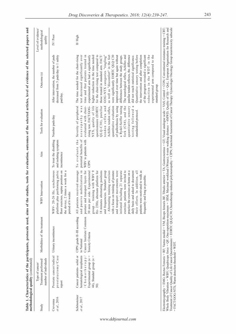
www.ddtjournal.com
Drug Discoveries & Therapeutics. 2018; 12(4):239-247. 243
Tabl
e 1.
Cha
ract
eris
tics
of t
he p
artic
ipan
ts, p
roto
cols
use
d, a
ims
of t
he s
tudi
es, t
ools
for
eva
luat
ion,
out
com
es o
f th
e se
lect
ed a
rtic
les,
leve
l of
evid
ence
of
the
sele
cted
pap
ers
and
met
hodo
logi
cal q
ualit
y (c
ontin
uted
)
Stud
y
Cre
venn
a et
al.,
201
6
Schö
nste
iner
et
al.,
201
7
Type
of c
ance
r/Tr
eatm
ent p
erfo
rmed
/nu
mbe
r of i
ndiv
idua
ls
Pros
tate
can
cer/
radi
cal
pros
tate
ctom
y/C
ase
repo
rt
Can
cer
patie
nts-
sol
id o
r he
mat
olog
ical
neo
plas
ms
/Ch
em
oth
era
py
/ Ex
perim
enta
l gro
up (
n =
44),
Stan
dart
grou
p (n
=
50)
Mor
bidi
ties o
f th
e tre
atm
ent
Urin
ary
inco
ntin
ence
CIPN
gra
de II
–III
acco
rdin
g to
Nat
iona
lC
ance
r In
stitu
te C
omm
on
Toxi
city
Crit
eria
Elec
trom
yogr
aphy
= E
MG
; Rec
tus F
emor
is =
RF; V
astu
s med
ial =
VM
, Bic
eps f
emor
is BF
, Tib
ialis
ant
erio
r = T
A; G
astro
cnem
ius
= G
T; V
isual
ana
logu
e sc
ale
= VA
S; C
ontro
l = C
ON
; Con
vent
iona
l res
istan
ce tr
aini
ng =
CRT
; W
hole
Bod
y Vi
brat
ion
= W
BV;
Qua
dric
eps
forc
e =
QF;
One
-repe
titio
n-m
axim
um =
1R
M; M
axim
al w
orkl
oad
(Wm
ax);
6-m
in w
alki
ng d
istan
ce =
6M
WD
; Qua
lity
of li
fe =
QoL
; Eur
opea
n O
rgan
isatio
n fo
r Res
earc
h an
d Tr
eatm
ent o
f Can
cer Q
ualit
y of
Life
Can
cer Q
uesti
onna
ire =
EO
RTC
QLQ
-C30
; Che
mot
hera
py-in
duce
d po
lyne
urop
athy
= C
IPN
; Fun
ctio
nal A
sses
smen
t of C
ance
r The
rapy
/ Gyn
ecol
ogic
Onc
olog
y G
roup
neu
roto
xici
ty su
bsca
le
= FA
CT/G
OG
-NTX
; War
m d
etec
tion
thre
shol
d =
WD
T.
WBV
Inte
rven
tion
WB
V-
20-2
6 H
z, s
ynch
rono
us
plat
form
plu
s pe
rfor
min
g pe
lvic
flo
or e
xerc
ises,
supi
ne p
ositi
on o
n th
e de
vice
, 2 ti
mes
a w
eek
for
a pe
riod
of 6
wee
ks
All
pat
ient
s re
ceiv
ed m
assa
ge
and
pass
ive
mob
iliz
atio
n in
po
stur
e an
d tr
ansp
ort
laye
rs f
or
30 m
in p
er s
ide.
Exp
erim
enta
l gr
oup
- t
rain
ing
with
WB
V 9
H
z- 2
3Hz,
alte
rnat
e pl
atfo
rm f
or
18 m
inut
es a
ltern
atin
g po
sitio
ns
and
freq
uenc
ies.
Sta
ndar
t gro
up
- A
ltern
atin
g tr
aini
ng e
xerc
ises
w
ith a
focu
s on
train
ing
of p
ostu
re
and
tran
spor
t m
ovem
ents
wer
e in
itiat
ed i
nclu
ding
21
sepa
rate
ex
erci
ses.
patie
nts
wer
e in
vite
d to
pr
actic
e th
e ex
erci
ses a
t hom
e on
a
daily
bas
is an
d as
ked
to d
ocum
ent
thei
r ef
fort
s. I
n ad
diti
on,
all
patie
nts w
ere
mot
ivat
ed to
wal
k as
fre
quen
tly a
nd lo
ng a
s pos
sible
.
Aim
To tr
eat t
he d
isab
ling
and
isola
ting
sym
ptom
in
cont
inen
ce
To
ev
alu
ate
the
pote
ntia
l be
nefi
ts o
f W
BV to
pat
ient
s w
ith
CIPN
T
ools
for e
valu
atio
n
Num
ber p
ads/d
ay
Seve
rity
of
peri
pher
al
ne
uro
pa
thy
wa
s ev
alua
ted
wit
h ch
air-
risin
g te
st, F
AC
T/G
OG
-N
TX
, qu
alit
y of
lif
e qu
estio
nnai
re (
EORT
C Q
LQ
-C30
), p
atel
lar
ten
do
n r
efle
x a
nd
Ach
illes
tend
on r
efle
x,
quan
titat
ive
eval
uatio
n of
pal
lest
hesi
a by
usi
ng
a Ry
del-
Seiff
er t
unin
g fo
rk (
C64
), a
nd t
he
quan
tita
tive
sen
sory
te
stin
g fo
llow
ed a
sta
ndar
dize
d pr
otoc
ol.
Out
com
e (s
)
Afte
r int
erve
ntio
n, th
e nu
mbe
r of p
ads
decr
ease
d fro
m 5
pad
s/day
to 1
saf
ety
pad/
day.
The
time
need
ed f
or th
e ch
air-
risin
g te
st d
ecre
ased
sig
nifi
cant
ly o
ver
tim
e an
d th
at p
atie
nts
trea
ted
in
expe
rimen
tal a
rm h
ad a
sig
nific
antly
hi
gher
red
uctio
n in
the
time
need
ed
for
the
chai
r-ris
ing
test
com
pare
d to
th
ose
treat
ed in
sta
ndar
d ar
m. F
AC
T/G
OG
-NT
X c
ateg
orie
s "t
ingl
ing"
as
wel
l as
"di
scom
fort
" in
the
fee
t w
ere
sign
ifica
ntly
EO
RTC
QLQ
C30
im
prov
ed o
ver t
ime
but a
gain
with
out
diffe
renc
es b
etw
een
the
study
gro
ups.
The
neur
olog
ical
ref
lex,
Ach
illes
and
pa
tella
r ten
don
refle
xes,
the.
diff
eren
ce
did
not r
each
sta
tistic
al s
igni
fican
ce.
Qua
ntita
tive
sens
ory
test
ing
befo
re
the
inte
rven
tion
and
afte
r co
mpl
etio
n of
the
prog
ram
rev
eale
d a
sign
ifica
nt
redu
ctio
n in
the
WD
T i
n th
e ex
peri
men
tal
arm
com
pare
d to
the
sta
ndar
d gr
oup.
Leve
l of e
vide
nce/
m
etho
dolo
gica
l qu
ality
IV/ P
oor
II/ H
igh
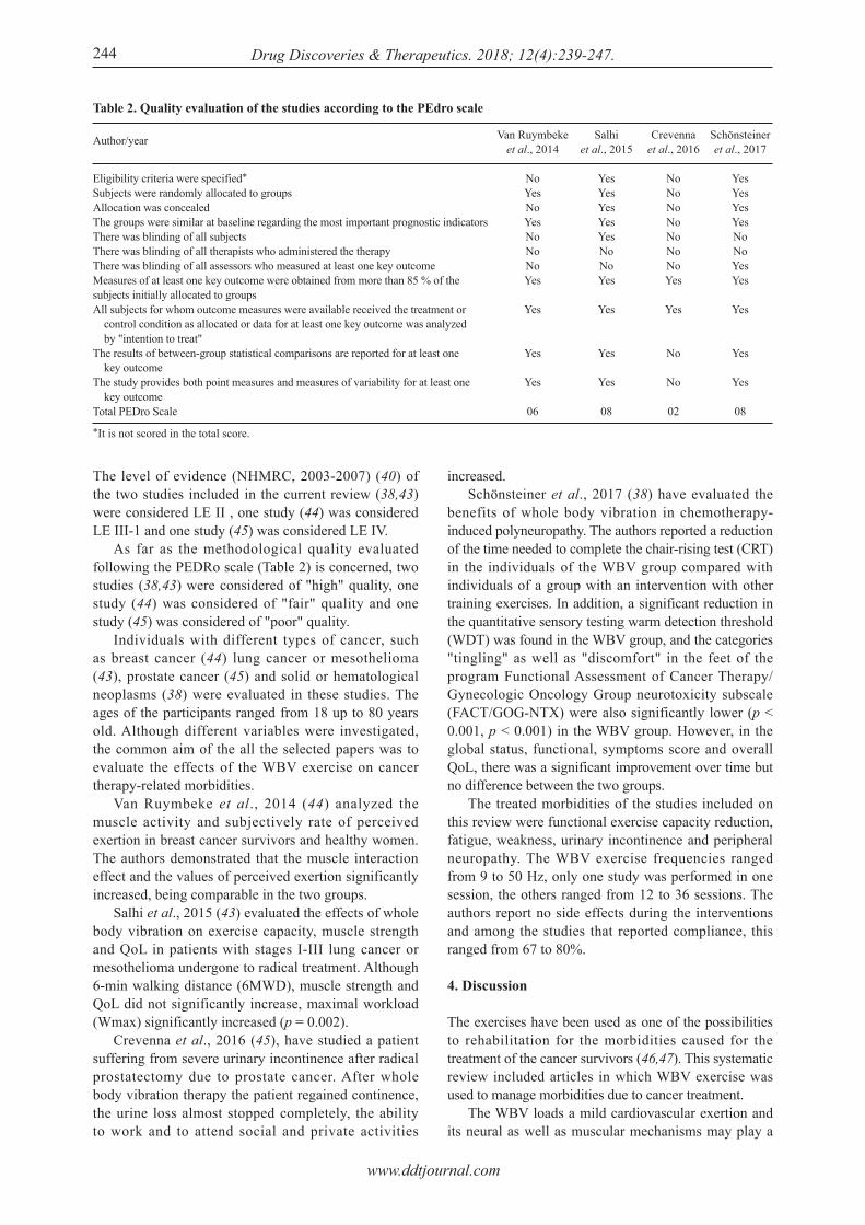
www.ddtjournal.com
Drug Discoveries & Therapeutics. 2018; 12(4):239-247.244
The level of evidence (NHMRC, 2003-2007) (40) of the two studies included in the current review (38,43) were considered LE II , one study (44) was considered LE III-1 and one study (45) was considered LE IV. As far as the methodological quality evaluated following the PEDRo scale (Table 2) is concerned, two studies (38,43) were considered of "high" quality, one study (44) was considered of "fair" quality and one study (45) was considered of "poor" quality. Individuals with different types of cancer, such as breast cancer (44) lung cancer or mesothelioma (43), prostate cancer (45) and solid or hematological neoplasms (38) were evaluated in these studies. The ages of the participants ranged from 18 up to 80 years old. Although different variables were investigated, the common aim of the all the selected papers was to evaluate the effects of the WBV exercise on cancer therapy-related morbidities. Van Ruymbeke et al., 2014 (44) analyzed the muscle activity and subjectively rate of perceived exertion in breast cancer survivors and healthy women. The authors demonstrated that the muscle interaction effect and the values of perceived exertion significantly increased, being comparable in the two groups. Salhi et al., 2015 (43) evaluated the effects of whole body vibration on exercise capacity, muscle strength and QoL in patients with stages I-III lung cancer or mesothelioma undergone to radical treatment. Although 6-min walking distance (6MWD), muscle strength and QoL did not significantly increase, maximal workload (Wmax) significantly increased (p = 0.002). Crevenna et al., 2016 (45), have studied a patient suffering from severe urinary incontinence after radical prostatectomy due to prostate cancer. After whole body vibration therapy the patient regained continence, the urine loss almost stopped completely, the ability to work and to attend social and private activities
increased. Schönsteiner et al., 2017 (38) have evaluated the benefits of whole body vibration in chemotherapy-induced polyneuropathy. The authors reported a reduction of the time needed to complete the chair-rising test (CRT) in the individuals of the WBV group compared with individuals of a group with an intervention with other training exercises. In addition, a significant reduction in the quantitative sensory testing warm detection threshold (WDT) was found in the WBV group, and the categories "tingling" as well as "discomfort" in the feet of the program Functional Assessment of Cancer Therapy/ Gynecologic Oncology Group neurotoxicity subscale (FACT/GOG-NTX) were also significantly lower (p < 0.001, p < 0.001) in the WBV group. However, in the global status, functional, symptoms score and overall QoL, there was a significant improvement over time but no difference between the two groups. The treated morbidities of the studies included on this review were functional exercise capacity reduction, fatigue, weakness, urinary incontinence and peripheral neuropathy. The WBV exercise frequencies ranged from 9 to 50 Hz, only one study was performed in one session, the others ranged from 12 to 36 sessions. The authors report no side effects during the interventions and among the studies that reported compliance, this ranged from 67 to 80%.
4. Discussion
The exercises have been used as one of the possibilities to rehabilitation for the morbidities caused for the treatment of the cancer survivors (46,47). This systematic review included articles in which WBV exercise was used to manage morbidities due to cancer treatment. The WBV loads a mild cardiovascular exertion and its neural as well as muscular mechanisms may play a
Table 2. Quality evaluation of the studies according to the PEdro scale
Author/year
Eligibility criteria were specified*Subjects were randomly allocated to groupsAllocation was concealedThe groups were similar at baseline regarding the most important prognostic indicatorsThere was blinding of all subjectsThere was blinding of all therapists who administered the therapyThere was blinding of all assessors who measured at least one key outcomeMeasures of at least one key outcome were obtained from more than 85 % of the subjects initially allocated to groupsAll subjects for whom outcome measures were available received the treatment or control condition as allocated or data for at least one key outcome was analyzed by "intention to treat"The results of between-group statistical comparisons are reported for at least one key outcomeThe study provides both point measures and measures of variability for at least one key outcomeTotal PEDro Scale
Van Ruymbekeet al., 2014
NoYesNoYesNoNoNoYes
Yes
Yes
Yes
06
Salhi et al., 2015
YesYesYesYesYesNoNoYes
Yes
Yes
Yes
08
*It is not scored in the total score.
Crevenna et al., 2016
NoNoNoNoNoNoNoYes
Yes
No
No
02
Schönsteineret al., 2017
YesYesYesYesNoNoYesYes
Yes
Yes
Yes
08

www.ddtjournal.com
Drug Discoveries & Therapeutics. 2018; 12(4):239-247. 245
role of fatigue (48,49). The frequency of mechanical vibration used to generate WBV exercise affects parts of the body through of which it is transmitted (50). The enhancement in muscle strength and power after vibration can be attributed to the increased muscle activity as a result of interaction of the mechanical vibration. Authors have investigated the effects of frequency on the muscle activity (51,52). Herrero et al., 2011 (51) verified an increase of the electromyography (EMG) activity of the vastus lateral (VL) and vastus medial (VM) muscles in patients with spinal cord injury (SCI) exposed to WBV. Liao et al., 2016 (52) have also reported an increase of the vastus lateral and gastrocnemius (GS) muscle activity in patients with chronic stroke exposed to WBV. Similarly, to these findings, Van Ruymbeke et al., 2014 (44) showed a muscle interaction effect with frequency of the mechanical vibration with an increase of the muscle activation in individuals with breast cancer after therapy. Salhi et al., 2015 (43) evaluated treated stages I-III lung cancer or mesothelioma performing aerobic training plus performed exercises on the synchronous vibration platform and found increase in maximal workload (Wmax) in these patients. In a study developed in the early phase after lung transplantation in which the patients remained in a static position on the platform and were exposed to WBV exercise, also revealed a significant improvement of the Wmax (53). It is relevant to consider that the Wmax might be a parameter to detect changes in aerobic endurance capacity (54) and it is suggested in this current revision that WBV exercise should be an important, safe and feasible intervention to improve aerobic capacity in individuals after cancer treatment. The urinary incontinence is other morbidity that has been found after radical prostatectomy in prostate cancer patients (55). This dysfunction has noninvasive modalities considered first-line treatment during the first 6-12 months following prostatectomy and conservative modalities include pelvic floor muscle training (56). Pelvic muscle exercises are important to active retention strength of the striated muscles improving coordination of the contraction and relaxation process for better control and quality of muscle contraction (57). Crevenna et al., 2016 (45) showed a benefit of the additional use of high-intensity whole body vibration therapy in a patient suffering from severe post radical prostatectomy urinary incontinence. WBV exercise also showed the beneficial effects in patients without cancer as women with stress urinary incontinence (58). The neuropathic disease is common in cancer survivors and may result from the infiltration of nerve tissue by the tumor, radiation treatment, chemotherapy, or cancer-related surgery, leading to symptoms such as pain and functional impairment (59). Schönsteiner
et al., 2017 (38) have demonstrated the beneficial impact (symptoms relieve, physical fitness and sensory function) on chemotherapy-induced polyneuropathy (CIPN) of a program including massage, mobilization as well as physical exercises and WBV. Moreover, other studies have shown the positive effects (reduction of pain (60), enhanced muscles strength and balance (61,62) of WBV exercise on peripheral neuropathy in diabetic patients. Several limitations of this review should be recognized. A relevant limitation is that we could not draw certain conclusions because there is still limited knowledge about WBV exercise on cancer patients. Moreover, it is difficult to prove the effects of whole body vibration on outcomes, because differences exist in multiple WBV parameters and morbidities among the studies. In addition, relevant studies published in other languages other than English may be missed; and there may be publication bias, due to the greater possibility of publication of studies with favorable intervention results. Despite the limitations, it is important to consider that, in our knowledge, this is the first review about the use of suitable, non-invasive and simple procedure. Moreover, relevant findings are presented and demonstrated that the WBV exercise must be more understood and known to be used in the management of morbidities due to the cancer therapy. In conclusion, the WBV exercise might be one modality of treatment to morbidities due cancer treatment for the benefits demonstrated, however other studies should be performed to determine the parameters and specific protocols that will be used to each morbidity.
Acknowledgements
The authors thank for the support of the Brazilian Government agencies (CNPq, FAPERJ) and UERJ.
References
1. World Health Organization. Cancer. http://www.who.int/cancer/en/ (accessed February 27, 2017).
2. Rock CL, Doyle C, Demark-Wahnefried W, Meyerhardt J, Courneya KS, Schwartz AL, Bandera EV, Hamilton KK, Grant B, McCullough M, Byers T, Gansler T. Nutrition and physical activity guidelines for cancer survivors. CA Cancer J Clin. 2012; 62:243-274.
3. World Health Organization. World Cancer Report. http://publications.iarc.fr/Non-Series-Publications/World-Cancer-Reports/World-Cancer-Report-2014. (accessed February 27, 2017).
4. Bower JE. Cancer-related fatigue--mechanisms, risk factors, and treatments. Nat Rev Clin Oncol. 2014; 10:597-609.
5. Paice JA, Ferrell B. The management of cancer pain. CA Cancer J Clin. 2011; 61:157-182.
6. Linden W, Vodermaier A, Mackenzie R, Greig D. Anxiety and depression after cancer diagnosis: Prevalence rates
www.ddtjournal.com
Drug Discoveries & Therapeutics. 2018; 12(4):239-247.246
by cancer type, gender, and age. J Affect Disord. 2012; 141:343-351.
7. Maltby KF, Sanderson CR, Lobb EA, Phillips JL. Sleep disturbances in caregivers of patients with advanced cancer: A systematic review. Palliat Support Care. 2017; 15:125-140.
8. Ferrer RA, Huedo-Medina TB, Johnson BT, Ryan S, Pescatello LS. Exercise interventions for cancer survivors: A meta-analysis of quality of life outcomes. Ann Behav Med. 2011; 41:32-47.
9. Blanchard CM, Courneya KS, Stein K, American Cancer Society's SCS II. Cancer survivors' adherence to lifestyle behavior recommendations and associations with health-related quality of life: Results from the American Cancer Society's SCS-II. J Clin Oncol. 2008; 26:2198-2204.
10. Speck RM, Courneya KS, Masse LC, Duval S, Schmitz KH. An update of controlled physical activity trials in cancer survivors: A systematic review and meta-analysis. J Cancer Surviv. 2010; 4:87-100.
11. Yang EJ, Chung SH, Jeon JY, Seo KS, Shin HI, Hwang JH, Lim JY. Current practice and barriers in cancer rehabilitation: Perspectives of Korean physiatrists. Cancer Res. 2015; 47:370-378.
12. National Institutes of Health. National Heart, Lung, and Blood Institute. (accessed February 27, 2017).
13. Al-Majid S, McCarthy DO. Cancer-induced fatigue and skeletal muscle wasting: The role of exercise. Biol Res Nurs. 2001; 2:186-197.
14. Cramp F, Daniel J. Exercise for the management of cancer-related fatigue in adults. Cochrane Database Syst Rev. 2008; 2:CD006145. doi: 10.1002/14651858.
15. Brown JC, Huedo-Medina TB, Pescatello LS, Pescatello SM, Ferrer RA, Johnson BT. Efficacy of exercise interventions in modulating cancer-related fatigue among adult cancer survivors: A meta-analysis Cancer Epidemiol Biomarkers Prev. 2011; 20:123-133.
16. Ibrahim EM, Al-Homaidh A. Physical activity and survival after breast cancer diagnosis: Meta-analysis of published studies. Med Oncol. 2011; 28:753-765.
17. Meadows R, Bonner T, Dobhal M, Borra S, Killion JA, Paxton R. Pathways between physical activity and quality of life in African-American breast cancer survivors. Support Care Cancer. 2017; 25:489-495.
18. Kenfield SA, Stampfer MJ, Giovannucci E, Chan JM. Physical activity and survival after prostate cancer diagnosis in the health professionals follow-up study. J Clin Oncol. 2011; 29:726-732.
19. Moorman PG, Jones LW, Akushevich L, Schildkraut JM. Recreational physical activity and ovarian cancer risk and survival. Ann Epidemiol. 2011; 21:178-187.
20. Bade BC, Brooks MC, Nietert SB, Ulmer A, Thomas DD, Nietert PJ, Scott JB, Silvestri GA. Assessing the correlation between physical activity and quality of life in advanced lung cancer. Integr Cancer Ther. 2018; 17:73-79.
21. Cochrane DJ. Is vibration exercise a useful addition to a weight management program? Scand J Med Sci Sports. 2012; 22:705-713.
22. Rauch F, Sievanen H, Boonen S, Cardinale M, Degens H, Felsenberg D, Roth J, Schoenau E, Verschueren S, Rittweger JM. International Society of and I. Neuronal. Reporting whole-body vibration intervention studies: Recommendations of the International Society of Musculoskeletal and Neuronal Interactions. J Musculoskelet Neuronal Interact. 2010; 10:193-198.
23. Rittweger J. Vibration as an exercise modality: How it may work, and what its potential might be. Eur J Appl Physiol. 2010; 108:877-904.
24. Sá-Caputo DC, Ronikeili-Costa P, Carvalho-Lima RP, Bernardo LC, Bravo-Monteiro MO, Costa R, Moraes-Silva J, Paiva DN, Machado CB, Mantilla-Giehl P, Arnobio A, Marin PJ, Bernardo-Filho M. Whole body vibration exercises and the improvement of the flexibility in patient with metabolic syndrome. Rehabil Res Pract. 2014; 2014:628518.
25. Cardinale M, Rittweger J. Vibration exercise makes your muscles and bones stronger: Fact or fiction? J Br Menopause Soc. 2006; 12:12-18.
26. Ritzmann R, Kramer A, Bernhardt S, Gollhofer A. Whole body vibration training-improving balance control and muscle endurance. PLoS One. 2014; 9:e89905.
27. Prioreschi A, Tikly M, McVeigh JA. A three month controlled intervention of intermittent whole body vibration designed to improve functional ability and attenuate bone loss in patients with rheumatoid arthritis. BMC Musculoskelet Disord. 2014; 15:403.
28. Tseng SY, Lai CL, Chang KL, Hsu PS, Lee MC, Wang CH. Influence of whole-body vibration training without visual feedback on balance and lower-extremity muscle strength of the elderly: A randomized controlled trial. Medicine (Baltimore). 2016; 95:e2709.
29. Karatrantou K, Gerodimos V, Dipla K, Zafeiridis A. Whole-body vibration training improves flexibility, strength profile of knee flexors, and hamstrings-to-quadriceps strength ratio in females. J Sci Med Sport. 2013; 16:477-481.
30. Dallas G, Paradisis G, Kirialanis P, Mellos V, Argitaki P, Smirniotou A. The acute effects of different training loads of whole body vibration on flexibility and explosive strength of lower limbs in divers. Biol Sport. 2015; 32:235-241.
31. Elfering A, Thomann J, Schade V, Radlinger L. Stochastic resonance whole body vibration reduces musculoskeletal pain: A randomized controlled trial. World J Orthop. 2011; 2:116-120.
32. Alguacil Diego IM, Pedrero Hernandez C, Molina Rueda F, Cano de la Cuerda R. Effects of vibrotherapy on postural control, functionality and fatigue in multiple sclerosis patients. A randomised clinical trial. Neurologia. 2012; 27:143-153.
33. Furness T, Bate N, Welsh L, Naughton G, Lorenzen C. Efficacy of a whole-body vibration intervention to effect exercise tolerance and functional performance of the lower limbs of people with chronic obstructive pulmonary disease. BMC Pulm Med. 2012; 12:71.
34. Gloeckl R, Heinzelmann I, Baeuerle S, Damm E, Schwedhelm AL, Diril M, Buhrow D, Jerrentrup A, Kenn K. Effects of whole body vibration in patients with chronic obstructive pulmonary disease – a randomized controlled trial. Respir Med. 2012; 106:75-83.
35. Sa-Caputo D, Goncalves CR, Morel DS, Marconi EM, Froes P, Rufino R, Costa CH, Lopes AJ, Arnobio A, Asad NR, Marin PJ, Furness T, Bernardo-Filho M. Benefits of whole-body vibration, as a component of the pulmonary rehabilitation, in patients with chronic obstructive pulmonary disease: A narrative review with a suitable approach. Evid Based Complement Alternat Med. 2016; 10:1-7.
36. Sanudo B, Carrasco L, Hoyo M, Oliva-Pascual-Vaca A, Rodriguez-Blanco C. Changes in body balance and

www.ddtjournal.com
Drug Discoveries & Therapeutics. 2018; 12(4):239-247.
functional performance following whole-body vibration training in patients with fibromyalgia syndrome: A randomized controlled trial. J Rehabil Med. 2013; 45:678-684.
37. Santos-Filho SD, Cameron MH, Bernardo-Filho M. Benefits of whole-body vibration with an oscillating platform for people with multiple sclerosis: A systematic review. Mult Scler Int. 2012. doi: 10.1155/2012/274728.
38. Schonsteiner SS, Missbach HB, Benner A, et al. A randomized exploratory phase 2 study in patients with chemotherapy-related peripheral neuropathy evaluating whole-body vibration training as adjunct to an integrated program including massage, passive mobilization and physical exercises. Exp Hematol Oncol. 2017; 6:5.
39. Liberati A, Altman DG, Tetzlaff J, Mulrow C, Gotzsche PC, Ioannidis JP, Clarke M, Devereaux PJ, Kleijnen J, Moher D. The PRISMA statement for reporting systematic reviews and meta-analyses of studies that evaluate health care interventions: Explanation and elaboration. J Clin Epidemiol. 2009; 62:1-34.
40. Merlin T, Weston A, Tooher R. Extending an evidence hierarchy to include topics other than treatment: Revising the Australian 'levels of evidence. BMC Med Res Methodol. 2009; 9:34.
41. PEDRo Scale. Physiotherapy Evidence Database. https://www.pedro.org.au/portuguese/downloads/pedro-scale/ (accessed March 22, 2017).
42. Walser RF, Meserve BB, Boucher TR. The effectiveness of thoracic spine manipulation for the management of musculoskeletal conditions: A systematic review and meta-analysis of randomized clinical trials. J Man Manip Ther. 2009; 17:237-246.
43. Salhi B, Haenebalcke C, Perez-Bogerd S, Nguyen MD, Ninane V, Malfait TL, Vermaelen KY, Surmont VF, Van Maele G, Colman R, Derom E, Meerbeeck JP. Rehabilitation in patients with radically treated respiratory cancer: A randomised controlled trial comparing two training modalities. Lung Cancer. 2015; 89:167-174.
44. Van Ruymbeke JB, Coorevits P, Vanderstraeten G, Bourgois J. Whole-body vibration in breast cancer survivors: A pilot study exploring its effects on muscle activity and subjectively perceived exertion. Int J Rehabil Res. 2014; 37:371-374.
45. Crevenna R, Cenik F, Margreiter M, Marhold M, Sedghi Komanadj T, Keilani M. Whole body vibration therapy on a treatment bed as additional means to treat postprostatectomy urinary incontinence. Wien Med Wochenschr. 2017; 167:139-141.
46. Campbell KL, Pusic AL, Zucker DS, McNeely ML, Binkley JM, Cheville AL, Harwood KJ. A prospective model of care for breast cancer rehabilitation: Function. Cancer. 2012; 108:2300-2311.
47. Sandler CX, Goldstein D, Horsfield S, Bennett BK, Friedlander M, Bastick PA, Lewis CR, Segelov E, Boyle FM, Chin MTM, Webber K, Barry BK, Lloyd AR. Randomized evaluation of cognitive-behavioral therapy and graded exercise therapy for post-cancer fatigue. J Pain Symptom Manage. 2017; 54:74-84.
48. Rittweger J, Beller G, Felsenberg D. Acute physiological effects of exhaustive whole-body vibration exercise in man. Clin Physiol. 2000; 9:134-142.
49. Figueroa, A, Kalfon R, Madzima TA, Wong A. Effects of whole-body vibration exercise training on aortic wave reflection and muscle strength in postmenopausal women
with prehypertension and hypertension. J Hum Hypertens. 2014; 28:118-122.
50. Cidem M, Karacan I, Cakar HI, Cidem M, Sebik O, Yilmaz G, Turker KS, Karamehmetoglu SS. Vibration parameters affecting vibration-induced reflex muscle activity. Somatosens Mot Res. 2017; 34:47-51.
51. Herrero AJ, Menendez H, Gil L, Martin J, Martin T, Garcia-Lopez D, Gil-Agudo A, Marin PJ. Effects of whole-body vibration on blood flow and neuromuscular activity in spinal cord injury. Spinal Cord. 2011; 49:554-559.
52. Liao LR, Ng GY, A. Y. Jones AY, Huang MZ, Pang MY. Whole-body vibration intensities in chronic stroke: A randomized controlled trial. Med Sci Sports Exerc. 2016; 48:1227-1238.
53. Brunner S, Brunner D, Winter H, Kneidinger N. Feasibility of whole-body vibration as an early inpatient rehabilitation tool after lung transplantation-a pilot study. Clin Transplant. 2016; 30:93-98.
54. Arts FIP, Kuipers H, Jeukendrup AE, Saris WHM. A short cycle ergometer test to predict maximal workload and maximal oxygen uptake. Int J Sports Med. 1993; 14:460-464.
55. Karlsen RV, Hvarness EBPH, Bagi P, Friis Lippert E, Permild R, Giraldi A, Lawaetz A, Krause E, Due U, Johansen C. Feasibility and acceptability of couple counselling and pelvic floor muscle training after operation for prostate cancer. Acta Oncol. 2017; 56:270-277.
56. Rajkowska-Labon E, Bakula S, Kucharzewski M, Sliwinski Z. Efficacy of physiotherapy for urinary incontinence following prostate cancer surgery. Biomed Res Int. 2014. doi: 10.1155/2014/785263.
57. Bernardo-Filho M, Barbosa Junior ML, Sa-Caputo DC, Aguiar EO, Lima RP, Santos-Filho SD, Paoli S, Presta GA, Monteiro MOB, Tavares A. The relevance of the procedures related to the physiotherapy in the interventions in patients with prostate cancer: Short review with practice approach. Int J Biomed Sci. 2014; 10:73-84.
58. Farzinmehr A, Moezy A, Koohpayehzadeh J, Kashanian M. A comparative study of whole body vibration training and pelvic floor muscle training on women's stress urinary incontinence: Three-month follow-up. J Family Reprod Health. 2015; 9:147-154.
59. McCarberg B, D'Arcy Y, Parsons B, Sadosky A, Thorpe A, Behar R. Neuropathic pain: A narrative review of etiology, assessment, diagnosis, and treatment for primary care providers. Curr Med Res Opin. 2017; 33:1361-1369.
60. Kessler NJ and Hong J. Whole body vibration therapy for painful diabetic peripheral neuropathy: A pilot study. J Body Mov Ther. 2013; 17:518-522.
61. Kordi Yoosefinejad A, Shadmehr A, Olyaei G, Talebian S, Bagheri H. The effectiveness of a single session of Whole-Body Vibration in improving the balance and the strength in type 2 diabetic patients with mild to moderate degree of peripheral neuropathy: A pilot study. J Body Mov Ther. 2014; 18:82-86.
62. Lee K, Lee S, Song C. Whole-body vibration training improves balance, muscle strength and glycosylated hemoglobin in elderly patients with diabetic neuropathy. Tohoku J Exp Med. 2013; 231:305-314.
(Received June 24, 2018; Revised August 24, 2018; Accepted August 25, 2018)
247
www.ddtjournal.com
Drug Discoveries & Therapeutics. 2018; 12(4):248-253.248
Criteria for the selection of switch OTC drugs based on patient benefits, efficacy, and safety [II]: Comparing the physicochemical and pharmaceutical properties of brand-name and switch OTC terbinafine hydrochloride cream
Motoari Takata1, Yuko Wada2, Yurika Iwasawa3, Miyuki Kumazawa4, Ken-ichi Shimokawa3, Fumiyoshi Ishii2,*
1 Welcia Yakkyoku Co. Ltd., Tokyo, Japan;2 Department of Self-medication and Health Care Sciences, Meiji Pharmaceutical University, Tokyo, Japan;3 Department of Pharmaceutical Sciences, Meiji Pharmaceutical University, Tokyo, Japan;4 Department of Mathematical Science, Meiji Pharmaceutical University, Tokyo, Japan.
1. Introduction
In Japan, the continued increase in national healthcare expenditures poses a serious social problem (1). One way to reduce medical expenditures is by promoting the use of over-the-counter (OTC) drugs in self-medication (2). Since January 1, 2017, if a person buys a "switch OTC" (an OTC drug switched from a prescription drug), the cost of the drug can be deducted from the person's income via an exemption for healthcare costs
under the system for taxation of self-medication as part of an effort to maintain and promote health and disease prevention (2). This reflects the government's policy to promote self-medication and encourage a shift from drugs that require a prescription to OTC drugs that require no prescription ("Patients should be responsible for their own health and treat mild physical ailments themselves (3)") (4). Pharmacists can contribute greatly to the promotion of self-medication by sufficiently understanding the equivalence of OTC drugs to brand-name drugs and their physicochemical characteristics and encouraging their use with appropriate evaluation. However, there are no clear criteria for the selection of OTC drugs, so drugs cannot be easily selected to suit individual patients.
Summary The physicochemical properties (pH, yield value, and squeeze force) of a drug for dermatomycosis, a terbinafine hydrochloride-containing cream (a brand-name product), and 12 over-the-counter drugs (OTCs) were measured and compared to ascertain the characteristics of each product. The pH of the brand-name product, Lamisil, was 4.1, and that of the OTC products ranged from 4.2 to 7.6; Lamisil Plus (7.6) had a significantly higher pH. Moreover, the yield value for Lamisil, as an index of cream ductility, was 128 dyn/cm2, and that for the OTC products ranged from 110 to 887 dyn/cm2. In particular, the OTC products Damalin (887 dyn/cm2), Barriact (512 dyn/cm2), and Exiv Deep (663 dyn/cm2) had a significantly higher yield value. In addition, the squeeze force was measured by attaching a HapLog® to the thumb and second finger. The squeeze force for Lamisil was 12.9 N, and that for the OTC products ranged from 1.8 to 14.6 N. The OTC product Bilumon (1.8 N) had a significantly lower squeeze force. These results indicated that there were marked differences in the pharmaceutical properties of brand-name and OTC products. External preparations are characterized by their feel during use. Based on the current results, the pharmaceutical characteristics of drugs resulted in differences in their feel during use, suggesting that products appropriate for individual patients can be recommended.
Keywords: Cream, terbinafine hydrochloride, brand-name drug, OTC, HapLog®
DOI: 10.5582/ddt.2018.01043Original Article
*Address correspondence to:Dr. Fumiyoshi Ishii, Department of Self-medication and Health Care Sciences, Meiji Pharmaceutical University, 2-522-1, Noshio, Kiyose, Tokyo 204-8588, Japan.E-mail: [email protected]
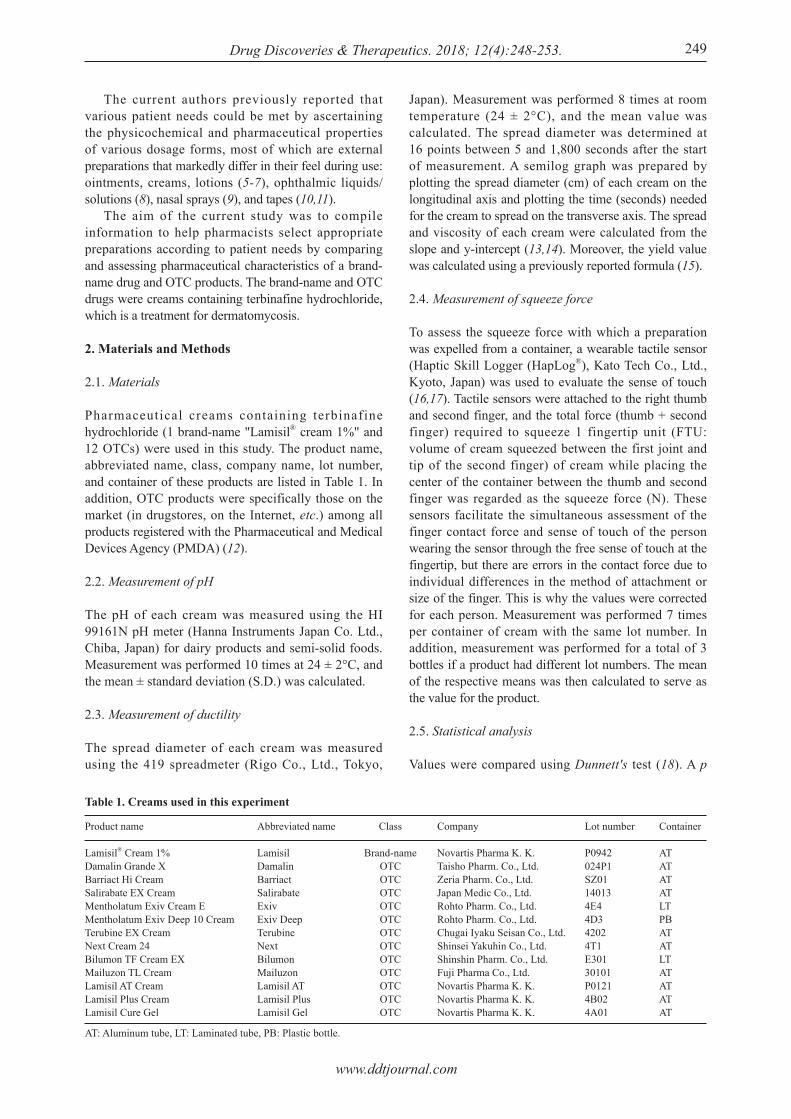
www.ddtjournal.com
Drug Discoveries & Therapeutics. 2018; 12(4):248-253.
The current authors previously reported that various patient needs could be met by ascertaining the physicochemical and pharmaceutical properties of various dosage forms, most of which are external preparations that markedly differ in their feel during use: ointments, creams, lotions (5-7), ophthalmic liquids/solutions (8), nasal sprays (9), and tapes (10,11). The aim of the current study was to compile information to help pharmacists select appropriate preparations according to patient needs by comparing and assessing pharmaceutical characteristics of a brand-name drug and OTC products. The brand-name and OTC drugs were creams containing terbinafine hydrochloride, which is a treatment for dermatomycosis.
2. Materials and Methods
2.1. Materials
Pharmaceutical creams containing terbinafine hydrochloride (1 brand-name "Lamisil® cream 1%" and 12 OTCs) were used in this study. The product name, abbreviated name, class, company name, lot number, and container of these products are listed in Table 1. In addition, OTC products were specifically those on the market (in drugstores, on the Internet, etc.) among all products registered with the Pharmaceutical and Medical Devices Agency (PMDA) (12).
2.2. Measurement of pH
The pH of each cream was measured using the HI 99161N pH meter (Hanna Instruments Japan Co. Ltd., Chiba, Japan) for dairy products and semi-solid foods. Measurement was performed 10 times at 24 ± 2°C, and the mean ± standard deviation (S.D.) was calculated.
2.3. Measurement of ductility
The spread diameter of each cream was measured using the 419 spreadmeter (Rigo Co., Ltd., Tokyo,
Japan). Measurement was performed 8 times at room temperature (24 ± 2°C), and the mean value was calculated. The spread diameter was determined at 16 points between 5 and 1,800 seconds after the start of measurement. A semilog graph was prepared by plotting the spread diameter (cm) of each cream on the longitudinal axis and plotting the time (seconds) needed for the cream to spread on the transverse axis. The spread and viscosity of each cream were calculated from the slope and y-intercept (13,14). Moreover, the yield value was calculated using a previously reported formula (15).
2.4. Measurement of squeeze force
To assess the squeeze force with which a preparation was expelled from a container, a wearable tactile sensor (Haptic Skill Logger (HapLog®), Kato Tech Co., Ltd., Kyoto, Japan) was used to evaluate the sense of touch (16,17). Tactile sensors were attached to the right thumb and second finger, and the total force (thumb + second finger) required to squeeze 1 fingertip unit (FTU: volume of cream squeezed between the first joint and tip of the second finger) of cream while placing the center of the container between the thumb and second finger was regarded as the squeeze force (N). These sensors facilitate the simultaneous assessment of the finger contact force and sense of touch of the person wearing the sensor through the free sense of touch at the fingertip, but there are errors in the contact force due to individual differences in the method of attachment or size of the finger. This is why the values were corrected for each person. Measurement was performed 7 times per container of cream with the same lot number. In addition, measurement was performed for a total of 3 bottles if a product had different lot numbers. The mean of the respective means was then calculated to serve as the value for the product.
2.5. Statistical analysis
Values were compared using Dunnett's test (18). A p
249
Table 1. Creams used in this experiment
Product name
Lamisil® Cream 1%Damalin Grande XBarriact Hi CreamSalirabate EX CreamMentholatum Exiv Cream EMentholatum Exiv Deep 10 CreamTerubine EX CreamNext Cream 24Bilumon TF Cream EXMailuzon TL CreamLamisil AT CreamLamisil Plus CreamLamisil Cure Gel
Abbreviated name
LamisilDamalinBarriactSalirabateExivExiv DeepTerubineNextBilumonMailuzonLamisil ATLamisil PlusLamisil Gel
Class
Brand-nameOTCOTCOTCOTCOTCOTCOTCOTCOTCOTCOTCOTC
AT: Aluminum tube, LT: Laminated tube, PB: Plastic bottle.
Company
Novartis Pharma K. K.Taisho Pharm. Co., Ltd.Zeria Pharm. Co., Ltd.Japan Medic Co., Ltd.Rohto Pharm. Co., Ltd.Rohto Pharm. Co., Ltd.Chugai Iyaku Seisan Co., Ltd.Shinsei Yakuhin Co., Ltd.Shinshin Pharm. Co., Ltd.Fuji Pharma Co., Ltd.Novartis Pharma K. K.Novartis Pharma K. K.Novartis Pharma K. K.
Lot number
P0942024P1SZ01140134E44D342024T1E30130101P01214B024A01
Container
ATATATATLTPBATATLTATATATAT

www.ddtjournal.com
Drug Discoveries & Therapeutics. 2018; 12(4):248-253.250
are shown in Figures 2 and 3. The slope for Lamisil was 0.21, whereas that for the OTC products ranged from 0.03 to 0.22 (Figure 2). In particular, the OTC products Damalin (0.04) and Barriact (0.03) had a significantly lower slope than that of Lamisil. In contrast, the y-intercept for Lamisil was 3.11, whereas that for the OTC products ranged from 2.22 to 3.42, as shown in Figure 3. In particular, the OTC product Exiv Deep (2.22) had a significantly lower y-intercept than that of Lamisil, and Barriact (3.34) and Bilumon (3.42) had a higher y-intercept than that of Lamisil. The yield value is known to be an index of cream ductility. As shown in Figure 4, the yield value for Lamisil was 128 dyn/cm2, whereas that for the OTC products ranged from 110 to 887 dyn/cm2. In particular, the OTC products Damalin (887 dyn/cm2), Barriact (512 dyn/cm2), and Exiv Deep (663 dyn/cm2) had a significantly higher yield value.
3.3. Measurement of the squeeze force
The squeeze force is expressed as the force required to expel a dose of each preparation. The squeeze force was measured by attaching a HapLog® to the right thumb
value of 0.05 or 0.01 was considered significant.
3. Results
3.1. Measurement of pH
Previous studies have indicated that the pH influences the stability of drugs. The pH of each preparation used in this experiment was measured, and the results are shown in Figure 1. The pH of the brand-name drug, Lamisil (pH 4.1), and that of the OTC products ranged from 4.2 to 7.6 (Figure 1). In particular, the OTC products Exiv Deep (pH 6.9), Next (pH 6.9), Lamisil Plus (pH 7.6), and Lamisil Gel (pH 6.7) had a significantly higher pH.
3.2. Measurement of ductility
When a cream is applied to the skin, its feel depends on its ductility and viscosity. The spread diameter and time needed for each cream to spread were measured using a spreadmeter. In addition, a semilog graph (longitudinal axis: spread diameter, transverse axis: time needed to spread) was prepared to calculate the slope and y-intercept for each preparation. The results
Figure 1. Measured pH of various preparations (n = 10). **p < 0.01 (vs. LLamisil cream, Dunnett's test)
Figure 2. Measured slope for various creams (n = 8). **p < 0.01 (vs. Lamisil cream, Dunnett's test).
Figure 3. Y-intercept for various creams (n = 8). *p < 0.05, **p < 0.01 (vs. Lamisil cream, Dunnett's test).
Figure 4. Yield value for various creams (n = 8). **p < 0.01 (vs. Lamisil cream, Dunnett's test).
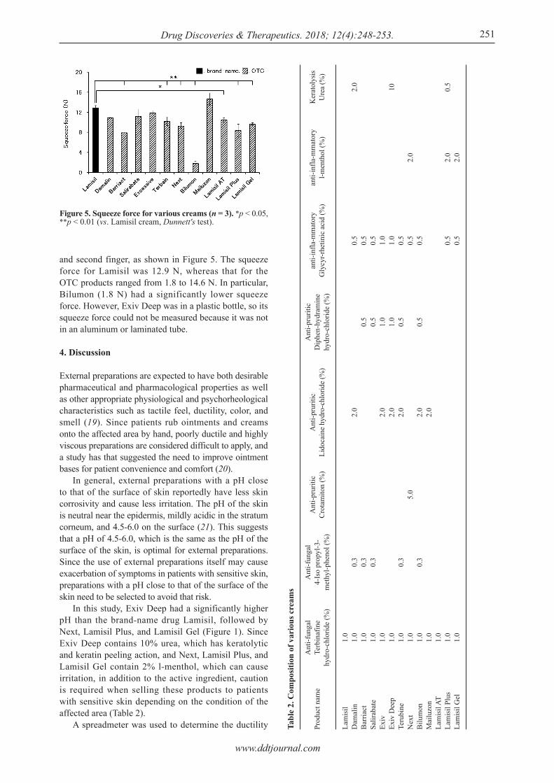
www.ddtjournal.com
Drug Discoveries & Therapeutics. 2018; 12(4):248-253. 251
and second finger, as shown in Figure 5. The squeeze force for Lamisil was 12.9 N, whereas that for the OTC products ranged from 1.8 to 14.6 N. In particular, Bilumon (1.8 N) had a significantly lower squeeze force. However, Exiv Deep was in a plastic bottle, so its squeeze force could not be measured because it was not in an aluminum or laminated tube.
4. Discussion
External preparations are expected to have both desirable pharmaceutical and pharmacological properties as well as other appropriate physiological and psychorheological characteristics such as tactile feel, ductility, color, and smell (19). Since patients rub ointments and creams onto the affected area by hand, poorly ductile and highly viscous preparations are considered difficult to apply, and a study has that suggested the need to improve ointment bases for patient convenience and comfort (20). In general, external preparations with a pH close to that of the surface of skin reportedly have less skin corrosivity and cause less irritation. The pH of the skin is neutral near the epidermis, mildly acidic in the stratum corneum, and 4.5-6.0 on the surface (21). This suggests that a pH of 4.5-6.0, which is the same as the pH of the surface of the skin, is optimal for external preparations. Since the use of external preparations itself may cause exacerbation of symptoms in patients with sensitive skin, preparations with a pH close to that of the surface of the skin need to be selected to avoid that risk. In this study, Exiv Deep had a significantly higher pH than the brand-name drug Lamisil, followed by Next, Lamisil Plus, and Lamisil Gel (Figure 1). Since Exiv Deep contains 10% urea, which has keratolytic and keratin peeling action, and Next, Lamisil Plus, and Lamisil Gel contain 2% l-menthol, which can cause irritation, in addition to the active ingredient, caution is required when selling these products to patients with sensitive skin depending on the condition of the affected area (Table 2). A spreadmeter was used to determine the ductility Tabl
e 2.
Com
posi
tion
of v
ario
us c
ream
s
Prod
uct n
ame
Lam
isil
Dam
alin
Bar
riact
Salir
abat
eEx
ivEx
iv D
eep
Teru
bine
Nex
tB
ilum
onM
ailu
zon
Lam
isil
ATLa
mis
il Pl
usLa
mis
il G
el
Ant
i-fun
gal
Terb
inaf
ine
hydr
o-ch
lorid
e (%
)
1.0
1.0
1.0
1.0
1.0
1.0
1.0
1.0
1.0
1.0
1.0
1.0
1.0
Ant
i-fun
gal
4-Is
o pr
opyl
-3-
met
hyl-p
heno
l (%
)
0.3
0.3
0.3
0.3
0.3
Ant
i-pru
ritic
Cro
tam
iton
(%)
5.0
Ant
i-pru
ritic
Lido
cain
e hy
dro-
chlo
ride
(%)
2.0
2.0
2.0
2.0
2.0
2.0
Ant
i-pru
ritic
Dip
hen-
hydr
amin
e hy
dro-
chlo
ride
(%)
0.5
0.5
1.0
1.0
0.5
0.5
anti-
infla
-mm
ator
yG
lycy
r-rhe
tinic
aci
d (%
)
0.5
0.5
0.5
1.0
1.0
0.5
0.5
0.5
0.5
0.5
anti-
infla
-mm
ator
yl-m
enth
ol (%
)
2.0
2.0
2.0
Ker
atol
ysis
Ure
a (%
)
2.0 10 0.5
Figure 5. Squeeze force for various creams (n = 3). *p < 0.05, **p < 0.01 (vs. Lamisil cream, Dunnett's test).

www.ddtjournal.com
Drug Discoveries & Therapeutics. 2018; 12(4):248-253.252
of various creams. A semilog graph was prepared with the diameter of the spread of the preparation (cm) along the vertical axis and the time that the cream needed to spread (seconds) along the horizontal axis. A linear formula was derived for the log approximation curve for the plot. Based on the brand-name drug (Lamisil), a higher "slope" is considered to indicate greater ductility, and a higher "y-intercept" is considered to indicate lower viscosity (Figures 2 and 3). Damalin and Barriact had a significantly lower "slope," and they were deemed to be poorly "ductile" (Figure 2). Exiv Deep had a significantly lower "y-intercept" than that of the brand-name drug Lamisil, indicating it has a high "viscosity" (Figure 3). The "yield value," which is the minimum stress that induces a semisolid such as a cream or ointment to deform or flow, is an index of the feel during use, and a preparation with a low "yield value" is considered to be easy to spread with minor force. When the "yield value" for the brand-name drug Lamisil and OTC products was compared, Damalin, Barriact, and Exiv Deep had a significantly higher "yield value." Damalin and Exiv Deep presumably had a low yield value partly because they contain urea at a respective concentration of 2 and 10% in addition to the active ingredient (Table 2). In contrast, Salirabate, Terbain, Bilumon, and Lamisil AT had a "yield value" close to that of the brand-name drug, and these preparations are considered to spread even with little force (Figure 4). Measurement of the force of finger contact would presumably be possible while maintaining the free feeling of contact of the fingertip by measuring the force exerted on the fingertip directly in contact with the container using HapLog® attached to the fingers. With this method, a dose could be expelled with a "squeeze force" similar to, or lower than, that needed to expel Lamisil; this was true for the OTC products other than Mailuzon (Figure 5). A dose of Bilumon could be expelled with a squeeze force one-seventh that of Lamisil presumably because Bilumon is contained in a laminated tube while the other products are contained in aluminum tubes (Table 1) (16). In conclusion, pharmacists need to recommend OTC products that cater to diverse patient needs based on the results of this study, but some OTC products contain a skin irritant (l-menthol) or skin-softening ingredients (Irish moss chondrus crispus, urea) and may exacerbate symptoms (Table 2). Since the properties of OTC products are not always appropriate for all patients, pharmacists need to properly select a preparation based on its physicochemical properties depending on the individual patient's skin condition.
Acknowledgements
The authors wish to thank Mr. Yusuke Gokan for providing technical assistance during part of the experimentation. The authors also wish to thank the
Frontier Science Business Division of Shiseido Co., Ltd., for supplying HapLog®.
References
1. Ministry of Health, Labor, and Welfare. 2012 Overview of National Healthcare Costs. http://www.mhlw.go.jp/toukei/saikin/hw/k-iryohi/12/dl/data.pdf (accessed August 6, 2018). (in Japanese)
2. Ministry of Health, Labor, and Welfare. Taxation of self-medication (special exemption for deduction of medical expenses). http://www.mhlw.go.jp/stf/seisakunitsuite/bunya/0000124853.html (accessed August 6, 2018). (in Japanese)
3. WHO. The Role of the Pharmacist in Self-Care and Self-Medication, (1998). http://apps.who.int/medicinedocs/pdf/whozip32e/whozip32e.pdf (accessed August 6, 2018).
4. A reconstruction strategy for Japan – "Japan is Back," Cabinet decision on June 14, 2013. http://www.kantei.go.jp/jp/singi/keizaisaisei/pdf/saikou_jpn.pdf (accessed August 6, 2018). (in Japanese)
5. Wada Y, Nozawa M, Goto M, Shimokawa K, Ishii F. Generic selection criteria for safety and patient benefit [I]: Comparing the original drugs and generic ones in pharmaceutical properties. J Comm Pharm Sci. 2014; 6:97-105. (in Japanese)
6. Nozawa M, Wada Y, Yamazaki N, Shimokawa K, Ishii F. Generic selection criteria for safety and patient benefit [Ⅱ]: Physicochemical characteristics of original and generic drugs for three different difluprednate-containing preparations (ointment, cream, and lotion). J Jpn Soc Comm Pharm. 2014; 2:37-48. (in Japanese)
7. Nozawa M, Goto M, Wada Y, Kumazawa M, Shimokawa K, Ishii F. Generic selection criteria for safety and patient benefit [VII]: Comparing the physicochemical and pharmaceutical properties of brand-name and generic terbinafine hydrochloride cream. Drug Discov Ther. 2018; 12:16-20.
8. Wada Y, Nozawa M, Goto M, Shimokawa K, Ishii F. Generic selection criteria for safety and patient benefit [III]: Comparing the pharmaceutical properties and patient usability of original and generic ophthalmic solutions containing timolol maleate. J Jpn Soc Pharm Health Sci. 2015; 41:394-403. (in Japanese)
9. Wada Y, Ami S, Nozawa M, Goto M, Shimokawa K, Ishii F. Generic selection criteria for safety and patient benefit [V]: Comparing the pharmaceutical properties and patient usability of original and generic nasal spray containing ketotifen fumarate. Drug Discov Ther. 2016; 10:88-92.
10. Wada Y, Kihara M, Nozawa M, Shimokawa K, Ishii F. Generic selection criteria for safety and patient benefit [IV]: Physicochemical and pharmaceutical properties of brand-name and generic ketoprofen tapes. Drug Discov Ther. 2015; 9:229-233.
11. Wada Y, Takaoka Y, Nozawa M, Goto M, Shimokawa K, Ishii F. Generic selection criteria for safety and patient benefit [VI]: Comparing the physicochemical and pharmaceutical properties of brand-name, generic, and OTC felbinac tapes. Drug Discov Ther. 2016; 10:300-306.
12. Pharmaceutical and Medical Devices Agency (PMDA). http://www.pmda.go.jp/english/index.html (accessed August 6, 2018).
www.ddtjournal.com
Drug Discoveries & Therapeutics. 2018; 12(4):248-253. 253
13. Watabe K, Nakamura S, Shimamoto T, Uramatsu T, Kishi H, Uemura T, Niiike H. Preparation and evaluation of new metronidazole gel using hydroxypropyl methylcellulose. Jpn J Pharm Palliat Care Sci. 2009; 2:39-43.
14. Yoshida M. Extensibility of beesoften oil cream at various temperatures of 0.3%. Jpn J Med Pharm Sci. 2014; 71:449-454. (in Japanese)
15. Komoda M, Akakuma S, Obara S, Koto A, Aono H, Kurahashi Y, Sugiyama N, Ishi N, Noda Y. Basic examination of bicarbonate sugar ointment neutralized reaction of leakage from gastrostomy fistula. Jpn Soc Par Ent Nutri. 2013; 28:819-825. (in Japanese)
16. Haptic Skill Logger, Kato Tech Co., Ltd. http://english.keskato.co.jp/ (accessed August 6, 2018).
17. Nakatani M, Kawasoe T. Haptic Skill Logger (HapLog): The wearable sensor for evaluating haptic behaviors. J Robo Soc Japan. 2012; 30:499-501. (in Japanese)
18. Yanai H. Excel Statistics in 4 Steps (4th Edition). OMS Publishing, Saitama, Japan, 2015. (in Japanese)
19. Shibata Y, Ikeda H, Kondou Y, Kihara K. Comparison of pharmaceutical properties of topical non-steroidal anti-inflammatory drug preparations on quality of life. Yakugaku Zasshi. 2005; 125:397-404. (in Japanese)
20. Nishizawa S, Yamaoka K, Yamaoka H, Yoshimura K, Hirabayashi S. Basic pharmaceutical research on formulation of kojic acid ointment for hospitals: In vitro release test and evaluation of permeability. Jpn J Pharm Health Care Sci. 2004; 30:20-26. (in Japanese)
21. Mizoguchi M, Ohara K, Aiba Y, Takado T, Hino H, Matsunaga K, Watanabe S. The pH of the skin. In: Encyclopedia of Skin. Asakura Bookstore, Tokyo, Japan, 2008; pp. 24. (in Japanese)
(Received July 27, 2018; Revised August 16, 2018; Accepted August 19, 2018)

www.ddtjournal.com
Drug Discoveries & Therapeutics. 2018; 12(4):254-258.254
Atypical cases of filariasis from a non-endemic area
Parul Kodan1, Nitin Gupta1, Ankita Baidya1, Ayan Basu1, Abdul Razik2, Ankit Mittal1, Nishant Verma3, Bijay Ranjan Mirdha3, Sandeep Mathur4, Madhu Rajeshwari4, Neeraj Nischal5, Manish Soneja5,*, Naveet Wig5
1 Department of Medicine and Microbiology, All India Institute of Medical Sciences, New Delhi, India;2 Department of Radiodiagnosis, All India Institute of Medical Sciences, New Delhi, India;3 Department of Microbiology, All India Institute of Medical Sciences, New Delhi, India;4 Department of Pathology, All India Institute of Medical Sciences, New Delhi, India;5 Department of Medicine, All India Institute of Medical Sciences, New Delhi, India.
1. Introduction
Filariasis is a neglected tropical disease which poses a significant burden in endemic areas. An estimated 68 million people are infected worldwide, with 40 million being symptomatic (1,2). India shares a heavy burden of the disease and despite having a successful national program, it is not rare to find people from endemic areas presenting with swollen limbs and scrotal swelling even today (3). The social, psychological and economic implications of this disease are enormous. Unfortunately, the disease is often missed in the early stages and when it manifests atypically. It can present in many different ways including fever, lymphangitis, cough or wheeze and limb or scrotal swelling. Since it may be incidentally detected in the blood or tissue aspirate, it is imperative for the laboratory technicians and clinicians to be aware of such findings. Timely diagnosis and appropriate management can significantly decrease the morbidity associated with the disease. We present four atypical cases of filariasis which created significant diagnostic dilemmas but were successfully managed.
2. Case Report
2.1. Case 1: Filariasis mimicking a malignant abdominal tumor
A forty-eight-year old female, resident of Uttar Pradesh, presented with a lump in the right lumbar area, which developed over a year. Her blood investigations were normal. Magnetic Resonance Imaging (MRI) of the abdomen revealed a large midline, complex cystic mass of size 8.5 cm in the greater pelvis showing fluid-debris level as well as non-dependent membranous structures within (Figure 1A). The ovaries were seen separately from the mass. There was another complex cystic mass of size 5 cm in the right pararenal space of retroperitoneum, inferior to the right kidney (Figure 1B). Some of the loculi showed T2-hypointense debris. In addition, conglomerate tubular channels were seen along the bilateral external and internal iliac vessels, which raised the suspicion of dilated lymphatics (Figure 1C). The magnetic resonance lymphangiogram subsequently performed confirmed the lymphatic nature of these tubular channels. The cystic lesions in the pelvis and retroperitoneum also showed partial opacification with gadolinium, suggesting lymphatic communication and hence the origin of these cysts. Imaging differentials of multifocal macrocystic lymphatic malformation as well as of
Summary Filariasis can present in many different ways and pose significant dilemma to the clinician. We report four atypical cases of filariasis which presented as abdominal mass, cervical lymph node enlargement, fever in pregnancy and nosocomial febrile illness respectively. All the four cases were treated successfully with oral antifilarial agents. It is essential to be aware of such atypical presentations of filariasis so that prompt therapy can be initiated.
Keywords: Wuchereria bancrofti, pregnancy, lymphadenopathy
DOI: 10.5582/ddt.2018.01026Case Report
*Address correspondence to:Dr. Manish Soneja, Department of Medicine, 3rd floor, Teaching block, All India Institute of Medical Sciences, New Delhi, India.E-mail: [email protected]

www.ddtjournal.com
Drug Discoveries & Therapeutics. 2018; 12(4):254-258.
cystic mesothelial and germ cell neoplasms were kept. Since imaging could not rule out malignancy, fine needle aspiration cytology (FNAC) of the pelvic mass was done, which revealed microfilaria (Figure 2). She was treated with oral diethycarbazine (DEC) for twenty-one days, subsequent to which the abdominal lump gradually decreased in size. The follow-up MRI performed at one year showed complete resolution of the pelvic and retroperitoneal lumps as well as the lymphatic dilatation (Figure 1D).
2.2. Case 2: Microfilariae in the lymph node
A eleven-year-old female, resident of Delhi, presented with on and off fever with chills for six months. On physical examination, she was found to have an enlarged right cervical lymph node measuring 4 × 2 cm. Her routine blood investigations were normal. Mantoux test was negative. Cytopathology of the lymph node aspirate revealed reactive changes with presence of microfilariae. She was treated with DEC and
255
Figure 1. Fat suppressed T2-weighted MRI showing (A) large complex pelvic cystic mass (asterisk) with fluid-debris level as well as numerous T2-hypointense membranous contents, and (B) a complex multiseptate cyst (arrow) in the retroperitoneum on the right side. Some of the loculi show differential T2-hypointensity, consistent with debris. (C) Coronal maximum intensity projection images of MR lymphangiogram showing dilated lymphatics (open arrows) along the bilateral external and common iliac vessels. (D) Representative post-treatment T2-weighted MRI at the level of the second image showing complete resolution after one year of therapy.
Figure 2. Fine needle aspiration smear from abdominal lump showing microfilaria in a background of blood and inflammatory cells (A). A higher power view of the microfilaria surrounded by a faintly staining sheath (B).

www.ddtjournal.com
Drug Discoveries & Therapeutics. 2018; 12(4):254-258.256
examination, serum procalcitonin levels and chest radiograph were normal. Blood mount showed motile microfilariae (Figure 3), which were identified as Wuchereria bancrofti on giemsa staining (Figure 4). He was started on oral DEC and became afebrile on the second day of treatment.
3. Discussion
Filariasis is a major public health problem in the tropical world with both lymphatic and extralymphatic manifestations. A retrospective analysis of the records of our outpatient clinics and infectious disease consultations from 2017 revealed 20 patients of proven filariasis. Five patients showed atypical presentations, with the findings of one among them (massive splenomegaly with bone marrow aspirate showing microfilariae) having been already published (4). Microfilariae have been detected incidentally in unsuspected swellings in many reports from across the world (5-8). In such cases, microfilariae may either be the direct etiologic agent for the swelling or may have gotten entrapped in an already existent swelling. Diagnosing extralymphatic filariasis remains a clinical challenge as it is often unsuspected and as it mimics non-filarial diseases. Our series describes four cases of filariasis which presented at unusual and rare sites, but were promptly diagnosed and managed. The first case showed complex cystic masses in the pelvis and retroperitoneum, which usually raise the suspicion of macrocystic lymphatic malformation or malignancy. Since the ovaries were separately visualized, the possible differentials included primary cystic mesothelial and germ cell neoplasms. Because of the unusual morphology, filariasis was not kept as a differential until the cytology revealed otherwise. Timely diagnosis and prompt institution of antihelminthic therapy enabled complete recovery
doxycycline, subsequent to which the fever resolved and did not recur at four months of follow. However, the lymph node swelling was persistent.
2.3. Case 3: Detection of microfilariae in the screening peripheral smear of a pregnant patient with fever
A 24-year-old pregnant female, resident of Delhi, presented in the second trimester of her pregnancy with fever, chills and joint pain lasting three days. The physical examination was unremarkable. Her routine peripheral blood smear showed microfilariae. Oral albendazole was initiated and the patient became afebrile on the second day of treatment. She remained asymptomatic at six months of follow-up and no microfilariae were seen in follow-up smear.
2.4. Case 4: From the postoperative ward for fever
A 55-year-old hypertensive male from Delhi presented acutely with altered sensorium and hemiparesis. Unenhanced computed tomography (CT) of the brain showed acute bleed in the left basal ganglion with midline shift and intraventricular extension. He underwent craniotomy with hematoma evacuation and frontal external ventricular drain (EVD) insertion. On the seventh postoperative day, he developed high-grade fever with chills. Physical examination revealed no focus of infection. He was started on broad-spectrum antibiotics, however there was no improvement in the fever. His blood counts, liver and kidney function tests, blood and urine cultures, cerebrospinal fluid
Figure 3. Wet mount of blood showing microfilaria.Figure 4. Peripheral blood smear (Giemsa stain) showing microfilaria of Wuchereria bancrofti.

www.ddtjournal.com
Drug Discoveries & Therapeutics. 2018; 12(4):254-258. 257
without surgery. Only four reports of filariasis presenting as abdominal masses or cysts are available in the current literature, all of which are from the Indian subcontinent (9-12). Except for one, all showed large unilocular retroperitoneal cysts which were resected. Giri et al. (12) reported a complex solid-cystic retroperitoneal mass which, similar to our case, resolved completely on oral DEC therapy. Although the exact mechanism of the origin of abdominal cysts in filariasis is not known, possible hypothesis includes lymphatic obstruction resulting in rupture and extravasation of chyle as well as the involvement of ectopic lymphatic tissue (9). Although rare, keeping a high index of suspicion can result in timely diagnosis, which avoids surgery and significantly changes the course of the disease. Many case series and reports have shown incidental detection of microfilaria in lymph node aspirates (6-8). Presence of microfilariae in lymph nodes may only be an incidental finding with no proven causal association. Cases of microfilariae in aspirates from malignant lymph nodes have also been reported, although the association with malignancy is controversial (13-17). It is important to be aware of this observation and so that the samples are carefully screened. Timely diagnosis can prevent future morbidity and complications. There is no definitive evidence as to whether pregnancy, surgery or other forms of stress can activate the dormant worm and release more microfilariae into blood. No cases of filariasis presenting as nosocomial fever has been described in the literature. However, two among the four cases in our series presented for the first time in periods of stress (pregnancy and postoperative period respectively). Whether these presentations were incidental or had any possible relation to stress remains speculative. Another noteworthy finding in our case series was that none of the patients had eosinophilia. This highlights the fact that eosinophil counts may be normal in filariasis and the diagnosis should not be ruled out for the mere absence of eosinophilia. There have been several reports of filariasis from non-endemic areas such as Delhi. Two among the four patients belonged to Delhi and had no history of travel to other endemic areas. Literature suggests that transmission of filariasis requires prolonged stay in endemic areas and hence is uncommon in travelers. Detection of microfilaria or adult worm in the laboratory samples should prompt the physician to initiate treatment so as to avoid any long-term consequences of the disease. DEC is highly effective in eliminating microfilaria, but has only modest activity against the adult worm. Many recent studies support the synergistic effect of combining this drug with doxycycline. Doxycycline acts by targeting Wolbachia, which engages in endosymbiotic relationship with the filarial parasite and is important in maintaining the reproductive capacity of the worm (18-20). Albendazole
and ivermectin have been used in combination as alternatives. Ivermectin and doxycycline should be avoided in pregnancy. In our series, all the patients responded well to treatment with oral anti-filarial drugs (DEC or albendazole). All patients tolerated the drug well and did not report any side effects or intolerance. All patients showed improvement in symptoms at follow up. As we envision a world free of filariasis, the World Health Organization (WHO) has launched the Global Programme to Eliminate Lymphatic Filariasis (GPELF) by 2020. WHO recommends preventive chemotherapy with annual mass drug administration (MDA) of albendazole together with ivermectin or with DEC. Enhanced public health strategies, disease awareness, prompt diagnosis and treatment can help us in paving the path towards elimination. In conclusion, these cases highlight the unusual and diagnostically challenging presentations of filariasis. It underscores the fact that timely diagnosis and prompt management can prevent severe complications and long-term sequelae. Each patient who gets diagnosed and treated moves us a step closer towards the vision of filariasis-free world.
References
1. Ramaiah KD, Ottesen EA. Progress and impact of 13 years of the global programme to eliminate lymphatic filariasis on reducing the burden of filarialdisease. PLoS Negl Trop Dis. 2014; 8:e3319.
2. Rebollo MP, Bockarie MJ. Toward the elimination of lymphatic filariasis by2020: Treatment update and impact assessment for the endgame. Expert Rev AntiInfect Ther. 2013; 11:723-731.
3. Raju K, Jambulingam P, Sabesan S, Vanamail P. Lymphatic filariasis in India:epidemiology and control measures. J Postgrad Med. 2010; 56:232-238.
4. Basu A, Kumar A, Manchanda S, Wig N. Filarial huge splenomegaly dramatically regressed by anti-filarial medication: A rare clinical scenario. Intractable Rare Dis Res. 2017; 6:215-218.
5. Sah SP, Rani S, Mahto R. Microfilariae in lymph node aspirates. Acta Cytol. 2002; 46:73-75.
6. Varghese R, Raghuveer CV, Pai MR, Bansal R. Microfilariae in cytologicsmears: A report of six cases. Acta Cytol. 1996; 40:299-301.
7. Sivakumar S. Role of fine needle aspiration cytology in detection ofmicrofilariae: Report of 2 cases. Acta Cytol. 2007; 51:803-806.
8. Mitra SK, Mishra RK, Verma P. Cytological diagnosis of microfilariae in filariasis endemic areas of eastern Uttar Pradesh. J Cytol. 2009; 26:11-14.
9. Kapoor AK, Puri SK, Arora A, Upreti L, Puri AS. Case report: Filariasis presenting as an intra-abdominal cyst. Indian J Radiol Imaging. 2011; 21:18-20.
10. Pillay CR. Retroperitoneal cyst of filarial origin. J Assoc Physicians India. 1961; 9:760-762.
11. Mehta RB, Gajendran V, Ananthakrishnan N, Parkash S. Retroperitoneal tumorous lesions. Indian J Surg. 1981; 43:731-742.

www.ddtjournal.com
Drug Discoveries & Therapeutics. 2018; 12(4):254-258.258
12. Giri A, Kundu AK, Chakraborty M, Das S. Microfilarial worms in retroperitoneal mass: A case report. Indian J Urol. 2000; 17:57-58.
13. Yadav R, Sagar M, Maurya M.K, A. A Sonkar, Kumar A. Microfilaria in fine needle aspiration smears of neoplastic lesions: A case series. Trop J Path Micro. 2017; 3:62-66.
14. Agarwal PK, Srivastava AN, Agarwal N. Microfialria in association with neoplasms. A report of six cases. Acta Cytol. 1982; 26:480-490.
15. Kol te SS, SatarkarRN, Mane PM. Microf i lar ia concomitant with metastatic deposits of adenocarcinoma in lymph node fine needle aspiration cytology: A chance finding. J Cytol. 2010; 27:78-80.
16. Jha A, Shrestha R, Aryal G, Pant AD, Adhikari RC, Sayami G. Cytological diagnosis of bancroftian filariasis in lesions clinically anticipated as neoplastic. Nepal Med Coll J. 2008; 10:108-114.
17. Pantola C, Kala S, Agarwal A, Khan L. Microfilaria in cytological smears at rare sites coexisting with unusual
pathology: A series of seven cases. Trop Parasitol. 2012; 2:61-63.
18. HoeraufA. Filariasis: New drugs and new opportunities for lymphatic filariasis and onchocerciasis. Curr Opin Infect Dis. 2008; 21:673-681.
19. Hoerauf A, Mand S, Fischer K, Kruppa T, Marfo-Debrekyei Y, DebrahAY, Pfarr KM, Adjei O, Büttner DW. Doxycycline as a novel strategy against bancroftian filariasis-depletion of Wolbachia endosymbionts from Wuchereria bancrofti and stop of microfilaria production. Med Microbiol Immunol. 2003; 192:211-216.
20. Thomsen, EK, Sanuku, N, Baea, Satofan S, Maki E, Lombore B, Schmidt MS, Siba PM, Weil GJ, Kazura JW, Fleckenstein LL, King CL. Efficacy, safety, and pharmacokinetics of coadministered diethylcarbamazine, albendazole, and ivermectin for treatment of bancroftian filariasis. Clin Infect Dis. 2016; 62:334-341.
(Received May 16, 2018; Revised August 22, 2018; Accepted August 25, 2018)

Drug Discoveries & Therapeutics. 2018; 12(4):E1. E1
www.ddtjournal.com
Erratum DOI: 10.5582/ddt.2018.E1
It has come to the authors’ attention that their article entitled "Anesthetic activity of plant essential oils
on Cyprinus carpio (koi carp)" (Drug Discoveries & Therapeutics. 2018; 12(1):21-30.) included an
error in Table 2. The corrected Table 2 is printed below.
Table 2. Chemical compounds existing in OBO, OCO, and OSO
Retention time (min) Components
Percentage of chemical compound
OBO OCO OSO
4.08 α-Pinene 0.09 - 0.06
4.39 Camphene - - 0.06
4.93 Sabinene 0.09 - -
5.03 β-Pinene 0.17 - 0.05
5.33 β-Myrcene 0.29 - -
6.43 Limonene 0.19 - -
6.60 1,8-Cineole 3.54 - -
7.02 1,3,6-Octatriene 2.37 - -
8.35 Terpinolene 0.16 - -
8.79 Linalool 0.53 3.63 0.94
10.42 L-Camphor 1.49 - -
11.23 Borneol 0.57 - 1.73
11.32 α Terpineol - 0.60 -
13.04 Methyl chavicol 78.12 - -
14.74 Z-Citral (Neral) - 37.04 -
15.38 Geraniol - 0.79 -
16.11 E-Citral (Geranial) - 41.01 -
18.66 α-Cubebene - - 0.16
19.23 Eugenol - - 11.56
19.76 α-Copaene - 0.17 2.39
20.55 β-Elemene 0.70 - 9.67
21.43 Methyl eugenol 0.44 - 46.18
21.81 β-Caryophyllene - 2.47 8.99
22.21 Trans-α-Bergamotene 3.02 - -
22.79 β-Selinene 0.34 0.75 2.06
23.08 β-Farnesene - 0.39 0.07
23.90 Germacrene-D 1.36 1.42 7.35
24.10 Trans-β-Farnesene 0.23 0.69 -
25.10 β-Bisaboloene - 0.12 -
25.66 β-Sesquiphellandrene 0.25 - -
26.48 Cis-α-Bisabolene - 3.01 -
30.08 δ-Cadinene 2.79 - -
Total 96.74 92.09 91.27

Guide for Authors
1. Scope of Articles
Drug Discoveries & Therapeutics welcomes contributions in all fields of pharmaceutical and therapeutic research such as medicinal chemistry, pharmacology, pharmaceutical analysis, pharmaceutics, pharmaceutical administration, and experimental and clinical studies of effects, mechanisms, or uses of various treatments. Studies in drug-related fields such as biology, biochemistry, physiology, microbiology, and immunology are also within the scope of this journal.
2. Submission Types
Original Articles should be well-documented, novel, and significant to the field as a whole. An Original Article should be arranged into the following sections: Title page, Abstract, Introduction, Materials and Methods, Results, Discussion, Acknowledgments, and References. Original articles should not exceed 5,000 words in length (excluding references) and should be limited to a maximum of 50 references. Articles may contain a maximum of 10 figures and/or tables.
Brief Reports definitively documenting either experimental results or informative clinical observations will be considered for publication in this category. Brief Reports are not intended for publication of incomplete or preliminary findings. Brief Reports should not exceed 3,000 words in length (excluding references) and should be limited to a maximum of 4 figures and/or tables and 30 references. A Brief Report contains the same sections as an Original Article, but the Results and Discussion sections should be combined.
Reviews should present a full and up-to-date account of recent developments within an area of research. Normally, reviews should not exceed 8,000 words in length (excluding references) and should be limited to a maximum of 100 references. Mini reviews are also accepted.
Policy Forum articles discuss research and policy issues in areas related to life science such as public health, the medical care system, and social science and may address governmental issues at district, national, and international levels of discourse. Policy Forum articles should not exceed 2,000 words in length (excluding references).
Case Reports should be detailed reports of the symptoms, signs, diagnosis, treatment, and follow-up of an individual patient. Case reports may contain a demographic profile of the patient but usually describe an unusual or novel occurrence. Unreported or unusual side effects or adverse interactions involving medications will also be considered. Case
Reports should not exceed 3,000 words in length (excluding references).
News articles should report the latest events in health sciences and medical research from around the world. News should not exceed 500 words in length.
Letters should present considered opinions in response to articles published in Drug Discoveries & Therapeutics in the last 6 months or issues of general interest. Letters should not exceed 800 words in length and may contain a maximum of 10 references.
3. Editorial Policies
Ethics: Drug Discoveries & Therapeutics r equ i r e s t ha t au tho r s o f r epo r t s o f investigations in humans or animals indicate that those studies were formally approved by a relevant ethics committee or review board.
Conflict of Interest : All authors are required to disclose any actual or potential conflict of interest including financial interests or relationships with other people or organizations that might raise questions of bias in the work reported. If no conflict of interest exists for each author, please state "There is no conflict of interest to disclose".
S u b m i s s i o n D e c l a r a t i o n : W h e n a manuscript is considered for submission to Drug Discoveries & Therapeutics, the authors should confirm that 1) no part of this manuscript is currently under consideration for publication elsewhere; 2) this manuscript does not contain the same information in whole or in part as manuscripts that have been published, accepted, or are under review elsewhere, except in the form of an abstract, a letter to the editor, or part of a published lecture or academic thesis; 3) authorization for publication has been obtained from the authors' employer or institution; and 4) all contributing authors have agreed to submit this manuscript.
Cover Letter: The manuscript must be accompanied by a cover letter signed by the corresponding author on behalf of all authors. The letter should indicate the basic findings of the work and their significance. The letter should also include a statement affirming that all authors concur with the submission and that the material submitted for publication has not been published previously or is not under consideration for publication elsewhere. The cover letter should be submitted in PDF format. For example of Cover Letter, please visit http://www.ddtjournal.com/downcentre.php (Download Centre).
C o p y r i g h t : A s i g n e d J O U R N A L PUBLISHING AGREEMENT (JPA) must be provided by post, fax, or as a scanned file before acceptance of the article. Only forms with a hand-written signature are accepted. This copyright will ensure the widest possible dissemination of information. A form facilitating transfer of copyright can be downloaded by clicking the appropriate link and can be returned to the e-mail address or fax number noted on the form (Please visit
Download Centre). Please note that your manuscript will not proceed to the next step in publication until the JPA form is received. In addition, if excerpts from other copyrighted works are included, the author(s) must obtain written permission from the copyright owners and credit the source(s) in the article.
Suggested Reviewers: A list of up to 3 reviewers who are qualified to assess the scientific merit of the study is welcomed. Reviewer information including names, affiliations, addresses, and e-mail should be provided at the same time the manuscript is submitted online. Please do not suggest reviewers with known conflicts of interest, including participants or anyone with a stake in the proposed research; anyone from the same institution; former students, advisors, or research collaborators (within the last three years); or close personal contacts. Please note that the Editor-in-Chief may accept one or more of the proposed reviewers or may request a review by other qualified persons.
Language Editing: Manuscripts prepared by authors whose native language is not English should have their work proofread by a native English speaker before submission. If not, this might delay the publication of your manuscript in Drug Discoveries & Therapeutics. The Editing Support Organization can provide English proofreading, Japanese-English translation, and Chinese-English translation services to authors who want to publish in Drug Discoveries & Therapeutics and need assistance before submitting a manuscript . Authors can vis i t this organization directly at http://www.iacmhr.com/iac-eso/support.php?lang=en. IAC-ESO was established to facilitate manuscript preparation by researchers whose native language is not English and to help edit works intended for international academic journals.
4. Manuscript Preparation
Manuscripts should be written in clear, grammatically correct English and submitted as a Microsoft Word file in a single-column format. Manuscripts must be paginated and typed in 12-point Times New Roman font with 24-point line spacing. Please do not embed figures in the text. Abbreviations should be used as little as possible and should be explained at first mention unless the term is a well-known abbreviation (e.g. DNA). Single words should not be abbreviated.
Title page: The title page must include 1) the title of the paper (Please note the title should be short, informative, and contain the major key words); 2) full name(s) and affiliation(s) of the author(s), 3) abbreviated names of the author(s), 4) full name, mailing address, telephone/fax numbers, and e-mail address of the corresponding author; and 5) conflicts of interest (if you have an actual or potential conflict of interest to disclose, it must be included as a footnote on the title page of the manuscript; if no conflict of interest exists for each author, please state "There is no conflict of interest to disclose"). Please visit Download Centre and refer to the title page of the manuscript sample.

Abstract: The abstract should briefly state the purpose of the study, methods, main findings, and conclusions. For article types including Original Article, Brief Report, Review, Policy Forum, and Case Report, a one-paragraph abstract consisting of no more than 250 words must be included in the manuscript. For News and Letters, a brief summary of main content in 150 words or fewer should be included in the manuscript. Abbreviations must be kept to a minimum and non-standard abbreviations explained in brackets at first mention. References should be avoided in the abstract. Key words or phrases that do not occur in the title should be included in the Abstract page.
Introduction: The introduction should be a concise statement of the basis for the study and its scientific context.
Materials and Methods: The description should be brief but with sufficient detail to enable others to reproduce the experiments. Procedures that have been published previously should not be described in detail but appropriate references should simply be cited. Only new and significant modifications of previously published procedures require complete description. Names of products and manufacturers with their locations (city and state/country) should be given and sources of animals and cell lines should always be indicated. All clinical investigations must have been conducted in accordance with Declaration of Helsinki principles. All human and animal studies must have been approved by the appropriate institutional review board(s) and a specific declaration of approval must be made within this section.
Results: The description of the experimental results should be succinct but in sufficient detail to allow the experiments to be analyzed and interpreted by an independent reader. If necessary, subheadings may be used for an orderly presentation. All figures and tables must be referred to in the text.
Discussion: The data should be interpreted concisely without repeating material already presented in the Results section. Speculation is permissible, but it must be well-founded, and discussion of the wider implications of the findings is encouraged. Conclusions derived from the study should be included in this section.
Acknowledgments: All funding sources should be credited in the Acknowledgments section. In addition, people who contributed to the work but who do not meet the criteria for authors should be listed along with their contributions.
References: References should be numbered in the order in which they appear in the text. Citing of unpublished results, personal communications, conference abstracts, and theses in the reference list is not recommended but these sources may be mentioned in the text. In the reference list, cite the names of all authors when there are fifteen or fewer authors; if there are sixteen or more authors, list the first three followed by et al. Names of journals should
be abbreviated in the style used in PubMed. Authors are responsible for the accuracy of the references. Examples are given below:
Example 1 (Sample journal reference): Nakata M, Tang W. Japan-China Joint Medical Workshop on Drug Discoveries and Therapeutics 2008: The need of Asian pharmaceutical researchers' cooperation. Drug Discov Ther. 2008; 2:262-263. Example 2 (Sample journal reference with more than 15 authors):Darby S, Hill D, Auvinen A, et al. Radon in homes and risk of lung cancer: Collaborative analysis of individual data from 13 European case-control studies. BMJ. 2005; 330:223. Example 3 (Sample book reference):Shalev AY. Post-traumatic stress disorder: Diagnosis, history and life course. In: Post-traumatic Stress Disorder, Diagnosis, Management and Treatment (Nutt DJ, Davidson JR, Zohar J, eds.). Martin Dunitz, London, UK, 2000; pp. 1-15.
Example 4 (Sample web page reference):World Health Organization. The World Health Report 2008 – primary health care: Now more than ever. http://www.who.int/whr/2008/whr08_en.pdf (accessed September 23, 2010).
Tables: All tables should be prepared in Microsoft Word or Excel and should be arranged at the end of the manuscript after the References section. Please note that tables should not in image format. All tables should have a concise title and should be numbered consecutively with Arabic numerals. If necessary, additional information should be given below the table.
Figure Legend: The figure legend should be typed on a separate page of the main manuscript and should include a short title and explanation. The legend should be concise but comprehensive and should be understood without referring to the text. Symbols used in figures must be explained.
Figure Preparation: All figures should be clear and cited in numerical order in the text. Figures must fit a one- or two-column format on the journal page: 8.3 cm (3.3 in.) wide for a single column, 17.3 cm (6.8 in.) wide for a double column; maximum height: 24.0 cm (9.5 in.). Please make sure that artwork files are in an acceptable format (TIFF or JPEG) at minimum resolution (600 dpi for illustrations, graphs, and annotated artwork, and 300 dpi for micrographs and photographs). Please provide all figures as separate files. Please note that low-resolution images are one of the leading causes of article resubmission and schedule delays. All color figures will be reproduced in full color in the online edition of the journal at no cost to authors.
Units and Symbols: Units and symbols conforming to the International System of Units (SI) should be used for physicochemical quantities. Solidus notation (e.g. mg/kg, mg/mL, mol/mm2/min) should be used. Please refer to the SI Guide www.bipm.org/en/si/ for standard units.
Supplementa l da ta : Supp l emen ta l data might be useful for supporting and enhancing your scientific research and Drug Discoveries & Therapeutics accepts the submission of these materials which will be only published online alongside the electronic version of your article. Supplemental files (figures, tables, and other text materials) should be prepared according to the above guidelines, numbered in Arabic numerals (e.g., Figure S1, Figure S2, and Table S1, Table S2) and referred to in the text. All figures and tables should have titles and legends. All figure legends, tables and supplemental text materials should be placed at the end of the paper. Please note all of these supplemental data should be provided at the time of initial submission and note that the editors reserve the right to limit the size and length of Supplemental Data.
5. Submission Checklist
The Submission Checklist will be useful during the final checking of a manuscript prior to sending it to Drug Discoveries & Therapeutics for review. Please visit Download Centre and download the Submission Checklist file.
6. Online submission
Manuscripts should be submitted to Drug Discoveries & Therapeutics online at http://www.ddtjournal.com. The manuscript file should be smaller than 5 MB in size. If for any reason you are unable to submit a file online, please contact the Editorial Office by e-mail at [email protected]
7. Accepted manuscripts
Proofs: Galley proofs in PDF format will be sent to the corresponding author via e-mail. Corrections must be returned to the editor ([email protected]) within 3 working days.
Offprints: Authors will be provided with electronic offprints of their article. Paper offprints can be ordered at prices quoted on the order form that accompanies the proofs.
Page Charge: A page charge of $140 will be assessed for each printed page of an accepted manuscript. The charge for printing color figures is $340 for each page. Under exceptional circumstances, the author(s) may apply to the editorial office for a waiver of the publication charges at the time of submission.
(Revised February 2013)
Editorial and Head Office:
Pearl City Koishikawa 6032-4-5 Kasuga, Bunkyo-kuTokyo 112-0003JapanTel: +81-3-5840-9697Fax: +81-3-5840-9698E-mail: [email protected]

JOURNAL PUBLISHING AGREEMENT (JPA)------------------------------------------------------------------------------------------------------------------------------------------Manuscript No.:
Title:
Corresponding author:------------------------------------------------------------------------------------------------------------------------------------------
The International Advancement Center for Medicine & Health Research Co., Ltd. (IACMHR Co., Ltd.) is pleased to accept the above article for publication in Drug Discoveries & Therapeutics. The International Research and Cooperation Association for Bio & Socio-Sciences Advancement (IRCA-BSSA) reserves all rights to the published article. Your written acceptance of this JOURNAL PUBLISHING AGREEMENT is required before the article can be published. Please read this form carefully and sign it if you agree to its terms. The signed JOURNAL PUBLISHING AGREEMENT should be sent to the Drug Discoveries & Therapeutics office (Pearl City Koishikawa 603, 2-4-5 Kasuga, Bunkyo-ku, Tokyo 112-0003, Japan; E-mail: [email protected]; Tel: +81-3-5840-9697; Fax: +81-3-5840-9698).
1. Authorship CriteriaAs the corresponding author, I certify on behalf of all of the authors that:1) The article is an original work and does not involve fraud, fabrication, or plagiarism.2) The article has not been published previously and is not currently under consideration for publication elsewhere. If accepted by Drug Discoveries & Therapeutics, the article will not be submitted for publication to any other journal.3) The article contains no libelous or other unlawful statements and does not contain any materials that infringes upon individual privacy or proprietary rights or any statutory copyright.4) I have obtained written permission from copyright owners for any excerpts from copyrighted works that are included and have credited the sources in my article.5) All authors have made significant contributions to the study including the conception and design of this work, the analysis of the data, and the writing of the manuscript.6) All authors have reviewed this manuscript and take responsibility for its content and approve its publication. 7) I have informed all of the authors of the terms of this publishing agreement and I am signing on their behalf as their agent.
2. Copyright Transfer AgreementI hereby assign and transfer to IACMHR Co., Ltd. all exclusive rights of copyright ownership to the above work in the journal Drug Discoveries & Therapeutics, including but not limited to the right 1) to publish, republish, derivate, distribute, transmit, sell, and otherwise use the work and other related material worldwide, in whole or in part, in all languages, in electronic, printed, or any other forms of media now known or hereafter developed and the right 2) to authorize or license third parties to do any of the above. I understand that these exclusive rights will become the property of IACMHR Co., Ltd., from the date the article is accepted for publication in the journal Drug Discoveries & Therapeutics. I also understand that IACMHR Co., Ltd. as a copyright owner has sole authority to license and permit reproductions of the article. I understand that except for copyright, other proprietary rights related to the Work (e.g. patent or other rights to any process or procedure) shall be retained by the authors. To reproduce any text, figures, tables, or illustrations from this Work in future works of their own, the authors must obtain written permission from IACMHR Co., Ltd.; such permission cannot be unreasonably withheld by IACMHR Co., Ltd.
3. Conflict of Interest DisclosureI confirm that all funding sources supporting the work and all institutions or people who contributed to the work but who do not meet the criteria for authors are acknowledged. I also confirm that all commercial affiliations, stock ownership, equity interests, or patent-licensing arrangements that could be considered to pose a financial conflict of interest in connection with the article have been disclosed.---------------------------------------------------------------------------------------------------------------------------------------------------------
Corresponding Author's Name (Signature):
Date:
Drug Discoveries & Therapeutics (www.ddtjournal.com)Pearl City Koishikawa 603, 2-4-5 Kasuga, Bunkyo-ku, Tokyo 112-0003, Japan; E-mail: [email protected]; Tel: +81-3-5840-9697; Fax: +81-3-5840-9698

