italian restructurings distressed debt and m&a - paul hastings
TRANSCRIPT
Appendix I
I.1 Related materials
I.1.1 Carbomer 934P
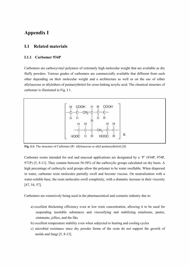
Carbomers are carboxyvinyl polymers of extremely high molecular weight that are available as dry
fluffy powders. Various grades of carbomers are commercially available that different from each
other depending on their molecular weight and a architecture as well as on the use of either
allylsucrose or allylethers of pentaerythritol for cross-linking acrylic acid. The chemical structure of
carbomer is illustrated in Fig. I.1.
Fig. I.1: The structure of Carbomer (R= allylsucrose or allyl pentaerythritol) [8]
Carbomer resins intended for oral and mucosal applications are designated by a ‘P’ (934P, 974P,
971P) [5, 8-11]. They contain between 56-58% of the carboxylic groups calculated on dry basis. A
high percentage of carboxylic acid groups allow the polymer to be water swellable. When dispersed
in water, carbomer resin molecules partially swell and become viscous. On neutralization with a
water-soluble base, the resin molecules swell completely, with a dramatic increase in their viscosity
[47, 54, 57].
Carbomers are extensively being used in the pharmaceutical and cosmetic industry due to:
a) excellent thickening efficiency even at low resin concentration, allowing it to be used for
suspending insoluble substances and viscosifying and stabilizing emulsions, pastes,
ointments, jellies, and the like
b) excellent temperature stability even when subjected to heating and cooling cycles
c) microbial resistance since dry powder forms of the resin do not support the growth of
molds and fungi [5, 8-13].
I.1.2 Microcrystlline cellulose (MCC)
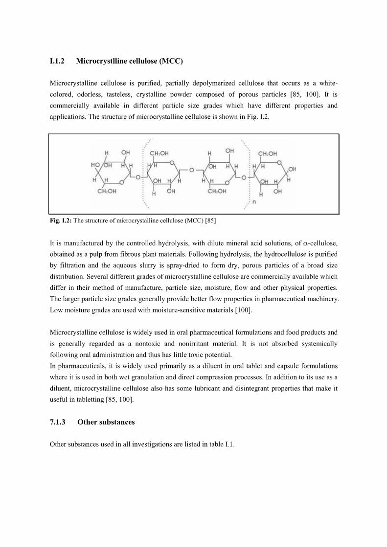
Microcrystalline cellulose is purified, partially depolymerized cellulose that occurs as a white-
colored, odorless, tasteless, crystalline powder composed of porous particles [85, 100]. It is
commercially available in different particle size grades which have different properties and
applications. The structure of microcrystalline cellulose is shown in Fig. I.2.
Fig. I.2: The structure of microcrystalline cellulose (MCC) [85]
It is manufactured by the controlled hydrolysis, with dilute mineral acid solutions, of α-cellulose,
obtained as a pulp from fibrous plant materials. Following hydrolysis, the hydrocellulose is purified
by filtration and the aqueous slurry is spray-dried to form dry, porous particles of a broad size
distribution. Several different grades of microcrystalline cellulose are commercially available which
differ in their method of manufacture, particle size, moisture, flow and other physical properties.
The larger particle size grades generally provide better flow properties in pharmaceutical machinery.
Low moisture grades are used with moisture-sensitive materials [100].
Microcrystalline cellulose is widely used in oral pharmaceutical formulations and food products and
is generally regarded as a nontoxic and nonirritant material. It is not absorbed systemically
following oral administration and thus has little toxic potential.
In pharmaceuticals, it is widely used primarily as a diluent in oral tablet and capsule formulations
where it is used in both wet granulation and direct compression processes. In addition to its use as a
diluent, microcrystalline cellulose also has some lubricant and disintegrant properties that make it
useful in tabletting [85, 100].
7.1.3 Other substances
Other substances used in all investigations are listed in table I.1.
Tab. I.1: Other substances used
Name Abbreviation Supplier
Bentonite
Calcium chloride Hexahydrate
Corn starch
Crospovidone
Di-Calcium phosphate
Lactose
Magnesium stearate
Polyethylene glycol
Polyvinylpyrrolidone K 90
Sodium chloride
Sodium citrate
Sodium sulfate Decahydrate
Talc
Theophylline
Tricalciumphosphat
Bentonite
CaCl2 x 6 H20
Corn starch
Polyplasdone XL
Di-Ca-Phosph.
Lactose
Mg-Stearate
PEG 6000
PVP K 90
NaCl
Na citrate
Na2SO4 x 10 H20
Talc
Theophylline anhydrous
Tri-Ca-Phosph.
Carl Roth GmbH (Quellton)
Merck AG
Cerestar
Polyplasdone XL 10, ISP
Dicalciumphosphat, Budenheim
Tablettose 80, Meggle
Merk AG
PEG 6000, Merk-Schuchardt
Polyvinylpyrrolidone 350, Serva
Merck AG
Isocommerz
Isocommerz
VEB Laborchemie Apolda
Boehringer Ingelheim
Tricalciumphosphat, Budenheim
I.2 Methods
I.2.1 Chelate titration method
Since tri-calcium phosphate is almost insoluble in water, it was assumed that its anti-tack action to
carbomer is not because the influence of its Ca ions. In order to elucidate this hypothesis, the
amount of Ca ions in the composition was measured by the chelate titration method.
The composition contains 30% (w/w) of tri-calcium phosphate, and 1ml of water / 1g of powder
were required to prepare wet mass. That is, 1ml of water contains 0.3g of tri-calcium phosphate.
Therefore, 0.3 g/ml of tri-calcium phosphate dispersion was prepared and stirred for 30min. After
centrifugation, 300ml of solution was prepared and 1N-NaOH was added. This solution was titrated
with 0.1N-EDTA solution. Eriochrome black T was used as an indicator. 0.5ml of EDTA solution
was required to reach the endpoint (pink → blue color).
1ml of EDTA solution = 4.008 mg Ca
Therefore, 0.5 X 4.008 = 2.004 mg Ca in 300 ml solution
Finally, 1ml of this solution contains 0.006 mg Ca.
I.2.2 Determination of flow rate of powder
50g of powder blends were filled in a glass funnel fixed on a clamp. The time was recorded from
when the powder started to flow until finish. Flow rate was calculated as g/s [306]. The mean of
five replicates was used as the result.
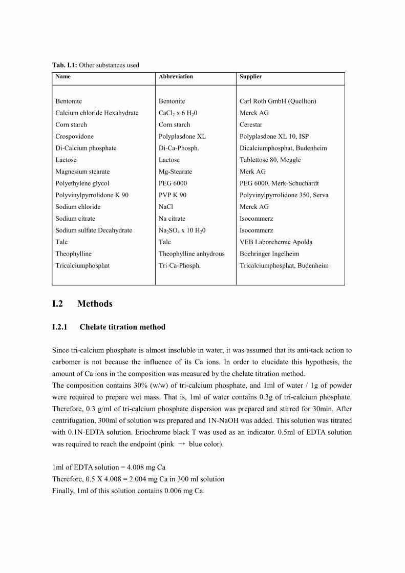
I.2.3 Determination of angle of repose of powder
Angle of repose is defined as the angle of the free
surface of a pile of powder to the horizontal plane [Fig.
I.3] [221, 306]. 50g of powder blends were flown from a
funnel and the height (h) of the powder cone and radius
(r) was measured. Angle of repose was calculated
using the equation: tanα = h/r. The mean of five
replicates was used as the result.
Fig. I.3: Angle of repose
I.2.4 Determination of enslin number of powder
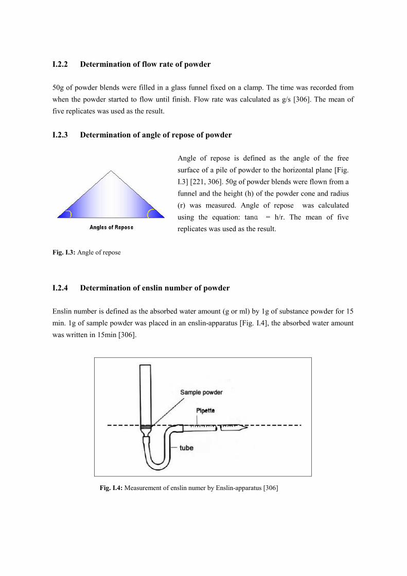
Enslin number is defined as the absorbed water amount (g or ml) by 1g of substance powder for 15
min. 1g of sample powder was placed in an enslin-apparatus [Fig. I.4], the absorbed water amount
was written in 15min [306].
Fig. I.4: Measurement of enslin numer by Enslin-apparatus [306]
I.2.5 Evaluation of pellets
I.2.5.1 Sieve analysis
Particle size distribution was determined by sieve analysis. 100g of sample was sieved using a
vibratory sieve shaker (Vibro, Retsch, Germany) at an amplitude 50 for 10min. 2000, 1700, 1400,
1180, 1000, 710, 500, 355, 250, 125, and 90µm sieves were used and the fraction retaining on each
screen was weighed and expressed as a percentage of the total weight.
Total yield and the yield of sieve fraction 500~1180 µm were calculated. All results presented are
the mean of three determinations. Mass median diameter was the spheroid diameter at the 50-
percentile mark on a cumulative percent oversize plot.
Granule yield is involved to determine the quantity of granulated product actually available, thus
excluding losses due to sticking to the sides of the bowl (related to static electricity) and the fine
particles clogging the filters. The mass of granules obtained at the end of the operation was related
to the theoretical quantity.
I.2.5.2 Image analysis
The sphericity, roughness, and aspect ratio of pellets were determined by optical microscopic image
analysis using the system Leco IA 3.11(Leco Instrumente GmbH, D-Kirchheim). The basic
principles of image analysis system are described in detail in the study of LINDNER und
KLEINEBUDDE [307].
400~600 pellets from every batch (sieve fraction 710~1000µm) were collected and analyzed. Pellets
were dispersed carefully on the microscope slides and a top light source was used to reduce the
influence of shadow on the image processing. The image analyzer consisted of a computer system
linked to a black/white-video camera, and a stereomicroscope. The digitized images were analyzed
by Scion image analyzing software. The Sphericity or roundness (R), and the aspect ratio are
defined as follows:
R= 4πA/C2 (A= area, C= circumference)
Aspect ratio= dmax/dmin (dmax: the longest Feret diameter, dmin: the hortest Feret diameter)
For a perfect spherical shape, aspect ratio is equal to 1.
I.2.5.3 Determination of density
The bulk density of each batch of pellets was measured by carefully pouring an accurately weighed
50g sample through a funnel into a graduated 250ml cylinder and was calculated by dividing the
weight of the material (g) by the volume (ml) occupied in the cylinder. The cylinder was then
tapped 10, 500, 1250 times on a tapping device and the tapped density was also determined in g per
ml. The tap setting was sufficient in all cases to reach a constant volume [306].
I.2.5.4 Friability
10g pellets of 710~1000µm fraction were rotated with 200 glass beads (4mm in diameter) in
friabilator (TAR, Erweka, Germany) at 20rpm for 30min [165]. The glass beads were then removed,
and the fine particles were sieved off, the weight loss was calculated as % friability. The results
were mean of triplicates.
I.2.5.5 Hardness
Hardness test was performed by measuring of required crushing force using texture Analyzer (EZ-
tester, Shimadzu, Japan). 10 pellets of each batch (710~1000 µm fraction) were tested at following
test conditions: a speed to perforce of 1mm/min, speed of 10mm/min during the test.
I.2.5.6 Moisture content
Moisture content of pellets were measured during and after granulation process using IR-balance
(Type MA 40, Sartorius, Germany) set a temperature of 105ºC. The sample was heated to 105ºC,
and evaporative moisture losses were recorded by the internal balance and automatically reported as
percent moisture content.
I.2.5.7 Powder layering efficiency
Powder layering efficiency was calculated by dividing the actual weight gain of coated samples
divided by the theoretical weight gain and multiplying by 100 [183, 184]. Theoretical drug content
was calculated by dividing the amount of drug present in the layering powder with the total of
charge load and the amount of the powder layering composition used.
I.2.5.8 Assay of drug content
Quantities (400 mg) of each batch of pellets were accurately weighed, ground to a fine powder
using a pestle and mortar and made up to 1000 ml of water and allowed to stand for 1h. Aliquots of
the solutions were filtered and assayed spectrophotometrically for theophylline at 271.
I.2.5.9 Dissolution test
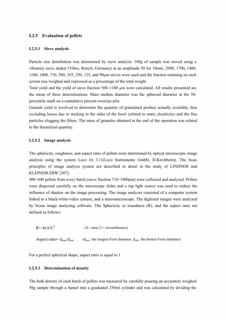
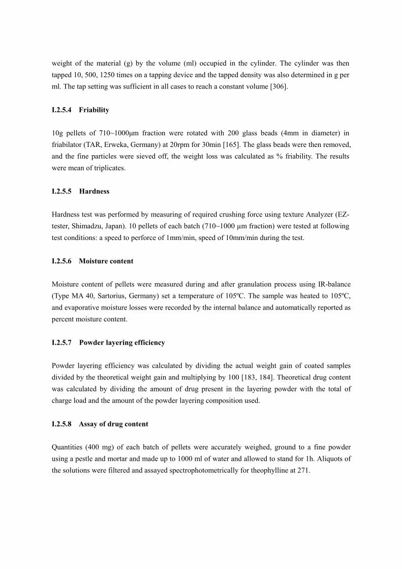
I.2.5.9.1 Standard curve of model drug (theophylline)
10mg of theophylline was dissolved in 1L of medium (demineralized water, pH 3 and pH 6.8
phosphate buffer solution). This solution was diluted to 0,2x10⁻², 0.3x10
-2, 0.5x10
-2, and 0.8x10
-2
mg/ml and UV absorbance was measured. The mean of three replicates was used as the result. The
results are shown in Fig. I.5~ I.7.
I.2.5.9.2 Dissolution test
Dissolution test was performed according to USP paddle method in 900ml of dissolution medium
(purified water, pH 3 and pH 6.8 phosphate buffer solution). 50mg of pellets were used for test. The
temperature of the medium was kept 37±0.5°C while the rotational speed of the paddles was set at
50rpm. 5ml samples were withdrawn at regular time intervals and spectrophotometrically
determined at 271nm. The mean of three replicates was used as the result.
I.2.5.9.3 Preparation of buffer solution
pH 3 phosphate buffer solution
3.40g of potassium dihydrogen phosphate were dissolved in demineralized water, and the pH value
was adjusted with phosphoric acid. This solution was made up to 1000ml by the addition of
demineralized water.
pH 6.8 phosphate buffer solution
5.94g of disodium hydrogen phosphate dihydrate and 4.54g of potassium dihydrogen phosphate
were dissolved in demineralized water. This solution was made up to 1000ml by the addition of
demineralized water.
0
0.126
0.189
0.308
0.475
0.584
y = 59.371x + 0.0091
R2 = 0.9983
0
0.1
0.2
0.3
0.4
0.5
0.6
0.7
0 0.002 0.004 0.006 0.008 0.01 0.012
theophylline conc. (mg / ml)
UV absorbance
Fig. I.5: standard curve of theophylline in water (pH 3.8) (Mean±S.D., n=3)
0
0.126
0.182
0.308
0.471
0.581
y = 57.929x + 0.0077
R2 = 0.9991
0
0.1
0.2
0.3
0.4
0.5
0.6
0.7
0 0.002 0.004 0.006 0.008 0.01 0.012
theophylline conc. (mg/ml)
UV absorbance
Fig. I.6: standard curve of theophylline in pH 3 phosphate buffer solution (Mean±S.D., n=3)
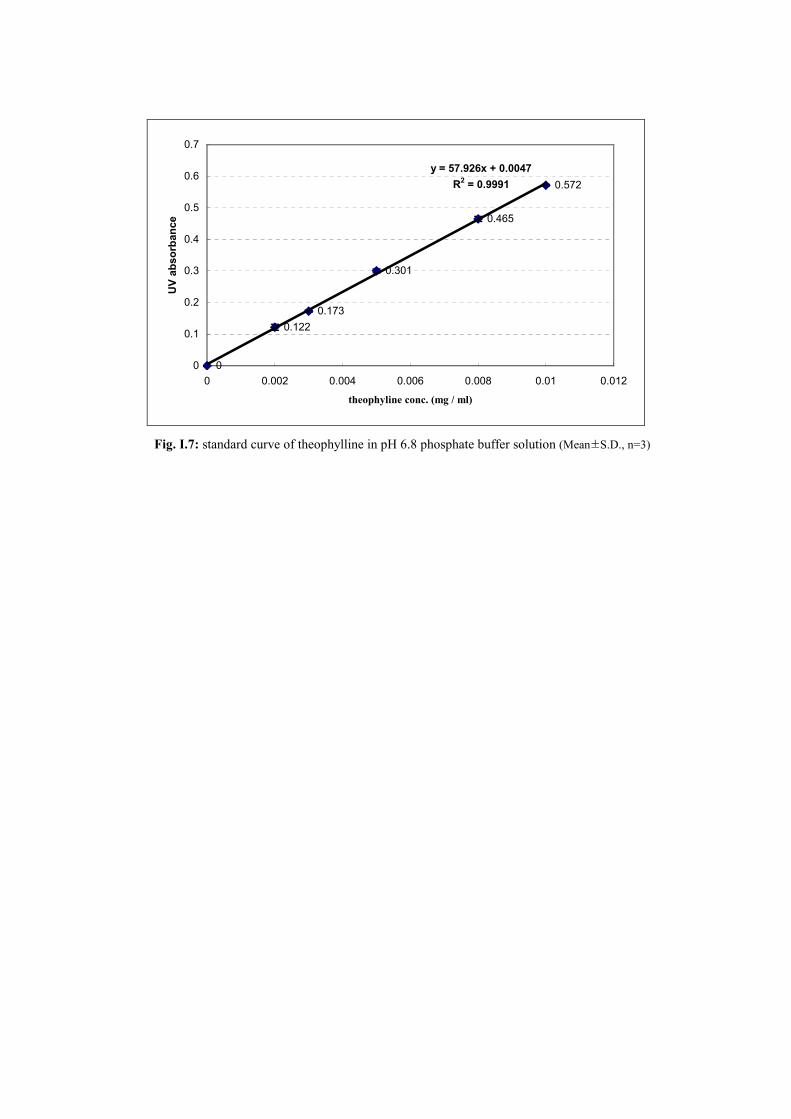
0
0.122
0.173
0.301
0.465
0.572
y = 57.926x + 0.0047
R2 = 0.9991
0
0.1
0.2
0.3
0.4
0.5
0.6
0.7
0 0.002 0.004 0.006 0.008 0.01 0.012
theophyline conc. (mg / ml)
UV absorbance
Fig. I.7: standard curve of theophylline in pH 6.8 phosphate buffer solution (Mean±S.D., n=3)
Appendix ⅡⅡⅡⅡ
References
1. Ahuja A., Khar R. K., Ali J. (1997). “Mucoadhesive Drug Delivery Systems.” Drug Development
and Industrial Pharmacy 23 (5): 489-515.
2. Jimenez-Castellanos R. M., Zia H., Rhodes C. T. (1993). “Mucoadhesive Drug Delivery Systems.”
Drug Development and Industrial Pharmacy 19(1&2): 143-194.
3. Smart, J. D. (2005). “The basics and underlying mechanisms of mucoadhesion.” Advanced Drug
Delivery Reviews 57: 1556– 1568.
4. Lee J. H., Park J. H., Robinson J. R. (2000). “Bioadhesive-Based Dosage Forms: The Next
Generation.” Journal of Pharmaceutical Sciences 89, No. 7: 850-866.
5. Luessen H. L., Rentel C. O., Junginger H. E. (1994). “Bioadhesive polymers for the peroral delivery of
peptide drugs.” Journal of Controlled Release 29: 329-338.
6. Newton J. M. (1990). “The preparation of spherical granules by extrusion/spheronisation.” S.T.P.
Pharma 6 (6): 396-398.
7. Ghebre-Sellassie I. (1989). “Pharmaceutical pelletization technology.” Marcel Dekker Inc., New York
and Basel.
8. Singla A. K., Chawla M., Singh A. (2000). “Potential Applications of Carbomer in Oral Mucoadhesive
Controlled Drug Delivery System: A Review.” Drug Development and Industrial Pharmacy 26(9):
913-924.
9. Muramatsu M., Kanada K., Nishida A., Ouchi K., Saito N., Yoshida M., Shimoaka A., Ozeki T.,
Yuasa H., Kanaya Y. (2000). “Application of Carbopol to controlled release preparations I. Carbopol
as a novel coating material.” International Journal of Pharmaceutics 199: 77-83.
10. Llabot J. M., Manzo R. H., Allemandi D. A. (2004). “Drug release from carbomer: carbomer sodium
salt matrices with potential use as mucoadhesive drug delivery system.” International Journal of
Pharmaceutics 276: 59-66.
11. Neau S. H., Chow M. Y., Durrani M. J. (1996). “Fabrication and characterization of extruded and
spheronized beads containing Carbopol 974P, NF resin.” International Journal of Pharmaceutics 131:
47-55.
12. Neau S. H., Chow M. Y., Hileman G. A., Durrani M. J., Gheyas F., Evans B. A. (2000). “Formulation
and process considerations for beads containing Carbopol 974P, NF resin made by extrusion-
spheronization.” International Journal of Pharmaceutics 199: 129-140.
13. Gomez-Carracedo A., Alvarez-Lorenzo C., Gomez-Amoza J. L., Concheiro A. (2001). “Extrusion-
Spheronization of Blends of Carbopol 934 and Microcrystalline Cellulose.” Drug Development and
Industrial Pharmacy 27(5): 381-391.
14. Yuasa H., Nakano T., Kanaya Y. (1997). “Suppression of agglomeration in fluidized bed coating 1.
Suppression of agglomeration by adding NaCl.” International Journal of Pharmaceutics 158: 195-201.
15. Nakano T., Yuasa H., Kanaya Y. (1999). “Suppression of Agglomeration in Fluidized Bed Coating. 3.
Hofmeister Series in Suppression of Particle Agglomeration.” Pharmaceutical Research 16(10): 1616-
1620.
16. Nakano T., Yuasa H. (2001). “Suppression of agglomeration in fluidized bed coating. 4. Effects of
sodium citrate concentration on the suppression of particle agglomeration and the physical properties
of HPMC film.” International Journal of Pharmaceutics 215: 3-12.
17. Yuasa H., Kanaya Y. (1999). “Suppression of agglomeration in fluidized bed coating. II. Measurement
of mist size in a fluidized bed chamber and effect of sodium chloride addition on mist size.”
International Journal of Pharmaceutics 178: 1-10.
18. Somavarapu S., He P., Alpar H. O. (1998). “Chitosan microspheres for nasal delivery of model antigen
bovine serum albumin.” J. Pharm. Pharmacol. 50.: 166.
19. Naffee N. A., Ismail F., Boraie N. A., Mortada L. M. (2004). “Mucoadhesive Delivery Systems. 1.
Evaluation of Mucoadhesive Polymers for Buccal Tablet Formulation.” Drug Development and
Industrial Pharmacy 30: 985-993.
20. Nafee N. A., Ismail F. A., Boraie N. A., Mortada L. M. (2004). “Mucoadhesive Delivery Systems. 2.
Formulation and In- Vitro/In-Vivo Evaluation of Buccal Mucoadhesive Tablets Containing Water-
Soluble Drugs.” Drug Development and Industrial Pharmacy 30: 995-1004.
21. Bernkop-Schnuerch A., Schwarz G. H., Kratzel M. (1997). “Modified mucoadhesive polymers for the
peroral administration of mainly elastase degradable therapeutic (poly)peptides.” Journal of Controlled
Release 47: 113-121.
22. Bernkop-Schnuerich A., Gilge A. (2000). “Anionic Mucoadhesive Polymers as Auxiliary Agents for
the Peroral Administration of (Poly) Peptide Drugs: Influence of the Gastric Juice.” Drug
Development and Industrial Pharmacy 26(2): 107-113.
23. Uraizee S., McPhillips A. M., Ritschel W. A., Sakr A. (1999). “Development of a Targeted
Mucoadhesive Drug Delivery System for the Peroral Administration of Insulin.” Pharm. Ind. 61,
No.6.: 569-573.
24. Martin L., Wilson C. G., Koosha F., Uchegbu I. F. (1998). “Chitosan based hydrogels for
macromolecular drug delivery.” J. Pharm. Pharmacol. 50.: 171.
25. Ishida M., Machida Y., Nambu N., Nagai T. (1981). “New Mucosal Dosage Form of Insulin.” Chem.
Pharm. Bull. 29(3): 810-816.
26. Pillai J. C., Babar A., Plakogiannis F. M. (1988). “Polymers in Cosmetic and Pharmaceutical
Industries.” PHARM. ACTA HELV. 63, No.2.: 46-53.
27. Park H. S., Robinson J. R. (1985). “Physico-chemical Properties of Water Insoluble Polymers
Important to Mucin/Epithelial Adhesion.” Journal of Controlled Release 2: 47-57.
28. Bernkop-Schnuerch A., Schwarz V., Steininger S. (1999). “Polymers with Thiol Groups: A New
Generation of Mucoadhesive Polymers.” Pharmaceutical Research 16, No.6.: 876-881.
29. Liu P., Krishnan T. R. (1999). “Alginate-Pectin-Poly-L-lysine Particulate as a Potential Controlled
Release Formulation.” J. Pharm. Pharmacol. 51: 141-149.
30. Rege P. R., Shukla D. J., Block L. H. (1999). “Chitinosans as tableting excipients for modified release
delivery systems.” International Journal of Pharmaceutics 181: 49-60.
31. Felt O., Buri P., Gurny R. (1998). “Chitosan: A Unique Polysaccharide for Drug Delivery.” Drug
Development and Industrial Pharmacy 24(11): 979-993.
32. Senel S., Ikinci G., Kas S., Hincal A. A. (2000). “Chitosan films and hydrogels of chlorhexidine
gluconate for oral mucosal delivery.” International Journal of Pharmaceutics 193: 197-203.
33. Oungbho K., Mueller B. (1997). “Chitosan sponges as sustained release drug carriers.” International
Journal of Pharmaceutics 156: 229-237.
34. Zaman M., Zuberi T., Lawrence M. J. (1998). “The bioadhesive properties of hydrophobized
polyvinylpyrrolidone.” J. Pharm. Pharmacol. 50.: 160.
35. Juhasz J. C., Pimienta C., Lenaerts V. (1991). “Adhesion of Poloxamer 407 Formulations on Dog Ileal
Segments in vitro.” Eur. J. Pharm. Biopharm. 37(4): 262-265.
36. Elkheshen S. A., Hosny E. (1999). “Bioadhesive Matrix as Controlled Release Dosage Form for
Verapamil Hydrochloride.” Pharm. Ind. 61(7): 666-670.
37. Takka S., Acartuerk F. (1998). “Calcium alginate microparticles for oral administration: 3. The effect
of crosslink agents and various additive polymers on drug release and drug entrapment efficiency.”
Pharmazie 54: 137-139.
38. Iveson S., Litster J. D., Hapgoog K., Ennis B. J. (2001). “Nucleation, growth and breakage phenomena
in agitated wet granulation processes: a review.” Powder Technology 117: 3-39.
39. Schaafsma S. H., Vonk P., Segers P., Kossen N. W.F. (1998). “Description of agglomerate growth.”
Powder Technology 97: 183-190.
40. Wan L. S. C., Heng P. W. S., Liew C. V. (1993). “Spheronization conditions on spheroid shape and
size.” Int. J. Pharm. 96: 59-65.
41. Cerea M., Zheng W., Young C. R., McGinity J. W (2004). “A novel powder coating process for
attaining taste masking and moisture protective films applied to tablets.” International Journal of
Pharmaceutics 279: 127-139.
42. Obara S., Maruyama N., Nashiyama Y., Kokubo H. (1999). “Dry coating: an innovative enteric
coating method using a cellulose derivative.” Eur. J. Pharm. Biopharm. 47: 51-59.
43. Pfeffer R., Dave R. N., Wei D., Ramlakhan M. (2001). “Synthesis of engineered particulates with
tailored properties using dry particle coating.” Powder Technology 117: 40-67.
44. Pearnchob N., Bodmeier R. (2003). “Coating of pellets with micronized ethylcellulose particles by a
dry powder coating technique.” International Journal of Pharmaceutics 268: 1-11.
45. Vuppala M. K., Parikh D. M., Bhagat H. R. (1997). “Application of Powder-Layering Technology and
Film Coating for Manufacture of Sustained-Release Pellets Using a Rotary Fluid Bed Processor.” Drug
Development and Industrial Pharmacy 23(7): 687-694.
46. Cole, G.C.(1995). “The Coating Process. Pharmaceutical Coating Technology.” Taylor & Francis,
London.
47. Sanz Taberner T., Martin-Villodre A., Pla-Delfina J. M., Herraez J. (2002). “Consistency of Carbopol
971-P NF gels and influence of soluble and cross-linked PVP.” International Journal of Pharmaceutics
233: 43-50.
48. Murata Y., Miyamoto E., Kawashima S. (1996). “Additive effect of chondroitin sulfate and chitosan
on drug release from calcium-induced alginate gel beads.” Journal of Controlled Release 38: 101-108.
49. Dewettinck K., Deroo L., Messens W., Huyghebaert A. (1998). “Agglomeration Tendency during
Top-Spray Fluidized Bed Coating with Gums.” Lebensm.-Wiss. u.-Technol. 31: 576-584.
50. Chan L. W., Wong T. W., Chua P. C., York P., Heng P. W. S. (2003). “Anti-tack Action of
Polyvinylpyrrolidone on Hydroxypropylmethylcellulose.” Chem. Pharm. Bull. 51(2): 107-112.
51. Cervera M. F., Nieto O. M., Colarte A. I. (2004). “Determination of tackiness of chitosan film-
coated pellets exploiting minimum fluidization velocity.” International Journal of Pharmaceutics 281:
119-127.
52. Barreiro-Iglesias R., Alvarez-Lorenzo C., Concheiro A. (2001). “Incorporation of small quantities of
surfactants as a way to improve the rheological and diffusional behavior of carbopol gels.” Journal of
Controlled Release 77: 59-75.
53. Pandit N., Kisaka J. (1996). “Loss of gelation ability of Pluronic F127 in the presence of some salts.”
International Journal of Pharmaceutics 145: 129-136.
54. Tamburic S., Craig D. Q. M. (1995). “An Investigation into the rheological, dielectric and
mucoadhesive properties of poly (acrylic acid) gel systems.” Journal of Controlled Release 37: 59-68.
55. Simovic S., Tamburic S., Milic-Askrabic J., Rajic D. (1999). “An investigation into interactions
between polyacrylic polymers and a non-ionic surfactant: an emulsion preformulation study.”
International Journal of Pharmaceutics 184: 207-217.
56. Duncan Q.M. Craig, S. T., Buckton G., Newton J.M. (1994). “An investigation into the structure and
properties of Carbopol 934 gels using dielectric spectroscopy and oscillatory rheometry.” Journal of
Controlled Release 30: 213-223.
57. Park H. S., Robinson J. R. (1987). “Mechanisms of Mucoadhesion of Poly(acrylic Acid) Hydrogels.”
Pharmaceutical Research 4(6): 457-464.
58. Shojaei A. H., Li Xiaoling (1997). “Mechanisms of buccal mucoadhesion of novel copolymers of
acrylic acid and polyethylene glycol monomethylether monomethacrylate.” Journal of Controlled
Release 47: 151-161.
59. Barreiro-Iglesias R., Alvarez-Lorenzo C., Concheiro A. (2003). “Poly(acrylic acid) microgels
(carbopol 934)/surfactant interactions in aqueous media Part 1:Nonionic surfactants.” International
Journal of Pharmaceutics 258: 165-177.
60. Barreiro-Iglesias R., Alvarez-Lorenzo C., Concheiro A. (2003). “Poly(acrylic acid) microgels
(carbopol 934)/surfactant interactions in aqueous media Part 2:Ionic surfactants.” International Journal
of Pharmaceutics 258: 179-191.
61. Mezreb N., Charrueau C., Boy P., Chaumeil J. C. (2004). “Production of Carbopol 974P and Carbopol
971P Pellets by Extrusion-Spheronization: Optimization of the Processing Parameters and Water
content.” Drug Development and Industrial Pharmacy 30: 481-490.
62. Wesseling M., Kuppler F., Bodmeier R. (1999). “Tackiness of acrylic and cellulosic polymer films
used in the coating of solid dosage forms.” Eur. J. Pharm. Biopharm. 47: 73-78.
63. Roberts G. P., Barnes H. A. (2001). “New measurements of the flow-curves for Carbopol dispersions
without slip artefacts.” Rheol Acta 40: 499-503.
64. Govender T., Ehtezazi T., Stolnik S., Illum L., Davis S. S. (1999). “Complex Formation Between The
Anionic Polymer (PAA) and a Cationic Drug (Procaine HCl): Characterization by Microcalorimetric
Studies.” Pharmaceutical Research 16, No.7.:1125-1131.
65. Tamburic S., Duncan Q. M. (1996). “The Effects of Ageing on the Rheological, Dielectric and
Mucoadhesive Properties of Poly (Acrylic Acid) Gel Systems.” Pharmaceutical Research 13, No.2.:
279-283.
66. Tanna S., Sahota T., Clark J., Taylor M. J. (2002). “Covalent coupling of concanavalin A to a
Carbopol 934P and 941P carrier in glucose-sensitive gels for delivery of insulin.” Journal of Pharmacy
and Pharmacology (JPP) 54: 1461-1469.
67. Khan G. M., Jiabi Z. (1998). “Formulation and in vitro evaluation of ibuprofen-carbopol 974PNF
controlled release matrix tablets III: influence of co-excipients on release rate of the drug.” Journal of
Controlled Release 54: 185-190.
68. Abd-Elbary A., Mansour F., Foda N. (1981). “Interaction of Carbopol 934 with Certain Antihistaminic
Drugs.” Pharmazie 36(5): 356-358.
69. Baun D. C., Walker G. C. (1970). “The prolonged release of carramiphen hydrochloride and atropine
sulfate from compressed tablets containing carbopol 934.” PHARMACEUTICA ACTA HELVETIAE
46.: 94-112.
70. Zhu Y., Shah N. H., McGinity J.W. (2002). “Solid-state plasticization of an acrylic polymer with
chlorpheniramine maleate and triethyl citrate.” International Journal of Pharmaceutics 241: 301-310.
71. Goskonda V. R., Reddy I. K., Durrani M. J., Wilber W., Khan M. A. (1998). “Solid-state stability
assessment of controlled release tablets containing Carbopol 971P.” Journal of Controlled Release 54:
87-93.
72. Leuenberger H. (2001). “New trends in the production of pharmaceutical granules: the classical batch
concept and the problem of scale-up.” Eur. J. Pharm. Biopharm. 52: 279-288.
73. Sigrist. S., Jullien R., Lahaye J. (2001). “Agglomeration of solid particles.” Cement & Concrete
Composites 23: 153-156.
74. Chohan R. K., Newton J. M. (1996). “Analysis of extrusion of some wet powder masses used in
extrusion/spheronisation.” International Journal of Pharmaceutics 131: 201-207.
75. Heng P. W. S., Wan L. S. C., Ling B. L. (1995). “Assessment of powder cohesiveness in
spheronization studies.” International Journal of Pharmaceutics 116: 119-123.
76. Upadrashta, S. R., Upadrashta S. M. (1993). “Avicel RC-591/Chitosan beads by Extrusion-
Spheronization Technology.” Drug Development and Industrial Pharmacy 19(8): 915-927.
77. Harrison P. J., Newton J. M., Rowe R. C. (1985). “The characterization of wet powder masses suitable
for extrusion/spheronization.” J. Pharm. Pharmacol. 37: 686-691.

78. Mort P. R., Capeci S. W., Holder J. W. (2001). “Control of agglomerate attributes in a continuous
binder-agglomeration process.” Powder Technology 117: 173-176.
79. He Y. (1999). “A criterion for particle agglomeration by collision.” Powder Technology 103: 189-193.
80. Simons S.J.R., Fairbrother R. J. (2000). “Direct observations of liquid binder particle interactions: the
role of wetting behaviour in agglomerate growth.” Powder Technology 110: 44-58.
81. Berggren J., Alderborn G. (2001). “Drying behaviour of two sets of microcrystalline cellulose pellets.”
International Journal of Pharmaceutics 219(1-2): 113-126.
82. Otsuka M., Gao J., Matsuda Y. (1994). “Effect of amount of added water during extrusion-
spheronization process on pharmaceutical properties of granules.” Drug Development and Industrial
Pharmacy 20(19): 2977-2992.
83. Varshosaz J., Kennedy R. A., Gipps E. M. (1997). “Effect of Binder Level and Granulating Liquid on
Phenylbutazone Pellets Prepared by Extrusion-Spheronization.” Drug Development and Industrial
Pharmacy 23(6): 611-618.
84. Mills P. J. T., Seville J. P. K., Knight P. C., Adams M. J. (2000). “The effect of binder viscosity on
particle agglomeration in a low shear mixer/agglomerator.” Powder Technology 113: 140-147.
85. Heng P. W. S., Staniforth J. N. (1988). “The effect of moisture on the cohesive properties of
microcrystalline celluloses.” J. Pharm. Pharmacol. 40: 360-362.
86. Rough S. L., Bridgwater J., Wilson D. I. (2000). “Effects of liquid phase migration on extrusion of
microcrystalline cellulose pastes.” International Journal of Pharmaceutics 204(1-2): 117-126.
87. Willett C. D., Adams M. J., Johnson S.A., Seville J. P. K. (2003). “Effects of wetting hysteresis on
pendular liquid bridges between rigid spheres.” Powder Technology 130: 63-69.
88. Alsop, S., Matchett A. J., Coulthard J.M., Peace J. (1997). “Elastic and cohesive properties of wet
particulate materials.” Powder Technology 91: 157-164.
89. Huang A. Y., Berg J. C. (2002). “Gelation of liquid bridges in spherical agglomeration.” Colloids and
Surfaces: 1-12.
90. Schaefer T. (2001). “Growth mechanisms in melt agglomeration in high shear mixers.” Powder
Technology 117: 68-82.
91. Elliasen H., Kristensen H. G., Schaefer T. (1999). “Growth mechanisms in melt agglomeration with a
low viscosity binder.” International Journal of Pharmaceutics 186: 149-159.
92. lveson S. M., Wauters P. A. L., Forrest S., Litster J. D. (2001). “Growth regime map for liquid-
bound granules: further development and experimental validation.” Powder Technology 117(1-2): 83-
97.
93. Iveson S., Page N. W., Litster J. D. (2003). “The importance of wet-powder dynamic mechanical
properties in understanding granulation.” Powder Technology 130: 97-101.
94. Szymocha K. (2003). “Industrial applications of the agglomeration process.” Powder Technology 130:
462-467.
95. York D. W. (2003). “An industrial user's perspective on agglomeration development.” Powder
Technology 130: 14-17.
96. Lustig-Gustafsson C., Podczeck F., Newton J. M. (1999). “The influence of water content and drug
solubility on the formulation of pellets by extrusion and spheronisation.” European Journal of
Pharmaceutical Sciences 8: 147-152.
97. Pinto J. F., Lameiro M. H., Martins P. (2001). “Investigation on the co-extrudability and
spheronization properties of wet masses.” International Journal of Pharmaceutics 227(1-2): 71-80.
98. Wauters P. A. L., Jakobson R. B., Litster J. D., Scarlett B. (2002). “Liquid distribution as a means to
describing the granule growth mechanism.” Powder Technology 123: 166-177.
99. Tomer G., Patel H., Podczeck F., Newton J. M. (2001). “Measuring the water retention
capacities(MRC) of different microcrystalline cellulose grades.” European Journal of Pharmaceutical
Sciences 12: 321-325.
100. Westermarck S., Juppo A. M., Kervinen L., Yliruusi J. (1999). “Microcrystalline cellulose and its
microstructure in pharmaceutical processing.” Eur. J. Pharm. Biopharm. 48: 199-206.
101. Rossetti D., Simons S. J. R. (2003). “A microscale investigation of liquid bridges in the spherical
agglomeration process.” Powder Technology 130: 49-55.
102. Betz G., Buergin P. J., Leuenberger H. (2003). “Power consumption profile analysis and tensile
strength measurements during moist agglomeration.” International Journal of Pharmaceutics 252: 11-25.
103. Miwa A., Yajima T., Itai S. (2000). “Prediction of suitable amount of water addition for wet
granulation.” International Journal of Pharmaceutics 195(1-2): 81-92.
104. Delalonde M., Baylac G., Bataille B., Jacob M., Puech A. (1996). “The rheology of wet powders: a
measuring instrument, the compresso-rheometer.” International Journal of Pharmaceutics 130: 147-151.
105. Chatlapalli R., Rohera B. D. (1998). “Rheological characterization of diltiazem HCl/cellulose wet
masses using a mixer torque rheometer.” International Journal of Pharmaceutics 175: 47-59.
106. Malamataris S., Kiortsis S. (1997). “Wettability parameters and deformational behaviour of powder-
liquid mixes in the funicular agglomeration phase.” International Journal of Pharmaceutics 154: 9-17.
107. Goldszal A., Bousquet J. (2001). “Wet agglomeration of powders: from physics toward process
optimization.” Powder Technology 117: 221-231.
108. Jokinen M., Gyoervary E., Rosenholm J. B. (1998). “Viscoelastic characterization of three different sol-
gel derived silica gels.” Colloids and Surfaces 141: 205-216.
109. Hariharan M., Mehdizadeh E. (2002). “The Use of Mixer Torque Rheometry to Study the Effect of
formulation Variables on the Properties of Wet Granulations.” Drug Development and Industrial
Pharmacy 28(3): 253-263.
110. Deasy P. B., Law M. F. L. (1997). “Use of extrusion-spheronization to develop an improved oral dosage
form of indomethacin.” International Journal of Pharmaceutics 148: 201-209.
111. Tuleu C., Chaumeil J. C. (1998). “Small-Scale Characterization of Wet Powder Masses Suitable for
Extrusion-Spheronization.” Drug Development and Industrial Pharmacy 24(5): 423-429.
112. Forsyth A. J., Rhodes M. J. (2000). “A Simple Model Incorporating the Effects of Deformation and
Asperities into the van der Waals Force for Macroscopic Spherical Solid Particles.” Journal of Colloid
and Interface Science 223(1): 133-138.
113. Kapsidou T., Nikolakakis I., Malamataris S. (2001). “Agglomeration state and migration of drugs in
wet granulations during drying.” International Journal of Pharmaceutics 227: 97-112.
114. Jorgensen A. C., Luukkonen P., Rantanen J. (2004). “Comparison of torque measurements and near-
infrared spectroscopy in characterization of a wet granulation process.” Journal of Pharmaceutical
Sciences 93: 2232-2243.
115. Delalonde M., Bataille B., Baylac G., Maurice J., Sabatier R. (1997). “Definition of indices for the
mechanical design of wet powders: application to the study of a natural polymer, microcrystalline
cellulose.” International Journal of Pharmaceutics 146: 159-165.
116. Tomer G., Newton J. M. (1999). “A centrifuge technique for the evaluation of the extent of water
movement in wet powder masses.” International Journal of Pharmaceutics 188: 31-38.
117. Johansen A., Schaefer T. (2001). “Effects of interactions between powder particle size and binder
viscosity on agglomerate growth mechanisms in a high shear mixer.” European Journal of
Pharmaceutical Sciences 12: 297-309.
118. Baert L., Remon J. P. (1993). “Influence of amount of granulation liquid on the drug release rate from
pellets made by extrusion spheronisation.” International Journal of Pharmaceutics 95: 135-141.
119. Pinto J. F., Podczeck F., Newton J. M. (1997). “Investigations of tablets prepared from pellets produced
by extrusion and spheronization Part1: The application of canonical analysis to correlate the properties
of the tablets to the factors studied in combination with principal component analysis to select the most
relavant factors.” International Journal of Pharmaceutics 147: 79-93.
120. Law M. F. L., Deasy P. B. (1997). “Use of canonical and other analyses for the optimization of an
extrusion-spheronization process for indomethacin.” International Journal of Pharmaceutics 146: 1-9.
121. Jones D. S., Brown A. F., Woolfson A. D., Dennis A. C. (2000). “Examination of the Physical State of
Chlorhexidine Within Viscoelastic, Bioadhesive Semisolids Using Raman Spectroscopy.” Journal of
Pharmaceutical Sciences 89: 563-571.
122. Vervaet C., Baert L., Remon J.P. (1995). “Extrusion-spheronisation A literature review.” International
Journal of Pharmaceutics 116: 131-146.
123. Vervaet C., Baert L., Risha P. A., Remon J. P. (1994). “The influence of the extrusion screen on pellet
quality using an instrumented basket extruder.” International Journal of Pharmaceutics 107: 29-39.
124. Lippis D. M., Sakr A. M. (1994). “Characterization of wet granulation process parameterss using
response surface methodology 1. Top-Spray Fluidized Bed.” Journal of Pharmaceutical Sciences 83:
937-947.
125. Seo A., Holm P., Schaefer T. (2002). “Effects of droplet size and type of binder on the agglomerate
growth mechanisms by melt agglomeration in a fluidised bed.” European Journal of Pharmaceutical
Sciences 16: 95-105.
126. Passos M. L., Mujumdar A. S. (2000). “Effect of cohesive forces on fluidized and spouted beds of wet
particles.” Powder Technology 110(3): 222-238.
127. Abberger T., Seo A., Schaefer T. (2002). “The effect of droplet size and powder particle size on the
mechanisms of nucleation and growth in fluid bed melt agglomeration.” International Journal of
Pharmaceutics 249: 185-197.

128. Abberger T. (2001). “The effect of powder type, free moisture and deformation behaviour of granules
on the kinetics of fluid-bed granulation.” Eur. J. Pharm. Biopharm. 52: 327-336.
129. Mackaplow M. B., Rosen L. A., Michaels J. M. (2000). “Effect of primary particle size on granule
growth and endpoint determination in high-shear wet granulation.” Powder Technology 108: 32-45.
130. Kage H., Abe R., Hattanda R., Zhou T., Ogura H., Matsuno Y. (2003). “Effect of solid circulation rate
on coating efficiency and agglomeration in circulating fluidized bed type coater.” Powder Technology
130: 203-210.
131. Fekete R., Marton S., Racz I. (1998). “Effect of the Formulation Parameters on the Characteristics of
Pellets.” Drug Development and Industrial Pharmacy 24(11): 1073-1076.
132. Rashid H., Heinaemaeki J., Antikainen O., Yliruusi J. (1999). “Effects of Process Variables on the size,
Shape, and Surface Characteristics of Microcrystalline Cellulose Beads Prepared in a Centrifugal
Granulator.” Drug Development and Industrial Pharmacy 25(5): 605-611.
133. Saleh K., Steinmetz D., Hemati M. (2003). “Experimental study and modeling of fluidized bed coating
and agglomeration.” Powder Technology 130: 116-123.
134. Jones D. M. (1985). “Factors to consider in fluid-bed processing.” Pharmaceutical Technology 4: 50-62.
135. Shaffsma S. H., Vonk P., Kossen N. W. F. (2000). “Fluid bed agglomeration with a narrow droplet size
distribution.” International Journal of Pharmaceutics 193: 175-187.
136. Gao J.H., Jain A., Motheram R., Gray D. B., Hussain M. A. (2002). “Fluid bed granulation of a poorly
water soluble, low density, micronized drug: comparison with high shear granulation.” International
Journal of Pharmaceutics 237: 1-14.
137. Hemati M., Cherif R., Saleh K., Pont V. (2003). “Fluidized bed coating and granulation: influence of
process-related variables and physicochemical properties on the growth kinetics.” Powder Technology
130: 18-34.
138. Becher R. D., Schluender E. U. (1998). “Fluidized bed granulation- the importance of a drying zone for
the particle growth mechanism.” Chemical Engineering and Processing 37: 1-6.
139. Vertommen J. Kinget R. (1997). “The Influence of Five Selected Processing and Formulation Variables
on the Particle Size Distribution, and Friability of Pellets Produced in a Rotary Processor.” Drug
Development and Industrial Pharmacy 23(1): 39-46.
140. Vertommen J., Kinget R. (1996). “The influence of five selected processing and formulation variables
on the release of riboflavin from pellets produced in a rotary processor.” S.T.P.Pharma Sciences 6 (5):
335-340.
141. Wan L. S. C., Heng P. W. S., Liew C. V. (1995). “The influence of liquid spray rate and atomizing
pressure on the size of spray droplets and spheroids.” International Journal of Pharmaceutics 118: 213-
219.
142. Liew C. V., Gu L., Heng P. W. S. (2002). “The influence of operational variables on mean size and size
distribution of spheroids produced by rotary spheronization using teardrop studs.” International Journal
of Pharmaceutics 242: 345-348.
143. Umprayn K., Chitropas P., Amarekajorn S. (1999). “Influence of Process Variables on Physical
Properties of the Pellets Using Extruder and Spheronizer.” Drug Development and Industrial Pharmacy
25(1): 45-61.
144. Newton J. M., Chapman S. R., Rowe R.C. (1995). “The influence of process variables on the
preparation and properties of spherical granules by the process of extrusion and spheronization.”
International Journal of Pharmaceutics 120: 101-109.
145. Rashid A., Antikainen O., Yliruusi J. (2001). “Influence of the centrifugal granulating process on the
properties of layered pellets.” Eur. J. Pharm. Biopharm. 51: 227-234.
146. Pont V., Saleh K., Steinmetz D., Hemati M. (2001). “Influence of the physicochemical properties on the
growth of solid particles by granulation in fluidized bed.” Powder Technology 120: 97-104.
147. Vertommen J., Rombaut P., Kinget R. (1998). “Internal and external structure of pellets made in a
rotary processor.” International Journal of Pharmaceutics 161: 225-236.
148. Bodea A., Leucuta S. E. (1997). “Optimization of propranolol hydrochloride sustained release pellets
using a factorial design.” International Journal of Pharmaceutics 154: 49-57.
149. Wang P. W. S., Heng L. S. C., Tan Y. T. P. (1996). “Optimization of spheroid production by centrifugal
rotary processing.” International Journal of Pharmaceutics 143: 107-112.
150. Korakianiti E. S., Rekkas D. M., Dallas P. P., Choulis N. H. (2000). “Optimization of the Pelletization
Process in a Fluid-Bed Rotor Granulator Using Experimental Design.” AAPS PharmSciTech 1 (4),
article 35.: 1-5.
151. Zhou F., Vervaet C., Massart D. L., Massart B., Remon J. P. (1998). “Optimization of the Processing of
Matrix Pellets Based on the Combination of Waxes and Starch Using Experimental Design.” Drug
Development and Industrial Pharmacy 24(4): 353-358.
152. Dussert A., Chulia D., Jeannin C., Ozil P. (1995). “Parametric study of Fluidized-bed granulation of a
low density micronized powder.” Drug Development and Industrial Pharmacy 21(12): 1439-1452.
153. Holm P. et al. (1996). “Pelletization by granulation in a Roto-processor RP-2. Part 1: Effects of process
and product variables on granule growth.” Pharm. Technol. Eur. 8.:22-36.
154. Holm P. (1996). “Pelletization by granulation in a Roto-processor RP-2. Part 2: Effects of process and
product variables on agglomerates' shape and porosity.” Pharm. Technol. Eur. 8.:38-44.
155. Holm P. (1996). “Pelletization by granulation in a Roto-processor RP-2. Part 3: Methods of process
control and the effect of microcrystalline cellulose on wet granulation.” Pharm. Technol. Eur. 8: 38-48.
156. Jaiswal S. B., Shamsuddin, Shehzad S.S., Brahmankar D.M. (1995). “Pelletization in Rotary Shaker:
Effect of Equipment Variables on Pelletization of Ferrous Fumarate.” Drug Development and Industrial
Pharmacy 21(18): 2109-2120.
157. Santos H., Veiga F., Pna M., Podczeck F., Sousa J. (2002). “Physical properties of chitosan pellets
produced by extrusion-spheronisation: influence of formulation variables.” International Journal of
Pharmaceutics 246: 153-169.
158. Vecchio C., Bruni G., Gazzaniga A. (1994). “Preparation of Indobufen Pellets by Using Centrifugal
Rotary Fluidized Bed Equipment without Starting Seeds.” Drug Development and Industrial Pharmacy
20(12): 1943-1956.
159. Faure A., York P., Rowe R. C. (2001). “Process control and scale-up of pharmaceutical wet granulation
processes: a review.” Eur. J. Pharm. Biopharm. 52: 269-277.
160. Hellen L., liruusi J. (1993). “Process variables of instant granulator and spheroniser: 3. Shape and shape
distributions of pellets.” International Journal of Pharmaceutics 96: 217-223.
161. Podczeck F., Wood A. W. (2003). “The relationship between granule growth mechanism, amount of
liquid binder added and properties of the wet powder mass determined using a split bed shear tester.”
International Journal of Pharmaceutics 257: 57-67.
162. Wan L. S. C., Heng P. W. S., Liew C. V. (1994). “The Role of Moisture and Gap Air Pressure in the
Formation of Spherical Granules by Rotary Processing.” Drug Development and Industrial Pharmacy
20(16): 2551-2561.
163. Vertommen J., Rombaut P., Kinget R. (1997). “Shape and surface smoothness of pellets made in a
rotary processor.” International Journal of Pharmaceutics 146: 21-29.
164. Gu L., Liew C. V., Heng P. W. S. (2004). “Wet Spheronization by Rotary Processing- A Multistage
Single-Pot Process for Producing Spheroids.” Drug Development and Industrial Pharmacy 30: 111-123.
165. Bains D., Boutell S. L., Newton J. M. (1991). “The influence of moisture content on the preparation of
spherical granules of barium sulfate and microcrystalline cellulose.” Int. J. Pharm. 69: 233-237.
166. Sunada H., Makino T., Sakamoto H., Fujita K., Tanino T., Kokubo H., Kawaguchi T. (1998). “Study of
Standard Tablet Formulation Based on Fluidized-Bed Granulation.” Drug Development and Industrial
Pharmacy 24(3): 225-233.
167. Baert L., Vermeersch H., Remon J. P., Smeyers-Verbeke J., Massart D. L. (1993). “Study of parameters
important in the spheronisation process.” International Journal of Pharmaceutics 96: 225-229.
168. Larsen C., Sonnergaard J., Bertelsen P., Holm P. (2003). “A new process control strategy for aqueous
film coating of pellets in fluidised bed.” European Journal of Pharmaceutical Sciences 20: 273-283.
169. Contreras M.D., Sanchez R. (2002). “Application of a factorial design to the study of specific
parameters of a Carbopol ETD 2020 gel. Part I. Viscoelastic parameters.” International Journal of
Pharmaceutics 234: 139-147.
170. Park T. (1990). “Characterization of a Hot-Melt Fluid Bed Coating Process for Fine Granules.” Eur. J.
Pharm. Biopharm. 48: 3-10.
171. Gotthardt S., Knoch A., Lee G. (1999). “Continuous wet granulation using fluidized-bed techniques I.
Examination of powder mixing kinetics and preliminary granulation experiments.” Eur. J. Pharm.
Biopharm. 48: 189-197.
172. Tobiska. S., Kleinnebudde P. (2003). “Coating uniformity and coating efficiency in a Bohle Lab-Coater
using oval tablets.” Eur. J. Pharm. Biopharm. 56: 3-9.
173. Gluba T. (2003). “The effect of wetting liquid droplet size on the growth of agglomerates during wet
drum granulation.” Powder Technology 130: 219-224.
174. Berggren J., Alderborn J. (2001). “Effect of drying rate on porosity and tabletting behaviour of cellulose
pellets.” International Journal of Pharmaceutics 227(1-2): 81-96.
175. Juslin L., Yliruusi J. (1996). “The effect of raw material and atomizing air pressure on the properties of
granules prepared in a fluidized bed granulator.” S.T.P.Pharma Sciences 6: 328-334.
176. Pieszczek B., Jachowicz R., Bataille B., Delalonde M., Maciejewska A. (2002). “Formulation and
Rheology of Piroxicam Solid Dispersion Pellets.” Proc.4th World Meeting ADRITELF/APGI/APV,
Florence, 8/11 April 2002: 1417-1418
177. Villar-Lopez M. E., Nieto-Reyes L., Anguiano-Igea S., Otero-Espinar F. J., Blanco-Mendez J. (1999).
“Formulation of triamcinolone acetonide pellets suitable for coating and colon targeting.” International
Journal of Pharmaceutics 179: 229-235.
178. Gohel M.C., Amin A. F. (1998). “Formulation optimization of controlled release diclofenac sodium
microspheres using factorial design.” Journal of Controlled Release 51: 115-122.
179. Shelukar S., Ho. J., Zega J., Reynolds S. (2000). “Identification and characterization of factors
controlling tablet coating uniformity in a Wurster coating process.” Powder Technology 110: 29-36.
180. Viana M., N’Dri-Stempfer B., Vachon M.G., Chulia D. (2002). “Influence of Manufacturing Process on
Tabletting Ability of Powder: Comparison Between Blending, Grinding, and Spray Drying of Two
Formulations Made of Theophylline and Lactose/Cellulose or Cellactose.” Drug Development and
Industrial Pharmacy 28(2): 119-127.
181. Williams R. O., Liu J. (2000). “Influence of processing and curing conditions on beads coated with an
aqueous dispersion of cellulose acetate phthalate.” Eur. J. Pharm. Biopharm. 49: 243-252.
182. Zhou F., Vervaet C., Remon J. P. (1997). “Influence of processing on the characteristics of matrix
pellets based on microcrystalline waxes and starch derivatives.” International Journal of Pharmaceutics
147: 23-30.
183. Gupta V.K., Assmus M. W., Beckert T. E., Price J. C. (2000). “A novel pH - and time-based multi-
unit potential colonic drug delivery system. 1. Development.” International Journal of Pharmaceutics
213: 83-91.
184. Gupta V.K., Assmus M. W., Beckert T. E. (2000). “A novel pH - and time-based multi-unit potential
colonic drug delivery system. 2. Optimization of multiple response variables.” International Journal of
Pharmaceutics 213: 93-102.
185. Lecomte F., Siepmann J., Walther M., Bodmeier R. (2004). “Polymer Blends Used for the coating of
Multiparticulates: Comparison of Aqueous and Organic Coating Techniques.” Pharmaceutical research
21: 882-890.
186. McCarron P. A., Woolfson A. D., Keating S. M. (1999). “Response surface methodology as a predictive
tool for determining the effects of preparation conditions on the physicochemical properties of
poly(isobutylcyanoacrylate) nanoparticles.” International Journal of Pharmaceutics 193: 37-47.
187. Gajdos, B. (1984). “Rotary granulators-Evaluation of Process Technology for Pellet Production Using a
Factorial Experimental Design.” Drugs Made Der. 27.: 30-36.
188. Rambali B., Baert L., Massart D. L. (2003). “Scaling up of the fluidized bed granulation process.”
International Journal of Pharmaceutics 252: 197-206.
189. Rambali B., Baert L., Massart D. L. (2001). “Using experimental design to optimize the process
parameters in fluidized bed granulation on a semi-full scale.” International Journal of Pharmaceutics
220: 149-160.

190. Kramar A., Turk S., Vrecer F. (2003). “Statistical optimisation of diclofenac sustained release pellets
coated with polymethacrylic films.” International Journal of Pharmaceutics 256: 43-52.
191. Sienkiewicz G., Pereira R., Rudnic E.M., Lausier J. M., Rhodes C.T. (1997). “Spheronization of
Theophylline-Avicel combinations Using a Fluidized-Bed Rotogranulation Technique.” Drug
Development and Industrial Pharmacy 23(2): 173-182.
192. Wan L. S. C., Heng P. W. S., Liew C. (1993). “Spheronization conditions on spheroid shape and size.”
International Journal of Pharmaceutics 96: 59-65.
193. Paterakis P. G., Korakianiti E. S., Dallas P. P., Rekkas D. M. (2002). “Evaluation and simultaneous
optimization of some pellets characteristics using a 33 factorial design and the desirability function.”
International Journal of Pharmaceutics 248: 51-60.
194. Iyer R. M., Augsburger L., Prikh D. M. (1993). “Evalution of Drug Layering and Coating: Effect of
Process Mode and Binder Level.” Drug Development and Industrial Pharmacy 19(9): 981-998.
195. Alander E. M., Rasmuson A. C. (2003). “Characterization of paracetamol agglomerates by image
analysis and strength measurement.” Powder Technology 130: 298-306.
196. Eriksson M, Alderborn G., Podczeck F., Newton J. M. (1997). “Comparison between and evaluation of
some methods for the assessment of the sphericity of pellets.” International Journal of Pharmaceutics
148: 149-154.
197. Johansson B., Alderborn G. (2001). “The effect of shape and porosity on the compression behaviour
and tablet forming ability of granular materials formed from microcrystalline cellulose.” Eur. J. Pharm.
Biopharm. 52: 347-357.
198. Kilicarslan M., Baykara T. (2003). “The effect of the drug/polymer ratio on the properties of the
verapamil HCl loaded microspheres.” International Journal of Pharmaceutics 252: 99-109.
199. Turkoglu M., Ritschel W. A., Sakr A. (1994). “In vivo evaluation of fluidized-bed coated pellets.”
International Journal of Pharmaceutics 103: 115-118.
200. Cope S. J., Whestone J., MacRae R. J., Melia C.D. (2002). “Measurement and Mapping of pH in
Hydrating Pharmaceutical Pellets Using Confocal Laser Scanning Microscopy.” Pharmaceutical
research 19, No. 10: 1554-1563.
201. Chatlapalli R., Rohera B. D. (1998). “Physical characterization of HPMC and HEC and investigation of
their use as pelletization aids.” International Journal of Pharmaceutics 161(2): 179-193.
202. Larsen C. C., Sonnergaard J. M., Bertelsen P., Holm P. (2003). “Validation of an image analysis method
for estimating coating thickness on pellets.” European Journal of Pharmaceutical Sciences 18: 191-196.
203. Rantanen J. T., Laine S., Antikainen O. K., Mannermaa J. P., Yliruui J. K. (2001). “Visualization of
fluid-bed granulation with self-organizing maps.” Journal of Pharmaceutical and Biomedical Analysis
24: 343-352.
204. Wong, A C. Y. (2000). “Characterisation of the flowability of glass beads by bulk densities ratio.”
Chemical Engineering Science 55: 3855-3859.
205. Begat. P., Morton D. A., Staniforth J. N., Price R. (2004). “The Cohesive-Adhesive Balances in Dry
Powder Inhaler Formulations 2: Influence on Fine Particle Delivery Characteristics.” Pharmaceutical
research 21: 1826-1833.
206. Nase S. T., Vargas W. L., Abatan A. A., McCarthy J. J. (2001). “Discrete characterization tools for
cohesive granular material.” Powder Technology 116(2-3): 214-223.
207. Meyer K., Zimmermann I. (2004). “Effect of glidants in binary powder mixtures.” Powder Technology
139: 40-54.
208. Zhou T., Li H. (1999). “Effects of adding different size particles on fluidization of cohesive particles.”
Powder Technology 102: 215-220.
209. Tatek Y., Stoll S., Pefferkorn E. (2001). “Internal cohesion of agglomerates I. An elementary approach
for assemblies of weakly agglomerated 2d-clusters.” Powder Technology 115: 221-225.
210. Tatek Y., Stoll S., Pefferkorn E. (2001). “Internal cohesion of agglomerates II. An elementary approach
for assemblies of weakly agglomerated 3d-clusters.” Powder Technology 115: 226-233.
211. Jones R., Pollock H. M., Geldart D., Verlinden A. (2003). “Inter-particle forces in cohesive powders
studied by AFM: effects of relative humidity, particle size and wall adhesion.” Powder Technology 132:
196-210.
212. Seville J. P. K., Willett C. D., Knight P. C. (2000). “Interparticle forces in fluidisation: a review.”
Powder Technology 113: 261-268.
213. Jonat S., Wagner K. G., Schmidt P. C. (2004). “Investigation of compacted hydrophilic and
hydrophobic colloidal silicon dioxides as glidants for pharmaceutical excipients.” Powder Technology
141: 31-43.
214. Sinka I. C., Schneider L. C. R., Cocks A. C. F. (2004). “Measurement of the flow properties of powders
with special reference to die fill.” International Journal of Pharmaceutics 280.: 27-38.
215. Jonat S., Hasenzahl S., Gray A., Schmidt P. C. (2004). “Mechanism of Glidants:Investigation of the
Effect of Diffrent Colloidal Silicon Dioxide Types on Powder Flow by Atomic Force and Scanning
Electron Microscopy.” Journal of Pharmaceutical Sciences 93(10): 2635-2644.
216. Surling R. J., Davidson J. F., Scott D. M. (2000). “The no-flow problem for granular material in rotating
kilns and dish granulators.” Chemical Engineering Science 55: 2303-2313.
217. Sudsakorn K., Turton R. (2000). “Nonuniformity of particle coating on a size distribution of particles in
a fluidized bed coater.” Powder Technology 110: 37-43.
218. Wulf M., Uhlmann P., Michel S., Grundke K. (2000). “Surface tension studies of levelling additives in
powder coatings.” Progress in Organic Coatings 38: 59-66.
219. Forsyth A. J., Hutton S., Rhodes M. J. (2002). “Effect of cohesive interparticle force on the flow
characteristics of granular material.” Powder Technology 126(2): 150-154.
220. Ohta K. M., Fuji M., Takei T., Chikazawa M. (2003). “Effect of geometric structure and surface
wettability of glidant on tablet hardness.” International Journal of Pharmaceutics 262: 75-82.
221. Zhou Y. C., Xu B. H., Yu A. B., Zulli P. (2002). “An experimental and numerical study of the angle of
repose of coarse spheres.” Powder Technology 125: 45-54.
222. Shchukin E. D. (2002). “Surfactant Effects on the Cohesive Strength of Particle Contacts:
Measurements by the Cohesive Force Apparatus.” Journal of Colloid and Interface Science 256(1): 159-
167.

223. Belder E.G., Rutten H. J. J., Perera D. Y. (2001). “Cure characterization of powder coatings.” Progress
in Organic Coatings 42: 142-149.
224. Pearnchob N., Bodmeier R. (2003). “Dry polymer powder coating and comparison with conventional
liquid-based coatings for Eudragitw RS, ethylcellulose and shellac.” Eur. J. Pharm. Biopharm. 56: 363-
369.
225. Savage G. V., Rhodes C. T. (1995). “The sustained release coating of solid dosage forms: A historical
review.” Drug Development and Industrial Pharmacy 21(1): 93-118.
226. Jabbari E., Wisniewski N., Peppas N. (1993). “Evidence of mucoadhesion by chain interpenetration at a
poly(acrylic acid)/mucin interface using ATR-FTIR spectroscopy.” Journal of Controlled Release 26:
99-108.
227. Tobyn, M. J., Johnson J. R., Dettmar P. W. (1995). “Factors Affecting in vitro Gastric Mucoadhesion 1.
Test Conditions and Instrumental Parameters.” Eur. J. Pharm. Biopharm. 41(4): 235-241.
228. Tobyn, M. J., Johnson J. R., Dettmar P. W. (1996). “Factors Affecting in vitro Gastric Mucoadhesion 2.
Physical Properties of Polymers.” Eur. J. Pharm. Biopharm. 42(1): 56-61.
229. Tobyn M. J., Johnson J. R., Dettmar P. W. (1996). “Factors Affecting in vitro Gastric Mucoadhesion 3.
Influence of Polymer Addition on the Observed Mucoadhesion of Some Materials.” Eur. J. Pharm.
Biopharm. 42(5): 331-335.
230. Satoh K., Takayama Y., Machida Y, Suzuki Y, Nakagaki M. (1989). “Factors Affecting the
Bioadhesive Property of Tablets Consisting of Hydroxypropyl Cellulose and Carboxyvinyl Polymer.”
Chem. Pharm. Bull. 37(5): 1366-1368.
231. Kriwet B., Kissel T. (1996). “Interactions between bioadhesive poly(acrylic acid) and calcium ions.”
International Journal of Pharmaceutics 127: 135-145.
232. Mortazavi S. A., Smart J. D. (1993). “An investigation into the role of water movement and mucus gel
dehydration in mucoadhesion.” Journal of Controlled Release 25: 197-203.
233. Sakuma S., Sudo R., Suzuki N., Kikuchi H., Akashi M., Hayashi M. (1999). “Mucoadhesion of
polystyrene nanoparticles having surface hydrophilic polymeric chains in the gastrointestinal tract.”
International Journal of Pharmaceutics 177: 161-172.
234. Leitner V.M., Marschuetz M. K., Bernkop-Schnuerich A. (2003). “Mucoadhesive and cohesive
properties of poly(acrylic acid)-cysteine conjugates with regard to their molecular mass.” European
Journal of Pharmaceutical Sciences 18(1): 89-96.
235. Shin S. H., Kim J. Y., Oh I. J. (2000). “Mucoadhesive and Physicochemical Characterization of
Carbopol-Poloxamer Gels Containing Triamcinolone Acetonide.” Drug Development and Industrial
Pharmacy 26(3): 307-312.
236. Amighi K., Timmermans J., Puigdevall J. (1998). “Peroral sustained-release film-coated pellets as a
means to overcome physicochemical and biological drug- related problems. In vitro development and
evaluation.” Drug Development and Industrial Pharmacy 24 (6): 509-515.
237. Khan M. Z. I., Prebeg Z., Kurjakovic N. (1999). “A pH-dependent colon targeted oral drug delivery
system using methacrylic acid copolymers 1. Manipulation of drug release using Eudragit L100-55 and
Eudragit S100 combinations.” Journal of Controlled Release 58: 215-222.

238. Cilurzo F., Minghetti P., Selmin F., Casiraghi A., Montanari L. (2003). “Polymethacrylate salts as new
low-swellable mucoadhesive materials.” Journal of Controlled Release 88: 43-53.
239. Nakanishi T., Hayashi M. (1998). “Use of sodium salt of Carbopol 934P in oral peptide delivery.”
International Journal of Pharmaceutics 171: 177-183.
240. Bernkop-Schnuerch A., Steininger. S. (2000). “Synthesis and characterisation of mucoadhesive
thiolated polymers.” International Journal of Pharmaceutics 194: 239-247.
241. Peppas N. A., Buri P. A. (1985). “Surface, Interfacial and Molecular Aspects of Polymer Bioadhesion
on Soft tissues.” Journal of Controlled Release 2: 257-275.
242. Maillard-salin D.G., Becourt Ph., Couarraze G. (2000). “A study of the adhesive-skin interface:
correlation between adhesion and passage of a drug.” International Journal of Pharmaceutics 200: 121-
126.
243. Li S.P., Mehta G.N., Karth M.G., Feld K.M. (1993). “Sucralfate as a Bioadhesive Gastric Intestinal
Retention System: Preliminary Evaluation.” Drug Development and Industrial Pharmacy 19(19): 2519-
2537.
244. Prudat-Christiaens C., Arnaud P., Allain P., Chaumeil J. C., (1996). “Aminophylline bioadhesive tablets
attempted by wet granulation.” International Journal of Pharmaceutics 141: 109-116.
245. Ugwoke M. I., Exaud S., Mooter V. D., Verbeke N. (1999). “Bioavailability of apomorphine following
intranasal administtration of mucoadhesive drug delivery systems in rabbits.” European Journal of
Pharmaceutical Sciences 9: 213-219.
246. Taylan B., Capan Y., Gueven O., Hincal A. A. (1996). “Design and evaluation of sustained-release and
buccal adhesive propranolol hydrochloride tablets.” Journal of Controlled Release 38: 11-20.
247. Agarwal V., Mishra B. (1999). “Design, Development, and Biopharmaceutical Properties of
Buccoadhesive Compacts of Pentazocine.” Drug Development and Industrial Pharmacy 25(6): 701-709.
248. Yun M. O., Jung J. H., Jung J. H., Kim C. K. (1999). “Development of a thermo-reversible insulin
liquid suppository with bioavailability enhancement.” International Journal of Pharmaceutics 189: 137-
145.
249. Dodane V., Khan M. A., Merwin J. R. (1999). “Effect of chitosan on epithelial permeability and
structure.” International Journal of Pharmaceutics 182: 21-32.
250. Aral C., Akbuga J. (1998). “Alternative approach to the preparation of chitosan beads.” International
Journal of Pharmaceutics 168: 9-15.
251. Noble L., Uchegbu I. F. (1998). “Drug delivery gels from palmitoyl glycol chitosan.” J. Pharm.
Pharmacol. 50: 168.
252. Wong C. F., Yuen K. H., Peh K. K. (1999). “Formulation and evaluation of controlled release Eudragit
buccal patches.” International Journal of Pharmaceutics 178: 11-22.
253. Ikinci G, et al. (1999). “Formulation and in vitro/in vivo investigation of carbamazepine controlled-
release matrix tablets.” Pharmazie 54: 27-32.
254. Vervoort L., Vinckier I., Moldenaers L. V. P., Van Den Mooter G., Augustijns P., Kinget R (1998).
“Inulin Hydrogels as Carriers for Colonic Drug Targeting. Rheological Characterization of the Hydrogel
Formation and the Hydrogel Network.” Journal of Pharmaceutical Sciences 88, No.2: 209-214.
255. Murata Y., Miyamoto E., Kawashima S. (1999). “Preparation of alginate gel beads containing chitosan
salt and their function.” International Journal of Pharmaceutics 176: 265-268.
256. Macleod G. S., Fell J. T., Collett J. H., Sharma H. L., Smith A. (1999). “Selective drug delivery to the
colon using pectin:chitosan:hydroxypropyl methylcellulose film coated tablets.” International Journal of
Pharmaceutics 187: 251-257.
257. Noble L., Gray A. I., Uchegbu I. F. (1999). “A non-covalently cross-linked chitosan based hydrogel.”
International Journal of Pharmaceutics 192: 173-182.
258. Ozeki T., Yuasa H., Kanaya Y. (1999). “Control of medicine release from solid dispersion composed of
the poly(ethylene oxide)-carboxyvinylpolymer interpolymer complex by varying molecular weight of
poly(ethylene oxide).” Journal of Controlled Release 58: 87-95.
259. Parodi B., Russo E., Gatti P., Cafaggi S., Bignardi G. (1999). “Development and In Vitro Evaluation of
buccoadhesive Tablets Using a New Model Substrate for Bioadhesion Measures: The Eggshell
Membrane.” Drug Development and Industrial Pharmacy 25(3): 289-295.
260. Le Ray A. M., Iooss P., Gouyette A., Vonarx V., Patrice T., Merle C. (1999). “Development of a
"Continuous-Flow Adhesion Cell" for the Assessment of Hydrogel Adhesion.” Drug Development and
Industrial Pharmacy 25(8): 897-904.
261. Ceulemans J., Ludwig A. (2002). “Development of a rheometric technique to measure the
mucoadhesive capacity of a dry powder formulation.” Pharmazie 57:181-185
262. Satoh K., Takayama K., Machida Y., Suzuki Y., Nagai T. (1989). “Disintegration and Dissolution
Characteristics of Compressed Tablets Consisting of Hydroxypropyl Cellulose and Carboxyvinyl
Polymer.” Chem. Pharm. Bull. 37(6): 1642-1644.
263. Bodmeier R., Paeratakul O. (1994). “The effect of curing on drug release and morphological properties
of ethylcellulose pseudolatex-coated beads.” Drug Development and Industrial Pharmacy 20(9): 1517-
1533.
264. Nesbitt R. U. (1994). “Effect of Formulation Components on Drug Release From Multiparticulates.”
Drug Development and Industrial Pharmacy 20(20): 3207-3236.
265. Li, S. P., Feld K. M., Kowarski C. R. (1997). “The effect of polymer coating systems on the preparation,
tableting, and dissolution properties of sustained-release drug pellets.” Drug Development and Industrial
Pharmacy 23 (7): 623-631.
266. Cvetkovic N., Nesic M., Moracic V., Rosic M. (1997). “Design of a method for in vitro studies of
polymer adhesion.” Pharmazie 52: 536-537.
267. Minghetti P., Cilurzo F., Montanari L. (1999). “Evaluation of Adhesive Properties of Patches Based on
Acrylic Matrices.” Drug Development and Industrial Pharmacy 25(1): 1-6.
268. Haegerstroem H., Paulsson M., Edsman K. (2000). “Evaluation of mucoadhesion for two
polyelectrolyte gels in simulated physiological conditions using a rheological method.” European
Journal of Pharmaceutical Sciences 9: 301-309.
269. Snyman D., Mamman H. H., Kotze A. F. (2003). “Evaluation of the Mucoadhesive Properties of N-
Trimethyl Chitosan Chloride.” Drug Development and Industrial Pharmacy 29, No.1: 61-69.

270. Nakanishi T., Kaiho F., Hayashi M. (1998). “Improvement of Drug Release Rate from Carbopol 934P
Formulation.” Chem. Pharm. Bull. 46: 171-173.
271. Bala Ramesha Chary R., Vani G., Rao Y. M. (1999). “In Vitro and In Vivo Adhesion Testing of
Mucoadhesive Drug Delivery Systems.” Drug Development and Industrial Pharmacy 25(5): 685-690.
272. Akiyama Y., Nagahara N., Kashihara T., Hirai S., Toguchi H. (1995). “In Vitro and in Vivo evaluation
of Mucoadhesive Microspheres Prepared for the Gastrointestinal Tract Using Polyglycerol Esters of
Fatty Acids and a Poly(acrylic acid) derivative.” Pharmaceutical research 12(3): 397-505.
273. Mortazavi, S. A. (1995). “An in vitro assessment of mucus/mucoadhesive interactions.” International
Journal of Pharmaceutics 124: 173-182.
274. Macleod G. S., Fell J. T., Collett J. H. (1999). “An in vitro investigation into the potential for bimodal
drug release from pectin/chitosan/HPMC-coated tablets.” International Journal of Pharmaceutics 188:
11-18.
275. Wan L. S. C., Lai W. F. (1992). “The influence of antitack additives on drug release from film-coated
granules.” International Journal of Pharmaceutics 94: 39-47.
276. Sousa J. J., Sousa A., Moura M. J., Podczeck F., Newton J. M. (2002). “The influence of core materials
and film coating on the drug release from coated pellets.” International Journal of Pharmaceutics 233:
111-122.
277. Sadeghi F., Ford J. L., Rajabi-Siahboomi A. (2003). “The influence of drug type on the release profiles
from Surelease-coated pellets.” International Journal of Pharmaceutics 254: 123-135.
278. Chopra R., Alderborn G., Podczeck F., Newton J. M. (2002). “The influence of pellet shape and surface
properties on the drug release from uncoated and coated pellets.” International Journal of Pharmaceutics
239: 171-178.
279. Bogataj M., Mrhar A., Korosec L. (1999). “Influence of physicochemical and biological parameters on
drug release from microspheres adhered on vesical and intestinal mucosa.” International Journal of
Pharmaceutics 177: 211-220.
280. Amighi K., Moes A. (1995). “Influence of plasticizer concentration and storage conditions on the drug
release rate from Edragit RS30D film-coated sustained-release theophylline pellets.” Eur. J. Pharm.
Biopharm. 42: 29-35.
281. Lorck C. A., Grunenberg P. C., Juenger H., Laicher A. (1997). “Influence of process parameters on
sustained-release theophylline pellets coated with aqueous polymer dispersions and organic solvent-
based polymer solutions.” Eur. J. Pharm. Biopharm. 43: 149-157.
282. Mortazavi S. A., Smart J. D. (1995). “An investigation of some factors influencing the in vitro
assessment of mucoadhesion.” International Journal of Pharmaceutics 116: 223-230.
283. Bredenberg S., Nystroem C. (2003). “In-vitro evaluation of bioadhesion in particulate systems and
possible improvement using interactive mixtures.” J. Pharm. Pharmacol. 55: 169-177.
284. Wong C. F. Yuen K. H., Peh K. K. (1999). “An in-vitro method for buccal adhesion studies: importance
of instrument variables.” International Journal of Pharmaceutics 180: 47-57.
285. Frenning G., Tunon A., Alderborn G. (2003). “Modelling of drug release from coated granular pellets.”
Journal of Controlled Release 92: 113-123.
286. Shimada Y., Yonezawa Y., Sunada H. (2002). “Measurement and Evaluation of the Adhesive Force
between Particles by the Direct Separation Method.” Journal of Pharmaceutical Sciences 92: 560-568.
287. Rillosi M., Buckton G. (1995). “Modelling Mucoadhesion by Use of Surface Energy terms obtained
from the Lewis Acid-Lewis Base Approach. 2. Studies on Anionic, Cationic, and Unionisable
Polymers.” Pharmaceutical Research 12: 669-675.
288. Young S., Smart J. D. (1998). “A novel in-vitro apparatus for evaluating the mucoadhesion of liquid
and semi-solid formulations.” J. Pharm. Pharmacol. 50: 167.
289. Nagarsenker M.S. (1998). “Physical characterization and optimisation of dissolution parameters of
prochlorperazine maleate coevaporates.” International Journal of Pharmaceutics 160: 251-255.
290. Bruce L. D., Petereit H. U., MaGinity J. W. (2003). “Properties of enteric coated sodium valproate
pellets.” International Journal of Pharmaceutics 264: 85-96.
291. Madsen F., Eberth K., Smart J. D. (1998). “A rheological examination of the mucoadhesive/mucus
interaction: the effect of mucoadhesive type and concentration.” Journal of Controlled Release 50: 167-
178.
292. Ascentiis A., Colombo P., Peppas N. A. (1995). “Screening of Potentially Mucoadhesive Polymer
Microparticles in Contact with Rat Intestinal Mucosa.” Eur. J. Pharm. Biopharm. 41(4): 229-234.
293. Musko Z., Pintye-Hodi K., Eros I., Falkay G. (2001). “Study of in vitro and in vivo dissolution of
theophylline from carbopol®-
coated pellets.” Eur. J. Pharm. Biopharm. 51: 143-146.
294. Khan G. M., Zhu J. G. (1999). “Studies on drug release kinetics from ibuprofen-carbomer hydrophilic
matrix tablets: influence of co-excipients on release rate of the drug.” Journal of Controlled Release 57:
197-203.
295. Patel M.M., Arkle A., Smart J. D. (1998). “Tensile studies comparing the use of frozen and live viable
rat small intestine as model mucosal surfaces.” J. Pharm. Pharmacol. 50: 152.
296. Thornton C., Ning Z. (1998). “A theoretical model for the stick/bounce behaviour of adhesive, elastic-
plastic spheres.” Powder Technology 99(2): 154-162.
297. Genc L., Guler E. (1999). “Studies on controlled release dimenhydrinate from matrix tablet
formulations.” PHARMACEUTICA ACTA HELVETIAE 74: 43-49.
298. Suzuki K., Price J. C. (1985). “Microencapsulation and Dissolution Properties of a Neuroleptic in a
Biodegradable Polymer, Poly(d,l-lactide).” Journal of Pharmaceutical Sciences 74, No.1.: 21-24.
299. Bonferoni M. C., Rossi S., Ferrari F., Bertoni M. (1995). “Characterization of Three
Hydroxypropylmethylcellulose Substitution Types: Rheological Properties and Dissolution Behaviour.”
Eur. J. Pharm. Biopharm. 41: 242-246.
300. Niskanen M. (1992). “Explaining the dissolution properties of theophylline pellets by their
microstructure.” Pharm. Technol. Int. Biophys. 4.: 20-28
301. Tsai T., San Y.P., Ho H. (1998). “Film-forming polymer-granulated excipients as the matrix materials
for controlled release dosage forms.” Journal of Controlled Release 51: 289-299.
302. French D. L., Himmelstein K. J., Mauger J. W. (1995). “Physicochemical aspects of controlled release
of substituted benzoic and naphthoic acids from Carbopol gels.” Journal of Controlled Release 37: 281-
289.
303. Durrani M. J., Todd R., Andrews A., Whiteaker R.W., Greenberg E., Benner S.C. (1992). “Polymer
concentration effects on carbopol 934P tablets for controlled release.” Proceed. Intern. Symp. Control.
Rel. Bioact. Mater. 19: 411-412.
304. Satturwar P. M., Fulzele S. V., Panyam J., Mandaogade P. M., Mundhada D. R., Gogte B. B., Dorle A.
K. (2004). “Evaluation of new rosin derivatives for pharmaceutical coating.” International Journal of
Pharmaceutics 270: 27-36.
305. Henriksen I., Green K. L., Smart J. D., Smistad G., Karlsen J. (1996). “Bioadhesion of hydrated
chitosans: an in vitro and in vivo study.” International Journal of Pharmaceutics 145: 231-240.
306. List P. H. (1985). “Arzneiformenlehre. Ein Lehrbuch für Pharmazeuten. 4 Auflage, Wissenschaftlicher
Verlag GmbH, Stuttgart.
307. Lindner H., Kleinebudde P. (1993). “Anwendung der automatischen Bildanalyse zur Charakterisierung
von Pellets.“ Pharm. Ind. 55 (7): 694 – 701.