j clinicalguidelines guidelines forthe management of
TRANSCRIPT

Postgrad Med J 1996; 72: 87-94 CThe Fellowship of Postgraduate Medicine, 1996
Clinical guidelines
Guidelines for the management ofthrombophilia
JD Cavenagh, BT ColvinSummaryAlthough there are numerous riskfactors for venous thrombo-embolic disease, the term 'throm-bophilia' refers only to thosefamilial or acquired disorders ofthe haemostatic system thatresult in an increased risk ofthrombosis. The inherited throm-bophilias include antithrombinIII deficiency, resistance to acti-vated protein C (factor V Leiden),protein C and protein Sdeficiencies as well as some rareforms ofdysfibrinogenaemia. It ispossible that other inherited con-ditions might also predispose tothrombosis. In contrast, whenusing the above definition, theantiphospholipid syndrome is theonly genuine acquired throm-bophilic state. Patients who havethromboembolic disease at ayoung age with no provokingevent or who have a positivefamily history or whose throm-bosis involves an unusual siteshould be investigated for throm-bophilia. The management of apatient identified as having alaboratory abnormality associ-ated with thrombophilia willdepend on a variety of factorssuch as the patient's individualand family thrombotic history,the site of the thrombosis and thepresence of other prothromboticrisk factors. The use of pro-phylactic anticoagulation duringpregnancy and the puerperiumrequires particularly careful con-sideration in thrombophilicwomen. As more becomes knownabout the thrombophilias it willbecome possible to formulatemore exact guidelines as to themanagement of these conditions.
Keywords: guidelines, thrombophilia,thromboembolism prophylaxis
Department ofHaematology, TheRoyal London Hospital, Whitechapel,London El 1BB,UKJD CavenaghBT Colvin
Correspondence to Dr JD Cavenagh
Accepted 15 July 1995
Thromboembolic disease, which can affect both arterial and venous vascularsystems, represents one of the major worldwide causes of morbidity andmortality. Although any assessment is fraught with inaccuracies, the bestpopulation-based studies of incidence estimate that approximately 70 000 casesof venous thromboembolic disease will be admitted to hospital in the UK eachyear. The in-hospital mortality is 12% and the three-year mortality reaches30% .1 Venous thromboembolic disease is therefore an important medicalproblem. In recent years several inherited and acquired predisposing conditionsfor thromboembolic disease have been identified and the recognition of theseentities presents challenges with respect to their appropriate management. Thispaper is largely restricted to venous thromboembolic disease because abnor-malities of coagulation are better understood in this context. Arterial throm-boembolism contributes to an even greater extent to mortality in the indust-ralised world and risk factors within the haemostatic system are increasinglyrecognised as contributing significantly to the development of atheromatousarterial disease.
Risk factors for venous thrombosis
Numerous factors, both inherited and acquired, may predispose an individual tovenous thromboembolic disease (box 1). Acquired factors may take the form ofphysiological processes, provoking events or specific pathologies. These distinc-tions are arbitrary and some factors may fall into more than one category. Forinstance, obesity has a genetic determinant and may well be considered apathological process in its own right. Also, it is becoming increasingly clear thatfactors interact, often in a synergistic manner. Thus, individuals are much morelikely to experience a post-surgical deep vein thrombosis if they already possessan inherited predisposition to thrombosis.The term thrombophilia' is probably best used to refer to those familial or
acquired disorders of the haemostatic system that result in an increased risk ofthrombosis.2 A large number of inherited abnormalities of the haemostaticsystem have been proposed as potential causes of thrombophilia, with varyingdegrees of credibility. However, care must be taken to establish a genuinerelationship between an inherited in vitro abnormality and thrombotic risk, sincereporting bias is likely to overestimate any prothrombotic tendency. Familialthrombophilia can only be said to exist if an identified laboratory abnormalitycosegregates with increased thrombotic risk within affected families after thepropositi have been excluded. Such analysis has ruled out a variety of putativeinherited thrombophilias such as factor XII deficiency, factor VII excess,heparin cofactor II deficiency, plasminogen deficiency, tissue plasminogenactivator (tPA) deficiency and plasminogen activator inhibitor-i (PAI-1) excess.The excess risks associated with particular thrombophilic states are similarly
hard to quantitate and the large prospective cohort studies which would berequired to do so have not been performed. Estimates of risk can vary wildlydepending on the exact group of individuals studied. Much higher estimates ofthrombotic risk result if the families of affected patients rather than normal,randomly selected, individuals are analysed. This discrepancy may well resultfrom distinct mutations with greater pathological potential being present inaffected families or indeed from the coexistence of a second prothrombotic genewithin such families. Another important factor is the method by which theincidence of thromboembolic disease is determined. A clinical, non-objective,diagnosis of venous thrombosis is accurate in only 50% of cases.
The inherited thrombophilias
The inherited thrombophilias that have so far been identified tend to result fromdefects in the naturally occurring systems that normally control and limit thehaemostatic process.
on Decem
ber 14, 2021 by guest. Protected by copyright.
http://pmj.bm
j.com/
Postgrad M
ed J: first published as 10.1136/pgmj.72.844.87 on 1 F
ebruary 1996. Dow
nloaded from

88 Cavenagh, Colvin
Risk factors for venousthrombosis
Inherited riskfactors* antithrombin III deficiency* resistance to activated protein C
(factor V Leiden)* protein C deficiency* protein S deficiency* dysfibrinogenaemias* possible risk factors: raised factorVIII level, raised fibrinogen level,hyperhomocysteinaemia
Acquired riskfactors* physiological factors: advancing
age, pregnancy and childbirth,obesity
* provoking events: surgery andtrauma, immobility
* pathological factors: damage orobstruction to the veins (includingprevious thrombosis),antiphospholipid syndrome,malignancy (Trousseau'ssyndrome), chemotherapy,oestrogen therapy (includingcontraceptive pills), nephroticsyndrome, paroxysmal nocturnalhaemoglobinuria, thromboticthrombocytopenic purpura,heparin-induced thrombocytopeniaand thrombosis syndrome,myeloproliferative syndromes,hyperviscosity syndromes
Box 1
ANTITHROMBIN III (AT-III) DEFICIENCYAT-III is the most important member of the serine protease inhibitor (serpin)family involved in the control of haemostasis. It circulates in the plasma andbinds to and inactivates a variety of activated serine proteases involved inhaemostasis (thrombin or factor IIa, factors Xa, IXa, XIa, and XIIa). Its activesite consists ofa peptide sequence that is a 'dummy' substrate for activated serineproteases which therefore bind to but fail to cleave it, instead forming a stablecomplex that is rapidly removed from the circulation by the liver. The binding ofheparin or heparin sulphate to a distinct domain within AT-III results in themolecule acquiring a much higher affinity for thrombin, factor Xa and the otherserine proteases. As with deficiencies of any functional protein, AT-IIIdeficiency is classified as Type I when subnormal quantities of a normal proteinare present or as Type II when a functionally abnormal protein is produced.3Mutations which result in Type II deficiency can involve the active site, theheparin-binding site or both. AT-III molecules with mutant heparin-bindingsites result in a significantly lower risk of thrombosis than do the other abnormalforms. AT-III deficiency is transmitted as an autosomal dominant trait andhomozygotes, except for those homozygous for mutations at the heparin-bindingsite, die in utero. Approximately 70% ofpatients with familial AT-III deficiencywill experience venous thromboembolic disease during their lifetime.4 Throm-boses tend to start at puberty, perhaps because of declining levels of aE-macroglobulin, an alternative thrombin inhibitor, which is relatively abundantduring the first two decades of life.5 Women with familial AT-III deficiency areparticularly susceptible to thrombosis during pregnancy (18% risk) and thepostpartum period (33% risk).6
RESISTANCE TO ACTIVATED PROTEIN C (FACTOR V LEIDEN)The thrombomodulin-protein C anticoagulant mechanism is of fundamentalimportance in regulating normal haemostasis. Stimulation of the coagulationcascades results in the generation ofthrombin which cleaves fibrinogen to form afibrin clot. Thrombin has multiple actions and, when it is bound to theendothelial cell membrane molecule thrombomodulin, it cleaves the vitaminK-dependent serine protease protein C to form activated protein C which, inturn, cleaves factors Va and VIIIa into inactive molecules and so inhibitscoagulation. Protein S, another vitamin K-dependent protein, is an importantco-factor for activated protein C which probably acts by enhancing its ability tobind to the phospholipid membranes of platelets and endothelial cells where thefactor Va- and VIIIa-containing complexes perform their enyzmatic functions.The underlying defect responsible for familial resistance to activated protein C
is a point mutation resulting in a single amino acid substitution at one of the twoactivated protein C-cleavage sites in factor Va (factor V Leiden).79 Thus, inaffected individuals, the coagulation system is rendered insensitive to theanticoagulant effects of activated protein C. Approximately 5% of the generalpopulation is heterozygous for this mutation and approximately 40% of patientswith otherwise unexplained thrombosis possess it.'0"' Thus it is by far the mostcommon known cause of inherited thrombophilia. Heterozygotes have asevenfold increased risk of thromboembolic disease'2 and 30% of such patientswill have developed a thrombosis by 60 years of age. However, unlike otherhomozygous deficiencies, patients with homozygous factor V Leiden do notnecessarily develop thromboembolic disease. Indeed, at 60 years of age, 40% ofsuch individuals will still be thrombosis-free.'3 It is becoming clear that thiscommon variant frequently results in thromboembolic disease in the context ofadditional provoking factors or when another inherited risk factor is also present,such as protein C deficiency.
PROTEIN C DEFICIENCYA large number of mutations result in protein C deficiency. In particular, TypeII deficiency can result from mutations affecting the thrombomodulin cleavagesite, the serine protease domain or the glutamate-rich domain that is y-carboxylated by vitamin K-dependent enzymes. It is not surprising, therefore,that the clinical phenotype of 'protein C deficiency' can vary widely. In someseverely affected kindreds, protein C deficiency results in a 7500 chance ofthromboembolic disease by the age of 60 years.'4 In contrast, a study of normalblood donors revealed a 0.500O prevalence ofprotein C deficiency with a negligibleincidence of thromboembolic disease.'5 Different mutations clearly result inwidely different clinical phenotypes which are not necessarily distinguishable bythe results of in vitro assays. Indeed, an autosomal recessive form of the diseasehas been described, which results from the inheritance of two individuallyclinically silent mutations.
on Decem
ber 14, 2021 by guest. Protected by copyright.
http://pmj.bm
j.com/
Postgrad M
ed J: first published as 10.1136/pgmj.72.844.87 on 1 F
ebruary 1996. Dow
nloaded from

Guidelines for management of thrombophilia 89
Case history
A 22-year-old woman presented withheadache, disorientation andweakness of the right side of the face.Papilloedema was found onexamination. A computedtomography (CT) scan showedthrombus in the superior sagittalsinus. All other initial investigationswere normal. She was treated withdexamethasone and acetazolamideand her neurological status slowlyimproved. Two weeks after hospitaldischarge she developed pleuriticchest pain and pulmonary embolismwas diagnosed by ventilation-perfusion scanning. She was treatedwith intravenous heparin andsubsequently started on oralanticoagulation.As treatment for acne, she had been
taking a contraceptive pill with a highoestrogen content. Her father haddeveloped a spontaneous deep-veinthrombosis at the age of22 years butthere was no other family history ofthromboembolic disease.Thrombophilia investigations were allnegative and she was maintained onwarfarin for the next seven years untilshe wished to become pregnant andthe drug was stopped. She soonbecame pregnant and prophylaxiswith adjusted dose heparin wasinitiated.Repeated thrombophilia testing
prior to commencing heparin revealedan AT-III level of64% (borderlinelow). Genetic analyses revealed apoint mutation in exon 4 resulting inType I deficiency. Labour wasinduced at 38 weeks and the heparinreduced to standard prophylacticdosage. Just prior to delivery, AT-IlI(40 IU/kg) was infused and, after anormal vaginal delivery, warfarin wasrecommended. A cord sample fromthe baby girl had an equivocal AT-IIIlevel but genetic analysis revealed thatshe had not inherited the mutantgene.This case highlights two points.
Firstly, repeated testing is sometimesrequired in order to detectabnormalities of the haemostaticsystem since there is considerablephysiological and pathologicalvariability in the levels of theseproteins. Secondly, the value ofgenetic analysis is demonstratedwhich, in this case, clinched a difficultdiagnosis and also confirmednormality in the offspring.
Patients from affected families typically develop thromboembolic disease fromthe age of 20 years onwards. The half-life of protein C is shorter than those of allthe other vitamin K-dependent proteins (other than factor VII). Therefore,when warfarin therapy is commenced, there is a period of dramatically increasedthrombotic risk which may be manifest as coumarin-induced skin necrosis.Concurrent heparin therapy is required to prevent this complication.Homozygous neonates are likely to develop neonatal purpura fulminans.'6"7Superficial thrombophlebitis is a characteristic feature of both protein C andprotein S deficiencies.
PROTEIN S DEFICIENCYProtein S circulates in an active, free form (40QO) and as an inactive form boundto C4b-binding protein (C4b-BP) (60%). Only free protein S can act as aco-factor for activated protein C and so any increase in C4b-BP will reduceprotein S activity. Since C4b-BP is an acute phase reactant, any inflammatoryprocess will result in a reduction in protein S activity. Indeed, 20% of allhospitalised patients have low free protein S levels. Deficiency has proveddifficult to define at the molecular level because of the presence of a homologouspseudogene. In vitro, functional protein S deficiency can be confused with themuch more common factor V Leiden defect. Patients from affected families haveup to a 70% risk of thrombosis by the age of60 years and homozygously affectedneonates develop neonatal purpura fulminans.
DYSFIBRINOGENAEMIAMost mutations within the fibrinogen gene are clinically silent, some result in ableeding tendency whilst others predispose to thromboembolic disease. Thereare a variety ofmolecular mechanisms which result in increased thrombotic risk,including abnormal thrombin binding and resistance to plasmin-mediated lysis.Although there are well-described families with inherited thrombophilia due todysfibrinogenaemias, they are rare and the actuarial risk of thromboembolicdisease is significantly lower than with the more well-recognised forms ofinherited thrombophilia.
OTHER POTENTIAL INHERITED THROMBOPHILIASRaised levels of fibrinogen are well known to be a risk factor for arterialthrombosis and indeed a large population-based case-control study has demon-strated that a fibrinogen concentration > 5 g/l results in a four-fold increasedrelative risk of thromboembolic disease.'8 The level of fibrinogen may well bepartly genetically determined. Similarly, raised levels of factor VIII areassociated with thromboembolic disease and there is likely to be polymorphic,genetically determined regulation offactor VIII expression.'9 Certainly, patientswith haemophilia A are most unlikely to develop thromboembolic disease. Theinborn error of metabolism homocystinuria has long been recognised as beingassociated with an increased risk of thromboembolic disease. More recently,hyperhomocysteinaemia (which is due to genetic variation in levels of geneexpression) has been implicated as a risk factor for thrombosis.20 It is proposedthat raised levels of this amino acid result in endothelial dysfunction anddisturbed protein C activation by the thrombin-thrombomodulin pathway.These recently identified risk factors confer a relatively low probability ofthromboembolic disease on 'affected' individuals. However, they appear to beimportant in population terms and may well be examples of those second factorsthat are required for the development ofthrombosis in patients with other, betterdescribed, inherited thrombophilias.
Table 1 Initial investigations
Investigation Potential diagnoses Laboratory abnormality
Full blood count polycythaemia rubra vera raised haematocritand blood film essential thrombocythaemia raised platelet count
paroxysmal nocturnal anaemia and polychromasiahaemoglobinaemia
hyperviscosity rouleauxProthrombin timeActivated partial thrombo- lupus anticoagulant prolonged APTT (in some
plastin time (APTT) cases)Thrombin time (TT) dysfibrinogenaemias shortened/prolonged TT
on Decem
ber 14, 2021 by guest. Protected by copyright.
http://pmj.bm
j.com/
Postgrad M
ed J: first published as 10.1136/pgmj.72.844.87 on 1 F
ebruary 1996. Dow
nloaded from

90 Cavenagh, Colvin
Situations when patientsshould be investigated forthrombophilia
* venous thromboembolism beforethe age of40-45 years
* recurrent venous thrombosis orthrombophlebitis
* thrombosis in an unusual site, eg,mesenteric vein, cerebral sinus
* unexplained neonatal thrombosis* skin necrosis, particularly ifon
coumarins* arterial thrombosis before the age of30 years
* relatives ofpatients withthrombophilic abnormality
* patients with clear family history ofvenous thrombosis
* unexplained prolonged activatedpartial thromboplastin time(suggests lupus anticoagulant)
* recurrent fetal loss, immunethrombocytopenic purpura orsystemic lupus erythematosus
Box 2
Case history
A 25-year-old man presented withoccipital headache and neck pain andrapidly developed diplopia.Examination revealed papilloedema,bilateral VIth nerve palsies andpyramidal weakness of the left arm.Sagittal sinus thrombosis wasconfirmed by digital subtractionangiography. Initial screening testswere all normal as were thrombophiliainvestigations. He was treated withintravenous heparin followed bywarfarin and he slowly improved,although there was a residual left VIthnerve palsy.
Six months later, whilst still onwarfarin, he was found to be protein Cdeficient. His mother was also foundto have low protein C and had hadthree deep-vein thromboses. Shedeclined oral anticoagulation becauseshe had experienced a serious psoashaematoma whilst previously onwarfarin.This case again demonstrates the
frequent necessity for repeated testingin order to diagnose thrombophilicstates. Also, it shows that protein Cdeficiency can be detected whenpatients are taking warfarin. Althoughthrombophilia testing is ideallyperformed when patients are off allanticoagulants, in some cases it isdeemed imprudent to stop warfarin.So long as the patient is tested whenoral anticoagulation is stable, adisproportionately low level ofprotein C compared to the othervitamin K-dependent coagulationproteins suggests protein Cdeficiency. It is important to attemptto make the diagnosis in thesecircumstances so that family memberscan be investigated when appropriate.
Acquired thrombophilia
Although there are many important acquired risk factors for thrombosis, few aregenuine thrombophilias as defined above, except for the antiphospholipidsyndrome. This entity is defined by the presence of antiphospholipid antibodies(a lupus anticoagulant or anticardiolipin antibodies) in conjunction with certainclinical events such as recurrent arterial and venous thrombosis, throm-bocytopenia or recurrent fetal loss.21,22 This syndrome can be primary, orsecondary to systemic lupus erythematosus and other autoimmune diseases,drugs or various infections including HIV. The pathogenesis of thrombophiliamay vary between individuals and may involve reduced endothelial prostacyclinproduction or impaired protein C activation.
Which patients to investigate for thrombophilia?
Venous thrombosis is an extremely common problem and extensive investigationfor thrombophilia should only be undertaken in patients with features whichsuggest a prothrombotic tendency. This is important not only because expensivelaboratory work is to be avoided when it is unnecessary, but also becauseuniversal screening ofpatients with thromboembolic disease will result in a largenumber of 'false positive' results. For instance, up to 3% of a normal populationmay have laboratory evidence of protein C deficiency but these individuals areunlikely to have a significant thrombophilia.'5The British Committee for Standards in Haematology has published
guidelines2 as to which patients should be investigated for thrombophilia (box 2).Illustrative cases (case histories) highlight some of the important issues aboutwhich patients should be selected for investigation.
Investigations
The initial assessment of any patient with thrombosis should be directed atdetermining if there is any overt risk factor for thromboembolic disease. Thehistory and physical examination may highlight evidence suggestive of underly-ing malignant disease, autoimmune disease, nephrotic syndrome or myelo-proliferative disorder. An initial screen will suggest further diagnoses (table 1). Ifno diagnosis has been made and thrombophilia is still suspected, then moreelaborate haemostatic investigations are indicated (table 2). A large number ofdiagnostic pitfalls exist, and these must be considered before a diagnosis of aninherited thrombophilia is made (table 3). As can be seen, a large number ofconditions can result in acquired deficiencies of natural anticoagulants. Sincereduced levels are found at the time of an acute thromboembolic event and sinceheparin can reduce AT-III and warfarin can reduce protein C and protein Slevels, thrombophilia investigations should ideally be performed when thepatient is off all anticoagulants and at a time remote from the thrombotic event.Normal ranges vary according to age and sex (as is well recognised with proteinC), and neonates have particularly low levels ofprotein C, protein S and AT-III.Protein S levels are reduced in normal pregnant women and can reach levels thatare usually associated with inherited protein S deficiency.23Any positive investigation must be repeated at least once before a firm
diagnosis can be made in order to rule out technical error or biological variability.For instance, a lupus anticoagulant can develop transiently after certain viralinfections. Finally, if a potentially inheritable defect is detected, then thoughtshould be given to the testing and counselling of family members.
Table 2 Investigations for thrombophilia
Investigation Comment
Resistance to activated protein CGenetic analysis for factor V LeidenAT-III (functional assay) will detect Type I & II deficienciesProtein C (functional assay) will detect Type I & II deficienciesProtein S (antigenic assay) functional assays give false positive results in the
presence of factor V Leiden.3" Free protein Slevel is what is functionally important
Lupus anticoagulantAnticardiolipin antibodies the antiphospholipid syndrome may be
associated with a lupus anticoagulant,anticardiolipin antibodies or both
on Decem
ber 14, 2021 by guest. Protected by copyright.
http://pmj.bm
j.com/
Postgrad M
ed J: first published as 10.1136/pgmj.72.844.87 on 1 F
ebruary 1996. Dow
nloaded from

Guidelines for management of thrombophilia 91
The investigation ofpotential thrombophilia
All patients with venous thrombosis* treat with anticoagulants in the
standard manner* assess the patient for the presence
ofany risk factors and, if present,manage accordingly
Does the patientfall into any of thecategories listed in box 2?* No: no further investigation
required* Yes: does the nature or extent ofthe
thrombosis warrant life-longanticoagulation in its own right (eg,sagittal sinus thrombosis)? If so,attempts can be made to diagnosethrombophilia whilst the patient ison warfarin and family memberscan be tested. Ifnot, then:
Perform thrombophilia testing at least 3weeks after cessation ofwarfarin* initial screening tests (full blood
count, blood film, platelets,activated partial thromboplastintime, thrombin time)
* thrombophilia tests (resistance toactivated protein C, factor VLeiden, AT-III, protein C, proteinS, lupus anticoagulant,anticardiolipin antibodies)
* if a positive result is obtained, thetest must be repeated at least oncebefore the laboratory abnormality isconfirmed
* antiphospholipid antibodies may betransient
* consider the potential causes ofacquired deficiencies ofAT-III,protein C and protein S (table 3)
* in certain circumstances, attempt todefine the precise molecular defect(eg, factor V Leiden, Type IIAT-III deficiences involving theheparin-binding site)
* test the family members ofpatientswith positive results
* remember that the failure to detecta laboratory abnormality (50% ofpatients who are included in box 2will have no such abnormality) doesnot mean that thrombophilia doesnot exist
Box 3
Table 3 Causes of acquired deficiencies of AT-Ill, protein C and protein S
Factor Decreased production Increased destruction
AT-III liver disease DICoestrogens acute thrombosis
heparin therapynephrotic syndromedrugs (asparaginase)
Protein C liver disease DICwarfarin acute thrombosis
drugs (asparaginase)Protein S liver disease DIC
warfarin acute thrombosispregnancy drugs (asparaginase)oestrogens raised C4b-BP (inflammation)
DIC = disseminated intravascular coagulation
The management of thrombophilia
If all patients with thromboembolic disease less than 45 years of age with noreadily identifiable cause for thrombosis are fully investigated as above, thenapproximately 5% will be found to be deficient in AT-III, protein C and proteinS, respectively. A further 40% will have resistance to activated protein C.Therefore, approximately halfof the patients suspected ofhaving thrombophiliawill have no detectable haemostatic defect, and it is important to appreciate thatthis does not mean that they are not thrombophilic, or indeed that they do nothave familial thrombophilia. In these cases only an individual assessment of thepatient's and his or her family's thrombotic history will enable one to propose arational management plan. It seems highly probable that further geneticabnormalities resulting in thrombophilia will be discovered in the future.
THE ACUTE THROMBOEMBOLIC EVENTIn virtually all cases, the management of a first thrombotic event in athrombophilic patient will be identical to that of all other patients since thediagnosis of thrombophilia is not usually made until after warfarin has beenstopped. The observation of heparin resistance should alert the clinician to thepossibility of AT-III deficiency. If a thrombosis occurs in an individual who isknown to come from a protein C or protein S deficient kindred, then extra caremust be taken to ensure that warfarin is introduced under heparin cover toprevent coumarin-induced skin necrosis. If neonatal purpura fulminans issuspected, then protein C and protein S assays should be performed urgentlyand, if profound protein C deficiency is confirmed (<1%), then protein Cconcentrates should be administered.'7 This unusual and very serious conditionhas been treated successfully by regular protein C concentrate infusions, oralanticoagulation and also by liver transplantation.
LIFE-LONG ANTICOAGULATIONAn accepted strategy in general hospital practice is to manage a first thrombosiswith three months' and a second with twelve months' oral anticoagulation. Athird thrombosis labels a patient as suffering from recurrent venous thrombosisand life-long anticoagulation is usually recommended. This is a reasonable basison which to plan management for patients with thrombophilia.When a patient who is known to have a laboratory abnormality associated with
thrombophilia suffers a second thromboembolic event, life-long anticoagulationshould be considered. This recommendation takes into account the greatlyincreased risks of recurrent thromboembolic disease in this group. However, it isimportant to bear in mind that thrombophilic families exist who have noidentifiable laboratory abnormality and the management of individuals fromsuch families will be determined by each individual's and their family'sthrombotic history.There are certain circumstances when life-long anticoagulation should be
considered after only one thrombotic event. A very high risk of a secondthrombosis in patients with the antiphospholipid syndrome has recently beenhighlighted, and for this group of patients life-long oral anticoagulation (INR3-4.5) has been recommended after one thrombotic event.24 Various otherfactors are important in assessing any individual case. AT-Ill deficiency resultsin a greater thrombotic risk that protein C or protein S deficiency, although thereis considerable variability within kindreds with AT-Ill deficiency, since some
on Decem
ber 14, 2021 by guest. Protected by copyright.
http://pmj.bm
j.com/
Postgrad M
ed J: first published as 10.1136/pgmj.72.844.87 on 1 F
ebruary 1996. Dow
nloaded from
92 Cavenagh, Colvin
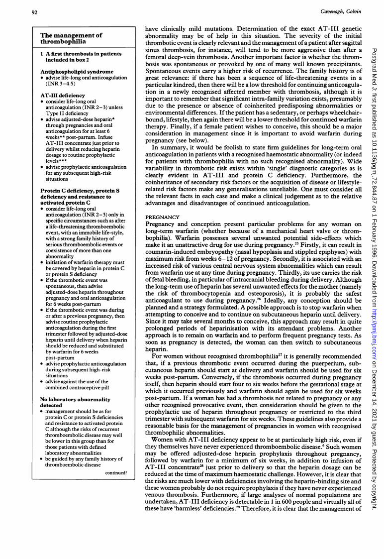
The management ofthrombophilia
1 A first thrombosis in patientsincluded in box 2
Antiphospholipid syndrome* advise life-long oral anticoagulation(INR 3-4.5)
AT-III deficiency* consider life-long oral
anticoagulation (INR 2-3) unlessType II deficiency
* advise adjusted-dose heparin*through pregnancies and oralanticoagulation for at least 6weeks** post-partum. InfuseAT-III concentrate just prior todelivery whilst reducing heparindosage to routine prophylacticlevels***
* advise prophylactic anticoagulationfor any subsequent high-risksituations
Protein C deficiency, protein Sdeficiency and resistance toactivated protein C* consider life-long oral
anticoagulation (INR 2-3) only inspecific circumstances such as aftera life-threatening thromboembolicevent, with an immobile life-style,with a strong family history ofserious thromboembolic events orcoexistence ofmore than oneabnormality
* initiation of warfarin therapy mustbe covered by heparin in protein Cor protein S deficiency
* if the thrombotic event wasspontaneous, then adviseadjusted-dose heparin throughoutpregnancy and oral anticoagulationfor 6 weeks post-partum
* if the thrombotic event was duringor after a previous pregnancy, thenadvise routine prophylacticanticoagulation during the firsttrimester followed by adjusted-doseheparin until delivery when heparinshould be reduced and substitutedby warfarin for 6 weekspost-partum
* advise prophylactic anticoagulationduring subsequent high-risksituations
* advise against the use ofthecombined contraceptive pill
No laboratory abnormalitydetected* management should be as for
protein C or protein S deficienciesand resistance to activated proteinC although the risks of recurrentthromboembolic disease may wellbe lower in this group than forthose patients with definedlaboratory abnormalities
* be guided by any family history ofthromboembolic disease
continued/
have clinically mild mutations. Determination of the exact AT-III geneticabnormality may be of help in this situation. The severity of the initialthrombotic event is clearly relevant and the management ofa patient after sagittalsinus thrombosis, for instance, will tend to be more aggressive than after afemoral deep-vein thrombosis. Another important factor is whether the throm-bosis was spontaneous or provoked by one of many well known precipitants.Spontaneous events carry a higher risk of recurrence. The family history is ofgreat relevance: if there has been a sequence of life-threatening events in aparticular kindred, then there will be a low threshold for continuing anticoagula-tion in a newly recognised affected member with thrombosis, although it isimportant to remember that significant intra-family variation exists, presumablydue to the presence or absence of coinherited predisposing abnormalities orenvironmental differences. If the patient has a sedentary, or perhaps wheelchair-bound, lifestyle, then again there will be a lower threshold for continued warfarintherapy. Finally, if a female patient wishes to conceive, this should be a majorconsideration in management since it is important to avoid warfarin duringpregnancy (see below).
In summary, it would be foolish to state firm guidelines for long-term oralanticoagulation in patients with a recognised haemostatic abnormality (or indeedfor patients with thrombophilia with no such recognised abnormality). Widevariability in thrombotic risk exists within 'single' diagnostic categories as isclearly evident in AT-III and protein C deficiency. Furthermore, thecoinheritance of secondary risk factors or the acquisition of disease or lifestyle-related risk factors make any generalisations unreliable. One must consider allthe relevant facts in each case and make a clinical judgement as to the relativeadvantages and disadvantages of continued anticoagulation.
PREGNANCYPregnancy and conception present particular problems for any woman onlong-term warfarin (whether because of a mechanical heart valve or throm-bophilia). Warfarin possesses several unwanted potential side-effects whichmake it an unattractive drug for use during pregnancy.25 Firstly, it can result incoumarin-induced embryopathy (nasal hypoplasia and stippled epiphyses) withmaximum risk from weeks 6-12 of pregnancy. Secondly, it is associated with anincreased risk of various central nervous system abnormalities which can resultfrom warfarin use at any time during pregnancy. Thirdly, its use carries the riskof fetal bleeding, in particular of intracranial bleeding during delivery. Althoughthe long-term use ofheparin has several unwanted effects for the mother (namelythe risk of thrombocytopenia and osteoporosis), it is probably the safestanticoagulant to use during pregnancy.26 Ideally, any conception should beplanned and a strategy formulated. A possible approach is to stop warfarin whenattempting to conceive and to continue on subcutaneous heparin until delivery.Since it may take several months to conceive, this approach may result in quiteprolonged periods of heparinisation with its attendant problems. Anotherapproach is to remain on warfarin and to perform frequent pregnancy tests. Assoon as pregnancy is detected, the woman can then switch to subcutaneousheparin.For women without recognised thrombophilia27 it is generally recommended
that, if a previous thrombotic event occurred during the puerperium, sub-cutaneous heparin should start at delivery and warfarin should be used for sixweeks post-partum. Conversely, if the thrombosis occurred during pregnancyitself, then heparin should start four to six weeks before the gestational stage atwhich it occurred previously and warfarin should again be used for six weekspost-partum. If a woman has had a thrombosis not related to pregnancy or anyother recognised provocative event, then consideration should be given to theprophylactic use of heparin throughout pregnancy or restricted to the thirdtrimester with subsequent warfarin for six weeks. These guidelines also provide areasonable basis for the management of pregnancies in women with recognisedthrombophilic abnormalities.Women with AT-III deficiency appear to be at particularly high risk, even if
they themselves have never experienced thromboembolic disease.6 Such womenmay be offered adjusted-dose heparin prophylaxis throughout pregnancy,followed by warfarin for a minimum of six weeks, in addition to infusion ofAT-Ill concentrate2 just prior to delivery so that the heparin dosage can bereduced at the time ofmaximum haemostatic challenge. However, it is clear thatthe risks are much lower with deficiencies involving the heparin-binding site andthese women probably do not require prophylaxis if they have never experiencedvenous thrombosis. Furthermore, if large analyses of normal populations areundertaken, AT-Ill deficiency is detectable in 1 in 600 people and virtually all ofthese have 'harmless' deficiencies.29 Therefore, it is clear that the management of
on Decem
ber 14, 2021 by guest. Protected by copyright.
http://pmj.bm
j.com/
Postgrad M
ed J: first published as 10.1136/pgmj.72.844.87 on 1 F
ebruary 1996. Dow
nloaded from

Guidelines for management of thrombophilia 93
/box 4 continued
2 A second thrombosis inpatients included in box 2
AT-III deficiency, protein Cdeficiency, protein S deficiencyand resistance to activatedprotein C* advise life-long oral anticoagulation(INR 2-3)
* initiation ofwarfarin therapy mustbe covered by heparin in protein Cand protein S deficiency
No laboratory abnormalitydetected* Management must be based on
individual and family history ofthromboembolic disease
3 Asymptomatic family memberswith laboratory abnormality
AT-III deficiency* advise adjusted dose heparin
throughout pregnancy and warfarinfor at least 6 weeks post-partum,except for the relatively benignType II defects
* advise prophylaxis during high-risksituations
* avoid the combined contraceptivepill
Protein C deficiency and proteinS deficiency* advise prophylaxis during high-risk
situations* consider the family history when
advising about subsequentpregnancies
* the combined contraceptive pill isprobably safe with protein Sdeficiency but should be avoided inprotein C deficiency until more dataare available
Resistance to activated protein C* the risks of thrombosis are much
lower than in the other identifiablegroups
* a low threshold for prophylaxisshould exist, especially for thoseindividuals with a strong familyhistory
* the combined contraceptive pillshould be avoided if othercontraception is acceptable33
*Adjusted dose heparin: heparin 12 hourlysubcutaneously, dose adjusted to achieve anactivated partial thromboplastin timewhich is just prolonged when tested 4-6hours after injection. There is limitedexperience of the use of low molecularweight heparin during pregnancy.**Anticoagulation during the puerperium:warfarin (INR 2-3) for a minimum of sixweeks***Prophylactic anticoagulation duringpregnancy: heparin 5000-7500 IU 12hourly subcutaneously
Box 4
individuals who are found to have potentially thrombophilic deficiencies during'routine' screening should not be equated with that of asymptomatic individualswho are identified from families with a thrombophilic history.Women with protein C or protein S deficiency who have previously had a
thrombosis should receive prophylaxis during pregnancy. Women who have hada thrombotic episode during late pregnancy or the puerperium may receivelow-dose heparin until the third trimester when adjusted dose heparin shouldcommence. Women who have had a spontaneous thrombosis unrelated topregnancy should receive adjusted dose heparin throughout pregnancy as withAT-III deficiency. The final subset of women with protein C or protein Sdeficiency who require consideration are asymptomatic individuals who havebeen detected following the identification of an affected family member. In thiscase, a clinical judgement will have to be made in the light ofthe kindred's familyhistory of thrombosis and after counselling the affected woman. The risk ofthromboembolic disease during pregnancy in women with resistance to activatedprotein C with no previous thrombosis is not clear at the present time and firmguidelines cannot be given.30Women with the anti-phospholipid syndrome and prior thromboembolic
disease should receive adjusted doses of heparin throughout pregnancy. Theproblem ofrecurrent fetal loss is a distinct one and optimal management remainsunresolved. Trials are under way comparing the relative benefits of aspirin,heparin and steroids.
In summary, prescriptive guidelines are not possible. Each individual casemust be considered on its own merits and a management decision made after fulldiscussion with the affected woman. Indeed, the decision-making process maybecome even more difficult as more women are found to have laboratory'abnormalities' that are known to be associated with thrombophilia in somekindreds, but which also have a relatively high frequency in the generalpopulation.
PROPHYLAXIS DURING HIGH-RISK PERIODSFor individuals who are known to have a laboratory abnormality associated withthrombophilia and who are not already receiving oral anticoagulation, thereshould be a very low threshold for recommending anticoagulant prophylaxisduring periods ofhigh risk for thromboembolism such as operations, immobilityand after trauma. Indeed, one of the main reasons for identifying affectedrelatives of patients with thrombophilia is to be able to offer them advice aboutprophylaxis.
FURTHER COUNSELLINGSeveral issues should be discussed with affected individuals and asymptomaticfamily members who are found to possess a thrombophilic laboratory abnor-mality. The advisability of prophylaxis during high-risk periods should bediscussed and the reversal of potentially avoidable risk factors encouraged, suchas smoking and obesity. The use of the combined oral contraceptive pill shouldgenerally be discouraged if alternative methods of contraception are acceptable.There is evidence that women with AT-III deficiency are at increased risk ofthromboembolic disease whilst using the contraceptive pill although womenwith protein S deficiency have no increased risk whilst taking a low-oestrogen pilland there is currently not enough evidence available to comment on women withprotein C deficiency.3' For all groups, the progesterone-only pill is probably safe.Hormone replacement therapy for post-menopausal women appears to be safe.The oestrogen component of hormone replacement therapy is lower than mostcontraceptive pills and hormone replacement therapy confers considerableadvantages with respect to cardiovascular mortality and the development ofosteoporosis.
Conclusion
Guidelines for the management ofthrombophilia are becoming clearer as more islearnt about the molecular mechanisms and natural histories of the variousunderlying disorders. Some suggested guidelines for testing and management ofthrombophilia are outlined in boxes 3 and 4. Although reasonably firm guidelinesare possible in certain situations (eg, recurrent thrombosis in patients with arecognised thrombophilic laboratory abnormality), it is not possible to giveadvice of proven validity in many other circumstances. For instance, there aremany individuals with unexplained thrombosis with no apparent predisposition(see box 2) who do not have any laboratory abnormality. It seems probable thatnew molecular defects will be discovered that might explain the thromboticpredisposition in this group of individuals, as indeed occurred when resistance to
on Decem
ber 14, 2021 by guest. Protected by copyright.
http://pmj.bm
j.com/
Postgrad M
ed J: first published as 10.1136/pgmj.72.844.87 on 1 F
ebruary 1996. Dow
nloaded from

94 Cavenagh, Colvin
activated protein C was first described. Meanwhile, after due consideration ofsuch a patient's personal and family thrombosis history, a clinical judgement hasto be made on an individual basis as to any long-term anticoagulation orprophylaxis. All of the guidelines discussed above are based on risk analyses ofindividuals who have suffered a venous thrombosis or are the asymptomaticaffected family members of such thrombotic individuals. In population termsthis is a highly selected group and it is becoming clear that large-scale screeningexercises of the general population will detect a very large number ofpeople whohave detectable abnormalities of the haemostatic system but no personal orfamily history of thrombosis. At the present time counselling such individualsremains very difficult and will remain so until more is known about the naturalhistories associated with specific defects.
1 Anderson FA, Brownell Wheeler H, et al. Apopulation-based perspective of the hospitalincidence and case-fatality rates of deep veinthrombosis and pulmonary embolism. ArchIntern Med 1991; 151: 933-8.
2 British Committee for Standards inHaematology. Guidelines on the investigationand management of thrombophilia. J ClinPathol 1990; 43: 703-9.
3 Olds RJ, Lane DA, Thein SL. The moleculargenetics of antithrombin deficiency. Br JHaematol 1994; 87: 221-6.
4 Thaler E, Lechner K. Antithrombin IIIdeficiency and thromboembolism. ClinHaematol 1981; 10: 369-90.
5 Mitchell L, Piovella F, Ofosu F, Andrew M.a2-Macroglobulin may provide protection fromthromboembolic events in antithrombin III-deficient children. Blood 1991; 78: 2299-304.
6 Conard J, Horellou MH, Van Dreden P,Lecompte T, Samama M. Thrombosis andpregnancy in congenital deficiencies ofAT III,protein C or protein S: study of 78 women.Thromb Haemost 1990; 63: 319-24.
7 Dahlback B, Carlsson M, Svensson PJ. Familialthrombophilia due to a previously unrecognisedmechanism characterised by poor anticoagulantresponse to activated protein C: prediction of acofactor to activated protein C. Proc Nati AcadSci USA 1993; 90: 1004-8.
8 Voorberg J, Roelse J, Koopman R, et al.Association of idiopathic venous thromboem-bolism with single point-mutation at Arg506 offactor V. Lancet 1994; 343: 1535-8.
9 Bertina RM, Koeleman BPC, Koster T, et al.Mutation in blood coagulation factor Vassociated with resistance to activated protein C.Nature 1994; 369: 64-7.
10 Svensson PJ, Dahlback B. Resistance toactivated protein C as a basis for venous throm-bosis. N Engl J Med 1994; 330: 517-22.
11 Beauchamp NJ, Daly ME, Hampton KK,Cooper PC, Preston FE, Peake IR. Highprevalence of a mutation in the factor V genewithin the UK population: relationship toactivated protein C resistance and familialthrombosis. Br J Haematol 1994; 88: 219-22.
12 Koster T, Rosendaal FR, de Ronde H, Briet E,Vandenbroucke JP, Bertina RM. Venousthrombosis due to poor anticoagulant responseto activated protein C: Leiden thrombophiliastudy. Lancet 1993; 342: 1503-6.
13 Greengard JS, Eichinger S, Griffin JH, BauerKA. Brief report: variability of thrombosisamong homozygous siblings with resistance toactivated protein C due to an Arg-Gln mutationin the gene for factor V. NEnglJMed 1994; 331:1559-62.
14 Allaart CF, Poort SR, Rosendaal FR, ReitsmaPH, Bertina RM, Briet E. Increased risk ofvenous thrombosis in carriers of hereditaryprotein C deficiency defect. Lancet 1993; 341:134-8.
15 Miletich J, Sherman L, Broze G. Absence ofthrombosis in subjects with heterozygous pro-tein C deficiency. N Engl J Med 1987; 317:991-6.
16 Marcianack E, Wilson HD, Marlar RA.Neonatal purpura fulminans: a genetic disorderrelated to the absence of protein C in blood.Blood 1985; 65: 15-20.
17 Dreyfus M, Magny JF, Bridey F, et al. Treat-ment of homozygous protein C deficiency andneonatal purpura fulminans with a purifiedprotein C concentrate. N EnglJ Med 1991; 325:1565-8.
18 Koster T, Rosendaal FR, Reitsma PH, van derVelden PA, Briet E, Vandenbroucke JP. FactorVII and fibrinogen levels as risk factors forvenous thrombosis. Thromb Haemost 1994; 71:719-22.
19 Koster T, Blann AD, Briet E, VandenbrouckeJP, Rosendaal FR. Role ofclotting factor VIII ineffect ofvon Willebrand factor on occurrence ofdeep-vein thrombosis. Lancet 1995; 345:152-5.
20 Den Heijer M, Blom HJ, Gerrits WBJ, et al. Ishyperhomocysteinaemia a risk factor for recur-rent venous thrombosis. Lancet 1995; 345:882-6.
21 Hughes GRV. The antiphospholipid syndrome:ten years on. Lancet 1993; 342: 341-4.
22 Khamashta MA, Hughes GRV. Antiphos-pholipid syndrome. A common cause of throm-bosis. BMJ 1993; 307: 883-4.
23 Comp PC, Thumau GR, Welsh J, Esmon CT.Functional and immunological protein S levelsare decreased during pregnancy. Blood 1986; 68:881-5.
24 Khamashta MA, Cuadrado MJ, Mujic F, TaubNA, Hunt BJ, Hughes GRV. The managementof thrombosis in the antiphospholipid-antibodysyndrome. N Engi J Med 1995; 332: 993-7.
25 Hirsh J. Oral anticoagulant drugs. N EnglJ Med1991; 324: 1865-75.
26 Hirsh J. Heparin. N Engl Med 1991; 324:1565-74.
27 British Committee for Standards inHaematology. Guidelines on the prevention,investigation and management of thrombosisassociated with pregnancy. J Clin Pathol 1993;46: 489-96.
28 De Stefano V, Leone G, De Carolis S, et al.Management of pregnancy in women withantithrombin III congenital defect: report offour cases. Thromb Haemost 1988; 59: 193-6.
29 Tait RC, Walker ID, Perry DJ, et al. Prevalenceof antithrombin deficiency in the healthypopulation. Br J Haematol 1994; 87: 106-12.
30 Cook G, Walker ID, McCall F, Conkie JA,Greer IA. Familial thrombophilia and activatedprotein C resistance: thrombotic risk in preg-nancy? Br J Haematol 1994; 87: 873-5.
31 Pabinger I, Schneider B and the GTH studygroup on natural inhibitors. Thrombotic risk ofwomen with hereditary antithrombin III-, pro-tein C- and protein S-deficiency taking oralcontraceptive medication. Thromb Haemost1994; 71: 548-52.
32 Cooper PC, Hampton KK, Makris M,Abuzenadah A, Paul B, Preston FE. Furtherevidence that activated protein C resistance canbe misdiagnosed as inherited functional proteinS deficiency. Br J Haematol 1994; 88: 201-3.
33 Vandenbroucke JP, Koster T, Briet E, ReitsmaPH, Bertina RM, Rosendaal FR. Increased riskofvenous thrombosis in oral-contraceptive userswho are carriers of factor V Leiden. Lancet 1994;344: 1453-7.
on Decem
ber 14, 2021 by guest. Protected by copyright.
http://pmj.bm
j.com/
Postgrad M
ed J: first published as 10.1136/pgmj.72.844.87 on 1 F
ebruary 1996. Dow
nloaded from