jiafm-33-2
TRANSCRIPT

J Indian Acad Forensic Med. April-June 2011, Vol. 33, No. 2 ISSN 0971-0973
i
Indian Academy of Forensic Medicine (IAFM)
(Registration No.349, 12th May, 1972, Panji, Goa)
Governing Council 2010-2012
President
Dr. D.S.Badkur
General Secretary Treasurer Dr.Adarsh Kumar Dr. A.S. Thind Vice Presidents North Zone: Dr. Dalbir Singh South Zone: Dr. P.Sampath Kumar East Zone: Dr. Tulsi Mahto West Zone: Dr. H.T. Katade Central Zone: Dr. R.K. Singh
Joint Secretaries North Zone: Dr. Dasari Harish South Zone: Dr. Cyriac Job East Zone: Dr. Shoban Das West Zone: Dr. Hasumati Patel Central Zone: Dr. P.S.Thakur
Editor Dr. Mukesh Yadav
Joint Editor Dr. Akash Deep Aggarwal
Executive Members
Dr.B.P. Dubey (Ex. President, IAFM) Dr. Aditya Kumar Sharma Dr.Sarvesh Tandon Dr.C.P.Bhaisora Dr.Pankaj Gupta Dr.Luv Sharma
Dr. Sanjoy Das (Ex. Secretary, IAFM) Dr.Amandeep Singh Dr.Mukesh K.Goyal Dr.C.B. Jani Dr.Jaynti Yadav Dr.P.K.Tiwari

J Indian Acad Forensic Med. April-June 2011, Vol. 33, No. 2 ISSN 0971-0973
ii
Journal of Indian Academy of Forensic Medicine
(JIAFM) The official publication of Indian Academy of Forensic Medicine
Editor,
Dr. Mukesh Yadav Professor & H.O.D., Forensic Medicine & Toxicology, School of Medical Sciences and Research, Sharda University, Greater Noida-201306, Uttar Pradesh, INDIA Residence: G-216, Parsvanath Edens, Alfa-II, Greater Noida, G.B. Nagar, U.P.INDIA Ph. No. 0120-2326060, Cell: 09411480753 Email: [email protected]
Joint Editor,
Dr. Akash Deep Aggarwal Assistant Professor,
Department of Forensic Medicine, Govt. Medical College, Patiala
Punjab, INDIA Residence:
H.No. 14, Desi Mehmandari, Patiala-147001, Punjab, INDIA
Cell: 9815652621 Email:[email protected]
Peer Review Group
Dr. A.K. Srivstava Professor & H.O.D., Forensic Medicine &Toxicology Subharti Medical College, Meerut, U.P. Dr. V.V.Pillay Professor & H.O.D., Analytical Toxicology, Chief of Poison Control Centre AIMS & R, Cochin-Kerala
Dr. R.K. Gorea Professor & H.O.D., Forensic Medicine &Toxicology Gian Sagar Medical College, Banur, Patiala, Punjab
Dr. C.B.Jani Professor & H.O.D., Forensic Medicine and Toxicology P.S.Medical College, Karamsad, Distt. Anand, Gujarat,
Dr. T.K Bose Professor & H.O.D., Forensic and State Medicine Govt. Medical College Kolkata, West Bengal
Dr. G. Pradeep Kumar Professor & H.O.D., Forensic Medicine &Toxicology Kasturba Medical College, Manipal, Karnatka
Advisory Board
Sharma G.K., (New Delhi) Verma S.K., (New Delhi) Kaur Balbir, (Srinagar) Bansal Y., (Chandigarh) Kumar Shantha B., (Tamilnadu) Gupta B.D., (Gujrat) Manju Nath K.H, (Karnatka) Das Sanjoy, (Uttarakhand) Bhaisora C.P, (Uttarakhand) Mahtoo Tulsi, (Jharkhand)
Ravindran K. (Pondicherry) Sabri Imran, (U.P.) Rastogi Prateek (Karnatka) Potwary AJ (Assam) Singh R.K. (Chhatisgarh) Dongre A.P. (Nagpur) Rastogi Pooja (U.P.) Sharma Aditya (H.P.) Khanagwal V. (Haryana) Gupta Pankaj (Punjab)
Pounder Derrick, (England) Khaja Shaikh (A.P.) Basu R (W.B.) Naik R.S. (Maharastra) Godhikirakar Madhu (Goa) Job Cyriac (Kerala) Vinita K. (U.P.) Yadav B.N. (Nepal) Mohite Shailesh (Mumbai) Singh Abhas Kumar (U.P.)
Printed and published by Dr. Mukesh Yadav, Editor, JIAFM and Dr. A. D. Aggarwal, Joint Editor, JIAFM on
behalf of Indian Academy of Forensic Medicine at name of the press [SHIVANI PRINTERS, NOIDA, U.P.]

J Indian Acad Forensic Med. April-June 2011, Vol. 33, No. 2 ISSN 0971-0973
95
Journal of Indian Academy of Forensic Medicine
Contents Sr. Page
I. From the Editor’s Desk 97-97
II. Editorial: Doctrine of Parens Patriae: Applicability in Medical Profession 98-101
Original Research Paper
1. Profile of Organophosphorus Poisoning at Maharani Hospital, Jagdalpur,
Chhattisgarh: A Three Years Study Dhaval J. Patel, Pawan R. Tekade
102-105
2. Trends of Poisoning and Gross Stomach Mucosal Appearance in Fatal
Poisoning Cases: An Autopsy Study Kishan R Siddapur, Gurudatta S Pawar,
Shashidhar C Mestri
106-111
3. Study of Palatal Rugae Pattern among the Student Population in Mangalore
Mahabalesh Shetty, Premalatha K
112-115
4. Tuberculous Lesions at Autopsy Monika Garg, Akash Deep Aggarwal, Sneh Singh,
Sant Prakash Kataria
116-119
5. Trends of Homicidal Deaths at a Tertiary Care Centre Bengaluru
B. C. Shivakumar, D. Vishwanath, Prem Chandra Srivastava
120-124
6. Epiphyseal Fusion at Lower End of Radius and Ulna Valuable Tool for Age
Determination D h a r m e s h S . P a t e l , H a r i s h A g a r w a l , J i g e s h V . S h a h 125-129
7. Age Estimation from Third Molar Development: A Radiological Study
Jashwant A. Darji, Ganesh Govekar, S.D. Kalele, Hareshwari Hariyani
130-134
8. An Epidemiological Survey of Fatal Road Traffic Accidents and their
Relationship with Head Injuries Ravindra S Honnungar, Sunil C Aramani, Vijay Kumar AG,
Ajay Kumar TS, Prasanna S Jirli
135-137
9. A Study of Left Hand Thumb Imprint Patterns among Medical Students at
Karamsad (Gujarat) Zalak Patel, Krupa Tarpara, Shruti Parikh, Sanjay Gupta 138-139
10. Placenta: The Wonder Organ Shashi Munjal Mongia, Sanjeev Kumar Jain, Mukesh Yadav 140-142
11. Use of Hair Root Sheath for Barr body Determination Harpreet Singh,
O.P. Aggarwal, Arsalaan F. Rashid
143-144
12. Bacteriological Profile of Biomedical Waste: Management Guidelines
Vichal Rastogi, Pooja Rastogi, Shalini Bhatia
145-148
13. Age Related Changes in Mechanical and Thermal Injuries-A Post Mortem
Study Lavlesh Kumar, Chaitanya B. V., Swapnil S Agarwal, Binaya Kumar Bastia
149-151
Volume: 33 • Number: 2 • April-June 2011
J Indian Acad Forensic Med. April-June 2011, Vol. 33, No. 2 ISSN 0971-0973
96
Review Research Papers
14. Role of Forensic Medicine in Administration of Justice: A Critical Review
U.S. Sinha, Mukesh Yadav, Soni Uttam, A.K. Pathak, Sanju Singh
152-160
15. An Approach for Identification of Individuals in a Mass Disaster in Indian
Set Up S. Das, S.K. Pandey, P. Chakraborty
161-162
16. Microbial Forensics – An Upcoming Investigative Discipline P. Aggarwal,
A.K. Chopra, S .Gupte, S.S. Sandhu
163-165
17 Digital Forensics- A Technological Revolution in Forensic Sciences
Pankaj Gupta, Jaspal Singh, Anterpreet Kaur Arora, Shashi Mahajan
166-170
18. Estimation of Post-Mortem Interval from the Changes in Vitreous
Biochemistry Nidhi Sachdeva, Yashoda Rani, Ritu Singh, Atul Murari
171-174
19. Fall: The Accidental Injury in Geriatric Population Alok Kumar, Archana Verma,
Mukesh Yadav, A. K.Srivastava
175-178
Case Reports
20. Cardiac Tamponade Following Post Myocardial Ventricular Wall Rupture- A
Case Report Amit Sharma, Jatin Bodwal.
179-181
21. Meticulous Examination of Body Pays…. Hitesh Chawla, Basant Lal Sirohiwal,
P.K. Paliwal
182-183
22. Diatoms-Role in Drowning R. K. Punia 184-186
Book Review
23 Textbook of Forensic Medicine & Toxicology: Principles & Practice
Krishna Vij
187-187
24 News and Views Imran Sabri, Pooja Rastogi, Mukesh Yadav 188-188
From Editor’s Desk
Copy Right ©All rights reserved: No part of this publication may be reprinted or publish without the
prior permission of the Editor, JIAFM. Submission of all manuscripts to the journal is understood to imply that it is not being considered for publication elsewhere. Submission of multi authored papers implies that the consent of each author has been obtained. In this journal, every effort has been made NOT to publish inaccurate or misleading information. However, the Editor, Joint Editor, Peer Review Group and Advisory
Board accept NO liability in consequences of such statements. The Journal of Indian Academy of Forensic Medicine is indexed in Index Copernicus [Poland] and IndMED [India] Print ISSN: 0971-0973. Electronic ISSN: 0974-0848. IndMED www.medind.nic.in/jal/jalm.shtml
Address request for reprint or further information relating to any article may please be made with author and
in case of multi authored article, please communicate to Corresponding Author or the First Author

J Indian Acad Forensic Med. April-June 2011, Vol. 33, No. 2 ISSN 0971-0973
97
JIAFM A Quarterly Publication
Volume 33, Number 2, April-June, 2011
I feel immense pleasure to present before you the second issue of 2011. I assure you
about the quality of research papers and quality of printing in future issues. Your valuable
suggestions are always encouraging me and I heartily welcome for future suggestions. On
behalf of Executive Committee of IAFM for the years 2010-2011, I took resolution to further
improve the quality and status of our Journal. We always learn from mistakes and try to improve
upon these. I am thankful to the advertisers who have provided additional financial resources for
improving the quality of this issue.
Professor [Dr.] Mukesh Yadav Editor, JIAFM
Subscription Information • Members of IAFM will receive the free of cost. • Non Members and Institutions (Annual Subscription rates) • Personal: In India, Rs. 1000/ (Rest of the world: US$ 200/ or equivalent) • Institutions: In India, Rs. 3000/ (Rest of the world: US$ 400/ or equivalent) • We Accept: Bank Cheque / Demand Drafts (Add Rs. 50/- for outstation Cheques) • The Scope of the Journal covers all aspects of Forensic Medicine and allied fields,
research and applied.
Subscription orders and payments should be made in favour of
“Editor, JIAFM, payable at Greater Noida”
Claims for missing issue: A copy will be sent free to the member / subscriber provided the claim is made within 2 months of publication of the issue & self addressed envelop of the size 9” x 12” is sent to the Editor. (Those who want the journals to be dispatched by Registered Post must affix Rs. 50/ worth postage stamps).
The journal is indexed with IndMed and made available online by following website:
www.medind.nic.in http://indmed.nic.in www.jiafm.com www.forensicindia.com www.indianjournals.com

J Indian Acad Forensic Med. April-June 2011, Vol. 33, No. 2 ISSN 0971-0973
98
Editorial: Doctrine of Parens Patriae: Applicability in Medical
Profession The doctrine of Parens Patriae (father of the country) had originated in British law as early as the
13th century. It implies that the King is the father of the country and is under obligation to look after the interest of those who are unable to look after themselves.
The idea behind Parens Patriae is that if a citizen is in need of someone who can act as a parent who can make decisions and take some other action, Sometimes the State is best qualified to take on this role.
In the Constitution Bench decision of Supreme Court in Charan Lal Sahu vs. Union of India (1990) 1 SCC 613 (vide paras 35 and 36), the doctrine has been explained in some details as follows: "In the "Words and Phrases" Permanent Edition, Vol. 33 at page 99, it is stated that parens patriae is the inherent power and authority of a legislature to provide protection to the person and property of persons non sui juris, such as minor, insane, and incompetent persons, but the words parens patriae meaning thereby `the father of the country', were applied originally to the King Andare used to designate the State referring to its sovereign power of guardianship over persons under disability.
What do you mean by Parens patriae jurisdiction? Parens patriae jurisdiction, is the right of the sovereign and imposes a duty on the sovereign, in
public interest, to protect persons under disability who have no rightful protector. The connotation of the term parens patriae differs from country to country, for instance, in England it is the King; in America it is the people, etc. The government is within its duty to protect and to control persons under disability".
The duty of the King in feudal times to act as parens patriae (father of the country) has been taken over in modern times by the State.
In Heller vs. DOE (509) US 312 Mr. Justice Kennedy speaking for the U.S. Supreme Court observed: "The State has a legitimate interest under its parens patriae powers in providing care to its citizens who are unable to care for themselves". In State of Kerala vs. N.M. Thomas, 1976(1) SCR 906, Mr. Justice Mathew observed:
"The Court also is ‘State’ within the meaning of Article 12 (of the Constitution)."
Views of Supreme Court of India: Hon’ble Supreme Court opined in Aruna Sahnbag case of 2010, in the case of an incompetent
person who is unable to take a decision whether to withdraw life support or not, it is the Court alone, as parens patriae, which ultimately must take this decision, though, no doubt, the views of the near relatives, next friend and doctors must be given due weight.
Under which provision of the law can the court apply doctrine of parens patriae: SC opined “it is the High Court under Article 226 of the Constitution of India which can grant
approval for withdrawal of life support to such an incompetent person. Article 226(1) of the Constitution states: "Notwithstanding anything in Article 32, every High Court shall have power, throughout the territories in relation to which it exercises jurisdiction, to issue to any person or authority, including in appropriate cases, any Government, within those territories directions, orders or writs, including writs in the nature of habeas corpus, mandamus, prohibition, quo warranto and certiorari, or any of them, for the enforcement of any of the rights conferred by Part III and for any other purpose".
A bare perusal of the above provisions shows that the High Court under Article 226 of the Constitution is not only entitled to issue writs, but is also entitled to issue directions or orders.
In Dwarka Nath vs. ITO AIR 1966 SC 81(vide paragraph 4) Supreme Court observed: "This article is couched in comprehensive phraseology and it ex facie confers a wide power on the High Courts to reach injustice wherever it is found. The Constitution designedly used a wide language in describing the nature of the power, the purpose for which and the person or authority against which it can be exercised. It can issue writs in the nature of prerogative writs as understood in England; but the scope of those writs also is widened by the use of the expression "nature", for the said expression does not equate the writs that can be issued in India with those in England, but only draws an analogy from them.
That apart, High Courts can also issue directions, orders or writs other than the prerogative writs. It enables the High Courts to mould the reliefs to meet the peculiar and complicated requirements of this country. Any attempt to equate the scope of the power of the High Court under Art. 226 of the

J Indian Acad Forensic Med. April-June 2011, Vol. 33, No. 2 ISSN 0971-0973
99
Constitution with that of the English Courts to issue prerogative writs are to introduce the unnecessary procedural restrictions grown over the years in a comparatively small country like England with a unitary form of Government to a vast country like India functioning under a federal structure."
The above decision has been followed by the Supreme Court in Shri Anadi Mukta Sadguru vs. V. R. Rudani AIR 1989 SC 1607
No doubt, the ordinary practice in our High Courts since the time of framing of the Constitution in 1950 is that petitions filed under Article 226 of the Constitution pray for a writ of the kind referred to in the provision. However, from the very language of the Article 226, and as explained by the above decisions, a petition can also be made to the High Court under Article 226 of the Constitution praying for an order or direction, and not for any writ.
SC opined “Hence, in our opinion, Article 226 gives abundant power to the High Court to pass suitable orders on the application filed by the near relatives or next friend or the doctors/hospital staff praying for permission to withdraw the life support to an incompetent person of the kind above mentioned.
Another landmark judgement where doctrine of parens patriae was applied by the Punjab & Haryana high court , which later on approved by the Supreme Court in an appeal.
Views of Supreme Court of India: The mentally challenged girl, with 19
th week’s pregnancy with the help of an NGO and a public
spirited advocate, moved the SC in July 2009 seeking protection of the unborn child with a delicate question to be answered by the Apex Court. The C.A. argued that even normal mothers found it difficult to raise a mentally retarded child. The attempt was to magnify the future trouble that the victim would face if the case was decided on the basis of emotion.
For the victim, advocate argued that the victim had no one in the world as her blood relative. Both arguments sounded cogent. And the SC appeared to be swinging from one view to the other when the contrast arguments were advanced, liberally sprinkled with emotion. But, at the end it chose life and said, ‘‘Nature will take care.’’
What was unique in this case? • She was major i.e. above 18 years of age (19-20 years) • She was mentally retarded with mental age of 7-9 years • She has no guardian or relatives • She had valid ground for MTP • She was willing to continue the pregnancy • She has no resources to rear her would be offspring and earn livelihood • Continuation of pregnancy does not pose any threat to her life and health • Legal authorities were not willing to give consent on her behalf
• No threat to child to be born with physical or mental handicapped Questions for consideration before the Court:
• Whether the pregnancy of the victim is liable to be terminated? • Whether or not the continuance of the pregnancy of a mentally retarded major pregnant woman
involves risk to her life or can cause grave injury to her physical or mental health? • Should the consent of the victim be considered mandatory to terminate her pregnancy or, • Who shall be the competent person to give consent for such termination? • Can Chandigarh Administration or Other Government Authorities competent to give consent on
her behalf? • Can High Court, in exercise of its parens patriae jurisdiction, assign such consent by issuing
appropriate directions? • Would a surrogate mother take care of the victim’s child? • Would it not be traumatic for the victim to lose her child to a surrogate mother? • Why could not the court permit her to have her first blood relative? • Can a mentally challenged woman be denied the right to motherhood? • Can the courts order abortion without the consent of the mother, which is prohibited under
Medical Termination of Pregnancy Act?
Consent for MTP by mentally ill / mentally retarded global scenario: Debate on this issue was relied upon the Mental Capacity Act, 2005 of UK and the views of
several subject-experts, advancement of Medical Science, universal recognition of the Fundamental Rights of the mentally retarded persons, recent theory of mixing them in the main social stream instead of
J Indian Acad Forensic Med. April-June 2011, Vol. 33, No. 2 ISSN 0971-0973
100
barricading at a secluded place. The legislative transformation on the issue of consent has also taken place whereby purposefully and knowingly, the non-competence to give consent for medical termination of a pregnancy in the cases of mentally ill pregnant woman on one hand and competence of major mentally retarded pregnant woman on the other hand, has now been distinguished.
Human Rights Principles for mentally ill: Emphasis has also been laid on 25 principles adopted by the General Assembly of the United
Nations for the protection of persons with mental illness and for improvement of mental healthcare, with a special reference to the following clauses:
Principal 1: Fundamental freedoms and basic rights: Any decision that, by reason of his or her mental illness, a person lacks legal capacity, and any
decision that, in consequence of such incapacity, a personal representative shall be appointed, shall be made only after a fair hearing by an independent and impartial tribunal established by domestic law. The person whose capacity is at issue shall be entitled to be represented by a counsel. If the person whose capacity is at issue does not himself or she secures such representation, it shall be made available without payment by that person to the extent that he or she does not have sufficient means to pay for it.
The counsel shall not in the same proceedings represent a mental health facility or its personnel and shall not also represent a member of the family of the person whose capacity is at issue unless the tribunal is satisfied that there is no conflict of interest. Decisions regarding capacity and the need for a personal representative shall be reviewed at reasonable intervals prescribed by domestic law. The person whose capacity is at issue, his or her personal representative, if any, and any other interested person shall have the right to appeal a higher court against any such decision”.
Principal 11: Consent to treatment: No treatment shall be given to a patient without his or her informed consent, except as provided
for in paragraphs 6, 7, 8, 13 and 15 of the present principle. The Courts cannot be oblivious of the fact that ours is a country inflicted with imbalanced male-
female sex-ratio; marred by female foeticide; ashamed of a vast majority of abandoned girls in orphanages; clouded with social evils like dowry; poor literacy rate amongst girls, with alarming increase in dowry deaths and, therefore, the freedom of consent given to a mentally retarded major pregnant woman by virtue of sub-section [4] of Section 3 of the 1971 Act, has to be taken as susceptible and can not be accepted on its face value by a Court while exercising its parens-patriae jurisdiction.
In majority of the eventualities to seek consent of a mentally retarded major pregnant woman for medical termination of her pregnancy might be of those who have been orphans and have no identified relative to act as their natural guardian. Could they also be placed at the same pedestal and at par with those who are under the direct care, control and guardianship of their parents, kith and kin etc. – is a question to be examined by the Law Makers and not to be commented on by us”.
Relying upon two Division Bench judgments and a judgment of the Supreme Court it was urged that even a court decree against a lunatic without the appointment of a guardian is a nullity.
A passionate reference to the medical reports/opinions on record and urged that having regard to the deficiencies in the areas of self-help grooming and socialisation and the fact that she is unable to look after herself and can not fend for herself if left to her own devices, coupled with the IQ level of the victim stated to be that of a nine years old, especially owing to the major spinal surgery undergone by the victim during her childhood and possibility of bony abnormalities to be genetically inherited by the baby, this Constitutional Court should come to the rescue of the victim and invoke its parens-patriae jurisdiction by granting permission to terminate the pregnancy, which is otherwise also a cause of anguish having been caused by a diabolic act of rape.
‘Judicial bye-pass procedure’: Some of the books have highlighted the increase in risks to the pregnant woman’s health after the
first trimester and how in the developed countries like the USA and Canada also the practice of parental consent has gained importance and the Courts also follow the ‘judicial bye-pass procedure’.
The literal interpretation as given above, however, completely falls short of achieving the legislative object of not only the 1971 Act, it may also tinker with the legislative object of the 1999 Act as well as the UN Declaration on the rights of the mentally retarded persons. We say so for the reason that in the context of termination of pregnancy being a penal offence prior to the 1971 Act came into force and one of the objects of the Act being permitting the termination of pregnancy on humanitarian grounds when it is caused by a sex crime like rape or intercourse with a lunatic woman the expression which has been amended by the Act of 2002 only and which prior to such amendment included mentally retarded

J Indian Acad Forensic Med. April-June 2011, Vol. 33, No. 2 ISSN 0971-0973
101
pregnant woman also, any interpretation should lean towards liberalizing medical termination of pregnancy.
Duty of the Guardian: Court observed that “We are unhesitatingly of the view that such like cases can not be decided on
the solitary strength of interpretation of legal provisions. Besides being vested with plenary and inherent jurisdiction to act as a custodian of the fundamental and human rights of the citizens, a writ Court while exercising parens-patriae jurisdiction owes a bounden duty to act in the best interest of the guardee, keeping in view his/her care, protection, health, education, intellectual development, comforts, contentment and congenial environment, along with moral and ethical values, as emphasised by their Lordships of the Supreme Court in Nil Rattan Kundu’s case.”
Exclusion of mentally retarded persons is not absolute: Court further observed that “In our view, the exclusion of mentally retarded persons from the
category of mentally ill persons under the 1971 Act is not absolute in the sense that irrespective of the foreseeable environment in which such mentally retardee is living or the degree and condition of mental retardedness, the Court even while exercising its parens-patriae jurisdiction cannot appoint a guardian to determine as to whether or not the continuance of the pregnancy of a mentally retarded major pregnant woman involves risk to her life or can cause grave injury to her physical or mental health”.
Parens-patriae jurisdiction of High Court: Court accordingly hold that notwithstanding the plain and literal meaning of Section 3(4) of the
MTP Act, 1971, every Court while exercising its parens-patriae jurisdiction is competent to act or appoint guardian ad-litem of a mentally retarded major pregnant woman for the purpose of deciding the retention or termination of her pregnancy in her best interest, though depending upon the individual facts and circumstances of each case”.
“Such guardian may consult or seek consent of the pregnant woman concerned for the purpose of formation of his final decision as to whether or not the pregnancy be medically terminated”.
Issue of major, orphan and mentally retarded woman victim of sexual assault leading to pregnancy was discussed and debated not only by the media but also by the scientific community and legal experts in details for the first time in India. Uniqueness of this case was the issue of consent and interpretation of the MTP Act, 1971 with Amendment 2002 and role of state, and court as a guardian to give consent on behalf of mentally retarded.Due to issue of human rights involved every body concerned with the case whether government authorities or medical experts or even judiciary was throwing the ball in each others court.
Finally, the case land up before the P & H High Court which directed for the MTP without the consent of the victim, after receiving Board of Expert’s opinion in this regard. Court was of the considered view that the many vital issues need to be answered by an Expert Body, who should be free from the administrative control and/or influence of the petitioner, the Chandigarh Administration.
Seeing the technicalities involved Punjab & Haryana High Court observed that “In the light of what has been held above, and taking into consideration the medical opinion/evidence on record, which we have no reason whatsoever to doubt or disbelieve, and taking notice of the predicament of the petitioner – State and for the absolute satisfaction of this Court in its capacity as a parens-patriae.
Aggrieved by this order victim with the help of a NGO and pubic spirited Advocate challenged the validity of direction for MTP before the Supreme Court of India, which stayed the order of the High Court.
Birth of baby girl in the month of December 2, 2009 poses many challenges before the State, NGO and medical fraternity, legal experts. Doctrine of Parens Patirae has great applicability in cases of incompetent, minors and other category of cases faced by the physicians in Indian Context such as in case of Aruna Sahnbag case decided by the Supreme Court in May 2010.
Recent controversy arises after the death of Sri Satya Sai, regarding order for cascade, this doctrine of Parens Patriae needs application to avoid misuse of decision for removal of life saving support.
Medical profession can benefit by application of this doctrine in appropriate cases to save many lives or preserve health of patients in similar cases.
Editor

J Indian Acad Forensic Med. April-June 2011, Vol. 33, No. 2 ISSN 0971-0973
102
Original Research Paper
Profile of Organophosphorus Poisoning at Maharani Hospital, Jagdalpur, Chhattisgarh: A Three Years Study
*Dhaval J. Patel, **Pawan R. Tekade
Abstract A number of Organophosphorus compounds have been introduced in Indian market as
agricultural insecticides, being effective against wide range of insects and pests. But a number of these compounds have proved to be more toxic to humans than its utility as insecticides, pesticides or fungicides. This study aims to evaluate the certain factors which are very significant in relation to outcome of Organophosphorus compound poisoning like age, sex, SE status, marital status, reason of committing suicide, etc. and attempt to know its prevalence in society and try to plan for future preventive strategy.
The study was carried out on 288 cases of Organophosphorus compound poisoning which come to Maharani Hospital, Jagdalpur, (C.G) from 01/01/2007 to 31/12/2009. M: F ratio was 1.3: 1.0. Majority of the cases were in age group of 21-30 yrs includes 128 cases (44.44%). Higher proportion of cases from lower class of society 141 cases (48.95%), from rural area 237 cases (82.29%), due to lack of education in affected community. Suicidal intent to consume the compound was the commonest - 250 cases (86.80%). Recovery rate was highest amongst those who consume less than 10 ml of poison – 120 cases (41.66%).
Key Words: Organophosphorus, Suicidal, Poisoning, Tick-20, Insecticide
Introduction: Poisoning-both intentional and accidental are significant contributor to mortality and morbidity throughout the world. According to WHO, three million acute poisoning cases with 2, 20,000 deaths occurs annually throughout the world. Out of these 90% of poisoning cases belongs to developing countries particularly among the agricultural workers. Pattern of poisoning in any region depends on variety of factors such as availability of poisons; SE status of population, religious beliefs and cultural influences. [8, 10] It is roughly estimated in India the 5 to 6 persons per lakhs population die due to poisoning every year. The commonest cause of poisoning in India and other developing countries is organophosphorus compounds.
Corresponding Author: *Associate Professor, Forensic Medicine Department, Shri Sathya Sai Medical College and Research Institute, Nellikuppam, Kanchepuram, (TN) Mahatma Gandhi Medical College &Research Institute, Pondy-Cuddalore Main Road, Pillaiyarkuppam, Pondicherry, 607 402, Mob – 08870080017, E-mail – [email protected] **Assistant Professor, Forensic Medicine Department, Govt. Medical College, Jagdalpur, Chhattisgarh
The common reasons behind this may be agricultural based economy, poverty and easy availability of highly toxic poisons easily.
The commonest poisons are organophosphates, carbonates, chlorinated hydrocarbons and aluminium phosphide. Occupational poisoning due to pesticides is also common in developing countries due to unsafe practices of its use, ignorance about toxicity of agents and lack of protective clothing. Poisoning of these compounds in children is almost entirely accidental while in adults mainly suicidal [8, 11].Mortality varies from place to place depending on the nature of poison, availability of facilities and treatment by qualified persons. [7]
Material and Methods: In present study from 01/01/2007 to 31/12/2009 (3 years) – cases of organophosphorus compound poisoning came to Maharani Hospital, Jagdalpur, (C.G) either from emergency department or through medicine OPD were considered. Detailed history of every case regarding type of the compound, its quantity, time/reason and manner (either intentionally or accidentally) of consumption, age/sex and occupation of person and marital status was taken. Patient’s indoor case records, Post mortem reports, Emergency notes and sometimes inquest reports were also considered for information.

J Indian Acad Forensic Med. April-June 2011, Vol. 33, No. 2 ISSN 0971-0973
103
Observations: In present three years study, 31,203 cases were admitted in Maharani Hospital, Jagdalpur, (C.G) and out of which 427 cases (1.36%) were due to poisoning. Out of total poisoning cases 288 cases (67.44%) were of organophosphorus compound poisoning victims. During 2008 no. of total hospital admissions – 11,748 (37.65%), poisoning cases – 178 (41.68%) and organophosphorus compound poisoning cases – 123 cases (42.70%) were highest in comparison to 2007 and 2009. (TABLE – 1) From total 288 cases of organophosphorus compound poisoning – 164 cases (56.94%) were males and 124 (43.05%) were females. Majority of victims fall in 21-30 yrs – 128 cases (44.44%) which is followed by 11-20 yrs – 86 cases (29.86%). The least no. of cases 3 (1.04%) were found in more than 50 yrs. (TABLE – 2) Nearly half of the incidence of organophosphorus compound poisoning amongst the lower class – 142 cases (48.95%). The middle and upper class had 83 (28.81%) and 64 (22.22%) of cases. (TABLE – 3) Out of 288 total cases, 237 (82.29%) were from rural area and only 51 (17.70%) from urban area. (TABLE – 4) Highest no. of victims consumed Tick-20 – 76 (26.78%) amongst 288 cases followed by Unknown compound poisoning – 61 (21.18%) and Methyl parathion – 55 (19.09%). The least consumed compound was Dichlorovos – 21 cases (7.29%) only. (TABLE – 5) Majority were married 215 (74.65%) and 73 (25.34%) unmarried victims. Male was dominantly affected sex in both category which was – 121 cases (73.78%) and 94 cases (75.80%). (TABLE – 6) Maximum no. of victims – 273 (94.79%) recovered from effects of poisoning and only 15 victims (5.20%) were died due to fatal effects of poisonous compounds. Out of 288 cases, highest no. of cases – 120 (41.66%) consumed less than 10 ml and least no. of victims -23 (7.98%) consumed more than 101 ml of poisonous compound. (TABLE – 7) In present study 250 cases (86.80%) cases were suicidal followed by accidental 36 (12.50%) and only 2 cases were of homicidal manner. Male more affected sex in each manner of poisoning in present study. (TABLE – 8) Financial problem was commonest in married male – 71 cases (58.67%) and Domestic problem was most frequent in married females – 51 cases (54.25 %). In unmarried
category also both the reasons were commonest in male – 30 cases (69.76%) and in females – 17 cases (56.66%). (TABLE – 9) Majority of the victims – 244 (84.72%) reached to hospital within 6 hrs. while only 15 cases (5.20%) delayed for more than 24 hrs. (TABLE – 10) Majority of organophosphorus poisoning victims suffered from muscarinic and nicotinic symptoms like nausea and vomiting (78.18%), muscular weakness (80.23%) and excessive sweating in (69.14%) of cases. Neurological involvement in form of altered sensorium observed in (21.08%) of cases. (TABLE – 11)
Discussion: According to the WHO, three million acute poisoning cases with 2, 20,000 deaths occur annually and of these 90% of fatal poisoning in developing countries, particularly among agricultural workers. Pattern of poisoning in a region depends on variety of factors such as availability of the poisons, SE status of the population and religious/cultural influences. [8] Male victims were commonly observed in present study (56.94%) than female (43.05%). Similar observations were made by DG Gannur et al [2] and even higher incidence of male observed by Agarwal et al [1] that was 72%. Age group 11-30 yrs was commonly affected and this finding correlates with the other workers also. [4, 9, 13, 14, 15] Bastar (Jagdalpur) is one of the biggest tribal areas of India and most of the population rely for their daily livelihood on labour or farming work. So, naturally very high percentage of organophosphorus poisoning belongs to lower class – 141 cases (48.95%). Same higher proportion in lower class observed by Goel et al [5] and SC Chatterjee. [13] Both observed 75% of cases belong to lower class. Availability of different types of compound is differs from area to area and information about consuming agent is depend on education of victims. As stated earlier most of the population belongs to poor class tribals and that‘s why can’t give proper information about exact compound consumed.
But some compound like Tick-20 is very common followed by agents used in spraying to prevent crops from insects. Highest no. of victims 76 (26.38%) in present study affected by taking Tick-20 followed by unknown type of poison – 61 (21.18%). Diazinon was observed by Singh et al [14] as commonly used compound which reflects the importance of difference in availability of poisonous agents from area to area.
J Indian Acad Forensic Med. April-June 2011, Vol. 33, No. 2 ISSN 0971-0973
104
Use of the organophosphorus compounds is more in rural areas that urban because of their utility as insecticides, pesticides and fungicides to protect the crops. So, naturally cases of poisoning mainly from rural area that is staggering 237 cases (82.29%) in present study. Otto K. R. et al [12] and Dalal et al [3] found same findings of 70.8% and 70.5% rural population affected respectively. Percentage of organophosphorus poisoning among married people was higher 215 (74.65%) Findings in same line were made by other workers. [4, 6, 13] Quantity of poison consumed is directly proportional to the outcome of the case and in present study only 15 cases (5.20%) died and 273 cases (94.79%) recovered from the poisonous effects. Maximum no. of cases 120 (41.66%) consumed less than 10 ml in which 100% recovery rate observed and highest 7 cases (2.43%) died in which they consumed more than 101 ml. Relation between amount of poisonous compound consumed and mortality was shared by Gupta et al. [4] Because of its easy availability from market organophosphorus compound remains one of the commonest poisons taken with suicidal intent. In this study 250 (86.80%) consumed organophosphorus with suicidal intent. In this regard - DG Gannur et al [2] observed 97.06% (900 out of 923), Gupta et al [4] observed 91.66% (55 out of 66) and SM Kar et al [15] observed 98.46% (64 out of 65). In 21
st century we live in so much of
competitive atmosphere which increasing the stress day by day. The factors may be related to finance, domestic problem, emotional problem like failure in love or exam phobia etc. in present study we found that amongst these factors financial problems was seen in 131 cases (45.48%) which followed by domestic problem 102 cases (35.41%) and group with no obvious reasons (15.27%). In present study majority of the victims brought to the hospital within the fatal period (6 hrs) for treatment. This is the reason why survival rate is very high in present study. The importance of early admission to hospital also inferred from work of Gupta et al [4] in which all cases which admitted after 8-10 hrs of consumption of poisonous compound were died. Amongst the observed symptoms nausea and vomiting, muscular weakness, excessive sweating and diarrhea were commonest. The same findings were observed by Vishwanath et al. [16, 14]
Conclusions: • Out of 427 cases of poisoning in this study,
288 cases (67.44%) were of organophosphorus compound poisoning.
• Sex ratio (M: F) is 1.3: 1.0 and 21-30 yrs is the age group which commonly affected.
• Most of the cases belong to rural area and due to low education and awareness, majority of victims from poisoning by unknown type of poisoning.
• In both sexes, married persons more involved and suicidal are commonest manner of death.
• Financial reason in case of married males and Domestic problem in case of married females is commonest.
• Education amongst the farmers of organophosphorus compounds regarding its proper manner of use and stringent laws in relation to its use as insecticides and pesticides in the burning need of the time to save the most commonly affected group by these toxic compounds.
References: 1. Aggarwal and Aggarwal. Trends of poisoning in Delhi, JIAFM,
1998, Vol.20, No.2, 32-35. 2. DG Gannur et al. organophosphorus compound poisoning in
Gulbarga region – A five year study, IJFMT, Jan to June-2008, Vol.2,No.1,3-11.
3. Dalal et al. Poisoning trends – A postmortem study, JIAFM, 1998, Vol.20, No.2, 27-31.
4. Gupta et al. Organophosphorus poisoning-facts and mights, medicine update, 1999, 1345-48.
5. Goel A, S Joseph, TK Gupta. Organophosphate poisoning - predicting the need for ventilator support,JAPI, 1998, 46,786-90.
6. Ingianna J, Herrero R. Comparative study of cases of poisoning by organophosphorus insecticides in various regions of Costa-Rica, Rex, Bio-Trop, 1983, Vol. 31, No. 1,139-44.
7. J. B. Mukherjee. Forensic Medicine and Toxicology, 2nd edition, 1994, Arnold.
8. K. S. Narayan Reddy. The Essentials of Forensic Medicine and Toxicology, 21st edition, 2002, Medical Book Company.
9. Kamath PG, Dalgi AJ, Patel BM. Diazinon poisoning, JAPI, 1964, 14, 477-81.
10. Krishna Vij. Textbook of Forensic Medicine and Toxicology – Pricciples and Practice, 4th edition, 2008, Elsevier.
11. Nageshkumar G Rao. Textbook of Forensic Medicine and Toxicology, 2nd edition, 2010, Jaypee publication.
12. Ottto KR, Spate HF. Suicidal trends in urban and rural districts of Brandenburg, Psychiatry Neuro Med. Psychol, 1975, Vol.27, No.4, 239-46.
13. S.C. Chaterjee. Poisoning with organic phosphate insecticides, JIMA, 1967, Vol.48, No.4, 153-57.
14. Singh et al. Parathion poisoning in Punjab, JAPI, 1969, Vol.17, 181-87.
15. Dr. S. M. Kar, Dr. Sidartha Timsinha, Dr. Prashant Agarwal. An epidemiological study of organophosphorus poisoning at Manipal Teaching Hospital, Pokhara, Nepal, JIAFM, 2010,Vol.32, No.2, 108-09.
16. Vishwanathan and Srinivasan. Poisoning by Bug poison, JIAM, 1962, Vol.39, No.7, 345-49.

J Indian Acad Forensic Med. April-June 2011, Vol. 33, No. 2 ISSN 0971-0973
105
Table 1: Year Wise Distribution Year Hospital
Admissions Poisoning Cases
Organophosphorus Poisoning
No. % No. % No. %
2007 09269 29.70 155 36.29 104 36.11
2008 11,748 37.65 178 41.68 123 42.70
2009 10,186 32.64 094 22.01 061 21.18
Table 2: Age and Sex wise Distribution Age Group (yrs)
Male Female Total
No. % No. % No. %
11-20 43 26.21 51 41.12 86 29.86
21-30 89 54.26 39 31.45 128 44.44
31-40 23 14.02 26 20.96 49 17.01
41-50 07 04.26 07 05.64 14 04.86
>50 02 01.21 01 00.80 03 01.04
Total 164 124 288
Table 3: Socio-Economic (SE) Status SE Status No. of cases %
Upper Class 64 22.22
Middle Class 83 28.81
Lower Class 141 48.95
Total 288
Table 4: Area wise distribution Area No. of Cases %
Urban 51 17.70
Rural 237 82.29
Total 288
Table 5: Type of Poison Type of Poison No. of Cases %
Tick-20 76 26.38
Methyl Parathion 55 19.09
Malathion 42 14.58
Dichlorovos 21 07.29
Diazinon 33 11.45
Unknown 61 21.18
Total 288
Table 6: Marital Status and Sex Marital Status
Male Female Total
No.(%) No. (%) No. (%)
Married 121(73.78) 94(75.80) 215(74.65)
Unmarried 43(26.21) 30 (24.19) 73 (25.34)
Total 164 124 288
Table 7: Quantity of Poison Consumed and Outcome of Cases
Quantity of Poison
Died Recovered Total
No % No. % No. %
<10 ml 00 00.00 120 43.95 120 41.66
11-50 ml 03 20.00 82 30.03 85 29.51
51-100 ml 05 33.33 55 20.14 60 20.83
>101 ml 07 46.66 16 05.86 23 07.98
Total 15 273 288
Table 8: Manner of Poisoning Manner Male Female Total
No. (%) No. % No. %
Suicidal 136 (82.92) 114 91.93 250 86.80
Accidental 26(15.85) 10 08.06 36 12.50
Homicidal 02(01.21) 00 00.00 02 0.69
Total 164 124 288
Table 10: Time Interval between Consumption of Poison and Hospital Admission
Time Interval No. of Cases %
< 6 hrs 244 84.72
7 to 24 hrs 29 10.06
> 24 hrs 15 5.20
Total 288
Table 11: Common Clinical Features
Clinical Feature %
Nausea and Vomiting 78.18
Muscular Weakness 80.23
Excessive Sweating 69.14
Altered Sensorium 21.08
Diarrhoea 41.70
Breathlessness 18.00
Table 9: Reason of Poisoning and Marital Status Reasons Married Male Married Female Unmarried Male Unmarried Female Total
No. (%) No. (%) No. (%) No. (%) No.(%)
Financial 71(58.67) 26 (27.65) 30 (69.76) 04(13.33) 131(45.48)
Domestic 32(26.44) 51(54.25) 02 (04.65) 17(56.66) 10 (35.41)
Failure in Love 02(01.65) 00(00.00) 03 (06.97) 05(16.66) 10(03.47)
Exam phobia 00(00.00) 00(00.00) 01 (02.32) 00(00.00) 01(0.34)
Unspecified 16(13.22) 17(18.08) 07(16.27) 04(13.33) 44(15.27)
Total 121 94 43 30 196
J Indian Acad Forensic Med. April-June 2011, Vol. 33, No. 2 ISSN 0971-0973
106
Original Research Paper
Trends of Poisoning and Gross Stomach Mucosal Appearance in Fatal Poisoning Cases: An Autopsy Study
*Kishan R Siddapur, **Gurudatta S Pawar, ***Shashidhar C Mestri
Abstract The study was of one year duration from 3
rd December 2007 to 2
nd December 2008 on fatal
poisoning cases autopsied at Chigateri Government District hospital mortuary, attached to JJM Medical College, Davangere. The purpose was to know the trends of fatal poisoning cases in the region and also to know the gross stomach mucosal appearances in those cases. Material for the study included fatal poisoning cases autopsied. These poisoning cases included fatal animal bites also. All the data collected in detail was entered in the Proforma and later critically analyzed, tabulated, & compared with other various studies. Significant correlations were seen between- types of poison ingested & survival period, and between treatment intervention & survival period. It was an attempt to try to furnish poison suspected to the FSL based on case files and autopsy findings so as to hasten analysis and reporting.
Key Words: Fatal poisoning; stomach mucosa; Organophosphates; Benzodiazepine
Introduction:
“All substances are poisons; there is no such thing as a non-poison”- Paracelsus. The word ‘poison’ is evolved from the Latin word ‘potion’ i.e. ‘to drink for health’. But in the due course of time , the definition of ‘poison’ has changed reversibly to its present form i.e. any substance , in any amount , by any route ; if it produces harmful effects (3 Ds – disease , deleterious effect or death ) over the body ; it will be labeled as poison.[1] Poisons were known to the mankind from ancient times. Primitive man was aware of natural poisons from animal & plants and indeed used them on his weapons. The study of poisons is started by 1500 B.C through Ebers papyrus (the earliest collection of medical records). [2]
Poisoning is the commonest method adopted in India to commit suicide. [3]
Pesticide
poisoning is an important cause of morbidity and mortality in many countries in the world. It has been estimated that 95% of fatal pesticide poisonings occur in developing countries, many of which are in the Asia-Pacific region.
Corresponding Author: * Assistant Professor, Department of Forensic Medicine, Karpaga Vinayaga Institute of Medical Sciences & Research, Chinnakolambakkam, Tamil Nadu Email: [email protected] ** Professor & Head, Deptt. of Forensic Medicine, Chennai Medical College, Trichy, Tamil Nadu *** Professor & Head, Deptt. Of Forensic Medicine, K. V. I. M.S. & R., Chinnakolambakkam, Tamil Nadu
Agriculture based economies, easy availability of pesticides, poverty related socioeconomic problems, lack of adequate protective clothing, and limited treatment facilities are some of the factors contributing to the high morbidity and mortality. [4]
The
incidence of insecticide poisoning has steadily increased in recent past and has reached a level where it can be called “a social calamity. [5]
Due to rapid development in the field of science and technology, and vast growth in industrial and agricultural sector, poisoning is spreading like wild fire. [6]
Many authors have quoted in literature about the stomach mucosa. Colour changes in cases of poisoning, like in fatal Arsenic poisoning, the stomach mucosa has a red velvet appearance. It’s leathery in Phenol (carbolic acid) poisoning and corroded & blackened in Corrosive poisonings.
Fatal poisons like oraganophosphorus, organochlorines, carbamates, aluminium phosphide, and alcohol were the commonest poisons in the area. Appearance of stomach mucosa in a particular kind of poisoning might be of immense help in making a provisional diagnosis. It helps one to mention in FSL requisition, poison suspected in the given case. This helps FSL to analyze the suspected poison first and thus furnish opinion at the earliest. Routinely one shall have to wait several weeks to get opinion from FSL. If suspected poison is furnished, it can narrow down the waiting period. Keeping in view of the foresaid facts, it was considered

J Indian Acad Forensic Med. April-June 2011, Vol. 33, No. 2 ISSN 0971-0973
107
worthwhile studying the trends of fatal poisoning cases and their gross stomach mucosal appearances, which were autopsied in Chigateri hospital mortuary.
Material & Methods: The material included the fatal poisoning
cases autopsied in the mortuary. These poisoning cases included fatal animal bites also. Total number of cases autopsied in the mortuary during the study period was 853, of which, 256 were suspected cases of poisoning whose samples were sent to RFSL (Regional Forensic science laboratory) Davanagere for chemical analysis. Poison was detected in 215 cases.
These cases were included in the present study. The cases, where no poison was detected (28 cases), were excluded. Snake bite cases (13 cases), diagnosed based on history, signs & symptoms, and post-mortem examination, were also included.
To evaluate the trends, following criteria were considered - Age, Sex, Region (rural/urban), Occupation, Literacy, Marital status, Socioeconomic status, Route of poisoning, Treatment status, Survival period, Manner of poisoning, Gross stomach mucosal appearances, and Type of poison.
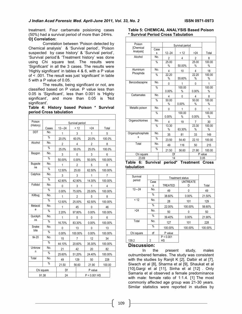
Correlation between ‘Poison detected by Chemical analysis’ & ‘Survival period’, ‘Poison suspected by case history’ & ‘Survival period’, ‘Survival period’& ‘Treatment history’ was done using Chi square test. The results, being significant or not, are classified based on P value. P value less than 0.05 is ‘Significant’, less than 0.001 is ‘Highly significant’, and more than 0.05 is ‘Not significant’.
All the data was collected in detail and later critically analyzed, tabulated, & compared with other various studies to analyze the aims and objectives of the present study.
Results: A] Sex wise distribution:
In the present study, of total 228 fatal cases of poisoning, 159 cases (69.7%) were males & 69 cases (30.3%) were females. Male: Female ratio was 2.3:1. B] Age wise distribution:
The most commonly affected age group was 21-30 years-70 cases (30.7%), followed by 31-40 years-48 cases (21.1%) & C] Marital Status:
Married were 151 cases (66.2%) and unmarried were 76 cases (33.3%). D] Region:
Out of 228 cases, 134 cases (58.8%) were from rural areas, and 93 cases (40.8%) were from urban area.
E] Educational Status: Cases with secondary school education-
73 cases (32%), were maximally affected, followed by PUC (Pre University education)-65 cases (28.5%), & Graduates-62 cases (27.2%). F] Employment Status:
Agriculturists, 70 cases (30.7%), were most commonly affected. Students were almost equally affected-65 cases (28.5%), followed by unemployed-46 cases (20.2 G] Socio-Economic Status:
Socio-economically lower class clearly outnumbered middle class. In lower class category, there were 156 cases (68.4%), followed by middle class-70 cases (30.7%) & upper class-1 case (0.4%). H] Route of Poisoning:
The commonest route of poisoning was ingestion-214 cases (93.8%). Injected poisons were 14 cases (6.1%), which included 13 cases of snake bite (5.7%) & 1 case of Benzodiazepine poisoning (0.5%). I] Manner of Poisoning:
The commonest manner of poisoning was suicidal-208 cases (93.1%), followed by accidental variety-19 cases (6.5%), which included 13 snake bite cases & 6 of the alcoholic cases. J] Treatment Status:
Of the 228 cases autopsied, 101 cases were brought dead. Treatment intervention was done in 127 cases. K] Survival Period:
Most of the cases (129 cases) died within 12 Hrs of getting poisoned. Some (49 cases) managed to survive until 24 Hrs and some (50 cases) beyond 24 Hrs. Stomach of the unidentified case contained undigested food particles. Therefore the person was categorized under less than 12 Hrs survival periods. L] Poison (History):
With the help of friends, relatives & police interviews, various poisons involved were noted & tabulated as follows. Poison consumed was unknown in most of the cases (83cases). Metacide poisoning was commonest, followed by Tik-20. M] Poison (Chemical Analysis):
Poisons detected through chemical analysis were noted & tabulated as follows. Organophosphates (OP) were most commonly used, followed by Organochlorines (OC), Aluminium Phosphide (ALP), Carbamates (CA), Alcohol (ALC), Metallic Poison (MP), & Benzodiazepine (BZD). Amongst all the poisons, Organophosphates (OP) were the most commonly used poisons-149 cases (69.3%),

J Indian Acad Forensic Med. April-June 2011, Vol. 33, No. 2 ISSN 0971-0973
108
followed by Organochlorines (OC)- 30 cases [14%], Aluminium Phosphide (ALP)-18 cases [8.4%], Carbamates (CA)-8 cases [3.7%], Alcohol (ALC)-8 cases [3.7%], Metallic Poison (MP)-1 case[0.5%], & Benzodiazepine (BZD)-1 case[0.5%]. This was based on chemical analysis. N] Gross Stomach Mucosal Appearances (SMA) in Various Fatal Poisonings:
Stomach Mucosal Appearances were pale(P), congested (C), Hemorrhagic (HC), Erosive (ER), Flattening of rugae(FR), Edematous (ED), specific Colour(CR) in various fatal poisoning cases. It was noted that these mucosal appearances were hardly seen individually and usually occurred in combinations. 1. In fatal poisoning cases who died without
prior treatment (Brought dead cases), it was also noted that none of these cases survived more than 12 Hrs. Of total 228 cases, 101 cases (44.3%) belonged this category, amongst which, 67 of 149 organophosphate(OP) cases, 11 of 30 organochlorine (OC) cases, 8 of 18 aluminum phosphide ( ALP) cases, 2 of 8 alcohol (ALC) cases, 11 of 13 snakebite(SB) cases and a single case each of metallic poison(MP) & benzodiazepine (BZD) belonged to this category. Haemorrhagic, erosive features and flattening of rugae (FR) predominated in most of these poisonings.
These features, to some extent, reflect the local effects of these poisons. Stomach mucosa of the metallic poison case was bright blue colour stained (significant with copper sulphate poisoning). Edematous mucosal feature was seen only in aluminum phosphide poisoning. Stomach mucosal appearances in these cases reflected the uninterfered (no interference in the form of treatment) action of the poisons on the stomach mucosa.
2. Stomach mucosal appearances of those fatal poisonings-28 cases (12.3%), where intervention in the form of treatment was done, but managed to survive for not more than 12 Hrs, hemorrhagic, erosive & flattening of rugae features were quite marked in these cases too, but not as severe as those in untreated cases. The survival period of the entire snake bite cases had been less than 12 Hrs in spite treatments in 2 of the cases, which indicates fatality of the snakebites.
Table 2: Stomach Mucosal Appearances in Various Fatal Poisonings Cases (Treated & SP<12Hrs)
OP(14) OC(8) ALP(2) ALC(2) SB(2)
Pale 0% 0% 0% 0% 50%
C 29.30% 37.50% 25% 25% 0%
HC 37.90% 31.30% 50% 50% 50%
ER 29.30% 25% 0% 25% 0%
FR 4.30% 6.30% 0% 0% 0%
ED 0% 0% 25% 0% 0%
The reason for short survival period in these cases in spite treatment could be due to delay in the treatment. 3. Stomach mucosal appearances of those
fatal poisoning cases-49 cases (21.5%), where intervention in the form of treatment was done and the survival period was 12—24 Hrs, hemorrhagic & erosive features were less marked in these cases. Congestive look of the mucosa had been predominant.
Table 3: Stomach Mucosal Appearances in various fatal poisonings Cases (Treated & SP12--24Hrs)
OP(35) OC(4) CA(4) ALP(4) ALC(2)
Pale 1.20% 0% 0% 0% 0%
C 40.50% 22.50% 80% 25% 25%
HC 16.50% 44.50% 20% 37.50% 50%
ER 26.60% 22.30% 0% 12.50% 25%
FR 15.20% 11% 0% 0% 0%
ED 0% 0% 0% 25% 0%
Alcohol had the worst effect on mucosa with red, haemorrgagic look. Intervention in the form of treatment had reduced the local effects of poisons in these cases. 4. Stomach mucosal appearances of those
fatal cases of poisoning-50 cases (21.9%), where treatment intervention was done and the cases managed to survive for more than 24 Hrs, haemorrhagic & erosive features were much lesser in these cases.
Table 4: Stomach Mucosal Appearances in various fatal poisonings Cases (Treated & SP>24Hrs)
OP(33) OC(7) CA(4) ALP(4) ALC(2)
C 52.50% 35.70% 50% 50% 66.50%
HC 13.50% 28.60% 12.50% 32.50% 0%
ER 22.10% 14.30% 25% 16.80% 33.50%
FR 11.90% 21.40% 12.50% 0% 0%
Early treatment had managed to keep the local effects of the poison to the minimum in most of these cases. Alcohol had the maximum erosive effect in these cases too in spite
J Indian Acad Forensic Med. April-June 2011, Vol. 33, No. 2 ISSN 0971-0973
109
treatment. Four carbamate poisioning cases (50%) had a survival period of more than 24Hrs. O] Correlation:
Correlation between ‘Poison detected by Chemical analysis’ & ‘Survival period’, ‘Poison suspected by case history’ & ‘Survival period’, ‘Survival period’& ‘Treatment history’ was done using Chi square test. The results were ‘Significant’ in all the 3 cases. The results were ‘Highly significant’ in tables 4 & 6, with a P value of < .001. The result was just ‘significant’ in table 5 with a P value of 0.05.
The results, being significant or not, are classified based on P value. P value less than 0.05 is ‘Significant’, less than 0.001 is ‘Highly significant’, and more than 0.05 is ‘Not significant’. Table 4: History based Poison * Survival period Cross tabulation
Poison
(History) Survival period
Total Cases 12—24 < 12 >24
DDT No. 1 3 1 5
% 20.0% 60.0% 20.0% 100.0%
Alcohol No. 2 4 2 8
% 25.0% 50.0% 25.0% 100.0%
Baygon No. 3 0 3 6
% 50.00% 0.00% 50.00% 100.00%
Bugsolene
No. 1 2 5 8
% 12.50% 25.00 62.50% 100.00%
Celphos No. 3 3 1 7
% 42.90% 42.90% 14.30% 100.00%
Follidol No. 0 3 1 4
% 0.00% 75.00% 25.00% 100.00%
Killbug No. 1 2 5 8
% 12.50% 25.00% 62.50% 100.00%
Metacide
No. 1 45 0 46
% 2.20% 97.80% 0.00% 100.00%
Quickphos
No. 1 5 0 6
% 16.70% 83.30% 0.00% 100.00%
Snake bite
No. 0 13 0 13
% 0.00% 100.00% 0.00% 100.00%
tik-20 No. 15 7 12 34
% 44.10% 20.60% 35.30% 100.00%
Unknown
No. 21 42 20 82
% 25.60% 51.20% 24.40% 100.00%
Total No. 49 129 50 228
% 21.50 56.60 21.90 100.00
Chi square Df P value
91.38 24 P < 0.001 HS
Table 5: CHEMICAL ANALYSIS Based Poison * Survival Period Cross Tabulation
Poison [Chemical Analysis]
Survival period
Total Case
s 12--24 < 12 >24
Alcohol No. 2 4 2 8
% 25.00% 50.00%
25.00%
100.00%
Aluminium Phosphide
No. 4 10 4 18
% 22.20% 55.60%
22.20%
100.00%
Benzodiazapine No. 0 1 0 1
% 0.00%
100.00% 0.00%
100.00%
Carbamates No. 4 0 4 8
% 50.00% 0.00%
50.00%
100.00%
Metallic poison No. 0 1 0 1
% 0.00%
100.00% 0.00%
100.00%
Organochlorines No. 4 19 7 30
% 13.30% 63.30%
23.30%
100.00%
Organophosphates
No. 35 81 33 149
% 23.50 54.40 22.10 100.00
Total No. 49 116 50 215
% 21.50 56.60 21.90 100.00
Chi square Df P value
23.69 14 0.05
Table 6: Survival period* Treatment Cross tabulation
Survival period
Treatment status
Total Case
s TREATED UNTREATE
D
12—24 No. 49 0 49
% 38.60% 0.00% 21.50%
< 12 No. 28 101 129
% 22.00% 100.00% 56.60%
>24 No. 50 0 50
% 39.40% 0.00% 21.90%
Total No. 127 101 228
% 100.00% 100.00% 100.00%
Chi square df P value
139.2 2 P < 0.001 HS
Discussion: In the present study, males
outnumbered females. The study was consistent with the studies by Ranjit K [2], Dalbir et al [7],
Siwach et al [8], Sharma et al [9], Shaukat et al [10],Gargi et al [11], Sinha et al [12]
. Only
Samaria et al observed a female predominance with male: female ratio of 1:1.4. [1]
The most
commonly affected age group was 21-30 years. Similar statistics were reported in studies by

J Indian Acad Forensic Med. April-June 2011, Vol. 33, No. 2 ISSN 0971-0973
110
Dalbir et al [7], Sharma et al [9] and Shaukat et al [10]. The majorities were married, lacked adequate education, and belonged to rural areas.
Since decades, males are exposed to stress, strain and occupational hazards to a greater extent when compared to females because they are bread earners. The 21-30yrs age group is the most active phase of life for men who are involved mentally, physically and socially. Married people have more responsibilities, duties and financial burden. Consequently, they get more frustrated and become liable to take their own lives. Unmarried people are more carefree and ‘happy go lucky’, which explains their lesser incidence. Poverty, unemployment, early marriage are some of the major stress factors responsible for high incidence of suicidal deaths in these rural areas. Illiteracy & poverty often go hand in hand, where in, one has an additive affect over the other in causing depression in these individuals. Depression is one of the major causes for suicidal deaths. Love related depressions, poverty, and academic stress were some of the major factors which led students, 65 cases (28.5 %) to consume poison. Farmers and unemployed persons were more prone to death by poisoning in the present study; as in previous studies. This is so because larger segment of our population comes from these groups. Early marriages, low education, poverty, marital problems, were the major factors which led housewives (13.6% of the cases) commit suicide.
Ingestion is the most convenient way to consume most of the poisons. Medical & Para-Medical professionals and drug addicts are more familiar with injectable poisons. The inference of manner of death was based on history given either by police or and relatives.
The police inquests usually mention chronic ailments, unbearable long standing body pain; etc. as the reasons for suicidal intent. This is not always the whole truth. In fact, more appropriate reasons are stress factors coming from financial, social, family problems, illiteracy, immaturity and many more aspects of life.
Easy availability of poisons made them easy victims also. Psychiatric causes (maniac depressive psychosis, severe depression) also have played a major role. Suicide is a subject of great sociological and psychological importance with many unexamined and unresolved problems. It is always interesting to consider the reasons, which compel a person to take his own life.
Organophosphates (OP) were the most commonly used poisons, because of their wide use in Agriculture. The results were consistent with the studies done by Singh et al[13]
,Sanjay
et al[15],Vinay et al [16] and Gupta et al[17] .In the studies conducted by Adarsh et al[1], Dalbir et al[7], Siwach et al[8], Sharma et al[9], Shaukat et al[10], Gargi et al[11], Sinha et al[12], aluminium phosphide was the commonest poison used. In the study by Shahin et al[14], deaths mostly occurred by opioids (41.54%). Thus, results of the present study were consistent with the previous studies, except for some differences in the kinds of poison preferred (Aluminium phosphide was preferred in northern India). Trends of the poisons seem to be a function of need and availability of specific substances.[18]
Conclusion: Significant correlations were seen
between- types of poison ingested & survival period, and between treatment intervention & survival period. However, there are numerous factors that determine the appearance of stomach mucosa in a particular fatal poisoning case. These are- the poison (its quantity, quality, diluent), biological factors, poison ingested with food or on empty stomach, treatment intervention, post-mortem interval. With all these factors affecting, it’s quite difficult to predict with a certainty about the stomach mucosal appearance in a particular fatal poisoning or vice-versa (i.e. to predict the type of poison based on stomach mucosal appearance), as the appearances are not consistent with a type of poison, and generally, the appearances are seen in combinations. In the present day scenario, the poisons available are less concentrated. Hence typical features mentioned in text books are not often seen. If poison suspected is mentioned in requisition to FSL, it shall help in early analysis and also avoids blind testing and wastage of chemicals.
Incidence, morbidity and mortality due to poisoning can be possibly curtailed by strict vigilance over the sale and distribution of insecticides, educating the users regarding the safety measures, good treatment facilities (i.e. antidotes etc) at rural areas, establishing Poison Information Centers, proper and correct implementation of social and economic projects aimed for the upliftment of rural, poor and the downtrodden. The educating of public as regards to carrying poison bottle consumed and information leaflet to the hospital helps the doctor to initiate proper treatment and use of specific antidote.

J Indian Acad Forensic Med. April-June 2011, Vol. 33, No. 2 ISSN 0971-0973
111
References: 1. Kumar A, Vij K. Trends of poisoning in Chandigarh – A six year
autopsy study. Journal of Forensic Medicine and Toxicology 2001 Jan-June; 18(1):8-10.
2. Das RK. Epidemiology of insecticide poisoning at A.I.I.M.S emergency services and role of its detection by gas liquid chromatography in diagnosis. Medico legal update 2007 April-June; 7(2):49-59.
3. Aggarwal P, Handa R, Wali JP. Common poisonings in India. JFMT 1998 Jan-June; 15(1):73-74.
4. Fernando R. Pesticide poisoning in the Asia-Pacific region and the role of the regional information network. Clinical toxicology 1995 Nov; 33(6):677-682.
5. Nigam M, Jain AK, Dubey BP, Sharma VK. Trends of organophosphorus poisoning in Bhopal region-An autopsy based study. JIAFM 2004; 26(2):62-65.
6. Tarunil NG, Bijoy TH, Momonchand A. A profile of poisoning cases admitted in RIMS hospital, Imphal. JFMT 2001 Jan-June; 18(1):31-33.
7. Singh, Dalbir MD, Jit MS, Tyagi, Seema. Changing trends in acute poisoning in Chandigarh zone – A 25 year autopsy experience from a tertiary care hospital in northern India. The American Journal of Forensic Medicine and Pathology 1999 June; 20(2): 203-210.
8. Siwach SB, Gupta A. The profile of acute poisonings in Haryana – Rohtak study. Journal of Association of Physicians of India 1995; 43(11):756-759.
9. Vij K, Sharma BR, Harish D. Poisoning in northern India – Changing trends, causes and prevention thereof. Med Sci Law 2002; 42(3):251-257.
10. Hanif SA, Rizvi SJ, Hussain M. The aluminium phosphide: A profile of preferred “Hemlock” in district Aligarh, U.P. JFMT 2002 July-Dec; 19(2):1-5.
11. Gargi J, Hakumat R, Ashok C, Gurmanjit R, Gaurav S, Baggai JS. Current trends of poisoning: A hospital profile. JPAFMT 2004; 3.
12. Sinha US, Kapoor AK, Agnihotri AK, Shrivastava PC. A profile of poisoning cases admitted in SRN hospital, Allahabad, with special reference to aluminium phosphide poisoning. JFMT 1999 Jan-Jun; 16(1):40-42.
13. Singh B, Unnikrishnan B. A profile of acute poisoning at Mangalore (South India). Journal of Clinical Forensic Medicine; 13(3):112-116.
14. Shadnia S. Pattern of acute poisoning in Tehran-Iran in 2003. HET 2007; 26(9):753-756.
15. Gupta S, Kumar S, Sheikh MI. Comparative study and changing trends of poisoning in year 2004-2005, Surat, India. IJMTLM 2007 July-Dec; 10(1):16-19.
16. Shetty VB, Pawar GS, Inamadar PI. Profile of poisoning cases in district and medical college hospitals of north Karnataka. IJFMT 2008; 2(2).
17. Gupta BD, Hapani JH, Shah VN. Current trend of poisoning in Jamnagar: An experience of tertiary care teaching hospital. JIAFM 2006; 28(3): 90-92.
18. Gupta BD, Vaghela PC. Profile of fatal poisoning in and around Jamnagar. Journal of Indian Academy of Forensic Medicine2005; 27(3).
Table 1: Stomach Mucosal Appearances in various fatal poisonings (Untreated cases)
OP (67)
OC (11)
ALP (8)
MP (1)
BZD (1)
ALC (2)
Snake (11)
Pale 0% 0% 0% 0% 0% 0% 36.45
C 10.70% 10.90% 13.80% 0% 100% 0% 0%
HC 45.70% 52.70% 53.80% 0.34% 0% 100% 63.60%
ER 29.30% 20.90% 20% 0.33% 0% 0% 0%
FR 14.30% 15.50% 5 0% 0% 0% 0%
ED 0% 0% 13.80% 0% 0% 0% 0%
CR 0% 0% 0% 0.33% 0% 0% 0%
149
3018
8 81 1
215
0
50
100
150
200
250
No.of Cases
OP OC ALP CA ALC MP BZD Total
Various Poisons
Poison ( Chemical Analysis )
Frequency
Percent
83
4634
13 8 6 8 7 5 4 8 6
228
020406080100120140160180200220240
No.of Cases
Unknown
Metacide
Tik-20
Snake bite
Alcohol
Baygon
Bugsolene
Celphos
DDT
Follidol
Killbug
Quickphos
Total
Various Poisons
Poison ( History )
Frequency
Percent
J Indian Acad Forensic Med. April-June 2011, Vol. 33, No. 2 ISSN 0971-0973
112
Original Research Paper
Study of Palatal Rugae Pattern among the Student Population in Mangalore
*Mahabalesh Shetty, ** Premalatha K
Abstract
Human identification is one of the most challenging tasks in Forensic identification. In mass disasters dental records, fingerprints and DNA comparisons are probably the most used techniques. However these techniques cannot be applied always. In some cases, it is necessary to apply different and less known techniques like Rugoscopy. The purpose of this study is to determine any gender difference in palatal rugae pattern. In this study100 subjects were randomly selected comprising 50 males and 50 females of age ranging from 17 to 25 years of student population belonging to Mangalore .The rugae pattern were assessed by applying Thomas & Kotze classification. Association between rugae forms and gender were tested using student unpaired T test. Gender wise, there were no significant differences in the total number of rugae. The incidence of curved ,straight and forwardly directed rugae were more among females than males, while that of wavy ,perpendicular and backwardly directed rugae were more among males. This study clearly demonstrates the gender difference and uniqueness of rugae pattern in different individuals. Thus it is a reliable source of identification.
Key Words: Palatal Rugae, Human identification, Rugoscopy.
Introduction: Identification of an individual is a pre-
requisite for certification of death and for personal, social and legal reasons. Human identification is a mainstay of civilization, whether in living or dead, and the identification of unknown individual has always been of paramount importance to our society.Human identification is based on scientific principles, mainly involving dental records, fingerprints and DNA comparisons. Sometimes, it becomes necessary to apply a lesser known and unusual technique like palatoscopy. Palatoscopy or palatal rugoscopy is the name given to the study of palatal rugae in order to establish a person’s identity [1, 2]. Transverse palatine folds or palatal rugae (PR) are asymmetrical and irregular elevations of the mucosa located in the anterior third of the palate, made from the lateral membrane of the incisive papilla, arranged in transverse direction from palatine raphe located in the mid sagittal plane. [3]
Corresponding Author: *Associate Professor & HOD Department of Forensic Medicine & Toxicology K. S. Hegde Medical Academy, Nitte University Mangalore, 575018 Karnataka Email- [email protected] **Professor &Head Deptt. of Maxillo Facial Surgery, Manipal College of Dental Sciences Mangalore, Manipal University
The palatal rugae, like fingerprints, do not change during the life of the individual. They are protected from trauma and high temperatures because of their internal position in the oral cavity, surrounded and protected by lips, cheeks, tongue, teeth and bone. Palatine rugae are unique to each individual and are reasonably stable during the person’s growth. Once formed, it only changes in its length, due to normal growth, staying in the same position throughout the life of a person. [2, 4]
Palatoscopy may be used as a necro-identification technique. Palatoscopy can be of special interest in those cases where there are no fingerprints available like decomposed bodies, burned bodies and conditions were both the upper limbs are missing. It is the most valuable technique in aeronautical accidents in order to ensure identification of pilots making use of ante mortem data. [2]
This present study is an attempt to determine the number and different pattern of rugae and to see if there is any gender difference in rugae pattern in Mangalore population thereby highlighting the importance of palatal rugae in establishing person’s identity.
Materials and Methods: Total of 100 subjects, 50 males and 50 females in the age group of 17 – 25 years of student population belonging to Mangalore were chosen randomly and included in the present study. This study was conducted after obtaining

J Indian Acad Forensic Med. April-June 2011, Vol. 33, No. 2 ISSN 0971-0973
113
institutional ethical committee clearance and informed written consent from the subjects.
Inclusion Criteria: Subjects without braces, removable partial dentures, fixed partial dentures and Student population belonging to Mangalore were included.
Exclusion Criteria: Subjects with abnormalities of palate and lips like the cleft palate and left lip, subjects who were wearing partial dentures and braces were excluded.
Methodology: The various steps involved in the sample study were as follows:
• Obtaining consent from the subjects, Preparation of maxillary dental casts.
• Interpretation of the rugae pattern in the casts obtained by Thomas & Kotze classification [5].
• Subjects, both male and female, in the age group 17-25 years of student population and who belonged to Mangalore were chosen randomly.
Collection of the Palatal Prints: The materials used were: Alginate
powder, perforated metal maxillary impression tray, Mixing bowl, Spatula, Dental Stone and Water.
The rugae seen as elevated impressions were marked on these casts using a black permanent marker pen. The rugae pattern was classified based on their length, shape, direction and unification. The parameters assessed were:
1) Total number of rugae 2) Number of primary rugae 3) Predominant Shape 4) Predominant Direction 5) Unification of rugae. • The rugae were classified based on their
length as: 1) Primary->5mm, 2) Secondary- 3 to 5mm 3) Fragmentary-<3mm
Rugae less than 2 mm were disregarded. A ruga’s length was determined by measuring its greatest dimension regardless of its shape.
• The rugae were divided into 4 types based on their shape as:
1) Curved: They had a crescent shape and curved gently.
2) Wavy: If there was a slight curve at the origin or termination of a curved rugae.
3) Straight: They run directly from their origin to termination.
4) Circular: Rugae that form a definite continuous ring were classified as circular.
• The direction of the rugae was determined by measuring the angle formed by the line joining its origin and termination and the line perpendicular to the median raphe. Based on the direction rugae were classified as:
1) Forwardly directed rugae - associated with positive angles
2) Backwardly directed rugae - associated with negative angles
3) Perpendicular rugae - associated with zero angles.
• Unification was said to have occurred when two rugae joined at their origin or termination:
1) Diverging- If two rugae had the same origin from the midline but immediately branched.
2) Converging- Rugae with different origins from midline, but which joined on their lateral portions. All the details from each dental cast
were observed as mentioned and documented in the Proforma. Association between rugae forms and gender were tested using student unpaired T test.
Results and Observation: A total of 100 maxillary dental casts
obtained from 50 males and 50 females student population of Mangalore, were examined for the palatal rugae patterns by applying the classification proposed by Thomas & Kotze. [5]
Total Number of Rugae: as shown in table I & II. Gender wise, there were no significant differences in the total number of rugae on the right side or left side of the palate among the males and females.
Number of primary rugae: as shown in table III. The mean number of primary rugae was 3 in both the sexes.
Predominant Shape: as shown in table IV. The incidence of curved rugae was more among the females than the males. The incidence of straight rugae was more among the females than the males .The incidence of wavy rugae was more among the males than the females. Circular rugae had no differences between either sex.
Predominant Direction: as shown in table V. The incidence of forwardly directed rugae was more among females compared to males. The incidence of backwardly directed rugae was more among males compared to females. The incidence of perpendicular rugae was more among males compared to females.

J Indian Acad Forensic Med. April-June 2011, Vol. 33, No. 2 ISSN 0971-0973
114
Discussion: Establishing a person's identity can be a
very difficult process. Fingerprints and dental means represent the most scientifically reliable methods of identification.
In the field of forensic odontology, rugoscopy is still in its infancy. [6] Despite the ongoing problem of describing palatal rugae pattern, quantitatively and qualitatively, their uniqueness to individuals has been recognized clearly as providing a potentially reliable source of identification. [1, 2, 6, 7, 8]
The present study was an attempt to determine the number, pattern and also to assess the predominant pattern of rugae if any in student population of Mangalore. An attempt was also made to determine the differences in rugae pattern among two genders in the selected groups.
Several studies done in the past have revealed and have statistically proved that the rugae patterns are highly individualistic and there are differences between races and gender. [6, 7, 8] In our study the number of primary rugae did not show any statistically significant differences among the sexes. This observation was in contrast to that of Shwetha et al [8], who reported that Mysorean males and Tibetan females had more primary rugae than their respective counterparts. [8] Kashima in her study among Indian and Japanese children reported that Japanese children had more primary rugae than Indians [9]. Kapali in her study reported that Australian Aborigines had more primary rugae than the Caucasoids. [1]
The predominant shape of the rugae in our study were curved and straight shaped rugae among females and wavy in males.The circular shaped rugae did not show any significant gender differences in our study but Faisal in his study among Saudi males and females ,reported an increased incidence of circular shaped rugae among females than males. [10]
The incidence of forwardly directed rugae was more in females, backwardly directed and perpendicularly directed rugae were more in males. These observations were in contrast to the study conducted by Shwetha [8].
Comparisons of the unification of rugae both converging & diverging did not show any specific trend. This was in agreement with the observations made by Shwetha [8] and in contrast to the findings made by Faisal who observed that the Saudi females had more converging rugae than males. [10]
This study clearly demonstrates that, palatal rugae are unique to each individual and rugae pattern with its different parameters can
be used successfully as a tool of identification.
Conclusion: Although this study fails to demonstrate
any significant gender difference in number of primary rugae, it clearly demonstrates distinct pattern of rugae associated with different gender.
This study also clearly demonstrated the uniqueness of rugae pattern in different individuals. Thus it is an effective & reliable source of identification.
As this study had a limited sample size of 100 subjects, it will be beneficial to conduct studies with larger sample & to compare with ethnic groups.
This method of identification can be used only when an ante mortem record of palatal rugae is available.
References: 1) Kapali S. Townsend G. Richards L. Parish T. Palatal rugae
patterns Australlin Aborigines and Caucasians. Aust Dent J 1997; 42: 129-33.
2) Caldas IM. Magathaes T. Afonso A. Establishing identity using cheiloscopy and palatoscopy. Forensic Sci Int 2007; 165:1-9.
3) Richard L Drake, A Wayne vogl, Adam W.M. Mitchell. Gray’s Anatomy for students .2nd Ed. Canada,Churchill Livingstone; 2010.1047-1048.
4) Acharya AB, Sivapathasundharam B. Forensic Odontology. In Rajendran R. Sivapathsundharam B.Eds. Shafer’s Textbook of Oral Pathology, Fifth Edn. Elsevier; 2006. p.1199-227.
5) Thomas CJ. Kotze TJ. The palatal rugae pattern: A new classification. J Dent. Assoc. S Afr 1983; 38: 153-76.
6) Thomas C.J. & van Wyk, C.W. The palatal rugae: an identification. J. Forensic Odontostomatol1988; 6(1): 21-7.
7) English WR. Robison SF. Summit JB. Oesterle IJ. Brannon RB. Mortang MW. Individually of human palatal rugae. J Forensic Sci, 1988; 33: 718-26.
8) Shetty S.K. Kalia S. Patil K Mahima VG. Palatal rugae pattern in Mysorean and Tibetan populations Indian J Dent Res 2005; 16:51-5.
9) Kashima K. Comparative study of the paltal rugae & shape in Japanese & Indian children.Aichi Gaukin Daigaku Shigakkai Shi 1990; 28(1-2): 295-320.
10) Fahmi FM. Al-Shamrani SM. Talic YF. Rugae pattern in a Saudi population sample of males and Female. Saudi Dental Journal 2001; 13: 92-5.
Table –I: Total Number of Rugae (Rt.Side) among the Males and Females
Gender Number Mean Standard Deviation
Males 50 4.1 1.2
Females 50 4.2 1.O
Table II: Total Number of Rugae (Left Side) among the Males and Females
Gender Number Mean Standard Deviation
Males 50 3.9 0.9
Females 50 3.9 0.9
Table III: Total Number of Primary Rugae in Relation to Gender
Gender Number Mean Standard Deviation
Males 50 3.3 0.18
Females 50 3.3 0.18

J Indian Acad Forensic Med. April-June 2011, Vol. 33, No. 2 ISSN 0971-0973
115
Table IV: Number of the Various Shapes of Rugae among the Males and Females
Gender Curved Wavy Straight Circular
Males Females
Mean 1.8 5.7 1.2 0.3
S.D 0.3 0.3 0.2 0.07
Mean 2.0 5.2 1.8 0.3
S.D 0.2 0.3 0.3 0.O5
Table V: Total Number of the Various Directions of Rugae among the Males and Females
Gender Forwardly Directed
Backward Directed
Perpendicular
Males Female
Mean 5.3 3.1 0.7
S.D 0.3 0.4 0.1
Mean 6.3 2.7 0.2
S.D 0.4 0.3 0.08
Fig .1: Classification of rugae based on Unifiaction. A- Converging, B- Diverging Fig.2: Rugae classified on the basis of shape. A- Circular., B- Straight, C -Wavy-, D- Curved
Fig 3: Classified on the basis of Direction. A- Forward, B- Backward
A B
C Fig .4: Cast Showing: a- Perpendicular /Straight, b - Forward, C- Backward
A B
Fig .5: Cast Showing: a-Diverging rugae, b-
Converging rugae

J Indian Acad Forensic Med. April-June 2011, Vol. 33, No. 2 ISSN 0971-0973
116
Original Research Paper
Tuberculous Lesions at Autopsy *Monika Garg, **Akash Deep Aggarwal, *** Sneh Singh, **** Sant Prakash Kataria
Abstract Tuberculosis (TB) today remains one of the world's most lethal infectious diseases. Despite the
availability of effective treatment for most cases, tuberculosis is still a cause of death in our environment. Some cases of active tuberculosis are not identified until after the patient had died and an autopsy has been performed. This study was done to determine the prevalence of tuberculosis in autopsy cases. We analysed all the autopsy cases sent for histopathology over a period of one month. 8.7% cases of active tuberculosis were found in autopsy specimens, of which 60% were males. Tuberculosis was suspected in only 40% of patients before death. Tuberculosis is not an uncommon finding at autopsy for most of autopsy surgeons, but as the cause of death it is not so common. The presence of unspecific symptomatology, insufficient cost-effectiveness of the diagnostic tests and precocious death, are identified as the most frequent causes of undiagnosed tuberculosis. Awareness of tuberculosis and its high prevalence in India is essential for minimising missed diagnoses. Absence of suspicion and delayed diagnosis mean increased risk in health care and at autopsy.
Key Words: Tuberculosis, Autopsy, Pulmonary, Granuloma, Intestine
Introduction:
Tuberculosis is still a relevant infectious disease, which is often not diagnosed during the lifetime of a patient. India is home to over 3.4 million tuberculosis patients - about one-fifth of the global figure - making it the most TB prevalent country. 325172 people in India had died of the disease in 2005 alone. In 2006, India recorded 1.9 million new cases. Across the globe, there were 9.2 million new cases of TB during the same period. Of all fresh cases in the country, 1.2 % is infected with HIV and 2.8 % of all new cases have been diagnosed with multi-drug resistant TB. Against all these figures, 30 % of the cases are not even detected in the country. [1] Unlike other countries, it is not mandatory in India for doctors to report TB cases.
Methods: We analysed all the autopsy cases sent
to the Pathology department of PGIMS Rohtak over a period of one month.
Corresponding Author: *Assistant Professor, Pathology Post Graduate Institute of Medical Sciences, Rohtak, Haryana Email: [email protected] **Assistant Professor, Forensic Medicine ***Senior Resident, Pathology ****Associate Professor, Pathology Post Graduate Institute of Medical Sciences, Rohtak, Haryana
The complete Post-Mortem report were accessed and analysed. Basic demographic data such as the deceased’s age, sex and available medical history were recorded.
The gross and histopathological findings were recorded. The cases of tuberculosis were again confirmed by Ziehl-Nielson stain.
The aim of the study was to investigate the frequency of tuberculosis in autopsy cases. The results were compared with the studies done by other authors. The main area of interest was to ascertain the proportion of cases of tuberculosis which were not diagnosed during life, in other words, cases which were not expected. We examined the possible reasons why this may have occurred. Further the issue how this could affect medical staff and also the public health was also looked into.
Observations: Out of a total of 115 autopsy case
samples received for histo-pathological examination over a one month period, we found 10 cases (8.7%) of active tuberculosis, 60% corresponding to men. Mean age of this group was 38 years. Tuberculosis was suspected in only 40% of patients before death. Principal cause of death was tuberculosis in 30% of patients, 90% of patients had pulmonary tuberculosis, 10% suffered from miliary tuberculosis and 30% had extra-pulmonary tuberculosis. The lungs were the most frequently affected organ. Caseous epithelioid granulomas

J Indian Acad Forensic Med. April-June 2011, Vol. 33, No. 2 ISSN 0971-0973
117
were found in all patients, however only 2 cases were positive for Acid-fast bacilli in ZN stain.
Of the 8.7% cases showing relevant tuberculosis, lung involvement was grossly present in 7 cases. Of these, 2 showed acinar nodal foci, 4 showed early cavities and 1 had caseous pneumonia. One case showed scarring, effusion and pleural adhesions. Table 1 summarizes the findings of the cases positive for tuberculosis. Fig 1: Adhesions and pleural effusion in left lung at autopsy in a female
Fig 2: H&E stain of lung revealing multiple caseous epitheloid granuloma
Fig 3: H&E stain of small intestine showing epitheloid granuloma
Fig 4: H&E stain of liver showing changes of fatty liver and epitheloid granuloma
Discussion: The morbidity and mortality of
tuberculosis has decreased tremendously in developed countries but is found to exist still in higher rates in developing countries and especially among the populations of low socio- economic status.
Our study shows that a large proportion (60%) of cases of tuberculosis found at autopsy are unexpected. Several similar studies have shown that many cases of tuberculosis were diagnosed only at autopsy, when awareness of possibility of the disease is low. There are several reasons to know why the diagnosis is missed or delayed. As per Table 2, large studies, conducted all over the world, looking at the frequency of undiagnosed tuberculosis at autopsy, similarly found high rates. Reasons range from symptomatic patients not seeking medical attention, to doctor not investigating the possibility of tuberculosis despite suspicious symptomatology. Occasionally the diagnosis is delayed or missed because the doctor did not enquire about exposure to tuberculosis and patient did not volunteer that information. Occasionally doctor does not consider the diagnosis because of concurrent illness that can produce symptoms that are indistinguishable from tuberculosis.
Active tuberculosis today is becoming a disease of elderly and is attributable mainly to recurrence of dormant infection and decrease in immune status of elderly. There is a tendency to confuse tuberculosis with lung carcinoma in adults because of high incidence of both the disease in upper lobe of lung and also both these disease presents clinically in a similar fashion. In our study 2 cases were over 50 years of age. Most of chest radiographs are mis-diagnosed. The lesions in lung are diagnosed mostly as pneumonia or as primary and secondary malignancy. In cases were pleural effusion is present, it is important to perform pleural tap in order to confirm diagnosis

J Indian Acad Forensic Med. April-June 2011, Vol. 33, No. 2 ISSN 0971-0973
118
of malignancy (cytology) and to demonstrate mycobacterium tuberculosis (acid fast stain) in active tuberculosis.
Tuberculosis of Gastro-intestinal tract is difficult to diagnose because symptoms of disease mimics many other abdominal conditions; and laboratory and radiological investigations usually do not contribute any helpful data. So tuberculosis should be included in the differential diagnosis of any unusual gastro-intestinal conditions. In our study 2 cases (20%) had gastrointestinal tuberculosis.
Military tuberculosis was found in single case. The lesions were distributed in lung, liver, kidney, intestine, ovary and mesenteric lymph nodes. These cases who presents with symptoms of fever, loss of weight, cachexia are usually suspected of having generalized malignancy.
The importance of diagnosis is that disease is curable, if diagnosed, but fatal if undiagnosed. An undiagnosed infective person is of epidemiological importance when he is in contact with person not immunised to the disease. There is evidence in literature to suggest that there are grounds for suspecting that the autopsy room might be a centre for dissemination for tuberculosis. Post mortem attendants might be exposed to risk, as many of them do not understand the elementary rules of hygiene, let alone the problem of bacterial contamination. Post mortem room is a source of potential hazard and risk, not only to doctors and technicians, but also to visitors to the mortuary and those handling body after autopsy.
Post mortem staff has ethical and legal responsibility to make themselves aware of, and to minimize these dangers. [15, 16] The upsurge of tuberculosis has been associated with HIV infection and immunodeficiency. It is therefore necessary to suspect and screen all tuberculosis patients for possible HIV positivity. Absence of suspicion and delayed diagnosis mean increased risk in health care and at autopsy. Staffs of laboratories and autopsy rooms are estimated to be between 100 and 200 times more likely than the general public to develop tuberculosis. [17]
Primary tuberculosis accounts for approximately 90% of cases and is acquired by inhalation of aerosols or dried material. Cutaneous infection accounts for 5–10% of cases, the bacillus being introduced into previously traumatised skin or via a skin puncture. Muco-cutaneous transmission of tuberculosis at autopsy has not been reported. [16]
Conclusion: Tuberculosis causes unnecessary
death, because of failure to diagnose and treat, what is today a curable disease. There is a need for more awareness of the disease, especially in the light of recent reports showing a growing incidence of extra-pulmonary tuberculosis. The risk of unrecognized tuberculosis not only extends to public but also to the health professional. Measures to minimise the risk to mortuary staff include the use of proper respiratory masks and performing the autopsy in the infection suit, which isolates the body and minimises exposure to staff. [9] Autopsies are still indispensable for providing quality control and disease statistics.
References: 1. Tuberculosis in the South-East Asia Region. The Regional Report:
2008. New Delhi: World Health Organisation: Regional Office for South East Asia. 2008. pp. 63.
2. Post C, Wartenhorst HS. Clinically unrecognised tuberculosis in autopsy material. Dtsch Med Wochenschr. 1979; 104(13): 461-6.
3. Andrion A, Bona R, Mollo F. Active tuberculosis unsuspected until autopsy. Minerva Med. 1981; 72(2): 73-80.
4. Ebert W. Frequency of tuberculosis in autopsy material of the Institute of Pathology of the Friedrich Schiller University Jena. Zentralbl Allg Pathol. 1983;128 (1-2):81-3.
5. Chastonay P, Gardiol D. Extensive active tuberculosis at autopsy: retrospective study of a collection of adult autopsies (1961-1985). Schweiz Med Wochenschr. 1987; 117(24):925-7.
6. MacGee W. The frequency of unsuspected tuberculosis found post-mortem in a geriatric population. Z Gerontol. 1989; 22(6):311-4.
7. Lee JK, Ng TH. Undiagnosed tuberculosis in hospitalized patients--an autopsy survey. J R Soc Health. 1990; 110(4):141-3.
8. Rowińska-Zakrzewska E, Szopiński J, Remiszewski P, Szymańska D, Miller M, Pawlicka L, Zwolska-Kwiek Z. Tuberculosis in the autopsy material: analysis of 1500 autopsies performed between 1972 and 1991 in the Institute of Tuberculosis and Chest Diseases, Warsaw, Poland. Tuber Lung Dis. 1995; 76(4):349-54.
9. Lum D, Koelmeyer T. Tuberculosis in Auckland autopsies, revisited. N Z Med J. 2005; 118(1211):U1356.
10. Theegarten D, Kahl B, Ebsen M. Frequency and morphology of tuberculosis in autopsies: increase of active forms. Dtsch Med Wochenschr. 2006; 131(24):1371-6.
11. Morales Conejo M, Guerra Vales JM, Moreno Cuerda VJ, Varona Arche JF, Hernando Polo S, Palenque Mataix E, Pérez de Oteyza C, Martínez Tello FJ. Tuberculosis in the autopsy. Clinical and pathological study: an analysis of 92 cases of active tuberculosis found in 2,180 autopsies. Rev Clin Esp. 2007; 207(6):278-83.
12. Flavin RJ, Gibbons N, O'Briain DS. Mycobacterium tuberculosis at autopsy--exposure and protection: an old adversary revisited. J Clin Pathol. 2007; 60(5):487-91.
13. Yamamoto A, Tsuchiya K, Kusajima K, Shimoide H, Nunomura M, Hebisawa A. Study on clinico-pathological features of active pulmonary tuberculosis found at autopsy in a general hospital. Kekkaku. 2009; 84(2):71-8.
14. Ozsoy S, Demirel B, Albay A, Kisa O, Dinc AH, Safali M. Tuberculosis prevalence in forensic autopsies. Am J Forensic Med Pathol. 2010; 31(1):55-7.
15. Smith GS. Tuberculosis as a necropsy room hazard. J Clin Pathol 1953;6: 132.
16. Burton JL. Health and safety at necropsy. J Clin Pathol 2003; 56:254–260.
17. Collins CH, Grange JM. Tuberculosis acquired in laboratories and necropsy rooms. Commun Dis Public Health. 1999; 2(3):161-7.

J Indian Acad Forensic Med. April-June 2011, Vol. 33, No. 2 ISSN 0971-0973
119
Table 1 Findings of the tuberculosis positive cases out of 115 autopsy specimens received over a month No. Age Sex Cause of death Tuberculosis Other findings
1. 22 Female Illness Lung, Intestine, Liver, Kidney, Ovary, Mesenteric lymph nodes Fatty liver
2. 28 Male TB Lung -
3. 25 Female Illness Lung -
4. 30 Female Maternal Ovary, Fallopian tubes -
5. 32 Female Heart attack Lung Atherosclerosis
6. 40 Male - Lung -
7. 42 Male Illness Lung Cirrhosis
8. 44 Male - Lung, Spleen -
9. 55 Male - Lung Fatty liver, Atherosclerosis
10. 60 Male Heart attack Lung Myocardial infarction
Table 2
Comparison of Different Studies Author Study year Total TB cases TB cases % Undiagnosed
cases % Place Prevalence per 100000
Post [2] 1955-1975 18724 811 4.33 58.9 Germany -
Andrion [3] 1969-1978 3195 61 1.91 70.0 Italy -
Ebert [4] 1961-1985 3714 165 4.44 72.9 Germany -
Chastonay [5] 1974-1989 - - 1.90 44.0 France -
Macgee [6] 1989 1911 66 3.45 56.1 Switzerland -
Lee [7] 1986-1989 1306 63 4.82 61.9 Hong Kong -
Rowinska [8] 1972-1991 1500 119 7.93 54.0 Poland -
Lum [9] 1975-1992 13866 30 0.22 70.0 New Zealand 9
Theegarten [10] 1990-2004 3947 148 3.75 45.5 Germany 5
Morales [11] 1974-2002 2180 92 4.22 54.0 Spain 23
Flavin [12] 1991-2004 4930 15 0.30 67.0 Ireland 11
Yamamoto [13] 1992-2005 489 18 3.68 77.8 Japan 28
Ozsoy [14] 2008 302 3 0.99 - Turkey 28
Present study 2010 115 10 8.70 60.0 India 283
Corrigendum JIAFM-32-4, Page 350
Paragraph 8th on page 350 of Journal of Indian Academy of Forensic Medicine-32(4) Oct.-Dec.
2010 Issue, under heading “Indian Scenario” (Indian police---- demonstrations) is hereby deleted.
Corrigendum JIAFM-33-1, Page 33
The title of paper “Trends of Homicidal Deaths at a Tertiary Care Centre at Bengaluru” on page 33 of Journal of Indian Academy of Forensic Medicine -33(1) Jan.-March (2011) Issue will be read as “A Profile of Abortion Cases in a Tertiary Care Hospital.” The same title has also been modified in the Index on Page 1. Author sequence: the name of the authors P.C. Srivastava, B.C. Shivakumar, Shikha Saxena, A.K. Kapoor may kindly be read in order as mentioned along with their designation as follows:
1. Dr. Prem Chandra Srivastava, M.D. Associate Professor Department Of Forensic Medicine Rohilkhand Medical College & Hospital Bareilly, U.P.
2. Dr. B. C. Shivakumar, M.D. Associate Professor
3. Dr. Shikha Saxena, D.G.O. Senior Resident Department Of Obstetrics & Gynaecology
4. Dr. A. K. Kapoor, M.D. Professor & Head Department Of Pharmacology

J Indian Acad Forensic Med. April-June 2011, Vol. 33, No. 2 ISSN 0971-0973
120
Original Research Paper
Trends of Homicidal Deaths at a Tertiary Care Centre Bengaluru
*B. C. Shivakumar, ** D. Vishwanath, *** Prem Chandra Srivastava
Abstract Homicide is the most heinous crime committed against the person due to interpersonal violence
and embraces every mode of violent death; today it has become one of the major threats to the modern society and this has been increasing at an alarming rate all over the world including India.
This study was conducted on 40 cases of alleged homicide of all age groups during the period of October 2004 to September 2006. Out of 840 total numbers of medico-legal cases autopsied, 40 cases (4.76%) were of alleged homicide.
A preponderance of victims (40%) in the age group 21-30 years with M:F ratio 3:1 was observed. 50% of the weapon used for inflicting injuries were sharp cutting weapons followed by blunt weapons (30%);32.5%of homicidal deaths were caused due to enmity. In 50% of cases, victims were known to offenders, 82.5% of victims were literates, 67.5% of the homicides were committed during night time and 55% of the homicides were committed in outdoor. All (100%) homicide victims belonged to urban area.
Keywords: Homicide, Sharp/Blunt weapons, Motive
Introduction: Violence is a significant public health
problem and homicide is severest form of violence, depriving a human being of his fundamental right to live. Homicide is prevalent widely almost all over the world [1]. Epidemiological investigations of homicide have hefty impact in court trials of any judicial system of the country. Globally around 520,000 people die each year as a result of interpersonal violence, which equates to 1400 deaths every single day [2]. This is probably due to rapidly increasing population; urbanization; poverty; unemployment; frustration; illiteracy; prevalent economic; social and political environment; insurgency; terrorism; drug addiction; easy availability of weapon; and the widening gap between the rich and the poor. Young offenders are becoming increasingly violent and this is a cause for concern, as they are tomorrow’s generation.
Corresponding Author: *Associate Professor, Deptt. of Forensic Medicine, Rohilkhand Medical College & Hospital, Bareilly, U.P. Mobile Number: 09448537930 E-Mail: [email protected] ** Assistant Professor, Deptt. of Forensic Medicine Hassan Institute of Medical Sciences, Hassan, Karnataka *** Associate Professor, Deptt. of Forensic Medicine Rohilkhand Medical College & Hospital, Bareilly, U.P.
Whatever barometer adopted homicide is of vital importance to every citizen, entire community, the law enforcement authority and to the judiciary necessitating that the assailant be promptly identified, apprehended and punished since homicide is well planned it therefore not normally witnessed.
Homicide investigation can never be complete without a detailed post-mortem examination. The detailed analysis and scientific interpretation of autopsy finding is imperative to reconstruct the crime. Forensic pathologists help to draw a conclusion for the cause of death of the victim by studying the circumstantial evidence, thus helping law to punish the person involved in committing the crime. The aim of the present study was to establish the incidence and patterns of various forms of homicidal deaths and to evaluate various epidemiological and demographical factors associated with them.
Material and Methods: This cross-sectional, descriptive, and
non-randomized study for the period of 2 years from October 2004 to September 2006 included 40 cases of all age groups with alleged history of homicide in and around Bengaluru city, that were autopsied at Kempegowda Institute of Medical Sciences Hospital and Research
Centre, Bengaluru.
Relevant autopsy findings related to each of these cases were taken for analysis.

J Indian Acad Forensic Med. April-June 2011, Vol. 33, No. 2 ISSN 0971-0973
121
Further details were obtained from inquest report, police, and hospital records to know the motive behind the alleged offences, and other particulars were obtained not only from police but also by direct interrogation of relatives, friends and others accompanying the deceased.
Information on various epidemiological factors involved such as age, sex, socio-economic status, pattern of homicide, place of incidence, assailant-victim relationship, method and weapon used for killing, type of injury sustained etc., were noted for each case. Modified Prasad’s classification of Per Capita Income was taken into consideration for estimation of socio-economic status in the present study.
Social class Modified Prasad’s Classification Per Capita Income in Rs./Month
I 2200 and above
II 1,100-2199
III 660-1099
IV 330-659
V Below 330
Statistical Methods: The statistical analysis of the data
collected was undertaken. 95% confidence intervals are computed to find the significance of proportion of study parameters. 1. 95% Confidence Interval: p±1.96* SE (P), Where SE (P) is the Standard error of proportion
= P*Q/√n. The statistical software namely SPSS
11.0 and Systat 8.0 were used for the analysis of the data and Microsoft word and Excel have been used.
Observations and Results: Out of total 840 medico-legal cases
autopsied at KIMS, Bangalore from October 2004 to September 2006, 40 cases (4.76%) were of alleged homicide.
Table 1 depicts age, sex socioeconomic and educational status of alleged homicide victims. Out of 40 cases studied, the peak incidence was noted in the age group of 21-30 years (40% cases), followed by the age group 31-40 years (20%) cases .Extremes of ages i.e. below 10 years and above 60 years showed 7.5% incidence of homicide. The incidence was least (5%) in the age group of 51-60 years. 77.5% of homicide victims were in the age group of <40 years with 95% Confidence Interval (CI) (62.50-87.68%), which is statistically significant. Males predominated (n=30, 75%) over females (n=10, 25%) with M: F ratio of 3:1.
As far as socioeconomic status was concerned, 72.5% of cases belonged to the SES I, II and III with 95% CI (57.17-83.99%) which is
statistically significant. Majority of the victims (82.5%) were literates with 95% CI (68.05-91.25%), rest 17.5% of them were illiterates.
Table 2 depicts motive, assailant-victim relationship and time period of day for alleged homicide. In 32.5% of cases the predominated motive was enmity, and in 22.5% cases motive was not known; financial matters accounted in 17.5% cases and incidence of 7.5% each was observed in cases due to sex, property dispute and robbery. Least incidence 5% was due to aggravated assault resulting in death.
In 50% cases victims were known to offenders and in 32.5% cases, victims were unknown to offenders. In 17.5% cases, victims and offenders were family members or relatives. 67.5% of homicides-offender relationship is either known or family members with 95% CI (52.02-79.92%), which is statistically significant.
Majority (67.5% cases) was committed during night time with 95% CI (52.02-79.92%), which is statistically significant, and in only 25% cases offence was committed during daytime. 55.0% of the homicides were committed in outdoor with 95% CI (39.03-69.29%), which is not significant, whereas 45% cases were committed indoors.
Table 3 shows methods used to commit homicide. Most commonly inflicted injury was by sharp cutting weapons in 50% cases followed by blunt trauma (30%).80.0% of homicide victims used weapons (Sharp/Blunt) with 95% CI (65.24-89.50%), which is statistically significant. Ligature materials were used in 15% of cases causing death due to mechanical asphyxia. Only 5% cases of burns were reported.
Discussion: The incidence of homicides is varying
from country to country and in same country from region to region. In the present study, out of total 840 medico-legal cases autopsied at KIMS, Bengaluru, 40 cases (4.76%) were of alleged homicide. In the present study incidence of homicide was much lower compared to those observed by Rekhi et al [3] 53.6%, Murty et al [4] 15.1%, Gupta et al [5] 7.5% and Sinha et al [6] 5.9%.However, Prajapati et al [7]and Ghambhir [8] respectively noted an incidence of 4.12%and 2.89% of homicides out of total medico-legal deaths.
Homicide is a crime against human by human. Various epidemiological, social and geographical factors as well as other factors such as type of population, economic and political condition, seasonal variation, etc. affect the pattern of homicides. In our study, M: F ratio was 3:1. Similar findings were also observed in

J Indian Acad Forensic Med. April-June 2011, Vol. 33, No. 2 ISSN 0971-0973
122
various other Indian and overseas studies. [3, 4, 7, 9, 10, 11] This is probably because males are more aggressive, violent, bread earners and have more opportunity for interpersonal interactions. Sometimes these interpersonal interactions turn into heated debate and conflict, which may culminate into end of one's life. Also, that male by nature indulges in more violent activities as compared to females. In society, revenge is also usually aimed at males, women and children being generally spared off.
In the present study, the peak incidence of homicide was reported in age group 21-30 years (n=16, 40%) followed by 31-40 years (n=8, 20%). All these observations go in favour that homicides involve the age bracket which is the most active and productive period of life. People in this age group are violent and more vulnerable to the fast changing social trends that is resulting in great interpersonal interaction, which may end up in misunderstanding and frustration and therefore predisposing to causation of felonies, including homicide. Most importantly they are the main earning member of the family and are usually married. A similar trend has also been observed by Gupta et al [1] and Prajapati et al [7] in Surat, Murty et al [4] in Kualalumpur, Meel [11] at Mthatha region of South Africa and Vij et al [12] in Mangalore. However, our study contradicted the findings of Rekhi et al [3] and Wahlsten et al [10] who reported 31-40 years and Kominato et al [13] 46–55 years as the most commonly involved age groups.
Regarding socioeconomic status of the deceased, it was observed that majority of the victims (35%) belonged to class I of Modified Prasad’s Classification. Bengaluru, being a metropolitan city with much affluence, since the homicide incidences cover the whole city therefore as expected a higher socioeconomic strata of the urban populace was involved. This is in contrast to the observations noted by Rekhi et al [3], Mohanty et al [14] and Gupta & Singh [15] who noted higher incidence among lower socioeconomic class.
Education exercises a moderating influence on criminal violence. In this connection, it is worth recalling that Kerala has some of the lowest crime rates in the country, not only for murders but also for other crimes [16].However, the observations of the present study are quite contrasting as 82.5% of alleged victims are literate and so were the assailants (as determined by cross questioning and records) and that homicides were committed in Southern states of India like Kerala.
All the alleged homicidal deaths (100%) were reported from urban region. Since our centre is authorized for conducting post-mortem examination of the police stations having their jurisdiction within the city limits of Bengaluru only. Thus, all cases belonged to urban area of Bengaluru.
Thus the data of the present study has the shortcoming that it does not reflect to the cases in the rural areas and thus cannot reflect to exact incidence of the homicidal deaths.
Present study shows that the most common motive for homicide is enmity (32.5%) followed by financial matters (17.5%), whereas the motive could not be ascertained in (22.5%) cases.Gupta et al [1] and Gupta et al [7] supporting our observations also noted that the revenge as the commonest motive behind homicide. Sinha et al [6] and Waslsten et al [10] contrary to our observations reported property dispute and quarrel as the common motive for homicide. In another study by Mohanty et al [14], the reasons for homicide were arguments and dowry problems. Sudden provocation was the main motive for homicide in another study conducted at RIMS, Imphal by Rekhi et al [3] followed by mob attack, land dispute, revenge, mental illness, etc. The argument/sudden provocation as main motive reflect to the fact that most of the fight start as non-serious contest or to punish without any real intention to kill but ultimately terminated in the death of other. These findings conclude to the facts that despite of variation in region, season, culture, political affairs, etc. psychological status (in term of motive and intention) of most of perpetrator are the same. Authors feel that psycho-social investigation and counselling at this juncture may prove effective to reduce incidence of homicide death.
The person who is responsible for killing of the victim is considered as "offender" and the relationship with victim is elaborated in our study. Majority of the victims (n=20, 50% cases) were known to the offenders. Yet 13 victims (32.5%) were not known by the offenders. In remaining 7 cases (17.5%), the assailants were the family members and relatives.
It was observed that close relationship with victims was the major factor for killing. Waslsten et al [10] (21 %), Ambade et al [9] (27.8%) and Kominato et al [13] (58.5%) have also observed the close relation/association between offender and victim in their respective studies on homicides similar to our findings. Gupta et al [1] noted that majority of the offenders (n=78, 40.41%) were not known may be due to lack of proper history from the

J Indian Acad Forensic Med. April-June 2011, Vol. 33, No. 2 ISSN 0971-0973
123
concerned persons (police and relatives of deceased); followed by friends (n=47, 24.35%), spouse or other intimate partner (n=40, 20.73%) and the least were neighbour (n=28, 14.51%).
Majority of homicide cases were committed during night time (67.5%) which is well supported by Gupta et al [5]. It could be due to the fact that homicide being heinous crime, it is well planned and the assailant tries to execute it without being witnessed by others and easy escape during night time. In the present study most of the homicides have been committed outdoor which accounted for 55% of total number of homicidal deaths. Our findings are in conformity with those of Rekhi et al [3] who also reported outdoor as the place of occurrence for homicide (n=25, 83.3% cases). Our findings are not in agreement with those of Rogde et al [18] where majority of the victims were killed in door. The reason for selecting outdoor while committing the act could be due to the fact that homicide is well planned and the assailant tries to execute it without being witnessed by others.
This study reflects that the commonest weapon of choice used for homicide purposes is sharp cutting weapons (n=20, 50%) followed by hard and blunt weapons (n=12, 30%). Our findings are consistence with findings of Vij et al [12] who noted sharp force injuries in 49.4% of the cases, while in 34.8% blunt force injuries were present. Upadhyay et al [19] observed sharp cutting weapon occupied the top most position in contrast to the findings of Prajapati et al [7] and Kominato et al [13] who observed the hard and blunt weapon occupying the top most position. Ligature strangulation (n=6, 15% cases) and burns (n=2, 5%) is a less common mode of killing as observed in our study.
Vij et al [12] observed homicide by strangulation in 8 cases (8.99%) and a low incidence of homicidal burns (2.2%). Their findings are almost in line with our study. This may be due to the fact that strangulation is a quiet way of killing without hearing the screams of the victim.
Interestingly, we did not find any firearm injury case. In contrast, Sinha et al [6] and Memchaubi et al [20] observed fire arms as the most commonly used weapons in homicide with an explanation of easy availability of license and non-licensed fire arms in their respective study areas. This contrasting geographical variation is an important feature.
Weapons in the majority of cases are used because when any person comes in heat of passion at any place, he finds all these types of weapons/objects more easily available from
surrounding vicinity, field work without any preparation. Type of weapon used in homicide cases depends on availability of weapon, region of country and mind set of perpetrator.
Conclusion: Homicide is one of the worst forms of
crime. State, society and legal officers should take firm steps to control this heinous crime. Socioeconomic wellbeing, removal of poverty and enhanced employment opportunities will also help to check it. Continuous research in this field is the need of hour to frame strategies to counter unlawful human killings.
Acknowledgement: The authors are grateful to Dr. A. K.
Kapoor, Professor & Head, Department of Pharmacology, Rohilkhand Medical College & Hospital, Bareilly, U. P. for his generous help and kind cooperation in guiding us for this manuscript preparation.
References: 1. Gupta S. and Prajapati P. Homicide Trends at Surat Region of
Gujarat, India. Journal of Forensic Medicine & Toxicology. 2009; 26(1): 45-48.
2. Preventing Violence. A guide to implementing the recommendations of the world report on violence and health. Geneva: World Health Organization, 2004.
3. Rekhi T, Singh K P and Nabachandra H. Study on Homicidal Blunt Force Injuries. Journal of Forensic Medicine & Toxicology. 2007; 24 (2): 3-5.
4. Murty O.P, Keong KS, Ghazali M F et al. Study of Homicidal Deaths at University Malaya Medical Center, Kuala Lumpur. International Journal of Medical Toxicology & Legal Medicine.2005; 7(2):4-9.
5. Gupta A, Mukta R, Mittal A K and Dikshit P C. A study of homicidal deaths in Delhi. Med Sci Law. 2004; 44(2): 127-32.
6. Sinha US, Kapoor A K and Pandey S K. Pattern of homicide deaths in SRN hospital's mortuary at Allahabad. J Forensic Med Toxicology. 2003; 20(2):33-36.
7. Prajapati P, Sheikh M I and Patel S. A study of homicidal deaths by mechanical injuries in Surat, Gujarat. J Indian Acad Forensic Med. 2010; 32(2): 134-38.
8. Gambhir O and Gupta B D. Evaluation of mechanical injury in homicidal deaths; JIAFM. 2007; 23(3): 18-22.
9. Ambade V N, Godbole H V and Kukde H G. Suicidal and homicidal deaths: A comparative and circumstantial approach. J Forensic Legal Med. 2007; 14(2007): 253-60.
10. Waslsten P, Koiranen V and Saukko P. Survey of medico-legal investigation of homicide in the city of Turku, Finland. J Forensic Legal Med. 2007; 14(2007): 243-52.
11. Meel B. Homicide trends in the Mthatha area between 1993 and 2005.South African Med J. 2008; 98(6): 477-80.
12. Vij A, Menon A, Menezes R G, Kanchan T. and Rastogi P. A retrospective review of homicides in Mangalore, South India. Journal of Forensic and Legal Medicine. 2010; 17(6): 312-15.
13. Kominato Y, Shimada I, Hata N and Takizawa H. Homicide patterns in the Toyama Prefecture, Japan. Med Sci Law. 1997; 37(4): 316-20.
14. Mohanty MK, Panigrahi M K, Mohanty S and Das SK. Victimiologic study of female homicide. Legal Medicine (Tokyo). 2004; 6(3): 151-56.
15. Gupta B D and Singh O G. A unique trend of murder-suicide in the Jamnagar region of Gujarat, India (A retrospective study of 5 years). Journal of Forensic and Legal Medicine. 2008; 4(15): 252-55.
16. Dreze J and Khera R. Crime, Gender, and Society in India: Insights from Homicide Data. Population and Development Review,
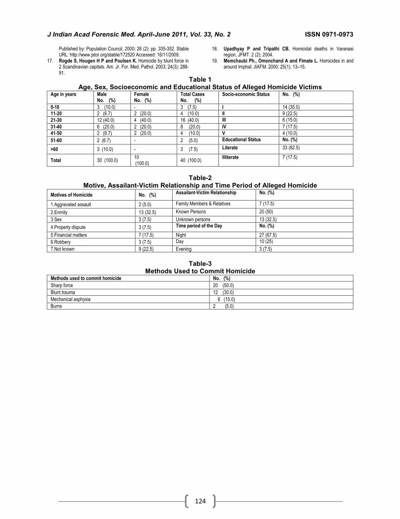
J Indian Acad Forensic Med. April-June 2011, Vol. 33, No. 2 ISSN 0971-0973
124
Published by: Population Council, 2000; 26 (2): pp. 335-352. Stable URL: http://www.jstor.org/stable/172520 Accessed: 16/11/2009.
17. Rogde S, Hougen H P and Poulsen K. Homicide by blunt force in 2 Scandinavian capitals. Am. Jr. For. Med. Pathol. 2003; 24(3): 288-91.
18. Upadhyay P and Tripathi CB. Homicidal deaths in Varanasi region, JFMT. 2 (2); 2004.
19. Memchaubi Ph., Omonchand A and Fimate L. Homicides in and around Imphal; JIAFM. 2000; 25(1): 13–15.
Table 1 Age, Sex, Socioeconomic and Educational Status of Alleged Homicide Victims
Age in years Male No. (%)
Female No. (%)
Total Cases No. (%)
Socio-economic Status No. (%)
0-10 3 (10.0) - 3 (7.5) I 14 (35.0)
11-20 2 (6.7) 2 (20.0) 4 (10.0) II 9 (22.5)
21-30 12 (40.0) 4 (40.0) 16 (40.0) III 6 (15.0)
31-40 6 (20.0) 2 (20.0) 8 (20.0) IV 7 (17.5)
41-50 2 (6.7) 2 (20.0) 4 (10.0) V 4 (10.0)
51-60 2 (6.7) - 2 (5.0) Educational Status No. (%)
>60 3 (10.0) - 3 (7.5) Literate 33 (82.5)
Total 30 (100.0) 10 (100.0)
40 (100.0) Illiterate 7 (17.5)
Table-2
Motive, Assailant-Victim Relationship and Time Period of Alleged Homicide
Motives of Homicide No. (%) Assailant-Victim Relationship No. (%)
1.Aggravated assault 2 (5.0) Family Members & Relatives 7 (17.5)
2.Enmity 13 (32.5) Known Persons 20 (50)
3.Sex 3 (7.5) Unknown persons 13 (32.5)
4.Property dispute 3 (7.5) Time period of the Day No. (%)
5.Financial matters 7 (17.5) Night 27 (67.5)
6.Robbery 3 (7.5) Day 10 (25)
7.Not known 9 (22.5) Evening 3 (7.5)
Table-3
Methods Used to Commit Homicide Methods used to commit homicide No. (%)
Sharp force 20 (50.0)
Blunt trauma 12 (30.0)
Mechanical asphyxia 6 (15.0)
Burns 2 (5.0)

J Indian Acad Forensic Med. April-June 2011, Vol. 33, No. 2 ISSN 0971-0973
125
Original Research Paper
Epiphyseal Fusion at Lower End of Radius and Ulna Valuable Tool for Age Determination
*Dharmesh S. Patel, ** Harish Agarwal, ***Jigesh V. Shah
Abstract Age estimation is an important task and valuable tool to assist in many civil and criminal
procedures, especially in developing countries like India where illiterate population is not aware of the importance of registration of births or the record of registration may not be properly maintained. Physical methods for age estimation are not accurate and eruption of teeth (except third molar) is complete by the age of 16 years. Epiphyseal fusion of long bones is relatively constant in timings and important for age estimation in such cases up to the age of 22 years. This present prospective study is carried out at Forensic Medicine Department of B. J. Medical College, Ahmedabad during the year 2009-10 on 104 subjects of either sex with known age from 15-21 years. It was noticed that the epiphyseal fusion at lower end of radius and ulna processes and progresses bilaterally symmetrical, begins at the age of 16-18 years and completes by the end of 20 year. It was further noticed union at lower end of ulna occurs in advance of radius and females show union in advance of male subjects.
Key Words: Age determination, Epiphyseal fusion, Lower end of radius and ulna
Introduction:
Age estimation in living as well as dead is one of the important tasks for a forensic practitioner. In developing countries like India, illiteracy is major factor for un-awareness regarding importance of registration of births or improper maintenance of records. It is a prerequisite for personal identification and it is increasingly important in criminal and civil matters. In fact, if doubt arises regarding the age of a person in any legal inquiry, forensic age estimation is promptly requested by authorities to ascertain whether the person concerned has reached the age of imputability. Here age estimation becomes a valuable tool to assist in administration of many civil and criminal procedures such as identification, consent, criminal responsibility, clinical examination, validity of will, attainment of majority, kidnapping or abduction, rape, criminal abortion etc. In the developed part of world, reference atlas for age estimation is also in routine use. Corresponding Author: *Associate Professor and Post Graduate Teacher, Forensic Medicine Department, B. J. Medical College, Ahmedabad – 380 016 E-mail: [email protected] **Resident Doctor, Forensic Medicine Department, B. J. Medical College, Ahmedabad – 380 016 ***Associate Professor, Forensic Medicin Department, Kesar SAL Medical College, Ahmedabad – 380 060
There are 3 steps for age estimation • Physical examination • Dental examination • Radiological examination
Physical examination includes measures of height, weight and body mass. They are closely related to biological maturation, but are not sufficiently accurate due to the wide variations in body size. Periods of eruption of deciduous and permanent teeth are reliable. But by the age of 15-16 years eruption of all deciduous and permanent teeth is complete except third molar. Eruption of third molar (wisdom tooth), if erupts, shows wide variation from person to person. So, dental examination will not be of much help after the age of 16 years.
After puberty the process of growth in length of the long bones stops at different ages in different parts of different long bones. This stoppage of growth process is indicative on x-ray examination by fusion of the epiphysis with its respective diaphysis, or can say secondary centre with primary centre. This process is complete by the age of 22 years as described by various authors. Further it is found that the age for the fusions is fairly constant with minor variations among different study groups of different geographic areas.Various studies have shown that the lower end (epiphysis) of radius and ulna fuses with their respective diaphysis within the age group of 16-20 years. The minor
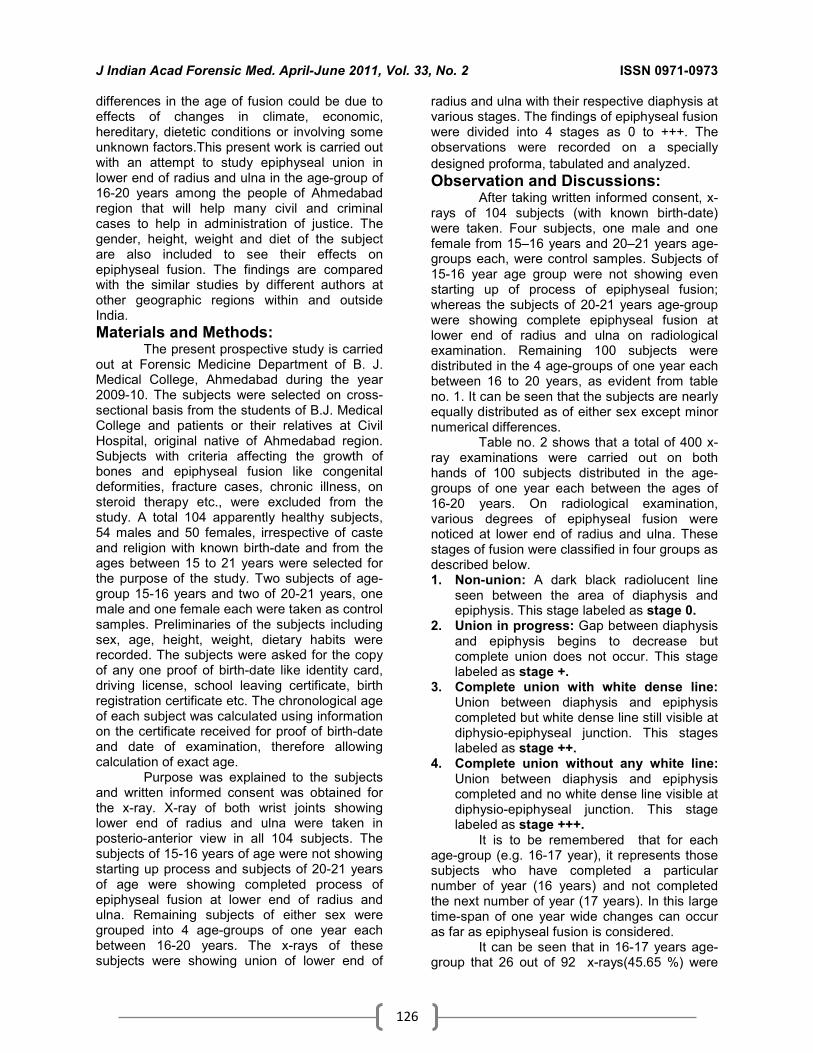
J Indian Acad Forensic Med. April-June 2011, Vol. 33, No. 2 ISSN 0971-0973
126
differences in the age of fusion could be due to effects of changes in climate, economic, hereditary, dietetic conditions or involving some unknown factors.This present work is carried out with an attempt to study epiphyseal union in lower end of radius and ulna in the age-group of 16-20 years among the people of Ahmedabad region that will help many civil and criminal cases to help in administration of justice. The gender, height, weight and diet of the subject are also included to see their effects on epiphyseal fusion. The findings are compared with the similar studies by different authors at other geographic regions within and outside India.
Materials and Methods: The present prospective study is carried
out at Forensic Medicine Department of B. J. Medical College, Ahmedabad during the year 2009-10. The subjects were selected on cross-sectional basis from the students of B.J. Medical College and patients or their relatives at Civil Hospital, original native of Ahmedabad region. Subjects with criteria affecting the growth of bones and epiphyseal fusion like congenital deformities, fracture cases, chronic illness, on steroid therapy etc., were excluded from the study. A total 104 apparently healthy subjects, 54 males and 50 females, irrespective of caste and religion with known birth-date and from the ages between 15 to 21 years were selected for the purpose of the study. Two subjects of age-group 15-16 years and two of 20-21 years, one male and one female each were taken as control samples. Preliminaries of the subjects including sex, age, height, weight, dietary habits were recorded. The subjects were asked for the copy of any one proof of birth-date like identity card, driving license, school leaving certificate, birth registration certificate etc. The chronological age of each subject was calculated using information on the certificate received for proof of birth-date and date of examination, therefore allowing calculation of exact age.
Purpose was explained to the subjects and written informed consent was obtained for the x-ray. X-ray of both wrist joints showing lower end of radius and ulna were taken in posterio-anterior view in all 104 subjects. The subjects of 15-16 years of age were not showing starting up process and subjects of 20-21 years of age were showing completed process of epiphyseal fusion at lower end of radius and ulna. Remaining subjects of either sex were grouped into 4 age-groups of one year each between 16-20 years. The x-rays of these subjects were showing union of lower end of
radius and ulna with their respective diaphysis at various stages. The findings of epiphyseal fusion were divided into 4 stages as 0 to +++. The observations were recorded on a specially
designed proforma, tabulated and analyzed. Observation and Discussions:
After taking written informed consent, x-rays of 104 subjects (with known birth-date) were taken. Four subjects, one male and one female from 15–16 years and 20–21 years age-groups each, were control samples. Subjects of 15-16 year age group were not showing even starting up of process of epiphyseal fusion; whereas the subjects of 20-21 years age-group were showing complete epiphyseal fusion at lower end of radius and ulna on radiological examination. Remaining 100 subjects were distributed in the 4 age-groups of one year each between 16 to 20 years, as evident from table no. 1. It can be seen that the subjects are nearly equally distributed as of either sex except minor numerical differences.
Table no. 2 shows that a total of 400 x-ray examinations were carried out on both hands of 100 subjects distributed in the age-groups of one year each between the ages of 16-20 years. On radiological examination, various degrees of epiphyseal fusion were noticed at lower end of radius and ulna. These stages of fusion were classified in four groups as described below. 1. Non-union: A dark black radiolucent line
seen between the area of diaphysis and epiphysis. This stage labeled as stage 0.
2. Union in progress: Gap between diaphysis and epiphysis begins to decrease but complete union does not occur. This stage labeled as stage +.
3. Complete union with white dense line: Union between diaphysis and epiphysis completed but white dense line still visible at diphysio-epiphyseal junction. This stages labeled as stage ++.
4. Complete union without any white line: Union between diaphysis and epiphysis completed and no white dense line visible at diphysio-epiphyseal junction. This stage labeled as stage +++.
It is to be remembered that for each age-group (e.g. 16-17 year), it represents those subjects who have completed a particular number of year (16 years) and not completed the next number of year (17 years). In this large time-span of one year wide changes can occur as far as epiphyseal fusion is considered.
It can be seen that in 16-17 years age-group that 26 out of 92 x-rays(45.65 %) were

J Indian Acad Forensic Med. April-June 2011, Vol. 33, No. 2 ISSN 0971-0973
127
not showing even starting up of process of epiphyseal fusion, 36 x-rays (39.13 %) were showing + degree epiphyseal fusion, whereas remaining 14 (15.22 %) showing ++ degree fusion at lower end of radius and ulna. Here, none of the x-ray was showing completed process of fusion (described as of +++ degree for the present study) at lower end of radius and ulna. On moving towards the age-group of 17-18 years, the x-rays showing + degree fusion are relatively reduced to 44 % and the x-rays showing ++ degree fusion are relatively increased (48 %). Here two x-rays of radius are not showing starting up of process of epiphyseal fusion (described as of 0 degree for the present study) and in contrast 6 x-rays of ulna are showing completed process of fusion at the lower end.
On advancing towards the age group of 18-19 years, the process of epiphyseal fusion is seen to be advancing further as majority of x-rays (94 %) are showing either ++ or +++ degree fusion. Only 6 x-rays (4 radius and 2 ulna) are showing + degree fusion. These suggest that on an average the epiphyseal fusion at lower end of radius and ulna begins at the age of 16-18 years. Here in 18-19 year age-group, 34 % x-rays (22 radius and 12 ulna) were showing ++ degree fusion. Interestingly 60 % x-rays (24 radius and 36 ulna) were showing +++ degree complete fusion. It means that these fusions start to complete at this age.
In the age group 19-20 years, most of the x-rays (106 out of 108 forming 98.15 %) are showing completed process of fusion (+++ degree) at lower end of radius and ulna. Only 2 x-rays (1.85 %) of radius of right and left hand of a subject were showing ++ degree fusion. This subject had just completed 19 years of age and was expected to show complete fusion by the end of 20 years. Thus from the present study the age for complete fusion among the people of Ahmedabad region is 19-20 years.On closer examination it can be well noticed from table no. 2 that in all the age groups the numbers of x-rays showing the different degrees of epiphyseal fusion in lower end of radius and ulna are not equally distributed. In 16-17 years age-group, the x-rays showing no starting up process of fusion are 42, which constitute 26 radius and 16 ulna. In this group, we can see that for + degree fusions, number of x-rays of ulna are more (22) as compared to that of radius (14).
This fact is also noticed for ++ degree fusion with 8 ulna and 6 radius. Thus, the epiphyseal fusion at lower end of ulna starts earlier when compared to radius, but the difference appears to be less than a year. In
general in all the age groups, as the level of degree of fusion advances, the number of x-rays of ulna increases when compared with that of radius. Importantly, in 18-19 year age group, for complete fusion (+++ degree), the number of x-rays of ulna (36) are far higher as compared to that of radius (24). These suggests that the epiphyseal fusion at the lower end of ulna completes earlier compared to radius. But here also the difference appears to be less than a year.
Majority of the authors are having conclusions parallel with our study. Davis D A and Parsons F G (1927) are one of them. In their study on the population of The Great Britain have described that the epiphyseal union at lower end of radius occurs as early as 17 years but commonest time is 19th-20th year. This finding is in consonance with our study. This suggests that the Indian race and Caucasoid people of England have same age for epiphyseal fusion as far as lower end of radius and ulna are concerned.
But soon Hepworth S M (1929) had carried out his studies on the people of Punjab, in 1929. Population of Punjab District of India is having comparable physiques with that of population of Europe. Hepworth had concluded the contrast, saying that the epiphyseal union is 2.5 to 3 years earlier in Indians than in England and America.
But, the people of Bihar (India) in the study by Prasad R S et al (1976) have shown results comparable to the study of Hepworth. According to their results 12% of total cases in age group of 16 years show complete union and 40% of total cases in age group of 17 years and 18 years each shows complete union. There was no difference noted regarding the age in distal end of radius and ulna. Loomba S D (Uttar Pradesh, India – 1958) has calculated this period to be about 2 years whereas Greulich W W et al (USA – 1950) has considered it to be 3-4 years. We have not found any reason for this.
India is a very large country comprising about world’s 16 % population; where every state has its own cultural background, dietary habits in different geographical areas. So, apart from Prasad (Bihar) the studies have been carried out by many other authors at different states to check whether there are any variations in the age of the epiphyseal fusion from state to state. Lall R & Nat B S had carried out their studies on the subjects of U P (India) in 1934. They suggested that epiphyseal fusion at lower end of radius and ulna occur at the same time and it completes at 19 year. Whereas Loomba S D in his studies on the subjects of Uttar Pradesh
J Indian Acad Forensic Med. April-June 2011, Vol. 33, No. 2 ISSN 0971-0973
128
(India) in the year 1958 has shown findings similar to the present study that the distal epiphysis of ulna shows early union as compared to distal end of radius.
Tables 3 to 6 are comparison tables. Here the table 3 shows the bilateral comparison for the epiphyseal fusion at lower end radius and the same is in table no. 4 for ulna. It can be pointed out from these two tables that the findings are exactly similar for the right and left hand in each and every age group under the study. The interpretation is that the process and progress of epiphyseal fusion at lower end of radius and ulna is bilaterally symmetrical. We have not found any literature regarding bilateral symmetry or asymmetry of epiphyseal fusion at lower end of radius and ulna. This conclusion will of great help to the researchers in the future.
Table no. 5 and 6 shows the effect of gender on epiphyseal fusion at lower end of radius and ulna respectively. All the findings are in double figures as already concluded from table no. 3 & 4 that the epiphyseal fusion at lower end of radius and ulna is bilaterally symmetrical.After taking the consideration from table no. 1 and on fine analysis of table no. 5 & 6, we see that the subjects are not proportionately distributed in all the age groups of either sex showing different degrees of epiphyseal fusion. In both of the tables in all the age groups as the stage of epiphyseal fusion advances, the numbers of x-rays are proportionally increasing for females as compared to males. Thus, on an average females show earlier epiphyseal fusion at lower end of radius and ulna as compared to male with a difference of less than one year.
Many of the authors like Pryor J W (USA – 1905, 1906, 1923, 1928), Greulich W W et al (USA – 1950), Loomba S D (U P, India – 1958) have also stressed that the bones of females ossify in advance of male. Loomba has described in his studies that in the majority, age of union in boys was 20-21 years and for girls it was 18-19 years.
Observations from his study of Kothari D R (Rajasthan, 1972) shows that the average age in boys for epiphyseal fusion to occur in distal end of radius is 18-19 years and that for girls is 17-18 years, no difference in terms of age is seen for union of distal end of ulna.
Conclusion: Epiphyseal fusion of lower end of radius: • The processes of union starts (grade +) in
33.3% individuals in the age group of 16-17 years in males. Rest of the 66.6% does not
show even starting up of process of fusion (grade 0).
• The processes of union (grade +) starts in 27.2% individuals in the age group of 16-17 years in females, while 27.2% individuals shows grade ++ fusion. Remaining 45.4% shows grade 0 fusion.
• From above results it can be concluded that starting of epiphyseal fusion in lower end of radius of both hand in male is 16-17 years and for female is also 16-17 years. Females show early age of fusion as compared to male but this difference is less than one year.
• In 19-20 years age group 93.3% males shows complete fusion (grade +++ )of lower end of radius in males.
• In 18-19 years age group 75% females shows complete fusion(grade +++) while 100% females in age group19-20 years shows complete fusion (grade +++ ) of lower end of radius. From above results it can be concluded that
completion of epiphyseal fusion in lower end of radius of both hand in male is 19-20 years and for female is also 19-20 years. Females show early age of fusion as compared to male but this difference is less than one year.
Epiphyseal Fusion at lower end of ulna: • The processes of union (grade +) starts in
50% individuals in the age group of 16-17 years in males. Rest of the 50% does not show even starting up of process of fusion (grade 0).
• The processes of union (grade +)starts in 45.4% individuals in the age group of 16-17 years in females, while 36.3% individuals shows grade ++ fusion. Remaining 18.2% shows grade 0 fusion. From above results it can be concluded that
starting up of epiphyseal fusion in lower end of ulna of both hand in male is 16-17 years and for female is also 16-17 years. Females show early age of fusion as compared to male but this difference is less than one year. • In 19-20 years age group 100% males
shows complete fusion of lower end of ulna in males.
• In 18-19 years age group 100% females shows complete fusion and 100% females in age group 19-20 shows complete fusion of lower end of radius.
From above results it can be concluded that completion of epiphyseal fusion in lower end of ulna of both hand in male is 19-20 years and for female is 18-19 years. Females show early

J Indian Acad Forensic Med. April-June 2011, Vol. 33, No. 2 ISSN 0971-0973
129
age of fusion as compared to male but this difference is less than one year.
Fusion at lower end of ulna shows epiphyseal fusion in advance of the lower end of radius but the difference is less than a year. Bilatrality: The age for epiphyseal fusion of lower end of radius and ulna is bilaterally similar, i.e. it occurs at the same age in both hands. Sex: The epiphyseal fusion of lower end of radius and ulna occurs earlier by about one year in females as compared to males. References: 1. Davies D A, Parsons F G. The age order of the appearance and
union of the normal epiphysis as seen by X ray. J Anat. 1927; 62: 58–71.
2. Gerulich W W, Pyle S I. Radiographic atlas of skeletal Development of the Hand and Wrist. 2nd edition, 1959; Stanford University press, Stanford.
3. Hepworth S N. Determination of age in Indians from a study of ossification of the epiphysis of long bones. Ind Med Gaz. 1939; 614–616.
4. Kothari D R: Age of epiphyseal union at the elbow and wrist joints in Marwar region of Rajasthan, Journal of Indian Medical Association. 1974; 63: 8.
5. Lall R and Nat B S. Ages of Epiphyseal union at the Elbow and Wrist Joints amongst Indians, Indian Journal of Medical Research. 1934; XXI (4): 683-687.
6. Loomba S D. Age of epiphyseal union at wrist joint in Uttar Pradesh. Journal of Ind Med Association. 1958; 30 (12): 389-395.
7. Prasad R S, Srivastav K P and Lala J K: Radiologic study of some long bones to determine the age of consent in females of Oraon and Munda tribes, Journal of Ind Medical Association. 1979; 72: 4.
8. Pryor J W. Time of ossification of the bones of the hand of male and female and union of epiphyses with diaphyses, American Journal of Physical Anthropology. 1925; 8(4): 401-410.
9. Reddy K S N. The Essentials of Forensic Medicine and Toxicology. 29th edition. Hyderabad: K Sugna Devi; 2009: 61–71.
10. Schmidt S, Koch B, Schultz R, Reisinger W and Schmeling A. Comparative analysis and the applicability of the skeletal age determination methods of Greulich-Pyle and Thiemann-Nitz for Forensic age estimation in living subjects. Int J Leg Med. 2007; 121 (4): 293–296.
Table 2 : Epiphyseal Fusion in Lower End of Radius and Ulna of Both Hands
Age Groups
(yrs)
Various Degree of Epiphyseal Fusion (Number Of Cases)
Total X-Rays
0 + ++ +++
R* U# R U R U R U
16-17 26 16 14 22 06 08 00 00 92
17-18 02 00 24 20 24 24 00 06 100
18-19 00 00 04 02 22 12 24 36 100
19-20 00 00 00 00 02 00 52 54 108
Total 28 16 42 44 54 44 76 96 400
* R = Radius # U = Ulna
Table 1: Age and Sex Wise Classification Age groups (in years) Male Female Total
16-17 12 11 23
17-18 12 13 25
18-19 13 12 25
19-20 15 12 27
Total 52 48 100
Table 5: Sex Wise Differences for Epiphyseal Fusion at Lower End of Radius:
Age groups (in years)
Gender Wise Comparison at Lower End of Radius(Number Of Cases) Total
x-rays
0 + ++ +++
M F M F M F M F
16-17 16 10 08 06 00 06 00 00 46
17-18 02 00 14 10 08 16 00 00 50
18-19 00 00 04 00 16 06 06 18 50
19-20 00 00 00 00 02 00 28 24 54
Total 18 10 26 16 26 28 34 42 200
Table 6: Sex Wise Differences for Epiphyseal Fusion at Lower End of Ulna:
Age groups (in years)
Gender Wise Comparison at Lower End Of Ulna (Number Of Cases) Total
x-rays
0 + ++ +++
M F M F M F M F
16-17 12 04 12 10 00 08 00 00 46
17-18 00 00 14 06 10 14 00 06 50
18-19 00 00 02 00 12 00 12 24 50
19-20 00 00 00 00 00 00 30 24 54
Total 12 04 28 16 22 22 42 54 200
Table 3: Comparison of Epiphyseal Fusion at Lower End of Radius of Rt &Lt Hand in either Sex:
Age groups (in years)
Various degree of epiphyseal fusion at lower end of radius (number of cases)
Total x-rays 0 + ++ +++
RT LT RT LT RT LT RT LT
16-17 13 13 07 07 03 03 00 00 46
17-18 01 01 12 12 12 12 00 00 50
18-19 00 00 02 02 11 11 12 12 50
19-20 00 00 00 00 01 01 26 26 54
Total 14 14 21 21 27 27 38 38 200
Table 4: Comparison of Epiphyseal Fusion at Lower End of Ulna of Right &Left Hand in either Sex:
Age groups (in years)
Bilateral Comparison At Lower End Of Ulna(number of cases) Total x-rays
0 + ++ +++
RT LT RT LT RT LT RT LT
16-17 08 08 11 11 04 04 00 00 46
17-18 00 00 10 10 12 12 03 03 50
18-19 00 00 01 01 06 06 18 18 50
19-20 00 00 00 00 00 00 27 27 54
Total 08 08 22 22 22 22 48 48 200

J Indian Acad Forensic Med. April-June 2011, Vol. 33, No. 2 ISSN 0971-0973
130
Original Research Paper
Age Estimation from Third Molar Development A Radiological Study
* Jashwant A. Darji, **Ganesh Govekar, ***S.D. Kalele, **** Hareshwari Hariyani
Abstract The present Study is based on orthopantographic evaluation of the development of the third
molar (n=84 persons with known age between 15 to 25 years and known sex male-49, female-35) based on eight stages(A-H ) method of Demirjian et al. Data was feed in computer and analyzed using Statistical package SPSS. Regression analysis was performed to obtain formulas dental age calculation with chronological age. No Statistically significant difference was revealed in third molar development between males &female in all eight stages of development(p>0.05).No Statistically significant difference was revealed in third molar development between left &right side in all eight stages of development where both third molars were scorable (p>0.05). Statistical analysis showed a strong positive correlation between various stages of third molar development and chronological age (r
> 0.60, p value<0.05).
Regression formulas for whole sample based on the third molar teeth present were estimated and new equations derived:
Age in years =13.70+1.50(Developmental stage of Mx Right third molar) Age in years = 14.82 + 1.25 (Developmental stage of Mn Right third molar)
Key Words: Third Molar, Chronological age, Age estimation
Introduction: Age estimation is one of the important
duties of the medico legal officers in recent time as crimes of varied nature are increasing and age constitutes an important factor in relation to the assessment for the award of sentences in all these instances and in all these matters age is inseparably related. To establish the identity of an individual, age estimation is necessary not only in cases of living but also in the dead too. For example in Civil cases like identification in aeroplane crashes, railway disasters, bomb blasts, earthquakes, floods, fire disasters, ship wrecks, employment and retirement, Marriage, Management of property ,Voting right, Competency as witness, insurance claims, passport, visa.
Corresponding Author:
*Assistant Professor, Deptt. of Forensic Medicine, Govt. Medical College, B/H, S.T. Stand, Jail Road, Bhavnagar (Gujarat) - 364001 Email- [email protected] **Prof. & Head, Deptt. of Forensic Medicine, Govt. Medical College, Surat ***Prof. & Head, Deptt. of Forensic Medicine, Govt Medical College, Bhavnagar **** Assoc. Prof., Deptt. of Dentistry, GMC, Bhavnagar.
In criminal cases like rape, criminal abortion, infanticide, murder, juvenile offenders and criminal responsibilities identification is essential. In cases in which dead body of a person claimed to be that of a missing person, cases of unnatural death of unidentified bodies due to suspected foul play. Many Factors which helps in age estimation like teeth, ossification of bones, secondary sex characters, and general development of body.
Teeth have been recognised since long back as a valuable tool in the hands of medico legal expert for establishing personal identity. Estimating age from teeth is generally reliable as they are naturally preserved long after all the tissues and even bones have disintegrated. Unlike the bones, the teeth can also be inspected in the living as well as dead individuals.
Age estimation becomes difficult after about 14 years since all the permanent teeth, except the third molar would have completed their development rendering them to be the only clue used for age estimation. Very few alternative methods during the interval between mid teens & early 20’s are available.
The third molar offers a unique advantage over other teeth because its development tends to continue over a longer period and until a later age. [4]

J Indian Acad Forensic Med. April-June 2011, Vol. 33, No. 2 ISSN 0971-0973
131
Most studies of the third molar have concerned with its presence or absence. Radio graphically distinguishable stages of tooth formation have been described in very few cases. [5] Some researchers prefer tooth formation to tooth eruption (i.e. actual emergence of the tooth in the oral cavity) for assessment of age because eruption can be influenced by exogenous factors such as infection, injury at the area, obstruction, overcrowding, earlier extraction of deciduous teeth etc. whereas, formation is a continuous process until tooth is completely calcified.
The present study on development of third molars has been adopted for the reason that there is paucity of similar studies in Gujarat and the age group here selected (15, 16, 18 years) bears immense medico legal importance.
Materials and Methods: Materials for the study comprised of 84
subjects of Bhavnagar region. Out of these 49 were male and 35 were female. The age group of study ranged from 15-25 years. These subjects were chosen randomly from different schools, colleges and patients visiting the Out Patient Department of Dentistry, Sir. T. Hospital and Medical College, Bhavnagar.
The selection of the subjects was based on the following criteria:
Inclusion Criteria: 1. Belongs to age group of 15-25 years 2. Good oral hygiene. 3. Voluntary consent for the study procedure. 4. Age proof in the form of Birth certificate or
School/College register or I.D. card/Driving license.
Exclusion Criteria: 1. Any congenital anomalies. 2. Malnutrition or other diseases that would
affect the skeletal growth and general development of the individual.
3. Subjects with history of third molar extraction
4. Pregnant female Informed consent was taken from every
individual, prior to examination. Orthopantogram of each individual was then taken. Each of these orthopantogram was studied for eight different stages of development of the third molars by methods adapted by Demirjian as follows [2]: • Stage A: Cusp tips are mineralized but have
not yet coalesced. • Stage B: Mineralized cusps are united so
the mature coronal Morphology is well defined.
• Stage C: The crown is about half formed; the pulp chamber is evident and dential deposition is occurring.
• Stage D: Crown formation is complete to the dentino-enamel junction. The pulp chamber has a trapezoidal form.
• Stage E: Formation of the inter-radicular bifurcation has begun. Root length is less than the crown length.
• Stage F: Root length is at least as great as crown length. Roots have funnel-shaped endings.
• Stage G: Root walls are parallel, but apices remain pen.
• Stage H: Apical ends of the roots are completely closed.
Each of the OPG film was then viewed on X-ray viewer. Radiographs were rated with the help of Orthodontist. Stage: A
Stage: E
Stage: B
Stage: F
Stage: C
Stage: G
Stage: D
Stage: H
Figure 3: Schematic and radiographic presentation of eight stages of development suggested by Demirjian used to score third molar development. Grades A, B, C do not occur in the age group included in this study.

J Indian Acad Forensic Med. April-June 2011, Vol. 33, No. 2 ISSN 0971-0973
132
All four 3rd molars were scored to test
left right symmetry and arch differences in development.Absent third molars were excluded from the study Data was feed in computer and analyzed using Statistical Package SPSS.
Observations:
Table 1: Gender Distribution of Mean Ages at Attainment of Stages of Third Molar Development in Maxilla and Mandible: Maxilla
Group Statistic Stage of Development
D E F G H
Male Mean age 16.17 16.58 16.60 18.54 22.26
SD 0.59 0.92 0.12 1.77 2.68
Female Mean age 17.17 17.33 18.20 19.68 20.90
SD 1.53 2.21 2.11 1.65 2.21
- P value 0.396 0.659 0.354 0.102 0.163
T-test, P value>0.05 non significant
Mandible
Group Statistic Stage of Development
D E F G H
Male Mean age 16.11 16.83 17.87 19.27 22.50
SD 0.38 1.10 1.92 2.15 2.48
Female Mean age 17.98 18.97 18.35 19.25 20.93
SD 2.23 4.13 1.91 0.98 2.51
- P value 0.219 0.355 0.620 0.986 0.164
T-test, P value>0.05 non significant
Table 2: Side (Lt & Rt) Wise Mean Ages at Attainment of Stages of Third Molar Development in Maxilla And Mandible Maxilla
Grouping Statistic Stage of Development
D E F G H
Left Mean age 16.58 16.88 17.58 19.17 21.60
SD 1.59 1.36 1.68 1.97 2.51
Right Mean age 16.48 16.70 18.12 19.00 21.72
SD 1.10 1.19 1.81 1.78 2.55
- P value 0.922 0.789 0.524 0.739 0.863
T-test, P value>0.05 non significant
Mandible
Group Statistic Stage of Development
D E F G H
Left Mean age 17.36 17.54 17.89 19.26 21.65
SD 1.99 2.62 1.68 1.78 2.42
Right Mean age 16.77 17.75 18.37 19.01 21.89
SD 1.10 2.76 1.99 1.81 2.59
- P value 0.515 0.883 0.449 0.674 0.746
T-test, P value>0.05 non significant
Table 3: Mean Ages at Attainment of Stages of Third Molar Development in Maxilla & Mandible Maxilla
Statistic Stage of Development
D E F G H
Mean age 16.85 16.87 17.74 19.00 21.60
SD 1.29 1.46 1.90 1.78 2.51
Mandible
Statistic Stage of Development
D E F G H
Mean age 17.18 17.75 18.06 19.26 21.75
SD 1.88 2.76 1.87 1.76 2.56
Table 4: Percentile Distributions of the at Attainment of Stages of Third Molar Development in Maxilla and Mandible Maxilla
Percentile Stage of Development
D E F G H
10 15.00 15.08 15.58 16.48 17.73
25 15.50 15.67 16.50 17.83 19.17
50 16.58 16.67 17.50 19.08 21.92
75 17.75 18.21 18.92 20.25 23.67
90 18.42 19.42 20.83 21.33 24.92
Mandible
Percentile Stage of Development
D E F G H
10 15.00 15.08 15.58 17.06 17.90
25 15.50 15.75 16.63 17.90 19.13
50 16.50 17.25 17.83 19.29 22.33
75 18.42 18.67 19.54 20.10 23.83
90 20.83 23.25 21.33 21.94 24.85
Percentiles for stage H presume that independent criteria can be used to eliminate cases over 25 years of age from consideration.
Table 5: regression analysis predicting chronological age from stages of 3
rd molar
development Univariate Regression
Intercept Regression Coefficient
r2 F P
Maxillary Left M3
13.638 1.504 .400 49.254 0.00
Maxillary Right M3
13.703 1.500 .411 50.966 0.00
Mandibular Left M3
14.817 1.284 .362 39.618 0.00
Mandibular Right M3
14.820 1.252 .352 38.58 0.00
P value<0.05 significant
Discussion: Chronologic age estimation by tooth
development has been used over a long period. Tooth development is an accurate measure of chronologic age that seems to be an independent of exogenic factors such as malnutrition or disease. [7]
The third molar
calcification stage is one of the few tools that can be used to assess age when development is nearing completion. However, age estimates based on dental methods have shortcomings, especially during adolescence when the third molar is the only remaining variable dental indicator. Indeed, a great variation in position, morphology, and time of formation exists. The differences between populations, the different methodology, and the dissimilarity among observers are other important shortcomings.
The single compelling reason to rely on third molar formation to estimate chronological age is that there are very few alternative methods during the interval roughly between the middle teens and early 20s. All of the hand- wrist bones have achieved their adult morphologies and their epiphyses have fused and the onset of secondary sex characteristics has occurred.
J Indian Acad Forensic Med. April-June 2011, Vol. 33, No. 2 ISSN 0971-0973
133
In the past different classifications were presented by Kohler et al, Kullman et al [7, 8]
and others.
However some of these classifications identify a large number of stages that are hard to delimit from each other. Demirjian et al [2] presented a classification distinguishing four stages of crown development (A-D) and four stages of root development (E-H).
In the present study development of third molar in all stages was found slightly earlier in males than in females but the difference in age at the various developmental stages was statistically non significant at P value >0.05. Similar observations were noticed by Garn et al [5], Engstorm [4], Levesque and Bhat Vrinda et al [3]. These observations are in contraindication to Kullman et al. [7, 8]
H. Mincer et al [6], Yuming bai et al [9]
who found a statistically significant sex difference in age at the some developmental stages. Here also males were reported to have their teeth calcified earlier than in females. This is unique finding for third molar as all other permanent teeth development is earlier in female as compared to males.
In the present study no significant difference is found in the age at the various developmental stages in left and right third molar in both arches, where both third molars were scorable. This is in contraindication to findings of H. Mincer et al [6]
who found significant
difference. In the present study maxillary third
molar tended to develop somewhat earlier than their mandibular counter part. Similar observation was noticed by H. Mincer et al. [6]
Mean and median ages at various developmental stages of third molars in the present study are slightly late as compared to observations made by H. Mincer et al. [6]
For medico legal purposes, 18 years of age is an important cut point. From table 6 we can say that if a subject presents with a developmental stage A to D there is less likelihood that subject is 18 years old. On the other side if subject presents with developmental stage of H, there are more chances that subject has crossed the 18 years age. Similar observations were made by H. Mincer et al. [6]
Summery and Conclusions: Third molar development was examined
in both sexes; the mean ages & SD for Demirjian stages described in table-1. Statistically no significant differences were revealed in third molar development between
males &female in all eight stages of development.
Third molar development was examined in left &right side in both arches, the mean ages & SD for Demirjan stages described in table-2. Statistically no significant difference were revealed in third molar development between left &right side in all eight stages of development where both third molars were scorable.
Development of third molar is faster in maxilla as compared to mandible in all stages of development than their counter parts (table 2&3).
Medico legal questions many times involve that whether individual is a juvenile or an adult, i.e. younger or older than 18 years of age. From percentile distribution of the age at attainment of stages of third molar development in this study shows that in D stage of development of third molar there are 75%of chances that individual has not attain the age of 18means that there are 25% of chances of individual being of above 18 years of age with D stage of development of third molar. If individual present with H stage of development of third molar there are 75%of chances that individual has attain the age of 18means that there are 25% of chances of individual being of below 18 (Table-4).
Linear regression coefficients (Pearson Correlation) provided to assess the correlation between the third molar development and chronological age. Statistical analysis showed a strong positive correlation between various stages of third molar development and chronological age (r
> 0.60, p value<0.05).
Regression formulas for whole sample based on the third molar teeth present were estimated and following equations derived (Table-5).
Age in years = 13.70 + 1.50 (Developmental stage of Mx Right third molar)
Age in years = 14.82 + 1.25 (Developmental stage of Mn Right third molar) In case of absence of right third molar left third molar should be used.
In present study developmental stages A,B,C not observed , in above derived equation values for developmental stages are – D=1, E=2, F=3, G=4,H=5.
Accuracy of this method is not high because here standard deviation for each stage of third molar development is average 2 years, that means the dental age assigned to a tooth is centered on +/-4years(95% statistical safety) (Table2to4). Regression equation involves the same problem (r
2=0.41).

J Indian Acad Forensic Med. April-June 2011, Vol. 33, No. 2 ISSN 0971-0973
134
In overview the developmental stages of the third molar can be the only quantitative biologic variable available for the estimating the age of a person in his/her late teens or early 20s, but due to the considerable variability of the third molar detracts from precise age estimates, it can be suggestive in the absence of better information.
This dissertation work has been approved by Institutional Review Board (Human Ethics Committee) Govt. Medical College, Bhavnagar.
References: 1. Arther B. Lewis, Stanley M. Garn. The relationship between tooth
formation & other maturational factors. Angle orthodontics, Vol.30, No.2, 1960:p70-77.
2. Demirjian, H. Goldstein, J.M. Tanner. A new system of dental age assessment. Human biology Vol. 45, No.2, 1973: p211-227.
3. Bhat Vrinda, Kamath Giridhar. Age estimation from the root development of mandibular third molars. Medico legal update.Vol.4, No.4, 2004: p127-130.
4. Christer Engstrom, Henle Engstrom and Soren Sagne. Lower third molar development in relation to skeletal maturity and chronological age. Angle orthodontics.Vol.53, No.2, 1983: p 97-106.
5. Garn SM, Lewis B& Bonne B. Third molar formation and its development. Angle orthodontics.Vol.32, No.4, 1962: p270-279.
6. Harry H Mincer, Edward F Harris, Hugh E Berryman. The A.B.F.O. study of third molar development and its use as an estimator of chronological age. J. Fore. Sci.; Vol.38.No.2, 1993: p379-390.
7. Leif Kullman. Accuracy of two dental and one skeletal age estimation in Swedish adolescents. Fore. Sci. Int. Vol.75, 1995: p225-236.
8. Leif Kullman, Johanson g, akesson L. Root development of the lower third molar and its relation to chronological age. Swed. Dent J. Vol.16, No.4, 1992,p 161-167.
9. Yuming Bai, Jing Mao, Shengrong Zhu, and Wei Wei. Third molar development in relation to chronological age in young adults of central China. Journal of Huazhong, University of Science and Technology -- Medical Sciences –Vol. 28, No.4, 2008: p487-490.
J Indian Acad Forensic Med. April-June 2011, Vol. 33, No. 2 ISSN 0971-0973
135
Original Research Paper
An Epidemiological Survey of Fatal Road Traffic Accidents
and their Relationship with Head Injuries
*Ravindra S Honnungar,*Sunil C Aramani, **Vijay Kumar AG, **Ajay Kumar TS, ***Prasanna S Jirli
Abstract Death from road traffic accidents (RTA) and in particular Motor vehicle Traffic Accidents have
been characterized worldwide as a hidden epidemic which affects all sectors of society. It account for 2.1% of global mortality. A retrospective study of fatal cases of RTA was conducted at Department of Forensic Medicine and Toxicology, Jawaharlal Nehru Medical College, Belgaum, Karnataka, India between 1-1-2004 to 31-12-2009. The majority of victims belonged to the age group 31-40 years (145 cases; 28.7%). Female were less involved than men with ratio of 1: 3.3. In our present study total vehicular accident fatalities comprised 506 (48.7 %) out of total 1039 medico legal cases autopsied during five years (2005 to 2009). Our study shows the overwhelming majority of the deceased (76.9%) were males. This study shows that most of the deaths in road traffic accidents, take place either within 24 hours or on the spot of injury which is very alarming and highlights the need for taking urgent steps for establishing good pre-hospital care and provision of trauma services at site in India.
Key Words: Road Traffic Accidents (RTA), Head injury, Mortality, Cause of Death
Introduction: Death from road traffic accidents (RTA)
and in particular Motor vehicle Traffic Accidents have been characterized worldwide as a hidden epidemic which affects all sectors of society. It account for 2.1% of global mortality. An estimated 1.26 million people worldwide died in 2000 from RTA, 90% of them in low and middle income countries. In 2000, the RTA mortality rate for the world was 20.8 per 1, 00,000 population. In the Americas, it was 26.7 for males and 8.4 for females. In the Americas during 1997-2000 mortality from all land transport accidents was the tenth leading cause of death in the general population.
The Asia-pacific region accounts for about 60% of global road deaths despite having only 16% of the world’s vehicles. India accounts for about 10% of RTA worldwide. It contributed 30.2 percent to all kind of natural and unnatural accidental deaths during 2005.
Corresponding Author: *Assistant Professor, Deptt. of Forensic Medicine & Toxicology, KLE University’s J.N. Medical College, Belgaum-590010, Karnataka State, INDIA E mail: [email protected] *Assist. Professor, **Post Graduate, ***Assoc. Professor, Deptt. of FMT KLE University’s J.N. Medical College, Belgaum-590010, Karnataka State, INDIA
The ratio of road area to total area of Delhi city (capital of INDIA) is 21% as compared to 23% in London and 25% in Paris. The road length has been reduced from 2.06 (2000-2001 to 1.88 (2006-2007) km per 1000 population and 8.45 (2000-2001) to 5.90 (2006-2007) km per 1000 vehicles. RTA is the public health issue and cost a lot to individuals, families, communities and nations. The estimated cost is around 1-2% of a country’s GNP in lower income countries. [1, 2]
Material and Methods: A retrospective study of fatal cases of
RTA was conducted at Department of Forensic Medicine and Toxicology, Jawaharlal Nehru Medical College, Belgaum, Karnataka, India between 1-1-2005 to 31-12-2009. During this period total of 506 deaths due to RTA were autopsied. The objective of this study is to assess prevalence of fatal RTA reporting to the hospital and an analysis of the epidemiological factors in relation to victims, vehicles and sites of impacts, etc. Besides, features pertaining to hosts (road users), the agents (vehicles) and the environmental condition (road, seasons, months etc), place of death and their period of survival will be studied. Results:
Table-1: The majority of victims belonged to the age group 31-40 years (145 cases) Female were less involved than men with ratio of 1: 3.3. Table-2: The maximum number

J Indian Acad Forensic Med. April-June 2011, Vol. 33, No. 2 ISSN 0971-0973
136
of victims died in 1 day (168 cases; 33.2%). Table-3: Head injury was the most cause of death in the present study (255 cases; 50.4%). Table-4: Skull fracture (77.7%) was the most common findings followed by sub-dural haemorrhage (73.9%). Table-5: Frontal bone fracture was the common injury (176 cases) Table-6: Liver laceration (69 cases) and Ribs fracture (89 cases; 35.5%) was most common findings other than the head injury. Table-7: Most of the RTA occurred during rainy season (251 cases; 49.6%). Table-8: Drivers were most commonly involved in fatal RTA (214 cases; 42.2%).Table-9: Heavy vehicles were responsible for most of the fatal RTA (223 cases; 44.2%). Table-10: In this study maximum number of accidents occurred on national highways.
Discussion: In our present study total vehicular
accident fatalities comprised 506 (48.7 %) out of total 1039 medico legal cases autopsied during five years (2005 to 2009). Our study shows the overwhelming majority of the deceased (76.9%) were males. It is due to greater male exposure on urban streets and similar higher incidence of traffic accidents among males has been found by many other researchers.[3,4,5,6,7,8] The most common age group affected in the study was between 31-40 years (30.6%) and is consistent with the studies available from India and other countries. [3, 4, 5, 7, 8, 9, 10]
This age group is the most active phase of life, physically and socially, and hence outnumbers the other road users. Considering the maximum involvement of individuals in the economically productive years, vehicular collision fatalities may have an important economic impact. Preventive measures targeting at these high-risk groups are important to reduce the incidence of severe RTA. The most commonly injury was to the head (50.4 %) followed by head and chest (29.2%). Similar observations were reported in studies from Iran and USA. [11, 12] Out of 403 head injury, 313 cases (77.7%) had a fatal skull fracture, consistent with other studies. [13, 14] Most commonly found intracranial haemorrhage was subdural haemorrhage (73.9 %) which is consistent with other studies. [14, 15, 16] No significant variation was evident in the incidence of fatal vehicular accidents by days of a week in our study. This pattern differs from earlier study conducted in Delhi [17]
according to which
highest numbers of accidents were on Saturdays. In the study conducted in Nepal [8] highest numbers of vehicular accidents were
observed on Sundays and lowest on Mondays. National Injury Mortality Surveillance System (2004) reported that most of the transport related deaths occurred on Saturday (20.8 percent) followed by Sunday (17.1 percent).[18] Maximum number of fatal accident took place between June to December (49.6%) in present study. In Nepal, maximum numbers of cases were reported in July followed by January. [8]
In
the earlier studies conducted in Delhi [17, 18, 19] reported maximum numbers of victims were seen in January. This difference in the peak is quite suggestive that fatal vehicular accidents had different temporal correlation with time, day and month as compared to nonfatal vehicular accidents.
Conclusion: This study shows that most of the
deaths in road traffic accidents occur either within 24 hours or on the spot of injury which is very alarming and highlights the need for taking urgent steps for establishing good pre-hospital care and provision of trauma services at site in India. It also shows that head injuries remain the most common and serious type of trauma seen in emergency department of our hospital and availability of good neurosurgical care is essential for these patients. A nationwide computerized trauma registry is urgent required to bring out the risk factors, chain of events leading to the accidents and will be extremely helpful in policy making and health management at the national level in India and worldwide.
Measures against RTA: The multidimensional approach
requirements to prevent RTA are, Road: This includes maintaining existing roads, improving road surface, removing obstacles, constructing guards, rails, proper signs and widening or narrow sections of roads. Vehicles: Vehicles design to improve visibility and protection in event of crash, restraining devices as seat belts compulsorily present and proper maintenance of vehicle and regular inspection of vehicles. Road Users: should be properly trained by authorised centers, medically fit and mentally alert, issuing licenses after strict testing of driving skills, medical fitness “and periodic review of driving skills specially annual medical examination of drivers above 50 years of age. Administrative Measures: Enforcing traffic rules strictly, proper legislation to avoid drunken driving and in repetitive offenders’ license should be cancelled.
References: 1. Arvind K, Sanjeev L, Deepak A, Ravi R, Dogra TD. Fatal road
traffic accidents and their relationship with head injuries: An epidemiological survey of five years. Indian Journal of Neurotrauma (IJNT) 2008; 5(2): 63-67.

J Indian Acad Forensic Med. April-June 2011, Vol. 33, No. 2 ISSN 0971-0973
137
2. Archana K, Sinha US, Kapoor AK, Pathak YK, Susheel S, Aparajita S, Sanju S. An epidemiological study of fatal road traffic accidents in Allahabad region. IIJFMT 2005; 3(1).
3. Salgado MSL, Colombage SM. Analysis of fatalities in road accidents. Forensic Sci Int 1998; 36; 91-6.
4. Sahdev P, Lacqua MJ, Singh B, Dogra TD. Road Traffic fatalities in Delhi: causes, injury patterns and incidence of preventable deaths. Accid Ann Prev 1994; 26: 377-84.
5. Friedman Z, Kungel C, Hiss J, Margovit K, Stein M, Shapira S. The Abbreviated injury scale –a valuable tool for forensic documentation of trauma. Am J For Med Patho 1996; 17: 233-8.
6. Henriksson EM, Ostrom M, Eriksson A. Preventability of vehicle-related fatalities. Accid Ann Prev 2001; 33: 467-75.
7. Sharma BR, Harish D, Sharma V, Vij K. RT A – a demographic & topographic analysis. Med Sci Law 2001; 41: 266-74.
8. Jha N, Agrawal CS. Epidemiological Study of Road Traffic Accident Cases: A Study from Eastern Nepal. Regional Health Forum WHO South-East Asia Region, 2004; 8(1): 15-22.
9. Chandra J, Dogra TD and Dikshit PC. Pattern of craniointracranial injuries in fatal vehicular accidents in Delhi, 1966-76. Med Sci Law 1979; 19: 188-94.
10. BL Meel. Trends in fatal motor vehicle accidents in Transkei region of South Africa. Med Sci Law 2007; 47: 64-8.
11. Montazeri A. Road-traffic-related mortality in Iran: a descriptive study. Public Health 2004; 118:110-3.
12. Friedman R, Harris JP, Sitzer M, Schaff HB, Marshall L, Shackford S. Injuries related to all vehicular accidents: a closer look at head and neck trauma. Laryngoscope 1988; 98:1251-4.
13. Elesha SO, and Daramola AO. Fatal head injuries: the Lagos University Teaching Hospital experience (1993-1997). Niger Post grad Med J 2002; 9:38-42.
14. Menon A, Pai VK, Rajeev A. Pattern of fatal head injuries due to vehicular accidents in Mangalore.J For Leg Med 2008 ; 15:75-7.
15. Akang EE, Kuti MA, Osunkoya AO, Komolafe EO, MalomoAO, Shokunbi MT, Amutta SB. Pattern of fatal head injuries in Ibadan—a 10 year review. Med Sci Law 2002; 42:160-6.
16. Freytag E. Autopsy findings in head injuries from blunt forces: statistical evaluation of 1367 cases. Arch Patho 1963; 75: 402-13.
17. Mehta SP. An epidemiological study of road traffic accident cases admitted in Safdarjang Hospital, New Delhi. Ind J Med Res 1968; 56: 456-66.
18. A profile of fatal injuries in South Africa, 6th Annual Report of the, National Injury Mortality surveillance System, Published by the Medical Research Council/UNISA Crime, Violence and Injury Lead Programme, 30 November 2005.
19. Ghosh PK. Epidemiological study of the victims of vehicular accidents in Delhi. J Ind Med Assoc. 1992; 90: 309-12.
Table 1: Age &Sex Wise Distribution of Cases:
Age (year) Male Female
No % No %
<10 14 03.6 06 05.1
11-20 17 04.4 09 07.8
21-30 91 23.4 18 15.4
31-40 121 31.1 24 20.5
41-50 79 20.3 30 25.6
51-60 44 11.3 19 16.2
>60 23 05.9 11 09.4
Total 389 100 117 100
Table 3: Death Due to Injury in Fatal RTA: Injury No. %
Head alone 255 50.4
Head + other 148 29.2
Other 103 20.4
TOTAL 506 100
Table 7: Season Wise Distribution of Cases: Season No. %
Rainy(June-Sep) 251 49.6
Winter(Oct-Jan) 88 17.4
Summer(Feb-May) 167 33.0
Table 4: Pattern of Cranial Trauma: Injury No. %
Total Head injury Cases 403 79.6
Skull fracture 313 77.7
Subdural hemorrhage 298 73.9
Subarachnoid hemorrhage 154 38.2
Intracerebral hemorrhage 124 30.8
Extradural hemorrhage 167 41.4
Brain laceration 99 24.6
Table 2: Cases Based on Survival Time: Duration of time No. %
Brought dead 17 03.4
<1 day 168 33.2
2 days-5 days 128 25.3
6 days-10days 36 07.1
11days-30days 101 19.9
>30 days 56 11.1
TOTAL 506 100
Table 5: Skull Fracture in Head Injury Cases: Bone involved No. %
Total No. Skull fracture 313 77.7
Temporal 126 40.3
Occipital 114 36.4
Parietal 147 46.9
Frontal 176 56.2
Sphenoid 44 14.1
Base of skull 69 22.1
Table 6: Regional Injuries in Fatal RTA: Injury No. %
Soft tissue injuries 251 49.6
Lungs 43 17.1
Heart 6 02.4
Liver laceration 69 27.5
Spleen 58 23.1
Kidney 44 17.5
Intestine 39 15.5
Bony injuries 251 49.6
Ribs 89 35.5
Clavicle 74 29.5
Sternum 46 18.3
Spine 28 11.2
Facial bones 92 36.7
Pelvis 61 24.3
Upper limbs 71 28.3
Lower limbs 53 21.1
Table 8: Road Users Involved In RTA: Type of road users No. %
Pedestrian 83 16.4
Pedal –cyclists 66 13.1
Motor cyclists 143 28.3
Drivers 214 42.2
Table 9: Types of vehicle involved in RTA: Type of vehicle involved No. %
Heavy vehicles. 223 44.2
Light vehicles 182 35.9
Two wheeled vehicles 101 19.9
Total 506 100
Table 10: Accidents in Relation to Types of Roads:
Types of Roads No. %
Highway 286 56.6
Road 110 21.7
Lane 65 12.8
Other places 45 08.9

J Indian Acad Forensic Med. April-June 2011, Vol. 33, No. 2 ISSN 0971-0973
138
Original Research Paper
A Study of Left Hand Thumb Imprint Patterns among Medical
Students at Karamsad (Gujarat)
*Zalak Patel, *Krupa Tarpara, *Shruti Parikh, **Sanjay Gupta
Abstract
Since the beginning of criminal investigation, police have sought an infallible means of human identification. Many criteria were used by for purpose of identification like race, sex, age, complexion, hair, deformity, scar, tattoo, footprint and occupation marks but fingerprint is found to be the most reliable. The present study was aimed to know distribution of various patterns of left hand thumb imprints among IInd year MBBS students. The present study was conducted on 81 Medical Students of Pramukh Swami
Medical College, Karamsad (Gujarat) to determine patterns of left hand thumb imprints. 47 males and 34 females were included for the study and left hand thumb imprints were taken from all by using rolling on method. The most frequent thumb imprint pattern was loop in males and whorls in female. Overall, almost equal distribution of loops and whorls pattern was observed. All cases of thumb imprints were matched.
Key Words: Thumb imprint; Loop; Whorl; Arch; Identification
Introduction:
Since the beginning of criminal investigation, police have sought an infallible means of human identification. [1] First systematic approach at personal identification was devised by a French police expert, Alphonse Bertillon in 1883. [1] The Bertillon system relied on detailed description (Portrait Parle) of the subject. [1] Several years before Bertillon work on this system, William Herschel, an English civil servant stationed in India, started the practice of requiring natives to sign their contracts with the imprint of a right hand that had been pressed against a stamp pad. [1] Herschel did not publish anything about his activities and first specific publication was made by Scottish physician, Henary Fauld in 1880 on the potential application of fingerprinting to personal identification. [1] It was the extensive research into fingerprinting was conducted by Francis Galton that provided the needed impetus that made police agencies aware of its potential application. [1] In 1892, Galton published a textbook Finger Prints, the first book of its kind on the subject. [1]
Corresponding Author: **Associate Professor, Deptt. of Forensic Medicine & Toxicology, Pramukhswami Medical College, Karamsad -388325, Dist. Anand (Gujarat) E-mail: [email protected] * Medical Students of II
nd MBBS
In 1897, Sir Edward Richard Henry proposed a classification of finger prints, four years later, Henry’s system was adopted by Scotland Yard. [1] Today, most English speaking countries, including the United States are using some version of Henry’s classification system to file fingerprints. [2]
A fingerprint is an individual characteristic and no two fingers have yet been found to possess identical ridge characteristics. [3] Fingerprints are a reproduction of friction skin ridges found on the palm side of the fingers and thumb and these will remain unchanged during an individual’s lifetime. [3] All fingerprints are classified on the basis of their general pattern; loops (30-35%), whorls (60-65%) and arches (5%). [4] In the present era, Henry’s system is replaced by Automated Fingerprint Identification system (AFIS) in which a computer do scanning and digital encoding of fingerprints. The use of AFIS has made process of matching and mis-matching is speedy and accurate. [1]Many criteria were used by for purpose of identification but fingerprint is found to be the most reliable. It has been estimated that chance of two persons having identical finger impression is just one in sixty four thousand million population of the world. [1] Fingerprints are different even in identical twins originated from one fertilized ova who sharing similar type of DNA. [5]
Aims and objectives: The study was aimed to know
distribution of various patterns of left hand thumb imprints among II
nd year MBBS students.

J Indian Acad Forensic Med. April-June 2011, Vol. 33, No. 2 ISSN 0971-0973
139
Materials and Methods: The present study was conducted in the
Department of Forensic Medicine, Pramukh Swami Medical College, Karamsad (Gujarat). Total 81 Medical Students of IInd MBBS, above 18 years were included for the study. Students who did not having left thumb or suffering from leprosy or scar of left thumb were excluded, even though no single such case was found.
The study proposal was submitted to Institutional Human Research Ethical Committee and approval was taken. The subjects were asked to wash and dry their hands to remove dirt and grease. Black colour Indian ink was applied at left thumb and thumb print was taken by rolling the thumb on unglazed white paper from outward to inward direction to obtain an impression of whole tip. The impressions were
taken twice from all participants. To get first imprint, all participants were instructed to put their name on the paper on which imprints were taken. To get second imprint all investigators were blind and participants were instructed to put some signs (for the purpose of identification later on) on the paper used to take thumb imprints. After submission of second imprints, all unknown data (total 81) were matched with known data (total 81) by considering at least 8 points for purpose of identification. These points include bifurcation, ridge ending, enclosure, short ridge, ridge crossing & island formation. Afterward, manually matched data were shown to all participants one by one for their confirmation. All data were analysed and descriptive statistics were deduced.
Observations & Results: Table-1: Gender Wise Distribution of Cases:
Trait Number Percentage
Male 47 58.02
Female 34 41.98
Total 81 100
Table-3: Matched v/s Mismatched: Trait Male Female Total
Matched 47 34 81
Mismatched 0 0 0
Total 47 34 81
Discussion: In the present study following patterns
were observed; 45.68% loop, 7.41% arch and 46.91% whorl. Rastogi & Pillai [6] had observed 60.95% of loop and 32.55% of whorls patterns which may be because of regional differences. In males, loop was predominated pattern amount to 51.06% of total males followed by whorls (44.68%) and arch (4.26%). In females, whorl pattern was commonest pattern amount to 50% of total females followed by loop (38.24%) and arch (11.76%). Contrary to this finding Rastogi & Pillai found whorls as most common pattern in males (55.78%) and loops predominating in females (52.42%). Ching Cho
observed that whorls were abundant over loops in males (55.6%) as well as in females (65.6%). [7] This can be explained from the reason that patterns are variable from place to place, region to region and country to country, but at the same time this is also to highlights that loops and whorls are predominant patterns at all places. Among the whorls, most common type was plain loop in both sexes. All cases of fingerprints were
matched and cross checked. Similar finding was observed by various authors. [2, 6, 7]
Conclusion: Following conclusions were drawn from the present study: 1. Loop and whorl are two common patterns
observed irrespective of sexes. 2. No two fingerprints patterns were found
same in the study. 3. Distribution of fingerprints varies from place
to place.
References: 1. Saferstein R. Crimnalistics –An introduction to Forensic Science.
4th Ed. New Jersy: Prentice-Hall, Inc.; 1990:266. 364-70. 2. Nithin MD, Balaraj BM, Manjunatha B & Mestri SC. Study of
fingerprint classification and their gender distribution among South Indian population. J Forensic & Legal Medicine 2009; 16(2009): 460-63.
3. Cunliffe F & Piazza PB. Criminalistics and scientific investigation. New Jersy: Prentice-Hall, Inc.; 1980:266.
4. Reddy KSN. The essentials of Forensic Medicine & Toxicology. 26th Ed. Hyderabad Suguna Devi; 2007: 74-75.
5. Mozayani A & Noziglia C. The laboratory handbook –procedures and practice. 1st ed. Totowa, New Jersy: Humana Press; 2006: 123.
6. Rastogi P & Pillai KR. A study of fingerprints in relation to gender and blood groups. J Indian Acad Forensic Med 2010; 32 (1): 11-13.
7. Ching Cho. A finger dermatographics of the Newzealand Samoans. Korean J Bio Sci 1998; 2: 50.
Table-2: Distribution of Left Thumb Print Patterns: Trait MaleN (%) Female N (%) Total N (%)
Loop 24 (51.06) 13(38.24) 37 (45.68)
Arch Plain Arch 2 (04.26) 2 (05.88) 4(4.94) 6 (7.41)
Tented Arch 0 (00.00) 2 (05.88) 2(2.47)
Whorl Plain Whorl 9 (19.15) 11(32.35) 20(24.69) 38 (46.91) Central pocket loop Whorl 4 (08.51) 2 (05.88) 6(7.41)
Double loop Whorl 8 (17.02) 4 (11.77) 12(14.81)
Total 47 (100) 34 (100) 81(100)
J Indian Acad Forensic Med. April-June 2011, Vol. 33, No. 2 ISSN 0971-0973
140
Original Research Paper
Placenta: The Wonder Organ
*Shashi Munjal Mongia, **Sanjeev Kumar Jain, ***Mukesh Yadav
Abstract
Placenta is the only organ to develop in adulthood and is the only one with a defined end date. The structure of placenta has a strong relation with pregnancy length, physical milieu of mother and a resemblance to the concept of ‘Imhotep(regarded as the first medico legal expert) showing relation between Forensic medicine histology, morphology and pathology. Anemia in pregnancy is found to be associated with variable histomorphological changes in placenta, which show a clear reflection for the poor fetal outcome. There is a threshold for the level of haemoglobin and consequently for oxygen transport below which placental function is impaired. This explains the increased frequency of premature births, foetal death and perinatal mortality and morbidity in anemia during pregnancy. The histomorphological findings of placenta in anemic mothers which are an adaptation to maternal hypoxia can correlate with the poor fetal outcome giving a documentary evidence and explanation against false implications of neonatal deaths. This makes placenta as the much required tool of present medico legal scenario.
Key Words: Placenta, Pregnancy Anaemia, Histomorphology, Perinatal Mortality, Neonatal Mortality
Introduction:
In an era of immortality, due to the advance nanotechnology the wonder organ i.e. the placenta in the preserved state can relate the foetal outcome with its histomorphology and enable us to rule out the neonatal deaths from the poor foetal outcome in anaemic mothers. The severity of anaemia among expectant mothers is judged by the criteria suggested by WHO. [1] According to this a level of haemoglobin below 11gm per dl during pregnancy is an indication of anaemia which has a 55.9% prevalence at global level. The commonest cause of anaemia in pregnancy is iron deficiency which may be due to nutritional insufficiency, increased demand or decreased absorption. Walter.M.Wolfe [2] stated that anaemia is the most frequently encountered complication of pregnancy which is responsible for high incidence of low birth weight babies.
Corresponding Author: *Assistant Professor, Anatomy SGRRIM&HS, Patel Nagar, Dehradun 248001 E-mail: [email protected] **Prof. & HOD, Anatomy SGRRIM&HS,Patel Nagar, Dehradun 248001 *** Prof. & HOD Deptt. of Forensic Medicine &Toxicology, SMS&R, Sharda University, Greater Noida, Gautam Budha Nagar, 206308
Exchange of metabolic and gaseous products between maternal and foetal tissue needs placenta, which is a complex dynamic interphase between maternal and embryonic tissue, as it is an organ of transport and endocrine function. [3]
Materials and Methods: For the present study 120 placentae
were taken from the mothers who delivered either vaginally or by caesarian section, from the Department of obstetrics and Gynaecology of SMIH, Patel Nagar, Dehradun. Out of these, 60 placentae were of control group i.e group I with Hb >11gm% and 60 placentae of study group with Hb<11gm% were taken. In both groups general particulars of the mother were noted with detailed history and thorough general examination. Routine investigations were done and mode of delivery noted along with foetal outcome. The placentae taken soon after the delivery were cleaned keeping one centimeter long umbilical cord. The weight, volume, shape and number of cotyledons of each placenta were noted. After the gross examination, tissue of the size of 2x2cm was taken and processed for histological observations. Histomorphological changes in placenta were noted down in both the groups.
Results: Factors undertaken to accomplish the
study were histomorphological examination of placentae and foetal outcome.In the

J Indian Acad Forensic Med. April-June 2011, Vol. 33, No. 2 ISSN 0971-0973
141
morphological parameter of placentae it was found that the mean placental weight was 480 gms in the control group and 350 gms,300gms and 250 gms respectively in the mild, moderate and severe anaemia cases. The mean placental volume was seen to be 360 cc, 320 cc, 300 cc and 250 cc in group I, IIa, IIb and IIc respectively. The number of cotyledons on the maternal surface was 18 in the control group and 15, 12 and 9 in group IIa, IIb and IIc respectively.
Fibrin deposits were seen in the ratio of 7%, 20%, 20% and 60% in group I, IIa, IIb and IIc respectively. The incidence of various types of umbilical cord insertions in the control group was 66%central, 34%eccentric and 0% velamentous whereas in study group it was 38%central, 59% eccentric and 3% velamentous respectively. In the histological findings it was found that the syncytial knots(localized aggregation of syncytiotrophoblastic nuclei in a villi) were only occasionally seen in the control group where as in the study group the number of syncytial knots kept increasing with the increase in the severity of anaemia.The cytotrophoblastic proliferation(more than one layer of cytotrophoblast in a villi) which was not seen in the control group was seen in the study group and it increased with the increase in severity of anaemia.
Also in fig I and II the capillaries per villi were seen to increase and dilate with the increase in the severity of anaemia in the study group where as in control group each villous had minimal number of small capillaries. It was also noted that there was vasculosyncytial membrane in the moderate and severe anaemia group whereas it was not found in control group cases. The thickening of basement membrane of trophoblast and villous capillaries increased with increase in the severity of anaemia in study group whereas it was found to be normal in cases of control group. The amount of fibrosis was also found to increase with the increase in the severity of anaemia in study group whereas the cases in control groups had minimum scattered fibrosis As seen in table II showing the foetal outcome, majority 52(87%) of cases in control group had term live births while in study group 16(27%) cases were of intrauterine death 14(23%) cases were of prematurity,14(23%)cases of low birth weight baby and only 16(27%)cases had full term live births.
Discussion: The present study deals with the effect
of maternal anaemia on the histomorphology of
placenta and correlating it with foetal outcome for medico legal purposes. In the present study correlation was seen between prematurity (23%), low birth weight 23% and I.U.D 27% with the increase in the severity of anaemia. It was observed that women with mild anaemia had a 30-40% increased risk and those with moderate to severe anaemia had a nearly 70% increased risk of preterm birth where placenta could not further compensate to the insult caused by hypoxia [4] In the morphological findings of placenta we have found statistically significant (pvalue<.05) decrease in weight of placenta with the increase in severity of anaemia (average weight was 400gm in control group where it was only 300 gms in study group).Same was the finding in volume of placentae (average vol 360cc in control and 270cc in study group) which was also statistically significant (p value<.005).
In the study group number of cotyledons decreased with the severity of anaemia (average number of cotyledons in control group were 18 and in study group 12). Same was observed by Olga etal [5]
in his study of placenta of anaemic
mothers. The present study also shows that the umbilical cord insertion was more towards the margin with the increase in severity of anaemia. In study group 59% of cases were eccentric of umbilical cord insertion whereas it was 34% cases in control group.As compared to the histological features of control group placenta histological features of the study group placenta showed an increase in the number of syncytial knots, cytotrophoblastic proliferation, basement membrane thickening, formation of vasculosyncytial membrane, capillaries per villi and stromal fibrosis with the increase in severity of anaemia. Same was observed by other authors that the placenta shows histomorphological changes in anaemia which are an adaptation to maternal hypoxia. [6]
The changes in placenta during pregnancy anaemia comprise of reduction in size, decrease in villous vascularity, increased syncytial knots and fibrinoid necroses [7],with decrease in placental weight, volume ,number of cotyledons (Singhla PN et al) [8]
.Further Fox H
[9] observed that there is also cytotrophoblastic proliferation with thickening of villous trophoblastic basement membrane as a villous response to decreased uteroplacental blood flow showing that the placenta is rarely insufficient. It is vigorous, energetic and resourceful organ which shows compensatory changes in response to unfavourable maternal milieu [10] (Nutritional or Medico legal)
.

J Indian Acad Forensic Med. April-June 2011, Vol. 33, No. 2 ISSN 0971-0973
142
References: 1. WHO Report. Technical Report: 1989; 776: p 308-310 2. Walter M. Wolfe, Douglas M. Haynes. Hematological
Complications of Pregnancy. In: Medical Complications during Pregnancy. Mc Graw Hill Book Company. The Blakiston Division New York Sydney. St. Louis Toronto; 1969: 581-597.
3. Pijnenborg. R, Mclaughlin. P. J., Vercruysee. L, Hanssens. M, Johnson P.M, Keith Jr. J.C et al. Immunolocalization of Tumour
Necrosis Factor - ∝ (TNF -∝) in the Placental bed of Normotensive and Hypertensive Human Pregnancies. Placenta. 1998; 19: 231 – 239.
4. Kelly S. Scanlon, Ray Yip, Laura A, Schieve and Mary E. Cogswell. High and low Haemoglobin levels during pregnancy: Diffrential risks for preterm birth and small for gestational age. Journal of obstetrics and Gynaecology. Nov 2000; 96 No. 5 Part I: 745-747.
5. Olga S, Reshetnikova, Graham J Burton and Olga V Teleshova. Placental Histomorphometry and Morphometric Diffusing capacity of the Villous Membrane in Pregnancy Copmlicated by Maternal Iron Deficiency Anaemia. American Journal Obstetrics Gynecology. Sept 1995; 173: (I) - 3: 724 - 727.
6. Asim Kumar Datta. The Placenta. In: Essentials of Human Emryology 4th Edition. Current Books International. 60 Lenin, Sarance Calcutta; 2000: p 57 – 69.
7. Usha Rusia, Arti Bhatia, Suman Kapoor, Nishi Madan, Vijaylaxmi Nair and S.K. Sood. Placental Morphology and Histochemistry in iron deficiency anaemia. Indian Journal Med Res. May 1998; 87: 468 – 474.
8. Singla P.N, Chand S., Sushila Khanna and Aggarwal K.N. Effect of maternal anaemia on the placenta and the new born infant. Acta Paediatr Scand. 1978; 67: 645 – 648.
9. Fox H. The Morphological Basis of Placental Insufficiency. The Journal of Obstetrics ad Gynaecology of India. Aug 1975; 25 (4). 28-33.
10. Vineeta Gupta and Sunita Mittal. Abnormal placentation clinical practice. Asian Journal of obstetrics & Gynaecology practice. Sept – Nov 2000; 4 (4): 40 - 42.
Table I: Correlation of Anaemia with Gestational Age at Delivery
Groups <37Weeks 37–40Weeks 40–42Weeks
No % No % No %
I-(Control group) 60 patients
4 6.5 52 87 4 6.5
II-(Study group) 60 patients
28 45 20 39 12 16
IIA-(Mild anaemia) 10 patients
4 40 2 20 4 40
IIB(Moderate anaemia) 28 patients
12 43 10 36 6 21
IIC(Severe anaemia) 22 patients
12 55 8 35.5 2 9.5
Table II: Showing Foetal Outcome Groups I.U.D.
(cases) Premature Baby (cases)
L.B.W. Baby (cases)
Term live Births (cases)
Group I 0 3 0 57
Group II 16 14 14 16
Group IIa 2 2 2 4
Group IIb 6 6 6 10
Group IIc 8 6 6 2
Fig.I: placental villi with intervillous spaces of study group(mild to moderate anaemia) showing cytotrophoblastic proliferation (arrow) with syncytial knots and increase in number of dilated capillaries (arrow head) with vasculosyncytial membrane (double arrow head). High magnification H. E. x 400
Fig.II: placental villi with intervillous spaces of study group (severe anaemia) villi are full of stromal fibrosis (arrow) Low magnification Van Gieson x 100:

J Indian Acad Forensic Med. April-June 2011, Vol. 33, No. 2 ISSN 0971-0973
143
Original Research Paper
Use of Hair Root Sheath for Barr body Determination *Harpreet Singh, ** O.P. Aggarwal, *** Arsalaan F. Rashid
Abstract
Determination of sex is useful in Forensic Medicine. Barr bodies can be seen hair root sheath cells. The presence of barr bodies indicates that sex of the person is female. They are inactive X chromosome. The Barr bodies are also known as sex chromatin. Hair can present in the crime scene. Hair is trace evidence. The stain used was aceto-orcein. The percentage of barr bodies was found to be 28-49 %. The study was carried out for eight months and it was found that barr bodies persisted for eight months. It can be present anywhere at the crime scene. Hair of the accused can be present in hands of victim due to cadaveric spasm. Hair of victim can be found on the clothes of the accused. There is average fall in percentage of barr bodies with the passage of time due to the start of decomposition changes in the root sheath cells.
Key Words: Root Sheath, Barr body, Hair, Trace evidence, Aceto-Orcein Stain, Sex Chromatin
Introduction: The sex can be determined from the hair by
the microscopic examination of the sex chromatin. Sex chromatin was first described in 1947 by Barr and Bertam. [1] The sex chromatin is of two types i.e. X-chromatin and Y-chromatin. X-chromatin is also known as Barr body. Y-chromatin is also known as Y chromosome and Y body. The aims and objectives of the present study were described as:
1) To determine the female sex by demonstration of Barr bodies in hair root sheath cells of the normal females.
2) To determine the time interval to which the sex could be identified from hair after plucking from scalp.
Methods: The present study conducted on the
scalp hairs obtained from the cadavers coming to the mortuary of M.M.I.M.S.R Mullana. There were 50 females. Hair root sheath cells studied for Barr bodies. In the present study we plucked nine hair from the scalp of each dead body. After plucking the hair they were stored in the plastic bags. The plastic bag was labelled. At the same time the Performa of case was filled.
Corresponding Author: *Assistant Professor Deptt. of Forensic Medicine, MMIMSR Mullana Ambala E-mail: [email protected] ** Prof. & Head ***Resident Deptt. of Forensic Medicine, MMIMSR Mullana Ambala
We used the Aceto-orcein stain for the staining of Barr bodies. Demonstration of Barr body by Aceto-orcein staining: (Sanderson and Stewart 1961) [2] Special Reagents Required: Stock aceto-orcein solution: It was prepared by dissolving the synthetic orcein (1gm) and Glacial acetic acid (45ml). This solution was boiled and allowed to get cool. At the end it was filtered by using the filter paper. 2. Staining solution: Staining solution was prepared from the stock aceto-orcein solution. 55 parts of the distilled water was added to 45 parts of the stock aceto-orcein solution. The staining solution was filtered at time of staining of the slides. Method of preparation of the slide of the root sheath cells of the hair. (1) Hair was taken out from the plastic bag by
using the forceps. (2) The hair was kept on the slide. With the help
of blade the bulb of the hair was cut. The root sheath slipped of the shaft.
(3) Without fixing, a drop of stain was placed on the root sheath and then No.1 cover slip laid on it.
(4) A layer of filter paper was over the cover slip and pressed down by drawing the thumb across from one end to another.
(5) Finally, the slide was examined with x40 or x100oil immersion objective lens on the Olympus microscope. Hundred clearly visible nuclei were examined. Only those nuclei were counted which were free from the indentations and which did not show any overlapping.

J Indian Acad Forensic Med. April
The Barr bodies in the marginal position were counted. The oil used was cedar wood oil. Results were entered in the Performa.
Observation: Table 1: Age Distribution of FemalesAge groups No of cases Percentage
Up to 10 years 3
11-20 years 12
21-30 16
31-40 11
41-50 5
51-60 3
61-70 0
Total 50 100%
Range 2 days to 60 years
Mean 29.18
±SD ±12.83
Fig: 1 Mean of Barr Bodies of Females
Table 2: Percentage of Barr Bodies at different Time Intervals among Females:
(%) 1st
day after 1 mth
after 2 mths
41-50 33 29 5
31-40 15 26 31
21-30 2 4 11 11-20 - - 2
1-10 - - -
0 - 1 1 Total 50 50 50
0
10
20
30
40
50
day
1
2 4 6 8
M e a nM o n t h l y I n t e r v a l s
J Indian Acad Forensic Med. April-June 2011, Vol. 33, No. 2
144
The Barr bodies in the marginal position were counted. The oil used was cedar wood oil. Results were entered in the Performa.
f Females: Percentage
6%
24%
32%
22%
10%
6%
0
100%
2 days to 60 years
f Females:
Discussion: The percentage of Barr bodies in the
female root sheath cells were 2842.2%). Culbertson et al [3] (1969) found 65.1%Katz and Wright [4] (1970) found the 60Das et al [5] (2004) found 42are of present study is as 28%Barr bodies on day one, 0%-46% (mean 38.42) after one month, 0%-44% (mean 34.70) after two months, 0%-41% (mean 30.08) after three months, 0%-40% (mean 25.30) aftmonths, 0%-39% (mean 20.90) after five months, 0%-35% (mean 17.34) after six months, 0%-30% (mean 12.82) after seven months and 0%-27% (mean 7.96) after eightshown in the Table II. After eight months, sex could be determined in 10 femaleTorr (1956) [6] observed for the longest period (23 days). Nagamori and Takedathis for 32 weeks in their study.
References: 1. Barr ML and Bertram EG. A morphological distinction between the
neurons of male and female and behavior of the nucleolar satellite during the accelerated nucleoprotein synthesis. Nature 1949; 163:676-677.
2. Sanderson AR and Stewart JSS. Nuclear sexing with Acetoorcein. Brit Med J 1961; 2:1065.
3. Culbertson JC, Breslau NA, Moore MK and Engel E.chromatin determination from hair. JAMA 1969 Jan 20; 207(3):560561.
4. Katz MM and Wright SW. The use of hair root sheath for Xchromatin determination. The Journal of Pediatrics 1970; 76(295.
5. Das N, Gorea RK, Gargi J and Singh JR.pulpal tissue. JIAFM 2004; 26(2):50-54.
6. Dixon AD and Torr JBD. Post–mortem persistence of sex chromatin. J Forens Med 1956; 3:161.
7. Nagamori H and Takeda K. Sex determination from pluhuman hair without epithelial root sheath .III. Fluorescent Feulgen reaction using Acriflavine. Forensic science international 1981; 17:85-90.
Table 2: Percentage of Barr Bodies at different Time Intervals among Females: No. of cases
after 3 mths
after 4 mths
after 5 mths
after 6 mths
after 7 mth
2 - - - -
29 20 7 4 -
9 17 26 24 186 3 3 5 9
1 1 1 1 4
3 9 13 16 1950 50 50 50 50
ISSN 0971-0973
The percentage of Barr bodies in the female root sheath cells were 28-49% (mean
(1969) found 65.1%. (1970) found the 60-90%.
Das et al [5] (2004) found 42-52%. The results are of present study is as 28%-49% (mean 42.2)
46% (mean 38.42) 44% (mean 34.70) after
41% (mean 30.08) after three 40% (mean 25.30) after four 39% (mean 20.90) after five
35% (mean 17.34) after six months, 30% (mean 12.82) after seven months and
after eight months as After eight months, sex
could be determined in 10 females. Dixon and Torr (1956) [6] observed for the longest period
Nagamori and Takeda (1981) [7] find weeks in their study.
A morphological distinction between the neurons of male and female and behavior of the nucleolar satellite during the accelerated nucleoprotein synthesis. Nature 1949;
Nuclear sexing with Aceto-
Culbertson JC, Breslau NA, Moore MK and Engel E. Sex chromatin determination from hair. JAMA 1969 Jan 20; 207(3):560-
The use of hair root sheath for X–chromatin determination. The Journal of Pediatrics 1970; 76(2):292-
Das N, Gorea RK, Gargi J and Singh JR. Sex determination from
mortem persistence of sex
Sex determination from plucked human hair without epithelial root sheath .III. Fluorescent Feulgen reaction using Acriflavine. Forensic science international 1981;
after 7 mths
after 8 mths
-
-
18 10 6
6
19 28 50 50
J Indian Acad Forensic Med. April-June 2011, Vol. 33, No. 2 ISSN 0971-0973
145
Original Research paper
Bacteriological Profile of Biomedical Waste: Management Guidelines
*Vichal Rastogi, **Pooja Rastogi, ***Shalini Bhatia
Abstract Biomedical waste (BMW) is generated in hospitals, research institutions, health care teaching institutes, clinics, laboratories, blood banks, animal houses and veterinary institutes. Hospital waste management has been brought into focus in India recently, particularly with the notification of the BMW (Management and Handling) Rules, 1998. This study was conducted in Sharda hospital, Greater Noida with the aim to find out bacteriological profile of BMW with study of practices being followed in management and disposal of this waste with standard procedure. Total 500 cases of biomedical waste samples were taken in the study for bacterial culture, 136 samples of biomedical waste showed growth of bacterias. Pseudomonas species was the predominant bacteria isolated from these cultures. This study also suggests about the optimum practice which is to be followed in management of biomedical waste.
Key Words: Biomedical Waste, Bacteriological Profile, Management
Introduction: Biomedical waste is represented by
solids, liquids, sharps and laboratory waste, which is generated as the result of healthcare activities for both human beings and animals.
It is dangerous due to its high harmful potential, not only for people, but also for the environment, if it is not properly managed. For this reason, medical waste sterilization is an important process to eliminate risks associated with handling and transport. This important advancement moreover provides a guarantee to hospital administrations that are responsible for such waste for as long as it presents a danger. Biomedical waste can be classified into four big categories: clinical waste, laboratory waste, non-clinical waste and kitchen waste.
Infectious or hazardous hospital waste represents only a small part of the total medical waste; yet, because of ethical questions and potential health risks, it is a focal point of public interest. Most hazardous and toxic waste is coming from clinical and hospital. Only a small amount is from domestic or industrial sources.
Corresponding Author: *Associate Professor, Department of Microbiology School of Medical Sciences & Research, Sharda University, Greater Noida E-mail: [email protected] **Associate professor, Department of Forensic Medicine & Toxicology ***Tutor, Department of Microbiology School of Medical Sciences & Research, Sharda University, Greater Noida
Among all these types, the first two categories at least should represent a serious concern for everyone who is implicated in healthcare activities. For this reason, all medical units should have proper medical waste treatment equipment. If not, all kind of accidents can happen and healthy people can become ill, just because they were not careful enough and they mishandled medical wastes. In this way, infections and pollutants can spread easily and affect a large number of people.
In the 1980s and 1990s, concerns about exposure to HIV and Hepatitis B Virus (HBV) led to questions about potential risks inherent in medical waste. Thus hospital waste generation has become a prime concern due to its multidimensional ramifications as a risk factor to the health of patients, hospital staff and extending beyond the boundaries of the medical establishment to the general population.
[1, 2]
Hospital waste refers to all waste, biologic or non biologic that is discarded and not intended for further use. Medical waste is a subset of hospital waste; it refers to the material generated as a result of diagnosis, treatment or immunization of patients and associated
biomedical research. [3] BMW is generated in hospitals, research institutions, health care teaching institutes, clinics, laboratories, blood banks, animal houses and veterinary institutes. Hospital waste management has been brought into focus in India recently, particularly with the notification of the BMW Rules, 1998, makes it mandatory for the health care establishments to segregate, disinfect and dispose their waste in
J Indian Acad Forensic Med. April-June 2011, Vol. 33, No. 2 ISSN 0971-0973
146
an eco-friendly manner. [4] The issue of indiscriminate BMW management in India has attracted the attention of the highest judicial body at the level of Hon’ble Supreme Court of India, from time to time issued instructions regarding management of Bio-Medical Waste.[5] In persuasion to the directive of the Court, the Ministry of Environment and Forests, Government of India notified the Bio-Medical Waste (Management and Handling) Rules on 27
th July 1998; Accordingly all the hospitals in
the public and private sector are now bound to follow these rules to evade legal actions. [6]
Material and Methods: In the present study bacteriological
profile of the samples like pus from used dressing material, IVline, catheters, urine are some of the biomedical waste material from patients admitted in the different wards in Sharda Hospital; Greater Noida from Jan-2010 till Dec-2010 was studied retrospectively. The 500 bacterial isolates from patients admitted in ICU, Gynecology & Obstetric, Surgery, Orthopaedics, were taken in account .The samples taken for study were transported immediately to the Microbiology Laboratory for culture of the sample on routine culture media and incubation at 37°C for overnight [7] . Any micro organism grown on culture was also taken in account of bacteriological profile of biomedical waste.
Results and Discussion: Total 136 cases shows isolates of
bacterias from biomedical waste culture, out of these 40(29.4 %) isolates were of Pseudomonas sp. ,31(22.79 %) cases of Escherichia coli, 25(18.38 %)cases of Staphylococcus aureus, 12(08.82 %) cases of coagulase negative Staphylococcus,12(08.82%)cases of Klebsiella sp.,08 (5.88 %)cases of Proteus vulgaris, 05(3.67%) cases of Enterococci,03(2.20%) cases of Citrobacter species(Table:-1).
Bacteria isolated from biomedical waste also included resistant strains of variety of bacterias. It is required that bacterias isolated should be disposed of properly to prevent spread of infection in other patients and community.
Potential Implication of Biomedical Waste:
Biomedical waste causes risk to healthcare workers, waste handlers and Public. Improperly contained contaminated sharps pose greatest infectious risk associated with hospital waste. There is also theoretical health risk to medical waste handlers from pathogens that may be aerosolized during the compacting,
grinding or shredding process that is associated with certain medical waste management or treatment practices. Physical (injury) and health hazards are also associated with the high operating temperatures of incinerators and steam sterilizers and with toxic gases vented into the atmosphere after waste treatment. There may be increased risk of nosocomial infections in patients due to poor waste management. Improper waste management can lead to change in microbial ecology and spread of antibiotic resistance.
Non-Hazardous Waste: This constitutes about 85% of the waste
generated in most healthcare set-ups. This includes waste comprising of food remnants, fruit peels, wash water, paper cartons, packaging material etc.
[10]
Provisions of the Law: Safe disposal of biomedical waste is
now a legal requirement in India. In accordance with BMW Management and Handling rules, it is the duty of every “occupier” i.e. a person who has the control over the institution or its premises, to take all steps to ensure that waste generated is handled without any adverse effect to human health and environment. The hospitals, nursing homes, clinics, dispensaries, pathological laboratories etc., are therefore required to set in place the biological waste treatment facilities.
It is however not incumbent that every institution has to have its own waste treatment facility. The rule also envisages that common facility or any other facilities can be used for waste treatment. However it is incumbent on the occupier to ensure that the waste is treated with in a period of 48 hours. This rule have six schedules as briefed in Table: 2. [6]
Hazardous Waste:
Potentially Infectious Waste: Over the years different terms for
infectious waste have been used in the scientific literature, in regulation and in the guidance manuals and standards. These include infectious, infective, medical, biomedical, hazardous, red bag, contaminated, medical infectious, regulated and regulated medical waste. All these terms indicate basically the same type of waste, although the terms used in regulations are usually defined more specifically.
[12] It constitutes 10% of the total waste which includes: 1. Dressings and swabs contaminated with
blood, pus and body fluids. 2. Laboratory waste including laboratory
culture stocks of infectious agents

J Indian Acad Forensic Med. April-June 2011, Vol. 33, No. 2 ISSN 0971-0973
147
3. Potentially infected material: Excised tumours and organs, placenta removed during surgery, extracted teeth etc.
4. Potentially infected animals used in diagnostic and research studies.
5. Sharps, which include needle, syringes, blades etc.
6. Blood and blood products. [10] (Table: 3, 4)
Steps in Waste Management 1. Washing of hands with soap and warm
water after handling BMW. Also, washing of all areas of body with soap and water that you may have come into contact with biomedical waste, even if we are not sure that body actually touched the biomedical waste.
2. Keeping all sores and cuts covered. Immediately replace wet bandages with clean, dry bandages.
3. Wear disposable latex gloves when handling BMW. Discard the gloves immediately after use.
4. Wear an apron or another type of cover to protect clothes from contact with the waste. If clothes become soiled, put on fresh clothes, and take a shower, if possible. Launder or throw away clothes soiled with biomedical waste.
5. Promptly clean and disinfect soiled, hard-surfaced floors by using a germicidal or bleach solution and mopping up with paper towels.
6. Clean soiled carpets. First blot up as much of the spill as possible with paper towels and put the soiled paper towels in a plastic lined, leak-proof container. Then try one of the following: Steams clean the carpet with an extraction method. Scrub the carpet with germicidal rug shampoo and a brush. Let the carpet dry, and then vacuum it.
7. Never handle syringes, needles, or lancets with hands. Use a towel, shovel, and/or broom and a dustpan to pick up these sharp objects. Dispose of them in a plastic soda pop bottle with a cap. Tape down the bottle
cap. Then throw the bottle in the trash. Rules and Regulations Governing the Disposal of Biomedical Waste: I. Biomedical waste Storage and
Containment: Sort and separate BMW from other
waste at the point where the waste is created. Keep it away from direct contact with humans, animals, insects, and environmental elements, such as rain and wind. Limit access only to
people who are trained and authorized to handle this waste.
Sharps must be contained in leak-proof, rigid, puncture-resistant, break-resistant containers which are labelled and tightly lidded during storage, handling, and transport.
For BMW, excluding sharps, dispose of the waste in leak-proof plastic bags strong enough to prevent ripping, tearing, breaking, or bursting under normal conditions of use. Rigid plastic, single-use, or approved multiple-use marked containers may also be used. BMW that is held in plastic bags should additionally be placed in another leak-proof container such as disposable or reusable pails, drums, or bins during storage or transport. Secure bags or containers to prevent leakage or expulsion during storage.
The container holding the BMW should be conspicuously labelled with the international biohazard symbol and the words "Biomedical Waste" (or words that clearly denote BMW).
II. Disposal: • BMW must not be compacted or placed into
the regular garbage before it is decontaminated.
• Trash chutes must not be used to transfer BMW.
• BMW, except sharps, must be treated or delivered to a BMW storage/treatment operator within fourteen (14) days, unless otherwise approved by the health officer.
• Sharps waste must be disposed of or be transported to a storage treatment facility within ninety (90) days starting from the time the sharps container is sealed.
Expenditure in Biomedical Waste Management:
The cost of construction, operation and maintenance of system for managing waste represents a significant part of overall budget of a hospital, if the BMW handling rules have to be implemented in their true spirit. Self-contained on-site treatment methods may be desirable and feasible for large healthcare facilities, but not be practical or economical for smaller institutes.
An acceptable common system should be in place which will provide regular supply of color coded bags, daily collection of infectious waste, and safe transportation of waste to offsite treatment facility and final disposal with suitable technology.
Conclusion: Safe and effective management of
waste is not only a legal necessity but also a social responsibility. Lack of concern, motivation, awareness and cost factor are some of the
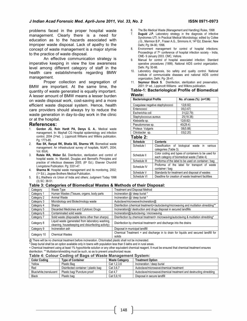
J Indian Acad Forensic Med. April-June 2011, Vol. 33, No. 2 ISSN 0971-0973
148
problems faced in the proper hospital waste management. Clearly there is a need for education as to the hazards associated with improper waste disposal. Lack of apathy to the concept of waste management is a major stymie to the practice of waste disposal.
An effective communication strategy is imperative keeping in view the low awareness level among different category of staff in the health care establishments regarding BMW management.
Proper collection and segregation of BMW are important. At the same time, the quantity of waste generated is equally important. A lesser amount of BMW means a lesser burden on waste disposal work, cost-saving and a more efficient waste disposal system. Hence, health care providers should always try to reduce the waste generation in day-to-day work in the clinic or at the hospital.
References: 1. Gordon JG, Rein Hardt PA, Denys G. A.. Medical waste
management. In: Mayhall CG Hospital epidemiology and infection control, 2004 (3rded. ). Lippincott Williams and Wilkins publication. Pg: 1773-85.
2. Rao SK, Ranyal RK, Bhatia SS, Sharma VR. Biomedical waste management: An infrastructural survey of hospitals, MJAFI, 2004; Vol. 60(4).
3. Rutala WA, Weber DJ. Disinfection, sterilization and control of hospital waste. In: Mandell, Douglas and Bennett's Principles and practice of infectious diseases 2005; (6th Ed.). Elsevier Churchill Livingstone Publication. Pg: 3331-47.
4. Sharma M. Hospital waste management and its monitoring, 2002; (1st Ed.), Jaypee Brothers Medical Publication.
5. B.L.Wadhera v/s Union of India and others; Judgment Today 1996 (3) SC: 38-51.
6. The Bio Medical Waste (Management and Handling) Rules, 1998 7. Duguid J.P. Laboratory strategy in the diagnosis of infective
Syndromes UTI; In Practical Medical Microbiology; edited by Collee J.G., Marmion B.P., Fraser A.G., Simmons A; 14th Ed, Elsevier, New Delhi; Pg: 84-90, 1996.
8. Environment management for control of hospital infections: Proceedings of 7th conference of hospital infection society - India, CME- 9 January 2003. CMC, Vellore.
9. Manual for control of hospital associated infection: Standard operative procedures (1999). National AIDS control organization. Delhi. Pg: 50-66.
10. Laboratory diagnosis, bio safety and quality control. National institute of communicable diseases and national AIDS control organization, Delhi. Pg- 26-41.
11. Seymour Block S. Disinfection, sterilization and preservation, 2001: 5th ed., Lippincott Williams and Wilkins publication.
Table-1: Bacteriological Profile of Biomedical Waste:
Bacteriological Profile No. of cases (%) (n=136)
Coagulase negative staphylococci 12(8.82)
Enterocooci 05(3.67)
Escherichia coli 31(22.79)
Staphylococcus aureus 25(18.38)
Klebsiella sp. 12(8.82)
Pseudomonas sp. 40(29.4)
Proteus Vulgaris 08(5.88)
Citrobacter sp. 03(2.20)
Table 2: Schedule Contents Schedule I Classification of biological waste in various
categories (Table 3).
Schedule II Color coding and types of containers to be used for each category of biomedical waste (Table 4)
Schedule III Proforma of the label to be used on container / bag
Schedule IV Proforma of the label for transport of waste container / bag
Schedule V Standards for treatment and disposal of wastes
Schedule VI Deadline for creation of waste treatment facilities
Table 3: Categories of Biomedical Wastes & Methods of their Disposal: Category Waste Type Treatment and Disposal Method
Category 1 Human Wastes (Tissues, organs, body parts Incineration @/ deep burial *
Category 2 Animal Waste Incineration @/ deep burial *
Category 3 Microbiology and Biotechnology waste Autoclave/microwave/incineration@
Category 4 Sharps Disinfection (chemical treatment)+/autoclaving/microwaving and mutilation shredding**
Category 5 Discarded Medicines and Cytotoxic Drugs Incineration@/ destruction and drugs disposal in secured landfills
Category 6 Contaminated solid waste Incineration@/autoclaving / microwaving
Category 7 Solid waste (disposable items other than sharps) Disinfection by chemical treatment+ microwaving/autoclaving & mutilation shredding*
Category 8 Liquid waste (generated from laboratory washing, cleaning, housekeeping and disiunfecting activity)
Disinfection by chemical treatment+ and discharge into the drains
Category 9 Incineration ash Disposal in municipal landfill
Category 10 Chemical Wastes Chemical Treatment + and discharge in to drain for liquids and secured landfill for solids
@ There will be no chemical treatment before incineration. Chlorinated plastic shall not be incinerated. * Deep burial shall be an option available only in towns with population less than 5 lakhs and in rural areas. + Chemical treatment using at least 1% hypochlorite solution or any other equivalent chemical reagent. It must be ensured that chemical treatment ensures disinfection. ** Mutilation/shredding must be such, so as to prevent unauthorized reuse.
Table 4: Colour Coding of Bags of Waste Management System: Color Coding Type of Container Waste Category Treatment Option
Yellow Plastic Bag Cat 1,2,3,6 Incineration / deep burial
Red Disinfected container / plastic bag Cat 3,6,7 Autoclave/microwave/chemical treatment
Blue/white,translucent Plastic bag/ Puncture proof Cat 4,7 Autoclave/microwave/chemical treatment and destructing shredding
Black Plastic bag Cat 5,9,10 Disposal in secure landfill

J Indian Acad Forensic Med. April-June 2011, Vol. 33, No. 2 ISSN 0971-0973
149
Original Research Paper
Age Related Changes in Mechanical and Thermal Injuries- A Post Mortem Study
*Lavlesh Kumar, **Chaitanya B. V., *** Swapnil S Agarwal, ****Binaya Kumar Bastia
Abstract
Dating of an injury in both living and dead is an important medico-legal issue in the field of crime investigation to fix the liability. A doctor is required to date injuries specifically and individually while preparing an injury report. In this paper we study the sequential changes in mechanical and thermal injuries and to compare the findings with available standard data. All the medico-legal deaths due to injuries, brought for post-mortem examination, over a period of four months were studied. On comparing with standard data, 71% (n=32) of the abrasions were dark red instead of bright red on first day. 92% (n=13) bruises appears bluish green instead of red colour on first day and on the 5
th day, the colour
appears black instead of greenish colour. In cases of incised and lacerated wound margins were difficult to separate on the 2
nd day, instead of separating easily. Observations of this study did not match with the
sequential colour changes mentioned in the available literature. Therefore, other parameters need to be explored for dating an injury.
Key Words: Abrasion, Bruise, Injury, Dating of Injury
Introduction: Dating of an injury both in living and
dead is of crucial medico-legal issue in the field of crime investigation to fix the responsibility. A doctor is required to mention the age of injury specifically and individually while certifying an injury. It is essential to do so, in as much as the guilt or innocence of a person charged with criminal act may be proved from the injury found on the body of victim or on his own body, for its appearance may or may not correspond to the time when it is alleged that all to have been inflicted according to the prosecution theory. In Indian scenario, usually, we adopt the ‘naked eye examination’ method to date an injury.
Corresponding Author: *Associate Professor, Deptt. of Forensic Medicine & Toxicology, S.B.K.S Medical Institute & Research Center, Piparia, Vadodara, PIN-391760. Gujarat INDIA E-mail:[email protected] **IIIrd year MBBS Student KAHE’s Jawaharlal Nehru Medical College, Belgaum, Karnataka, PIN 590010, INDIA ***Assoc. Prof., Deptt. of FMT Pramukhswami Medical College, Karamsad, Anand, Gujarat, INDIA **** Professor, Forensic Medicine, S.B.K.S Medical Institute & Research Center, Piparia, Vadodara, PIN- 391760 Gujarat INDIA
It is not possible to determine exactly
the age of an injury on the basis of its appearance to the naked eye. The results obtained are erroneous because of variability of the intensity of local inflammatory reaction. [1] There is obvious incongruity existing in the standard textbooks, sufficient enough to pose difficulty for dating of mechanical and thermal injuries. An abrasion is bright red between 12-24 hrs and the scab becomes reddish brown in 2-3 days [2]
is documented by one author, but
reddish brown scab within 12-24 hrs and brownish scabbed abrasion in 24-48 hours by another author. [3]
An eminent author [4] claims that it is impossible to comment on the age of a bruise less than 24 hours. Another reported bluish discoloration of bruises not later than 18 hours. [5] A bruise looks bluish black, brown or livid red by 2 to 3 days and become greenish from 5
th to
6th day was the observation of a distinguished
Indian author. [6] An aseptic incised wound
edges (if approximated) will glue in 3 days and the wound heals by the 7
th day, when a red,
tender, linear scar is visible [6] while another authors observation states that in cases of lacerated, incised, stab wounds, edges will be difficult to separate by 3-7 days and a red, soft, tender scar will result in duration of 1-4 weeks time. [7]

J Indian Acad Forensic Med. April-June 2011, Vol. 33, No. 2 ISSN 0971-0973
150
We made an attempt to gather first hand information by direct observation. The findings observed were compared with available data to either validate it or suggest changes to be made so that medico-legal issues are solved in the approved manner.
Objectives of the Study: 1. To study the changes occurring in
mechanical and thermal injuries in course of time.
2. To compare the findings with available standard data.
3. To propose new supposition if different from what is already being followed.
Material and Methods: Source: Autopsies performed on deceased admitted and died at a tertiary care hospital. Study design- Cross-sectional study. Sample size: 35, Total number of injuries observed- n=116. Methodology: In our study, all the medico legal cases admitted, treated and died due to injuries sustained, brought for post-mortem examination at our hospital were studied. During post mortem examination, the injuries over the body were recorded on a pre made Proforma. The findings thus noted were compared with available data to find out whether the changes that take place in injuries with course of time are same or have changed now and suitable suggestions were put forward. Inclusion criteria: Deceased who were admitted because of mechanical and thermal injuries and died in the hospital. Exclusion criteria: Cases, in which death is from causes other than mechanical or thermal injuries and those case died due to injuries but brought dead at hospital. The cases where the exact time of infliction of injuries is not known are also excluded.
Observation and Results: A total of 35 cases in the age group of
1-80 years, maximum cases belong to 31-40years, were observed during the period of July to October’09, comprising of 25 male and 10 female. A total of 116 injuries were studied, comprising of abrasion 79(68%), contusion 22 (19%), Incised wound 08 (7%), lacerated wound 06 (5%) and burns 01(0.8%). Only six cases of lacerated wounds were observed and the red colour of margins were appreciated on first as well as second day and margins were found glued, can be separated with difficulty on very first day itself. Discussion:
In this study, total numbers of injuries studied were 116. Out of that, 79 (67%) cases of
abrasion were observed constituting the largest of the total injuries studied. Textbooks mention four different colour changes occurring sequentially in the process of healing of an abrasion. The colour changes observed in this study were poles apart from what is documented in standard literatures. On the first day, out of 45 cases 32 cases appeared dark red instead of bright red. On the 2
ndday out of 6 cases, 3(50%)
cases appeared reddish black instead of dark red. On the 3
rdday out of 6 cases, 4(66%) cases
appeared dark red instead of brownish red. On the 5
th day out of 6 cases,(84%) cases appeared
dark red instead of dark brownish red scab. On the 9
th day all are dark brown in colour. The healing changes of abrasions
described in the textbooks are ambiguous. When a person is sick the healing response will be slower. In contrast, when an injured person is otherwise healthy, the healing process with be definitely faster. Therefore the sequential healing changes will be different in bed-ridden injured patients from the otherwise healthy injured ones. Moreover the hospitalized patients due to head injuries are frequently devoid of essential nutrients that are essential for normal wound healing. Hence, the observed results in comatose patients are likely to differ from that of
A total of 22 (18%) contusions were studied. Literatures mention five different sequential colour changes taking place starting from infliction of injury till the skin regains its normal appearance. The colour changes observed were unlike from what is mentioned in standard textbooks. On the 1
st day out of 14
cases, 13(92%) cases appeared bluish green instead of red colour. On the 2
nd day, all the 6
contusions appeared bluish green which is comparable to that mentioned in standard textbook. On the 5
th day, the colour appeared
black instead of greenish colour. On the 10th
day, the colour changed to yellow which is same to that mentioned in standard textbooks.
What the Indian literature cites seems to be borrowed from the foreign surveys, where the colour changes are described for white-skinned people. As the final colour observed is a blend of the colour of blood or its breakdown products, and the colour of the skin; the observed results are bound to differ. Therefore we should have our own database based on the colour of the skin belonging to different geographical location.
A total of 8(6%) incised injuries were studied. Textbooks state five different stages of healing in an incised wound starting from infliction of injury till the non tender scar is formed. On the 1
st day out of 5 cases, 3 (66%)
cases appeared red instead of inflamed edges

J Indian Acad Forensic Med. April-June 2011, Vol. 33, No. 2 ISSN 0971-0973
151
.On the 2nd day margins were separated with
difficulty instead of separating easily. On the 3rd
day and 5th day, the injuries studied were having
glued margins and were separated with difficulty, which is comparable in standard text book. A total of 6(5%) laceration injuries are studied. On the 2
nd day, it appeared red in
colour, but margins were difficult to separate. However, available literatures states that gluing of a lacerated wound starts after third day from the infliction of the injury. In case of burns, only one injury could be studied which was red in colour which is similar to the observations mentioned in the standard textbooks.
Summary: In the present study, cases studied were
more of males in their third decades of life. The most common injury observed was abrasion followed by contusion. The findings with respect to healing of abrasion were 1] during the first three days, the colour appeared bright- to dark-red, 2] by 9
th day the colour changed to dark
brown and 3] complete healing was seen by 20th
day in 8.6% of cases.With regards to healing pattern of contusions, red colour of a contusion was rarely observed. 1] The first colour change observed was bluish-green which persists for first two days, 2] the colour became black by 5
th
day, 3] the final colour change observed was yellowish-green which is seen by 10
th day.
Healing pattern was non-specific for lacerations and incised wounds, and hence no inferences could be made out by external examination. Also we could not give any inference as to the healing pattern for burn injuries due to non-availability of sufficient number of cases.
Conclusion: Traditionally, we have been relying on
the colour changes documented in books and literature for dating an injury. The colour changes observed in this study were poles apart from what is documented in standard literatures. The Indian doctors have been trained to observe the findings which are erroneous and borrowed from western observations. Though the healing of an injury is influenced by many factors like infection, nutrition, pre existing pathology and many more, we have been trained to ignore it. Another important aspect is interpretation of colour changes in artificial light. In our study, though the colour changes have been studied in the post-mortem room, it had facility for sufficient natural light to reduce the colour biases of artificial light. But one must also not forget a fact that a forensic expert is asked to opine the age
of injuries by examining a patient indoors and not outdoors.
But we need to evolve a particular criterion that can be followed.
Therefore, because of the uncertain and variable results of the naked-eye examination of injury it is important to study the injuries microscopically. Histological appearances can be backed up by including fluorescence studies, histo-chemical and biochemical assays. This shall help in removing subjectivity and bring in more objectivity in the observations and interpretation.
Acknowledgement: This study was undertaken and
successfully completed under Short Term Studentship [STS] Program of Indian Council of Medical Research by Mr. Chaitanya B. V., MBBS student under the guidance of Dr.
Lavlesh Kumar.
References: 1. Tedeschi C.G., Eckert GW. & Tedeschi LG. Forensic
Medicine- a study in trauma and environmental hazards (1st
ed.) W B Saunders Company. Philadelphia, London, Toronto; 1977.
2. Reddy KSN .The Essentials of Forensic Medicine and
Toxicology.24thed.K Suguna Devi, Hyderabad (AP); 2005. 3. Mukherjee JB. Forensic medicine and toxicology
(1).2nded.Arrnold Associates, New Delhi; 1994.
4. Knight B and Saukko P. Knight’s Forensic Pathology. 3rd ed. Arnold, a member of the Hodder Headline Group, 338
Euston Road, London NW 13B H; 2004.
5. Langlois NE, Gresham GA. The ageing of bruises: A review and study of the colour changes with time. Forensic
Sci Int. 1991; 50: 227-38.
6. Modi JP. Modi’s Medical Jurisprudence and Toxicology.22nded.Buttertworths-Tipathi publication, New
Delhi; 1999.
7. Agarwal SS., Kumar L and Chavali K. Legal Medicine Mannual.1st ed. Jaypee brothers’ medical publishers (p) Ltd.
New Delhi; 2008
Contusions [n=22] Day 1 2 3 4 5 6 7 8 10
Red 1 _ _ _ _ _ _ _ _
Bluish green 13 6 _ _ _ _ _ _ _
Black _ _ _ _ 1 _ _ _ _
Yellow green _ _ _ _ _ _ _ _ 01
Incised wounds [n=8] Day 1 2 3 4 5
Red 3 - - - -
Margins Separated with difficulty
2 1 1 - 1
Abrasions: [n=79] Day 1 2 3 4 5 6 7 8 9 20
Bright red 13 1 - - - - - - - -
Dark red 32 2 4 _ 5 _ _ _ _ _
Reddish-black
_ 3 2 _ 1 _ _ _ _ _
Dark brown
_ _ _ _ _ _ _ _ 6 5
Healed with white scar
_ _ _ _ _ _ _ _ _ 5

J Indian Acad Forensic Med. April-June 2011, Vol. 33, No. 2 ISSN 0971-0973
152
Review Research Paper
Role of Forensic Medicine in Administration of Justice A Critical Review
* U.S. Sinha, ** Mukesh Yadav, *** Soni Uttam, **** A.K. Pathak, *****Sanju Singh
Abstract Two small round opaque shadows of metallic densities opined as that the injury was not a firearm
injury but was self inflicted one by the first Medical Board under the Chairmanship of Chief Medical Officer. Opinion of another Medical Board comprising Forensic Medicine expert considered it as a: Clear cut case of firearm wound. Based on two contradictory opinions, Allahabad Court held that the Charge: sheet submitted under sections 323, 504, and 506 IPC, ought to have been submitted under some graver sections too. This paper deals with a critical review of case where two Medical Boards were constituted in the interest of justice. Views of various other relevant judgments of Hon’ble Supreme Court and various High Courts emphasizes the importance of Forensic Medicine in the administration of justice and need for refresher courses and training for medical officers suggested to avoid future recurrences of such nature are discussed in brief.
Key Words: Medical Board, Expert Opinion, High Court, Training, Forensic Medicine Expert
Introduction:
Opinion of doctor as expert witness has been given very much importance by the judges. Circumstances do arise in which need for constitution of Medical Board is felt in the interest of justice. But in some cases where doubt is raised about the validity of opinion given by the Medical Board, another Medical Board may be constituted on the direction of the Court involving Forensic Medicine Experts to protect the fundamental rights of victim as well as that of accused. Allahabad High Court observed that “Further the disturbing trend that doctors are becoming too careless and often willing tools ever ready to rubber stamp the opinions of I.O. without application of their independent minds appears to have been again high-lighted before this Court in the instant case”. An opinion erroneously, collusively or dishonestly given by the doctor, who examines a victim, can have far reaching consequences.
Corresponding Author: **Professor & HOD, Deptt. of Forensic Medicine School of Medical Sciences & Research, Sharda University, Greater Noida, U.P E-mail:[email protected] *Prof. & HOD, Deptt. of Forensic Medicine MLN Medical College, Allahabad ***Junior Resident –II ****Junior Resident-I *****UCMS, Delhi
It is true that subsequently during trial the opinion of the doctor if considered only as opinion evidence under Section 45 of the Evidence Act, which is relevant, but not conclusive and the Court, is free to judicially estimate the value of the doctor’s opinion evidence by examining how well it can explain and clarify the facts in issue.” [Para 23]
A prayer through writ petition before the Allahabad High Court was made for constituting another Medical Board under Article 226 of the Constitution of India, 1950, It was alleged that erroneous conclusion by doctors are made that the injuries caused to petitioner was not firearm injury in an incident that has been taken place on July 1, 2008. FIR was registered under sections 308, 323 and 504, IPC by the order of the Magistrate.
It was prayed that another Medical Board be constituted either in district Allahabad or in any other district except Kaushambi for medical examination of the injured informant Smt. Savita Devi, on the ground that the report of the Medical Board headed by the Chief Medical Officer (CMO), Kaushambi dated 20.10.2008 had arrived at a wholly erroneous conclusion. [Para 1]
First Medical Board opined that “two small rounded opaque shadows of metallic densities opined as that the injury was not a firearm injury but was self inflicted one”. Opinion of another Medical Board comprising Forensic Medicine, Head of Department, M.L.N. Medical College, Allahabad considered it as a-Clear cut

J Indian Acad Forensic Med. April-June 2011, Vol. 33, No. 2 ISSN 0971-0973
153
case of firearm wound. Based on two contradictory opinions of Medical Boards, Court held that the Charge sheet submitted under sections 323, 504, and 506 IPC, ought to have been submitted under some graver sections too. However, alleged history of use firearm and other dangerous weapons: Bomb, Lathi, Danda was there in FIR, along with head injury, I.O. and examining doctors completely disregard these alleged facts. (Para 5) [1]
Brief Facts of the Case: • It was alleged that the incident had taken
place at 8.00AM on 1.7.2008 when the accused Banwari Lal and other had arrived at her door variously armed with gun, country-made pistol and bomb and started erecting a wall there, which could have blocked the passage of the complainant and her family members, they had objected, whereupon the accused persons hurled a bomb and resorted to firing.
• The fire by Bhukhan struck her head. The informant’s brother-in-law (Devar) Rajesh and Ram Prakash were also injured and that in order to escape, the informant and other had to rush inside her house and to lock the door. [Para 4]
Alleged controversial role of Police: • It was further alleged that the police
minimized the incident and only lodged a non-cognizable report under sections 323/504 IPC which merely mentioned that the accused persons had resorted to abuses and used lathi and danda in the assault, which were plied by both sides resulting in the head injury to Savita Davi. [Para 5]
• In these circumstances Savita Davi was constrained to move an application on 8.7.2008 before the Judicial Magistrate, Kaushambi under section 155 (2) Cr.PC which mentioned inter alia that the S.O. of the Police Station, Sarai Aqil had even threatened to send her jail on a fabricated case, when she had gone to lodge the report on .2.07.2008. [Para 5]
• However, on her insistence the S.O. concerned had orally lodged and got her medical examination done on 02.07.2008, by the medical officer, CHC Sarai Aqil, Kaushambi.
• Medical Report: The medical report showed a lacerated wound 1.5 cm 3 cm skin on her head, 11 cm from the right ear, but no blackening and tattooing was found, and X- ray of the skull was advised. Dr. A.N. Rastogi the Radiologist. CHC/ District Hospital, Kaushambi, who conducted the
X- ray on 03.07.2008 found two small rounded opaque shadows of metallic densities over the skull vault on the frontal bone of skull as noted above.
• Hence, on the order of the Magistrate the FIR was registered under sections 308/323/504 IPC. [Para 6]
• Incident had taken place on 1.7.2008 • FIR Registered under sections 308, 323 and
504, IPC by the order of Magistrate • Two small rounded opaque shadows of
metallic densities – Medical Board reported that the injury was not a firearm injury was self inflicted
• Opinion of Forensic Head of Medical College considered – Clear cut case of firearm wound.
• Controversy: Two opinion of Medical Boards held contradictory –
• Charge-sheet submitted under sections 323, 504, and 506 IPC
• Petitioner is free to move an application before competent Court for showing that charge-sheet ought not to have submitted under sections 323, 504 and 506 IPC but under some graver sections too.
• Direction for doctor to apply their independent mind.
• Petition disposed of • Although the prayer made in the writ petition
was unusual, however on noticing that the Radiologist’s report showed that there were two small rounded opaque shadows of metallic density over the skull, vault on the frontal bone of the scalp. [Para 2]
• Court observed that “Medical Board had themselves noted that there was a scar mark of the size 1.5 cm x 0.2 cm over the scalp on the frontal bone and a small hard object was palpable, which was moveable in all directions and was subcutaneous, an earlier Bench consisting of one of us, Amar Saran J. and R.N. Mishra J. began to entertain some doubt about the reliability of the opinion given by the Medical Board and directed by an order dated 09.02.2009 that the case be listed on 17.02.2008.
• On that date the Court required the presence of the Investigating Officer (I.O.) with the case diary. [Para 2]
Formation of Medical Board: • Ordinarily Medical Board may be constituted
by the CMO before submission of the charge sheet before the trial court on the request of I.O.

J Indian Acad Forensic Med. April-June 2011, Vol. 33, No. 2 ISSN 0971-0973
154
• However, on 17.02.2009 when the I.O. appeared, it was argued by learned Additional Government Advocate, without filing any counter affidavit or document to that effect, that as the I.O. had informed him that he had already submitted a charge sheet against the accused only under sections 323/504 and 506 IPC, hence there was no occasion for this Court to pass an order constituting another Medical Board at that stage and it was open to the complainant petitioner to raise all her objections against the said opinion of the Board during trial as well as to cross – examine the concerned doctors. [Para 3]
• But as the accused after making allegations against the integrity of the Radiologist, Dr. Rastogi, prayed for constituting a Medical Board the same was got constituted by the I.O., which consisted of Dr. Rajesh Chaddha (Orthopedic Surgeon), Dr. Manu Gopal (Radiologist), Dr. B.L. Jaiswal (Medical officer) and opinion was counter signed by the CMO.
Prayers: • Prayer for constituting another Medical
Board • That another Medical Board be constituted
either in district Allahabad or in any other district except Kaushambi for medical examination of Smt. Savita Devi the petitioner, on the ground that the report of the Medical Board headed by the CMO, Kaushambi dated 20.10.2008 had arrived at a wholly erroneous conclusion that the head injury on Savita Devi was not a firearm injury, but that it was self-inflicted. [Para 1]
Discussion: Questions before the High Court for
consideration were following: 1. How the injury could have been caused? 2. Whether if the injury was due to firearm, how
was the injury only received on the head and not on the other parts?
3. Whether the Ballistic Expert needed to opine about firearm injury after medical examination of the victim?
4. Whether the injury is due to a firearm? The Bench consisting of one of us (Amar
Saran,J) and Hon’ ble R.N. Misra. J examined the said opinion of the Medical Boards as well as other papers and observed in the order dated 17.02.2009 as under:
“Thus it is pointed out that the board while noticing that the injury report wound, but there was no injury on any other part of the body, nor any entry or exit wound. The radiologist report
dated 03.07.2008 conducted by the Radiologist C.H.C., Sarainkil shows two small rounded, shadows of metallic densities over the skull vault on the frontal bone of skull. It also mentions that the medical officer who conducted the medical examination and gave the injury report was examined by the police officer on 23.08.2008 and he has clearly mentioned in his statement that there was no sign of fire arm injury. We have examined the said statement, which has been produced by the I.O. present today. This is not the statement of the medical officer who conducted the medical examination on 02.07.2008 at all but the opinion of the Radiologist who conducted the X-ray. In the statement of the said radiologist in reply to the question as to how the injury could have been caused the reply was nil. To another question as to whether if the injury was due to firearm, how was the injury only received on the head and not on the other parts, Radiologist replied that there were two small rounded opaque shadows of metallic densities, which were seen in the x- ray on the head and he was not in a position to state as to how said injury could have been caused.
Thus, this averment in the report of the medical board that according to the doctor who had examined the injury of Savita Devi that it could not possibly have been caused by a fire arm appears to be based on a complete misreading of the Radiologist, and there is no such opinion of the doctor as aforesaid. Another fact which is mentioned in the report of Medical Board was that the physical examination of Savita Devi by board members shows a scar mark of size 1.5 cm 0.2 cm over the scalp on the frontal bone which seems to be as a result of a healed lacerated wound and a small hard object could be felt which was moveable in all directions and is subcutaneous.”
Hon’ble Court further observed that “In the light of above averments the Medical Board reported that the injury was not firearm injury instead it was self inflicted Prima facie in our opinion this opinion of Medical Board appears to be perverse and wholly unwarranted. The said firearm injury was received by a woman by a firearm on 1.7.2008. Her medical examination was conducted on 2.7.2008 when the examining doctor found a lacerated injury on the scalp on the frontal bone region which was kept under observation and x- ray advised. The x–ray on 3.7.2008 showed two radio opaque shadows of metallic density confirming the allegations of firing. The pellet (hard object) even appeared palpable on touch on 20.10.2008 when the opinion was given by the medical board. Simply because only one or two pellets struck the head
J Indian Acad Forensic Med. April-June 2011, Vol. 33, No. 2 ISSN 0971-0973
155
possibly from a distant fire as there was no blackening and tattooing on the injury and there were no injuries on the rest or the body or exit and entry wound, could it lead to an inference that no firearm injury at all was received by the injured woman. [Para 6]
Court further added that “Thus, it is incomprehensible to us at this stage as to how the board reached a positive opinion that the so-called firearm injury was not due to a firearm or that the injured woman, Savita Devi had self inflicted the said injury on her forehead on the frontal bone over the skull vault.” [Para 6]
Court, however, decided to summon the concerned doctors on 26.2.2009 who constituted the Medical Board because court thought it would be proper to reach a finding about the opinion of the Medical Board and the conduct of the doctors only after giving them an opportunity to explain as to how they had reached the aforesaid opinion. [Para 8, 9]
On 26.2.2009 when the doctors appeared, the earlier Bench questioned them at length as to how they had reached the opinion, but they failed to giver any satisfactory basis for their opinion.
As the learned Additional Government Advocate produced the note of Dr.M.M. Ahmed dated 23.8.2008, who had conducted the initial medical examination of Smt.Savita Devi on 2.7.2008 at 9.00 PM at CHC, Kaushambi which was referred to in the opinion of the Medical Board dated 20.10.2008 and observed as follows:
“We also regret to note that Dr.M.M. Ahmad, who conducted the initial medical examination of Smt. Savita Devi on 2.7.2008 at 9.00 PM at CHC, Kaushambi even after observing that Smt. Savita Devi had a lacerated wound 1.5 cm x 3 cm skin on head 11 cm from right ear no blackening and tattooing was found and X-ray of the skull (AP and lateral view) was advised yet the said doctor has given his opinion to I.O. to the effect that at the time of examination there was no wound of firearm injury which Prima facie appears inconsistent with the finding of two small rounded shadow metallic densities over the skull as noted by Dr. Rastogi, who conducted the X-ray examination at the CHC on 3.7.2008.” [Para 10] Also as the members of the Medical Board appeared to be willing to revise their opinion dated 20.10.2008 Court further observed as under in the order dated 26.2.2009:
“They agree to the observations of the Court that the opinion that the injury received by the victim Smt.Savita Devi was not due to
firearm and that it was self inflicted was not correct.” [Para 11]
Hon’ble Court thereupon directed Smt. Savita Devi to appear before the Medical Board on 6.3.2009 on which date the Medical Board was to examine her with regard to the documents and papers of this case and to submit its opinion within three weeks. [Para 12]
Court categorically added that “To our dismay instead of examining the matter properly applying their minds, they again recorded an opinion on 2.3.2009 that the shadows seen in the X-ray may or may not be said definitely. A view of ballistic expert can be taken regarding nature of these radio opaque shadows of metallic density.” [Para 13]
Clear and cogent opinion of Forensic Medicine Expert:
The opinion of Forensic Medicine Expert was placed before the High Court on 07.04.2009. As the earlier Bench was not satisfied with the opinion of the Medical Board, Court sent the photocopies of the medical opinion of the Medical Board dated 02.03.2009 and earlier opinion dated 20.10.2008, photo copy of the case diary, orders of the High Court dated 17.02.2009 and 26.02.2009 injury report of Smt. Savita Devi dated 02.07.2008 and X-ray report and opinion of the Radiologist to Medico Legal Expert Dr.U.S. Sinha, Forensic Head of the Medical College. Allahabad, for his expert opinion. Dr.Sinha was also required to be present in the High Court on 16.04.2009 to give his opinion in the matter. [Para 15]
Dr.U.S. Sinha has appeared on 16.04.2009 and has submitted a clear cut and categorical opinion in writing which is as follows:
“I thoroughly examined the injury report of Smt.Savita Devi dated 2.7.2008 and x-ray report and opinion of the Radiologist dated 3.7.2008 along with medical board examination dated 20.10.2008 and 2.3.2009 regarding the physical examination of Smt. Savita Devi injury report and subsequent Radiologist report of 3.7.2008. [Para 16]
Observation: On the basis of medical documents
consists of injury report, Radiological findings clearly shows that Smt. Savita Devi injured by a firearm injury a lacerated wound measuring in 1.5 cm 3cm over the scalp on the frontal bone which is situated 13 cm above the right ear root and 10.2 cm above the right eyebrow. It is well positive findings in relation with two small metallic shadows of 0.3 cm.
The type of injury is lacerated wound which is usually mentioned in gunshot wound because

J Indian Acad Forensic Med. April-June 2011, Vol. 33, No. 2 ISSN 0971-0973
156
the character and size of the wound which clearly indicates the nature of the weapon. Which has caused such type of injury along with 2 small rounded radio opaque shadows of metallic density are seen over skull of frontal bone.
This type of injury may usually produced by firearm wounds from the distance >6 yard with standard shot gun & 1 yard with country made pistol.
Complainant has already placed FIR to S.P. Kaushambi on 2.7.2008 regarding the firing incident before the medical examination as well as radiological examination.
Hence, considering the wound size, shape, character of the wound along with presence of 2 small round radio opaque shadow of metallic density over skull of frontal bone is clear cut case of firearm wound from the distance. In view of the facts and observation regarding above matter there is no doubt about a firearm injury. However the small, rounded metallic pellets may be sent to the ballistic expert to know the nature of fired gun”. [Para 16]
Court accepted Forensic Medicine Experts by noting that “He based his opinion on the books on medical jurisprudence and his wide experience in the field of Forensic Medicine large number of publications and his conduct of about 10,000 autopsies. He also endorsed his written opinion on being orally examined by the Court.” [Para 17]
Court concluded that “From the clear and cogent opinion expressed by the medico legal expert Dr.Sinha, and from the observations of the High Court referred to hereinabove apparently the opinion of Dr.M.M. Ahmad dated 23.8.2008 does not appear very reliable. As Dr.Ahmad had initially seen the lacerated wound 1.5 cm 3 cm over the scalp on the frontal bone which is situated 13 cm above the right ear root and 10.2 cm above the right eyebrow on the head of Smt.Savita on 2.7.2008 and had also advised X-ray of the skull region, which revealed two radio opaque shadows Dr.Ahmad’s opinion on 23.8.2008 that at the time of examination there was no wound of firearm injury, appears on the face of it, to be erroneous.” [Para 18]
Opinion of Medical Board: Hon’ble High Court observed that
“Likewise the two opinions of the medical boards consisting of 4 doctors including the CMO, Kaushambi the initial one being dated 20.10.2008 which notes that “In the light of the above mentioned findings we members of the medical board are in the opinion that injury is not a firearm injury, instead it is self inflicted” and
the opinion of the Board dated 2.3.2009 that “Shadows seen in the X- ray may or may not be due to pellets of firearm and it cannot ne said definitely. A view of the ballistic expert can be taken regarding nature of these radio-opaque shadows of metallic density” seem to be erroneous and self contradictory too.” [Para 19] Court further observed that “Self contradictory because the first opinion of the medical board dated 20.10.2008 reaches a conclusion that the injury was not due to fire arm and at the same time it goes on to suggest that the lady had herself inflicted the injury on her head.” [Para 19]
Opinion of Forensic Medicine Expert: In this regard Dr.Sinha informed the
Court that in his vast experience he had never come across such a case where such a firearm injury on the head of woman was self-inflicted. The opinion of the medical board could also be deemed as incompetent in as much as in the second opinion of the Medical Board dated 2.3.2009 a vague suggestion has been made that the radio opaque shadows may or may not be due to pellets of a firearm and a ballistic expert was needed for clarifying this position.
Role of Ballistic Expert: It is made clear that it is only a doctor
and not a ballistic expert at all who on a perusal of the x-ray and initial injury reports and after touching the moveable hard object on the head of an injured can determine whether the injury is due to a firearm. If at all, after the still present pellets on Savita Devi’s head are extracted, the Ballistic Expert might succeed in venturing any opinion about the nature of the firearm which may have caused those pellets to be embedded over the frontal bone of Savita Devi as suggested by Dr.U.S. Sinha. [Para 19]
Direction to the Trial Court: In this view of the matter, the High Court
directed that the copy of the written medical opinion of Dr.U.S. Sinha dated 16.4.2009 and copy of the High Court order was forwarded to the lower Court for appreciation of the medical opinion in this case. It is of course expected that the Trial Court shall apply its independent mind in appreciating the medical opinion evidence on record. Also if it appears to the Trial Judge that some evidence has been fabricated, falsified or suppressed so as to pollute the course of justice in this case by either side the Trial Court shall take appropriate legal actions against the persons responsible for the same. [Para 20]
Application under section 155(2) Cr. P.C. to the Judicial Magistrate:
In this case Court observed that in spite of two radio opaque shadows of metallic density

J Indian Acad Forensic Med. April-June 2011, Vol. 33, No. 2 ISSN 0971-0973
157
being seen in the radiological examination of Savita Devi on 3.7.2008 which was preceded by the medical examination report on 2.7.2008 which revealed a lacerated injury on Savita’s head precisely at that point where x- ray was done, the S.O. concerned had only got an FIR under sections 323 and 504 IPC registered on 2.7.2008 and that the use of fire arms appears to have been concealed, compelling Smt. Savita to move the application under section 155(2) Cr. P.C. to the Judicial Magistrate Kaushambi dated 8.7.2008 complaining about these facts where after section 308 IPC was added to the earlier sections. [Para 21]
High Court observations: But again the I.O. appears to have
adopted a somewhat unprecedented and unusual procedure in getting a medical board constituted and obtaining as opinion form the board on 20.10.2008 that the injury to Savita’s head was not due to firearm and that it was self- inflicted. There after hurriedly on 23.10.2008 a charge sheet was submitted only under sections 323, 504 and 506 IPC at Case Crime No. 234- A by the Police of P.S. Sarai Aqil, so as to nullify the petitioner’s application and radiological reports etc. and claim that she received a firearm injury. [Para 21]
Prima facie view of the Court: In these circumstances Hon’ble High
Court was prima facie of the view that use of extraneous pressures cannot be ruled out in this case and Court directed the S.P. Kaushambi to get the matter thoroughly probed and in case he is satisfied after an independent enquiry that any of the police officers concerned have engaged in any foul play or malpractice he should take strict action in the matter against the said officers. [Para 21]
Need for Departmental Enquiry: In the circumstances of the case the
High Court leave it open for the concerned Secretary, Medical Health, U.P. to consider whether it would not be advisable to get a departmental enquiry conducted in the matter for enquiring as to whether the concerned doctors who have constituted the Medical Board and Dr. M .M. Ahmad who initially examined Savita Devi on 2.7.2008, but gave his opinion to the I.O. on 23.8.2008 have acted incompetently or collusively. [Para 24]
Need for training and refresher courses for medical officers:
Court opined that “In the face of such incompetent expressions of medical opinions by the doctors, which are becoming increasingly prevalent, and which can seriously prejudice the
fair conduct of investigations and trials in criminal cases, we would appreciate if the concerned Secretary Medical Health could develop a system for conducting refresher courses in Medical Jurisprudence for Government doctors who have to undertake Medical examinations or post mortem examination of the victims, deceased and others in medico-legal matters.”
Madras High Court Observations: It is to be noted that whenever a
requisition from the investigating officer is forwarded to the medical officer concerned, it shall accompany the history of the case also. As usual, in this case also, a requisition was sent, followed by the history of the case, in which the investigating officer had stated about the occurrence. [Para 13] [12]
Duty of Medical Officer: When the cases of this nature are
brought to the notice of the medical expert, it is his bounden duty to clarify all the doubts, which are more likely to get posed at the time of decision by the Court.
What is expected of a doctor? The doctor need not care about the
nature of disposal of the case by the police officer. He has to take every possible step to furnish what are all the material particulars which he could gather at the time of post-mortem examination in his certificate. [Para 14] As far as the present case is concerned, no doubt, the doctor has furnished the particulars required to some extent, but it could not be stated that the post-mortem certificate is exhaustive in all aspects.
The subject 'Forensic Medicine' is concerned with the application of medical knowledge and expertise in various legal issues. Forensic Medicine is a science of comparatively recent growth. Forensic or Legal Medicine is a branch of medicine, which deals with application of medical knowledge, for the purpose of law and administration of justice. [Para 17, 12]
Motto of Forensic Medicine: Its earnest motto is the elucidation of
truth on scientific basis. Its particular field of activity is that of investigation in civil and criminal cases. [Para 17, 12]
What is desirable of Medical Officer? It is desirable that every Medical
Practitioner should have considerable exposure in the area of procedure in Courts of Law, the nature of medical evidence and other evidence, some of the peculiarities of crime in this country and the deceitful means commonly employed to conceal crime in the like cases. [Para 17] [12]

J Indian Acad Forensic Med. April-June 2011, Vol. 33, No. 2 ISSN 0971-0973
158
The police generally conduct all preliminary enquiries as to the offences affecting human body. The Medical Practitioner is liable to be called upon to give evidence as a Medical Jurist. It is consequentially advisable that he should learn to look medico legal standpoint upon such of his case as may possibly become the subject matter of judicial investigation. He should know carefully everything likely to be of medico legal importance, so also to be aware of the fact that medical evidence is not substantive one but usually opinion evidence, which has great corroborative value. The reliability, completeness and objective investigation of the forensic pathologist facilitate formation of definite opinion by the Court. [Para 17] [12]
Views of Hon’ble Supreme Court: In this context, it is not out of place to
mention the law of the land as regards opinion reached by a medical expert. The Honble Supreme Court of India while discussing import of the opinion rendered by a medical expert has formulated principles to the effect that once opinion of the expert is accepted by the Court, it becomes the opinion of the Court and not of the Medical Officer.
The operative portion of the judgment reported in 2006 (1) Supreme Court cases in Vishnu alias Undrya Vs. State of Maharashtra, in which earlier decision of the Apex Court has been culled out, is as under: [Para 29][13]
In Madan Gopal Kakkad vs. Naval Dubey, [14] the Supreme Court has considered a similar question and pointed out as under:
Medical Officer as an Expert: What is expected by the Court of Law?
A medical witness called in as an expert to assist the Court is not a witness of fact and the evidence given by the medical officer is really of an advisory character given on the basis of symptoms found on examination. The expert witness is expected to put before the court all materials inclusive of the data which induced him to come to the conclusion and enlighten the court on the technical aspect of the case by explaining the terms of science so that the court, although not an expert, may form its own judgment on those materials after giving due regard to the experts opinion because once the experts opinion is accepted, it is not the opinion of the medical officer but of the Court. [Para 34] [12], [14] [Para 34 at SCC pp.221-22]
Medical Curriculum of Forensic Medicine and Role of MCI:
High Court remarked that “Medicine as well as the Medico Legal Curriculum taught in the Indian Medical Schools are to be borne in
mind. The education curriculums in the medical educational institutions have to be arranged, so as to render an appreciable background and exposure in this particular area. Initiation of steps may be undertaken with an avowed object of alleviating the flaws, which could be noted by the Judiciary in the certificates issued by the medical experts.” [12]
Unlike in developed countries, bulk of medico-legal work is still being done by the government medical officers, who are mostly general medical practitioners. Therefore, it becomes all the more necessary to train the medical graduates in a more integrated manner so that they could perform qualitative medico-legal functions, which would be invaluable in dispensation of justice. [Para 30] [12]
The medico-legal curriculum in India is traditionally packed with a large quantity of theoretical knowledge. Once qualified, students are expected to blend this information and apply it while performing medico-legal duties. The knowledge in medico legal features, expected in a fresh undergraduate, should be considered in the curriculum itself which he/she has gone through during his/her course. Though a fresh undergraduate is anticipated to come out from the institution with the knowledge in clinical and surgical areas of medical science, yet, he/she should not be lagging behind in the field of Forensic Medicine. [Para 31] [12]
Role of Medical Evidence: Medical Evidence is a scientific factor,
which plays crucial role for determining many of the crimes perpetrated against the human body. [Para 32] [12]
Suggestions by the Madras High Court: [12]
At this juncture, this Court, after going through the materials available in this case and in the backdrop of the authorities on the subject, deems it appropriate and feels compelled to place some suggestions for consideration and implementation by the authorities concerned, in the following manner:
Need for imparting Training: a. Directorate of Medical Sciences and
Directorate of Medical Education with the concurrence of the State Government may contemplate imparting periodical training to the Medical Officers, who are in Government service, on Forensic Medicine, to make their efficiency updated in the field. A standardized format of noting down the injuries and their signs can be evolved so that a uniform procedure for issuing medical certificate be followed state wide. The said

J Indian Acad Forensic Med. April-June 2011, Vol. 33, No. 2 ISSN 0971-0973
159
authorities may constitute a team of experts to prepare the format, so as to make the job of doctors, who perform medico-legal functions, easier. [Para suggestion 1(a)] [12]
b. Every doctor posted in any Government hospital may undergo a week's training in Forensic Science Department in the nearby Government Medical College periodically and the State Government may evolve a scheme in this regard. [Para suggestion 1(b)] [12]
c. The Government may provide sufficient infrastructural facilities to the mortuaries and places where autopsies are conducted. [Para suggestion 1(c)] [12]
d. In certain cases, the internal organs extracted from the corpse by the Doctor at the time of necropsy have not been properly preserved, resulting the de-composition or dis-integration of the tissues by autolysis. When they are subjected to histopathological examination, desired result could not be secured leading to the loss of valuable evidence, which plays a crucial role in determination of the case by the Courts. [Para suggestion 2(a)] [12]
e. The knowledge of the Medical and Para-medical staff should be updated by imparting periodical training to them. Adequate exposure to the preservation techniques is the need of the hour in order to secure accurate results. Hence, exhaustive examination of viscera can be obtained, only if the Medical and Para-medical personnel are possessing updated knowledge in preservation technique. The Government through Department of Health have been issuing circulars containing the procedures to be observed and followed by Medical and Para-medical Staff then and there. In some cases, errors occur on account of misapplication or improper handling of the procedures concerned, resulting in confusion in procedures, which screen the crimes from getting exposed to the eyes of Judiciary. So, it is desirable to issue the circulars whenever necessity arises, that is to say, if settled or codified procedures were violated or ignored, followed improperly and misapplied even for a single occasion. [Para suggestion 2(b)]
f. It is high time, the Governments, the Medical Council of India and the Medical Universities, which control the quality of medical education in this country, took serious view of this aspect and brought
about appropriate measures. [Para suggestion 5(a)] Every student of medicine should get familiarized with the intricacies of Forensic Medicine apart from academic knowledge, right from his/her undergraduate level i.e., from the educational institution itself, after passing through the curriculum, prescribed for him/her. [Para suggestion 5(b)] [12]
To facilitate a better understanding of nuances of Forensic Medicine, the teaching of the subject may be taken up in the later part of the clinical years. In case of their having this subject in the early years of their study, they may not be in a position to know the importance of the principles applicable to the given circumstances. The students may read this subject for the purpose of getting through the examination, but the real involvement therein could not be expected. [Para suggestion 5(c)] [12]
During the internship, all the House Surgeons (Compulsory Rotating Resident Internees) may be compulsorily posted in the Department of Forensic Medicine for a reasonable period, for a better comprehension of the subject. This suggestion is made, viewing that the medical students would sufficiently be equipped at the later part of their studies and the niceties of the features in the subject would be appropriately appreciated by them. [Para suggestion 5(d)] [12]
g. It is bounden duty of the Government to produce Medical Experts in the educational institutions with a strong academic background, who would be fit for becoming members in the Health Delivery System and also for rendering man services, to assist the Justice Delivery System, for which their exposure in the field of Forensic Medicine is indispensable. [Para suggestion 6] [12]
h. The authorities may initiate efforts to increase number of admissions to PG Course in Forensic Medicine and to get due recognition from Medical council of India, and ensure output of such experts cater to the needs to a greater extent. [Para suggestion 7] [12]
Role of Police: The brass of police may issue directions
to the Investigating Officers of the crimes, to get final opinion as regards the nature of wounds in injury cases and time and cause of death, in cases where unnatural death has occurred, before taking any decision either to proceed with the case or to drop further action. Instructions

J Indian Acad Forensic Med. April-June 2011, Vol. 33, No. 2 ISSN 0971-0973
160
may also be issued, not to make any slipshod or improper investigation in the cases handled by the police, on the strength of wound certificates or post-mortem certificates, which lack material particulars. Necessary training may also be given to the police personnel, with regard to the appreciation of medical records, at the time of investigation in medico legal cases. [Para suggestion 8] [12]
Summary & Conclusions: Direction for doctor to apply their
independent mind and need for training of doctors was felt by the Hon’ble High Court and accordingly directed Principal Secretary Health, Govt. of U.P. This is high time if Medical Council should emphasis on exhaustive training of M.B.B.S. doctors in newly proposed curriculum. Concerned state governments should plan for refresher courses for medical officers before involving them in medicolegal work. There is need for implementation of recommendations of the Survey Committee Report, 1964 along with various recommendations of the Central Medicolegal Advisory Committee made from time to time since its inception in 1956. The Central Medico-legal Advisory Committee during its first session in 1956, considered the suggestion of the Ministry of Home Affairs, Government of India, to create a special cadre of medico-legal officers. [Chapter V] [[10], [11]
However, the Committee recommended that each State should give advance medico-legal training to at least one officer in each district and in important cities and towns and such an officer should undertake the specialised medico-legal work himself and also co-ordinate all general medico-legal work by other Government Medical Officers in his jurisdiction. [Chapter V] [10, [11]
In discussing the training of Medical Officers during the sixth session, the Committee recommended that every medical officer on his first appointment to Government service should receive three months training in medico-legal work under a professor of Forensic Medicine. Officers engaged in medico-legal work at the district level should receive further advanced training for six months under a Professor of Forensic Medicine. The specialised training of selected medical officers should be in Medico-legal Institutes. [Chapter V] [10], [11]
The Ministry of Health, Government of India, emphasised on the State Governments the need for training of medical officers in medico-legal work. The recommendations of the
Committee regarding the establishment of Medico-legal Institutes and the grant of special pay to officers engaged in medico-legal work were brought to the notice of the State Government. [11]
Madras High Court has shown his concern in following words “High This Court is much desirous and concerned of expressing that the branch of science of Forensic Medicine is an effective scientific method, which plays a vital role in assisting the Justice Delivery System to render justice to the society, in the administration of Criminal Justice. In order to make this particular subject more viable, more teeth have to be provided by the legislature and the authorities concerned, to make it trendsetting.
The service rendered by the Forensic Medicine Experts in this regard is unique and deserves for admiration, but the real state of affairs remain that medico legal cases are handled in this country by the non-forensic experts and none could be blamed in this regard. No one can expect that a Forensic Medicine Expert alone would be deputed for conducting post-mortem examination and to attend the medico legal cases, since the strength of such expert is comparatively minimal, to cater to the needs.” [Para 28] [12]
References: 1. Smt.Savita Devi and others vs. State of U.P. and others, 2009 (65)
ACC 869. Criminal Misc. Writ Petition No. 1871 of 2009, judgment dated May 8, 2009, February 17, 2009.of Allahabad.
2. FIR to S.P. Kaushambi on 2.7.2008 3. Chief Medical Officer, Kaushambi Report of Medical Board, No.
2515, Dt. 20.10.2008 4. Lletter No. CMO/ Medical Board /2009/474 dated 27.02.2009 5. Office of the Chief Medical Officer Kaushambi, Medical Examination
Report of Medical Board, Letter No.Medical Board / Savita Devi/ 2008/09/491, Dated 02.03.2009
6. Injury Report and Opinion of Dr. M.M. Ahmad, dated 02.07.2008 dated 23.8.2008 respectively
7. Radiological Report No. 620 dated 03.07.2008 Dr. Rastogi the Radilogist who conducted the X – ray examination at the CHC on 3.7.2008.”
8. Medical Officer (who did the medical examination and given injury report) statement dated 23.08.2008
9. Medical Opinion of Dr. U.S. Sinha, Prof. & HOD, Forensic Medicine & Toxicology, M.L.N.M. Medical College, Allahabad, dated 16.4.2009
10. Survey Committee Report on Medicolegal Practices in India, 1964 11. Central Medicolegal Advisory Committee Recommendations, 1956-
1973. 12. Muniammal vs. The Superintendent of Police, Kancheepuram
District, Kancheepuram, Criminal Original Petition No.12582 Of
2007, in the High Court of Judicature at Madras, order Dated:
February 16, 2008
13. Vishnu alias Undrya vs. State of Maharashtra, 2006(1) 217 at 224.
14. Madan Gopal Kakkad V.Naval Dubey (1992-3 SCC 204 = 1992
SCC (Cri) 598) at Para 34 at SCC pp.221-22.

J Indian Acad Forensic Med. April-June 2011, Vol. 33, No. 2 ISSN 0971-0973
161
Review Research Paper
An Approach for Identification of Individuals in a Mass
Disaster in Indian Set Up
*S. Das, **S.K. Pandey, ***P. Chakraborty
Abstract In the changing scenario of mass disaster, which has become almost one of the stories of daily
news paper, Disaster Victim Identification has become important for its medico-legal as well as nations socio-economical aspects. This expertise gives the Forensic Experts a resource advice in dealing with human identification in a simpler way in Indian context. The idea of this work was born after the Ganeswari express accident that occurred in West Bengal, in May 2010, after which the situation demeaned a discussion between the Forensic Experts, Forensic Scientists and police personnel to frame a simpler guideline for Disaster Victim Identification. The recommendations made in this paper, as well as in many cited references, are intended to provide the forensic experts the minimum guidance for victim identification by photography and storing samples suitable for DNA analysis. It also gives a guideline for matching the DNA samples with the relative of the victims that will provide better chances of victim identification.
Key Words: Mass Disaster, Victim Identification, DNA Analysis
Introduction: “DVI” or Disaster Victim Identification is
perhaps one if the toughest job that a forensic medicine expert might have faced in his career. Mass disaster may involve natural (like earthquake, volcanoes, tsunamis, hurricanes etc) or non natural catastrophes (like transportation mishaps, terrorist activities etc). Though each incidence deserves its own approach, but the basis is the same. The author was a part of the recent occurring mass disaster namely the Gyaneshwari Express accident occurred on May 2010. In spite of having the knowledge of more or less all aspects of international multidisciplinary DVI approach, the author felt for formulation of minimum requirements for DVI in Indian context, the country where the theoretical and practical aspects usually differ.
The Preface: Author was informed on 27/05/10 at
2:00 A.M. about the railway accident. Corresponding Author: *Assist. Prof., Deptt. of Forensic & State Medicine Midnapore Medical College, Paschim Medinipur W.B. E-mail: [email protected] **Assist. Prof., Deptt. of Forensic Medicine IMS, BHU. Varanasi ***Assist. Prof., Deptt. of Forensic & State Medicine, Bankura Sammilani Medical College, Bankura, W.B.
The tragic incidence had the following forensic aspects: I. Total number of reported deaths was 145. II. 49 bodies were sent on the same day, 72 on
the second and the rest on the third day of the incidence. Of them some were body parts.
III. Most of the bodies were in the state of early decomposition which was received beyond the first day.
IV. None of the bodies were numbered, even though some were identified at the accident site by the surviving relatives of the victims.
So what it stood was a lot of work with least workforce.
Discussion: Though it is hard to avoid the chaos that
is an integral part of any mass disaster, it is important to be prepared and have a multi agency first responder plan. The collection and transportation of deceased must not interfere with any rescue operation directed towards the survivors. In any such incidence occurring in India, the main drawback is the lack of working force. In such a massacre there will invariably shortage of vehicles carrying the dead bodies, the stretchers, dead body carrying peoples, the dissectors (Domes), the instruments and the place of preservation of the dead bodies after autopsy. In such a situation the DVI gets more and more difficult.
The Interpol Guideline to Disaster Victim Identification, the Pan American Health

J Indian Acad Forensic Med. April-June 2011, Vol. 33, No. 2 ISSN 0971-0973
162
Organization disaster manual and the report of U N Nation Institute for Justice gives elaborate guidelines of the Disaster Victim Identification, any of which perhaps is the ultimate for individual identification. But in India it is better to get the maximum with the minimum resources in hand. The basis of identification in India with fair chances of accuracy should be based on photography of the individual and DNA profiling.
The difficulties in victim identification faced in any incidence shows the ways of corrections. Here also what was felt was that the photography of the victims at the site itself, a proper numbering of the dead bodies, early transportation would have given a better result. The method of identification in such disasters is usually based as told earlier on Photography and DNA profiling. For photography it is better to go for a digital photograph of the individual at
the site and also at the morgue as they can be stored and displayed easily. The photograph should be a close one showing the face and a distant one showing the wearing apparel. One number tag must be present over the body in each shot. The reason for photography at the site is that most of the bodies get disfigured due to decomposition during the time of transport due to the hot and humid weather of our country.
The numbering of the body must be precise unique and stable using laminated papers or plastic wrapped papers or plastic number cards (used for teaching infants) etc tied to any body parts. Body parts must be numbered separately.
The process of DNA profiling has its effectiveness on the methods of sample collection. The recommended samples are:
Blood (in EDTA vials) or on swabs (Air dried) For bodies not decomposed and non fragmented
Blood (if available) and deep red muscle tissue For non decomposed Fragmented Bodies
Long compact bone (cut 4-6 cm, using window For decomposed body or Body parts
cut) or healthy teeth without fillings
Any of the above or swab inside the urinary Bladder For severely burnt bodies
These samples are to be matched with the family members. Preferred samples for the family members are EDTA blood. The preferred family reference samples are:
a) Both parents
b) One parent, spouse and children
c) Children and spouse
d) One parent and sibling
e) Siblings (two or more)
f) Known identical twin For direct reference classification good source of DNA is tooth brush, electric and manual razors and hair brushes and combs.
Recommendations: AT SITE: 1. Digital photography of the victims at the site
of occurrence, if possible even before recovery.
2. To note down the wearing apparels and the names of the individuals (from the surviving relatives) at the site.
3. Numbering of the bodies, preferably by laminated papers, tied to the body part (other means may be by covering the numbered paper with plastic, or putting it inside the gloves and tying it to the body part). Numbers must have an index with the names of the identified individuals already identified at the site. Number 6 and 9, wherever present must be written in words.
4. Early transportation to the Morgue. AT MORTUARY: 5. Fresh numbering of the bodies during P.M.
(as in most of the cases the bodies are sent
in a haphazard manner), in the same manner as mentioned earlier; however keeping the old number as a reference.
6. Body parts must be separately numbered as A, B, C etc or by any other unique system.
7. Collection of blood (with EDTA) and deep red muscle tissue (in saturated sol. of NaCl) for DNA profiling, if the body is not decomposed and if the body is decomposed, 6cm of long bone cut in situ or healthy teeth without filling and numbered in accordance to the body number.
8. Preservation of the bodies in the cooling chambers with body numbers written outside the door.
OPERATIONAL: 9. Mobilize man power at the Autopsy site
especially the Dissectors (Domes) and people who can carry the bodies.
10. Contact information of relevant authority dealing with Emergency Situations.
Conclusion: While it is unpleasant to think about future
mass fatalities it is none the less important for our field to train ourselves in DVI tasks. A multi disciplinary approach will be highly encouraging.
References: 1. C.H.Brenner, Reuniting El Salvadore Families, 2006. 2. B.Brodowle, J.V.Planz, et al. Single nucleotide polymorphisms
and micro array techniques in forensic genetics, 2004. 3. Interpol Disaster Victim Identification Guide, 2002 – 2005. 4. Pan American Health Organization, Disaster Manuals and Guideline
Series, 2004 5. F. Pollner, Forensics meets medical genetics in mass fatality victim
identification,200

J Indian Acad Forensic Med. April-June 2011, Vol. 33, No. 2 ISSN 0971-0973
163
Review Research Paper
Microbial Forensics – An Upcoming Investigative Discipline
*P. Aggarwal, **A.K. Chopra, ***S .Gupte, ***S.S. Sandhu
Abstract Microorganisms make good weapons and bioterorism has been known to exist since centuries.
This has most recently been highlighted by the terrorist attack using anthrax in the fall of 2001 in U.S. Although such attacks of bioterrorism are few, forensic evidence to criminally prosecute the perpetrator is necessary. To strengthen defence against bio crimes, a comprehensive technological network involving various fields needs to be developed. Microbial forensics is one such new discipline combining microbiology and forensic science. It uses advanced molecular techniques like microarray analysis and DNA fingerprinting etc. to associate the source of the causative agent with a specific individual or group by measuring variations between related strains. High quality assurance and quality control standards for microbial forensics will ensure highly reliable results that will stand up in the court of law. The more precise and refined a microbial system becomes, the more proper guidelines for investigations will be defined. An integrated approach towards developing this field of microbial forensics needs to be followed, to meet the challenges of bioterrorism more effectively.
Key Words: Bioterrorism, Microbial Forensics, Molecular Signature
Introduction:
Bioterrorism has been known to exist since ages. A variety of microorganisms and their products have been used since a long time. History of bioterrorism and bio warfare dates back to 6
th Century B.C. when Assyrians
poisoned the wells of their enemies with rye ergot. The most well known attack of bioterrorism in the present century is that by the anthrax spores in New York in Oct.2001.[1] Bioterrorism as defined by the Centers for Disease Control is the “intentional or threatened use of bacteria, fungi or toxins from living organisms to produce death or disease in humans, animals and plants,” and involves “intimidation of nations or people to accomplish political or social ends.”[2] It is evident that even minor bioterrorism incidents have the power to disrupt a large infrastructure of society and lead to a devastating effect on commerce and communication of a country.
Corresponding Author: *Assistant Professor, Department of Microbiology Gian Sagar Medical College & Hospital, Ramnagar, Rajpura, Distt. Patiala. E-mail: [email protected] **Associate Prof., Department of Microbiology ***Prof. & Head, Department of Microbiology Gian Sagar Medical College & Hospital, Ramnagar, Rajpura, Distt. Patiala. *** Professor, Department of Forensic Science
Microorganisms act as good weapons since their mass production from a single cell is easy without much skill and infrastructure in comparison to the devastation and disease they cause.
Microbial Forensics: Microbial Forensics is an upcoming
discipline of microbiology and forensic science, developed principally to curb the menace of bioterrorism. Extending the investigation of an infectious outbreak beyond public health is a purview of microbial forensics. Molecular techniques have been used for years to trace outbreaks of microbial diseases, a practice called molecular epidemiology. In fact, there are currently surveillance systems that store and make available DNA fingerprints for microbes that are likely to be involved in hospital-acquired infections and food borne infections .[3]
Although these surveillance systems are still only a few years old, they are rapidly growing in sophistication. The distinguishing feature of microbial forensics from molecular epidemiology is that microbial forensic data must hold up not only to the scrutiny of scientists in the health care community, but also to the scrutiny of judges and juries.[4 ] The application of microbial forensics is to assist in resolving bio crimes, with a focus on research and education needs to facilitate the use of microbial forensics in criminal investigations and the subsequent
J Indian Acad Forensic Med. April-June 2011, Vol. 33, No. 2 ISSN 0971-0973
164
prosecution of bio crimes, including acts of bioterrorism.
Developing systems and methods to detect and track bio attacks will lead to greater safety and security for a nation against international terrorists. It will also benefit the investigation of all bio crimes, including those carried out in a personal manner. In a very fundamental way, bio crimes are a public health concern and, as such, involve the public health infrastructure. Bio crimes against agriculture and the food supply system, in addition to impacting economic and political stability, have had and will continue to have consequences for human health. Partnerships among the law enforcement, public health, and agricultural communities could lead to long-term programs that will enhance efforts. [5]
Procedure for Investigation: The first step in case of any suspected
bio crime is the gathering of evidence at the scene of crime. The experts should understand the biology of the organisms and be aware of maintaining the chain of custody to preserve validity for subsequent criminal proceedings. [5]
The next major step is the identification of the organism. Though phenotypic evidences offer initial clues about the type of microorganism involved, they cannot be used as forensic marker. Currently, more emphasis is being given to molecular signatures or molecular markers, i.e. Finger prints and polymorphisms, which are reliable and quantifiable.[6] Microbial forensics is an extension of fingerprint analysis to microbial agents that are known as bio weapon agents and is primarily intended for identification at strain level for attribution purposes. [7]
Nucleotide sequencing and comparative evaluation of the sequence polymorphism is the classical approach to detect variations in signature sequences. Even comparative evaluation of specific gene targets having more number of synonymous mutations has been explored for molecular typing and tracing the source of pathogens in disease outbreaks.[8] Though such information tells clearly about molecular phylogeny of a given isolate [9,10], it is insufficient for criminal investigation.
Development in DNA-fingerprinting class has made it easier to screen expression libraries and patterns. DNA microarray has been well established for its potential use in study of population structure, species evolution and acquisition of virulence. [11, 12]
Hyphenated technologies like MALDI-TOF (Matrix assisted laser desorption ionisation
time-of-flight), GC-MS (Gas chromatography-mass spectroscopy), LC-MS (Liquid chromatography-mass spectroscopy) are becoming popular for detection of differences in protein or small molecules. One of the major advantages of MALDI-TOF-MS instrument is the speed of analysis. This technique can be applied directly to crude cellular fractions or cellular suspensions to produce chemotaxonomic signature profiles, analysis of bacterial RNA and DNA and rapid characterization of bacteria at the genus, species and strain level. Studies have shown that subtle differences between closely related strains can be clearly dilineated using MALDI-TOF-MS. [13]
Stable isotope ratio as a tool in microbial forensics has also been explored recently. [14] Krueuzer – Martin et al [15, 16] demonstrated that source of a microorganism carry specific signature of the environment where in they were grown. The range of variation of
13C,
15N and
2H
content of bacteriological media can yield differences in microbe isotope ratios, which are readily measurable. [16]
Future Needs in Microbial Forensics: Microbial Forensics is currently in its
developing stage. It will be most effective if there is sufficient basic scientific information concerning microbial genetics, evolution, physiology, and ecology. Better controls are needed to protect legitimate users and to deter criminal dissemination of dangerous microorganisms or their toxic by-products. Better information and/or access to information is required on those individuals who have access to these pathogens so threats can be deterred or effectively traced back to possible sources.
Such information can direct law enforcement officials to those with expertise on specific threat agents for assistance. To achieve this goal, a national database(s) of pathogens, pathogen profiles and individuals authorized to have access to these pathogens must be established. Critical to the success of law enforcement investigations and criminal prosecutions is the development and validation of methods to type the various threat agents in ways that can be used to attribute criminal acts. Lastly, it would be desirable to extend capabilities to encompass microorganisms studied worldwide. [17]
Conclusions: While the main aim of microbial
forensics program is to support law enforcement, the genetic database will provide baseline information that will foster technology development in the field of genetics, improve

J Indian Acad Forensic Med. April-June 2011, Vol. 33, No. 2 ISSN 0971-0973
165
clinical diagnostics for health care and agriculture, and develop assays that may be used to screen imports that may be contaminated with infectious agents. Developing systems and methods to detect and track bio crimes will lead to greater safety and security of a nation. An integrated national and international effort needs to be promoted to meet bioterrorism challenges effectively and rapidly.
References: 1. Eitzen EM, Takafuji ET. Historical Overview of Biological Warfare.
Textbook of Military Medicine, Medical Aspects of Chemical and Biological Warfare, 1997, Published by the Office of The Surgeon General Department of the Army, USA, Pages 415-424.
2. Bioterrorism: An Overview: Centers for Disease Control, Atlanta, USA.
3. Centers for Disease Control and Preventions’s PulseNet site (the U.S. National Molecular Subtyping Network for Foodborne Disease Surveillance)
4. Abigail A. Salyers. Microbial Forensics: Action Bioscience; January 2004.
5. Paul Keim. Microbial Forensics: A Scientific Assessment. A Report based on a colloquium sponsored by the American Academy of Microbiology held June 7-9,2002, in Burlington, Vermont.
6. Pattnaik P., Sekhar K. Forensics for tracing microbial signatures: Biodefence perspective and preparedness for the unforeseen. Indian J of Biotechnology, Vol 7, Jan 2008,pp 23-31.
7. Bodowle B, Schutzer S E, Einseln A et al. Building microbial forensics as a response to bioterrorism, Science, 301(2003) 1852- 1853.
8. Dash P K,Parida M M, Saxena P, et al. Emergence and continued circulation of Dengue-2(genotype IV) irus strains in northern India, J Med Virol, 74 (2004), 314-322.
9. Venugopal K, Gritsun T, Lashkevich V A & Gould E A. Analysis of the stuctural protein gene sequence shows Kyasanur Forest Disease virus as a distinct member in the tick-borne encephalitis virus serocomplex, J Gen Virol, 75 (1994) 227-232.
10. Gould E A, Moss S & Turner S L. Evolution and dispesal of encepalitic flaviviruses, Arch Virol [Suppl], 18 (2004) 1-20.
11. Fitzgerald J R, Sturdevant D E, Mackie S M et al. Evolutionary genomics of Staphylococcus aureus: Insights into the origin of methicillin-resistant strains and the toxic shock syndrome epidemic, Proc Natl Acad Sci, USA, 98 (2001) 8821-8826.
12. Israel D A, Salama N, Krishna U et al. Helicobacter pylori genetic diversity within the gastric niche of a single human host, Proc Natl Acad Sci, USA, 98 (2001),14625-14630.
13. Edwards-Jones V, Claydon M A, Evason D J et al. Rapid discrimination between methicilin –sensitive and methicillin-resistant Staphylococcus aureus using intact cell mass spectrophotometery, J Med Microbiol, 49 (2000) 345-351.
14. Kreuzer –Martin H W, Licott M J, Dorigan J & Ehleringer J R. microbe forensics. Oxygen and hydrogen stable isotope ratios in Bacillus subtilis cells and spores, Proct Natl Acad Sci, USA, 100 (2003) 815-819.
15. Kreuzer –Martin H W, Chesson L A,Licott M J, Dorigan J & Ehleringer J R. Stable isotope ratios as a tool the microbial forensics I. Microbial isotope composition as a function of growth medium. J Forensic Sci, 49 (2004a) 1-7.
16. Kreuzer –Martin H W, Chesson L A,Licott M J, Dorigan J & Ehleringer J R. Stable isotope ratios as a tool the microbial forensics II. Isotope variation among different growth media as a tool for sourcing origin of bacterial cells or spores, Forensic Sci, 49 (2004b), 8-17.
17. Budowle B. Quality assurance guidelines for laboratories performing microbial forensic work. Science supporting Online Material. Doi: 10.1126/science. 1090270. 2003.

J Indian Acad Forensic Med. April-June 2011, Vol. 33, No. 2 ISSN 0971-0973
166
Review Research Paper
Digital Forensics- A Technological Revolution in Forensic Sciences
*Pankaj Gupta, **Jaspal Singh, ***Anterpreet Kaur Arora, ****Shashi Mahajan
Abstract Computer Forensics World is a growing community of professionals involved in the digital
forensics industry. It is an open resource, free for all to access and to use. Digital investigations and crime regularly cross international and language borders today. Open database connectivity technology is now providing access to a wide range of database technologies, such as neural networks and pattern recognition databases, which are being used to analyze shoe prints and tool marks. These new computer-aided analysis tools can link and chart case information, allowing the investigator to question the data and pose scenarios as well as suggest and follow possible investigative paths. As a result, connections that often took weeks or months to come to light are revealed in a fraction of the time. Forensics is just beginning to get looked at, but the problems are cost and government is slow to adopt new technology. In this paper we discussed about the kingdom of Digital Forensics and Computer Forensic World. The study consists of Computer Forensic World, Digital Forensics (MEK, AFF, and Keyword Search etc.), and the Future Forensics Labs.
Key Words: Digital Forensic, LIMS (Laboratory Information Management System), Digital Media Exploitation Kit (MEK), Advanced Forensic Formats (AFF)
Introduction:
Reading in the daily newspapers about nabbing criminals through modern methods of DNA testing and finger printing has aroused the interest of many a potential Sherlock Holmes Many a youngster today enjoy watching programmers like Cold Files and Detectives on Discovery channel, finding out how much easier it is to collect evidence about a crime today. Today, police officers and intelligence agents are utilising the expertise of forensic scientists with the latest investigative techniques to provide invaluable evidence of a crime.
Computer Forensics World is a growing community of professionals involved in the digital forensics industry. It is an open resource, free for all to access and to use. It strongly encourages the sharing of information and peer to peer assistance.
Corresponding Author: *Associate Professor, Deptt. of Forensic Medicine **System Administrator, IT Cell **Professor, Department of Anatomy. ***Assistant professor, Department of Physiology Sri Guru Ram Das Institute of Medical Sciences and Research, Vallah, Sri Amritsar, Mob: 09888891390. E mail: [email protected]
To support this initiative, a range of interactive facilities are available, including surveys, forums and posting areas for information and papers. As with all user groups and communities, its success ultimately depends upon its members. Greater involvement by larger numbers will always create a more vibrant and useful experience.
Discussion: The Realm of Digital Forensics: Digital forensics answers questions about computer events and files.
Corporate investigators want to know who broke into a critical server and what websites an employee visited. Military and intelligence analysts want to examine documents and e-mails in a seized computer. Law enforcement officers seek perpetrators of both physical and digital crimes.
The process is similar to investigating a physical crime scene, except that it requires specialized tools to work on digital evidence.
• First the digital crime scene is preserved by making a copy of the data storage device.
• The data is then searched to find clues and evidence and to unravel events from emails, disk access logs, and hidden and deleted files.
J Indian Acad Forensic Med. April-June 2011, Vol. 33, No. 2 ISSN 0971-0973
167
New Method Developed to Capture Fingerprints on Difficult Surfaces:
Forensics experts cannot always retrieve fingerprints from objects, but a conformal coating process can reveal hard-to-develop fingerprints on nonporous surfaces without altering the chemistry of the print.
[1]This
happens because most of the techniques currently used for developing fingerprints rely on the chemistry of the print. Fingerprints are made up of a mixture of secretions from the body that reacts with different chemicals to form a visible or fluorescent product. Infrared and x-ray imaging also target specific chemicals left behind by the ridges and valleys in the skin.
Digital Forensics (The next generation of digital investigation tools):
A forensic investigation can be initiated for a variety of reasons. The most high profile are usually with respect to criminal investigation, or civil litigation, but digital forensic techniques can be of value in a wide variety of situations, including perhaps, simply re-tracking steps taken when data has been lost. [2]Digital investigations and crime regularly cross international and language borders today. Companies like Basis Technology's next-generation Odyssey Digital Forensics™ products dissolve linguistic boundaries enabling analysts to search multilingually as easily as in English. The Computer Forensics Toolkit was created by eminent practitioners, with many years experience in the industry. The items included have been tried and tested in the field countless times, and are in everyday use. [3]
Odyssey cuts through technical complexities that digital investigators increasingly encounter: How to capture data from computers that may not be brought into the lab? How to search through data in languages the investigator doesn’t know? How to take full advantage of the array of available digital forensics tools, each with its own proprietary file formats? a) Capture: the Media Exploitation Kit
enables experts and non-experts alike to capture data off hard disks, while also documenting the integrity and source of the data.
b) Analysis: Odyssey Digital Forensics Keyword Searching System’s smart search crosses language and file format “barriers.” Analysts need not know all the languages of the data to perform searches that quickly bring significant files to the fore.
c) Portability – the Advanced Forensic Format (AFF) for storing captured data is open and extensible to make that data available for analysis by any tool the investigator chooses.
a) Digital Media Exploitation Kit (MEK): Capturing Forensic Data from PC Hard Drives:
An ever increasing number of PCs are being used to commit or plan crimes. Digital investigations identify digital evidence and collect intelligence about the actions of authorized and unauthorized computer users.
The typical digital investigation [8] begins with removing the hard drive from a PC, copying the drive contents to another storage location, reinstalling the hard drive in the PC, and finally analyzing the contents. These are difficult and time-consuming steps that risk the loss of critical data and require substantial training. The automated alternative:
Basis Technology’s Digital Media Exploitation Kit (MEK) speeds up and simplifies the process of acquiring data from PCs to make a drive image. MEK is an easy-to-use forensics tool for capturing the entire contents of a hard drive without removing it from the PC
• Step 1: Connect the external MEK capture drive to the PC with the USB or FireWire cable
• Step 2: Insert the bootable MEK CD-ROM in the PC
• Step 3: Boot the PC The remainder of the process is automatic.
No specialized training is required, and the user need not perform any operation that risks damaging the hard drive or data. MEK stores a perfect copy of each PC hard drive on its own capture drive. It also stores forensic metadata, such as the serial number of the drive and the time the data is captured. An MEK-produced cryptographic hash allows the integrity of the data to be verified. The user may also store a cryptographic signature of the data and other user-specified metadata. Where can we use MEK?
MEK can capture data from any PC with an x86 CPU (i386 through Pentium, AMD Athlon, etc.), a bootable optical drive, and a USB or FireWire interface. MEK will capture from IDE, ATA, SATA, SCSI, USB, and FireWire drives on the target computer. What’s included in the package?
• A bootable software CD-ROM for capturing the content of PC hard drives
J Indian Acad Forensic Med. April-June 2011, Vol. 33, No. 2 ISSN 0971-0973
168
• An external hard drive with power supply, USB interface, and FireWire interfaces. Rugged drive options are also available.
The external capture drive: The user can capture images from as
many PC hard drives as fit on the MEK capture drive. The user can later off-load the image files from the capture drive to a repository and reuse the drive to capture additional images. Flexible formats for hard-drive data:
MEK can capture the drive image in one of three formats of the Advanced Forensics Format™ (AFF).
AFF is an open format for storing a compressed drive image. Typically AFF can store twice as many images as the raw format on a capture drive – more if the hard drives were not fully used. AFF metadata includes the number of empty sectors, the number of unreadable (corrupted) blocks, forensic metadata, and the cryptographic hash.
B) Keyword Search: More and more, digital investigators are
finding hard disks containing foreign language text. Many analysts take it for granted that their search tools can be used to locate important keywords in all languages. Since popular forensics tools do not include linguistic processing modules, this is an incorrect and potentially dangerous assumption.
These tools may only be finding a small percentage of documents which contain the specific keywords.
Basis Technology’s Odyssey Digital Forensics™ Keyword Search[6] ensures that one query can locate different linguistic forms of search terms in 16 different languages, including Middle Eastern (Arabic, Persian), East Asian (Chinese, Korean, and Japanese) and 12 European languages.
Thus, a Chinese document can be discovered whether it includes Simplified script used in Mainland China or Traditional script used in Taiwan. Arabic documents can be found with prefixes like “al-“ignored on keywords, and European verbs can be matched in different conjugation patterns. How it works:
From a captured disk image, Odyssey analyzes the file system to extract and recover files and extract text from them. Since good search comes from good data, Odyssey uses the Rosette® Linguistics Platform to preprocess multilingual text with its text normalization functions.
Odyssey uses the normalized text to build a search index. Then analysts type in search terms through a graphical interface to search this linguistically enhanced index. Rosette Core Library for Unicode:
Enable applications to display and process information in any language Unicode is an international standard that provides a single encoding for all the world’s languages. By adding support for Unicode, applications can create process and display information in any language. This ensures that information isn’t "lost" or misinterpreted because it’s not recognized by an application. It also enables developers to easily add new languages to applications while supporting a single code base, streamlining source code and eliminating the need to test and support multiple versions.
Rosette Core Library for Unicode (RCLU) enables software engineers to quickly add support for the world’s languages to their applications. The product’s rich C++ classes, ISO C-compliant and wide-character run-time library support flexible conversions between Unicode and a broad range of language-specific legacy encodings. RCLU also tackles complex multilingual text processing challenges, including character classification, conversions, and collation. Each of these operations is accomplished quickly and accurately, so applications continue to meet memory, speed, and feature requirements.
C) Advanced Forensic Formats: — A digital forensics-friendly, open, and extensible file format for storing disk images
Until recently, if a digital investigator wished to use multiple tools to hasten the time-intensive labor of analyzing data from a PC hard drive, the raw format – an exact byte-for-byte copy of data – was the only format available. But being uncompressed, raw data is unwieldy and also unable to store critical metadata about the data’s source.
Use of proprietary file formats means converting from one format to another to use multiple tools. Conversion between formats risks data corruption if the formats are not well understood. Metadata may be lost if all formats do not support the same forms of metadata.
Basis Technology has developed an open and extensible file format to store disk images called the Advanced Forensic Format™ (AFF). Using AFF, the user is not locked into a proprietary format that may limit how he or she may analyze it. An open standard enables investigators to quickly and efficiently use their

J Indian Acad Forensic Med. April-June 2011, Vol. 33, No. 2 ISSN 0971-0973
169
preferred tools to solve crimes, gather intelligence, and resolve security incidents.
Extensible Design: Use AFF to store any type of metadata
such as GPS coordinates, chain of custody information, or any other user-defined data.
AFF supports the definition of arbitrary metadata by storing all data as name and value pairs, called segments. Some segments store the disk data and others store metadata. Because of this general design, any metadata can be defined by simply creating a new name and value pair. Each of the segments can be compressed to reduce the size of drive images, and cryptographic hashes can be calculated for each segment to ensure data integrity.
Flexible Design: For flexibility, there are three variations
of AFF files – AFF, AFD and AFM – and freely available tools to easily convert between the variations.
The original AFF format is a single file that contains segments with drive data and metadata. Its contents can be compressed, but it can be quite large as the data on modern hard disks often reach 100GB in size.
For ease of transfer, large AFF files can be broken into multiple AFD format files. The smaller AFD files can be readily moved around a FAT32 file system which limits files to 2GB or stored on DVDs, which have similar size restrictions.
The AFM format stores the metadata in an AFF file, and the disk data in a separate raw file. This format allows analysis tools that support the raw format to access the data, but without losing the metadata.
The concept of "geometrically compatible images" applies the identification from two-dimensional (2D) images (video-surveillance tapes, for example) by comparison with a three-dimensional (3D) facial model of a suspect.[9] The application is intended to provide a tool which can help in analyzing compatibility or incompatibility between a criminal and a suspect's facial traits. A scanner is used to reconstruct a 3D facial model of a suspect and to compare it to a frame extracted from the video-surveillance sequence which shows the face of the perpetrator.
[4]
Helping Investigators Gather Crime Evidence from PDAs:
Tech savvy criminals are just as likely as anyone else to use high-tech devices, such as personal digital assistants (PDAs), to help keep track of their activities. PDAs are relatively inexpensive and highly portable and can store
documents, spreadsheets, databases and many other resources usually associated with a laptop or desktop computer. When these devices are used in a crime, law enforcement investigators [7] need to know how to find, properly retrieve and examine the information they store, even if the criminal tried to hide or delete the data.
Researchers from the National Institute of Standards and Technology recently examined a number of software tools designed to acquire information from operating systems used in most PDAs: Palm OS, Microsoft Pocket PC and Linux. The researchers examined the tools in a range of situations commonly encountered during a forensic examination of PDAs. For example, the researchers wanted to determine if tools could find information, including deleted information, associated with applications such as calendars, contacts and task lists. [10] The tools also were examined to see if someone could obtain the user's password and gain access to the contents of the device.
NIST's review of the current state of the art of forensic software [11], PDA Forensic Tools: An Overview and Analysis (NISTIR 7100), will help investigators better understand the capabilities and limitations of these software tools. Sponsored by the Department of Homeland Security, the study was not intended to be exhaustive or serve as a formal product evaluation but to complement the more rigorous specifications and test methods being developed as part of the Computer Forensics Tool Testing project. The CFTT is a joint effort of NIST, the National Institute of Justice, and law enforcement organizations.
This story has been adapted from a news release issued by National Institute of Standards And Technology.
The Future Forensic Lab: Several social and technological trends
are converging that will profoundly change the forensic laboratory. Already, computer crimes are opening up a new area of criminal investigation that is becoming incorporated into lab methodology. The new breed of forensic computer experts will fall under the heading of behavioural specialists and will work closely with forensic psychologists in pursuit of the computer criminal.
It is expected that the microscope will be partly replaced in the future with three-dimensional laser scanning equipment, such as a laser profilometry system [12]. Currently, laser profilometry is slow and there are artefacts in the scanning. But these problems will be resolved,
J Indian Acad Forensic Med. April-June 2011, Vol. 33, No. 2 ISSN 0971-0973
170
and the equipment will become faster and more accurate.
Computerizing criminal investigations has been the result of departments investing in records management systems. These help the department process and track every bit of information captured by the Computer Aided Dispatching system and by the agency's officers. The information is fed into the records management system and manipulated to help administrators do case management, analyze beats, track budgets, assign personnel, inventory the property and evidence room, and write statistical reports.
Image processing systems are another facet of computer technology. Documents, photos, fingerprints, or crime scene photographs are scanned, digitized, and stored in the department's database. Photographs of suspects and crime scenes can also be taken with a digital camera. The picture is transferred to a color monitor, where it appears as an electronic image. When the operator gets the best picture possible on the screen, the computer freezes the image, digitizes and stores it. That information is then filed in a case file, an individual's criminal history file, or in the department's records management system.
Open database connectivity technology is now providing access to a wide range of database technologies, such as neural networks and pattern recognition databases, which are being used to analyze shoe prints and tool marks. Soon, data-mining tools will have a significant impact. These new computer-aided analysis tools can link and chart case information, allowing the investigator to question the data and pose scenarios as well as suggest and follow possible investigative paths. As a result, connections that often took weeks or months to come to light are revealed in a fraction of the time.
Unlike some industries, there is no overseeing organization to determine how
testing gets done or even if they need to use a LIMS. The American Society of Crime Laboratory Directors (ASCLD) suggests ways of doing business in a crime lab, mainly covering general procedures.
ASCLD will inspect the lab, documentation, and procedures to ensure consistency and conformity to its standards. The lab can then be classified as ASCLD-certified, which is becoming increasingly important.
Using a laboratory information management system (LIMS) [5] greatly simplifies information handling and documentation. Because a LIMS can track large amounts of evidence and case-related information across various sections of the laboratory, and even across networked laboratory systems that span large geographical areas, it is ideal for forensic laboratories. In addition, because a LIMS uses bar coding and password security, when the integrity of an evidence trail is challenged in court, the records maintained by the LIMS can show who handled the evidence, how and when it was transferred to another person or location, and who examined it.
References: 1. ScienceDaily2010.http://www.computerforensicsworld.com/www.sci
encedaily.com. Accessed on 21.10.2010. 2. http://www.computerworld.com. Accessed on 22.10.2010. 3. http://www.computer-forensics.privacyresources.org. Accessed on
22.10.2010. 4. http://www.ncbi.nlm.nih.gov/pubmed/19082838. Accessed on
23.10.2010. 5. Lele RD. Computers in Medicine (Progress in Medical Informatics)
By R D Lele (Tata McGraw-Hill Publishing Company Limited. 6. http://www.basistech.com. Accessed on 22.10.2010. 7. Seaskate. A Technical Report prepared for The National Committee
on Criminal Justice Technology, National Institute of Justice, By Seaskate, Inc. July 1998.
8. http://www.vifm.org. Accessed on 22.10.2010. 9. Manning, Peter K. "Information Technologies and the Police," pp.
349-398 in Michael Tonry and Norval Morris (eds.), Modern Policing (Chicago: The University of Chicago, 1992).
10. Rosen, Daniel M. "Police and the Computer: The Revolution That Never Happened," Police Magazine, 5:5 (September 1982).
11. http://www.pubmed.gov. Accessed on 22.10.2010. 12. http://www.pubs.acs.org. Accessed on 22.10.2010.

J Indian Acad Forensic Med. April-June 2011, Vol. 33, No. 2 ISSN 0971-0973
171
Review Research Paper
Estimation of Post-Mortem Interval from the Changes in Vitreous Biochemistry
*Nidhi Sachdeva, **Yashoda Rani, ***Ritu Singh, ****Atul Murari
Abstract Accurate estimation of the post-mortem interval is an important research topic in Forensic
Medicine. But unfortunately all methods till now in use to determine the time of death are to a degree unreliable. A vitreous Biochemical change, in the post-mortem period has been researched for many years, but till date it has not been practically used. Various authors have found the co- relation of vitreous humour Na
+, K
+ & Ca
2+ concentration, with increasing post-mortem interval. Some authors have used
flame photometry method, while others have used Beckman coulter analyser method. Some authors have found changes in Vitreous Na
+, K
+ & Ca
2+ concentration, between the two eyes of the study subjects and
gender variation was also noticed. But still various lacunae exist in the present knowledge because of which estimation of post-mortem interval from vitreous biochemistry is not being practically used till date. So this topic needs further research to bring it in to routine use.
Key Words: Vitreous Humour Biochemistry, Post-Mortem Interval, Beckman coulter analyser
Introduction: The interval between death and time of
examination of body is called post-mortem interval. This is important to know when the crime was committed. It gives the police a starting point for their inquiries and allows dealing more efficiently with the information available. It might enable to exclude some suspects and search for the likely culpritsand to confirm or disprove an alibi and to check on suspects statement. The most important medico- legal issue in any post-mortem examination is to determine time since death. Accurate determination of post-mortem interval , applies not only to civil law, in which ascertaining the exact time of death is of practical necessity in settling family , social and business matters but also to criminal law, where the accurate determination of time since death may either exclude or include a suspect or accused of particular homicide.Since time immemorial, it was known that stiffening, cooling and certain other changes in the physical appearance of a body, takes a short time after death, but these changes are not much accurate in estimating PMI.
Corresponding Author: *3rd year post-graduate student, Deptt. of Forensic Medicine & Toxicology Lady Hardinge Medical College, New Delhi E-mail: [email protected]
**Prof. & HOD, *** Professor, Deptt. of Biochemistry
**** Director
So biochemical estimations were
introduced to assist precise estimation of the time since death. Sophisticated, automatic, biochemical methods were used in determination of time since death. Out of various body fluids, vitreous humour is chosen to study post-mortem interval, due to various reasons-
It is widely used in Forensic Pathology due to:
It is a well protected and isolated fluid. It is relative stable as compared to other body fluids such as blood and CSF. In vitreous humour, energy metabolism continues for a relatively longer period, as compared to blood and CSF. The diffusion process is erratic and fast in other body fluids than in Vitreous. The autolytic process is slower in Vitreous as compared to blood and CSF. It is well preserved even in cases of severe head trauma. It is less susceptible to contamination and putrefactive changes as compared to blood and CSF. It can be easily obtained. Due to its post-mortem stability, vitreous humour has high utility in forensic pathology. Vitreous humour biochemical constituents, especially K
+, have been widely used in the
post-mortem interval estimations. The time dependant rise of vitreous K
+ levels in the post-
mortem period has been considered to be helpful in Post-mortem interval determination.
Embryology of the Vitreous: Proper vitreous biosynthesis during
embryogenesis depends on normal retinal development since at least some of the vitreous structural components are synthesized by retinal

J Indian Acad Forensic Med. April-June 2011, Vol. 33, No. 2 ISSN 0971-0973
172
Muller cells. A clear gel, typical of normal secondary vitreous, appears only over normally developed retina. Thus in various developmental anomalies, such as retinopathy of prematurity, familial exudative vitreo-retinopathy, and related entities, the vitreous overlying undeveloped retina in the peripheral fundus is a viscous liquid and not a gel.[1]
Aging of the Corpus Vitreous: During aging, there are substantial
rheological, biochemical and structural alterations in the corpus vitreous. Rheology is a term that refers to the gel- liquid state of the vitreous. After the ages of 45 to 50 years, there is a significant decrease in the gel volume and concomitant increase in the liquid volume of human vitreous. Post-mortem studies of dissected vitreous qualitatively confirmed these findings and determined that liquefaction begins in the central vitreous. [2]
Balaz and Flood Actually found evidence of liquid vitreous after the age of 4 years and observed that, by the time the human eye reaches its adult size (ages 14 to 18 years), 20% of the total vitreous volume consist of liquid vitreous. After the age of 80-90 year, more than half the corpus vitreous is liquid.
Vitreous Humour Anatomy: Vitreous humour is an inert, transparent,
colourless, jelly like, hydrophilic gel that serves the optical function and also acts as an important supporting structure for the eyeball. Vitreous cavity is bounded anteriorly by the lens and ciliary body and posteriorly by the retina. It weighs nearly 4g and occupies a volume of almost 4cc which is approximately two thirds the volume of the entire globe. Vitreous is an extracellular material composed of approximately 99 percent water. [1, 2]
Biochemistry and Physiology of Vitreous humour: Biochemical composition:
The vitreous body is composed of three major structural components like Water, collagen fibres, hyaluronic acid (glycosaminoglycans) and other minor components.99% of the wet weight of the vitreous is formed by water. All the remaining solids together form 1% of the vitreous wet weight. Solids present in the vitreous are: macromolecular constituents and the low molecular weight constituents. (1)
Macromolecular Constituents: Collagen, Hyaluronic acid, soluble proteins, Low molecular weight constituents, Sugars, Ascorbic acid, Amino acids.
Electrolyte Concentration: Sodium ion (Na
+)
The concentration of sodium ion in the anterior vitreous is almost equal to that of plasma and aqueous humour indicating a passive diffusion. Potassium ion (K
+)
Potassium concentration of anterior vitreous and posterior chamber aqueous is higher than the plasma. This occurs because of an active transport of potassium across the ciliary body in to the posterior chamber and also because of the active transport through the anterior capsule of the lens and passive diffusion through the posterior capsule of the lens in to the vitreous. Calcium ion (Ca
2+)
Concentration is equal to that of aqueous humour and plasma Chloride ion (Cl
-)
Concentration of chloride in the vitreous is higher than that of posterior chamber, anterior chamber and plasma. This is because the exchange of chloride ion seems to occur across both the retina and the posterior chamber. Bicarbonate, Phosphate, Lactic Acid
Metabolic Activities: Cortical vitreous, though represents only
2% of the total vitreous volume, is the metabolic centre of the vitreous body. (2)
Transport process The active pump mechanism located at
the level of ciliary body pigment epithelium and possible retinal vessels are concerned with active transport of material across the vitreous.
Blood vitreous barrier: It consists of three components:
1) Tight junctional complexes: at the level of retinal vascular endothelium, pigment epithelium, and non pigmented epithelium of ciliary body. It inhibits passage of high molecular weight constituents.
2) Basal lamina of the vitreo-retinal junction: it physically blocks the passage of large molecules.
3) Vitreous cortex: the physiochemical characteristics of the vitreous hyaluronic acid network. Of the three components of vitreo-retinal barrier, the first component is of greatest importance. The barrier restricts inflow of serum proteins, reducing the Tyndall effect. When this barrier is broken down, there occurs an inflow of proteins and consequently, there is a decrease in the vitreous transparency. In addition to the vitreo-retinal barrier, two other important factors also prevent the vitreous

J Indian Acad Forensic Med. April-June 2011, Vol. 33, No. 2 ISSN 0971-0973
173
constitutes to equilibrate with blood and the surrounding fluids. These are: active transport pumping mechanism located at the level of ciliary epithelium, retinal vascular epithelium and retinal pigment epithelium and small surface area to volume ratio of the vitreous. [2]
Various studies have been done on vitreous humour electrolyte conc. change, in an attempt to know the post-mortem interval.
Nauman etal [3] in 1959 did extensive study on 211 post-mortem cases. He demonstrated rise in the Vitreous K
+ values, but
did not attempt to correlate this with the Post mortem interval. He also found an average conc. of 7.2 mg/dl with an average Post-mortem interval of 9 hrs.
Jaffe [4] in 1962 analyzed 31 cases (none of them had uraemia or electrolyte imbalance) and related the K
+ concentration in
vitreous to the post-mortem interval. He found a consistent rise in the level of K
+ starting shortly
after death, which continues for 125 hours. There was no significant difference between refrigerated bodies and those kept at room temperature.
Adelson et al [5] in 1963 used 349 samples from 269 cases. No significant difference was noted in the K
+ levels of two eyes
as determined by flame photometry. They found a linear relationship between vitreous K
+
concentration and post-mortem interval. Sturner in 1963 [6] alone and later with
Gantner in 1964, did a more detailed study (54 coroner and 37 hospital cases). In 15 of these, vitreous humour was drawn from both the eyes simultaneously and the average difference between the two eyes was 0.1 Meq/L. In the 54 coroner cases, there was a linear relationship of the K
+ values, obtained by flame photometry and
the post-mortem interval. Coe[7] in 1969 found a linear rise in
vitreous K+ levels with increasing Post-mortem
interval up to 100 hrs, but found this to be biphasic. There was more rapid rise in the first few hrs after death and the 95% confidence limit was approximately ± 12 hrs. He also found concentration of Ca
2+ to remain constant, during
the early Post-mortem interval and vary from 6 to 8.4 mg/dl, with an average of 6.8 mg/dl.
Coe [7] in 1969 in 1979 have reported that there is no significant change in vitreous Ca
2+ concentration with increasing Post-mortem
interval. Gregora et al [8] in 1978 using the
atomic absorption Spectrophotometry estimated the proportion of K
+ & Ca
2+ in the vitreous
humour of 47 subjects. They observed a linear
increase of both K+ & Ca
2+ with Post-mortem
interval. Gregora, Kratochvil, Vavrova et al [8] in
1978, employed the method of Atomic absorption Spectrophotometry, to estimate the proportion of K
+ & Ca
2+ in vitreous humour in 47
deceased persons. The amount of both was found to increase in linear proportion, to the time elapsed since death. Catherman et al [9] in 1979, studied vitreous humour from the eyes of 127 children and found that the vitreous K
+ concentration
increased with increasing Post-mortem interval in a linear fashion but 95% confidence limit was ±26 hrs. Hence they concluded that the vitreous K+ concentration cannot be used to establish the
time of death. Mckoy et al [10] in 1983 analyzed 105
cases and found a linear relationship between vitreous K
+ concentration and the Post-mortem
interval. The rise was biphasic having steeper slope in early hours of Post-mortem period than the later hours.
Balasooriya et al[11] in 1984 analyzed
vitreous humour and studied Na+, K
+ and Urate
concentration and showed highly significant changes in the levels of Na
+, K
+ and found that
these changes are proportional to the post-mortem interval. There is a gradual linear increase in K
+ concentration and significant
decrease in Na+ concentration during the first 85
hours after death. They also found that when Na
+ and K
+ concentration in each eye is
measured separately, there is often a difference between the two eyes.
Balasooriya et al [11] in 1984 analyzed the vitreous humour from both the eyes collected at the same time after the death. They found that there were significant changes (p < 0.0.0001) in vitreous potassium levels after death.
Coe et al[12] in 1985 studied the difference in vitreous K
+ values obtained with
different instrumentation for a number of substances and demonstrated that K
+
concentration obtained by flame photometry were lower than the values obtained by direct potentiometry with a K
+ ion selective electrode.
Madea et al [13] in 1990 disagreed on the utility of vitreous Ca
2+ Conc. in establishing
Post-mortem interval. They refuted an earlier study by Nowak and Balabonova (34) 1989, that had suggested a significant relationship between vitreous Ca
2+ concentration and Post-mortem
interval in asphyxial deaths. They argued that though significant correlation ship (R = 0.356) existed between these two parameters, the
J Indian Acad Forensic Med. April-June 2011, Vol. 33, No. 2 ISSN 0971-0973
174
range of scatter is grossly limited in its practical usefulness.
Singh et al [14] in 2005 studied 1026 subjects in which 698 were males and 328 were females. Only those subjects in which time, mode, manner, cause of death and other demographic profile were precisely known and the dead bodies kept at room temperature were taken into consideration. Subjects with significant ante-mortem electrolyte imbalance or on diuretics were excluded from the study.
Measurement of Na+ and K
+ was carried
out after centrifugation using flame photometry. They found that the mean vitreous Na
+/ K
+ ratio
was slightly more in the left eye (13.50 ± 5.27) then in the right eye (13.48 ± 4.95), however this difference was found to be statistically insignificant(p <0.05).
Prasad et al [15] in 2003 studied
correlation of K+ level of vitreous and the post-
mortem interval and found that the rise in K+
level after death has a strong correlation with the Post-mortem interval’s.
Mulla A [16] study in 2005 hypothesised that the concentration of vitreous biochemical constituents in the same pair of eyes change at the same rate and this change that occurs in a time dependent fashion may be utilized in accurately estimating the POST-MORTEM INTERVAL.
Many equations and corresponding formulae have been proposed in the literature, to precisely estimate the post-mortem interval. The earliest and widely used equation was developed by Sturner in 1963: Post-mortem interval (hrs) = 7.14{K
+} - 39.1
Madea et al [17], devised a linear regression equation Post-mortem interval (hrs) = 5.26{K
+}
- 30.9 Thierauf et al [18] in 2009 did a study
and the aim of study was a methodical investigation of two methods of sample pre-treatment as influencing variables. The compared methods were centrifugation and treatment in the ultrasonic bath. The determined parameters were Na
+, K
+, Cl
-, Ca
2+, lactate,
urea, glucose and creatinine. Analyses were performed photometrically or by an ion-selective electrode. For some of the analytes, a dilution was necessary before analysing. Regarding to the two pre-treatment methods, significant differences in the measured concentrations were not found. The precision proved to be mostly unsatisfying and was clearly better in diluted
samples than in undiluted aliquots. A comparison of the vitreous humour of the two eyes did not lead to significant differences.
Conclusion: So from the above literature it was found
that there still occur lacunae in the present knowledge. As estimation of post-mortem interval from Vitreous Biochemistry by Ion selective electrode method (New technique) has been used only by few authors. Not many studies have been conducted to compare Vitreous Biochemical changes between the two eyes. So, further studies are needed in this subject.
References: 1. Khurana AK: Anatomy and Physiology of Eye: 85,113,114 2. Jakobiec A Frederick. Age related changes in vitreous structure,
Principles and practice of ophthalmology 2nd edition; 1792-95. 3. Nauman HN. Post-mortem chemistry of vitreous body in man. Arch
Opthal, 1959; 62:356-363. 4. Jaffe FA. Chemical post-mortem changes in the intraocular fluid. J
Forensic Science 1962; 7:231-237. 5. Adelson L , Sunshine I , Rushforth NB and Mankoff M. Viterous
potassium concentration as indicator of the post-mortem interval . J Forensic Science 1963; 8:503-514.
6. Sturner WQ. The vitreous humour: post-mortem potassium change. The lancet 1963; 1:807-808.
7. Coe JI. Post-mortem chemistries on humour vitreous humour. American J Clinical pathology 1969; 51:741-750.
8. GREGORA Z, KRATOCHVIL J, VAVROVA J AND OPLISTIL L. The proportion of potassium and calcium in the vitreous body in relation to the time of death. Cesk Patol 1978 MAR; 14(1):1-7
9. Blumenfeld TA, Mantell CH, Catherman RL and BlancWA. Postmortem vitreous humor chemistry in sudden infant death syndrome and in other causes of death in childhood. American J Clinical Pathology 1979; 71:219-223.
10. McKoy C, Choo-Kang E and Escoffrey C. Vitreous humour analytes in assessing the post-mortem interval and ante-mortem clinical status. W E Medical J 1983; 32:23-26
11. Balasooriya BA, St Hill CA and Williams AR. The biochemistry of vitreous humor. A comparative study of potassium, sodium and urate concentration in the eyes at identical time intervals after death. Forensic Science International 1984 OCT; 26(2):85-91.
12. Coe JI and Apple FS. Variation in vitreous humor chemical values as a result of instrumentation. J Foensic Science 1985; 30:828-835.
13. Madea B, Hermann N, Henssge C. [Calcium concentration in vitreous humor—a means for determining time of death?] [Article in German]. Beitr Gerichtl Med 1990b; 48:489-99.
14. Singh D, Prashad R, Sharma S, Pandeya. double logarithmic, linear relationship between postmortem vitreous sodium/potassium electrolytes concentration ratio and time since death in subjects of Chandigarh zone of North-west India’s iafm,2005: 27 (3):159-164
15. Prasad BK, Choudhary A, Sincha T N.A Study of correlation between Vitreous Potassium level and Postmortem interval; Kathmandu Univ. Med. J 2003;1(2):132-34
16. Amith Mulla, Jawahar Kalra. Vitreous Humour Biochemical Constituents- Evaluation of eye differences; Am J Forensic Med. Pathol. June 2005; 26( 2): 146-149
17. Madea B, Kaferstein H, Hermann N, Sticht G. Hypoxanthine in vitreous humour and cerebrospinal fluid--a marker of postmortem interval and prolonged (vital) hypoxia? Remarks also on hypoxanthine in SIDS. Forensic Sci Int 1994; 65:19
18. Thierauf etal .in forensic sci int. 2009, nov20; 192(1-3):78-82.

J Indian Acad Forensic Med. April-June 2011, Vol. 33, No. 2 ISSN 0971-0973
175
Review Research Paper
Fall: The Accidental Injury in Geriatric Population
*Alok Kumar, **Archana Verma, ***Mukesh Yadav, ****A. K.Srivastava
Abstract The term 'accident' is often used to describe a multitude of disparate events from falls and road
accidents to suicides and violence. A 'fatal accident' for this purpose is defined as one in which death occurs within 30 days. Falls, road accidents and burns are major categories of accidents which lead to the highest rates of mortality among Geriatric Population. In most of the studies cited 'elderly people' are defined as those aged 60 or over and falls are the accidents with the highest mortality rates, particularly in the age group 85 or over. In 2006, with approximately 76.6 million (above 7.7% of total population) India alone
accounted for one-seventh of world’s elderly. Their population has been steadily growing. It is projected to rise to
about 12.4 % in 2026, doubling from 76.6 million in 2006 to 173.1 million in 2026. The major area of concern is their health with multiple medical and psychological problems. Falls are one of the major problems in the elderly and are considered one of the “Geriatric Giants” (immobility, instability, incontinence and impaired intellect/memory).
Key Words: Accidents, Elderly, Fall, Road Traffic Accidents
Introduction: Falls are an important cause of
morbidity and mortality in the elderly.
Definitions of ‘Fall’: The falls' discussed include slips and
trips occurring both inside and outside the home. Definitions given in the literature range from 'events that cause subjects to fall to the ground against their will' [1] to 'falling all the way down to the floor or ground, or falling and hitting an object like a chair or stair' [2] to 'an event which results in a person coming to rest inadvertently on the ground or other lower level, and other than as a consequence of the following: sustaining a violent blow, loss of consciousness, sudden onset of paralysis (as in a stroke), an epileptic seizure'. [3] Most authors simply do not state how they defined falls in their studies so it is not possible to provide a single definition.
Corresponding Author: *Associate Professor & Head, Deptt. of Forensic Medicine & Toxicology, Rural Institute of Medical Sciences and Research, Saifai, Etawah – 206301 (U.P.) India. Email: [email protected] mob. : 09412801852 **Assistant professor, Department of Neurology, Rural Institute of Medical Sciences and Research, Saifai, Etawah – 206301 (U.P.), India ***Professor & Head, Deptt. of FMT, School of Medical Sciences & Research, Sharda University, Greater Noida-201306 (UP) ****Professor & Head, Deptt. of FMT Subharti Medical College, Meerut (U.P.)
Fall can simplified as an involuntary change in position not due to an overwhelming process like trauma, syncope or seizure. Recurrent fall is defined as 2 or more falls in 6 month period.
Epidemiology: For people aged over 75, falls is the
leading cause of accidental death. Among younger elderly people (aged 65-74) it is the second highest cause of accidental deaths and death following traffic accidents. Death rates from falls increase with age, for both men and women. In the age group 65-74, men have higher death rates than women but, after the age of 75, women are more likely than men to die as a result of a fall. [4]
Several community studies have shown that elderly women are more likely than men to sustain fall-related fractures. [5, 6, 7] This is partly due to a higher prevalence of osteoporosis among older women [8]. It has been observed that hip fractures in elderly people are almost always the result of falls. A significant proportion of falls are thought to be under-reported by older people simply because they are accepted as a 'normal' accompaniment to old age.
Etiology / Risk factors: Falls are multi-factorial. One half to two
thirds of falls occur in or around the patient’s home.
1. Intrinsic Factors: Age-related changes can impair systems involved in maintaining balance and stability (e.g. while standing,

J Indian Acad Forensic Med. April-June 2011, Vol. 33, No. 2 ISSN 0971-0973
176
walking, or sitting). Visual acuity, contrast sensitivity, depth perception, and dark adaptation decline. Impairment of the ability to maintain or recover balance in response to perturbations (e.g. stepping onto an uneven surface) occurs in old age. Different disorders and drugs (Psychoactive drugs) are major risk factors for falls. In a cross-sectional community study, Blake et al. [9] found no association between falls and diuretics, tranquillizers or antihypertensives. It is generally agreed that depression and anxiety are associated with falls [10, 11, 12]. The mean BMI was low in patients who sustained fractures. Fractures occurred in 48% of sedentary group as against 12% of exercising group
2. Extrinsic Factors: Environmental factors can increase the risk of falls independently or, more importantly, by interacting with intrinsic factors. Risk is highest when the environment requires greater postural control and mobility (eg, when walking on a slippery surface) and when the environment is unfamiliar (eg, when relocated to a new home).
3. Situational factors: Certain activities or decisions may increase the risk of falls and fall-related injuries. Examples are walking with high heels, rushing to the bathroom (especially at night when not fully awake or when lighting may be inadequate), and rushing to answer the telephone.
Complications: Falling, particularly falling repeatedly,
increases risk of injury, hospitalization, and death. Long-term complications can include decreased physical function, fear of falling & institutionalization. Over 50% of falls among elderly people result in an injury and most are minor. About 5% fall-related injuries account for hospitalizations in patients >= 65. About 5% of falls result in fractures. Other serious injuries (e.g. head and internal injuries, lacerations) occur in about 10% of falls. Some fall-related injuries are fatal. About 5% of elderly people with hip fractures die while hospitalized; overall mortality in the 12 mo after a hip fracture ranges from 18 to 33%. [13]
Diagnosis: Immediately after a fall the evaluation
should be done, (a) for assessing injuries due to fall and (b) for assessment of risk of falling. After initial assessment and management of injuries, once the patient is stabilized, he should be evaluated for the cause of fall by taking a relevant history, doing pertinent physical examination and asking for
relevant investigations, to prevent further falls.
I. History Taking: 1. Circumstances of Fall:
1) Location, time of fall. Activities b) Relationship to changes in posture,
turning of head, cough. urination 3) Accident-trip. Slip
2. Medications 3. Loss of Consciousness
a) Duration b) Eye witness account of events during
unconsciousness 4. Premonitory or Associated
Symptoms a) Dizziness - vertigo. faintness b) Palpitations, chest pain, dyspnea c) Aura, incontinence of urine/stool.
tongue bite d) Sudden focal neurological deficit-
TIAs/stroke
II. Physical Examinations: Key points in physical examination-
1. Vital Signs.
• Respiratory rate • Pulse rate and rhythm • Supine and standing B.P • Fever and Hypothermia
2. Neurological Examination
• Vision and hearing • Higher function • Neck movements - do they precipitate
dizziness • Focal neurological deficit • Balance. gait • Proximal muscle power/tone • Peripheral nerves (position/vibration) • Cerebellar function • Resting tremors. bradykinesia
(parkinsonism) 3. Musculoskeletal
• Knee joint stability • Foot deformities • Fractures
4. Cardiovascular
• Carotid bruits • Carotid sinus sensitivity • Aortic stenosis
Get Up and Go Test: It is a simple test of gait and balance: • Patient is seated in a straight backed high seat
chair with arm rests located 3 mts away from wall. • Patient is asked to rise, stand still momentarily,
and walk toward the wall. Turn around without touching the wall, walk back to chair, turn around and sit down.
• Test is scored on a 5 point scale from I = normal

J Indian Acad Forensic Med. April-June 2011, Vol. 33, No. 2 ISSN 0971-0973
177
to 5 = severely abnormal. • Standing on one feet turning 360" and balance
after gentle push/tap on sternum is other tests.
Laboratory Testing: There is no standard diagnostic
evaluation. Testing should be based on the history and examination and helps rule out various causes: a CBC for anemia, plasma glucose measurement for hypoglycemia or hyperglycemia, and electrolyte measurement for dehydration. Tests (ECG, ambulatory cardiac monitoring, and echocardiography) are only recommended, when a cardiac cause is suspected. Spinal x-rays and cranial CT or MRI are indicated only when the history and physical examination detect new neurological abnormalities.
Management: In a patient with fall, broad guidelines for management are as follows: 1) Treat underlying condition. 2) Chalk out an exercise programme for physical
conditioning with help of a physiotherapist (a) Gait retraining (b) Muscle strengthening.
3) Environmental modification-to remove hazards. Ensure safe pathways and proper light.
4) Assistive devices-proper shoes and foot care. 5) Reduce the psychotropic drugs. 6) Regular eye checkup. 7) Good nutrition with restricted alcohol. Nutritional
supplements (of vitamin D3 and calcium) can prevent fractures even among very elderly women. [14]
8) To organize an emergency call system in case of fall.
Treatment of Underlying Causes of Falls: A – Neurological:
1. TlAs Aspirin/Surgery if indicated 2.
Cervical
Cervical collar, Physiotherapy, Surgery
3.
Parkinson' s disease
L-Dopa,Dopa agonist
4. Seizures Anticonvulsants 5. NPH Surgery (shunt) 6. Dementia Drugs /Supervised
activities 7. BPPV Vestibular rehabilitation
B – Gait & Foot Disorder: • Physical therapy assistive devices • Podiatric evaluation and treatment
C – Drugs: • Eliminate the drugs or reduce the dose
D – Cardiovascular:
1.
Postural hypotension with venous or autonomic insufficiency
Support stockings, leg elevation, Mineralocorticoid, Adaptive behaviors
2. Drug related Elimination of drugs 3. Aortic stenosis Valve surgery 4. Tachyarrhythmias Antiarrhythmics 5. Bradyarrhythmias Pacemaker
Guidelines for Physical Exercises: • Emphasis should be put upon exercising at lower
intensity for long periods. • Exercise should begin al a level similar to that
already known to be safe for the individual. • Walking is most generally applicable aerobic
exercise. • Water exercises arc best for elderly, allowing
movement with low impact on diseased joints &bones.
• Stepwise progression of 1ow impact aerobic and strength training is used for first 4 to 5 months after which maintenance phase is reached. Increase in duration should precede increase in intensity.
• A warm up for 10-15 minutes before aerobic exercise is beneficial.
Prevention of Falls: Patients who report a single fall and who
do not have problems with balance or gait should be properly informed about reducing risk of falls. Patients who have fallen more than once or who have problems during initial balance and gait testing should be referred to physical therapy or an exercise program. Patients who have fallen repeatedly should be evaluated for osteoporosis. Osteoporosis is an emerging health issue in India resulting in the higher rate of fractures.
Drugs that can increase the risk of falls should be stopped, or given to the lowest effective dose. Environmental hazards should be corrected. Elderly should also be advised on how to reduce risk due to situational factors. For example, use of footwear with flat heels and firm mid soles.
Hip protectors may help protect elderly people who have fallen and are at risk of a hip injury, but many people are reluctant to wear them indefinitely. Having frequent contact with family members or friends, a phone that can be reached from the floor, a remote alarm, or a wearable emergency response system device can decrease the likelihood of lying on the floor for a long time after a fall. Nutritional supplements (of vitamin D3 and calcium) can prevent fractures even among very elderly women. [14]

J Indian Acad Forensic Med. April-June 2011, Vol. 33, No. 2 ISSN 0971-0973
178
Conclusion: Falls in elderly people are a common
presenting complaint to emergency departments. Current practice commonly focuses on the injury, with little systematic assessment of the underlying cause, functional consequences, and possibilities for future prevention. Elderly patients should be asked about falls at least once per year because many patients do not volunteer that information. As the population ages, the problems related to falls and fractures are expected to grow and pose an even greater challenge to the health care systems. A majority of falls are predictable and therefore potentially preventable. An interdisciplinary approach to this high-risk population and Multifaceted programs, including exercise, vision correction, medication review and adjustment, and environmental modification can significantly decrease the risk of further falls and limit functional impairment.
References: 1. Vellas B, Cayla F, Bocquet H, de Pemille F, Albarede JL.
Prospective study of restriction of activity in old people after falls. Age Ageing 1987; 16: 189-93.
2. Schelp K, Svanstrom L. One year incidence of home accidents in a rural Swedish municipality. Scand F Soc Med 1986; 4:75-82.
3. Gibson M,ed. The prevention of falls in later life. Dan Med Bull Gerontol Spec Suppl Series No. 4, 1987.
4. Office of Population Censuses and Surveys. 1991 Mortality statistics: cause, England and Wales. Series DH2, no. 18 1993; 104-5,
5. Sattin RW, Lambert Huber DA, Devito CA et al. The incidence of fall injury events among the elderly in a defined population. Am F Epidemiol 1990:131:1028-3 7.
6. Sheldon JH. On the natural history of falls in old age. Br Med F 1960; 2:1685-90.
7. Exton-Smith AN. Clinical manifestations. In: Exton-Smith AN, Evans JG, eds. Care of the elderly: meeting the challenge of dependency. London: Academic Press, 1977.
8. Cummings SR, Kelsey JL, Nevitt MC et al. Epidemiology of osteoporosis and osteoporotic fractures. Epidemiol Rev 1985;7:178-208.
9. Blake AJ, Morgan K, Bendall MJ et al. Falls by elderly people at home: prevalence and associated factors. Age Ageing 1988; 17:365-72.
10. Cwikel J, Fried AV, Galinsky D. Falls and psychosocial factors among community dwelling elderly persons: a review and integration of findings from Israel. Public Health Rev 1990; 17:39-50.
11. [13.] Vetter NJ, Ford D. Anxiety and depression scores in elderly fallers. Int F Geriatr Psychiatry 1989; 4:159- 63.
12. Downton JH, Andrews K. Prevalence, characteristics and factors associated with falls among the elderly living at home. Ageing 1991; 3:219-28.
13. The Merck Manual of Geriatrics, Section 2. Falls, Fractures, and Injury
14. Chapuy MC, Arlot ME, Duboeuf F, et al. Vitamin [D.sub.3] and calcium to prevent hip fractures in elderly women. N Engl F Med 1992; 327: 1637-42.
J Indian Acad Forensic Med. April-June 2011, Vol. 33, No. 2 ISSN 0971-0973
179
Case Report
Cardiac Tamponade Following Post Myocardial Ventricular Wall Rupture: A Case Report
*Amit Sharma, **Jatin Bodwal
Abstract Cardiac tamponade is a clinical syndrome caused by an increase in intrapericardial pressure due
to the accumulation of blood, pus, other fluid, or gas in the pericardial space. Cardiac tamponade typically leads to a crisis by decreasing venous return, which impairs diastolic ventricular filling. Ventricular wall rupture is an uncommon complication after a myocardial infarction that is associated with a high mortality rate from pericardial tamponade, especially in the elderly. Cardiac ruptures following acute myocardial infarction include rupture of the left ventricle free-wall, ventricular septal defects, and papillary muscle rupture. Cardiac tamponade is a life-threatening clinical syndrome that requires timely diagnosis. A high index of suspicion of this clinical entity as cause of death during autopsy in suspected cardiac cases is imperative. In recent years, several different therapeutic approaches have been described including percutaneous seals and surgical mechanical closure of ventricular free wall rupture. We present a case of a 41 year-old man who suffered myocardial infarction, had findings of ventricular wall rupture complicated by pericardial tamponade. A brief overview of the clinical presentation, diagnosis, and management of this challenging and potentially fatal complication is presented.
Key Words: Post Myocardial Infarction; Ruptured Ventricle; Cardiac tamponade
Introduction:
Cardiac tamponade is a clinical syndrome caused by an increase in intrapericardial pressure due to the accumulation of blood, pus, other fluid, or gas in the pericardial space. Cardiac tamponade is prevalent in 25% to 30% of large pericardial effusions, which can manifest themselves as exudative or (rarely) transudative accumulations. [1,2] Cardiac tamponade typically leads to a crisis by decreasing venous return, which impairs diastolic ventricular filling.
If the tamponade is untreated, hemodynamic compromise ensue consequent to the diminishing cardiac output. The patient’s risk of death depends on the speed of diagnosis and treatment, the volume and rate of fluid accumulation, and the cause of the tamponade. [3] The diagnosis of free wall rupture is seldom made before death, and death is inevitable if intervention is not prompt. Advanced age is one of the risk factors for the development of this complication. [4]
Corresponding Author: *Assistant Professor Department of Forensic Medicine, Saveetha Medical College, Chennai E-mail: [email protected] **Senior Resident, Deptt. of FMT, Maulana Azad Medical College, New Delhi
Case Report: A 41 yrs old male was taken to the
emergency department of hospital in unconscious state. The relatives gave the history that he was complaining of throbbing chest pain, excessive sweating and restlessness for the past 30 mins. On examination by the attending casualty medical office, the patient was found to be in a state of cardiac arrest. Resuscitation measures were started immediately but the patient could not be revived and was declared brought dead. The dead body was sent to the mortuary for autopsy.
The autopsy was conducted next day. On external examination it was dead body of a male of obese built. The face was markedly congested and there were no external injuries present over the body. In the internal examination a large clot of blood, of approximate volume of 300 ml, was found inside the pericardial cavity (Fig 1).
The Heart was enlarged and weighs 476gm. A large tear, 1.5 cm X 0.6 cm, with irregular margins communicating with the left ventricular cavity was present over antero-lateral surface of left ventricular wall (Fig 2).
Hyperemia of surrounding myocardial tissues was also noticed. Left ventricular wall hypertrophy present and the walls of all major coronaries were calcified.

J Indian Acad Forensic Med. April-June 2011, Vol. 33, No. 2 ISSN 0971-0973
180
Fig 1: clot of blood inside the pericardial cavity
Fig 2: Heart showing rupture of left ventricular wall
All internal organs were congested. On interrogating about the past medical history the close relatives reveals that the decrease was hypertensive. The cause of death given was cardiac tamponade consequent to rupture of left ventricular wall secondary to acute myocardial infarction. Discussion:
Left ventricular rupture is the 2nd leading cause of in hospital death among patients with acute myocardial infarction.[5] It has been noted that there has been an increase in the frequency of left ventricular free wall rupture during myocardial infarction since the use of coronary care units became widespread.[6] The complication is almost universally fatal, and the diagnosis is seldom made before death; therefore, a high index of suspicion is required to detect this potentially devastating complication. Certain patient characteristics increase the risk of developing left ventricular rupture secondary to myocardial infarction: they include age >55 years, 1st transmural infarction, Killip class I or II, persistent ST segment elevation, persistent or recurrent chest pain, sudden or progressive hypotension, and sudden electromechanical dissociation. [7] Eighty-five percent of ventricular wall ruptures occur within the 1st week and 40%
within the first 24 hours after myocardial infarction. The rupture is frequently caused by an increase in wall tension in the zone where necrosis and ischemia have created dilatation and thinning of the myocardium.[8] The common initial clinical symptom is substernal chest pain, and the duration can range from 30 minutes to 6 hours.4 The important clinical signs include hypotension, bradycardia, increased venous pressure, quiet heart sounds, pulsus paradoxus, and cardiogenic shock (out of proportion to the myocardial damage that is implied by the electrocardiogram).[9] Continuous electrocardiographic monitoring often shows electromechanical dissociation after free wall rupture. In patients who experienced a 1st myocardial infarction and were not in heart failure, electromechanical dissociation was found to have a very high sensitivity and specificity as a sign of left ventricular free wall rupture.[10]
Cardiac Tamponade’s Common Causes:
1. Neoplasm
• Lung: • Breast • Renal • Lymphoma • Leukemia
2. Viral infection
• HIV • Coxsackievirus • Influenza • Echoviruses • Herpes
3. Bacterial infection
• Staphylococcus aureus • Mycobacterium tuberculoses • Staphylococcus pneumoniae
4. Fungal infection
• Histoplasma capsulatum • Histoplasmosis • Blastomycosis
5. Drug-induced
• Hydralazine • Procainamide hydrochloride • Isoniazid • Minoxidil
6. Trauma
• Myocardial infarction • Dressler’s syndrome
7. Connective-tissue disease
• Lupus • Rheumatoid arthritis • Dermatomyositis

J Indian Acad Forensic Med. April-June 2011, Vol. 33, No. 2 ISSN 0971-0973
181
8. Iatrogenic
• Cardiovascular surgery • Postoperatively Central venous catheters • Coronary intervention • Pacemakers leads • Pericardiocentesis • Radiation therapy • Uraemia
9. Idiopathic cause
• Pneumopericardium • Mechanical ventilation • Gastric or esophageal fistula • Hypothyroidism
The quickest and most sensitive imaging test to confirm cardiac rupture is a transthoracic echocardiogram; fluid in the pericardial sac is the most consistent finding. [11] Other echocardiographic findings compatible with rupture are visible wall defects and echogenic masses in the effusion.
Right cardiac catheterization may show equal pressure in the right cardiac chambers. [12] The initial objective of management is resuscitation of the patient to achieve hemodynamic competence and urine output. This is best done by rapid infusion of fluids, together with inotropic support.[13]
Pericardiocentesis should be carried out if the patient remains hypotensive with signs of peripheral hypoperfusion. A small quantity of pericardial fluid is withdrawn in order to achieve hemodynamic stability. Prompt surgical repair is the definitive treatment for cardiac rupture. However, operative mortality rates are high. Lopez-Sendon and colleagues [14] reported an immediate operative mortality rate of 24% and a hospital mortality rate of 52%. Long-term survival with partial or complete recovery is reported with increasing frequency, and this may become more common with greater awareness of the complication and the availability of echocardiography, both of which enable earlier attempts at surgical repair. [15]
In the end it was said that this case is important in the sense that ventricular wall rupture was a well known but rarely reported complication of acute myocardial infarction.
References: 1. Braunwald E. Pericardial disease. In: Isselbacher KJ, Braunwald E,
Wilson JD, Martin JB, Fauci AS, Kasper DL, editors. Harrison’s principles of internal medicine. 13th ed. New York: McGraw-Hill; 1994. p. 1334-41.
2. Shabetai R. Diseases of the pericardium. In: Hurst JW, Schlant RC, Alexander RW, editors. The heart: arteries and veins. 8th ed. New York: McGraw-Hill; 1994. p. 1654-62.
3. Corey GR, Campbell PT, Van Trigt P, Kenney RT, O’Connor CM, Sheikh KH, et al. Etiology of large pericardial effusions. Am J Med 1993;95: 209-13.
4. Dellborg M, Held P, Swedberg K, Vedin A. Rupture of the myocardium. Occurrence and risk factors. Br Heart J 1985; 54:11-6.
5. Pollak H, Nobis H, Mlczoch J. Frequency of left ventricular free wall rupture complicating acute myocardial infarction since the advent of thrombolysis. Am J Cardiol 1994; 74:184-6.
6. Reddy SG, Roberts WC. Frequency of rupture of the left ventricular free wall or ventricular septum among necropsy cases of fatal acute myocardial infarction since introduction of coronary care units. Am J Cardiol 1989; 63:906-11.
7. Figueras J, Cortadellas J, Soler-Soler J. Left ventricular free wall rupture: clinical presentation and management. Heart 2000; 83:499-504.
8. Van Tassel RA, Edwards JE. Rupture of heart complicating myocardial infarction. Analysis of 40 cases including nine examples of left ventricular false aneurysm. Chest 1972; 61:104-16.
9. Sutherland FW, Geull FJ, Pathi VL, Naik SK. Postinfarction ventricular free wall rupture: strategies for diagnosis and treatment. Ann Thorac Surg 1996; 61:1281-5.
10. Figueras J, Curos A, Cortadellas J, Sans M, Soler-Soler J. Relevance of electrocardiographic findings, heart failure, and infarct site in assessing risk and timing of left ventricular free wall rupture during acute myocardial infarction. Am J Cardiol 1995; 76:543-7?
11. Pollak H, Diez W, Spiel R, Enenkel W, Mlczoch J. Early diagnosis of subacute free wall ruptures complicating acute myocardial infarction. Eur Heart J 1993; 14:640-8.
12. Greenberg MA, Gitler B. Left ventricular rupture in a patient with coexisting right ventricular infarction. N Engl J Med 1983; 309:539-42.
13. Hoit BD, Gabel M, Fowler NO. Hemodynamic efficacy of rapid saline infusion and dobutamine versus saline infusion alone in a model of cardiac rupture. J Am Coll Cardiol 1990; 16:1745-9.
14. Lopez-Sendon J, Gonzalez A, Lopez de Sa E, Coma-Canella I, Roldan I, Dominguez F, et al. Diagnosis of subacute ventricular wall rupture after acute myocardial infarction: sensitivity and specificity of clinical, hemodynamic and echocardiographic criteria. J Am Coll Cardiol 1992; 19:1145-53.
15. Reardon MJ, Carr CL, Diamond A, Letsou GV, Safi HJ, Espada R, Baldwin JC. Ischemic left ventricular free wall rupture: prediction, diagnosis, and treatment. Ann Thorac Surg 1997; 64:1509-13.

J Indian Acad Forensic Med. April-June 2011, Vol. 33, No. 2 ISSN 0971-0973
182
Case Report
Meticulous Examination of Body Pays…. *Hitesh Chawla, ** Basant Lal Sirohiwal, *** P.K. Paliwal
Abstract Owing to climatic conditions in tropical countries like ours, decomposition of bodies takes place rapidly, especially in hot and rainy seasons. It is very essential to make as thorough an examination as practicable, even in decomposed bodies, in order to find out the cause of death and manner of death, especially in a case where there is suspicion of foul play. When a dead body is recovered from sewer drainage in the stage of decomposition, the police are in dilemma about its cause of death and manner of death and the relatives are also having speculations about the cause of death of their near and dear ones. The case presented in this paper illustrates the importance of a thorough post-mortem examination and not just relying on the information furnished by the police. The authors stress that a thorough and meticulous examination of the body is the need of hour and if done meticulously, it pays.
Key Words: Decomposition, Firearm Injuries, Death, Post-mortem
Introduction: Decomposition is not a hindrance in
conducting autopsy and decomposition is usually found in country like ours where multiple climatic variations are there. In forensic work decomposed bodies are common place especially in warm climates. Though the value of autopsy is progressively reduced as the state of putrefaction advances, no shortcuts should be taken by the pathologist merely because of unpleasant nature of examination. [1]
Sometimes even the police officer at the place of occurrence of incidence may not touch the dead body and scrutinize it for any marks of violence but may depend on the illiterate villagers present at the inquest, who may have some motive in concealing the real facts. [2] In decomposition there is peeling of epidermis, loss of hair from the skin, also the margins of wound may become ragged due to disintegration of the tissues at the margins. [3] The importance of thorough post mortem examination has already been emphasized even when there is a long post mortem interval between death and post mortem examination. [4]
In the case presented here, a decomposed body was brought for examination which was having sharp injuries over the body as per police papers, while meticulous examination reveals something else.
Corresponding Author: *Resident, Deptt. of Forensic Medicine Pt. B. D. Sharma PGIMS, Rohtak, Haryana
E-mail: [email protected], ** Professor, *** Sr. Prof. & Head
Case Report: A decomposed body was referred in the
Department of Forensic Medicine, PGIMS Rohtak, which was recovered from sewer drainage with a history that person was missing for a period of three days. The father of deceased complained to police that his son had a quarrel with someone who threatened to kill him. Later on when the dead body was found in sewer drain, father of deceased suspected that his son has been killed by some sharp edged weapon. As per police papers the body had sharp cut injuries over the body. The post-mortem examination on the dead body was carried out in the Department of Forensic Medicine. The dead body was of a young male individual aged 22 years.
On general examination of the body, the body was in a stage of decomposition with bloating of facial features, eyeballs softened and protruding out, tip of tongue protruding out, peeling of epidermis etc. (Fig. 1)
On further examination of body seeing the injuries externally, it was suspected that the injury may be likely due to firearm, so the x-ray examination of body was conducted before doing the post-mortem which revealed the firearm injuries (Fig. 2). On examination following injuries were found over the body: 1. A firearm entry wound of size 1×1 cm
situated over left side of forehead, 2 cm away from midline, 4 cm above eyebrow and 173 cm above left heel. The skin was missing for an area of 7×6 cm around that

J Indian Acad Forensic Med. April-June 2011, Vol. 33, No. 2 ISSN 0971-0973
183
entry wound over skull (Fig. 3). Inward beveling of skull was seen.
Fig 1
Fig 2 There were multiple fracture lines radiating from superior and inferior margins of wound (Fig. 4). The wound was directed backward and medially from left to right, piercing the dura and brain matter to emerge out as wound no.2 (Fig. 5).
Fig 3
Fig 4 2. A firearm exit wound of size 2.5 × 2.0 cm
situated over right parietal area, 11 cm above right pinna and 3 cm away from midline. There was outward beveling of skull seen. The margins of wound were everted (Fig. 5).
3. A firearm entry wound of size 1×1 cm situated above left nipple, 17 cm away from midline and 141 cm above left heel.
Fig 5: Probe Showing Track of the Firearm
Abrasion collar seen around the entry wound more on its upper and outer aspect (Fig. 6). The wound was directed inward, downwards and medially to pierce the 5
th intercostals space,
piercing the left pleura, lung, the pericardium, apex of heart. The bullet was found embedded in T8-T9 vertebral space.
The examination revealed that police history is not as reliable as it seems to be and a thorough examination is need of the hour and if done meticulously it will pay .i.e. positive results are bound to occur and that is the primary requirement of post-mortem examination to help the investigating agencies to find out not only the cause but also supply as much information as possible based on our scientific knowledge and medico-legal background. Here, I would like to share the findings with the readers. Fig 6
Conclusion:
To conclude with, our emphasis is on a thorough and meticulous examination as much as possible so we can say it will pay……………
References: 1. Saukko P, Knight B. Knight’s Forensic Pathology. 3rd ed. London:
Oxford University Press Inc.; 2004.p.39-40. 2. Mathiharan K, Patnaik AK. Modi’s medical jurisprudence and
toxicology. 23rd Ed. New Delhi: LexisNexis Butterworths; 2006. p. 22-24.
3. Reddy KSN. The Essential of Forensic Medicine and Toxicology. 28th ed. Hyderabad: Om Sai Graphics; 2009. p. 427-31.
4. Sirohiwal BL, Sirohiwal D, Sharma LK, Paliwal, PK, Sirohiwal S. A Case of Strangulation In A Skeletonised Body Exhumed From A Pit. Anil Aggrawal's Internet Journal of Forensic Medicine and Toxicology [serial online], 2008; Vol. 9, No.(January-June, 2008):http://www.geradts.com/anil/ij/vol_009_no_001/papers/paper002.html
Exit
Entry

J Indian Acad Forensic Med. April-June 2011, Vol. 33, No. 2 ISSN 0971-0973
184
Case Report
Diatoms: Role in Drowning
*R. K. Punia
Abstract Drowning is a form of asphyxia due to aspiration of fluid into air passages caused by submersion
in water or other fluid and is mostly accidental. The main question in case of a body recovered from water is whether the individual was alive at the time he entered water. If a body is found in water is does not necessarily mean, that this person has drowned.Deaths by drowning are difficult to determine and are often diagnosed by eliminating all other potential causes of death. Diatoms found inside the body of a drowned victim may serve as corroborative evidence in the diagnosis of cause of death. It can be ascertained whether the drowning is ante-mortem or post-mortem. The diatom test stands as the only direct screening test for drowning. The present case is one such example wherein the presence of diatoms has helped in arriving at a decision of ante mortem drowning, thereby emphasising its significance.
Key Words: Drowning, Death, Diatoms, Bone Marrow
Introduction: Drowning is a form of asphyxia due to aspiration of fluid into air passages caused by submersion in water or other fluid and is mostly accidental. The main question in case of a body recovered from water is whether the individual was alive at the time he entered water.
Circumstantial evidences, external appearances like cutis anserina, washerwoman feet and internal findings like emphysema aqueosum and froth in airways up to the terminal bronchioles give substantial amount of evidence for the diagnosis of ante mortem drowning. The discovery of diatoms and its application in diagnosis of drowning has increased the corroborative evidences for drowning. The present case is one such example wherein the presence of diatoms has helped in arriving at a decision of ante mortem drowning, thereby emphasising its significance.
Case history: Police authorities brought the dead body
of a 21 year old male to the mortuary of SMS Hospital Jaipur for autopsy. On eliciting history, it was found that the person had gone to attend a pool side party at a farm house in the outskirts in Jaipur. There was provision of alcoholic beverages in the party.
Corresponding Author: *Associate Professor, Department of Forensic Medicine & Toxicology,
SMS Medical College, Jaipur E-mail: [email protected] Mob: 09414780654
He was swimming and taking drinks in between. At one point he dived into the pool and did not resurge in the usual time. It is when his friends panicked and jumped in to rescue him from the pool. Immediately they brought him to the Emergency of SMS Hospital and informed relatives on the way. The person was declared dead in the Emergency and dead body was shifted to mortuary.
On arrival, the immediate relatives suspected foul play as the deceased was a swimmer and there were meagre chances of him drowning in the pool. Also there was an abrasion on the forehead thereby raising the possibility of assault and submersion in water. They expressed their concern to the police authorities who requested the Department of Forensic Medicine to conduct the autopsy by a panel doctors. After routine inquest procedures, a Board constituting of Forensic Personnel including the author, and a surgeon was constituted.
External examination: The subject was a moderately built and
nourished 21year old adult male. Rigor mortis was present all over the body and post-mortem staining was present over back. Eyes and mouth were partially open and nails and lips were bluish in colour. An abrasion of size 1 X 1 cm

J Indian Acad Forensic Med. April-June 2011, Vol. 33, No. 2 ISSN 0971-0973
185
was present over left side forehead which was red in colour.
Internal examination: All internal organs were grossly intact
and congested. The larynx and trachea on opening showed scanty frothy fluid mixed with brownish colour particles and brownish semi liquid material. Both lungs were edematous with blood stained froth exuding on squeezing the lungs. Mucous membrane of stomach was congested and contained about 50ml of brownish semi liquid material. Viscera for routine chemical analysis were preserved along with Gall bladder as a whole for narcotic drug analysis. Blood sample for chemical analysis of common poisons was preserved. A separate sample with Sodium fluoride preservative was also preserved for qualitative and quantitative estimation of alcohol. A piece of sternum was preserved for diatom examination. On receiving the final reports, the Histopathological examinations of the viscera were unremarkable other than congestion. Chemical examiner’s report revealed the presence of Morphine in viscera along with Ethyl alcohol in a concentration of 103.5mg/100ml. diatoms were detected in the piece of sternum and the control sample of water taken from the site of recovery of the deceased body. Diatoms were found in good numbers in the bone marrow and water sample forwarded to the Forensic lab. In viscera as well as control water, the nature of diatoms was found almost similar. The final opinion regarding cause of death was given as due to ‘asphyxia’ as a result of drowning with cumulative effect of aspiration of gastric contents and effect of alcohol and morphine.
Discussion: Drowning is a form of asphyxia due to aspiration of fluid into air passages caused by submersion in water of other fluid. Complete submersion is not necessary for drowning to take place. [1] The post-mortem examination of drowning, is one of the most difficult problems in forensic. Although there are some typical signs of drowning known, it is still difficult to determine a death by drowning. [4] Therefore one has to come up with these questions:
• Drowned the person by conscious (e.g. non swimmer, suicide)
• Drowned by unconsciousness (e.g. beaten, surfing accident)
• Sudden Death (e.g. heart stroke) • Already Dead (e.g. get rid of a body)
[3]
Deaths by drowning are difficult to determine and are often diagnosed by
eliminating all other potential causes of death. The diatom test stands as the only direct screening test for drowning. However, the field of forensic limnology has stagnated in the literature, at least in terms of the emergence of new and more accurate collection, testing, and confirmation modalities. Diatoms are a major group of algae, and are one of the most common types of phytoplankton. Most diatoms are unicellular, although they can exist as colonies in the shape of filaments or ribbons. A characteristic feature of diatom cells is that they are encased within a unique cell wall made of silica (hydrated silicon dioxide) called a frustule. These frustules show a wide diversity in form, but usually consist of two asymmetrical sides with a split between them, hence the group name. Fossil evidence suggests that they originated during, or before, the early Jurassic Period. Diatom communities are a popular tool for monitoring environmental conditions, past and present, and are commonly used in studies of water quality. There are more than 200 genera of living diatoms, and it is estimated that there are approximately 100,000 extant species. Diatoms are a widespread group and can be found in the oceans, in freshwater, in soils and on damp surfaces. Diatoms generally range in size from ca. 2-200µm, and are composed of a cell wall comprising silica. This siliceous wall can be highly patterned with a variety of pores, ribs, minute spines, marginal ridges and elevations; all of which can be utilised to delineate genera and species. The cell itself consists of two halves, each containing an essentially flat plate, or valve and marginal connecting, or girdle band. [3] Diatoms found inside the body of a drowned victim may serve as corroborative evidence in the diagnosis of cause of death. It can be ascertained whether the drowning is ante-mortem or post-mortem. Diatoms are not always there in all of the drowning cases but if present and present in distant organs in abundance they definitely provide a positive evidence in favour of ante-mortem drowning. There is a lot of controversy about the reliability of diatom tests. Many authors do not consider this as a valuable and a fool proof method. The fact which supports their opinion is that diatoms are not only inhaled through water, they can also be inhaled through air as they can also be found in the air and from there they can gain entry by the respiratory system. But the older studies form a view that diatom test is very reliable in ascertaining ante-mortem or post-mortem drowning by taking each and every aspect with great care and keen observation. A definite
J Indian Acad Forensic Med. April-June 2011, Vol. 33, No. 2 ISSN 0971-0973
186
conclusion can be drawn if proper care is taken to avoid every sort of contamination and by knowing all necessary specification of the diatom test, it can provide a great assistance in the investigation of drowning cases.
[2]
According to this criterion, the diatoms found in the blood and organs of the victim (such as femur), must be the same. This is to refute the objections of many pathologists who assert that diatoms are ubiquitous in human tissues. Researchers have found diatom like particles in hepato-portal circulation indicating thereby that they might have entered through ingested food or water. It is implied that they would then get distributed among the tissues of the body during the life of the person.
[2]
A number of methods are described for the digestion of the tissue for the isolation of diatoms by doing minimum damage to the frustules. The samples if seen under Scanning Electron Microscope probably give the best
results. No doubt diatom test has been an excellent remark in the diagnosis of drowning cases but somehow it can also be used more potentially in those cases where recovered body is under suspicion for drowning site or where drowned body is found on land. A fresh outlook is necessary to use of this important application for medico legal investigations.
References: 1. Dixit P.C: Textbook of Forensic Medicine & Toxicology, Pee-Pee
Publishers and Distributors, New Delhi, 307-316. 2. Singh R, Singh R, Kumar S, Thakar M.K: Forensic Analysis of
Diatoms- A Review, Anil Aggrawal's Internet Journal of Forensic Medicine and Toxicology [serial online], 2006; Vol. 7, No. 2 (July - December 2006) Published July 10, 2006, (Accessed: February 17, 2011)
3. Rohn EJ, Frade PD: The role of Diatoms in medico-legal investigations II: a case for the development and testing of new modalities applicable to the diatom test for drowning, Forensic Examiner, The, Winter, 2006.
4. Reddy KSN: The Essentials of Forensic Medicine & Toxicology, K Suguna Devi, Hyderabad, Tentyninth edition, 2010, 326-337.

J Indian Acad Forensic Med. April-June 2011, Vol. 33, No. 2 ISSN 0971-0973
187
Book Review Textbook of Forensic Medicine & Toxicology
Principles and Practice Fifth edition of Textbook of Forensic Medicine & Toxicology: Principles and Practices written
by Dr.Krishna Vij, a renowned Forensic Medicine Expert, a good teacher, researcher and an author contributed a large number of research papers to national and international scientific journals. Currently serving as Professor & HOD of Forensic Medicine & Toxicology at Adesh Institute of Medical Sciences & Research, Bhatinda, Punjab. He has served in various capacities in the Govt. and Private Medical Colleges. He has rich teaching experience of more than 30 years in the field of Forensic Medicine to share with students and teaching community.
He was nominated fellow of Indian Congress of Forensic Medicine & Toxiclogy in recognition of his outstanding contributions towards the subject.
Radical changes have been effected in the chapters Death & its Medicolegal Aspects; Forensic Thanatology; Sudden & Unexpected Deaths; Asphyxia Deaths; Deaths Associated with Surgery, Anasethseia and Blood Transfusion; Custody Related Torture and/or Deaths; Medicolegal Examination of the Living; Injuries by Firearms; Complications of Trauma: Was Wounding Responsible for Death?; Consent to and Refusal of Treatment; Medical Negligence and Intricacies of Forensic Toxicology.
Book is well illustrated, to the point and easily understandable by the students both Under Graduate and Post Graduate. Flow charts and excellent quality of photographs make it favorite among students especially during examination hours.
It is also useful for medical officers and police personals working in the medicolegal field and involved in crime investigation. The legal profession, the judiciary and the law enforcement agency can be benefited from this textbook.
It is my pleasure to write a review of this textbook, a treasure of an exemplary piece of communication skill conglomerated with experiences and intellectual potentials.
Mukesh Yadav

J Indian Acad Forensic Med. April-June 2011, Vol. 33, No. 2 ISSN 0971-0973
188
News and Views Imran Sabri, webmaster, forensicindia.com
Tool for Identification using very degraded DNA Samples
Many a time, the only biological material available to identify persons or find next of kin is DNA in a very degraded state. In these cases, the kits usually employed to carry out DNA identifications do not produce very positive results, given that all the DNA is not available, and that which is available is highly fragmented. Biochemist Adrian Odriozola has developed a tool for identifying persons from these small fragments of DNA. The research work is based on the thesis work presented at the University of the Basque Country (UPV/EHU), is entitled "Developing molecular tools for analysis of highly degraded DNA samples." what is involved is a tool that can not only be applied to Forensic Genetics, but to other genetic disciplines also (for example, to hereditary disorders).
ScienceDaily [ April 12, 2011]
Forensics Go High-Tech with CT Autopsies
Radiologists are investigating the use of
computed tomography (CT) as a tool for civilian medical examiners' autopsies in the United States. According to findings presented November 27 at the annual meeting of the Radiological Society of North America, CT autopsy has the potential to replace conventional autopsy in determining the cause of certain accidental deaths. While CT has previously been used in autopsies of American soldiers and in a few countries outside the U.S., the technology is only now generating strong interest within the nation's forensic community.
ScienceDaily [Nov. 28, 2007]
Hospitals to issue Post-Mortem Reports on computerised format
Delhi Government has issued directions to its hospitals to prepare post-mortem reports in computerised format instead of producing them manually as illegible handwriting of doctors caused confusion. The decision was taken after a Delhi court suggested a slew of measures, including reparation of post-mortem and medical
reports on computer for better comprehension. "The MLCs, especially the postmortem reports be prepared by computer typing, rather than handwriting to save the time of the court, defence lawyers and theaccused and to give better clarity to the accused persons, as to what is against them," the court said.
Mukesh Yadav, Quality of Medical Education
PIL against reduction of Forensic medicine staff
NAGPUR: Eight post graduate medical students from Indira Gandhi Government Medical College (IGGMC) and Government Medical College and Hospital (GMCH) in city have filed a petition in the high court here against drastic reduction in staff in forensic medicine (FM) and toxicology subject in medical colleges.The petition has named Medical Council of India (MCI), central health ministry, the state department of medical education as respondents. In addition to routine job of teaching, setting papers, examining, and research FM teachers have many other tasks. They are expected to conduct post mortems, examine victims of sexual assaults, cases of injuries as and when referred by the courts and even test DNA in complicated legal cases.
Times of India, March 4, 2011, TNN
Free Online Courses in Forensic Medicine
Many website are providing online teaching and training courses in forensic medicine and toxicology. Some courses are free of cost and some are with some amount of fee. DNA initiative is providing free online courses on many aspects of DNA. This initiative is being funded by USA government and is maintained byDepartment of Defence. The certificate will be issued on successful completion of the couse. In order to take any course a person must be registered to the course. These courses are very beneficial to forensic medicine experts and students. For more information and links about these courses log on to www.forensicindia.com and visit online training section.
Imran Sabri, webmaster, forensicindia.com