journal of biological vol. 268, no. 17, issue of june 15...
TRANSCRIPT
THE JOURNAL OF BIOLOGICAL CHEMISTRY Q 1993 by The American Society for Biochemistry and Molecular Biology, Inc
Vol. 268, No. 17, Issue of June 15, pp. 12764-12774,1993 Printed in U.S.A.
Characterization of a Specific Ligand for P-selectin on Myeloid Cells A MINOR GLYCOPROTEIN WITH SIALYLATED 0-LINKED OLIGOSACCHARIDES*
(Received for publication, November 10, 1992, and in revised form, January 21, 1993)
Karin E. Norgard$$, Kevin L. MooreOllII, Sandra Diaz$, Nancy L. Stults**, Shigeru Ushiyamall, Rodger P. McEverll, Richard D. Cummingsll, and Ajit Varki$ $$ From the $Glycobiology Program, UCSD Cancer Center, and Division of Cellular and Molecular Medicine, University of California, San Diego, La Jolla, California 92093, the lDepartments of Medicine and Biochemistry, W. K. Warren Medical Research Institute, University of Oklahoma Health Sciences Center, and Cardiovascular Biology Research Program, Oklahoma Medical Research Foundation, Oklahoma City, Oklahoma 73104, and the **Department of Biochemistry, University of Georgia, Athens, Georgia 30602
Lectin-carbohydrate recognition between the selec- tins and their ligands are among the earliest events in leukocyte recirculation, leukocyte recruitment into in- flamed areas, and abnormal egress of leukocytes in diseases. Previously, we have described a dimeric si- aloglycoprotein from myeloid cells with subunits of molecular mass = 120 kDa, which is selectively rec- ognized by P-selectin (Moore, K. L., Stults, N. L., Diaz, S., Smith, D. F., Cummings, R. D., Varki, A., and McEver, R. P. (1992) J. Cell Biol. 188, 445-456). Here, we demonstrate that this P-selectin ligand car- ries a2-3-linked sialic acids and the sialyl-Lewis’ (SLe’) tetrasaccharide motif. This glycoprotein con- tains <1% of the total membrane-bound sialic acids and a very small fraction of the total SLe’ on neutrophil membranes. In spite of a relative resistance to sialidase digestion, the predominant form of sialic acid on the ligand is N-acetylneuraminic acid. Selective periodate oxidation of the side chain of sialic acids does not affect P-selectin binding and allows the introduction of trit- ium label into the truncated sialic acids. &Elimination with alkaline borohydride releases labeled 0-linked oligosaccharides both from the labeled neutrophil li- gand and from the ligand purified from HL-60 cells metabolically labeled with [SH]glucosamine. The li- gand from both neutrophils and HL-60 cells is also susceptible to cleavage by the enzyme O-sialoglycopro- tease from Pasteurella hernolytica. Analysis of the specificity of this enzyme suggests that the P-selectin ligand carries large numbers of closely spaced sialyl- ated 0-linked oligosaccharides. 0-Sialoglycoprotease abolishes both direct binding of P-selectin to HL-60 cells and the adhesion of HL-60 cells to immobilized P- selectin, without significantly decreasing overall cell surface SLe’ expression. This indicates that the 120- kDa ligand may be the major determinant of P-selec- tin:myeloid cell interaction in vivo. Finally, based on
V.), HL 34363 (to R. P. M.), CA37626 (to R. D. C.), and HL 45510 * This research was supported in part by grants CA38701 (to A.
(to R. P. M. and K. L. M.) from the United States Public Health Service. The costs of publication of this article were defrayed in part by the payment of page charges. This article must therefore be hereby marked “advertisement” in accordance with 18 U.S.C. Section 1734 solely to indicate this fact.
5 These authors should be considered equal contributors to this paper.
11 Supported by Clinician Scientist Award 900403 from the Amer- ican Heart Association with funds contributed in part by the AHA Oklahoma Affiliate, Inc.
$$ To whom all correspondence should be addressed UCSD Cancer Center, 0063, La Jolla, CA 92093.
the current and previous data, we hypothesize that the high affinity recognition site(s) of this P-selectin ligand may be derived from a “clustered saccharide patch” of sialylated fucosylated 0-linked oligosaccharide se- quences.
The selectins are a family of calcium-dependent mamma- lian lectins that initiate interactions between blood cells and endothelium (1-7). A large body of work from many labora- tories indicates that all three known selectins recognize sial- ylated fucosylated lactosaminyl oligosaccharides, and perhaps some sulfated glycoconjugates as well (2,7-19). The structural motif Siaa2-3Gal~l-4(Fucal-3)GlcNAc, called sialyl-Lewis’ (SLe3’ and its isomer Siaa2-3Gal~l-3(Fucal-4)GlcNAc, called sialyl-Lewis”, can be recognized by all three known selectins (12, 13, 17-19). Since SLe” is found in abundant amounts on the surface of myeloid cells (20), it is natural to assume that it plays a major role in recognition of such cells by E- and P-selectin. However, even partial inhibition of selectin interactions by the SLe” tetrasaccharide requires concentrations in the FM to mM range, and direct binding requires multivalent presentation in the form of immunobi- otin complexes, neoglycoproteins, or glycolipids (12, 13, 17, 19, 21). Other evidence indicates that the detailed structure of the natural ligands for each selectin molecule may be distinct from one another. Thus, the binding specificities of E-selectin and P-selectin on neutrophils appears to be differ- ent by many criteria, and specific glycoprotein ligands have been identified for P- and L-selectin (16, 19, 22-26). Because of the role of selectins in the early phases of leukocyte emigration and thrombosis (27-30), and the hope of altering this process in pathological states (31-33), the detailed struc- tural analysis of these natural ligands has become a major priority.
We have previously shown that neutrophils and HL-60 cells express a distinct sialoglycoprotein that binds with high affin- ity to P-selectin. This glycoprotein (apparent molecular mass
The abbreviations used are: sialyl-Lewis’ (SLe’), Siaa2-3GalSl- 4(Fucal-3)GlcNAc; Neu5Ac, N-acetylneuraminic acid; Neu5,9Ac2, 9- 0-acetyl-N-acetylneuraminic acid; Neu5,7Ac2, 7-0-acetyl-N-acetyl- neuraminic acid; Neu5,(7/8)9Ac3, di-0-acetyl-N-acetylneuraminic acid mAb, monoclonal antibody; DMB, 1,2-diamino-4,5-methylene- dioxybenzene; PAGE, polyacrylamide gel electrophoresis; HBSS, Hanks’ balanced salt solution; Neu2en5Ac, 2,3-dehydro-2,6-anhydro- N-acetylneuraminic acid (sialidase inhibitor); FITC, fluorescein iso- thiocyanate; WGA, wheat germ agglutinin; HPLC, high performance liquid chromatography; NDV, Newcastle disease virus; MOPS, 4- morpholinopropanesulfonic acid; FCS, fetal calf serum.
12764

P-selectin Ligand 12765
= 240 kDa unreduced, 120 kDa reduced) was first detected using a P-selectin blotting assay (26). It was found only in myeloid cells, could bind selectively to columns of immobilized P-selectin in a calcium-dependent manner, and could be met- abolically labeled with [6-3H]glucosamine in HL-60 cells, which are of myeloid origin (26). Evidence was also presented that this protein was distinct from several known neutrophil sialoglycoproteins, such as lamp-1, lamp-2, L-selectin, or leu- kosialin (26). The sialic acid(s) on this novel ligand were required for binding by P-selectin but appeared to be some- what resistant to sialidase treatment. We have therefore explored whether this resistance is due to sialic acid modifi- cation (e.g. with an 0-acetyl group) or to a specific linkage of the sialic acid to the underlying oligosaccharide structure. We have also explored the role of the side chain of the sialic acid in recognition and found a simple way to introduce a tritium label into this minor component of neutrophil membranes. Finally, we demonstrate the sensitivity of the P-selectin li- gand to a recently described enzyme of unusual specificity called 0-sialoglycoprotease (34, 35), and we have used this enzyme to explore the role of 0-linked oligosaccharides in mediating P-selectin binding to intact cells.
EXPERIMENTAL PROCEDURES
Enzymes-New Castle disease virus sialidase was prepared as previously described (36), Vibrio cholerae sialidase and Pronase were from Boehringer Mannheim, Arthrobacter ureafaciens sialidase from Calbiochem-Behring Corp. (La Jolla, CA), and peptide:N-glycosidase F from Genzyme (Cambridge, MA). The 0-sialoglycoprotease from Pasteurella hemolytica was a generous gift from Dr. Alan Mellors, University of Guelph, Canada. Under the conditions used, it is known to be free of other proteases or sialidases. The specific activity of the enzyme preparation is defined by glycophorin A as a substrate (1 pl cleaves 5 pg/h).'
Radiois~tope-[~H]Glucosamine and [3H]sodium borohydride were from Du Pont-New England Nuclear, and carrier-free Nai2'I from Amersham Corp.
Antibodies-The monoclonal antibody (mAb) CSLEXl (37) against SLe' was kindly provided by Dr. Terasaki, UCLA Vectastain ABC kits and biotinylated human anti-p chain were from Vector Laboratories Inc. (Burlingame, CA); phycoerythrin-streptavidin, anti-human leukosialin (CD43) mAb Leu-22, and anti-human leu- kocyte (CD45) mAb HLe-1 were purchased from Becton-Dickinson (San Jose, CA). HLe-1 is an "isoform-unrestricted CD45 antibody that recognizes all CD45 isoforms. FITC-conjugated, human ad- sorbed, goat anti-mouse IgG/IgM F(ab')Z was from Caltag Labora- tories (South San Francisco, CA), and anti-P-selectin mAbs S12 and G1 were prepared and characterized as previously described (38). mAb G1 inhibits the interaction of P-selectin with myeloid cells, whereas S12 does not (22,38,39).
Resins-Bio-Gel P-2 and P-10, Enzymobeads, AG3x4A, AG50 1x8, and Affi-Gel-15 were from Bio-Rad, and wheat germ agglutinin (WGA)-agarose was from Sigma.
HPLC Columns-TSK-gel ODS-12OT (250 X 4.6 mm, inner di- ameter; particle size = 5 pm) was from TosoHass, and Micro Pak AX-5 (300 X 4 mm, inner diameter; particle size = 5 pm) from Varian.
Chemicals-2-Mercaptoethanol, glycerol, sodium borohydride, EDTA, 1,2-diamino-4,5-methylenedioxybenzene (DMB), ammonium acetate, Triton X-100, and sodium phosphate were all purchased from Sigma. Sodium metaperiodate, sodium hydroxide, acetone, and so- dium acetate were from Fisher Scientific (Tustin, CAI. Neu2en5Ac was from Boehringer Mannheim. Brij-58 was purchased from Pierce Chemical Co.
Miscellaneous-Immobilon-P membranes were from Millipore Corp. (Bedford, MA), En3Hance from Du Pont-New England Nu- clear, Kodak X-Omat AR film from Sigma, and Ecoscint from Na- tional Diagnostics (Mayville, NJ). All HPLC solvents were HPLC- grade. All other reagents were from commercial sources and of the highest grade available.
P-selectin Purification and Column Preparation-P-selectin was purified from outdated human platelets obtained from the Oklahoma
' A. Mellors, personal communication.
Blood Institute and the American Red Cross of Tulsa, as previously described (22). Purified P-selectin was linked to Affi-Gel-15 at a density of 1-2 mg/ml according to the supplier's instructions. The columns were extensively washed and pre-eluted prior to use.
Partial Purification of the P-selectin Ligand from Human Neutro- phils-Human neutrophils isolated by discontinuous leukopheresis from volunteer donors were obtained from the Oklahoma Blood Institute. Neutrophil membranes were prepared and the P-selectin ligand was isolated as described previously (26). Briefly, neutrophil membranes were extracted with 1% Lubrol-PX in 0.1 M NaC1,20 mM MOPS, pH 7.5, 0.02% NaN3, 20 p~ leupeptin, 30 p M antipapain, 1 mM benzamidine. Extracts were applied to a WGA-Sepharose col- umn, washed extensively, and eluted with 100 mM GlcNAc. The WGA eluate was dialyzed in preparation for P-selectin affinity chom- atography (see below).
P-selectin Affinity Chomatography-Samples were loaded onto a P-selectin Affi-Gel-15 column and washed extensively with Buffer A: 100 mM NaCl, 20 mM MOPS, pH 7.4, 1 mM CaC12, 1 mM MgC12, 0.02% azide, 0.01% Brij-58 or Triton X-100. Elution of the ligand was done using Buffer B: 100 mM NaC1,20 mM MOPS, pH 7.4,0.02% azide, 0.01% Brij-58 or Triton X-100, 5 mM EDTA.
Metabolic Radiolabeling of the P-selectin Ligand-This was per- formed as previously described (26). Briefly, HL-60 cells were labeled for 48 h with 500 pCi/ml [6-3H]glucosamine in RPMI 1640 media. The cells were then washed and solubilized with 0.1 M NaCl, 10 mM MOPS, pH 7.5, 4 mM CaC12, 4 mM MgC12, 1% Triton x-100, 20 pg/ ml aprotinin, 20 pg/ml leupeptin, 8 pg/ml pepstatin, 2 mM phenyl- methylsulfonyl fluoride, 10 mM benzamidine, and 0.5 mM dichloro- isocoumarin. The extract was centrifuged and the supernatant sub- jected to P-selectin affinity chomatography (see above).
Periodate OnidutionlTritiated Borohydride Reduction-The WGA eluate or purified P-selectin ligand from neutrophil membranes was incubated with freshly prepared 2 mM periodate in phosphate-buff- ered saline, pH 6.5, at 4 "C for 30 min in the dark. The periodate was quenched with excess glycerol and the sample dialyzed against phos- phate-buffered saline, pH 7.0, overnight at 4 'C. The pH was adjusted to 8.0 by addition of 1 M NaOH and [3H]NaBH4 (6-10 mCi) was added. After 1 h at room temperature, excess NaBH4 was added to complete the reduction. After an additional 1 h, the excess NaBH4 was quenched with acetone and the labeled sample was separated from unreacted [3H]NaBH4 by-products by gel filtration on a Bio- Gel P-2 column run in Buffer A (see "P-selectin Affinity Chromatog- raphy").
P-selectin Blotting Assay-Samples were subjected to electropho- resis on 7.5% SDS-polyacrylamide gels under reducing conditions and transferred to Immohilon-P membranes. The membranes were blocked and probed with lZ5I-labeled P-selectin (0.5-1 nM) as previ- ously described (26). After extensive washing, they were exposed to X-Omat AR film at -70 "C for 6-10 h.
Release and Purification of Sialic Acids-Sialic acids were released from samples by incubation with 5-10 milliunits of A. ureafaciens sialidase in 100 pl of 100 mM sodium acetate, pH 5.5, containing 0.5% Triton X-100 for 14-16 h at 37 "C under a toluene atmosphere, Released sialic acids were purified by dialysis, followed by sequential ion exchange chomatography on Bio-Rad AG50 1x8 (hydrogen form) and Bio-Rad AG3x4A (formate form) as previously described (40, 41). These purification conditions were previously shown to avoid destruction of 0-acetyl esters and to limit migration of 7-0-acetyl esters to less than 10%.
HPLC Analysis of Released Sialic Acids-Sialidase-released sialic acids were purified, derivatized with DMB, and analyzed by reverse- phase HPLC as described (41, 42), using a TSK-gel ODs-12OT column with an isocratic run of CH3CN:MeOH.H20 (9:7:84) at a 0.9 ml/min flow. Fluorescence was monitored with a SpectroVision Flu- orometer using excitation and emission settings of 373 and 448 nm, respectively. Derivatization was done before and after induced migra- tion or de-0-acetylation (40, 41), and changes in peak area were monitored to confirm the identification of different sialic acids.
HPLC Analysis of Underivatized Sialic Acids-The P-selectin li- gand was metabolically labeled with [3H]glucosamine in HL-60 cells as described above and affinity-purified. This purified ligand was fractionated by SDS-PAGE under reducing conditions and the 120- kDa ligand was excised and digested with Pronase. The resulting labeled glycopeptides were then digested with A. ureafaciens sialidase as described above and the released sialic acids separated from the unreleased material by gel filtration on a Bio-Rad P-2 column in 100 mM ammonium acetate. The released sialic acids were analyzed on a Varian AX-5 HPLC column under isocratic conditions of CH3CN:
12766 P-selectin Ligand H20:(0.25 M)N~H~PO. , pH 4.45 (72:18:10), a t 1 ml/min (41). Frac- tions of 0.33 min were collected for scintillation counting.
Jacalin Lectin Binding-Samples were applied to a 2-ml Jacalin- Sepharose column (2 mg lectin/ml resin) equilibrated with 0.1 M NaCI, 10 mM MOPS, pH 7.5, 2 mM CaC12, 2 mM MgC12,0.1% Triton X-100. The column was washed with equilibration buffer and eluted with 100 mM a-methylgalactoside in the same buffer. One-ml frac- tions were collected directly into scintillation vials and counted.
8-Eliminution-The [3H]glucosamine-labeled or ['HH]sialic acid- labeled P-selectin ligands were P-eliminated as described previously (43, 44). Following the alkaline-borohydride treatment, the samples were digested with Pronase (10 mg/ml) overnight a t 60 "C. The samples were boiled and then passed over a 3-ml column of Dowex- 50 (H+ form) to remove Na+ and amino acids. The unbound radio- activity was then analyzed by chomatography on Bio-Gel P-10 as described previously (45).
0-Sialoglycoprotease Treatments-Samples of the P-selectin ligand were incubated with 1 pl of enzyme in 100 mM HEPES, pH 7.4, in the presence of bovine serum albumin (stabilizer) for 1 h a t 37 "C. The enzyme was inactivated by heating at 100 "C for 5 min. Samples (50 pg) of WGA eluate were incubated with or without 1 p1 0- sialoglycoprotease in 0.1 M NaCl, 20 mM MOPS, pH 7.5,0.02% NaN3 for 18 h at 37 "C. HL-60 cells (100 pl, lo7 cells/ml) in HBSS, 1% FCS, 0.1% NaN3, 10 mM Neu2en5Ac (sialidase inhibitor) were incu- bated for 60 min at 37 "C in the presence or absence of 2 pl of 0- sialoglycoprotease. The cells were then washed with HBSS, 1% FCS, 0.1% NaN3 prior to immunostaining. SDS-PAGEIFluorography-Samples were pooled, lyophilized, and
boiled in sample buffer with or without 2-mercaptoethanol. Gels were run a t 100 V constant until dye fronts ran to the bottom of the gel, stained with Coomassie Blue, and then exposed to En3Hance as per the manufacturer's directions. The dried gels were then exposed to X-Omat AR film a t -70 "C and developed.
Flow Cytometry-P-selectin binding to HL-60 cells was assessed as previously described (46). Briefly, lo5 HL-60 cells were incubated with 50 pl of purified P-selectin (10 pg/ml) in the presence or absence of 20 pg/ml G1, a mAb which inhibits P-selectin binding to myeloid cells. Bound P-selectin was detected by sequential incubation of cells with biotin-conjugated S12 (10 pg/ml) followed by 20 p1 of phycoer- ythrin-streptavidin (undiluted). For immunostaining with Leu-22 (anti-CD43), HLe-1 (anti-CD45), and CSLEXl (anti-sialyl-Le'), lo5 HL-60 cells were incubated with 2 pg/ml mAb followed by 50 pl of FITC-conjugated goat anti-mouse IgG/IgM (25 pglml). Each incu- bation was for 30 min a t 4 "C in HBSS, 1.0% FCS, 0.1% NaN3, between which the cells were washed with HBSS, 1.0% FCS, 0.1% NaN3. After the last wash the cells were fixed with 1% paraformal- dehyde and analyzed using a Becton-Dickinson FACScan flow cytom- eter.
HL-60 Cell Adhesion Assay-P-selectin was coated onto microtiter plates (5 pg/ml in HBSS, 100 pllwell) overnight a t 4 "C. The wells were washed twice with HBSS and blocked with 300 pl of 0.1% casein in HBSS for 2 h at 22 "C. After washing the wells three times with HBSS, HL-60 cells were added (2 X 10⁶/ml in HBSS, 1% FCS, 100 pl/well) and incubated for 20 min a t 22 "C. The wells were filled with HBSS, sealed with acetate tape, and inverted for 10 min. Non- adherent cells were removed and the number of adherent cells was quantified by myeloperoxidase activity as previously described (38). All assays were performed in triplicate.
RESULTS
A 120-kDa P-selectin Ligand on Neutrophils Carries a2-3- Linked Sialic Acids That Are Relatively Resistant to Siali- dases-In a previous study, we noted that binding of 1251- labeled P-selectin to a 120-kDa ligand from neutrophils and HL-60 cells was abolished by prolonged digestion of WGA- enriched sialoglycoproteins with sialidase from A. ureafaciens, which can release both a2-6- and a2-3-linked sialic acids (26). However, short digestions resulted only in an increase in apparent M, of the P-selectin ligand, indicating that sub- stantial removal of sialic acids had occurred without much loss of binding. These observations suggested that there might be two or more populations of sialic acids that differ with respect to their linkage to the underlying oligosaccharide and/ or the presence of modifications such as 0-acetyl esters. To study the linkage of the sialic acids, we treated the WGA-
Sialidase: None NDV VC
200-
116- ? g 97- X
2 66-
k3-
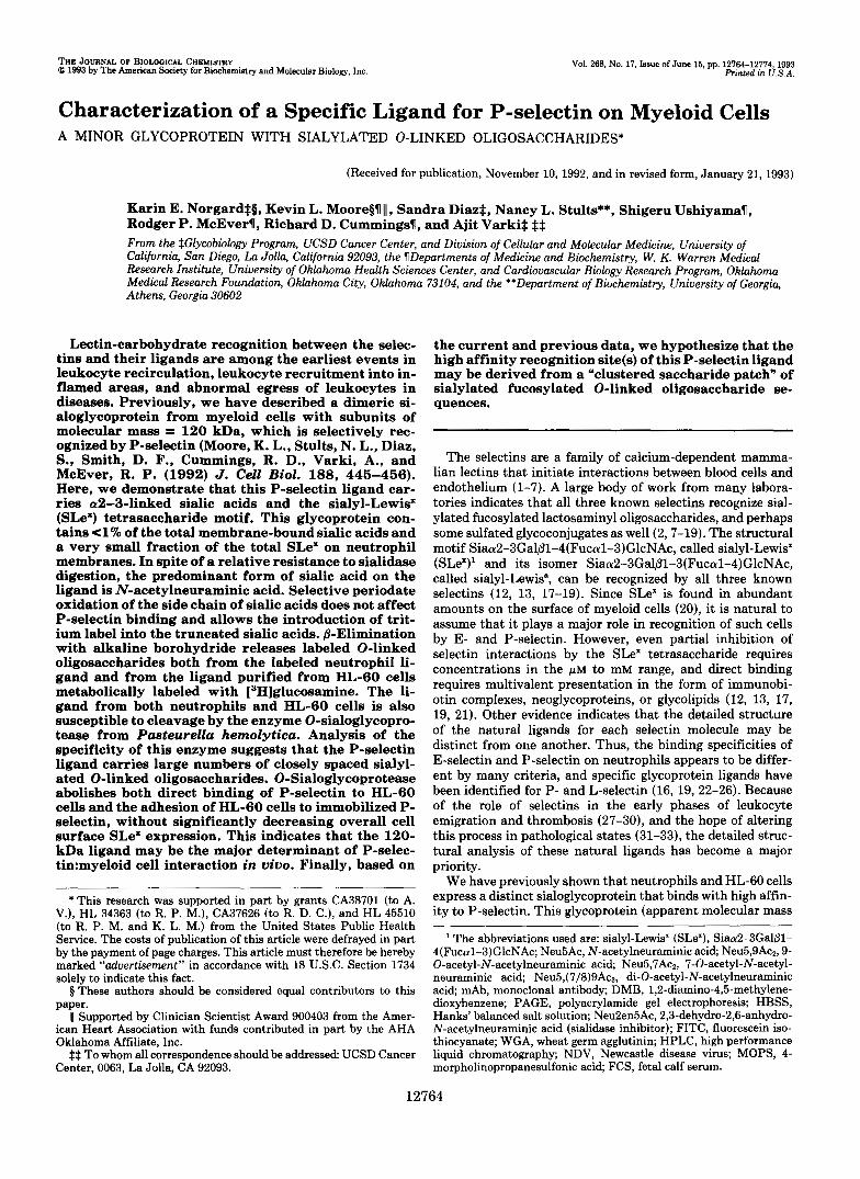
FIG. 1. Effect of sialidases on the myeloid P-selectin ligand. A WGA eluate of a neutrophil membrane lysate (50 pg of protein) in 0.1 M NaCI, 50 mM acetate, pH 5.5, 9 mM CaC12, 0.02% NaN3, 0.01% Lubrol-PX was either sham-treated or digested with the indicated sialidase (200 milliunits/ml) for 60 min at 37 "C. Samples were then electrophoresed on 7.5% SDS-polyacrylamide gels under reducing conditions, transferred to Immobilon membranes, and probed with '251-labeled P-selectin. NDV, New Castle disease virus sialidase; VC, V. cholerae sialidase.
enriched glycoproteins from neutrophil membranes with the sialidase from New Castle disease virus (NDV), which specif- ically cleaves a2-3-linked sialic acids (whether or not they are 0-acetylated), or sialidase from V. cholerae, which cleaves a2-6- and a2-3-linked sialic acids but is less tolerant of sialic acids with modifications (47, 48). As shown in Fig. 1, a short treatment with these enzymes caused an upward shift in the apparent M , of the P-selectin ligand without affecting P- selectin binding. The effect of V. cholerae sialidase treatment was very similar to that obtained with the enzyme from Arthrobacter (data not shown). NDV treatment caused some upward shift in apparent M,, indicating that a2-3-linked sialic acids are present on the P-selectin ligand. Although the shift with NDV was not as great as that seen with either A. ureafaciens or V. cholerae sialidase, prolonged treatment with the NDV sialidase was not attempted, since the enzyme is not very stable to longer incubations and its specificity is not as stringent under such conditions? Thus, while a2-3-linked sialic acids are present on the P-selectin ligand, we cannot rule out the possibility that there are also sialic acids in a 2 4 linkage. Regardless, the fact that some sialic acids on the ligand are relatively resistant to several sialidases raised the possibility that they might be substituted (49).
Mild Periodate Oxidation of the Sialic Acid Side Chain Does Not Abolish Recognition of the Ligand by P-selectin-Mild periodate oxidation (2 mM at 4 "C) can be used to selectively cleave the side chains of sialic acids while maintaining the integrity of the underlying oligosaccharide and protein struc-
B. K. Hayes and A. Varki, unpublished observations.

P-selectin Ligand 12767
tures (50-53). The side chain aldehyde generated by mild periodate can then be reduced to an alcohol by borohydride reduction, leaving a truncated side chain with a primary hydroxyl group (50). To examine the role of sialic acid side chains in the binding of P-selectin to this glycoprotein ligand, the effects of mild periodate oxidation, borohydride reduction, and the combination of both treatments were explored. Treated glycoprotein mixtures were separated by SDS-PAGE and detected by blotting with lz5I-1abeled P-selectin. None of the treatments had any effect upon the binding of P-selectin to this P-selectin ligand (data not shown). This result can be interpreted in one of two ways. One possibility is that sialic acid side chains are unimportant for recognition by P-selectin. The other possibility is that the critical side chains are sub- stituted (e.g. with 9-0-acetyl groups) and are therefore resist- ant to cleavage (54, 55).
N-Acetylneuraminic Acid is the Single Major Sialic Acid on the P-selectin Ligand from Neutrophils and from HL-60 Cells-Since two lines of evidence suggested the possibility of sialic acid substitutions, it was necessary to examine this question directly. The sialidase from A. ureafaciens has a broad spectrum of action against most, but not all, sialic acids in a variety of types and linkages. However, since recognition of the 120-kDa ligand by P-selectin was completely abolished by the Arthrobacter enzyme, it is reasonable to assume that the sialic acids relevant for binding are released by this enzyme. Sialic acids from total neutrophil membranes were released with A. ureafaciens sialidase and compared with those from a WGA-enriched pool and from the P-selectin ligand purified by affinity chomatography on a P-selectin column. The released sialic acids were purified, derivatized, and ana- lyzed by a TSK-ODS HPLC system that has previously been shown to detect most major forms of substituted sialic acids (41, 42). As shown in Fig. 2, the major sialic acid in all fractions studied was N-acetylneuraminic acid (Neu5Ac), the unmodified parent sialic acid. Although a very small amount of 9-0-acetyl-Neu5Ac was seen in the starting material, no enrichment of this species was seen in the purified ligand.
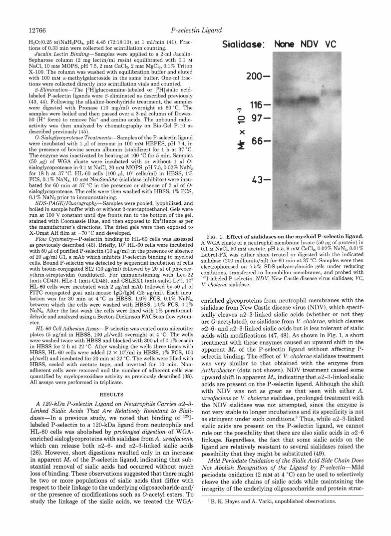
The P-selectin ligand isolated from neutrophils shares many properties with a similar protein found on HL-60 cells (26). To study the sialic acids on the HL-60 cell ligand, these cells were labeled with [6-3H]glucosamine. Because of meta- bolic interconversions, the tritium from [6-3H]glucosamine will be found in GlcNAc, GalNAc, and sialic acid residues (56). This makes it possible to enzymatically release, purify, and analyze radiolabeled sialic acids from a glycoprotein. The P-selectin ligand isolated from HL-60 cells labeled with [6- 3H]glucosamine was purified by affinity chomatography on a P-selectin column. After separation by SDS-PAGE under reducing conditions, the radioactivity in the P-selectin ligand band was released from the gel by Pronase digestion as described under “Experimental Procedures.” Released glyco- peptides were large enough to void on a Bio-Gel P-2 column. Treatment with A. ureafaciens sialidase released -30% of the label as free sialic acids, which were separated from the remaining glycopeptides by Bio-Gel P-2 gel filtration. The released sialic acids were purified and analyzed using an AX- 5 HPLC system, which can separate most major forms of substituted sialic acids but requires no derivatization prior to analysis. As shown in Fig. 3, we only found evidence for the unmodified sialic acid, [3H]Neu5Ac. These data indicate that the sialic acids responsible for P-selectin binding to the 120- kDa ligand are not substituted in neutrophils or in HL-60 cells. Rather, the partial resistance to sialidase treatment might be determined by features such as steric hindrance by the polypeptide and/or close spacing of the oligosaccharides.
W tn z g tn W a K
Y I- W n
1
TIVES OF;
1 = Neu5Gc
2 = Neu5Ac
3 = Neu5,7Ac2
4 Neu5Gc9Ac
5 Neu5,9Ac2
6 I Neu5,(7/8)9Ac3
7 Un-identified peak
X = Purification Buffer Peak
R = Reagent Peaks
A I STANDARDS
B WGA COLUMN
ELUATE
C = P-SELECTIN
ELUATE
TIME (min)
FIG. 2. TSK-ODS HPLC Profiles of DMB-derivatized sialic acids of the P-selectin ligand. Aliquots of a total neutrophil membrane lysate and the P-selectin ligand isolated from it were treated with A. ureafaciens sialidase, and the released sialic acids purified and derivatized with DMB reagent. The derivatized sialic acids were analyzed on a TSK-ODS 120T column with fluorescent detection. The elution position of standards derived from bovine submaxillary mucin are indicated. The peak marked X was seen in all samples purified from neutrophils, including buffer blanks, and probably arises from one of the protease inhibitors.
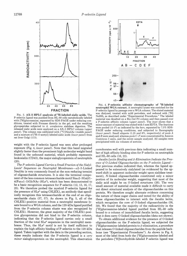
The P-selectin Ligand Carries a Very Small Fraction of the Total Glycoprotein-bound Sialic Acids of the Neutrophil- Using the sensitive fluorescent HPLC assay, the yield of sialic acids was monitored in a purification in which quantitative recovery of the P-selectin ligand was obtained at every step as determined by P-selectin blotting (data not shown). Start- ing from a membrane extract containing 852 nmol of sialic acids, the final preparation of ligand contained only 0.74 nmol (0.09%) of the starting sialic acids. Thus, the amount of material available for direct analysis was very small. We took advantage of the fact that the ligand carries unmodified Neu5Ac whose side chains are not required for P-selectin binding, and specifically introduced tritium label into the sialic acid by truncation of the side chain with mild periodate oxidation, followed by reduction with NaB3H4 (50). A WGA- enriched glycoprotein fraction from neutrophils was first la- beled in this manner, resulting in the introduction of tritium into many sialoglycoproteins. When applied to a P-selectin affinity column, almost all of the radioactivity ran through. However, a minor fraction of the tritium was specifically eluted with 5 mM EDTA (see Fig. 4, upperpanel). When this small peak (representing 0.29% of the total radioactivity loaded on the column) was pooled, concentrated by acetone precipitation, and studied by SDS-PAGE and fluorography, a single glycoprotein that coincides in apparent molecular
12768 P-selectin Ligand
FRACTION FIG. 3. AX-5 HPLC analysis of ‘H-labeled sialic acids. The
P-selectin ligand was purified from HL-60 cells metabolically labeled with [3H]glucosamine, separated by SDS-PAGE under reducing con- ditions, treated with Pronase directly in the gel, and the resulting glycopeptides subjected to A. ureafaciens sialidase digestion. The released sialic acids were analyzed on a AX-5 HPLC column (upper panel). The column was calibrated with [“C]Neu5Ac (middle panel) and a mixture of [‘H-0-acetyl]-labeled sialic acids (lower panel) from rat liver Golgi (1 13).
weight with the P-selectin ligand was seen after prolonged exposure (Fig. 4, lower panel). Note that this band migrated slightly faster than the prominent high molecular weight band found in the unbound material, which probably represents leukosialin (CD43), the major sialoglycoprotein of neutrophils (20).
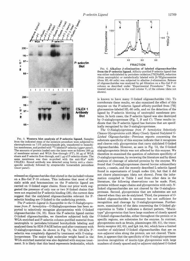
The P-selectin Ligand Carries a Small Fraction of the Sialyl- Lewis’ Sequences on Neutrophil Membranes-a2-3-Linked Neu5Ac is very commonly found at the non-reducing termini of oligosaccharide structures. It is also the terminal compo- nent of the less common tetrasaccharide motif SiaaPSGal/31- 4(FucalS)GlcNAc (SLe’), which has been demonstrated to be a basic recognition sequence for P-selectin (12, 13, 16, 17, 25). We therefore probed the myeloid P-selectin ligand for the presence of SLe’ using CSLEX1, an antibody that specif- ically recognizes this tetrasaccharide at the end of an oligo- saccharide chain (37, 57). As shown in Fig. 5, all of the CSLEX1-positive material from a neutrophil membrane ly- sate bound to a WGA column, and the 120-kDa ligand purified from the P-selectin column eluate was also recognized by CSLEX1. However, the great majority of the CSLEX1-posi- tive glycoproteins did not bind to the P-selectin column, indicating that the P-selectin ligand carries only a small fraction of the total SLe’ sequences in the neutrophil mem- brane. Thus, the SLe’ motif is not by itself sufficient to explain the high affinity binding of P-selectin to the 120-kDa ligand. Taken together with the data in the preceding section, these results indicate that the P-selectin ligand is a uery minor sialoglycoprotein on the neutrophil. This observation
EDTA
IC x 500
1 0 2 0 30 4 0
FRACTION
A B - C D E F
FIG. 4. P-selectin affinity chomatography of ‘H-labeled neutrophil WGA extract. A neutrophil lysate was enriched for the P-selectin ligand by passage over a WGA column. The eluted material was dialyzed, treated with mild periodate, and reduced with [’HI NaBH4 as described under “Experimental Procedures.” The labeled material was desalted on a Bio-Gel P2 column and then passed over a P-selectin affinity column (upper panel). The inset shows that a minor amount of material was eluted with 5 mM EDTA. The fractions were pooled (A-F) as indicated by the bars, separated by 7.5% SDS- PAGE under reducing conditions, and subjected to fluorography (lower panel). Small aliquots (1.25 and 8%, respectively) of pools A and B were analyzed, whereas pools C-F were concentrated by Amicon Centricon 3 units, and the entire sample analyzed. All samples were precipitated with six volumes of acetone.
corroborates well with previous data indicating a small num- ber of high affinity binding sites for P-selectin on neutrophils and HL-60 cells (16, 22).
Jacalin Lectin Binding and @-Eliminution Indicate the Pres- ence of 0-Linked Oligosaccharides on the P-selectin Ligand- Our previous studies indicated that, whereas the ligand ap- peared to be extensively sialylated (as evidenced by the up- ward shift in apparent molecular weight upon sialidase treat- ment), N-linked oligosaccharides constituted only a minor portion of its molecular weight, suggesting that most of the sialic acid might be on 0-linked structures (26). The very small amount of material available made it difficult to carry out direct structural analysis of the oligosaccharides on this protein. We therefore sought alternate evidence concerning the nature of these sugar chains and tested for the ability of these oligosaccharides to interact with the Jacalin lectin, which recognizes the core of 0-linked oligosaccharides (58, 59). We found that the majority of the radioactivity in the [SH]gluc~samine-labeled ligand purified from HL-60 cells bound to a column of immobilized Jacalin lectin, suggesting that it does carry 0-linked oligosaccharides (data not shown).
To obtain additional evidence for the presence of 0-linked oligosaccharides on the P-selectin ligand, we subjected the [3H]glucosamine-labeledprotein to @-elimination, a procedure that releases 0-linked oligosaccharides from the peptide back- bone (see “Experimental Procedures”). As shown in Fig. 6, most of the label from both the [3H]glucosamine-labeled and the peri~date-[~H]borohydride-labeled P-selectin ligand was
P-selectin Ligand 12769
97- 9
* x
66-
200-
1%- 0
9 97-
* X
66-
1 1 1 1 1 1 1 1 1 1
P- selectin Blot
CSLEX-1 Antibody
Blot
FIG. 5. Western blot analysis of P-selectin ligand. Samples from the indicated steps of the isolation procedure were subjected to electrophoresis on 7.5% polyacrylamide gels, transferred to Immobi- Ion membranes, and probed with ’251-labeled P-selectin (upperpanel). The amounts of protein loaded onto the lanes were as follows: 200 pg of membrane extract and WGA flow-through (FT), 50 pg of WGA eluate and P-selectin flow-through, and 1 pg of P-selectin eluate. The same membrane was then re-probed with the anti-SW mAb CSLEX1. Bound antibody was detected using biotin anti-p chain- specific antibody followed by streptavidin horseradish peroxidase (lower panel).
released as oligosaccharides that eluted in the included volume on a Bio-Gel P-10 column. This indicates that most of the sialic acids and hexosamines on the P-selectin ligand are carried on 0-linked sugar chains. Since our prior work sug- gested the presence of only one or two N-linked chains that were not required for P-selectin binding (26), the current data suggest that the sialylated oligosaccharides relevant for P- selectin binding are O-linked to the underlying protein.
The P-selectin Ligand Is Susceptible to the 0-Sialoglycopro- tease from P. hemolytica-0-Sialoglycoprotease has been re- ported to selectively recognize glycoproteins with 0-linked oligosaccharides (34, 35). Since the P-selectin ligand carries 0-linked oligosaccharides, we therefore subjected both the WGA-enriched and P-selectin column-purified material from neutrophils to periodate/tritiated borohydride labeling and analyzed them by SDS-PAGE before and after treatment with 0-sialoglycoprotease. As shown in Fig. 7A, the 120-kDa P- selectin was completely digested by treatment with O-sialog- lycoprotease. The major high molecular weight band in the WGA-enriched material was also depleted with enzyme treat- ment. It is likely that this band represents leukosialin, which
4 0 6 0 8 0
FRACTION FIG. 6. Alkaline @-elimination of labeled oligosaccharides
from the P-selectin ligand. Affinity-purified P-selectin ligand that was either radiolabeled by periodate 0xidation/[~H]NaBH4 reduction (from neutrophils) or metabolically labeled with [6-3H]glucosamine (from HL-60 cells) was subjected to alkaline @-elimination. Release of oligosaccharides was analyzed by gel filtration on a Bio-Gel P-10 column as described under “Experimental Procedures.” The un- treated material ran in the void volume V, of the column (data not shown).
is known to have many 0-linked oligosaccharides (34). To corroborate these results, we also examined the effect of this enzyme on the P-selectin ligand affinity-purified from [3H] glucosamine-labeled HL-60 cells, and on the detection of the ligand by P-selectin blotting of neutrophil membrane pro- teins. In both cases, the P-selectin ligand was also destroyed by 0-sialoglycoprotease (Fig. 7 , B and C). These results in- dicate that the P-selectin ligand has features that are specif- ically recognized by the 0-sialoglycoprotease.
The 0-Sialoglycoprotease from P. hemolytica Selectively Cleaves Glycoproteins with Many Closely Spaced Sialylated 0- Linked Oligosaccharides-Previous reports concerning the substrate specificity of this enzyme indicate that it recognizes and cleaves only glycoproteins that carry sialylated 0-linked oligosaccharides. However, as seen in Fig. 7A, the 0-linked sialoglycoprotein fetuin was not affected by this enzyme. We therefore re-examined the question of substrate specificity of 0-sialoglycoprotease, by reviewing the literature and by direct analysis of cleavage of selected proteins by the enzyme. We found that 0-sialoglycoprotease cleaved bovine submaxillary mucin, K-casein, and the recently described L-selectin ligand found in supernatants of lymph nodes (24), but that it did not cleave plasminogen (data not shown). From the infor- mation compiled in Table I and from other data in the literature, the following observations can be made. First, proteins without sugar chains and glycoproteins with only N- linked oligosaccharides are not cleaved by the O-sialoglyco- protease. Second, glycoproteins that are substrates are not cleaved when they are de-sialylated. Third, the presence of 0- linked oligosaccharides is necessary but not sufficient for recognition and cleavage by 0-sialoglycoprotease. Further- more, examination of the data concerning the 0-linked gly- coproteins in Table I indicates a distinct pattern. “Mucin- type” glycoproteins that have large numbers of closely spaced 0-linked oligosaccharides, either throughout the protein or in specific regions, are substrates for the enzyme. In contrast, glycoproteins such as fetuin, plasminogen (60, 61), the trans- ferrin receptor (62,63), and IgA (44,64), which have a limited number of sialylated 0-linked oligosaccharides that are on mn-adjacent sites along the protein, are not cleaved. There- fore, it appears that the specificity of the 0-sialoglycoprotease involves recognition of mucin-type glycoproteins with large numbers of closely spaced and/or adjacent sialylated 0-linked

12770 P-selectin Ligand
A
peri~date/[~HH] BH,
B
[6-3H]GI~NH,
Fetuin Eluate Liaand WGA P-Selectin
- 0-glycoprotease: - + - + - + "~
0
z 200-
XL 97- X 116 -
66-
1 5 -
""
C ['25t]P-seIectin
Blot
P-Selectin Liaand
WGA Eluate
- +
200-
116- t I
97- 62-
15-
- + 200-
116,
80-
FIG. 7. Effect of 0-sialoglycoprotease on the P-selectin ligand. Samples were either sham-treated or digested with O-sialoglycopro- tease and analyzed by SDS-PAGE under reducing conditions. A , fetuin, neutrophil WGA eluate, and affinity-purified P-selectin ligand were radiolabeled by periodate oxidation/['H]NaBH4 reduction and detected by fluorography; B, P-selectin ligand was affinity-purified from [6- 3H]glucosamine-labeled HL-60 cells and detected by fluorography; C, P-selectin ligand in a WGA eluate from a neutrophil membrane lysate was directly detected by transferring to Immobilon membranes and probing with "'1-labeled P-selectin.
TABLE I Sensitivity of various proteins to P. hemolytica 0-sialoglycoprotease
Glycoprotein Occurrence of linear stretches of amino acids with
different numbers of adjacent 0-linked sites"
1 2 3 4 >4
Total 0- 0-Sialoglycoprotease linked sites sensitivity
Immunoglobulin A1 (44, 64) 1 Resistant (35) 5 Transferrin receptor (62,63) 1 Resistant (34) 1 Fetuin (61) 3 Resistantb 3 Plasminogen (60) 1 Resistantb 1 Glycophorin A (108) 15 Sensitive (35) 6 1 1 Sgp50 L-selectin ligand from lymph 42' Sensitiveb 17 8 3
Bovine K-casein 31' Sensitiveb 21 2 2 Bovine submaxillary mucin (110) 163' Sensitiveb 64 25 11 4 CD43 (leukosialin) (111) 112' Sensitive (34) 38 26 6 1 CD34 (112) 95' Sensitive (34) 38 17 3 2 1
a This refers to the spacing of 0-linked glycosylation sites along the linear protein backbone, e.g. for glycophorin A, there are six individual attachment sites, one group of three adjacent sites (Ser', Thr', Thr'), and one group of six adjacent sites (Thr", Thr", Thr", Ser", Ser", Ser").
Proteins examined in this study. The individual proteins were incubated with 1 ml of the enzyme at 37 "C for 1 h and re-examined by SDS-PAGE and Coomassie Blue staining, PAS staining, or autoradiography as appropriate.
In these instances, all of the glycosylation sites have not been precisely identified. These numbers represent the total number of serine and threonine residues in the protein, and therefore the maximum number of 0-linked sites possible. In these "mucin-type'' proteins, most but not all of these sites are 0-glycosylated.
dIndependently noted to be sensitive by Dr. A. Mellors (personal communication). Note: thus far, serum glycoproteins with N-linked oligosaccharides (34, 35), and many other proteins and glycoproteins that contain no 0-linked oligosaccharides (34, 35) have proven to be resistant to 0-sialoglscoprotease. Additionally, CD45 isoforms which show extensive but variable 0-glycosylation in the amino-terminal
nodes (24)
domains show variabie sensitivity to 0-sialoglycoprotease.
oligosaccharides. Since the P-selectin ligand from myeloid cells is cleaved by this enzyme, it is predicted to be a mucin- type glycoprotein.
0-Sialoglycoprotease Treatment of HL-60 Cells Eliminates Binding to P-selectin-The data presented in this and a previous paper (26) indicate that the 120-kDa ligand binds P- selectin with high affinity. However, they do not directly demonstrate that this ligand accounts for the small number of high affinity binding sites observed on intact neutrophils or HL-60 cells. The 0-sialoglycoprotease will not cleave cell surface glycoproteins with N-linked oligosaccharides or cell surface glycolipids (34, 35). On the other hand, it selectively cleaves cell-surface mucin-like molecules such as CD34 from intact cells (34, 65) and allows subsequent recovery of viable stem cells (66). We therefore examined the effects of this
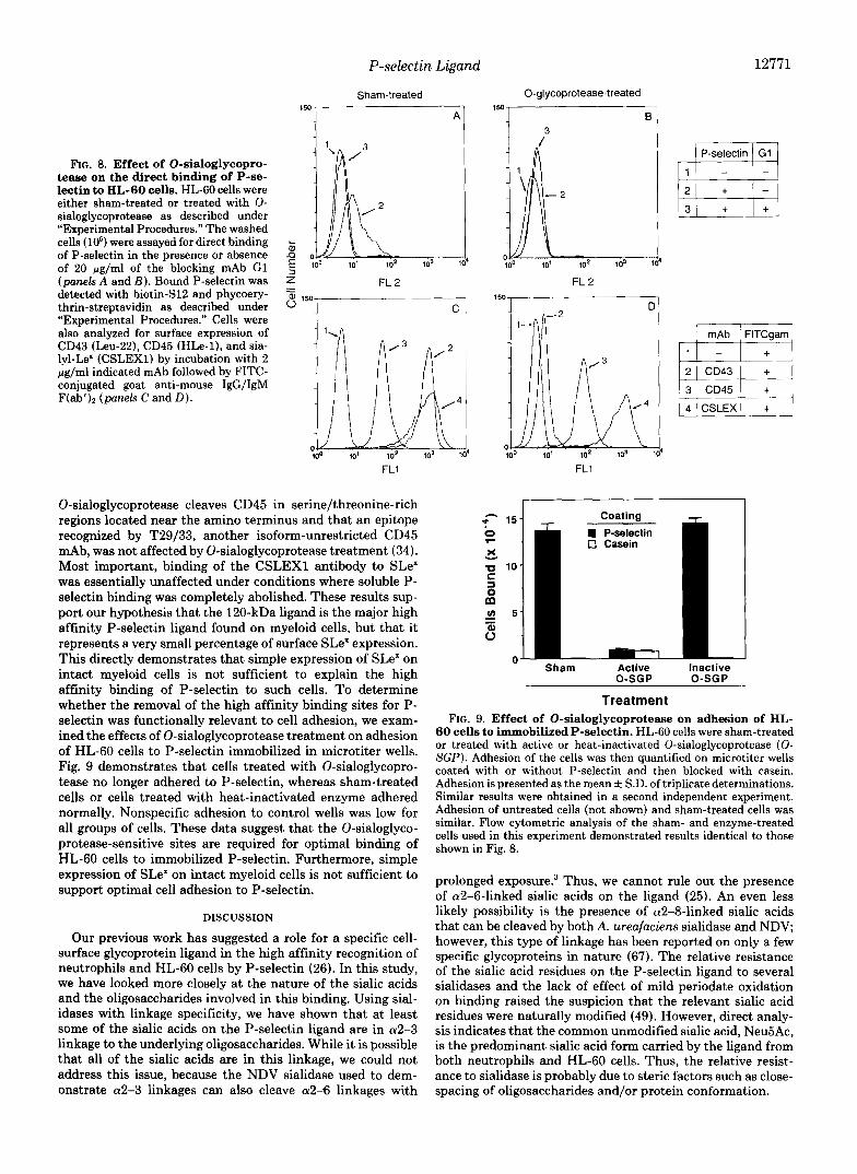
enzyme upon the direct binding of P-selectin to HL-60 cells. As seen in Fig. 8 (panels A and B), HL-60 cells were tested for their ability to bind soluble P-selectin after treatment with 0-sialoglycoprotease. Sham-treated cells maintained their ability to bind to P-selectin, and this binding was blocked by co-incubation with the mAb G1, which has been previously shown to block P-selectin binding to myeloid cells (22, 38, 39). Treatment with 0-sialoglycoprotease abolished binding by P-selectin. As controls, we also examined the effects of 0-sialoglycoprotease on other sialylated surface molecules on HL-60 cells. As seen in Fig. 8 (panels C and D), binding of a mAb to CD43 (leukosialin) was abolished by 0- sialoglycoprotease treatment, whereas binding of HLe-1, an isoform-unrestricted CD45 mAb was unaffected. These results are consistent with previous work, which indicates that the
P-selectin Ligand
Sham-treated
12771
FIG. 8. Effect of O-sialoglycopro- tease on the direct binding of P-se- lectin to HL-60 cells. HL-60 cells were either sham-treated or treated with 0- sialoglycoprotease as described under “Experimental Procedures.” The washed cells ( lo6) were assayed for direct binding of P-selectin in the presence or absence of 20 pg/ml of the blocking mAb G1 (panels A and E ) . Bound P-selectin was detected with biotin-S12 and phycoery- thrin-streptavidin as described under “Experimental Procedures.” Cells were also analyzed for surface expression of CD43 (Leu-22), CD45 (HLe-l), and sia- lyl-Le’ (CSLEX1) by incubation with 2 pg/ml indicated mAb followed by FITC- conjugated goat anti-mouse IgG/IgM F(ab’), (panels C and D).
FL1
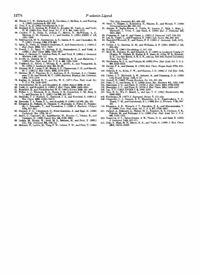
0-sialoglycoprotease cleaves CD45 in serine/threonine-rich regions located near the amino terminus and that an epitope recognized by T29/33, another isoform-unrestricted CD45 mAb, was not affected by 0-sialoglycoprotease treatment (34). Most important, binding of the CSLEXl antibody to SLe” was essentially unaffected under conditions where soluble P- selectin binding was completely abolished. These results sup- port our hypothesis that the 120-kDa ligand is the major high affinity P-selectin ligand found on myeloid cells, but that it represents a very small percentage of surface SLe‘ expression. This directly demonstrates that simple expression of SLe” on intact myeloid cells is not sufficient to explain the high affinity binding of P-selectin to such cells. To determine whether the removal of the high affinity binding sites for P- selectin was functionally relevant to cell adhesion, we exam- ined the effects of 0-sialoglycoprotease treatment on adhesion of HL-60 cells to P-selectin immobilized in microtiter wells. Fig. 9 demonstrates that cells treated with O-sialoglycopro- tease no longer adhered to P-selectin, whereas sham-treated cells or cells treated with heat-inactivated enzyme adhered normally. Nonspecific adhesion to control wells was low for all groups of cells. These data suggest that the O-sialoglyco- protease-sensitive sites are required for optimal binding of HL-60 cells to immobilized P-selectin. Furthermore, simple expression of SLe’ on intact myeloid cells is not sufficient to support optimal cell adhesion to P-selectin.
DISCUSSION
Our previous work has suggested a role for a specific cell- surface glycoprotein ligand in the high affinity recognition of neutrophils and HL-60 cells by P-selectin (26). In this study, we have looked more closely at the nature of the sialic acids and the oligosaccharides involved in this binding. Using sial- idases with linkage specificity, we have shown that at least some of the sialic acids on the P-selectin ligand are in a2-3 linkage to the underlying oligosaccharides. While it is possible that all of the sialic acids are in this linkage, we could not address this issue, because the NDV sialidase used to dem- onstrate a2-3 linkages can also cleave a2-6 linkages with
0-glycoprotease-treated 150
0 3
1
FL1
E, P-selectin GI
U c 0 a m
FL 2
mAb FlTCgarn
4 CSLEX
I I :I 5 0 S h a m
Coating P-selectin
0 Casein
Active Inactive 0-SGP 0-SGP
Treatment FIG. 9. Effect of 0-sialoglycoprotease on adhesion of HL-
60 cells to immobilized P-selectin. HL-60 cells were sham-treated or treated with active or heat-inactivated 0-sialoglycoprotease (O- SGP). Adhesion of the cells was then quantified on microtiter wells coated with or without P-selectin and then blocked with casein. Adhesion is presented as the mean f S.D. of triplicate determinations. Similar results were obtained in a second independent experiment. Adhesion of untreated cells (not shown) and sham-treated cells was similar. Flow cytometric analysis of the sham- and enzyme-treated cells used in this experiment demonstrated results identical to those shown in Fig. 8.
prolonged exp~sure .~ Thus, we cannot rule out the presence of a2-6-linked sialic acids on the ligand (25). An even less likely possibility is the presence of a2-8-linked sialic acids that can be cleaved by both A. ureafaciens sialidase and NDV; however, this type of linkage has been reported on only a few specific glycoproteins in nature (67). The relative resistance of the sialic acid residues on the P-selectin ligand to several sialidases and the lack of effect of mild periodate oxidation on binding raised the suspicion that the relevant sialic acid residues were naturally modified (49). However, direct analy- sis indicates that the common unmodified sialic acid, Neu5Ac, is the predominant sialic acid form carried by the ligand from both neutrophils and HL-60 cells. Thus, the relative resist- ance to sialidase is probably due to steric factors such as close- spacing of oligosaccharides and/or protein conformation.

12772 P-selectin Ligand
In this study, we have also shown that the side chain of sialic acids is not important for the binding of P-selectin to the ligand, and we have taken advantage of this fact to introduce high specific activity tritium label into this minor sialoglycoprotein. Other studies have indicated that the side chain of sialic acids is not required for recognition by E- selectin (14) or L-selectin (68). This observation contrasts with the absolute requirement for the side chain in recognition of ligands by sialoadhesin (69), influenza virus hemagglutinins (70), CD22P (71, 72), and certain monoclonal antibodies (53, 73). It remains to be seen what, if any effects, the natural modifications of side chains (e.g. O-acetylation) will have on recognition of sialylated ligands by the selectins.
A large body of prior evidence indicates that binding by all three selectins can involve either SLe’ (sialyl-Lewis”) or SLe” (sialyl-Lewis”) as a minimal recognition structure (2, 7-19). We have shown that the P-selectin ligand contains the SLe’ sequence that is recognized by the CSLEXl antibody. How- ever, the great majority of the glycoprotein-bound SLe” from neutrophils failed to bind to P-selectin with similar affinity. This correlates well with the finding that the P-selectin ligand carries a very small fraction of the total sialic acids from neutrophil or HL-60 cells. Taken together with other recent reports (7,12,16,26), our present study indicates that if SLe’ is necessary for binding, it is not sufficient for high affinity binding of this myeloid ligand by P-selectin. In this regard, it is striking that treatment of HL-60 cells with O-sialoglyco- protease destroys P-selectin binding, but leaves total cell surface SLe’ largely intact (see Fig. 8). These data also indi- cate that other recently proposed ligands for P-selectin such as sulfatide (15, 74, 75) and sulfated glycosaminoglycans (76, 77) cannot be responsible for high affinity P-selectin binding, at least on myeloid cells. The paucity of the P-selectin ligand on myeloid cells also fits well with the small number of high affinity binding sites for P-selectin on these cells (22, 76). There is as yet no direct evidence that these sites can be directly attributed to, and completely accounted for, by this particular P-selectin ligand. However, our finding that 0- sialoglycoprotease treatment of HL-60 cells destroys binding to P-selectin supports this notion.
The recognition of SLe’ by the selectins can be compared to some other situations involving specific biological recog- nition. For example, the discovery of the critical motifs in- volved in recognition of lysosomal enzymes by certain traf- ficking receptors (mannose 6-phosphate) or of matrix proteins by the integrins (RGD sequences) were landmark events (78, 79). However, these basic recognition motifs have relatively low affinities for their receptors. Subsequent studies showed much greater complexity in the natural ligands for these receptors (80, 81), with biologically relevant recognition re- quiring other specific features of the ligands such as tertiary structure and/or multivalency. Thus, while sialylated fuco- sylated lactosamines such as SLe‘ may form basic recognition motifs for the selectins, biologically relevant binding may require more complex structures such as those presented by the P-selectin ligand on myeloid cells described here. SLe’ found on other glycoconjugates on the neutrophil surface might also support cell adhesion if such low affinity interac- tions, due to their density, resulted in significant avidity on an intact cell surface. However, Chinese hamster ovary cells transfected with a fucosyltransferase, while expressing large amounts of SLe’ on their surface, lack high affinity binding sites for P-selectin and adhere only weakly to immobilized P- selectin (16). Here we demonstrate that HL-60 cells treated with O-sialoglycoprotease do not adhere to P-selectin, even though total surface expression of SLe” is not detectably
altered. The enzyme-treated HL-60 cells might bind to wells coated with higher densities of P-selectin like those used for the transfected Chinese hamster ovary cells. However, the weak adhesion of the latter cells may reflect the fact that they express much higher levels of SLe” than do HL-60 cells (16). Therefore, low affinity ligands for P-selectin may not play a major role in adhesion unless perhaps they are positioned or clustered in favorable sites such as pseudopodia on the cell surface (82).
This study and our previous results (26) indicate that the P-selectin ligand has very few N-linked oligosaccharides and many O-linked sugar chains. O-Sialoglycoprotease is a re- cently described enzyme from P. hemolytica that has been reported to cleave only sialylated O-linked glycoproteins (34, 35). Based on the glycoproteins that are reported substrates for this enzyme, we suspected that the specificity of this enzyme might be more complex than suggested. Taken to- gether with the prior literature, the additional studies we have performed indicate that besides the presence of sialylated 0- linked sugar chains, an additional requirement of the enzyme appears to be the presence of a large number oligosaccharides that are closely spaced and/or adjacent to one another. Of course, the exact recognition moiety may be only a subgroup of the total oligosaccharides present. Since the P-selectin ligand is susceptible to O-sialoglycoprotease, it is reasonable to suggest that it also carries closely spaced groupings of sialylated O-linked oligosaccharides. Such close-packing or “clustering” may also be the cause of the relative resistance to sialidase treatments.
The observation that the ligand can bind P-selectin even after denaturation with SDS and disulfide bond reduction suggests that P-selectin recognition is due solely to lectin- carbohydrate interaction. The specificity of P-selectin for the 120-kDa ligand might therefore be due to a unique structural feature of the oligosaccharide(s). For example, the protein backbone of the ligand might specify the addition of uncom- mon oligosaccharide sequences during biosynthesis, as is seen with mannose 6-phosphorylation of lysosomal enzymes (83, 84). However, it is also possible that the O-linked oligosac- charides on the 120-kDa ligand are not unusual or distinct from those on other neutrophil glycoproteins. In this case, the specificity of P-selectin for the ligand may be conferred by co-recognition of peptide sequences and/or by the clustering of common oligosaccharide structures, Detailed structural analysis of the oligosaccharide on the 120-kDa ligand will be required to distinguish these possibilities.
It is worth noting that a large number of monoclonal antibodies known to detect “tumor-specific” glycoproteins from normal and cancer cells have “sialidase-sensitive” or “sialic acid-dependent” epitopes (see Refs. 73 and 85-87 for examples). Many monoclonal antibodies apparently specific for the proteins CD24 (88, 89), CD45 (90-931, and CD43 (94, 95) also show sialidase sensitivity. In the case of CD43 (leu- kosialin), a heavily glycosylated O-linked glycoprotein from leukocytes (96), it is rare to find a specific mAb that does not require sialylation for recognition (97). In fact, expression cloning of a glycosyltransferase required the co-expression of a specific polypeptide, leukosialin, in the same cell type to permit recognition by a specific mAb (98). In many such cases, when the sialyl-oligosaccharides on such proteins are directly examined ( Z O ) , they are not unusual in structure compared with those from other proteins of the same cells. Thus, the oligosaccharides cannot by themselves have gener- ated the specificity seen by the antibodies. Many of the proteins with such sialidase-sensitive epitopes are either mu- cins with clustered O-linked oligosaccharides or have large

12774 P-select rl Ligand Clin. Exp. Immunol. 8 1,489-495
90. B a d , V.. Hilwrt. I.. Kristofova. H.. Maurer. D.. and Horeisi. V. (1989) 66.
68. 67.
69.
70.
71.
72.
73.
74.
75.
76.
77.
78.
79. 80. 81. 82.
83.
84. 85.
Marsh, J. C. W., Sutherland, D. R., Davidson, J., Mellors, A,, and Keating, A. 11992) T s u b p r n i n (i. 92fi-934
Troy, F. A., I1 (1992) Glycobiology 2,5-23 Norgard, K. E., Huiling, H., Powell, L., Kriegler, M., Varki, A,, and Varki,
N. M. (1993) Proc. Nutl. Acud. Sci. U. S. A. ,90 , 1068-1072 Crocker, P. R., Kelm, S., Dubois, C., Martln, B., McWllliam, A. S.,
Shotton, D. M., Paulson, J. C., and Gordon, S. (1991) EMBO J. 10 ,
...~ " _
1661-1669 Matrosovich, M. N., Gambaryan, A. S., Reizin, F. N., and Chumakov, M.
P. (1991) Virology 182.879-882 Sgroi, D., Varki, A., Braesch-Andersen, S., and Stamenkovic, I. (1993) J.
Biol. Chern. 2 6 8 , 7011-7018 Powell, L. D., Sgroi, D., Sjoberg, E. R., Stamenkovic, I., and Varki, A.
(1993) J. BWL Chem. 268,7019-7027 Bara, J., Decaens, C., Loridon-Rosa, B., and Oriol, R. (1992) J. Immunol.
Methods 149,105-113 Aruffo, A., Dietsch, M. T., Wan, H., Hellstrom, K. E., and Hellstrom, I.
(1992) Proc. Nutl. A m i . Sei. U. S. A. 89,2292-2296 Tcddemd, G., Alford, J., Millsap, K. A., Aruffo, A., and Tramposch, K.
M. (1992) J. Leukocyte Biol. 62,85-88 Skinner, M. P., Lucas, C. M., Burns, G. F., Cbesterman, C. N., and Berndt,
M. C. (1991) J. Biol. Chem. 266,5371-5374 Skinner, M. P., Fournier, D. J., Andrews, R. K., Gorman, J. J., Chester-
man, C. N., and Berndt, M. C. (1989) Biochem. Biophys. Res. Commun.
Kaplan, A., Achord, D. T., and Sly, W. S. (1977) Proc. Nutl. Acud. Sci. 164 , 1373-1379
U. S. A. 74,2026-2030 Pierschbacher, M. D., and Ruoslahti, E. (1984) Nature 309,30-33 Varki, A,, and Kornfeld, S. (1983) J. Biol. Chem. 268,2808-2818 Ruoslahti, E., and Pierschbacher, M. D. (1987) Science 238,491-497 Picker, L. J., Warnock, R. A,, Burns, A. R., Doerschuk, C. M., Berg, E.
Baranski, T. J., Koelsch, G., Hartsuck, J. A., and Kornfeld, S. (1991) J.
Baranski, T. J., Faust, P. L., and Kornfeld, S. (1990) Cell 63,281-291 Kitagawa, H., Nakada, H., Numata, Y., Kurosaka, A,, Fukui, S., Funako-
shi. I.. Kawasaki. T.. and Yamashina. I. (1988) J. Biochem. (Tokvo)
L., and Butcher, E. C. (1991) Cell 66,921-933
Biol. Chem. 2 6 6 , 23365-23372
, ~, 104,817-821
Curbohydr. Res. 178,29-47
Carmann, H. (1988) Cancer Res. 48,6799-6802
Clin. Ex Immunol. 86,536-541
~ , . ~~, . " ,
86. Hanisch, F. G., Uhlenbruck, G., Peter-Katalinic, J., and Egge, H. (1988)
87. Stahli, C., Caravatti, M., Aeschbacher, M., Kocyba, C., Takacs, B., and
88. Larkin, M., Knapp, W., Stoll, M. S., Mehmet, H., and Feizi, T. (1991)
89. Mehmet, &, Larkin, M., Tang, P. W., Lebien, T. W., and Feizi, T. (1990)
. . Immunoge&t&s 29, 202-205
91. Alsinet, E., InglBs-Esteve, J., Vilella, R., Lozano, F., Mild, J., Rojo, I., Martorell, J., Vives, J., and Gaya, A. (1990) Eur. J . Immuml. 2 0 , 2801-2804
. , _ I . I
92. Poppema, S., Lai, R., and Visser, L. (1991) J. Immunol. 147,218-223 93. Lai, R., Visser, L., and Poppema, S. (1991) Lab. Invest. 6 4 , 844-854 94. Remold-ODonnell, E., and Rosen, F. S. (1990) J. Immunol. 146 , 3372-
95. Cyster, J. G., Shotton, D. M., and Williams, A. F. (1991) EMBO J. 10,
96. Fukuda, M. (1991) Glycobiology 1,347-356 97. Stoll, M., Dalchau, R., and Schmidt, R. E. (1989) in Leukocyte Typing IV
R. E., von dem Borne, A. E. G. K., eds) pp. 604-608, Oxford University (Knapp, W., Dorken, B., Rieber, E. P., Stein, H., Gilks, W. R., Schmidt,
~ ~
3378
893-902
98. Bierhuizen, M. F. A., and Fukuda, M. (1992) Proc. Nutl. Acud. Sci. U. S. A.
99. Devme, P. L., and Harada, H. (1991) Biol. Chem. Hoppe-Seyler 372,935-
Press, New York
89,9326-9330
942 100.
101.
102. 103. 104. 105. 106. 107.
108. 109.
OrGndi, P. A., Klotz, F. W., and Haynes, J. D. (1992) J. Cell Biol. 116 ,
Carver, J. P., Michnick, S. W., Imberty, A., and Cumming, D. A. (1989)
Jentoft, N. (1990) Trends Biochern. Sci. 16,291-294 Pabo, C. O., and Sauer, R. T. (1992) Annu. Rev. Biochem. 6 1 , 1053-1095 Baenziger, J. U., and Fiete, D. (1979) J. Biol. Chem. 264,9795-9799 Baenziger, J. U., and Fiete, D. (1979) J. Biol. Chem. 264,2400-2407 Lee, Y. C. (1992) FASEB J. 6,3193-3200 Rice, K. G., Wu, P., Brand, L., and Lee, Y. C. (1991) Biochemistry 30 ,
Furthmayr, H. (1977) J. Supramol. Struct. 7 , 121-134 Alexander, L. J., Stewart, A. F., Mackinlay, A. G., Kapelinskaya, T. V.,
Tkach, T. M., and Gorodetsky, S. I. (1988) Eur. J. Biochern. 178,395-
901-909
Cibn Found. Symp. 146,6-18
6646-6655
401 110. Bh&ava. A. K.. Woitach. J. T.. Davidson. E. A.. and Bhavanandan. V.
P"1990) Prdc. Nutl. Acud. Sci. U. S. A. 87,6798-6802 111. Pallant, A., Eskenazi, A., Mattei, M. G., Fournier, R. E., Carlsson, S. R.,
Fukuda. M.. and Frelinzer. J. G. (1989) Proc. Nutl. Acud. Sei. U. S. A. 86,1328-1332
J, Immunol. 148,267-271
264,19416-19426
- . . .
112. Simmons, D. L., Satterthwaite, A. B., Tenen, D. G., and Seed, B. (1992)
113. Diaz, S., Higa, H. H., Hayes, B. K., and Varki, A. (1989) J. Biol. Chem.