equine and canine influenza h3n8 viruses show...
TRANSCRIPT

Equine and Canine Influenza H3N8 Viruses Show Minimal BiologicalDifferences Despite Phylogenetic Divergence
Kurtis H. Feng,a Gaelle Gonzalez,b Lingquan Deng,c Hai Yu,d Victor L. Tse,e Lu Huang,a Kai Huang,a Brian R. Wasik,a Bin Zhou,f
David E. Wentworth,f Edward C. Holmes,g Xi Chen,d Ajit Varki,c Pablo R. Murcia,b Colin R. Parrisha
Department of Microbiology and Immunology, Baker Institute for Animal Health, College of Veterinary Medicine, Cornell University, Ithaca, New York, USAa; MedicalResearch Council-University of Glasgow Centre for Virus Research, Glasgow, Scotland, United Kingdomb; Cellular and Molecular Medicine, Glycobiology Research andTraining Center, University of California, San Diego, La Jolla, California, USAc; Department of Chemistry, University of California, Davis, Davis, California, USAd; Departmentof Microbiology and Immunology, College of Veterinary Medicine, Cornell University, Ithaca, New York, USAe; Infectious Disease Group, J. Craig Venter Institute, Rockville,Maryland, USAf; Marie Bashir Institute for Infectious Diseases and Biosecurity, Charles Perkins Centre, School of Biological Sciences and Sydney Medical School, TheUniversity of Sydney, Sydney, New South Wales, Australiag
ABSTRACT
The A/H3N8 canine influenza virus (CIV) emerged from A/H3N8 equine influenza virus (EIV) around the year 2000 through thetransfer of a single virus from horses to dogs. We defined and compared the biological properties of EIV and CIV by examiningtheir genetic variation, infection, and growth in different cell cultures, receptor specificity, hemagglutinin (HA) cleavage, andinfection and growth in horse and dog tracheal explant cultures. Comparison of sequences of viruses from horses and dogs re-vealed mutations that may be linked to host adaptation and tropism. We prepared infectious clones of representative EIV andCIV strains that were similar to the consensus sequences of viruses from each host. The rescued viruses, including HA and neur-aminidase (NA) double reassortants, exhibited similar degrees of long-term growth in MDCK cells. Different host cells showedvarious levels of susceptibility to infection, but no differences in infectivity were seen when comparing viruses. All viruses pre-ferred �2-3- over �2-6-linked sialic acids for infections, and glycan microarray analysis showed that EIV and CIV HA-Fc fusionproteins bound only to �2-3-linked sialic acids. Cleavage assays showed that EIV and CIV HA proteins required trypsin for effi-cient cleavage, and no differences in cleavage efficiency were seen. Inoculation of the viruses into tracheal explants revealed simi-lar levels of infection and replication by each virus in dog trachea, although EIV was more infectious in horse trachea than CIV.
IMPORTANCE
Influenza A viruses can cross species barriers and cause severe disease in their new hosts. Infections with highly pathogenic avianH5N1 virus and, more recently, avian H7N9 virus have resulted in high rates of lethality in humans. Unfortunately, our currentunderstanding of how influenza viruses jump species barriers is limited. Our aim was to provide an overview and biologicalcharacterization of H3N8 equine and canine influenza viruses using various experimental approaches, since the canine virusemerged from horses approximately 15 years ago. We showed that although there were numerous genetic differences betweenthe equine and canine viruses, this variation did not result in dramatic biological differences between the viruses from the twohosts, and the viruses appeared phenotypically equivalent in most assays we conducted. These findings suggest that the cross-species transmission and adaptation of influenza viruses may be mediated by subtle changes in virus biology.
Influenza A viruses are maintained in aquatic birds as intestinalinfections, occasionally transfer to and become established as
respiratory infections in mammals, including humans, and some-times spread from one mammal to another (1, 2). Mammalianhosts that have been commonly seen to maintain avian-derivedviruses include swine, horses, humans, mink, seals, and, recently,dogs (2–5). Host transfers between different birds, from birds tomammals, or between different mammalian hosts are relativelycommon but mostly result in single infections or limited out-breaks. On rare occasions, the host-transferred viruses go on tocause sustained epidemics or pandemics in their new hosts. Influ-enza viruses causing epidemics in new hosts often have mutationsthat appear to be specific to the new hosts in several gene seg-ments, and in some cases these have been shown to control hostadaptation (6–8). In many cases, the transferred virus was ob-served to be a reassortant with segments from a number of differ-ent ancestors, or it soon reassorted with another influenza virusinfecting that host (9, 10).
A number of different viral functions have been associated withhost adaptation of influenza viruses. Specific sialic acid binding
and/or cleavage is often a key factor in host adaptation becausesialic acids are primary influenza virus receptors, and mutations inthe receptor-interacting proteins, the hemagglutinin (HA) andneuraminidase (NA), often appear upon host transfer. Key traitsinclude HA recognition of �2-3- or �2-6-linked sialic acids; avianviruses are generally specific for �2-3-linked receptors, and hu-man viruses are generally specific for �2-6-linked receptors (11–
Received 25 February 2015 Accepted 14 April 2015
Accepted manuscript posted online 22 April 2015
Citation Feng KH, Gonzalez G, Deng L, Yu H, Tse VL, Huang L, Huang K, Wasik BR,Zhou B, Wentworth DE, Holmes EC, Chen X, Varki A, Murcia PR, Parrish CR. 2015.Equine and canine influenza H3N8 viruses show minimal biological differencesdespite phylogenetic divergence. J Virol 89:6860 –6873. doi:10.1128/JVI.00521-15.
Editor: B. Williams
Address correspondence to Colin R. Parrish, [email protected].
Copyright © 2015, American Society for Microbiology. All Rights Reserved.
doi:10.1128/JVI.00521-15
6860 jvi.asm.org July 2015 Volume 89 Number 13Journal of Virology
on June 3, 2015 by Cornell U
niversity Libraryhttp://jvi.asm
.org/D
ownloaded from

13). There is often a coordination of the NA activity and specificitythat correlates with HA binding and the sialic acid linkages that arepresent in the host (14). Importantly, mutations in other genesegments, including PB2, PA, NP, M, and NS, are often seen (15–18). In particular, polymerase subunits PB2 and PA control repli-cation in different host cells and at different temperatures (19).Some mutations in the M gene segment have been associated withtransmission (20), while NP mutations control the interactionswith MxA, a host-derived antiviral molecule (21). Mutations inthe NS1 gene control a variety of host-specific functions and in-nate immune responses (22, 23). Despite the identification ofthese mutations, we lack a complete understanding of the factorsthat control specific virus host range, particularly in nature, or ofthe host barriers that regulate the transfer of viruses to new hosts.
In this study, we examined the host tropism associated with thetransfer to and continuing replication of the A/H3N8 equine in-fluenza virus (EIV) in dogs to create the phylogenetically distinctlineage of A/H3N8 canine influenza viruses (CIVs) (Fig. 1) (3,24–26). CIV was first identified in Florida in 2004, when it causedan outbreak in greyhounds in a training facility, and it was soonrecognized to be closely related to EIV (3). Infected greyhounds
carried the virus to different regions of the United States, andmany other breeds of dogs have since been infected (24, 27, 28).CIV has continued to circulate in some regions of the UnitedStates, and for the past several years it appears to have been pri-marily maintained in several hot spots where there are high-den-sity and high-turnover dog populations, including animal sheltersin New York, NY, Philadelphia, PA, Colorado Springs, CO, andDenver, CO (26, 28, 29). Dogs appear to be a naturally receptivehost of influenza virus, because in addition to the equine-originA/H3N8 virus, an avian-origin A/H3N2 subtype has spreadamong dogs in Korea and China since 2006 (30, 31).
Examination of A/H3N8 isolates collected from dogs hasshown that all segments contained CIV-specific mutations notseen in equine viruses, or seen only at very low frequencies (26,28). However, it is not known how these CIV-specific mutationsalter host range and tropism or whether there has been ongoingselection of canine-adaptive mutations during the extended pas-sage in dogs. To determine whether any of the CIV-specific mu-tations play a role in virus host switching and adaptation, weexamined protein sequences of all available CIV sequences depos-ited in GenBank (NCBI). We also prepared reverse-genetics plas-
EIV
CIV2003-2013
1963-19721.0
1.0
2003-20120.80
0.99
1.0
2001-2014
0.99
0.95
1989-2006
0.961993-2006
1975-19881.0
FIG 1 Maximum likelihood phylogenetic tree of 400 EIV and CIV HA sequences, with the latter shown in red. All tip labels were removed for clarity ofrepresentation. SH-like branch supports are shown for key nodes, as are time ranges of sampling for the main clusters of viral sequences. The CIV sequencesclearly form a single monophyletic group, indicative of a single viral emergence event in dogs.
Equine and Canine Influenza H3N8 Virus Host Tropisms
July 2015 Volume 89 Number 13 jvi.asm.org 6861Journal of Virology
on June 3, 2015 by Cornell U
niversity Libraryhttp://jvi.asm
.org/D
ownloaded from

mid sets of EIV and CIV and used these to derive viruses thatwe tested for host tropism and infectivity in cells. Additionally, weexamined receptor specificity and HA cleavage, and lastly, welooked at growth and infectivity of these viruses in horse and dogtracheal explants.
MATERIALS AND METHODSCells and cell culture. All mammalian cells were grown at 37°C under 5%CO2 and included Marbin-Darby canine kidney (MDCK) cells, caninetumor (A72) fibroblasts, Norden laboratory feline kidney (NLFK) cells,human lung cancer (A549) cells, human kidney embryonic kidney(HEK293T) cells, ferret (Mpf) fibroblasts, Chinese hamster ovary (CHO)cells, and equine kidney (EQKD) cells. Cells were grown in Dulbecco’smodified Eagle medium (DMEM) supplemented with 10% fetal bovineserum (FBS). Insect cells (Sf9 and High Five) were grown in Grace’s insectmedium supplemented with 10% FBS at 23°C. CHO cells express only�2-3-linked sialic acids (32). To generate CHO cells with various levelsof �2-6-linked sialic acids, cells were transfected with a plasmid expressing�2-6 sialyltransferase (33) using Lipofectamine 2000 (Life Technologies,Carlsbad, CA) and selected with hygromycin B (Life Technologies) at 250�g/ml. Stably transfected cells were double stained to detect the levels ofexpression of �2-3- or �2-6-linked sialic acids. Biotinylated Maackiaamurensis agglutinin type 1 (MAA1) (Vector Laboratories, Burlingame,CA) was used to detect �2-3-linked sialic acids, and fluorescein isothio-cyanate (FITC)-conjugated Sambucus nigra agglutinin (SNA) (VectorLaboratories) was used to detect �2-6-linked sialic acids. Cells were incu-bated with biotinylated MAA1 for 1 h on ice and then with FITC-conju-gated SNA and phycoerythrin (PE)-conjugated streptavidin (Life Tech-nologies). Cells were assayed by flow cytometry by following thecommercial protocol using the Millipore Guava EasyCyte Plus flow cy-tometer (EMD Millipore, Billerica, MA), and expression levels were ana-lyzed by FlowJo software (TreeStar, Ashland, OR).
Viruses, plasmids, and virus rescue. A/equine/NY/61191/2003 andA/canine/NY/dog23/2009 were plaque purified and then passaged inMDCK cells. The eight gene segments of each virus were cloned intomodified pDZ plasmids (34). Cocultures of HEK293T and MDCK cells(2:1 ratio) in 6-well plates were transfected with 300 ng of each of the eightinfluenza virus plasmids using TransIT-LT1 (Mirus Bio, Madison, WI).At 24 h posttransfection, medium was changed to DMEM with 0.3%bovine serum albumin (BSA) containing 1 �g/ml of tosylsulfonyl pheny-lalanyl chloromethyl ketone (TPCK) trypsin from bovine pancreas (Sig-ma-Aldrich, St. Louis, MO). After 48 h, the supernatant was harvested andused to inoculate MDCK cells supplemented with 1 �g/ml of trypsin in6-well plates to grow P2 virus. The supernatant was harvested 72 h laterand clarified by low-speed centrifugation. Standard hemagglutination as-says using 0.5% chicken erythrocytes (Lampire Biological Laboratories,Pipersville, PA) confirmed the presence of P2 virus (35). The virus wasused to infect MDCK cells supplemented with 1 �g/ml of trypsin in 75-cm2 flasks to grow up working stocks of P3 virus. At 72 h postinfection,supernatant was harvested and clarified, and virus was frozen down at�80°C in 500-�l aliquots.
Virus titration. Virus stocks were quantified by 50% tissue cultureinfectious dose (TCID50), HA assays, and genome copies (by real-timequantitative reverse transcription-PCR [real time qRT-PCR]). TCID50
was determined using 96-well plates seeded with MDCK cells. Briefly,virus stocks were 10-fold serially diluted in DMEM and 50-�l volumeswere inoculated into each well across 8 rows. After 48 h of incubation, cellswere fixed with 4% paraformaldehyde (PFA) for 10 min. Cells were thenwashed with phosphate-buffered saline (PBS), and mouse IgG anti-nu-cleoprotein (anti-NP) antibody (Creative Diagnostics, Shirley, NY) wasadded to each well in permeabilization buffer (PBS with 0.5% saponin).After 1 h of incubation, cells were washed with PBS and then incubated for1 h with goat IgG anti-mouse Alexa Fluor 488-conjugated antibody (LifeTechnologies) in permeabilization buffer. Cells were washed with PBSand viewed by a Nikon TE300 fluorescence microscope. Each well was
scored as positive for infection as long as there was a single infected cell,and TCID50 was calculated using the Reed and Muench method. Standardhemagglutination assays were performed using 0.5% chicken erythrocytesas mentioned above. Virus genome copies were calculated by real-timeqRT-PCR targeting the influenza virus M gene segment (36). First, viralRNA was extracted using a QIAmp viral RNA minikit (Qiagen, Venlo,Netherlands). Next, the viral RNA, two outside primers specific for influ-enza virus M, and a TaqMan probe were used to set up standard reactioncocktails using the QuantiTect Probe PCR kit (Qiagen) supplementedwith ImProm-II reverse transcriptase (Promega, Madison, WI). Sampleswere exposed to a reverse transcription step and subsequent 40-cycle am-plification step in an AB StepOnePlus real-time qRT-PCR machine (Ap-plied Biosciences, Foster City, CA), and genome copies per microliterwere calculated based on the threshold cycle (CT) values of a standardcurve generated using the influenza virus M gene plasmid.
Phylogenetic analysis. A total of 400 representative HA sequences ofEIV and CIV sequences were downloaded from GenBank. A minimumsequence length comprising at least the HA1 domain was set, and identicalsequences were excluded. The sequences were easily aligned using theMAFFT method available in Geneious (37), resulting in a total alignmentlength of 1,710 bp. The phylogenetic relationships among these sequenceswere then determined using the maximum likelihood (ML) approachavailable in the PhyML program (38). This analysis utilized the general-ized time-reversible plus (GTR�) model of nucleotide substitution and acombination of subtree pruning and regrafting (SPR) and nearest neigh-bor interchange (NNI) branch swapping. The robustness of individualnodes on the phylogeny was assessed using Shimodaira-Hasegawa-like(SH-like) branch supports.
Virus sequencing. Virus RNA was extracted using a QIAmp viral RNAminikit. The cDNA was synthesized using avian myeloblastosis virus(AMV) reverse transcriptase (Promega) and universal primer Uni12 (5=-AGCAAAAGCAGG-3=). Influenza HA and NA gene segments were am-plified by PCR with EIV and CIV gene-specific primers. The PCR prod-ucts were purified using the E.Z.N.A. Cycle-Pure kit (Omega Bio-Tek,Norcross, GA). Purified DNA was sequenced using an Applied Biosystems3730xl DNA analyzer (Life Technologies) at the Cornell University Insti-tute of Biotechnology, and full-length genes were assembled using Laser-gene software (DNASTAR, Madison, WI).
Virus sequence analysis. Consensus protein sequences of EIV andCIV representing differences in host and time of sampling were generatedand compared. In this context, a consensus sequence is used to define themost common amino acid in all available EIVs and CIVs (i.e., epidemio-logical scale) and not simply those from a single host. All available EIV andCIV sequences in GenBank were used. Sequence alignments were per-formed using MEGA (Arizona State University, Phoenix, AZ), and ClustalOmega (EMBL-EBI, Cambridge, United Kingdom) was used to find themost common amino acid at each position. Three groups of consensusEIV and CIV protein sequences (HA, NA, M1, NP, NS1, PA, PB1, andPB2) were generated. The first group represented EIV isolates sampledclose to the ancestor of the CIVs, starting from 1990 to 2011, the secondgroup represented CIV isolates sampled soon after the emergence in dogs(between 2003 and 2007), and the third group represented CIV isolatessampled after the virus had been circulating in dogs for at least 8 years(since 2000), from 2008 to 2013. Both EIV and CIV plasmid sets werecompared with their respective consensus sequences to ensure that theywere good representatives of EIV and CIV.
Virus growth curves. MDCK cells were seeded in 12-well plates. Uponreaching confluence, cells were washed with DMEM and incubated withvirus diluted in DMEM containing 0.3% BSA and 1 �g/ml of trypsin at amultiplicity of infection (MOI) of 0.0006 based on TCID50 for 1 h at 37°C.Cells were then washed with and replenished with fresh DMEM contain-ing 0.3% BSA and 1 �g/ml of trypsin. Supernatants were harvested fromeach well every 24 h for 5 days and stored at �80°C. TCID50 and genomecopies for all time points were determined as described above.
Feng et al.
6862 jvi.asm.org July 2015 Volume 89 Number 13Journal of Virology
on June 3, 2015 by Cornell U
niversity Libraryhttp://jvi.asm
.org/D
ownloaded from

Virus infection of cell lines derived from different hosts. Differenthost cells were seeded in 48-well plates. Upon reaching confluence, cellswere washed and incubated with virus diluted in DMEM containing 0.3%BSA and 0.5 �g/ml of trypsin at an MOI of 0.05 based on TCID50 for 1 hat 37°C. Cells were then washed and replenished with fresh DMEM con-taining 0.3% BSA and 0.5 �g/ml of trypsin. Cells were fixed with 4% PFAfor 10 min at 24 h postinfection and stained for NP expression as de-scribed above and with 4=,6-diamidino-2-phenylindole (DAPI; Life Tech-nologies) for 5 min following the commercial protocol. Stained cells werevisualized by fluorescence microscopy. Infections of different host cellswere also quantified by flow cytometry. Briefly, cells were grown in 48-well plates and, upon reaching confluence, inoculated with virus as de-scribed above at an MOI of 0.1 based on TCID50. Cells were harvested andfixed with 4% PFA for 10 min at 24 and 48 h postinfection. Cells werestained for NP and quantified by flow cytometry as described above, andresults were analyzed by FlowJo.
Lectin staining. Cells were grown in 48-well plates, and upon reachingconfluence, they were collected and fixed using 4% PFA. Cells were incu-bated with either FITC-conjugated MAA1 or FITC-conjugated SNA todetect �2-3- or �2-6-linked sialic acids, respectively. After 1 h of incuba-tion, cells were washed with PBS with 1% BSA. Cells were assayed by flowcytometry as described above, and results were analyzed using FlowJo. In
addition to flow cytometry, fluorescence microscopy was also used to lookat lectin-stained cells.
Construction and purification of HA-Fc fusion proteins. The EIVand CIV HA ectodomains (the same sequences as in the reverse-geneticsplasmids) were fused to human IgG1 Fc at the C terminus, followed by ahexahistidine tag (39, 40). The baculovirus gp64 secretion peptide wasfused to the constructs at the N terminus. The genes were synthesized byGeneScript (Piscataway, NJ) and cloned into pFastBac-1 (Life Technolo-gies) to generate recombinant bacmids by following the commercial pro-tocol. Recombinant baculoviruses were recovered by bacmid transfectioninto Sf9 insect cells using Cellfectin II (Life Technologies). Viruses werethen used to infect suspension High Five cells, and 2 days postinfection,the proteins were purified by binding to a HiTrap ProteinG HP 5-mlcolumn (GE Healthcare Life Sciences, Piscataway, NJ) and eluted with 0.1M citrate, pH 3.0 (pH neutralization to 7.8 with 1 M Tris, pH 9.0) using anÄKTA fast protein liquid chromatography (FPLC) system (GE HealthcareLife Sciences). The HA-Fc containing fractions were dialyzed in PBS andconcentrated using 30-kDa Amicon Ultra-15 centrifugal filter tubes(EMD Millipore). The proteins were stored at �80°C in aliquots. Con-centration was measured using Beer-Lambert law calculation based on theA280 reading.
N CPB2S-N-N
107
A-V-V
221
V-V-II-T-T
292
L-I-I
374
R-R-K I-N-N
559
N CM-M-I
164
V-I-I
200
S-S-N
338
R-R-Q
584PB1
227389
D-D-N
398
V-V-I
591
Q-Q-H
687
R-R-K
754
R-K-K
256
L-L-I
348
N CPAD-N-N
27
E-K-K
327
S-N-N
388
T-A-A
400
N-N-D
444
N-D-D
675
I-T-TN C
I-M-M
29
G-G-D
328HA
N-K-K
54
H-H-Q
75
N-S-S
83
S-N-N
92
L-V-V
118 216
N-N-H
222
W-L-L
223
V-V-I
261
K-N-N
262
T-T-P
464
G-E-E
479
N-T-T
483
V-V-I
529
N-S-S
159
A-T-TN C
A-A-T
27 359NP
52
H-H-Y D-N-N
375
S-S-N
498
N CNA64
I-L-L V-I-I
14970
T-T-I R-R-K
172
K-K-N
251
N CM115
V-V-I
138
V-V-I
208
K-R-R
N C21
R-R-Q
77
L-P-P
156
V-I-I
185
L-L-F
193
R-R-KF-F-L
NS1
EIV 1990-2011 CIV 2003-2007CIV 2008-2013
FIG 2 Comparison of EIV and CIV epidemiological-scale consensus protein sequences. The consensus sequences of eight major influenza virus proteins weregenerated for EIV (1990 to 2011), for early CIV (2003 to 2007), and for more recent CIV (2008 to 2013). The three consensus sequences were aligned, anddifferences are indicated by vertical bars along each protein. Each vertical bar indicates the amino acid position (H3 and N2 numbering) and specific amino acidsdifferences for EIV (red), early CIV (blue), and more recent CIV (green) sequences. A full vertical bar indicates a change comparing EIV to early CIV and nochange between early CIV and more recent CIV. Half of a vertical bar indicates no change between EIV and early CIV but a change between early CIV and morerecent CIV. Additionally, EIV NS1 can be truncated by 11 amino acids at the C terminus, while CIV NS1 is never truncated except for the first reported CIVsequence in 2003 (A/canine/Florida/242/2003 [H3N8]).
Equine and Canine Influenza H3N8 Virus Host Tropisms
July 2015 Volume 89 Number 13 jvi.asm.org 6863Journal of Virology
on June 3, 2015 by Cornell U
niversity Libraryhttp://jvi.asm
.org/D
ownloaded from

Testing virus receptor specificity. CHO cells and CHO cells express-ing �2-6-linked sialic acids (6H4 cells) were grown in 48-well plates.When confluent, cells were inoculated with viruses as described above atan MOI of 1 based on TCID50. After 24 h postinfection, cells were col-lected and fixed by 4% PFA. Cells were stained for NP and quantified byflow cytometry as described above, and results were analyzed usingFlowJo. Purified EIV and CIV HA-Fc proteins were used in glycanbinding microarrays. The microarrays were fabricated using epoxide-derivatized glass slides, and the high-throughput protein bindingscreening was carried out as previously described (41, 42). Briefly,freshly printed glycan microarray slides were blocked by ethanol-amine, washed and dried, and then fitted in a multiwell microarrayhybridization cassette (ArrayIt, CA) to divide them into subarrays.The subarrays were blocked with ovalbumin (1%, wt/vol) in PBS (pH7.4) for 1 h at room temperature in a humid chamber with gentleshaking. Subsequently, the diluted HA-Fc samples were added to thesubarrays and incubated for 2 h at room temperature with gentle shak-ing, and lastly, the slides were extensively washed. Fluorescently la-beled antibody (Cy3-labeled goat anti-human IgG; Jackson Immu-noResearch Laboratories) was then applied and incubated for 1 h.Following final washes and drying, the developed glycan microarrayslides were scanned with a Genepix 4000B microarray scanner (Mo-lecular Devices Corp., Union City, CA). Data analysis was done usingGenepix Pro 7.0 analysis software (Molecular Devices Corp., UnionCity, CA).
HA cleavage assays. HEK293T cells were seeded in 24-well platescoated with poly-D-lysine. Upon reaching 70% confluence, 500 ng of eachrespective HA plasmid was transfected using Lipofectamine 2000 (LifeTechnologies) by following the manufacturer’s protocol. After 18 h post-transfection, cells were washed with PBS and incubated with trypsin at 3�g/ml for 15 min. Cells were kept at 4°C and surface biotinylated usingsulfo-NHS-SS-biotin (Thermo Scientific) at 250 �g/ml for 30 min byfollowing the manufacturer’s protocol. Excess biotin was quenched using50 mM glycine for 10 min. Cells were lysed using radioimmunoprecipi-tation assay (RIPA) buffer (EMD Millipore) with complete protease in-hibitor cocktail tablets (Roche, Nutley, NJ) for 10 min. Lysed cells werehigh-speed centrifuged for 20 min at 4°C. Supernatant was collected andincubated with a 50% suspension of streptavidin agarose beads (ThermoScientific) for 18 h with rotation at 4°C. Beads were then washed withRIPA buffer and resuspended in 2� Laemmli sample buffer containing10% beta-mercaptoethanol for Western blotting (43). HA bands weredetected using goat IgG polyclonal anti-H3 HA antibody (BEI Resources,Manassas, VA) followed by rabbit IgG anti-goat antibody conjugated tohorseradish peroxidase (Thermo Scientific). Western blot images weretaken using a FujiFilm LAS-3000 imaging system. The pixel density of HAbands was measured by ImageJ (National Institutes of Health, Bethesda,MD), and relative cleavage efficiencies were calculated based on the fol-lowing formula: (HA1/HA0 � HA1) � 100 (43).
Infection of horse and dog tracheal explants. Tracheal explant cul-tures were acquired, prepared, maintained, and tested for viability as de-scribed previously (44–46). Viruses were used to infect explants by inoc-ulating 400 TCID50 units of each virus directly on the epithelium layer.
Virus growth was assayed by plaque assays in MDCK cells every 24 h asdescribed previously (44). Explant sections were used for hematoxylinand eosin staining and also for virus antigen NP staining on days 1, 3, and5 postinfection (44). Due to the difficulty of obtaining fresh horse tracheafrom healthy animals, only one experimental replicate was done using thehorse tracheal explants.
Statistics. Statistical significance was measured by the Student t testusing GraphPad Prism when appropriate.
RESULTSEIV and CIV genetic analysis. Three sets of epidemiological-scaleconsensus protein sequences were generated: EIV (1990 to 2011),early CIV (2003 to 2007), and more recent CIV (2008 to 2013)(Fig. 2). Sequence alignments revealed consensus amino acid mu-tations in the eight major proteins. Some mutations were seenonly between EIV and early CIV sequences, while others appearedonly between the early CIV and more recent CIV sequences. HAexhibited the greatest number of mutations, while M1 showed theleast. In addition, a phylogenetic analysis of 400 EIV and CIV HAsequences clearly showed that the CIV sequences formed a singlemonophyletic group distinct from EIV (Fig. 1).
Our analysis revealed substitutions at putative HA antigenicsites (residues 54, 75, 83, 92, 159, and 216), the receptor bindingpocket (residues 222 and 223), and sites that may influence HAcleavage (residues 328 and 483) (47–50). Although there were noNA mutations in the active site, changes at position 149 may affectsialidase activity because the 150 loop can incorporate sialic acid
TABLE 1 Reverse-genetics rescue of EIV, CIV, and reassortant virusesa
VirusNo. of logTCID50/ml
No. of log genomecopies/�l
No. ofHA units
EIV 6.82 7.99 64CIV 6.63 7.28 32EIV/CIV HA�NA 6.40 7.11 32CIV/EIV HA�NA 6.47 7.26 32a A representative EIV (A/equine/NY61191/2003) and CIV (A/canine/NY/dog23/2009)were recovered from 8 reverse-genetics plasmids along with HA and NA doublereassortant viruses. Virus rescue was confirmed by TCID50, virus genome copies, andhemagglutination assays using chicken erythrocytes.
(B)
0 1 2 3 4 5012345678
EIVCIV
(A)
EIV / CIV HA+NACIV / EIV HA+NA
Days
Log
TCID
50/m
l
0 1 2 3 4 5012345678
EIVCIVEIV / CIV HA+NACIV / EIV HA+NALo
g ge
nom
e co
pies
/µl
FIG 3 EIV, CIV, and reassortant virus growth curves in MDCK cells. Viruseswere used to inoculate MDCK cells at an MOI of 0.0006 in the presence of 1�g/ml of trypsin. Infectious titer (A) and genome copies per microliter (B)were determined every 24 h postinfection. Error bars represent standard devi-ations from three independent experiments.
Feng et al.
6864 jvi.asm.org July 2015 Volume 89 Number 13Journal of Virology
on June 3, 2015 by Cornell U
niversity Libraryhttp://jvi.asm
.org/D
ownloaded from

derivatives to inhibit NA enzymatic activity (51). Mutations at theN terminus of NP (residues 27 and 52) were located in RNA andPB2 binding domains, and mutations at the C terminus (residues359, 375, and 498) may influence NP polymerization and bindingto host actin (52). The M1 138 mutation was in a domain that isresponsible for polymerization and binding to NP (53). Muta-tions in NS1 could potentially change a number of interactionswith host proteins, including poly(A)-binding proteins I and II,importin-�, nucleolin, and translation initiation factors (54).
Structural insights into the polymerase proteins have revealedfunctional domains, and our analysis revealed mutations in thoseregions. For example, mutations in PB2 (residues 374 and 389)were located in the host RNA cap binding domain, C-terminalmutations in PB1 (residues 687 and 754) may affect binding toPB2, and mutations in PA (residues 327, 348, 388, 400, 444, and675) may alter interactions with PB1 (55). The mutation at posi-tion 27 in PA was located in the endonuclease domain, and withthe recent discovery of PA-X, the mutation may affect PA-X-
FIG 4 Immunofluorescence images of EIV, CIV, and reassortant virus infections in different host cells. Viruses were used to inoculate different cells at an MOIof 0.05 in the presence of 0.5 �g/ml of trypsin. Anti-NP staining (green) was used to detect virus at 24 h postinfection, and DAPI (blue) was used for nuclearstaining. Images were taken at a magnification of �100 as overlays using a fluorescence microscope. The overlays were kept as separate panels for clarity.
Equine and Canine Influenza H3N8 Virus Host Tropisms
July 2015 Volume 89 Number 13 jvi.asm.org 6865Journal of Virology
on June 3, 2015 by Cornell U
niversity Libraryhttp://jvi.asm
.org/D
ownloaded from

specific host gene suppression (56–59). Additionally, positions400 in PA and 292 in PB2 may be host determinants and playroles in virus adaptation (60, 61).
Rescued viruses and virus growth. An EIV sampled close tothe ancestral sequence of CIV (A/equine/NY/61191/2003) and aCIV sampled from 9 years after the virus transferred into dogs(A/canine/NY/dog23/2009) were chosen as representatives ofthese viruses based on comparing their sequences to the consen-sus, and they were rescued by reverse genetics. Additionally, tworeassortant viruses were recovered: EIV with CIV HA and NA andthe reciprocal virus, CIV with EIV HA and NA. All viruses reachedhigh infectious titers after minimal passages in MDCK cells andwere able to hemagglutinate chicken erythrocytes (Table 1). TheHA and NA genes of the virus stocks were sequenced, and therewere no mutations determined by comparing the sequences to thereverse-genetics plasmids. Five-day growth curves for the viruseswere determined in MDCK cells, and there was no significantdifference (P � 0.05) in yields of infectious particles (Fig. 3A) orRNA copies (Fig. 3B). Infectious titers peaked for all viruses be-tween 48 and 72 h and then steadily declined. RNA copies reachedtheir peaks at similar time points and then plateaued.
EIV and CIV infections in different host cells. Viruses wereused to inoculate cells (MOI � 0.05) from several hosts: MDCK(dog), A72 (dog), NLFK (cat), Mpf (ferret), A549 (human), andEQKD (horse). Cells were stained for virus NP at 24 h postinfec-tion, and visually, the infectivities of the viruses looked similar;however, different host cells exhibited various levels of suscepti-bility to infection (Fig. 4). MDCK, A72, and NLFK cells were allheavily infected. Mpf cells were moderately infected, and A549and EQKD cells were poorly infected. To further analyze infectiv-ity in different host cells, viruses were used to inoculate four typesof host cells (two that were permissive to infection, MDCK andA72 cells, and the two that were least permissive to infection, A549and EQKD cells) (MOI � 0.1), and the percentage of infected cells
was quantified by flow cytometry at 24 and 48 h postinfection(Fig. 5A). There was no significant difference (P � 0.05) in infec-tivity between the viruses. At 24 h postinfection, around 40% ofMDCK and A72 cells were infected by all viruses. In contrast,about 15% of A549 and EQKD cells stained positive for infection.At 48 h postinfection, the percentage of MDCK and A72 infectedcells rose to around 70 to 80%, while the percent increase of in-fected A549 and EQKD cells was much more modest, around 20%(Fig. 5A). To ensure that A549 and EQKD cells can be infected byinfluenza virus, the laboratory-adapted human virus A/Puerto Ri-co/8/1934 H1N1 (PR8) was used to infect A549 and EQKD cells(MOI � 0.05), and its infectivity was compared to those of EIVand CIV at 24 h postinfection (Fig. 5B). PR8 infected high pro-portions of A549 and EQKD cells, in contrast to EIV and CIV.
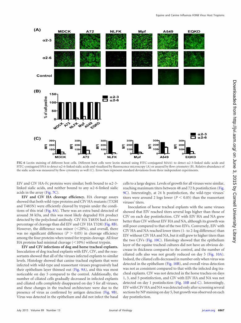
EIV and CIV receptor specificity. To determine if the differ-ence in infectivity of various host cells was due to virus receptoravailability and/or differences, cells were lectin stained and visu-alized by microscopy and fluorescence was measured by flow cy-tometry (Fig. 6). MDCK, A72, A549, and EQKD cells all stainedpositive for both �2-3- and �2-6-linked sialic acids, while NLFKand Mpf cells were stained predominately for �2-3-linked sialicacids (Fig. 6A and B). Overall, there was a higher relative concen-tration of �2-3-linked sialic acids compared to �2-6-linked sialicacids in all cell lines (Fig. 6C). CHO cells expressed only �2-3-linked sialic acids, while CHO cells stably transfected with �2-6sialyltransferase (6H4) expressed both �2-3- and �2-6-linkedsialic acids (Fig. 7A). Viruses were used to inoculate these cells(MOI � 1), and infectivity was assayed by flow cytometry 24 hpostinfection. There was no significant difference (P � 0.05) ininfectivity among the viruses, but all viruses showed around a 50%reduction in their ability to infect 6H4 cells compared to CHOcells (Fig. 7B). To further analyze EIV and CIV receptor specific-ity, HA-Fc fusion proteins were generated and used to bind dif-ferent glycans on microarrays. The binding specificities for the
FIG 5 Quantification of EIV, CIV, and reassortant virus infections in different host cells, and PR8 infections of cells least permissive to EIV and CIV. Viruses wereused to inoculate different host cells at an MOI of 0.1 in the presence of 0.5 �g/ml of trypsin and quantified by flow cytometry at 24 h and 48 h postinfection (A).Similarly, PR8, EIV, CIV, and reassortant viruses were used to inoculate A549 and EQKD cells at an MOI of 0.05 in the in the presence of 0.5 �g/ml of trypsin andvisualized by fluorescence microscopy (anti-NP staining) 24 h postinfection (B). Error bars represent standard deviations from three independent experiments.
Feng et al.
6866 jvi.asm.org July 2015 Volume 89 Number 13Journal of Virology
on June 3, 2015 by Cornell U
niversity Libraryhttp://jvi.asm
.org/D
ownloaded from
EIV and CIV HA-Fc proteins were similar; both bound to �2-3-linked sialic acids, and neither bound to any �2-6-linked sialicacids in the array (Fig. 7C).
EIV and CIV HA cleavage efficiency. HA cleavage assaysshowed that both wild-type proteins and CIV HA mutants (T328Iand T483N) were efficiently cleaved by trypsin under the condi-tions of this trial (Fig. 8A). There was an extra band detected ofaround 38 kDa, and this was most likely degraded HA productdetected by the polyclonal antibody. CIV HA T483N had a lowerpercentage of cleavage than did EIV and CIV HA T328I (Fig. 8B).However, the difference was minor (20%), and overall, therewas no significant difference (P � 0.05) in cleavage efficiencyamong the four proteins when tested for trypsin cleavage. All fourHA proteins had minimal cleavage (10%) without trypsin.
EIV and CIV infections of dog and horse tracheal explants.Inoculation of dog tracheal explants with EIV, CIV, and the reas-sortants showed that all of the viruses infected explants to similarlevels. Histology showed that canine tracheal explants that wereinfected with wild-type and reassortant viruses progressively hadtheir epithelium layer thinned out (Fig. 9A), and this was mostnoticeable on day 5 compared to the control. Additionally, thenumber of ciliated cells gradually decreased in infected explantsand ciliated cells completely disappeared on day 5 for all viruses,and these changes in the tracheal architecture were due to thepresence of virus as confirmed by antigen detection (Fig. 9B).Virus was detected in the epithelium and did not infect the basal
cells to a large degree. Levels of growth for all viruses were similar,reaching maximum titers between 48 and 72 h postinfection (Fig.9C). Interestingly, at 24 h postinfection, the wild-type viruses’titers were around 2 logs lower (P 0.05) than the reassortantviruses’ titers.
Inoculation of horse tracheal explants with the same virusesshowed that EIV reached titers several logs higher than those ofCIV on each day postinfection. CIV with EIV HA and NA grewbetter than CIV without EIV HA and NA, although its growth wasstill poor compared to that of the two EIVs. Conversely, EIV withCIV HA and NA reached lower titers (1- to 2-log difference) thanEIV without CIV HA and NA, but it still grew to higher titers thanthe two CIVs (Fig. 10C). Histology showed that the epitheliumlayer of the equine tracheal cultures did not have an obvious de-crease in thickness compared to the control, and the number ofciliated cells also was not greatly reduced on day 5 (Fig. 10A).Indeed, the ciliated cells decreased in number only when virus wasdetected in the epithelium (Fig. 10B), and overall virus detectionwas not as consistent compared to that with the infected dog tra-cheal explants. CIV was not detected in the horse trachea on days1, 3, and 5 postinfection, and CIV with EIV HA and NA was notdetected on day 1 postinfection (Fig. 10B and C). Interestingly,EIV with CIV HA and NA was detected only after screening severalsections by NP staining on day 5, but growth was observed on eachday postinfection.
FIG 6 Lectin staining of different host cells. Different host cells were lectin stained using FITC-conjugated MAA1 to detect �2-3-linked sialic acids andFITC-conjugated SNA to detect �2-6-linked sialic acids and visualized by fluorescence microscopy (A) or assayed by flow cytometry (B). Relative abundance ofthe sialic acids was measured by flow cytometry as well (C). Error bars represent standard deviations from three independent experiments.
Equine and Canine Influenza H3N8 Virus Host Tropisms
July 2015 Volume 89 Number 13 jvi.asm.org 6867Journal of Virology
on June 3, 2015 by Cornell U
niversity Libraryhttp://jvi.asm
.org/D
ownloaded from

DISCUSSIONGenetic differences in the EIV and CIV HA showed subtle dif-ferences in receptor specificity and cleavage efficiency. There arefive “signature” mutations that distinguish CIV HA from EIV HA:N54K, N83S, W222L, I328T, and N483T (Fig. 2) (25, 62). TheW222L mutation is located in the sialic acid receptor bindingpocket, and studies have shown that HA mutations in the 220 loopcan alter binding to �2-3- and �2-6-linked sialic acids (35, 49, 63).There is also evidence that the W222L mutation in CIV H3N2allowed the virus to infect dogs more efficiently (64), and so thespecies jump and subsequent adaptation of EIV H3N8 in dogsmay have been mediated by changes in receptor recognition aswell. However, no infectivity differences were seen between theviruses (or the reassortants) when those were inoculated intoCHO or into 6H4 cells. However, all viruses exhibited a 50% re-duction in their ability to infect 6H4 cells relative to CHO cells(Fig. 7B). This suggests that the viruses did not differ in theirrecognition of �2-3- and �2-6-linked sialic acids and in fact may
prefer �2-3-linked sialic acids for infections. Furthermore, therewere no significant differences in infectivity among viruses basedon the infection of various host cells, which further suggests min-imal differences in receptor specificity (Fig. 4 and 5A). Further-more, a recent study showed EIV and CIV bound strongly to�2-3- and not �2-6-linked sialic acids using sialic acid glycopoly-mers (62), consistent with our conclusion.
Binding of EIV and CIV HA-Fc proteins to glycan microarraysshowed support for the CHO and 6H4 cell infection results. In-deed, the two HA-Fc proteins bound strongly to �2-3-linked sialicacids in similar patterns, and neither protein bound to any �2-6-linked sialic acids (Fig. 7C). This preference toward binding to�2-3-linked (classical avian receptors) over �2-6-linked (classicalhuman receptors) sialic acids suggests a potential human hostrestriction barrier for both viruses. Interestingly, there were dif-ferences in the relative preferences for binding to the various �2-3-linked glycans. For example, EIV HA-Fc showed higher bindingtoNeu5Ac�3Gal3GlcNAc3Gal4GlcR1andNeu5Ac�3Gal4
FIG 7 EIV and CIV receptor specificity. Lectin staining showed that CHO cells expressed only �2-3-linked sialic acids, while 6H4 cells expressed both �2-3- and�2-6-linked sialic acids (A). EIV, CIV, and reassortant viruses were used to inoculate MDCK (control), CHO, and 6H4 cells at an MOI of 1 in the presence of 0.5�g/ml of trypsin. After 24 h postinfection, cells were stained for anti-NP and quantified by flow cytometry (B). EIV and CIV HA-Fc fusion proteins were used tobind �2-3- and �2-6-linked sialic acids on glycan microarrays (C). Error bars represent standard deviations from three independent experiments.
Feng et al.
6868 jvi.asm.org July 2015 Volume 89 Number 13Journal of Virology
on June 3, 2015 by Cornell U
niversity Libraryhttp://jvi.asm
.org/D
ownloaded from

(Fuc�3)GlcNAcR1, while CIV HA-Fc showed higher binding toNeu5Ac�3Gal4GlcNAc6SR1 and Neu5Ac�3GalR1. Thesefindings suggest that there are subtle differences between the abil-ities of EIV and CIV HAs to recognize specific sialic acids. Indeed,a recent study revealed the atomic structures of EIV and CIV HAto be nearly identical, and both proteins preferred binding to�2-3- over �2-6-linked sialic acids, but there were subtle differ-ences: CIV HA bound better to sulfated sialic acids than did EIVHA (65). Small differences such as these may be important forunderstanding virus host adaptation and tropism.
Previous research has suggested that the signature HA I328Tmutation, which is at the P2 position of the cleavage site, mayinfluence influenza virus HA cleavage (25, 62). However, we mu-tated the CIV HA to the EIV background, and it did not influenceprotein cleavage efficiency with and without trypsin compared tothat of the wild type under the conditions of our assay (Fig. 8B).We also tested whether the signature HA N483T mutation, a gly-cosylation site that is close to the cleavage site in the HA structure,might influence cleavage by sterically hindering protease activitydue to the presence of glycans. Changing the site in CIV to the EIVcodon did not dramatically influence HA cleavage efficiency with
and without trypsin (Fig. 8B). Overall, both EIV and CIV HArequired trypsin activation, and there were no major differences inefficiency of cleavage between the wild type and the mutant pro-teins. Different hosts have been shown to express different pro-teases that can cleave HA (66), and it is therefore possible thatthere are dog-specific proteases that have selected the I328T andN483T mutations in CIV HA which allow for better replication.Testing dog-specific proteases might provide further insight intoany differences in cleavage efficiency.
Other genetic differences between EIV and CIV may playroles in host adaptation and tropism. Although many mutations
FIG 8 EIV and CIV HA cleavage assays. Both wild-type HA proteins, CIV HAT328I, and CIV HA T483N were surface expressed and biotinylated. Westernblotting was used to look at HA0 cleavage into HA1 and HA2 (A). Cleavageefficiency was determined by band density of the Western blots and the fol-lowing formula: (HA1/HA0 � HA1) � 100 (B). Error bars represent standarddeviations from three independent experiments.
Day 1
Day 3
Day 5
Mock CIV EIVCIV withEIV HA+NA
EIV withCIV HA+NA
Day 1
Day 3
Day 5
Mock CIV EIVCIV withEIV HA+NA
EIV withCIV HA+NA
Hematoxylin and Eosin staining
Virus anti-NP staining
(A)
(B)
(C)
0 1 2 3 4 50
2
4
6
8EIVCIVEIV / CIV HA+NACIV / EIV HA+NA
Log
PFU/
ml
FIG 9 EIV, CIV, and reassortant virus infections of dog tracheal explants.Viruses (400 TCID50 units) were used to inoculate dog tracheal explants,and tissues were collected for hematoxylin and eosin staining (A) and foranti-NP staining (B) at days 1, 3, and 5 postinfection. Virus growth wasassayed every 24 h for 5 days by plaque assays in MDCK cells (C). Error barsrepresent standard deviations from three independent experiments. Blackscale bars represent 50 �m.
Equine and Canine Influenza H3N8 Virus Host Tropisms
July 2015 Volume 89 Number 13 jvi.asm.org 6869Journal of Virology
on June 3, 2015 by Cornell U
niversity Libraryhttp://jvi.asm
.org/D
ownloaded from

documented here reside in known functional domains (Fig. 2), itis still unclear whether the specific mutations actually affect influ-enza virus host adaptation and replication or contribute to anyhost-specific functions. Interestingly, most (8 of 9) of the muta-tions in PB1 occurred between early and more recent CIV se-quences (Fig. 2), whereas in the other proteins most mutationsoccurred between EIV and the earliest (2003 or 2004) CIV isolates(PB2, PA, and HA), or there was no difference between EIV andthe two CIV groups (NP, NA, M1, and NS1) (Fig. 2). Whethermutations that occurred between early and more recent CIV iso-lates indicate adaptive changes to its canine host remains to beelucidated. Of the PB2 mutations, one (I292T) has been suggestedto facilitate human H1N1 and H3N2 adaptation (60). Conse-
quently, its possible role in dog adaptation needs to be considered.One mutation in PA (T400A) has also been reported to distin-guish between human and avian influenza viruses. Human virusesalmost always have a leucine at this site, while avian viruses showeither serine or proline (61), such that it may represent anotherhost range determinant in CIV. In addition, we found that EIVNS1 can be naturally truncated by 11 amino acids at the C termi-nus, while CIV NS1 is never truncated, except for the first CIVisolate. A previous study showed the 2009 human pandemicH1N1 virus also had an 11-amino-acid truncation which resultedin inefficient suppression of host genes, and extension of NS1 tofull length restored its binding to host poly(A)-binding protein II,which increased NS1 host gene suppression activity (67). As men-tioned above, the mutation at the N terminus of PA could affectPA-X host gene suppression. Furthermore, both CIVs (H3N8 andH3N2) have a 20-amino-acid truncation at the C terminus ofPA-X, implying that the truncation may be a host determinant indogs (57). In fact, a comparison of human full-length and trun-cated PA-X revealed a difference in gene suppression activity (58).Taken together, these genetic differences between EIV and CIVmay be a consequence of dog-specific host pressures, includingthose generated by the innate and adaptive immune responses.
EIV and CIV did not show infectivity differences in varioushost cells. Despite the relatively high number of mutations ob-served in the virus genomes, wild-type and reassortant virusesgrew similarly in dog (MDCK) cells, which are generally consid-ered the most susceptible cells for influenza viruses (Fig. 3). Othermammalian cells tested showed differences in susceptibility to in-fection (Fig. 4). The reassortant viruses appeared similar to thewild types, which was not surprising since the wild-type virusesdid not show any infectivity differences. Interestingly, the virusesinfected human (A549) and horse (EQKD) cells poorly (Fig. 4 and5A). Previous results have indicated there is a host barrier forrespiratory infection of CIV in horses (36, 62, 68, 69), so a lowinfectivity for CIV in EQKD cells may reflect a host difference thatis also seen in kidney cells. However, EIV also infected EQKD cellspoorly, so the block in infection may be due to biological differ-ences between horse kidney and airway respiratory cells. The poorinfection of CIV in A549 cells was interesting because there havebeen no reported cases of CIV (or EIV) naturally transmitting tohumans, including people who were regularly exposed to CIV-infected dogs (70); whether there is a correlation between the poorinfectivity in A549 cells and virus transmission to humans is un-known. Interestingly, the highly laboratory-adapted strain PR8was able to infect both A549 and EQKD cells to high levels com-pared to those of the horse and dog viruses (Fig. 5B). This suggeststhat both cell types were permissive to influenza virus infectionand that the limited infection from EIV and CIV was attributableto those specific viruses’ biology. The poor infectivity in these cellscould not be explained by sialic acid linkages, because both A549and EQKD cells expressed high levels of both �2-3- and �2-6-linked sialic acids (Fig. 6), similar to cells that were permissive toinfection. Indeed, the infectivity differences between host cellswere likely not due to differences in sialic acid binding, becauseEIV and CIV HAs bound to sialic acid similarly, as describedabove. The viruses were able to infect cat (NLFK) and ferret (Mpf)cells, although the infection in ferret cells was poorer (Fig. 4). Bothcells stained positive predominately for �2-3-linked sialic acids(Fig. 6), so this suggests that the infectivity difference betweenNLFK and Mpf cells was likely not due to receptor differences.
Day 1
Day 3
Day 5
Mock CIV EIVCIV withEIV HA+NA
EIV withCIV HA+NA
Day 1
Day 3
Day 5
Mock CIV EIVCIV withEIV HA+NA
EIV withCIV HA+NA
(A)
(B)
(C)
0 1 2 3 4 50
2
4
6
8
10EIVCIVEIV / CIV HA+NACIV / EIV HA+NA
Log
PFU/
ml
Hematoxylin and Eosin staining
Virus anti-NP staining
FIG 10 EIV, CIV, and reassortant virus infections of horse tracheal ex-plants. Viruses (400 TCID50 units) were used to inoculate dog trachealexplants, and tissues were collected for hematoxylin and eosin staining (A)and for anti-NP staining (B) at days 1, 3, and 5 postinfection. Virus growthwas assayed every 24 h for 5 days by plaque assays in MDCK cells (C). Blackscale bars represent 50 �m.
Feng et al.
6870 jvi.asm.org July 2015 Volume 89 Number 13Journal of Virology
on June 3, 2015 by Cornell U
niversity Libraryhttp://jvi.asm
.org/D
ownloaded from

These results were interesting because ferrets are susceptible toinfluenza and are used as a model for human virus infection andtransmission (71), and there is already evidence that CIV H3N2can infect cats (72, 73). Additionally, a previous study showed thatEIV replicated in the upper respiratory tract in live ferrets but wasrestricted in the lungs (74). There are no extended analysis orreports of CIV H3N8 infections in ferrets, but seroconversion hasbeen observed after inoculating ferrets (25). Interestingly, there isevidence of limited CIV H3N2 infection and transmission be-tween ferrets in laboratory settings, but the virus could not trans-mit from dogs to ferrets (75, 76). Taken together, these findingsshow that there is potential for EIV and CIV to infect other mam-malian hosts and subsequently adapt.
EIV and CIV infection of tracheal explants revealed a host-specific barrier. To simulate the natural environment in whichEIV and CIV cause infections, dog and horse tracheal explantcultures were prepared and used for virus infections. Overall, theresults showed that the viruses could infect dog trachea, and therewere no major differences among the viruses with respect to dam-aging the epithelium layer or the location of virus antigens in thetracheal explants (Fig. 9). The reassortant viruses reached a greatertiter (around 2 logs) than did the wild types at 24 h postinfection,and this suggests that the protein mismatching altered viral biol-ogy (Fig. 9C). However, the change was not dramatic overall, be-cause growth levels were very similar after 24 h postinfection,reaching peak titer between 48 and 72 h postinfection, similar togrowth in MDCK cells (Fig. 3). In horse trachea all viruses causedsome level of infection, but the physical damage of the epitheliumwas less pronounced than with the infections of the dog trachea,which may be due to the horse trachea being more structurallyrobust (Fig. 10). Interestingly, EIV grew much better than CIV,which provides further evidence of a host-specific barrier (Fig.10C). Replacing the CIV HA and NA with EIV HA and NA al-lowed the virus to grow slightly better and stabilized (detectedvirus on days 3 and 5 postinfection) the virus compared to thewild-type CIV. Furthermore, replacing the EIV HA and NA withCIV HA and NA attenuated virus growth by around 1 to 2 logsacross 5 days compared to that of the wild-type EIV. These find-ings suggest that the difference in growth between EIV and CIV inhorse trachea may be attributed to the mutations in the glycopro-teins. Overall, the reassortant viruses grew similarly with respectto their wild-type counterparts, and the experiment was carriedout in one experimental replicate; thus, further experimentationusing horse trachea and reassortant viruses is needed for valida-tion. Also, the apparent attenuation of growth of CIV and EIVwith CIV glycoproteins in horse trachea compared to dog tracheamay not have been caused by differences in �2-3- and �2-6-linkedsialic acids, as past studies have shown the existence of both in dogand horse tracheal tissues (77, 78). However, the distributionswere shown to be different. While horse trachea epithelial cellsstained positive for both sialic acids, dog trachea epithelial cellsshowed stronger staining for �2-3-linked sialic acids. Interest-ingly, the lamina propria of dog trachea stained positive for both.However, it is important to note that sialic acid distribution, va-riety, and presence can vary depending on individual animals andtheir age (79).
Overall, these results showed that despite 6 years of continuousevolution in dogs that separated the two viruses tested in thisstudy, and the accumulation of mutations in all of the genomicsegments such that EIV and CIV are clearly phylogenetically dis-
tinct (Fig. 1), there appeared to be minimal biological differencesbetween them. We showed that the viruses infected various hostcells with no infectivity differences among the viruses, althoughdifferent host cells exhibited various degrees of permissiveness.We also showed that the viruses preferred �2-3- over �2-6-linkedsialic acid receptors, and there may be subtle differences in recep-tor recognition. Virus inoculation in tracheal explants revealedlimited CIV infectivity in horse trachea, and the restriction factorsmay reside in the receptor binding proteins. Notably, althoughEIV has circulated in many parts of the world since it emerged in1963, CIV has not spread in a sustained fashion beyond NorthAmerica, where it has been maintained mainly in large, high-turn-over animal shelters. Given the scarce phenotypic differences be-tween EIV and CIV, the relatively limited spread of CIV amongthe domestic dog population may reflect a lack of epidemiologicalcontacts rather than constraints on viral fitness. This suggests thatinterspecies transmission and adaptation of influenza virus in thiscase are mediated by subtle factors.
ACKNOWLEDGMENTS
This work was supported by NIH/NIGMS grant R01 GM080533 to C.R.P.and E.C.H. and by grant R01GM32373 to A.V. E.C.H. is supported by anNHMRC Australia Fellowship (AF30). K.H.F. is supported by an NSFGraduate Research Fellowship. P.R.M. and G.G. are supported by theMedical Research Council of the United Kingdom (grant numberG0801822).
REFERENCES1. Parrish CR, Kawaoka Y. 2005. The origins of new pandemic viruses:
the acquisition of new host ranges by canine parvovirus and influenzaA viruses. Annu Rev Microbiol 59:553–586. http://dx.doi.org/10.1146/annurev.micro.59.030804.121059.
2. Morens DM, Taubenberger JK. 2010. Historical thoughts on influenzaviral ecosystems, or behold a pale horse, dead dogs, failing fowl, and sickswine. Influenza Other Respir Viruses 4:327–337. http://dx.doi.org/10.1111/j.1750-2659.2010.00148.x.
3. Crawford PC, Dubovi EJ, Castleman WL, Stephenson I, Gibbs EP,Chen L, Smith C, Hill RC, Ferro P, Pompey J, Bright RA, Medina MJ,Johnson CM, Olsen CW, Cox NJ, Klimov AI, Katz JM, Donis RO. 2005.Transmission of equine influenza virus to dogs. Science 310:482– 485.http://dx.doi.org/10.1126/science.1117950.
4. Anthony SJ, St Leger JA, Pugliares K, Ip HS, Chan JM, Carpenter ZW,Navarrete-Macias I, Sanchez-Leon M, Saliki JT, Pedersen J, Karesh W,Daszak P, Rabadan R, Rowles T, Lipkin WI. 2012. Emergence of fatalavian influenza in New England harbor seals. mBio 3(4):e00166 –12. http://dx.doi.org/10.1128/mBio.00166-12.
5. Yoon KJ, Schwartz K, Sun D, Zhang J, Hildebrandt H. 2012. Naturallyoccurring influenza A virus subtype H1N2 infection in a Midwest UnitedStates mink (Mustela vison) ranch. J Vet Diagn Invest 24:388 –391. http://dx.doi.org/10.1177/1040638711428349.
6. Ince WL, Gueye-Mbaye A, Bennink JR, Yewdell JW. 2013. Reassortmentcomplements spontaneous mutation in influenza A virus NP and M1genes to accelerate adaptation to a new host. J Virol 87:4330 – 4338. http://dx.doi.org/10.1128/JVI.02749-12.
7. Mänz B, Schwemmle M, Brunotte L. 2013. Adaptation of avian influenzaA virus polymerase in mammals to overcome the host species barrier. JVirol 87:7200 –7209. http://dx.doi.org/10.1128/JVI.00980-13.
8. Resa-Infante P, Gabriel G. 2013. The nuclear import machinery is adeterminant of influenza virus host adaptation. Bioessays 35:23–27. http://dx.doi.org/10.1002/bies.201200138.
9. Rose N, Herve S, Eveno E, Barbier N, Eono F, Dorenlor V, Andraud M,Camsusou C, Madec F, Simon G. 2013. Dynamics of influenza A virusinfections in permanently infected pig farms: evidence of recurrent infec-tions, circulation of several swine influenza viruses and reassortmentevents. Vet Res 44:72. http://dx.doi.org/10.1186/1297-9716-44-72.
10. Trebbien R, Bragstad K, Larsen LE, Nielsen J, Botner A, Heegaard PM,Fomsgaard A, Viuff B, Hjulsager CK. 2013. Genetic and biological
Equine and Canine Influenza H3N8 Virus Host Tropisms
July 2015 Volume 89 Number 13 jvi.asm.org 6871Journal of Virology
on June 3, 2015 by Cornell U
niversity Libraryhttp://jvi.asm
.org/D
ownloaded from

characterisation of an avian-like H1N2 swine influenza virus generated byreassortment of circulating avian-like H1N1 and H3N2 subtypes in Den-mark. Virol J 10:290. http://dx.doi.org/10.1186/1743-422X-10-290.
11. Imai M, Kawaoka Y. 2012. The role of receptor binding specificity ininterspecies transmission of influenza viruses. Curr Opin Virol 2:160 –167. http://dx.doi.org/10.1016/j.coviro.2012.03.003.
12. Sassaki GL, Elli S, Rudd TR, Macchi E, Yates EA, Naggi A, Shriver Z,Raman R, Sasisekharan R, Torri G, Guerrini M. 2013. Human (al-pha2¡6) and avian (alpha2¡3) sialylated receptors of influenza A virusshow distinct conformations and dynamics in solution. Biochemistry 52:7217–7230. http://dx.doi.org/10.1021/bi400677n.
13. Shichinohe S, Okamatsu M, Sakoda Y, Kida H. 2013. Selection of H3avian influenza viruses with SAalpha2,6Gal receptor specificity in pigs.Virology 444:404 – 408. http://dx.doi.org/10.1016/j.virol.2013.07.007.
14. Chen Q, Huang S, Chen J, Zhang S, Chen Z. 2013. NA proteins ofinfluenza A viruses H1N1/2009, H5N1, and H9N2 show differential ef-fects on infection initiation, virus release, and cell-cell fusion. PLoS One8:e54334. http://dx.doi.org/10.1371/journal.pone.0054334.
15. Le QM, Sakai-Tagawa Y, Ozawa M, Ito M, Kawaoka Y. 2009. Selectionof H5N1 influenza virus PB2 during replication in humans. J Virol 83:5278 –5281. http://dx.doi.org/10.1128/JVI.00063-09.
16. Yan S, Wu G. 2010. Evidence for cross-species infections and cross-subtype mutations in influenza A matrix proteins. Viral Immunol 23:105–111. http://dx.doi.org/10.1089/vim.2009.0080.
17. Dankar SK, Wang S, Ping J, Forbes NE, Keleta L, Li Y, Brown EG. 2011.Influenza A virus NS1 gene mutations F103L and M106I increase replicationand virulence. Virol J 8:13. http://dx.doi.org/10.1186/1743-422X-8-13.
18. Sakabe S, Ozawa M, Takano R, Iwastuki-Horimoto K, Kawaoka Y.2011. Mutations in PA, NP, and HA of a pandemic (H1N1) 2009 influenzavirus contribute to its adaptation to mice. Virus Res 158:124 –129. http://dx.doi.org/10.1016/j.virusres.2011.03.022.
19. Kiseleva IV, Voeten JT, Teley LC, Larionova NV, Drieszen-van der Crui-jsen SK, Basten SM, Heldens JG, van den Bosch H, Rudenko LG. 2010. PB2and PA genes control the expression of the temperature-sensitive phenotypeof cold-adapted B/USSR/60/69 influenza master donor virus. J Gen Virol91:931–937. http://dx.doi.org/10.1099/vir.0.017996-0.
20. Chou YY, Albrecht RA, Pica N, Lowen AC, Richt JA, Garcia-Sastre A,Palese P, Hai R. 2011. The M segment of the 2009 new pandemic H1N1influenza virus is critical for its high transmission efficiency in the guinea pigmodel. J Virol 85:11235–11241. http://dx.doi.org/10.1128/JVI.05794-11.
21. Turan K, Mibayashi M, Sugiyama K, Saito S, Numajiri A, Nagata K.2004. Nuclear MxA proteins form a complex with influenza virus NP andinhibit the transcription of the engineered influenza virus genome. Nu-cleic Acids Res 32:643– 652. http://dx.doi.org/10.1093/nar/gkh192.
22. Kim SH, Samal SK. 2010. Inhibition of host innate immune responsesand pathogenicity of recombinant Newcastle disease viruses expressingNS1 genes of influenza A viruses. J Gen Virol 91:1996 –2001. http://dx.doi.org/10.1099/vir.0.021766-0.
23. Gao S, Song L, Li J, Zhang Z, Peng H, Jiang W, Wang Q, Kang T, ChenS, Huang W. 2012. Influenza A virus-encoded NS1 virulence factor pro-tein inhibits innate immune response by targeting IKK. Cell Microbiol14:1849 –1866. http://dx.doi.org/10.1111/cmi.12005.
24. Gibbs EP, Anderson TC. 2010. Equine and canine influenza: a review ofcurrent events. Anim Health Res Rev 11:43–51. http://dx.doi.org/10.1017/S1466252310000046.
25. Payungporn S, Crawford PC, Kouo TS, Chen LM, Pompey J, CastlemanWL, Dubovi EJ, Katz JM, Donis RO. 2008. Influenza A virus (H3N8) indogs with respiratory disease, Florida. Emerg Infect Dis 14:902–908. http://dx.doi.org/10.3201/eid1406.071270.
26. Rivailler P, Perry IA, Jang Y, Davis CT, Chen LM, Dubovi EJ, DonisRO. 2010. Evolution of canine and equine influenza (H3N8) viruses co-circulating between 2005 and 2008. Virology 408:71–79. http://dx.doi.org/10.1016/j.virol.2010.08.022.
27. Jirjis FF, Deshpande MS, Tubbs AL, Jayappa H, Lakshmanan N, Was-moen TL. 2010. Transmission of canine influenza virus (H3N8) amongsusceptible dogs. Vet Microbiol 144:303–309. http://dx.doi.org/10.1016/j.vetmic.2010.02.029.
28. Hayward JJ, Dubovi EJ, Scarlett JM, Janeczko S, Holmes EC, ParrishCR. 2010. Microevolution of canine influenza virus in shelters and itsmolecular epidemiology in the United States. J Virol 84:12636 –12645.http://dx.doi.org/10.1128/JVI.01350-10.
29. Holt DE, Mover MR, Brown DC. 2010. Serologic prevalence of antibod-ies against canine influenza virus (H3N8) in dogs in a metropolitan animal
shelter. J Am Vet Med Assoc 237:71–73. http://dx.doi.org/10.2460/javma.237.1.71.
30. Li S, Shi Z, Jiao P, Zhang G, Zhong Z, Tian W, Long LP, Cai Z, Zhu X,Liao M, Wan XF. 2010. Avian-origin H3N2 canine influenza A viruses inSouthern China. Infect Genet Evol 10:1286 –1288. http://dx.doi.org/10.1016/j.meegid.2010.08.010.
31. Zeng XJ, Lin Y, Zhao YB, Lu CP, Liu YJ. 2013. Experimental infectionof dogs with H3N2 canine influenza virus from China. Epidemiol Infect141:2595–2603. http://dx.doi.org/10.1017/S0950268813000472.
32. Lim SF, Lee MM, Zhang P, Song Z. 2008. The Golgi CMP-sialic acidtransporter: a new CHO mutant provides functional insights. Glycobiol-ogy 18:851– 860. http://dx.doi.org/10.1093/glycob/cwn080.
33. Bragonzi A, Distefano G, Buckberry LD, Acerbis G, Foglieni C, LamotteD, Campi G, Marc A, Soria MR, Jenkins N, Monaco L. 2000. A newChinese hamster ovary cell line expressing alpha2,6-sialyltransferase usedas universal host for the production of human-like sialylated recombinantglycoproteins. Biochim Biophys Acta 1474:273–282. http://dx.doi.org/10.1016/S0304-4165(00)00023-4.
34. Zhou B, Donnelly ME, Scholes DT, St George K, Hatta M, Kawaoka Y,Wentworth DE. 2009. Single-reaction genomic amplification acceleratessequencing and vaccine production for classical and Swine origin humaninfluenza A viruses. J Virol 83:10309 –10313. http://dx.doi.org/10.1128/JVI.01109-09.
35. Pawar SD, Parkhi SS, Koratkar SS, Mishra AC. 2012. Receptor specificityand erythrocyte binding preferences of avian influenza viruses isolatedfrom India. Virol J 9:251. http://dx.doi.org/10.1186/1743-422X-9-251.
36. Quintana AM, Hussey SB, Burr EC, Pecoraro HL, Annis KM, Rao S,Landolt GA. 2011. Evaluation of infectivity of a canine lineage H3N8influenza A virus in ponies and in primary equine respiratory epithelialcells. Am J Vet Res 72:1071–1078. http://dx.doi.org/10.2460/ajvr.72.8.1071.
37. Kearse M, Moir R, Wilson A, Stones-Havas S, Cheung M, Sturrock S,Buxton S, Cooper A, Markowitz S, Duran C, Thierer T, Ashton B,Meintjes P, Drummond A. 2012. Geneious Basic: an integrated andextendable desktop software platform for the organization and analysis ofsequence data. Bioinformatics 28:1647–1649. http://dx.doi.org/10.1093/bioinformatics/bts199.
38. Guindon S, Dufayard JF, Lefort V, Anisimova M, Hordijk W, GascuelO. 2010. New algorithms and methods to estimate maximum-likelihoodphylogenies: assessing the performance of PhyML 3.0. Syst Biol 59:307–321. http://dx.doi.org/10.1093/sysbio/syq010.
39. Ayora-Talavera G, Shelton H, Scull MA, Ren J, Jones IM, Pickles RJ,Barclay WS. 2009. Mutations in H5N1 influenza virus hemagglutinin thatconfer binding to human tracheal airway epithelium. PLoS One 4:e7836.http://dx.doi.org/10.1371/journal.pone.0007836.
40. Shelton H, Ayora-Talavera G, Ren J, Loureiro S, Pickles RJ, Barclay WS,Jones IM. 2011. Receptor binding profiles of avian influenza virus hem-agglutinin subtypes on human cells as a predictor of pandemic potential. JVirol 85:1875–1880. http://dx.doi.org/10.1128/JVI.01822-10.
41. Deng L, Song J, Gao X, Wang J, Yu H, Chen X, Varki N, Naito-MatsuiY, Galan JE, Varki A. 2014. Host adaptation of a bacterial toxin from thehuman pathogen Salmonella Typhi. Cell 159:1290 –1299. http://dx.doi.org/10.1016/j.cell.2014.10.057.
42. Deng L, Bensing BA, Thamadilok S, Yu H, Lau K, Chen X, Ruhl S,Sullam PM, Varki A. 2014. Oral streptococci utilize a Siglec-like domainof serine-rich repeat adhesins to preferentially target platelet sialoglycansin human blood. PLoS Pathog 10:e1004540. http://dx.doi.org/10.1371/journal.ppat.1004540.
43. Tse LV, Marcano VC, Huang W, Pocwierz MS, Whittaker GR. 2013.Plasmin-mediated activation of pandemic H1N1 influenza virus hemag-glutinin is independent of the viral neuraminidase. J Virol 87:5161–5169.http://dx.doi.org/10.1128/JVI.00210-13.
44. Gonzalez G, Marshall JF, Morrell J, Robb D, McCauley JW, Perez DR,Parrish CR, Murcia PR. 2014. Infection and pathogenesis of canine,equine, and human influenza viruses in canine tracheas. J Virol 88:9208 –9219. http://dx.doi.org/10.1128/JVI.00887-14.
45. Nunes SF, Murcia PR, Tiley LS, Brown IH, Tucker AW, Maskell DJ,Wood JL. 2010. An ex vivo swine tracheal organ culture for the study ofinfluenza infection. Influenza Other Respir Viruses 4:7–15. http://dx.doi.org/10.1111/j.1750-2659.2009.00119.x.
46. Anderton TL, Maskell DJ, Preston A. 2004. Ciliostasis is a key early eventduring colonization of canine tracheal tissue by Bordetella bronchiseptica.Microbiology 150:2843–2855. http://dx.doi.org/10.1099/mic.0.27283-0.
Feng et al.
6872 jvi.asm.org July 2015 Volume 89 Number 13Journal of Virology
on June 3, 2015 by Cornell U
niversity Libraryhttp://jvi.asm
.org/D
ownloaded from

47. Wiley DC, Wilson IA, Skehel JJ. 1981. Structural identification of theantibody-binding sites of Hong Kong influenza haemagglutinin and theirinvolvement in antigenic variation. Nature 289:373–378. http://dx.doi.org/10.1038/289373a0.
48. Stray SJ, Pittman LB. 2012. Subtype- and antigenic site-specific differ-ences in biophysical influences on evolution of influenza virus hemagglu-tinin. Virol J 9:91. http://dx.doi.org/10.1186/1743-422X-9-91.
49. Lin YP, Xiong X, Wharton SA, Martin SR, Coombs PJ, Vachieri SG,Christodoulou E, Walker PA, Liu J, Skehel JJ, Gamblin SJ, Hay AJ,Daniels RS, McCauley JW. 2012. Evolution of the receptor binding prop-erties of the influenza A(H3N2) hemagglutinin. Proc Natl Acad Sci U S A109:21474 –21479. http://dx.doi.org/10.1073/pnas.1218841110.
50. Sun X, Tse LV, Ferguson AD, Whittaker GR. 2010. Modifications tothe hemagglutinin cleavage site control the virulence of a neurotropicH1N1 influenza virus. J Virol 84:8683– 8690. http://dx.doi.org/10.1128/JVI.00797-10.
51. Wu Y, Qin G, Gao F, Liu Y, Vavricka CJ, Qi J, Jiang H, Yu K, Gao GF.2013. Induced opening of influenza virus neuraminidase N2 150-loopsuggests an important role in inhibitor binding. Sci Rep 3:1551. http://dx.doi.org/10.1038/srep01551.
52. Portela A, Digard P. 2002. The influenza virus nucleoprotein: a multi-functional RNA-binding protein pivotal to virus replication. J Gen Virol83:723–734.
53. Noton SL, Medcalf E, Fisher D, Mullin AE, Elton D, Digard P. 2007.Identification of the domains of the influenza A virus M1 matrix proteinrequired for NP binding, oligomerization and incorporation into virions.J Gen Virol 88:2280 –2290. http://dx.doi.org/10.1099/vir.0.82809-0.
54. Hale BG, Randall RE, Ortin J, Jackson D. 2008. The multifunctional NS1protein of influenza A viruses. J Gen Virol 89:2359 –2376. http://dx.doi.org/10.1099/vir.0.2008/004606-0.
55. Boivin S, Cusack S, Ruigrok RW, Hart DJ. 2010. Influenza A viruspolymerase: structural insights into replication and host adaptation mech-anisms. J Biol Chem 285:28411–28417. http://dx.doi.org/10.1074/jbc.R110.117531.
56. Yewdell JW, Ince WL. 2012. Virology frameshifting to PA-X influenza.Science 337:164 –165. http://dx.doi.org/10.1126/science.1225539.
57. Shi M, Jagger BW, Wise HM, Digard P, Holmes EC, Taubenberger JK.2012. Evolutionary conservation of the PA-X open reading frame in seg-ment 3 of influenza A virus. J Virol 86:12411–12413. http://dx.doi.org/10.1128/JVI.01677-12.
58. Desmet EA, Bussey KA, Stone R, Takimoto T. 2013. Identification of theN-terminal domain of the influenza virus PA responsible for the suppres-sion of host protein synthesis. J Virol 87:3108 –3118. http://dx.doi.org/10.1128/JVI.02826-12.
59. Khaperskyy DA, Emara MM, Johnston BP, Anderson P, Hatchette TF,McCormick C. 2014. Influenza A virus host shutoff disables antiviralstress-induced translation arrest. PLoS Pathog 10:e1004217. http://dx.doi.org/10.1371/journal.ppat.1004217.
60. Miotto O, Heiny A, Tan TW, August JT, Brusic V. 2008. Identificationof human-to-human transmissibility factors in PB2 proteins of influenzaA by large-scale mutual information analysis. BMC Bioinformatics9(Suppl 1):S18. http://dx.doi.org/10.1186/1471-2105-9-S1-S18.
61. Allen JE, Gardner SN, Vitalis EA, Slezak TR. 2009. Conserved aminoacid markers from past influenza pandemic strains. BMC Microbiol 9:77.http://dx.doi.org/10.1186/1471-2180-9-77.
62. Pecoraro HL, Bennett S, Garretson K, Quintana AM, Lunn KF, LandoltGA. 2013. Comparison of the infectivity and transmission of contempo-rary canine and equine H3N8 influenza viruses in dogs. Vet Med Int 2013:874521. http://dx.doi.org/10.1155/2013/874521.
63. Wu C, Cheng X, Wang X, Lv X, Yang F, Liu T, Fang S, Zhang R,Jinquan C. 2013. Clinical and molecular characteristics of the 2009 pan-demic influenza H1N1 infection with severe or fatal disease from 2009 to2011 in Shenzhen, China. J Med Virol 85:405– 412. http://dx.doi.org/10.1002/jmv.23295.
64. Yang G, Li S, Blackmon S, Ye J, Bradley KC, Cooley J, Smith D, Hanson
L, Cardona C, Steinhauer DA, Webby R, Liao M, Wan XF. 2013.Mutation tryptophan to leucine at position 222 of haemagglutinin couldfacilitate H3N2 influenza A virus infection in dogs. J Gen Virol 94:2599 –2608. http://dx.doi.org/10.1099/vir.0.054692-0.
65. Collins PJ, Vachieri SG, Haire LF, Ogrodowicz RW, Martin SR, WalkerPA, Xiong X, Gamblin SJ, Skehel JJ. 2014. Recent evolution of equineinfluenza and the origin of canine influenza. Proc Natl Acad Sci U S A111:11175–11180. http://dx.doi.org/10.1073/pnas.1406606111.
66. Peitsch C, Klenk HD, Garten W, Bottcher-Friebertshauser E. 2014.Activation of influenza A viruses by host proteases from swine airwayepithelium. J Virol 88:282–291. http://dx.doi.org/10.1128/JVI.01635-13.
67. Tu J, Guo J, Zhang A, Zhang W, Zhao Z, Zhou H, Liu C, Chen H, JinM. 2011. Effects of the C-terminal truncation in NS1 protein of the 2009pandemic H1N1 influenza virus on host gene expression. PLoS One6:e26175. http://dx.doi.org/10.1371/journal.pone.0026175.
68. Yamanaka T, Tsujimura K, Kondo T, Matsumura T, Ishida H, Kiso M,Hidari KI, Suzuki T. 2010. Infectivity and pathogenicity of canine H3N8influenza A virus in horses. Influenza Other Respir Viruses 4:345–351.http://dx.doi.org/10.1111/j.1750-2659.2010.00157.x.
69. Yamanaka T, Nemoto M, Bannai H, Tsujimura K, Kondo T, Mat-sumura T, Muranaka M, Ueno T, Kinoshita Y, Niwa H, Hidari KI,Suzuki T. 2012. No evidence of horizontal infection in horses kept inclose contact with dogs experimentally infected with canine influenzaA virus (H3N8). Acta Vet Scand 54:25. http://dx.doi.org/10.1186/1751-0147-54-25.
70. Krueger WS, Heil GL, Yoon KJ, Gray GC. 2014. No evidence for zoo-notic transmission of H3N8 canine influenza virus among US adults oc-cupationally exposed to dogs. Influenza Other Respir Viruses 8:99 –106.http://dx.doi.org/10.1111/irv.12208.
71. Thangavel RR, Bouvier NM. 2014. Animal models for influenza viruspathogenesis, transmission, and immunology. J Immunol Methods 410:60 –79. http://dx.doi.org/10.1016/j.jim.2014.03.023.
72. Park SJ, Kang BK, Jeoung HY, Moon HJ, Hong M, Na W, Park BK, PooH, Kim JK, An DJ, Song DS. 2013. Complete genome sequence of acanine-origin H3N2 feline influenza virus isolated from domestic cats inSouth Korea. Genome Announc 1(2):e00253–12. http://dx.doi.org/10.1128/genomeA.00253-12.
73. Jeoung HY, Lim SI, Shin BH, Lim JA, Song JY, Song DS, Kang BK,Moon HJ, An DJ. 2013. A novel canine influenza H3N2 virus isolatedfrom cats in an animal shelter. Vet Microbiol 165:281–286. http://dx.doi.org/10.1016/j.vetmic.2013.03.021.
74. Baz M, Paskel M, Matsuoka Y, Zengel J, Cheng X, Jin H, Subbarao K.2013. Replication and immunogenicity of swine, equine, and avian H3subtype influenza viruses in mice and ferrets. J Virol 87:6901– 6910. http://dx.doi.org/10.1128/JVI.03520-12.
75. Kim H, Song D, Moon H, Yeom M, Park S, Hong M, Na W, Webby RJ,Webster RG, Park B, Kim JK, Kang B. 2013. Inter- and intraspeciestransmission of canine influenza virus (H3N2) in dogs, cats, and ferrets.Influenza Other Respir Viruses 7:265–270. http://dx.doi.org/10.1111/j.1750-2659.2012.00379.x.
76. Lee YN, Lee DH, Park JK, Yuk SS, Kwon JH, Nahm SS, Lee JB, Park SY,Choi IS, Song CS. 2013. Experimental infection and natural contactexposure of ferrets with canine influenza virus (H3N2). J Gen Virol94(Part 2):293–297. http://dx.doi.org/10.1099/vir.0.042473-0.
77. Ning ZY, Wu XT, Cheng YF, Qi WB, An YF, Wang H, Zhang GH, LiSJ. 2012. Tissue distribution of sialic acid-linked influenza virus receptorsin beagle dogs. J Vet Sci 13:219 –222. http://dx.doi.org/10.4142/jvs.2012.13.3.219.
78. Scocco P, Pedini V. 2008. Localization of influenza virus sialoreceptors inequine respiratory tract. Histol Histopathol 23:973–978.
79. Varki A, Schauer R. 2009. Chapter 14. Sialic acids. In Varki A, CummingsRD, Esko JD, Freeze HH, Stanley P, Bertozzi CR, Hart GW, Etzler ME (ed),Essentials of glycobiology, 2nd ed. Cold Spring Harbor Laboratory Press,Cold Spring Harbor, NY.
Equine and Canine Influenza H3N8 Virus Host Tropisms
July 2015 Volume 89 Number 13 jvi.asm.org 6873Journal of Virology
on June 3, 2015 by Cornell U
niversity Libraryhttp://jvi.asm
.org/D
ownloaded from