journal of dermatological science - tego cell · · 2017-07-17anti-melanogenic activity of...
TRANSCRIPT

Journal of Dermatological Science 87 (2017) 19–28
Anti-melanogenic activity of phytosphingosine via the modulation ofthe microphthalmia-associated transcription factor signaling pathway
Eun Jeong Jang, Yoonho Shin, Hyen Joo Park, Donghwa Kim, Cholomi Jung,Ji-Young Hong, Sanghee Kim, Sang Kook Lee*College of Pharmacy, Natural Products Research Institute, Seoul National University, Seoul 151-742, Republic of Korea
A R T I C L E I N F O
Article history:Received 22 September 2016Received in revised form 7 March 2017Accepted 17 March 2017
Keywords:PhytosphingosineAnti-melanogenic activityMITFTyrosinaseMelan-a cellsMAPK kinase
A B S T R A C T
Background: Microphthalmia-associated transcription factor (MITF) suppresses the expression ofenzymes controlling the production of melanin. Phytosphingosine is a well-known cosmetic agent, butits anti-melanogenic activity and mechanism of action remain unclear.Objective: This study was designed to investigate the effects of phytosphingosine on melanin synthesisand elucidate the plausible mechanism of actions in vitro and ex vivo systems.Methods: Melanin content, cell viability, tyrosinase activity, p-CREB DNA binding activity, and the proteingene expression levels of the enzymes and proteins involved in melanogenesis were measured with thetreatment of phytosphingosine.Results: Phytosphingosine inhibits melanin synthesis in cultured melan-a cells and a reconstructedhuman skin model. One possible mechanism of the anti-melanogenic activity of phytosphingosineappears to be associated with the modulation of MITF, which suppresses the expression of tyrosinase,tyrosinase-related protein-1 (TRP-1), and TRP-2. Further analysis revealed that phytosphingosinesuppressed paired box 3 and SRY-related HMG-box 10, critical transcription factors of MITF.Phytosphingosine also effectively downregulated the protein levels of b-catenin and the phospho-cAMP response element binding protein, an upstream regulatory factor of MITF. These results are closelyrelated to the suppression of MITF gene expression. In addition, treatment with phytosphingosine forover 12 h, which is a relatively long period of time, did not directly suppress these MITF transcriptionalfactors. Instead, phytosphingosine induced ERK activation, which led to MITF phosphorylation, followedby its degradation. Therefore, the downregulation of MITF protein levels by phytosphingosine with a longtime exposure is in part associated with MITF protein degradation through the MAPK kinase activationpathway.Conclusion: The modulation of MITF by phytosphingosine is closely related with the signaling pathways,such as the suppression of the MITF gene expression and the degradation of the MITF protein, dependingon the duration of treatment time. These results suggest that phytosphingosine might serve as aneffective melanogenesis inhibitor in melanocytes via the regulation of the MITF signaling pathways.
© 2017 Japanese Society for Investigative Dermatology. Published by Elsevier Ireland Ltd. All rightsreserved.
Contents lists available at ScienceDirect
Journal of Dermatological Science
journa l home page : www.jds journal .com
1. Introduction
Many environmental factors such as UV light can affect skinpigmentation. Melanin is the primary cause of skin pigmentation.Melanin in the skin is produced by melanocytes found in the basallayer of the epidermis. The progression of melanin formation iscalled melanogenesis. Melanin is synthesized in a melanosome,and at least three enzymes are required. Those enzymes are
* Corresponding author.E-mail address: [email protected] (S.K. Lee).
http://dx.doi.org/10.1016/j.jdermsci.2017.03.0110923-1811/ © 2017 Japanese Society for Investigative Dermatology. Published by Elsevi
tyrosinase (TYR), tyrosinase-related protein-1 (TRP-1), and TRP-2[1]. TYR is the key enzyme associated with the colors of skin, eyes,and hair in animals [2]. It catalyzes the oxidation of L-tyrosine to3,4-dihydroxyphenylalanine (L-DOPA) and leads to DOPA quinoneformation, which is the first step in melanin synthesis [3]. TRP-2allows a quicker conversion of dopachrome to 5,6-dihydroxyindol-2-carboxylic acid (DHICA), while TRP-1 facilitates the formation ofcarboxyl group-containing DHICA oxidase eumelanins [4,5]. TYR isrequired to synthesize pheomelanin and eumelanin, which arefound in mammals, but TRP-1 and TRP-2 are especially crucial forsynthesizing eumelanin [6].
er Ireland Ltd. All rights reserved.
Fig. 1. The chemical structure of phytosphingosine.
20 E.J. Jang et al. / Journal of Dermatological Science 87 (2017) 19–28
Melanogenesis is known for its complex process that isbalanced by various signal transduction pathways. One of thepathway involves microphthalmia-associated transcription factor(MITF), which is a key transcription regulator for melanogenesisand the expression of TYR, TRP-1, and TRP-2 [7,8]. The expression ofMITF is regulated by various upstream transcription regulators,including cAMP response element-binding protein (CREB), pairedbox 3 (PAX3), SRY-related HMG-box 10 (SOX10), and lymphoid-enhancing factor-1 (LEF-1) [9]. CREB is an important MITFpromoter [10,11], and the phosphorylation of CREB at serine 133in melanocytes increases MITF expression by binding to the CRE inmelanocytes [12].
The stimulation of c-Kit receptor tyrosine kinase is also knownto have a critical role in the migration and development ofmelanocytes. The c-Kit signaling is known to activate the mitogen-activated protein kinase (MAPK), glycogen synthase kinase 3b(GSK3b) and phosphatidylinositol 3-kinase (PI3K) pathways [13–17]. In addition, previous studies showed that MITF transcription isable to be effectively modulated by MAPKs, such as extracellularsignal-regulated kinase (ERK) and AKT [18]. ERK phosphorylatesMITF at serine 73, which also causes MITF ubiquitination anddegradation [19]. Since melanogenesis can be affected by severalsignaling pathways, it is also important to find an efficientwhitening agent and to understand its mechanism of action inmelanogenesis.
Sphingolipids are universal constituents of the plasma mem-brane in plants, animals, and fungi. Although phytosphingosine is awell-known sphingolipid molecule and is commonly used incosmetic ingredients, the mechanism of action of its biologicalactivity is poorly elucidated. Sphingolipid molecules, such asceramide, sphingosine-1-phosphate, FTY720, and N,N,N-trime-thylphytosphingosine iodine, are also previously reported as anti-melanogenic ingredients [20–23]. However, the effects of phytos-phingosine, which is a sphingolipid metabolite, on melanogenesisare unclear. Because some sphingolipid molecules have alreadybeen clinically studied and commercialized into cosmetic prod-ucts, phytosphingosine has a strong potential to be used as a skinwhitening product. In this study, the anti-melanogenic activity ofphytosphingosine and its underlying molecular mechanism wereinvestigated in cultured melan-a cells and human skin mimic areconstructed pigmented skin models.
2. Materials and methods
2.1. Materials
Roswell Park Memorial Institute (RPMI) 1640 medium, fetalbovine serum (FBS), antibiotics-antimycotics solution, and TRIreagent were purchased from Invitrogen (Grand Island, NY, USA).Bovine serum albumin (BSA), 3-(4,5-dimethylthiazol-2-yl)-2,5-diphenyltetrazolium bromide (MTT), and all other agents, unlessotherwise indicated, were purchased from Sigma-Aldrich (St.Louis, CA, USA). Mouse p-GSK3b (Tyr216), mouse anti-b-catenin,and mouse anti-GS K3b were purchased from BD Biosciences (SanDiego, CA, USA). Goat polyclonal anti-MITF, anti-tyrosinase, anti-TRP-1, anti-TRP-2, mouse anti-p-ERK, mouse anti-Pax3, mouseanti-Sox10, b-actin, goat anti-rabbit IgG-HRP, goat anti-mouseIgG-HRP, rabbit anti-goat IgG-HRP, rabbit anti-mouse IgG-HRP,mouse anti-goat IgG-HRP, and mouse anti-rabbit IgG-HRP werepurchased from Santa Cruz Biotechnology (Santa Cruz, CA, USA).Mouse anti-p-GSK3b (ser21/9), Rabbit anti-c-Kit, Rabbit anti-p-AKT (ser473), Rabbit anti-p-PI3K and Rabbit anti-p-CREB werepurchased from Cell Signaling Technology (Beverly, MA, USA).Rabbit anti-p-MITF (ser73) was purchased from the AssayBiotechnology Company (Sunnyvale, CA, USA). Complete proteaseinhibitor cocktail was purchased from Roche Applied Science
(Penzberg, Germany). Gene-specific primers for real-time PCRwere synthesized from Bioneer (Daejon, Korea). The Trans AMELISA Kit was purchased from Active Motif Japan (Tokyo, Japan).The Fugene and Dual Luciferase1 Reporter Assay Systems werepurchased from Promega (Madison, MA, USA) and New EnglandBioLabs (Ipswich, MA, USA), respectively.
Phytosphingosine (chemical structure in Fig. 1) was supplied bythe Sphingolipid Bank at Seoul National University (#SLB000141),and was dissolved in 100% dimethyl sulfoxide (DMSO).
2.2. Cell culture
Melan-a cells (originally established by Dr. Bennett at theUniversity of London) were kindly provided by the Skin ResearchInstitute, Amore-Pacific Co., Korea. Melan-a cells were grown inRPMI 1640 medium supplemented with antimyosin (penicillin100 unit/ml, streptomycin 100 unit/ml, and amphotericin B 250 ng/ml), 10% FBS, and 200 nM TPA. The cells were incubated at 37 �C in ahumidified atmosphere of 10% CO2.
2.3. Melanin content assay
Confluent cultures of melan-a cells were rinsed with Ca2+ andMg2+-free phosphate-buffered saline (PBS) and lysed with 0.25%trypsin/EDTA. The cells were plated into 6-well plastic cultureplates at a density of 1 �105 cells/well. 48 h after plating, the mediawas replaced with or without (control) the test sample. After anadditional 72 h incubation, the adherent cells exposed to the testsamples were assayed. The melanin content was determined asfollows: after removing the media and washing the cells with Ca2+
and Mg2+-free PBS, the cell pellet was dissolved in 0.1 ml of 1NNaOH in 10% DMSO, and incubated at 60 �C for 10 min. The opticaldensity at 475 nm was measured by an ELISA reader.
2.4. Cell proliferation assay
Cell proliferation assay was performed according to theprocedure of Choi et al. [24]. MTT solution (final concentrationof 500 mg/ml) was added to each well and further incubated for 4 hat 37 �C. The medium was discarded, and dimethyl sulfoxide(DMSO) was added to each well to dissolve generated formazan.The absorbance was measured at 570 nm, and the survivalpercentage was determined by comparison with a control group.
2.5. Western blotting
Melan-a cells were incubated with a 10 mM concentration ofphytosphingosine for the indicated times. After washing withDPBS, the cells were lysed in extraction buffer (250 mM Tris–HCl ata pH of 6.8, 4% SDS, 10% glycerol, 0.006% bromophenol blue, 2%b-mercaptoethanol, 2 mM sodium ortho-vanadate, and proteaseinhibitor cocktail) at 4 �C. The same amount of protein in eachlysate was loaded and separated by SDS-polyacrylamide gelelectrophoresis and then transferred to a PVDF membrane. The

E.J. Jang et al. / Journal of Dermatological Science 87 (2017) 19–28 21
membranes were blocked with 5% bovine serum albumin in Tris-buffered saline (TBS) containing 0.01% Tween-20 for 2 h at roomtemperature prior to an overnight incubation with the primary theantibody at 4 �C. After incubation, the membranes were rinsedthree times with TBS and were incubated with HRP-conjugatedsecondary antibodies for 1 h at room temperature. After washing,the membranes were subjected to WestZol (iNtRON Biotechnology,Gyeonggi-do, Korea) and were visualized using the LAS-4000imaging system.
2.6. RNA extraction and real-time PCR
Total RNA was isolated using the TRIzol reagent and werereverse transcribed at 42 �C for 60 min with 0.5 mg of oligo(dT)15
Fig. 2. Effect of phytosphingosine on melanin biosynthesis and tyrosinase activities. (A)The melanin contents were then measured as described in Section 2. The amount of intcontrol groups (% of control). (B) Cell viability was determined by a crystal violet asphytosphingosine on tyrosinase activity in melan-a cells. The tyrosinase activity was
Section 2. (D) Effect of phytosphingosine on cell-free mushroom tyrosinase activity.
compared to the control group.
primer in a reaction volume, using the reverse transcriptionsystem. The PCR primers were as follows: 50-CTAAGTGGTCTGCGGTGTCTC-30 (forward) and 50-GGTTTTCCAGGTGGGTCTG-30 (reverse) for MITF; 50-CACCCT-GAAAATCCTAACTTACTCA-30 (forward) and 50-CTCTTCTGATCTGC-TACAAATGATCT-30 (reverse) for Tyrosinase; 50-TGGGAACACTTTGTAACAGCA-30 (forward) and 50-ACTGCTGGTCTCCCTACATTTC-30 (reverse) for TRP-1; 50-GGCTA-CAATTACGCCGTTG-30 (forward) and 50-CACTGAGAGAGTTGTGGAC-CAA-30 (reverse) for TRP-2; and 50-AAGGCCAACCGTGAAAAGAT-30
(forward) and 50-GTGGTACGACCAGAGGCATAC-30 (reverse) forb-actin.
Cells were treated with the indicated concentrations of phytosphingosine for 72 h.racellular melanin contents in cultured cells were compared to the vehicle-treatedsay with the indicated concentrations of phytosphingosine for 72 h. (C) Effect ofmeasured with the indicated concentrations of phytosphingosine as described inData are shown as the mean � SD. *p < 0.05 is considered statistically significant

22 E.J. Jang et al. / Journal of Dermatological Science 87 (2017) 19–28
2.7. Nuclear extraction
Nuclear extracts were prepared using the method described byBeg et al. [25]. Briefly, the cells were washed inphosphate-bufferedsaline, pelleted, and resuspended in lysis buffer (10 mM Tris–HCl,pH 8.0, 60 mM KCl, 1 mM EDTA, 1 mM dithiothreitol, 100 mM PMSF,and 1.0% NP-40). After 5 min on ice, the lysates were spun at2500 rpm in a microcentrifuge at 4 �C for 4 min. The pelleted nucleiwere briefly washed in lysis buffer without NP-40. The nuclearpellet was then resuspended in an equal-volume of nuclear extractbuffer (20 mM Tris–HCl, pH 8.0, 420 mM NaCl, 1.5 mM MgCl2,0.2 mM EDTA, and 25% glycerol). After a 10 min incubation at 4 �C,the nuclei were briefly vortexed and spun at 14,000 rpm for 5 min.The supernatant was then removed and used as a nuclear extract.Protein concentrations were determined using the Bradford assay[26].
2.8. p-CREB-DNA binding assay
p-CREB-DNA binding activity was measured using the Trans AMELISA kit (Active Motif Japan) according to the manufacturer'sinstructions. Nuclear extracts (2 mg) were incubated with a plate-coated with a double-stranded oligonucleotide containing theconsensus CRE site (TGACGTCA). The plates were washed, and anti-Ser133-pCREB rabbit antibody was added to the well plates.Antibody binding was detected with the incubation of an HRP-conjugated secondary antibody and was developed with tetra-methylbenzidine (TMB) substrate. The reaction intensity wasmeasured by the absorbance at 450 nm.
2.9. Cellular tyrosinase activity assay
Melan-a cells were cultured in 100 mm dishes and treated withor without for 72 h. The cells were washed with ice-cold PBS andlysed with 1% (v/v) Triton-X/phosphate-buffered saline (pH 6.8).
Fig. 3. Effect of phytosphingosine on the expression of melanogenesis-related proteinstimes, and then the expression of TYR, TRP-1 and TRP-2 proteins were determined by Westhe levels of TYR, TRP-1 and TRP-2 genes were determined by real-time RT-PCR. (C) Cells
of MITF proteins were examined by Western blotting. b-actin was used as an internal stseparate experiments.
The cells were disrupted by freezing and thawing, and the lysateswere clarified and centrifuged at 12000 rpm for 5 min at 4 �C. Afterquantifying the protein levels and adjusting concentrations withdouble distilled water (DDW), 5 ml of each lysate, containing thesame amount of protein, were placed in a 96-well plate with 20 mlof lysis buffer, 20 ml of 100 mM L-DOPA and 20 ml of 0.5 mM L-tyrosine. Following incubation overnight at 37 �C, the absorbancewas measured at 490 nm using an ELISA reader.
2.10. Mushroom tyrosinase activity assay
L-DOPA was used as a substrate and tyrosinase (EC 1.14.18.1)activity was monitored by dopachrome formation at 475 nm forthe appropriate time (usually not longer than 10 min). The assaywas performed according to the procedure of Masatomo et al. [27]with minor modifications. Briefly, the preincubation mixtureconsisted of 1.8 ml of 0.1 M phosphate buffer (pH 6.8), 0.6 ml ofwater, 0.1 ml of the sample solution, and 0.1 ml of the aqueoussolution with mushroom tyrosinase (130 units). After preincuba-tion for 5 min at room temperature, L-DOPA solution (0.4 ml of6.3 mM) was added, the reaction was monitored at 475 nm for5 min.
2.11. Transient transfection and dual luciferase assay
Melan-a cells were plated in 24 wells and incubated at 37 �C.The pMITF-Gluc reporter system harboring the promoter region(494 bp) of MITF and the pTyrosinase-Gluc reporter system werekindly provided by the Amore-Pacific R&D Center (Seoul, Korea).pGL3-FL was obtained from S. Oh (Inje University, Busan, Korea). At40–50% confluency, the cells were washed with PBS and theGaussian luciferase reporter construct pMITF-Gluc and the controlFirefly luciferase vector (pGL3-FL) were transfected for 24 h usingFugene (Promega). After a 24 h incubation, the cell lysates wereprepared, and the determination of Gaussia and firefly luciferase
and genes. (A) Cells were treated with 10 mM phytosphingosine for the indicatedtern blotting. (B) Cells were treated with 10 mM phytosphingosine for 24 h, and thenwere treated with 10 mM phytosphingosine for the indicated times. The expressionsandard. Data shown represent the mean � SD (n = 3) and are representative of three

Fig. 4. Effect of phytosphingosine on MITF transcriptional factors. (A) Cells were treated with 10 mM phytosphingosine for the indicated times. The expression level of MITFrelated signaling molecules were examined by Western blotting. (B) Cells were treated with 10 mM of phytosphingosine in the absence or presence of LY294002 and PD98059for 1 h. The protein levels were determined by Western blotting. (C) Cells were treated with 10 mM of phytosphingosine for the indicated times. The phospho-CREB DNAbinding activity in phytosphingosine-treated cells was measured as described in Section 2. (D) Cells were treated with 10 mM of phytosphingosine for 1 h, and then theexpression of Pax3 and Sox 10 genes were examined by real-time RT-PCR. b-actin was used as an internal standard. The data shown represent the mean � SD. *p < 0.05 isconsidered statistically significant compared to the control group.
E.J. Jang et al. / Journal of Dermatological Science 87 (2017) 19–28 23
activities was conducted using the Gaussia luciferase assay kit(New England BioLabs) and the luciferase reporter assay system(Promega), respectively, according to the manufacturers’ protocols,and were measured using a luminometer (MicroLumat Plus,Berthold Technologies, Dortmund, Germany).
2.12. Immunocytochemistry
For immunocytochemistry, the cells were grown on cover slipsin dishes. After treatment, the cells were fixed with 4%paraformaldehyde (in PBS) for 15 min, and the fixed cells werepermeabilized with 0.1% Triton X-100 (PBS) for 5 min. Afterblocking with 1% BSA (in PBS) for 30 min at room temperature, thecells were incubated with the primary antibody at 4 �C overnight.Following the overnight incubation, the cells were incubated withFITC-conjugated secondary antibody for 2 h at room temperature.DAPI (0.5 mg/ml) was used to counterstain the nuclei. The imageswere acquired using a Zeiss ApoTome microscope (Carl Zeiss, Jena,Germany).
2.13. Reconstructed human skin model
The reconstructed human skin model, Neoderm-ME (Tego-science, Seoul, Korea), was purchased from Tego Science (Seoul,Korea). Neoderm-ME, a 3-dimensionally cultured human skinmodel, consists of human primary keratinocytes and melanocytes,which shows a similar morphology and physiology of human skin.
The Neoderm-ME was irradiated with UVB doses of 40 mJ/cm2.After irradiation of UVB, Neoderm-ME was incubated for 3 daysand then various concentrations of phytosphingosine were treatedfor 72 h. The Neoderm-ME was dissolved in 1 N NaOH andsonicated to extract melanin, and the absorbance of the super-natants was measured at 405 nm.
2.14. Human skin primary irritation test
31 healthy subjects without skin problems participated in theskin primary irritation test. The age range of the subjects were from36 years to 50 tears and the average age was 43.8 � 4.3.Phytosphingosine (1%) formulated with squalene was patchedon to the skin for 48 h. After the patch removed, the condition ofthe skin attached with the patch was evaluated according to themodified Frosch&Kligman and CTFA guidelines. Skin reactionintensity was measured twice at 30 min and 24 h after the patchremoval. This test was performed by Dermapro Skin ScienceResearch Center (Seoul, Korea) and conducted according to thehuman test guidelines regulations based on the Declaration ofHelsinki.
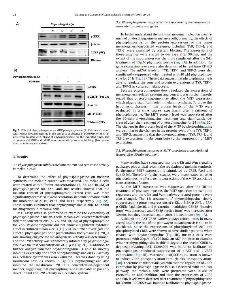
Fig. 5. Effect of phytosphingosine on MITF phosphorylation. (A) Cells were treatedwith 10 mM phytosphingosine in the presence or absence of PD98059 for 36 h. (B)Cells were treated with 10 mM of phytosphingosine for the indicated times. Theexpressions of MITF and p-ERK were examined by Western blotting. b-actin wasused as an internal standard.
24 E.J. Jang et al. / Journal of Dermatological Science 87 (2017) 19–28
3. Results
3.1. Phytosphingosine inhibits melanin content and tyrosinase activityin melan-a cells
To determine the effect of phytosphingosine on melaninsynthesis, the melanin content was measured. The melan-a cellswere treated with different concentrations (5, 7.5, and 10 mM) ofphytosphingosine for 72 h, and the results showed that themelanin content of phytosphingosine-treated cells was moresignificantly decreased in a concentration-dependent manner withthe inhibition of 21.3%, 39.5%, and 46.1%, respectively (Fig. 2A).These results exhibited that phytosphingosine is able to inhibitmelanogenesis in melan-a cells.
MTT assay was also performed to examine the cytotoxicity ofphytosphingosine in melan-a cells. Melan-a cells were treated withdifferent concentrations (5, 7.5, and 10 mM) of phytosphingosinefor 72 h. Phytosphingosine did not show a significant cytotoxiceffect in cultured melan-a cells (Fig. 2B). To further investigate theeffect of phytosphingosine on pigmentation, the tyrosinase (TYR), arate-limiting enzyme for melanogenesis, activity was determined,and the TYR activity was significantly inhibited by phytosphingo-sine over the test concentrations of 10 mM (Fig. 2C). In addition, tofurther analyze whether phytosphingosine is able to directlyinhibit TYR activity, the effect of phytosphingosine on TYR activityin a cell-free system was also evaluated. This was done by usingmushroom TYR. As shown in Fig. 2D, phytosphingosine alsoinhibited the mushroom TYR in a concentration-dependentmanner, suggesting that phytosphingosine is also able to possiblydirect inhibit the TYR activity in a cell-free system.
3.2. Phytosphingosine suppresses the expression of melanogenesis-associated proteins and genes
To better understand the anti-melanogenic molecular mecha-nism of phytosphingosine in melan-a cells, primarily, the effects ofphytosphingosine on the protein expressions of the majormelanogenesis-associated enzymes, including TYR, TRP-1, andTRP-2, were examined by western blotting. The expressions ofthose enzymes were started to decrease after 30 min, and theextent of the suppression was the most significant after the 24 htreatment of 10 mM phytosphingosine (Fig. 3A). In addition, thegene expression levels were also determined by real time RT-PCRanalysis. The mRNA levels of TYR, TRP-1 and TRP-2 were alsosignificantly suppressed when treated with 10 mM phytosphingo-sine for 24 h (Fig. 3B). These data suggest that phytosphingosine isable to regulate the gene and protein expressions of TYR, TRP-1,and TRP-2 in cultured melanocyets.
Because phytosphingosine downregulated the expressions ofmelanogenesis-related proteins and genes, it was further hypoth-esized that phytosphingosine may affect the MITF expression,which plays a significant role in melanin synthesis. To prove thishypothesis, changes in the protein levels of the MITF wereevaluated in a time course experiment after treatment ofphytosphingosine. The MITF protein level was suppressed afterthe 30-min phytosphingosine treatment and significantly de-creased after the treatment of phytosphingosine for 24 h (Fig. 3C).The changes in the protein level of the MITF by phytosphingosinewere similar to the changes in the protein levels of the TYR, TRP-1,and TRP-2, suggesting that the downregulation of TYR, TRP-1, andTRP-2 expressions might contribute to the suppression of MITFexpression.
3.3. Phytosphingosine suppresses MITF-associated transcriptionalfactors after 30 min treatment
Many studies have suggested that the c-Kit and Wnt signalingpathways play critical roles in the regulation of melanin synthesis.Furthermore, MITF expression is stimulated by CREB, Pax3 andSox10 [9]. Therefore, further studies were investigated whetherphytosphingosine affects to the expressions of the MITF-associatedtranscriptional factors.
As the MITF expression was suppressed after the 30 mintreatment of phytosphingosine, the MITF upstream transcriptionregulators and the c-Kit and Wnt pathway-related proteins werealso changed. The 1 h treatment of phytosphingosine clearlysuppressed the protein expressions of c-Kit, p-PI3K, p-AKT, p-ERK,p-CREB, Pax3, Sox10, and b-catenin. In addition, GSK3b (inactiveform) was decreased and GSK3b (active form) was increased after30 min, but they increased again after 1 h treatment (Fig. 4A).
Although the AKT/CREB pathway plays critical roles in manyareas [28,29], the role of the pathway in melanogenesis is still to beelucidated. Since the expressions of phosphorylated AKT andphosphorylated CREB were shown to have similar patterns whentreated with phytosphingosine (Fig. 4B), melan-a cells werepretreated with 20 mM of LY294002, an AKT inhibitor, to examinewhether phytosphingosine is able to degrade the level of CREB bydephosphorylating AKT. LY294002 was found to facilitate thephytosphingosine-induced suppression of p-AKT and p-CREBexpressions (Fig. 4B). Moreover, c-kit/SCF stimulation is knownto induce CREB phosphorylation through ERK phosphorylation[30]. Therefore, to further analyze whether the regulation of CREBexpression by phytosphingosine is also associated with the ERKpathway, the melan-a cells were pretreated with 20 mM ofPD98059, an ERK inhibitor, and then the expressions of CREBand ERK levels were detected after treatment of phytosphingosinefor 30 min. PD98059 was found to facilitate the phytosphingosine-

Fig. 6. Effect of phytosphingosine on tyrosinase expression determined by immunocytochemistry. Cells were treated with 10 mM of phytosphingosine for 1 h or 36 h, and thenstained with tyrosinase and DAPI antibodies. The merged images are shown in the right panels.
E.J. Jang et al. / Journal of Dermatological Science 87 (2017) 19–28 25
induced suppression of ERK level and the phytosphingosine-dependent suppression of CREB level, suggesting phytosphingo-sine is able to degrade the level of CREB by dephosphorylating ERK(Fig. 4C).
Since phytosphingosine regulated p-CREB, the role of phytos-phingosine in mediating the DNA-binding activity of phosphory-lated CREB was further examined. Using the ELISA-based Trans AMmethod and specific p-CREB primary Ab, phytosphingosine(10 mM) significantly inhibited the DNA-binding activity ofphosphorylated CREB in a time-dependent manner. These findingsindicate that phytosphingosine downregulated CREB activationand its related DNA-binding activity (Fig. 4D). Phytosphingosinewas also effectively found to downregulate the expressions of Pax3and Sox10 proteins and mRNA levels (Fig. 4E). Therefore, thesuppression of the mRNA and protein levels of these MITFupstream regulators might eventually affect to the downregulationof the MITF protein level at the same time point.
3.4. Phytosphingosine induces ERK activation after 12 h treatment,causing MITF phosphorylation
We also found that phytosphingosine induces the phosphory-lation of ERK after 12 h, which was coincided with the reduction ofthe MITF protein level (Fig. 5A). However, the MITF upstreamtranscription regulators and the Wnt pathway-related proteins didnot change after the 36-h treatment (data not shown).
It is known that MITF exhibits different patterns of subcellularlocalization depending on types of cells [31]. The levels of MITF innucleus are important as a transcriptional factor. Therefore, wedetermined the MITF protein level in the nuclear extract aftertreatment of phytosphingosine in melan-a cells. Phytosphingosine(10 mM) clearly decreased the MITF levels in a time-dependentmanner up to 36 h (Fig. 5A). The levels of MITF were also found tobe reversely correlated with the levels of p-ERK (Fig. 5A). To furtherclarify whether the activation of ERK affects to the degradation ofMITF in nucleus, the ERK inhibitor PD98059 was pretreated, andthen p-ERK and MITF, and p-MITF levels by phytosphingosine weredetermined by western blotting. The treatment of PD98059effectively downregulated p-ERK and p-MITF, but MITF levelwas increased in nucleus. However, the co-treatment of phytos-phingosine increased the levels of p-ERK and p-MITF, but the MITFlevel was decreased in nucleus (Fig. 5B).
3.5. The suppression of the TYR levels by phytosphingosine iscorrelated with the MITF levels
To further determine whether the TYR levels by phytosphin-gosine is affected by the MITF levels, the expression levels of TYRwere examined using immunocytochemistry after treatment ofphytosphingosine for 1 h and 36 h. Phytosphingosine effectivelydecreased the TYR levels both time points compared to the vehicle-treated control cells, indicating that the suppression of MITF levelsis able to affect to the TYR levels (Fig. 6).

Fig. 7. Effects of phytosphingosine on gene expression and transactivation of MITFand tyrosinase. (A) Cells were treated with 10 mM phytosphingosine for 30 min, andthen the levels of MITF were determined by real-time RT-PCR. (B) Cells weretransfected with Gaussia luciferase reporter constructs, pMITF-Gluc and pTyr-Glucplasmid, and then treated with the indicated concentrations of phytosphingosinefor 24 h. Cells were also co-transfected with the Firefly luciferase control vector(pGL3-FL) to normalize transfection rates. Data shown represent the mean � SD.*p < 0.05, **p < 0.005 are considered statistically significant compared to the controlgroup.
Fig. 8. Effect of phytosphingosine on melanin biosynthesis in a reconstructedhuman skin model, Neoderm-ME. UVB-induced Neoderm ME was treated with theindicated concentrations of phytosphingosine, and then melanin production wasmeasured as described in Section 2. Data shown represent the mean � SD (n = 3) andare representative of three separate determinations. *p < 0.05 or **p < 0.01 isconsidered statistically significant compared to the control group.
26 E.J. Jang et al. / Journal of Dermatological Science 87 (2017) 19–28
3.6. Phytosphingosine suppresses promoter activities of MITF and TYR
To further elucidate the suppression of MITF protein levels byphytosphingosine is related with the decrease of MITF geneexpression, the mRNA expression of the MITF was investigated byreal time RT-PCR after treatment of phytosphingosine for 30 min.Phytosphingosine effectively suppressed the MITF mRNA level,indicating that the level of MITF was transcriptionally regulated byphytosphingosine (Fig. 7A). In addition, to further determinewhether phytosphingosine affects to the MITF and TYR promoteractivity, dual luciferase assay was performed. Phytosphingosineeffectively downregulated the promoter activities of MITF and TYRin a concentration-dependent manner (Fig. 7B and C).
3.7. Phytosphingosine inhibits melanin synthesis in a reconstructedhuman skin model
To examine the effect of phytosphingosine on the melaninsynthesis in human skin, a reconstructed 3D human skin epidermismodel (Neoderm-ME) was employed. Neoderm-ME was irradiatedwith UVB (40 mJ/cm2), and then incubated for 3 days to inducemelanin formation. After incubation, the irradiated human skinswere treated with phytosphingosine for 72 h, and then determinedthe melanin contents as described in Methods. Phytosphingosineeffectively inhibited the UVB-induced melanin synthesis in areconstructed human skin model (Fig. 8).
3.8. Evaluation of phytosphingosine on human primary skin irritation
Skin irritation is also one of the major considerable issues forcosmetic ingredients. To further determine whether phytosphin-gosine is an applicable ingredient for cosmetic components,primarily, the effect of phytosphingosine on human skin irritationwas performed by employing a patch test in human arm skins. Asshown in Table 1, phytosphingosine did not demonstrate anyspecial skin irritation detected at 30 min and 24 h after removal ofpatches. This finding suggests that phytosphingosine is consider-able to be a relatively safe for application as a cosmetic ingredient.
4. Discussion
Sphingolipids are universal constituents of the plasma mem-brane in plants, animals, and fungi [32]. Of the structural analogs of

Table 1Human skin primary irritation test.
No. Test material 48 h 72 h Reaction grade
� 1+ 2+ 3+ 4+ � 1+ 2+ 3+ 4+ 48 h 72 h Mean
1 Control – – – – – – – – – – 0 02 Phytosphingosine (1%) – – – – – – – – – – 0 0
No reaction; Reaction grade =P
[{Grade � No. of Responders}/{4(Maximum grade) � 30(Total subject)}] � (1/2).
Fig. 9. The proposed mechanisms for the anti-melanogenic activity of phyto-sphingosine.
E.J. Jang et al. / Journal of Dermatological Science 87 (2017) 19–28 27
sphingolipids, phytosphingosine is well-known to be involved inmany significant cellular responses including apoptosis, differen-tiation, and migration [32–34]. Moreover, phytosphingosine iscommonly used as acne treatments because of its anti-bacterialand anti-inflammatory activities [35–37]. Therefore, phytosphin-gosine and its derivatives are applicable for maintaining healthyhuman skin.
To our knowledge, this is the first study demonstrating the anti-melanogenic activity of phytosphingosine. Previous study reportedthat the representative sphingolipid molecule, sphingosine-1-phosphate (S1P), is also known as an anti-melanogenic ingredient.The plausible mechanism was suggested by the induction of MITFphosphorylation and subsequently proteasome-mediated degra-dation of MITF by S1P [21]. Another study reported by Sprong et al.demonstrated that glycosphingolipids are also required for sortingmelanosomal proteins from the Golgi complex to the melanosome[38]. Therefore, the biological functions of sphingolipids onmelanogenesis are still unclear. In the present study, we foundthat the anti-melanogenic activity of phytosphingosine is in partassociated with both the suppression of MITF gene expression anddegradation of MITF. As confirmed by MTT assay, phytosphingosinewas not cytotoxic up to 10 mM, and the level of melanin productionat 10 mM was inhibited by 46.1%. To further understand themechanisms of actions in detail, we firstly examined the effects ofphytosphingosine on the enzymatic activity in melanogenesis. TheTYR activity was effectively inhibited by phytosphingosine in boththe cell-based and cell-free systems. Phytosphingosine alsosignificantly suppressed the gene and protein expressions of theTYR, TRP-1, and TRP-2, which are main enzymes in melanogenesis,suggesting that the anti-melanogenic activity of phytosphingosineis in part associated with the direct inhibition of the enzymaticactivity and transcriptional and translational regulation of maintarget enzymes in melanin biosynthesis.
In addition, the MITF, a major transcription factor in melaninbiosynthesis, protein level was found to be dramatically decreasedtwo times shortly after the 30 min and 12–24 h treatment ofphytosphingosine. This dual shift similarly occurred in TYR, TRP-1and TRP-2, suggesting that the downregulation of TYR, TRP-1, andTRP-2 by phytosphingosine can be caused by the MITF suppression.
Many evidences suggested that the Wnt signaling pathway isalso involved in the MITF expression. When the canonical Wntpathway is activated, which leads to the GSK3b inactivation(phosphorylated at ser21/9) and b-catenin accumulation [39]. Theaccumulated b-catenin in cytoplasm subsequently triggers totranslocate into the nucleus and enhance the expression of MITF[40]. Therefore, the melanogenesis can be promoted by inhibitingGSK3b. Moreover, activated GSK3b (phosphorylated at Tyr216) isable to induce the phosphorylation of b-catenin, which causes theubiquitination and degradation of b-catenin [41]. As a result, thepresent findings showed that the inhibition of MITF levels byphytosphingosine is in part due to the decrease of GSK3b inactiveform and increase of GSK3b active form, which leads to thedecrease of b-catenin in nucleus. These data also suggest thatphytosphingosine is able to inhibit MITF transcription by inhibitingthe formation of b-catenin and TCF/LEF complex in nucleus.Further study revealed that the regulation of MITF by phytos-phingosine is also correlated with the downregulation of MITFupstream transcriptional regulators including CREB, Pax3, andSOX10. We also confirmed that c-Kit-mediated MAPK/PI3K path-ways were also inhibited by the treatment of phytosphingosine. Inaddition, the involvement of ERK and CREB in the regulation ofMITF by phytosphingosine was also elucidated with the co-treatment of the ERK and AKT inhibitors. In particular, the analysisof signaling pathways indicate that the downregulation of MITF byphytosphingosine is in part associated with both the suppressionof MITF gene expression in a short period time and the degradationof MITF protein via ERK-mediated MITF phosphorylation and itssubsequent proteasomal degradation in a longer period time(Fig. 9). Furthermore, the in vivo human skin mimic model using areconstructed 3D human skin also exhibited the UVB-inducedmelanin biosynthesis was effectively inhibited by phytosphingo-sine.
In conclusion, herein, we for the first time demonstrate theanti-melanogenic activity of phytosphingosine in both culturedmelanocytes and a reconstructed 3D human skin models. Theplausible mechanisms of actions for the inhibition of melaninbiosynthesis in melanocytes by phytosphingosine were alsoelucidated by analyzing the signaling pathways in melanogenesis.
Acknowledgements
This research was supported by a grant of the Korea HealthTechnology R&D Project through the Korea Health IndustryDevelopment Institute (KHIDI), funded by the Ministry of Health& Welfare, Republic of Korea (grant number: HN14C0088).
28 E.J. Jang et al. / Journal of Dermatological Science 87 (2017) 19–28
References
[1] V.J. Hearing, K. Tsukamoto, Enzymatic control of pigmentation in mammals,FASEB J. 5 (1991) 2902–2909.
[2] I. Kubo, I. Kinst-Hori, S.K. Chaudhuri, Y. Kubo, Y. Scanchez, T. Ogura, Flavonolsfrom Heterotheca inuloides: tyrosinase inhibitory activity and structuralcriteria, Bioorg. Med. Chem. 8 (2000) 1749–1755.
[3] N.H. Shin, S.Y. Ryu, E.J. Choi, S.H. Kang, I.M. Chang, K.R. Min, Y. Kim,Oxyresveratrol as the potent inhibitor on dopa oxidase activity of mushroomtyrosinase, Biochem. Biophys. Res. Commun. 243 (1998) 801–803.
[4] V.J. Hearing, Determination of melanin synthetic pathway, J. Invest. Dermatol.131 (2011) E8–E11.
[5] J. Vachtenheim, J. Borovansky, “Transcription physiology” of pigmentformation in melanocytes: central role of MITF, Exp. Dermatol. 19 (2010)617–627.
[6] R. Busca, R. Ballotti, Cyclic AMP a key messenger in the regulation of skinpigmentation, Pigment Cell Res. 13 (2000) 60–69.
[7] C. Levy, M. Khaled, D.E. Fisher, MITF: master regulator of melanocytedevelopment and melanoma oncogene, Trends Mol. Med. 12 (2006) 406–414.
[8] M.O. Villareal, J. Han, P. Tamada, H. Shingemori, H. Isoda, Hirseins inhibitmelanogenesis by regulating the gene expressions of Mitf and melanogenesisenzymes, Exp. Dermatol. 19 (2010) 450–457.
[9] P. Wan, Y. Hu, L. He, Regulation of melanocyte pivotal transcription factor MITFby some other transcription factors, Mol. Cell. Biochem. 354 (2011) 241–246.
[10] E. Jung, W. Hwang, S. Kim, Depigmenting of platycodin D depends on thecAMP/Rho-dependent signaling pathway, Exp. Dermatol. 20 (2011) 986–991.
[11] Y.G. Kang, E.J. Choi, Y. Choi, 5,7-Dimethoxyflavone induces melanogenesis inB16F10 melanoma cells through cAMP-dependent signaling, Exp. Dermatol. 20(2011) 445–447.
[12] B. Saha, S.K. Singh, C. Sarkar, Activation of the Mitf promoter by lipid-stimulated activation of p38-stress signaling to CREB, Pigment Cell Res. 19(2006) 595–605.
[13] V. Alexeev, K. Toon, Distinctive role of the c-Kit receptor tyrosine kinasesignaling in mammalian melanocytes, J. Invest. Dermatol. 126 (2006) 1102–1110.
[14] S. Jeon, N.H. Kim, J.Y. Kim, A.Y. Lee, Stem cell factor induces ERM proteinsphosphorylation through PI3K activation to mediate melanocyte proliferationand migration, Pigment Cell Res. 22 (2009) 77–85.
[15] L. Larribere, M. Khaled, S. Tartare-Deckert, R. Busca, F. Luciano, K. Bille, PI3Kmediates protection against TRAIL-induced apoptosis in primary humanmelanocytes, Cell Death Differ. 11 (2004) 1084–1091.
[16] K.S. Smalley, R. Contractor, T.K. Nguyen, M. Xiao, R. Edwards, V. Muthusamy,Identification of a novel subgroup of melanomas with KIT/cyclin-dependentkinase-4 overespression, Cancer Res. 68 (2008) 5743–5752.
[17] G. Lefevre, A.L. Glotin, A. Calipel, F. Mouriaux, T. Tran, Z. Kherrouche, Roles ofstem cell factor/c-Kit and effects of Gilvec/STI571 in human uveal melanomacell tumorigenesis, J. Biol. Chem. 279 (2004) 31769–31779.
[18] H.H. Ko, Y.C. Chiang, M.H. Tsai, C.J. Liang, L.F. Hsu, S.Y. Li, et al., Eupafolin, a skinwhitening flavoonid isolated from phyla nodiflora, downregulatedmelanogenesis: role of MAPK and Akt pathways, J. Ethnopharmacol. 151 (1)(2014) 386–393.
[19] M. Wu, T.J. Hemesath, C.M. Takemoto, C-kit triggers dual phosphorylations,which couple activation and degradation of the essential melanocyte factorMi, Genes Dev. 14 (2000) 301–312.
[20] D.S. Kim, S.Y. Kim, J.H. Chung, H.C. Eum, K.C. Park, Delayed ERK activation byceramide reduces melanin synthesis in human melanocytes, Cell. Signal. 14(2002) 779–785.
[21] D.S. Kim, E.S. Hwang, J.S. Lee, S.Y. Kim, S.B. Kwon, K.C. Park, Sphingosine-1-phosphate decreases melanin synthesis via sustained ERK activation andsubsequent MITF degradation, J. Cell Sci. 116 (2003) 1699–1706.
[22] J.E. Lee, S.Y. Kim, Y.M. Jeong, H.Y. Yun, K.J. Baek, N.S. Kwon, et al., The regulatorymechanism of melanogenesis by FTY720, a sphingolipid anlogue, Exp.Dermatol. 20 (2010) 237–241.
[23] W.J. Lee, S. Bang, B.Y. Chung, H. Jung, E.S. Oh, S.E. Chang, Inhibitory effects of N,N,N-trimethyl phytosphingosine-iodine on melanogenesis via ERK activation-mediated MITF degradation, Biosci. Biotechnol. Biochem. 80 (1) (2015) 121–127.
[24] E.O. Choi, E.J. Cho, J.W. Jeong, C. Park, S.H. Hong, H.J. Hwang, et al., Baicaleininhibits the migration and invasion of B16F10 mouse melanoma cells throughinactivation of the PI3K/AKT signaling pathway, Biomol Ther. 25 (2) (2017)213–221.
[25] A.A. Beg, T.S. Finco, P.V. Nanterment, A.S. Baldwin, Tumor necrosis factor andinterleukin-1 lead to phosphorylation and loss of I kappa B alpha: amechanism for NF-kappa B activation, Mol. Cell. Biol. 13 (1993) 3301–3310.
[26] M.M. Bradford, A rapid and sensitive method for the quantitation ofmicrogram quantities of protein utilizing the principle of protein-dyebinding, Anal. Biochem. 72 (1976) 248–254.
[27] T. Masamoto, S. Iida, M. Kubo, Inhibitory effect of Chinese crude drugs ontyrosinase, Planta Med. 40 (1980) 361–365.
[28] A.J. Shaywitz, M.E. Greenberg, CREB: A stimuls-induced transcription factoractivated by a diverse array of extracellular signals, Annu. Rev. Biochem. 68(1999) 821–861.
[29] M.P. Delghandi, M. Johannessen, U. Moens, The cAMP signalling pathwayactivates CREB through PKA, p38 and MSK1 in NIH 3T3 cells, Cell. Signal. 17(2005) 1343–1351.
[30] G. Imokawa, Y. Yada, N. Morisaki, M. Kimura, Signaling mechanisms ofendothelin-induced mitogenesis and melanogenesis in human melanocytes,Biochem. J. 330 (1996) 1235–1239.
[31] S.R. Granter, K.N. Weilbaecher, C. Quigley, D.E. Risher, Role for microphthalmiatranscription factor in the diagnosis of metastatic malignant melanoma, Appl.Immunohistochem. Mol. Morphol. 10 (2002) 47–51.
[32] H.J. Kim, H.J. Kim, S.H. Kim, Tetraacetyl phytosphingosine-induced caspaseactivation and apoptosis occur through G2 arrest in human keratinocyteHaCaT cells, J. Invest. Dermatol. 121 (2003) 1135–1137.
[33] H.J. Kim, W. Shin, C.S. Park, Differnetial regulation of cyclooxygenase-2expression by phytosphingosine derivatives, NAPS and TAPS, and its role in theNAPS or TAPS-mediated apoptosis, J. Invest. Dermatol. 121 (2003) 1126–1134.
[34] H.J. Kim, S.Y. Kang, S.J. Kim, Potentiation of UVB-induced apoptosis by novelphytosphingosine derivative, tetraacetyl phytosphingosine in HaCaT cell andmouse skin, Apoptosis 9 (2004) 449–456.
[35] S. Kim, I. Hong, J.S. Hwang, Phytosphingosine stimulates the differentiation ofhuman melanocytes and inhibits TPA-induced inflammatory epidermalhyperplasia in hairless mouse skin, Mol. Med. 12 (2006) 17–24.
[36] T. Pavicic, U. Wollenweber, M. Farwick, Anti-microbial and inflammatoryactivity and efficacy of phytosphingosine: an in vitro and in vivo studyaddressing acne vulgaris, Int. J. Cosmet. Sci. 29 (2007) 181–190.
[37] C.L. Fischer, D.R. Drake, D.V. Dawson, Antibacterial activity of sphingoid basesand fatty acids against Gram-positive and Gram-negative bacteria,Antimicrob. Agents Chemother. 56 (2012) 1157–1161.
[38] H. Sprong, S. Degroote, T. Claessens, V. Van Drunen, B.H. Westerink, et al.,Glycosphingolipids are required for sorting melanosomal proteins in the Golgicomplex, J. Cell Biol. 155 (2001) 369–380.
[39] L. Larue, V. Delmas, The WNT/beta-catenin pathway in melanoma, Front.Biosci. 11 (2006) 733–742.
[40] J. Wu, J.P. Saint-Jeannet, P.S. Klein, Wnt-frizzled signaling in neural crestformation, Trends Neurosci. 26 (2003) 40–45.
[41] B. Bellei, E. Flori, E. Izzo, V. Maresca, M. Picardo, GSK3beta inhibition promotesmelanogenesis in mouse B16 melanoma cells and normal human melanocytes,Cell. Signal. 20 (2008) 1750–1761.