the melanogenic system of the liver pigmented … · tyrosinase is the enzyme involved in the...
TRANSCRIPT
Summary. The enzyme system responsible forAmphibian Kupffer Cell (KC) melanogenesis has notbeen entirely elucidated. This research demonstrates thatthe KC melanosomes of Rana esculenta L. possess atyrosine-hydroxylase (TH) activity, showing that atyrosinase is the enzyme involved in the melanogenesis.The TH reaction depends on catalytic Dopa as a cofactorand is not affected by catalase or H2O2, showing that it iscatalysed by the tyrosinase and not by the peroxidasepresent in the melanosomes.
The TH reaction is activated by Cu2+ ions but not byother tyrosinase activators such as limited proteolysis,protein ageing, and Sodium Dodecyl Sulphate (SDS).SDS inhibited the KC TH activity even below thecritical micelle concentration. All these results suggestthat the KC-tyrosinase differs in structure from otherknown tyrosinases.
Using anti-KC-tyrosinase antobodies, we observedthat the sites of the tyrosinase location within the cell arethe same as those described in the melanocytes. In theimmunoblots, the anti-KC-tyrosinase antibodies alsorecognised two protein bands, at the higher molecularweight ranges, in the protein electrophoretic pattern.Moreover, the tyrosinase activity was limited to thehighest molecular weight band of about 260 kDa,suggesting that the enzyme activity could depend on amolecular aggregate. The melanin produced in the liverwas found to be a 5,6-dihydroxyindole-rich eumelaninsimilar to the Sepia melanin. Key words: Pigmented Kupffer Cells, Melanogenesis,Tyrosine-Hydroxylase Activity, Tyrosinase, Melanins
Introduction
Melanins are pigments occurring at all phylogeneticlevels. In Mammals, they are synthesized by pigmentcells, such as the melanocytes deriving from the neuralcrest mesenchyme; the melanoma cells, theirtransformed counterparts; and by the extracutaneouspigment cells deriving from the neural tube. Lowervertebrates possess, in addition, an extracutaneouspigment cell system localized in visceral sites, such asthe liver, spleen, kidney, and other body districts. Thesepigment cells are different from the melanocytes andother pigment cells of neural origin. The pigment cells ofthe Amphibians and Reptiles liver have been identifiedas Kupffer Cells (Sichel, 1948; David and Freytag, 1963;Bani, 1966). Studies carried out on Rana esculenta L.have shown that the pigmented macrophages from liver(Pintucci et al., 1990; Sichel et al., 1997; Guida et al.,1998; Corsaro et al., 2000) and spleen (Gallone at al.,2002) derive from monocyte/macrophage cell lineages.Since they do not arise from the neural crestmesenchyme, they were not included in the classificationof the pigment cells (Fitzpatrick et al., 1966). Recently, anew classification of the pigment cells has beenproposed (Sichel et al., 1997), which includes thepigment cells of the extracutaneous system ofAmphibians, Reptiles and Fishes. According to thisclassification the pigmented macrophages are includedin one group of pigment cells (melanin-synthesizingcells) deriving from haemopoietic stem cells. In fact, ithas been demonstrated in different species ofAmphibians and Reptiles that the pigmentedmacrophages of the liver are able to synthesize melanins(Bani, 1966; Cicero et al., 1982, 1989; Scalia et al.,1988; Sichel, 1988). They are able to express thetyrosinase gene (Guida et al., 2000; Purrello et al.,2001), and respond to α -MSH by increasing the
The melanogenic system of the liver pigmentedmacrophages of Rana esculenta L. - Tyrosinase activityA. Gallone1,2, A. Sagliano1, G. Guida3, S. Ito4, K. Wakamatsu4, V. Capozzi1,2, G. Perna1,2, P. Zanna3 and R. Cicero31Dipartimento di Scienze Biomediche, Facoltà di Medicina e Chirurgia, Università degli Studi di Foggia, via L. Pinto c/o A.O.U.“Ospedali Riuniti”, Foggia, Italy, 2Centro di Ricerca Interdipartimentale BIOAGROMED, Università degli Studi di Foggia, Foggia, Italy,3Dipartimento di Biochimica Medica, Biologia Medica e Fisica Medica, Facoltà di Medicina e Chirurgia – Università degli Studi di Bari,Policlinico – Piazza Giulio Cesare, Bari, Italy and 4Department of Chemistry, Fujita Health University School of Health Science,Toyoake, Aichi, Japan
Histol Histopathol (2007) 22: 1065-1075
Offprint requests to: R. Cicero, Dipartimento di Biochimica Medica,Biologia Medica e Fisica Medica, Facoltà di Medicina e Chirurgia –Università degli Studi di Bari, Policlinico – Piazza Giulio Cesare, 70124Bari, Italy. e-mail: [email protected]
DOI: 10.14670/HH-22.1065
http://www.hh.um.es
Histology andHistopathologyCellular and Molecular Biology
tyrosinase gene transcripts (Guida et al., 2004). Previous enzymatic studies have shown that the
melanosomes of the pigmented macrophages from theliver and spleen of R. esculenta L. possess dopaoxidaseand peroxidase activities, which feature peculiarcharacteristics (Gallone et al., 2002). The dopaoxidaseactivity appears to be directly related with the livermelanogenesis because its activity levels varyseasonally, in parallel with the liver melanin content(Cicero et al., 1977; Corsaro et al., 1990), showingmaximum levels in winter and minimum in summer.
As regards the tyrosine-hydroxylase activity,although autoradiographic or cytological analyses havesuggested its presence in the pigmented macrophages(Cicero et al., 1982; Sichel et al., 1997), attempts toclearly evidence this activity on protein extracts frommelanosomes were not always successful. Thefunctional role of the melanins in the visceral organs ofthe lower vertebrates has long been discussed (Sichel,1988; Agius and Roberts, 2003, for reviews). Generally,authors attribute the presence of black-brown melaninsto cytoprotective functions against cytotoxic chemicalspecies generating stress conditions (Geremia et al.,1984; Sarna et al., 1986; Scalia et al., 1990). Someauthors have drawn a relationship between the activationof visceral melanogenesis and particular metabolic statesin animals, such as the decline of anti-oxidant enzymaticactivities during hibernation (Barni et al., 1999), andexperimentally induced hypoxia (Frangioni et al., 2000).
The aim of the present research is to study theenzyme responsible for the melanogenic ability of theKupffer Cells of the frog Rana esculenta L. in order tobetter understand the mechanism which triggers themelanogenesis reaction. Tyrosinase is the key enzyme ofthe melanin pathway which catalyses two differentreactions: 1) the hydroxylation of monophenols to o-diphenols (monophenolase or cresolase activity EC1.14.18.1), and 2) the oxidation of o-diphenols to o-quinones (diphenolase or catechol oxidase activity EC1.10.3.1). The above functions were defined by Raper(1927), Mason (1948), Lerner et al. (1949). Theevidence of a tyrosine-hydroxylase activity beside thedopaoxidase activity, shown in a previous research(Cicero et al., 1989), would confirm the existence of atyrosinase in the pigmented Kupffer Cells of R.esculenta. With this aim, we performed experiments tosearch for the presence of a tyrosine-hydroxylaseactivity in these cells. Vertebrate tyrosinase is aglycoprotein of the melanosome membrane, and itsbiogenesis steps have been shown in melanocytes, aswell as in melanoma cells (Orlow, 1998, for a review).In the present study we have also tried to detect the sitesof tyrosinase location within the Kupffer Cells, to betterunderstand the enzyme processing path.
Finally, to gain a more extensive knowledge of theliver pigment system, we carried out chemicalcharacterization of the melanin synthesized by theKupffer Cells, using the HPLC microanalytical methodsof Wakamatsu and Ito, 2002.
Materials and methods
Source of the material
Experiments were carried out on wild specimens ofRana esculenta L. randomly collected near Catania(Sicily) at monthly intervals. The liver from at least 50animals (an average of 30 g of tissue) was minced andhomogenized in a Potter Elvejem homogenizer in a 0.05M Na-phosphate buffer, pH 7.2, on ice. Melanosomeprotein isolation was performed following the method ofCicero et al. (1989), with minor modifications. 0.1 mMphenylmethylsulphonyl fluoride (PMSF) (SigmaChemical Co, St Louis, MO, USA) was used as proteaseinhibitor. The protein content was determined by the“Bio-Rad Protein Assay “ kit according to the Bradfordmethod (1976) and then used for enzymatic assays. Allthe assays were performed on dialysed or not dialysedsamples. Dialyses were performed over-night in 0.05 M,pH 7.2 phosphate buffer at 4°C.
Since the attempts to purify the enzyme alwaysproduced loss of activity, we used the crude melanosomeprotein extracts for this study. Biochemical experiments
Tyrosine-hydroxylase (TH) activity (EC 1.14.18.1).Assays of the TH activity were carried out with a doublebeam Perkin Elmer Lambda-5 Spectrophotometer, at aconstant temperature of 25°C, following the dopachromeformation at 475 nm (Mason, 1948). The TH activitywas determined on 700 µg of melanosome proteinssuspended in a 0.1 M Na-phosphate buffer, pH 6.8. Thereaction was started by adding L-tyrosine (Sigma) at afinal concentration of 1 mM, and catalytic amounts of L-Dopa at a final concentration ranging from 5 µM, 10µM, to 25 µM, in a volume of 0.5 ml. The 10 µMconcentration of catalytic L-Dopa was chosen for theroutine analyses. The velocity of product formation wasregistered in a time drive mode. The reaction was alsorecorded by spectrophotometer scanning of the samplesfrom 200 nm to 600 nm, at 1-hour intervals for at least20 cycles. Peroxidase (POD) activity (EC 1.11.1.7). POD activitywas determined using p-phenylenediamine-pyrocatechol(PPD-PC) substrates. The reaction mixture contained 40µg of melanosome proteins in a 0.1 M K-phosphatebuffer pH 6.5, plus 0.17 mM PPD (Sigma) and 4.5 mMPC (Sigma) in a volume of 500 µl. The reaction wasstarted by adding 250 µM H2O2. The absorbanceincrease was measured at 575 nm (Imberty et al., 1984). Inhibition experiments. The following inhibitors wereused: KCN, at a final concentration of 4 or 6 mM; PTU(phenylthiourea) at a final concentration of 4 mM . Heat inactivation. Heat inactivation was performed byboiling aliquots of the melanosome proteins at 100°C for15 min. Catalase (EC 1.11.1.6) treatment. Melanosome proteinswere incubated with 1000 or 2000 E.U. catalase
1066Tyrosinase of R. esculenta Kupffer cells
(Sigma), for 60 min at room temperature before theassay.H2O2 treatment. TH activity was assayed in the presenceof 250 µM or 500 µM H2O2, added to the reactionmixture, with melanosome proteins, just before thetyrosine. CuSO4 activation. Aliquots of dialysed melanosomeproteins were incubated in the presence of variousconcentrations of CuSO4, for times ranging from 10 minto 60 min, at room temperature. The TH activity wasassayed using the dopachrome method. To excludeunspecific responses due to possible interactionsbetween substrates and other components of the reactionmixture, measurements of absorbance at 475 nm weremade on mixtures only composed of tyrosine and/or L-Dopa + CuSO4. Melanosome proteins + CuSO4 withoutsubstrates were also assayed.Proteolysis. The melanosome proteins were incubatedwith trypsin at various concentrations (from 0.0001 to0.1 %) in a Na- phosphate buffer pH 7.2, at 37°C, forincreasing times: 1 min – 30 min. After incubation,samples were assayed for the TH activity.Protein ageing. Aliquots of melanosome protein, freshlyextracted, were left for a week in a refrigerator, at 4°C.Aliquots of protein were taken each day and assayed forthe TH activity. Sodium Dodecyl Sulphate (SDS) treatment. Themelanosome proteins underwent different treatmentswith SDS at 0.01%, 0.02%, 0.05%, 0.1%, 1%. SDS wasadded to the melanosome protein in the reaction mixtureimmediately before the tyrosine. The reaction wasrecorded for at least one hour at 475 nm. In otherexperiments, samples were incubated for 10 - 15 minwith SDS before the assay. Polyacrylamide-gel-electrophoresis (PAGE)
The melanosome proteins and mushroom tyrosinase(Sigma), as a positive control, were subjected to PAGE(Laemmli, 1970). To evidence the enzymatic activity,samples (25 µg of melanosome proteins or 20 µg ofmushroom tyrosinase) were diluted twice in a samplebuffer not containing SDS and separated onpolyacrylamide (PAA) gels (Bio-Rad, Richmond, CA,USA) 8% + 0.1% SDS, at 4°C. The gel was equilibratedin Na-phosphate 0.05 M, pH 6.8 buffer, at 25°C for 30min. The dopaoxidase (DO) reaction on gel wasperformed in the presence of 4 mM L-Dopa in a Na-phosphate buffer 0.05 M, pH 6.8, at room temperatureovernight. After testing for dopa positivity, the samePAA gel was extensively washed in Na-phosphate buffer0.05 M and stained with Coomassie Brilliant Blue R250(Bio-Rad). Blue Ranger molecular weight markers(PIERCE, Rockford, IL, US) were included in all gels.Preparation of the anti-KC-tyrosinase antibodies
Anti-KC-tyrosinase antibodies were preparedfollowing the method of Guida et al. (2004). In brief,
using a fragment of KC-tyrosinase gene of Ranaesculenta highly homologous (94%) with the exon Isequence of the skin tyrosinase of Rana nigromaculata(Takase et al., 1992), a 15 amino acid oligopeptide wassynthesized and used to obtain polyclonal rabbitantibodies, prepared by Genosys Sigma-Aldrich Ltd,London, UK. The oligopeptide sequence was publishedby Guida et al. (2004). The presence of antibodiesagainst the oligopeptide as well as their specificrecognition by the immunized rabbit serum, incomparison to the pre-immunized serum, wasdemonstrated by ELISA analysis following a routineprotocol. Western-blotting
The anti-KC-tyrosinase antiserum was used toperform immunoblotting of the protein component ofliver melanosomes. Samples were diluted twice in asample buffer, without SDS, and then loaded on 8%PAA gels. The proteins were separated in Tris-Glycinebuffer containing 0.1% SDS and then transferred onto0.45 µm nitrocellulose membranes, blotted in PBScontaining Tween-20 0.1% and supplemented with 5%milk (Bio-Rad). The membrane was first incubated withthe anti-KC-tyrosinase antiserum (diluted 1/1000)overnight at 4°C and then with a peroxidase-labeledsecondary antibody (diluted 1/5000). Incubation wascarried out at room temperature for one hour. The SuperSignal West-Pico Chemiluminescent Substrate(PIERCE) was used for detection. Immunogold staining
Fragments of liver tissue were fixed with 2%paraformaldehyde,1% glutaraldehyde (TAAB LabEquipment Ltd, Reading, England) in PBS, dehydratedin ethanol and embedded in LR White resin (Sigma).The immuno-recognition reaction, by antibodiesconjugated to 10 nm particles of colloidal gold, wasperformed on ultra-thin sections mounted on nickelgrids. The anti-KC-tyrosinase antiserum was used at adilution of 1/100.Chemical analysis of the liver melanin
For this purpose, only adult specimens of frogsfished in winter were used. Liver melanosomes wereisolated as indicated above. A partial purification ofmelanin was carried out by incubating the melanosomeswith 1% BRIJ-35 in 0.1 M Na-phosphate buffer, pH 7.2,overnight at 4°C. Then the suspension was centrifuged at22000xg for 60 min, at 4°C, and the melanin recoveredwith the pellet.
One hundred µg of dry melanin was oxidized withKMnO4 in 1 M H2SO4, and the oxidized material wasanalyzed for pyrrole-2,3,5-tricarboxylic acid (PTCA)(Ito and Fujita, 1985; Ito and Wakamatsu, 1998; Liu etal., 2003). Pyrrole-2,3-dicarboxylic acid (PDCA) was
1067Tyrosinase of R. esculenta Kupffer cells
produced only in a trace quantity. The melanin (200 µg) was also oxidized with H2O2in alkali to form PTCA and PDCA. PTCA serves as a
specific degradation product for DHICA-derived unitswhile PDCA serves for DHI-derived units (Ito andWakamatsu, 1998).
Hydriodic acid (HI) hydrolysis to detectpheomelanic pigment was performed on 200 µg melaninas described in Wakamatsu et al. (2002) and Liu et al.(2003). 4-Amino-3-hydroxyphenylalanine (4-AHP)serves as a specific degradation product of pheomelanin.
Soluene-350 solubilization of melanin (200 µg) wasalso performed to measure total melanin (TM), asdescribed in Ozeki et al. (1995) and Liu et al. (2003). Results
Tyrosine-hydroxylase activity
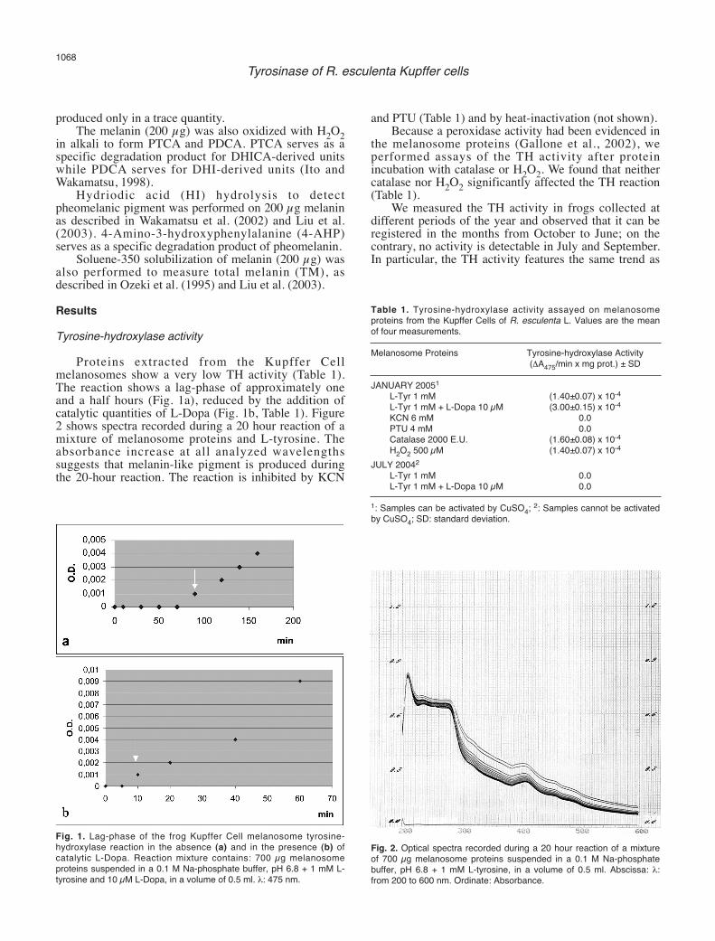
Proteins extracted from the Kupffer Cellmelanosomes show a very low TH activity (Table 1).The reaction shows a lag-phase of approximately oneand a half hours (Fig. 1a), reduced by the addition ofcatalytic quantities of L-Dopa (Fig. 1b, Table 1). Figure2 shows spectra recorded during a 20 hour reaction of amixture of melanosome proteins and L-tyrosine. Theabsorbance increase at all analyzed wavelengthssuggests that melanin-like pigment is produced duringthe 20-hour reaction. The reaction is inhibited by KCN
and PTU (Table 1) and by heat-inactivation (not shown). Because a peroxidase activity had been evidenced in
the melanosome proteins (Gallone et al., 2002), weperformed assays of the TH activity after proteinincubation with catalase or H2O2. We found that neithercatalase nor H2O2 significantly affected the TH reaction(Table 1).
We measured the TH activity in frogs collected atdifferent periods of the year and observed that it can beregistered in the months from October to June; on thecontrary, no activity is detectable in July and September.In particular, the TH activity features the same trend as
1068Tyrosinase of R. esculenta Kupffer cells
Fig. 1. Lag-phase of the frog Kupffer Cell melanosome tyrosine-hydroxylase reaction in the absence (a) and in the presence (b) ofcatalytic L-Dopa. Reaction mixture contains: 700 µg melanosomeproteins suspended in a 0.1 M Na-phosphate buffer, pH 6.8 + 1 mM L-tyrosine and 10 µM L-Dopa, in a volume of 0.5 ml. λ: 475 nm.
Fig. 2. Optical spectra recorded during a 20 hour reaction of a mixtureof 700 µg melanosome proteins suspended in a 0.1 M Na-phosphatebuffer, pH 6.8 + 1 mM L-tyrosine, in a volume of 0.5 ml. Abscissa: λ:from 200 to 600 nm. Ordinate: Absorbance.
Table 1. Tyrosine-hydroxylase activity assayed on melanosomeproteins from the Kupffer Cells of R. esculenta L. Values are the meanof four measurements.
Melanosome Proteins Tyrosine-hydroxylase Activity(ΔA475/min x mg prot.) ± SD
JANUARY 20051
L-Tyr 1 mM (1.40±0.07) x 10-4
L-Tyr 1 mM + L-Dopa 10 µM (3.00±0.15) x 10-4
KCN 6 mM 0.0PTU 4 mM 0.0Catalase 2000 E.U. (1.60±0.08) x 10-4
H2O2 500 µM (1.40±0.07) x 10-4
JULY 20042
L-Tyr 1 mM 0.0L-Tyr 1 mM + L-Dopa 10 µM 0.0
1: Samples can be activated by CuSO4; 2: Samples cannot be activatedby CuSO4; SD: standard deviation.

the DO activity, showed by Cicero et al. (1989). Table 1presents the values of TH activity recorded in twoperiods of the year, demonstrating the activity-inactivityof the system.Activation experiments
Because the TH activity is always very low, wecarried out some activation experiments on the proteinsextracted from melanosomes.
High values of TH activity were found afterincubating the melanosome protein with CuSO4. Fig. 3shows the dependence of the TH activation on the Cu2+ions concentration. All the controls made to ascertain thespecificity of the activation showed that the absorbancevariation at 475 nm does not depend on unspecificinteractions among the components of the reactionmixture. The reaction is totally inhibited by KCN.
In previous studies we have demonstrated that theDO activity of the frog Kupffer Cells is also activated byCu2+ ions (Cicero et al., 1990). It is interesting toobserve that TH activity, like DO activity, does notrespond to treatments with CuSO4 in all months of theyear. In fact, it is impossible to activate TH activity inthe months from July to September, when this activity isnot detectable (Table 1).
For a comparison with the melanocyte tyrosinase,we also performed treatments that are able to activatethis activity; such as, 1) limited proteolysis, 2) ageing ofthe protein, and 3) the action of SDS (Penafiel et al.,1982; Wittenberg and Triplett, 1985a,b; Zuasti et al.,1998; Lopez-Serrano et al., 2002). Limited proteolysis ofthe melanosome proteins did not produce increases ofTH activity; on the contrary it caused a completedisappearance of the activity even at very lowconcentrations of trypsin (0.0001%). Nor did ageing ofthe melanosome protein, at 4°C in the refrigerator,
produce any activation of TH. The samples remainedstable for the first four days of treatment andsubsequently began to lose their activity. After six daysin the refrigerator they had little or no activity, unlike theskin tyrosinase (Zuasti et al., 1998). Treatment of theprotein with various concentrations of SDS did notproduce any activation of the TH reaction too. On thecontrary, it invariably suppressed it, even at theconcentration of 0.02% which is lower than the criticalmicelle concentration, activating tyrosinases or otherpolyphenol oxidases (Wittenberg and Triplett, 1985a,b;Lopez-Serrano et al., 2002).Cell location of the hepatic tyrosinase by immuno-gold
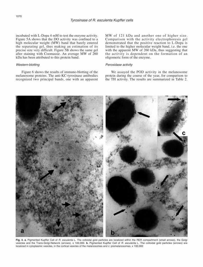
We endeavoured to highlight the sites of tyrosinaselocalization in the Kupffer Cells using immuno-goldtechniques. Figure 4 shows cell fields where there isevident recognition by the anti-KC-tyrosinaseantibodies. In Figure 4a, the grains of the colloidal goldappear localized, alone or in small groups, inside therough endoplasmic reticulum (RER), the Golgivescicles, and the Trans-Golgi-Network; in Figure 4band c, the grains of the colloidal gold appear localizedwithin cytoplasmic vescicles, as well as on the surface ofthe melanosomes and premelanosomes. Polyacrylamide-gel-electrophoresis (PAGE)
The melanosome proteins were separated by PAGEunder non reducing conditions and the gels were
1069Tyrosinase of R. esculenta Kupffer cells
Fig. 3. Increases in the tyrosine-hydroxylase activity in the melanosomeproteins of the Kupffer Cells of R. esculenta L., after incubation withincreasing concentrations of CuSO4 (µM), in the presence of constantconcentrations of L-tyrosine (1 mM) and 700 µg of melanosomeproteins, suspended in a 0.1 M Na-phosphate buffer, pH 6.8, in avolume of 0.5 ml. λ: 475 nm. The reaction is inhibited by KCN.
Table 2. Peroxidase activity of crude melanosome proteins from theKupffer Cells of Rana esculenta L.
Melanosome Proteins Peroxidase Activity(ΔA575/min x mg prot.)±SD
MARCH 2004Melanosome protein 0.12±0.0006KCN 4mM 0.0
MAY 2004Melanosome protein 0.30±0.015Dialysed protein 0.20±0.010Catalase 1000 E.U. 0.0
JULY 2004Melanosome protein 0.52±0.026Dialysed melanosome protein 0.18±0.009
JANUARY 2005Melanosome protein 0.22±0.011
Values are the mean of three measurements. SD: standard deviation.The decrease of peroxidase activity after dialysis is likely due to removalof H2O2 .
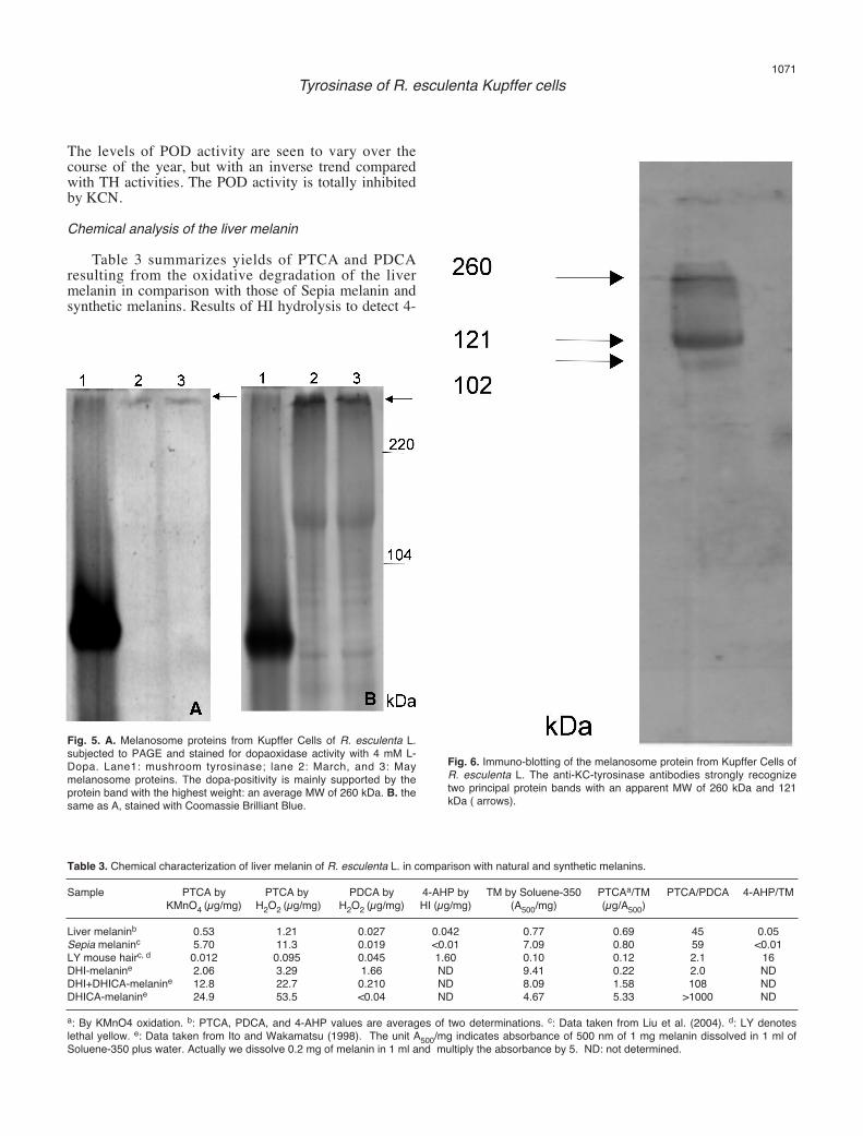
incubated with L-Dopa 4 mM to test the enzyme activity.Figure 5A shows that the DO activity was confined to ahigh molecular weight (MW) band that barely enteredthe separating gel, thus making an estimation of itsprecise size very difficult. Figure 5B shows the same gelafter staining with Coomassie. An average MW of 260kDa has been attributed to this protein band.Western-blotting
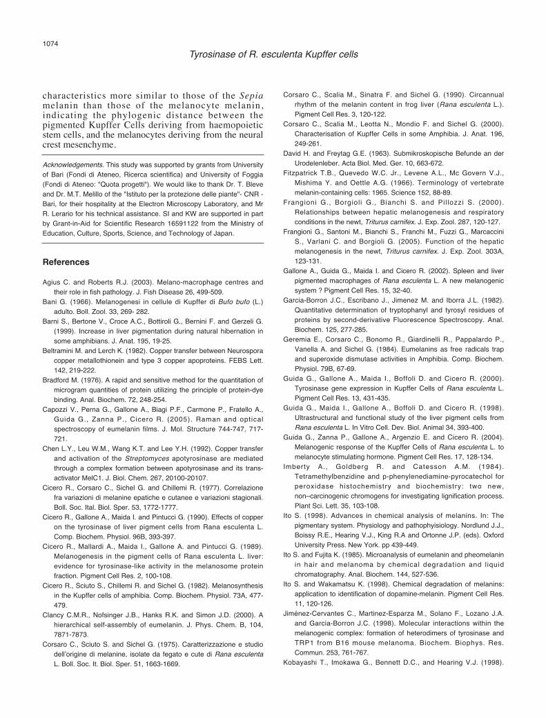
Figure 6 shows the results of immuno-blotting of themelanosome proteins. The anti-KC-tyrosinase antibodiesrecognized two principal bands, one with an apparent
MW of 121 kDa and another one of higher size.Comparison with the activity electrophoresis geldemonstrated that the positive reaction to L-Dopa islimited to the higher molecular weight band, i.e. the onewith the apparent MW of 260 kDa, thus suggesting thatthe activity is dependent on the formation of anoligomeric form of the enzyme. Peroxidase activity
We assayed the POD activity in the melanosomeprotein during the course of the year, for comparison tothe TH activity. The results are summarized in Table 2.
1070Tyrosinase of R. esculenta Kupffer cells
Fig. 4. a. Pigmented Kupffer Cell of R. esculenta L. The colloidal gold particles are localized within the RER compartment (small arrows), the Golgivesicles and the Trans-Golgi-Network (arrows). x 100,000. b. Pigmented Kupffer Cell of R. esculenta L. The colloidal gold particles (arrows) arelocalized in cytoplasmic vesicles, in the cortical vesicles of the melanosomes and c: premelanosomes. x 100,000
The levels of POD activity are seen to vary over thecourse of the year, but with an inverse trend comparedwith TH activities. The POD activity is totally inhibitedby KCN. Chemical analysis of the liver melanin
Table 3 summarizes yields of PTCA and PDCAresulting from the oxidative degradation of the livermelanin in comparison with those of Sepia melanin andsynthetic melanins. Results of HI hydrolysis to detect 4-
1071Tyrosinase of R. esculenta Kupffer cells
Fig. 6. Immuno-blotting of the melanosome protein from Kupffer Cells ofR. esculenta L. The anti-KC-tyrosinase antibodies strongly recognizetwo principal protein bands with an apparent MW of 260 kDa and 121kDa ( arrows).
Fig. 5. A. Melanosome proteins from Kupffer Cells of R. esculenta L.subjected to PAGE and stained for dopaoxidase activity with 4 mM L-Dopa. Lane1: mushroom tyrosinase; lane 2: March, and 3: Maymelanosome proteins. The dopa-positivity is mainly supported by theprotein band with the highest weight: an average MW of 260 kDa. B. thesame as A, stained with Coomassie Brilliant Blue.
Table 3. Chemical characterization of liver melanin of R. esculenta L. in comparison with natural and synthetic melanins.
Sample PTCA by PTCA by PDCA by 4-AHP by TM by Soluene-350 PTCAa/TM PTCA/PDCA 4-AHP/TMKMnO4 (µg/mg) H2O2 (µg/mg) H2O2 (µg/mg) HI (µg/mg) (A500/mg) (µg/A500)
Liver melaninb 0.53 1.21 0.027 0.042 0.77 0.69 45 0.05Sepia melaninc 5.70 11.3 0.019 <0.01 7.09 0.80 59 <0.01LY mouse hairc, d 0.012 0.095 0.045 1.60 0.10 0.12 2.1 16DHI-melanine 2.06 3.29 1.66 ND 9.41 0.22 2.0 NDDHI+DHICA-melanine 12.8 22.7 0.210 ND 8.09 1.58 108 NDDHICA-melanine 24.9 53.5 <0.04 ND 4.67 5.33 >1000 ND
a: By KMnO4 oxidation. b: PTCA, PDCA, and 4-AHP values are averages of two determinations. c: Data taken from Liu et al. (2004). d: LY denoteslethal yellow. e: Data taken from Ito and Wakamatsu (1998). The unit A500/mg indicates absorbance of 500 nm of 1 mg melanin dissolved in 1 ml ofSoluene-350 plus water. Actually we dissolve 0.2 mg of melanin in 1 ml and multiply the absorbance by 5. ND: not determined.

AHP and solubilization in Soluene-350 are alsocompared. Comparison of the PTCA/TM ratio of 0.69(µg/A500) with those of synthetic melanins from DHI,DHI+DHICA (1:1), and DHICA indicates that the livermelanin consists of DHI and DHICA units, DHI beingthe major unit. The PTCA/TM ratio of the liver melaninis close to that of Sepia melanin (0.80; Liu et al., 2004).This conclusion is supported by the fact that thePTCA/PDCA ratio of 45.0 obtained by H2O2 oxidationof the liver melanin is similar to that of Sepia melanin(59.0) and is intermediate between DHI-melanin (2.0)and DHI+DHICA-melanin (108.0). Comparison of 4-AHP/TM ratios between the liver melanin (0.05µg/A500) and pheomelanic lethal yellow mouse hair(16.0) indicates that the liver melanin contains mostlyeumelanin. The liver melanin preparation contained ca.10% of melanin pigment as judged by the total melaninvalue of 0.77 (A500/mg) in comparison with that ofDHI+DHICA-melanin (8.09). Discussion
This study confirms that the Kupffer Cells of R.esculenta L. possess a tyrosinase, as shown by the co-existence of both TH and DO activity in the melanosomeprotein. The TH activity typically shows a lag phase anddepends on catalytic Dopa as a cofactor. The TH activityregistered in the melanosome protein is very low, andlimited to certain months of the year. Activationexperiments showed that copper addition was onlyeffective in activating the TH reaction. The activationkinetics with copper of the TH activity are similar tothose shown by the DO activity (Cicero et al., 1990) andsuggest that Cu2+ ions activate an apoenzymatic form ofthe tyrosinase. On the basis of these results it can behypothesized that the KC tyrosinase activity depends onthe cell copper ion concentration and in turn on a copperion releasing system. It should be kept in mind that liveris involved in storage and detoxification of copper, andthe functioning of the liver tyrosinase could depend, atleast in part, on these metabolic events. Beltramini andLerch (1982) have shown that Cu-metallothionein is ableto mediate the transfer of copper to the Neurosporatyrosinase. Chen et al. (1992) have shown that inStreptomyces antibioticus, a trans-activator proteinMelC1, can mediate the incorporation of copper into theapotyrosinase MelC2. Martinez et al. (1987) evidencedforms of apo-tyrosinase which could be activated byCu(II) in both soluble and microsomal fractions ofHarding-Passey murine melanoma cells, while activetyrosinase was limited to melanosomes. These authorssuggested that the cytoplasmic copper was accumulatedin the melanosomes, thus maintaining the copperconcentrations at low levels. They attribute a possibleregulatory role to variations of the copperconcentrations. In the melanosome of the Kupffer Cells,an apoenzymatic form of tyrosinase could coexist withsmall quantities of an active form which would supportthe low basic levels of activity. The lack of both the TH
and DO (Cicero et al.,1990) activities in summer,coinciding with a very low content of liver melanin(Cicero et al., 1977; Corsaro et al.,1990), as well as theunresponsiveness of the system to copper addition, couldbe related either to an absence of the enzyme or tomelanogenesis inactivation events, whose mechanism isat present unknown.
Other attempts to activate the KC tyrosinase wereunsuccessful. The apoenzymatic form of skin tyrosinasesfrom numerous vertebrate species is activated byproteolytic removal of an inhibitor peptide (Garcia-Borron et al., 1982; Peñafiel et al., 1982; Valverde et al.,1992). The observation that the KC tyrosinase isinactivated by limited proteolysis suggests that theapoenzymatic form of these cells could be structurallydifferent from that of the melanocytes, and proteolysiscould damage functional parts of the enzyme,suppressing its activity. The negative results of theprotein ageing, which would also affect removal of theinhibitor from the apo-tyrosinase form, are in agreementwith this view, too. The response of TH activity to SDStreatments shows differences between the KC tyrosinaseand other known tyrosinases, too. It has beendemonstrated in some skin tyrosinases, as well as inother polyphenol oxidases, that SDS and other anionicdetergents are able to activate their catalytic functions(Wittenberg and Triplett, 1985a,b; Lopez-Serrano et al.,2002). The activation mechanism is exerted by thedetergent in a monomer status, under the critical micelleconcentration (0.7 mM SDS, corresponding to the 0.02%concentration), but not as a micellar aggregate, above thecritical micelle concentration. Our results show that theKC tyrosinase is totally inhibited by SDS at allconcentrations tested. All these results strongly suggestthat structural differences between the KC tyrosinase andthe other known tyrosinases exist, accounting for itspeculiar chemical-physical behaviour.
Immuno-blots of the melanosome protein show thatthe anti-KC-tyrosinase antibodies recognise twoprincipal bands in the higher molecular weights range(Fig. 6). By comparing the immuno-blotting patterns tothose of the activity test on PAGE (Fig. 5) it can beobserved that the dopaoxidase activity is onlymanifested by the protein band with the highestmolecular weight, among the two bands recognized bythe antibodies. This result strongly indicates that theenzyme activity could depend on the formation of amolecular aggregate. In this respect, it is possible thatthe SDS inhibitory effects could be exerted throughdisaggregation of an active macromolecular complex. Ithas been demonstrated in mammalian melanocytes(Kobayashi et al., 1998) and melanoma cells (Jiménez-Cervantes et al., 1998) that the catalytic functions oftyrosinase depend on a multi-enzymatic complex, andthat Tyrp1 (tyrosinase related protein-1) participates inits formation. The tyrosinase system of pigmentedmacrophages seems to act similarly, in respect to acatalytically active molecular aggregate formation.
The results of immuno-gold-recognition experiments
1072Tyrosinase of R. esculenta Kupffer cells

are also in accord with the presence of a tyrosinase in theKupffer Cells of R. esculenta. In fact, when used on theultra-thin sections (Fig. 4) the KC-tyrosinase antibodiesrecognise RER elements, Golgi vesicles, trans-Golginetwork, as well as premelanosomes and melanosomes;i.e., all the cell compartments involved in the stages oftyrosinase biogenesis described in melanocytes andmelanoma cells (Orlow, 1998, for a review). The resultsare also in harmony with other research describing thesites of tyrosine incorporation by the Kupffer Cells ofamphibian species (Sichel et al., 1997).
Attempts to purify the enzyme cause loss of activity.For this reason it was necessary to use the crudemelanosome proteins for the analyses. The use of crudemelanosomal extracts poses the problem of the co-existence of enzyme activities that may interfere withtyrosinase, including peroxidase (Gallone et al., 2002).Peroxidases have been shown to be able to catalyse theTH activity in various systems, such as those of theSaepia officinalis ink sac (Palumbo and Jackson, 1995),human eosinophils and neurons, as well as murinemelanoma cells (Okun et al., 1974, 1982; Okun,1997).Assays of POD activity in liver melanosome protein,over a period of one year, have shown that the levels ofthe POD activity vary seasonally and with an inversetrend to both the TH activity (Table 2) as well as themelanin content of the liver (Cicero et al., 1977; Corsaroet al., 1990). These results together with the observationthat TH activity is not affected by either catalase norH2O2, suggest that: 1) the POD activity and the THactivity are supported by different enzymes following aninverse trend of seasonal variation in their activitylevels; 2) the TH activity is catalysed by the tyrosinaseand not by the peroxidase present in the Kupffer Cellmelanosomes. Schraermeyer and Stieve (1994) showedthat tyrosinase and peroxidase co-exist in secondarylysosomes of the retinal pigmented epithelium (RPE)degrading the shedded photoreceptors membranes. As isknown, melanosomes are thought to be lysosomederivatives. Though the pigmented KCs areontogenetically distant from the RPE, both cell typescarry out phagocytosis and exert protective functionstowards generated stress conditions. The roles played byperoxidase in the melanosomes of the pigmented KCsremain to be clarified.
The macrophage pigment system is still poorlyknown as far as the melanin structural organization isconcerned. The possibility of understanding the functionof melanins also depends on a better knowledge of itsstructure. Study of the chemical structure of the melaninis mostly based on analyses of its oxidation andreduction products (Piattelli et al., 1962; Nicolaus et al.,1964; Ito, 1998; Wakamatsu and Ito, 2002).
Chemical analysis of melanins present in amphibianliver were first reported by Nicolaus et al. (1964). Theyidentified PTCA and PDCA among permanganateoxidation products of the melanins from Amphiuma andAxolotl (Urodeles) liver. On the basis of their resultsthey classified the liver melanin as “indole” melanin.
Corsaro et al. (1975) obtained similar results in the livermelanin from R. esculenta L. Sciuto et al. (1988) furtherdemonstrated differences in the elemental compositionof the liver and skin melanin of R. esculenta, and in turn,of the synthetic dopa-melanin. These authors affirm thatboth the skin and the liver melanins are eumelanin.
In the present study we have demonstrated that theliver melanin synthesized by the frog Kupffer Cells ismostly composed by a DHI-rich eumelanin showingcharacteristics very similar to those of the Sepiamelanin.
In a recent work Frangioni et al. (2005) report theresults of an elemental analysis of melanin extracted byoxidative bleaching of liver fragments from hypoxicspecimens of the newt Triturus cristatus carnifex(Laurenti)(Urodeles). These authors affirm that the C/Nratio differs from that of a pure indole-melanin, becauseof a very low carbon content.
Nicolaus et al. (1964) reported that the liver melaninof the Urodeles Amphiuma and Axolotl is an indolemelanin, which is in accordance with the results of thepresent chemical analysis, carried out on liver melaninof frog, as well as with previous results of Corsaro et al.(1975) and Sciuto et al. (1988). Taking into account theabove data, the differences reported by Frangioni et al.(2005) cannot be imputable to the phylogenic distancebetween the two orders of Amphibians. Frangioni et al.(2005) hypothesize that, in the hypoxic animals, melaninmay entrap purine molecules deriving from thedestroyed red blood cells. Unfortunately, the authors donot report the elemental analysis of the normoxicanimals, so preventing a comparison between normoxicand hypoxic conditions. In addition, the differentextractive methods used prevent a comparison betweenthe results of Frangioni et al. (2005) and the results ofthe present chemical analysis, as well as with those ofNicolaus et al. (1964), Corsaro et al. (1975) and Sciutoet al. (1988).
In previous works, we studied the structure of livermelanins using Raman Spectroscopy andphotoluminescence methods, in order to identify thestructure of melanin monomer/s in their native form, aswell as the biopolymer organization (Capozzi et al.,2005; Perna et al., 2005). We demonstrated that livermelanins mainly contain indolic units, consistently withthe present chemical analysis, and that the polymerorganization can be described as a network ofnanoclusters of different sizes. In this respect livermelanins show some features in common withtheoretical models (Powell et al., 2004) and are in goodaccordance (unpublished data) with the structural modelof the sepiomelanin proposed by Clancy et al. (2000).
In conclusion, all the results of this workdemonstrate that the liver pigment system of R.esculenta, formed by Kupffer Cells, depends on atyrosinase accounting for their melanogenic ability. Thistyrosinase shows differences both at the structural andregulatory levels from those of a “typical” tyrosinase.The melanin itself shows chemical and structural
1073Tyrosinase of R. esculenta Kupffer cells
characteristics more similar to those of the Sepiamelanin than those of the melanocyte melanin,indicating the phylogenic distance between thepigmented Kupffer Cells deriving from haemopoieticstem cells, and the melanocytes deriving from the neuralcrest mesenchyme. Acknowledgements. This study was supported by grants from Universityof Bari (Fondi di Ateneo, Ricerca scientifica) and University of Foggia(Fondi di Ateneo: "Quota progetti"). We would like to thank Dr. T. Bleveand Dr. M.T. Melillo of the "Istituto per la protezione delle piante"- CNR -Bari, for their hospitality at the Electron Microscopy Laboratory, and MrR. Lerario for his technical assistance. SI and KW are supported in partby Grant-in-Aid for Scientific Research 16591122 from the Ministry ofEducation, Culture, Sports, Science, and Technology of Japan.
References
Agius C. and Roberts R.J. (2003). Melano-macrophage centres andtheir role in fish pathology. J. Fish Disease 26, 499-509.
Bani G. (1966). Melanogenesi in cellule di Kupffer di Bufo bufo (L.)adulto. Boll. Zool. 33, 269- 282.
Barni S., Bertone V., Croce A.C., Bottiroli G., Bernini F. and Gerzeli G.(1999). Increase in liver pigmentation during natural hibernation insome amphibians. J. Anat. 195, 19-25.
Beltramini M. and Lerch K. (1982). Copper transfer between Neurosporacopper metallothionein and type 3 copper apoproteins. FEBS Lett.142, 219-222.
Bradford M. (1976). A rapid and sensitive method for the quantitation ofmicrogram quantities of protein utilizing the principle of protein-dyebinding. Anal. Biochem. 72, 248-254.
Capozzi V., Perna G., Gallone A., Biagi P.F., Carmone P., Fratello A.,Guida G., Zanna P., Cicero R. (2005). Raman and opticalspectroscopy of eumelanin films. J. Mol. Structure 744-747, 717-721.
Chen L.Y., Leu W.M., Wang K.T. and Lee Y.H. (1992). Copper transferand activation of the Streptomyces apotyrosinase are mediatedthrough a complex formation between apotyrosinase and its trans-activator MelC1. J. Biol. Chem. 267, 20100-20107.
Cicero R., Corsaro C., Sichel G. and Chillemi R. (1977). Correlazionefra variazioni di melanine epatiche e cutanee e variazioni stagionali.Boll. Soc. Ital. Biol. Sper. 53, 1772-1777.
Cicero R., Gallone A., Maida I. and Pintucci G. (1990). Effects of copperon the tyrosinase of liver pigment cells from Rana esculenta L.Comp. Biochem. Physiol. 96B, 393-397.
Cicero R., Mallardi A., Maida I., Gallone A. and Pintucci G. (1989).Melanogenesis in the pigment cells of Rana esculenta L. liver:evidence for tyrosinase-like activity in the melanosome proteinfraction. Pigment Cell Res. 2, 100-108.
Cicero R., Sciuto S., Chillemi R. and Sichel G. (1982). Melanosynthesisin the Kupffer cells of amphibia. Comp. Biochem. Physiol. 73A, 477-479.
Clancy C.M.R., Nofsinger J.B., Hanks R.K. and Simon J.D. (2000). Ahierarchical self-assembly of eumelanin. J. Phys. Chem. B, 104,7871-7873.
Corsaro C., Sciuto S. and Sichel G. (1975). Caratterizzazione e studiodell’origine di melanine, isolate da fegato e cute di Rana esculentaL. Boll. Soc. It. Biol. Sper. 51, 1663-1669.
Corsaro C., Scalia M., Sinatra F. and Sichel G. (1990). Circannualrhythm of the melanin content in frog liver (Rana esculenta L.).Pigment Cell Res. 3, 120-122.
Corsaro C., Scalia M., Leotta N., Mondio F. and Sichel G. (2000).Characterisation of Kupffer Cells in some Amphibia. J. Anat. 196,249-261.
David H. and Freytag G.E. (1963). Submikroskopische Befunde an derUrodelenleber. Acta Biol. Med. Ger. 10, 663-672.
Fitzpatrick T.B., Quevedo W.C. Jr., Levene A.L., Mc Govern V.J.,Mishima Y. and Oettle A.G. (1966). Terminology of vertebratemelanin-containing cells: 1965. Science 152, 88-89.
Frangioni G., Borgioli G., Bianchi S. and Pil lozzi S. (2000).Relationships between hepatic melanogenesis and respiratoryconditions in the newt, Triturus carnifex. J. Exp. Zool. 287, 120-127.
Frangioni G., Santoni M., Bianchi S., Franchi M., Fuzzi G., MarcacciniS., Varlani C. and Borgioli G. (2005). Function of the hepaticmelanogenesis in the newt, Triturus carnifex. J. Exp. Zool. 303A,123-131.
Gallone A., Guida G., Maida I. and Cicero R. (2002). Spleen and liverpigmented macrophages of Rana esculenta L. A new melanogenicsystem ? Pigment Cell Res. 15, 32-40.
Garcia-Borron J.C., Escribano J., Jimenez M. and Iborra J.L. (1982).Quantitative determination of tryptophanyl and tyrosyl residues ofproteins by second-derivative Fluorescence Spectroscopy. Anal.Biochem. 125, 277-285.
Geremia E., Corsaro C., Bonomo R., Giardinelli R., Pappalardo P.,Vanella A. and Sichel G. (1984). Eumelanins as free radicals trapand superoxide dismutase activities in Amphibia. Comp. Biochem.Physiol. 79B, 67-69.
Guida G., Gallone A., Maida I., Boffoli D. and Cicero R. (2000).Tyrosinase gene expression in Kupffer Cells of Rana esculenta L.Pigment Cell Res. 13, 431-435.
Guida G., Maida I., Gallone A., Boffoli D. and Cicero R. (1998).Ultrastructural and functional study of the liver pigment cells fromRana esculenta L. In Vitro Cell. Dev. Biol. Animal 34, 393-400.
Guida G., Zanna P., Gallone A., Argenzio E. and Cicero R. (2004).Melanogenic response of the Kupffer Cells of Rana esculenta L. tomelanocyte stimulating hormone. Pigment Cell Res. 17, 128-134.
Imberty A., Goldberg R. and Catesson A.M. (1984).Tetramethylbenzidine and p-phenylenediamine-pyrocatechol forperoxidase histochemistry and biochemistry: two new,non–carcinogenic chromogens for investigating lignification process.Plant Sci. Lett. 35, 103-108.
Ito S. (1998). Advances in chemical analysis of melanins. In: Thepigmentary system. Physiology and pathophyisiology. Nordlund J.J.,Boissy R.E., Hearing V.J., King R.A and Ortonne J.P. (eds). OxfordUniversity Press. New York. pp 439-449.
Ito S. and Fujita K. (1985). Microanalysis of eumelanin and pheomelaninin hair and melanoma by chemical degradation and l iquidchromatography. Anal. Biochem. 144, 527-536.
Ito S. and Wakamatsu K. (1998). Chemical degradation of melanins:application to identification of dopamine-melanin. Pigment Cell Res.11, 120-126.
Jiménez-Cervantes C., Martinez-Esparza M., Solano F., Lozano J.A.and Garcia-Borron J.C. (1998). Molecular interactions within themelanogenic complex: formation of heterodimers of tyrosinase andTRP1 from B16 mouse melanoma. Biochem. Biophys. Res.Commun. 253, 761-767.
Kobayashi T., Imokawa G., Bennett D.C., and Hearing V.J. (1998).
1074Tyrosinase of R. esculenta Kupffer cells

Tyrosinase stabilization by Tyrp1 (the brown Locus Protein). J. Biol.Chem. 273, 31801-31805.
Laemmli U.K. (1970). Cleavage of structural proteins during theassembly of the head of bacteriophage T4. Nature 227, 680-684.
Lerner A.B., Fitzpatrick T.B., Calkins E., Summerson W.H. (1949).Mammalian tyrosinase – preparation and properties. J. Biol. Chem.178, 185-195.
Liu Y., Hong L., Kempf V.R., Wakamatsu K., Ito S. and Simon J.D.(2004). Ion-exchange and adsorption of Fe(III) by Sepia melanin.Pigment Cell Res. 17, 262-269.
Liu Y., Kempf V.R., Nofsinger J.B., Weinert E.E., Rudnicki M.,Wakamatsu K., Ito S. and Simon J.D. (2003). Comparison of thestructural and physical properties of human hair eumelanin followingenzymatic or acid/base extraction. Pigment Cell Res. 16, 355-365.
Lòpez-Serrano D., Sanchez-Amat A. and Solano F. (2002). Cloning andmolecular characterization of a SDS-activated tyrosinase fromMarinomonas mediterranea. Pigment Cell Res. 15, 104-111.
Martinez J.H., Solano F., Arocas A. Garcia-Borron J.C., Iborra J.L. andLozano J.A. (1987). The existence of apotyrosinase in the cytosol ofHarding-Passey mouse melanoma melanocytes and characteristicsof enzyme reconstitution by Cu(II). Biochim. Biophys. Acta 923, 413-420.
Mason H.S. (1948). The chemistry of melanin. III. Mechanism of theoxidation of dihydroxyphenylalanine by tyrosinase. J. Biol. Chem.172, 83-99.
Nicolaus R.A., Piattelli M. and Fattorusso E. (1964). The structure ofmelanins and melanogenesis – IV. Tetrahedron 20, 1163-1172.
Okun M.R. (1997). The role of peroxidase in neuromelanin synthesis: Areview. Physiol. Chem. Phys. Med. NMR 29, 15-22.
Okun M.R., Donnellan B., Pearson S.H. and Edelstein L.M. (1974 ).Melanin: a normal component of human eosinophils. Lab. Invest. 30,681-685.
Okun M.R., Schley L., Ziegelstein R. and Blair H. (1982). Oxidation oftyrosine to dopachrome by peroxidase isolated from murinemelanoma. Physiol. Chem. Phys. 14, 8-12.
Orlow S.J. (1998). The biogenesis of melanosomes. In: The pigmentarysystem. Physiology and pathophysiology. Nordlund J.J., BoissyR.E., Hearing V.J., King R.A. and Ortonne J.P. (eds). OxfordUniversity Press. New York. pp 97-106.
Ozeki H., Ito S., Wakamatsu K. and Hirobe, T. (1995). Chemicalcharacterization of hair melanins in various coat-color mutants ofmice. J. Invest. Dermatol. 105, 361-366.
Palumbo A. and Jackson I.J. (1995). Peroxidase activity in the ink glandof Sepia officinalis and partial nucleotide sequence of a candidatecDNA encoding the enzyme. Biochim. Biophys. Acta 1247, 173-178.
Penafiel R., Galindo J.D., Pedreno E. and Lozano J.A. (1982). Theprocess for the activation of frog epidermis pro-tyrosinase. Biochem.J. 205, 397-404.
Perna G., Gallone A., Capozzi V., Biagi P.F., Fratello A., Guida G.,Zanna P., Argenzio E. and Cicero R. (2005). Optical spectra ofmelanin films extracted from Rana esculenta L. Physica Scripta.T118, 89-92.
Piattelli M., Fattorusso E., Magno S. and Nicolaus R.A. (1962). Thestructure of melanins and melanogenesis – II. Tetrahedron 18, 941-949.
Pintucci G., Manzionna M., Maida I., Boffi M., Gallone A., Boffoli D. andCicero R. (1990). Morpho-functional characterization of cultured
pigment cells from Rana esculenta L. liver. In Vitro Cell. Dev. Biol.26, 659-664.
Powell B.J., Baruah T., Bernstein N., Brake K., McKenzie R.H., MeredithP. and Pederson M.R. (2004). A first-principles density-functionalcalculation of the electronic and vibrational structure of the keymelanin monomers. J. Chem. Phys. 120, 8508-8615.
Purrello M., Scalia M., Corsaro C., Di Pietro C., Piro S. and Sichel G.(2001). Melanosynthesis, differentiation, and apoptosis in KupfferCells from Rana esculenta. Pigment Cell Res. 14, 126-131.
Raper H.S. (1927). The tyrosinase-tyrosine reaction. VI. Production fromtyrosine of 5,6-dihydroxyindole and 5,6-dihydroxyindole-2-carboxylicacid the precursor of melanin. Biochem. J. 21, 89-96.
Sarna T., Pilas B., Land E.J. and Truscott T.G. (1986). Interaction ofradicals from water radiolysis with melanin. Biochim. Biophys. Acta883, 162-167.
Scalia M., Geremia E., Corsaro C., Santoro C., Sciuto S. and Sichel G.(1988). The extracutaneous pigmentary system: Evidence for themelanosysnthesis in Amphibia and Reptilia liver. Comp. Biochem.Physiol. 89B, 715-717.
Scalia M., Geremia E., Corsaro C., Santoro C., Baratta D. and Sichel G.(1990). Lipid peroxidation in pigmented and unpigmented livertissues: protective role of melanin. Pigment Cell Res. 3, 115-119.
Schraermeyer U. and Stieve H. (1994). A newly discovered pathway ofmelanin formation in cultured retinal pigment epithelium of cattle.Cell Tissue Res. 276, 273-279.
Sciuto S., Chillemi R., Patti A., Sichel G. and Scalia M. (1988).Melanosomes from liver and skin of Rana esculenta L. Acomparative chemical study. Comp. Biochem Physiol. 90B, 397-400.
Sichel G. (1948). Qualche osservazione sulle cellule pigmentarie delfegato dei Rettili. Monit. Zool. Ital. suppl. 57, 3-7.
Sichel G. (1988). Biosynthesis and function of melanins in hepaticpigmentary system. Pigment Cell Res. 1, 250-258.
Sichel G., Scalia M., Mondio F. and Corsaro C. (1997). The amphibianKupffer Cells build and demolish melanosomes: An ultrastructuralpoint of view. Pigment Cell Res. 10, 271-287.
Takase M., Miura I., Nakata A., Takeuchi T. and Nishioka M. (1992).Cloning and sequencing of the cDNA encoding tyrosinase of theJapanese pond frog, Rana nigromaculata. Gene 121, 359-363.
Valverde P., Garcia-Borron J.C., Solano F. and Lozano J.A. (1992).Proteolysis with trypsin of mammalian tyrosinase isoforms from B16mouse melanoma. Arch. Biochem. Biophys. 297, 221-227.
Wakamatsu K. and Ito S. (2002). Advanced chemical methods inmelanin determination. Pigment Cell Res. 15, 174-183.
Wakamatsu K., Ito S. and Rees J.L. (2002). Usefulness of 4-amino-3-hydroxyphenylalanine as a specific marker of pheomelanin. PigmentCell Res. 15, 225-232.
Wittenberg C. and Triplett E.L. (1985a). A detergent-activatedtyrosinase from Xenopus laevis I. Purif ication and partialcharacterization. J. Biol. Chem. 260, 12535-12541.
Wittenberg C. and Triplett E.L. (1985b). A detergent-activatedtyrosinase from Xenopus laevis II. Detergent activation and binding.J. Biol. Chem. 260, 12542-12546.
Zuasti A., Jiménez-Cervantes C., Garcia-Borrón J.C. and Ferrer C.(1998). The melanogenic system of Xenopus laevis. Arch. Histol.Cytol. 61, 305-316.
Accepted March 28, 2007
1075Tyrosinase of R. esculenta Kupffer cells