journal of theoretical biology - ucsb physicsweb.physics.ucsb.edu/~hhansma/hhansmajtb2010.pdf · a...
TRANSCRIPT

Journal of Theoretical Biology 266 (2010) 175–188
Contents lists available at ScienceDirect
Journal of Theoretical Biology
0022-51
doi:10.1
n Tel.:
E-m
journal homepage: www.elsevier.com/locate/yjtbi
Possible origin of life between mica sheets
Helen Greenwood Hansma n
Department of Physics, University of California, Santa Barbara, CA 93106, USA
a r t i c l e i n f o
Article history:
Received 11 December 2009
Received in revised form
21 April 2010
Accepted 8 June 2010Available online 15 June 2010
Keywords:
Protocells
Chemical confinement effects
Mechanochemistry
Entropy reduction
Muscovite
93/$ - see front matter & 2010 Elsevier Ltd. A
016/j.jtbi.2010.06.016
+1 805 7292119.
ail addresses: [email protected], hel
a b s t r a c t
The mica hypothesis is a new hypothesis about how life might have originated. The mica hypothesis
provides simple solutions to many basic questions about the origins of life. In the mica hypothesis, the
spaces between mica sheets functioned as the earliest cells. These ‘cells’ between mica sheets are filled
with potassium ions, and they provide an environment in which: polymer entropy is low; cyclic wetting
and drying can occur; molecules can evolve in isolated spaces and also migrate and ligate to form larger
molecules. The mica hypothesis also proposes that mechanical energy (work) is a major energy source
that could have been used on many length scales to form covalent bonds, to alter polymer
conformations, and to bleb daughter cells off protocells. The mica hypothesis is consistent with many
other origins hypotheses, including the RNA, lipid, and metabolic ‘worlds’. Therefore the mica
hypothesis has the potential to unify origins hypotheses, such that different molecular components and
systems could simultaneously evolve in the spaces between mica sheets.
& 2010 Elsevier Ltd. All rights reserved.
1. Introduction
Life imitates mica in many ways. Life as we know it isorganized into compartments – cells – whose contents are high inpotassium and filled with nano-structured surfaces that interactamong themselves. Mica (Fig. 1) is a mineral source of potassium-rich compartments with nano-structured surfaces that interactamong themselves. Muscovite mica is proposed, in the micahypothesis (Hansma, 2007, 2009), to be the place where lifeoriginated.
The mica hypothesis brings new ideas about the origins of lifeon mineral surfaces. In the mica hypothesis, life originated in amineral: between the sheets of mica (Fig. 2). In the micahypothesis, mica provides a multitude of nano- and micro-environments in which extensive pre-cellular and proto-cellularevolution was possible, sheltered within the compartmentsbetween mica sheets. These tiny micaceous environments consistof vast numbers of separate spaces connected to one another at theedges of the sheets and by percolation of water in changingpatterns in the spaces between sheets. These separate spaces allowseparate evolution, while the connections between spaces allownew molecular combinations and increases in complexity.
The mica hypothesis proposes a new energy source for theorigins of life – mechanical energy, i.e., work (Figs. 3 and 4). Simplemechanical energy could have been a significant energy source forthe origins of life. Mechanical energy came from the up-and-downmovements of mica sheets in response to temperature changes and
ll rights reserved.
water flows. This mechanical energy of moving mica might havebeen used to form covalent bonds (Fig. 4), to change polymerconformations (Fig. 3), and to bleb daughter cells off of protocells,in the earliest form of cell division (Fig. 2B).
The mica hypothesis proposes that potassium ions (K+) betweenclay mineral sheets are the original source of intracellular potassium.More specifically, the mica hypothesis proposes that the K+ betweenmica sheets is the origin of intracellular K+. The mica hypothesisproposes that the spaces between mica sheets functioned asprebiotic cells before living cells were enclosed by membranes.Fluid percolates into and out of the spaces between mica sheets(Fig. 6). This provided alternating wet and dry cycles in the spacesbetween mica sheets.
The mica hypothesis proposes that confinement betweenmineral sheets is a form of entropy reduction used in the originsof life (Fig. 5). Confinement between mica sheets could alsoincrease the specificity of solid phase synthesis. The micahypothesis proposes that this confinement between crystallinemineral surfaces resulted in chiral biopolymers.
Darwinian evolution occurs, according to the mica hypothesis,because molecules between mica sheets have the potential toreact and evolve, while molecules that leave the mica sheets arelost. The mica environment selects for larger molecules oversmaller ones. The mica environment selects for polymers thatinteract with mica’s crystal lattice and rejects polymers that lackany complementarity with the mica surface.
The mica hypothesis also complements much previous researchon the origins of life, because the spaces between mica sheets providea unique environment suitable for the RNA world (Figs. 7 and 8),prebiotic origins of life in lipid vesicles, and primitive metabolism(Chen et al., 2004; Gesteland et al., 2006; Gilbert, 1986; Segre and

Fig. 1. Mica and Clay. A. Crystal structure of mica, with atoms, coordinates, and layer spacing, for the doubled unit formula: K2Al4(Si6Al2)O20(OH)4 (http://
www.britannica.com/EBchecked/topic/379747/mica, 2009). Tetrahedral and Octahedral layers are as in B. B. Crystal structure of 2:1 layer type clay mineral, where small
black circles in the top and bottom T layers are Si, open circles are O, shaded circles are usually OH and large black circles in the middle O layer are metals such as Al, Mg, Fe,
etc. (Sposito et al., 1999) T¼Tetrahedral layers; O¼Octahedral layer. C. Molecules on mica, imaged with an AFM (Digital Instruments Multi-Mode) in air. The 4 yellow
molecules are circular DNA coated with a single-stranded binding protein. The 2-nm-high mica step edges are due to damage during the splitting of mica, creating the
diagonal defects on the upper left (purple) in the otherwise atomically flat mica surface (blue). D. Mica from an abandoned mica quarry, with water between some layers,
showing step edges (e.g., black arrows at top), air bubbles in water (e.g., red arrows at bottom), and brown bands of organic matter and dirt. Step edges are hundreds of
mica sheets high, because these step edges are visible at low magnification. Photographed through a dissecting microscope (Edmund Scientific) with a Pentax Optio S40
camera. Image width �5 mm. (For interpretation of the references to colour in this figure legend, the reader is referred to the web version of this article.)
H.G. Hansma / Journal of Theoretical Biology 266 (2010) 175–188176
Lancet, 2000; Smith et al., 2009; Szostak et al., 2001; Wachtershauser,2007). Therefore, ‘between mica sheets’ is a location likely to befavorable for many of the prebiotic chemistry experiments that havebeen done and much of the theoretical work on the origins of life.
2. Mica and mica chemistry
Micas are sheet alumino-silicates arranged in ‘books’ of nm-thick mineral sheets. The monoclinic crystal sheets have perfectbasal cleavage. Muscovite, a common dioctahedral mica, has thechemical structure, KAl2(Si3Al)O10(OH)2. Each mica sheet has aTOT (tetrahedral–octahedral–tetrahedral) layer structure, withthe ‘O’ layer (aluminum oxide) sandwiched between 2 ‘T’ layers ofsilicon oxide (Fig. 1A, B). The silicon oxide ‘T’ layers have randomsubstitutions of Al for Si, which give mica its negative surfacecharge (Pashley and Israelachvili, 1984). In unsplit muscovite, K+
holds adjacent sheets together by bridging recessed hydroxylsand oxygen anions on adjacent sheets, in a hexagonal grid with aperiodicity of 0.5 nm (Pauling, 1930). These recessed oxygensoccur at the centers of a hexagonal silicon-oxide network thatforms the mineral surfaces. The recessed oxygens are bonded toaluminum, in the ‘O’ layer, slightly below the ‘T’ layer of the micasurface. Bound K+ can exchange with other mono- or multi-valentcations in binding to these recessed oxygens. This can be seen in
Fig. 1A, in which large yellow K+ bridge small yellow oxygens inadjacent mica sheets. The surface of freshly cleaved mica ishydrophilic; but the surface gradually becomes hydrophobic uponexposure to air, as organic contaminants bind to it.
Muscovite mica has an illite clay mineral structure and doesnot shrink or swell with drying and wetting. Although mica hasthe same crystal structure as illite clay minerals, mica differs fromclay minerals in having many fewer random cation substitutionsin its crystals, with the result that mica’s crystalline sheets are farlarger than the sheets of clay crystals (Sposito et al., 1999). Theselarge mica sheets can have surface areas of many squarecentimeters, and there are large and stable spaces between them.This provides a larger and more stable environment than clays forlife’s emergence. The large mica sheets are also needed fortransducing forces large enough to make covalent bonds.
Mica is old. Some micas are estimated to be over 4 billion yearsold (Hazen et al., 2008). Micas such as biotite have been found inregions containing the earliest evidence for the origins of life, ca.3.8 billion years ago.
2.1. Ions and ion exchange on mica
Muscovite is the hypothetical mica of choice for the micahypothesis, because it is bridged by K+. Lepidolite is a pink mica

Fig. 2. Diagrams of the mica hypothesis for life’s origins. A. At an early stage in the
‘Mica World,’ various molecules and lipid vesicles are seen. B. At a later stage,
protocells are the largest structures. Curly bracket ({) indicates the small region of
B that is shown in A. As compared with the vesicles in A, the protocells in B are
much larger and are filled with a proto-cytoplasm that is more concentrated than
the aqueous fluids inside the vesicles in A. Green lines are individual mica sheets;
white spaces between the green lines contain potassium ions that bridge adjacent
mica sheets. A stack of 10 mica sheets is �10 nm thick. Gray curving linear
structures represent linear organic polymers; gray blobs represent larger
aggregates of organic material; blue represents aqueous fluid surrounding mica,
between separated mica sheets, and inside vesicles in A. Up-and-down move-
ments of mica sheets are hypothesized to bleb off vesicles and protocells. (For
interpretation of the references to colour in this figure legend, the reader is
referred to the web version of this article.)
Fig. 3. Work done by moving mica sheets is potentially capable of mechanochemistry.
A. Mica sheets move up and down in response to water movements. B. Bubble
between mica sheets functions as a heat pump in which hot and cold cycles cause
bubble to expand and contract, exerting forces within the bubble. A and B show work-
induced changes in polymer extension, but work from moving mica could also operate
over shorter or longer distances to form covalent bonds (Fig. 4) or bleb off vesicles
(Fig. 2A) and protocells (Fig. 2B). Mechanochemistry from moving mica basically
mashes and stretches whatever is between the mica sheets.
Fig. 4. Diagram of the hypothesis that mica’s up-and-down movements could
power mechanochemistry, such as the polymerization of alanine, by mashing
molecules together: A, by pushing molecules into the attractive regime of the
energy profile, in this schematic free energy diagram. B, work per molecule to
make or break a peptide bond in water, estimated from the data in (Martin, 1998)
C, Tri-alanine and alanine, before mechanochemical polymerization and, D, tetra-
alanine, after mica sheets have mashed molecules together in a mechanochemical
reaction. Asterisks (n) indicate site of bond formation. Atoms in mica are identified
in Fig. 1A. Mica structure adapted from (http://www.britannica.com/EBchecked/
topic/379747/mica, 2009).
H.G. Hansma / Journal of Theoretical Biology 266 (2010) 175–188 177
bridged by Li+ , and biotite is a black mica bridged primarily byions of Mg and Fe. Muscovite mica sheets are transparent tovisible light but opaque to UV light. ‘Books’ of muscovite sheetshave different hues of red, brown or green that are due to slightsubstitutions of elements such as Mg, Ca and Fe.
Ion-exchange occurs on the surfaces of mica sheets but not in theanhydrous spaces between mica sheets (Gaines, 1957). Ammoniumions (NH4
+) exchange readily with K+ on mica sheets. This is useful forthe origins of life because of the role of ammonia in the synthesis ofnitrogen-containing organic molecules. In some non-K micas, NH4
+ isthe main cation between the mica sheets (Tischendorf et al., 2007).
There is an extensive literature on the interactions of inorganiccations with the mica surface (Ducker and Pashley, 1989;
McGuiggan and Pashley, 1988; Pashley, 1981, 1984; Pashley andIsraelachvili, 1984; Xu and Salmeron, 1998b). Ions differ in theiraffinity for the mica surface. K+ on the mica surface can beexchanged for Ca++, Mg++, or H+, but not Na+ (Xu and Salmeron,

Fig. 5. Entropy is much higher for polymers in solution (A), or attached to a
surface (B), as compared with polymers confined between sheets (C). Both the
internal motions and the translational motions of the polymer are constrained by
the increasing confinement from A to C.
Fig. 6. Mica provides a good environment for cycles of wetting and drying, as shown
50 mm�75 mm (Ted Pella, Inc.), were wetted with tap water and pressed together to fo
sheets (A). Photographs were taken (Canon SD1100 in ambient light) at intervals after a
water had evaporated completely from the space between the mica sheets. Mica-water
(mean7std. dev.; n¼7; H. Hansma, unpublished).
H.G. Hansma / Journal of Theoretical Biology 266 (2010) 175–188178
1998a). Ion-exchanged mica was prepared by soaking the mica insolutions of Ca, Mg or Na salts or in water (for H+-mica). Afterrinsing with water, Ca++ and Mg++ remained on the mica, but Na+
washed off, leaving H+-mica. These same cations – K+, Mg++, andCa++ – bridge biopolymers in living cells. Like mica sheets, thebiopolymers in cells are primarily anionic.
Also relevant to the mica hypothesis is the extensive body ofresearch on forces between mica sheets in liquids, using theSurface Forces Apparatus (Israelachvili and McGuiggan, 1988). In1 mM K+ solutions, forces between mica sheets oscillate as thesheets approach each other, over the last �2 nm before contact.The periodicity of these oscillations, �2.5 A, is approximately the
in this time series of water drying between pieces of mica. Two dry mica pieces,
rm a mica–water–mica sandwich with the water reaching to the edges of the mica
dding water: B, 15 min; C, 30 min; D, 1 h; E, 3 h; F, 6.5 h; G,7.5 h; H, 21 h. By 21 h,
sandwiches in air, like the one in A, have water layers that are �0.270.1 mm thick
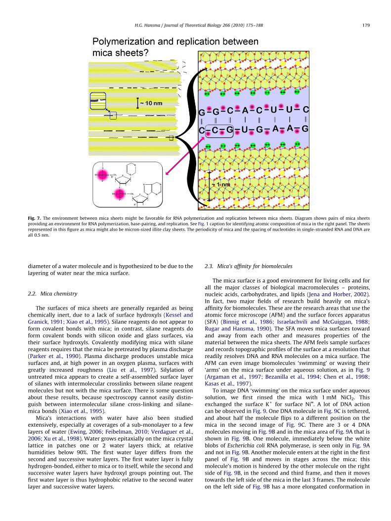
Fig. 7. The environment between mica sheets might be favorable for RNA polymerization and replication between mica sheets. Diagram shows pairs of mica sheets
providing an environment for RNA polymerization, base-pairing, and replication. See Fig. 1 caption for identifying atomic composition of mica in the right panel. The sheets
represented in this figure as mica might also be micron-sized illite clay sheets. The periodicity of mica and the spacing of nucleotides in single-stranded RNA and DNA are
all 0.5 nm.
H.G. Hansma / Journal of Theoretical Biology 266 (2010) 175–188 179
diameter of a water molecule and is hypothesized to be due to thelayering of water near the mica surface.
2.2. Mica chemistry
The surfaces of mica sheets are generally regarded as beingchemically inert, due to a lack of surface hydroxyls (Kessel andGranick, 1991; Xiao et al., 1995). Silane reagents do not appear toform covalent bonds with mica; in contrast, silane reagents doform covalent bonds with silicon oxide and glass surfaces, viatheir surface hydroxyls. Covalently modifying mica with silanereagents requires that the mica be pretreated by plasma discharge(Parker et al., 1990). Plasma discharge produces unstable micasurfaces and, at high power in an oxygen plasma, surfaces withgreatly increased roughness (Liu et al., 1997). Silylation ofuntreated mica appears to create a self-assembled surface layerof silanes with intermolecular crosslinks between silane reagentmolecules but not with the mica surface. There is some questionabout these results, because spectroscopy cannot easily distin-guish between intermolecular silane cross-linking and silane-mica bonds (Xiao et al., 1995).
Mica’s interactions with water have also been studiedextensively, especially at coverages of a sub-monolayer to a fewlayers of water (Ewing, 2006; Feibelman, 2010; Verdaguer et al.,2006; Xu et al., 1998). Water grows epitaxially on the mica crystallattice in patches one or 2 water layers thick, at relativehumidities below 90%. The first water layer differs from thesecond and successive water layers. The first water layer is fullyhydrogen-bonded, either to mica or to itself, while the second andsuccessive water layers have hydroxyl groups pointing out. Thefirst water layer is thus hydrophobic relative to the second waterlayer and successive water layers.
2.3. Mica’s affinity for biomolecules
The mica surface is a good environment for living cells and forall the major classes of biological macromolecules – proteins,nucleic acids, carbohydrates, and lipids (Jena and Horber, 2002).In fact, two major fields of research build heavily on mica’saffinity for biomolecules. These are the research areas that use theatomic force microscope (AFM) and the surface forces apparatus(SFA) (Binnig et al., 1986; Israelachvili and McGuiggan, 1988;Rugar and Hansma, 1990). The SFA moves mica surfaces towardand away from each other and measures properties of thematerial between the mica sheets. The AFM feels sample surfacesand records topographic profiles of the surface at a resolution thatreadily resolves DNA and RNA molecules on a mica surface. TheAFM can even image biomolecules ‘swimming’ or waving their‘arms’ on the mica surface under aqueous solution, as in Fig. 9(Argaman et al., 1997; Bezanilla et al., 1994; Chen et al., 1998;Kasas et al., 1997).
To image DNA ‘swimming’ on the mica surface under aqueoussolution, we first rinsed the mica with 1 mM NiCl2. Thisexchanged the surface K+ for surface Ni++. A lot of DNA actioncan be observed in Fig. 9. One DNA molecule in Fig. 9C is tethered,and about half the molecule flips to a different position on themica in the second image of Fig. 9C. There are 3 or 4 DNAmolecules moving in Fig. 9B and in the mica area of Fig. 9A that isshown in Fig. 9B. One molecule, immediately below the whiteblobs of Escherichia coli RNA polymerase, is seen only in Fig. 9Aand not in Fig. 9B. Another molecule enters at the right in the firstpanel of Fig. 9B and moves in stages across the mica; thismolecule’s motion is hindered by the other molecule on the rightside of Fig. 9B, in the second and third frame, and then it movestowards the left side of the mica in the last 3 frames. The moleculeon the left side of Fig. 9B has a more elongated conformation in

Fig. 8. The environment between mica sheets might be good for ribozyme
replication, migration, ligation and evolution in an RNA World. Diagram shows
mica sheets providing an environment for: A, ribozyme replication and migration
to other niches between mica sheets; B, ribozymes evolving in two of the niches
into different ribozymes and replicating within these niches; C, ribozymes
migrating to another occupied niche and ligating with a ribozyme in this niche
to form a new longer ribozyme. Mica provides massive redundancy in the many
spaces between its sheets.
Fig. 9. DNA swimming on mica. Time-lapse movie of double-stranded DNA
molecules moving on mica. White blobs are molecules of E. coli RNA Polymerase.
Images were taken at successive intervals of �1 min. A. This image shows the DNA
molecules at the beginning of the movie. B and C show selected successive frames
from the movie. Blue arrows indicate which regions of the field in A are seen in B
and C. Experimental details are in Bezanilla et al. (1994). (For interpretation of the
references to colour in this figure legend, the reader is referred to the web version
of this article.)
H.G. Hansma / Journal of Theoretical Biology 266 (2010) 175–188180
Fig. 9A and then remains in a somewhat compact conformationfor the last 4 frames of Fig. 9B.
DNA binds weakly to mica in Mg++ or Ca++ and strongly in Ni++, Co++
or Zn++ (Hansma and Laney, 1996). Cation-assisted binding of DNAto mica correlates with the enthalpy of hydration and the radius ofthe cation (Fig. 10). Ni++ also affects the sequence-dependentconformations of DNA on mica (Sitko et al., 2003). Oligomericnucleic acids as small as 25 mers bind readily to mica (Hansmaet al., 1996). RNA is likely to have similar interactions with divalentcations on mica; but this old work was done with DNA, which isnot cleaved by the RNases that are ubiquitous in today’s world.Research with RNA on mica includes tecto-RNA structures andtobacco mosaic virus RNA (Chworos et al., 2004; Drygin et al.,1998).
Mica also binds other biomolecules, including proteins andcarbohydrates (Chen and Hansma, 2000; Drake et al., 1989; Kirbyet al., 1995; McIntire et al., 1995). Laminin is a cross-shaped
protein from the extracellular matrix that waves its arms underaqueous solution in the AFM (Chen et al., 1998).
Mica is a preferred substrate for depositing lipid layers withthe Langmuir–Blodgett technique, including monolayers, vesicles,bilayers, and multilayers (Hansma et al., 1991; Richter andBrisson, 2005; Zasadzinski et al., 1994). Surfactant micellesabove the critical micellar concentration assemble on mica as
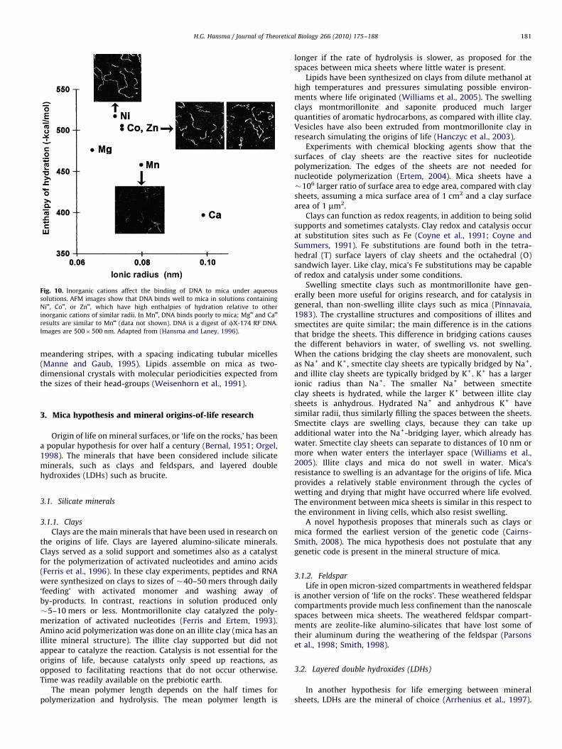
Fig. 10. Inorganic cations affect the binding of DNA to mica under aqueous
solutions. AFM images show that DNA binds well to mica in solutions containing
Ni++, Co++, or Zn++, which have high enthalpies of hydration relative to other
inorganic cations of similar radii. In Mn++, DNA binds poorly to mica; Mg++ and Ca++
results are similar to Mn++ (data not shown). DNA is a digest of fX-174 RF DNA.
Images are 500�500 nm. Adapted from (Hansma and Laney, 1996).
H.G. Hansma / Journal of Theoretical Biology 266 (2010) 175–188 181
meandering stripes, with a spacing indicating tubular micelles(Manne and Gaub, 1995). Lipids assemble on mica as two-dimensional crystals with molecular periodicities expected fromthe sizes of their head-groups (Weisenhorn et al., 1991).
3. Mica hypothesis and mineral origins-of-life research
Origin of life on mineral surfaces, or ‘life on the rocks,’ has beena popular hypothesis for over half a century (Bernal, 1951; Orgel,1998). The minerals that have been considered include silicateminerals, such as clays and feldspars, and layered doublehydroxides (LDHs) such as brucite.
3.1. Silicate minerals
3.1.1. Clays
Clays are the main minerals that have been used in research onthe origins of life. Clays are layered alumino-silicate minerals.Clays served as a solid support and sometimes also as a catalystfor the polymerization of activated nucleotides and amino acids(Ferris et al., 1996). In these clay experiments, peptides and RNAwere synthesized on clays to sizes of �40–50 mers through daily‘feeding’ with activated monomer and washing away ofby-products. In contrast, reactions in solution produced only�5–10 mers or less. Montmorillonite clay catalyzed the poly-merization of activated nucleotides (Ferris and Ertem, 1993).Amino acid polymerization was done on an illite clay (mica has anillite mineral structure). The illite clay supported but did notappear to catalyze the reaction. Catalysis is not essential for theorigins of life, because catalysts only speed up reactions, asopposed to facilitating reactions that do not occur otherwise.Time was readily available on the prebiotic earth.
The mean polymer length depends on the half times forpolymerization and hydrolysis. The mean polymer length is
longer if the rate of hydrolysis is slower, as proposed for thespaces between mica sheets where little water is present.
Lipids have been synthesized on clays from dilute methanol athigh temperatures and pressures simulating possible environ-ments where life originated (Williams et al., 2005). The swellingclays montmorillonite and saponite produced much largerquantities of aromatic hydrocarbons, as compared with illite clay.Vesicles have also been extruded from montmorillonite clay inresearch simulating the origins of life (Hanczyc et al., 2003).
Experiments with chemical blocking agents show that thesurfaces of clay sheets are the reactive sites for nucleotidepolymerization. The edges of the sheets are not needed fornucleotide polymerization (Ertem, 2004). Mica sheets have a�106 larger ratio of surface area to edge area, compared with claysheets, assuming a mica surface area of 1 cm2 and a clay surfacearea of 1 mm2.
Clays can function as redox reagents, in addition to being solidsupports and sometimes catalysts. Clay redox and catalysis occurat substitution sites such as Fe (Coyne et al., 1991; Coyne andSummers, 1991). Fe substitutions are found both in the tetra-hedral (T) surface layers of clay sheets and the octahedral (O)sandwich layer. Like clay, mica’s Fe substitutions may be capableof redox and catalysis under some conditions.
Swelling smectite clays such as montmorillonite have gen-erally been more useful for origins research, and for catalysis ingeneral, than non-swelling illite clays such as mica (Pinnavaia,1983). The crystalline structures and compositions of illites andsmectites are quite similar; the main difference is in the cationsthat bridge the sheets. This difference in bridging cations causesthe different behaviors in water, of swelling vs. not swelling.When the cations bridging the clay sheets are monovalent, suchas Na+ and K+, smectite clay sheets are typically bridged by Na+,and illite clay sheets are typically bridged by K+. K+ has a largerionic radius than Na+. The smaller Na+ between smectiteclay sheets is hydrated, while the larger K+ between illite claysheets is anhydrous. Hydrated Na+ and anhydrous K+ havesimilar radii, thus similarly filling the spaces between the sheets.Smectite clays are swelling clays, because they can take upadditional water into the Na+-bridging layer, which already haswater. Smectite clay sheets can separate to distances of 10 nm ormore when water enters the interlayer space (Williams et al.,2005). Illite clays and mica do not swell in water. Mica’sresistance to swelling is an advantage for the origins of life. Micaprovides a relatively stable environment through the cycles ofwetting and drying that might have occurred where life evolved.The environment between mica sheets is similar in this respect tothe environment in living cells, which also resist swelling.
A novel hypothesis proposes that minerals such as clays ormica formed the earliest version of the genetic code (Cairns-Smith, 2008). The mica hypothesis does not postulate that anygenetic code is present in the mineral structure of mica.
3.1.2. Feldspar
Life in open micron-sized compartments in weathered feldsparis another version of ‘life on the rocks’. These weathered feldsparcompartments provide much less confinement than the nanoscalespaces between mica sheets. The weathered feldspar compart-ments are zeolite-like alumino-silicates that have lost some oftheir aluminum during the weathering of the feldspar (Parsonset al., 1998; Smith, 1998).
3.2. Layered double hydroxides (LDHs)
In another hypothesis for life emerging between mineralsheets, LDHs are the mineral of choice (Arrhenius et al., 1997).

H.G. Hansma / Journal of Theoretical Biology 266 (2010) 175–188182
LDHs, like clays, are layered minerals. LDHs include brucite, whichis the solid mineral form of Mg(OH)2, and other minerals such asmagnesium aluminum oxides (Arrhenius et al., 1997; Sideriset al., 2008) Unlike clays, the sheets of LDHs have a positivesurface charge, neutralized by anions between the mineral layers.They have been proposed as sites for the origins of life because ofthe affinity of cationic surfaces for anionic polymers. Manybiological lipids and other biopolymers such as DNA and RNAare anionic. This is the opposite of the mica hypothesis, becausethe mica hypothesis says that anionic polymers evolved on ananionic mineral surface with mobile cationic bridges.
3.3. Life’s origins on positively vs. negatively charged mineral
surfaces
In life today, when biopolymers of one charge interact withminerals of the opposite charge, stable biomineralized structuresform. For example, bones and teeth have anionic aspartate-richprotein polymers that provide the matrix for the Ca++-rich cationicmineral phase (Weiner, 2008). Similarly, there are stable Si-richstructures in organisms such as diatoms and sponges. Thepredominant proteins in biomineralized Si are silicateins, whichhave a positive charge at neutral pH (Shimizu et al., 1998).Negatively charged silicates in vitro also form composites withpositively charged proteins (Bassindale et al., 2009).
In contrast, in the mica hypothesis, mobile inorganic cationsbridge the anionic prebiotic polymers to the life-supportinganionic mineral surface. Instead of a stable electrostatic attrac-tion, mica provides transient interactions with anionic polymersvia a variety of mobile inorganic cations and a gentle entrapmentof the anionic polymers within the physical space between themica sheets. Transient interactions can have sensitive responsesto the environment and are seen today in the many transientinteractions that regulate life, such as the DNA–ligand interac-tions that regulate gene expression. Therefore, according to themica hypothesis, the anionic polymers of life originated on ananionic mineral surface.
4. Mica world
The following sections describe the hypothetical Mica Worldand the relationship of prior research to the origin of life in a micaworld. In the mica world, life evolved in large part from theorganic molecules that were synthesized in ‘molecule mashers.’These molecule mashers are pairs of mica sheets that move upand down, towards each other and away from each other,mashing together and pulling apart whatever molecules are inthe way. The energy input to the molecules is mechanical, in theform of work.
4.1. Energy, work, and mechanochemistry
Energy input is usually necessary for chemical reactions tooccur and for physical processes to occur that increase the orderof a system. In the mica world, a major energy input is mechanicalenergy from moving mica sheets. In thermodynamics, Free energy(DG) is a measure of the ease with which a chemical reaction willoccur. Thermodynamic free energy is the sum of enthalpy andentropy terms (DG¼DH�TDS, where DH is the enthalpy changeand TDS is the product of entropy change and temperature).Energy is used, in this paper, to refer to the free energy of areaction or process, as in the schematic free energy diagram of(Schnitzer et al., 2000) and in Fig. 4A. In at least one system (Bestand Clarke, 2002), free energy includes solvation, electrostatic
effects, disperson forces, and entropy of the polymer chain. Therelationship between free energy and mechanochemistry isanalyzed in (Keller and Bustamante, 2000), which points out thatmechanochemistry is involved in any enzymatic reaction thatinvolves a conformation change of the enzyme.
The following discussion of work from mica sheets is quitelacking in quantitative analysis, partly due to the properties ofmica. Work is the product of a force and a distance. The force, F,exerted by the mica depends on the spring constant, k, of themica: F¼kx, where x is the distance of the displacement of themica. The spring constant depends on the thickness and length ofthe mica lever. Thickness will vary over a wide range, dependingon the number of nm-thick mica sheets in each mica lever, andlength will vary among mica levers and even within a single micalever, depending on the energy of the water or heat that pushesthe mica sheets apart and the strength of adhesion between themica sheets that are being pushed apart. These sheets, in Fig. 3A,are labeled ‘y’ and ‘z’ in the regions of the sheets that affect thelength of the mica lever. The mica sheets at y and z will interactmore or less strongly as K+ exchanges with other ions and asorganic and inorganic matter adsorb to or desorb from the micasheets. The length of the mica lever will be shorter if the micasheets interact strongly at y and z (i.e., are ‘stuck’ together), andthe length of lever will be longer if the mica sheets can separate atpositions y and z and at positions on the sheets that are fartherinto the mica ‘book.’
Simple up-and-down movements of mica sheets may havebeen a major form of energy for the origins of life (Fig. 3). Thisenergy is endlessly renewable and potentially capable of carryingout many types of chemistry. Work from moving mica sheetsmight have formed many of the prebiotic covalent bonds. Thesecovalent bonds would have formed at stages in the synthesis ofmonomers and/or in the joining of monomers to form oligomersand larger polymers. Any small or large molecules with sufficientaffinity to mica should be able to form covalent bonds with eachother through mica’s movements if sufficient energy from mica’smovements pushed the molecules into each other as opposed todislodging the molecules from the mica sheets.
The up-and-down movements of mica sheets would have beenpowered by water motion and cyclic solar energy (Hansma, 2009). Asdescribed above, some of the resulting mechanical energy might beused to form bonds between molecules. This is mechanochemistry(Figs. 3 and 4). Mechanochemistry is a growing field, in whichmechanical energy is being used to make or break bonds (Hickenbothet al., 2007; Rosen and Percec, 2007), sometimes using single-molecule techniques (Grandbois et al., 1999; Lenhardt and Craig,2009; Yang et al., 2009). Angstrom-sized motions can form covalentbonds, as envisioned in Fig. 4. Nanometer-sized motions alter theconformations of polymers (Fig. 3). Micron-sized motions can bleb offvesicles (Fig. 2A) or protocells (Fig. 2B) between mica sheets andwould also manipulate the macromolecular aggregates or ‘blobs ofgoo’ that often result from prebiotic chemistry experiments in the lab(Hazen, 2005). Mechanical manipulation might rearrange the goo intoprimitive structures.
Two sources of energy for mechanochemistry are shown inFig. 3. Water movements in and out at the edges of mica sheetspush the mica sheets apart and pull them together (Fig. 3A).Bubbles in mica sheets expand and contract in response to theheating and cooling of day and night (Fig. 3B). These cyclingbubbles are heat pumps, which have been proposed as an energysource for life’s origins (Schulze-Makuch and Irwin, 2006).
Another source of energy between mica sheets comes frommeniscus forces, caused by surface tension at air-water interfaces,such as those indicated by the arrows in Fig. 6C. Air–water interfacesappear when water recedes or advances at the edges or in theinterstitial spaces of mica sheets. Meniscus forces were attractive

H.G. Hansma / Journal of Theoretical Biology 266 (2010) 175–188 183
forces of 10–100 nN when measured between mica and the probetip of an atomic force microscope (Weisenhorn et al., 1989).Therefore meniscus forces are strong enough to make and breakcovalent bonds, which rupture at �4 nN, according to theoreticalmodeling for C–C and C–N bonds (Grandbois et al., 1999). Meniscusforces or convection forces in an evaporating droplet have been usedto stretch DNA molecules (Wang et al., 1998).
4.1.1. Mechanochemistry research
In the lab, mechanochemical research usually results in bondsbeing broken, due to technical limitations; but bond formation isalso possible (Beyer and Clausen-Schaumann, 2005). When theenergy source is mechanical as opposed to thermal, the relativebond strengths are sometimes significantly different. This isdemonstrated by Molecular Dynamics simulations of thiols oncopper, in which carbon–sulfur bonds cleave when heat is theenergy source, while copper–copper bonds cleave when the energysource is an upward-moving mechanical force (Konopka et al.,2008). In another example of mechanochemistry, ultrasonic forcesare better than heat at activating a catalyst by dissociating its ligand(Piermattei et al., 2009). Disadvantages of ultrasonic mechanochem-istry are its irreversibility and lack of control over the mechan-ochemical forces. Simple up-and-down movements of mica sheetswould provide more reproducible mechanical forces, distances, anddirections, as compared with ultrasonic mechanochemistry.
4.1.2. Entropy
Entropy is discussed separately from energy, even thoughentropy is a term in the thermodynamic equation for free energy.Thermodynamics describes systems at equilibrium, and life is notat equilibrium. One example where entropy is not described wellby the equation for free energy (DG¼DH�TDS) is in single-molecule pulling experiments: The energy to stretch a polymer istypically an order of magnitude larger than the typical free energyof unfolding, as measured from the area under the force-vs.-distance pulling curve (Thompson et al., 2002). The experimentwas done by pulling a protein molecule with the probe tip of anAFM, stretching the molecule between the surface of the substrateand the tip of the AFM probe. The best explanation for thepeculiarly high energy is that there is a huge reduction in entropyas the protein molecule is extended nearly completely. Theprotein molecule experiences changes in entropy rather like thehypothetical polymer molecule in Fig. 5, as it goes from being freein solution to extended between mica sheets.
One challenge with origins-of-life hypotheses is that entropy islow in living systems (Arrhenius et al., 1997; Root-Bernstein andDillon, 1997). As the diagram in Fig. 5 shows, the entropy ofpolymers decreases progressively as they move from being free insolution, to tethered on a surface, to confined between sheets,such as mica. Entropy measures molecules’ freedom of motion,both internal and translational, and this freedom of motion ismuch lower in the crowded intracellular environment of aunicellular organism than in its aqueous extracellular environ-ment. Protein molecules in a cell are typically so close that there isonly a space equal to the size of one protein molecule betweenthem (Phillips et al., 2008). Entropy reduction in today’s livingcells requires enzymatic systems with complex coupling tochemical energy. Confinement between mica sheets may havebeen a mechanism for reducing entropy before complex enzy-matic systems existed.
Entropy also may have driven some molecular ordering duringprebiotic molecular evolution between mica sheets. Overcrowd-ing can produce entropy-driven molecular order, as in thenematic-to-smectic phase transitions of liquid crystals and thealignment of elongated molecules on a membrane (Almeida andWiegel, 2006).
4.1.3. Confinement effects
Entropy reduction between mica sheets also produces chemi-cal confinement effects. Chemistry in confined spaces is moreselective than chemistry in solution. Zeolites, for example, areporous crystalline alumino-silicates that produce fewer productsfrom reactants, as compared with the reaction in solution (Turro,2000). Proteins in confined spaces are stabilized against dena-turation (Thirumalai et al., 2003; Zhou and Dill, 2001). Pressure isa form of confinement that induces actin polymerization andglycine polymerization (Cipolla et al., 2002; Ohara et al., 2007).
Confinement between mineral sheets may also favor somechemical reactions over others, thus reducing the number ofreaction products, as compared with the same reaction in a fluidor on a surface. One problem with the origins of life is the largenumber of possible organic molecules as opposed to the smallernumber of molecules actually found in living systems. Aminoacids produced in the Urey–Miller electric discharge experiments,for example, are primarily amino acids not found in proteins, suchas b-alanine and norvaline (Johnson et al., 2008). Somewhere inthe origins of life, the possible diversity of amino acids in proteinswas reduced to a subset of �20 different L-a-amino acids.
4.1.4. Chirality
Confinement between mica sheets might be the way in whichlife evolved with chiral polymers. Adjacent mica sheets imposesteric and chemical constraints on the molecules between them.These constraints might bias monomer packing such thatmonomers of the same handedness would pack closer on micasheets than their mirror–image enantiomers, thus facilitatingbond formation between same-handed monomers. This argu-ment, that chirality can come from confinement, is an alternativeto other work on the origins of chirality. One proposal for theorigins of chirality involves chiral mineral surfaces. Chiral surfacesof calcite give a few percent enrichment of the D- or the L-isomerof aspartic acid (Hazen, 2005; Hazen and Sholl, 2003). Anotherproposal for the origins of chirality involves the differentialsolubility of chiral and racemic organic crystals (Breslow andCheng, 2009).
4.1.5. Solid phase synthesis
Mica is a highly structured solid support that provides anamazing environment for solid phase synthesis. Solid phasesynthesis is used experimentally and industrially to avoidproblems with synthesis in solution (Bergbreiter and Kobayashi,2009). The mica environment contains a mechanical energysource applied to spaces consisting of parallel structured mineralsurfaces. The ceilings and floors of the spaces alternately squeezeand stretch whatever is within the spaces.
The ceilings and floors of the spaces between mica sheets arealso anionic grids with periodicities of 0.5 nm, similar to theperiodicities of monomers in biopolymers such as nucleic acidsand peptides. Therefore the biopolymers being synthesized wouldbe able to interact directly with the mica ceiling and the micafloor of the molecule masher via specific chemical interactions.
4.1.6. Molecular complementarity
Molecular complementarity fits nicely with the mica world.Molecular complementarity is a way of unifying many aspects oflife’s origins and dealing with their problems by treatingmolecules not as individuals but as complements to othermolecules (Dillon and Root-Bernstein, 1997; Hunding et al.,2006; Root-Bernstein and Dillon, 1997). Molecular complemen-tarity is a ubiquitous phenomenon that stabilizes moleculesagainst degradation, because the molecules are part of largercomplexes of molecules. Molecular complementarity results in

H.G. Hansma / Journal of Theoretical Biology 266 (2010) 175–188184
Darwinian natural selection in much the same way as the micaworld: molecules that associate with other molecules – or withmica sheets – are selected for, and molecules that do not associatewith other molecules or with mica sheets are lost. Mica world isan analog system, like living systems, with continuous variationsin quantities, rates, lengths and other variables.
4.2. Wet–dry cycles in the origin of life
Cycles of wetting and drying are ubiquitous on earth today andwere likely to be present on the primordial earth as well. Theabsence of water favors polymerization, and the presence of waterfavors hydrolysis of polymers. For example, drying of the aminoacid alanine produces alanine di-peptides (Napier and Yin, 2006).Origins research has used wet–dry cycles to encapsulate an RNApolymerase in lipid vesicles and to synthesize an RNA-likepolymer non-enzymatically from a nucleotide monophosphate,when stabilized in layers with lipids (Chakrabarti et al., 1994;Rajamani et al., 2008).
Although water is necessary to life, cells regulate their watercontent to prevent the accumulation of water to toxic levels. Highconcentrations of solute and careful regulation of water content inthe cytoplasms of living cells are consistent with life’s origins inlimited water, not exposed to prolonged wet and dry periods.
4.2.1. Fluid flow
Besides applying mechanical energy to the material betweenthe mica sheets, the squeezing and stretching of material betweenmica sheets involves fluid flow between the sheets (Fig. 3). Fluidflow would exchange unbound or soluble material between thesheets with material in the fluid outside the mica. Such fluid flowwould remove molecules not bound to mica and might bring innew monomers or other molecules for reaction. Such ‘feeding’ andwashing has been used to synthesize biopolymers on clays tolengths greater than the lengths synthesized in solution (Ferriset al., 1996).
Water does not enter the spaces between mica sheets at avisible level even after long incubation times (Gaines, 1957).There are exceptions, however. Water rapidly entered betweenthe sheets of the low-grade mica in Fig. 1D, which was collectedfrom an abandoned mica mine. Water slowly entered between thesheets of high-grade mica when the mica was submerged in wateror aqueous salt solutions and subjected to daily temperaturechanges (data not shown). After two weeks of daily cyclingbetween room temperature and �4 1C, water or salt solutionentered a few millimeters into the spaces between the sheets.Water entered farther into the mica sheets when the submergedsheets were cycled daily between freezing and boiling watertemperatures. With these temperature extremes, water entered acentimeter or more into the spaces between the sheets. The micaused in these experiments was like the mica pieces in Fig. 6.
The mica pieces in Fig. 6 were treated in the opposite way:drying was measured instead of wetting. The mica pieces weresandwiched together with water and allowed to dry in air. Waterevaporated slowly from the spaces between mica sheets, oftenforming intricate patterns of wet and dry areas in the process(Fig. 6). The water evaporated completely within 12�24 h. Theseresults show how mica provides both wet environments and dryenvironments simultaneously in close proximity.
4.3. Internal potassium ions
Living cells have high internal K+ concentrations. Where didthis come from? Blood and extracellular fluids are high in Na+,supposedly because life originated in the Na+-rich ocean, but
there is no corresponding theory for why the intracellular fluid ishigh in K+.
Mica sheets are held together by K+, which provides a goodenvironment for molecules destined to exist in a K+-richcytoplasm. Muscovite mica has a K+ concentration of �100 mMbetween adjacent sheets that are separated to a distance of�0.7 nm. This is calculated from the 0.5-nm hexagonal spacing ofK+ between mica sheets. Intracellular K+ concentrations in livingcells are �100 mM. Although some clay minerals are also rich inK+, these clays only have small micron-sized sheets.
4.4. Possible synthesis of biomolecules between mica sheets
The spaces between mica sheets, their 0.5-nm anionic grid, andtheir chemical composition may have supported the synthesis ofthe earliest biomolecules and biopolymers, with energy suppliedby mechanochemistry in the mica molecule mashers. In thisrespect, the mica hypothesis resembles the metabolic originsproposed by others, in which a useful subset of small biomole-cules were synthesized from smaller organic and inorganicmolecules (Smith et al., 2009; Wachtershauser, 2007). Transitionmetal catalysis is key to these approaches, and life is viewed as anatural outcome of the chemical and energetic processes on theearly earth.
For example, Lewis acid sites provided by a divalent metalcation on a surface can be a component for non-enzymaticcatalysis of thiol-mediated reduction reactions in the reductivetri-carboxylic acid (rTCA) cycle proposed for life’s origins (Smithet al., 2009). Divalent metal cations stabilize negative charges,thus facilitating some substitution and elimination reactions oforganic molecules.
The following sections discuss the possible synthesis ofpeptides, nucleotides, oligonucleotides, and ribozymes betweenmica sheets.
4.4.1. Peptides
Peptide synthesis, when confined between mica sheets, mayfavor the polymerization of only a-amino acids. This hypothesis isdiagrammed in Fig. 4 for alanine and polyalanine but is applicableto other amino acids, too. If moving mica sheets can forcemolecules close enough to reach the attractive regime of thepotential energy well, then covalent bonds may form. In Fig. 4, acovalent peptide bond forms by mechanochemistry. Whether ornot this is possible, moving mica sheets would move molecules onadjacent sheets toward and away from each other.
The free energy of hydrolysis of the peptide amide bond (DGm)actually favors the synthesis of amide bonds: DGm¼5–6 kcal/molat 25 1C or 37 1C (Martin, 1998). The driving energy for hydrolysisis the ionization of the carboxyl and amino groups produced byhydrolysis of the amide bond, for which DGi��8 kcal/mol. Theseresults are for several hydrolysis reactions of glycine (G) dimersand glycine oligomers, such as
GG-G+G
GGGG-GG+GG
GGGG-GGG+G
Considering the reverse reactions, as in prebiotic syntheses,the most energetically favorable reaction is the joining of 2peptides, each of which is a dimer or larger. Forming the dimers isthe most energetically difficult. Although the calculations are forglycine and its oligomers, other amino acids should behavesimilarly (Martin, 1998).

H.G. Hansma / Journal of Theoretical Biology 266 (2010) 175–188 185
Good sites for amino acid polymerization on alumino-silicatesconsist of adjacent Lewis acid and Bronsted acid sites (Rimolaet al., 2007). This was modeled ab initio for the formation of asimplified analog of a glycine–glycine dipeptide. The authorspropose that the dipeptide chain would proceed to elongate onthe silicate surface due to the dipeptide’s affinity for the silicatesurface.
4.4.2. Nucleotides
Nucleotides contain a purine or pyrimidine base and aphosphate attached to a sugar, which is ribose in RNA anddeoxyribose in DNA. Prebiotic synthesis of these complexmolecules is an unanswered question in the origins of life. Eventhe prebiotic syntheses of the bases and ribose are unansweredquestions.
Ribose and other sugars could have been synthesized pre-biotically via the formose reaction. The formose reaction is anautocatalytic reaction that produces, in solution, a large andunstable diversity of racemic branched and unbranched sugars(Joyce, 2002b; Ricardo et al., 2004). Aqueous silicate solutionshave recently been shown to benefit the formose reaction invarious ways related to the origins of life (Lambert et al., 2010).Silicate stabilizes some products of the formose reaction; silicatereduces the complexity of the reaction products, and silicate evenselects for some stereoisomers. Five-carbon (5-C) sugars includingribose are a large part of the product when the reaction mixturestarts with equimolar concentrations of 2-C and 3-C sugars.
Mica, being a highly structured aluminosilicate mineral, mightconfer even greater benefits on the formose reaction, with regardto producing ribose for RNA in life’s origins. In another recentsolution to the ribose problem, direct ribose synthesis iseliminated altogether in a reaction pathway for synthesizingentire nucleotides (Powner et al., 2009).
4.4.3. Oligonucleotides
Nucleotide polymerization produces oligonucleotides andlarger molecules of RNA and DNA. Nucleotide polymerizationmay also benefit from the confines of mica sheets and their clay-mineral lattice structures (Fig. 7). Nucleotide polymerization insolution often produces bent oligonucleotides with unnaturallinkages in addition to the 30-50 linkage found in nucleic acids(Joyce and Orgel, 2006). These unnatural linkages include 50, 50
pyrophosphate linkages and 20-50 linkages.The periodicity of phosphate groups in extended oligonucleo-
tides and single-stranded nucleic acids (0.6 nm) is similar to the0.5-nm periodicity of anionic sites on mica sheets (Calladine andDrew, 1997). Therefore one might expect better binding to micafor linear oligonucleotides having only a single phosphate betweeneach pair of nucleotides. Polymerization of nucleotides arrayed onthe 0.5-nm mica lattices and confined between mica sheets mightproduce mostly linear unbent oligonucleotides. Thus, the confinesof mica sheets and their clay-mineral lattice chemistry mayfunction to reduce the number of different oligonucleotidelinkages that form during nucleotide polymerization.
4.4.4. Ribozymes
Ribozymes may have evolved when life originated to do thecatalytic work now done by protein enzymes as well as theinformation storage now done by DNA (Gesteland et al., 2006;Gilbert, 1986). Ribozymes are catalytic RNA molecules that mayhave catalyzed the synthesis and replication of RNA molecules inan early RNA World. RNA self-replication in solution is hinderedby intramolecular base pairing of RNA oligonucleotides (Joyce,2002b). Confinement between mica sheets could reduce theproblem of intramolecular base pairing, as diagrammed in Fig. 7.
Cation-mediated interactions between oligonucleotides and micamight compete successfully with intramolecular oligonucleotideinteractions.
4.4.5. RNA world: ribozyme replication and evolution
The many spaces between mica sheets provide niches for thereplication and evolution of many different ribozymes atrelatively high concentrations and in close proximity (Fig. 8). Thismight solve a problem with the origin of life: how to generate alarge genome without accurate replication machinery, and how togenerate accurate replication machinery without having a largegenome. Modeling shows that a population of different self-replicating ribozymes in close proximity degenerate, duringrepeated replication, into the same ribozyme (Joyce, 2002a; Szaboet al., 2002). This happens because a small ‘selfish’ ribozymeexcels at getting itself replicated by all the other ribozymes, whichthen become extinct. This has now been demonstrated experi-mentally with pairs of ribozymes that can catalyze each other’sreplication by ligation (Lincoln and Joyce, 2009). A few of thesepairs eventually dominated the ribozyme population. With manyadjacent pairs of mica sheets, more pairs of ribozymes would beable to continue replicating, due to their isolation from competingribozymes. Ribozymes with enough time and space wouldgradually evolve large genomes with accurate replication ma-chinery. Mica’s compartments provide an environment in whichmultiple ribozymes could self-replicate, migrate, and ligate toevolve large genomes and accurate replication machineries,without facing extinction from competition with other ribozymes(Fig. 8).
Cycles of replication, migration, and ligation could havecreated great variety and massive redundancy in the ribozymeand RNA populations. Most ligations between RNAs would beexpected to produce ‘unsuccessful’ big RNAs that became extinct,but only an occasional successful ligation would have beenenough to generate large enough RNAs for growing a geneticmessage.
4.5. Prebiotic mica ‘cells’ and protocells
Before the existence of protocells enclosed by lipid membranes,mica’s thin flexible mineral sheets may have provided some of theprotection now provided by the lipid membranes. Life is soincredibly cellular today – multi-cellular even – that it makessense to imagine some form of cellularity in the early stages oflife’s origins, even before primitive biomolecules were enclosed inlipid bilayers. Forms of multi-cellularity are seen even in bacteriaand archaea today. Bacteria form biofilms. Biofilms of Pseudomonas
aeruginosa resist antibiotic treatment that kills free-livingP. aeruginosa. An extremophilic archaea, Pyrodictium abyssi,produces extracellular cannulae that hold clumps of the cellstogether in macroscopic cobweb-like flakes in the culture medium(Rieger et al., 1995). In the mica hypothesis, multi-cellularityexisted independently of lipids, in the multiple niches betweenmica sheets, as in the ‘RNA world’ diagrammed in Fig. 8.
Eventually the hypothetical mica world became populatedwith lipids. These lipids formed vesicles that encapsulatedmaterial between the mica sheets (Fig. 2A). This was at an earlystage in the origin of life, and the vesicles were still small. There isnothing resembling cytoplasm in the vesicles; they contain only acollection of molecules and some of the aqueous liquid betweenthe mica sheets. Later in the origins of life, lipids encapsulatedlarger and more complex biomolecules and biomacromolecules,including biopolymers and complex biomolecular aggregates, afew of which carried out primitive functions. At some point(Fig. 2B), the lipid-encapsulated structures were stable enough

H.G. Hansma / Journal of Theoretical Biology 266 (2010) 175–188186
that they began to develop a primitive cytoplasm, quite distinctfrom the aqueous fluid between the mica sheets. Most of theselipid-encapsulated structures were rather useless, but occasion-ally some of them contained the necessary ingredients forprimitive metabolism and became self-sustaining protocells. Weare living proof that these ancient protocells succeeded, in someancient environment, at replicating and transmitting what wenow call genetic information.
Mica also provides a possible solution to the problem of celldivision in protocells. Cells of wall-less L-form bacteria divide byblebbing off daughter cells (Leaver et al., 2009). This blebbingprocess is proposed to be a remnant of the earliest form of celldivision; but the question is raised, where did the energy forblebbing off come from? Mica sheets’ up-and-down movementscould have provided mechanical energy to bleb off daughter cells(Fig. 2B).
Most of the earliest daughter cells may have remained in place,simply pushing the mica sheets farther and farther apart.Daughter cells that drifted away from the ‘family’ are less likelyto have survived, until they had a robust biochemistry capable ofself-sustained metabolism and replication, and a robust structureable to resist external environmental forces.
5. Possible mica-origins research
What experimental research is needed to provide directsupport for the mica hypothesis? Mechanochemistry is onedistinguishing characteristic of the mica hypothesis. Mechan-ochemistry from moving mica sheets could be investigated in thelaboratory with approaches involving the AFM or the SFA.Prebiotic reaction mixtures between mica sheets could besubjected to cycles of pushing the mica surfaces together andpulling them apart. This process could go on for days, which is stilla very short period of time, compared with the time scales overwhich life originated. The SFA’s standard mechanism of operationis like the proposed mechanochemistry of moving mica sheets:the SFA pushes mica sheets toward and away from each other, incycles, to produce data about the interactions of the mica sheetsor the materials deposited onto them. The AFM would need asmall modification: one could take a cantilever with no tip, glueonto it a piece of mica, and use this as the top sheet of mica, whichwould press against the mica of the sample surface. This isreminiscent of the early days of AFM, when methods had not yetbeen developed for integrating tips into the micro-fabricatedcantilevers. Fragments of crushed diamond were glued onto thetipless cantilevers, using an eyelash hair glued to a small stick toput the glue on the cantilever (Drake et al., 1989). Nucleotidepolymerization is one system worth testing with mechanochem-istry and mica sheets, following the observations that nucleotidescan polymerize in other confined spaces such as pockets withinice or in combination with lipids (Monnard and Szostak, 2008;Rajamani et al., 2008). The failure rate of mica mechanochemistryexperiments will be high, as is typical of research into the originsof life.
Entropy reduction is another characteristic of the micahypothesis. Entropy reduction by confinement is accompaniedby chemical confinement effects, including the prediction thatchiral polymer syntheses would occur within the crystallineconfines of mica sheets. A prediction of entropy reduction is thatthe formose reaction for synthesizing sugars would produce fewerreaction products between mica sheets than in solution. Theformose reaction is perhaps the easiest place to start mechan-ochemical research with mica, too, because reaction products areeasily obtained with the formose reaction. Are there differencesbetween the products from the formose reaction between moving
mica sheets and the products of the formose reaction free insolution?
Other predictions of the mica hypothesis are that mica is thesource of high intracellular K+ levels and that the spaces betweenmica sheets functioned as primitive cells both before and afterlipid membranes. Thus the mica hypothesis opens new researchareas for origins of life research, but some of these areas will bevery difficult to test.
6. Concluding remarks
Natural complexity often arises from fundamentally simpleprocesses. Capillary action raises water to the tops of tall trees.Stochastic fluctuations in the numbers of molecules turn bacterialgenes on and off (Choi et al., 2008; Beaumont et al., 2009). Chargedensity in the minor groove guides DNA binding proteins to theirtarget site (Rohs et al., 2009; Tullius, 2009). An ion diffuses into anion channel and stabilizes a conformation of the channel until itdiffuses away.
Similarly, in the mica hypothesis, simple processes combine toform progressively more complex bioorganic structures. Micamicro-habitats fostered the evolution of considerable prebioticdiversity in a loose communal system for a long time. Theprotection afforded by mica sheets evolved into the walls andmembranes of cells. This subsequent origin of cells fostered thegeographic spread of life and its increasing complexity anddiversity.
The mica hypothesis views life’s origins as resembling anecosystem (Hunding et al., 2006; Woese, 2002) that evolved onlygradually into the autonomous cells of today’s primitive lifeforms. The mica hypothesis also provides a large error tolerance,through molecular redundancy in the multitudes of spacesbetween the mica sheets, as in Fig. 8A. Error tolerance is proposedto be the primary requirement for the origins of life, becausealmost everything will fail (Dyson, 1999).
Acknowledgments
Thank you to Arkadiusz Chworos, Ching Kung, Kevin Plaxco,Robert Geller, Nathan Fay, Sarah Bedichek, Dick Cousineau, DavidDeamer, Gozen Ertem, Jacob Israelachvili, Lynn Jelinski, Rachel Kaplan,Lori Kohlstaedt, Tonya Kuhl, Srin Manne, Stan Parsons, AndrewPohorille, Nina Thayer, Bob Hazen and many others for helpfuldiscussions; to the reviewers for their useful comments; to JimGreenwood, Joy Hansma, Rich Greenwood, and Connie Wieneke, forfinding the mica quarry; and to NSF BIO for support.
References
Almeida, P.F., Wiegel, F.W., 2006. A simple theory of peptide interactions on amembrane surface: excluded volume and entropic order. J. Theor. Biol. 238,269–278.
Argaman, M., Golan, R., Thomson, N.H., Hansma, H.G., 1997. Phase imaging ofmoving DNA molecules and DNA molecules replicated in the atomic forcemicroscope. Nucleic Acids Res. 25, 4379–4384.
Arrhenius, G., Sales, B., Mojzsis, S., Lee, T., 1997. Entropy and charge in molecularevolution—the case of phosphate. J. Theor. Biol. 187, 503–522.
Bassindale, A.R., Taylor, P.G., Abbate, V., Brandstadt, K.F., 2009. Simple and mildpreparation of silica–enzyme composites from silicic acid solution. J. Mater.Chem. 19, 7606–7609.
Beaumont, H.J.E., Gallie, J., Kost, C., Ferguson, G.C., Rainey, P.B., 2009. Experimentalevolution of bet hedging. Nature 462, 90–93.
Bergbreiter, D.E., Kobayashi, S., 2009. Introduction to facilitated synthesis. Chem.Rev. 109, 257–258.
Bernal, J.D., 1951. The Physical Basis of Life. Routledge & Kegan Paul, London.Best, R.B., Clarke, J., 2002. What can atomic force microscopy tell us about protein
folding? Chem. Commun. (Camb) 183–192.

H.G. Hansma / Journal of Theoretical Biology 266 (2010) 175–188 187
Beyer, M.K., Clausen-Schaumann, H., 2005. Mechanochemistry: the mechanicalactivation of covalent bonds. Chem. Rev. 105, 2921–2948.
Bezanilla, M., Drake, B., Nudler, E., Kashlev, M., Hansma, P.K., Hansma, H.G., 1994.Motion and enzymatic degradation of DNA in the atomic force microscope.Biophys. J. 67, 2454–2459.
Binnig, G., Quate, C.F., Gerber, C., 1986. Atomic force microscope. Phys. Rev. Lett.56, 930–933.
Breslow, R., Cheng, Z.L., 2009. On the origin of terrestrial homochirality fornucleosides and amino acids. Proc. Natl. Acad. Sci. USA 106, 9144–9146.
Cairns-Smith, A.G., 2008. Chemistry and the missing era of evolution. Chemistry14, 3830–3839.
Calladine, C.R., Drew, H.R., 1997. Understanding DNA : the Molecule and How itWorks. Academic Press, San Diego.
Chakrabarti, A.C., Breaker, R.R., Joyce, G.F., Deamer, D.W., 1994. Production of RNAby a polymerase protein encapsulated within phospholipid vesicles. J. Mol.Evol. 39, 555–559.
Chen, C.H., Hansma, H.G., 2000. Basement membrane macromolecules: insightsfrom atomic force microscopy. J. Struct. Biol. 131, 44–55.
Chen, C.H., Clegg, D.O., Hansma, H.G., 1998. Structures and dynamic motion of laminin-1 as observed by atomic force microscopy. Biochemistry 37, 8262–8267.
Chen, I.A., Roberts, R.W., Szostak, J.W., 2004. The emergence of competitionbetween model protocells. Science 305, 1474–1476.
Choi, P.J., Cai, L., Frieda, K., Xie, X.S., 2008. A stochastic single-molecule eventtriggers phenotype switching of a bacterial cell. Science 322, 442–446.
Chworos, A., Severcan, I., Koyfman, A.Y., Weinkam, P., Oroudjev, E., Hansma, H.G.,Jaeger, L., 2004. Building programmable Jigsaw puzzles with RNA. Science 306,2068–2072.
Cipolla, M.J., Gokina, N.I., Osol, G., 2002. Pressure-induced actin polymerization invascular smooth muscle as a mechanism underlying myogenic behavior.FASEB J. 16, 72–76.
Coyne, L., Mariner, R., Rice, A., 1991. Air-oxidation of hydrazine. 1. Reactionkinetics on natural kaolinites, halloysites, and model substituent layers withvarying iron and titanium oxide and oxygen(1-) center contents. Langmuir 7,1660–1674.
Coyne, L.M., Summers, D.P., 1991. Surface activation of air-oxidation of hydrazineon kaolinite. 2. Consideration of oxidizing/reducing entities in relationship toother compositional, structural, and energetic factors. Langmuir 7, 1675–1688.
Dillon, P.F., Root-Bernstein, R.S., 1997. Molecular complementarity – II: Energeticand vectoral basis of biological homeostasis and its implications for death.J. Theor. Biol. 188, 481–493.
Drake, B., Prater, C.B., Weisenhorn, A.L., Gould, S.A., Albrecht, T.R., Quate, C.F.,Cannell, D.S., Hansma, H.G., Hansma, P.K., 1989. Imaging crystals, polymers,and processes in water with the atomic force microscope. Science 243,1586–1589.
Drygin, Y.F., Bordunova, O.A., Gallyamov, M.O., Yaminsky, I.V., 1998. Atomic forcemicroscopy examination of tobacco mosaic virus and virion RNA. FEBS Lett.425, 217–221.
Ducker, W.A., Pashley, R.M., 1989. The forces between mica surfaces inammonium chloride solutions. J. Colloid Interface Sci. 131, 433–439.
Dyson, F.J., 1999. Origins of Life. Cambridge University Press, Cambridge(England), New York.
Ertem, G., 2004. Montmorillonite, oligonucleotides, RNA and origin of life. Orig.Life Evol. Biosph. 34, 549–570.
Ewing, G.E., 2006. Ambient thin film water on insulator surfaces. Chem. Rev. 106,1511–1526.
Feibelman, P.J., 2010. The first wetting layer on a solid. Phys. Today, 34–39.Ferris, J.P., Ertem, G., 1993. Montmorillonite catalysis of RNA oligomer formation
in aqueous solution. A model for the prebiotic formation of RNA. J. Am. Chem.Soc. 115, 12270–12275.
Ferris, J.P., Aubrey, R., Hill, J., Liu, R., Orgel, L.E., 1996. Synthesis of long prebioticoligomers on mineral surfaces. Nature 381, 59–61.
Gaines, G.L., 1957. The ion-exchange properties of muscovite mica. J. Phys. Chem.61, 1408–1413.
Gesteland, R.F., Cech, T.R., Atkins, J.F. (Eds.), 2006. The RNA World : the Nature ofModern RNA Suggests a Prebiotic RNA. Cold Spring Harbor Laboratory Press,Cold Spring Harbor, New York.
Gilbert, W., 1986. The RNA World. Nature 319, 618.Grandbois, M., Beyer, M., Rief, M., Clausen-Schaumann, H., Gaub, H.E., 1999. How
strong is a covalent bond? Science 283 1727–1730.Hanczyc, M.M., Fujikawa, S.M., Szostak, J.W., 2003. Experimental models of
primitive cellular compartments: encapsulation, growth, and division. Science302, 618–622.
Hansma, H., Revenko, I., Kim, K., Laney, D., 1996. Atomic force microscopy of longand short double-stranded, single- stranded and triple-stranded nucleic acids.Nucl. Acids Res. 24, 713–720.
Hansma, H.G., 2007. Mica and the origin of life: cells without membranes. Mol.Biol. Cell 18 (suppl.) Abstract ] 1910.
Hansma, H.G., 2009. Could life originate between mica sheets?: Mechanochemicalbiomolecular synthesis and the origins of life. In: Tamura, N. (Ed.), ProbingMechanics at Nanoscale Dimensions, vol. 1185. Materials Research Society,Warrendale, PA, pp. II03–II15.
Hansma, H.G., Laney, D.E., 1996. DNA binding to mica correlates with cationicradius: assay by atomic force microscopy. Biophys. J. 70, 1933–1939.
Hansma, H.G., Gould, S.A.C., Hansma, P.K., Gaub, H.E., Longo, M.L., Zasadzinski,J.A.N., 1991. Imaging nanometer scale defects in Langmuir–Blodgett films withthe atomic force microscope. Langmuir 7, 1051–1054.
Hazen, R.M., 2005. Genesis: the Scientific Quest for Life’s Origin. Joseph HenryPress, Washington, DC.
Hazen, R.M., Sholl, D.S., 2003. Chiral selection on inorganic crystalline surfaces.Nat. Mater. 2, 367–374.
Hazen, R.M., Papineau, D., Bleeker, W., Downs, R.T., Ferry, J.M., McCoy, T.J.,Sverjensky, D.A., Yang, H., 2008. Mineral evolution. Am. Mineral. 93,1693–1720.
Hickenboth, C.R., Moore, J.S., White, S.R., Sottos, N.R., Baudry, J., Wilson, S.R., 2007.Biasing reaction pathways with mechanical force. Nature 446, 423–427.
/http://www.britannica.com/EBchecked/topic/379747/micaS, muscovite: micastructure, vol. 2009, Encyclopedia Britannica Online 2009.
Hunding, A., Kepes, F., Lancet, D., Minsky, A., Norris, V., Raine, D., Sriram, K., Root-Bernstein, R., 2006. Compositional complementarity and prebiotic ecology inthe origin of life. Bioessays 28, 399–412.
Israelachvili, J.N., McGuiggan, P.M., 1988. Forces between surfaces in liquids.Science 241, 795–800.
Jena, B.P., Horber, J.K.H., 2002. Atomic Force Microscopy in Cell Biology. AcademicPress, Amsterdam.
Johnson, A.P., Cleaves, H.J., Dworkin, J.P., Glavin, D.P., Lazcano, A., Bada, J.L., 2008.The Miller volcanic spark discharge experiment. Science 322, 404.
Joyce, G.F., 2002a. Molecular evolution: booting up life. Nature 420, 278–279.Joyce, G.F., 2002b. The antiquity of RNA-based evolution. Nature 418, 214–221.Joyce, G.F., Orgel, L.E., 2006. Progress toward understanding the origin of the RNA
World. In: Gesteland, R.F. (Ed.), The RNA World : the Nature of Modern RNASuggests a Prebiotic RNA. Cold Spring Harbor Laboratory Press, Cold SpringHarbor, New York, pp. 23–56.
Kasas, S., Thomson, N.H., Smith, B.L., Hansma, H.G., Zhu, X., Guthold, M.,Bustamante, C., Kool, E.T., Kashlev, M., Hansma, P.K., 1997. E. coli RNApolymerase activity observed using atomic force microscopy. Biochemistry 36,461–468.
Keller, D., Bustamante, C., 2000. The mechanochemistry of molecular motors.Biophys. J. 78, 541–556.
Kessel, C.R., Granick, S., 1991. Formation and characterization of a highly orderedand well-anchored alkylsilane monolayer on mica by self-assembly. Langmuir7, 532–538.
Kirby, A.R., Gunning, A.P., Morris, V.J., Ridout, M.J., 1995. Observation of the helicalstructure of the bacterial polysaccharide acetan by atomic force microscopy.Biophys. J. 68, 360–363.
Konopka, M., Turansky, R., Reichert, J., Fuchs, H., Marx, D., Stich, I., 2008.Mechanochemistry and thermochemistry are different: stress-inducedstrengthening of chemical bonds. Phys. Rev. Lett. 100, 115503.
Lambert, J.B., Gurusamy-Thangavelu, S.A., Ma, K., 2010. The silicate-mediatedformose reaction: bottom-up synthesis of sugar silicates. Science 327,984–986.
Leaver, M., Dominguez-Cuevas, P., Coxhead, J.M., Daniel, R.A., Errington, J., 2009.Life without a wall or division machine in Bacillus subtilis. Nature 457,849–853.
Lenhardt, J.M., Craig, S.L., 2009. Mechanochemistry: force probes in a bottle. Nat.Nano 4, 284–285.
Lincoln, T.A., Joyce, G.F., 2009. Self-sustained replication of an RNA enzyme.Science 323, 1229–1232.
Liu, Z.-H., Brown, N.M.D., McKinley, A., 1997. Characterisation of oxygenplasma-modified mica surfaces using XPS and AFM. Appl. Surf. Sci. 108,319–332.
Manne, S., Gaub, H.E., 1995. Molecular organization of surfactants at solid–liquidinterfaces. Science 270, 1480–1482.
Martin, R.B., 1998. Free energies and equilibria of peptide bond hydrolysis andformation. Biopolymers 45, 351–353.
McGuiggan, P.M., Pashley, R.M., 1988. Forces between mica surfaces in dilutesolutions of a double-chained quaternary ammonium ion surfactant. J. ColloidInterface Sci. 124, 560–569.
McIntire, T.M., Penner, R.M., Brant, D.A., 1995. Observations of a circular, triple-helical polysaccharide using noncontact atomic force microscopy. Macro-molecules 28, 6375–6377.
Monnard, P.A., Szostak, J.W., 2008. Metal-ion catalyzed polymerization in theeutectic phase in water–ice: a possible approach to template-directed RNApolymerization. J. Inorg. Biochem. 102, 1104–1111.
Napier, J., Yin, J., 2006. Formation of peptides in the dry state. Peptides 27,607–610.
Ohara, S., Kakegawa, T., Nakazawa, H., 2007. Pressure effects on the abioticpolymerization of glycine. Orig. Life Evol. Biosph. 37, 215–223.
Orgel, L.E., 1998. Polymerization on the rocks: theoretical introduction. Orig. LifeEvol. Biosph. 28, 227–234.
Parker, J.L., Claesson, P.M., Cho, D.L., Ahlberg, A., Tidblad, J., Blomberg, E., 1990.Plasma modification of mica. J. Colloid Interface Sci. 134, 449–458.
Parsons, I., Lee, M.R., Smith, J.V., 1998. Biochemical evolution – II: Origin of life intubular microstructures on weathered feldspar surfaces. Proc. Natl. Acad. Sci.USA 95, 15173–15176.
Pashley, R.M., 1981. DLVO and hydration forces between mica surfaces in Li+, Na+, K+,and Cs+ electrolyte solutions: a correlation of double-layer and hydration forceswith surface cation exchange properties. J. Colloid Interface Sci. 83, 531–546.
Pashley, R.M., 1984. Forces between mica surfaces in La3+ and Cr3+ electrolytesolutions. J. Colloid Interface Sci. 102, 23–35.
Pashley, R.M., Israelachvili, J.N., 1984. DLVO and hydration forces between micasurfaces in Mg2 +, Ca2 +, Sr2+ , and Ba2 + chloride solutions. J. Colloid InterfaceSci. 97, 446–455.

H.G. Hansma / Journal of Theoretical Biology 266 (2010) 175–188188
Pauling, L., 1930. The structure of the micas and related minerals. Proc. Natl. Acad.Sci. 16, 123–129.
Phillips, R., Kondev, J., Theriot, J., 2008. Physical Biology of the Cell. GarlandScience, New York.
Piermattei, A., Karthikeyan, S., Sijbesma, R.P., 2009. Activating catalysts withmechanical force. Nat. Chem. 1, 133–137.
Pinnavaia, T.J., 1983. Intercalated clay catalysts. Science 220, 365–371.Powner, M.W., Gerland, B., Sutherland, J.D., 2009. Synthesis of activated
pyrimidine ribonucleotides in prebiotically plausible conditions. Nature 459,239–242.
Rajamani, S., Vlassov, A., Benner, S., Coombs, A., Olasagasti, F., Deamer, D., 2008.Lipid-assisted synthesis of RNA-like polymers from mononucleotides. Orig.Life Evol. Biosph. 38, 57–74.
Ricardo, A., Carrigan, M.A., Olcott, A.N., Benner, S.A., 2004. Borate mineralsstabilize ribose. Science 303, 196.
Richter, R.P., Brisson, A.R., 2005. Following the formation of supported lipidbilayers on mica: a study combining AFM, QCM-D, and ellipsometry. Biophys.J. 88, 3422–3433.
Rieger, G., Rachel, R., Hermann, R., Stetter, K.O., 1995. Ultrastructure of theHyperthermophilic Archaeon Pyrodictium abyssi. J. Struct. Biol. 115, 78–87.
Rimola, A., Sodupe, M., Ugliengo, P., 2007. Aluminosilicate surfaces as promotersfor peptide bond formation: an assessment of Bernal’s hypothesis by ab initiomethods. J. Am. Chem. Soc. 129, 8333–8344.
Rohs, R., West, S.M., Sosinsky, A., Liu, P., Mann, R.S., Honig, B., 2009. The role ofDNA shape in protein–DNA recognition. Nature 461, 1248–1253.
Root-Bernstein, R.S., Dillon, P.F., 1997. Molecular complementarity – I: Thecomplementarity theory of the origin and evolution of life. J. Theor. Biol. 188,447–479.
Rosen, B.M., Percec, V., 2007. Mechanochemistry: a reaction to stress. Nature 446,381–382.
Rugar, D., Hansma, P.K., 1990. Atomic force microscopy. Phys. Today 43, 23–30.Schnitzer, M.J., Visscher, K., Block, S.M., 2000. Force production by single kinesin
motors. Nat. Cell. Biol. 2, 718–723.Schulze-Makuch, D., Irwin, L.N., 2006. Life in the Universe : Expectations and
Constraints. Springer-Verlag, Berlin; Heidelberg.Segre, D., Lancet, D., 2000. Composing life. EMBO Rep. 1, 217–222.Shimizu, K., Cha, J., Stucky, G.D., Morse, D.E., 1998. Silicatein alpha:
cathepsin L-like protein in sponge biosilica. Proc. Natl. Acad. Sci. USA 95,6234–6238.
Sideris, P.J., Nielsen, U.G., Gan, Z., Grey, C.P., 2008. Mg/Al ordering in layereddouble hydroxides revealed by multinuclear NMR spectroscopy. Science 321,113–117.
Sitko, J.C., Mateescu, E.M., Hansma, H.G., 2003. Sequence-dependent DNAcondensation and the electrostatic zipper. Biophys. J. 84, 419–431.
Smith, E., Morowitz, H.J., Copley, S.D., 2009. Core metabolism as a self-organizedsystem. In: Rasmussen, S. (Ed.), Protocells: Bridging Nonliving and LivingMatter. The MIT Press, Cambridge, MA, pp. 433–460.
Smith, J.V., 1998. Biochemical evolution. I. Polymerization on internal, organo-philic silica surfaces of dealuminated zeolites and feldspars. Proc. Natl. Acad.Sci. USA 95, 3370–3375.
Sposito, G., Skipper, N.T., Sutton, R., Park, S.-h, Soper, A.K., Greathouse, J.A., 1999. Surfacegeochemistry of the clay minerals. Proc. Natl. Acad. Sci. USA 96, 3358–3364.
Szabo, P., Scheuring, I., Czaran, T., Szathmary, E., 2002. In silico simulations revealthat replicators with limited dispersal evolve towards higher efficiency andfidelity. Nature 420, 340–343.
Szostak, J.W., Bartel, D.P., Luisi, P.L., 2001. Synthesizing life. Nature 409, 387–390.Thirumalai, D., Klimov, D.K., Lorimer, G.H., 2003. Caging helps proteins fold. Proc.
Natl. Acad. Sci. USA 100, 11195–11197.Thompson, J.B., Hansma, H.G., Hansma, P.K., Plaxco, K.W., 2002. The backbone
conformational entropy of protein folding: experimental measures fromatomic force microscopy. J. Mol. Biol. 322, 645–652.
Tischendorf, G., Forster, H.-J., Gottesmann, B., Rieder, M., 2007. True and brittlemicas: composition and solid-solution series. Miner. Mag. 71, 285–320.
Tullius, T., 2009. Structural biology: DNA binding shapes up. Nature 461,1225–1226.
Turro, N.J., 2000. From boiling stones to smart crystals: supramolecular andmagnetic isotope control of radical–radical reactions in zeolites. Acc. Chem.Res. 33, 637–646.
Verdaguer, A., Sacha, G.M., Bluhm, H., Salmeron, M., 2006. Molecular structure ofwater at interfaces: ah% wetting at the nanometer scale. Chem. Rev. 106,1478–1510.
Wachtershauser, G., 2007. On the chemistry and evolution of the pioneerorganism. Chem. Biodivers 4, 584–602.
Wang, W., Lin, J., Schwartz, D.C., 1998. Scanning force microscopy of DNAmolecules elongated by convective fluid flow in an evaporating droplet.Biophys. J. 75, 513–520.
Weiner, S., 2008. Biomineralization: a structural perspective. J. Struct. Biol. 163,229–234.
Weisenhorn, A.L., Hansma, P.K., Albrecht, T.R., Quate, C.F., 1989. Forces in atomicforce microscopy in air and water. Appl. Phys. Lett. 54, 2651–2653.
Weisenhorn, A.L., Egger, M., Ohnesorge, F., Gould, S.A.C., Heyn, S.-P., Hansma, H.G.,Sinsheimer, R.L., Gaub, H.E., Hansma, P.K., 1991. Molecular-resolution imagesof langmuir-blodgett films and DNA by atomic force microscopy. Langmuir 7,8–12.
Williams, L.B., Canfield, B., Voglesonger, K.M., Holloway, J.R., 2005. Organicmolecules formed in a ‘‘primordial womb’’. Geology 33, 913–916.
Woese, C.R., 2002. On the evolution of cells. Proc. Natl. Acad. Sci. USA 99,8742–8747.
Xiao, X.-D., Liu, G.-y, Charych, D.H., Salmeron, M., 1995. Preparation, structure, andmechanical stability of alkylsilane monolayers on mica. Langmuir 11,1600–1604.
Xu, L., Salmeron, M., 1998a. An XPS and scanning polarization force microscopystudy of the exchange and mobility of surface ions on mica. Langmuir 14,5841–5844.
Xu, L., Salmeron, M., 1998b. Effects of surface ions on the friction and adhesionproperties of mica. Langmuir 14, 2187–2190.
Xu, L., Lio, A., Hu, J., Ogletree, D.F., Salmeron, M., 1998. Wetting and capillaryphenomena of water on mica. J. Phys. Chem. B 102, 540–548.
Yang, Q.-Z., Huang, Z., Kucharski, T.J., Khvostichenko, D., Chen, J., Boulatov, R.,2009. A molecular force probe. Nat. Nano. 4, 302–306.
Zasadzinski, J.A., Viswanathan, R., Madsen, L., Garnaes, J., Schwartz, D.K., 1994.Langmuir–Blodgett films. Science 263, 1726–1733.
Zhou, H.X., Dill, K.A., 2001. Stabilization of proteins in confined spaces.Biochemistry 40, 11289–11293.