kajorn kitiphongspattana, tarannum a. khan, katrin ishii
TRANSCRIPT

doi:10.1152/ajpendo.00620.2006 292:1543-1554, 2007. First published Jan 30, 2007;Am J Physiol Endocrinol Metab
Roe, Louis H. Philipson and H. Rex Gaskins Kajorn Kitiphongspattana, Tarannum A. Khan, Katrin Ishii-Schrade, Michael W.
You might find this additional information useful...
94 articles, 39 of which you can access free at: This article cites http://ajpendo.physiology.org/cgi/content/full/292/6/E1543#BIBL
1 other HighWire hosted article: This article has been cited by
[PDF] [Full Text] [Abstract]
, August 28, 2009; 284 (35): 23602-23612. J. Biol. Chem.N. Li, T. Brun, M. Cnop, D. A. Cunha, D. L. Eizirik and P. Maechler
{beta}-Cell DysfunctionTransient Oxidative Stress Damages Mitochondrial Machinery Inducing Persistent
including high-resolution figures, can be found at: Updated information and services http://ajpendo.physiology.org/cgi/content/full/292/6/E1543
can be found at: AJP - Endocrinology and Metabolismabout Additional material and information http://www.the-aps.org/publications/ajpendo
This information is current as of May 28, 2010 .
http://www.the-aps.org/.20814-3991. Copyright © 2005 by the American Physiological Society. ISSN: 0193-1849, ESSN: 1522-1555. Visit our website at organization. It is published 12 times a year (monthly) by the American Physiological Society, 9650 Rockville Pike, Bethesda MD
publishes results of original studies about endocrine and metabolic systems on any level ofAJP - Endocrinology and Metabolism
on May 28, 2010
ajpendo.physiology.orgD
ownloaded from
Protective role for nitric oxide during the endoplasmic reticulum stressresponse in pancreatic �-cells
Kajorn Kitiphongspattana,1 Tarannum A. Khan,2 Katrin Ishii-Schrade,2
Michael W. Roe,1 Louis H. Philipson,1 and H. Rex Gaskins2,3,4,5
1Department of Medicine, The University of Chicago, Chicago; and Departments of2Animal Sciences and 3Pathobiology, 4Division of Nutritional Sciences, and 5Institutefor Genomic Biology, University of Illinois at Urbana-Champaign, Urbana, Illinois
Submitted 16 November 2006; accepted in final form 3 January 2007
Kitiphongspattana K, Khan TA, Ishii-Schrade K, Roe MW,Philipson LH, Gaskins HR. Protective role for nitric oxide duringthe endoplasmic reticulum stress response in pancreatic �-cells. Am JPhysiol Endocrinol Metab 292: E1543–E1554, 2007. First publishedJanuary 30, 2007; doi:10.1152/ajpendo.00620.2006.—Higher require-ments for disulfide bond formation in professional secretory cells mayaffect intracellular redox homeostasis, particularly during an endo-plasmic reticulum (ER) stress response. To assess this hypothesis, weinvestigated the effects of the ER stress response on the major redoxcouple (GSH/GSSG), endogenous ROS production, expression ofgenes involved in ER oxidative protein folding, general antioxidantdefense, and thiol metabolism by use of the well-validated MIN6�-cell as a model and mouse islets. The data revealed that glucoseconcentration-dependent decreases in the GSH/GSSG ratio were fur-ther decreased significantly by ER-derived oxidative stress induced byinhibiting ER-associated degradation with the specific proteasomeinhibitor lactacystin (10 �M) in mouse islets. Notably, minimal celldeath was observed during 12-h treatments. This was likely attributedto the upregulation of genes encoding the rate limiting enzyme forglutathione synthesis (�-glutamylcysteine ligase), as well as genesinvolved in antioxidant defense (glutathione peroxidase, peroxi-redoxin-1) and ER protein folding (Grp78/BiP, PDI, Ero1). Geneexpression and reporter assays with a NO synthase inhibitor (N�-nitro-L-arginine methyl ester, 1–10 mM) indicated that endogenousNO production was essential for the upregulation of several ERstress-responsive genes. Specifically, gel shift analyses demonstrateNO-independent binding of the transcription factor NF-E2-relatedfactor to the antioxidant response element Gclc-ARE4 in MIN6 cells.However, endogenous NO production was necessary for activation ofGclc-ARE4-driven reporter gene expression. Together, these datareveal a distinct protective role for NO during the ER stress response,which helps to dissipate ROS and promote �-cell survival.
endoplasmic reticulum-associated degradation; glutathione; protea-some
PANCREATIC �-CELLS EXHIBIT intrinsically low expression of thehydrogen peroxide-inactivating enzymes catalase, superoxidedismutase, and glutathione peroxidase and are thus particularlysensitive to oxidative and nitrosative stress (33, 58, 86). There-fore, glutathione (�-glutamyl-L-cysteinyl-glycine, GSH), themajor thiol redox buffer, may be especially important for�-cell antioxidant defense. In support of this possibility, intra-cellular GSH concentrations appear to vary in conjunction with�-cell sensitivity to insulin secretagogues (23, 26, 27, 52).Furthermore, high glucose concentrations increase intracellularconcentrations of reactive oxygen species (ROS) in pancreatic
islets (25, 26, 45, 46, 75, 84, 85, 92). Both mitochondrial andnonmitochondrial pathways are thought to contribute ROS tothe glucotoxic process that impairs �-cell function (3–5, 25,26, 32, 45, 46, 58, 71, 75, 77, 84–86, 92). Although multiplebiochemical pathways and mechanisms of action have beenimplicated in the deleterious effects of chronic hyperglycemiaand oxidative stress on the function of vascular, retinal, andrenal tissues, less work has been done with pancreatic islets(25, 26, 38, 45, 46, 92).
The robust driving force for disulfide formation occurs by aprotein relay involving endoplasmic reticulum (ER) oxido-reductin 1 (Ero1), a conserved FAD-dependent enzyme, andprotein disulfide isomerase (PDI). Specifically, Ero1 is oxi-dized by molecular oxygen and in turn acts as a specificoxidant of PDI, which then directly oxidizes disulfide bonds infolding proteins. The transfer of electrons required for disulfidebond formation occurs via interactions between Ero1 and PDIin conjunction with GSH as a buffer (17, 27, 88). This process,referred to as “ER oxidation,” contributes to reduction-oxida-tion (redox) homeostasis in the ER and enables proper foldingof membrane and secretory proteins (17, 27, 88). However, thereduction of oxygen by Ero1 also produces ROS, particularlyduring an ER stress response (14, 38).
The enhanced generation of ROS during disulfide bond forma-tion [“disulfide stress” (13, 58)] has particular relevance forpancreatic �-cells, which possess a highly developed ER. Recentdata demonstrate that the degradation of misfolded proteins by theubiquitin-proteasome pathway prevented ER-derived oxidativestress (38). However, relatively little is known about the effects ofvarious pathologies associated with diabetes on thiol metabolismand quality control in the ER of the �-cell (36, 61, 70). Therefore,in the present study, we examined the effects of glucose and ERstress on the major redox couple (GSH/GSSG) in mouse islets andutilized the well-established MIN6 �-cell line as a model to assessthe effects of ER stress induced by proteasome inhibition on theproduction of cellular ROS (and NO) and stability of theGSH/GSSG redox couple as well as molecular mechanismsregulating these processes. The data reveal a working modelthrough which the �-cell may regulate redox homeostasis inresponse to ER stress.
MATERIALS AND METHODS
Chemical agents. The proteasome inhibitor lactacystin (LC) waspurchased from Dr. E. J. Corey (Harvard University, Boston, MA).
Address for reprint requests and other correspondence: H. R. Gaskins, Univ. ofIllinois, 1207 W. Gregory Dr., Urbana, IL 61801 (e-mail: [email protected]).
The costs of publication of this article were defrayed in part by the paymentof page charges. The article must therefore be hereby marked “advertisement”in accordance with 18 U.S.C. Section 1734 solely to indicate this fact.
Am J Physiol Endocrinol Metab 292: E1543–E1554, 2007.First published January 30, 2007; doi:10.1152/ajpendo.00620.2006.
0193-1849/07 $8.00 Copyright © 2007 the American Physiological Societyhttp://www.ajpendo.org E1543
on May 28, 2010
ajpendo.physiology.orgD
ownloaded from

�-Mercaptoethanol (�-ME), n-acetyl-L-cysteine (NAC), N�-nitro-L-arginine methyl ester [L-NAME, a NO synthase (NOS) inhibitor], andtunicamycin (Tm) were obtained from Sigma (St. Louis, MO).
Mouse islet isolation and cell culture. Mouse islets were isolatedfrom Hsd:ICR (CD-1) mice (Harlan, Indianapolis, IN) at 6–8 wk ofage by use of a collagenase inflation method (32). Pancreatic inflationswere performed with Hanks’ balanced salt solution (HBSS; unlessotherwise specified, cell culture reagents were purchased from Invitro-gen Life Technologies, Grand Island, NY) containing 1.6 mg/mlcollagenase P (Boehringer Mannheim, Mannheim, Germany), 4�g/ml DNase I (Sigma), 9.2 mmol/l HEPES, 100 U/ml penicillin, and100 �g/ml streptomycin. Islets were purified by centrifugation with aHistopaque gradient (1). Hand-picked islets were placed in Costarultralow attachment polystyrene cluster plates (Corning Glass, Corn-ing, NY). MIN6 cells were established from �-cell adenomas derivedfrom transgenic mice harboring a hybrid rat insulin promoter-SV40(simian virus 40) large T-antigen gene construct (37). MIN6 cellswere maintained in 25 mM glucose-Dulbecco’s modified Eagle’smedium (DMEM) supplemented with Eagle’s minimal essential me-dium nonessential amino acid supplement, 44 mM sodium bicarbon-ate, 15 mM HEPES, 10,000 U/ml penicillin plus 10,000 �g/mlstreptomycin, 10% (vol/vol) heat-inactivated fetal bovine serum(FBS), 250 �l/ml Fungizone (containing 250 �g/ml amphotericin Band 250 �g/ml sodium deoxycholate). MIN6 cells and islets weremaintained at 37°C in 95% air-5% CO2. Experiments were performedwhen cells were �70% confluent (passages 20–30) and after isletswere precultured for 2 days in 5.6 mM glucose DMEM supplementedwith 5% heat-inactivated FBS.
RNA extraction and cDNA synthesis. Total RNA was extractedwith TRIzol reagent (Invitrogen, Grand Island, NY) from MIN6 cellstreated in triplicate in the presence or absence of LC (10 �M, 4 h) orTm (10 �g/ml, 4 h) with or without L-NAME (5–10 mM, 4 h). Afterquantification by spectrophotometry, equal quantities of RNA pertreatment were reverse transcribed to cDNA using a GeneAmp PCRSystem 2400 thermocycler (Applied Biosystems, Foster City, CA) ina final reaction volume of 50 �l containing 1,000 ng of RNA, 10 �lof 5� PCR buffer, 1 mM MgCl2, 40 �M dNTP, 1.25 �l RNAsin, 4mM DTT, 1.25 �l of random hexamers, and 0.75 �l of MultiScribereverse transcriptase from a GeneAmp Gold RNA PCR Core Kit(Applied Biosystems). The reaction cycle consisted of a 10-minincubation at 25°C followed by a 20-min incubation at 42°C, afterwhich the cDNA was stored at 4 or �20°C.
Real-time quantitative RT-PCR. Quantitative RT-PCR analysis wasperformed in a GeneAmp 5700 Sequence Detection System (AppliedBiosystems) in a final reaction volume of 25 �l containing SYBRGreen PCR Master Mix (Applied Biosystems), 0.5 �l of (each)primer, and 5 �l of cDNA template. The primers used to detectCHOP/Gadd153 (Ddit3, 397 bp), forward (5�-CAC ATC CCA AAGCCC TCG-3�) and reverse (5�-CTC AGT CCC CTC CTC AGC-3�);Ero1-l� (Ero1l; 202 bp), forward (5�-CGG GAT CCT GCG AGCTAC AAG TAT TC-3�) and reverse (5�-GGA ATT CGC CAC ATACTC AGC ATC G-3�); Ero1-l� (Ero1lb; 219 bp), forward (5�-CGGGAT CCC TTT TGT GAA CTT GAT GA-3�) and reverse (5�-GGAATT CAG_CCA CGT ATA GAA TGA T-3�); ERp61 (Pdia3, 223bp), forward (5�-GTG CCT TCT CCA TAT GAA GT-3�) and reverse(5�-GGG TTT GTA GCT TCT CGT TG-3�); Gapdh (225 bp),forward (5�-GGA AGC TTG TCA TCA AC-3�) and reverse (5�-GGTGTG AAC CAC GAG AAA T-3�); Gpx-1, (120 bp), forward (5�-AAAA/GTG TGA G/CGT G/CAA TGG GC-3�) and reverse (5�-CTCCAA/T ATG ATG AGC TTG GG-3�); Gclc (336 bp), forward(5�-CTG YCC AAT TGT TAT GGC TT-3�) and reverse (5�-TCAAAM AGK GTS AGT GGG TC-3�); Grp78/BiP (Hspa5, 398 bp),forward (5�-CTG GGT ACA TTT GAT CTG ACT GG-3�) andreverse (5�-GCA TCC TGG TGG CTT TCC AGC CAT TC-3�); PDI(Pdia3, 186 bp), forward (5�-ACA GCT GGC AGG GAA GCTGA-3�) and reverse (5�-AGC CTC TGC TGC CAG CAA GA-3�);Prx-1 (Prdx1, 470 bp), forward (5�-GTG GAT TCT CAC TTC TGT
CAT CT-3�) and reverse (5�-GGC TTA TCT GGA ATC ACA CCACG-3�). 18S ribosomal RNA (Rn18s, 137 bp) were forward (5�-CATTCG AAC GTC TGC CCT ATC-3�) and reverse (5�-CCT GCT GCCTTC CTT GGA-3�); unspliced Xbp-1 (Xbp-1u or Nfx1, 56 bp),forward (5�-CTG AGT CCG AAT CAG GTG CAG-3�) and reverse(5�-GTC CAT GGG AAG ATG TTC TGG-3�); spliced Xbp-1 (Xbp-1s; 76 bp), forward (5�-CAG CAC TCA GAC TAT GTG CA-3�) andreverse (5�-GTC CAT GGG AAG ATG TTC TGG-3�). Rn18s orGapdh expression was the constitutive control for determining rela-tive RNA concentrations between samples and to confirm equalefficiency of the reverse transcription reactions. Serial dilutions of thetemplate were prepared to verify that detection occurred in the linearrange of amplification. All primers were previously validated in-houseor by others (18, 24, 34, 41, 50, 66, 81). Standard curves generated foreach primer set using MIN6 RNA as controls were used to calculatemRNA concentrations.
Quantification of endogenous ROS production. Oxidative activitywas detected by flow cytometric analysis using the fluorescein-labeleddye dichlorodihydrofluorescein diacetate (H2DCF, Molecular Probes).The acetoxymethyl ester derivative readily permeates cell membranesand is trapped within the cell after cleavage by esterases. Oxidation byROS converts the dye from its nonfluorescent to its fluorescent form.In brief, cells were cultured with 10 �M H2DCF for 30 min at 37°C(85). After incubation with the dye, cells were washed with PBS anddispersed with trypsin, and endogenous peroxides were measured withthe EPICS XL-MCL flow cytometer controlled by SYSTEM IIsoftware (Beckman Coulter, Miami, FL). Fifty thousand events wererecorded for each analysis. Results were calculated as the meanfluorescence intensity of treated relative to control cells.
Plate reader fluorometry. To characterize DAF-FM (4-amino-5-methylamino-2�,7�-difluorofluorescein) diacetete (NO-specific re-agent, Molecular Probes) measurements of endogenous NO produc-tion and endogenous cNOS activity, plate reader fluorometry exper-iments were performed as described (83). In brief, plate fluorometryexperiments were performed with 5 � 105 MIN6 cells/well in a96-well plate by use of a SpectraMax Gemini microplate spectroflu-orometer (Molecular Devices). MIN6 cells were loaded with 10 �MDAF-FM diacetate (in 2.8 mM glucose for 1 h) and exposed toglucose (2.8, 5.6, or 25 mM) in the presence or absence of LC (10�M) or Tm (10 �g/ml) for 1 h, during which fluorescence measure-ments (495/515 nm) were taken every 36 s. Measurements wereanalyzed with SOFTmax Pro version 4.3.
GSH measurements and calculation of intracellular redox poten-tial. Total GSH was derivatized with orthophthalaldehyde and mea-sured by reverse-phase HPLC (as described previously in Ref. 54). Tocalculate the redox potential for the GSH/GSSG redox couple, theNernst equation, Eh E0 RT/2F ln ([GSSG]/[GSH]2), was used, inwhich R is the gas constant, T is the absolute temperature, and F isFaraday’s constant (80). E0 at pH 7.2 was 252 mV (19, 80). Todetermine GSH and GSSG concentrations, cell volume was estimatedas 2 pl by a Beckman Coulter counter Model ZM.
Western blot analysis. MIN6 cells were treated in triplicate in thepresence or absence of LC (10 �M, 4 h) or Tm (10 �g/ml, 4 h) andwith or without L-NAME (5–10 mM, 4 h). Nuclear proteins andcytoplasmic proteins were extracted as described previously (80).Equal protein concentrations (15 �g) were size separated by 12%SDS-PAGE and transferred to nitrocellulose membranes. Blots wereprobed with a rabbit polyclonal anti-�-GCLC (Lab Vision, Fremont,CA), rabbit polyclonal anti-Nrf2, goat polyclonal anti-lamin B, goatpolyclonal anti-�-tubulin (Santa Cruz Biotechnology) and horseradishperoxidase-conjugated anti-mouse IgG, anti-goat IgG, or anti-rabbitIgG (Sigma). Bands were detected with the enhanced chemilumines-cence (ECL) system (Amersham Biosciences, Piscataway, NJ).Immunoblots were scanned by optical densitometry to quantify therelative level of protein expression among treatments.
E1544 REDOX HOMEOSTASIS IN ER-STRESSED PANCREATIC �-CELLS
AJP-Endocrinol Metab • VOL 292 • JUNE 2007 • www.ajpendo.org
on May 28, 2010
ajpendo.physiology.orgD
ownloaded from

Apoptosis and cell viability by flow cytometry using Hoechst 33342and propidium iodide double staining. Flow cytometric analysis wasused to distinguish among normal, apoptotic, and necrotic cellsstained with Hoechst 33342 and propidium iodide (PI) (9, 22). Inbrief, after cells were treated as indicated, a low concentration ofHoechst 33342 (1 mg/l, �106 cells) was added to cells at 37°C for 3min. By counterstaining with a viability stain, such as PI (1 mg/l),normal, apoptotic, and necrotic cells were distinguished. Ten thousandcells were analyzed, and apoptotic cells were identified as those withincreased Hoechst 33342 fluorescence, low forward angle light scat-ter, and no PI fluorescence.
Measurement of intracellular ATP and ADP concentrations. ATPand ADP were determined as described previously (54). In brief, cellswere washed twice with PBS, and an ice-cold, nitrogen-saturatedprecipitation solution (3 vol acetonitrile 1 vol 10 mM KH2PO4, pH7.4) was added to each well. Precipitation solution was made weekly,stored at 4°C, and sparged with nitrogen for 20 min prior to usage.Upon addition of precipitation solution, cells were harvested andtransferred to an Eppendorf tube, vortexed, and centrifuged at 16,000 gfor 4 min at 4°C. Ice-cold HPLC-grade chloroform (500 �l) wasadded to supernatants, vortexed for 60 s, and centrifuged at 16,000 gfor 4 min at 4°C. Chloroform extraction of the aqueous phase wasrepeated twice, and samples were filtered through a 0.45-�m, 4-mmsyringe filter and transferred to an autosampler vial. Samples (100 �l)were analyzed by HPLC immediately after sample preparation. HPLCseparation was performed on a Waters 2695 separations module.Compounds were separated on a reverse-phase column (KromasilC18, 5 �m, 250 mm � 4.6 mm) coupled to a guard column (WatersSymmetry C18, 5 �m, 20 mm � 3.2 mm) using tetrabutylammoniumhydroxide as ion-pairing reagent. ATP and ADP were detected with aWaters 2487 dual-absorbance detector (260 nm). The detectors wereconnected in line. Peak areas were integrated using Millenniumsoftware version 3.2. Compound concentrations were calculated usingstandard curves.
Plasmids and constructs. The full-length cDNA clones encodingmouse Nrf2 (Clone ID 3663276) and mouse MafK (Clone ID4189276) were purchased from Invitrogen. The coding region of Nrf2cDNA was amplified by PCR using the primers GTG GTA CCA GCATGA TGG ACT TGG AGT TGC C and ACT CGA GCT AGT TTTTCT TTG TAT CTG G containing KpnI and XhoI restriction sites(underlined) and subcloned into vector pcDNA 3.1() to generate theconstruct pcDNA-Nrf2. The coding region of MafK was also ampli-fied by PCR using the primers GGT AAG CTT GTT ATG ACG ACTAAT CCC AAG CC and AGA ATT CCT AGG AGG CGG CTGAGA AGG G containing HindIII and EcoRI restriction sites (under-lined) and subcloned into the vector pcDNA3.1() to generate theconstruct pcDNA-MafK. The 6.5-kb mouse Gclc promoter-luciferaseconstruct was a kind gift from Dr. Michael Rosenfeld (University ofWashington, Seattle, WA) and has been described previously (8).
ARE4 gene reporter assays. MIN6 cells were seeded at a density of2 � 105 per well in a 24-well plate. Forty-eight hours after seeding,cells were cotransfected using the Lipofectamine 2000 reagent (In-vitrogen) with 1 �g of the mouse Gclc promoter-luciferase constructand 0.5 �g each of the pcDNA-Nrf2 and pcDNA-MafK constructs.0.01 �g of the control plasmid pRL-TK encoding Renilla luciferasewas included in each transfection to account for variability in trans-fection efficiency. Twenty-four hours after transfection, the mediumwas aspirated, and the cells were pretreated with medium withoutglucose for 1 h, after which it was replaced with medium containing5.6 mM glucose � L-NAME (10 mM). After 1 h, it was followed bytreatment with or without 10 �M LC or Tm (10 �g/ml) for 8 h. Celllysates were obtained using the passive lysis buffer (Promega), andluciferase activity was measured using the Dual Luciferase ReporterSystem (Promega), according to the manufacturer’s instructions, on aZylux FB12 luminometer. Luciferase activity from the reporter plas-mid was normalized to that obtained from the Renilla luciferase, and
the results are expressed as means � SE of four independent exper-iments.
Nuclear extract preparation and electrophoretic mobility shiftassay. Nuclear extracts from cells were prepared as described (80) andkept at �80°C until use. Protein content was measured using theBradford method. The sequences of the oligonucleotides encompassedthe ARE4 element (81) as follows: ARE4wt 5�-CCC CGT GAC TCAGCG CTT TGT-3�, ARE4m2 5�-CCC CGT GAC Ttg GCG CTTTGT-3�. ARE4wt was used as a positive control. Equimolar amountsof single-stranded oligonucleotides were annealed and radiolabeledusing T4 polynucleotide kinase (Roche) and [�-32P]dATP (AmershamBiosciences). Radiolabeled probes were purified by gel filtrationchromatography on mini Quick Spin Columns (Roche). Nuclearproteins (2.5–5 �g) were preincubated for 20 min on ice in 20 �l ofbinding buffer (20 mM Tris �HCl, pH 7.5, 50 mM NaCl, 5 mM MgCl2,1 mM DTT, 10% glycerol) with 1 �g of poly(dI-dC) (Roche). Aradiolabeled DNA probe (5 fmol) was added, and the reaction was leftfor another 20 min on ice. For competition experiments, 1,000 �excess of the cold probe was added 20 min before the radiolabeledprobe was added. To verify binding of Nrf2 and MafK to the ARE4element, 1 �l of anti-Nrf2 (sc-722X) and/or anti-MafK antibody(sc-16872X; both from Santa Cruz Biotechnology) was added to theproteins and left for 30 min at room temperature before the radiola-beled probe was added. Samples were loaded onto a 6% nondenatur-ing polyacrylamide gel and subjected to electrophoresis as described.Gels were vacuum dried and autoradiographed for 2 to 48 h at �80°C.
RESULTS
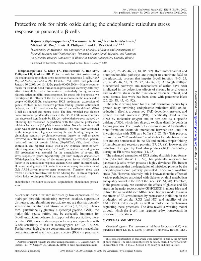
Effects of glucose and ER stress on the GSH/GSSG redoxcouple in mouse islets and MIN6 cells. To examine the effectsof glucose and ER stress on the redox state, islets were exposedto 2.8, 5.6, or 20 mM glucose in the absence or presence ofproteasome inhibitor LC (10 �M) for 4 h. Intracellular GSHand GSSG concentrations were measured by HPLC and nor-malized on a per cellular DNA basis. Relative to the physio-logical 5.6 mM glucose treatment, total islet GSH concentra-tions were elevated (P � 0.05) in response to low and highglucose (2.8 mM and 20 mM, Fig. 1A). LC further increasedthe total GSH concentration in islets cultured in low glucose(2.8 mM). The GSH/GSSG ratio, indicative of redox status,revealed that islets became more oxidized in response toincreasing concentrations of glucose. The GSH/GSSG ratio ofislets cultured in 2.8 and 5.6 mM glucose decreased (P � 0.05)in response to LC, whereas this ratio was similar in controland LC-treated islets in the presence of high glucose (20mM; Fig. 1B). Similar glucose effects on GSH/GSSG ratioswere observed in MIN6 cells (data not shown).
To quantify intracellular redox state, the concentrations ofGSH and GSSG in MIN6 cells treated with 25 mM glucosewere used to calculate the intracellular GSH redox potentialwith the Nernst equation (78). Untreated control cells exhibitedredox potentials similar to cells exposed to LC (10 �M) in 25mM glucose for 4 but not 12 h of treatment [Eh �160 mV,apoptotic range (78)]. Notably, exposure to LC for 4 h signif-icantly decreased intracellular GSH concentrations, whereas4-h exposure to Tm significantly increased intracellular GSHconcentrations (Table 1). Supplementation with �-ME andNAC prevented (P � 0.05) the loss of intracellular GSHattributed to LC.
Measurements of endogenous ROS and NO production inMIN6 cells. Exposure of MIN6 cells to LC or Tm increasedintracellular ROS concentrations (Fig. 2). The increase in
E1545REDOX HOMEOSTASIS IN ER-STRESSED PANCREATIC �-CELLS
AJP-Endocrinol Metab • VOL 292 • JUNE 2007 • www.ajpendo.org
on May 28, 2010
ajpendo.physiology.orgD
ownloaded from
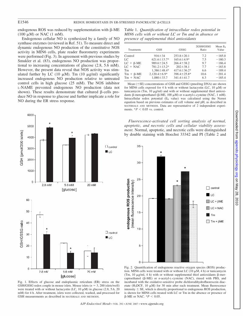
endogenous ROS was reduced by supplementation with �-ME(100 �M) or NAC (1 mM).
Endogenous cellular NO is synthesized by a family of NOsynthase enzymes (reviewed in Ref. 51). To measure direct anddynamic endogenous NO production of the constitutive NOSactivity in MIN6 cells, plate reader fluorometry experimentswere performed (Fig. 3). In agreement with previous studies bySmukler et al. (83), endogenous NO production was propor-tional to increasing concentrations of glucose (2.8, 5.6 mM).However, the present data reveal that NOS activity was stim-ulated further by LC (10 �M). Tm (10 �g/ml) significantlyincreased endogenous NO production relative to untreatedcontrol cells in high glucose (25 mM). The NOS inhibitorL-NAME prevented endogenous NO production (data notshown). These results demonstrate that cultured �-cells pro-duce NO in response to glucose and further implicate a role forNO during the ER stress response.
Fluorescence-activated cell sorting analysis of normal,apoptotic, and necrotic cells and cellular viability assess-ment. Normal, apoptotic, and necrotic cells were distinguishedby double staining with Hoechst 33342 and PI (Table 2 and
Fig. 1. Effects of glucose and endoplasmic reticulum (ER) stress on theGSH/GSSG redox couple in mouse islets. Mouse islets (n 3, 200 islets/well)were treated with or without lactacystin (LC, 10 �M) in glucose (2.8, 5.6, 20mM) for 4 h. After treatment, islets were collected, washed, and processed forGSH measurements as described in MATERIALS AND METHODS.
Table 1. Quantification of intracellular redox potential inMIN6 cells with or without LC or Tm and in absence orpresence of supplemental thiol antioxidants
Treatments GSH GSSG2GSH/GSSG
RatioMean Eh
Value
Control 916�34 253.8�20.1 7.2 �185.0LC 621.6�13.7* 165.6�4.9* 7.5 �180.3LC �-ME 989.6�24.5 266.4�38.2 9.7 �186.4LC NAC 781.2�13.2* 202�38.1 7.7 �183.8Tm 1,386�48.4* 417.6�36.2* 6.6 �189.4Tm �-ME 2,120.4�6.9* 398.4�25.8* 10.6 �201.4Tm NAC 1,080�33.7 341.4�41.7 6.3 �185.4
Mean (�SE) concentrations of GSH and GSSG (pmol/mg DNA) are shownfor MIN6 cells exposed for 4 h with or without lactacystin (LC, 10 �M) ortunicamycin (Tm, 10 �g/ml) and with or without supplemental thiol antioxi-dants �-mercaptoethanol (�-ME, 100 �M) or n-acetyl-L-cysteine (NAC, 1 mM).Intracellular redox potential (Eh value) was calculated using the Nernstequation based on previous estimates of cell volume and pH, as described inMATERIALS AND METHODS. Data are representative of 2 independent experi-ments. *P � 0.05 vs. control.
Fig. 2. Quantification of endogenous reactive oxygen species (ROS) produc-tion. MIN6 cells were treated with or without LC (10 �M, 4 h) or tunicamycin(Tm, 10 �g/ml, 4 h) with or without supplemental thiol antioxidants �-mer-captoethanol (�-ME) or n-acetyl-L-cysteine (NAC), rinsed with PBS, andincubated with the oxidative-sensitive probe dichlorodihydrofluorescein diac-etate (H2DCF, 10 �M) for 30 min after each treatment. Mean fluorescenceintensity � SE, which is directly proportional to endogenous ROS production,is shown for MIN6 cells treated with LC or Tm in the absence or presence of�-ME or NAC. *P � 0.05.
E1546 REDOX HOMEOSTASIS IN ER-STRESSED PANCREATIC �-CELLS
AJP-Endocrinol Metab • VOL 292 • JUNE 2007 • www.ajpendo.org
on May 28, 2010
ajpendo.physiology.orgD
ownloaded from

Refs. 9, 22). The number of Hoechst 33342- and PI-positivecells was quantified by fluorescence-activated cell sorting(FACS) analysis. The percentage of Hoechst 33342 and PI-positive cells was similar for MIN6 cells incubated in theabsence or presence of LC (10 �M) or Tm (10 �g/ml) for 4 h.After a 12-h exposure to LC, the percentage of apoptotic andnecrotic cells increased significantly relative to untreated cellsin standard culture conditions (Table 2).
Enhanced expression of genes involved in oxidative pro-tein folding in MIN6 cells. The formation of disulfide bondsin the ER requires PDI and ERO1, which reoxidizes PDI(60). To determine the effects of LC on Ero1 and PDI, themRNA concentrations of mouse Ero1 homologs (Ero1-l�and Ero1-l�), and two PDI enzymes (PDI and ERp61) wereexamined (Fig. 4). A significant increase in Ero1-l�, but notEro1-l� gene expression was observed in response to LC. ThemRNA of both PDI family members (PDI and ERp61) weresignificantly increased in LC-treated MIN6 cells (Fig. 4).
Effects of proteasome and NOS inhibition on expression ofgenes involved in thiol metabolism and antioxidant defense. Todetermine whether ER stress induced by LC (4 h, 10 �M)altered the expression of genes involved in thiol metabolism,mRNA expression of Gclc [encoding catalytic subunit of GCL,the rate-limiting enzyme for the synthesis of GSH (20, 39, 60,65)], glutathione peroxidase, glutathione reductase, catalase,and manganese superoxide dismutase were examined. GclcmRNA expression was below the detection limit for controlMIN6 cells via standard RT-PCR. In contrast, Gclc mRNAtranscripts were detected after exposure to LC and Tm(Fig. 5A). When the NOS inhibitor L-NAME (5 or 10 mM) wascombined with LC (4 h, 10 �M) treatment, steady-state GclcmRNA expression was similar to that of untreated controls.Quantitative PCR demonstrated that LC-induced Gclc mRNAexpression was reduced significantly in response to L-NAME(5 mM; Fig. 5B). The protein expression of GCLC was similaramong control and LC- and Tm-treated MIN6 cells (Fig. 5C).These data indicate that endogenous NO production or NOSactivity contributes to the transcriptional upregulation of Gclcin MIN6 cells.
The mRNA expressions of glutathione reductase, catalase,and manganese superoxide dismutase were similar amongcontrol and LC- and Tm-treated MIN6 cells (data not shown).However, a significant increase in Gpx-1 and Prx-1 mRNAexpression was observed in response to LC and Tm (Fig. 5D).When both the proteasome and NOS were inhibited, steady-state mRNAs of Gpx-1 and Prx-1 were lower (P � 0.05) orsimilar to untreated control mRNA (Fig. 5D). These dataindicate that endogenous NO production or NOS activity
Fig. 3. Endogenous NO production in re-sponse to glucose stimulation and ER stress.MIN6 cells were loaded with DAF-FM diac-etate (NO-specific reagent in 2.8 mM glucosefor 1 h) and exposed to glucose (2.8, 5.6, 25mM) with or without LC (10 �M) or Tm (10�g/ml) for 1 h, during which measurements(495/515 nm) were recorded every 36 s. Rep-resentative graphs (n 4) are shown for 3independent experiments. *P � 0.05.
Table 2. FACS analysis of apoptotic and necrotic MIN6cells � lactacystin or tunicamycin
Control Lactacystin Tunicamycin
Apoptotic Necrotic Apoptotic Necrotic Apoptotic Necrotic
4 h 3.4�0.6 3.2�0.1 4.6�1.2 2.1�0.2 3.3�0.2 4.2�0.412 h 2.0�0.4 3.5�0.4 5.1�1.1* 10.6�1.7* 3.6�0.8 6.8�0.3*
Percentage (means � SE) of MIN6 apoptotic cells was measured byfluorescence-activated cell sorting (FACS) analysis after 4- and 12-h exposuresto LC (10 �M) and Tm (10 �g/ml). Data are representative of 3 independentexperiments. *P � 0.05, vs. control.
E1547REDOX HOMEOSTASIS IN ER-STRESSED PANCREATIC �-CELLS
AJP-Endocrinol Metab • VOL 292 • JUNE 2007 • www.ajpendo.org
on May 28, 2010
ajpendo.physiology.orgD
ownloaded from

contributes to the transcriptional upregulation of Gpx-1 andPrx-1 in MIN6 cells.
To determine whether the transcriptional upregulation of anER stress-responsive gene is dependent on endogenous NOproduction, mRNA expression of ER chaperone GRP78/BiPwas examined by quantitative real-time RT-PCR in MIN6 cellstreated with LC (10 �M) in the absence or presence of a NOSinhibitor (L-NAME, 10 mM) for 4 h (Fig. 5D). As expected,proteasome inhibition alone increased Grp78/BiP mRNA ex-pression (Fig. 5D). When both the proteasome and NOS wereinhibited, steady-state Grp78/BiP mRNA expression waslower (P � 0.05) or similar to that of untreated control cells(Fig. 5D).
Effects of proteasome and NOS inhibition on unfolded pro-tein response activation and ER stress-responsive genes. Real-time quantitative RT-PCR was used to examine the activationof an unfolded protein response (UPR) in response to simul-taneous proteasome and NOS inhibition in MIN6 cells. Tran-scription factor X-box-binding protein-1 (Xbp-1) is a regulatorof the UPR (12, 41, 56, 94). On sensing misfolded proteins, anER transmembrane endoribonuclease and kinase (Ire1) excisesan intron from mammalian Xbp-1 mRNA, resulting in theconversion of unspliced Xbp-1 (Xbp-1u) to spliced Xbp-1(Xbp-1s, 13, 42, 57, 94). The Xbp-1s then translocates to thenucleus where it binds its target sequence in the regulatoryregion of chaperone genes to induce their transcription. Highexpression of Xbp-1s relative to Xbp-1u is indicative of UPRactivation (13, 42, 57, 94). Treatment of MIN6 cells with LCresulted in an accumulation of Xbp-1s mRNA and a concom-itant decrease in Xbp-1u mRNA (Fig. 6). Inhibition of NOS didnot alter this response.
The gene encoding C/EBP-homologous protein (CHOP),also known as growth arrest and DNA damage-inducible gene153 (Gadd153), is a stress-inducible transcription factor that isactivated by agents that adversely affect ER function (72).Expression of CHOP/Gadd153 mRNA was detected by stan-dard RT-PCR after exposure of MIN6 cells to LC (10 �M, 4 h)but not in untreated control cells (Fig. 7A). To determinewhether CHOP/Gadd153 induction was mediated by endoge-nous NO production, MIN6 cells were treated simultaneouslywith LC and NOS inhibitor L-NAME for 4 h. Induction ofCHOP/Gadd153 was not detected during simultaneous inhibi-tion of proteasome and NOS. Quantitative PCR revealed thatL-NAME decreased LC- and Tm-induced CHOP/Gadd153expression (Fig. 7B). These data indicate that endogenous NOproduction is necessary for the induction of CHOP/Gadd153 inLC-treated MIN6 cells.
Constitutive nuclear localization of Nrf2. Nuclear localiza-tion of Nrf2 is essential for the transactivation of a variety ofstress-responsive genes, including GCL (41, 81). Western blotanalysis of nuclear and cytoplasmic extracts using anti-Nrf2antibody revealed the accumulation of Nrf2 protein in thenucleus of LC-treated MIN6 cells (Fig. 8A). Inhibition of NOSdid not significantly alter the localization of Nrf2. Nrf2 was notdetected in the cytosol regardless of treatment. Lamin B and�-tubulin are shown as markers for nuclear and cytoplasmiccompartments, respectively (Fig. 8A).
NOS inhibition prevented Gclc ARE4-mediated transactiva-tion in ER-stressed MIN6 cells. Previous studies demonstratedNrf and MafK transactivation of the Gclc promoter (21). Todetermine the role of NOS inhibition on ARE4-mediated geneexpression, MIN6 cells were cotransfected with a mouse Gclcpromoter-luciferase construct (1.0 �g) and 0.5 �g each of thepcDNA-Nrf2 and pcDNA-MafK constructs. Twenty-fourhours after transfection, cells were treated with LC (10 �M) orTm (10 �g/ml) in the absence or presence of L-NAME (10mM) for 8 h in 5.6 mM glucose (Fig. 8, B and C). NOSinhibition repressed LC and Tm-induced activation of GclcARE4. These data further demonstrate an essential role for NOin regulating the Nrf2 pathway.
Electromobility shift assays demonstrate DNA binding ofNrf2 to ARE4 during UPR activation. To determine whetherthe binding of Nrf2 to an ARE is altered by UPR activation,electrophoretic mobility shift assays (EMSA) were performed.
Fig. 4. Expression of genes involved in oxidative protein folding. Expressionsof Ero1-l�, Ero1-l�, PDI, and ERp61 were determined by quantitative PCR.Open bars, control; filled bars, LC. *P � 0.05.
E1548 REDOX HOMEOSTASIS IN ER-STRESSED PANCREATIC �-CELLS
AJP-Endocrinol Metab • VOL 292 • JUNE 2007 • www.ajpendo.org
on May 28, 2010
ajpendo.physiology.orgD
ownloaded from

ER stress induced by LC or Tm enhanced the binding ofnuclear factors to the Gclc ARE4 [Fig. 8D, lane 2 (control) vs.lanes 4 (LC) and 6 (Tm)]. To determine whether Nrf2 andMafK are involved in ARE4 binding, band shift reactions wereincubated for 1 h with anti-Nrf2 and anti-MafK antibodies. Foreach treatment, incubation with a mixture of anti-Nrf2 andanti-MafK antibodies disrupted ARE4 DNA binding activity
(Fig. 8D, lanes 3, 5, and 7 relative to lanes 2, 4, and 6,respectively). A similar EMSA was performed with eachantibody separately. Anti-Nrf2 (Fig. 8E, lane 3), but notanti-MafK (Fig. 8E, lane 4) disrupted binding of the Nrf2-MafK complex to the Gclc ARE4. This observation may beexplained by the fact that the anti-Nrf2 reagent used is directedagainst the COOH terminus of Nrf2, which includes the DNA
Fig. 5. Transcriptional upregulation of Gclc is decreased by NO synthase (NOS) inhibition. MIN6 cells were treated with LC (10 �M) with or withoutN�-nitro-L-arginine methyl ester (L-NAME, 10 mM) in 5.6 mM glucose for 4 h. After treatment, cells were washed with PBS, and protein or RNA was isolated.A: ethidium bromide-stained agarose gel. B: quantitative PCR (Gclc per Gapdh) demonstrated that NOS inhibition (5 mM L-NAME) downregulated LC-inducedGclc gene expression. Correctly sized RT-PCR products for Gclc (346 bp), and Rn18s (137 bp, constitutive control) are shown. C: Western blot analysis showsthat Gclc protein expression was unaffected by LC or Tm treatment. Open bars, control; filled bars, LC; gray bars, Tm. D: quantitative PCR demonstrated thatNOS inhibition repressed LC-mediated upregulation of genes involved in ER protein folding (Grp78/BiP), thiol metabolism (cellular glutathione peroxidase,Gpx-1), and antioxidant defense (peroxiredoxin-1, Prx1). MIN6 cells were treated with LC (10 �M) with or without L-NAME (N, 10 mM) in 5.6 mM glucosefor 1 h. After treatment, cells were washed with PBS, and protein or RNA was isolated. Representative data are per Rn18s and from 1 of 3 independentexperiments performed in triplicate are shown; means � SE. *P � 0.05, LC vs. control; **P � 0.05, LC vs. LC N.
Fig. 6. Effects of proteasome and NOS inhibi-tion on unfolded protein response activation.MIN6 cells were treated with LC (10 �M) withor without L-NAME (10 mM) in 5.6 mM glu-cose for 1 h. After treatment, cells were washedwith PBS, and RNA was isolated. The relativesteady-state active, spliced form of Xbp-1 (Xbp-1s) and unspliced Xbp-1 (Xbp-1u) per Rn18s, asdetermined by quantitative PCR, are shown.*P � 0.05.
E1549REDOX HOMEOSTASIS IN ER-STRESSED PANCREATIC �-CELLS
AJP-Endocrinol Metab • VOL 292 • JUNE 2007 • www.ajpendo.org
on May 28, 2010
ajpendo.physiology.orgD
ownloaded from

binding domain. NOS inhibition did not significantly alter thebinding of the Nrf2-MafK complex to the Gclc ARE4 inducedby LC and Tm (data not shown).
DISCUSSION
The present studies investigated the effects of an ER stressresponse on the major redox couple (GSH/GSSG), endogenousROS production, expression of genes involved in ER oxidativeprotein folding, general antioxidant defense, and thiol metab-olism using mouse islets and the well-validated MIN6 �-cellline as a model system. Although the ER stress response is vitalto ensure quality control, it also enhances the endogenousproduction of ROS, formed as byproducts during oxidativeprotein folding in the ER.
A glucose concentration-dependent decrease in the GSH/GSSG ratio was observed in islets after 4-h exposure toincreasing concentrations of glucose (2.8, 5.6, 20 mM). Glu-cose itself may be an ER stressor through its ability to stimulateinsulin synthesis and increase the number of proteins withdisulfide bonds that must be folded properly in the ER lumen.To accommodate newly synthesized ER client proteins, the ERenvironment may adapt acutely through induction of a UPR.Persistent exposure to high glucose may lead to chronic UPRactivation, (i.e., ERO1 and PDI expression) and thereby in-crease endogenous ROS production and redox signaling duringhigh insulin demand. In agreement, constitutive expression ofEro1-l� transcripts observed in professional secretory cellslikely reflect their higher requirements in disulfide bond for-mation (27).
The physiological status of eukaryotic cells correlates withthe redox potential (Eh value) of the GSH/GSSG couple (78).For example, the Eh value is most negative during proliferationand becomes more positive as cells differentiate (78). Thepresent data demonstrate that MIN6 �-cells maintained a redoxpotential of approximately �185 mV. Relative to other con-fluent or differentiated cell lines [fibroblasts, HT-29 (78)], thisEh value is more oxidized and likely reflects the highly devel-oped �-cell ER and its respective vital metabolic and secretoryactivities. Additionally, �-cells are also more sensitive tooxidative and nitrosative stress due to their intrinsically lowexpression of antioxidant enzymes (57, 71, 86).
It was suggested recently that the same pathways used in theactivation of glucose-dependent insulin secretion (increasedglycolytic flux, ATP-to-ADP ratio, and intracellular Ca2
concentration) can dramatically enhance ROS production andmanifestations of oxidative stress and, possibly, apoptosis (29).Indeed, it is generally acknowledged that ROS are byproductsof mitochondrial aerobic metabolism. However, in the presentstudies, the specific metabolic consequences of ER oxidationare highlighted as a contributor of endogenous ROS. Wehypothesize that “ER oxidation” (also referred to as disulfidestress) generates excessive endogenous ROS, which contrib-utes to the chronic oxidative stress observed in pancreatic�-cells during hyperglycemia. Our data revealed a glucoseconcentration-dependent decrease in the GSH/GSSG ratio,which was further decreased (i.e., oxidized) with ER stress inmouse islets. Notably, the maximal effects of ER oxidationwere observed only in mouse islets stimulated with lower (2.8or 5.6 mM) glucose concentrations. In high glucose (20–25mM), the GSH/GSSG ratio was similar in ER-stressed anduntreated control mouse islets and MIN6 cells. These dataindicate the importance of determining the relative contribu-tions of ROS generated from mitochondrial vs. ER metabo-lism. Specifically, the extent to which ER-derived ROS maycontribute to the chronic oxidative stress observed in �-cellsduring hyperglycemia is unclear.
Persistent ER stress eventually leads to cell death (27, 36,48, 67, 71), which may be attributed to excessive ROS. Inagreement, prolonged ER stress (12 h) correlated with a sig-nificant decrease in the GSH/GSSG ratio that was not preventedby supplemental thiol antioxidants (data not shown). However,after 4 h of treatment with lactacystin or tunicamycin, thepercentage of apoptotic and necrotic cells combined did notdiffer significantly from control cells and reached only 15% oftotal cells after 12 h of treatment. Upregulation of the rate-limiting enzyme for GSH synthesis (Gclc) and genes involvedin antioxidant defense (Gpx-1, Prx-1) likely contributed to themaintenance of �-cell redox homeostasis and cell survivalduring this acute period.
The extent to which increased endogenous ROS productionor enhanced redox signaling may contribute to cell survivalduring the ER stress response in �-cells is questionable. NO is
Fig. 7. Induction of CHOP/Gadd153 is repressed by NOS inhibition. MIN6 cells were treated with LC (10 �M) or Tm (10 �M) with or without NOS inhibitorL-NAME (1, 5, 10 mM) in 5.6 mM glucose for 4 h. After treatment, cells were washed with PBS, and RNA was isolated. A: ethidium bromide-stained agarosegel demonstrates that CHOP/Gadd153 was not induced when LC was incubated with L-NAME (10 mM). Correctly sized RT-PCR products for CHOP/Gadd153(397 bp), and Rn18s (137 bp, constitutive control) are shown. B: quantitative PCR revealed that L-NAME (N) decreased LC- or Tm-induced CHOP/Gadd153expression. Representative data from 1 of 3 independent experiments performed in triplicate are shown; means � SE per Gapdh. *P � 0.05, LC vs. control;**P � 0.05, LC vs. LC N; #P � 0.05, Tm vs. control; ##P � 0.05, Tm vs. Tm N.
E1550 REDOX HOMEOSTASIS IN ER-STRESSED PANCREATIC �-CELLS
AJP-Endocrinol Metab • VOL 292 • JUNE 2007 • www.ajpendo.org
on May 28, 2010
ajpendo.physiology.orgD
ownloaded from

a signaling molecule that, in excess, causes cell death. Exces-sive NO exerts cytotoxic effects by reacting with superoxideand thereby generating the highly reactive free radical per-oxynitrite, which causes nonspecific DNA, protein, and lipiddamage (11). However, recent studies suggest that NO is alsoa potent antioxidant (reviewed in Ref. 73). Specifically, lowNO concentrations produced by the constitutive NOS (cNOS)enzyme, which have been shown to regulate �-cell insulinrelease (2, 53, 69, 76, 79, 87, 89), may also terminate oxidativestress by 1) suppressing iron-induced generation of hydroxylradicals ( �OH) via the Fenton reaction, 2) interrupting thechain reaction of lipid peroxidation, 3) augmenting the anti-
oxidative potency of reduced GSH, and 4) inhibiting cysteineproteases. The present data reveal that endogenous NO pro-duction or cNOS activity was enhanced by ER stress. Further-more, inhibition of cNOS by L-NAME repressed the ERstress-induced expression of genes involved in thiol metabo-lism, antioxidant defense, and ER protein folding (Gclc, Prx-1,Gpx-1, Grp78/BiP). These data indicate that endogenous NO isvital for induction of antioxidant defense genes and thoseinvolved in ER protein folding in the �-cell.
The protective effects of NO in �-cells may be mediated byactivation of Nrf2. Nrf2 is a critical transcription factor thatbinds to the ARE in the promoter region of a number of genes
Fig. 8. Effects of NOS inhibition on antioxidant response element (ARE)4-mediated gene expression in ER-stressed MIN6 cells. A: nuclear localization of Nrf2in response to ER stress. MIN6 cells were grown on 6-well plates and treated with LC (10 �M) with or without L-NAME (10 mM) in 5.6 mM glucose for 4 h.After treatment, cells were washed with PBS, and nuclear and cytoplasmic extracts were prepared. Ten micrograms of the nuclear proteins (nu) or cytoplasmicproteins (cy) were loaded onto a 12% polyacrylamide gel, and Western blotting was performed with anti-Nrf2, anti-lamin B, and anti-�-tubulin antibodies. LaminB and �-tubulin are shown as markers for nuclear and cytoplasmic proteins, respectively. B and C: NOS inhibition prevents induction of ARE4-mediated geneexpression in ER-stressed MIN6 cells. MIN6 cells were cotransfected with mouse Gclc promoter-luciferase construct along with pcDNA-Nrf2 and pcDNA-MafKconstructs and pRL-TK plasmid encoding Renilla luciferase as an internal control. Twenty-four hours after transfection, cells were pretreated with mediumwithout glucose for 1 h, after which it was replaced with medium containing glucose (5.6 mM, B and C) with or without L-NAME (N, 10 mM). After 1 h, cellswere treated with or without LC (10 �M) or Tm (10 �M) for 8 h. Cell lysates were obtained and luciferase activities normalized against Renilla luciferase activity.Results are represented as means � SE from 3 independent experiments. *P � 0.05. Electromobility shift assays demonstrate Nrf2 binding to ARE4. D: LC andTm enhanced the binding of nuclear factors to the Gclc ARE4. The Gclc ARE4 was end labeled with [�-32P]ATP. Labeled ARE4wt probe (5 fmol) was incubatedwith nuclear extracts (NE, 5 �g) from MIN6 cells treated with LC (10 �M) or Tm (10 �g/ml) for 4 h and analyzed in a 6% nondenaturing polyacrylamide gel.Gels were dried and autoradiographed. E: in a similar experiment as in D, anti-Nrf2 and anti-MafK antibodies were incubated separately with nuclear extracts(lanes 3 and 4, respectively). ARE4 DNA binding activity was abrogated with unlabeled ARE4m2 but not unlabeled oligonucleotides containing the m2 mutation(ARE4m2, data not shown).
E1551REDOX HOMEOSTASIS IN ER-STRESSED PANCREATIC �-CELLS
AJP-Endocrinol Metab • VOL 292 • JUNE 2007 • www.ajpendo.org
on May 28, 2010
ajpendo.physiology.orgD
ownloaded from

encoding for antioxidant and phase 2 enzymes in numeroustissues and cell types (40, 68). However, to our knowledge, ithas not been previously reported whether Nrf2 signaling con-trols the coordinated expression of antioxidant pathways andphase 2 enzymes in pancreatic �-cells. Our data demonstratethat inhibition of NO production did not prevent Nrf2 bindingto the ARE. This observation suggests that Nrf2 may beconstitutively localized to the nucleus of �-cells or implicatesthe existence of a NO-independent pathway for nuclear local-ization and activation of Nrf2. The present data support theformer possibility in that Nrf2 was detected primarily in thenucleus in both control and treated cells. However, recentreports showed that PERK-dependent activation of Nrf2 con-tributes to redox homeostasis and cell survival following ERstress (15, 16). Therefore, independently of endogenous NOproduction, PERK may activate Nrf2. The potential redun-dancy of this pathway underscores the importance of maintain-ing redox homeostasis in pancreatic �-cells. Indeed, recentstudies indicate that Nrf2 activation involves a coordinatedprocess and is regulated at multiple levels (reviewed in Ref.16). The present data demonstrate that cNOS inhibition re-pressed Gclc expression and ARE4 promoter activity despiteUPR (and PERK) activation. In this regard, although NO-independent pathways for initiating the nuclear localization ofNrf2 exist, endogenous NO production is necessary for ulti-mately enhancing Gclc expression during the ER stress re-sponse. In agreement, NO-induced transcriptional upregulationof protective genes by Nrf2 via the ARE counteracts apoptosisin neuroblastoma cells (21). This and the present findingssupport the involvement of a NO-dependent mechanism un-derlying redox homeostasis in �-cells.
�-Cell dysfunction resulting from oxidative or disulfidestress may reflect differential genetic or environmental regula-tion of ER stress responses. Consistent with this hypothesis isa report that describes an “integrated stress response” consist-ing of the UPR and further regulation of amino acid metabo-lism and resistance to oxidative stress (37). Furthermore, ge-netic differences in the constitutive ability to dissipate ROScorrelated with differential inbred strain susceptibility or resis-tance to alloxan- and streptozotocin-induced diabetes (62–64).
Collectively, the present data indicate that oxidative proteinfolding machinery in the ER may generate excessive ROS thataccumulate over time and contribute to chronic oxidative stressduring hyperglycemic conditions. Accordingly, �-cells re-spond to disulfide stress by increasing the total glutathionepool, possibly to buffer the excessive ROS produced during theER stress response. In addition to changes in thiol metabolism,�-cells possess the capacity to regulate their intracellular redoxstate via induction of antioxidant defense genes. These effectsare, in part, mediated by enhanced redox signaling via NO.
ACKNOWLEDGMENTS
K. Ishii-Schrade is presently at the Graduate School of PharmaceuticalSciences, University of Tokyo, Tokyo, Japan.
GRANTS
This work was supported by National Institute of Diabetes and Digestiveand Kidney Diseases Grants DK-49192 (H. R. Gaskins), DK-68822 andDK-64162 (M. W. Roe). K. Kitiphongspattana was supported in part by a RuthKirschstein Institutional National Research Service Award 5T32 DK-59802(Division of Nutritional Sciences, University of Illinois at Urbana-Champaign)and an American Diabetes Association Mentor-based Fellowship.
REFERENCES
1. Ablamunits V, Elias D, Cohen IR. The pathogenicity of islet-infiltratinglymphocytes in the non-obese diabetic (NOD) mouse. Clin Exp Immunol115: 260–267, 1999.
2. Alm P, Ekstrom P, Henningsson R, Lundquist I. Morphological evi-dence for the existence of nitric oxide and carbon monoxide pathways inthe rat islets of Langerhans: an immunocytochemical and confocal micro-scopical study. Diabetologia 42: 978–986, 1999.
3. Ammon HP, Abdel-Hamid M, Rao PG, Enz G. Thiol-dependent andnon-thiol-dependent stimulations of insulin release. Diabetes 33: 251–257,1984.
4. Ammon HP, Mark M. Thiols and pancreatic beta-cell function: a review.Cell Biochem Funct 3: 157–171, 1985.
5. Ammon HP, Wahl MA. The impact of thiols for insulin secretion. ExpClin Endocrinol 93: 136–142, 1989.
6. Aridor M, Balch WE. Integration of endoplasmic reticulum signaling inhealth and disease. Nat Med 5: 745–751, 1999.
7. Bast A, Wolf G, Oberbaumer I, Walther R. Oxidative and nitrosativestress induces peroxiredoxins in pancreatic beta cells. Diabetologia 45:867–876, 2002.
8. Belloc F, Dumain P, Boisseau MR, Jalloustre C, Reiffers J, Bernard P,Lacombe F. A flow cytometric method using Hoechst 33342 and pro-pidium iodide for simultaneous cell cycle analysis and apoptosis determi-nation in unfixed cells. Cytometry 17: 59–65, 1994.
9. Bea F, Hudson FN, Chait A, Kavanagh TJ, Rosenfeld ME. Inductionof glutathione synthesis in macrophages by oxidized low-density lipopro-teins is mediated by consensus antioxidant response elements. Circ Res92: 386–393, 2003.
10. Brownlee M. The pathobiology of diabetic complications: a unifyingmechanism. Diabetes 54: 1615–1625, 2005.
11. Brune B, von Knethen A, Sandau KB. Nitric oxide (NO): an effector ofapoptosis. Cell Death Differ 6: 969–975, 1999.
12. Buckley BJ, Marshall ZM, Whorton AR. Nitric oxide stimulates Nrf2nuclear translocation in vascular endothelium. Biochem Biophys ResCommun 307: 973–979, 2003.
13. Calfon M, Zeng H, Urano F, Till JH, Hubbard SR, Harding HP, ClarkSG, Ron D. IRE1 couples endoplasmic reticulum load to secretorycapacity by processing the XBP-1 mRNA. Nature 415: 92–96, 2002.
14. Chakravarthi S, Jessop CE, Bulleid NJ. The role of glutathione indisulphide bond formation and endoplasmic-reticulum-generated oxida-tive stress. EMBO Rep 7: 271–275, 2006.
15. Cullinan SB, Diehl JA. PERK-dependent activation of Nrf2 contributesto redox homeostasis and cell survival following endoplasmic reticulumstress. J Biol Chem 279: 20108–20117, 2004.
16. Cullinan SB, Diehl JA. Coordination of ER and oxidative stress signal-ing: the PERK/Nrf2 signaling pathway. Int J Biochem Cell Biol 38:317–332, 2006.
17. Cuozzo JW, Kaiser CA. Competition between glutathione and proteinthiols for disulphide-bond formation. Nat Cell Biol 1: 130–135, 1999.
18. Demasi M, Shringarpure R, Davies KJA. Glutathiolation of the protea-some is enhanced by proteolytic inhibitors. Arch Biochem Biophys 389:254–263, 2001.
19. Demaurex N. pH Homeostasis of cellular organelles. News Physiol Sci17: 1–5, 2002.
20. Deplancke B, Gaskins HR. Redox control of the transsulfuration andglutathione biosynthesis pathways. Curr Opin Nutr Metab Care 5: 85–92,2002.
21. Dhakshinamoorthy S, Porter AG. Nitric oxide-induced transcriptionalup-regulation of protective genes by Nrf2 via the antioxidant responseelement counteracts apoptosis of neuroblastoma cells. J Biol Chem 279:20096–20107, 2004.
22. Dive C, Gregory CD, Phipps DJ, Evans DL, Milner AE, Wyllie AH.Analysis and discrimination of necrosis and apoptosis (programmed celldeath) by multiparameter flow cytometry. Biochim Biophys Acta 1133:275–282, 1992.
23. Ellgaard L, Molinari M, Helenius A. Setting the standards: qualitycontrol in the secretory pathway. Science 286: 1882–1888, 1999.
24. Efrat S, Leiser M, Surana M, Tal M, Fusco-Demane D, Fleischer N.Murine insulinoma cell line with normal glucose-regulated insulin secre-tion. Diabetes 42: 901–907, 1993.
25. Evans JL, Goldfine ID, Maddux BA, Grodsky GM. Oxidative stressand stress-activated signaling pathways: a unifying hypothesis of type 2diabetes. Endocr Rev 23: 599–622, 2002.
E1552 REDOX HOMEOSTASIS IN ER-STRESSED PANCREATIC �-CELLS
AJP-Endocrinol Metab • VOL 292 • JUNE 2007 • www.ajpendo.org
on May 28, 2010
ajpendo.physiology.orgD
ownloaded from

26. Evans JL, Goldfine ID, Maddux BA, Grodsky GM. Are oxidativestress-activated signaling pathways mediators of insulin resistance andbeta-cell dysfunction? Diabetes 52: 1–8, 2003.
27. Fassio A, Sitia R. Formation, isomerisation and reduction of disulphidebonds during protein quality control in the endoplasmic reticulum. Histo-chem Cell Biol 117: 151–157, 2002.
28. Fleming JV, Fontanier N, Harries DN, Rees WD. The growth arrestgenes gas5, gas6, and CHOP-10 (gadd153) are expressed in the mousepreimplantation embryo. Mol Reprod Dev 48: 310–316, 1997.
29. Fridlyand LE, Philipson LH. Does the glucose-dependent insulin secre-tion mechanism itself cause oxidative stress in pancreatic beta-cells?Diabetes 53: 1942–1948, 2004.
30. Gess B, Holfauer KH, Wenger RH, Lohaus C, Meyer HE, Kurtz A.The cellular oxygen tension regulates expression of the endoplasmicoxidoreductase ERO1-Lalpha. Eur J Biochem 270: 2228–2235, 2003.
31. Go YM, Gipp JJ, Mulcahy T, Jones DP. H2O2-dependent activation ofGCLC-ARE4 reporter occurs by mitogen-activated protein kinase path-ways without oxidation of cellular glutathione or thioredoxin-1. J BiolChem 279: 5837–5845, 2004.
32. Gotoh M, Maki T, Kiyoizumi T, Satomi S, Monaco AP. An improvedmethod for isolation of mouse pancreatic islets. Transplantation 40:437–438, 1985.
33. Grankvist K, Marklund SL, Taljedal IB. CuZn-superoxide dismutase,Mn-superoxide dismutase, catalase and glutathione peroxidase in pancre-atic islets and other tissues in the mouse. Biochem J 199: 393–398, 1981.
34. Hanahan D. Heritable formation of pancreatic beta-cell tumours intransgenic mice expressing recombinant insulin/simian virus 40 onco-genes. Nature 315: 115–122, 1985.
35. Harding HP, Calfon M, Urano F, Novoa I, Ron D. Transcriptional andtranslational control in the mammalian unfolded protein response. AnnuRev Cell Dev 18: 575–599, 2002.
36. Harding HP, Ron D. Endoplasmic reticulum stress and the developmentof diabetes: a review. Diabetes 51: S455–S461, 2002.
37. Harding HP, Zhang Y, Zeng H, Novoa I, Lu PD, Calfon M, Sadri N,Yun C, Popko B, Paules R, Stojdl DF, Bell JC, Hettmann T, LeidenJM, Ron D. An integrated stress response regulates amino acid metabo-lism and resistance to oxidative stress. Mol Cell 11: 619–633, 2003.
38. Haynes CM, Titus EA, Cooper AA. Degradation of misfolded proteinsprevents ER-derived oxidative stress and cell death. Mol Cell 15: 767–776,2004.
39. Huang CS, Chang LS, Anderson ME, Meister A. Catalytic and regu-latory properties of the heavy subunit of rat kidney gamma-glutamyl-cysteine synthetase. J Biol Chem 268: 19675–19680, 1993.
40. Itoh K, Chiba T, Takahashi S, Ishii T, Igarashi K, Katoh Y, Oyake T,Hayashi N, Satoh K, Hatayama I, Yamamoto M, Nabeshima Y. AnNrf2/small Maf heterodimer mediates the induction of phase II detoxifyingenzyme genes through antioxidant response elements. Biochem BiophysRes Commun 236: 313–322, 1997.
41. Itoh K, Wakabayashi N, Katoh Y, Ishii T, Igarashi K, Engel JD,Yamamoto M. Keap1 represses nuclear activation of antioxidant respon-sive elements by Nrf2 through binding to the amino-terminal Neh2domain. Genes Dev 13: 76–86, 1999.
42. Iwakoshi NN, Lee AH, Glimcher LH. The X-box binding protein-1transcription factor is required for plasma cell differentiation and theunfolded protein response. Immunol Rev 194: 29–38, 2003.
43. Jaiswal AK. Nrf2 signaling in coordinated activation of antioxidant geneexpression. Free Radic Biol Med 36: 1199–1207, 2004.
44. Jonas JC, Sharma A, Hasenkamp W, Ilkova H, Patane G, Laybutt R,Bonner-Weir S, Weir GC. Chronic hyperglycemia triggers loss ofpancreatic beta cell differentiation in an animal model of diabetes. J BiolChem 274: 14112–14121, 1999.
45. Kajimoto Y, Kaneto H. Role of oxidative stress in pancreatic beta-celldysfunction. Ann NY Acad Sci 1011: 168–176, 2004.
46. Kaneto H, Kajimoto Y, Miyagawa J, Matsuoka T, Fujitani Y, Umaya-hara Y, Hanafusa T, Matsuzawa Y, Yamasaki Y, Hori M. Beneficialeffects of antioxidants in diabetes: possible protection of pancreaticbeta-cells against glucose toxicity. Diabetes 48: 2398–2406, 1999.
47. Kaneto H, Xu G, Song KH, Suzuma K, Bonner-Weir S, Sharma A,Weir GC. Activation of the hexosamine pathway leads to deterioration ofpancreatic beta-cell function through the induction of oxidative stress.J Biol Chem 276: 31099–31104, 2001.
48. Kaufman RJ. Stress signaling from the lumen of the endoplasmic retic-ulum: coordination of gene transcriptional and translational controls.Genes Dev 13: 1211–1233, 1999.
49. Kitiphongspattana K, Mathews CE, Leiter EH, Gaskins HR. Protea-some inhibition alters glucose-stimulated (pro)insulin secretion and turn-over in pancreatic �-cells. J Biol Chem 280: 15727–15734, 2005.
50. Knaack D, Fiore DM, Surana M, Leiser M, Laurance M, Fusco-DeMane D, Hegre OD, Fleischer N, Efrat S. Clonal insulinoma cell linethat stably maintains correct glucose responsiveness. Diabetes 43: 1413–1417, 1994.
51. Knowles RG, Moncada S. Nitric oxide synthases in mammals. BiochemJ 298: 249–258, 1994.
52. Kretz-Remy C. and Arrigo AP. Gene expression and thiol redox stateMethods Enzymol 348: 200–15, 2002.
53. Lajoix AD, Reggio H, Chardes T, Peraldi-Roux S, Tribillac F, RoyeM, Dietz S, Broca C, Manteghetti M, Ribes G, Wollheim CB, Gross R.A neuronal isoform of nitric oxide synthase expressed in pancreatic �-cellscontrols insulin secretion. Diabetes 50: 1311–1323, 2001.
54. Lazzarino G, Amorini AM, Fazzina G, Vagnozzi R, Signoretti S,Donzelli S, Di Stasio E, Giardina B, Tavazzi B. Single-sample prepa-ration for simultaneous cellular redox and energy state determination. AnalBiochem 332: 51–59, 2003.
55. Lee AS. Mammalian stress response: induction of the glucose-regulatedprotein family. Curr Opin Cell Biol 4: 267–273, 1992.
56. Lee AS. The glucose-regulated proteins: stress induction and clinicalapplications. Trends Biochem Sci 26: 504–510, 2001.
57. Lee K, Tirasophon W, Shen X, Michalak M, Prywes R, Okada T,Yoshida H, Mori K, Kaufman RJ. IRE1-mediated unconventionalmRNA splicing and S2P-mediated ATF6 cleavage merge to regulateXBP1 in signaling the unfolded protein response. Genes Dev 16: 452–466,2002.
58. Lenzen S, Drinkgern J, Tiedge M. Low antioxidant enzyme geneexpression in pancreatic islets compared with various other mouse tissues.Free Radic Biol Med 20: 463–466, 1996.
59. Li K, Hein S, Zou W, Klug G. The glutathione-glutaredoxin system inRhodobacter capsulatus: part of a complex regulatory network controllingdefense against oxidative stress. J Bacteriol 186: 6800–6808, 2004.
60. Lu SC. Regulation of glutathione synthesis. Curr Top Cell Regul 36:95–116, 2000.
61. Marciniak SJ, Ron D. Endoplasmic reticulum stress signaling in disease.Physiol Rev 86: 1133–1149, 2006.
62. Mathews CE, Graser RT, Savinov A, Serreze DV, Leiter EH. Unusualresistance of ALR/Lt mouse beta cells to autoimmune destruction: role forbeta cell-expressed resistance determinants. Proc Natl Acad Sci USA 98:235–240, 2001.
63. Mathews CE, Leiter EH. Resistance of ALR/Lt islets to free radical-mediated diabetogenic stress is inherited as a dominant trait. Diabetes 48:2189–2196, 1999.
64. Mathews CE, Leiter EH. Constitutive differences in antioxidant defensestatus distinguish alloxan-resistant and alloxan-susceptible mice. FreeRadic Biol Med 27: 449–455, 1999.
65. Meister A, Anderson Glutathione ME. Annu Rev Biochem 52: 722–760,1983.
66. Miyazaki J, Araki K, Yamato E, Ikegami H, Asano T, Shibasaki Y,Oka Y, Yamamura K. Establishment of a pancreatic beta cell line thatretains glucose-inducible insulin secretion: special reference to expressionof glucose transporter isoforms. Endocrinology 127: 126–132, 1990.
67. Mori K. Tripartite management of unfolded proteins in the endoplasmicreticulum. Cell 101: 451–454, 2000.
68. Motohashi H, Yamamoto M. Nrf2-Keap1 defines a physiologicallyimportant stress response mechanism. Trends Mol Med 10: 549–557,2004.
69. Nakata M, Yada T, Nakagawa S, Kobayashi K, Maruyama I. Citrul-line-argininosuccinate-arginine cycle coupled to Ca2-signaling in ratpancreatic beta-cells. Biochem Biophys Res Commun 235: 619–624, 1997.
70. Nardai G, Korcsmaros T, Papp E, Csermely P. Reduction of theendoplasmic reticulum accompanies the oxidative damage of diabetesmellitus. Biofactors 12: 259–267, 2003.
71. Oyadomari S, Araki E, Mori M. Endoplasmic reticulum stress-mediatedapoptosis in pancreatic beta-cells. Apoptosis 7: 335–345, 2002.
72. Oyadomari S, Mori M. Roles of CHOP/GADD153 in endoplasmicreticulum stress. Cell Death Differ 11: 381–389, 2004.
73. Pryor WA, Houk KN, Foote CS, Fukuto JM, Ignarro LJ, SquadritoGL, Davies KJ. Free radical biology and medicine: it’s a gas, man! Am JPhysiol Regul Integr Comp Physiol 291: R491–R511, 2006.
E1553REDOX HOMEOSTASIS IN ER-STRESSED PANCREATIC �-CELLS
AJP-Endocrinol Metab • VOL 292 • JUNE 2007 • www.ajpendo.org
on May 28, 2010
ajpendo.physiology.orgD
ownloaded from

74. Robertson RP, Harmon JS. Diabetes, glucose toxicity, and oxidativestress: a case of double jeopardy for the pancreatic islet beta cell. FreeRadic Biol Med 41: 177–184, 2006.
75. Robertson RP, Harmon J, Tran PO, Tanaka Y, Takahashi H. Glucosetoxicity in beta-cells: type 2 diabetes, good radicals gone bad, and theglutathione connection. Diabetes 52: 581–587, 2003.
76. Salehi A, Carlberg M, Henningson R, Lundquist I. Islet constitutivenitric oxide synthase: biochemical determination and regulatory function.Am J Physiol Cell Physiol 270: C1634–C1641, 1996.
77. Sato H, Kuriyama-Matsumura K, Siow RCM, Ishii T, Bannai S,Mann E. Induction of cystine transport via system x-c and maintenance ofintracellular glutathione levels in pancreatic acinar and islet cell lines.Biochim Biophys Acta 1414: 85–94, 1998.
78. Schafer FQ, Buettner GR. Redox environment of the cell as viewedthrough the redox state of the glutathione disulfide/glutathione couple.Free Radic Biol Med 30: 1191–1212, 2001.
79. Schmidt HH, Warner TD, Ishii K, Sheng H, Murad F. Insulin secretionfrom pancreatic B cells caused by L-arginine-derived nitrogen oxides.Science 255: 721–723, 1992.
80. Schreiber E, Matthias P, Muller M, Schaffner M. Rapid detection ofoctamer binding proteins with “mini-extracts”, prepared from a smallnumber of cells. Nucleic Acids Res 17: 6419, 1989.
81. Sekhar KR, Soltaninassab SR, Borrelli MJ, Xu ZQ, Meredith MJ,Domann FE, Freeman ML. Inhibition of the 26S proteasome inducesexpression of GLCLC, the catalytic subunit for gamma-glutamylcysteinesynthetase. Biochem Biophys Res Commun 270: 211–217, 2000.
82. Siman R, Flood DG, Thinakaran G, Neumar RW. Endoplasmic retic-ulum stress-induced cysteine protease activation in cortical neurons: effectof an Alzheimer’s disease-linked presenilin-1 knock-in mutation. J BiolChem 276: 44736–44743, 2001.
83. Smukler SR, Tang L, Wheeler MB, Salapatek AM. Exogenous nitricoxide and endogenous glucose-stimulated �-cell nitric oxide augmentinsulin release. Diabetes 51: 2450–2460, 2002.
84. Takahashi H, Tran PO, LeRoy E, Harmon JS, Tanaka Y, RobertsonRP. D-Glyceraldehyde causes production of intracellular peroxide inpancreatic islets, oxidative stress, and defective beta cell function vianon-mitochondrial pathways. J Biol Chem 279: 37316–37323, 2004.
85. Tanaka Y, Tran POT, Harmon J, Robertson RP. A role for glutathioneperoxidase in protecting pancreatic beta cells against oxidative stress in amodel of glucose toxicity. Proc Natl Acad Sci USA 99: 12363–12368,2002.
86. Tiedge M, Lortz S, Drinkgern J, Lenzen S. Relation between antioxi-dant enzyme gene expression and antioxidative defense status of insulin-producing cells. Diabetes 46: 1733–1742, 1997.
87. Tsuura Y, Ishida H, Shinomura T, Nishimura M, Seino Y. Endogenousnitric oxide inhibits glucose-induced insulin secretion by suppression ofphosphofructokinase activity in pancreatic islets. Biochem Biophys ResCommun 252: 34–38, 1998.
88. Tu BP, Weissman JS. The FAD- and O(2)-dependent reaction cycle ofEro1-mediated oxidative protein folding in the endoplasmic reticulum.Mol Cell 10: 983–994, 2002.
89. Umehara K, Kataoka K, Ogura T, Esumi H, Kashima K, Ibata Y,Okamura H. Comparative distribution of nitric oxide synthase (NOS) inpancreas of the dog and rat: immunocytochemistry of neuronal type NOSand histochemistry of NADPH-diaphorase. Brain Res Bull 42: 469–478,1997.
90. Wang J, Takeuchi T, Tanaka S, Kubo SK, Kayo T, Lu D, Takata K,Koizumi A, Izumi T. A mutation in the insulin 2 gene induces diabeteswith severe pancreatic beta-cell dysfunction in the Mody mouse. J ClinInvest 103: 27–37, 1999.
91. Wild AC, Gipp JJ, Mulcahy T. Overlapping antioxidant response ele-ment and PMA response element sequences mediate basal and beta-naphthoflavone-induced expression of the human gamma-glutamyl-cysteine synthetase catalytic subunit gene. Biochem J 332: 373–381, 1998.
92. Wu L, Nicholson W, Knobel SM, Steffner RJ, May JM, Piston DW,Powers AC. Oxidative stress is a mediator of glucose toxicity in insulin-secreting pancreatic islet cell lines. J Biol Chem 279: 12126–12134, 2004.
93. Xu W, Liu L, Charles IG, Moncada S. Nitric oxide induces coupling ofmitochondrial signalling with the endoplasmic reticulum stress response.Nature Cell Biol 6: 1129–1134, 2004.
94. Yoshida H, Matsui T, Yamamoto A, Okada T, Mori K. XBP1 mRNAis induced by ATF6 and spliced by IRE1 in response to ER stress toproduce a highly active transcription factor. Cell 107: 881–891, 2001.
E1554 REDOX HOMEOSTASIS IN ER-STRESSED PANCREATIC �-CELLS
AJP-Endocrinol Metab • VOL 292 • JUNE 2007 • www.ajpendo.org
on May 28, 2010
ajpendo.physiology.orgD
ownloaded from