kalpesh lalla - wiredspace.wits.ac.za
TRANSCRIPT
i
“Does Human Papilloma Virus play a role in the histogenesis of the orthokeratinised
jaw cyst?”
KALPESH LALLA
A research report submitted to the Faculty of Health Sciences, University of the
Witwatersrand, Johannesburg, in partial fulfilment of the requirements for the degree
of
Master of Science in Dentistry
Johannesburg, 2015
ii
DECLARATION
I, Kalpesh Lalla, declare that this research report is my own work. It is being submitted for
the degree of Master of Science in Dentistry at the University of the Witwatersrand,
Johannesburg. It has not been submitted before for any degree or examination at this or
any other University.
...................................
.......................day of .................................. 2015.
iii
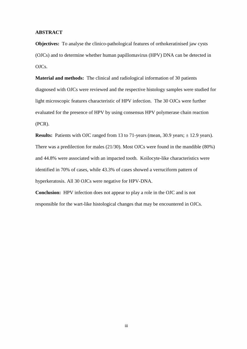
ABSTRACT
Objectives: To analyse the clinico-pathological features of orthokeratinised jaw cysts
(OJCs) and to determine whether human papillomavirus (HPV) DNA can be detected in
OJCs.
Material and methods: The clinical and radiological information of 30 patients
diagnosed with OJCs were reviewed and the respective histology samples were studied for
light microscopic features characteristic of HPV infection. The 30 OJCs were further
evaluated for the presence of HPV by using consensus HPV polymerase chain reaction
(PCR).
Results: Patients with OJC ranged from 13 to 71-years (mean, 30.9 years; ± 12.9 years).
There was a predilection for males (21/30). Most OJCs were found in the mandible (80%)
and 44.8% were associated with an impacted tooth. Koilocyte-like characteristics were
identified in 70% of cases, while 43.3% of cases showed a verruciform pattern of
hyperkeratosis. All 30 OJCs were negative for HPV-DNA.
Conclusion: HPV infection does not appear to play a role in the OJC and is not
responsible for the wart-like histological changes that may be encountered in OJCs.
iv
ACKNOWLEDGEMENTS
I would like to express my gratitude to the Faculty Research Committee Individual
Research Grant (Medical Endowment Grant) for financing this project. Grant number: 001
410 8469101 LALK000.
Mrs S Naidoo for her expertise in the preparation of the histological slides and her
assistance in the undertaking of the PCR molecular analysis.
To my supervisor, Dr F Mahomed, for all your support, patience, guidance and constant
encouragement, without which this research project would not have become a reality.
To my co-supervisor, Professor S Meer, for all your support, advice and assistance with
my research project.
v
TABLE OF CONTENTS
DECLARATION.......................................................................................................... ii
ABSTRACT................................................................................................................ iii
ACKNOWLEDGMENTS......................................................................................... iv
TABLE OF CONTENTS............................................................................................ v
LIST OF FIGURES...................................................................................................... viii
LIST OF TABLES......................................................................................................... x
1.0 INTRODUCTION.................................................................................................... 1
2.0 LITERATURE REVIEW....................................................................................... 4
2.1. Clinical, radiological and histological features of OJC............................... ...... 4
2.2. A historical perspective on the separation between the OJC and OKC………….. 5
2.3 Human papillomavirus.................................................................................. ..... 7
2.4 Laboratory methods for the detection of Human papillomavirus............................ 8
2.5 Review of studies on HPV in odontogenic tumours and cysts........................... 11
2.6 HPV in extragnathic lesions that share overlapping histological features
with OJC.................................................................................................................. 16
3.0 AIM AND OBJECTIVES..................................................................................... 18
3.1 Aim....................................................................................................................... 18
3.2 Objectives.......................................................................................................... 18
3.2.1 To determine the age and gender of the patients diagnosed with OJC. 18
3.2.2 To determine the anatomical location of the OJC.............................. 18
3.3.3 To determine the prevalence of the follicular type of OJC.............. 18
3.3.4 To compare the sites of occurrence and histological features between
the follicular type of OJC and the extrafollicular OJC………….. 18
vi
3.2.5 Histological examination of the OJC for light microscopic features
suggestive of HPVinfection......................................................... 18
3.2.6 To investigate for the presence of HPV DNA in the OJC by using
consensus HPV polymerase chain reaction (PCR), also known as qualitative
end point PCR.......................................................................................... 18
4.0 MATERIALS AND METHODS...................................................................... 19
4.1 Study Sample.......................................................................................................... 19
4.2 Histological Studies............................................................................................... 19
4.3 Polymerase chain reaction..................................................................................... 20
4.3.1 DNA extraction....................................................................................... 20
4.3.2 Control of contamination........................................................................ 20
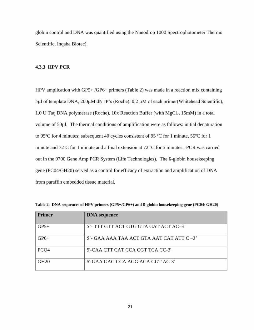
4.3.3 HPV PCR................................................................................................. 21
4.3.4 Gel Electrophoresis ................................................................................. 22
4.3.5 Real-time amplification of the β-globin gene............................................ 22
4.4 Data collection and statistical analysis............................................................... ...... 23
4.5 Ethical considerations..................................................................................... 23
5.0 RESULTS...................................................................................................... 24
5.1 Clinico-pathological findings............................................................................ ...... 24
5.1.1 Clinical findings................................................................................. ....... 25
5.1.2 Light microscopic findings................................................................. ....... 25
5.2 Molecular findings............................................................................................ ....... 31
6.0 DISCUSSION................................................................................................ 32
7.0 CONCLUSIONS........................................................................................... 38

vii
8.0 REFERENCES.................................................................................................. 39
9.0 APPENDICES................................................................................................... 44
9.1 Ethics Clearance Form..................................................................... 44
9.2 Turnitin Form................................................................................. 45
viii
LIST OF FIGURES
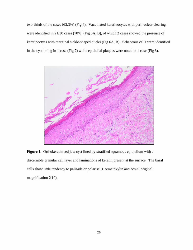
Figure 1: Orthokeratinised jaw cyst lined by stratified squamous epithelium with a
discernible granular cell layer and laminations of keratin present at the surface. The basal
cells show little tendency to palisade or polarise (Haematoxylin and eosin; original
magnification X10)............................................................................................................. 26
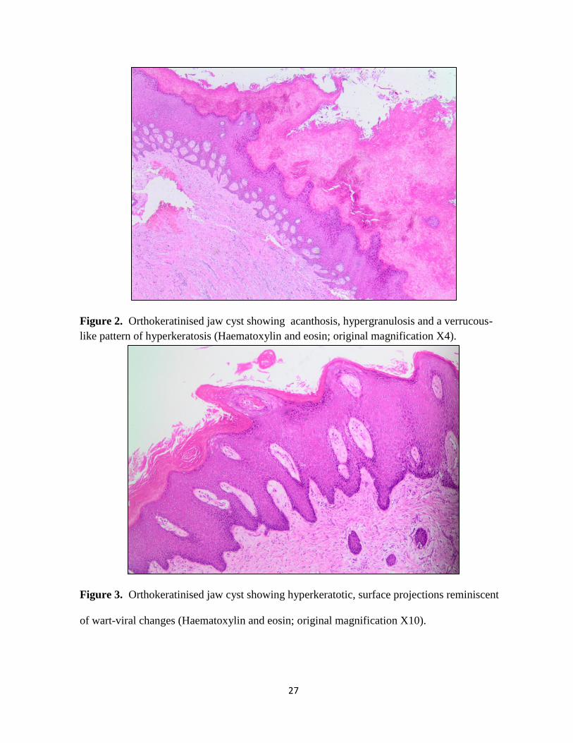
Figure 2: Orthokeratinised jaw cyst showing acanthosis, hypergranulosis and a
verrucous-like pattern of hyperkeratosis (Haematoxylin and eosin; original magnification
X4)...................................................................................................................................... 27
Figure 3: Orthokeratinised jaw cyst showing hyperkeratotic, surface projections
reminiscent of viral wart changes (Haematoxylin and eosin; original magnification
X10)..................... ...............................................................................................................27
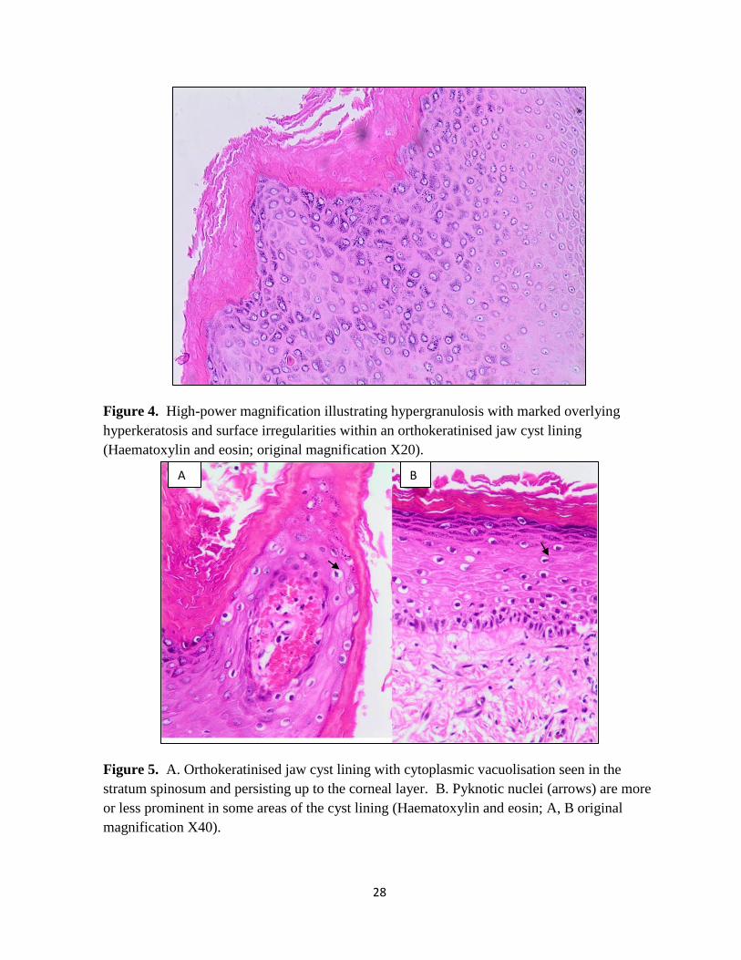
Figure 4: High-power magnification illustrating hypergranulosis with marked overlying
hyperkeratosis and surface irregularities within an orthokeratinised jaw cyst lining
(Haematoxylin and eosin; original magnification X20)......................................................28
Figure 5: Orthokeratinised jaw cyst lining with cytoplasmic vacuolisation seen in the
stratum spinosum and persisting up to the corneal layer. B. Pyknotic nuclei (arrows) are
more or less prominent in some areas of the cyst lining (Haematoxylin and eosin; A, B
original magnification X40).................................................................................................28
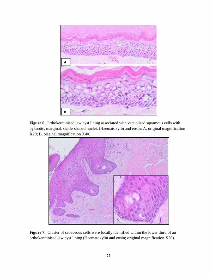
Figure 6: Orthokeratinised jaw cyst lining associated with vacuolised squamous cells with
pyknotic, marginal, sickle-shaped nuclei. (Haematoxylin and eosin; A, original
magnification X20; B, original magnificationX40)........................................................... 29
Figure 7: Cluster of sebaceous cells were focally identified within the lower third of an
orthokeratinised jaw cyst lining (Haematoxylin and eosin; original magnification X20).. 29
ix
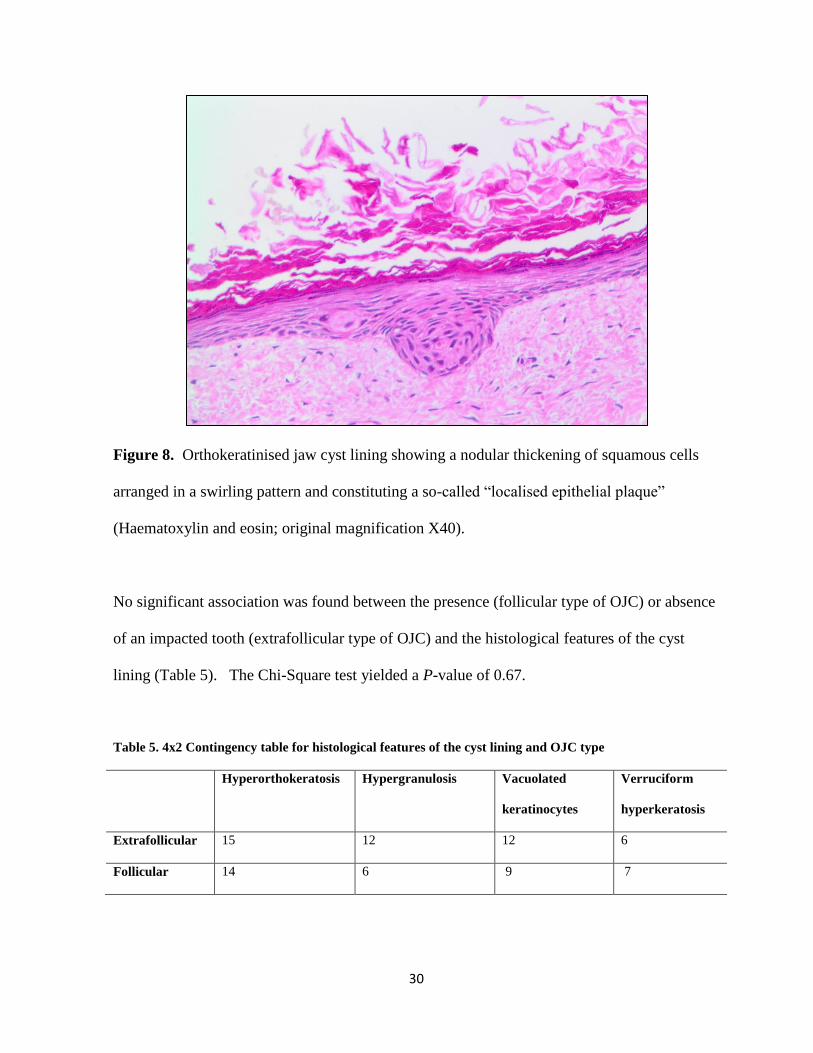
Figure 8: Orthokeratinised jaw cyst lining showing a nodular thickening of squamous
cells arranged in a swirling pattern and constituting a so-called “localised epithelial
plaque” (Haematoxylin and eosin; original magnification X40)........................................ 30
Figure 9: The polymerase chain reaction amplified products were separated on an agarose
gel and photographed after ethidium-bromide staining. The molecular marker constitutes
50 base pairs; lanes 1 to 30, orthokeratinised jaw cyst samples; +C, positive controls
indicating the expected size of the HPV amplified product; B, blank; -C, negative
control................................................................................................................................. 31

x
LIST OF TABLES
Table 1: Summary of studies on human papillomavirus in odontogenic lesions............... 11
Table 2: DNA sequences of HPV primers (GP5+/GP6+) and ß-globin housekeeping gene
(PC04/ GH20)..................................................................................................................... 21
Table 3: Clinical data and histopathological findings in patients with orthokeratinised jaw
cyst...................................................................................................................................... 24
Table 4: 2x2 Contingency table for follicular OJC and extrafollicular OJC in the mandible
versus the maxilla............................................................................................................... 25
Table 5: 4x2 Contingency table for histological features of the cyst lining and OJC
type…...................................................................................................................................30
1
CHAPTER 1
1.0 INTRODUCTION
Cystic lesions occurring within the jaw bones and which are lined by orthokeratinised
epithelium are known as orthokeratinised jaw cysts (OJCs).1 Theories regarding the
histogenesis of the OJC remain speculative. It is nevertheless currently endorsed that the OJC
is not a histological variant of the odontogenic keratocyst (OKC),2 as was described in earlier
reports.3-5
The rationale for this presumption includes the following findings; the OJC
presents more frequently in a dentigerous relationship and shows a significantly lower
recurrence rate than the OKC following conservative enucleation of the lesion.1,6,7
Furthermore, aggressive features such as multiplicity, satellite cysts, proliferating epithelial
islands and association with the nevoid basal cell carcinoma syndrome, although well
documented for the OKC, have not been reported for the OJC.8 Other theories suggest origin
of the OJC from heterotopic ectodermal tissues or from post-traumatic implantation.5 The
high frequency of occurrence of these lesions in association with the crowns of impacted third
molar teeth forms the basis of the premise that the follicular OJC most likely represents a
dentigerous (follicular) cyst, in which the cyst lining has undergone histomorphological
transformation (metaplasia) from a fairly non-descript, tenuous layer of flattened cuboidal
cells to a robust orthokeratinised lining.1,8
This orthokeratinised lining bears histological
resemblance to the epidermoid cyst of the skin and other extragnathic sites.9, 10
Since the early nineties there has been a surge of interest in the association between human
papillomavirus (HPV) and epidermoid cysts of the skin, in particular those of palmoplantar
2
location.11
Although the origin of these cysts is still controversial, Egawa et al.12
proposed the
idea that certain palmoplantar epidermoid cysts might be a reflection of epidermoid metaplasia
of eccrine duct epithelium and that HPV may play a role in this development. Cutaneous
epidermoid cysts that occur in non-palmoplantar location are thought to arise from epidermal
implantation due to trauma or a dilated follicular infundibulum.13
Interestingly, even in these
cases the presence of HPV has been shown by histopathological and molecular techniques.13
Likewise HPV has also been identified in cholesteatomas, which are histologically defined as
epidermoid cysts developing in the middle ear.10
Although papilloma viruses replicate exclusively in body surface tissues such as the skin or
the mucosal surfaces of the anogenital region, mouth, or airways, it is thought that infection
that is acquired at parturition or in utero may infect the invaginating primitive enamel organ.14
After a period of latency, conversion from latent into productive infection is thought to
occur.14,15
It is considered unlikely that HPV acts alone, but rather that it may be a co-factor
or initiator in the histogenesis of certain odontogenic epithelial lesions.16
Odontogenic cysts
and tumours that have been investigated for the presence of HPV DNA include the
ameloblastoma,14,16,17
and OKC.18
In the study by Sand et al.14
HPV DNA was detected by the
polymerase chain reaction in 44.4% of ameloblastomas. Kahn16
found HPV 16/18 in a
peripheral ameloblastoma with the in situ hybridisation technique. Subsequently, HPV-18
was also demonstrated by in situ hybridisation in a verrucous hyperplastic area of a primary
intraosseous ameloblastoma.17
HPV-16 DNA was isolated by Cox et al.18
from an OKC using
Southern blot hybridisation.
3
HPV-induced histomorphological changes, as observed by light microscopy, have often been
used as an adjunctive measure when assessing tissue specimens for underlying HPV
infection.19,20
The histological features that characterise flat viral warts (verruca plana), for
example, includes hyperkeratosis, often absence of parakeratosis, slight or no papillomatosis,
hypergranulosis and vacuolated keratinocytes with perinuclear clearing.20
Despite the
histological similarities between the OJC, cutaneous epidermoid cyst,9, 19
cholesteatoma10
and
verruca plana,20, 21
the OJC has not yet been studied for a possible association with HPV.
4
CHAPTER 2
2.0 LITERATURE REVIEW
2.1 Clinical, radiological and histological features of the orthokeratinised jaw cyst
OJCs are distinctly uncommon with less than 100 cases reported in the literature. This rare
developmental cyst accounts for about 10% of cases that were previously coded as OKCs.8
Clinically, orthokeratinised jaw cysts (OJCs) are usually non-expanding, asymptomatic,
solitary lesions which occur predominantly in adults with an approximately 2:1 male to female
ratio.22
OJCs occur more frequently in the mandible than in the maxilla with a predilection for
the posterior areas of the jaw.22
Radiographically, they present with no features that distinguish them from any other
developmental or inflammatory odontogenic cyst.23
The OJC frequently appears as a
unilocular radiolucency associated with the crown of an unerupted mandibular third molar
mimicking a dentigerous cyst. The size of the OJC may vary from 1cm to as large as 7cm in
diameter.22
The clinical behaviour of the OJC is similar to the dentigerous and radicular cyst
showing little tendency for recurrence following conservative surgical enucleation.8,22
It is,
however, the histological features of the OJC that sets this particular cyst apart from the other
jaw cysts. The OJC has an orthokeratinised stratified squamous epithelial often with a
prominent stratum granulosum.22,24
The basal layer comprises of cuboidal or flattened cells,
which show little tendency to palisade or polarise.25,26
Mature fibrous connective tissue
constitutes the cyst wall that, unless secondarily infected, is devoid of inflammation.25
5
2.2 A historical perspective on the separation between the orthokeratinised jaw cyst and
the odontogenic keratocyst
The OJC was first described as a dermoid cyst by Schultz27
in 1927 and was later typed as a
histological variant of the odontogenic keratocyst (OKC).4 In 1981 Wright
5 began to describe
the difference in clinical aggressiveness between the OJC and OKC. In subsequent studies the
OJC was considered as a separate entity and no longer as a subtype of the OKC.1,6-8
Orthokeratinised odontogenic cyst is a synonymous term for OJC that has been used in the
literature.6,8
The are several reasons for not considering the OJC as part of the spectrum of the
OKC. From a clinical point of view, the OJC presents more frequently in a dentigerous
relationship (60.8%) compared to the OKC (25-40%).1,3,5,7,8,22
The OJC shows a significantly
lower recurrence rate compared to the OKC following conservative enucleation of the
lesion.1,3,5,8
Immunohistochemical studies have demonstrated that the epithelial linings of the OJC and the
OKC are different in many respects. Ki-67-postive proliferating cells are dominant in the
OKC and not the OJC lining.28
The high suprabasal proliferative activity present in OKC is
not evident in the OJC.28
The presence of a high concentration of p53 expression in OKC is
thought to be responsible for the maintenance of and differentiation of epithelial stem cells,
suggesting that the lining of the OKC has a higher self-renewal potential when compared to
the OJC.29
Keratin profiling of the OJC and the OKC has also revealed these lesions to be
distinct entities.30
It was found in a study by Aragaki et al.30
that the keratin profiling of the
OJC lining resembled that of epidermis. By contrast the keratin profiling of the OKC
resembled the dental lamina.30
6
Most authors believe that the OKC originates from the dental lamina or from surface mucosal
epithelium.22
Cohen and Shear4 suggested that the origin of the OJC could also be related to
the dental lamina which has the ability to keratinise. Other theories suggest origin of the OJC
from heterotopic ectodermal tissues or from post-traumatic implantation.5 The OJC that
develops in association with the crowns of impacted teeth (follicular OJC) is further thought to
represent a dentigerous (follicular) cyst in which the cyst lining has undergone squamous
metaplasia.31
The stimulus for transformation from a tenuous layer of flattened cuboidal cells
to a robust orthokeratinised lining is still unknown.
Li et al.24
suggested that the OJC origin cannot simply be attributed to metaplasia. These
authors raised the possibility that those growth factors and their receptors that have been
implicated in normal tooth development and in the pathogenesis of some odontogenic lesions
may well be involved in the pathogenesis of the OJC.24
Other authors have suggested that the
underlying capsular fibrous tissue has an inductive influence on the lining of the cyst and that
mesenchymal factors may influence the diversity of odontogenic lesions.32
Vuhahula et al.1 suggested that the reduced enamel epithelium, a derivative of the odontogenic
apparatus, has the ability to keratinise under the appropriate stimuli. Aldred et al.33
later
reported on the unusual histological findings of an odontogenic cyst associated with an
impacted maxillary canine. In this case, transition from a thin layer of reduced enamel
epithelium into a thicker, hyperplastic, orthokeratinised, verrucoid, cystic epithelium was
observed prompting the authors to consider human papillomavirus (HPV) as a possible
pathogenetic mechanism for this phenomenon.33
Likewise there is vast research into HPV as a
possible aetiologic trigger in a variety of extragnathic lesions that may show verrucoid
7
features. Such lesions include epidermoid cysts,9 cholesteatoma,
10 cystic warts of the skin and
flat warts.19-21
2.3 Human papillomavirus
The human papillomavirus (HPV) is part of a large family of viruses known as the
papovaviridae.34
HPVs are DNA viruses of the papovavirus subgroup A.34
The virus is
capable of becoming totally integrated with the DNA of the host cell.34
The HPV genome
encodes for 6 early DNA sequences namely (E1, E2, E4-E7).34
These proteins are involved in
viral gene regulation and cell transformation.35
The late proteins (L1 and L2) are responsible
for forming the shell of the virus as well as one region of the regulatory DNA sequences.35
The characterisation of each HPV type is based on the genotypic variations in the DNA base-
sequences of E6 and E7.34
It was the discovery of these genotypic differences that allowed
classification of the HPV into high, intermediate and low-risk oncogenic types.34, 35
HPV is one of the most prevalent infections in the world. These viruses have an exclusive
tropism for epidermal and squamous mucosal epithelium.35
Transmission by sexual and non-
sexual person-to-person contact, contaminated objects, saliva and autoinoculation has been
proposed.34, 36
Of the more than 150 different types of HPV that have been identified, at least
25 types are associated with lesions of the head and neck (HPV-1, 2, 3, 4, 6, 7, 10, 11, 13, 16,
18, 31, 32, 33, 35, 40, 45, 52, 55, 57, 58, 59, 69, 72 and 73).36,37
The association between
HPV and the development of oral and oropharyngeal tumours (benign and malignant) is well
established in the literature.36,37
HPV has been demonstrated in diverse lesions such as
8
verruca vulgaris, condyloma acuminatum, papilloma, focal epithelial hyperplasia, proliferative
verrucous leukoplakia, verruca plana and a subset of squamous cell carcinomas.36,37
HPV is capable of producing a variety of proliferative surface changes at the site of
infection.35
On light microscopy the presence of hyperkeratosis, koilocytosis and a verruco-
papillary surface strongly suggest HPV related viral modification.9,20
It is these alterations
that reflect the ability of HPV to integrate viral DNA into that of the host cell, thus enabling
itself to reproduce and act as an agent of clinico-pathological change.35,38-39
2.4 Laboratory methods for the detection of human papillomavirus
HPV can be found in normal oral mucosa, in varying degrees of concentration, depending on
the method of detection used.40-42
HPV is only able to replicate in the basal cells of stratified
epithelium.35
The virus infects epithelial tissues via micro-abrasions or trauma that exposes
the cell basement membrane.34, 39
The HPV virus has a slow infection process and takes
anywhere from 12 to 24 hours to initiate transcription.35
The lesions associated with HPV are
believed to arise from the proliferation of infected basal keratinocytes. The release of viral
particles occurs as a result of degeneration of desquamative cells.35
HPV itself is a hardy virus
and is able to survive for months at a time at low temperatures without a host cell.34,35
The detection of HPV can only be performed by molecular analysis of HPV DNA sequences
as HPV cannot be cultured in vitro and serology assay has limited accuracy.39,40
There are
various molecular methods available for the analysis of HPV in clinical specimens.40-42
These
methods are largely based on the principles that were used for the discovery of the various
9
HPV subtypes.34
Early testing methods relied on the use of nuclei acid probes labelled with
radioactive phosphorus in a slot-blot hybridisation technique.41
Besides being technically
cumbersome, another key disadvantage of this technique is the lack of sensitivity and the
inability to detect all HPV genotypes.41,42
In the late eighties, nucleic acid in situ hybridisation
(ISH) became available and has enjoyed a recent revival with the introduction of automated
platforms that are manufactured by various companies.40-42
HPV DNA ISH offers the
advantages of high specificity, it provides visual detection of infected cells through the
preservation of tissue context and allows for the distinction between episomal and integrated
viral DNA.40
To date, however, a major drawback of HPV DNA ISH is its reduced
sensitivity, particularly when viral loads are low. Hence, while it is more sensitive than the
Southern blot technique, it has a lower sensitivity than the polymerase chain reaction (PCR)
technique.42
In essence, PCR is a technique that is able to amplify specific gene sequences of nuclei
acids.43
It then enables the user to apply the information in a variety of fields such as forensic
science, prenatal diagnostics and histopathology.43
PCR has become one of the most valuable
and flexible biomedical tools in research and subsequent refinements in the technique have
made PCR an accepted diagnostic tool in many pathology disciplines.41,43
There are several PCR screening assays available. The majority of these use consensus primer
sets that bind to highly conserved regions, so-called consensus HPV PCR or qualitative end
point PCR.40
This method allows for the simultaneous identification of a large range of HPV
types. The MY09/11 and GP5+/6+ primer sets fall into the consensus HPV PCR category and
their efficacy has been compared by Remmerbach et al.44
The authors concluded that
10
GP5+/6+ was more sensitive, especially in low viral load samples.44
GP5+/6+ primer sets are
also used more frequently with formalin-fixed paraffin-embedded (FFPE) tissue.44
This is
because the fixation process tends to cause fragmentation of the DNA into sequences that are
often shorter than 200 base pairs.45
The GP5+/6+ primer sets are well suited for this purpose
as they target these short DNA sequences in FFPE samples thereby resulting in increased
sensitivity.44
The disadvantages of the PCR-based methods include the requirement for DNA
extraction, the test yields no direct evidence of transcriptional activity, and there is a risk of
contamination and detection of HPV from surrounding healthy tissue.43
Recently, p16 (INK4A) has emerged as a potential surrogate marker of HPV infection.41
p16
is a tumour suppressor gene that prevents phosphorylation of the retinoblastoma (Rb) protein
by inhibiting the cyclin D and cyclin dependent kinase (CDK) 4 complex at the G1-S
interphase of the cell cycle.46
In high-risk HPV related cancers p16 overexpression is
frequently seen as a consequence of functional inactivation of the Rb gene by the E7 viral
oncogene.47
While p16 immunohistochemistry is a highly sensitive technique, it has been
criticised as lacking specificity for the presence of HPV.41,46
In other words, while p16
overexpression and high-risk HPV infections are highly correlated they are not always
synonymous.41,46
Given the advantages and disadvantages of the currently available HPV
detection methods as explained above, consensus HPV PCR emerges as a highly sensitive,
cost effective technique, that is feasible on FFPE material, targeting numerous HPV strains
and adequate for screening for the presence or absence of HPV in most laboratories.43,47
11
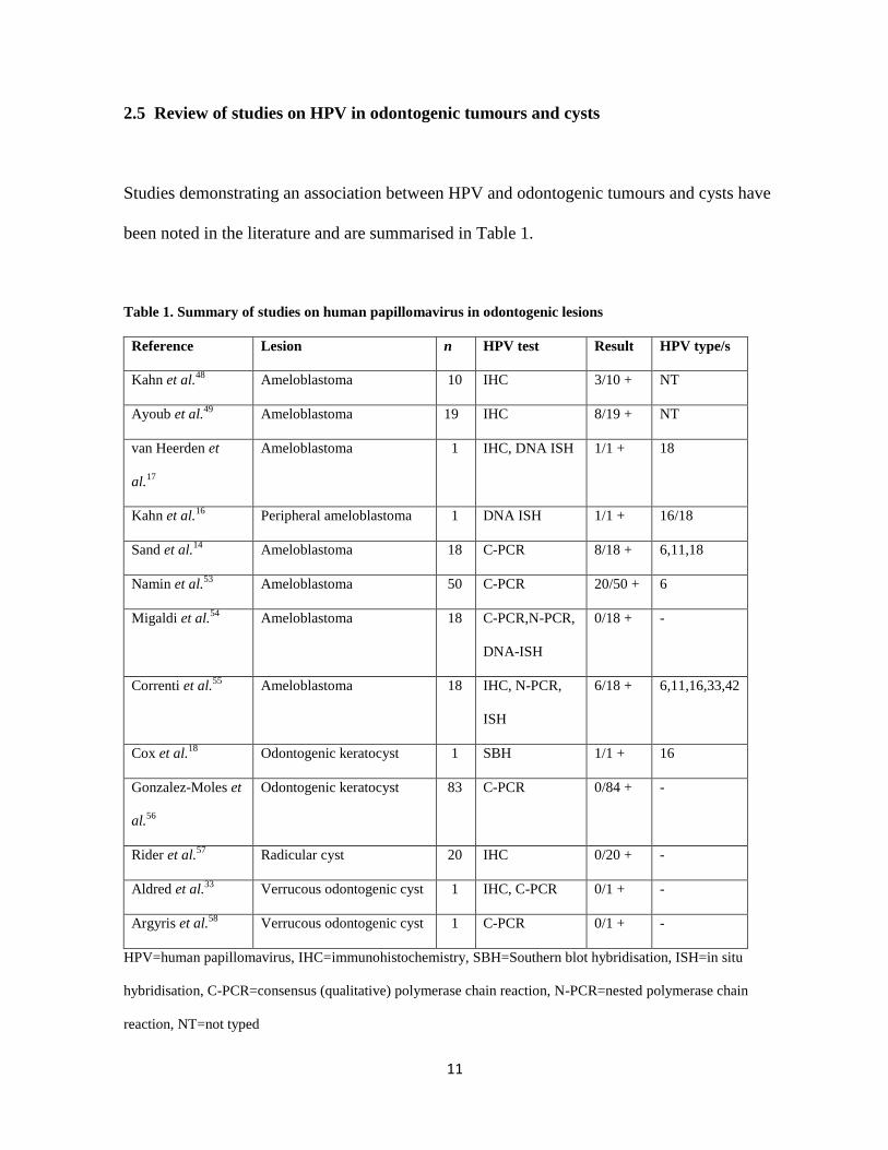
2.5 Review of studies on HPV in odontogenic tumours and cysts
Studies demonstrating an association between HPV and odontogenic tumours and cysts have
been noted in the literature and are summarised in Table 1.
Table 1. Summary of studies on human papillomavirus in odontogenic lesions
Reference Lesion n HPV test Result HPV type/s
Kahn et al.48
Ameloblastoma 10 IHC 3/10 + NT
Ayoub et al.49
Ameloblastoma 19 IHC 8/19 + NT
van Heerden et
al.17
Ameloblastoma 1 IHC, DNA ISH 1/1 + 18
Kahn et al.16
Peripheral ameloblastoma 1 DNA ISH 1/1 + 16/18
Sand et al.14
Ameloblastoma 18 C-PCR 8/18 + 6,11,18
Namin et al.53
Ameloblastoma 50 C-PCR 20/50 + 6
Migaldi et al.54
Ameloblastoma 18 C-PCR,N-PCR,
DNA-ISH
0/18 + -
Correnti et al.55
Ameloblastoma 18 IHC, N-PCR,
ISH
6/18 + 6,11,16,33,42
Cox et al.18
Odontogenic keratocyst 1 SBH 1/1 + 16
Gonzalez-Moles et
al.56
Odontogenic keratocyst 83 C-PCR 0/84 + -
Rider et al.57
Radicular cyst 20 IHC 0/20 + -
Aldred et al.33
Verrucous odontogenic cyst 1 IHC, C-PCR 0/1 + -
Argyris et al.58
Verrucous odontogenic cyst 1 C-PCR 0/1 + -
HPV=human papillomavirus, IHC=immunohistochemistry, SBH=Southern blot hybridisation, ISH=in situ
hybridisation, C-PCR=consensus (qualitative) polymerase chain reaction, N-PCR=nested polymerase chain
reaction, NT=not typed
12
The first documented study investigating for the presence of HPV in an odontogenic tumour
was performed in 1989 by Kahn.48
In this study ten cases of histologically confirmed
ameloblastomas were subjected to HPV immunohistochemistry.48
Of the 10 cases studied,
three were positive for HPV. The HPV detected in these three cases were more prevalent in
the columnar epithelial cell nuclei than in the stellate reticulum. No features of koilocytosis
were, however, noted.48
In a more recent study, also using the immunohistochemical method of HPV detection, Ayoub
et al.49
analysed 19 cases of ameloblastoma for the presence of HPV. Fifteen of these cases
were benign multicystic ameloblastomas and 4 were recurrent ameloblastomas with malignant
features. The anti-HPV mouse monoclonal antibody used in this study is formulated for
detecting major capsid proteins on most HPV subtypes.49
The results demonstrated 6/15
(40%) of the benign ameloblastomas to be positive for HPV, with most of the reactions
occurring in the cytoplasmic or membranous regions of the tumour cells.49
The cells layers
that were immunopositive were the basal ameloblastoma-like cells and the suprabasal stellate
reticulum-like cells.49
The two positive recurrent ameloblastomas with malignant features
showed the HPV reactions to be mainly nuclear, suggesting viral integration and a role for the
malignant transformation of these tumours. Ayoub et al.,49
nevertheless, concluded that more
sophisticated research techniques would have to be employed to accurately determine the
exact aetiological role HPV plays in the development and progression of ameloblastoma.
With regard to viral transmission, Ayoub et al.49
postulated that the HPV infection may be
acquired in utero or during parturition involving the invagination of the enamel organ.49
HPV
may subsequently influence various growth factors to induce pathologic proliferation of
odontogenic epithelial residues that are normally present within the jaws. The alternative
13
possibility that these authors considered was that the virus could merely be a passenger and
have no pathogenic potential in ameloblastomas.49
Studies that have been conducted to
ascertain the prevalence of HPV in normal oral mucosa have, however, yielded conflicting
findings and no definitive conclusions can thus far be drawn thereby precluding a definitive
role for HPV as a “by-stander” in these pathologic lesions.50-52
More sensitive techniques were used by other researchers to demonstrate the presence or
absence of HPV in ameloblastomas. These techniques included HPV DNA ISH16,17, 54, 55
and/or PCR.14, 53,54,55
van Heerden et al.17
were able to demonstrate the presence of HPV-18
DNA using DNA in situ hybridisation in an ameloblastoma. The HPV DNA was
demonstrated in a papillomatous area within the ameloblastoma.17
However, since HPV was
only detected in the papillomatous area, the authors suggested that acquisition of HPV DNA
within this lesion was most probably due to secondary infection from the overlying oral
mucosa and that HPV could not be considered an aetiological factor in this instance.17
In
another study also using HPV DNA ISH, a peripheral ameloblastoma occurring in a 16-year
old patient tested positively for HPV.16
These authors maintained that a role for HPV in
peripheral ameloblastomas and other benign and malignant lesions of the oral cavity remains a
possibility.16
Sand et al.14
examined 18 samples of central ameloblastoma from 12 patients for the presence
of HPV DNA by PCR using the L1 consensus primer. The results of the study demonstrated
that at primary surgery four of the samples tested positive for HPV-18 and HPV type 6/11, and
three were positive for HPV type 6/11.14
One HPV positive sample could not be typed.14
The
authors speculated that the presence of HPV at primary surgery may indicate a correlation
14
between ameloblastoma and HPV, while the presence of HPV at secondary surgery may be
due to traumatic manipulation of the tissues which may decrease the resistance of the tissue to
HPV infection.14
The authors also suggested that the HPV detection could be attributed to
presence of HPV in the overlying oral mucosa. This possibility could not be further
investigated by Sands et al.14
because normal oral mucosal samples were not included in their
study. In the subsequent work by Terai et al.50
and Sand et al.51
the episomal form of HPV
DNA was demonstrated in normal oral mucosa and the presence of HPV without any
histological or clinical changes was considered to be latent infection.50,51
With regard to viral
transmission, Sand et al.14
concurred with the hypothesis proposed by Kahn,48
where it is
speculated that the HPV in intrabony ameloblastomas may be acquired in utero or at
parturition, involving the invaginating primitive enamel organ. The virus is then, at a later
stage, thought to stimulate growth factors or inhibit natural control mechanisms which in turn
may lead to the development of an ameloblastoma.14
Namin et al.53
conducted a retrospective study for the presence of HPV by using the PCR
technique on 50 cases of ameloblastomas and 50 impacted third molar follicles as the control
group. The results of this study showed 20 samples in the study group positive for HPV while
nine samples were HPV positive in the control group.53
Statistical analysis proved this result
to be significant. Eight cases were typed as HPV-6 while the remaining positive cases could
not be subtyped. Namin et al.53
also noted that further investigations would need to be carried
out to determine the unidentifiable HPV types as this may be key to understanding the role
that HPV has, if any, in the pathogenesis of the ameloblastoma.53

15
A study by Magaldi et al.54
investigated 18 ameloblastomas for the presence of HPV using
ISH, laser capture microdissection and nested PCR analysis. The authors used both the
conventional as well as nested PCR approach.54
The microdissection technique prevented
contamination from the adjacent oral epithelium.54
The results were negative on PCR and
only a weak positive band was noted in the neoplastic tissue in one case and in one control
sample. Magaldi et al.54
concluded that that HPV should not be implicated in the development
of ameloblastoma. It was also noted that previous studies failed to show any real viral
integration.14,48,49
Rather some authors eluded towards surgical contamination or a secondary
event being the reason for HPV positivity in their studies.14
In a more recent study by Correnti et al.55
33% of intraosseous ameloblastomas were shown to
be HPV positive. Methodology included immunohistochemistry, nested PCR, and DNA
ISH.55
Their results revealed six cases to be positive of which four were HPV-6 positive, one
case was HPV-11 positive, and two cases were HPV-6 and HPV-42 and HPV-16 and HPV-33
positive respectively. Correnti et al.55
were able to demonstrate koilocytic changes in the
unicystic variants of ameloblastoma. These results were similar to those of Namin et al.53
where most of the cases were positive for HPV-6. Correnti et al.55
proposed that for HPV to
replicate it requires differentiating epithelial cells to complete its life cycle. It was suggested
by these authors that the source of this differentiating epithelium would have been provided
for either by the lining of an odontogenic cyst or enamel organ residues.55
As far as odontogenic cysts are concerned, OKC,18,56
radicular cysts57
and two cases of
verrucous odontogenic cysts were studied for the presence of HPV.33, 58
Rider et al.57
used a
polyclonal rabbit anti-papilloma antibody to evaluate for the presence of HPV DNA in

16
radicular cysts. All 20 cases were negative for the presence of HPV by immunostaining.57
The theory that was proposed by Rider et al.57
was that in the case of radicular cysts the
granulation tissue could be the source of growth factors, which in turn influence the
proliferation of the epithelial rests of Malassez.
In the study by Cox et al.18
HPV was detected in an OKC with signs suggestive of viral
intergration. Eighty-three samples of OKC were analysed by Gonzalez-Moles et al.56
for the
presence of HPV DNA using PCR. Their results yielded no positive cases for HPV DNA.
The OKCs used in their study included sporadic OKCs, recurrent OKCs and nevoid basal cell
carcinoma syndrome associated OKCs. The PCR findings on two samples of verrucous
odontogenic cysts that have thus far been investigated were negative for HPV DNA.33,58
2.6. HPV in extragnathic lesions that share overlapping histological features with OJC
The orthokeratinised lining of the OJC bears histomorphological resemblance to the
epidermoid cyst,9 cholesteatoma,
10 cystic warts of the skin and flat warts (verruca plana).
11,20,21
Whilst not confirmatory, HPV-induced histomorphological changes, as observed on light
microscopy, are often used as an adjunctive measure when assessing tissue specimens for
underlying HPV infection.10, 19,20
In their study, Gross et al.20
analysed the correlation
between the HPV type and the histology of warts. It was found that the histology of HPV-
induced warts is heterogenous.19
The classical pattern of HPV-induced warts includes
acanthosis, hypergranulosis, hyperkeratosis, papillomatosis, keratinocytes with perinuclear
halos and pyknotic nuclei.19
In addition to the classical pattern, HPV-induced warts with
sickle-shaped nuclei that were pushed to the margin of vacuolated squamous cells were also

17
documented by Gross et al.19
Whilst papillomatosis is a characteristic feature of some solid
HPV-associated lesions such as squamous papillomas and verruca vulgaris, papillomatosis is
notably slight or absent in HPV-associated epidermoid cysts (cystic warts).11-13
Another
variation in the morphological pattern is seen in verruca plana.59
The histological features that
characterise this HPV-associated lesion are hyperkeratosis, often absence of parakeratosis,
slight or no papillomatosis, hypergranulosis and vacuolated keratinocytes with perinuclear
clearing.19,59
Egawa et al.60
were able to demonstrate HPV antigens in 37 of 119 palmoplantar epidermal
cysts studied. HPV DNA was detected not only in cysts demonstrating all the HPV-induced
light microscopic changes but also in those cysts demonstrating a single HPV-associated
histological feature.60
In another study, Jeon et al.61
demonstrated, by the PCR technique, the
presence of HPV in non-palmoplantar epidermal cysts. The epidermoid cysts were positive
for HPV-60, which are typically found in palmoplantar epidermal cysts.13
Cholesteatomas which share a similar histological resemblance to OJCs have also been shown
by various authors to have HPV present in their linings. Researchers postulate that the
proliferative epithelium of some cholesteatomas may be associated with HPV infection.10,62,63
Among the odontogenic cysts, those lined by orthokeratinised epithelium exhibit by far the
most striking histological resemblance to HPV-induced histomorphological changes.
However, other than two previous case reports on verrucous odontogenic cysts where HPV
testing was undertaken,33,58
the OJC has not yet been rigorously analysed for a possible
association with HPV.

18
CHAPTER 3
3.0. AIM AND OBJECTIVES
3.1 Aim
To analyse the clinico-pathological features of orthokeratinised jaw cysts (OJCs) and to
determine whether human papillomavirus (HPV) DNA can be detected in a series of OJCs.
3.2 Objectives
3.2.1. To determine the age and gender of patients diagnosed with OJC.
3.2.2. To determine the anatomical location of the OJC.
3.3.3. To determine the prevalence of the follicular type of OJC.
3.3.4. To compare the sites of occurrence and histological features between the follicular type
of OJC and the extrafollicular OJC.
3.2.5. To histologically examine the OJC for light microscopic features suggestive of HPV
infection.
3.2.6. To investigate for the presence of HPV DNA in the OJC by using consensus HPV PCR,
also known as qualitative end point PCR.
19
CHAPTER 4
4.0 MATERIALS AND METHODS
4.1 Study sample
Formalin-fixed and paraffin embedded material of 30 biopsy specimens coded as OJCs were
retrieved from the archives of the Department of Oral Pathology at the University of the
Witwatersrand. For haematoxylin and eosin staining (H&E), 5-μm sections were cut from the
tissue blocks and examined by light microscopy to confirm the diagnosis of OJC. OJCs
comprised cystic lesions that were lined by orthokeratinised epithelium with a non-palisaded
rows of basal cells. The age and gender of the patients, anatomical location of the cyst and the
presence or absence of an associated impacted tooth was determined, wherever possible, from
the surgical histopathology reports. The intraosseous location of the OJC was ascertained by
evaluation of the radiographic findings as was described in the original histopathology
laboratory report. Locations of the lesions were classified as anterior (incisor-canine region),
premolar region, molar region or ramus.
4.2 Histological studies
The H&E stained OJC tissue sections were examined simultaneously by the student and
supervisor with the use of a Nikon Eclipse 50i double-headed light microscope. The cyst
linings were examined for the presence or absence of histomorphological features suggestive
of HPV infection. The features included hyperkeratosis, hypergranulosis, vacuolated
20
keratinocytes with perinuclear clearing and verruciform hyperkeratosis. Verruciform
hyperkeratosis was defined as squamous epithelium with an irregular surface exhibiting
pointed projections with marked overlying hyperkeratosis (orthokeratosis or parakeratosis).
Exclusion criteria included insufficient cyst lining for histological analysis, cysts showing a
palisaded, hyperchromatic basal cell layer characteristic of OKC and those cysts where an
intraosseous location could not be confirmed from the clinical information provided in the
histopathology report.
4.3. Polymerase chain reaction
4.3.1 DNA extraction
Sections (10µm) were prepared from each formalin-fixed paraffin-embedded sample. These
sections were deparaffinised using 1ml xylene and subsequently treated with 1ml ethanol.
After centrifugation, DNA was extracted from the samples using the DNA Micro QIA amp kit
(Qiagen, Whitehead Scientific) according to manufacturer’s instructions.
4.3.2 Control of contamination
Tissue blocks were sectioned using new blades for each sample to prevent cross
contamination. Work area and work tools were cleaned with 3% Virkon between each block
handled. Work areas were decontaminated with ultraviolet light between subsequent
procedures. The extraction procedure was assessed by PCR amplification of the internal ß-
21
globin control and DNA was quantified using the Nanodrop 1000 Spectrophotometer Thermo
Scientific, Inqaba Biotec).
4.3.3 HPV PCR
HPV amplication with GP5+ /GP6+ primers (Table 2) was made in a reaction mix containing
5µl of template DNA, 200µM dNTP’s (Roche), 0,2 µM of each primer(Whitehead Scientific),
1.0 U Taq DNA polymerase (Roche), 10x Reaction Buffer (with MgCl2, 15mM) in a total
volume of 50µl. The thermal conditions of amplification were as follows: initial denaturation
to 95ºC for 4 minutes; subsequent 40 cycles consistent of 95 ºC for 1 minute, 55ºC for 1
minute and 72ºC for 1 minute and a final extension at 72 ºC for 5 minutes. PCR was carried
out in the 9700 Gene Amp PCR System (Life Technologies). The ß-globin housekeeping
gene (PC04/GH20) served as a control for efficacy of extraction and amplification of DNA
from paraffin embedded tissue material.
Table 2. DNA sequences of HPV primers (GP5+/GP6+) and ß-globin housekeeping gene (PC04/ GH20)
Primer DNA sequence
GP5+ 5’- TTT GTT ACT GTG GTA GAT ACT AC–3’
GP6+ 5’- GAA AAA TAA ACT GTA AAT CAT ATT C –3’
PCO4 5'-CAA CTT CAT CCA CGT TCA CC-3'
GH20 5'-GAA GAG CCA AGG ACA GGT AC-3'
22
4.3.4 Gel Electrophoresis
Amplified PCR products were examined by agarose gel electrophoresis. Samples were
electrophoresed at 100 volts using a 3 % agarose gel (Celtic Diagnostics), stained with
ethidium bromide (Merck). The gel was visualised under ultraviolet light. Positive samples
appeared as a visible band with a molecular size of 150 base pairs (bp).
Controls used: The PCR procedure was controlled with the use of both positive and negative
controls. The positive control included paraffin embedded samples that had previously tested
positive with HPV PCR. The negative control used included a no-template control in which
nuclease- free water was substituted as a template.
4.3.5 Real-time amplification of the ß-globin gene
This assay was performed in a Corbett Research RotoGene 6000 (Whitehead Scientific) RT-
PCR machine using the Bioline SensiMix™SYBR No-Rox kit(Celtic Diagnostics). A final
volume of 20 µl reaction mix was made using 0.2 mM of each primer, 10 µl 2x
SensiMix™SYBR No-Rox Master Mix (with MgCl2, 50mM) and 2µl of template DNA. The
thermal cycling profile of this assay consisted initial denaturation step at 95ºC for 10 minutes,
followed by 50 cycles consistent of 95ºC for 10 seconds (denaturation), 55ºC for 10
seconds(annealing) and 72ºC for 15 seconds (elongation). After amplification, melt curve
analysis was carried out at 95ºC with a ramp rate 1ºC/5seconds. The average melting
temperature (Tm) of the -globin amplicon is 85.5 +/-1.0C. Gel electrophoresis was
performed on specimens with doubtful Tm values to confirm the presence of the 268bp PCR
fragment.
23
4.4 Data collection and statistical analysis
Descriptive statistics was used to describe the clinico-pathological features of the data in this
study. This was in the form of basic statistical analyses and table summaries describing what
the data showed. Fisher’s exact test was used to determine the association between the
frequency of the occurrence of the extrafollicular OJC and follicular OJC in the upper and
lower jaws. The Chi-Square test was used to determine the association between the presence
or absence of an impacted and whether that had an impact on the histological features of the
cyst lining. P-values of <0.05 were considered statistically significant.
The histological features observed on light microscopy were also captured in the form of a
table. The histological features that were analysed included: hyperorthokeratosis (HK),
hypergranulosis (HG), vacuolated keratinocytes with perinuclear clearing (VK) and
verruciform hyperkeratosis (VH).
The PCR findings were captured by photographing of the gel electrophoresis.
4.5 Ethical considerations
Ethics approval for this study was granted by the Human Research Ethics Committee of the
University of the Witwatersrand for the use of stored tissue and archived patient
histopathology reports. The ethics clearance reference number is M120956 (Appendix 1).
24
CHAPTER 5
5.0. RESULTS
5.1 Clinico-pathological findings
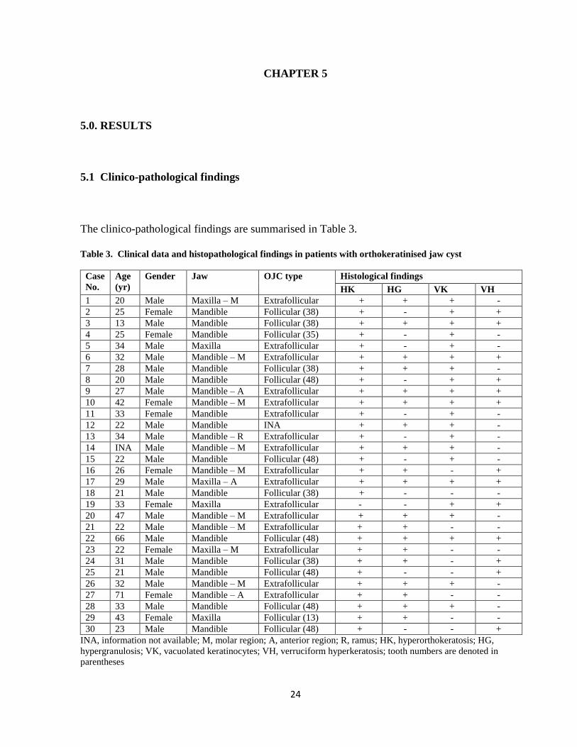
The clinico-pathological findings are summarised in Table 3.
Table 3. Clinical data and histopathological findings in patients with orthokeratinised jaw cyst
Case
No.
Age
(yr)
Gender Jaw OJC type Histological findings
HK HG VK VH
1 20 Male Maxilla – M Extrafollicular + + + -
2 25 Female Mandible Follicular (38) + - + +
3 13 Male Mandible Follicular (38) + + + +
4 25 Female Mandible Follicular (35) + - + -
5 34 Male Maxilla Extrafollicular + - + -
6 32 Male Mandible – M Extrafollicular + + + +
7 28 Male Mandible Follicular (38) + + + -
8 20 Male Mandible Follicular (48) + - + +
9 27 Male Mandible – A Extrafollicular + + + +
10 42 Female Mandible – M Extrafollicular + + + +
11 33 Female Mandible Extrafollicular + - + -
12 22 Male Mandible INA + + + -
13 34 Male Mandible – R Extrafollicular + - + -
14 INA Male Mandible – M Extrafollicular + + + -
15 22 Male Mandible Follicular (48) + - + -
16 26 Female Mandible – M Extrafollicular + + - +
17 29 Male Maxilla – A Extrafollicular + + + +
18 21 Male Mandible Follicular (38) + - - -
19 33 Female Maxilla Extrafollicular - - + +
20 47 Male Mandible – M Extrafollicular + + + -
21 22 Male Mandible – M Extrafollicular + + - -
22 66 Male Mandible Follicular (48) + + + +
23 22 Female Maxilla – M Extrafollicular + + - -
24 31 Male Mandible Follicular (38) + + - +
25 21 Male Mandible Follicular (48) + - - +
26 32 Male Mandible – M Extrafollicular + + + -
27 71 Female Mandible – A Extrafollicular + + - -
28 33 Male Mandible Follicular (48) + + + -
29 43 Female Maxilla Follicular (13) + + - -
30 23 Male Mandible Follicular (48) + - - +
INA, information not available; M, molar region; A, anterior region; R, ramus; HK, hyperorthokeratosis; HG,
hypergranulosis; VK, vacuolated keratinocytes; VH, verruciform hyperkeratosis; tooth numbers are denoted in
parentheses

25
5.1.1 Clinical findings
The OJC showed a male predominance with 21 cases occurring in males and 9 in females
yielding a 2.3:1 male to female ratio. The mean age at presentation was 30.9 ±12.9 years (age
range = 13-71 years; median = 27.5years). There was a strong predilection for the mandible
(24/30 cases; 80%). The mandibular molar region was the most commonly affected site
(18/26; 69.2%). The maxilla was affected in 6 cases (20%). In 13 patients (44.8%) the OJC
occurred in association with the crown of an impacted tooth (follicular OJC), most frequently
an impacted mandibular third molar (11/13; 84.6%). There was no significant association
between the frequency of occurrence of the follicular OJC versus extrafollicular OJC in the
upper or lower jaw. A P-value of 0,18 was obtained with the Fisher’s exact test (Table 4).
Table 4. 2x2 Contingency table for follicular OJC and extrafollicular OJC in mandible versus maxilla
Follicular OJC Extrafollicular OJC
Mandible 12 11
Maxilla 1 5
5.1.2 Light microscopic findings
On light microscopy, all the cysts were lined by stratified squamous epithelium, showing
keratinisation of the epidermoid type in 29/30 (96.7%) cases (Fig 1). None of the cases
showed a palisaded basal cell layer, a characteristic histological feature of the OKC. Cyst
lining showing foci of verruciform hyperkeratosis was present in slightly less than half of all
cases (13/30; 43.3%) (Fig 2; Fig 3), while a prominent stratum granulosum was noted in about
26
two-thirds of the cases (63.3%) (Fig 4). Vacuolated keratinocytes with perinuclear clearing
were identified in 21/30 cases (70%) (Fig 5A, B), of which 2 cases showed the presence of
keratinoctyes with marginal sickle-shaped nuclei (Fig 6A, B). Sebaceous cells were identified
in the cyst lining in 1 case (Fig 7) while epithelial plaques were noted in 1 case (Fig 8).
Figure 1. Orthokeratinised jaw cyst lined by stratified squamous epithelium with a
discernible granular cell layer and laminations of keratin present at the surface. The basal
cells show little tendency to palisade or polarise (Haematoxylin and eosin; original
magnification X10).
27
Figure 2. Orthokeratinised jaw cyst showing acanthosis, hypergranulosis and a verrucous-
like pattern of hyperkeratosis (Haematoxylin and eosin; original magnification X4).
Figure 3. Orthokeratinised jaw cyst showing hyperkeratotic, surface projections reminiscent
of wart-viral changes (Haematoxylin and eosin; original magnification X10).
28
Figure 4. High-power magnification illustrating hypergranulosis with marked overlying
hyperkeratosis and surface irregularities within an orthokeratinised jaw cyst lining
(Haematoxylin and eosin; original magnification X20).
Figure 5. A. Orthokeratinised jaw cyst lining with cytoplasmic vacuolisation seen in the
stratum spinosum and persisting up to the corneal layer. B. Pyknotic nuclei (arrows) are more
or less prominent in some areas of the cyst lining (Haematoxylin and eosin; A, B original
magnification X40).
B
B
B
A
A
A
A
A
B
B
29
Figure 6. Orthokeratinised jaw cyst lining associated with vacuolised squamous cells with
pyknotic, marginal, sickle-shaped nuclei. (Haematoxylin and eosin; A, original magnification
X20; B, original magnification X40)
Figure 7. Cluster of sebaceous cells were focally identified within the lower third of an
orthokeratinised jaw cyst lining (Haematoxylin and eosin; original magnification X20).
A
B
A
B
30
Figure 8. Orthokeratinised jaw cyst lining showing a nodular thickening of squamous cells
arranged in a swirling pattern and constituting a so-called “localised epithelial plaque”
(Haematoxylin and eosin; original magnification X40).
No significant association was found between the presence (follicular type of OJC) or absence
of an impacted tooth (extrafollicular type of OJC) and the histological features of the cyst
lining (Table 5). The Chi-Square test yielded a P-value of 0.67.
Table 5. 4x2 Contingency table for histological features of the cyst lining and OJC type
Hyperorthokeratosis Hypergranulosis Vacuolated
keratinocytes
Verruciform
hyperkeratosis
Extrafollicular 15 12 12 6
Follicular 14 6 9 7

31
5.2 Molecular findings
The PCR products obtained from the positive controls were strongly amplified with the GP5+
and GP6+ primers, whereas all OJCs were consistently negative.
Figure 9. The polymerase chain reaction amplified products were separated on an agarose gel
and photographed after ethidium-bromide staining. The molecular marker constitutes 50 base
pairs; lanes 1 to 30, orthokeratinised jaw cyst samples; +C, positive controls indicating the
expected size of the HPV amplified product; B, blank; -C, negative control.
32
CHAPTER 6
6.0. DISCUSSION
It is generally estimated that the OJC represents about 10% of cases that were previously
classified as orthokeratinised variants of the OKC.8 This relatively uncommon cyst has been
shown to exhibit significant clinico-pathological differences from the OKC thereby warranting
its designation as a distinct and separate entity.1,2,6-8
The present study analysed the clinico-
pathological features in 30 cases of OJC in a South African population. The average age at
diagnosis of patients with OJC was 30.9 years. Previous studies on OJC have shown a
predilection for patients in their third and fourth decades.1,8
The male to female ratio in the
current study was 2.3:1, a very near distribution to the 2.26:1 male to female ratio reported in
data pooled from all OJC cases reported in the English language literature.1,3,5,7,8,24
Further, in
keeping with previous studies,8,22
the mandible was found to be more commonly affected than
the maxilla (80% versus 20%). When comparing the frequencies of occurrence of follicular
versus extrafollicular OJC in the upper and lower jaw no significant differences were found
(P=0.18).
Thirteen cysts (44.8%) were found to be associated with an impacted tooth. This frequency is
relatively lower than the 60.8% average described in the literature for the follicular type of
OJC.8 It is possible that this discrepancy may be the result of a relatively small sample size in
our study. Of interest, nevertheless, is the observation that 11/13 follicular OJCs occurred in
association with an impacted mandibular third molar while the case in the maxilla occurred in
relation to an impacted canine. Since these teeth are among those most frequently associated
33
with the development of dentigerous cysts, this finding may support the hypothesis that the
follicular OJC most likely represents a dentigerous (follicular) cyst in which the cyst lining has
undergone squamous metaplasia with transformation to a robust orthokeratinised lining that
bears histological resemblance to epidermoid cysts of other sites.9,10
The stimulus for
metaplasia in the context of the OJC, however, remains unknown.
On light microscopy, all the cysts (follicular and extrafollicular OJC) were lined by
hyperkeratinised stratified squamous epithelium, which was of the epidermoid type
(anucleated cells in the stratum corneum) in 29/30 (96.7%) cases. There was no significant
difference in the histological features of the cyst linings between follicular and extrafollicular
OJCs (P=0.67). None of the cases showed a palisaded basal cell layer, which is a
characteristic histological feature of the OKC.64
Sebaceous cells were noted in an
extrafollicular OJC and epithelial plaques were identified in a follicular OJC associated with
an impacted mandibular premolar tooth. While sebaceous cells are common to cutaneous
tissue and may also be seen in normal oral mucosa, to the best of our knowledge, epithelial
plaques have not yet been described in non-odontogenic epithelia. These epithelial plaques
are furthermore a histological feature associated with odontogenic cysts derived from reduced
enamel epithelium.64
The OJC shows some histological features that are reminiscent of HPV-induced
histomorphological changes seen in cutaneous and mucosal squamous epithelia. HPV has
been demonstrated in intraosseous and peripheral odontogenic lesions with the ameloblastoma
being most frequently investigated for the presence of HPV. Of the eight studies that have
thus far been undertaken on HPV in ameloblastoma,14,16,17,48,49,53-55
HPV was detected in seven
34
studies with various methodologies and frequencies reported across these
studies.14,16,17,48,49,53,55
By consensus PCR, HPV was detected in 40% to 44.4% of
ameloblastomas,14,53
and by nested PCR and ISH in 33.3% of ameloblastomas.55
Correnti et
al.55
proposed that for HPV to replicate it requires differentiating epithelial cells to complete
its life cycle. It was suggested by these authors that the source of this differentiating
epithelium would have been provided for either by the lining of an odontogenic cyst or enamel
organ residues.55
In the odontogenic cyst category, OKC and radicular cysts were investigated
for the presence of HPV. Of the 84 OKCs tested thus far,18,56
HPV viral integration was
demonstrated in one case using Southern blot hybridisation.18
All twenty cases of radicular
cysts were HPV negative in the study by Rider et al.57
These findings correlate well with the
lack of HPV associated histomorphological changes in these lesions. By contrast, the OJC
shares overlapping histomorphological features with lesions that have shown an HPV
association. These lesions include cystic papillomas,11,19
flat warts,20,38
a subset of
cholesteatomas,10,62,63
and epidermoid cysts.9-13,60,61
The OJC was investigated for the
presence of HPV DNA for the first time in this study. Hypergranulosis was noted in 63.3% of
cases while cyst linings showing a verruciform pattern of hyperkeratosis were present in
43.3% of cases. Two cases also showed the presence of vacuolated keratinoctyes with
marginal sickle-shaped nuclei. The latter feature was described by Gross et al.20
as an HPV-2
cytopathic effect in cutaneous verruca vulgaris.
All 30 cases in this study were subjected to HPV-DNA PCR using consensus HPV GP5+ and
GP6+ primers. The PCR products obtained from the positive controls in the current study
were strongly amplified with the GP5+ and GP6+ primers, however, all 30 OJC samples were
negative for HPV DNA. Previous studies have shown that GP5+/6+ primers are highly
35
sensitive, especially in low viral load samples.43
GP5+/6+ primer sets are also used more
frequently with FFPE tissue since the fixation process tends to cause fragmentation of the
DNA sequences.43,44
The GP5+/6+ primer sets are well suited for this purpose as they target
these short DNA sequences in FFPE samples thereby resulting in increased sensitivity.44
The
GP5+/GP6+ primers are further capable of amplifying 20 HPV genotypes (HPV-6, -11, -13, -
16, -18, -30, -31, 32,-33,-35, -39, -40, -43, -45, -51, -54, -55, -56, -59 and -66),65
which
includes five of the six HPV genotypes that have thus far been identified in odontogenic
epithelial tumours.14,16-18,53,55
The absence of HPV in the 30 cases studied makes it unlikely
that HPV plays a role in the histogenesis of the OJC, despite the wart-like morphology that
may be noted in some cases. Similarly, HPV has been suggested as a possible causative agent
in benign verrucous acanthomas and non-genital seborrheic keratosis based on their
morphological overlap with verruca, but despite extensive research HPV involvement has not
yet been detected in these lesions.66,67
Future studies using larger samples of cases and
primers covering a larger spectrum of HPV types are required to clarify whether the same
applies to the OJC.
While HPV may play no role in the development of the OJC, it is tempting to speculate
whether those cysts that exhibited a verruciform pattern of hyperkeratosis in this study fall
under the category of cysts termed as “verrucous odontogenic cysts”. There are three reported
examples in the literature of these keratinising odontogenic cysts with a verrucous pattern of
the cyst lining.33,58,68
Two of these cases were extrafollicular and involved the third molar
region of the mandible,58,68
while one occurred in association with an unerupted maxillary
canine.33
In the latter case the cyst lining comprised a thin layer of epithelium resembling
reduced enamel epithelium, whilst in other areas the cyst lining showed verrucous projections
36
into the lumen with areas of hypergranulosis and some cells resembling koilocytes raising the
possibility of a viral aetiology.33
The possibility of HPV involvement was, however, not
supported in their case by immunohistochemical and PCR amplification for HPV DNA.33
Aldred et al.33
have suggested that this verrucous change in odontogenic cysts may represent
an unusual secondary change in a pre-existing cyst.
In the verrucous odontogenic cyst reported by Argyris et al.58
the cyst was lined by
hyperorthokeratinising and partially verrucoid stratified squamous epithelium, a prominent
granular cell layer, multiple sharp and blunt rounded epithelial projections. Argyris et al.58
also recorded epithelial dysplasia focally. The authors further noted numerous keratinocytes
within the superficial epithelial layers showing round, pyknotic nuclei surrounded by a clear
halo or vacuolated cytoplasm resembling koilocytes phenotypically.58
The presence of
transcriptionally active HPV infection was investigated by HPV DNA PCR and the tissue was
also subject to p16 immunohistochemistry.58
Areas of nuclear and cytoplasmic
immunopositivity were observed in the cyst epithelium.58
The PCR assay, however, failed to
identify HPV-DNA in both incisional and excisional biopsy thereby ruling out HPV
infection.58
There is no data on the HPV status in the case reported by Ueeck et al.68
because
technical limitations impeded further investigation.
Two of the reported verrucous odontogenic cysts were treated by enucleation with no signs of
recurrence,33,58
while the third case required segmental mandibulectomy following recurrence
of the lesion 7-months after enucleation.68
We are unable to make comparisons regarding the
biologic behaviour of these lesions, since the lack of clinical follow-up and incomplete data on
the therapeutic approach are limitations of this study. Notwithstanding these limitations, the

37
current study findings suggest that HPV infection is not a pathogenetic factor for the OJC.
The results further support the findings by Aldred et al.33
and Argyris et al.58
which suggested
that HPV is probably not responsible for the verrucous histological changes that may be
encountered in some cysts of the jaws.
38
CHAPTER 7
7.0. CONCLUSIONS
The average age at diagnosis of patients with OJC was 30.9 years.
OJC had a male predominance with a male to female ratio of 2.3:1.
The mandible was more commonly affected than the maxilla, with mandibular involvement
seen in 80% of cases.
44.8% of OJCs were found to be associated with an impacted tooth.
There was no significant difference in the histological features of the cyst linings between
follicular and extrafollicular OJCs.
Vacuolated keratinocytes with perinuclear clearing were identified in 66.7% of cases,
while 63.3% of cases showed a verruciform pattern of hyperkeratosis suggesting a possible
human papilloma virus (HPV) aetiology.
PCR amplification using consensus GP5+/6+ primer sets for HPV DNA was negative in all
30 OJC analysed in this study.
The study findings suggest that OJC is not associated with HPV DNA even though some of
these lesions may histologically mimic the architecture of verruca.
HPV infection does not play a role in OJC.
39
CHAPTER 8
8.0 REFERENCES
1. Vuhahula E, Nikai H, Ijuhin N, et al. Jaw cysts with orthokeratinization: analysis of 12
cases. J Oral Pathol Med 1993; 22: 35-40.
2. Barnes L, Everson JW, Reichart P, et al. World Health Organization classification of
tumours: pathology and genetics of head and neck tumours. Lyon: International Agency for
Research on Cancer, 2005.
3. Crowley TE, Kaugars GE, Gunsolley JC. Odontogenic keratocysts: a clinical and histologic
comparison of the parakeratin and orthokeratin variants. J Oral Maxillofac Surg 1992; 50: 22-
6.
4. Cohen MA, Shear M. Histological comparison of parakeratinised and orthokeratinised
primordial cysts (keratocysts). J Den Assoc S Afr. 1980; 35: 161-5.
5. Wright JM. The odontogenic keratocyst: orthokeratinized variant. Oral Surg 1981; 51: 609-
18.
6. da Silva MJ, de Sousa SOM, Correa L, et al. Immunohistochemical study of the
orthokeratinized odontogenic cyst: A comparison with the odontogenic keratocyst. Oral Surg
Oral Med Oral Pathol Oral Radiol Endod 2002; 94: 732-7.
7. Siar CH, Ng KH. Orthokeratinized odontogenic keratocysts in Malaysians. Br J Oral
Maxillofac Surg 1988; 26: 215-20.
8. Dong Q, Pan S, Sun LS, et al. Orthokeratinized odontogenic cyst: a clinicopathologic study
of 61 cases. Arch Pathol Lab Med 2010; 134: 271-5.
9. Tomasini C, Aloi F, Pippione M. Papillomavirus-infected epidermoid cysts. J Cutan Pathol
1994; 21: 94.
10. Ferekidis E, Nikolopoulos TP, Yiotakis J, et al. Correlation of clinical and surgical
findings to histological features (koilocytosis, papillary hyperplasia) suggesting
papillomavirus involvement in the pathogenesis of cholesteatoma. Med Sci Monit 2006; 12:
CR368-71.
11. Egawa K, Inaba Y, Ono T, et al. 'Cystic papilloma' in humans? Demonstration of human
papillomavirus in plantar epidermoid cysts. Arch Dermatol 1990; 126: 1599-603.
12. Egawa K, Egawa N, Honda Y. Human papillomavirus-associated plantar epidermoid cyst
related to epidermoid metaplasia of the eccrine duct epithelium: a combined histological,
immunohistochemical, DNA-DNA in situ hybridization and three-dimensional reconstruction
analysis. Br J Dermatol 2005; 152: 961-7.
40
13. Lee S, Lee W, Chung S, et al. Detection of human papillomavirus 60 in epidermal cysts of
nonpalmoplantar location. Am J Dermatopathol 2003; 25: 243-7.
14. Sand L, Jalouli J, Larsson PA, et al. Presence of human papilloma viruses in intraosseous
ameloblastoma. J Oral Maxillofac Surg 2000; 58: 1129-34; discussion 1135-6.
15. Gravitt PE. The known unknowns of HPV natural history. J Clin Invest 2011; 121: 4593-9.
16. Kahn MA. Demonstration of human papillomavirus DNA in a peripheral ameloblastoma
by in situ hybridization. Hum Pathol 1992; 23: 188-91.
17. van Heerden WF, van Rensburg EJ, Raubenheimer EJ, et al. Detection of human
papillomavirus DNA in an ameloblastoma using the in situ hybridization technique. J Oral
Pathol Med 1993; 22: 109-12.
18. Cox M, Eveson J, Scully C. Human papillomavirus type 16 DNA in an odontogenic
keratocyst. J Oral Pathol Med 1991; 20: 143-5.
19. Meyer LM, Tyring SK, Little WP. Verrucous cyst. Arch Dermatol 1991; 127: 1810-2.
20. Gross G, Pfister H, Hagedorn M, et al. Correlation between human papillomavirus (HPV)
type and histology of warts. J Invest Dermatol 1982; 78: 160-4.
21. Egawa K, Inaba Y, Honda Y, et al. 'Cystic papilloma': human papillomavirus in a palmar
epidermoid cyst. Arch Dermatol 1992; 128: 1658-9.
22. Damm DD, Bouquot JE, Neville BW, et al. Oral and Maxillofacial Pathology. 2nd
edition.
Philadelphia, PA; Saunders. 2002: 590-7.
23. MacDonald-Jankowski DS, Li TK. Orthokeratinized odontogenic cyst in a Hong Kong
community: the clinical and radiological features. Dentomaxillofac Radiol 2010; 39: 240-5.
24. Li TJ, Kitano M, Chen XM, et al. Orthokeratinized odontogenic cyst: a clinicopathological
and immunocytochemical study of 15 cases. Histopathology 1998; 32: 242-51.
25. Deyhimi P, Hashemzade Z. Comparative study of TGF-alpha and P53 markers' expression
in odontogenic keratocyst and orthokeratinaized odontogenic cyst. Dent Res J (Isfahan) 2012;
9: S39-44.
26. Thosaporn W, Iamaroon A, Pongsiriwet S, et al. A comparative study of epithelial cell
proliferation between the odontogenic keratocyst, orthokeratinized odontogenic cyst,
dentigerous cyst, and ameloblastoma. Oral Dis 2004; 10: 22–6.
27. Schultz L. Cysts of the maxillae and mandible. J Am Dent Assoc 1927; 14: 1395-402.
41
28. Gani F, Mahomed F, Meer S. Evaluation of Ki-67 and cyclin D1 expression in
odontogenic keratocysts and orthokeratinised jaw cysts. Sadj; 67: 370-3.
29. Baghaei F, Eslami M, Sadri D. Evaluation of Ki-67 antigen and protein P53 expression in
orthokeratinized and parakeratinized odontogenic keratocyst. J Dentistry (Iran). 2004; 1: 53-8.
30. Aragaki T, Michi Y, Katsube K, et al. Comprehensive keratin profiling reveals different
histopathogenesis of keratocystic odontogenic tumor and orthokeratinized odontogenic cyst.
Hum Pathol 2010; 41: 1718-25.
31. Pavelic B, Levanat S, Crnic I. et al. PTCH gene altered in dentigerous cysts. J Oral Pathol
Med 2001; 30: 569-76.
32. Gadbail AR, Hande A, Chaudhary M, et al. Tumor angiogenesis in keratocystic
odontogenic tumor assessed by using CD-105 antigen. J Oral Pathol Med 2011; 40: 263-9.
33. Aldred MJ, Talacko AA, Allan PG, et al. Odontogenic cyst with verrucous proliferation. J
Oral Pathol Med 2002; 31: 500-3.
34. zur Hausen H, de Villiers EM. Human papillomaviruses. Annu Rev Microbiol 1994; 48:
427-47.
35. Doorbar J, Quint W, Banks L, et al. The biology and life-cycle of human
papillomaviruses. Vaccine; 30 Suppl 5: F55-70.
36. Kumaraswamy KL, Vidhya M. Human papilloma virus and oral infections: an update. J
Cancer Res Ther 2011; 7: 120-7.
37. Castro TP, Bussoloti Filho I. Prevalence of human papillomavirus (HPV) in oral cavity
and oropharynx. Braz J Otorhinolaryngol 2006; 72: 272-82.
38. Mitsuishi T, Ohsawa I, Kato T, et al. Molecular cloning and characterisation of a novel
type of human papillomavirus 160 isolated from a flat wart of an immunocompetent patient.
PLoS One 2013; 8: e79592.
39. Schiller JT, Day PM, Kines RC. Current understanding of the mechanism of HPV
infection. Gynecol Oncol 2010; 118: S12-7.
40. Molijn A, Kleter B, Quint W, et al. Molecular diagnosis of human papillomavirus (HPV)
infections. J Clin Virol 2005; 32 Suppl 1: S43-51.
41. Mirghani H, Amen F, Moreau F, et al. Human papilloma virus testing in oropharyngeal
squamous cell carcinoma: what the clinician should know. Oral Oncol 2014; 50: 1-9.
42. Hubbard RA. Human papillomavirus testing methods. Arch Pathol Lab Med 2003; 127:
940-5.
42
43. Baumforth KRN, Nelson PN, Digby JE, et al. The polymerase chain reaction. J Clin
Pathol: Mol Pathol 1999; 52: 1-10.
44. Remmerbach TW, Brinckmann UG, Hemprich A, et al. PCR detection of human
papillomavirus of the mucosa: comparison between MY09/11 and GP5+/6+ primer sets. J Clin
Virol 2004; 30: 302-8.
45. Specht K, Richter T, Muller U, et al. Quantitative gene expression analysis in
microdissected archival formalin-fixed and paraffin-embedded tumor tissue. Am J Pathol
2001; 158: 419-29.
46. Singhi AD, Westra WH. Comparison of human papillomavirus in situ hybridization and
p16 immunohistochemistry in the detection of human papillomavirus-associated head and
neck cancer based on a prospective clinical experience. Cancer 2010; 116: 2166-73.
47. zur Hausen H. Papillomaviruses and cancer: from basic studies to clinical application. Nat
Rev Cancer 2002; 2: 342-50.
48. Kahn MA. Ameloblastoma in young persons: a clinicopathologic analysis and etiologic
investigation. Oral Surg Oral Med Oral Pathol 1989; 67: 706-15.
49. Ayoub MS, Elmalahy MH, Elshafei MM. Expression of human papilloma virus and
Epstein Barr virus in benign and malignant ameloblastoma. International journal of academic
research 2011; 3: 393-7.
50. Terai M, Hashimoto K, Yoda K, et al. High prevalence of human papillomaviruses in the
normal oral cavity of adults. Oral Microbiol Immunol 1999; 14: 201-5.
51. Sand L, Jalouli J, Larsson PA, et al. Human papilloma viruses in oral lesions. Anticancer
Res 2000; 20: 1183-8.
52. Boy S, Van Rensburg EJ, Engelbrecht S, et al. HPV detection in primary intra-oral
squamous cell carcinomas--commensal, aetiological agent or contamination? J Oral Pathol
Med 2006; 35: 86-90.
53. Namin AK, Azad TM, Eslami B, et al. A study of the relationship between ameloblastoma
and human papilloma virus. J Oral Maxillofac Surg 2003; 61: 467-70.
54. Migaldi M, Pecorari M, Rossi G, et al. Does HPV play a role in the etiopathogenesis of
ameloblastoma? An immunohistochemical, in situ hybridization and polymerase chain
reaction study of 18 cases using laser capture microdissection. Mod Pathol 2005; 18: 283-9.
55. Correnti M, Rossi M, Avila M, et al. Human papillomavirus in ameloblastoma. Oral Surg
Oral Med Oral Pathol Oral Radiol Endod 2010; 110: e20-4.
43
56. González-moles MA, Mosqueda-Taylor A, Delgado-Rodríguez M, et al. Analysis of p53
protein by PAb240, Ki-67 expression and human papillomavirus DNA detection in different
types of odontogenic keratocysts. Anticancer Research 2006; 26: 175-82.
57. Rider CA, Rupkalvis R, Miller AS, et al. Search for evidence of three viral agents in
radicular (periapical) cysts with immunohistochemistry. Oral Surg Oral Med Oral Pathol Oral
Radiol Endod 1995; 80: 87-91.
58. Argyris PP, Nelson AC, Koutlas IG. Keratinizing odontogenic cyst with verrucous pattern
featuring negative human papillomavirus status by polymerase chain reaction. Oral Surg Oral
Med Oral Pathol Oral Radiol 2014; [Epub ahead of print].
59. Chen SL, Tsao YP, Lee JW, et al. Characterization and analysis of human
papillomaviruses of skin warts. Arch Dermatol Res 1993; 285: 460-5.
60. Egawa K, Honda Y, Inaba Y, et al. Detection of human papillomaviruses and eccrine ducts
in palmoplantar epidermoid cysts. Br J Dermatol 1995; 132: 533-42.
61. Jeon J, Lee HS, Ahn HH, et al. Human papillomavirus 60-positive epidermal cyst and
wart at a nonpalmoplantar location. Br J Dermatol 2005; 152: 1361-2.
62. Bergmann K, Hoppe F, He Y, et al. Human-papillomavirus DNA in cholesteatomas. Int J
Cancer 1994; 59: 463-6.
63. Chao WY, Chang SJ, Jin YT. Detection of human papillomavirus in cholesteatomas. Eur
Arch Otorhinolaryngol 2000; 257: 120-3.
64. Shear M, Speight PM. Cysts of the oral and maxillofacial regions. 4th
edition. Oxford, UK;
Blackwell Munksgaard. 2007: 45-58.
65. de Roda Husman AM, Walboomers JM, van den Brule AJ, et al. The use of general
primers GP5 and GP6 elongated at their 3' ends with adjacent highly conserved sequences
improves human papillomavirus detection by PCR. J Gen Virol 1995; 76: 1057-62.
66. Zhu WY, Leonardi C, Kinsey W, et al. Irritated seborrheic keratoses and benign verrucous
acanthomas do not contain papillomavirus DNA. J Cutan Pathol 1991; 18: 449-52.
67. Lee ES, Whang MR, Kang WH. Absence of human papillomavirus DNA in nongenital
seborrheic keratosis. J Korean Med Sci 2001; 16: 619-22.
68. Ueeck BA, Woo BM, Stewart JC, et al. Keratinizing odontogenic cyst with verrucous
proliferation. J Oral Maxillofac Surg 2007; 65: 585-8.
44
CHAPTER 9
9.0 APPENDICES
9.1 Ethics clearance form

45
9.2 Turnitin