kindler syndrome: 2 case reports from india
TRANSCRIPT

Salodkar et al.
Iranian Journal of Dermatology, Vol 12, No 3 (Suppl), Autumn 2009 S1
Kindler Syndrome: 2 Case Reports from India Abstract Kindler syndrome (KS) is a rare autosomal recessive genodermatosis. We report two cases of KS with classical clinical presentations involving skin and mucus membranes. Clinically, both patients had four major features of KS in the form of acral skin blistering, photosensitivity, progressive poikiloderma, and diffuse cutaneous atrophy. Case 1 had associated features in the form of urethral stenosis, skin fragility and palmoplantar keratoderma with extension of the scaling onto the flexor aspect of the wrist and loss of palmar creases. Case 2 had associated features in form of anal stenosis, oesophageal stenosis, skin fragility and palmoplantar keratoderma with loss of palmar creases. An Interesting finding in our report is that both cases have prominent telengectasia involving face and neck regions. (Iran J Dermatol 2009;12 (Suppl): S1-S4)
Keywords: acral skin blistering, photosensitivity, poikiloderma, Kindler syndrome, cutaneous atrophy
Introduction
Kindler syndrome (KS) is a rare autosomal recessive genodermatosis, first described by Kindler in 1954 1. It is due to mutations in the KIND1 gene characterized by congenital acral skin blistering, photosensitivity, progressive poikiloderma, and diffuse cutaneous atrophy. The syndrome is a combination of features of inherited blistering skin disorders, e.g. dystrophic epidermolysis bullosa, and congenital poikilodermas, e.g. Rothmund-Thompson syndrome. Morbidity and mortality are mostly related to secondary infections arising from cutaneous bullae and to cosmetic disfigurement.
Patients usually present with initial skin manifestations during the first year of life. Both blistering and photosensitivity begin in infancy or early childhood and improve significantly with age. Poikiloderma appears gradually and becomes more prominent later in life. Diffuse poikiloderma (reticular telangiectasia, patchy hypopigmentation and hyperpigmentation, epidermal atrophy), skin fragility, and atrophic changes are most prominent in sun-exposed areas, most commonly on the dorsal surfaces of the hands and feet.
Dental abnormalities occur commonly in affected persons and include advanced periodontal bone loss, mild-to-severe gingivitis, dental caries, and leukokeratosis of buccal mucosa. Mucosal involvement is frequent and leads to urethral, anal, and esophageal stenosis. Ophthalmic abnormalities
such as ectropion, keratoconjunctivitis and conjunctival scarring have also been described.
We are reporting two cases of Kindler syndrome with classical clinical presentations involving skin and mucus membranes1-3.
Case History Case 1
A 15-year-old boy, born to a consanguineous marriage, presented with blisters on hands and feet since birth. History of difficulty in micturation was present and his voiding took a long time since birth. Blisters used to occur spontaneously on acral parts of the body and used to heal within one week. As the child grew, he continued to develop blisters but with less frequency and severity until he was eight years old. History of photosensitivity was present since he was 2 years old. History of increased fragility of skin was also present. Cutaneous examination revealed reticulate erythematous telengectasia on his neck. Atrophic scars on the dorsum of his hands (Figure1a), feet, ankle, knuckles, knees and elbows were also present. The entire skin over the trunk and limbs was thinned out and wrinkled with hyperpigmented brownish macules and patches and hypopigmented to depigmented macules. Diffuse palmo-plantar hyperkeratosis with extension of the scaling onto the flexor aspect of the wrist and loss of palmar creases were present (Figure1b). Dental abnormalities were not present. Other systemic
Atul Salodkar, MD Sanjiv Choudhary, MD Sankha Koley, MD Jawaharlal Nehru Medical College, Sawangi, Maharashtra state, India Corresponding author: Atul Salodkar, MD Jawaharlal Nehru Medical College, Sawangi, Maharashtra state, India Email: [email protected] Received: October 11, 2009 Accepted: November 10, 2009
Case Report

Kindler Syndrome: 2 Case Reports From India
S2 Iranian Journal of Dermatology © 2009 Iranian Society of Dermatology
examinations were within normal limits. Routine blood investigations were also within normal limits. Histopathological findings of the atrophic skin were consistent with poikiloderma. Ultrasonography of the abdomen and pelvis revealed a grossly distended urinary bladder with a 10 mm thickened wall and 169 cc post voidal residual urine volume with normal prostrate, dilated prostetic urethra and moderate hydroureteronephrosis. Findings of ultrasonography were suggestive of urinary bladder outlet obstruction. Retrograde urethrography revealed urethral stenosis.
Case 2 A 23-year-old male patient, born to a
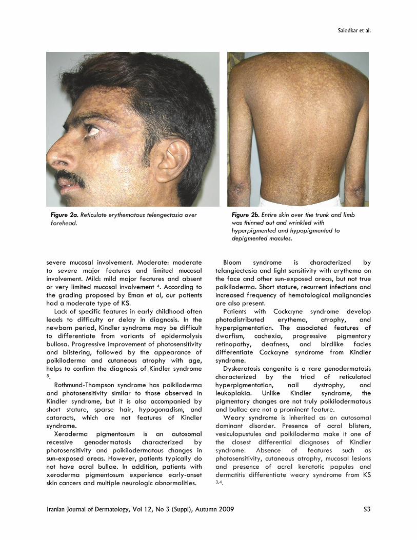
consanguineous marriage, presented with blisters on hands and feet since birth. Patient had 3 elder sisters and 1 younger brother who were all normal except for the younger brother who had similar complaints. Blisters used to occur spontaneously and used to heal within one week. Blisters continued to develop over acral parts with less frequency and severity until he was 9 years. History of photosensitivity was present. History of the increased fragility of the skin was present. The patient had undergone surgery in the past for anal stenosis. History of dysphasia was also present. Cutaneous examination revealed reticulate erythematous telengectasia on his forehead, face and neck (Figure2a). Atrophic scars on dorsum of the hands and feet were present. The entire skin over the trunk and limbs was thinned out and wrinkled with hyperpigmented and hypopigmented to depigmented macules (Figure2b). Diffuse palmo-plantar hyperkeratosis was present with loss of palmar creases. Dental abnormalities were not present. Esophageal endoscopy revealed stenosis.
Routine blood investigations were within normal limits. Histopathological findings of the atrophic skin were consistent with poikiloderma.
Discussion Most patients with KS present with a combination
of four major features and a variable number of associated features. The four major features are acral blisters in infancy and childhood, progressive poikiloderma, cutaneous atrophy and photosensitivity 2. The associated features include mucosal involvement in the form of oral problems, ectropion, and laryngeal, esophageal, urethral and anal stenosis. Skin fragility, PPK, webbing of the fingers and toes, nail dystrophy, skeletal deformities, cutaneous malignancy, hypohidrosis, pseudoainhum and phimosis are other associated features reported at variable frequencies 3.
Parental consanguinity was found in both of our cases, supporting the recessive mode of inheritance in KS. Clinically, both patients had four major features suggesting the diagnosis of KS. Case 1 had associated mucosal involvement in the form of urethral stenosis, skin fragility, palmoplantar keratoderma with extension of the scaling onto the flexor aspect of the wrist and loss of palmar creases. Case 2 had associated features in the form of anal stenosis, esophageal stenosis, skin fragility and palmoplantar keratoderma with loss of palmar creases.
An Interesting finding in our report is that both cases had prominent telengectasia involving face and neck. Our case 1 had extension of the scaling onto the flexor aspect of the wrist.
Eman Nofal et al, proposed a grading system for the severity of KS depending upon the mucosal involvement as Severe: severe major features and
Figure 1b. Diffuse palmo-plantar hyperkeratosis with extension of scaling on to the flexor aspect of wrist and loss of palmar creases.
Figure 1a. Atrophic scars involving dorsum of both hands.
Salodkar et al.
Iranian Journal of Dermatology, Vol 12, No 3 (Suppl), Autumn 2009 S3
severe mucosal involvement. Moderate: moderate to severe major features and limited mucosal involvement. Mild: mild major features and absent or very limited mucosal involvement 4. According to the grading proposed by Eman et al, our patients had a moderate type of KS.
Lack of specific features in early childhood often leads to difficulty or delay in diagnosis. In the newborn period, Kindler syndrome may be difficult to differentiate from variants of epidermolysis bullosa. Progressive improvement of photosensitivity and blistering, followed by the appearance of poikiloderma and cutaneous atrophy with age, helps to confirm the diagnosis of Kindler syndrome 5.
Rothmund-Thompson syndrome has poikiloderma and photosensitivity similar to those observed in Kindler syndrome, but it is also accompanied by short stature, sparse hair, hypogonadism, and cataracts, which are not features of Kindler syndrome.
Xeroderma pigmentosum is an autosomal recessive genodermatosis characterized by photosensitivity and poikilodermatous changes in sun-exposed areas. However, patients typically do not have acral bullae. In addition, patients with xeroderma pigmentosum experience early-onset skin cancers and multiple neurologic abnormalities.
Bloom syndrome is characterized by telangiectasia and light sensitivity with erythema on the face and other sun-exposed areas, but not true poikiloderma. Short stature, recurrent infections and increased frequency of hematological malignancies are also present.
Patients with Cockayne syndrome develop photodistributed erythema, atrophy, and hyperpigmentation. The associated features of dwarfism, cachexia, progressive pigmentary retinopathy, deafness, and birdlike facies differentiate Cockayne syndrome from Kindler syndrome.
Dyskeratosis congenita is a rare genodermatosis characterized by the triad of reticulated hyperpigmentation, nail dystrophy, and leukoplakia. Unlike Kindler syndrome, the pigmentary changes are not truly poikilodermatous and bullae are not a prominent feature.
Weary syndrome is inherited as an autosomal dominant disorder. Presence of acral blisters, vesiculopustules and poikiloderma make it one of the closest differential diagnoses of Kindler syndrome. Absence of features such as photosensitivity, cutaneous atrophy, mucosal lesions and presence of acral keratotic papules and dermatitis differentiate weary syndrome from KS 3,4.
Figure 2b. Entire skin over the trunk and limb was thinned out and wrinkled with hyperpigmented and hypopigmented to depigmented macules.
Figure 2a. Reticulate erythematous telengectasia over forehead.

Kindler Syndrome: 2 Case Reports From India
S4 Iranian Journal of Dermatology © 2009 Iranian Society of Dermatology
Although the clinical features of KS have been well described, the histologic findings are not specific, and definite ultrastructural features have not been well characterized 6. Because histopathologic and ultrastructural findings are not pathognomonic, clinical features remain the mainstay for the diagnosis of KS and the need for immunostaining with kindling antibody and genetic studies may be restricted to early cases with incomplete features 4.
Only more than 120 cases have been reported since the original report by Kindler in 1954 3. Paucity of the reports of Kindler syndrome and classical clinical presentations involving skin and mucous membranes with few interesting findings initiated us to report these cases here.
References 1. Kindler T. Congenital poikiloderma with traumatic
bulla formation and progressive cutaneous atrophy. Br J Dermatol 1954;66:104-11.
2. Yasukawa K, Sato-Matsumura KC, McMillan J, Tsuchiya K, Shimizu H. Exclusion of COL7A1 mutation in Kindler syndrome. J Am Acad Dermatol 2002;46:447-50.
3. Penagos HG, Jaen M, Sancho MT, et al. Kindler syndrome in Native Americans from Panama. A report of 26 cases. Arch Dermatol 2004;140:939-44.
4. Nofal E, Assaf M, Elmosalamy K. Kindler syndrome: A study of five Egyptian cases with evaluation of severity. Int J Dermatol 2008;47:658-62.
5. Shimizu H, Sato M, Ban M, Kitajima Y, Ishizaki S, Harada T, et al. Immunohistochemical, ultrastructural, and molecular features of Kindler syndrome distinguish it from dystrophic epidermolysis bullosa. Arch Dermatol 1997;133:1111-7.
6. Senturk N, Usubutun A, Sahin S, et al. Kindler syndrome: absence of definite ultrastructural features. J Am Acad Dermatol 1999;40:335-7.