kinetics of water disinfection with moringa oleifera seeds extract
TRANSCRIPT

Journal of Environment and Earth Science www.iiste.org
ISSN 2224-3216 (Paper) ISSN 2225-0948 (Online) Vol 2, No.7, 2012
58
Kinetics of Water Disinfection with Moringa Oleifera Seeds
Extract
Mustapha Hassan Bichi1*
Jonah Chukwuemeka Agunwamba2 Suleyman Aremu Muyibi
3
1. Department of Civil engineering, Faculty of Engineering, Bayero University, P.M.B. 3011, Kano-Nigeria
2. Department of Civil Engineering, Faculty of Engineering, University of Nigeria, Nsukka-Nigeria.
3. Bioenvironmental Engineering Research Unit (BERU), Department of Biotechnology Engineering, Faculty of
Engineering, International Islamic University Malaysia, 50728 Kuala Lumpur, Malaysia *E-mail of the corresponding author: [email protected]
The funding for this research work was provided by the Bayero University, Kano-Nigeria through the
MacArthur Foundation Grant. Part of the work was carried out in the laboratories of the Biotechnology
Engineering Research Unit (BERU), Department of Biotechnology Engineering, International Islamic University
Malaysia.
Abstract
Moringa seeds extract was prepared using de-fatted seed cake and aqueous extraction. One mL of Moringa extract
was added to 10mL of the cell cultures and incubated for 0, 30min, 60min, 90min, and 120min without agitation.
Cell survival was assessed by making dilution series of bacterial suspensions obtained after each incubation
period, plating on non-selective LB medium agar dishes, and incubating for 48hours at 37oC. Duplicates were
made of every individual assay. Colonies were counted on dishes and the cell survival ratio was estimated by
comparison to a control experiment where no Moring extract was added (0 min incubation time).
Pseudo-first-order kinetics was fitted to the data. The log-inactivation of the “viable” organisms at time t (Nt)
divided by the number of “viable” organisms at time zero (N0) was plotted as a function of chemical dose, Ct (mg
min L -1
). The coefficient of specific lethality, Ʌcw, was determined by linear regression using MINITAB for
Windows Release 11.2 32Bit. A kinetic model was developed for the application of Moringa Oleifera seeds extract
in water disinfection. The coefficient of specific lethality (Ʌcw) was determined as 3.76 L mg-1
min-1
for E.Coli
inactivation using Moringa Oleifera seeds extracts. The mode of attack of the Moringa seeds extract on the E.Coli
cell was explained as by rupturing the cell and damaging the intercellular components, when water dips in to the
cell which causes it to swell more and burst leading to death.
Keywords: Moringa Oleifera, Seed Extract, Antimicrobial Activity, Disinfection Kinetics.
1. Introduction
1.1 Disinfection and Disinfectants
Although the addition of halogens readily comes to mind when talking about disinfection, other unit processes
also provide rather substantial degree of disinfection. Robeck et al. (1962) for instance, found that up to 98%
of poliovirus type I was removed in a coal and sand filter and up to 99% was removed when alum dose was
increased and conventional flocculation and sedimentation carried out ahead of the filter. Alum coagulation
was also found to remove 95% to 99% of Coxsackie virus, and Ferric chloride was found to remove 92% to 94%
of the same Virus (Berg 1967; Fair et al. 1958). Although these methods cannot be relied upon to provide the
only means of pathogenic removal, Hudson (1962) noted that they perform an important role in assuring the
ability of other disinfectants to completely disinfect the water.
Traditionally, water disinfection has been achieved by the use of chlorine in many water supply agencies.
However, the production of by-products of chlorination such as halogenated organic compounds has been shown
to be associated with various ailments in humans (Hwang et al. 2008). Goveas et al. (2010) for instance, reported
that although chlorine is widely used as an inexpensive and potent disinfectant in the United States for drinking
water, it has the potential of forming carcinogenic and mutagenic disinfection by-products (DBPs). These, and
other similar problems like the high cost of chlorine in developing countries, necessitates the search for natural
disinfectants that are safer and cheaper to use.
Humeirah et al. (2010) reported that the essential oils from the twig and root of Goniothalamus
macrophyllus (Annonaceae) obtained from Pasoh Forest, Malaysia exhibited the most notable inhibitory activity
(0.3 mg/ml) against Vancomycin intermediate-resistance Staphylococcus aureus (VISA 24), Staphylococcus
epidermidis and Candida albicans. Oluseyi et al. (2009) also reported that Buchholzia coriacea (wonderful kola)
posses antimicrobial properties. The report showed that the fresh kola, its hexane extract, and its methanolic
extracts showed inhibitory zone of 62mm, 21mm, and 30mm respectively with E. Coli.
A lot of work has been done on the use of Moringa Oleifera seeds extract in Coagulation and Softening
(Folkard et al. 1989; Jahn 1988; Bina, 1991; Okuda, et al 1999; Buthelezi, et al 2009; Okuda, et al, 2001a, b). It

Journal of Environment and Earth Science www.iiste.org
ISSN 2224-3216 (Paper) ISSN 2225-0948 (Online) Vol 2, No.7, 2012
59
has also been found to contain an active anti-microbial agent (Eilert et al, 1981; Madsen et al. 1987; Fisch, et al.
2004; Suarez et al. 2005; Bukar et al. 2010, and Walter et al. 2011).
Moringa Oleifera seeds extract, having been shown to possess some anti-microbial properties by earlier
researchers, needs further investigation in terms of evaluating the best method of processing the seeds for its
application in the disinfection of portable water supplies, and understanding the mechanism of its disinfection
action (Suarez et al. 2005).
Suarez et al (2003) had earlier reported that Moringa seeds protein may be a viable alternative to chemicals
commonly used as food preservatives or for water disinfection. They are unlikely to have toxic effects as the
seeds are currently used for the treatment of drinking water and for preparation oils for various foods. Another
advantage reported is their biodegradability, unlike other chemicals (e.g. Aluminium salts and chlorine) which
remain as contaminants of treated waters and of the sediments.
Bichi (2011) has shown that its highest disinfection action was achieved with the use of de-fatted seed cake
and extracting the active ingredients by aqueous extraction. Bichi (2012) also found that the optimal conditions
for the extraction of the bioactive compounds to be 31 minutes mixing time, 85 rpm mixing speed and 3.25
mg/mL Moringa dosage. This research further developed a Quadratic model that can be used to optimize the
process of antimicrobial bioactive compound extraction from de-fatted Moringa Oleifera seeds.
The aim of this segment of the research is to develop the kinetic equation for the application of Moringa
Oleifera seeds extract in water disinfection, and investigate the mode of attack of the extract on the microbes
using E. Coli as an indicator organism.
1.2 Rate of Disinfection
Under ideal conditions, all cells of a single species of organisms are discrete units equally susceptible to a single
species of disinfectant; both cells and disinfectants are uniformly distributed (or dispersed) in the water, the
disinfectant stay. Substantially unchanged in chemical composition and substantially constant in concentration
throughout the period of contact; and the water contains no interfering substances. Under such conditions
according to Metcalf & Eddy (1991), the rate of disinfection is a function of (i) time of contact, (ii) Concentration
of Disinfectant, (iii) Concentration of organisms and (iv) Temperature of Disinfection.
1.2.1 Concentration of Organism: Chick’s Law
The specific mechanism of microorganism inactivation during chemical disinfection is not well understood (Fair et
al. 1958; MetCalf & Eddy 2003). It was, however, understood to be a function of chemical disinfection agent,
properties of the microorganism, and properties of the water. One simple kinetic model is widely used, but many
other models describe the mechanism of disinfection. According to Dr. Harriet Chick ‐ Chick’s Law (1908) –
disinfection could be modeled as a pseudo‐first‐order reaction with respect to the concentration of the organisms.
The time rate of kill follows chick’s law of disinfection. This states that Y, the number of organisms destroyed in
unit time, is proportional to N, the number of organism remaining, the initial number being No. The rate of change
of organisms with time, ��
��, is given by equation (1).
��
�� = k (N0 – y) (1)
Where, k represents the Chick’s law rate constant (t-1
).
Integrating between the limits y = 0 at t = 0 and y = y at t = t we obtain equation (2) which is simplified into
equations (3a) and (3b).
In [(N0 – y)/N0] = In (��
�� ) = - kt (2)
(��
�� ) = exp (-kt) (3a)
(��
�� ) = e
– kt (3b)
A plot of log Nt/N0 Vs t traces a straight line with slope of – klog e = -k1 and an intercept of 1 (or 100%) at t = 0.
When kt = 1 or k1t = 0.4343, the surviving fraction is 0.368 (Fair et al. 1958; MetCalf & Eddy 2003).
1.2.2 Concentration of Disinfectant: Watson Law Watson (1908) reported that the microbial inactivation rate increased with disinfectant concentration. The
disinfecting efficiency of a disinfectant is given by the relationship in equation (4).
Cn tp = Constant (4)
Where, C represents the Concentration of disinfectant (mg/L), tp is the time required to effect a constant percentage
kills of organisms, n is the coefficient of dilution, and the constant is a value of given percentage of inactivation.
When n > 1, the efficiency of the disinfectant decreases rapidly as it is diluted; when n < 1, time of contact is more
important than dosage.
1.2.3 Kinetics of Disinfection – Chick‐‐‐‐Watson Law
Since the early 1900s, it was discovered that disinfectant concentration and contact time were the primary
variables affecting microbial inactivation kinetics and efficiency (Cunningham et al. 2008). Chick (1908)
reported that for a given concentration of disinfectant, longer contact times resulted in an increased microbial
Journal of Environment and Earth Science www.iiste.org
ISSN 2224-3216 (Paper) ISSN 2225-0948 (Online) Vol 2, No.7, 2012
60
death. Additionally, Watson (1908) reported that the microbial inactivation rate increased with disinfectant
concentration. Combining the expressions of Chick and Watson yields the equation that is known as the
“Chick–Watson Law”:
Taking logs on both sides of equation (4) yields equations (5a) and combining this with the Chicks’ equation
(3a) produces equation (5b).
n. log(C) + log(t) = log(constant) (5a)
��
�� = -ɅcwC
n N = -Kcw N (5b)
The parameters are as previously defined. After integration we obtain the Chick’s-Watson equation (6).
In (�
��) = - Ʌcw Ct (6)
The constant Ʌcw is an empirical constant known as the Chick–Watson coefficient of specific lethality.
Many other researchers have since modified the Chick–Watson Law to account for specific features of the dose
response relationships such as lag or tailing (Haas 1980; Lambert & Johnson 2000).
1.3 Inactivation Mechanism
Cunningham et al. (2008) have documented the evidence of Chlorine inactivation of bacteria by many
researchers. The current understanding among many researchers is that a major mechanism of bacterial
inactivation by free chlorine is the alteration of cell permeability, resulting in the leakage of intracellular
materials such as proteins, RNA and DNA (Venkobachar et al. 1977; Haas & Engelbrecht 1980). In addition,
damage to nucleic acids and enzymes, oxidation of sulph-hydryl groups, damage to iron-sulphur centres,
disruption of nutrient transport and inhibition of cell respiration have been reported (Hoyana et al. 1973; Barrette
et al. 1988; Leyer & Johnson 1997). The mechanisms of microbial inactivation by iodine, like chlorine, are not
clearly understood (Gottardi 1999). Although various forms of iodine have been shown to possess antimicrobial
properties, it is believed by most researchers that molecular iodine (I2) is the chemical form most responsible for
bacterial inactivation (Gottardi 1999).
Silver has also been shown to be an effective disinfectant against bacteria (Zhao & Stevens 1998).
However, the biocidal action of silver requires contact times on the order of hours and therefore it has been
primarily used as a bacterial inhibitory compound for water storage. The current understanding of silver bacterial
inactivation involves the reaction between silver ion (Ag3+
) and thiol groups of amino acids and key enzyme
functional groups (Liau et al. 1997). Furthermore, Batarseh (2004) concluded that intracellular silver complexes
resulted in DNA unwinding and were the primary causes for bacterial inactivation
Myriam et al. (2010) has documented the mechanism of inactivation of UV light. The effectiveness of UV
light in biological inactivation arises primarily from the fact that DNA molecules absorb UV photons between
200 and 300 nm, with peak absorption at 260 nm (Byrd et al. 1990). This absorption creates damage in the DNA
by altering nucleotide base pairing, thereby creating new linkages between adjacent nucleotides on the same
DNA strand. If the damage goes unrepaired, the accumulation of DNA photoproducts can be lethal to cells
through the blockage of DNA replication and RNA transcription, which ultimately result in reproductive cell
death (Zimmer & Slawson 2002).
The antimicrobial activity of Moringa extracts was previously attributed to plant-produced benzyl
isothiocynate derivatives (Eilert et al. 1981). Suarez et al. (2003) showed that at least part of the antimicrobial
activity of Moringa seeds extract may stem from Flo-like polypeptides. Antimicrobial peptides have attracted
increasing attention recently because they can efficiently kill fungi and bacteria that are otherwise resistant to
many commonly used antibiotics. According to Zasloff (2002), they act by forming essential enzymes, leading to
cell deaths. Suarez et al (2003) reported that the antimicrobial action of Flo might results from similar activities
or from its bacterial flocculation effect. The report further noted that the former is more likely because for
example, Flo concentration required to obtain half-mixed effect on E.Coli are 0.1 mg/L for bacteriostatic action,
1.0mg/L for bacterial activity, and 6.0mg/L for cell flocculation. The study concluded that visual inspection of
Flo-incubated E.Coli revealed that the peptide aggregated the bacteria, as indicated by defined particles or flocs.
Chuang et al. (2007) has studied the mode of attack of Moringa Oleifera seeds extract on fungus. The
results showed that the cytoplasmic membrane of the fungal cell was ruptured and the intercellular components
were seriously damaged after treatment with M. Oleifera seed crude extract. However, the intercellular
components did not leak out. Based on previous studies of cell lysis pathways of antimicrobial peptides on
bacteria (Cham et al. 1998; Chen et al. 2003), this indicated that extracted compounds interacted with the lipid
bilayers in membranes leading to the separation of the two membranes (outer and inner). Subsequently, water
dips in to the cell, which causes cell to swell more and leads to death.
Structural morphological study of microbes after treatment with antimicrobial agents is an important
parameter in understanding the mechanism of action of these agents (Kitajim et al. 1998). However, not much

Journal of Environment and Earth Science www.iiste.org
ISSN 2224-3216 (Paper) ISSN 2225-0948 (Online) Vol 2, No.7, 2012
61
review is available on the mechanistic microscopic study of Moringa oleifera (Jabeen et al. 2008). Microscopic
evaluation of E.Coli strains were carried out after treatment with Moringa Oleifera seeds extract.
2. Materials and Methods
2.1 Preparation of Moringa Oleifera Seeds Extract
Good quality dry M. Oleifera seeds were selected and the seed coat and wings were removed manually. The
kernel was ground to fine powder using the coffee mill attachment of the Moulinex domestic food blender. The
ground powder was then sieved through 210 µm sieve. The seed powder was de-fatted using hexane in
electro-thermal Soxhlet extractor.
The best method of Moringa seed processing determined by (Bichi 2011) was used for this study. Measured
quantities of the de-fatted Moring seed powder was dissolved in a beaker and made up to 1000mL with distilled
water. The active ingredients were extracted by mixing with a stirrer at a pre-set mixing speed and for a pre-set
mixing time as outlined in the experimental design. The mixture was filtered through No.1 whatt-man filter
paper and the extract used for the disinfection studies.
2.2 Preparation of E. Coli Culture
The E.Coli culture was prepared as described in Obire et al. (2005). Nutrient broth (130.0gm) was dissolved in
1000mL distilled water by heating slightly. The mixture was sterilized at 130oC for 15minutes at 15 SPT in
autoclave. The sterilized broth was cooled to room temperature and was used to prepare the E.Coli culture.
Escherchia Coli (ER2566) strain was grown in 10mL broth at 37oC overnight to obtain an exponential growth
phase. This was then used for the disinfection studies.
2.3 Disinfection Studies
The procedure for the disinfection study was as described in Suarez et al. (2003) and Fisch et al. (2004). The
Moringa Oleifera dosage obtained from the optimization studies (3.25mg/L, 31min mixing time, and 85rpm
mixing speed) was used for this study (Bichi 2012).
One mL of Moringa extract was added to 10mL of the cell cultures and incubated for 0, 30min, 60min, 90min,
and 120min without agitation. Cell survival was assessed by making dilution series of bacterial suspensions
obtained after each incubation period, plating on non-selective LB medium agar dishes, and incubating for
48hours at 37oC. Duplicates were made of every individual assay (Suarez et al. 2003). Colonies were counted on
dishes and the cell survival ratio was estimated by comparison to a control experiment where no Moring extract
was added (0 min incubation time).
2.4 Inactivation kinetics
Pseudo-first-order kinetics was determined using the method described by Cunningham et al. (2008). The
log-inactivation of the “viable” organisms at time t (Nt) divided by the number of “viable” organisms at time
zero (N0) was plotted as a function of chemical dose, Ct (mg min L -1
). The coefficient of specific lethality, Ʌcw,
was determined by linear regression using MINITAB for Windows Release 11.2 32 Bit (Minitab Inc 1996).
2.5 Model Verification
The kinetic model equation was verified using surface water collected from Rimin Gado dam reservoir about
15Km from the Bayero University New Campus. The procedure was as described in Suarez et al. (2003), Fisch
et al. (2004), and Gan et al. (2011) as earlier presented above.
2.6 Investigation of Mode of Attack of Moringa Extract on E.Coli
This was based on the microscopic observation of bacterial cell morphological changes after treatment with
Moringa seeds extract. The procedure is as described in the Standard Methods (APHA 1990). Moringa seeds
extract was added to a 24 hr E.Coli culture and incubated for 0, 60, 90, 120 minutes. A smear of the culture at
the different stages was made on a slide to which few drops of crystal violet were added for 60 seconds. The
slide was washed off with water and Gram Iodine was added for 60 seconds. The slide was de-colourized with
alcohol until no colour appears. It was then immediately washed with water and counter-stained with safranin for
60 seconds. It was then washed with water and blotted dry with filter paper. The morphological changes in the
structure of the E.Coli cell at the various stages of its destruction were examined under microscope (Nikon,
Japan), and photographs of these slides were captured by camera (Nikon FDX-35).
3. Results and Discussions
3.1 Kinetics of Disinfection of Moringa Oleifera Seeds Extract
Table 1 gives the results of the disinfection studies using synthetic water. Figure 1a shows the log-inactivation
plot of the E.Coli and the microbial consortium exposed to Moringa seeds extract for synthetic water.
The regression equation (7) is given below:
In (N0/Nt) = 0.0746 - 0.00423 Ct (7)

Journal of Environment and Earth Science www.iiste.org
ISSN 2224-3216 (Paper) ISSN 2225-0948 (Online) Vol 2, No.7, 2012
62
The correlation coefficient is 99.2% and the Adj R2 was 98.9%, indicating goodness of fit. The normal plot of
residuals (Figure 1b) also indicated good fit. The analysis of variance (Table 2) indicated no significant lack of
fit (P> 0.1). The slope of line gives the Chicks-Watson coefficient of specific lethality. The coefficient of
specific lethality (Ʌcw) was 3.77 L mg-1
min-1
for an E.Coli in synthetic water.
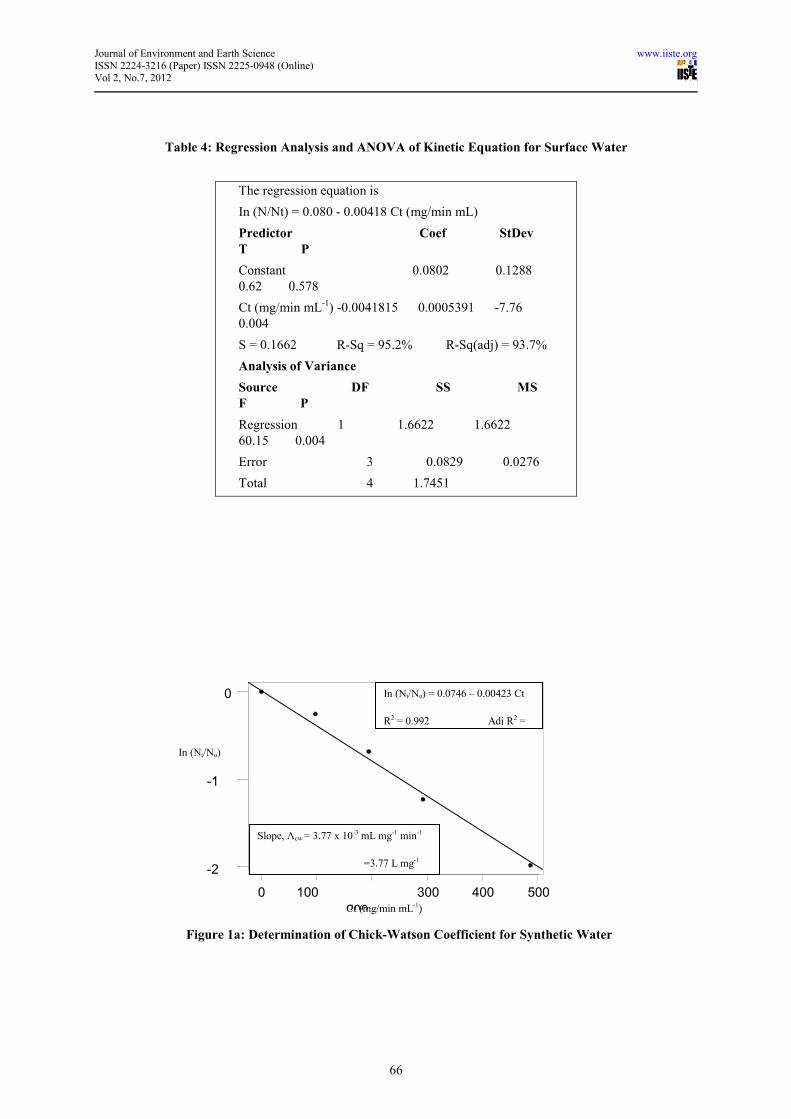
Table 3 gives the results of the disinfection studies using surface water obtained from Rimin Gado Dam
reservoir. Figure 3 shows the log-inactivation plot of the E.Coli and the microbial consortium exposed to
Moringa seeds extract for the surface water. Figure 2a shows log-inactivation plot of the E.Coli and the
microbial consortium exposed to Moringa seeds extract for surface water. The regression equation (8) is given
below:
In (Nt/No) = 0.080 - 0.00418 Ct (8)
The correlation coefficient is 95.2% and the Adj R2 was 93.7%, indicating goodness of fit. The normal plot of
residuals (Figure 2b) also indicated good fit. The analysis of variance (Table 4) indicated no significant lack of
fit with P=0.57 (P> 0.1). The slope of line gives the Chicks-Watson coefficient of specific lethality. The
coefficient of specific lethality (Ʌcw) was 3.75 L mg-1
min-1
for an E.Coli in surface water. Thus the average
Coefficient of lethality for Moringa disinfection (Ʌcw) was 3.76 L mg-1
min-1
.
The disinfection kinetic on E.Coli was found to be consistent with those determined by earlier researchers using
chlorine. Butterfield et al. (1943) reported a coefficient of specific lethality (Ʌcw) of 3.75 L mg-1
min-1
for an
E.Coli exposed to chlorine while Cunningham et al. (2008) reported a Ʌcw value of 4.71 L mg-1
min-1
for an
E.Coli exposed to chlorine.
3.2 Mode of Attack of Moringa Disinfection Extract on E.Coli
The antimicrobial activity of Moringa extracts was previously attributed to plant-produced benzyl isothiocynate
derivatives (Eilert et al. 1981). Suarez et al. (2003) later showed that at least part of the antimicrobial activity of
Moringa seeds extract may stem from Flo-like polypeptides. Antimicrobial peptides act by forming essential
enzymes, leading to cell deaths.
The results obtained are shown in Plates 3a – 3d. The Plates shows the changes in the E.Coli cell
morphology from the active cell (Plate 3a) to the cell condition after the Moringa extract application for 1 hour
(Plate 3b), then cell rupture after Moringa application for 1
hrs (Plate 3c), and finally the destroyed cells after
Moringa extract was applied for 2 hours Plate (3d). The result of the study on the mode of attack showed that the
cytoplasmic membrane of the E.Coli bacterial cell was ruptured and the intercellular components were seriously
damaged after treatment with M. Oleifera seed crude extract. Plate 3d indicated that the intercellular components
leaked out after cell burst. Based on previous studies of cell lysis pathways of antimicrobial peptides on bacteria
(Cham et al. 1998; Chen et al. 2003), this indicated that extracted compounds interacted with the lipid bi-layers
in membranes leading to the separation of the two membranes (outer and inner). Subsequently, water dips in to
the cell, which causes cell to swell more and burst leading to death. Thus the results obtained agreed with the
explanations given by earlier researchers.
4. Conclusion
The coefficient of specific lethality (Ʌcw) was 3.75 – 3.77 L mg-1
min-1
, with an average value of 3.76 L mg-1
min-1
for E.Coli inactivation using Moringa Oleifera seeds extracts. The mode of attack of the Moringa seeds
extract on the E.Coli cell was explained as by rupturing the cell and damaging the intercellular components,
allowing water to dip in to cell which causes it to swell more and burst leading to death.
Refernces
APHA (1990); Standard Method for the Examination of Water and Waste Water, 18th ed., American Public
Health Association.
Barrette, W., Hannum, D., Wheeler, W. & Hurst, J.(1988); General mechanism for the bacterial toxicity of
hypochlorous acid: abolition of ATP production. Biochemistry 28, 9172–9178.
Batarseh, K.(2004); Anomaly and correlation of killing in the therapeutic properties of silver (i) chelation with
glutamic and tartaric acids. J. Antimicrob. Chemother. 54, 546–548.
Berg, G.(ed) (1967); Transmission of Viruses by the water route; Wiley, New York.
Bichi, M. H. (2011); Application of Moringa Oleifera Seeds Extract in Water Treatment; Ph.D. Seminar
Paper; Department of Civil Engineering, Bayero University Kano.
Bichi, M. H. (2012); Optimization of Antimicrobial Ingredient Extraction and the Application of Moringa
Oleifera Seeds Extract in Water Disinfection; Ph.D. Seminar Paper; Department of Civil Engineering, Bayero
University Kano.
Bina, B. (1991); Investigations into the use of natural plant coagulants in the removal of bacteria and
bacteriophage from turbid waters; Ph.D Thesis, University of Newcastle Upon Tyne.

Journal of Environment and Earth Science www.iiste.org
ISSN 2224-3216 (Paper) ISSN 2225-0948 (Online) Vol 2, No.7, 2012
63
Bukar, A., Uba, A. and Oyeyi, T.I.(2010); Antimicrobial Profile of Moringa Oleifera Lam. Extracts against some
Food – borne Microorganisms; Bayero Journal of Pure and Applied Sciences, 3(1): 43 – 48
Buthelezi, S.P., Olaniran, A.O., and Pillay, B. (2009); Turbidity and antimicrobial Load removal from river
water using bioflocculants from indigenous bacteria isolated from wastewater in South Africa; African Journal
of Biotechnology, Vol. 8(14), pp. 3261-3266, 20 July 2009.
Butterfield, C. T., Wattie, E., Megregian, S., Chambers, C. W.(1943); Influence of pH and temperature on the
survival of coliforms and enteric pathogens when exposed to free chlorine. U.S. Public Health Service Report
58.
Chan, S.C., Yau, W.L., Wang, W., Smith, D., Sheu, F.S., Chen, H.M.,(1998); Microscopic observations of the
diVerent morphological changes by the anti-bacterial peptides on Klebsiella pneumoniae and HL-60 leukemia
cells. Journal of Peptide Science 4, 413–425.
Chen, H.M., Chan, S.C., Lee, J.C., Chang, C.C., Murugan, M., Jack, R.J.,(2003); Transmission electron
microscopic observations of membrane effects of antibiotic cecropin B on Escherichia coli. Microscopy
Research and Technique 62, 423–430.
Chick, H. (1908); Investigation of the laws of disinfection. J. Hygiene (British),Vol 8, 92–157.
Chuang Ping-Hsien, Chi-Wei Lee, Jia-Ying Chou, Murugan M., Bor-Jinn Shieh and Hueih-Min Chen (2007);
Anti-fungal activity of crude extracts and essential oil of Moringa oleifera Lam; Journal of Bioresource
Technology, 98 (1), 232-236.
Cunningham, J. H., Cunningham, C., Van Aken, B. and Lin, L.-S. (2008); Feasibility of disinfection kinetics and
minimum inhibitory concentration determination on bacterial cultures using flow cytometry; Water Science and
Technology WST 58 (4) Q IWA Publishing 2008 Water Science & Technology—WST | 58.4 | 2008
Davey, H. M., Kell, D. B., Weichart, D. H. & Kaprelyants, S. (2004); Current Protocols in Cytometry,
11.3.1-11.3.21. New York, John Wiley & Sons, Inc.
Eilert U., Wolters B., and Nahrstedt (1981); The antibiotic Principle of Moringa Oleifera and Moringa
Stenopeta/a; Plant Medica, N°'i2, pp 55-61
Fair, G. M., Gayer, J. C., Okun, D. A. (1958); Water and Waste Water Engineering: Vol.2- Water Purification
and Waste Water Treatment and Disposal; John Wiley & Sons, N.Y.
Fisch, F., Suarez, M., and Mermoud, N.,(2004) . Flo antibacterial peptide from the tropical tree Moringa
oleifera: A template for novel antibacterial agents; Travail de diploma, Universite De Lausanne, Lausanne,
février 2004
Folkard G.K., Sutherland J.P. and Grant W.P. (1989); Optimization on the use of natural coagulants for water
purification; Technical report No.R4254; Department of Engineering, University of Leicester.
Gottardi, W. (1999); Iodine and disinfection: theoretical study on mode of action, efficiency, stability, and
analytical aspects in the aqueous system. Arch. Pharm. 332(5), 151–157.
Goveas, J.L., Sinha, R., Krishnan, E.R., Patterson, C.L., and Namboodiri, V. (2010); Bench-Scale Evaluation of
Peracetic Acid and Twin Oxide as Disinfectants in Drinking Water; Proc. The World Environmental and Water
Resources Congress 2010, ASCE
Haas, C. (1980); A mechanistic model for chlorine disinfection. Environ. Sci. Technol. 14, 339–340.
Haas, C. & Engelbrecht, R.(1980); Physiological alterations of vegetative microorganisms resulting from
chlorination. J. Water Pollut. Control Fed. 52, 1976–1989.
Hoyanna, Y., Bacon, V., Summons, R., Pereira, W., Helpern, B. & Duffield, A. (1973); Chlorination studies: IV.
Reactions of aqueous hypochlorous acid with pyrimidine and purine bases. Biochem. Biophys. Res. Commun. 53,
1195–2001.
Humeirah, A. G., Nor Azah, A. G. S., Mastura, M. A., Mailina, M., Saiful, J., Muhajir, J. A., and Puad, A. M.
(2010); Chemical constituents and antimicrobial activity of Goniothalamus macrophyllus (Annonaceae) from
Pasoh Forest Reserve, Malaysia; African Journal of Biotechnology Vol. 9(34), pp. 5511-5515, 23 August, 2010.
Hwang, B-F., Jakkola, J. J. K., and Guo, H-R (2008); Water Disinfection by-products and the risk of specific
birth defects: a population-based cross-sectional study in Taiwan; Environmental Health 7(23).
Jabeen, R., Shahid, M., Jamil, A., and Ashraf, M. (2008); Microscopic Evaluation of the Antimicrobial Activity
of Seeds Extracts of Moringa Oleifera; Pak. J. Bot., 40(4): 1349-1358, 2008.
Jahn, S.A.A. (1988); Using Moringa Oleifera seeds as coagulant in developing countries; J.A.W.W.A., 43-50.
(June, 1988).
Kitajima, Y. (1998). Introduction: electron microscopy for fungal cell ultrastructure. Nippon Ishinkin Gakkai
Zasshi, 39(3) 121 – 122.
Lambert, R. & Johnson, M. (2000); Disinfection kinetics: a new hypothesis and model for the tailing of
log-survivor/time curves. J. Appl. Microbiol. 88, 907–913.
Leyer, G. & Johnson, E. (1997); Acid adaptation sensitizes Salmonella typhimurium to hypochlorous acid. Appl.
Environ. Microbiol. 63, 461–467.
Journal of Environment and Earth Science www.iiste.org
ISSN 2224-3216 (Paper) ISSN 2225-0948 (Online) Vol 2, No.7, 2012
64
Liau, S., Read, D., Pugh, W., Furr, J. & Russell, A. (1997); Interaction of silver nitrate with readily identifiable
groups: relationship to the antibacterial action of silver ions. Lett. Appl. Microbiol. 25, 279–283.
Madsen, M., Schlundt. J., and EI Fadil E. Omer (1987); Effect of Water coagulated by seeds of Moringa
Oleifera on bacterial concentrations; J. Tropical Hygiene and Medicine; 90,101-109.
MetCalf, L. and Eddy, H.P. (2003); Waste Water Engineering: Treatment, and Re-use; 4th
ed., Tata
McGraw-Hill, New Delhi.
Myriam, B.S., Otaki, M., Shinobu, K., and Abdennaceur, H (2010); Detection of active Escherichia coli after
irradiation by pulsed UV light using a Q_ phage; African Journal of Microbiology Research; Vol. 4 (11) pp.
1128-1134, 4 June, 2010
Obire, O., Tamuno, D. C., and Wemedo, S. A. (2005); Bacteriological Water Quality of Elechi Creek in
PortHarcourt, Nigeria; J. Applied Sci and Environmental Management; Vol. 9(1), pp79-84.
Okuda, T., Baes, A.U., Nishijima, W., and Okada, M. (1999). Improvement of extraction method of coagulation
active components from Moringa oleifera seed. Water Res., 33, 3373-3378.
Okuda, T., Baes, A.U., Nishijima, W., and Okada, M. (2001a). Isolation and Characterization of Coagulant
Extracted from Moringa oleifera seed by Salt Solution. Wat. Res., 35 (2), 405-410.
Okuda, T., Baes, A.U., Nishijima, W., and Okada, M. (2001b). Coagulation mechanism of salt solution extracted
active component in Moringa oleifera seeds. Wat. Res., 35 (3), 830-834.
Oluseyi, E.O and Francisca, O.N. (2009); Preliminary Studies on antimicrobial properties of Buchholzia
Coriacea (Wonderful Kola); African Journal of Biotechnology Vol. 8 (3), pp. 472-474, 4 February, 2009
Robeck, G.G., Clarke, N.A., and Dustal, J.A. (1962); Effectiveness of water treatment processes in Virus
removal; J.A.W.W.A., P. 1272.
Suareze, M., Entenza, J.M., Doerries, C., Meyer, E., Bourquin, L., Sutherland, J., Marison, I., Moreillon, P.,
Mermod, N.(2003); Expression of a Plant-Derived Peptide Harbouring Water-Cleaning and Antimicrobial
Activities; Biotechnol Bioeng. 81(1): Jan 2003, 13-20.
Suarez, M., Haenni, M., Canarelli, S., Fisch, F., Chodanowski, P., Servis, C., Michielin, O., Freitag, R.,
Moreillon, P., and Mermod, N. (2005). Structure-Function Characterization and Optimization of a Plant-Derived
Antibacterial Peptide. Antimicrobial Agents and Chemotherapy., 49 (9), 3847-3857.
Venkobachar, C., Lyengar, L. & Rao, A. (1977); Mechanism of disinfection: effect of chlorine on cell membrane
functions. Water Res. 11, 727–729.
Walter, A., Samuel, W., Peter, A., and Joseph, O. (2011); Antibacterial activity of Moringa Oleifera and
Moringa stenopetala methanol and n-hexane seed extracts on bacteria implicated in water borne diseases;
African Journal of Microbiology Research; Vol. 5(2) pp. 153-157, 18 January, 2011
Watson, H. E. (1908); A note on the variation of the rate of disinfection with change in the concentration of the
disinfectant. J. Hygiene (British), 8, 536–542.
Zasloff, M. (2002); Antimicrobial peptides of multicellular organisms. Nature. 415(6870): 389-395.
Zhao, G. & Stevens, S.(1998); Multiple parameters for the comprehensive evaluation of the susceptibility of
Escherichia coli to the silver ion. Biometals 11(1), 27–32.
Ziglio, G., Andreottola, G., Barbesti, S., Boschetti, G., Bruni, L., Foladori, P. & Villa, R.(2002); Assessment of
activated sludge viability with flow cytometry. Water Res. 36, 460–468.
Zimmer, J. L., Slawson, R. M. (2002); Potential Repair of Escherichia coli DNA following exposure to UV
radiation from both medium- and low-pressure UV sources used in drinking water treatment; Appl. Environ.
Microbiol. (68), pp3293-3299.

Journal of Environment and Earth Science www.iiste.org
ISSN 2224-3216 (Paper) ISSN 2225-0948 (Online) Vol 2, No.7, 2012
65
Table 1: Determination of Chiks-Watson Coefficient (Ʌcw) Using Synthetic Water
Time (mins) 0 30 60 90 150
E.Coli Count (Nt)
(X 105 cfu/mL)
Plate 1 1048 840 450 336 134
Plate 2 1008 728 624 300 150
Plate 3 1064 832 495 270 133
Average 1039 800 523 302 141.66
Ct (mg/min L-1
) 0 97.5 195.0 292.5 487.5
Nt/N0 1 0.9699 0.5033 0.2907 0.1363
In (Nt/N0) 0 -0.260 -0.686 -1.230 -1.990
Table 2: Regression Analysis and ANOVA for Kinetic Equation for Synthetic Water
Predictor Coef Std Dev
T P
Constant 0.07457 0.05970
1.25 0.300
Ct (mg/min mL-1
) -0.0042320 0.0002192 -19.30
0.000
S = 0.08223 R-Sq = 99.2% R-Sq(adj) = 98.9%
Analysis of Variance
Source DF SS MS
F P
Regression 1 2.5198 2.5198
372.62 0.000
Error 3 0.0203 0.0068
Total 4 2.5401
No evidence of lack of fit (P > 0.1)
Table 3: Determination of Chiks-Watson Coefficient (Ʌcw) Using Surface Water
Time (mins) 0 30 60 90 120
E.Coli Count (Nt)
(X 105 cfu/mL)
Plate 1 1048 840 450 336 134
Plate 2 1008 728 624 300 150
Plate 3 1064 832 495 270 133
Average 1039 800 523 302 141.66
Ct (mg/min L-1
) 0 97.5 195.0 292.5 390
Nt/N0 1 0.9699 0.5033 0.2907 0.1363
In (Nt/N0) 0 -0.260 -0.686 -1.230 -1.990
Journal of Environment and Earth Science www.iiste.org
ISSN 2224-3216 (Paper) ISSN 2225-0948 (Online) Vol 2, No.7, 2012
66
Table 4: Regression Analysis and ANOVA of Kinetic Equation for Surface Water
The regression equation is
In (N/Nt) = 0.080 - 0.00418 Ct (mg/min mL)
Predictor Coef StDev
T P
Constant 0.0802 0.1288
0.62 0.578
Ct (mg/min mL-1
) -0.0041815 0.0005391 -7.76
0.004
S = 0.1662 R-Sq = 95.2% R-Sq(adj) = 93.7%
Analysis of Variance
Source DF SS MS
F P
Regression 1 1.6622 1.6622
60.15 0.004
Error 3 0.0829 0.0276
Total 4 1.7451
500 400 300
200
100 0
0
-1
-2
Ct (mg/min mL-1)
In (Nt/No)
Figure 1a: Determination of Chick-Watson Coefficient for Synthetic Water
Slope, Ʌcw = 3.77 x 10⁻³ mL mg-1 min-1
=3.77 L mg-1 -1
In (Nt/No) = 0.0746 – 0.00423 Ct
R2 = 0.992 Adj R2 =

Journal of Environment and Earth Science www.iiste.org
ISSN 2224-3216 (Paper) ISSN 2225-0948 (Online) Vol 2, No.7, 2012
67
500400300200 1000
0
-1
-2
Ct (mg/min mL-1
)
In (Nt/No)
Figure 2a: Determination of Chick-Watson Coefficient for Surface Water
Slope, Ʌcw = 3.75 x 10⁻³
mL mg-1
min-1
=3.75 L mg-1
min-1
In (Nt/No) = 0.0746 – 0.00423 Ct
R2 = 0.952 Adj R2 =
10-1
0.08
0.06
0.04
0.02
0.00
-0.02
-0.04
-0.06
-0.08
Normal Score
Resi
dual
(Response is In (Nt/No)
Figure 1b: Normal Probability Plot of the Residuals for

Journal of Environment and Earth Science www.iiste.org
ISSN 2224-3216 (Paper) ISSN 2225-0948 (Online) Vol 2, No.7, 2012
68
PLATE 3a: E. Coli Without Extract PLATE 3b: E. Coli With Extract after 1 hr
PLATE 3c: E. Coli With Extract after 1�
� hr PLATE 3d: E. Coli With Extract after 2 hrs
10-1
0.2
0.1
0.0
-0.1
Normal Score
Residual
Figure 2b: Normal Probability Plot of the Residuals for Surface Water
(Response is In (Nt/No)

This academic article was published by The International Institute for Science,
Technology and Education (IISTE). The IISTE is a pioneer in the Open Access
Publishing service based in the U.S. and Europe. The aim of the institute is
Accelerating Global Knowledge Sharing.
More information about the publisher can be found in the IISTE’s homepage:
http://www.iiste.org
The IISTE is currently hosting more than 30 peer-reviewed academic journals and
collaborating with academic institutions around the world. Prospective authors of
IISTE journals can find the submission instruction on the following page:
http://www.iiste.org/Journals/
The IISTE editorial team promises to the review and publish all the qualified
submissions in a fast manner. All the journals articles are available online to the
readers all over the world without financial, legal, or technical barriers other than
those inseparable from gaining access to the internet itself. Printed version of the
journals is also available upon request of readers and authors.
IISTE Knowledge Sharing Partners
EBSCO, Index Copernicus, Ulrich's Periodicals Directory, JournalTOCS, PKP Open
Archives Harvester, Bielefeld Academic Search Engine, Elektronische
Zeitschriftenbibliothek EZB, Open J-Gate, OCLC WorldCat, Universe Digtial
Library , NewJour, Google Scholar