l-arginine sensing regulates virulence gene expression and
TRANSCRIPT

L-Arginine sensing regulates virulence gene expressionand disease progression in enteric pathogensZelia Menezes-Garciaa,b, Aman Kumara,b, Wenhan Zhua, Sebastian E. Wintera, and Vanessa Sperandioa,b,1
aDepartment of Microbiology, University of Texas Southwestern Medical Center, Dallas, TX 75390; and bDepartment of Biochemistry, University of TexasSouthwestern Medical Center, Dallas, TX 75390
Edited by Roy Curtiss III, University of Florida, Gainesville, FL, and approved April 8, 2020 (received for review November 8, 2019)
Microbiota, host and dietary metabolites/signals compose the richgut chemical environment, which profoundly impacts virulence ofenteric pathogens. Enterohemorrhagic Escherichia coli (EHEC) en-gages a syringe-like machinery named type-III secretion system(T3SS) to inject effectors within host cells that lead to intestinalcolonization and disease. We previously conducted a high-throughputscreen to identify metabolic pathways that affect T3SS expression.Here we show that in the presence of arginine, the arginine sensorArgR, identified through this screen, directly activates expressionof the genes encoding the T3SS. Exogenously added arginine in-duces EHEC virulence gene expression in vitro. Congruently, a mutantdeficient in arginine transport (ΔartP) had decreased virulence geneexpression. ArgR also augments murine disease caused by Citrobacterrodentium, which is a murine pathogen extensively employed as asurrogate animal model for EHEC. The source of arginine sensed byC. rodentium is not dietary. At the peak of C. rodentium infection,increased arginine concentration in the colon correlated with down-regulation of the host SLC7A2 transporter. This increase in the con-centration of colonic arginine promotes virulence gene expression inC. rodentium. Arginine is an important modulator of the host im-mune response to pathogens. Here we add that arginine also directlyimpacts bacterial virulence. These findings suggest that a delicatebalance between host and pathogen responses to arginine occurduring disease progression.
EHEC | arginine | ArgR
Enteric pathogens integrate sensing and responding to multi-ple metabolites/signals within the gastrointestinal tract to
regulate expression of key virulence genes. This surveillance al-lows for the spatial–temporal control of virulence gene expression,preventing unnecessary energy expenditure. EnterohemorrhagicEscherichia coli (EHEC) colonizes the human colon causing bloodydiarrhea that may lead to hemolytic-uremic syndrome (HUS) (1, 2).EHEC virulence determinants include the locus of enterocyte ef-facement (LEE) pathogenicity island that encodes a type III se-cretion system (T3SS), and the potent Shiga toxin (Stx2a) that isresponsible for HUS. EHEC utilizes the T3SS to translocate mul-tiple effectors into colonocytes promoting cytoskeletal rearrange-ment and attaching and effacing (AE) lesion formation (1). TheLEE is organized in five major operons LEE1 to LEE5. Transcriptionof all LEE genes is under the control of a master regulator, Ler,encoded within the LEE1 operon (1) (Fig. 1A). Transcription ofler is controlled by several host and microbiota-derived signalssuch as epinephrine, norepinephrine, succinate, fucose, serine,cysteine, and ethanolamine among others (3).To dissect the metabolic pathways that influence virulence of
EHEC, we previously performed a high-throughput screen (HTS)that identified the arginine sensor ArgR, and the transmembranearginine importer ArtP as possible regulators of EHEC virulencegene expression (4). ArgR is a transcription factor encoded by theargR gene that inhibits the transcription of multiple genes includingthe arginine biosynthesis regulon, the arginine transport genes, aswell as its own transcription (5, 6). ArgR also functions as a coactivatorof the astCADBE operon encoding the arginine succinyltransferasepathway for arginine catabolism (7). Therefore, the biosynthesis,
catabolism, and uptake of arginine are tightly regulated by ArgR.Biochemical and structural analyses have shown that the ArgRmonomer consists of two domains separated by a protease-accessible linker: An N-terminal DNA-binding domain and aC-terminal arginine-binding domain (8). L-Arginine binding isrequired for ArgR oligomerization (9, 10). Mechanistically, inthe presence of arginine, two trimers of ArgR dimerize forming ahexamer, which can bind to whole ARG boxes present in pro-moter regions, while trimeric ArgR still binds to half of the ARGbox in the absence of L-arginine (11).
L-Arginine is an important amino acid essential for proteinsynthesis and plays an equally important role in stress conditions.L-Arginine acts as a precursor molecule for the biosynthesis ofothers amino acids, polyamines, and nitric oxide, and it alsoserves as a source of nitrogen upon its degradation via distinctcatabolic routes (12). Arginine uptake in E. coli is mediated bythree transport systems of the ATP-binding cassette transporterfamily (6, 13). These import systems are encoded by two geneclusters, artPIQM-artJ and argT-hisJQMP, which are repressed byArgR in E. coli. These import systems diverge in substratespecificity and affinity for arginine, and display differences in theregulation of their own synthesis and activity (5). The importerencoded by artPIQM-artJ has the highest affinity for arginine (Kd0.4 μM), where ArtP is an ATP-binding protein present in themembrane (5).Here, we show that exogenous L-arginine up-regulates the
expression of LEE-encoded genes and Shiga toxin (Stx) via ArgRin the enteric pathogens EHEC and Citrobacter rodentium (sur-rogate murine infection model). Disruption of L-arginine uptake
Significance
Enteric pathogens successfully colonize the host intestinal en-vironment by regulating the expression of their metabolic andvirulence genes. A high-throughput screen identified the argi-nine sensor ArgR as a regulator of virulence expression inenterohemorrhagic Escherichia coli. Here we show that ArgRsenses arginine fluctuations and regulates the virulence ofenterohemorrhagic E. coli and Citrobacter rodentium, bothin vitro and during murine infections. Our work highlights theimportance of gut metabolite sensing in affecting the diseaseoutcome and vulnerability to pathogens.
Author contributions: Z.M.-G. and V.S. designed research; Z.M.-G. and A.K. performedresearch; Z.M.-G., A.K., W.Z., S.E.W., and V.S. analyzed data; and Z.M.-G. and V.S. wrotethe paper.
The authors declare no competing interest.
This article is a PNAS Direct Submission.
Published under the PNAS license.
Data deposition: The data reported in this paper have been deposited in the EuropeanNucleotide Archive (ENA) database, https://www.ebi.ac.uk/ena (accession no.PRJEB35294).1To whom correspondence may be addressed. Email : [email protected].
This article contains supporting information online at https://www.pnas.org/lookup/suppl/doi:10.1073/pnas.1919683117/-/DCSupplemental.
First published May 14, 2020.
www.pnas.org/cgi/doi/10.1073/pnas.1919683117 PNAS | June 2, 2020 | vol. 117 | no. 22 | 12387–12393
MICRO
BIOLO
GY
Dow
nloa
ded
by g
uest
on
Nov
embe
r 30
, 202
1

systems in EHEC also decreases the expression of their virulencedeterminants. Moreover, we also show that C. rodentium in-fection increases the L-arginine levels in the colon that correlateswith increased pathogenesis. In summary, our findings add an-other node to the complex metabolic interactions that intersectwith virulence gene regulation to promote enteric disease.
ResultsEHEC Virulence Is Increased by Exogenous L-Arginine. Our previousHTS screen identified the ArgR transcription factor that is anarginine sensor, and ArtP that is a transmembrane arginine im-porter as involved in LEE gene regulation (4). To confirmwhether arginine affects the expression of virulence genes, weperformed RNA-sequencing (RNA-seq; data deposited at theEuropean Nucleotide Archive, accession no. PRJEB35294) inEHEC grown in low-glucose DMEM under microaerophilicconditions [the optimal in vitro condition for LEE gene ex-pression in EHEC (14)] with or without L-arginine. We observedthat the presence of L-arginine increased the expression of LEEvirulence genes in EHEC (Fig. 1 B–D), as well as non-LEE–encodedgenes (SI Appendix, Figs. S1A and S3B). L-Arginine augments theexpression of LEE-encoded (ler, escC, escV, tir, eae, espA, andespB) and non-LEE–encoded (stx2a, nleA, espN, espW, espX, espY,espR, espJ, espK, espM) genes (Fig. 1 B and C and SI Appendix,Figs. S1A and S3B). Congruent with the transcription effect ofL-arginine on LEE genes, L-arginine also increased the secretionof both EspA and EspB proteins (Fig. 1D). Importantly, EHECgrowth remains unaffected by L-arginine under these conditions(SI Appendix, Fig. S1B). This suggests that L-arginine–dependentLEE regulation is not due to a metabolic defect caused by theabsence of L-arginine. Because LEE gene expression is necessaryfor AE lesion formation, we next assessed the role of L-arginine on
AE lesion formation. AE lesions are characterized by remodelingof the epithelial cell cytoskeleton and formation of a pedestal-likestructure beneath the bacteria (15). In accordance with the LEE-expression phenotype, L-arginine enhanced AE lesion formationand increased attachment of bacteria to HeLa cells (Fig. 1 E andF). L-Arginine also increased LEE gene expression in less per-missive in vitro conditions (DMEM high glucose under micro-aerophilic conditions) (SI Appendix, Fig. S1C). Overall, these dataindicate that arginine increases the virulence gene expression andAE lesion formation in EHEC.In E. coli, L-arginine is imported by binding to three peri-
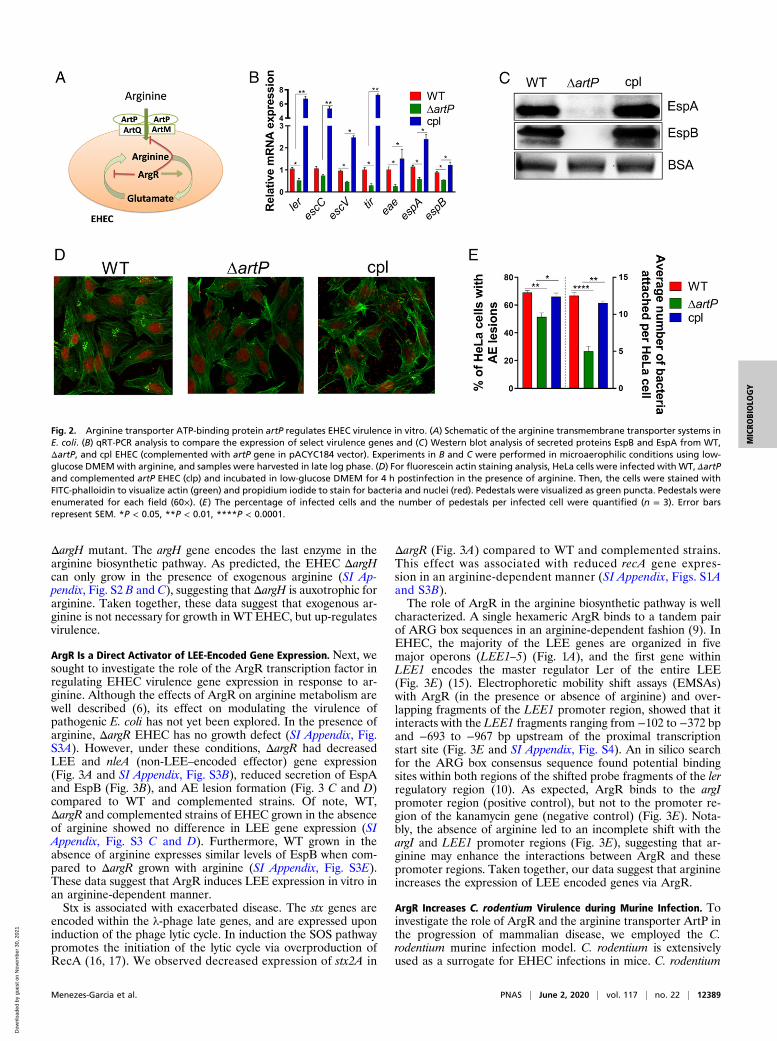
plasmic proteins, and is subsequently transported into the cell bytwo different transmembrane complexes (Fig. 2A). ArtP is atransmembrane protein necessary for the function of both arginineimport systems (5). To investigate whether exogenous arginineregulates virulence gene expression in EHEC, we generated anartP mutant. Deletion of artP decreases LEE gene expression (ler,escC, escV, tir, eae, espA, and espB), and these phenotypes arerescued upon complementation (Fig. 2B). The overall decrease intranscription of LEE-encoded T3SS genes is corroborated withthe decrease in the levels of the secreted proteins EspA and EspB(Fig. 2C), and decreased AE lesion formation in ΔartP EHEC(Fig. 2 D and E). Of note, EHEC growth was unaffected bydeletion of artP (SI Appendix, Fig. S2A). These data furtherindicate that ArtP-dependent LEE regulation is not due to ametabolic defect, but to the lack of import of exogenous arginine(SI Appendix, Fig. S2A).In the absence of exogenous L-arginine, the arginine in-
tracellular demand is likely met by the endogenous biosynthesisof arginine. The arginine biosynthetic pathway in E. coli requireseight enzymatic steps (12). To understand the relevance of en-dogenous arginine biosynthesis for EHEC, we generated a
Fig. 1. Arginine induces EHEC virulence in vitro. (A) Schematic of the LEE and the representative genes used in this work. The LEE1 operon encodes ler thatactivates expression of all five LEE operons. (B) Heat map showing the effect of arginine on LEE-encoded genes fromWT EHEC grown in the presence (+arg) orabsence (−arg) of 482 μM arginine. (C) qRT-PCR of LEE-encoded genes and Stx2a. (D) Western blot analysis of secreted proteins EspB and EspA from WT EHECgrown in the absence or presence of arginine. Experiments in B–D were performed in microaerophilic conditions using low-glucose DMEM, and samples wereharvested in late log phase. (E) Fluorescein actin staining analysis. HeLa cells were infected with WT EHEC and incubated in the presence or absence ofarginine for 4 h postinfection. HeLa cells were stained with FITC-phalloidin to visualize actin (green) and propidium iodide to stain for bacteria and nuclei(red). Pedestals were visualized as green dots. Pedestals were enumerated per cell (60×). (F) The percentage of infected cells and the number of pedestals perinfected cell were quantified (n = 3). Error bars represent SEM. *P < 0.05, **P < 0.01, ***P < 0.001, ****P < 0.0001.
12388 | www.pnas.org/cgi/doi/10.1073/pnas.1919683117 Menezes-Garcia et al.
Dow
nloa
ded
by g
uest
on
Nov
embe
r 30
, 202
1
ΔargH mutant. The argH gene encodes the last enzyme in thearginine biosynthetic pathway. As predicted, the EHEC ΔargHcan only grow in the presence of exogenous arginine (SI Ap-pendix, Fig. S2 B and C), suggesting that ΔargH is auxotrophic forarginine. Taken together, these data suggest that exogenous ar-ginine is not necessary for growth in WT EHEC, but up-regulatesvirulence.
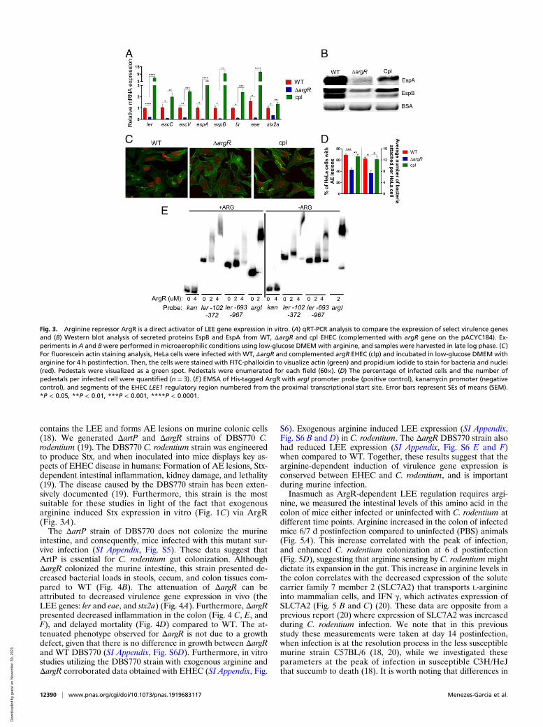
ArgR Is a Direct Activator of LEE-Encoded Gene Expression. Next, wesought to investigate the role of the ArgR transcription factor inregulating EHEC virulence gene expression in response to ar-ginine. Although the effects of ArgR on arginine metabolism arewell described (6), its effect on modulating the virulence ofpathogenic E. coli has not yet been explored. In the presence ofarginine, ΔargR EHEC has no growth defect (SI Appendix, Fig.S3A). However, under these conditions, ΔargR had decreasedLEE and nleA (non-LEE–encoded effector) gene expression(Fig. 3A and SI Appendix, Fig. S3B), reduced secretion of EspAand EspB (Fig. 3B), and AE lesion formation (Fig. 3 C and D)compared to WT and complemented strains. Of note, WT,ΔargR and complemented strains of EHEC grown in the absenceof arginine showed no difference in LEE gene expression (SIAppendix, Fig. S3 C and D). Furthermore, WT grown in theabsence of arginine expresses similar levels of EspB when com-pared to ΔargR grown with arginine (SI Appendix, Fig. S3E).These data suggest that ArgR induces LEE expression in vitro inan arginine-dependent manner.Stx is associated with exacerbated disease. The stx genes are
encoded within the λ-phage late genes, and are expressed uponinduction of the phage lytic cycle. In induction the SOS pathwaypromotes the initiation of the lytic cycle via overproduction ofRecA (16, 17). We observed decreased expression of stx2A in
ΔargR (Fig. 3A) compared to WT and complemented strains.This effect was associated with reduced recA gene expres-sion in an arginine-dependent manner (SI Appendix, Figs. S1Aand S3B).The role of ArgR in the arginine biosynthetic pathway is well
characterized. A single hexameric ArgR binds to a tandem pairof ARG box sequences in an arginine-dependent fashion (9). InEHEC, the majority of the LEE genes are organized in fivemajor operons (LEE1–5) (Fig. 1A), and the first gene withinLEE1 encodes the master regulator Ler of the entire LEE(Fig. 3E) (15). Electrophoretic mobility shift assays (EMSAs)with ArgR (in the presence or absence of arginine) and over-lapping fragments of the LEE1 promoter region, showed that itinteracts with the LEE1 fragments ranging from −102 to −372 bpand −693 to −967 bp upstream of the proximal transcriptionstart site (Fig. 3E and SI Appendix, Fig. S4). An in silico searchfor the ARG box consensus sequence found potential bindingsites within both regions of the shifted probe fragments of the lerregulatory region (10). As expected, ArgR binds to the argIpromoter region (positive control), but not to the promoter re-gion of the kanamycin gene (negative control) (Fig. 3E). Nota-bly, the absence of arginine led to an incomplete shift with theargI and LEE1 promoter regions (Fig. 3E), suggesting that ar-ginine may enhance the interactions between ArgR and thesepromoter regions. Taken together, our data suggest that arginineincreases the expression of LEE encoded genes via ArgR.
ArgR Increases C. rodentium Virulence during Murine Infection. Toinvestigate the role of ArgR and the arginine transporter ArtP inthe progression of mammalian disease, we employed the C.rodentium murine infection model. C. rodentium is extensivelyused as a surrogate for EHEC infections in mice. C. rodentium
Fig. 2. Arginine transporter ATP-binding protein artP regulates EHEC virulence in vitro. (A) Schematic of the arginine transmembrane transporter systems inE. coli. (B) qRT-PCR analysis to compare the expression of select virulence genes and (C) Western blot analysis of secreted proteins EspB and EspA from WT,ΔartP, and cpl EHEC (complemented with artP gene in pACYC184 vector). Experiments in B and C were performed in microaerophilic conditions using low-glucose DMEMwith arginine, and samples were harvested in late log phase. (D) For fluorescein actin staining analysis, HeLa cells were infected with WT, ΔartPand complemented artP EHEC (clp) and incubated in low-glucose DMEM for 4 h postinfection in the presence of arginine. Then, the cells were stained withFITC-phalloidin to visualize actin (green) and propidium iodide to stain for bacteria and nuclei (red). Pedestals were visualized as green puncta. Pedestals wereenumerated for each field (60×). (E) The percentage of infected cells and the number of pedestals per infected cell were quantified (n = 3). Error barsrepresent SEM. *P < 0.05, **P < 0.01, ****P < 0.0001.
Menezes-Garcia et al. PNAS | June 2, 2020 | vol. 117 | no. 22 | 12389
MICRO
BIOLO
GY
Dow
nloa
ded
by g
uest
on
Nov
embe
r 30
, 202
1
contains the LEE and forms AE lesions on murine colonic cells(18). We generated ΔartP and ΔargR strains of DBS770 C.rodentium (19). The DBS770 C. rodentium strain was engineeredto produce Stx, and when inoculated into mice displays key as-pects of EHEC disease in humans: Formation of AE lesions, Stx-dependent intestinal inflammation, kidney damage, and lethality(19). The disease caused by the DBS770 strain has been exten-sively documented (19). Furthermore, this strain is the mostsuitable for these studies in light of the fact that exogenousarginine induced Stx expression in vitro (Fig. 1C) via ArgR(Fig. 3A).The ΔartP strain of DBS770 does not colonize the murine
intestine, and consequently, mice infected with this mutant sur-vive infection (SI Appendix, Fig. S5). These data suggest thatArtP is essential for C. rodentium gut colonization. AlthoughΔargR colonized the murine intestine, this strain presented de-creased bacterial loads in stools, cecum, and colon tissues com-pared to WT (Fig. 4B). The attenuation of ΔargR can beattributed to decreased virulence gene expression in vivo (theLEE genes: ler and eae, and stx2a) (Fig. 4A). Furthermore, ΔargRpresented decreased inflammation in the colon (Fig. 4 C, E, andF), and delayed mortality (Fig. 4D) compared to WT. The at-tenuated phenotype observed for ΔargR is not due to a growthdefect, given that there is no difference in growth between ΔargRand WT DBS770 (SI Appendix, Fig. S6D). Furthermore, in vitrostudies utilizing the DBS770 strain with exogenous arginine andΔargR corroborated data obtained with EHEC (SI Appendix, Fig.
S6). Exogenous arginine induced LEE expression (SI Appendix,Fig. S6 B and D) in C. rodentium. The ΔargR DBS770 strain alsohad reduced LEE expression (SI Appendix, Fig. S6 E and F)when compared to WT. Together, these results suggest that thearginine-dependent induction of virulence gene expression isconserved between EHEC and C. rodentium, and is importantduring murine infection.Inasmuch as ArgR-dependent LEE regulation requires argi-
nine, we measured the intestinal levels of this amino acid in thecolon of mice either infected or uninfected with C. rodentium atdifferent time points. Arginine increased in the colon of infectedmice 6/7 d postinfection compared to uninfected (PBS) animals(Fig. 5A). This increase correlated with the peak of infection,and enhanced C. rodentium colonization at 6 d postinfection(Fig. 5D), suggesting that arginine sensing by C. rodentium mightdictate its expansion in the gut. This increase in arginine levels inthe colon correlates with the decreased expression of the solutecarrier family 7 member 2 (SLC7A2) that transports L-arginineinto mammalian cells, and IFN γ, which activates expression ofSLC7A2 (Fig. 5 B and C) (20). These data are opposite from aprevious report (20) where expression of SLC7A2 was increasedduring C. rodentium infection. We note that in this previousstudy these measurements were taken at day 14 postinfection,when infection is at the resolution process in the less susceptiblemurine strain C57BL/6 (18, 20), while we investigated theseparameters at the peak of infection in susceptible C3H/HeJthat succumb to death (18). It is worth noting that differences in
Fig. 3. Arginine repressor ArgR is a direct activator of LEE gene expression in vitro. (A) qRT-PCR analysis to compare the expression of select virulence genesand (B) Western blot analysis of secreted proteins EspB and EspA from WT, ΔargR and cpl EHEC (complemented with argR gene on the pACYC184). Ex-periments in A and Bwere performed in microaerophilic conditions using low-glucose DMEM with arginine, and samples were harvested in late log phase. (C)For fluorescein actin staining analysis, HeLa cells were infected with WT, ΔargR and complemented argR EHEC (clp) and incubated in low-glucose DMEM witharginine for 4 h postinfection. Then, the cells were stained with FITC-phalloidin to visualize actin (green) and propidium iodide to stain for bacteria and nuclei(red). Pedestals were visualized as a green spot. Pedestals were enumerated for each field (60×). (D) The percentage of infected cells and the number ofpedestals per infected cell were quantified (n = 3). (E) EMSA of His-tagged ArgR with argI promoter probe (positive control), kanamycin promoter (negativecontrol), and segments of the EHEC LEE1 regulatory region numbered from the proximal transcriptional start site. Error bars represent SEs of means (SEM).*P < 0.05, **P < 0.01, ***P < 0.001, ****P < 0.0001.
12390 | www.pnas.org/cgi/doi/10.1073/pnas.1919683117 Menezes-Garcia et al.
Dow
nloa
ded
by g
uest
on
Nov
embe
r 30
, 202
1

susceptibility to C. rodentium infection between these murinestrains are not associated with differences in Toll-like receptor 4signaling, and are due to the more severe diarrhea caused by C.rodentium in C3H/HeJ mice (21, 22). Additionally, we also useddifferent C. rodentium strains. Singh et al. (20) used C. rodentiumstrain DBS100, while we used DBS770. Both strains harbor theLEE, as EHEC, which is necessary to form AE lesions onenterocytes of mice leading to colonic hyperplasia and colitis (23,24). We used the C. rodentium DBS770 strain engineered toproduce Stx, which when inoculated into mice display key aspectsof EHEC disease in humans: Formation of AE lesions, Stx-dependent intestinal inflammation, kidney damage, and lethal-ity. The disease caused by the DBS770 strain has been exten-sively documented, and this model is a strong in vivo model forstudying EHEC pathogenesis (4, 19, 25, 26). The source of ar-ginine modulating C. rodentium virulence in the colon is notdictated by diet, given that mice fed chow with or without argi-nine had similar pathogen burdens (SI Appendix, Fig. S7A).Importantly, there was no difference in arginine levels obtainedfrom the colons of mice fed chow with or without arginine (SIAppendix, Fig. S7B).
DiscussionThe gastrointestinal environment is complex and profoundly im-pacted by the membership of the resident microbiota, host genet-ics, diet, and enteric diseases. Bacterial enteric pathogens evolvedintricate regulatory systems to integrate multiple cues toward ex-quisite control of the expression of their virulence repertoire (3,27). The significance of this molecular circuitry is conspicuous inEHEC, which colonizes the densely populated colon with a re-markable low infectious dose (50 CFUs) (1, 3).EHEC integrates metabolism and virulence at multiple levels,
with nutrients also serving as signals to modulate virulence geneexpression (3, 4). We previously performed a comprehensiveHTS screen to map metabolic pathways that impacted virulenceexpression (with emphasis on LEE gene expression) in EHEC(4). This screen pulled ArgR, the arginine sensor that controlsarginine utilization and biosynthesis, and the ArtP arginine im-porter (5, 6), as potential regulators of LEE gene expression.Here we show that exogenous arginine enhances LEE and Stxexpression in both EHEC and C. rodentium through the ArgRtranscription factor (Figs. 1–3 and SI Appendix, Figs. S3–S6). Theimpact of arginine in EHEC/C. rodentium virulence is not due to
Fig. 4. ArgR increases C. rodentium virulence in vivo. (A) Expression of LEE gene and Stx2 from bacteria recovered from stools of C3H/HeJ mice infected witheither WT or ΔargR C. rodentium 8 d postinfection. (B) CFUs recovered from cecum, colon, and stool samples of mice with infected either WT or ΔargR C.rodentium. (C) Expression of nos2 in the colon tissue obtained from C. rodentium or PBS-treated mice, 8 d postinfection. (D) Survival curve of mice infectedwith either WT or ΔargR C. rodentium. A total of n = 10 mice per group were used in this study. (E) Histological score and (F) representation (10×) of cecum8 d after C. rodentium infection (n = 3). Each symbol represents an individual mouse. Error bars represent SEM. *P < 0.05, ***P < 0.001, ****P < 0.0001.
Fig. 5. Arginine levels is incresead in colon content after C. rodentium infection. (A) Arginine measurements in colon contents, (B) slc7a2, and (C) infγ mRNAexpression in colon tissue of mice in the third and sixth days after WT C. rodentium infection or not (PBS). (D) CFU in stools of mice in the first, third, andsixth days after WT C. rodentium infection. Each symbol represents an individual mouse. Error bars represent SEM. *P < 0.05, **P < 0.01.
Menezes-Garcia et al. PNAS | June 2, 2020 | vol. 117 | no. 22 | 12391
MICRO
BIOLO
GY
Dow
nloa
ded
by g
uest
on
Nov
embe
r 30
, 202
1

its role in metabolism, given that no growth defects exist betweenΔargR, ΔartP, and WT strains. C. rodentium infection increasesthe concentration of arginine in the colon, correlated with thedown-regulation of the mammalian SLC7A2 importer, leading toincreased pathogen loads and disease (Figs. 4 and 5). This is incontrast to a report that during infection with C. rodentium ex-pression of SLC7A2 is up-regulated (20). A notable differencebetween these two studies is that ours investigates the role ofarginine in C. rodentium pathogenesis at the peak of infection, ina very susceptible host, while Singh et al. (20) assess these pa-rameters at the onset of disease resolution, in a less susceptiblehost. Arginine is known to be present in the colon content, and isabsorbed by the intestinal epithelial cells where it can lead to theproduction of nitrogen oxide (NO) (25, 28). In fact, argininemodulates the host immune responses changing host suscepti-bility to infections (29). Taking these data together, we find thatfluctuations of arginine concentrations in the intestine duringinfection impact the onset and resolution of disease.The presence or absence of dietary arginine did not impact the
colonic levels of this amino acid, nor pathogen burden (SI Ap-pendix, Fig. S7). Our studies investigated the role of dietary ar-ginine in the peak of C. rodentium infection in a susceptiblemurine strain. However, a previous report suggested that sup-plementation of arginine in the diet decreased pathogen burden,in a less susceptible murine strain at a later time point, closeto the resolution of the disease (30). Interestingly, in both our andthe previous studies the levels of arginine in the colon remainedthe same regardless of its presence or concentration in the diet (SIAppendix, Fig. S7) (30). However, arginine dietary differencesimpacted the levels of this amino acid in the serum under ho-meostasis, and its levels were enhanced in both the colon andserum upon the onset of inflammation due to dextran sulfate so-dium treatment (30). The combination of these studies suggeststhat arginine orchestrate a complex interaction between pathogenand host responses, and arginine dietary supplementation to en-hance gut health grants further research.
Materials and MethodsBacterial Strains, Plasmids, and Growth and Culture Condition. All strains andplasmids used in this study are listed in SI Appendix, Table S1. Bacterialcultures were grown in low glucose DMEM (Gibco) with or without 482 μMarginine in microaerophilic conditions. For microaerophilic conditions, bac-terial cultures were grown standing in the 37 °C incubator. The cultures wereharvested in late logarithmic growth phase unless stated otherwise. HeLacells were routinely cultured in high-glucose DMEM (4.5 g/L glucose), 10%FBS, and penicillin plus streptomycin plus glutamine (PSG) mixture.
Recombinant DNA Techniques. All primers used for qRT-PCR, mutant gener-ation, and plasmid construction can be found in SI Appendix, Table S2.Knockout strains were constructed using the λ-red method (31). PlasmidpACYC184 (New England Biolabs) was used as complementation vector.
RNA-Seq Library Preparation and Analysis. Briefly, RNAs extracted from threebiological replicates were used to perform RNA-seq experiments. Sequencingwas run at the University of Texas Medical Center Next Generation Se-quencing core. RNA libraries were prepared using Epicentre Bacteria Kit.RNA libraries were run on an Illumina HiSEq. 2500 sequencer with SE-50. Toanalyze the data, Microbial Ecology software package (v1.91) (32, 33) wasused. Reads were mapped to the E. coli O157:H7 strain EDL 933 genome(National Center for Biotechnology Information NZ_CP008957.1). The dataanalysis was conducted using the software DESeq2. Statistical significancewas calculated using Student’s t test followed by false-discovery rate(Benjamini–Hochberg) correction. A P value of less than 0.05 was consideredsignificant. (RNA-seq data have been deposited at the European NucleotideArchive, accession no. PRJEB35294)
qRT-PCR. RNA from three biological replicates was extracted using theRiboPure bacterial isolation kit according to the manufacturer’s protocols(Ambion). qRT-PCR was performed as follows. Briefly, 2 μg of diluted extractedRNAwas converted to cDNAwith addition of superscript, random primers, DTT,and dNTPs. Validated Primers (SI Appendix, Table S2) and SYBR Green were
added to the cDNA and the mix run in Quantstudio 6 flex (Applied Biosystems).Data were collected using QuantStudio Real-Time PCR Software v1.3, normal-ized to endogenous rpoA levels, and analyzed using the comparative criticalthreshold (CT) method. For all of the in vitro experiments, error bars indicateSD. A P value of less than 0.05 was considered significant.
Western Blotting. Secreted proteins were isolated as previously described (34)from cultures grown to the same OD600 in DMEM at 37 °C. A total of 10 μgBSA was added to secreted proteins as a loading control. Proteins wereseparated on a 5 to 20% SDS polyacrylamide gel, transferred to a poly-vinylidene fluoride (PVDF) membrane, and blocked with 5% milk in PBScontaining 0.05% Tween (PBST). Membranes were probed with either anti-EspB or anti-EspA primary antibody and then incubated with a secondaryrabbit antibody conjugated to streptavidin-horseradish peroxidase. En-hanced chemiluminescence reagent was added, and the membranes weredeveloped using the ChemiDoc touch imaging system (software 1.0.0.15)with Image Lab 5.2.1 software for image display.
Fluorescein Actin Staining Assays. For the fluorescein actin staining assays,HeLa cells were grown overnight to about 70 to 80% confluence at 37 °C and5% CO2 on coverslips in wells containing high-glucose (4.5 g/L) DMEM. Priorto infection, fresh low-glucose DMEM lacking antibiotics replaced overnightcell medium. Then, HeLa cells were infected with late log-phase bacterialcultures with equal CFU for 4 h. HeLa cells were infected with a multiplicityof infection of 100. After 4 h, the coverslips were washed, fixed, and per-meabilized. The samples were treated with FITC-labeled phalloidin to visu-alize actin accumulation and propidium iodide to visualize bacterial DNAand HeLa nuclei. The coverslips were then mounted on slides and imagedwith a confocal microscope (Zeiss LSM 880 confocal/multiphoton) at 60×resolution. The number of pedestals per HeLa cell was quantified.
Protein Purification and EMSA. ArgR was cloned downstream to the arabinoseoperon promoter (pBAD) via Gibson cloning (SI Appendix, Table S2) to createan N-terminal His-tagged construct. This was transformed into Top10 cellsand later into BL21 cells. His-tagged ArgR was purified through Ni-NTA columnaccording to the manufacturer’s instructions. For EMSA, DNA probes wereprepared by PCR (see SI Appendix, Table S2 for primers) from genomic tem-plates. Probes were purified by gene electrophoresis and labeled with 32Pγ-ATP by T4 PNK (New England Biolabs). Labeled probes were further purifiedby Qiagen PCR purification kit. EMSA reactions were prepared as protein di-luted into a 3× EMSA buffer (10 mM Tris·HCl [pH 7.4], 5.0 mM MgCl2, 250 mMKCl, 2.5 mM CaCl2, 0.5 mM DTT, 2.5% [v/v] glycerol, 10 mM L-arginine) andincubated at 37 °C for 30 min. The EMSA reaction products were ran on 5%polyacrylamide gels in 1× TBE buffer. Gels were dried onto filter paper andexposed to phosphorimager screens and assessed on a GE Typhoon scanner.
Animal Experiments. SPF female C3H/HeJ mice between 4 and 6 wk of agewere used. Mice were infected by oral gavage of 108 CFU of DBS770,ΔargR orΔartP mutants in PBS or PBS alone. Mice were checked daily for survival.Stools and tissues were collected for analysis of CFU, inflammation, and LEEgene expression after infection on different days. At the indicated timepoints, mice were killed, and the colon tissue and content were collected.The tissue was washed in PBS twice to remove any residual fecal content.The content and tissues were snap-frozen in liquid nitrogen. The feces frominfected mice were collected in tubes. The tubes were snap-frozen in liquidnitrogen and stored in −80 °C until use. RNA was isolated from individualmice fecal pellets using RNeasy Power Microbiome kit (Qiagen) as per themanufacturer’s instructions. qRT-PCR was performed as described earlierusing Quantstudio 6 flex (Applied Biosystems). rpoA was used as an internalcontrol for C. rodentium. Significance was determined either by unpairedt test or Mann–Whitney U test, as stated in the figure legends. For dietexperiment, mice were given an arginine-plus (12.1 g/kg) or an arginine-minus chow. Research involving animals has been approved by the Univer-sity of Texas Southwestern Institutional Animal Care and Use Committee.
Arginine Measurements. Colon content from a standard arginine-plus(12.1 g/kg), or an arginine-minus diet fed mice were collected on first, third,and sixth day after C. rodentium infection. Colon contents were vortexed for2 min in a 10-fold volume of PBS:protease inhibitor (10 mL PBS+17 μL proteaseinhibitor − 10× weight of tissue in grams = volume of PBS:protease inhibitoradded; total homogenate volume = 10× weight of tissue). The samples werespun twice, and supernatants were collected. An aliquot of each colon contentsupernatant was pooled and diluted 10× in methanol and centrifuged forprotein and amino acid precipitation. After, the pellets were dried down in avacuum centrifuge. The dried pellets were resuspended in water in the initial
12392 | www.pnas.org/cgi/doi/10.1073/pnas.1919683117 Menezes-Garcia et al.
Dow
nloa
ded
by g
uest
on
Nov
embe
r 30
, 202
1

volume. The resuspended pellet was then transferred to an HPLC vial withinsert and analyzed by LC-MS/MS. The Qtrap 6500 + analytical conditions usedwere: Buffer A: ddH20 + 0.1% formic acid; buffer B: ACN + 0.1% formic acid;column: Luna Phenomenex 5 μ C18 100A − 100 × 4.6 mm; gradient conditions:0 to 1.9 min 3% B; 2 to 3.5 min 100% B; 3.6 to 4 min 3% B; flow rate: 1.5 L/min;ion source/gas parameters: CUR = 35, CAD = 9, IS = 4500, TEM = 700, GS1 = 60,GS2 = 60. 13C6 15N4 Butyl-Arginine; Butyl-Arginine.
Data Availability Statement. The RNA-seq data are deposited at the EuropeanNucleotide Archive, accession no. PRJEB35294. All materials and de-tailed protocols are available upon request to the corresponding author([email protected]).
ACKNOWLEDGMENTS. This study was supported by NIH Grants AI053067,AI05135, AI077613, and AI114511.
1. J. B. Kaper, J. P. Nataro, H. L. Mobley, Pathogenic Escherichia coli. Nat. Rev. Microbiol.2, 123–140 (2004).
2. A. J. Bäumler, V. Sperandio, Interactions between the microbiota and pathogenicbacteria in the gut. Nature 535, 85–93 (2016).
3. N. C. A. Turner, J. P. R. Connolly, A. J. Roe, Control freaks-signals and cues governingthe regulation of virulence in attaching and effacing pathogens. Biochem. Soc. Trans.47, 229–238 (2019).
4. R. Pifer, R. M. Russell, A. Kumar, M. M. Curtis, V. Sperandio, Redox, amino acid, andfatty acid metabolism intersect with bacterial virulence in the gut. Proc. Natl. Acad.Sci. U.S.A. 115, E10712–E10719 (2018).
5. M. Caldara, P. N. Minh, S. Bostoen, J. Massant, D. Charlier, ArgR-dependent repressionof arginine and histidine transport genes in Escherichia coli K-12. J. Mol. Biol. 373,251–267 (2007).
6. D. Charlier, I. Bervoets, Regulation of arginine biosynthesis, catabolism and transportin Escherichia coli. Amino Acids 51, 1103–1127 (2019).
7. C. D. Lu, J. E. Houghton, A. T. Abdelal, Characterization of the arginine repressor fromSalmonella typhimurium and its interactions with the carAB operator. J. Mol. Biol.225, 11–24 (1992).
8. M. Sunnerhagen, M. Nilges, G. Otting, J. Carey, Solution structure of the DNA-bindingdomain and model for the complex of multifunctional hexameric arginine repressorwith DNA. Nat. Struct. Biol. 4, 819–826 (1997).
9. O. E. Torres Montaguth, I. Bervoets, E. Peeters, D. Charlier, Competitive repression ofthe artPIQM operon for arginine and ornithine transport by arginine repressor andleucine-responsive regulatory protein in Escherichia coli. Front. Microbiol. 10, 1563(2019).
10. S. Cho et al., The architecture of ArgR-DNA complexes at the genome-scale in Es-cherichia coli. Nucleic Acids Res. 43, 3079–3088 (2015).
11. L. T. Cherney, M. M. Cherney, C. R. Garen, G. J. Lu, M. N. James, Crystal structure of thearginine repressor protein in complex with the DNA operator from Mycobacteriumtuberculosis. J. Mol. Biol. 384, 1330–1340 (2008).
12. D. Charlier, N. Glansdorff, Biosynthesis of arginine and polyamines. Ecosal Plus 1,10.1128/ecosalplus.3.6.1.10 (2004).
13. E. Biemans-Oldehinkel, M. K. Doeven, B. Poolman, ABC transporter architecture andregulatory roles of accessory domains. FEBS Lett. 580, 1023–1035 (2006).
14. K. M. Carlson-Banning, V. Sperandio, Catabolite and oxygen regulation of enter-ohemorrhagic Escherichia coli virulence. MBio 7, e01852-16 (2016).
15. M. P. Stevens, G. M. Frankel, The locus of enterocyte effacement and associated viru-lence factors of enterohemorrhagic Escherichia coli. Microbiol. spectrum 2, EHEC-0007-2013 (2014).
16. P. L. Wagner et al., Role for a phage promoter in Shiga toxin 2 expression from apathogenic Escherichia coli strain. J. Bacteriol. 183, 2081–2085 (2001).
17. M. N. Neely, D. I. Friedman, Functional and genetic analysis of regulatory regions ofcoliphage H-19B: Location of shiga-like toxin and lysis genes suggest a role for phagefunctions in toxin release. Mol. Microbiol. 28, 1255–1267 (1998).
18. C. Mullineaux-Sanders et al., Citrobacter rodentium-host-microbiota interactions:Immunity, bioenergetics and metabolism. Nat. Rev. Microbiol. 17, 701–715 (2019).
19. E. M. Mallick et al., A novel murine infection model for Shiga toxin-producing Es-cherichia coli. J. Clin. Invest. 122, 4012–4024 (2012).
20. K. Singh et al., The L-arginine transporter solute carrier family 7 member 2 mediatesthe immunopathogenesis of attaching and effacing bacteria. PLoS Pathog. 12,e1005984 (2016).
21. D. Borenshtein et al., Diarrhea as a cause of mortality in a mouse model of infectiouscolitis. Genome Biol. 9, R122 (2008).
22. D. Borenshtein, M. E. McBee, D. B. Schauer, Utility of the Citrobacter rodentium in-fection model in laboratory mice. Curr. Opin. Gastroenterol. 24, 32–37 (2008).
23. R. Mundy et al., Identification of a novel type IV pilus gene cluster required forgastrointestinal colonization of Citrobacter rodentium. Mol. Microbiol. 48, 795–809(2003).
24. W. Deng et al., Dissecting virulence: Systematic and functional analyses of a patho-genicity island. Proc. Natl. Acad. Sci. U.S.A. 101, 3597–3602 (2004).
25. M. M. Curtis et al., The gut commensal Bacteroides thetaiotaomicron exacerbatesenteric infection through modification of the metabolic landscape. Cell Host Microbe16, 759–769 (2014).
26. A. Kumar, V. Sperandio, Indole signaling at the host-microbiota-pathogen interface.MBio 10, e01031-19 (2019).
27. A. Kumar, M. Ellermann, V. Sperandio, Taming the beast: Interplay between gut smallmolecules and enteric pathogens. Infect. Immun. 87, e00131-19 (2019).
28. J. O. Lundberg, E. Weitzberg, M. T. Gladwin, The nitrate-nitrite-nitric oxide pathwayin physiology and therapeutics. Nat. Rev. Drug Discov. 7, 156–167 (2008).
29. J. H. Fritz, Arginine cools the inflamed gut. Infect. Immun. 81, 3500–3502 (2013).30. K. Singh et al., Dietary arginine regulates severity of experimental colitis and affects
the colonic Microbiome. Front. Cell. Infect. Microbiol. 9, 66 (2019).31. K. A. Datsenko, B. L. Wanner, One-step inactivation of chromosomal genes in Es-
cherichia coli K-12 using PCR products. Proc. Natl. Acad. Sci. U.S.A. 97, 6640–6645(2000).
32. J. G. Caporaso et al., QIIME allows analysis of high-throughput community sequencingdata. Nat. Methods 7, 335–336 (2010).
33. Y. Vázquez-Baeza, M. Pirrung, A. Gonzalez, R. Knight, EMPeror: A tool for visualizinghigh-throughput microbial community data. Gigascience 2, 16 (2013).
34. K. G. Jarviset al, Enteropathogenic Escherichia coli contains a putative type III secre-tion system necessary for the export of proteins involved in attaching and effacinglesion formation. Proc. Natl. Acad. Sci. U.S.A. 92 (17):), 7996–8000, 10.1073/pnas.92.17.7996 (1995).
Menezes-Garcia et al. PNAS | June 2, 2020 | vol. 117 | no. 22 | 12393
MICRO
BIOLO
GY
Dow
nloa
ded
by g
uest
on
Nov
embe
r 30
, 202
1